Use of toxins to study potassium channels
32
Burnal o[ Bioenergetics and Biomembranes, Vol. 23, No. 4, 1991 MINI-REVIEW Use of Toxins to Study Potassium Channels Maria L. Garcia, ~ Antonio Galvez, ~ Margarita Garcia-Calvo, ~ V. Frank King, ~ Jesus Vazquez, j and Gregory J. Kaczorowski ~ Received July 9, 1990," revised September 27, 1990 Abstract Potassium channels comprise groups of diverse proteins which can be dis- tinguished according to each membcr's biophysical properties. Some types of K + channels are blocked with high affinity by specific peptidyl toxins. Three toxins, charybdotoxin, iberiotoxin, and noxiustoxin, which display a high degree of homology in their primary amino acid sequences, have been purified to homogeneity from scorpion venom. While charybdotoxin and noxiustoxin are known to inhibit more than one class of channel (i.e., several Ca -~+- activated and voltage-dependent K + channels), iberiotoxin appears to be a selective blocker of the high-conductance, Ca -~+-activated K + channei that is present in muscle and neuroendocrine tissue. A distinct class of small- conductance Ca-~+-activated K + channel is blocked by two other toxins, apamin and leiurotoxin-l, that share no sequence homology with each other. A family of homologous toxins, the dendrotoxins, have been purified from venom of various related species of snakes. These toxins inhibit several inacti- vating voltage-dependent K + channels. Although molecular biology approaches have been employed to identify and characterize several species of voltage- gated K + channels, toxins directed against a particular channel can still be useful in defining the physiological role of that channel in a particular tissue. In addition, for those K + channels which are not yet successfully probed by molecular biology techniques, toxins can be used as biochemical tools with which to purify the target protein of interest. Key Words: Potassium channels: venoms; charybdotoxin; noxiustoxin, iberio- toxin; apamin; leiurotoxin l; dendrotoxin; membrane vesicles: peptides. lntroduction Ion channcls arc integral membrane proteins that perform the remarkable task of allowing transmembrane movement of ions through aqueous-pores ~Department of Membrane Biochemistry and Biophysics, Merck Institute for Therapeutic Research, Rahway, New Jersey 07065. 615 0145-479X/91/0800-0615506.50/0 I 1991 Plenum Publishing Corporation
Transcript of Use of toxins to study potassium channels

Burnal o[ Bioenergetics and Biomembranes, Vol. 23, No. 4, 1991
MINI-REVIEW
Use of Toxins to Study Potassium Channels
Maria L. Garcia, ~ Antonio Galvez, ~ Margarita Garcia-Calvo, ~ V. Frank King, ~ Jesus Vazquez, j and Gregory J. Kaczorowski ~
Received July 9, 1990," revised September 27, 1990
Abstract
Potassium channels comprise groups of diverse proteins which can be dis- tinguished according to each membcr's biophysical properties. Some types of K + channels are blocked with high affinity by specific peptidyl toxins. Three toxins, charybdotoxin, iberiotoxin, and noxiustoxin, which display a high degree of homology in their primary amino acid sequences, have been purified to homogeneity from scorpion venom. While charybdotoxin and noxiustoxin are known to inhibit more than one class of channel (i.e., several Ca -~+- activated and voltage-dependent K + channels), iberiotoxin appears to be a selective blocker of the high-conductance, Ca -~+-activated K + channei that is present in muscle and neuroendocrine tissue. A distinct class of small- conductance Ca-~+-activated K + channel is blocked by two other toxins, apamin and leiurotoxin-l, that share no sequence homology with each other. A family of homologous toxins, the dendrotoxins, have been purified from venom of various related species of snakes. These toxins inhibit several inacti- vating voltage-dependent K + channels. Although molecular biology approaches have been employed to identify and characterize several species of voltage- gated K + channels, toxins directed against a particular channel can still be useful in defining the physiological role of that channel in a particular tissue. In addition, for those K + channels which are not yet successfully probed by molecular biology techniques, toxins can be used as biochemical tools with which to purify the target protein of interest.
Key Words: Potassium channels: venoms; charybdotoxin; noxiustoxin, iberio- toxin; apamin; leiurotoxin l; dendrotoxin; membrane vesicles: peptides.
lntroduction
Ion channcls arc integral membrane proteins that perform the remarkable task of allowing t ransmembrane movement of ions through aqueous-pores
~Department of Membrane Biochemistry and Biophysics, Merck Institute for Therapeutic Research, Rahway, New Jersey 07065.
615
0145-479X/91/0800-0615506.50/0 I 1991 Plenum Publishing Corporation

616 Garcia et aL
with unusually high selectivity. Conformational changes within these proteins which control ion conduction are regulated by membrane potential and/or ligand binding. With the introduction of patch-clamp techniques, an exten- sive characterization of different ion channels has been accomplished. These studies provide insight into the biophysical properties of individual channel proteins, allowing their classification into different categories based on ion selectivity, single-channel conductance, kinetics of activation and inacti- vation, and dependence on membrane potential and/or ligand interactions. Although biophysical methods have furnished unique tools with which to study ion channels, our conceptual understanding of how these proteins function at the molecular level is limited. Nonetheless, some progress has been made in this area with the purification and genomic cloning of certain channel proteins. Purification of some channels (e.g., Na + and Ca 2+ chan- nels, the acetylcholine receptor) has been made possible because specific high-affinity probes exist for the protein of interest. These ligands, which are either peptidyl toxins (e.g., as in the case with Na + channels or the acetyl- choline receptor) or small organic molecules (e.g., Ca 2+ entry blockers that interact with L-type Ca 2+ channels), have allowed not only purification to homogeneity and subsequent cloning of the target channel, but also the extensive biochemical characterization of these proteins. In addition, such ligands have been used to define the physiological and pharmacological role of specific channels in intact tissues.
K + channels comprise a family of different proteins which are all selectively permeable to K + . Although well defined biophysically, the bio- chemical and pharmacological characterization of these channels is incom- plete because of lack of appropriate high-affinity ligands. However, two important approaches employed during the last few years have started to provide information about the structure and function of several types of K + channels. One such approach is the discovery of high-affinity toxins which interact with selected K + channels. The other is the molecular cloning of one type of K + channel, the product of the "Shaker" gene from Drosophila, followed by subsequent cloning of other K + channels that share homology with Shaker through low-stringency hybridization techniques. Although the purpose of this review is to describe the properties of toxins that interact with K + channels, it is obvious that the combined application of biochemical, molecular biological, and biophysical techniques is necessary to gain a better understanding of how K + channels function.
Classes of Toxin-Sensitive Potassium Channels
As a first approximation, K + channels can be subdivided into four general categories: (a) voltage-gated channels; (b) Ca 2+-activated channels;

Using Toxins to Study Potassium Channels 617
(c) receptor-coupled channels; and (d) ATP-sensitive channels. Some rep- resentatives of the first two channel types are blocked with high affinity by specific toxins. Although no toxin inhibitors have been identified for mem- bers of categories (c) and (d), antidiabetic sulfonylurea drugs are potent inhibitors of ATP-dependent K + channels present in pancreatic /%cells. They also inhibit similar channels found in cardiac and smooth muscle tissue, but with lower affinity. [3H]glybenclamide, a member of this class of drugs, has recently been used to identify and purify a protein from brain that may correspond to one subtype of these channels (Bernardi et al., 1988). In the following sections, only those K + channels which are inhibited by specific toxins will be discussed,
Voltage-gated K + channels area heterogeneous group of proteins that can be subdivided into noninactivating, or "delayed," and rapidly inactivat- ing, or "transient," outward current carriers based on their biophysical properties (Hille, 1984). The delayed rectifier K + channel of squid axon was originally described by Hodgkin and Huxley (1952) in their studies on currents underlying the genesis of action potentials. In this work, the voltage- dependent K + channel was considered to be noninactivating. However, delayed rectifying channels differ greatly in their inactivation kinetics among various tissues, and are found in most nerve and muscle cells. Thei r primary function is to repolarize the cell membrane during the late phase of the action potential.
The transient, rapidly inactivating ("A-type") K + channel has been observed in many cell types (Rogawski, 1985). This channel rapidly acti- vates and then becomes inactivated upon membrane depolarization. It is thought to regulate the firing frequency of neurons (Connor and Stevens, 1971). More information about this type of K + channel is available because of the successful cloning and functional expression of the Drosophila Shaker gene (Tempel et aI., 1987; Timpe et al., 1988). Mutations at the Shaker loeus affect rapidly inactivating K + currents in Drosophila, and mRNA transcribed from Shaker cDNA clones induces functional A-type K + channels when expressed in frog oocytes (Timpe et al., 1988) or mammalian cells (Leonard et al., 1989). The Shaker locus is a complex transcriptional unit producing transcripts with different 5' and/or 3' ends joined to a constant central region that shares homology with Na + (Noda et al., 1986) and Ca 2+ (Tanabe et al., 1987) channels. This suggests that alternative splicing may be responsible for the multiplicity of A-type K + channels found (Papazian et al., 1987; Kamb et al., 1988; Pongs et al., 1988; Schwarz et al., 1988). Consequently, when different Shaker channel proteins are expressed in Xenopus oocytes, a variety of A-type currents are observed which differ in their kinetic properties (Iverson et al., 1988; Timpe et al., 1988). Using the Shaker gene, homologous genes have been identified in genomic libraries from mouse and rat using

618 Garcia et aL
low-stringency hybridization protocols (Tempel et al., 1988; Christie et al., 1989; McKinnon, 1989; Stuhmer et al., 1989). The primary structures de- duced for the mammalian proteins suggest a high degree of homology with those of Drosophila. Expression of these mammalian proteins in Xenopus oocytes yields voltage-activated K ÷ channels with different voltage depen- dences, channel conductances, and pharmacological properties (Stuhmer et al., 1989). One of these proteins, the product of the RCK1 gene, inactivates very slowly during a prolonged depolarization step, more closely resembling the classical delayed rectifier channel than the A-type channel. This finding suggests that there may be considerable structural similarity between these distinct classes of channel proteins. A related Drosophila K ÷ channel gene, "Shab," also exhibits alternative splicing of a primary transcript, giving rise to two different proteins (Butler et al., 1989). Recently, a novel K ÷ channel gene was isolated by expression cloning from rat brain using Xenopus oocytes (Frech et al., 1989). This gene, drK1, encodes a type of delayed rectifier K ÷ channel, and structurally, it is more closely related to the Drosophila Shab gene than to the Shaker gene.
Ca 2÷ -activated K ÷ channels (PK,ca) are found in a variety of electrically excitable and nonexcitable cells (Schwartz and Passow, 1983; Petersen and Maruyama, 1984; Blatz and Magleby, 1987). Channel conductance, calcium sensitivity, voltage dependence, and pharmacological properties have all been used to distinguish between different channels in this family. Since channel conductances range from a few to several hundred picosiemens (pS), PK.ca channels have been subdivided according to this property.
High-conductance Pr,.Ca, also known as BK or maxi-K channels because of their large conductance (130-300pS), are the best studied PK.c~. These channels are widely distributed in neurons, striated and smooth muscle, endocrine and exocrine glands, kidney tubules, and the choroid plexus (Latorre et al., 1989). They provide a pathway for cell repolarization sub- sequent to membrane depolarization. Consequently, they have been implicated in regulation of neuro-endocrine secretion, in control of muscle contractility, and in a variety of other cellular processes, such as K ÷ secretion by the cortical collecting tubule of kidney. PK.c~ are activated by both cytoplasmic Ca 2÷ and depolarizing voltage. In the absence of other divalent cations, binding of at least 2-4 Ca 2+ are required for channel opening. However, in the presence of millimolar internal Mg 2÷ , the Hill coefficient for Ca 2÷ increases to 6 with an increase in the apparent affinity of the channel for this ion. Increasing concentrations of internal Ca 2÷ cause a shift in the voltage dependence of activation of PK.c~ along the voltage axis toward more nega- tive potentials. A paradoxical property of these channels is their large con- ductance and high cation selectivity. High conductance can be explained by postulating that the channel is a multi-ion pore which contains fixed negative
Using Toxins to Study Potassium Channeis 619
charges in its mouth. These channels have recently been a major focus of attention since they are blocked by low nanomolar concentrations of the scorpion toxin, charybdotoxin (ChTX), making this peptide a usefut tool with which to study the biochemistry, pharmacology, and physiology of PK,ca.
Small-conductance Ca2+-activated K + channels, also known as SK channels, are in general very sensitive to internal Ca 2+ , and can be divided into two different categories: voltage-dependent SK channels, such as those present in Aplysia and Helix neurons, and voltage-independent channels that are found in muscle, olfactory neurons, and erythrocytes. The SK channel in rat skeletal muscle displays a conductance of 12 pS and possesses the proper- ties necessary to account for the after-hyperpolarization in myotubes (Blatz and Magleby, 1986). This channel is blocked by low concentrations of the bee venom peptide, apamin. Although apamin does not block high-conductance P~.ca, ChTX cross-reacts with some SK channels. For example, ChTX inhibits the 35-pS SK channel present in Aplysia neurons (Hermann and Erxleben, 1987), and Ca 2+ -activated K + fluxes in red blood cells, most likely by affecting the 40-pS SK channel (Wolff et al., 1988). However, neither of these channels are sensitive to apämin.
Unlike voltage-gated K + channels, limited structural information is available for PK,ca channels. This may suggest that although some pharma- cological characteristics are similar for both classes of channels, significant differences in primary structure taust exist. If molecular biology approaches are not successful in clarifying this issue, then biochemical techniques taust be used to purify these proteins to homogeneity, and to define their molecular properties.
Charybdotoxin, Iberiotoxin and Noxiustoxin: Three Homoiogous Peptides Directed against K ÷ Channels
Charybdotoxin
Venom of the scorpion Leiurus quinquestriatus has been used in bio- chemical studies to probe Na + channels since it contains several peptide toxins that modify the gating kinetics of these channels (Caterall, 1980). It has recently been noted that this venom is also a rich source of peptide toxins which inhibit the activity of several different types of K + channels. The first K + channel toxin identified in Leiurus quinquestriatus var hebraeus venom, and perhaps the most extensively studied member of this series, was named charybdotoxin by Miller et al. (1985). This group found that crude scorpion venom reversibly blocked the large-conductance PK.Ca present in mammalian skeletal muscle when added to the membrane surface opposite from that at which Ca 2+ activates the channel (i.e., at the external side). ChTX block of

620 Garcia et aL
channel activity is characterized by silent periods, during which time no activity is observed, separated by "bursts" of typical channel activity. Within such a burst, channel gating kinetics, as well as the unitary conductance, are identical to those observed under control conditions. Silent periods of activity have been interpreted as time intervals during which a molecule of toxin is bound to the channel and ion conduction is blocked. Since binding of this toxin is a reversible reaction, when toxin dissociates from the channel, normal behavior is observed because kinetics of gating are faster than those of toxin association. ChTX reversibly binds to the channel through a bimol- ecular reaction as demonstrated by the fact that average blocked time is independent of toxin concentration, while average time in the active state is inversely proportional to toxin concentration (Smith et al., 1986).
Purification of ChTX to homogeneity has been achieved in a number of different laboratories using combinations of ion-exchange and reversed- phase chromatographies (Gimenez-Gallego et al., 1988; Valdivia et al., 1988; Luchesi et al., 1989; Strong et al., 1989; Schweitz et al., 1989a). The complete amino acid sequence of ChTX has also been elucidated (Gimenez-Gallego et al., 1988), and later confirmed (Luchesi et al., 1989; Strong et al., 1989; Schweitz et al., 1989a). ChTX is a 37 amino acid peptide that contains six cysteine residues (Fig. 1). Its N-terminal amino group is blocked in the form of pyroglutamine, but treatment of toxin with pyroglutamate amino- peptidase cleaves this residue allowing the unblocked peptide to be sequenced by automated Edman degradation. A striking characteristic of ChTX is its high content of positively charged residues (i.e., 4 Lys, 3 Arg, and His), which gives this peptide a net charge of + 5. These residues play an important role in the mechanism by which the toxin blocks K + channel activity (see below). A partial sequence of ChTX corresponding to the carboxyl-terminal portion of the peptide has assigned one more residue, a Ser, at the C-terminus (Valdivia et al., 1988). In this investigation, ChTX was purified from venom of Leiurus quinquestriatus var quinquestriatus, whereas Leiurus quinquestriatus var hebraeus venom was used for the other studies. This difference could account for the small discrepancy observed at the C-terminus of the peptides.
The chemical synthesis of ChTX has been accomplished using solid- phase FMOC pentafluorophenyl ester methodology (Sugg et al., 1990a). The
ChTX ~ S ~ T S-K-E-C-W-S-'V-C L~R ['~ H-N-T L~-R-G- LK-C-B-N K-K-C-R-c-YI S ~~~x ~ v ~ o~ ~~_v~_~_~_c_~+~_~-~lot,i ~Mv;~-o--~-~-~~~-~-~+~/o
Fig. 1. Amino acid sequences of charbydotoxin (ChTX), iberiotoxin (IbTX), and noxiustoxin (NxTX). Standard one-letter notation is used to depict the amino acids. Homologies are high- lighted in boxes.
Using Toxins to Study Potassium Channels 621
reduced synthetic peptide can be dioxidized in a facile manner to yield biologically active material which is indistinguishable from native toxin. Enzymatic digestion of either native or synthetic oxidized ChTX results in identical peptide fragmentation patterns. When the amino acid content of these fragments are analyzed, data from both species are equivalent, indicat- ing that both are folded in an identical fashion. Analysis of these peptides allows the assignment of the disulfide bonds in ChTX as: Cys7-Cys 2s, Cys 13- Cys 33, and Cys~7-Cys 35. This disulfide pattern differs from one previously postulated by Gimenez-Gallego et al. (1988) that was based on sequence homologies between ChTX and «-bungarotoxin, whose three-dimensional X-ray structure is known. The current results predict a model for ChTX in which the C-terminus is buried within the molecule. This is consistent with the poor reactivity of the carboxyl group at the C-terminal residue of ChTX, and also with the difficulties encountered in iodinating Tyr 36, the residue penultimate from the C-terminus.
Since the discovery and purification of ChTX, a number of investigators have studied the effects of this toxin on different types of K + channels. It is well known that ChTX does not affect Na + or Ca 2+ channels (Miller et al., 1985; Hermann and Erxleben, 1987; Gimenez-Gallego et al., 1988; Sands et al., 1989). However, its selectivity as an inhibitor of high-conductance PK.cù has been questioned (Schweitz et al., 1989b), and there is accumulating evidence that ChTX blocks several different types of K + channels (see below). Nevertheless, the ability of ChTX to block PK.Ca in different tissues has been widely explored. This toxin inhibits with nanomolar potency the high-conductance channel in skeletal muscle (Anderson et al., 1988; Valdivia et al., 1988), cultured kidney epithelia (Guggino et al., 1987), rat anterior pituitary (GH3) cells (Gimenez-Gallego et al., 1988), bovine aortic smooth muscle (Vazquez et al., 1989), and rat brain (Reinhart et al., 1989). In this last study, four types of PK, Cù were found after incorporation of synaptic plasma membrane vesicles into planar lipid bilayers. These exhibit unitary conduc- tances of 242, 236, 135, and 76pS in symmetrical KC1 media. One of these channels, with a conductance of 236 pS, is unique in that it is not blocked by ChTX, and it also displays slow gating kinetics. At present, this channel is the only high-conductance PK,Ca that is not sensitive to inhibition by ChTX. In addition, the toxin has been used to study the pharmacological properties of high-conductance PK.C~ in vascular (Winquist et al., 1989) and airway (Jones et al., 1990) smooth muscle. In portal vein, ChTX causes contraction by blocking a repolarization pathway in this spontaneously firing tissue. In trachea, basal tone is increased, perhaps because PK,Cù controls resting mem- brane potential in airway smooth muscle.
The mechanism by which ChTX blocks P~,c~ has been the subject of detailed studies using channels incorporated into planar lipid bilayers from

622 Garcia et aL
skeletal muscle t-tubule membranes (Anderson et al., 1988; MacKinnon and Miller, 1988). By monitoring ChTX block of channel activity to determine the association and dissociation rates of toxin, it was noted that the interac- tion of ChTX with the channel protein senses voltage, as well as the channel's conformational stare. ChTX can bind to both open and closed PK,ca, but the affinity for the open stare is slightly higher. This is a consequence of a 7-fold faster association rate of toxin for the open channel. The dissociation rate of ChTX is the same for both channel conformations. Membrane depolariza- tion enhances the dissociation rate e-fold per 28 tuV, but toxin association rates are voltage-independent if the channel's open probability is maintained constant as voltage is varied. It was also found that increasing ionic strength of the external medium by varying salt concentration from 20 to 300 mM reduces the association rate of toxin by two orders of magnitude, with little effect on dissociation kinetics. These data suggest that a region of negatively charged residues exist near the ChTX receptor in the channel. The observed voltage dependence of ChTX binding appears related to voltage eftects on K + entering from the internal medium, and binding at a site located along the ion conduction pathway to destabilize ChTX at its receptor, rather than direct action of voltage on the toxin. As internal K + is raised, the dissociation rate of ChTX increases, and as the membrane is depolarized, internal K + more effectively accelerates toxin dissociation. Rb + , another permeant ion, also accelerates toxin dissociation when applied at the inner surface of the channel, while impermeant ions such as Li + , Na + , Cs + , and arginine do not. These results suggest that ChTX binds at the external pore of the channel and blocks ion conduction by direct occlusion.
Support for this conclusion also comes from experiments in which the effect of externally applied tetraethylammonium ion (TEA) was studied on ChTX blocking kinetics (Miller, 1988). TEA aftects ChTX on-rates in exact proportion to its blocking of single-channel currents, but has no effect on the oft-rate of toxin, suggesting that TEA and ChTX interact at a common site on the channel. Since the TEA site is known to be located on the external side of the conduction pathway (Villarroel et al., 1988), these results support the hypothesis that the ChTX receptor is in the exterior mouth of the channel. The idea that negatively charged residues in this region influence ChTX binding was further explored in experiments where residues at the external pore were chemically modified wirb the carboxyl-group selective reagent trimethyloxonium ion (TMO). Alkylation of the channel by this agent lowers single-channel conductance, shifts the voltage activation curve to more depolarized potentials, and greatly reduces the affinity of ChTX (MacKinnon and Miller, 1989a). ChTX bound to the channel during exposure to the methylating reagent prevents TMO-induced reduction of single-channel con- ductance. Moreover, TMO modification not only reduces the association rate

Using Toxins to Study Potassium Channels 623
of ChTX, but also greatly reduces its ionic strength dependence (MacKinnon et al., 1989). This suggests that surface charges around the pore may cause a locally increased concentration of toxin.
In addition to high-conductance Ca2+-activated K + channels, other PK.ca have been shown to be inhibitable by ChTX. In neurons from Aplysia californica, ChTX has no effect on a transient K + channel, or a delayed rectify- ing K + channel, but it inhibits a 35-pS PK,ca with a Ki of 30 nM at V~, = - 3 0 mV (Hermann and Erxleben, 1987). In this system, block by ChTX is voltage-dependent, with the Ki increasing e-fold for 50-70 mV of depolariz- ation, indicating that the toxin is sensitive to ca. 35% of the membrane elec- tric field. ChTX also suppresses a membrane leakage conductance, and a resting K + conductance activated by internal Ca 2+, in this preparation. In electrically active pacemaker cells from Aplysia, ChTX increases the number of action potentials per burst, and prolongs the duration of the last action potential in the burst, suggesting that P~:,ca contributes to repolarization during firing.
In bullfrog ganglion B neurons, ChTX prolongs action potential duration, consistent with blockade of the voltage- and calcium-dependent K + channels known to control membrane repolarization in these cells (Goh and Penne- father, 1987). In addition, ChTX also reduces an apamin-sensitive, Ca 2+- dependent, after-hyperpolarizing K + current. However, in a later study it was shown that inhibition ofthis apamin-sensitive channel was not due to ChTX, but to contamination of the ChTX preparation used by another peptide present in Leiurus quinquestriatus hebraeus venom which specifically blocks that channel (Chicchi et al., 1988; Pennefather et al., 1989). These findings bespeak the importance of ascertaining whether a given toxin preparation is homogeneous. Several laboratories have reported that crude Leiurus quinquestriatus venom, as well as purified ChTX, inhibit Ca2+-activated K + efflux from human erythrocytes mediated by the "Gardos" channel (Abia et al., 1986; Castle and Strong, 1986; Wolffet al., 1988; Lucchesi et al., 1989). In the latter investigation, ChTX blocked Ca 2+ -activated K + etItux in a partial fashion (ca. 70%), and displayed potent inhibitory activity (ICs0 of 0.8 nM).
Voltage-activated K + channels are the major ion conduction pathway present in human T lymphocytes, murine T lymphocytes, thymocytes, and related cell lines (Lewis and Cahalan, 1988a). In routine lymphoid cells, three distinct types of K + channels have been characterized. These channels differ in their voltage dependences, inactivation and closing kinetics, and sensitivity to inhibition by agents such as TEA. The most commonly found channel in either human or routine T cells has been termed n type (Cahalan et al., 1985; Deutsch et al., 1986; DeCoursey et al., 1987a; Lewis and Cahalan, 1988b). A second class of K + channel, called n' because of similar but not identical properties to n type channels, is found in routine thymocytes (Lewis and

624 Garcia et aL
Cahalan, 1988a, b). A third category of K + channel, i-type, is present in abnormally proliferating T cells from mice homozygous for the lpr mutation (Chandy et al., 1986; DeCoursey et aI., 1987a; Lewis and Cahalan, 1988b). None of these channels are activated by intracellular Ca 2+ . However, ChTX blocks both n and n' types of K + channels in human and murine T lympho- cytes. In human quiescent peripheral blood T cells of both the helper (CD4 + ) and suppressor (CD8 +) phenotype, ChTX blocks voltage-dependent K + channels with a Kd of ca. 0.3 nM (Price et al., 1988). A similar kind of K + channel is present in the Jurkat human leukemia cell line. In this preparation, ChTX reversibly inhibits K + currents with a Ki of ca. 1 nM (Sands et al., 1989). Calculated rate constants for ChTX association and dissociation are 5.6 x 10⁷M -j s -I and 0.1 s -~, respectively. In murine thymocytes, n type channels are also blocked by ChTX, and similar sensitivities to toxin appear characteristic of these channels in double negative, double positive, and helper phenotypes, as well as in rat thymocytes. In lymphocytes, ChTX can bind to either closed or open channels, and at comparable ionic strengths, toxin association and dissociation kinetics are more rapid for the lymphocyte K + channel than for PK,ca. In analogy with the profile observed for high- conductance PK.c~ block, inhibition of lymphocyte K + channels by ChTX is sensitive to the ionic strength of the medium (Price et al., 1989). However, cleavage of the two N-terminal amino acids by «-chymotrypsin does not alter toxin affinity for the lymphocyte channel, but abolishes its activity as an inhibitor of P~.ca from skeletal muscle.
Because ChTX inhibits the T lymphocyte K + channel at low concen- trations, it is a useful tool with which to investigate the physiological role o f these channels. Two lines of evidence suggest that T cell K + channels are involved in controlling proliferation, as well as underlie volume regulation. First, an increased current density precedes DNA synthesis following mitogen stimulation (Deutsch et al., 1986, Lee et al., 1986, DeCoursey et al., 1987b), and K + channel-dependent volume regulation correlates with changes in K + conductance (Lee et al., 1988). Second, agents that inhibit K + conductance also inhibit volume regulation and cell proliferation (Chandy et al., 1984; Deutsch et al., 1986; Lee et al., 1986, 1988). ChTX inhibits volume regulation in T lymphocytes (Grinstein et al., 1988), and, in addition, blocks lymphocyte mitogenesis (Price et al., 1989). In this system, toxin inhibits mitogen- and antigen-stirnulated proliferation with a Ki of 0.5 nM. Inhibition of mitogenesis can be overcome selectively by addition of exogenous interleukin 2. Endogenous levels of interleukin 2 in culture supernatants of stimulated cells are decreased in the presence of ChTX, suggesting that the voltage-gated K + channel is somehow involved in interleukin 2 synthesis and/or secretion. In an indepen- dent report, ChTX was also shown to block phytohemaglutinin-induced activation of human peripheral blood lymphocytes (Bono et al., 1989).
Using Toxins to Study Potassium Channels 625
Perhaps, one of the most striking findings for ChTX is the observation that Shaker K-- channels, expressed in either Xenopus oocytes or mammalian cells, are sensitive to inhibition by toxin (MacKinnon et al., 1988; Leonard et al., 1989; Zagotta et al., 1989). Channels with biophysical properties similar to the Shaker channel are present in several other preparations, but are not inhibited by ChTX (Gimenez-Gallego et al., 1988). In addition, ChTX does not block the A-type K + channel in Drosophila (Zagotta et al., 1989). One possibility to account for these findings, given that all preparations of ChTX tested are homogeneous, is that different post-translational processing mechanisms exists between Drosophila and vertebrate cells. In Xenopus oocytes injected with the Drosophila Shaker clone H4, transient K + currents are expressed which are reversibly suppressed by ChTX with a Ki of 3.6 nM (MacKinnon et al., 1988). Toxin does not alter the time course of K + channel activation or inactivation, and it binds to the closed channel since full block is observed within the first few milliseconds of the activating pulse. In addition, the effect of ionic strength on inhibition of Shaker H4 channels is identical to that seen for the large-conductance P~:.c~, suggesting that both channels contain negatively charged residues at or dose to the ChTX receptor.
The primary amino acid sequence of proteins coded by the Shaker gene is known (Pongs et al., 1988; Schwarz et al., 1988), and a model for the secondary structure of this class of K ÷ channel has been proposed based on homologies with the voltage-dependent Na ÷ channel. (Catterall, 1988). With this model in hand and the finding that interaction between ChTX and the channel relies strongly on electrostatic attraction, site-directed mutagenesis has been employed to search for residues involved in ChTX binding (Mac- Kinnon and Miller, 1989b). Using this strategy, acidic residues located in putative external loops were replaced with neutral amino acids, and the effects of these alterations on ChTX block of K ÷ currents was determined. Four mutations in the first two outer loops have no effect on ChTX inhi- bition. However, a residue in the third external loop, Glu 422, is associated with a dramatic effect on ChTX action when changed to a neutral Gln or a positive Lys. Although these mutations have no effect on the characteristics of the K ÷ current, ChTX blocking activity is lowered by a factor of 3.5 or 12, depending on whether Gln or Lys is substituted at position 422. Replacement of Glu 422 by Asp had no effect on toxin binding. These results suggest that Glu 4= plays a role in the electrostatic interaction between toxin and channel, although it is not part of the ChTX receptor, but rather is presumably located at a distance of 17 Ä from the putative binding site.
With the use of molecular biological techniques, several cDNA clones have been isolated from rat brain which code for a variety of K + channels when examined in an expression system (Baumann et al., 1988; Stuhmer et al., 1988; Tempel et al., 1988; Christie et al., 1989; Frech et al., 1989;

626 Garcia et aL
McKinnon, 1989; Stuhmer et al., 1989; Koren et al., 1990). These cDNA clones have been identified by either cross-hybridization with probes based on the Drosophila Shaker gene, or by expression cloning in Xenopus oocytes. DrK1 codes for a selective K + channel with properties of a delayed rectifier, and is not sensitive to ChTX inhibition (Frech et al., 1989). Four other cDNA clones (RCK1, RCK3, RCK4, and RCK5) express K + channels in Xenopus oocytes with dissimilar biophysical and pharmacological properties. Interest- ingly, two of these clones, RCK3 and RCK5, display high-affinity block by ChTX, while RCK1 is less sensitive, and RCK4 has little sensitivity to toxin at all (Stuhmer et al., 1989). A comparison of the sequences of these four clones suggests that the number of acidic amino acid residues in the $3-$4 bend region may correlate with the affinity of the RCK channels for ChTX. Although sequences in this region are quite variable among RCK proteins, the ChTX insensitivity of RCK4 channels could be due to the replacement of acidic amino acids by glutamine residues immediately adjacent to the carboxy-terminal end of proposed transmembrane segment $3. A clone identi- cal to RCK1, RBK1, has been isolated from rat hippocampus and shown to to express a K + channel that is also sensitive to ChTX (Christie et al., 1989).
The finding that some of the expressed rat brain K + channels are ChTX-sensitive is not surprising since there is biochemical precedence for this observation. Using S6Rb+ ftuxes from rat brain synaptosomes as an assay of K + channel activity, it was shown that ChTX blocks both Ca 2+-activated and Ca2+-independent, voltage-gated K + channels (Schneider et al., 1989; Sorensen et al., 1989). The voltage-gated K + channel in this preparation is a rapidly inactivating, A-type, K + channel that is sensitive to inhibition by «-dendrotoxin («Da-TX, see below); Blaustein et al., 1988). By direct electrophysiological measurements, it has also been shown that ChTX blocks a voltage-activated K + channel in rat dorsal root ganglion cells, which is Ca2+-insensitive (Schweitz et al., 1989b), but «-DaTX-sensitive. Moreover, binding studies employing the neuronal K + channel inhibitors [125I]~DaTX, [12sI]/3-dendrotoxin, and [~2sI]-mast cell degranulating peptide have identified high-affinity receptors in brain for each of these agents, the binding of which is inhibited by ChTX through allosteric mechanisms (Sorensen et al., 1988, 1989; Schweitz et al., 198%,b).
In summary, ChTX is not a selective agent because of its cross-reactivity with several different types of K + channels. Given this situation, can ChTX still be useful as a probe of specific K + channels? Traditionally, selective high-affinity probes have been required to develop the biochemistry of ion channels and aid in their purification. Using this approach, voltage-dependent Na + and Ca 2+ channels have been purified, and partial sequence information obtained from these preparations were useful in constructing cDNA probes that allowed cloning of the full-length protein. In the K + channel field,
Using Toxins to Study Potassium Channels 627
molecular biology approaches seem to have yielded faster results than tra- ditional biochemical methodologies. However, not all K + channels of interest have been identified using cloning strategies. One such channel is the high conductance PK.Ca- The fact that low-stringency hybridization protocols using dissimilar K + channel cDNA probes have failed to detect this channel suggests that its primary sequence is unique, despite sharing some common properties with other K + channels, such as sensitivity to ChTX. Therefore, a more successful approach might be to use ChTX to purify this class of K + channels to homogeneity, and then use sequence information obtained from the pure protein to clone and express PK.c~.
ChTX contains a single Tyr residue penultimate from its C-terminus that is a potential site for labelling of the molecule to high specific activity with ~25I. Using Iodo-Gen or lactoperoxidase/glucose oxidase protocols, ChTX can be iodinated, and the resulting species separated by reversed-phase chromatography (Luchessi et al., 1989; Vazquez et al., 1989). In one study employing Iodo-Gen methodology, monoiodinated toxin was prepared and characterized (Vazquez et al., 1989). Sequence analysis confirms that a single iodine is incorporated into the Tyr residue of the peptide. Monoiodotyrosine- ChTX ([12vI]ChTX) reversibly blocks high-conductance P~,cù in isolated outside-out patches of cultured bovine aortic smooth muscle cells by a mechanism identical to that of native toxin. However, the potency of [127I]ChTX is reduced ca. 10-fold when compared to native toxin, and block only occurs when the patch is bathed on low ionic strength media. Although these characteristics could make it difficult to study toxin binding in intact cells, because of ionic strength considerations, they do not prevent binding studies using isolated membrane vesicles. Smooth muscle is a rich source of PK.c~, and highly purified preparations of sarcolemmal membrane vesicles can be derived from either bovine aortic (Slaughter et al., 1989) or tracheal smooth muscle (Slaughter et al., 1987). These preparations are clearly of sarcolemmal origin, and ChTX-sensitive PK.cù have been observed after incorporation of membranes into planar lipid'bilayers. When bovine aortic sarcolemmal vesicles are incubated with [125I]ChTX, there is time- and concentration-dependent association of toxin. Saturation experiments indicate that [~2»I]ChTX binds to a single class of sites which display a K« of 100 pM and a Bm~x of 0.5 pmol/mg protein, under defined conditions (Vazquez et al., 1989. The Bma x of detergent-permeabilized vesicles is slightly higher (Fig. 2). Toxin binding is a freely reversible bimolecular reaction (Fig. 3), and displays characteristics similar to those found for the functional inter- action of ChTX with the channel. Thus, binding is highly sensitive to ionic strength, and is blocked by a number of metal ions that are known to bind with high affinity to sites along the ion conduction pathway of the channel (Fig. 4). In addition, TEA blocks ChTX binding by an apparently competitive

6 2 8 Garcia e t al.
õ [3.
'õ
E
ù',n
x I -
6
1.2
0.8
0.4
A
O O
100 300 500 [125I-ChTX] (pM)
I 0.2 0.4 0,6 0.8
B (pmol/mg Protein)
16
"13
12 ~ o_
8 -o ä 5"
4 ~
Fig. 2. Binding of [~2sI]ChTX to bovine aortic sarcolemmal membrane vesicles. (A) Sarcolem- mal membrane vesicles were incubated with increasing concentrations of [~2»I]ChTX in a medium consisting of 20 mM NaCI, 20 mM Tris-HC1, pH 7.4, 0.1% bovine serum albumin, and 0.1% digitonin. Incubations were carried out at room temperature for 60 min. At the end of the incubation period, samples were diluted with 4 ml of iee-cold 100 mM NaC1 and 20 mM Hepes- Tris, pH7.4, filtered through GF/C glass fiber filters which have been presoaked in 0.3% polyethylenimine, and washed twice with ice-cold media. Total binding (o); nonspecific binding determined in the presence of 10nM ChTX (A); specific binding defined as the difference between total and nonspecific binding (O). (B) Specific binding data from (A) is presented in the form of a Scatchard representation. K d = 68 pM; B m a x = 0.81 pmol/mg protein.
mechanism with a Ki of 150#M, a value identical to that necessary for inhibition of PK.ca. The effect of TEA is specific since other quaternary ammonium ions, such as te trabutylammonium ion, have no effect on toxin binding at concentrations up to 5 mM. Therefore, binding sites identified for [~2» I]ChTX in this preparation display the properties expected for an inter- action with PK,C~. An identical class of high-affinity receptors has been discovered in purified sarcolemmal preparations derived from bovine tracheal smooth muscle (Slaughter et al., 1988). In addition, high-affinity ChTX binding sites with slightly different properties are found in membranes made from porcine uterine smooth muscle (E. Stefani and M. L. Garcia, unpub- lished data) and rabbit skeletal muscle t-tubule membranes (S. Fleischer and M. L. Garcia, unpublished data).
[12»I]ChTX binding sites from aortic sarcolemma have been solubilized in functional form using digitonin, and partially purified through a combi- nation of conventional chromatographic techniques (Vazquez et al., 1988). Purification of the ChTX receptor to homogeneity is necessary before protein sequence information can be obtained for constructing cDNA probes to clone the protein. In addition, the purified receptor must be incorporated into artificial bilayers to demonstrate whether or not it constitutes the basic components necessary to form a functional K + channel. Although protein
Using Toxins to Study Potassium Channeis 629
0.18 õ [3,..
O E 0.12
~ 0.06 ó
5 110 Time (min)
i [ lO 1; 2'0 2'5 Time (min)
B 0 •
m e (min)
I
5 1~0 Tirne (min)
100
75 o .--t X ~J
5o ~_
0 ä 25 S
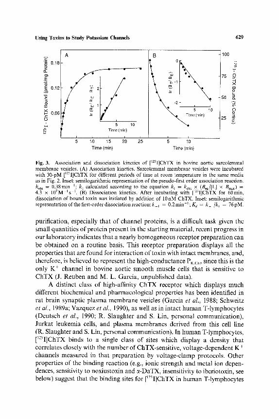
Fig. 3. Association and dissociation kinetics of [~2sI]ChTX in bovine aortic sarcolemmal membrane vesicles. (A) Association kinetics. Sarcolemmal membrane vesicles were incubated with 30-pM [~2sI]ChTX for different periods of time at room temperature in the same media as in Fig. 2. Inset: semilogarithmic representation of the pseudo-first order association reaction. kob s = 0.38min t; kl calculated according to the equation k I = ko~ ~ x (Beq/[L ] x Bmax) = 4.3 x 10⁷M-Is -t . (B) Dissociation kinetics. After incubating with [125I]ChTX for 60min, dissociation of bound toxin was initiated by addition of I0 nM ChTX. Inset: semilogarithmic representation of the first-order dissociation reaction: k ~ = 0.2min-~; Kd = k l/kl = 76pM.
purification, especially that of channel proteins, is a difficult task given the small quantities of protein present in the starting material, recent progress in our laboratory indicates that a nearly homogeneous receptor preparation can be obtained on a routine basis. This receptor preparation displays all the properties that are found for interaction of toxin with intact membranes, and, therefore, is believed to represent the high-conductance PK.ca, since this is the only K + channel in bovine aortic smooth muscle cells that is sensitive to ChTX (J. Reuben and M. L. Garcia, unpublished data).
A distinct class of high-affinity ChTX receptor which displays rauch different biochemical and pharmacological properties has been identified in rat brain synaptic plasma membrane vesicles (Garcia et al., 1988; Schweitz et al., 1989a; Vazquez et al., 1990), as well as in intact human T-lymphocytes (Deutsch et al., 1990; R. Slaughter and S. Lin, personal communication), Jurkat leukemia cells, and plasma membranes derived from this cell line (R. Slaughter and S. Lin, personal communication). In human T-lymphocytes, [I2sI]ChTX binds to a single class of sites which display a density that correlates closely with the number of ChTX-sensitive, voltage-dependent K + channels measured in that preparation by voltage-clamp protocols. Other properties of the binding reaction (e.g., ionic strength and metal ion depen- dences, sensitivity to noxiustoxin and «-DaTX, insensitivity to iberiotoxin, see below) suggest that the binding sites for [12sI]ChTX in human T-lymphocytes

630 Garcia et al.
o = o
£B X I.-
6
100
50
12 10 8 6 -log [Inhibitor] (M)
4 2
Fig.4 Effect of PK,C~ modulators on [~25I]ChTX binding to bovine aortic sarcolemmal mem- brane vesicles. Sarcolemmal membrane vesicles were incubated with 30 pM [~25I]ChTX in the absence or presence of increasing concentrations of either ChTX (e), IbTX (0), BaC12 (zx), or TEA (A) at room temperature until equilibrium was achieved. Inhibition of binding was assessed relative to an untreated controi.
represent the n type voltage-dependent K + channel. Similarly, the ChTX site in rat brain appears to be functionally associated wtih an RCK3 or RCK5- like protein.
Therefore, by using [~Œ»I]ChTX and appropriate vesicle systems, high- affinity binding sites coupled to single types of ChTX-sensitive K + channels can be studied in isolation. Although our laboratory has been successful in preparing radiolabelled toxin that is biologically active, reports from other laboratories have indicated problems in accomplishing this. In work by Luchessi et al. (1989), the monoiodotyrosine derivative of ChTX was prepared and analyzed for its ability to inhibit high-conductance PK,Ca of skeletal muscle t-tubule membranes incorporated into lipid bilayers. It was found that iodinated ChTX was ca. 103-fold weaker than native ChTX as a blocker of PK.ca, which could be accounted for by alterations in both the association and dissociation rate constant of modified toxin. Binding studies with [~25I]ChTX indicate the presence of a low-affinity site which does not correspond with PK,Cù monitored in bilayers. Another laboratory also reported failure in making a biologically active derivative of ChTX (Harvey et al., 1989), while yet another group has made iodinated toxin that has diminished potency in binding experiments (Schewitz et al., 1989a). The reason for such discrepancies is presently unclear.
Iberio toxin
The observation that ChTX is not a selective K + channel probe has spured research efforts to find a specific modulator of PKùca. Using [~25 I]ChTX
Using Toxins to Study Potassium Channels 631
binding to bovine aortic sarcolemma, a variety of scorpion venoms were tested for their ability to modulate the binding reaction. Several scorpion venoms were noted to possess inhibitory activity, and venom of the scorpion Buthus tamulus was further fractionated in order to isolate and identify its active component(s) (Galvez et al., 1990). When an extract of crude venom is loaded onto a MonoS cation-exchange FPLC column equilibrated at pHg.0, and then eluted with a linear gradient of NaC1, ChTX binding inhibitory activity is found in two well-separated fractions. Further frac- tionation of the less basic material by reversed-phase chromatography leads to the purification ofa homogeneous peptide that upon SDS-PAGE migrates as a 4.3-kDa protein. This peptide has been named iberiotoxin (IbTX), and determination of its primary sequence reveals 70% homology with ChTX (Galvez et al., 1990; Fig. 1). Like ChTX, the N-terminus of IbTX is blocked in the form of a pyroglutamic acid residue, and it also contains 6 Cys. IbTX, however, is a much less basic peptide than ChTX since it contains four more acidic amino acids. In single-channel recordings of PK,ca incorporated into planar lipid bilayers from either bovine aortic sarcolemmal membranes (O. McManus, unpublished data) or rabbit skeletal muscle t-tubules mem- branes (R. Latorre, unpublished data), IbTX reversibly inhibits channel activity by causing much longer silent periods than those observed in the presence of ChTX. However, IbTX is a noncompetitive inhibitor of [125 I]ChTX binding in smooth muscle, suggesting that it interacts at a unique site on the channel.
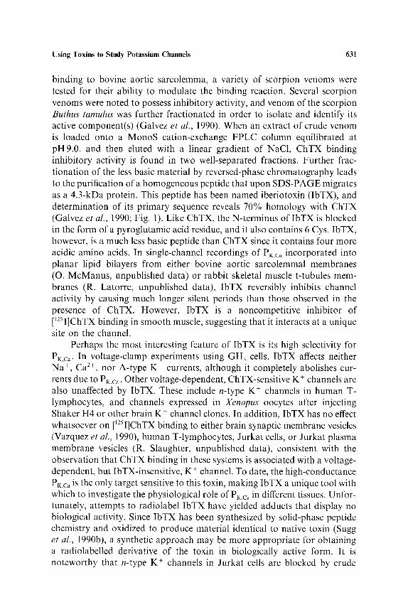
Perhaps the most interesting feature of IbTX is its high selectivity for PK.ca- In voltage-clamp experiments using GH 3 cells, IbTX affects neither Na +, Ca 2÷, nor A-type K ÷ currents, although it completely abolishes cur- rents due to PK,Ca. Other voltage-dependent, ChTX-sensitive K ÷ channels are also unaffected by IbTX. These include n-type K + channels in human T- lymphocytes, and channels expressed in Xenopus oocytes after injecting Shaker H4 or other brain K ÷ channel clones. In addition, IbTX has no effect whatsoever on [J25I]ChTX binding to either brain synaptic membrane vesicles (Vazquez et al., 1990), human T-lymphocytes, Jurkat cells, or Jurkat plasma membrane vesicles (R. Slaughter, unpublished data), consistent with the observation that ChTX binding in these systems is associated with a voltage- dependent, but IbTX-insensitive, K ÷ channel. To date, the high-conductance PK,ca is the only target sensitive to this toxin, making IbTX a unique tool with which to investigate the physiological role of PK.C~~ in different tissues. Unfor- tunately, attempts to radiolabel IbTX have yielded adducts that display no biological activity. Since IbTX has been synthesized by solid-phase peptide chemistry and oxidized to produce material identical to native toxin (Sugg et al., 1990b), a synthetic approach may be more appropriate for obtaining a radiolabelled derivative of the toxin in biologically active form. It is noteworthy that n-type K ÷ channels in Jurkat cells are blocked by crude

632 Garcia et aL
Buthus tamulus venom (Sands et al., 1989). However, the inhibitory activity of this K + channel is associated with the second more basic fraction from ion-exchange chromatography that blocks ChTX binding in smooth muscle. Further fractionation of this material by reversed-phase chromatography has led to the purification of a homogeneous peptide that produces inhibition of n-type K + channels in human T-lymphocytes (M. L. Garcia and C. Deutsch, unpublished data).
To determine those characteristics of the ChTX and IbTX structures which are responsible for each respective type of inhibitory activity, hybrid molecules were synthesized in which the N-terminus 19 amino acid residues of one peptide were joined to the C-terminus of the other, and vice versa (Sugg et al., 1990b). Evaluation by [125I]ChTX binding in vascular smooth muscle and brain, coupled with single-channel recording of PK.ca in aortic smooth muscle, indicates that IbTXl_19ChTX20_37 is ChTX-like, while ChTXH9 IbTX20_37 displays IbTX-like behavior. Thus, the c-terminus of each peptide dictates receptor specificity. Moreover, residues 21-24 of IbTX appear to be critical for an interaction at the IbTX site, while both N- and C-termini are involved in high-affinity binding to the ChTX receptor in brain and smooth muscle.
Noxiustoxin
A third peptide displaying significant primary sequence homology with both ChTX and IbTX is noxiustoxin (NxTX), a 39 amino acid peptide isolated from venom of the scorpion Centruroides noxius (Fig. 1). In squid axon preparations, it has been demonstrated that this toxin specifically depresses peak permeability due to K ÷ channels without affecting their voltage-dependent opening or closing kinetics (Carbone et al., 1982). This effect of toxin is selective for K ÷ currents, K ÷ channel block is fully revers- ible, and the reaction can be described by a bimolecular binding scheme at a single binding site with a K a of 390 nM. NxTX causes increased [3 H]GABA release and also blocks 86Rb+ efflux from perfused mouse brain synap- tosomes (Sitges et al., 1986), consistent with the finding that toxin affects K + permeability. Subsequently, it was shown that lower concentrations of NxTX block K ÷ currents in a voltage-independent manner, but, at concentrations above 1.5 #M, inhibition by NxTX becomes voltage-dependenL and could be partially removed by repetitive pulsing with strong depolarizations (Carbone et al., 1987). However, long repolarizations, and more negative holding potentials, favor channel block. Using axons from the squid Loligo vulgaris and a NxTX affinity column, four polypeptides have been isolated after membrane solubilization (Prestipino et al., 1989). Three of these polypeptides (molecular weights of 66, 160, and 220 kDa) were identified as NxTX binding
Using Toxins to Study Potassium Channels 633
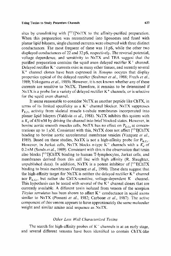
sites by crosslinking with [12»I]NxTX to the affinity-purified preparation. When this preparation was reconstituted into liposomes and fused with planar lipid bilayers, single channel currents were observed with three distinct conductances. The most frequent of these was 11 pS, while the other two displayed conductances of 22 and 32 pS, respectively. The reversal potential, voltage dependence, and sensitivity to NxTX and TEA suggest that the purified preparation contains the squid axon delayed rectifier K + channel. Delayed rectifier K + currents exist in many other tissues, and recently several K + channel clones have been expressed in Xenopus oocytes that display properties typical of the delayed rectifier (Stuhmer et al., 1988; Frech et al., 1989; Yokogama et al., 1989). However, it is not known whether any of these currents are sensitive to NxTX. Therefore, it remains to be determined if NxTX is a probe for a variety of delayed rectifier K + channels, or is selective for the squid axon channel.
It seems reasonable to consider NxTX as another peptide like ChTX, in terms of its limited specificity as a K + channel blocker. NxTX suppresses PK.Ca activity from skeletal muscle t-tubule membranes incorporated into planar lipid bilayers (Valdivia et al., 1988). NxTX inhibits this system with a K« of 450 nM by driving the channel into brief blocked states. However, in bovine aortic smooth muscles cells, NxTX has no effect on PK.ca at concen- trations up to 1 #M. Consistent with this, NxTX does not affect [~25I]ChTX binding to bovine aortic sarcolemmal membrane vesicles (Vazquez et al., 1989). Based on these studies, NxTX is not a high-affinity probe for PK.Ca. However, in Jurkat cells, NxTX blocks n-type K + channels with a Kd of 0.2 nM (Sands et aI., 1989). Consistent with this is the observation that toxin also blocks [~25I]ChTX binding to human T-lymphocytes, Jurkat cells, and membranes derived from this cell line with high affinity (R. Slaughter, unpublished data). In addition, NxTX is a potent inhibitor of [~25I]ChTX binding to brain membranes (Vazquez et al., 1990). These data suggest that the high-affinity target for NxTX is neither the delayed rectifier K + channel nor PK.Ca, but rather the ChTX-sensitive, voltage-dependent K + channel. This hypothesis can be tested with several of the K + channel clones that are currently available. A different toxin isolated from venom of the scorpion Tityius serrulatus has been shown to affect K + conductance in squid axons similar to NxTX (Possani et al., 1982; Carbone et al., 1987). The active component of this venom appears to have approximately the same molecular weight and similar amino acid sequence as NxTX.
Other Less WeH Characterized Toxins
The search for high-affinity probes of K + channels is at an early stage, and several different venoms have been identified to contain ChTX-like
634 Garcia e t al.
LeTX A-F-C-N-L-R-B-C-Q-L-S-C-R-S-L-G-L-L-G-K-C-I-G-D-K-C-E-C-V-K-HNH 2
Apamin C-N-C-K-A-P-E-T-A-L-C-A-R-R-C-Q-Q-H NH 2
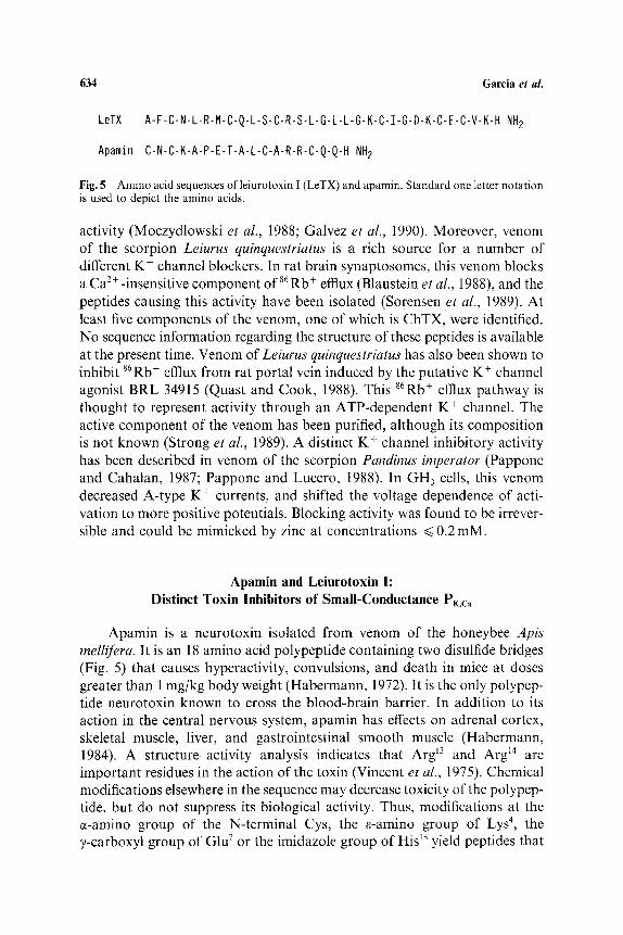
Fig. 5 Amino acid sequences of leiurotoxin I (LeTX) and apamin. Standard one letter notation is used to depict the amino acids.
activity (Moczydlowski et al., 1988; Galvez et al., 1990). Moreover, venom of the scorpion Leiurus quinquestriatus is a rich source for a number of different K + channel blockers. In rat brain synaptosomes, this venom blocks a Ca 2+ -insensitive component of 86 Rb + efflux (Blaustein et al., 1988), and the peptides causing this activity have been isolated (Sorensen et al., 1989). At least five components of the venom, one of which is ChTX, were identified. No sequence information regarding the structure of these peptides is available at the present time. Venom of Leiurus quinquestriatus has also been shown to inhibit 86Rb+ efflux from rat portal vein induced by the putative K + channel agonist BRL 34915 (Quast and Cook, 1988). This 86Rb+ efftux pathway is thought to represent activity through an ATP-dependent K + channel. The active component of the venom has been purified, although its composition is not known (Strong et al., 1989). A distinct K + channel inhibitory activity has been described in venom of the scorpion Pandinus imperator (Pappone and Cahalan, 1987; Pappone and Lucero, 1988). In GH 3 cells, this venom decreased A-type K + currents, and shifted the voltage dependence of acti- vation to more positive potentials. Blocking activity was found to be irrever- sible and could be mimicked by zinc at concentrations ~< 0.2 mM.
Apamin and Leiurotoxin I: Distinct Toxin lnhibitors of Small-Conductance PK,cù
Apamin is a neurotoxin isolated from venom of the honeybee Apis mellifera. It is an 18 amino acid polypeptide containing two disulfide bridges (Fig. 5) that causes hyperactivity, convulsions, and death in mice at doses greater than 1 mg/kg body weight (Habermann, 1972). It is the only polypep- tide neurotoxin known to cross the blood-brain barrier. In addition to its action in the central nervous system, apamin has effects on adrenal cortex, skeletal muscle, liver, and gastrointestinal smooth muscle (Habermann, 1984). A structure-activity analysis indicates that Arg ~3 and Arg ~4 are important residues in the action of the toxin (Vincent et al., 1975). Chemical modifications elsewhere in the sequence may decrease toxicity of the polypep- tide, but do not suppress its biological activity. Thus, modifications at the c~-amino group of the N-terminal Cys, the ~-amino group of Lys 4, the ~-carboxyl group of Glu vor the imidazole group of His ~8 yield peptides that

Using Toxins to Study Potassium Channels 635
are decreased in relative toxicity only 2- to 3-fold. The solid phase synthesis of apamin and related analogs has been successfully completed (Cosland and Merrifield, 1977; Granier et al., 1978). These studies confirm that the two adjacent arginine residues at positions 13 and 14 are essential for toxin activity. Apamin has not yet been crystallized. However, spectroscopic studies using NMR techniques indicate that the toxin is a highly ordered structure with an e-helical core of residues 9-15 and flanking regions consisting of B-type turns (Bystrov et al., 1980; Wemmer and Kallenbach, 1983).
The inhibitory action of apamin against PK.cù was suspected from exper- iments where this toxin was found to inhibit K + efflux from guinea pig hepatocytes (Banks et al., 1979) and taenia coli (Maas et al., 1980) induced by either ATP or noradrenaline. A direct demonstration of the action of apamin was obtained using voltage clamp techniques with neuroblastoma cells (Hugues et al., 1982a), cultured rat myotubes (Hugues et al., 1982b), and bullfrog sympathetic ganglion cells (Pennefather et al., 1985). At concen- trations of 100nM or less, apamin blocks a slow after-hyperpolarization following the firing of action potentials in these cells. Voltage clamp studies demonstrate that this phenomenon is due to inhibition of a Ca 2+-activated K + current which is insensitive to TEA, and which does not display a voltage dependence. High-conductance PK.ca is not a major contributor to the after- hyperpolarization in non-innervated skeletal muscle or neuronal cells because apamin does not block this cbannel, and a well-known inhibitor of these channels, TEA, does not reduce the after-hyperpolarization. The ident- ity of the after-hyperpolarization K + current in cultured rat muscle cells appears related to a small-conductance channel of 10-14pS (Blatz and Magleby, 1986). These channels are blocked by apamin, but not by 5mM external TEA, and are highly sensitive to Ca 2 + at negative membrane potentials associated with the after-hyperpolarization response.
Radiolabelled monoiodoapamin can be prepared at high specific activity by incorporation of iodine into the imidazole ring of His Is, the C-terminal residue of the toxin (Hugues et al., 1982c; Seagar et al., 1984). Iodinated apamin is about 2-fold less active than native toxin as measured in exper- iments where either the mean lethal dose was determined by intracisternal injection in mice, or the inhibition of neurotensin-induced relaxation of guinea-pig proximal colon was monitored (Hugues et al., 1982c). [125I] Apamin has been used to identify specific high-affinity receptors for this toxin in different tissues including brain synaptosomes (Hugues et al., 1982c), cultured rat embryonic neurons (Seagar et al., 1984), neuroblastoma cells (Hugues et al., 1982a), cultured rat muscle cell membranes (Hugues et al., 1982b), smooth muscle tissue (Hugues et al., 1982d), and hepatocytes (Cook and Haylett, 1985). In these studies, high-affinity binding sites were observed with K«'s in the range of 10-400pM. However, the maximum density of
636 Garcia et aL
binding sites is very low, with Bma x values between 1-30 frnol/rng protein. As a cornparison, rat brain synaptosornes contain 125 times less aparnin-sensitive K + channels than tetrodotoxin-sensitive Na + channels (Hugues et al., 1982c). Higher levels of apamin receptors have been found in undifferentiated pheochromocytorna cells (Schrnid-Antomarchi et al., 1986). These cells express receptor levels that are at least 50-fold greater than those found in other cell types, rnaking these cells a potential source for purification of the aparnin receptor.
The interaction of apamin with its receptor displays several interesting features. Dissociation of toxin frorn its binding site is slow. Dissociation rate constants of 1.5-4 x 10 4s-l have been determined in various nerve and muscle preparations (Hugues et al., 1981a, c). Nontoxic analogs of apamin do not block the interaction of apamin, and binding of [125 I] apamin is highly sensitive to cations. In rat neurons, binding increases 3-fold as extracellular K + is raised from 0.1 to 10rnM, at constant ionic strength (Seagar et al., 1984). This effect is due to an increase in the maximum number of binding sites, without any effect on Kd. In rat brain synaptosornes, concentrations of K + and Rb + between 10#M and 5mM are able to increase the binding of apamin to its receptor by a factor of 1.8, while other cations such as Li +, Na +, and guanidinium cornpletely inhibit binding in the 1-100mM range (Hugues et al., 1982c). In this preparation, the stimulatory effect of K + is due to a decrease in the Kj of toxin, rather than an effect on B,ùax. The effects of cations on [1~5I] apamin binding have been interpreted to indicate the existence of two different ion binding sites: one site is specific for K + and Rb +, but distinct frorn the apamin receptor, and binding to this site at a Kd of ca. 500 #M results in an increased affinity for toxin; the second site likely corresponds to the location where the two essential Arg residues of apamin bind since molecules which contain guanidinium groups (e.g., arniloride, neurotensin, which has two contiguous arginine residues like apamin, or guanidiurn itself) possess higher affinity for the apamin receptor than metal ions or other positively charged molecules (Hugues et al., 1982a, c). Interest- ingly, the apamin receptor in rat muscle is only expressed in noninnervated cells (Schrnid-Antomarchi et al., 1985). In normal adult rat muscle, binding of aparnin is not detectable, and the action potential in this preparation is insensitive to apamin. After denervation, binding sites for apamin, and an apamin-sensitive, after-hyperpolarization current, both appear in a time- dependent fashion.
Data concerning the structure of the apamin-sensitive channel have been obtained frorn studies with membrane vesicles. The rnolecular weight of the intact receptor was initially determined by employing radiation inactivation techniques. A molecular weight of 250 kDa has been estimated using lyophilized rat brain synaptic rnembranes (Schrnid-Antomarchi et al.,

Using Toxins to Study Potassium Channels 637
1984). However, with frozen rat brain membranes, the target size of the functional binding site appears to be 84-115 kDa (Seagar et al., 1986). Other attempts to identify the component(s) that constitute the apamin receptor have also yielded conflicting results. Covalent incorporation of [125I] apamin into rat brain membranes with the bifunctional crosslinking reagent disuccini- midyl suberate has identified a single polypeptide of 30 kDa (Hugues et al., 1982e; Schmid-Antomarchi et al., 1984). In another study, photoreactive apamin derivatives were prepared with aryl azide groups located at two different positions within the molecule (Seagar et al., 1986). The Lys 4 derivative labelled polypeptides of 33 and 22 kDa in both cultured neurons and synaptic plasma membranes, whereas the Cys 1 derivative labelled only an 86kDa polypeptide, or 86 and 59kDa components, in these two preparations, respectively. These results suggest that the apamin binding site may be located at the frontier between three or more putative K ÷ channel subunits which are not equally accessible to the different receptor-associated photo- probes. Further studies have shown that the 59 kDa component is found in cultured rat astrocytes, but not in neurons, indicating that glial cells may be responsible for the component of that molecular weight which was labelled in brain membranes (Seagar et al., 1987a). In a recent report, both photo- reactive derivatives of apamin and disuccinimidyl suberate crosslinking pro- cedures were used to identify apamin receptor components in rat brain membranes and pheochromocytoma cell membranes (Auguste et al., 1989). Results of these experiments indicate that a variety of polypeptides are labelled with the different apamin derivatives. One major component has a molecular mass of ca. 30 kDa, but other polypeptides of 45, 58, and 86 kDa were also identified. Data obtained with PC12 cell membranes are not identical to brain, consistent perhaps with differences observed in the affinity of apamin for its receptor in these two systems.
The apamin receptor has been solubilized in functional form from rat brain synaptic membranes using sodium cholate (Seagar et al., 1987b). [J25I] apamin binding was stimulated in the solubilized preparation by K + with an ED»0 of 0.6 mM, suggesting that the regulatory K + site is associated with the soluble complex. Analysis of the covalently labelled apamin receptor/ sodium cholate complex by density gradient centrifugation indicates an apparent sedimentation coefficient of 20S, which corresponds to a molecular weight of ca. 700 kDa, although as much as 50% of this mass may be con- tributed by detergent. Assuming a molecular weight of 250-350 kDa, a homo- geneous preparation should contain 3-4 nmol receptor/mg protein. Given the amount of receptor present in most apamin-sensitive tissues, isolation of the pure protein will require purification by a factor of 200,000 to 400,000-fold to reach homogeneity. To achieve this goal, use of affinity columns contain- ing either apamin or anti-receptor antibodies may be required.
638 Garcia et aL
Venom of the scorpion Leiurus quinquestriatus var. hebraeus, was found to contain an apamin binding inhibitory activity (Castle and Strong, 1986). It also blocks an apamin-sensitive K + channel in guinea pig hepatocytes (Abia et al., 1986; Castle and Strong, 1986). The agent responsible for this activity has been purified to homogeneity and shown to consist of a 3.4 kDa peptide (Fig. 5) with little structural homology to apamin (Chicchi et al., 1988). However, the peptide has some homology with other scorpion toxins such as ChTX, NxTX, and neurotoxin P2. This peptide, termed leiurotoxin I, completely inhibits [125I] apamin binding to rat brain synaptic plasma mem- branes with a Ki of 75 pM, which is 10-fold less potent than apamin, and acts by an apparently noncompetitive mechanism. Like apamin, leiurotoxin I blocks epinephrine-induced relaxation of guinea pig taenia coli, but has no effect on the rate, or force of contraction, of guinea pig atrial or rabbit portal vein preparations. In addition, leiurotoxin I blocks the apamin-sensitive, Ca 2+-dependent, after-hyperpolarization K + current in bullfrog ganglion B cells (Pennefather et al., 1989). Therefore, leiurotoxin I represents a new structural class of inhibitors directed against the small-conductance PK.ca-
Recently, another laboratory has reported the independent purification of leiurotoxin I, to which they assigned the name, scyllatoxin (Auguste et al., 1990). Primary amino acid sequence determination reveals that both toxins are identical. Scyllatoxin and two analogs have been synthesized by solid- phase methodologies. One of these analogs, in which a Tyr residue is intro- duced at position 2 to replace Phe, displays activity equivalent to native toxin, and has been used to obtain a radiolabelled derivative for binding studies. [~2~ i](Tyr 2) scyllatoxin binds to a single class of sites in rat brain membranes with a Kd of 80 pM. In these studies, apamin and scyllatoxin were shown to be competitive. Moreover, as found for the interaction of apamin, [1~5I] scylla- toxin binding is activated by low K + concentrations, inhibited at high K + levels, and blocked by Ca Œ+, Na +, and guanidinium ions. In addition, in crosslinking experiments with scyllatoxin, two polypeptides of 27 and 57 kDa were identified. Thus, these observations are particularly interesting since they indicate that there may be a common binding site for two toxins which share no sequence homology. It will be important to determine the three- dimensional structure of these toxins in order to rationalize these findings.
Dendrotoxins: a Family of Snake Toxins Directed against K + Channels
Neurotransmitter release is stimulated at peripheral (Harvey and Karlsson, 1982), and central (Docherty et al., 1983; Dolly et al., 1984) neurons by a group of homologous polypeptides derived from snake venoms. These neurotoxins, which are now known to modulate K + channel activity, have been identified in crude venom from several related species of snakes:

Using Toxins to Study Potassium Channels 639
~-DaTX
B-DaTX
7-DaTX
8-DaTX
Z-P-R-R[~ L ~ I ~ ] H - R - N - P ~ Y - D [ ~ - ~ A ~ N - Q [ K ] K [ ~
. . . . o xo ,[~<-~,,[G-~,, ~- .... ~-x--~ù~~i ~ .......
. . . . . . . . . . . . . . . IL-P]A-E-X]G-!~ . . . . . . . . Q-F-X[~]F . . . . . . . . .
«-DaTX ~--~ E-R~~-~ WIS-G-C-G-G-N ~ N-R-F ~K~ T-I ~E~ E-C-R-R-T-C I I []
B-DaTX - IC-L-P-F-L-F-S-G-C-G-G-N-A-N-R-F-Q-T-I-G-E-C-R .........
~-DaTX - IC-L-P-F-L-F-S-G-C-G-G - A - - F-Q-T-I-G-E-C-R I
~-DaTX Q~C-L-P-F~ S-G-C-G-G-N-A-N-R-F ~ T-I ~ E C-R'-R-T-C] V []
Fig. 6. Amino acid sequences of ~, Ô, ,/- and ~-dendrotoxin (DaTX). In this representation, ( x ) indicates an unidentified residue, while ( - ) indicates that no sequence is available in this region. Homologies are highlighted in boxes.
two types of the green mamba, Dendroaspis angusticeps and Dendroaspis viridus; and a species of black mamba, Dendrdaspis polylepsis (Harvey and Anderson, 1985). ~-Dendrotoxin, ~-DaTX, is a basic 7kDa polypeptide isolated from Dendroaspis angusticeps which contains 59 amino acids, and is structurally similar to toxin I, or DaTX, isolated from Dendroaspispolylepsis. In addition, three other polypeptide toxins have been isolated from Den- droaspis angusticeps venom. 6-DaTX contains 57 amino acids and has marked sequence homology with «-DaTX in the C-terminal region: 25 or 30 amino acids in this segment of the peptide are identical. There is also considerable homology in the N-terminal region: 8 of 15 amino acids between positions 12 and 26 are identical. /~ and 7-DaTX have been partially sequenced. Their sequences from position 30 through 52 are identical. In addition, their primary structures are very similar to that of «- and ~-DaTX in the C-terminal region where 20 of 23 amino acids are conserved in all four dendrotoxins (Fig. 6). Few structural similarities exist between these snake toxins and scorpion K + channel toxins, However, ~-DaTX shows consider- able homology with the smaller/~-subunit of/%bungarotoxin (/~-BTX), an inhibitory presynaptic neurotoxin from Bungarus multicinctus.
Dendrotoxins are highly neurotoxic when injected intracerebrally into rats (Mehraban et al., 1985). Electrophysiological records in hippocampal slices from guinea pig or rat demonstrate that e-DaTX is a potent convulsant, causing enhancement of cell excitability, with a consequent potentiation of transmitter release (Docherty et al., 1983; Dolly et al., 1984). The electro- physiological basis for the central convulsive action of e-DaTX was examined using intracellular recording of CAj neurons from hippocampal slices (Halliwell et aI., 1986). In the presence of 50nM e-DaTX, depolarizing currents elicited repetitive firing of Ca 2+-action potentials. Œ-DaTX caused

640 Garcia et aL
little change in the responses to hyperpolarizing current or in resting mem- brane potential. Voltage-clamp experiments indicate that the toxin does not directly affect a Ca 2+- activated K + conductance, a noninactivating, voltage- dependent K + conductance, inactivating or noninactivating Ca 2+ con- ductances, or an inwardly rectifying K + conductance. The only direct effect of «-DaTX was found to be a reduction of a transient, voltage-dependent K + current. More recently, Œ-DaTX has also been shown to inhibit a slowly inactivating K + channel present in various neurons (Penner et al., 1986; Stansfeld et al., 1986, 1987). Thus, electrophysiological data obtained from a variety of neuronal sources indicate that «-DaTX recognizes a family of K + channels that differ greatly in their inactivation kinetics.
This has been confirmed in experiments in which rat brain cDNA clones encoding different types of voltage-dependent K + channels were assayed for their sensitivity to «-DaTX after expression in Xenopus oocytes (Stuhmer et al., 1989). RCK1 and RCK5 clones expressing slowly inactivating K ÷ channels were much more sensitive to «-DaTX than the more rapidly inactivat- ing channels encoded for by RCK3 and RCK4. In different types of exper- iments where 86Rb+ efflux from synaptosomes was used to monitor the activity of K + channels, «- and õ-DaTX were found to preferentially block a component of flux corresponding to a rapidly inactivating K + channel (Blaustein et al., 1988; Sorensen et al., 1989). Moreover, using the same experimental system,/~- and 7-DaTX inhibit a noninactivating K + channel- mediated component of 86Rb+ flux with greater affinity. Given the high homology in primary amino acid sequences between the different types of voltage-dependent K + channels that have been cloned, it is not surprising that the dendrotoxin family can recognize a wide variety of these moieties, just as the scorpion K + channel toxins do. It is interesting, however, that PK,Ca does not appear to be sensitive to these toxins. To date, the only report concerning an effect of «-DaTX on these channels indicates that toxin may interact with an internal binding site on the channel to produce reversibly a long-lived, inwardly rectifying subconductance state (Lucchesi and Moczydlowski, 1990). In this study, toxin binding was shown to modify ion conduction through the pore without affecting the Ca 2+, or voltage depen- dence of gating.
Using biologically active [~zSI]DaTX, autoradiography was used to demonstrate that toxin binding sites reside in synapse-rich, white matter regions of the hippocampus, with lower levels in cell body layers (Halliwell et al., 1986). [~2SI]DaTX has also been used to identify receptor sites in different membrane preparations. [12sI] DaTX binds with high affinity to a homogeneous population of sites in rat brain synaptosomes (Mehraban et al., 1984; Black et al., 1986; Halliwell et al., 1986). In preparations of rat synaptic plasma membrane vesictes, both [12sI] «-DaTX and [L25I]/?-DaTX

Using Toxins to Study Potassium Channels 641
bind to a single class of receptors (Sorensen and Blaustein, 1989). Although B-DaTX inhibits the binding of [~25I]~-DaTX competitively, inhibition of [125 I]/%DaTX by ~-DaTX is more complex, indicating perhaps that/~-DaTX binds with similar affinities to two different sites, only one of which may be identical to the ~-DaTX receptor. Binding sites for [125I] 6-DaTX have also been identified in guinea pig synaptosomes (Muniz et al., 1990). ~-DaTX inhibits [125I] 6-DaTX binding in a noncompetitive fashion, suggesting that 6-DaTX interacts at a site distinct from the receptor for the former peptide.
Binding sites for ~-DaTX are also modulated by mast cell degranulating peptide (MCD) isolated from venom of the bee Apis mellifera, and by/~-BTX (Bidard et al., 1987; Black et al., 1988; Rehm et al., 1988; Rehm and Lazdunski, 1988a, b; Breeze and Dolly, 1989). Different binding sites for these agents seem to reside on the same molecular entity since each toxin can allosterically inhibit the binding of the other molecules. Such studies have identified two subtypes of«-DaTX binding proteins. In one subtype (60-70% of the total population), binding of both [125I] «-DaTX and [12»I] MCD is inhibited by/~-BTX with low affinity, while in the second subtype, /%BTX inhibits these interactions with high affinity (Rehm and Lazdunski, 1988a). The «-DaTX binding subtype, with low affinity for/%BTX, usually contains binding sites for [J25 I] MCD. The receptor for «-DaTX has been purified from a detergent extract of rat brain membranes (Rehm and Lazdunski, 1988b). Analysis of the purified material reveals the existence of three polypeptide bands of M.W. 76-80kDa, 38kDa, and 35kDa. Interestingly, [125I] MCD and [125I] ~-DaTX binding activities copurify together, and binding of these two agents in the purified preparation is inhibited by fl-BTX. Incubation of this material with the catalytic subunit of c-AMP-dependent protein kinase leads to phosphorylation of the 76-80kDa toxin-binding subunit (Rehm et al., 1989a). Reconstitution of this receptor complex into artificial bilayers results in the appearance of multiple conductance levels of unitary current. The majority of openings correspond to a channel that displays a conduc- tance of ca. 21 pS in 150mM symmetrical K + medium. However, up to seven different conductance levels have been described, suggesting that reconstitution of the «-DaTX receptor is associated with incorporation of multiple types of functional K + channels into the membrane. This observed heterogeneity in conductance levels probably indicates that the purified ~-DaTX receptor is a mixture of closely related K + channel structures, with similar, hut not identical, pharmacological properties (Rehm and Lazdunski, 1988a). Activity of the 21-pS K + channel «-DaTX receptor is enhanced by phosphorylation, and the phosphorylated channel is still sensitive to inhibition by toxin. Polyclonal antibodies have been raised against two synthetic peptides derived from different regions of the predicted amino acid sequence of MBK1, the mouse homolog of the Drosophila Shaker K + channel (Rehm et al., 1989b).
642 Garcia et al.
These antibodies cross-react with the toxin-binding subunit of the Œ-DaTX receptor from rat and bovine brain. This suggests that the ct-DaTX binding protein is the product of the mammalian homolog of the Shaker gene.
Conclusions
Over the last few years, combined molecular biology and biochemical investigations have led to the identification and characterization of several different types o f K + channel proteins. Peptidyl toxins isolated from a variety of organisms are currently being used to define the pharmacology of these channels. For those K + channels where molecular biological techniques have not yet identified the corresponding proteins, biochemical approaches will be needed to achieve this end. Therefore, it is significant that toxins which interact specifically with single classes of K + channels have been identified. It has only recently become apparent how many different types o f K + channel toxins can be found in a given venom source. These toxins surely will provide unique tools for future work, and will enable investigators to define the biochemical, pharmacological, and physiological properties of different K + channel proteins in target tissues of interest.
Acknowledgments
We would like to thank Ms. Patricia Hardiman for her help in the preparation of this manuscript. We would also like to thank Drs. R. Slaughter and S. Lin for sharing their data prior to publication.
References
Abia, A., Lobaton, C. D., Moreno, A., and Garcia-Sancho, J. (1986). Biochim. Biophys. Acta 856, 403-407.
Anderson, C., MacKinnon, R., Smith, C., and Miller, C. (1988). J. Gen. Physiot. 91,317-333. Auguste, P., Hugues, M., and Lazdunski, M. (1989). FEBS Lett. 248, 150-154. Auguste, P., Hugues, M., Grave, B., Gesquiere, J-C., Maes, P., Tartar, A., Romey, G., Schweitz,
H., and Lazdunski, M. (1990). J. Biol. Chem. 265, 4753-4759. Banks, B. E. C., Brown, C., Burgess, G. M., Burnstock, G., Claret, M., Cocks, T. M., and
Jenkinson, D. H. (1979). Nature (London) 282, 415-417. Baumann, A., Grupe, A., Ackermann, A., and Pongs, O. (1988). EMBO J. 7, 2457-2463. Bernardi, H., Fosset, M., and Lazdunski, M. (1988). Proc. Natl. Acad, Sci. USA 85, 9816-9820. Bidard, J.-N., Mourre, C., and Lazdunski, M. (1987). Biochem. Biophys. Res. Commun. 143,
383-389. Black, A. R., Breeze, A. L., Othman, I. B., and Dolly, J. O. (1986). Bioehem. J. 237, 397-404. Black, A. R., Donegan, C. M., Denny, B. J., and Dolly, J. O. (1988). Biochemistry 27, 6814-6820. Blatz, A. L., and Magleby, K. L. (1986). Nature (London) 323, 718-720. Blatz, A. L., and Magleby, K. L. (1987). Trends Neurosci. 10, 463-467.

Using Toxins to Study Potassium Channels 643
Blaustein, M. P., Bartschat, D. K., Benishin, C. G., Brown, W. E., Colby, K. A., Krueger, B. K., Schneider, M. J., and Sorensen, R. G. (1988). In Cellular and Molecular Basis of Synaptic Transmission (Zimmerman, H., ed.), Vol. H21, Springer-Verlag Berlin, Heidelberg, pp. 241-261.
Bono, M. R., Simon, V., and Rosemblatt, M. S. (1989). Cell Biochem. Funct. 7, 219 226. Breeze, A. L., and Dolly, J. O. (1989), Eur. J. Biochem. 178, 771-778. Butler, A., Wei, A., Baker, K., and Salkoff, L. (1989). Science 243, 943 947. Bystrov, V. F., Okhanõv, V. V., Miroshnikov, A. I., and Ovchinnikov, Y. A. (1980). FEBS Lett.
19, 113-117. Cahalan, M. D., Chandy, K. G., DeCoursey, T. E., and Gupta, S. (19853. J. Physiol. (London)
358, 197-237. Carbone, E., Wanke, E., Prestipino, G., Possani, L, D., and Maelicke, A. (1982). Nature
(London) 296, 90-91. Carbone, E., Prestipino, G., Spadavecchia, L., Franciolini, F., and Possani, L. D. (1987).
Pflügers Arch. Eur. J. Physiol. 408, 423-431. Castle, N. A., and Strong, P. N. (1986). FEBS Lett. 209, 117-121. Catterall, W. A. (1980). Annu. Rer. Pharmacol. Toxicol. 20, 15-43. Catterall, W. A. (1988). Science 242, 50-61. Chandy, K. G., DeCoursey, T. E., Cahalan, M. D., McLaughlin, C., and Gupta, S. (1984).
J. Exp. Med. 160, 369-385. Chandy, K. G., DeCoursey, T. E., Fischbach, M., Talal, N., Cahalan, M. D., and Gupta, S.
(19863. Science 233, 119%1200. Chicchi, G. G., Gimenez-Gallego, G., Ber, E., Garcia, M. L., Winquist, R., and Cascieri, M. A.
(1988). J. Biol. Chem. 263, 10192-10197. Christie, M. J., Adelman, J. P., Douglass, J., and North, R. A. (1989). Science 244, 221-224. Connor, J. A., and Stevens, C. F. (1971). J. Physiol. (London) 213, 31-53. Cook, N. S., and Haylett, D. G. (1985). J. Physiol. (London) 358, 373-394. Cosland, W. L., and Merrifield, R. B. (1977). Proc. Natl. Acad. Sci. USA 74, 2771-2775. DeCoursey, T. E., Chandy, K. G., Gupta, S., and Cahalan, M. D. (1987a). J. Gen. Physiol. 89,
379-404. DeCoursey, T. E., Chandy, K. G., Gupta, S., and Cahalan, M. D. (1987b). J. Gen. Physiol. 89,
405-420. Deutsch, C., Krause, D., and Lee, S. C. (1986). J. Physio[. (London) 372, 405-423. Deutsch, C., Price, M., Lee, S., Garcia, M., and King, V. F. (1990). Biophys. J. 57, l16a. Docherty, R. J., Dolly, J. O., Halliwell, J. V., and Othman, I. (1983). J. Physiol. (London) 336,
58-59P. Dolly, J. O., Halliwell, J. V., Black, J. D., Williams, R. S., Pelchen-Mathews, A., Breeze, A. L.,
Mehraban, F., Othman, 1. B., and Black, A. R. (1984). J. Physiol. (Paris') 79, 280-303. Frech, G. C., VanDongen, A.M.J., Schuster, G., Brown, A. M., and Joho, R. H. (1989). Nature
(London) 340, 642-645. Galvez, A., Gimenez-Gallego, G., Reuben, J. P., Roy-Contancin, L., Feigenbaum, P.,
Kaczorowski, G. J., and Garcia, M. L. (1990). J. Biol. Chem. 265, 11083-11090. Garcia, M. L., Vazquez, J., Gimenez-Gallego, G., Navia, M. A., Reuben, J. P., Katz, G. M., and
Kaczorowski, G. L. (1988). FASEB J. 2, A799. Gimenez-Gallego, G., Navia, M. A., Reuben, J. P., Katz, G. M., Kaczorowski, G. J., and
Garcia, M. L. (1988). Proc. Nall. Acad. Sci. USA 85, 3329-3333. Goh, J. W., and Pennefather, P. S. (19873. J. Physiol. (London) 394, 315-330. Granier, C., Pedroso-Muller, E., and Van Reitschoten, J. (1978). Eur. J. Biochem. 82, 293-299. Grinstein, S., Smith, J. D., and Miller, C. (1988). Biophys. J. 53, 148 (abstract). Guggino, S. E., Guggino, W. B., Green, N., and Sacktor, B. (1987). Am. J. Physiol. 252,
C128-C137. Habermann, E, (1972). Science 177, 314-322. Habermann, E. (1984). Pharmacol. Ther. 25, 255-270. Halliwell, J. V., Othman, I. B., Pelchen-Matthews, A., and Dolly, J. O. (1986). Proc. Natl. Acad.
Sci. USA 83, 493-497. Harvey, A. L., and Karlsson, E. (19823. Br. J. Pharmacol. 77, 153-161.
644 Garcia et aL
Harvey, A. L., and Anderson, A. J. (1985). Pharmaeol. Ther. 31, 35-55. Harvey, A. L., Marshall, D. L., DeAllie, F. A., and Strong, P. N. (1989). Bioehem. Biophys. Res.
Commun. 163, 394-397. Hermann, A., and Erxleben, C. (1987). J. Gen. Physiol. 90, 27-47. Hille, B. (1984). In lonic Channels in Excitable Membranes Sinauer Associates, Sunderland,
Massachusetts. Hodgkin, A. L., and Huxley, A. F. (1952). J. Physiol. (London) 117, 500-504. Hugues, M., Romey, G., Duval, D., Vincent, J. P., and Lazdunski, M. (1982a). Proe. Natl. Acad.
Sei. USA 79, 1308-1312. Hugues, M., Schmid, H., Romey, G., Duval, D., Frelin, C., and Lazdunski, M. (1982b). EMBO
J. 9, 1039-1042. Hugues, M., Duval, D., Kitabgi, P., Lazdunski, M., and Vincent, J. P. (1982c). J. Biol. Chem.
257, 2762-2769. Hugues, M., Duval, D., Schmid, H., Kitabgi, P., and Lazdunski, M. (1982d). Life Sei. 31,
437-443. Hugues, M., Schmid, H., and Lazdunski, M. (1982e). Biochem. Biophys. Res. Commun. 107,
1577-1582. Iverson, L. E., Tanouye, M. A., Lester, H. A., Davidson, N., and Rudy, B. (1988). Proe. Natl.
Acad. Sei. USA 85, 5723-5727. Jones, T. R., Charette, L., Garcia, M. L., and Kaczorowski, G. J. (1990), J. Pharmacol Exp.
Ther. 255, 697-706. Kamb, A., Tseng-Crank, J., and Tanouye, M. A. (1988). Neuron 1,421-430. Koren, G., Liman, E. R., Logothetis, D. E., Nadal-Ginard, B., and Hess, P. (1990). Neuron 2,
39-51. Latorre, R., Oberhauser, A., Labarca, P., and Alvarez, O. (1989). Annu. Rer. Physiol. 51,
385-399. Lee, S. C., Sabath, D. E., Deutsch, C., and Prystowsky, M. B. (1986). J. Cell. Biol. 102,
1200-1208. Lee, S. C., Price, M., Prytowsky, M. B., and Deutsch, C. (1988). Am. J. Physiõl. 254, C286-C296. Leonard, R. J., Karschin, A., Jayashree-Aiyar, S., Davidson, N., Tanouye, M. A., Thomas, L.,
Thomas, G., and Lester, H. A. (1989). Proc. Natl. Acad. Sci. USA 86, 7629-7633. Lewis, R. S., and Cahalan, M. D. (1988a). Trends Neurosci. 11, 214-218. Lewis, R. S., and Cahalan, M. D. (1988b). Seience 239, 771-775. Lucchesi, K., and Moczydlowski, E. (1990). Neuron 2, 141-148. Lucchesi, K., Ravindran, A., Young, H., and Moczydlowski, E. (1989). J. Membr. Biol. 109,
269-281. Maas, A. J. J., Den Hertog, A., Ras, R., and Van Den Akker, J. (1980). Eur. J. Pharmacol. 67,
265-274. MacKinnon, R., and Miller, C (1988). J. Gen. Physiol. 91, 335-349. MacKinnon, R., and Miller, C. (1989a). Biochemistry 28, 8087-8092. MacKinnon, R., and Miller, C. (1989b). Seience 243, 1382-1385. MacKinnon, R., Latorre, R., and Miller, C. (1989). Bioehemistry 28, 8092-8099. MacKinnon, R., Reinhart, P. H., and White, M. M. (1988). Neuron 1,997-1001. McKinnon, D. (1989). J. Biol. Chem. 14, 8230-8236. Mehraban, F., Breeze, A. L., and Dolly, J. O. (1984). FEBS Lett. 174, 116-122. Mehraban, F., Black, A. R., Breeze, A. L., Green, D. G., and Dolly, J. O. (1985). Biochem. Soc.
Trans~ 13, 507-508. Milter, C. (1988). Neuron 1, 1003-1006. Miller, C., Moczydlowski, E., Latorre, R., and Phillips, M. (1985). Nature (London) 313,
316-318. Moczydlowski, E., Lucchesi, K., and Ravindran, A. (1988). J. Membr. Biol. 105, 95-111. Muniz, Z. M., Diniz, C. R., and Dolly, J. O. (1990). J. Neurochem. 54, 343-346. Noda, M., Ikeda, T., Kayano, T., Suzuki, H., Takeshima, H., Takasashi, H., and Numa, S.
(1986) Nature (London) 320, 188-192. Papazian, D. M., Schwarz, T. L., Tempel, B. L., Jan, Y. N., and Jan, L. Y. (1987). Seienee 237,
749-753.
Using Toxins to Study Potassium Channels 645
Pappone, P. A., and Cahalan, M. D. (1987). J. Neurosci. 7, 3305-3330. Pappone, P. A., and Lucero, M. T. (1988). J. Gen. Physiol. 91, 817-833. Pennefather, P., Lancaster, B., Adams, P. R., and Nicoll, R. A. (1985). Proc. Natl. Acad. Sci.
USA 82, 3040-3044. Pennefather, P. S., Kelley, M. E. M., Goh, J. W., Chicchi, G. G., Cascieri, M. A., Garcia, M. L.,
and Kaczorowski, G. J. (1989). Canadian Federation of Biological Societies, Proceedings of the 32nd Annual Meeting, p. 95.
Penner, R., Petersen, M., Pierau, F. K., and Dreyer, F. (1986). Pflügers Arch. 407, 365-369. Petersen, O. H., and Maruyama, Y. (1984). Nature (London) 307, 693-696. Pongs, O., Kecskemethy, Muller, R., Krah-Jentgens, I., Baumann, A., Klitz, H. H., Canal, I.,
Llamazares, S., and Ferrus, A. (1988). EMBO J. 7, 1087-1096. Possani, L. D., Martin, B. M., and Svendensen, 1. (1982). Carlsberg Res. Commun. 47, 285-289. Prestipino, G., Valdivia, H. H., Lievano, Darszon, A., Ramirez, A. N., and Possani, L. D.
(1989). FEBS Lett. 250, 570-574. Price, M., Lee, S., Krause, D., and Deutsch, C. (1988). J. Cell. Biol. 107, 80 (abstract). Price, M., Lee, S. C., and Deutsch, C. (1989). Proc. Natl. Acad. Sci. USA 86, 10171-10175. Quast, U., and Cook, N. S. (1988). Life Sci. 42, 805-810. Rehm, H., and Lazdunski, M. (1988a). Biochem. Biophys. Res. Commun. 153, 231-240. Rehm, H., and Lazdunski, M. (1988b). Proc. Natl. Acad. Sci. USA 85, 4919-4923. Rehm, H., Bidard, J.-N., Schweitz, H., and Lazdunski, M. (1988). Bioehemistry 27, 1827-1832. Rehm, H., Pelzer, S., Cochet, C., Chambaz, E., Tempel, B. L., Trautwein, W., Pelzer, D., and
Lazdunski, M. (1989a). Biochemistry 28, 6455-6460. Rehm, H., Newitt, R. A., and Tempel, B. L. (1989b). FEBS Lett. 249, 224-228. Reinhart, P. H., Chung, S., and Levitan, I. B. (1989). Neuron 2, 1031-1041. Rogawski, M. A. (1985). Trends Neurosci. 8, 214-219. Sands, S. B., Lewis, R. S, and Cahalan, M. D. (1989). J. Gen. Physiol. 93, 1061-1074. Schmid-Antomarchi, H., Hugues, M., Norman, R., Ellory, C., Borsotto, M., and Lazdunski, M.
(1984). Eur. J. Biochem. 142, 1-6. Schmid-Antomarchi, H., Renaud, J. F., Romey, G., Hugues, M., Schmid, A., and Lazdunski, M.
(1985). Proe. Natl. Aead. Sei. USA 82, 2188-2191. Schmid-Antomarchi, H., Hugues, M., and Lazdunski, M. (1986). J. Biol. Chem. 261, 8633-8637. Schneider, M. J., Rogowski, R. S., Krueger, B. K., and Blaustein, M. P. (1989). FEBSLett. 250,
433-436. Schwarz, T. L., Tempel, B. L., Papazian, D. M., Jan, Y. N., and Jan, L. Y. (1988). Nature
(London) 331, 137-142. Schwarz, W., and Passow, H. (1983). Annu. Rer. Physiol. 5, 359-374. Schweitz, H., Bidard, J.-N., Maes, P., and Lazdunski, M. (1989a). Biochemistry 28, 9708-9714. Schweitz, H., Stansfeld, C. E., Bidard, J.-N., Fagni, L., Maes, P., and Lazdunski, M. (1989b).
FEBS Lett. 250, 519-522. Seagar, M. J., Granier, C., and Couraud, F. (1984). J. Biol. Chem. 259, 1491-1495. Seagar, M. J., Labbe-Jullie, C., Granier, C., Goll, A., Glossmann, H., Van Rietschoten, J., and
Couraud, F. (1986). Biochemistry 25, 4051-4057. Seagar, M. J., Deprez, P., Martin-Moutot, N., and Couraud, F. (1987a). Brain Res. 411,
226-230. Seagar, M. J., Marqueze, B., and Couraud, F. (1987b). J. Neurosci. 7, 565-570. Sitges, M., Possani, L. D., and Bayon, A. (1986). J. Neurosci. 6, 1570-1574. Slaughter, R. S., Welton, A. F., and Morgan, D. W. (1987). Biochim. Biophys. Aeta 904, 92-104. Slaughter, R. S., Kaczorowski, G. J., and Garcia, M. L. (1988). J. Cell. Biol. 107, 143a. Slaughter, R. S., Shevell, J. L., Felix, J. P., Garcia, M. L., and Kaczorowski, G. J. (1989).
Bioehemistry 28, 3995-4002. Smith, C., Phillips, M., and Miller, C. (1986). J. Biol. Chem. 261, 14607-14613. Sorensen, R. G., and Blaustein, M. P. (1989). Mol. Pharmacol. 36, 689-698. Sorensen, R. G., Schneider, M. J., and Blaustein, M. P. (1988). Biophys. J. 55, 550a. Sorensen, R. G., Schneider, M. J., Rogowski, R. S., and Blaustein, M. P. (1989). In Potassium
Channels: Basie Funetion and Therapeutic Aspects (Colatsky, T. J., ed.), Alan R. Liss, Inc., New York, pp. 279-301.

646 Garcia et aL
Stansfeld, C. E., Marsh, S. J., Halliwell, J. V., and Brown, D. A. (1986). Neurosci. Lett. 64, 299-304.
Stansfeld, C., Marsh, S., Parcej, D., Dolly, J. O., and Brown, D. (1987). Neuroscience 23, 893-902.
Strong, P. N., Weit, S. W., Beech, D. J., Heistand, P., and Kocher, H. P. (1989). Br. J. Pharmacol. 98, 817-826.
Stuhmer, W., Stocker, M., Sakmann, B., Seeburg, P., Baumann, A., Grupe, A., and Pongs, O. (1988). FEBS Lett. 242, 199-206.
Stuhmer, W., Ruppersberg, J. P., Schrotet, K. H., Sakmann, B., Stocker, M., Giese, K. P., Perschke, A., Baumann, A., and Pongs, O. (1989). EMBO J. 8, 3235-3244.
Sugg, E. E., Garcia, M. L., Reuben, J. P., Patchett, A. A., and Kaczorowski, G. J. (1990a). J. Biol. Chem. 265, 18745-18748.
Sugg, E. E., Garcia, M. L., Johnson, B. A., Kaczorowski, G. J., Patchett, A. A., and Reuben, J. P. (1990b) in Peptides: Chemistry, Structure and Biology (Rivier, J. E., and Marshall, G. R., eds.), Escom Sci. Pub., Leiden, pp. 1069-1070.
Tanabe, T., Takeshima, H., Mikami, A., Flockerzi, V., Takahashi, H., Kangawa, K., Kojima, M., Matsuo, H., Hirose, T., and Numa, S. (1987). Nature (London) 328, 313- 318.
Tempel, B. L., Papazian, D. M., Schwarz, T. L., Jan, Y. N., and Jan, L. Y. (1987). Science 237, 770-775.
Tempel, B. L., Jan, Y. N., and Jan, L. Y. (1988). Nature (London) 332, 837-389. Timpe, L. C., Schwarz, T. L., Tempel, B. L., Papazian, D. M., Jan, Y. N., and Jan, L. Y. (1988).
Nature (London) 331, 143-145. Valdivia, H. H., Smith, J. S., Martin, B. M., Coronado, R., and Possani, L. D. (1988). FEBS
Lett. 2, 280-284. Vazquez, J., Feigenbaum, P., Kaczorowski, G. J., and Garcia, M. L. (1988). J. Cell. Biol. 107,
143a. Vazquez, J., Feigenbaum, P., Katz, G., King, V. F., Reuben, J. P., Roy-Contancin, L.,
Slaughter, R. S., Kaczorowski, G. J., and Garcia, M. L. (1989). J. Biol. Chem. 264, 20902-20909.
Vazquez, J., Feigenbaum, P., King, V. F. Kaczorowski, G. J., and Garcia, M. L. (1990). J. Biol. Chem. 265, 15564-15571.
Villarroel, A., Alvarez, O., Oberhauser, A., and Latorre, R. (1988). Pflügers Arch. Eur. J. Physiol. 413, 118-126.
Vincent, J. P., Schweitz, H., and Lazdunski, M. (1975). Biochemistry 14, 2521-2525. Wemmer, D., and Kallenbach, N. R. (1983). Biochemistry 22, 1901-1906. Winquist, R. J., Heaney, L. A., Wallace, A. A., Baskin, E. P., Garcia, M. L., and Kaczorowski,
G. (1989). J. Pharmacol. Exp. Ther. 248, 149-156. Wolff, D., Cecchi, X., Spalvins, A., and Canessa, M. (1988). J. Membr. Biol. 106, 243-252. Yokoyama, S., Imoto, K., Kawamura, T., Higashida, H., Iwabe, N., Miyata, T., and Numa, S.
(1989). FEBS Lett. 259, 37-42. Zagotta, W. N., Hoshi, T., and Aldrich, R. W. (1989). Proc. Natl. Acad. Sci. USA 86, 7243
7247.