Acidosis-induced relaxation of human internal mammary artery is due to activation of ATP-sensitive...
7
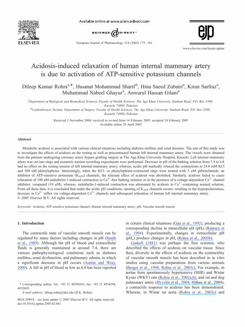
Acidosis-induced relaxation of human internal mammary artery is due to activation of ATP-sensitive potassium channels Dileep Kumar Rohra a, * , Hasanat Mohammad Sharif b , Hina Saeed Zubairi a , Kiran Sarfraz a , Muhammad Nabeel Ghayur a , Anwarul Hassan Gilani a a Department of Biological and Biomedical Sciences, Faculty of Health Sciences, The Aga Khan University, Stadium Road, P.O. Box 3500, Karachi 74800, Pakistan b Cardiothoracic Section, Department of Surgery, Faculty of Health Sciences, The Aga Khan University, Stadium Road, P.O. Box 3500, Karachi 74800, Pakistan Received 1 November 2004; received in revised form 14 February 2005; accepted 24 February 2005 Available online 29 April 2005 Abstract Metabolic acidosis is associated with various clinical situations including diabetes mellitus and renal diseases. The aim of this study was to investigate the effects of acidosis on the resting as well as precontracted human left internal mammary artery. The vessels were obtained from the patients undergoing coronary artery bypass grafting surgery at The Aga Khan University Hospital, Karachi. Left internal mammary artery was cut into rings and isometric tension recording experiments were performed. Decrease in pH of the bathing solution from 7.4 to 6.8 had no effect on the resting tension of left internal mammary artery, whereas, acidic pH markedly relaxed the contractions to 24.8 mM KCl and 300 nM phenylephrine. Interestingly, when the KCl- or phenylephrine-contracted rings were treated with 3 AM glibenclamide; an inhibitor of ATP-sensitive potassium (K ATP ) channels, the relaxant effect of acidosis was abolished. Similarly, acidosis failed to cause relaxation of 100 nM endothelin-1-induced contraction in Ca 2+ -free bathing solution or in the presence of a voltage-dependent Ca 2+ channel inhibitor, verapamil (10 AM), whereas, endothelin-1-induced contraction was attenuated by acidosis in Ca 2+ -containing normal solution. From all these data, it is concluded that under the acidic pH conditions, opening of K ATP channels occurs; resulting in the hyperpolarization, decrease in Ca 2+ influx via voltage-dependent Ca 2+ channels and subsequent relaxation of human left internal mammary artery. D 2005 Elsevier B.V. All rights reserved. Keywords: Acidosis; ATP-sensitive potassium channel; Human internal mammary artery; pH; Vascular smooth muscle 1. Introduction The contractile state of vascular smooth muscle can be regulated by many factors including changes in pH (Smith et al., 1989). Although the pH of blood and extracellular fluids is generally maintained at around 7.4, there are various pathophysiological conditions such as diabetes mellitus, renal dysfunction, and pulmonary edema, in which a significant decrease in pH occurs (Austin and Wray, 2000). A fall in pH of blood as low as 6.8 has been reported in certain clinical situations (Gan et al., 1992), producing a corresponding decline in intracellular pH (pH i )(Ramsey et al., 1994). Experimentally, changes in extracellular pH (pH o ) produce changes in pH i (Rohra et al., 2003b). Gaskell (1881) was perhaps the first scientist, who described the effects of acidosis on vascular tissue. Since then, diversity in the effects of acidosis on the contractility of vascular smooth muscle has been described in in vitro studies using vascular preparations from various animals (Berger et al., 1998; Rohra et al., 2003c). For example, in aortas from spontaneously hypertensive (SHR) and Wistar Kyoto (WKY) rats (Rohra et al., 2002a,b), and rat and dog pulmonary artery (Hyvelin et al., 2004; Hfhne et al., 2004), a contractile response to acidosis has been demonstrated. Whereas, in Wistar rat aorta (Rohra et al., 2003c) and 0014-2999/$ - see front matter D 2005 Elsevier B.V. All rights reserved. doi:10.1016/j.ejphar.2005.02.041 T Corresponding author. Tel.: +92 21 48594563; fax: +92 21 4934294, 4932095. E-mail address: [email protected] (D.K. Rohra). European Journal of Pharmacology 514 (2005) 175 – 181 www.elsevier.com/locate/ejphar
-
Upload
independent -
Category
Documents
-
view
0 -
download
0
Transcript of Acidosis-induced relaxation of human internal mammary artery is due to activation of ATP-sensitive...
www.elsevier.com/locate/ejphar
European Journal of Pharmaco
Acidosis-induced relaxation of human internal mammary artery
is due to activation of ATP-sensitive potassium channels
Dileep Kumar Rohraa,*, Hasanat Mohammad Sharif b, Hina Saeed Zubairia, Kiran Sarfraza,
Muhammad Nabeel Ghayura, Anwarul Hassan Gilania
aDepartment of Biological and Biomedical Sciences, Faculty of Health Sciences, The Aga Khan University, Stadium Road, P.O. Box 3500,
Karachi 74800, PakistanbCardiothoracic Section, Department of Surgery, Faculty of Health Sciences, The Aga Khan University, Stadium Road, P.O. Box 3500,
Karachi 74800, Pakistan
Received 1 November 2004; received in revised form 14 February 2005; accepted 24 February 2005
Available online 29 April 2005
Abstract
Metabolic acidosis is associated with various clinical situations including diabetes mellitus and renal diseases. The aim of this study was
to investigate the effects of acidosis on the resting as well as precontracted human left internal mammary artery. The vessels were obtained
from the patients undergoing coronary artery bypass grafting surgery at The Aga Khan University Hospital, Karachi. Left internal mammary
artery was cut into rings and isometric tension recording experiments were performed. Decrease in pH of the bathing solution from 7.4 to 6.8
had no effect on the resting tension of left internal mammary artery, whereas, acidic pH markedly relaxed the contractions to 24.8 mM KCl
and 300 nM phenylephrine. Interestingly, when the KCl- or phenylephrine-contracted rings were treated with 3 AM glibenclamide; an
inhibitor of ATP-sensitive potassium (KATP) channels, the relaxant effect of acidosis was abolished. Similarly, acidosis failed to cause
relaxation of 100 nM endothelin-1-induced contraction in Ca2+-free bathing solution or in the presence of a voltage-dependent Ca2+ channel
inhibitor, verapamil (10 AM), whereas, endothelin-1-induced contraction was attenuated by acidosis in Ca2+-containing normal solution.
From all these data, it is concluded that under the acidic pH conditions, opening of KATP channels occurs; resulting in the hyperpolarization,
decrease in Ca2+ influx via voltage-dependent Ca2+ channels and subsequent relaxation of human left internal mammary artery.
D 2005 Elsevier B.V. All rights reserved.
Keywords: Acidosis; ATP-sensitive potassium channel; Human internal mammary artery; pH; Vascular smooth muscle
1. Introduction
The contractile state of vascular smooth muscle can be
regulated by many factors including changes in pH (Smith
et al., 1989). Although the pH of blood and extracellular
fluids is generally maintained at around 7.4, there are
various pathophysiological conditions such as diabetes
mellitus, renal dysfunction, and pulmonary edema, in which
a significant decrease in pH occurs (Austin and Wray,
2000). A fall in pH of blood as low as 6.8 has been reported
0014-2999/$ - see front matter D 2005 Elsevier B.V. All rights reserved.
doi:10.1016/j.ejphar.2005.02.041
T Corresponding author. Tel.: +92 21 48594563; fax: +92 21 4934294,
4932095.
E-mail address: [email protected] (D.K. Rohra).
in certain clinical situations (Gan et al., 1992), producing a
corresponding decline in intracellular pH (pHi) (Ramsey et
al., 1994). Experimentally, changes in extracellular pH
(pHo) produce changes in pHi (Rohra et al., 2003b).
Gaskell (1881) was perhaps the first scientist, who
described the effects of acidosis on vascular tissue. Since
then, diversity in the effects of acidosis on the contractility
of vascular smooth muscle has been described in in vitro
studies using vascular preparations from various animals
(Berger et al., 1998; Rohra et al., 2003c). For example, in
aortas from spontaneously hypertensive (SHR) and Wistar
Kyoto (WKY) rats (Rohra et al., 2002a,b), and rat and dog
pulmonary artery (Hyvelin et al., 2004; Hfhne et al., 2004),a contractile response to acidosis has been demonstrated.
Whereas, in Wistar rat aorta (Rohra et al., 2003c) and
logy 514 (2005) 175–181

D.K. Rohra et al. / European Journal of Pharmacology 514 (2005) 175–181176
cerebral artery (Peng et al., 1998) and porcine coronary
arterioles (Ishizaka et al., 1999), acidosis induces a
relaxation. Acidosis has been suggested to produce vascular
relaxation by utilizing various mechanisms such as activa-
tion of ATP-sensitive potassium (KATP) channels (Ishizaka
et al., 1999), production of cAMP (Leffler et al., 1999), and
a direct inhibition of L-type Ca2+ channels in vascular
smooth muscle cells (Smirnov et al., 2000). Different
subtypes of K+ channels have been described in vascular
smooth muscle cells, but activation of KATP channels
appears to have a major role in relaxation of various types
of vessels in response to several stimuli including acidosis
(for review; see Brayden, 2002).
Experimentally, acidosis can be induced by producing
hypercapnia (Peng et al., 1998; Bayerle-Eder et al., 2000) or
simply making the extracellular environment acidic (Rohra
et al., 2003a). There are a few studies to document the
hypercapnia-induced changes in the diameter of the human
vessels, but these were carried out in vivo (Bayerle-Eder et
al., 2000). Although the effects of acidosis on the
cardiovascular system are surmised to be depressant, no
such direct study has been carried out to evaluate the impact
of extracellular acidic environment on the vascular reactivity
of human vessels. Thus the aim of the present study was to
investigate the effects of acidosis on the contractility of
human internal mammary artery. To accomplish this aim,
isolated vessel techniques were used to eliminate the
confounding influences from neurohumoral and local
control mechanisms. The findings of the present study for
the first time show that extracellular acidosis induces a
relaxation of human internal mammary artery which is
associated with the activation of KATP channels.
2. Materials and methods
This study was carried out in accordance with the World
Medical Association Declaration of Helsinki regarding the
Ethical Principles for Medical Research involving human
subjects and was reviewed and approved by the Institutional
Ethical Review Committee.
2.1. Tissue preparation
The human left internal mammary artery was obtained
from the Cardiothoracic Section of the Department of
Surgery, The Aga Khan University Hospital. The arteries
of patients, who underwent coronary artery bypass
grafting operation, were used in the study. The standard
bypass operation involves using the left internal mammary
artery as one of the bypass conduits. The artery is
harvested in almost all operations and a portion of it is
usually discarded after tailoring it to the appropriate
length for the recipient (left anterior descending) artery.
This normally discarded distal end of the human left
internal mammary artery was used in the isometric tension
recording experiments. Informed consent prior to surgery
was obtained from all the patients whose left internal
mammary artery was used for experiments. The artery
was quickly immersed in an ice-cold HEPES-buffered
physiological salt solution (PSS) and was cleaned of
adherent connective tissue. Rings of approximately 3 mm
width were made from the artery. The endothelium was
removed by gently rubbing the endothelial surface with
cotton pellets in order to obviate the involvement of the
endothelium and to examine the function of vascular
smooth muscle alone. The lack of endothelium was
confirmed by failure of carbachol (1 AM) to cause
relaxation of phenylephrine (1 AM)-induced contraction.
2.2. Experimental protocol
All experiments were carried out at 37 8C. The rings
were fixed between two hooks and suspended in a 5 ml
organ bath, containing well aerated (95% O2+5% CO2) PSS
of pH 7.4. The hook anchoring the upper end of the ring was
connected to a force displacement transducer (Transbridge
4M, WPI Ltd. UK). The tissues were adjusted to a preloaded
resting tension of 1 g and equilibrated for at least 1 h. The
arterial rings were contracted 3 times with 64.8 mM KCl to
acclimatize the tissue and to verify that the artery is viable.
The last contractile response to high KCl was the
reproduction of the second one and later it was employed
as a standard and other contractions were normalised to it.
The pH of the bathing solution was changed from control
value of 7.4 to 6.8 by addition of HCl and the pH inside the
bath was measured with the help of a micro pH electrode
(Model PHR 146, Lazar Research Lab. Inc. CA, USA)
attached with a pH meter (Model 611, Orion Research
Instruments, Cambridge, USA). In experiments investigat-
ing the effect of acidic pH on the contractile state of the left
internal mammary artery, the concentrations of 300 nM for
phenylephrine and 24.8 mM for KCl were selected in order
to obtain a contractile response that should be 70–90% of
the 64.8 mM KCl-induced contraction. The tissues showing
b70% or N90% of the 64.8 mM KCl-induced contraction
were discarded and not included in the study. The effect of
acidosis on the vessels in Ca2+-free conditions was
investigated in the following protocol. Bathing PSS was
changed by Ca2+-free solution containing 0.5 mM EGTA
[ethyleneglycol-bis(2-aminoethyl ether)-N,N,NV,NV-tetraace-tic acid]. After 10 min, etdothelin-1 was added and when
maximum contraction was achieved, the pH of bathing
solution was changed from 7.4 to 6.8.
2.3. Solutions
The composition of the PSS was (in mM): NaCl 120,
KCl 4.8, MgSO4 1.3, CaCl2 1.2, NaHCO3 25.2, glucose 5.8,
KH2PO4 1.2 and HEPES 20. 64.8 mM KCl was made by
replacing 60 mM NaCl of PSS with equimolar KCl, while
Ca2+-free PSS was prepared by omitting Ca2+ from normal

D.K. Rohra et al. / European Journal of Pharmacology 514 (2005) 175–181 177
PSS and adding 0.5 mM EGTA in order to chelate any
residual Ca2+.
2.4. Materials
All chemicals were purchased from Sigma-Aldrich
Chemical Co. (St. Louis, MO, USA). Phenylephrine,
endothelin-1 and verapamil were dissolved in water, while
glibenclamide was dissolved in dimethyl sulphoxide.
2.5. Statistics
Data from tension recording experiments are expressed
as meansFS.E.M. The relaxation of phenylephrine- or KCl-
induced contractions caused by acidosis has been expressed
as the % of the contraction observed at pH 7.4. n represents
the number of experiments performed. Data were analysed
by Student’s t-test and the differences were considered
statistically significant at P b0.05.
Fig. 1. Representative recordings showing the effects of acidosis on the
resting tension as well as on the phenylephrine- and KCl-contracted isolated
human internal mammary artery. (A) Arterial rings were precontracted with
64.8 mM KCl 3 times and after washout, the pH of the bathing solution was
changed from 7.4 to 6.8 by adding HCl to it. Arterial rings were contracted
with either 24.8 mM KCl (B) or 300 nM phenylephrine (C) at pH 7.4. After
the peak response was achieved, pH of the bathing solution was decreased
to 6.8 by adding HCl. Recordings are the representative of 4–5 independent
experiments from different vessels.
3. Results
3.1. Effect of acidosis on the resting tension and the
contractile state
Effect of decreasing pH extracellularly on the resting
state of the isolated human left internal mammary artery
rings was first characterized. Changing pH from 7.4 to 6.8
by adding HCl to the bathing solution failed to produce any
effect on the resting tension of the vessel (Fig. 1A).
In order to assess the effects of acidic pH on the
precontracted artery, left internal mammary artery rings
were contracted with sub-maximal concentrations of either
KCl (24.8 mM) or phenylephrine (300 nM). When the
contractile response reached a steady level, the pH of the
bathing solution was lowered from 7.4 to 6.8. Acidosis
caused a relaxation of both KCl (Fig. 1B) and phenylephrine
(Fig. 1C) contracted arterial rings. However, the extent of
acidosis-induced relaxation was remarkably greater in
phenylephrine-induced contraction compared to the one
caused by KCl. As quantified in Fig. 2, the magnitude of
24.8 mM KCl-induced contraction at pH 6.8 was
76.0F1.3% (n =4) of the contraction of the same stimulus
but at pH 7.4. Whereas at similar acidic pH, phenylephrine-
induced contraction was 39.2F9.8% (n =4) of the one at
pH 7.4. The difference between the magnitudes of relaxa-
tion of both types of contractions was statistically significant
(P b0.01).
3.2. Effect of acidosis in the presence of glibenclamide
To evaluate the role of KATP channels, experiments were
performed using a selective KATP channel inhibitor, gliben-
clamide (Nelson and Quayle, 1995; Teramoto et al., 2000;
Rosenblum, 2003). The KCl- or phenylephrine-precon-
tracted arterial rings were treated with 3 uM glibenclamide,
and after 10 min, the pH of the bathing solution was lowered
from 7.4 to 6.8. Interestingly, the relaxant effect of acidosis
was observed to be nearly abolished in the presence of
glibenclamide (Fig. 3). Glibenclamide by itself did not show
any effect on the resting tension as well as on the
contractions caused by KCl or phenylephrine at pH 7.4
(data not shown).
3.3. Effect of acidosis in Ca2+-free solution
The role of Ca2+ influx in acidosis-induced relaxation
was investigated. The artery was contracted in Ca2+-free
solution by 100 nM endothelin-1. The level of contraction
produced by endothelin-1 in Ca2+-free conditions was
markedly low, that was 49.2F4.4% of the 64.8 mM KCl-
induced contraction. When the endothelin-1-induced con-
traction in Ca2+-free solution attained a steady level, the
bathing solution was acidified up to pH 6.8. Acidification
failed to produce any relaxation of endothelin-1-induced
contraction in Ca2+-free medium (Fig. 4A). In separate
experiments, the effect of acidosis on endothelin-1-induced
contraction in the presence of Ca2+-containing normal PSS
was also studied. Under the normal Ca2+-containing
conditions, endothelin-1 induced a marked contraction,
which was 118.0F6.8% of the 64.8 mM KCl-induced

Fig. 4. Representative recordings showing the effects of acidosis on the
endothelin-1-induced contraction in Ca2+-free solution, Ca2+-containing
normal solution, or in the presence of verapamil in isolated human internal
mammary artery. Arterial rings were contracted with 100 nM endothelin-1
in Ca2+-free solution (A), Ca2+-containing normal solution (B), or in the
presence of 10 AM verapamil, a voltage-dependent Ca2+ channel inhibitor
(C). After the peak response was achieved, pH of the bathing solution was
decreased to 6.8 by adding HCl. Recordings are the representative of 4
independent experiments from different vessels in each group.
Fig. 2. Quantification of the effects of acidosis on contractile responses to
KCl and phenylephrine in isolated human internal mammary artery. The
contractions to 24.8 mM KCl or 300 nM phenylephrine at pH 7.4 were
considered as 100%. The levels of the contractions at pH 6.8 are plotted as
percentage of those at pH 7.4. The data represent the meansFS.E.M.
n =4–5 experiments from different vessels in each group. **P b0.01 versus
contractile response at pH 7.4 in respective group; ##P b0.01 versus KCl-
induced contraction at pH 6.8.
D.K. Rohra et al. / European Journal of Pharmacology 514 (2005) 175–181178
contraction. Interestingly and contrary to the observation in
Ca2+-free solution, acidification of the bathing solution to
6.8 did relax the contractile response to 100 nM endothelin-
1 in Ca2+-containing normal PSS (Fig. 4B). The residual
contraction in the continuous presence of endothelin-1
under the acidic conditions was found to be 75.7F4.5%.
The acidosis-induced relaxation of endothelin-1-induced
contraction in Ca2+-containing normal PSS was again
abolished by glibenclamide (data not shown).
3.4. Effect of acidosis in the presence of verapamil
One of the most common mechanism by which vascular
smooth muscle contraction is brought about is the rise in
intracellular Ca2+ following opening of voltage-dependent
Fig. 3. Effects of acidosis on contractile responses to KCl and phenyl-
ephrine in isolated human internal mammary artery in the presence of
glibenclamide. Arterial rings were contracted with 24.8 mM KCl or 300 nM
phenylephrine at pH 7.4. After the peak response was achieved, ATP-
sensitive K+ channel inhibitor, glibenclamide (3 AM) was added to the
bathing solution. 10 min later, pH of the bathing solution was changed to
6.8. The contractions to 24.8 mM KCl or 300 nM phenylephrine at pH 7.4
were considered as 100%. The levels of the contractions at pH 6.8 in the
presence or absence of glibenclamide are plotted as percentage of those at
pH 7.4. The data represent the meansFS.E.M. n =4–5 experiments from
different vessels in each group. *P b0.05; **P b0.01 versus contractile
response at pH 6.8 in the absence of glibenclamide, in respective group.
Ca2+ channels. To investigate the involvement of Ca2+
channels in acidosis-induced relaxation, 10 AM verapamil, a
blocker of voltage-dependent Ca2+ channels, was added to
the bathing solution 10 min prior to the addition of
endothelin-1. When the contraction to endothelin-1 reached
a steady level (78.6F5.6% of the 64.8 mM KCl-induced
contraction), the pH of the bathing solution was changed to
6.8. As shown in Fig. 4C, acidosis in the continued presence
of verapamil failed to produce any relaxation of the
endothelin-1 contracted vessel.
4. Discussion
This study is the continuation of our previous reports
studying the effects of acidosis on the vascular tissue
obtained from different animals (for review, Rohra, 2003).
We have shown previously the strain-specificity of the
effects of acidosis; the vessels of SHR and WKY rats show
a contractile response, whereas in Wistar rats, there is a
relaxant response towards acidosis (Rohra et al., 2003c,d).
Since metabolic acidosis is not an uncommon clinical
situation, we investigated the effects of experimental

D.K. Rohra et al. / European Journal of Pharmacology 514 (2005) 175–181 179
acidosis on the contractility of human left internal mammary
artery.
Decrease in pH of the bathing solution showed no
effect on the resting tension of the arterial rings. However,
acidosis relaxed the vessels precontracted with KCl or
phenylephrine. Since the arteries used in the experiments
were devoid of endothelium, the relaxant effect of
acidosis seems to be endothelium-independent and direct
on the vascular smooth muscle cells. The reason why KCl
was used along with phenylephrine for studying the
effects of acidosis is that one may argue that acidosis may
interfere with the binding of phenylephrine with a-
adrenergic receptors or inhibit receptor mediated activa-
tion of second messenger pathways. The use of KCl to
observe effects of acidosis was a means to bypass
receptor activation and any receptor-dependent signal
transduction pathways. Since at resting level, the arterial
rings were in absolute relaxed state in PSS at pH 7.4, the
decrease in pH could not produce a further relaxation,
which was revealed when the tissue was precontracted.
Under physiological conditions, vascular tone is modu-
lated not only by the local chemical environments (Feigl,
1983) but also by the changes in pressure (Kuo et al.,
1988) and hence the arteries are in partially contracted
state. Due to this fact, it is reasonable to believe that in
vivo, acidosis produces vasodilation. This belief is in
consistence with the observations of Bayerle-Eder et al.
(2000), who have shown that hypercapnic acidosis
induces cerebral and ocular vasodilation in vivo, in
humans.
Having characterized the relaxant effect of acidosis, the
next step was to investigate the mechanism underlying the
acidosis-induced relaxation. KATP channels are expressed
in various cells including vascular smooth muscle cells,
where they couple the intermediary metabolism to cellular
excitability (Quayle et al., 1997). In this respect, several
lines of evidence gathered from the animal tissue studies
suggested that vascular smooth muscle KATP channels
may be involved in the mediation of acidosis-induced
relaxation in human vessels. First, changes in pH
reportedly modulate the open-state probability of several
K+ channel families including KATP channels (Ishizaka
and Kuo, 1996). These same channels have been shown
to be involved in the regulation of membrane potential in
vascular smooth muscle cells (Nelson and Quayle, 1995).
Second, vascular smooth muscle cells hyperpolarize in
response to acidosis indicating that factors that regulate
membrane potential may be pH-sensitive (Dietrich and
Dacey, 1994). Third, KATP channel activation and
subsequent hyperpolarization has been shown to be the
cause of relaxation produced by acidosis in isolated
arterial preparations taken from animals (Lindauer et al.,
2003; Wang et al., 2003). Thus glibenclamide, a selective
inhibitor of KATP channels, was used to examine whether
inhibition of KATP channel activation with this blocker
influences the effects of acidosis on human left internal
mammary artery. In our study, pretreatment with gliben-
clamide eliminated the relaxant effect of acidosis on both
phenylephrine- and KCl-induced contractions. Glibencla-
mide has been shown to be a selective inhibitor of KATP
channels (Nelson and Quayle, 1995; Teramoto et al.,
2000; Rosenblum, 2003). Furthermore, this compound did
not influence the resting tone or the contractions to KCl
and phenylephrine at pH 7.4, indicating that the inhibitory
effect of glibenclamide on acidosis-induced relaxation is
not nonspecific. From these findings, it can be concluded
that acidosis induces relaxation of human left internal
mammary artery via activation of KATP channels.
The glibenclamide-sensitive relaxation induced by
acidosis was further assessed. We were hypothesizing that
if acidosis-induced relaxation is due to opening of KATP
channels, hyperpolarization, and subsequent decrease in
Ca2+ influx, then acidosis should not produce relaxation
of the contraction, which is independent of Ca2+ influx.
This assumption would also apply if acidosis had the
capacity to block voltage-dependent Ca2+ channels
directly. In order to produce a persistent contraction,
KCl is totally and phenylephrine is mainly dependent
upon the presence of extracellular Ca2+, therefore KCl did
not produce and phenylephrine induced a very small
transient contraction under Ca2+-free conditions (unpub-
lished observation). That is the reason that both of these
agents could not be used as contractile agents in Ca2+-free
solution or in the presence of an inhibitor of voltage-
dependent Ca2+ channels. To test the hypothesis that
acidosis does not produce relaxation of Ca2+ influx-
independent contraction; endothelin-1 was used as a
constrictor agent in Ca2+-free solution or in the presence
of verapamil, an inhibitor of voltage-dependent Ca2+
channels. Endothelin-1 even in the absence of extrac-
ellular Ca2+ or in the continued presence of verapamil
was able to induce a contraction albeit relatively smaller
one. Acidosis failed to cause relaxation of endothelin-1-
induced contraction under these conditions. Interestingly,
decrease in pH had a relaxant effect on the contractile
response to endothelin-1 in the normal Ca2+-containing
PSS. From the results of experiments using Ca2+-free
solution and verapamil, it can be deduced that acidosis-
induced relaxation is consequent upon the inhibition of
Ca2+ influx via voltage-dependent Ca2+ channels. Inter-
preting all the data from the experiments using gliben-
clamide, Ca2+-free solution, and verapamil, it can safely
be concluded that activation of KATP channels and
subsequent inhibition of Ca2+ influx via voltage-dependent
Ca2+ channels is the mechanism by which a fall in pH
produces a relaxation.
In this study, there are certain questions that are not
addressed and need some explanation. First, in addition to
plasma membrane, KATP channels are also present in the
mitochondrial wall of the vascular smooth muscle cells
(Yokoshiki et al., 1998) and glibenclamide is a non-
selective inhibitor of both types of KATP channels (Chen

Fig. 5. Simplified scheme illustrating the events occurring under the acidic
pH conditions and resulting in relaxation of human internal mammary
artery. VDCC; voltage-dependent Ca2+ channels.
D.K. Rohra et al. / European Journal of Pharmacology 514 (2005) 175–181180
et al., 2001). Thus, it can be argued that the relaxant
effect of acidosis may be due to modulation of
mitochondrial activity rather than the function of plasma
KATP channels. Nevertheless, the disappearance of gliben-
clamide-sensitive relaxation in the presence of verapamil
and in Ca2+-free conditions raises the possibilities of the
involvement of sarcolemmal KATP channels in the present
study. Second, how extracellular acidosis is sensed and
linked to activation of KATP channels. Recent reports
indicating that the pHi levels in vascular smooth muscle
cells may be particularly sensitive to environmental
acidosis, with 70–80% of pHo change transmitted to the
cytoplasm (Ramsey et al., 1994), suggest that pHi
represents a potential stimulus for modulating vascular
reactivity. We also have conclusively shown that it is the
decline in pHi, which is closely correlated with the effects
of acidosis in the aorta from SHR and WKY rats (Rohra
et al., 2003b). Because enzymes function optimally within
a narrow cytosolic pH range, it is possible that the
acidosis may result in the inhibition of certain enzymes of
the metabolic pathways. That might lead to depletion of
ATP and increase in ADP (Crumrine et al., 1991) and
activation of KATP channels, which are sensitive to
increased levels of intracellular ATP (Brayden, 2002).
Recently, direct activation of KATP channels has also been
shown by acidosis independent of intracellular ATP levels
(Xu et al., 2001). Third, why phenylephrine-induced
contraction is markedly sensitive towards relaxant effects
of acidosis compared to the one induced by KCl. One
explanation of this phenomenon may be that for similar
level of contraction, KCl causes greater depolarization and
more influx of Ca2+ via voltage-dependent Ca2+ channels
than agonists including a-adrenergic agonists (Neild and
Kotecha, 1987; Videbaek et al., 1988). Furthermore, an
agent (acidosis in this case) that activates K+ channels
does not efficiently relax a contraction produced by a
markedly depolarizing agent like KCl, but does relax
contraction produced by a small depolarization (Hamilton
et al., 1986; Wu et al., 2001). That may be the reason
why KCl-induced contraction is more resistant to the
hyperpolarizing effect of acidosis.
In conclusion, our study demonstrates that the acidosis
causes the opening of KATP channels, inhibition of Ca2+
influx via voltage-dependent Ca2+ channels, and relaxation
of human left internal mammary artery. The chain of
events occurring under the acidic pH conditions is
explained as a schematic diagram in Fig. 5.
Acknowledgements
This work was supported by a Seed Money Grant
(SM030707) of The Aga Khan University, Karachi,
Pakistan.
References
Austin, C., Wray, S., 2000. Interaction between Ca2+ and H+ and functional
consequences in vascular smooth muscle. Circ. Res. 86, 355–363.
Bayerle-Eder, M., Wolzt, M., Polska, E., Langenberger, H., Pleiner, J.,
Theherani, D., Rainer, G., Polak, K., Eichler, H.-G., Schmetterer, L.,
2000. Hypercapnia-induced cerebral and ocular vasodilation is not
altered by glibenclamide in humans. Am. J. Physiol. 278,
R1667–R1673.
Berger, M.G., Vandier, C., Bonnet, P., Jackson, W.F., Rusch, N.J., 1998.
Intracellular acidosis differentially regulates Kv channels in coronary
and pulmonary vascular muscle. Am. J. Physiol. 275, H1351–H1359.
Brayden, J.E., 2002. Functional roles of KATP channels in vascular smooth
muscle. Clin. Exp. Pharmacol. Physiol. 29, 312–316.
Chen, Y., Traverse, J.H., Zhang, J., Bache, R.J., 2001. Selective blockade of
mitochondrial KATP channels does not impair myocardial oxygen
consumption. Am. J. Physiol. 281, H738–H744.
Crumrine, R.C., LaManna, J.C., Lust, W.D., 1991. Regional changes in
intracellular pH determined by neutral red histophotometry and high
energy metabolites during cardiac arrest and following resuscitation in
the rat. Metab. Brain Dis. 6, 145–155.
Dietrich, H.H., Dacey, R.G., 1994. Effects of extravascular acidification
and extravascular alkalinization on constriction and depolarization in rat
cerebral arteries in vitro. J. Neurosurg. 81, 437–442.
Feigl, E.O., 1983. Coronary physiology. Physiol. Rev. 63, 1–205.
Gan, S.C., Barr, J., Arieff, A.I., Pearl, R.G., 1992. Biguanide-associated
lactic acidosis. Case report and review of literature. Arch. Int. Med. 152,
2333–2336.
Gaskell, W.H., 1881. On the tonicity of the heart and blood vessels. J.
Physiol. (Lond.) 3, 48–75.
Hamilton, T.C., Werr, S.W., Weston, A.H., 1986. Comparison of the effects
of BRL 34915 and verapamil on electrical and mechanical activity in rat
portal vein. Br. J. Pharmacol. 88, 103–111.
Hfhne, C., Krebs, M.O., Seiferheld, M., Boemke, W., Kaczmarczyk, G.,
Swenson, E.R., 2004. Acetazolamide prevents hypoxic pulmonary
vasoconstriction in conscious dogs. J. Appl. Physiol. 97, 515–521.
Hyvelin, J.-M., O’Connor, C., McLoughlin, P., 2004. Effect of changes in
pH on wall tension in isolated rat pulmonary artery: role of the RhoA/
Rho-kinase pathway. Am. J. Physiol. 287, L673–L684.
Ishizaka, H., Kuo, L., 1996. Acidosis induced coronary arteriolar dilation is
mediated by ATP-sensitive potassium channels in vascular smooth
muscle. Circ. Res. 78, 50–57.
Ishizaka, H., Gudi, S.R., Frangos, J.A., Kuo, L., 1999. Coronary artery
dilation to acidosis. Role of ATP-sensitive potassium channels and
pertussis toxin-sensitive G proteins. Circulation 99, 558–563.
Kuo, L., Davis, M.J., Chilian, W.M., 1988. Myogenic activity in isolated
subepicardial and subendocardial coronary arterioles. Am. J. Physiol.
255, H1558–H1562.
Leffler, C.W., Balabanova, L., Williams, K., 1999. cAMP production by
piglet cerebral vascular smooth muscle cells: pHo, pHi, and permissive
action of PGI2. Am. J. Physiol. 277, H1878–H1883.

D.K. Rohra et al. / European Journal of Pharmacology 514 (2005) 175–181 181
Lindauer, U., Vogt, J., Schuh-Hofer, S., Dreier, J.P., Dirnagl, U., 2003.
Cerebrovascular vasodilation to extraluminal acidosis occurs via
combined activation of ATP-sensitive and Ca2+-activated potassium
channels. J. Cereb Blood Flow Metab. 23, 1227–1238.
Neild, T.O., Kotecha, N., 1987. Relation between membrane potential and
contractile force in smooth muscle of the rat tail artery during
stimulation by norepinephrine, 5-hydroxytryptamine, and potassium.
Circ. Res. 60, 791–795.
Nelson, M.T., Quayle, J.M., 1995. Physiological roles and properties of
potassium channels in arterial smooth muscle. Am. J. Physiol. 268,
C799–C822.
Peng, H.-L., Jensen, P.E., Nilsson, H., Aalkj&r, C., 1998. Effect of acidosison tension and [Ca2+]i in rat cerebral arteries: is there a role for
membrane potential? Am. J. Physiol. 274, H655–H662.
Quayle, J.M., Nelson, M.T., Standen, N.B., 1997. ATP-sensitive and
inwardly rectifying potassium channels in smooth muscle. Physiol. Rev.
77, 1165–1232.
Ramsey, J., Austin, C., Wray, S., 1994. Differential effects of external pH
alteration on intracellular pH in rat coronary cardiac myocytes. Pflqgers.Arch. Eur. J. Physiol. 428, 675–676.
Rohra, D.K., 2003. Effects of changes in pH on the contractility of vascular
smooth muscle. J. Coll. Phys. Sur. Pak. 13, 544–548.
Rohra, D.K., Saito, S., Ohizumi, Y., 2002a. Functional role of Cl� channels
in acidic pH-induced contraction of the aorta of spontaneously
hypertensive and Wistar Kyoto rats. Eur. J. Pharmacol. 453, 279–286.
Rohra, D.K., Yamakuni, T., Furukawa, K., Ishii, N., Shinkawa, T., Isobe,
T., Ohizumi, Y., 2002b. Stimulated tyrosine phosphorylation of
phosphatidylinositol 3-kinase causes acidic pH-induced contraction
in spontaneously hypertensive rat aorta. J. Pharmacol. Exp. Ther. 303,
1255–1264.
Rohra, D.K., Saito, S., Ohizumi, Y., 2003a. Functional role of ryanodine-
sensitive Ca2+ stores in acidic pH-induced contraction in Wistar Kyoto
rat aorta. Life Sci. 72, 1259–1269.
Rohra, D.K., Saito, S., Ohizumi, Y., 2003b. Extracellular acidosis results in
higher intracellular acidosis and greater contraction in spontaneously
hypertensive rat aorta. Eur. J. Pharmacol. 465, 141–144.
Rohra, D.K., Saito, S., Ohizumi, Y., 2003c. Strain-specific effects of acidic
pH on contractile state of aortas from Wistar and Wistar Kyoto rats. Eur.
J. Pharmacol. 476, 123–130.
Rohra, D.K., Saito, S., Ohizumi, Y., 2003d. Mechanism of acidic pH-
induced contraction in spontaneously hypertensive rat aorta: role of
Ca2+ release from the sarcoplasmic reticulum. Acta Physiol. Scand. 179,
273–280.
Rosenblum, W.I., 2003. ATP-sensitive potassium channels in the cerebral
circulation. Stroke 34, 1547–1552.
Smirnov, S.V., Knock, G.A., Belevych, A.E., 2000. Mechanism of effect of
extracellular pH on L-type Ca2+ channel currents in human mesenteric
arterial cells. Am. J. Physiol. 279, H76–H85.
Smith, J.B., Dwyer, S.D., Smith, L., 1989. Lowering extracellular pH
evokes inositol polyphosphate formation and calcium mobilization. J.
Biol. Chem. 264, 8723–8728.
Teramoto, N., Nakashima, T., Ito, Y., 2000. Properties and pharmacological
modification of ATP-sensitive K+ channels in cat tracheal myocytes. Br.
J. Pharmacol. 130, 625–635.
Videbaek, L.M., Aalkjaer, C., Mulvany, M.J., 1988. Effect of pinacidil on
norepinephrine- and potassium-induced contractions and membrane
potential in rat and human resistance vessels and in rat aorta. J.
Cardiovasc. Pharmacol. 12 (Suppl. 2), S23–S29.
Wang, X., Wu, J., Li, L., Chen, F., Wang, R., Jiang, C., 2003. Hypercapnic
acidosis activates KATP channels in vascular smooth muscles. Circ. Res.
92, 1225–1232.
Wu, B.-N., Lin, R.-J., Lin, C.-Y., Shen, K.-P., Chiang, L.-C., Chen, I.-J.,
2001. A xanthine-based KMUP-1 with cyclic GMP enhancing and K+
channels opening activities in rat aortic smooth muscle. Br. J.
Pharmacol. 134, 265–274.
Xu, H., Cui, N., Yang, Z., Wu, J., Giwa, L.R., Abdulkadir, L., Sharma, P.,
Jiang, C., 2001. Direct activation of cloned K(ATP) channels by
intracellular acidosis. J. Biol. Chem. 276, 12898–12902.
Yokoshiki, H., Sunagawa, M., Seki, T., Sperelakis, N., 1998. ATP-sensitive
K+ channels in pancreatic, cardiac, and vascular smooth muscle cells.
Am. J. Physiol. 274, C25–C37.