A mathematical model for brain tumor response to radiation therapy
Upload
independentCategory
view
2download
0
BioMed CentralMolecular Cancer
ss
Open AcceResearchCalcium-activated potassium channels mediated blood-brain tumor barrier opening in a rat metastatic brain tumor modelJinwei Hu, Xiangpeng Yuan, MinHee K Ko, Dali Yin, Manuel R Sacapano, Xiao Wang, Bindu M Konda, Andres Espinoza, Ksenia Prosolovich, John M Ong, Dwain Irvin and Keith L Black*Address: Department of Neurosurgery, Maxine Dunitz Neurosurgical Institute, Cedars-Sinai Medical Center, Los Angeles, CA 90048. USA
Email: Jinwei Hu - [email protected]; Xiangpeng Yuan - [email protected]; MinHee K Ko - [email protected]; Dali Yin - [email protected]; Manuel R Sacapano - [email protected]; Xiao Wang - [email protected]; Bindu M Konda - [email protected]; Andres Espinoza - [email protected]; Ksenia Prosolovich - [email protected]; John M Ong - [email protected]; Dwain Irvin - [email protected]; Keith L Black* - [email protected]
* Corresponding author
AbstractBackground: The blood-brain tumor barrier (BTB) impedes the delivery of therapeutic agents tobrain tumors. While adequate delivery of drugs occurs in systemic tumors, the BTB limits deliveryof anti-tumor agents into brain metastases.
Results: In this study, we examined the function and regulation of calcium-activated potassium(KCa) channels in a rat metastatic brain tumor model. We showed that intravenous infusion ofNS1619, a KCa channel agonist, and bradykinin selectively enhanced BTB permeability in braintumors, but not in normal brain. Iberiotoxin, a KCa channel antagonist, significantly attenuatedNS1619-induced BTB permeability increase. We found KCa channels and bradykinin type 2receptors (B2R) expressed in cultured human metastatic brain tumor cells (CRL-5904, non-smallcell lung cancer, metastasized to brain), human brain microvessel endothelial cells (HBMEC) andhuman lung cancer brain metastasis tissues. Potentiometric assays demonstrated the activity of KCachannels in metastatic brain tumor cells and HBMEC. Furthermore, we detected higher expressionof KCa channels in the metastatic brain tumor tissue and tumor capillary endothelia as compared tonormal brain tissue. Co-culture of metastatic brain tumor cells and brain microvessel endothelialcells showed an upregulation of KCa channels, which may contribute to the overexpression of KCachannels in tumor microvessels and selectivity of BTB opening.
Conclusion: These findings suggest that KCa channels in metastatic brain tumors may serve as aneffective target for biochemical modulation of BTB permeability to enhance selective delivery ofchemotherapeutic drugs to metastatic brain tumors.
BackgroundThe blood-brain barrier (BBB), formed by the capillaryendothelial cells surrounded by astrocytes, protects the
brain, but it also poses an obstacle for the delivery of ther-apeutic molecules into the brain. Microvessels supplyingbrain tumors retain some characteristics of the BBB and
Published: 14 March 2007
Molecular Cancer 2007, 6:22 doi:10.1186/1476-4598-6-22
Received: 31 January 2007Accepted: 14 March 2007
This article is available from: http://www.molecular-cancer.com/content/6/1/22
© 2007 Hu et al; licensee BioMed Central Ltd. This is an Open Access article distributed under the terms of the Creative Commons Attribution License (http://creativecommons.org/licenses/by/2.0), which permits unrestricted use, distribution, and reproduction in any medium, provided the original work is properly cited.
Page 1 of 12(page number not for citation purposes)

Molecular Cancer 2007, 6:22 http://www.molecular-cancer.com/content/6/1/22
form a blood-brain tumor barrier (BTB). While adequatedelivery of chemotherapeutic drugs has been achieved insystemic tumors, the BTB limits such delivery to brainmetastases. Therefore, understanding the biochemicalmodulation of BBB and BTB is critical for developing strat-egies to deliver therapeutic agents into metastatic braintumors.
During the past decade, various strategies have been usedto deliver therapeutic drugs selectively to brain tumorsand injured brain, including, biodegradable polymersimplanted into the tumor cavity [1], convection-enhanceddelivery [2,3], and BBB/BTB disruption [4,5]. Our labora-tory has focused on pharmacologic modulations toincrease BTB permeability and increase delivery of thera-peutic drugs selectively to brain tumors with little or nodrug delivery to normal brain tissue [6-9]. This strategyexploits the function of certain vasomodulators that playa key role in modulation of BBB/BTB permeability. It hasbeen demonstrated that bradykinin [10], leukotriene(LTC4) [11-13], nitric oxide (NO) [14], c-GMP [8], andpotassium channel agonists [15,16] can selectivelyincrease capillary permeability in primary brain tumors,while leaving normal brain unaffected. These findingshave already been translated into clinical studies toincrease drug delivery selectively to tumor tissue in braintumor patients [7,17-19]. Modulation of critical mole-cules involved in selectively increasing BTB permeabilitycould lead to the development of effective strategy toincrease chemotherapy delivery to brain tumors.
Large conductance calcium-activated potassium (KCa)channels are a unique class of ion channel coupling intra-cellular chemical and electrical signaling. These channelsgive rise to outwardly rectifying potassium currents andrespond not only to changes in membrane voltage, butalso to changes in intracellular calcium. Recent studiessuggest that KCa channel expression levels correlate posi-tively with the malignancy grade of glioma [20]. KCa chan-nels are also present in cerebral blood vessels, where theyregulate cerebral blood vessel tone [21] and, probably,BBB/BTB permeability [15,22]. Evidence from severalstudies further indicate that KCa channels play an impor-tant role in vasodilation when it is mediated by bradyki-nin [23,24], NO donors [25], and cyclic GMP [26]. Inresponse to the binding of bradykinin to its type 2 recep-tors (B2R), intracellular Ca2+ is increased either by mobi-lization of Ca2+ from internal sites and influx [27] or byNO production from NO synthase activation [14]. Theincrease in intracellular Ca2+ level activates KCa channelsand alters the membrane potential of cells [28]. Further-more, previous studies have also shown that bradykinin-induced KCa channel activation in endothelial cells ispotentiated by NS1619, a selective KCa channel agonist[29], and attenuated by a highly selective inhibitor, iberi-
otoxin (IBTX) [29-31]. We previously demonstrated thatKCa channels are overexpressed in primary brain tumorsand tumor microvessels, and such channels respond toNS1619, which selectively increases BTB permeability.The accelerated formation of pinocytotic vesicles appearsto be the cellular mechanism by which KCa channels medi-ate increases in BTB permeability [15]. Moreover, in a ratbrain tumor model, we showed that the B2R expressionlevel on brain tumors directly correlates with bradykinin-induced BTB permeability increases [32]. Co-infusion ofcarboplatin with either NS1619 or a bradykinin analog,RMP-7, led to enhanced survival in intracranial tumor-bearing rats and brain tumor patients [17-19,22,33].These data indicate that KCa channels serve as a conver-gence point in the modulation of BTB permeability in pri-mary brain tumors.
Brain metastasis is a frequent complication in patients suf-fering from lung and breast cancer, and a significant causeof morbidity and mortality. Brain metastases are found inapproximately 10% of lung cancer patients at the time ofdiagnosis, and up to 40% of all patients develop brainmetastases during the course of their disease [34]. Theprognosis of brain metastases from lung cancer is poor,with median survival of 4~5 month. Lung cancer cells thatspread to the brain are generally sensitive to chemothera-peutic drug compared with primary brain tumor cells. TheBTB, however, prevents the delivery of non-lipid-permea-ble chemotherapeutic drugs and monoclonal antibodiesin sufficient amounts to achieve a therapeutic benefit[35], especially in early stage of brain metastases.Although metastatic brain tumors have ten times morethan the incidence of primary brain tumors in the UnitedStates, the role and regulation of KCa channels in meta-static brain tumors to selectively open BTB have not beenelucidated. As new therapeutic agents are developedwhich effectively treat primary tumors, an efficient deliv-ery of these agents selectively to metastatic brain tumorsacross the BTB will significantly improve treatment effi-cacy. Here, we studied the role of KCa channel activation inBTB permeability in a metastatic brain tumor model.
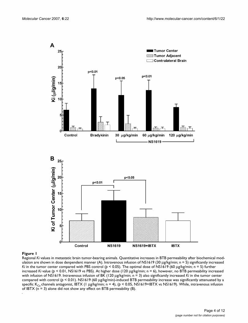
ResultsKCa channel mediates BTB permeability increase in a CRL-5904 metastatic brain tumor modelTo determine whether KCa channels mediate BTB permea-bility in a metastatic brain tumor model, intracranial CRL-5904 tumor bearing-rats received intravenous infusion ofNS1619 (0~120 μg/kg/min), bradykinin, IBTX, or PBS.The transport constant (Ki) was determined by radiotracer[14C] sucrose uptake in the tumor core, tumor-adjacentbrain tissue, and contralateral brain tissue. The data wereobtained from 3~6 rats for each group. Intravenous infu-sion of NS1619 at 30 μg/kg/min resulted in a significantincrease of Ki values in the tumor center (11.24 ± 1.99 μl/
Page 2 of 12(page number not for citation purposes)

Molecular Cancer 2007, 6:22 http://www.molecular-cancer.com/content/6/1/22
g/min) as compared with PBS control (6.58 ± 0.75 μl/g/min; p < 0.05, NS1619 vs PBS). A higher concentration ofNS1619 at 60 μg/kg/min further increased Ki values(12.77 ± 1.99 μl/g/min; p < 0.01, NS1619 vs PBS). While,increasing dose to120 μg/kg/min did not elicit any furtherKi increase. Intravenous infusion of bradykinin (120 μg/kg/min) also significantly increased BTB permeabilitywithin the tumor, with average Ki values at 13.31 ± 2.48μl/g/min(p < 0.01, bradykinin vs PBS). Furthermore,NS1619- and bradykinin- induced BTB permeabilityincreases resulted in enhanced delivery of radiotracer[14C] sucrose to the tumor center without any increase totumor adjacent brain tissue and contralateral normalbrain (Figure 1A). In a separate experiment, we found thatco-administration of a specific KCa channel inhibitor, IBTX(1 μg/kg/min), blocked the increase of BTB permeabilityinduced by NS1619 (Ki value of NS1619+IBTX is 7.58 ±1.31 μl/g/min; NS1619+IBTX vs NS1619, p < 0.05). Intra-venous infusion of IBTX alone did not show any effect onBTB permeability (Figure 1B). These data confirm thatactivation of KCa channel selectively induces BTB openingin tumor tissue, but not tumor adjacent tissue or contral-ateral normal brain, in a metastatic brain tumor animalmodel.
Expression of KCa channels and B2R in CRL-5904 cells, HBMEC and human tumor tissue of lung cancer brain metastasesTo examine whether KCa channels were present in tumortissue, immunostaining of paraffin-embedded tissue sec-tions from lung cancer brain metastases patients were per-formed. The results demonstrated that KCa channels(Figure 2A) and B2R (Figure 2B) expressed extensively intumor masses and microvessels within the tumor. Nega-tive control experiments of KCa channels (Figure 2C) andB2R (Figure 2D) did not show specific staining on the cor-responded specimens. Elevated mRNA level of KCa chan-nels was also detected in lung cancer brain metastasestissues from patients using real time PCR (data notshown). To further determine whether KCa channels andB2Rs are present in metastatic brain tumor and endothe-lial cells, we examined their expression by immunocyto-chemistry. Fluorescence immunostaining showed robustKCa channel expression in cultured CRL-5904 cells, whichdistributed on the cell membrane, cytoplasm and perinu-clear components (Figure 3A~C). KCa channels were alsodetected in HBMEC (Figure 3G~I), but the signal intensi-ties were lower compared with that in CRL-5904 cells.B2R expression was detected in both CRL-5904 cells (Fig-ure 3D~F) and HBMEC (Figure 3J~L) with a higher levelof expression in CRL-5904 cells. These data illustrate thepresence of KCa channels in cultured metastatic braintumor cells, endothelial cells, and most importantly, inhuman metastatic brain tumor tissue.
Activity of functional KCa channels in cultured CRL-5904 cells and HBMECSince the above data demonstrated the presence of KCachannels in metastatic brain tumors and endothelial cellsas well as metastatic brain tumor tissue, we determinewhether the overexpressed KCa channels were functional.Therefore we examined membrane potential activity ofKCachannels in cultured tumor and endothelial cells andby using a fluorescent dye-based potentiometric assay thatmeasures changes in membrane potential. To activate KCachannel, NS1619 (25 μM), a KCa channel agonist, wasadded to the cells, and membrane potential changes weremonitored for up to 300 seconds. Upon activation adecrease in membrane potential was observed in CRL-5904 cells (Figure 4A) and HBMEC (Figure 4C), an effectthat lasted more than 300 seconds. Furthermore, we intro-duced bradykinin (25 μM) to the cultured cells andobserved membrane potential changes in CRL-5904 cells(Figure 4B) and HBMEC (Figure 4D) that lasted forapproximately 100 seconds. Moreover, both NS1619 andbradykinin elicited greater hyperpolarization on CRL-5904 cells compared with HBMEC. IBTX (20 nM), a KCachannel antagonist, reversed the membrane potentialchanges on both cells caused by NS1619 and bradykinin.Furthermore, we found that NS1619 and bradykinincould induce a dose-dependent membrane potentialchange in CRL-5904 and HBMEC (data not shown). Thesedata indicate that KCa channels are functional on bothCRL-5904 cells and HBMEC. The KCa channels can be acti-vated directly by NS1619 or indirectly through B2R sign-aling by bradykinin.
Co-culture of metastatic brain tumor and endothelial cells increases KCa Channel expressionWe further investigated whether KCa channels expressionis modulated by the interaction of metastatic brain tumorcells and endothelial cells. CRL-5904 cells were co-cul-tured with HBMEC, and protein and mRNA levels of KCachannels were examined subsequently. KCa channel wereoverexpressed in CRL-5904/HBMEC co-cultures com-pared to single cultures of either CRL-5904 cells orHBMEC by western blot assay (Figure 5A). Image quanti-fication analysis showed an approximately 30% increaseof KCachannel expression in co-culture of CRL-5904/HBMEC compared to individual cultures by normalizedto β-actin as an internal control (Figure 5B). Also, individ-ual cultures of CRL-5904 tumor cells had higher KCa chan-nel expression than HBMEC. RT-PCR analysis alsoshowed an increase in KCachannel mRNA levels in co-cul-ture cells compared to individual cultures (Figure 5C).These data suggest that co-culture of metastatic tumor andbrain endothelial cells results in upregulation of KCa chan-nel.
Page 3 of 12(page number not for citation purposes)
Molecular Cancer 2007, 6:22 http://www.molecular-cancer.com/content/6/1/22
Page 4 of 12(page number not for citation purposes)
Regional Ki values in metastatic brain tumor-bearing animalsFigure 1Regional Ki values in metastatic brain tumor-bearing animals. Quantitative increases in BTB permeability after biochemical mod-ulation are shown in dose denpendent manner (A). Intravenous infusion of NS1619 (30 μg/kg/min; n = 5) significantly increased Ki in the tumor center compared with PBS control (p < 0.05). The optimal dose of NS1619 (60 μg/kg/min; n = 5) further increased Ki value (p < 0.01, NS1619 vs PBS). At higher dose (120 μg/kg/min; n = 6), however, no BTB permeability increased with infusion of NS1619. Intravenous infusion of BK (120 μg/kg/min; n = 3) also significantly increased Ki in the tumor center compared with control (p < 0.01). NS1619 (60 μg/kg/min)-induced BTB permeability increase was significantly attenuated by a specific KCa channels antagonist, IBTX (1 μg/kg/min; n = 4), (p < 0.05, NS1619+IBTX vs NS1619). While, intravenous infusion of IBTX (n = 3) alone did not show any effect on BTB permeability (B).

Molecular Cancer 2007, 6:22 http://www.molecular-cancer.com/content/6/1/22
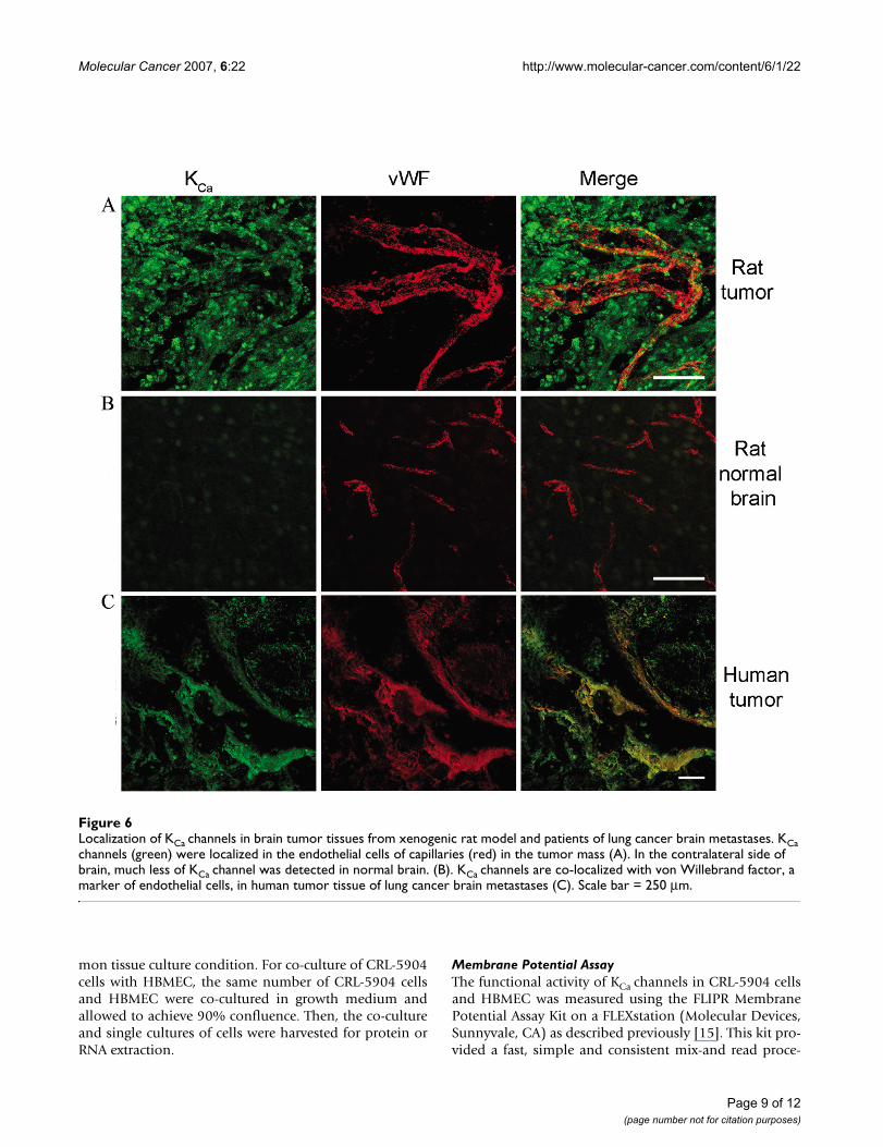
Immuno-colocalization of KCa channel expression in a CRL5904 metastatic brain tumor animal model and human lung cancer brain metastases tissueSince KCa channel modulators can selectively increase BTBpermeability without affecting normal brain, we wantedto know whether KCa channels were differentiallyexpressed within the tumor mass compared with normalbrain tissue. To address this question, we examined KCachannel and endothelial cell marker von Willebrand fac-tor (vWF) expression in CRL-5904 tumors and humanlung cancer brain metastases tissue. An abundance ofKCachannel expression (green) was detected within the
tumor mass with robust microvessel formation (red)within the tumor area of CRL5904 tumor (Figure 6A). Incontrast, there was less KCa channel expression in normalbrain tissue adjacent to the tumor mass as well as contral-ateral normal brain (Figure 6B). More importantly, colo-calization of KCa channels with vWF within microvesselswas observed only within the tumor mass and not in nor-mal brain. These results demonstrate elevated expressionof KCachannels on endothelial cells of capillaries withinthe tumor mass. Immunostaining of human tissue fromlung cancer brain metastases also revealed that KCa chan-nel expression was colocalized with vWF expression in
Expression of KCa channels and B2R in the lung cancer metastatic brain tumor tissue from patientsFigure 2Expression of KCa channels and B2R in the lung cancer metastatic brain tumor tissue from patients. KCa channels (A) and B2R (B) expression were detected on the tumor cells in the tissue. Negative control experiments of KCa channels (C) and B2R (D) did not show specific staining on the corresponded specimens by deleting of primary antibodies. Scale bar = 50 μm.
Page 5 of 12(page number not for citation purposes)

Molecular Cancer 2007, 6:22 http://www.molecular-cancer.com/content/6/1/22
Page 6 of 12(page number not for citation purposes)
Expression of KCa channels and B2R in CRL-5904 cells and HBMECFigure 3Expression of KCa channels and B2R in CRL-5904 cells and HBMEC. KCa channels (green) distributed in CRL-5904 cells (A~C) and HBMEC (G~I). The B2R (green) were expressed in CRL-5904 cells (D~F) and HBMEC (J~L). Cells were located by coun-terstaining with DAPI (blue, B, E, H, K) and merged images are also shown (C, F, I, L). Scale bar = 50 μm.

Molecular Cancer 2007, 6:22 http://www.molecular-cancer.com/content/6/1/22
tumor capillary endothelia (Figure 6C). These resultsstrongly suggest that the selective BTB permeabilityincrease induced by modulation of KCa channel in themetastatic brain tumor model is likely due to the overex-presion of KCa channels on tumor cells and tumor capil-lary endothelia.
DiscussionWe have studied the presence of KCa channels and B2R inprimary brain tumors, however, their occurrence andfunction in metastatic brain tumors remained to be inves-tigated. In this study, we detected high level expression ofKCa channels in CRL-5904 tumor and brain endothelialcells, which is consistent with previous studies showingKCa channels expression in RG2 glioma and endothelialcells [15]. Other investigators have also demonstrated thatthe expression level of KCa channels correlates with themalignancy grade of glioma in human [36]. Therefore,these data suggest there is an intimate association betweenKCa channel expression and brain tumor development,which remains to be fully investigated. Additionally, wedetected the presence of B2R in CRL-5904 tumor andendothelial cells. Liu et al also showed that B2R areexpressed in cultured RG2, C6 and 9L glioma cells, moreinterestingly, the expression levels of B2R in tumor cellswas directly correlated with the increase of BTB permea-bility induced by bradykinin in a rat glioma model [32].Thus, the presence of KCachannels or B2R in metastatic
brain tumor cells or HBMEC may play a functional role inBTB permeability of metastatic brain tumors.
We examined KCa channel activity on metastatic tumorcells and capillary endothelial cells using a membranepotential assay, which is well-correlated with the patch-clamp method, used to measure changes in membranepotential (FLIPR application note, Molecular devices,CA). We found that NS1619 and bradykinin elicitedgreater hyperpolarization effects on CRL-5904 than onHBMEC. These findings may reflect a higher level ofexpression for KCa channels on metastatic tumor cells ascompared to endothelial cells. Importantly, the mem-brane potential change induced by NS1619 lasted 3 timeslonger than that induced by bradykinin. These data fur-ther support the finding, from cellular level, that NS1619-elicited increases in BTB permeability in a glioma modellast up to 60 minutes compared to the transient effect ofbradykinin, which lasts for about 15~20 minutes [15],partially due to B2R internalization [37]. The current dataillustrates that the presence of KCa channel are functionalin metastatic brain tumor and endothelial cells. Similar toour findings, Reiser et al demonstrated that bradykinincan directly activate KCa channels in rat glioma cells [38].Other studies have shown that bradykinin can activate KCachannels through a NO-cGMP signalling pathway [39].Hence, our present study indicates that bradykinin-acti-vated downstream signals, such as activation of KCachan-nels, may be modulated to induce membrane potentialchanges on brain metastatic tumor and endothelial cells.
The presence of functional KCa channels in metastaticbrain tumor and brain endothelial cells suggests that bio-chemical modulation of KCachannels could play animportant role in therapeutic BTB opening. We furtherinvestigated whether the KCa channels agonist, NS1619and bradykinin could selectively enhance BTB permeabil-ity in a metastatic brain tumor xenograft model. Theseresults showed that intravenous infusion of NS1619yielded a two-fold increase the unidirectional transport ofa radiotracer into metastatic brain tumors; similar tobradykinin-induced BTB permeability increase in meta-static brain tumor-bearing rats. Our previous studies havedemonstrated that higher doses of intravenous bradyki-nin are required to increase BTB permeability comparedto intracarotid infusion of bradykinin, reflecting the influ-ence of the first pass effect with intracarotid delivery [15].In a glioma model, it has been reported that the effects ofbradykinin on BTB permeability mediated by B2Rresulted in enhanced drug delivery to glioma [40]; andthis effect could be attenuated by coinfusion with IBTX[15]. In this metastatic brain tumor model, we furtherdemonstrate the presence of B2R and confirm that thebradykinin effect on permeability is mediated via KCachannels. Consistent with previous studies [41], current
Measurement of functional KCa channels activity in CRL-5904 cells and HBMECFigure 4Measurement of functional KCa channels activity in CRL-5904 cells and HBMEC. Membrane potential changes in relative flu-orescence units (RFU) were detected during a 300-second response to 25 μM of NS1619 (A) and BK (B) respectively on the CRL-5904 cells. NS1619 and BK also elicited membrane potential changes on HBMEC (C, D). Addition of IBTX (20 nM) reversed the membrane potential to resting values.
Page 7 of 12(page number not for citation purposes)

Molecular Cancer 2007, 6:22 http://www.molecular-cancer.com/content/6/1/22
data confirms the selective increase of BTB permeability inbrain metastatic tumors but not normal brain tissue.These results suggest that biochemical modulation of KCachannel induces a selective BTB opening in metastaticbrain tumor.
Confocal images showed KCa channels overexpression intumor tissue and tumor microvessels as compared withnormal brain. More importantly, the tumor capillariesshowed co-localization of KCa channels and vWF in tumorarea of CRL-5904 tumor and in human metastatic braintumor tissue. To further study the interaction betweentumor and endothelial cells, we co-cultured CRL-5904metastatic brain tumors and brain endothelial cells. Weshow that mRNA expression of KCa channels is upregu-lated in co-cultured cells compared to indivdual cultures.These data suggest that increased KCa channel expressionand their activity in tumor endothelial cells maybe due tothe tumor micro-environment or cell-to-cell communica-tion between tumor and microvessel endothelial cells.This is consistent with reports that brain tumor cellsincrease KATP channel expression in endothelial cells[16].
These findings support a pivotal role for KCa channels inBTB permeability regulation. Recently clinical studyshowed that trastuzumab, anti-HER2 antibody, whileeffective in treating tumors outside the brain, fails to treatbrain metastases due to its inability to cross the bloodbrain tumor barrier [42]. Our research has shown thatKCachannel-mediated BTB permeability modulationcould be a useful strategy to increase therapeutic agents,such as antibody-based therapies, delivery into metastaticbrain tumors.
ConclusionWe present evidence that activation of KCa channels by achannel-specific agonist can selectively enhance BTB per-meability in a metastatic brain tumor rat model. We showKCa channel and B2R are highly expressed in brain meta-static tumor cells, endothelial cells and lung cancer brainmetastatic tissue. The expression level is correlated withKCa channel activity in these cells. In a metastatic braintumor model, we demonstrate that NS1619 and bradyki-nin can selectively open BTB and significantly enhance theradiotracer delivery specifically to metastatic braintumors. It is also demonstrated that KCa channels expres-sion can be upregulated in the co-cultures of tumor cellsand endothelial cells, as well as in the microvesselendothelia of brain metastases tissue. KCachannels may beexploited as specific target for selectively pharamacologicmodulation of BTB to increase delivery of chemothera-peutic drugs to brain metastases.
MethodsCell CultureCRL-5904 cells (human non-small cell lung cancer; meta-static site: brain poorly differentiated carcinoma) andhuman brain microvessel endothelial cells (HBMEC)were obtained from the American tissue culture collection(ATCC, VA) and maintained in RPMI 1640 with 10% fetalbovine serum. Both cell lines were maintained in the com-
Modulation of KCa channel expression in co-cultured cells (CRL-5904 cells and HBMEC)Figure 5Modulation of KCa channel expression in co-cultured cells (CRL-5904 cells and HBMEC). Western blot analysis for KCa channel expression on cultured cells. Lane1, CRL-5904 cells; Lane 2, CRL-5904 and HBMEC co-culture; Lane 3, HBMEC (A). Semi-quantitative analyses of the protein bands were normalized by internal control, β-actin (B). mRNA transcrip-tion levels of different cell cultures were determined by RT-PCR. L: 1 kb ladder; Lane1, CRL-5904 cells; Lane 2, CRL-5904 and HBMEC co-culture; Lane 3, HBMEC (C).
Page 8 of 12(page number not for citation purposes)
Molecular Cancer 2007, 6:22 http://www.molecular-cancer.com/content/6/1/22
mon tissue culture condition. For co-culture of CRL-5904cells with HBMEC, the same number of CRL-5904 cellsand HBMEC were co-cultured in growth medium andallowed to achieve 90% confluence. Then, the co-cultureand single cultures of cells were harvested for protein orRNA extraction.
Membrane Potential AssayThe functional activity of KCa channels in CRL-5904 cellsand HBMEC was measured using the FLIPR MembranePotential Assay Kit on a FLEXstation (Molecular Devices,Sunnyvale, CA) as described previously [15]. This kit pro-vided a fast, simple and consistent mix-and read proce-
Localization of KCa channels in brain tumor tissues from xenogenic rat model and patients of lung cancer brain metastasesFigure 6Localization of KCa channels in brain tumor tissues from xenogenic rat model and patients of lung cancer brain metastases. KCa channels (green) were localized in the endothelial cells of capillaries (red) in the tumor mass (A). In the contralateral side of brain, much less of KCa channel was detected in normal brain. (B). KCa channels are co-localized with von Willebrand factor, a marker of endothelial cells, in human tumor tissue of lung cancer brain metastases (C). Scale bar = 250 μm.
Page 9 of 12(page number not for citation purposes)

Molecular Cancer 2007, 6:22 http://www.molecular-cancer.com/content/6/1/22
dure. In brief, the cells were seeded in sterile, clearbottom, black 96-well plates (Corning Inc., MA) at den-sity of 2 × 10³ cells/well to achieve monolayer within 24h. The monolayer cells were incubated with the mem-brane potential assay kits reagents for 30 min before load-ing the compounds. The anionic potentiometric dye thattransverses between cells and extracellular solution in amembrane potential-dependent manner serves as an indi-cator of vasomodulator-induced voltage changes acrossthe cell membrane. Dose response studies were per-formed with 0 to 50 μM NS1619 or bradykinin with orwithout IBTX (20 nM). The FLEXstation was set up usingthe following parameters: excitation 530 nm, emission565 nm, and emission cut of 550 nm wavelengths. Obser-vations and recordings were made for 300 seconds afteradding the compounds. NS1619, bradykinin and IBTXwere obtained from Sigma (St. Louis, MO).
In Vivo BBB/BTB PermeabilityAll of the animals used were conducted in accordancewith the Institutional Animal Care and Usage Committeein force at Cedars-Sinai Medical Center. A metastatic braintumor xenograft model was established using athymicnude rats (180–200 g; Charles River Laboratories, Inc.,MA) for BBB/BTB permeability studies. Athymic nude ratswere anesthetized with i.p. ketamine and xylazine, andstereotactically implanted with CRL-5904 cells (2 × 10⁵)in 4 μl of 1.2% methylcellulose/PBS using a Hamiltonsyringe into the right striatum. The Coordinates were 3.4mm lateral to bregma and 5.0 mm deep from dura. Tendays after tumor implantation, the femoral arteries of ratswere cannulated to measure blood pressure and collectblood, and the femoral vein was also cannulated toadminister the drugs and radiotracer. Body temperaturewas maintained at 37°C. Arterial blood gases, blood pres-sure and hematocrit were monitored. Animals withabnormal physiological parameters were eliminated fromthis study. In regional permeability studies, either intrave-nous drug or PBS was infused into the femoral vein at arate of 66.7 μl/min for 15 minutes. Five minutes after thestart of the intravenous infusion, 50 μCi/kg of the radi-otracer [14C] sucrose was injected as an intravenous bolus.Arterial blood pressure was monitored throughout theexperimental period with a blood pressure monitor(DigiMed, KY). The unilateral transport constant Ki (μl/g/min), which is an initial rate for blood-to-brain transfer ofradiotracer, was calculated as described by Ohno et al[43].The Ki was determined by radiotracer [14C] sucrose in thetumor core, tumor-adjacent brain tissue, and contralateralbrain tissue using the quantitative autoradiographic(QAR) method as describe previously [44]. Quantitativeanalysis of the regional radioactivity was performed usinga computer (Power Macintosh 7100) and Image 1.55 soft-ware (National Institutes of Health, Bethesda, MD). Anoptimum dose (120 μg/kg/min, i.v.) of bradykinin estab-
lished previously was used for Ki measurements. To estab-lish the optimal and safe dose range that would result inselective increase in BTB permeability without appreciablyaltering system blood pressure, various doses (0~120 μg/kg/min, i.v.) of NS1619 were administered in metastaticbrain tumor bearing-nude rats. Additional experimentswere performed by coinfusing of NS1619 with IBTX (1.0μg/kg/min) to investigate whether inhibition of KCa chan-nels by IBTX has any effects on NS1619-induced permea-bility increase.
Western Blot AnalysisThe extracted protein samples were quantified to deter-mine total protein concentrations using a protein assay kit(BioRad, CA). Same amount of each sample was fraction-ated on 10% SDS-polyacrylamide gel and then transferredto a nitrocellulose membrane. The membrane was probedwith primary antibodies anti-MaxiKá (1:200; Santa CruzBiotechnology, CA) and β-actin (1:5000; Sigma, MO), fol-lowed by peroxidase-conjugated secondary antibodies.The signals were detected with an enhanced chemilumi-nescence kit (Amersham Biosciences Corp., NJ). β-actinserved as an internal control.
Reverse Transcription-PCRThe extracted RNA was reverse transcripted using a Bio-script kit (Bioline) and Oligo (dT) 12–18 primer (Invitro-gene). The resulting cDNA products were used astemplates for PCR assay. The genes of the KCa channelswere concurrently amplified with internal control β-actinin the same reaction tube as described previously [45].Sequence specific primers were used for amplification ofKCa channels (forward: 5'-tccaaaacaaccaggctctc-3'; reverse:5'- gggggagatgttgtgaagaa-3') and β-actin (forward: 5'- gcac-cacaccttctacaatgagc-3'; reverse: 5'- ttgaaggtagtttcgtggatgcc-3'). PCR products were identified using agarose gel elec-trophoresis and ethidium bromide staining.
Immunocytochemistry StainingCRL-5904 cells and HBMEC were fixed with 4% parafor-moldehyde for 15 min, and then incubated with anti-B2R(1:200; BD Bioscience, NJ) or anti-MaxiKα (1:200; SantaCruz, CA) antibodies. The signals were detected withFITC-conjugated secondary antibodies (1:200; JacksonImmnoResearch, CA). The cells were counterstained with4', 6-diamidino-2-phenylindole (DAPI; Vector Laborato-ries, CA) and cover-slipped. Paraffin-embedded, meta-static brain tumor samples from lung cancer weredeparaffinized and rehydrated. The slides were incubatedwith primary anti-MaxiKá and anti-B2R antibodies, andfollowed by biotinylated secondary antibodies (1:1000;Jackson ImmunoResearch, CA). Biotinylated conjugateswere detected with avidin-biotin peroxide complex (Vec-tor Laboratories), and then developed with 3, 3'-Diami-nobenzidine (DAB) method. The sections were
Page 10 of 12(page number not for citation purposes)

Molecular Cancer 2007, 6:22 http://www.molecular-cancer.com/content/6/1/22
counterstained with hematoxylin. For the double stain-ing, the sections were incubated with primary antibodies,anti-MaxiKá and anti-von Willebrand Factor (1:200;Chemicon, CA), and then subjected to FITC-and Tex-Red-conjugated secondary antibodies. The slides were exam-ined under confocal microscopy. Negative control experi-ments were performed on all the corresponded specimensby deleting of primary antibodies.
Competing interestsThe author(s) declare that they have no competing inter-ests.
Authors' contributionsJH and KLB conceived the study. JH designed and per-formed all experiments. DY, MRS, XW, MKK, BMK, AE, KPparticipated in vivo BBB/BTB permeability assay. JH andXY drafted the original manuscript, and MKK, JMO, DIand KLB helped modify the final version of the manu-script.
AcknowledgementsThis research was supported by National Institutes of Health grants NS32103, NS25554, RR13707, a Jacob Javits Award (KLB), and an American Cancer Society grant (NSN).
References1. Guerin C, Olivi A, Weingart JD, Lawson HC, Brem H: Recent
advances in brain tumor therapy: local intracerebral drugdelivery by polymers. Invest New Drugs 2004, 22(1):27-37.
2. Kunwar S: Convection enhanced delivery of IL13-PE38QQRfor treatment of recurrent malignant glioma: presentationof interim findings from ongoing phase 1 studies. Acta Neuro-chir Suppl 2003, 88:105-111.
3. Li C, Hall WA, Jin N, Todhunter DA, Panoskaltsis-Mortari A, ValleraDA: Targeting glioblastoma multiforme with an IL-13/diph-theria toxin fusion protein in vitro and in vivo in nude mice.Protein Eng 2002, 15(5):419-427.
4. Bhattacharjee AK, Nagashima T, Kondoh T, Tamaki N: Quantifica-tion of early blood-brain barrier disruption by in situ brainperfusion technique. Brain Res Brain Res Protoc 2001, 8(2):126-131.
5. Siegal T, Rubinstein R, Bokstein F, Schwartz A, Lossos A, Shalom E,Chisin R, Gomori JM: In vivo assessment of the window of bar-rier opening after osmotic blood-brain barrier disruption inhumans. J Neurosurg 2000, 92(4):599-605.
6. Black KL, Baba T, Pardridge WM: Enzymatic barrier protectsbrain capillaries from leukotriene C4. J Neurosurg 1994,81(5):745-751.
7. Black KL, Cloughesy T, Huang SC, Gobin YP, Zhou Y, Grous J, NelsonG, Farahani K, Hoh CK, Phelps M: Intracarotid infusion of RMP-7, a bradykinin analog, and transport of gallium-68 ethylene-diamine tetraacetic acid into human gliomas. J Neurosurg1997, 86(4):603-609.
8. Sugita M, Black KL: Cyclic GMP-specific phosphodiesteraseinhibition and intracarotid bradykinin infusion enhances per-meability into brain tumors. Cancer Res 1998, 58(5):914-920.
9. Matsukado K, Nakano S, Bartus RT, Black KL: Steroids decreaseuptake of carboplatin in rat gliomas--uptake improved byintracarotid infusion of bradykinin analog, RMP-7. J Neuroon-col 1997, 34(2):131-138.
10. Nomura T, Inamura T, Black KL: Intracarotid infusion of bradyki-nin selectively increases blood-tumor permeability in 9L andC6 brain tumors. Brain Res 1994, 659(1-2):62-66.
11. Black KL, Hoff JT, McGillicuddy JE, Gebarski SS: Increased leukot-riene C4 and vasogenic edema surrounding brain tumors inhumans. Ann Neurol 1986, 19(6):592-595.
12. Black KL, Chio CC: Increased opening of blood-tumour barrierby leukotriene C4 is dependent on size of molecules. NeurolRes 1992, 14(5):402-404.
13. Hashizume K, Black KL: Increased endothelial vesicular trans-port correlates with increased blood-tumor barrier permea-bility induced by bradykinin and leukotriene C4. J NeuropatholExp Neurol 2002, 61(8):725-735.
14. Nakano S, Matsukado K, Black KL: Increased brain tumor micro-vessel permeability after intracarotid bradykinin infusion ismediated by nitric oxide. Cancer Res 1996, 56(17):4027-4031.
15. Ningaraj NS, Rao M, Hashizume K, Asotra K, Black KL: Regulationof blood-brain tumor barrier permeability by calcium-acti-vated potassium channels. J Pharmacol Exp Ther 2002,301(3):838-851.
16. Ningaraj NS, Rao MK, Black KL: Adenosine 5'-triphosphate-sen-sitive potassium channel-mediated blood-brain tumor bar-rier permeability increase in a rat brain tumor model. CancerRes 2003, 63(24):8899-8911.
17. Gregor A, Lind M, Newman H, Grant R, Hadley DM, Barton T,Osborn C: Phase II studies of RMP-7 and carboplatin in thetreatment of recurrent high grade glioma. RMP-7 EuropeanStudy Group. J Neurooncol 1999, 44(2):137-145.
18. Ford J, Osborn C, Barton T, Bleehen NM: A phase I study of intra-venous RMP-7 with carboplatin in patients with progressionof malignant glioma. Eur J Cancer 1998, 34(11):1807-1811.
19. Prados MD, Schold SJS, Fine HA, Jaeckle K, Hochberg F, Mechtler L,Fetell MR, Phuphanich S, Feun L, Janus TJ, Ford K, Graney W: A ran-domized, double-blind, placebo-controlled, phase 2 study ofRMP-7 in combination with carboplatin administered intra-venously for the treatment of recurrent malignant glioma.Neuro-oncol 2003, 5(2):96-103.
20. Liu X, Chang Y, Reinhart PH, Sontheimer H: Cloning and charac-terization of glioma BK, a novel BK channel isoform highlyexpressed in human glioma cells. J Neurosci 2002,22(5):1840-1849.
21. Kitazono T, Faraci FM, Taguchi H, Heistad DD: Role of potassiumchannels in cerebral blood vessels. Stroke 1995,26(9):1713-1723.
22. Ningaraj NS, Rao M, Black KL: Calcium-dependent potassiumchannels as a target protein for modulation of the blood-brain tumor barrier. Drug News Perspect 2003, 16(5):291-298.
23. Sobey CG, Heistad DD, Faraci FM: Mechanisms of bradykinin-induced cerebral vasodilatation in rats. Evidence that reac-tive oxygen species activate K+ channels. Stroke 1997,28(11):2290-4; discussion 2295.
24. Berg T, Koteng O: Signalling pathways in bradykinin- and nitricoxide-induced hypotension in the normotensive rat; role ofK+-channels. Br J Pharmacol 1997, 121(6):1113-1120.
25. Bolotina VM, Najibi S, Palacino JJ, Pagano PJ, Cohen RA: Nitric oxidedirectly activates calcium-dependent potassium channels invascular smooth muscle. Nature 1994, 368(6474):850-853.
26. Lohse MJ, Forstermann U, Schmidt HH: Pharmacology ofNO:cGMP signal transduction. Naunyn Schmiedebergs Arch Phar-macol 1998, 358(1):111-112.
27. Hall JM: Bradykinin receptors. Gen Pharmacol 1997, 28(1):1-6.28. Miura H, Liu Y, Gutterman DD: Human coronary arteriolar dila-
tion to bradykinin depends on membrane hyperpolarization:contribution of nitric oxide and Ca2+-activated K+ channels.Circulation 1999, 99(24):3132-3138.
29. Faraci FM, Heistad DD: Regulation of the cerebral circulation:role of endothelium and potassium channels. Physiol Rev 1998,78(1):53-97.
30. Ransom CB, Sontheimer H: BK channels in human glioma cells.J Neurophysiol 2001, 85(2):790-803.
31. Wanner SG, Koch RO, Koschak A, Trieb M, Garcia ML, KaczorowskiGJ, Knaus HG: High-conductance calcium-activated potas-sium channels in rat brain: pharmacology, distribution, andsubunit composition. Biochemistry 1999, 38(17):5392-5400.
32. Liu Y, Hashizume K, Chen Z, Samoto K, Ningaraj N, Asotra K, BlackKL: Correlation between bradykinin-induced blood-tumorbarrier permeability and B2 receptor expression in experi-mental brain tumors. Neurol Res 2001, 23(4):379-387.
33. Matsukado K, Inamura T, Nakano S, Fukui M, Bartus RT, Black KL:Enhanced tumor uptake of carboplatin and survival in gli-oma-bearing rats by intracarotid infusion of bradykinin ana-log, RMP-7. Neurosurgery 1996, 39(1):125-33; discussion 133-4.
Page 11 of 12(page number not for citation purposes)

Molecular Cancer 2007, 6:22 http://www.molecular-cancer.com/content/6/1/22
Publish with BioMed Central and every scientist can read your work free of charge
"BioMed Central will be the most significant development for disseminating the results of biomedical research in our lifetime."
Sir Paul Nurse, Cancer Research UK
Your research papers will be:
available free of charge to the entire biomedical community
peer reviewed and published immediately upon acceptance
cited in PubMed and archived on PubMed Central
yours — you keep the copyright
Submit your manuscript here:http://www.biomedcentral.com/info/publishing_adv.asp
BioMedcentral
34. Rizzi A, Tondini M, Rocco G, Rossi G, Robustellini M, Radaelli F, DellaPona C: Lung cancer with a single brain metastasis: therapeu-tic options. Tumori 1990, 76(6):579-581.
35. Doolittle ND, Peereboom DM, Christoforidis GA, Hall WA, PalmieriD, Brock PR, Campbell KC, Dickey DT, Muldoon LL, O'Neill B P,Peterson DR, Pollock B, Soussain C, Smith Q, Tyson RM, NeuweltEA: Delivery of chemotherapy and antibodies across theblood-brain barrier and the role of chemoprotection, in pri-mary and metastatic brain tumors: report of the eleventhannual blood-brain barrier consortium meeting. J Neurooncol2007, 81(1):81-91.
36. Pizard A, Blaukat A, Muller-Esterl W, Alhenc-Gelas F, Rajerison RM:Bradykinin-induced internalization of the human B2 recep-tor requires phosphorylation of three serine and two threo-nine residues at its carboxyl tail. J Biol Chem 1999,274(18):12738-12747.
37. Greiser E, Soehring K: [On the transport of pentobarbitalthrough biological membranes and its influencing by etha-nol]. Arzneimittelforschung 1967, 17(2):207-214.
38. Zhou XB, Schlossmann J, Hofmann F, Ruth P, Korth M: Regulationof stably expressed and native BK channels from humanmyometrium by cGMP- and cAMP-dependent proteinkinase. Pflugers Arch 1998, 436(5):725-734.
39. Matsukado K, Sugita M, Black KL: Intracarotid low dose bradyki-nin infusion selectively increases tumor permeabilitythrough activation of bradykinin B2 receptors in malignantgliomas. Brain Res 1998, 792(1):10-15.
40. Ningaraj NS, Rao M, Groysman L, Black KL: Role of Ca2+-depend-ent and ATP-sensitive potassium channels in blood-brainbarrier permeability after transient focal ischemia/reper-fusion in rats. Stroke 2002, 33(1):361.
41. Okita R, Saeki T, Takashima S, Aogi K, Ohsumi S: Progressive cen-tral nervous system metastases in responder patients foroutside central nervous system metastases on trastuzumab-based therapy--report of two cases of refractory breast can-cer. Hiroshima J Med Sci 2005, 54(1):35-38.
42. Ohno K, Pettigrew KD, Rapoport SI: Lower limits of cerebrovas-cular permeability to nonelectrolytes in the conscious rat.Am J Physiol 1978, 235(3):H299-307.
43. Sugita M, Hunt GE, Liu Y, Black KL: Nitric oxide and cyclic GMPattenuate sensitivity of the blood-tumor barrier permeabil-ity to bradykinin. Neurol Res 1998, 20(6):559-563.
44. Yuan X, Curtin J, Xiong Y, Liu G, Waschsmann-Hogiu S, Farkas DL,Black KL, Yu JS: Isolation of cancer stem cells from adult gliob-lastoma multiforme. Oncogene 2004, 23(58):9392-9400.
Page 12 of 12(page number not for citation purposes)
Copyright © 2022 FDOKUMEN