Post-transcriptional gene silencing triggers dispensable DNA ...
Upload
independentCategory
view
0download
0
The Rockefeller University Press, 0021-9525/2002/07/63/15 $5.00The Journal of Cell Biology, Volume 158, Number 1, July 8, 2002 63–77http://www.jcb.org/cgi/doi/10.1083/jcb.200202088
JCB
Article
63
The cytoplasmic filaments of the nuclear pore complex are dispensable for selective nuclear protein import
Tobias C. Walther,
1
Helen S. Pickersgill,
2
Volker C. Cordes,
3,4
Martin W. Goldberg,
2
Terry D. Allen,
2
Iain W. Mattaj,
1
and Maarten Fornerod
1,5
1
Gene Expression Program, EMBL, D-69117 Heidelberg, Germany
2
Paterson Institute CRUK/CMB, M20 9BX Manchester, U.K.
3
Karolinska Institute, S-17177 Stockholm, Sweden
4
German Cancer Research Center, D-69120 Heidelberg, Germany
5
Netherlands Cancer Institute H4, 1066 CX Amsterdam, Netherlands
he nuclear pore complex (NPC) mediates bidirectionalmacromolecular traffic between the nucleus andcytoplasm in eukaryotic cells. Eight filaments project
from the NPC into the cytoplasm and are proposed tofunction in nuclear import. We investigated the localizationand function of two nucleoporins on the cytoplasmic faceof the NPC, CAN/Nup214 and RanBP2/Nup358. Consistent
with previous data, RanBP2 was localized at the cytoplasmicfilaments. In contrast, CAN was localized near the cyto-plasmic coaxial ring. Unexpectedly, extensive blocking ofRanBP2 with gold-conjugated antibodies failed to inhibitnuclear import. Therefore, RanBP2-deficient NPCs weregenerated by in vitro nuclear assembly in RanBP2-depleted
Xenopus
egg extracts. NPCs were formed that lacked cyto-
T
plasmic filaments, but that retained CAN. These nucleiefficiently imported nuclear localization sequence (NLS) orM9 substrates. NPCs lacking CAN retained RanBP2 andcytoplasmic filaments, and showed a minor NLS importdefect. NPCs deficient in both CAN and RanBP2 displayedno cytoplasmic filaments and had a strikingly immaturecytoplasmic appearance. However, they showed only aslight reduction in NLS-mediated import, no change inM9-mediated import, and were normal in growth andDNA replication. We conclude that RanBP2 is the majornucleoporin component of the cytoplasmic filaments of theNPC, and that these filaments do not have an essential rolein importin
�
/
�
– or transportin-dependent import.
Introduction
The nuclear pore complex (NPC)* is the mediator of allmacromolecular transport between the nucleus and thecytoplasm of the eukaryotic cell. NPCs serve the cell’s require-ment for bidirectional, selective, diverse, and high-volumetransport between these two compartments (for review see
Allen et al., 2000; Ryan and Wente, 2000; Fahrenkrog et al.,2001; Rout and Aitchison, 2001; Vasu and Forbes, 2001).
Ultrastructural analyses of the NPC using transmission electron(Akey, 1989; Ris, 1989, 1991; Jarnik and Aebi, 1991;Hinshaw et al., 1992; Akey and Radermacher, 1993), scan-ning electron (Goldberg and Allen, 1993, 1996; Ris andMalecki, 1993; Ris, 1997), and atomic force (Rakowska etal., 1998; Stoffler et al., 1999b) microscopy has shown thevertebrate NPC to be an
�
65-nm deep, plugged channel,decorated on each side with eight filaments attached to acoaxial ring that project away from the nuclear envelope(NE). Although the
�
40–50-nm filaments of the nuclearface join together at their distal ends to form a so-callednuclear basket or fishtrap (Goldberg and Allen, 1993, 1996;Ris and Malecki, 1993; Ris, 1997), the
�
35–50-nm filamentsof the cytoplasmic side are not joined (Jarnik and Aebi,1991; Goldberg and Allen, 1993).
Address correspondence to Maarten Fornerod, The Netherlands CancerInstitute H4, Plesmanlaan 121, 1066 CX Amsterdam, The Netherlands.Tel.: 31-20-512-2024. Fax: 31-20-512-2029. E-mail: [email protected]
T.C. Walther and H.S. Pickersgill contributed equally to this work.H.S. Pickersgill’s present address is The Netherlands Cancer InstituteH4, 1066 CX Amsterdam, The Netherlands.*Abbreviations used in this paper: AL, annulate lamellae;
FEISEM, fieldemission in-lens scanning EM;
mAb, monoclonal antibody;
NE, nuclearenvelope; NLS, nuclear localization sequence; NPC, nuclear pore com-plex; TEM, transmission EM.Key words: nuclear pore complex; nuclear import; nuclear localizationsignal; RanBP2/Nup358; CAN/Nup214
on Septem
ber 26, 2013jcb.rupress.org
Dow
nloaded from
Published July 8, 2002 on S
eptember 26, 2013
jcb.rupress.orgD
ownloaded from
Published July 8, 2002
on Septem
ber 26, 2013jcb.rupress.org
Dow
nloaded from
Published July 8, 2002 on S
eptember 26, 2013
jcb.rupress.orgD
ownloaded from
Published July 8, 2002
on Septem
ber 26, 2013jcb.rupress.org
Dow
nloaded from
Published July 8, 2002 on S
eptember 26, 2013
jcb.rupress.orgD
ownloaded from
Published July 8, 2002
on Septem
ber 26, 2013jcb.rupress.org
Dow
nloaded from
Published July 8, 2002 on S
eptember 26, 2013
jcb.rupress.orgD
ownloaded from
Published July 8, 2002
on Septem
ber 26, 2013jcb.rupress.org
Dow
nloaded from
Published July 8, 2002 on S
eptember 26, 2013
jcb.rupress.orgD
ownloaded from
Published July 8, 2002
on Septem
ber 26, 2013jcb.rupress.org
Dow
nloaded from
Published July 8, 2002 on S
eptember 26, 2013
jcb.rupress.orgD
ownloaded from
Published July 8, 2002
on Septem
ber 26, 2013jcb.rupress.org
Dow
nloaded from
Published July 8, 2002 on S
eptember 26, 2013
jcb.rupress.orgD
ownloaded from
Published July 8, 2002
on Septem
ber 26, 2013jcb.rupress.org
Dow
nloaded from
Published July 8, 2002
64 The Journal of Cell Biology
|
Volume 158, Number 1, 2002
In parallel to these structural studies, 30–40 different pro-teins, termed nucleoporins, have been identified as compo-nents of the NPC in vertebrates and yeast (Rout et al., 2000;Ryan and Wente, 2000; Fahrenkrog et al., 2001). To help un-derstand the molecular organization of the NPC, these nucle-oporins have been mapped to NPC subdomains using im-munogold electron microscopy. In yeast, transmission EM(TEM) studies of protein A–tagged nucleoporins have shownthat the majority of these map to both sides of the NPC (Routet al., 2000). Exceptions that map exclusively to the cytoplas-mic face are Nup159/Rat7 and Nup82 (Kraemer et al., 1995;Hurwitz et al., 1998; Rout et al., 2000). Even though regionalinformation has been obtained for many nucleoporins, therather low degree of structural definition of the NPC in trans-mission immuno-EM did not generally allow individual nu-cleoporins to be attributed to particular NPC substructures. Inhigher eukaryotes, three nucleoporins have been mapped tothe cytoplasmic side of the NPC: CAN/Nup214 (Kraemer etal., 1994; Pante et al., 1994), Nup88/Nup84 (Bastos et al.,1997), and RanBP2/Nup358 (Wu et al., 1995; Yokoyama etal., 1995). RanBP2/Nup358, which has no identifiable yeasthomologue, is the largest nucleoporin and has been localizedto the cytoplasmic filaments of the NPC (Wilken et al., 1995;Wu et al., 1995; Yokoyama et al., 1995). It contains fourRanGTP binding domains, highly similar in sequence to theone present in RanBP1. RanBP1 and isolated RanBP1-homol-ogous domains of RanBP2 serve as Ran GTPase coactivators(Beddow et al., 1995; Bischoff et al., 1995; Richards et al.,1995; Schlenstedt et al., 1995; Villa Braslavsky et al., 2000),assisting the dissociation of RanGTP from importins (Bischoffand Gorlich, 1997; Floer et al., 1997; Schlenstedt et al., 1997)and exportin–cargo complexes (Kutay et al., 1998; Askjaer etal., 1999; Kehlenbach et al., 1999). RanBP2 also contains twoZinc fingers of the type predicted to bind RanGDP (Nakielnyet al., 1999; Yaseen and Blobel, 1999b), and a number ofloosely spaced FG repeats, providing potential binding sites fortransport receptors. Furthermore, RanBP2/Nup358 serves as abinding site for the SUMO1-modified form of RanGAP1, theRanGTPase activating protein (Matunis et al., 1996, 1998;Mahajan et al., 1997). Recently, it has been shown thatRanBP2 itself acts as an E3 ligase in the sumoylation reaction(Pichler et al., 2002). Purified RanBP2/Nup358 forms
�
36-nm long, 5-nm thick filaments (Delphin et al., 1997), corre-sponding approximately to the size of the cytoplasmic fila-ments (Jarnik and Aebi, 1991), suggesting that RanBP2 couldbe the main constituent of the cytoplasmic NPC filaments.
CAN/Nup214 and Nup88/Nup84 form a cytoplasmi-cally oriented nucleoporin subcomplex (Bastos et al., 1997;Fornerod et al., 1997). Based on immunogold EM studies(Kraemer et al., 1994; Pante et al., 1994), CAN/Nup214 isgenerally also referred to as a component of the cytoplasmicfilaments. Genetic depletion of CAN/Nup214 in earlymouse embryos resulted in reduced nuclear localization se-quence (NLS)–mediated protein import, and strong nuclearpoly(A)
�
RNA accumulation (van Deursen et al., 1996),whereas mutation of the
Drosophila
homologue of Nup88,
mbo
, results in a specific protein import defect without pro-nounced nuclear mRNA retention (Uv et al., 2000).
Several lines of evidence have suggested a role of the cytoplas-mic filaments in nuclear import. EM analysis of nuclear import
of NLS-conjugated gold was found to involve multiple stages(Feldherr et al., 1984) that were subsequently specified as en-ergy-independent binding (“docking”) of the import complexto the nuclear periphery and translocation to the nucleus (New-meyer and Forbes, 1988; Richardson et al., 1988). Import com-plexes were enriched at fibrillar structures on the cytoplasmicface of the NPC (Newmeyer and Forbes, 1988; Richardson etal., 1988; Rutherford et al., 1997). Furthermore, it was foundthat inhibition of nuclear import by nonhydrolysable analoguesof GTP lead to accumulation of RanGTP at the cytoplasmic fil-aments (Melchior et al., 1995). In vitro, RanGTP binds toRanBP2/Nup358 and stimulates binding of the NLS importreceptor importin
�
to this nucleoporin (Melchior et al., 1995;Chi et al., 1996; Saitoh et al., 1996; Delphin et al., 1997;Yaseen and Blobel, 1999a). Antibodies to RanBP2-boundRanGAP1 reduced nuclear import efficiency, that could onlybe partially rescued by soluble RanGAP (Mahajan et al., 1997).These data were interpreted as evidence that RanBP2 serves as adocking site for incoming NLS import cargos, and thatRanGTP played a controlling role in nuclear protein import(Melchior et al., 1995; Delphin et al., 1997; Mahajan et al.,1997; Yaseen and Blobel, 1999a). Observations of colloidalgold-labeled substrates during import showed association of thegold particles at two or three discrete sites of the cytoplasmic fil-aments (Akey and Goldfarb, 1989; Pante and Aebi, 1996), andfilament bending toward the center of the NPC was reported(Pante and Aebi, 1996; Rutherford et al., 1997). It was sug-gested that docking of NLS cargos provokes the filaments tobend inwards and deliver them to the translocation channel.Cytoplasmic NPC filaments have also been proposed to con-tribute to excluding nonimport cargos from the nucleus bytheir Brownian movement (Rout et al., 2000). Antibodies to alarge region of the RanBP2 protein inhibit import of an NLS-containing substrate (Yokoyama et al., 1995), strengthening theidea that RanBP2/Nup358 and the cytoplasmic NPC filamentsplay an essential role in nuclear import.
Biochemical depletion of nucleoporins from
Xenopus
eggextracts in which nuclear assembly on added chromatin tem-plates takes place has been used to produce nuclei whoseNPCs lack specific components (Finlay and Forbes, 1990;Finlay et al., 1991; Powers et al., 1995; Grandi et al., 1997;Walther et al., 2001). Here we address the question of thecomposition of the cytoplasmic filaments of the NPC andtheir role in nuclear import by analysis of two cytoplasmicallyoriented nucleoporins, CAN/Nup214 and RanBP2/Nup358.We find that whereas RanBP2/Nup358 is an essential part ofthe cytoplasmic filaments, CAN/Nup214 is not part of thesestructures. Surprisingly, given the indirect evidence for an im-port role cited above, NPCs lacking cytoplasmic filamentsshow no deficiency in NLS or M9 mediated nuclear accumu-lation, indicating that these structures have no essential func-tion in the nuclear import of bulk import cargos.
Results
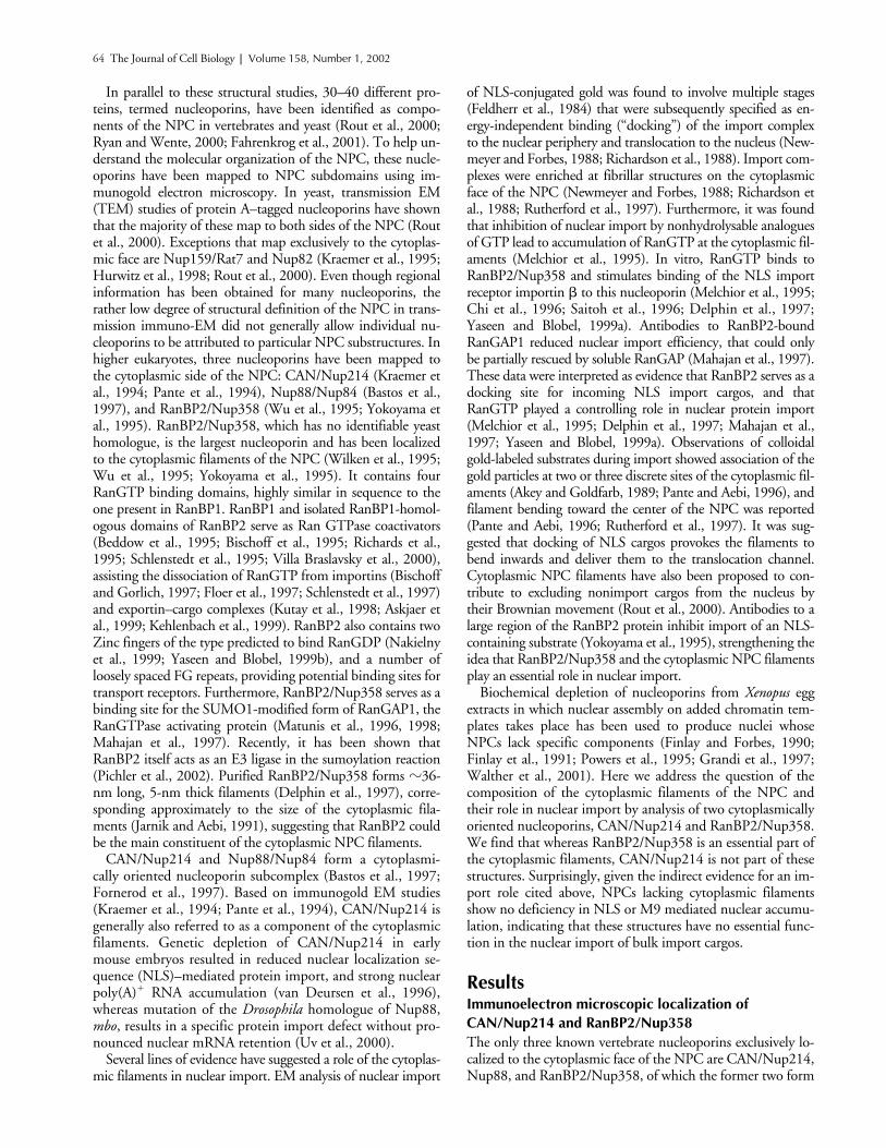
Immunoelectron microscopic localization of CAN/Nup214 and RanBP2/Nup358
The only three known vertebrate nucleoporins exclusively lo-calized to the cytoplasmic face of the NPC are CAN/Nup214,Nup88, and RanBP2/Nup358, of which the former two form
NPC filaments and nuclear import |
Walther et al. 65
a subcomplex. Because we intended to functionally character-ize the role of the cytoplasmic filaments in nuclear transport,we first wished to reinvestigate the localization of RanBP2/Nup358 and CAN/Nup214 within the NPC. To this end weanalyzed immunogold labeled
Xenopus laevis
oocyte NEs us-ing field emission in-lens scanning EM (FEISEM), whichprovides a surface view of the NPC, and TEM, providing across-sectional view. For immunolocalization of RanBP2/Nup358, two polyclonal antibodies were used. One, anti-Nup358F, had been raised against a recombinant COOH-terminal segment, comprising amino acids 2501–2900 of thehuman homologue. The other, anti-Nup358V, was directedagainst amino acids 2285–2314 of human Nup358, of whichresidues 2290–2314 are identical in
Xenopus
and mammals.For immunolocalization of CAN/Nup214, polyclonal anti-bodies were raised against an NH
2
-terminal segment of the
Xenopus
protein, comprising amino acids 1–213. All antibod-ies were affinity purified and recognized proteins of expectedsizes in Western blots of
Xenopus
cell extracts (see Fig. 3 A).For immuno-EM, isolated NEs were incubated with
primary antibodies, followed by labeling with 10-nmgold-conjugated secondary antibodies. Representative im-ages of FEISEM micrographs are shown in Fig. 1, A(CAN/Nup214), B (RanBP2/Nup358, antibody 358F),and C (RanBP2/Nup358, antibody 358V). The localiza-tion of at least 100 gold-labeled antibodies was deter-mined for each nucleoporin by measuring the distancefrom the center of the NPC to the center of the gold-labeled antibodies. No significant labeling of the nuclearface was observed for any of the antibodies. The summaryof the data collected for each of the three antibodies isshown in Table I. Anti-CAN/Nup214 antibody labeled
Figure 1. Isolated Xenopus oocyte NEs were fixed and labeled with affinity-purified anti-CAN or anti-RanBP2 antibodies, followed by 10-nm gold-conjugated secondary antibodies, and analyzed by FEISEM and TEM. (A–C) Representative FEISEM images of the cytoplasmic face of the NE labeled with 10-nm gold particles (artificially colored yellow) recognizing (A) anti-CAN/Nup214 antibodies; (B) anti-RanBP2/Nup358F antibodies; and (C) anti-RanBP2/Nup358V antibodies. Bars, 100 nm. (D–F) Repre-sentative TEM images of thin sections cut through the NE decorated with 10-nm gold labeling (D) anti-CAN/Nup214 antibodies; (E) anti-RanBP2/Nup358F antibodies; and (F) anti-RanBP2/Nup358V antibodies. CF, cytoplasmic filaments; NP, nuclear pore; ONM, outer nuclear membrane; INM, inner nuclear mem-brane; N, nucleus; C, cytoplasm. Bar, 100 nm. (G) Diagrammatical representation of the NPC summarizing the localization data for CAN/Nup214 and RanBP2/Nup358 using anti-RanBP2/Nup358F (green), anti-RanBP2/Nup358V (red), and anti-CAN/Nup214 (blue) antibodies. Putative positioning of substructures of the NPC are shaded in gray. Sizes and positions of substructures were derived from (Akey, 1989; Jarnik and Aebi, 1991; Akey and Radermacher, 1993; Goldberg and Allen, 1996) and our own measurements (unpublished data). Bar, 50 nm. Error bars represent standard deviations of the means.
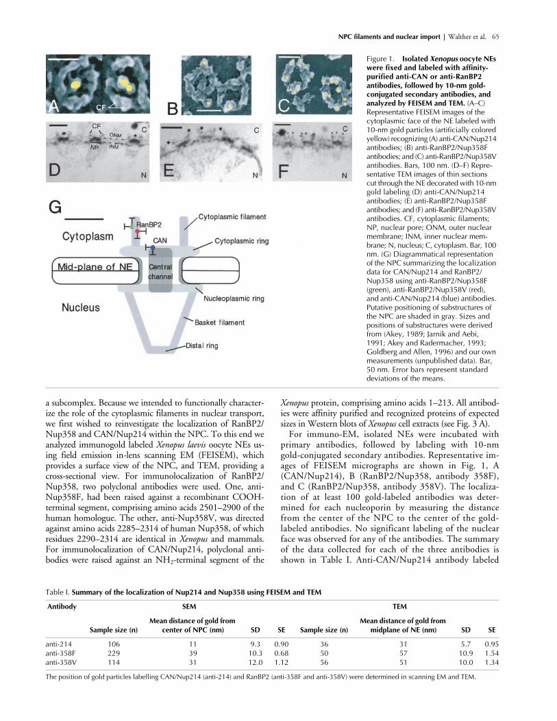
Table I.
Summary of the localization of Nup214 and Nup358 using FEISEM and TEM
Antibody SEM TEM
Sample size (n)Mean distance of gold from
center of NPC (nm) SD SE Sample size (n)Mean distance of gold from
midplane of NE (nm) SD SE
anti-214 106 11 9.3 0.90 36 31 5.7 0.95anti-358F 229 39 10.3 0.68 50 57 10.9 1.54anti-358V 114 31 12.0 1.12 56 51 10.0 1.34
The position of gold particles labelling CAN/Nup214 (anti-214) and RanBP2 (anti-358F and anti-358V) were determined in scanning EM and TEM.
66 The Journal of Cell Biology
|
Volume 158, Number 1, 2002
centrally, at a mean distance of 11 nm (
�
SE 0.9) from thecenter of the NPC, corresponding to the inner aspect ofthe cytoplasmic ring or the cytoplasmic entrance of thetranslocation channel. In contrast, both anti-Nup358 an-tibodies labeled more peripheral structures of the NPC, ata mean distance of 39 nm
�
0.68 for anti-Nup358F, and31 nm
�
1.12 for anti-Nup358V, corresponding to a po-sition on the cytoplasmic filaments.
To obtain measurements along the central axis, isolatedNEs were labeled with anti-CAN/Nup214, anti-RanBP2/Nup358F (Fig. 1 E), and anti-RanBP2/Nup358V (Fig. 1F), and processed for visualization using TEM. Gold parti-cle distribution in cross-sectional images through the NEwas measured as distance from the NE midplane. Again,Nup214 labeling was found closer to the core structures ofthe NPC, with a mean distance of 31 nm
�
0.95 (
n
�
36).In contrast, Nup358F labeling was at a mean distance of 57nm
�
1.54 (
n
�
50), and Nup358V labeling was found sig-nificantly more proximal to the NE at a mean distance of51
�
1.34 nm (
n
�
56;
P
�
0.01).As summarized in Table I and Fig. 1 G, a significant
difference between the distribution of labeling of CANand RanBP2 was observed, with the anti RanBP2 anti-bodies labeling more peripherally and deeper into the cy-toplasm. Compared with the labeling for CAN/Nup214,the anti-Nup358 labeling generally resulted in a widerdata spread. These data indicate that RanBP2 but notCAN localizes to the cytoplasmic filaments of the NPC,and suggest that the COOH terminus of RanBP2 is ori-ented toward the cytoplasm.
In vivo decoration of cytoplasmic NPC filaments with RanBP2 antibodies does not prevent NLS-mediated nuclear import in
Xenopus
oocytes
As a first approach to assess a possible involvement of the cy-toplasmic filaments in nuclear protein import, we attemptedto sterically occlude these filaments in vivo, and thus preventthem from binding partner proteins. To this end, high con-centrations of anti-Nup358V were injected into the cyto-plasm of living
Xenopus
oocytes. These injections resulted inantibody sequestration at the cytoplasmic fibers bound eitherto NPCs or the pore complexes of the annulate lamellae (AL)(see below; Cordes et al., 1997a). At later time points, thesame cells were reinjected with transport substrates, incubatedfurther, and finally analyzed for nuclear import. Given that astage VI oocyte contains an average total of
�
2.4
�
10
8
porecomplexes (
�
80% of which are AL pore complexes, 20%NPCs; Cordes et al., 1995), and supposing that maximally 16copies of RanBP2 are present per pore complex, the uppervalue of RanBP2 copies per oocyte, including a minor pool ofsoluble RanBP2, can be estimated at
�
4
�
10
9
. To be able tosaturate this cellular pool of RanBP2, affinity-purified anti-bodies in initial experiments were injected at concentrationswhich resulted in a
10
2
-fold molar surplus of antibody toRanBP2 in the oocyte. No inhibitory effects on BSA-NLS im-port were observed (unpublished data; Cordes et al., 1997a).
These investigations did not rule out that some NPCshad escaped immunoblocking and were still capable ofRanBP2-mediated nuclear protein import. To visualize theeffect of RanBP2 antibody-binding on nuclear transloca-tion through individual NPCs, subsequent microinjection
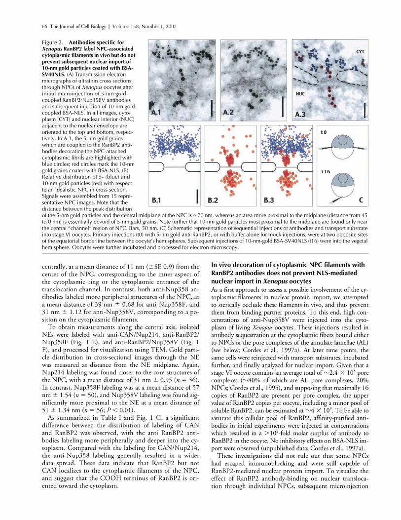
Figure 2. Antibodies specific for Xenopus RanBP2 label NPC-associated cytoplasmic filaments in vivo but do not prevent subsequent nuclear import of 10-nm gold particles coated with BSA-SV40NLS. (A) Transmission electron micrographs of ultrathin cross sections through NPCs of Xenopus oocytes after initial microinjection of 5-nm gold-coupled RanBP2/Nup358V antibodies and subsequent injection of 10-nm gold-coupled BSA-NLS. In all images, cyto-plasm (CYT) and nuclear interior (NUC) adjacent to the nuclear envelope are oriented to the top and bottom, respec-tively. In A.3, the 5-nm gold grains which are coupled to the RanBP2 anti-bodies decorating the NPC-attached cytoplasmic fibrils are highlighted with blue circles; red circles mark the 10-nm gold grains coated with BSA-NLS. (B) Relative distribution of 5- (blue) and 10-nm gold particles (red) with respect to an idealistic NPC in cross section. Signals were assembled from 15 repre-sentative NPC images. Note that the distance between the peak distribution of the 5-nm gold particles and the central midplane of the NPC is �70 nm, whereas an area more proximal to the midplane (distance from 45 to 0 nm) is essentially devoid of 5-nm gold grains. Note further that 10-nm gold particles most proximal to the midplane are found only near the central “channel” region of NPC. Bars, 50 nm. (C) Schematic representation of sequential injections of antibodies and transport substrate into stage VI oocytes. Primary injections (t0) with 5-nm gold anti-RanBP2, or with buffer alone for mock injections, were at two opposite sites of the equatorial borderline between the oocyte’s hemispheres. Subsequent injections of 10-nm-gold BSA-SV40NLS (t16) were into the vegetal hemisphere. Oocytes were further incubated and processed for electron microscopy.
NPC filaments and nuclear import |
Walther et al. 67
experiments were performed with RanBP2 antibodies cou-pled to 5-nm gold, and the transport cargo BSA-NLS wascoated onto 10-nm gold particles. As illustrated in Fig. 2 C,the gold-coupled RanBP2 antibodies were injected into thecytoplasm of several oocytes first, followed by several hoursincubation to allow sequestration of antibodies at the nu-clear periphery as confirmed by electron microscopy ofsome oocytes fixed directly afterwards (see Materials and
methods; Cordes et al., 1997a). The remaining oocyteswere reinjected with the 10-nm gold-coupled transportcargo and later fixed and processed for TEM.
A selection of resulting micrographs is shown in Fig. 2A. The 5-nm gold-coupled RanBP2 antibodies specificallyaccumulated at the cytoplasmic side of the NE, oftendensely decorating the cytoplasmic fibrils projecting fromthe NPCs. After prolonged incubation, RanBP2 antibod-
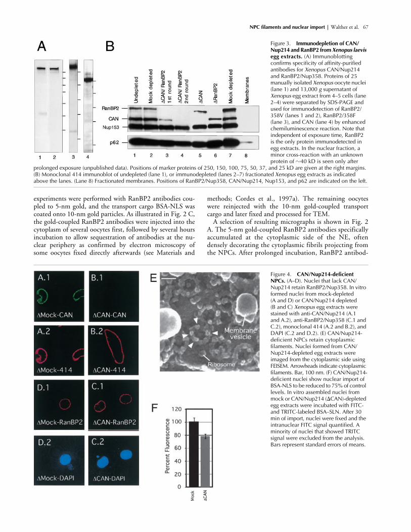
Figure 3. Immunodepletion of CAN/Nup214 and RanBP2 from Xenopus laevis egg extracts. (A) Immunoblotting confirms specificity of affinity-purified antibodies for Xenopus CAN/Nup214 and RanBP2/Nup358. Proteins of 25 manually isolated Xenopus oocyte nuclei (lane 1) and 13,000 g supernatant of Xenopus egg extract from 4–5 cells (lane 2–4) were separated by SDS-PAGE and used for immunodetection of RanBP2/358V (lanes 1 and 2), RanBP2/358F (lane 3), and CAN (lane 4) by enhanced chemiluminescence reaction. Note that independent of exposure time, RanBP2 is the only protein immunodetected in egg extracts. In the nuclear fraction, a minor cross-reaction with an unknown protein of �40 kD is seen only after
prolonged exposure (unpublished data). Positions of marker proteins of 250, 150, 100, 75, 50, 37, and 25 kD are given at the right margins. (B) Monoclonal 414 immunoblot of undepleted (lane 1), or immunodepleted (lanes 2–7) fractionated Xenopus egg extracts as indicated above the lanes. (Lane 8) Fractionated membranes. Positions of RanBP2/Nup358, CAN/Nup214, Nup153, and p62 are indicated on the left.
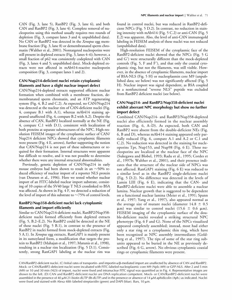
Figure 4. CAN/Nup214-deficient NPCs. (A–D). Nuclei that lack CAN/Nup214 retain RanBP2/Nup358. In vitro formed nuclei from mock-depleted (A and D) or CAN/Nup214 depleted (B and C) Xenopus egg extracts were stained with anti-CAN/Nup214 (A.1 and A.2), anti-RanBP2/Nup358 (C.1 and C.2), monoclonal 414 (A.2 and B.2), and DAPI (C.2 and D.2). (E) CAN/Nup214-deficient NPCs retain cytoplasmicfilaments. Nuclei formed from CAN/Nup214-depleted egg extracts were imaged from the cytoplasmic side using FEISEM. Arrowheads indicate cytoplasmic filaments. Bar, 100 nm. (F) CAN/Nup214-deficient nuclei show nuclear import of BSA-NLS to be reduced to 75% of control levels. In vitro assembled nuclei from mock or CAN/Nup214 (CAN)-depleted egg extracts were incubated with FITC- and TRITC-labeled BSA–SLN. After 30 min of import, nuclei were fixed and the intranuclear FITC signal quantified. A minority of nuclei that showed TRITC signal were excluded from the analysis. Bars represent standard errors of means.
68 The Journal of Cell Biology
|
Volume 158, Number 1, 2002
ies were not only found attached to these NPC-proximalfibrils alone, but started to sequester within a region of upto 500 nm surrounding the nucleus (unpublished data).With the exception of the dense labeling of the AL porecomplexes described earlier (Cordes et al., 1997a), no ac-
cumulation of 5-nm gold grains were seen in cytoplasmicareas more distal from the NE. 5-nm gold grains were notdetectable in the nuclear interior. 10-nm gold-labeledBSA-NLS particles were enriched at NPCs (Fig. 2 A) andAL pore complexes (unpublished data; Cordes et al.,
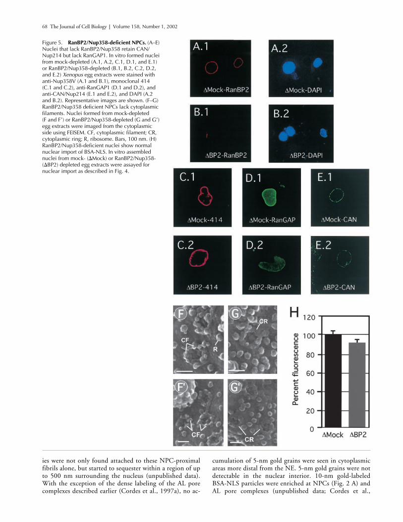
Figure 5. RanBP2/Nup358-deficient NPCs. (A–E) Nuclei that lack RanBP2/Nup358 retain CAN/Nup214 but lack RanGAP1. In vitro formed nuclei from mock-depleted (A.1, A.2, C.1, D.1, and E.1) or RanBP2/Nup358-depleted (B.1, B.2, C.2, D.2, and E.2) Xenopus egg extracts were stained with anti-Nup358V (A.1 and B.1), monoclonal 414 (C.1 and C.2), anti-RanGAP1 (D.1 and D.2), and anti-CAN/Nup214 (E.1 and E.2), and DAPI (A.2 and B.2). Representative images are shown. (F–G) RanBP2/Nup358 deficient NPCs lack cytoplasmic filaments. Nuclei formed from mock-depleted(F and F�) or RanBP2/Nup358-depleted (G and G�) egg extracts were imaged from the cytoplasmic side using FEISEM. CF, cytoplasmic filament; CR, cytoplasmic ring; R, ribosome. Bars, 100 nm. (H) RanBP2/Nup358-deficient nuclei show normal nuclear import of BSA-NLS. In vitro assembled nuclei from mock- (Mock) or RanBP2/Nup358- (BP2) depleted egg extracts were assayed for nuclear import as described in Fig. 4.
NPC filaments and nuclear import |
Walther et al. 69
1997a), and were also found throughout the nuclear inte-rior, independently of whether cells had initially been in-jected with RanBP2 antibodies or mock injected withbuffer alone. Despite this dense decoration of the cyto-plasmic fibrils with 5-nm gold grains, the 10-nm goldparticles in such NPCs were found at both the outer andinner side of the cross-sectioned electron-dense centralmidplane (Fig. 2 B). This indicated that the central trans-location site within the NPC had remained accessible foreven large cargo molecules (the diameter of a BSA-coated10-nm gold grain can reach
�
16 nm) and capable of me-diating nuclear import.
Immunodepletion of CAN/Nup214 andRanBP2/Nup358 from
Xenopus laevis
egg extracts
To more directly assess the role of CAN and RanBP2 in nu-clear import, we made use of in vitro nuclear assembly usingCAN and/or RanBP2 immunodepleted
Xenopus laevis
egg ex-tracts (Finlay and Forbes, 1990). Affinity-purified anti-CAN,anti-RanBP2 (358V), or a mixture of the two antibodies werecrosslinked to protein A Sepharose, and 400
�
l of fraction-ated interphase egg cytosol was subjected to two rounds ofimmunodepletion (Fig. 3). Western blot analysis of single- ordouble-depleted extracts probed with monoclonal antibody(mAb) 414 showed a very efficient and specific removal of
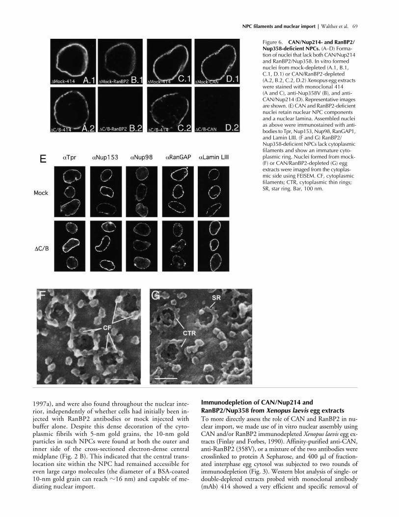
Figure 6. CAN/Nup214- and RanBP2/Nup358-deficient NPCs. (A–D) Forma-tion of nuclei that lack both CAN/Nup214 and RanBP2/Nup358. In vitro formed nuclei from mock-depleted (A.1, B.1, C.1, D.1) or CAN/RanBP2-depleted (A.2, B.2, C.2, D.2) Xenopus egg extracts were stained with monoclonal 414 (A and C), anti-Nup358V (B), and anti-CAN/Nup214 (D). Representative images are shown. (E) CAN and RanBP2-deficient nuclei retain nuclear NPC components and a nuclear lamina. Assembled nuclei as above were immunostained with anti-bodies to Tpr, Nup153, Nup98, RanGAP1, and Lamin LIII. (F and G) RanBP2/Nup358-deficient NPCs lack cytoplasmic filaments and show an immature cyto-plasmic ring. Nuclei formed from mock- (F) or CAN/RanBP2-depleted (G) egg extracts were imaged from the cytoplas-mic side using FEISEM. CF, cytoplasmic filaments; CTR, cytoplasmic thin rings; SR, star ring. Bar, 100 nm.
70 The Journal of Cell Biology
|
Volume 158, Number 1, 2002
Figure 7.
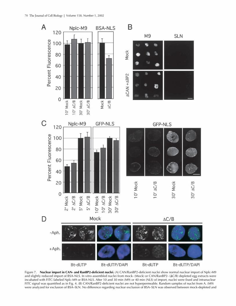
Nuclear import in CAN- and RanBP2-deficient nuclei.
(A) CAN/RanBP2-deficient nuclei show normal nuclear import of Nplc-M9 and slightly reduced import of BSA-NLS. In vitro assembled nuclei from mock- (Mock) or CAN/RanBP2- (
C/B) depleted egg extracts were incubated with FITC-labeled Nplc-M9 or BSA-NLS. After 10 and 30 min (M9) or 40 min (NLS) of import, nuclei were fixed and intranuclear FITC signal was quantified as in Fig. 4. (B) CAN/RanBP2-deficient nuclei are not hyperpermeable. Random samples of nuclei from A. (M9) were analyzed for exclusion of BSA–SLN. No difference regarding nuclear exclusion of BSA–SLN was observed between mock-depleted and
NPC filaments and nuclear import |
Walther et al. 71
CAN (Fig. 3, lane 5), RanBP2 (Fig. 3, lane 6), and bothCAN and RanBP2 (Fig. 3, lane 4). Complete removal of nu-cleoporins using this method usually requires two rounds ofdepletion (Fig. 3, compare lanes 3 and 4; unpublished data).No CAN or RanBP2 was detected in the
Xenopus
egg mem-brane fraction (Fig. 3, lane 8) or demembranated sperm chro-matin (Walther et al., 2001). Nontargeted nucleoporins werestill present in depleted extracts (Fig. 3, lanes 4–6); however, asmall fraction of p62 was consistently codepleted with CAN(Fig. 3, lanes 4 and 5; unpublished data). Mock-depleted ex-tracts were not affected in mAb414-reactive nucleoporincomposition (Fig. 3, compare lanes 1 and 2).
CAN/Nup214-deficient nuclei retain cytoplasmic filaments and have a slight nuclear import defect
CAN/Nup214-depleted extracts supported efficient nuclearformation when combined with a membrane fraction, de-membranated sperm chromatin, and an ATP regeneratingsystem (Fig. 4, B.2 and C.2). As expected, no CAN/Nup214was detected at the nuclear rim of CAN-deficient nuclei (Fig.4, compare B.1 with A.1), whereas mAb414 staining ap-peared unaffected (Fig. 4, compare B.2 with A.2). Despite theabsence of CAN, RanBP2 localized normally at the NE (Fig.4, compare C.1 with D.1), consistent with localization ofboth proteins at separate substructures of the NPC. High-res-olution FEISEM images of the cytoplasmic surface of CAN/Nup214 deficient NPCs showed that cytoplasmic filamentswere present (Fig. 4 E, arrows), further supporting the notionthat CAN/Nup214 is not part of these substructures or re-quired for their formation. Internal structures were apparentbut difficult to resolve, and it was not possible to determinewhether there were any internal structural abnormalities.
Previously, genetic depletion of CAN/Nup214 in earlymouse embryos had been shown to result in an
�
50% re-duced efficiency of nuclear import of a reporter NLS protein(van Deursen et al., 1996). Here we tested whether nuclearimport of an FITC-labeled nuclear import substrate, consist-ing of 10 copies of the SV40 large T NLS crosslinked to BSAwas affected. As shown in Fig. 4 F, we detected a reduction ofthe level of import of this substrate to
�
75% of control levels.
RanBP2/Nup358-deficient nuclei lack cytoplasmic filaments and import efficiently
Similar to CAN/Nup214-deficient nuclei, RanBP2/Nup358-deficient nuclei formed efficiently from depleted extracts(Fig. 5, B.2–E.2). No RanBP2 could be detected at the NEof these nuclei (Fig. 5 B.1), in contrast to the presence ofRanBP2 in nuclei formed from mock-depleted extracts (Fig.5 A.1). In
Xenopus
egg extracts, RanGAP1 is mainly presentin its sumoylated form, a modification that targets the pro-tein to RanBP2 (Mahajan et al., 1997; Matunis et al., 1998),resulting in a nuclear rim localization (Fig. 5 D.1). Consis-tently, strong RanGAP1 labeling at the nuclear rim was
found in control nuclei, but was reduced in RanBP2-defi-cient NPCs (Fig. 5 D.2). In contrast, no reduction in stain-ing intensity with mAb414 (Fig. 5 C.2) or anti-CAN (Fig. 5E.2) was apparent. Also, the level of anti-CAN immunogoldlabeling in FEISEM analysis of these nuclei was not reduced(unpublished data).
High-resolution FEISEM of the cytoplasmic face of theRanBP2-deficient nuclei showed that the NPCs (Fig. 5 Gand G
�
) were structurally different than the mock-depletedcontrols (Fig. 5, F and F
�
), and that only the coaxial cyto-plasmic ring, but not the filaments, was still visible. How-ever, in the absence of cytoplasmic filaments, nuclear importof BSA-NLS (Fig. 5 H) or nucleoplasmin core M9 (unpub-lished data; see below) was not significantly affected (Fig. 5H). Nuclear import was signal dependent, as BSA coupledto a nonfunctional “reverse NLS” peptide was excludedfrom RanBP2 deficient nuclei (see below).
CAN/Nup214- and RanBP2/Nup358-deficient nuclei exhibit aberrant NPC morphology but show no further import defectCombined CAN/Nup214- and RanBP2/Nup358-depletednuclei also efficiently formed in the nuclear assemblyreaction (Fig. 6, A–D). As expected, both CAN andRanBP2 were absent from the double-deficient NEs (Fig.6, B and D), whereas mAb414 staining appeared only par-tially reduced (Fig. 6, compare A.1 and A.2 or C.1 andC.2). No reduction was detected in the staining for nucle-oporins Tpr, Nup153, and Nup98 (Fig. 6 E). These nu-cleoporins are localized at the nuclear face of the NPC(Sukegawa and Blobel, 1993; Radu et al., 1995; Cordes etal., 1997b; Walther et al., 2001), and their presence indi-cates that the structure of the nuclear side of NPCs wasnot affected. RanGAP1 staining (Fig. 6 E) was reduced toa similar level as in the RanBP2 single-deficient nuclei(Fig. 5 D.2). No difference was detected in the levels ofLamin LIII (Fig. 6 E), indicating that the CAN- andRanBP2-deficient nuclei were able to assemble a nuclearlamina. Nuclear growth that is suggested to be dependenton a functional nuclear lamina (Meier et al., 1991; Spannet al., 1997; Yang et al., 1997), also appeared normal asthe average size of mutant nuclei (diameter 14.8 � 0.5�m) was similar to control nuclei (15.6 � 0.6 �m).FEISEM imaging of the cytoplasmic surface of the dou-ble-deficient nuclei revealed a striking structural NPCphenotype (Fig. 6 F and G). None of the NPCs visualizedappeared completely assembled; instead, most had eitheronly a star ring or a cytoplasmic thin ring, which havebeen recognized as NPC assembly intermediates (Gold-berg et al., 1997). The tips of some of the star ring sub-units appeared to be buried in the NE as previously de-scribed (Fig. 6 G, arrow). No obvious cytoplasmic coaxialrings or cytoplasmic filaments were present.
CAN/RanBP2-deficient nuclei. (C) Initial rates of transportin- and importin �/�-mediated import are unaffected by absence of CAN and RanBP2. Mock- or CAN/RanBP2-defecient nuclei were incubated with FITC-labeled nucleoplasmic core M9 (Nplc-M9) or GFP-NLS. After 2 and 5 min (M9) or 10 and 30 min (NLS) of import, nuclei were fixed and intranuclear FITC signal was quantified as in Fig. 4. Representative images are shown to the left. (D) CAN and RanBP2-deficient nuclei are DNA replication competent. Mock- or CAN/RanBP2-deficient nuclei were assembled in the presence of 40 �M 21-biotin dUTP (Bt-dUTP) and in the presence or absence of 14 �M aphidicolin (Aph.) as indicated. Nuclei were fixed and stained with Alexa 488–labeled streptavidin (green) and DAPI (blue). Bars, 10 �m.

72 The Journal of Cell Biology | Volume 158, Number 1, 2002
In spite of the strikingly aberrant cytoplasmic phenotypeof the CAN/Nup214 and RanBP2/Nup358 double-defi-cient NPCs, import of BSA-NLS was only reduced to�70% of control levels (Fig. 7 A) similar to the reductionobserved for the CAN/Nup214 single-depleted nuclei (Fig.4 G). Import of fluorescently labeled Nplc-M9 (Englmeieret al., 1999) was equally efficient in nuclei formed fromCAN/RanBP2 double- or mock-depleted extract (Fig. 7 A).Nuclear import into CAN- and RanBP2-deficient nucleiwas signal dependent, as the inert control substrate BSA–“reverseNLS” (BSA–SLN) was equally well excluded fromcontrols as from the double deficient nuclei (Fig. 7 B). Totest for possible kinetic differences in nuclear protein importbetween control and CAN/RanBP2-deficient nuclei, nuclearaccumulation of Nplc-M9 and GFP-NLS was measured atearly time points (2 and 10 min, respectively) after substrateaddition. As shown in Fig. 7 C, no differences in importrates were measured between nuclei formed from mock- andCAN/RanBP2-depleted extracts. Note that compared withthe multivalent BSA-NLS substrate (Fig. 7 A), nuclear im-port of the smaller, monovalent GFP-NLS import substratein CAN- and RanBP2-deficient nuclei was less affected.
Synthetic nuclei are able to initiate a single round of semi-conservative DNA replication (Blow and Laskey, 1986),which is dependent on their ability to concentrate factors forDNA replication (Walter et al., 1998). As a functional testfor import of natural cargos, we assayed incorporation of thethymidine analogue 21-biotin-dUTP into the DNA of nu-clei lacking CAN and RanBP2. As shown in Fig. 7 D, bothmutant and control nuclei showed a similar level of diffusenuclear staining with numerous bright foci. The majority ofthis signal was absent when DNA polymerase � was inhib-ited by aphidicolin (Fig. 7 D), indicating that the signal re-sulted from DNA replication. We conclude that in the ab-sence of CAN and RanBP2, nuclei are capable of normalaccumulation of replication factors.
DiscussionIn this study, we assessed the function of the cytoplasmicNPC filaments in nuclear protein import. Previously, thecytoplasmic filaments have been implicated in protein im-port due to their colocalization with injected NLS–gold con-jugates, association with RanGTP and importin �, by anti-body inhibition experiments, and because of their potentialto exclude macromolecules with no affinity for nucleoporins(Melchior et al., 1995; Yokoyama et al., 1995; Pante andAebi, 1996; Mahajan et al., 1997; Rutherford et al., 1997;Yaseen and Blobel, 1999a; Rout et al., 2000).
The composition of the cytoplasmic filamentsTwo proteins are commonly referred to as components ofthe cytoplasmic filaments, RanBP2/Nup358 and CAN/Nup214 (Ohno et al., 1998; Stoffler et al., 1999a; Allen etal., 2000). Our localization of CAN places it away from thefilaments, near the cytoplasmic entrance of the translocationchannel. This conclusion is derived from three observations.First, we have used a high resolution scanning immuno-EMtechnique that allows simultaneous visualization of the cyto-plasmic surface of the NPC and CAN/Nup214-reactive im-
munogold labeling and thereby shown a localization ofCAN close to the center of the NPC. Second, transmissionimmuno-EM analysis shows labeling of CAN/Nup214 at aposition close to the center of the NPC rather than a local-ization at the cytoplasmic filaments. Third, immunodeple-tion of RanBP2/Nup358 resulted in formation of NPCslacking cytoplasmic filaments, yet CAN/Nup214 remainedpresent on these NPCs. The immature phenotype of the cy-toplasmic rings in NPCs codepleted for CAN and RanBP2,as compared with RanBP2-deficient ones, further argues fora localization of CAN/Nup214 near the cytoplasmic ringrather than on the filaments.
In two previous studies using TEM, it had been concludedthat Nup214 is a component of the cytoplasmic filaments(Kraemer et al., 1994; Pante et al., 1994). However, the im-ages presented in one of these studies (Kraemer et al., 1994)revealed a localization that is consistent with ours, using anantibody directed to a central domain of CAN. From the po-sition of the 11 gold particles presented in their study, amean distance of 27 nm (� a standard deviation of 7) fromthe midplane of the NE and 10 nm � 7 from the center ofthe NPC can be determined. These values lie within thespread, and even within the error of our localization data.The second study, using a serum against full-length nativerat protein, presented conclusive labeling of the cytoplasmicfilaments (Pante et al., 1994). Therefore, we cannot excludethat the COOH-terminal part of CAN is localized in thisposition. However, the specificity of the anti–rat CANantiserum for the Xenopus protein has not been reported.Biochemical identification of a nucleoporin subcomplexcontaining CAN/Nup214 and p62 (Finlay et al., 1991; Mat-suoka et al., 1999) is consistent with the localization ofCAN/Nup214 at the cytoplasmic face of the translocationchannel. p62 itself has been localized at both entrances of theNPC translocation channel using preembedding (Cordes etal., 1995; Guan et al., 1995) and postembedding immuno-TEM (Grote et al., 1995). Our study shows a CAN/Nup214localization similar to the localization of Nup159/Rat7(Kraemer et al., 1995; Rout et al., 2000), the closest yeasthomologue to vertebrate CAN/Nup214, suggesting a greaterdegree of functional conservation between these two nucle-oporins than could earlier be assumed. The position ofCAN/Nup214 close to the translocation channel may allowits FG repeats to contribute to the hydrophobic FG repeatnetwork that is hypothesized to fill the translocation channel(Grote et al., 1995; Ribbeck and Gorlich, 2001).
Decoration with two different antibodies localizedRanBP2/Nup358 on the cytoplasmic filaments of the NPC,in agreement with previous studies (Wilken et al., 1995; Wuet al., 1995; Yokoyama et al., 1995). Two additional lines ofevidence suggest that RanBP2 is the major structural compo-nent of the cytoplasmic filaments. First, electron microscopicanalysis of purified RanBP2 shows particles with a filamen-tous shape and a length of �36 nm, which is both consistentwith the approximate size of the cytoplasmic filaments andthe length of RanBP2’s amino acid sequence (Delphin et al.,1997). Second, depletion of RanBP2 leads to absence of fila-ments from the cytoplasmic face of the NPC.
The use of the two antibodies raised against different re-gions of RanBP2 in the same study has allowed us to com-
NPC filaments and nuclear import | Walther et al. 73
pare their localization relative to each other. The 358V anti-bodies recognize a small region corresponding to aminoacids 2290–2314 of the human RanBP2 protein (Wu et al.,1995; Yokoyama et al., 1995), whereas the 358F antibodiesrecognize a region closer to the COOH terminus and corre-spond to amino acids 2501–2900. Our data show that the358F epitopes are localized at a significantly more distal po-sition than the 358V epitopes, suggesting that the protein isoriented with its COOH terminus pointing outward intothe cytoplasm. Consequently, its NH2 terminus, containingpredicted coiled-coil and leucine zipper sequences (Wu etal., 1995; Yokoyama et al., 1995), may provide the interac-tion domains with the core NPC.
RanBP2/Nup358 is conserved among metazoa but absentin the yeast Saccharomyces cerevisiae. Therefore, it remains tobe determined whether yeast contains cytoplasmic filamentsthat are functionally or structurally equivalent to those inmetazoa.
The cytoplasmic NPC filaments and nuclear importNuclei lacking CAN/Nup214 showed a relatively minor,25% reduction in basic NLS-mediated protein import. Ear-lier studies in CAN / mouse embryos showed a 50% re-duction in NLS protein import (van Deursen et al., 1996).Even though there is a difference in the degree of reductionbetween the two model systems, in both cases protein im-port is reduced but not eliminated. This is consistent withthe situation in yeast, where mutations in Nup159/Rat7have a limited effect on protein import (Del Priore et al.,1997; Belgareh et al., 1998; Hurwitz et al., 1998).
Most surprisingly, our results show that removal of RanBP2and the complete cytoplasmic filaments affects neither NLS-or M9-mediated nuclear import, nor the exclusion of nonim-port cargos. This holds true for multivalent exogenous cargos,as well as for the endogenous cargos Lamin LIII (Hennekes etal., 1993; Lourim and Krohne, 1993), Tpr (Cordes et al.,1998), and factors required for DNA replication (Walter etal., 1998). Also, Nup153, an endogenous cargo for transpor-tin (Nakielny et al., 1999), was localized correctly. These re-sults indicate that the cytoplasmic filaments do not act as es-sential docking sites for incoming import complexes as hadbeen suggested previously (Newmeyer and Forbes, 1988;Richardson et al., 1988; Akey and Goldfarb, 1989; Mooreand Blobel, 1992; Melchior et al., 1995; Pante and Aebi,1996; Mahajan et al., 1997; Rutherford et al., 1997), and theefficient exclusion of BSA–SLN conjugate from depleted nu-clei suggests they are also not required for entropic exclusionof nonkaryophilic proteins (Rout et al., 2000).
How can this conclusion be reconciled with previous datalinking RanBP2 and the cytoplasmic filaments to an earlystep in protein import? EM studies have observed NLS cargoassociation with cytoplasmic NPC filaments during normalimport (Newmeyer and Forbes, 1988; Richardson et al.,1988; Akey and Goldfarb, 1989), suggesting a temporal ar-rest. However, these studies have not addressed the necessityof these associations. A stronger indication that the cytoplas-mic filaments were directly involved in import was the accu-mulation of NLS cargos at the NE, or more specifically at thefilaments, under certain conditions including depletion ofNTPs or Ran, addition of O-linked N-acetylglucosamine
binding lectin, or chilling to 4�C (Dabauvalle et al., 1988;Featherstone et al., 1988; Richardson et al., 1988; Mooreand Blobel, 1992; Chi et al., 1996; Rutherford et al., 1997).As argued previously (Englmeier et al., 1999), the perinuclearaccumulation of classical NLS substrates observed under con-ditions that deplete NTPs or Ran (Newmeyer et al., 1986;Richardson et al., 1988; Moore and Blobel, 1992; Melchioret al., 1995) is likely the result of depletion of RanGTP thatis needed to dissociate importin�-�/cargo complexes fromthe NPC (Rexach and Blobel, 1995; Gorlich et al., 1996).This accumulation may block the translocation channel(Kutay et al., 1997). Chilling is likely to stabilize interactionsof import complexes with NPC components, causing a simi-lar block of the NPC with endogenous cargos (Englmeier etal., 1999). Lectin or antibody binding to carbohydrate moi-eties on some nucleoporins is also likely to arrest NPC trans-location. Therefore, the observed “docking” of import com-plexes to the cytoplasmic filaments under these conditionsmay be caused by obstruction of nucleoporin sites within theNPC that mediate the import reaction. By blocking these,import complexes may associate or reassociate with sites thatwould normally not contribute to the import reaction, butserve another function. In this regard it is noteworthy thatthe FG repeats in RanBP2 are relatively sparse (Wu et al.,1995; Yokoyama et al., 1995), and have a low affinity for im-portin � (Ben-Efraim and Gerace, 2001). Alternatively, theassociation of import complexes with the filaments may func-tion to concentrate them in the vicinity of the core NPC, in-creasing import efficiency (Rout et al., 2000; Ben-Efraim andGerace, 2001). However, we have not detected reduced im-port rates due to lack of RanBP2 or cytoplasmic filaments,whereas we did detect a reduction in NLS-mediated importdue to depletion of CAN/Nup214.
The association of Ran with the cytoplasmic filaments inthe presence of nonhydrolysable GTP analogs (Melchior etal., 1995), can be explained by binding of RanGTP to theRBD domains of RanBP2 and does not necessarily representan early step of nuclear import. Transport induced bendingof the cytoplasmic filaments, described as a mechanism forimport (Pante and Aebi, 1996; Rutherford et al., 1997) maybe caused by the multivalence of the transport substratesthat were employed in these studies. NLS-coated gold parti-cles are likely to interact with multiple copies of importin �,enabling them to occasionally link FG repeats at the en-trance of the translocation channel (e.g., those of CAN/Nup214 or p62), to those of RanBP2.
Polyclonal antibodies raised against a GST-fused RanBP2fragment comprising two of the RanGTP binding domainsplus several Zinc fingers and FG repeats inhibited nuclearimport of an NLS containing substrate (Yokoyama et al.,1995). Even though we cannot exclude that certain antibod-ies to RanBP2/Nup358 interfere specifically with NLS im-port, these antibodies also labeled the nuclear face of theNPC (Yokoyama et al., 1995), suggesting that they mayhave crossreacted with other Zinc finger or FG repeat con-taining nucleoporins such as Nup153 or p62.
In conclusion, our data argue against an essential role of thecytoplasmic NPC filaments in NLS and M9 dependent pro-tein import, and by default support translocation models thatdo not involve them (Rexach and Blobel, 1995; Ben-Efraim
74 The Journal of Cell Biology | Volume 158, Number 1, 2002
and Gerace, 2001; Ribbeck and Gorlich, 2001). It remainspossible that the cytoplasmic filaments are required for importof cargos not imported via the classical NLS- and M9-importpathways, or for very large cargos, although the experimentsin which we coated RanBP2 with colloidal gold-labeled anti-bodies without affecting the import of substrates of diameterbetween 10–16 nm would argue against the latter.
It is noteworthy that the cyclophilin-like domain ofRanBP2 has been shown to interact with the 19S regulatoryparticle of the 26S proteasome (Ferreira et al., 1998). The19S particle is known to have unfolding/chaperone activity.Specific binding to RanBP2 may indicate a demand for anunfolding activity at the cytoplasmic periphery of the NPC.Although such an activity seems unlikely to be required forNPC translocation it may serve some other function. A fur-ther property of RanBP2 which may not be directly relatedto the actual nuclear translocation process is the recentlyidentified ability of RanBP2 to act as a SUMO-1 E3 ligase(Pichler et al., 2002). With the exception of RanGAP1 (Ma-tunis et al., 1996; Mahajan et al., 1997; Matunis et al.,1998) and RanBP2 itself (Pichler et al., 2002), all knownsumoylated proteins are predominately nuclear (for reviewsee Muller et al., 2001). The association of import cargoswith the filaments therefore may represent a temporal trans-port arrest to allow modification of specific cargos, ratherthan a general facilitating step in nuclear import.
Materials and methodsAntibodiesAnti-RanBP2 antibodies (358V) were raised in guinea pig against a peptidecorresponding to amino acids 2285–2314 of human RanBP2 (Wu et al.,1995; Yokoyama et al., 1995) of which aa 2290–2314 are identical to apartial sequence of RanBP2 from Xenopus laevis (GenBank/EMBL/DDBJaccession no. BG233383). The peptide was coupled via an NH2-terminalcysteine residue to maleimide-activated keyhole limpet hemocyanin andused for immunization as described (Cordes et al., 1997b). For affinity pu-rification of Igs, the peptide was coupled to Ultra Link Iodoacetyl on 3MEmphaze Biosupport Medium AB1 (Pierce Chemical Co.) and purified asdescribed (Cordes et al., 1997b). Antiserum 358F was provided by A. Gastand F. Melchior (Max Planck Institute for Biochemistry, Munich, Ger-many). To generate specific antibodies against Xenopus CAN (anti-214) anNH2-terminal fragment spanning amino acids 1–213 of the published se-quence (Askjaer et al., 1999) was expressed as a His6-tagged fusion pro-tein in pQE31 (QIAGEN) in XL1-Blue and purified under denaturing condi-tions. Antibodies were raised in rabbits and affinity purified against theantigen crosslinked to Affigel-15 (Biorad). To generate a resin for the de-pletion from Xenopus egg extracts, saturating amounts of antibody werebound to protein A Sepharose (Amersham Pharmacia Biotech) andcrosslinked with 10 mM dimethylpimelimidate (Sigma-Aldrich).
Preparation of colloidal gold-coupled proteinsCoupling to 5-nm colloidal gold, as well as preparation of BSA-SV40NLSconjugates and their coupling to 10-nm colloidal gold was performed essen-tially as described (Cordes et al., 1997a). To avoid overloads of proteinwhich may result in subsequent leaching of bound material, Igs or BSA-SV40NLS were added to the gold particles at amounts just sufficient to main-tain colloidal stability upon addition of NaCl (Slot and Geuze, 1984). Re-maining binding sites were blocked by incubating the gold suspension with0.1% BSA for 15 min before dilution in 20 vol of injection buffer (88 mMNaCl, 1 mM KCl, 20 mM Hepes, pH 7.4). To remove the excess of unboundprotein, the suspension was centrifuged at 10,000 g for 1 or 2 h, dependingon particle size. The supernatant and an occasionally observed minor solidpellet consisting of coagulated gold were discarded, whereas the fluffy andvery loose major part of the sediment, containing the gold conjugates in col-loidal form, was resuspended in injection buffer and washed again. Follow-ing final centrifugation, the loose sediment was collected as a concentrated,black suspension and used either directly for injection or stored at 4�C.
Microinjection and electron microscopy of injected oocytesMicroinjections into the cytoplasm of nondefolliculated Xenopus stage VIoocytes were performed at room temperature essentially as described(Cordes et al., 1997a). Four to six oocytes were injected for each experi-ment. Injections of gold-coupled antibodies were done at two sites oppo-site to each other at the equatorial borderline between animal and vegetalhemisphere. Injections of gold-coupled BSA-SV40NLS, or of gold-coupledBSA without NLS as control, were performed at a single site near the poleof the vegetal hemisphere. Oocytes were then incubated in modifiedBarth’s medium at either 2�C in the cold room or in a 19�C incubator for 3,5, 8, 16, 22, or 30 h. For experiments in which antibody injections pre-ceded the injection of gold-coupled BSA-SV40NLS, oocytes injected with5-nm gold anti-RanBP2, or mock-injected with buffer alone, were first in-cubated at 2�C for 16 h. Following subsequent injection of 10-nm goldBSA-SV40NLS, oocytes were further incubated at 19�C for 6 h. Oocyteswere then fixed in glutaraldehyde and processed for electron microscopyexactly as described (Cordes et al., 1997a).
EM immunolocalizationImmunolocalization of Nup214 and Nup358 on isolated Xenopus oocyteNEs using TEM and FEISEM was performed as previously described(Walther et al., 2001). For FEISEM analysis, antibodies were diluted 1:100with PBS before being incubated with isolated Xenopus oocyte NEs. The214 and 358F primary antibodies were labeled with 10-nm gold-conju-gated anti–rabbit secondary antibodies and the 358V antibody with 10-nmgold-conjugated anti–guinea pig antibody (Amersham Pharmacia Biotech),all at 1:20 dilution. Negative controls were performed and revealed thesecondary antibodies to be specific (unpublished data). Isolated NEs werevisualized at 100–300 kx magnification. To quantify the immunolocaliza-tion, measurements were made using AnalySIS (SIS) software. For TEMquantification, the midplane of the NE (i.e., a line parallel to the NE andequidistant from both the inner and outer nuclear membranes) for eachNPC was determined. The distances between the midplane of the NE andthe gold-labeled antibodies were measured at right angles to the midplane.For quantification of the position of nucleoporins by FEISEM, the gold-labeled antibodies were identified on the secondary electron image fromthe corresponding backscatter image. Circles of best fit were placed overeach NPC and each gold-labeled antibody, and the distances between thecenter of the NPCs and the center of the gold-labeled antibodies weremeasured.
FEISEM analysis of depleted nucleiAfter nuclear assembly, 4 �l of the assembly reaction was diluted with 1 mlMWB250 (250 mM sucrose, 50 mM KCl, 2.5 mM MgCl2, 50 mM Hepes,pH 8), and nuclei were spun down onto silicon chips at 800 g and fixed inmembrane fix (80 mM Pipes, pH 6.8; 1 mM MgCl2; 150 mM sucrose; 2%paraformaldehyde; 0.5% glutaraldehyde), for 30 min at room temperature.Samples were postfixed with 1% (wt/vol) OsO4, 0.2 M cacodylate, pH 7.4,and 1% (wt/vol) aqueous uranyl acetate for 10 min each, dehydratedthrough an ethanol series, and critical point dried from high purity CO2.Samples were sputter coated with 3 nm chromium and viewed using a Top-con DS130F FEISEM (Topcon Corporation) at 30 kV accelerating voltage.
Nuclear assembly, import and replication assays, andindirect immunofluorescenceFractionated egg extracts were prepared and depleted as described(Walther et al., 2001) except that the cytosolic fraction was depletedby two incubations with an equal volume of mock, anti-214, anti-RanBP2(358V), or a mixture of anti-214 and anti-RanBP2 resin, which waspreblocked with 5% BSA for 1 h. Assembly, import, and immunofluores-cence reactions were performed as described (Hetzer et al., 2000). In brief,for a 200-�l reaction, 13.5 �l cytosol, 1.5 �l energy mix (10 mM ATP, 100mM creatine phosphate, 2 mg/ml creatine kinase), 20 mg/ml glycogen(USB; Amersham Pharmacia Biotech) were mixed. 1 �l sperm chromatinfollowed by 2 �l of membrane fraction were added and the reaction incu-bated for 2 h at 20�C. For nuclear import reactions, a fluorescently labeledBSA-NLS conjugate (Palacios et al., 1996) or Nplc-M9-M10 (Englmeier etal., 1999) and an inert control substrate (BSA–SLN) were added and furtherincubated for various times. The samples were subsequently fixed in 4%formaldehyde and centrifuged through a cushion of 30% (wt/vol) sucroseonto an L-polylysine–coated coverslip and either directly monitored byconfocal microscopy or further processed for immunofluorescence as de-scribed (Arts et al., 1997). Xenopus RanGAP1 was detected by an antise-rum provided by M. Dasso (National Institutes of Health, Bethesda, MD),Nup98 with an antiserum to human Nup98 (Wu et al., 2001) that recog-
NPC filaments and nuclear import | Walther et al. 75
nizes Xenopus Nup98 (unpublished data), Tpr with purified antipeptideantibodies (Cordes et al., 1997b), Lamin LIII with monoclonal antibodyS49 (Lourim and Krohne, 1993), and Nup153 as described (Walther et al.,2001). Images were recorded with a Zeiss LSM 510 or a Leica TCS confo-cal microscope. Import reactions were quantified using NIH Image. Themean fluorescence of 10 or more images containing several nuclei eachwas determined. Error bars represent standard errors. DAPI staining forchromatin was used to verify that quantified objects were nuclei. Replica-tion of DNA was assayed by addition of 40 �M 21-Biotin-dUTP (CLON-TECH Laboratories, Inc.) to the nuclear assembly reactions in the presenceor absence of 14 �M of aphidicolin (Sigma-Aldrich). Samples were treatedas for immunofluorescence microscopy and incorporated biotin was de-tected with Alexa 488-linked streptavidin (Molecular Probes).
We gratefully acknowledge Dr. Hans-Richard Rackwitz for advice andhelp in colloidal gold-labeling of proteins, Drs. Andreas Gast and FraukeMelchior (Max Planck Institute for Biochemistry, Munich, Germany) forproducing and characterizing the anti-RanBP2 358F antibodies, and Drs.Mary Dasso and Jan van Deursen for antisera to RanGAP1 and Nup98. Wethank Martin Hetzer and Wolfram Antonin for discussions and Elisa Izaur-ralde, and Jan Ellenberg and members of the Mattaj laboratory for sugges-tions on the manuscript. Many thanks also to Sonja Reidenbach and San-dra Rutherford for technical assistance.
H.S. Pickersgill, M.W. Goldberg, and T.D. Allen were supported byCancer Research UK.
Submitted: 19 February 2002Revised: 29 April 2002Accepted: 24 May 2002
ReferencesAkey, C.W. 1989. Interactions and structure of the nuclear pore complex revealed
by cryo-electron microscopy. J. Cell Biol. 109:955–970.Akey, C.W., and D.S. Goldfarb. 1989. Protein import through the nuclear pore
complex is a multistep process. J. Cell Biol. 109:971–982.Akey, C.W., and M. Radermacher. 1993. Architecture of the Xenopus nuclear pore
complex revealed by three-dimensional cryo-electron microscopy. J. CellBiol. 122:1–19.
Allen, T.D., J.M. Cronshaw, S. Bagley, E. Kiseleva, and M.W. Goldberg. 2000.The nuclear pore complex: mediator of translocation between nucleus andcytoplasm. J. Cell Sci. 113:1651–1659.
Arts, G.J., L. Englmeier, and I.W. Mattaj. 1997. Energy- and temperature-depen-dent in vitro export of RNA from synthetic nuclei. Biol. Chem. 378:641–649.
Askjaer, P., A. Bachi, M. Wilm, F.R. Bischoff, D.L. Weeks, V. Ogniewski, M.Ohno, C. Niehrs, J. Kjems, I.W. Mattaj, and M. Fornerod. 1999. RanGTP-regulated interactions of CRM1 with nucleoporins and a shuttling DEAD-box helicase. Mol. Cell. Biol. 19:6276–6285.
Bastos, R., L. Ribas de Pouplana, M. Enarson, K. Bodoor, and B. Burke. 1997.Nup84, a novel nucleoporin that is associated with CAN/Nup214 on thecytoplasmic face of the nuclear pore complex. J. Cell Biol. 137:989–1000.
Beddow, A.L., S.A. Richards, N.R. Orem, and I.G. Macara. 1995. The Ran/TC4GTPase-binding domain: identification by expression cloning and characteriza-tion of a conserved sequence motif. Proc. Natl. Acad. Sci. USA. 92:3328–3332.
Belgareh, N., C. Snay-Hodge, F. Pasteau, S. Dagher, C.N. Cole, and V. Doye.1998. Functional characterization of a Nup159p-containing nuclear poresubcomplex. Mol. Biol. Cell. 9:3475–3492.
Ben-Efraim, I., and L. Gerace. 2001. Gradient of increasing affinity of importinbeta for nucleoporins along the pathway of nuclear import. J. Cell Biol. 152:411–417.
Bischoff, F.R., and D. Gorlich. 1997. RanBP1 is crucial for the release of RanGTPfrom importin beta-related nuclear transport factors. FEBS Lett. 419:249–254.
Bischoff, F.R., H. Krebber, E. Smirnova, W. Dong, and H. Ponstingl. 1995. Co-activation of RanGTPase and inhibition of GTP dissociation by Ran-GTPbinding protein RanBP1. EMBO J. 14:705–715.
Blow, J.J., and R.A. Laskey. 1986. Initiation of DNA replication in nuclei and pu-rified DNA by a cell-free extract of Xenopus eggs. Cell. 47:577–587.
Chi, N.C., E.J. Adam, G.D. Visser, and S.A. Adam. 1996. RanBP1 stabilizes the in-teraction of Ran with p97 nuclear protein import. J. Cell Biol. 135:559–569.
Cordes, V.C., S. Reidenbach, and W.W. Franke. 1995. High content of a nuclearpore complex protein in cytoplasmic annulate lamellae of Xenopus oocytes.Eur. J. Cell Biol. 68:240–255.
Cordes, V.C., H.R. Rackwitz, and S. Reidenbach. 1997a. Mediators of nuclear
protein import target karyophilic proteins to pore complexes of cytoplasmicannulate lamellae. Exp. Cell Res. 237:419–433.
Cordes, V.C., S. Reidenbach, H.R. Rackwitz, and W.W. Franke. 1997b. Identifi-cation of protein p270/Tpr as a constitutive component of the nuclear porecomplex-attached intranuclear filaments. J. Cell Biol. 136:515–529.
Cordes, V.C., M.E. Hase, and L. Muller. 1998. Molecular segments of protein Tprthat confer nuclear targeting and association with the nuclear pore complex.Exp. Cell Res. 245:43–56.
Dabauvalle, M.C., B. Schulz, U. Scheer, and R. Peters. 1988. Inhibition of nuclearaccumulation of karyophilic proteins in living cells by microinjection of thelectin wheat germ agglutinin. Exp. Cell Res. 174:291–296.
Delphin, C., T. Guan, F. Melchior, and L. Gerace. 1997. RanGTP targets p97 toRanBP2, a filamentous protein localized at the cytoplasmic periphery of thenuclear pore complex. Mol. Biol. Cell. 8:2379–2390.
Del Priore, V., C. Heath, C. Snay, A. MacMillan, L. Gorsch, S. Dagher, and C.Cole. 1997. A structure/function analysis of Rat7p/Nup159p, an essentialnucleoporin of Saccharomyces cerevisiae. J. Cell Sci. 110:2987–2999.
Englmeier, L., J.C. Olivo, and I.W. Mattaj. 1999. Receptor-mediated substratetranslocation through the nuclear pore complex without nucleotide triphos-phate hydrolysis. Curr. Biol. 9:30–41.
Fahrenkrog, B., D. Stoffler, and U. Aebi. 2001. Nuclear pore complex architectureand functional dynamics. Curr. Top. Microbiol. Immunol. 259:95–117.
Featherstone, C., M.K. Darby, and L. Gerace. 1988. A monoclonal antibodyagainst the nuclear pore complex inhibits nucleocytoplasmic transport ofprotein and RNA in vivo. J. Cell Biol. 107:1289–1297.
Feldherr, C.M., E. Kallenbach, and N. Schultz. 1984. Movement of a karyophilicprotein through the nuclear pores of oocytes. J. Cell Biol. 99:2216–2222.
Ferreira, P.A., C. Yunfei, D. Schick, and R. Roepman. 1998. The cyclophilin-likedomain mediates the association of Ran-binding protein 2 with subunits of the19 S regulatory complex of the proteasome. J. Biol. Chem. 273:24676–24682.
Finlay, D.R., and D.J. Forbes. 1990. Reconstitution of biochemically altered nu-clear pores: transport can be eliminated and restored. Cell. 60:17–29.
Finlay, D.R., E. Meier, P. Bradley, J. Horecka, and D.J. Forbes. 1991. A complex ofnuclear pore proteins required for pore function. J. Cell Biol. 114:169–183.
Floer, M., G. Blobel, and M. Rexach. 1997. Disassembly of RanGTP-karyopherinbeta complex, an intermediate in nuclear protein import. J. Biol. Chem. 272:19538–19546.
Fornerod, M., J. van Deursen, S. van Baal, A. Reynolds, D. Davis, K.G. Murti, J.Fransen, and G. Grosveld. 1997. The human homologue of yeast CRM1 isin a dynamic subcomplex with CAN/Nup214 and a novel nuclear porecomponent Nup88. EMBO J. 16:807–816.
Goldberg, M.W., and T.D. Allen. 1993. The nuclear pore complex: three-dimen-sional surface structure revealed by field emission, in-lens scanning electronmicroscopy, with underlying structure uncovered by proteolysis. J. Cell Sci.106:261–274.
Goldberg, M.W., and T.D. Allen. 1996. The nuclear pore complex and lamina:three-dimensional structures and interactions determined by field emissionin-lens scanning electron microscopy. J. Mol. Biol. 257:848–865.
Goldberg, M.W., C. Wiese, T.D. Allen, and K.L. Wilson. 1997. Dimples, pores,star-rings, and thin rings on growing nuclear envelopes: evidence for structuralintermediates in nuclear pore complex assembly. J. Cell Sci. 110:409–420.
Gorlich, D., N. Pante, U. Kutay, U. Aebi, and F.R. Bischoff. 1996. Identificationof different roles for RanGDP and RanGTP in nuclear protein import.EMBO J. 15:5584–5594.
Grandi, P., T. Dang, N. Pane, A. Shevchenko, M. Mann, D. Forbes, and E. Hurt.1997. Nup93, a vertebrate homologue of yeast Nic96p, forms a complexwith a novel 205-kDa protein and is required for correct nuclear pore assem-bly. Mol. Biol. Cell. 8:2017–2038.
Grote, M., U. Kubitscheck, R. Reichelt, and R. Peters. 1995. Mapping of nucle-oporins to the center of the nuclear pore complex by post-embedding immu-nogold electron microscopy. J. Cell Sci. 108:2963–2972.
Guan, T., S. Muller, G. Klier, N. Pante, J.M. Blevitt, M. Haner, B. Paschal, U.Aebi, and L. Gerace. 1995. Structural analysis of the p62 complex, an assem-bly of O-linked glycoproteins that localizes near the central gated channel ofthe nuclear pore complex. Mol. Biol. Cell. 6:1591–1603.
Hennekes, H., M. Peter, K. Weber, and E.A. Nigg. 1993. Phosphorylation on pro-tein kinase C sites inhibits nuclear import of Lamin B2. J. Cell Biol. 123:1293–1304.
Hetzer, M., D. Bilbao-Cortes, T.C. Walther, O.J. Gruss, and I.W. Mattaj. 2000.GTP hydrolysis by Ran is required for nuclear envelope assembly. Mol. Cell.5:1013–1024.
Hinshaw, J.E., B.O. Carragher, and R.A. Milligan. 1992. Architecture and design

76 The Journal of Cell Biology | Volume 158, Number 1, 2002
of the nuclear pore complex. Cell. 69:1133–1141.Hurwitz, M.E., C. Strambio-de-Castillia, and G. Blobel. 1998. Two yeast nuclear
pore complex proteins involved in mRNA export form a cytoplasmically ori-ented subcomplex. Proc. Natl. Acad. Sci. USA. 95:11241–11245.
Jarnik, M., and U. Aebi. 1991. Toward a more complete 3-D structure of the nu-clear pore complex. J. Struct. Biol. 107:291–308.
Kehlenbach, R.H., A. Dickmanns, A. Kehlenbach, T. Guan, and L. Gerace. 1999.A role for RamBP1 in the release of CRM1 from the nuclear pore complexin a terminal step of nuclear export. J. Cell Biol. 145:645–657.
Kraemer, D., R.W. Wozniak, G. Blobel, and A. Radu. 1994. The human CANprotein, a putative oncogene product associated with myeloid leukemogene-sis, is a nuclear pore complex protein that faces the cytoplasm. Proc. Natl.Acad. Sci. USA. 91:1519–1523.
Kraemer, D.M., C. Strambio-de-Castillia, G. Blobel, and M.P. Rout. 1995. Theessential yeast nucleoporin NUP159 is located on the cytoplasmic side of thenuclear pore complex and serves in karyopherin-mediated binding of trans-port substrate. J. Biol. Chem. 270:19017–19021.
Kutay, U., E. Izaurralde, F.R. Bischoff, I.W. Mattaj, and D. Gorlich. 1997. Domi-nant-negative mutants of importin-beta block multiple pathways of importand export through the nuclear pore complex. EMBO J. 16:1153–1163.
Kutay, U., G. Lipowsky, E. Izaurralde, F.R. Bischoff, P. Schwarzmaier, E. Hart-mann, and D. Gorlich. 1998. Identification of a tRNA-specific nuclear ex-port receptor. Mol. Cell. 1:359–369.
Lourim, D., and G. Krohne. 1993. Membrane-associated lamins in Xenopus egg ex-tracts: identification of two vesicle populations. J. Cell Biol. 123:501–512.
Mahajan, R., C. Delphin, T. Guan, L. Gerace, and F. Melchior. 1997. A smallubiquitin-related polypeptide involved in targeting RanGAP1 to nuclearpore complex protein RanBP2. Cell. 88:97–107.
Matsuoka, Y., M. Takagi, T. Ban, M. Miyazaki, T. Yamamoto, Y. Kondo, and Y.Yoneda. 1999. Identification and characterization of nuclear pore subcom-plexes in mitotic extract of human somatic cells. Biochem. Biophys. Res. Com-mun. 254:417–423.
Matunis, M.J., E. Coutavas, and G. Blobel. 1996. A novel ubiquitin-like modifica-tion modulates the partitioning of the Ran-GTPase-activating proteinRanGAP1 between the cytosol and the nuclear pore complex. J. Cell Biol.135:1457–1470.
Matunis, M.J., J. Wu, and G. Blobel. 1998. SUMO-1 modification and its role intargeting the Ran GTPase-activating protein, RanGAP1, to the nuclear porecomplex. J. Cell Biol. 140:499–509.
Meier, J., K. Campbell, C. Ford, R. Stick, and C. Hutchison. 1991. The role ofLamin LIII in nuclear assembly and DNA replication, in cell-free extracts ofXenopus eggs. J. Cell Sci. 98:271–279.
Melchior, F., T. Guan, N. Yokoyama, T. Nishimoto, and L. Gerace. 1995. GTPhydrolysis by Ran occurs at the nuclear pore complex in an early step of pro-tein import. J. Cell Biol. 131:571–581.
Moore, M.S., and G. Blobel. 1992. The two steps of nuclear import, targeting tothe nuclear envelope and translocation through the nuclear pore, require dif-ferent cytosolic factors. Cell. 69:939–950.
Muller, S., C. Hoege, G. Pyrowolakis, and S. Jentsch. 2001. SUMO, ubiquitin’smysterious cousin. Nat. Rev. Mol. Cell Biol. 2:202–210.
Nakielny, S., S. Shaikh, B. Burke, and G. Dreyfuss. 1999. Nup153 is an M9-con-taining mobile nucleoporin with a novel Ran-binding domain. EMBO J. 18:1982–1995.
Newmeyer, D.D., D.R. Finlay, and D.J. Forbes. 1986. In vitro transport of a fluo-rescent nuclear protein and exclusion of nonnuclear proteins. J. Cell Biol.103:2091–2102.
Newmeyer, D.D., and D.J. Forbes. 1988. Nuclear import can be separated into dis-tinct steps in vitro: nuclear pore binding and translocation. Cell. 52:641–653.
Ohno, M., M. Fornerod, and I.W. Mattaj. 1998. Nucleocytoplasmic transport: thelast 200 nanometers. Cell. 92:327–336.
Palacios, I., K. Weis, C. Klebe, I.W. Mattaj, and C. Dingwall. 1996. RAN/TC4mutants identify a common requirement for snRNP and protein import intothe nucleus. J. Cell Biol. 133:485–494.
Pante, N., and U. Aebi. 1996. Sequential binding of import ligands to distinct nu-cleopore regions during their nuclear import. Science. 273:1729–1732.
Pante, N., R. Bastos, I. McMorrow, B. Burke, and U. Aebi. 1994. Interactions andthree-dimensional localization of a group of nuclear pore complex proteins.J. Cell Biol. 126:603–617.
Pichler, A., A. Gast, J.S. Seeler, A. Dejean, and F. Melchior. 2002. The nucle-oporin RanBP2 has SUMO1 E3 ligase activity. Cell. 108:109–120.
Powers, M.A., C. Macaulay, F.R. Masiarz, and D.J. Forbes. 1995. Reconstitutednuclei depleted of a vertebrate GLFG nuclear pore protein, p97, import but
are defective in nuclear growth and replication. J. Cell Biol. 128:721–736.Radu, A., M.S. Moore, and G. Blobel. 1995. The peptide repeat domain of nucle-
oporin Nup98 functions as a docking site in transport across the nuclearpore complex. Cell. 81:215–222.
Rakowska, A., T. Danker, S.W. Schneider, and H. Oberleithner. 1998. ATP-induced shape change of nuclear pores visualized with the atomic force mi-croscope. J. Membr. Biol. 163:129–136.
Rexach, M., and G. Blobel. 1995. Protein import into nuclei: association and dis-sociation reactions involving transport substrate, transport factors, and nu-cleoporins. Cell. 83:683–692.
Ribbeck, K., and D. Gorlich. 2001. Kinetic analysis of translocation through nu-clear pore complexes. EMBO J. 20:1320–1330.
Richards, S.A., K.M. Lounsbury, and I.G. Macara. 1995. The C terminus of thenuclear RAN/TC4 GTPase stabilizes the GDP-bound state and mediates in-teractions with RCC1, RAN-GAP, and HTF9A/RANBP1. J. Biol. Chem.270:14405–14411.
Richardson, W.D., A.D. Mills, S.M. Dilworth, R.A. Laskey, and C. Dingwall.1988. Nuclear protein migration involves two steps: rapid binding at the nu-clear envelope followed by slower translocation through nuclear pores. Cell.52:655–664.
Ris, H. 1989. Three-dimensional imaging of cell ultrastructure with high resolu-tion low voltage SEM. Inst. Physiol. Conf. Ser. 98:657–662.
Ris, H. 1991. The three-dimensional structure of the nuclear pore complex as seenby high voltage electron microscopy and high resolution low voltage scan-ning electron microscopy. EMSA Bull. 21:54–56.
Ris, H. 1997. High-resolution field-emission scanning electron microscopy of nu-clear pore complex. Scanning. 19:368–375.
Ris, H., and M. Malecki. 1993. High-resolution field emission scanning electronmicroscope imaging of internal cell structures after Epon extraction fromsections: a new approach to correlative ultrastructural and immunocy-tochemical studies. J. Struct. Biol. 111:148–157.
Rout, M.P., and J.D. Aitchison. 2001. The nuclear pore complex as a transportmachine. J. Biol. Chem. 276:16593–16596.
Rout, M.P., J.D. Aitchison, A. Suprapto, K. Hjertaas, Y. Zhao, and B.T. Chait.2000. The yeast nuclear pore complex: composition, architecture, and trans-port mechanism. J. Cell Biol. 148:635–651.
Rutherford, S.A., M.W. Goldberg, and T.D. Allen. 1997. Three-dimensional visu-alization of the route of protein import: the role of nuclear pore complexsubstructures. Exp. Cell Res. 232:146–160.
Ryan, K.J., and S.R. Wente. 2000. The nuclear pore complex: a protein machinebridging the nucleus and cytoplasm. Curr. Opin. Cell Biol. 12:361–371.
Saitoh, H., C.A. Cooke, W.H. Burgess, W.C. Earnshaw, and M. Dasso. 1996. Di-rect and indirect association of the small GTPase ran with nuclear pore pro-teins and soluble transport factors: studies in Xenopus laevis egg extracts. Mol.Biol. Cell. 7:1319–1334.
Schlenstedt, G., D.H. Wong, D.M. Koepp, and P.A. Silver. 1995. Mutants in ayeast Ran binding protein are defective in nuclear transport. EMBO J. 14:5367–5378.
Schlenstedt, G., E. Smirnova, R. Deane, J. Solsbacher, U. Kutay, D. Gorlich, H.Ponstingl, and F.R. Bischoff. 1997. Yrb4p, a yeast ran-GTP-binding proteininvolved in import of ribosomal protein L25 into the nucleus. EMBO J. 16:6237–6249.
Slot, J.W., and H.J. Geuze. 1984. Goldmarkers for single and double immunola-belling of ultrathin cryosections. In Immunolabelling for Electron Micros-copy. J.M. Polak and I.M. Varndess, editors. Elsevier, New York.
Spann, T.P., R.D. Moir, A.E. Goldman, R. Stick, and R.D. Goldman. 1997. Dis-ruption of nuclear lamin organization alters the distribution of replicationfactors and inhibits DNA synthesis. J. Cell Biol. 136:1201–1212.
Stoffler, D., B. Fahrenkrog, and U. Aebi. 1999a. The nuclear pore complex: frommolecular architecture to functional dynamics. Curr. Opin. Cell Biol. 11:391–401.
Stoffler, D., K.N. Goldie, B. Feja, and U. Aebi. 1999b. Calcium-mediated struc-tural changes of native nuclear pore complexes monitored by time-lapseatomic force microscopy. J. Mol. Biol. 287:741–752.
Sukegawa, J., and G. Blobel. 1993. A nuclear pore complex protein that containszinc finger motifs, binds DNA, and faces the nucleoplasm. Cell. 72:29–38.
Uv, A.E., P. Roth, N. Xylourgidis, A. Wickberg, R. Cantera, and C. Samakovlis.2000. Members only encodes a Drosophila nucleoporin required for rel pro-tein import and immune response activation. Genes Dev. 14:1945–1957.
van Deursen, J., J. Boer, L. Kasper, and G. Grosveld. 1996. G2 arrest and impairednucleocytoplasmic transport in mouse embryos lacking the proto-oncogeneCAN/Nup214. EMBO J. 15:5574–5583.

NPC filaments and nuclear import | Walther et al. 77
Vasu, S.K., and D.J. Forbes. 2001. Nuclear pores and nuclear assembly. Curr.Opin. Cell Biol. 13:363–375.
Villa Braslavsky, C.I., C. Nowak, D. Gorlich, A. Wittinghofer, and J. Kuhlmann.2000. Different structural and kinetic requirements for the interaction ofRan with the Ran-binding domains from RanBP2 and importin-beta. Bio-chemistry. 39:11629–11639.
Walter, J., L. Sun, and J. Newport. 1998. Regulated chromosomal DNA replica-tion in the absence of a nucleus. Mol. Cell. 1:519–529.
Walther, T.C., M. Fornerod, H. Pickersgill, M.W. Goldberg, T.D. Allen, and I.W.Mattaj. 2001. The nucleoporin Nup153 is required for nuclear pore basketformation, nuclear pore complex anchoring and import of a subset of nu-clear proteins. EMBO J. 20:5703–5714.
Wilken, N., J.L. Senecal, U. Scheer, and M.C. Dabauvalle. 1995. Localization ofthe Ran-GTP binding protein RanBP2 at the cytoplasmic side of the nuclearpore complex. Eur. J. Cell Biol. 68:211–219.
Wu, J., M.J. Matunis, D. Kraemer, G. Blobel, and E. Coutavas. 1995. Nup358, acytoplasmically exposed nucleoporin with peptide repeats, Ran-GTP bind-
ing sites, zinc fingers, a cyclophilin A homologous domain, and a leucine-rich region. J. Biol. Chem. 270:14209–14213.
Wu, X., L.H. Kasper, R.T. Mantcheva, G.T. Mantchev, M.J. Springett, and J.M.van Deursen. 2001. Disruption of the FG nucleoporin NUP98 causes selec-tive changes in nuclear pore complex stoichiometry and function. Proc. Natl.Acad. Sci. USA. 98:3191–3196.
Yang, L., T. Guan, and L. Gerace. 1997. Lamin-binding fragment of LAP2 inhib-its increase in nuclear volume during the cell cycle and progression into Sphase. J. Cell Biol. 139:1077–1087.
Yaseen, N.R., and G. Blobel. 1999a. GTP hydrolysis links initiation and termina-tion of nuclear import on the nucleoporin nup358. J. Biol. Chem. 274:26493–26502.
Yaseen, N.R., and G. Blobel. 1999b. Two distinct classes of Ran-binding sites onthe nucleoporin Nup-358. Proc. Natl. Acad. Sci. USA. 96:5516–5521.
Yokoyama, N., N. Hayashi, T. Seki, N. Pante, T. Ohba, K. Nishii, K. Kuma, T.Hayashida, T. Miyata, U. Aebi, et al. 1995. A giant nucleopore protein thatbinds Ran/TC4. Nature. 376:184–188.
Copyright © 2022 FDOKUMEN