Regulating filopodial dynamics through actin-depolymerizing factor/cofilin
Alpha-Herpesvirus Infection Inducesthe Formation of Nuclear Actin FilamentsBecket Feierbach
1[*, Silvia Piccinotti
1[, Margaret Bisher
1, Winfried Denk
2, Lynn W. Enquist
1
1 Department of Molecular Biology, Princeton University, Princeton, New Jersey, United States of America, 2 Max Planck Institute for Medical Research, Heidelberg, Germany
Herpesviruses are large double-stranded DNA viruses that replicate in the nuclei of infected cells. Spatial control ofviral replication and assembly in the host nucleus is achieved by the establishment of nuclear compartments that serveto concentrate viral and host factors. How these compartments are established and maintained remains poorlyunderstood. Pseudorabies virus (PRV) is an alpha-herpesvirus often used to study herpesvirus invasion and spread inthe nervous system. Here, we report that PRV and herpes simplex virus type 1 infection of neurons results in formationof actin filaments in the nucleus. Filamentous actin is not found in the nucleus of uninfected cells. Nuclear actinfilaments appear physically associated with the viral capsids, as shown by serial block-face scanning electronmicropscopy and confocal microscopy. Using a green fluorescent protein-tagged viral capsid protein (VP26), we showthat nuclear actin filaments form prior to capsid assembly and are required for the efficient formation of viral capsidassembly sites. We find that actin polymerization dynamics (e.g., treadmilling) are not necessary for the formation ofthese sites. Green fluorescent protein-VP26 foci co-localize with the actin motor myosin V, suggesting that viral capsidstravel along nuclear actin filaments using myosin-based directed transport. Viral transcription, but not viral DNAreplication, is required for actin filament formation. The finding that infection, by either PRV or herpes simplex virustype 1, results in formation of nuclear actin filaments in neurons, and that PRV infection of an epithelial cell line resultsin a similar phenotype is evidence that F-actin plays a conserved role in herpesvirus assembly. Our results suggest amechanism by which assembly domains are organized within infected cells and provide insight into how the viralinfectious cycle and host actin cytoskeleton are integrated to promote the infection process.
Citation: Feierbach B, Piccinotti S, Bisher M, Denk W, Enquist LW (2006) Alpha-herpesvirus infection induces the formation of nuclear actin filaments. PLoS Pathog 2(8): e85.DOI: 10.1371/journal.ppat.0020085
Introduction
Herpesviruses are widespread animal pathogens, produc-ing a variety of diseases of medical and economic impact,including mucocutaneous infections, infections of the centralnervous system, and occasionally infections of visceral organs.Herpesviruses are large double-stranded DNA viruses thatreplicate and encapsidate their genomes inside the nuclei ofinfected cells. The virions have a complex structure consist-ing of four components: membrane envelope, tegument,capsid, and core [1,2]. The core consists of the double-stranded DNA-genome. During assembly, the genome ispackaged into a pre-formed capsid within nuclei of infectedcells. The capsid is surrounded by a protinaceous layer calledthe tegument, and the entire particle is enclosed by a host-derived lipid envelope containing many different viralmembrane proteins. The capsid is assembled in the nucleusas an immature procapsid and undergoes cleavage-inducedrearrangements to form a mature capsid filled with DNA. Thecoordination of herpesvirus capsid assembly and subsequentnuclear egress is currently the subject of intense study [3–21].
Viral replication, late gene expression, and capsid for-mation take place within distinct intranuclear structurescalled replication compartments, originally defined by thelocalization of herpes simplex virus (HSV) single-strandedDNA-binding protein ICP8 [3–5,22,23]. Precursors to thesecompartments are distinct structures called pre-replicativesites, which form adjacent to cellular nuclear matrix-associated ND10 sites [4,19,20,24]. Pre-replicative sites under-go intranuclear movements that result in the formation ofreplication compartments [5]. Subsequently, replication
compartments undergo movements that result in theircoalescence into larger replication compartments [5]. Duringreplication compartment formation, cellular chromatin ismarginated, the nuclear lamina is disrupted, and the nucleusenlarges [6,9,10,14]. Although such domains certainly act toconcentrate viral and cellular factors required for viralreplication and assembly, the formation and maintenanceof such compartments during viral assembly remain poorlyunderstood.The alpha-herpesvirinae subfamily includes pseudorabies
virus (PRV), varicella-zoster virus, and herpes simplex virustype 1 and type 2 (HSV-1 and HSV-2). PRV has anexceptionally broad host range and is often used to studyalpha-herpesvirus invasion and spread in the nervous system.Using a combination of serial-section scanning electron
Editor: Skip Virgin, Washington University School of Medicine, United States ofAmerica
Received May 11, 2006; Accepted June 12, 2006; Published August 18, 2006
DOI: 10.1371/journal.ppat.0020085
Copyright: � 2006 Feierbach et al. This is an open-access article distributed underthe terms of the Creative Commons Attribution License, which permits unrestricteduse, distribution, and reproduction in any medium, provided the original authorand source are credited.
Abbreviations: GFP, green fluorescent protein; hpi, hours post-infection; HSV-1,herpes simplex virus type 1; jasp, jasplakinolide; lat A, latrunculin A; PAA,phosphonoacetic acid; PRV, pseudorabies virus; SBFSEM, serial block-face scanningelectron microscopy; SCG, superior cervical ganglion; SMG, submandibularganglion; TEM, transmission electron microscopy; UV, ultraviolet light
* To whom correspondence should be addressed. E-mail: [email protected]
[ These authors contributed equally to this work.
PLoS Pathogens | www.plospathogens.org August 2006 | Volume 2 | Issue 8 | e850763

microscopy [25], confocal microscopy, and transmissionelectron microscopy (TEM), we show that PRV and HSV-1infections of peripheral neurons result in the formation ofnuclear actin filaments. Using PRV expressing a greenfluorescent protein (GFP)-tagged VP26 capsid protein, wedemonstrate by confocal microscopy that nuclear actinfilaments associate with viral capsids and form prior tocapsid assembly. By using the actin-depolymerizing druglatrunculin A, we show that F-actin is required for theefficient assembly of capsid-rich foci in the nucleus. Incontrast, treatment with the actin-stabilizing drug jasplaki-nolide increased the number and the size of individualcapsid-rich foci. We have found that GFP-VP26 nuclear focico-localize with the actin motor myosin V, suggesting thatviral capsids travel along nuclear actin filaments usingmyosin-directed transport. We have also found that the earlystage of viral infection, but not viral genome replication andlate gene expression, is required for actin filament formation.Our results suggest a mechanism by which assembly domainscan be organized within infected cells and provide insightinto how viral gene expression and host actin cytoskeletonmay be integrated to organize and promote the infectionprocess.
Results
Filaments Associate with Viral Capsids in Neuronal NucleiTo better understand PRV assembly in neurons, we used
serial block-face scanning electron microscopy (SBFSEM)[25]. For SBFSEM, the sample is embedded in a block of resinand then imaged using back-scattered electrons, which showsthe distribution of heavy atoms within a superficial layer ofthe block. A diamond knife blade is then driven across thesurface of the sample block by an automated ultramicrotomedrive to remove a thin (;50 nm) layer of resin. The cuttingand imaging processes are iterated through a large number ofcycles to obtain a well-registered volume image in the form of
serial images, allowing the automated acquisition of 3-Ddatasets at nanoscopic resolution. We imaged mouse sub-mandibular ganglia (SMG) infected with PRV. The SMG is aparasympathetic ganglion in the salivation circuit and hasbeen used extensively to study synapse formation duringdevelopment [26–28]. Using SBFSEM volume images, wenoticed filaments in the nuclei of infected neurons thatpreferentially associated with genome-filled capsids, whichappear as dark spheres (Figure 1A; see Figure 1Ac forenlargement of capsids; Video S1). Filaments were presentonly in the nuclei but not in the cytoplasm of infected cells(Video S2). In contrast, uninfected cells did not containintranuclear filaments, as determined by analysis of serialSBFSEM images (compare Video S2 with Video S3). A stack of65 images, sectioned at 50-nm thickness, clearly shows theaggregation of capsids, with genome-filled capsids at the edgeof the aggregate, associating with fibers that extend to thenuclear envelope (Figure 1Af; Video S1). Most of the filament-capsid associations are end-on, although some capsidsassociated with the sides of filaments (Figure 1Ae and 1B).The filaments also associate with structures that appeared tobe partially formed capsids (Figure 1Ad). A 2-D projection ofthe stack revealed a network of criss-crossing filamentssurrounding accumulations of filled and unfilled capsidsand other material (Figure 1Af). The filaments varied inlength between 1 and 5 lm, with an average length of 3 lm (n¼ 30, SD 6 1.2 lm). The width of filaments rangedapproximately from 25 to 100 nm (1–4 pixels), with the widthvarying along the filament length, suggesting the filaments areactually bundles composed of several filaments.
Cultured Neurons Infected with PRV Have NumerousPolarized Nuclear F-Actin Filaments That Associate withCapsidsIn SBFSEM, the filaments were reminiscent of F-actin. To
test the hypothesis that actin filaments form in the nuclei ofPRV-infected cells, we fixed SMG and stained with AF568-phalloidin, which binds to F-actin, but not actin monomers.Unfortunately, immunofluorescence microscopy of wholeganglia resulted in high background at the surface andinefficient penetration of the phalloidin at the center of theganglion (unpublished data). Consequently, we utilized invitro dissociated cultures of superior cervical ganglion (SCG)neurons fixed and stained with AF568-phalloidin. The single-step growth kinetics of PRV in SCG neurons is similar to thatof standard cell lines, with maximum production of infectiousvirus at 12 h post-infection (hpi) [29,30]. Confocal microscopy(versus wide-field microscopy) was required for the visual-ization of the nuclear actin filaments, due to the strong actinsignal in the cytoplasm. In mock-infections, neurons showednormal actin staining with increased fluorescence in theactin-rich cortex (Figure 2, first row) and other structures,such as axons, known to be actin-rich. Mock-infected cells didnot show any nuclear actin structures as shown by theantibody staining against a nuclear lamin-associated protein,a-LAP2 (Figure 2A, first row). We infected cells with arecombinant PRV strain expressing a GFP-VP26 (a capsidprotein) fusion protein that is readily incorporated intoinfectious virions [31]. The number of GFP-VP26 proteins ineach capsid is theoretically 900, enabling detection ofindividual capsids by fluorescence imaging [32]. The nuclearGFP-VP26 foci in Figure 2A are most likely aggregates of
PLoS Pathogens | www.plospathogens.org August 2006 | Volume 2 | Issue 8 | e850764
Herpes Induces Nuclear Actin Filaments
Synopsis
Regulation of subcellular organization and transport is essential forcontrol of crucial biological processes. However, our knowledgeoften is hampered because these processes tend to be transient anddifficult to study. Studies of how opportunistic microbes hijackcellular machinery have provided insights into various normal cellprocesses. For example, studies with intracellular microorganisms,such as Listeria monocytogenes, Shigella spp., Rickettsia spp., andvaccinia virus, have significantly increased our understanding of thedynamic nature of the actin cytoskeleton. However, much less isknown about subcellular organization and transport of cargo in thenucleus. The authors have discovered that alpha-herpesvirusinfection of neurons leads to the transient formation of actinfilaments in the nucleus. These filaments do not fill the nucleus, butrather associate with newly formed viral capsids. The nuclear actinfilaments were initially identified in peripheral nervous system tissueusing a new imaging technology, serial section scanning electronmicroscopy pioneered by Winfried Denk (a co-author). Their resultssuggest that nuclear actin filaments form as part of a general stressresponse to infection, but then are co-opted, perhaps to directcapsid transport to sites of budding along the nuclear envelope.This work illuminates a less well understood part of the viral lifecycle and sets the stage for future work investigating control of howcargo is organized and moved in the nucleus.

individual capsids. These foci may be analogous to HSV-1VP26 foci, which serve as areas of capsid assembly [33].Smaller punctae may be single whole, or partially formedcapsids [31]. When infected cells were fixed at 15 hpi andstained with AF568-phalloidin, we saw actin filaments in thenucleus (Figure 2A, second and third rows). These additionalfilaments were restricted to the area within the a-LAP2staining, indicating that the actin filaments were inside thenucleus. In PRV-infected neurons, 95% of nuclei containedactin filaments (n ¼ 102 cells), with 51% of nuclei exhibitingextensive actin filament networks (Figure 2B). To verify that itwas not the GFP-capsid fusion that triggered the formation of
actin filaments, we infected cells with the wild-type PRV-Becker strain and found that these cells contained nuclearactin filaments to the same degree and number as the GFPstrain (unpublished data).The vast majority of cells containing nuclear actin
filaments (81%, n¼102 cells) showed a polarized distribution,with filaments primarily formed at one side of the nucleus(Figure 3). To determine whether this asymmetry reflects theoverall polarity of the cell, we stained infected cells with anantibody to the Golgi marker, GM130. We found that thefilaments were associated with the side of the nucleus thatfaced the Golgi apparatus (Figure 3, first and second row).
Figure 1. Filaments Associate with PRV Capsids in the Nuclei of Peripheral Neurons
(A) SBFSEM images. Scale bar is indicated in each image. (a) Filaments (magnified in b) associated with genome-filled capsids in the nucleus. Note thatthe genome-packaged viral capsids appear as dark spots. Nuclear envelope is indicated (ne). Red arrows point out selected filament-capsid associations.(c) Enlargement of (a) showing aggregate of capsids, both empty and genome-filled. (d) Putative immature capsids (red arrow) appear associated withfilamentous network. (e) Note side versus end-on associations between capsids and filaments. This image was processed by Volume Viewer (ImageJplugin) in which a stack can be re-sliced along a selected plane. 30 consecutive images were rotated around the Z-axis and selected slice is shown. (f) Aminimum-intensity projection from 30 consecutive layers in an image stack, taken 50-nm apart.(B) Histogram showing end-on versus side associations between nuclear filaments and viral capsids (n¼ 105).DOI: 10.1371/journal.ppat.0020085.g001
Figure 2. Actin Filaments Form in the Nuclei of PRV-Infected Neurons
(A) Confocal images are 2-D projections from five consecutive layers in an image stack, taken 0.5 lm apart. GFP-VP26 is visualized by directfluorescence. Scale bar ¼ 20 lm. An enlargement of one of the nuclei is shown for clarity. Scale bar for enlargement ¼ 10 lm.(B) Quantitation of actin filament formation within nuclei (infected versus mock-infected neurons).DOI: 10.1371/journal.ppat.0020085.g002
PLoS Pathogens | www.plospathogens.org August 2006 | Volume 2 | Issue 8 | e850765
Herpes Induces Nuclear Actin Filaments

However, in neurons with two axons on opposite sides of thecell body where the nucleus is located in the center of the cellbody, the filaments appeared to emanate from all sides of thenuclear lamina; in these cells, the Golgi apparatus alsoformed a ring around the nucleus (Figure 3, bottom row).These findings suggest that the nuclear actin filaments reflectthe overall polarity of the cell.
Nuclear Actin Filaments Form before GFP-VP26 Foci andCo-Localize with GFP-VP26
To determine when actin filaments were formed duringPRV infection, we recorded images of infected neurons every3 h during a 24-h interval. At 3 hpi, we could not detect GFP-VP26 protein or actin filaments in the nucleus (Figure 4 andTable 1), suggesting either that actin filaments had notformed and GFP-VP26 had not yet been imported, or werepresent only in amounts too small to be detected by ourmethods. At 6 hpi, we still could not detect GFP-VP26 proteinin the nucleus, but we did detect diffuse actin webs and veryfine filaments in the nucleus (Figure 4 and Table 1). At 9 hpi,both GFP-VP26 protein and actin filaments were present inthe same cells (29%, n¼ 99 cells) (Figure 4 and Table 1). By 12hpi, 48% (n ¼ 102) of the cells showed GFP-VP26 fluo-rescence, with 4% of cells having bright GFP-VP26 foci, andover 50% of the cells containing nuclear actin filaments(Figure 4 and Table 1). At 15 hpi, 95% of cells (n ¼ 103) hadnuclear actin filaments, with almost 40% of cells alsocontaining GFP-VP26 foci (Figure 4 and Table 1). After 15hpi, both GFP-VP26 foci and nuclear actin filaments becameprominent, with the number of cells showing diffuse GFP-VP26 fluorescence decreasing and GFP-VP26 foci markedlyincreasing (Figure 4 and Table 1). Nuclear actin filamentsappeared at the same time as diffuse GFP-VP26 fluorescence,
but prior to the foci of GFP-VP26 (Figure 4B) suggesting thatformation of GFP- VP26 into larger foci depends on F-actin.These foci may represent the accumulations of capsids in thenucleus that are seen by SBFSEM.During the 24-h time course following infection, we
noticed precise co-localization between GFP-VP26 and actinfilaments in the nucleus at specific time points: 9, 12, and 15hpi (Figure 5). In some cells, the GFP-VP26 was discontinuousalong the F-actin filament (Figure 5, 9 hpi); in other cells, theGFP-VP26 fluorescence was continuous along the entirefilament length, with GFP-VP26 foci detected at the edges ofthe nuclear envelope (Figure 5, row 2). Some cells showedGFP-VP26 fluorescence directly adjacent to as well as alignedwith the filaments. At 9 hpi, nuclear actin filaments co-localized with VP26 in 81% of neurons (n¼48 cells). The peakof co-localization occurred at 12 hpi, with 91% of neurons (n¼ 55 cells) containing nuclear actin filaments showing co-localization with VP26. At 15 hpi, the amount of co-localization had markedly decreased with now only 25% ofneurons (n¼51 cells) with nuclear actin filaments showing co-localization. These data are in agreement with the recentfinding that HSV-1 capsids undergo active directed move-ments that are sensitive to an actin depolymerizing drug,latrunculin A (latA) and most of which are movements awayfrom GFP-VP26 foci and toward the nuclear periphery [34].
Nuclear Actin Filaments Coordinate the Assembly of GFP-VP26 FociTo test whether nuclear actin filaments organize capsids in
the nucleus, we treated cells with the actin-depolymerizingcompound latA. We infected cells with PRV expressing GFP-VP26 and added 5 lm of latA at 3 hpi, after viral entry wascomplete. At 15 hpi, cells were fixed and then stained with
Figure 3. Polarity of Nuclear Actin Filaments Reflect the Overall Polarity of the Cell
Neurons were stained with AF568-phalloidin, anti-GM130 to stain the Golgi. GFP-VP26 is visualized by direct fluorescence. Each image is a 2-Dprojection from four consecutive layers in a confocal image stack, taken 0.5 lm apart. Scale bar¼ 20 lm. Top two rows show polarized SCG neuronswith one axon. Bottom row shows a single SCG neuron with two axons emanating from opposite sides of the cell body.DOI: 10.1371/journal.ppat.0020085.g003
PLoS Pathogens | www.plospathogens.org August 2006 | Volume 2 | Issue 8 | e850766
Herpes Induces Nuclear Actin Filaments

AF-568 phalloidin. All nuclear actin filaments had depoly-merized, yet some cortical and axonal actin structuresremained intact (Figure 6, row 2). Cells treated with latAcontained half as many nuclei with capsid foci (18% of latA-treated cells, n ¼ 110 cells; 39% of untreated neurons at 15hpi, n¼ 302; Table 1). At 15 hpi, 80% of latA-treated cells (n¼
110 cells) exhibited only diffuse GFP-VP26 fluorescencethroughout the nucleus, rather than discrete VP26 foci,compared with 61% of untreated neurons (n¼ 302). When wereplaced the media containing latA at 15 hpi with freshneuronal media and allowed the infection to progress until 24hpi, we found that nuclear actin filaments and GFP-VP26 foci
Figure 4. Time Course of Actin Filament Formation and Capsid Assembly in the Nucleus
(A) SCG neurons were infected with PRV expressing GFP-VP26 at fixed times shown. The inset at 6 hpi is an enlargement of the nucleus from the cell onthe right, which has small nuclear actin filaments. The brightness of the inset image has been enhanced in order to more clearly visualize the filaments.The arrowheads indicate actin filaments that appear to emanate from the nuclear envelope at 9 hpi. A single focal plane through the nucleus is shown.Scale bar ¼ 20 lm.(B) Chart showing the relative formation of nuclear actin filaments, the presence of GFP-VP26 fluorescence, and emergence of GFP-VP26 foci over thecourse of infection.DOI: 10.1371/journal.ppat.0020085.g004
PLoS Pathogens | www.plospathogens.org August 2006 | Volume 2 | Issue 8 | e850767
Herpes Induces Nuclear Actin Filaments

were restored to control levels (unpublished data). Theseresults indicate that the establishment and/or maintenance ofnuclear GFP-VP26 foci is partially dependent upon nuclearF-actin filaments.
To test whether F-actin polymerization dynamics arerequired for proper VP26 organization, we treated cells withthe actin filament-stabilizing drug jasplakinolide (jasp).Addition of jasp prevents the remodeling of actin filaments
and inhibits the intracellular movement of organisms (e.g.,Listeria monocytogenes) that use actin polymerization. Weinfected cells with PRV expressing GFP-VP26 and added100 nM of jasp at 3 hpi. At 15 hpi, we fixed the cells andstained them with AF-568 phalloidin. Since jasp binds thesame domain on the actin filament as phalloidin, the intensityof phalloidin staining was markedly reduced, even thoughpre-existing actin structures are not affected by the drug [35].
Table 1. Quantitation of GFP-VP26 Foci and Nuclear Actin Filaments during Infection
Hours Post-Infection 3 6 9 12 15 18 21 24
Capsid foci (%) 0 0 0 4 37 63 83 83
Diffuse capsid (%) 0 0 29 48 6 37 17 17
Actin filaments (%) 0 0a 29 51 95 99 98 100
Cells counted (n) 97 98 99 102 103 104 93 101
ax% of cells exhibited non-filament actin staining in the nucleus in the form of clouds and wisps.DOI: 10.1371/journal.ppat.0020085.t001
Figure 5. GFP-VP26 Co-Localizes with Nuclear Actin Filaments
(A) Neurons were infected with PRV expressing GFP-VP26 and were fixed at time points shown. A single focal plane through the nucleus is shown,which can result in actin filaments appearing ‘‘discontinous’’ due to filaments weaving in and out of the plane of focus. An enlarged image of thenucleus (inset) is shown for clarity. Merged image was created in ImageJ and color adjusted linearly to appear yellow. Scale bar ¼ 10 lm.(B) Histogram shows percentage of cells with co-localized GFP-VP26 and nuclear actin filaments in cells positive for nuclear actin at time pointsindicated. This histogram represents the percentage of cells within a population that show co-localization (this histogram does not show the degree towhich GFP-VP26 and nuclear actin filaments are co-localized within a given cell).DOI: 10.1371/journal.ppat.0020085.g005
PLoS Pathogens | www.plospathogens.org August 2006 | Volume 2 | Issue 8 | e850768
Herpes Induces Nuclear Actin Filaments

When neurons were treated with jasp, there was an increasein neurons showing GFP-capsid foci (57% of jasp-treatedneurons had foci, n¼ 100 cells; 39% untreated neurons at 15hpi, n ¼ 302) with a concomitant decrease in diffuse GFP-capsid fluorescence (43% of jasp-treated neurons showeddiffuse GFP-capsid, n ¼ 100 cells; 61% of untreated neuronsat 15 hpi, n¼ 302; Table 1 and Figure 6). This result indicatesthat actin polymerization-based movement is not requiredfor the establishment of GFP-capsid foci, and rather suggeststhat stabilizing actin filaments may actually promote orstabilize the assembly of GFP-capsid foci.
From the confocal microscopy results, we were unable todetermine if latA treatment caused the disassembly of GFP-VP26-capsid foci or of the capsids themselves. To distinguishthese possibilities, we examined infected latA-treated neu-rons by TEM and found that infected cells treated with latAhad dispersed, but distinct capsids, rather than capsidaccumulations, as seen in the DMSO control (Figure 6B;arrows highlight dispersed capsids; asterisk indicates capsidaccumulation). That individual capsids were intact indicatesthat latA does not disrupt capsid structure. When infected
neurons were treated with jasp, capsid accumulations werestill present and were, in fact, larger than those in the DMSOcontrol (Figure 6B, asterisk indicates capsid accumulation). Inaddition, we detected filaments in the jasp-treated cells thatwere similar in structure to those seen with SBFSEM (Figure6B, inset). It is likely that filaments in the jasp-treated cells aredue to its actin-stabilizing activity [35]. These data are inagreement with the confocal microscopy results indicatingthat actin filaments are involved in establishing and main-taining capsid foci.
Myosin V Co-Localizes with GFP-VP26 Foci in the NucleusThe effects seen with latA and jasp provide support for a
mechanism in which individual capsids are using nuclearactin filaments as tracks for directed transport. To under-stand the basis for these capsid movements, we sought to finda myosin motor that associates with the capsids. The threemain families of myosin responsible for intracellular move-ments are myosin I, II (non-muscle), and V. Since a nuclearmyosin I (NM1) has been implicated in chromatin rearrange-ments and interaction with actin in the nucleus, we stained
Figure 6. Drug Effects on Actin Filament Formation and Capsid Assembly Organization in the Nucleus
Infected cells were treated with latA, jasp, or DMSO as indicated.(A) Each image is a 2-D projection from four consecutive layers in an image stack, taken 0.5 lm apart. Scale bar¼ 20 lm. The contrast of the a-LAP2signal has been enhanced in jasp-treated cells.(B) TEM of infected cells treated with with latA, jasp, or DMSO as indicated. Asterisks indicate capsid assemblies; arrows point out individual capsids.Inset shows filaments from a different jasp-treated cell; arrows point to filaments. Scale bar¼ 2 lm.DOI: 10.1371/journal.ppat.0020085.g006
PLoS Pathogens | www.plospathogens.org August 2006 | Volume 2 | Issue 8 | e850769
Herpes Induces Nuclear Actin Filaments
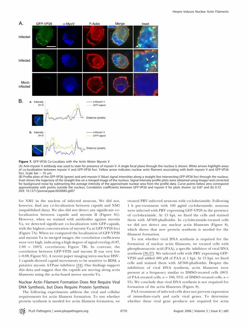
for NM1 in the nucleus of infected neurons. We did not,however, find any co-localization between capsids and NM1(unpublished data). We also did not detect any significant co-localization between capsids and myosin II (Figure S1).However, when we stained with antibodies against myosinVa, we detected significant co-localization with GFP-capsids,with the highest concentration of myosin Va at GFP-VP26 foci(Figure 7A). When we compared the localization of GFP-VP26and myosin Va in merged images, the correlation coefficientswere very high, indicating a high degree of signal overlap (0.87,1.00 ¼ 100% correlation; Figure 7B). In contrast, thecorrelation between GFP-VP26 and myosin II was very low(�0.08; Figure S1). A recent paper imaging intra-nuclear HSV-1 capsids showed capsid movements to be sensitive to BDM, aputative myosin ATPase inhibitor [34]. Our findings supportthis data and suggest that the capsids are moving along actinfilaments using the actin-based motor myosin Va.
Nuclear Actin Filament Formation Does Not Require ViralDNA Synthesis, but Does Require Protein Synthesis
The following experiments address the viral and cellularrequirements for actin filament formation. To test whetherprotein synthesis is needed for actin filament formation, we
treated PRV-infected neurons with cycloheximide. Following1 h pre-treatment with 100 lg/ml cycloheximide, neuronswere infected with PRV expressing GFP-VP26 in the presenceof cycloheximide. At 15 hpi, we fixed the cells and stainedthem with AF568-phalloidin. In cycloheximide-treated cellswe did not detect any nuclear actin filaments (Figure 8),which shows that new protein synthesis is needed for thefilament formation.To test whether viral DNA synthesis is required for the
formation of nuclear actin filaments, we treated cells withphosphonoacetic acid (PAA), a specific inhibitor of viral DNAsynthesis [36,37]. We infected cells with PRV expressing GFP-VP26 and added 400 lM of PAA at 1 hpi. At 15 hpi, we fixedcells and stained them with AF568-phalloidin. Despite theinhibition of viral DNA synthesis, actin filaments werepresent at a frequency similar to DMSO-treated cells (86%of PAA-treated cells, n¼ 106; 95% of DMSO-treated cells, n¼91). We conclude that viral DNA synthesis is not required forformation of the actin filaments (Figure 8).PAA treatment of infected cells does not prevent expression
of immediate-early and early viral genes. To determinewhether these viral gene products are required for actin
Figure 7. GFP-VP26 Co-Localizes with the Actin Motor Myosin V
(A) Anti-myosin V antibody was used to stain for presence of myosin V. A single focal plane through the nucleus is shown. White arrows highlight areasof co-localization between myosin V and GFP-VP26 foci. Yellow arrow indicates nuclear actin filament associating with both myosin V and GFP-VP26foci. Scale bar¼ 10 lm.(B) Profile plots of the GFP-VP26 (green) and anti-myosin V (blue) signal intensities along a straight line intersecting GFP-VP26 foci through the nucleus.Inset shows the trajectory of the straight line on a merged image of the nucleus. Signal intensity profile plots were obtained using ImageJ and correctedfor background noise by subtracting the average intensity of the approximate nuclear area from the profile data. Curve points below zero correspondapproximately with points outside the nucleus. Correlation coefficients between GFP-VP26 and myosin V for plots shown: (a) 0.87 and (b) 0.72.DOI: 10.1371/journal.ppat.0020085.g007
PLoS Pathogens | www.plospathogens.org August 2006 | Volume 2 | Issue 8 | e850770
Herpes Induces Nuclear Actin Filaments

filament formation, we inactivated PRV stocks with ultravioletlight (UV), which reduced infectivity by four orders ofmagnitude. UV-treated virions can still bind to and entercells, but viral transcription and DNA replication are blocked.UV-treated virions also deliver tegument proteins after entry[38]. We did not detect nuclear actin filaments after infectionby UV-treated virions (Figure 8), which indicates that newsynthesis of immediate-early and/or early viral proteins isrequired for actin filament formation. Taken together, theseresults are evidence that viral and/or host protein synthesis isrequired for the formation of actin filaments.
Nuclear Actin Filament Formation Is Not Specific for PRV
or for NeuronsTo test whether the formation of nuclear actin filaments is
specific to neurons, we infected PK15 cells, a transformed
porcine kidney epithelial cell line, with PRV and stained cellswith AF568-phalloidin. Infected, but not uninfected, PK15cells exhibited nuclear actin filaments (Figure 9A and 9B).But, in distinction to infected neurons, the nuclear filamentswere finer and less organized in PK15 cells (Figure 9A and9B). Furthermore, nuclear actin filament formation is notspecific to PRV infections since we also observed theformation of nuclear actin filaments when we infected SCGneurons with HSV-1 (KOS) (Figure 9C). This suggests thatnuclear actin filament formation is a mechanism that isconserved within the alpha-herpesvirus subfamily.
Discussion
In this report, we show that herpesvirus infection inducesthe formation of actin filaments in the nucleus. The timing of
Figure 8. Host and Viral Requirements for Nuclear Actin Filament Formation
Cells were treated with cycloheximide or PAA, as indicated. Alternatively, viral stocks were treated with UV irradiation. Each image is a 2-D projectionfrom four consecutive layers in an image stack, taken 0.5 lm apart. Scale bar¼ 20 lm.DOI: 10.1371/journal.ppat.0020085.g008
PLoS Pathogens | www.plospathogens.org August 2006 | Volume 2 | Issue 8 | e850771
Herpes Induces Nuclear Actin Filaments

filament formation (Figure 4), the association of capsids withfilaments (Figures 1 and 5) and myosin V (Figure 7), and thedependence of VP26 organization on filaments (Figure 6),suggests that F-actin plays a key role in the formation andorganization of viral assembly centers in the nucleus. Ourresults support the recent finding showing that directedmovement of HSV-1 capsids in the nucleus is actin-, andlikely, myosin-dependent [34]. We propose that nuclear actinfilaments provide tracks for myosin—rather than actinpolymerization-based movement, because elimination ofactin dynamics by jasp did not disrupt VP26 localization(Figure 6). A diverse group of intracellular microorganisms,including Listeria monocytogenes, Shigella spp., Rickettsia spp.,and vaccinia virus, utilize the host actin cytoskeleton to movewithin and spread between mammalian host cells. We believethat herpesviruses provide another distinct example of apathogen appropriating the host actin cytoskeleton.
We first observed nuclear actin filaments in infected mouseSMG tissue in volume electron-microscopic images obtainedusing SBFSEM (Figure 1). In those images, we detected anetwork of filaments in the nucleus surrounding the PRVcapsids present only in infected cells. Most of the filamentsassociated with individual genome-filled capsids, showingboth end-on and side-on associations. Given the associationof filaments exclusively with genome-filled capsids, wepropose that the filaments play a role in capsid assemblyand/or the transport of capsids in preparation for egress.Similar filaments described as ‘‘interwoven fine fibrils’’ havebeen found to associate with HSV-1 and HSV-2 nucleocapsidsin infected CNS, PNS, and glial cell nuclei [39]. Thus, theformation of such filaments may be a conserved feature in thealpha-herpesvirus life cycle.We tested the hypothesis that infection induces filament
formation using dissociated, cultured peripheral SCG neu-
Figure 9. Conservation of Formation of Nuclear Actin Filaments
(A) PK15s infected with PRV expressing GFP-VP26, fixed at 9 hpi. Asterisks show cells that are infected and show short actin filaments that appear toassociate with nuclear membrane. Each image is a 2-D projection from four consecutive layers in an image stack, taken 0.1 lm apart. Scale bar¼20 lm.(B) Enlarged image of nucleus labeled with yellow asterisk in (A) Arrowhead indicates nuclear actin filaments. Scale bar ¼ 10 lm.(C) SCG neurons infected with HSV-1 (KOS), fixed at 15 hpi. Cells were stained for the presence of viral capsid with anti-VP5 (capsid protein) antibody.Arrowheads indicate nuclear actin filaments. Each image is a 2-D projection from four consecutive layers in an image stack, taken 0.5 lm apart. Scale bar¼ 20 lm.DOI: 10.1371/journal.ppat.0020085.g009
PLoS Pathogens | www.plospathogens.org August 2006 | Volume 2 | Issue 8 | e850772
Herpes Induces Nuclear Actin Filaments

rons. Neurons infected with PRV or HSV-1 contained anetwork of nuclear actin filaments, visualized by fluorescentlylabeled phalloidin (Figures 2 and 9). However, the extent towhich actin filaments were present in the nucleus wasvariable: some cells contained a single large bundle runningalong one face of the inner nuclear membrane whereas otherspossessed larger, more complex structures. We do not yetunderstand the factors responsible for these differences, butamount of viral production or the size of the actin monomerpool could play a role. The average length of nuclear actinfilaments seen by confocal microscopy (4.5 6 1.9 lm) wassimilar to that seen with SBFSEM (3.0 6 1.2 lm) and in bothcases filaments specifically associated with capsids and werepresent only in infected cells. Thus, they may be the samestructures. In PK15 cells, nuclear actin filaments appearedfiner and less organized than those in SCG neurons, perhapsreflecting differences between polarized and non-polarizedcells or primary and transformed cells.
Actin filaments associate with the nuclear lamina that facesthe Golgi (Figure 3) which implies that, if actin filaments areindeed utilized for nuclear egress, capsids traveling along thefilaments towards the nuclear membrane would emerge fromthe nuclear envelope ready to engage the secretory pathway.Consistent with this speculation, live imaging of HSV-1capsids in the nucleus showed directed movements towardsthe nuclear envelope [34]. This result also suggests that ifactin nucleators are involved in the formation of thesefilaments, then one would expect to find them asymmetricallylocalized on the nuclear lamina.
Since actin filaments are required for capsid foci for-mation, GFP-VP26 foci presumably reflect viral assembly sitesas they have been observed and characterized in both PRVand HSV-1-infected cells [31,33]. Experiments with latA(Figure 6) suggest that actin filaments are important forestablishment and/or maintenance of GFP-VP26 foci. Arecent paper has shown that nuclear expansion in epithelialcells infected with HSV-1 is dependent on actin [10]. LatAalso inhibits replication, compartment maturation, andchromatin dispersal [10], providing support for the notionthat actin filaments provide a scaffold for viral assembly.Testing whether such a scaffold is also used for viral DNAreplication will require further study.
Jasp, an inhibitor of actin treadmilling dynamics thatstabilizes actin filaments, increased the number of cellscontaining GFP-VP26 foci by 30% (Figure 6). This findingsuggests that capsid foci may be stabilized as a result offilament stabilization. At present, we do not know the degreeto which the filaments are dynamic or if they are undergoingrapid turnover; Fluorescence Recovery after Photobleaching(FRAP) experiments with neurons expressing GFP-actinshould answer this question. The effects of jasp imply thatindividual capsids use the actin filaments as tracks fordirected movements rather than by using actin polymer-ization-based movement, which is distinct from the way thatListeria and vaccinia virus use the actin cytoskeleton. Theresult that capsids co-localize with the actin motor myosin Va(Figure 7) supports this idea. Class V myosins are among themost thoroughly studied forms of unconventional myosinsand considerable evidence supports a role in transport oforganelles and vesicles [40]. Myosin Va is a two-headedmyosin that shows processive movement along actin fila-ments, similar to that of two-headed kinesins and dynein
along microtubules [40,41]. Myosin Va is one of the fastestmyosins, moving along actin filaments at a speed of 300–400nm/s [42], which is comparable with the speed reported fordirected movements of HSV-1 capsids [34].We do not know whether actin filaments form as a result of
rearrangement of the available nuclear monomer pool or ifmonomeric actin is recruited from the cytoplasm. Severalrecent publications [43–45] have demonstrated that actin ispresent in the nucleus and is critical for transcription,chromatin remodeling, mRNA export, and nuclear structureand integrity. However, the actin present in the nucleus maynot be filamentous, since it is not recognized by phalloidin,which binds only to filaments more than seven monomerslong [46]. By analogy, actin may also play a role intranscription of viral genes. b-actin has been shown to co-purify with RNA polymerase (I, II, and III), is a component ofpre-initiation complexes, and appears to be recruited topromoters of genes about to be transcribed [47–49]. However,recent work [10] demonstrates that treatment with inhibitorsof actin polymerization does not affect HSV-1 viral repli-cation (cytochalasin D treatment even increases viral titerabout 15 times). These observations taken together with ourfindings that actin filaments still form when viral DNAreplication is prevented (Figure 8) suggest that actin plays anancillary rather than an essential role in the virus life cycle.Recent work also shows that host-derived actin is incorpo-
rated into the PRV virion and becomes an integral part of theouter tegument layer [50,51]. The amount of actin incorpo-rated increased in the absence of VP22, one of the majortegument proteins, providing support for the view of theouter tegument layer as dynamic outer shell [50,51]. Virion-associated actin has been reported in other herpesviruses[1,52–56] and other enveloped viruses including paramyx-ovirus, retrovirus, and rhabdovirus [57–61]. Although actinincorporation may be required as a structural element of thevirion, actin may also serve an additional function later ininfection, such as nuclear egress or envelopment.We do not know how actin filament formation occurs after
infection, but we do know that new protein synthesis isrequired. We also know that at least one viral immediate-earlyor early protein is required (Figure 8). This protein maypromote the formation of actin filaments directly, perhaps asan actin nucleator. Studies of the mammalian stress responsehave revealed that formation of nuclear actin filaments,increased nuclear invaginations, and decondensation ofnucleoli, events all associated with viral infection, occur inresponse to heat shock [62]. During the heat-shock responseand during HSV-1 infection, cellular chaperones Hsp70 andHsp40 are redistributed to the nucleus during infection andco-localize with ICP0, adjacent to replication complexes,thereby promoting sequestration and compartmentalizationof the nucleus [21]. Herpesvirus infection induces cellularstress responses, which may be exploited to concentrate viraland host proteins required for viral assembly and packaging.Consistent with this speculation, baculovirus-infected cellsform nuclear actin filaments at the time of virus assembly;these filaments co-localize with nucleocapsids and thebaculovirus major capsid protein has been shown to bind F-actin [63]. Whether actin filament formation is a viral-inducedresponse or a general stress response, viruses that replicate inthe nucleus may utilize these actin filaments as a scaffold forassembly and genome packaging. As a first step toward testing
PLoS Pathogens | www.plospathogens.org August 2006 | Volume 2 | Issue 8 | e850773
Herpes Induces Nuclear Actin Filaments

these ideas, it will be important to understand how viralcapsids interact with nuclear actin filaments by identifyingviral proteins that interact with actin and/or myosin.
Materials and Methods
Virus and cells. The swine kidney epithelial cell line (PK15) waspurchased from the American Type Culture Collection (CCL-22). Allnon-neuronal cells were cultured in Dulbecco’s modified Eaglemedium supplemented with 10% of fetal bovine serum and 1%penicillin/streptomycin. All PRV stocks were produced in the PK15cell line. PRV stocks used in this report include PRV Becker, avirulent isolate [64] and PRV-GS443, a recombinant expressing GFPfused to the VP26 capsid protein [31].
Infection of mouse SMG. Each 6–8-wk-old C57 B6 mouse wasanesthetized with 100 ll of a freshly prepared, sterile filtered solutionof ketamine (100 6 10 mg/kg)/xylazine (10 6 1 mg/kg) by intra-peritoneal (IP) injection. The neck of the mouse, from the base of thechin to just above the ribcage, was shaved using a platinum razorblade. The shaved area was prepared for surgery in the laminar flowhood using aseptic technique by applying disinfectant scrub andswabbing the area with 70% isopropyl alcohol. The mice were at asurgical plane of anesthesia prior to incising the neck region toexpose the salivary glands. An approximately 1.5-cm incision wasmade with a sterile scalpel blade on a scalpel handle, with the skingrasped using forceps in order to ensure a shallow incision. Theanimal was monitored during the entire surgical procedure; if at anytime the animal is no longer in a surgical plane of anesthesia (e.g.,increased respiratory rate, movement), then we injected the animalwith additional ketamine/xylazine or ketamine alone (up to 10–20 mg/kg) to induce a deeper plane of anesthesia. Four separate 2-linjections of PRV inoculum (diluted in sterile PBS) was injected intothe submandibular glands. The incision was closed with 6–0 silksutures. The mouse was administered an IP injection in the scapularregion of 2.0 mg/kg of buprenorphine for prophylaxis against post-surgical pain. At 48 hpi, the mouse was euthanized by CO2 inhalationand fixed with 4% paraformaldehyde by cardiac perfusion. Thesalivary glands were surgically removed and the SMG dissected out onSylgard plates. This experimental protocol related to animal use hasbeen approved by the Institutional Animal Care and Use Committeeof the Princeton University Research Board under protocol number1539-AR1 in accordance with the regulations of the AmericanAssociation for Accreditation of Laboratory Animal Care and thosein the Animal Welfare Act (Public Law 99–198).
Block-face serial section scanning electron microscopy. SMG werestained in a manner similar to what is described for TEM. Post-staining, the infected ganglia were embedded in Epon resin (EMSciences). The embedded samples were mounted on an aluminumrivet and trimmed following the procedure given in Denk andHorstman, 2004 [25]. All data shown were taken on an environmentalSBFSEM with a field-emission electron gun (QuantaFEG 200, FEI,Eindhoven, The Netherlands) at a gas (H20) pressure of 23 P, and anelectron energy of 3.0 keV. The mounted, trimmed samples wereplaced on the SEM microtome and sequential images from the block-face were acquired in between cut cycles. The images were taken at adigital resolution of 26 nm/pixel. The data were analyzed usingImageJ 1.32j software (National Institutes of Health). The reslicing ofimage stacks (see Figure 1e) was done using the ImageJ VolumeViewer plugin, which interpolates the z-axis data so that the digitalresolution matches that of the lateral direction.
Neuron culture. Detailed protocols for dissecting and culturingneurons are found in Ch’ng et al. [65]. Briefly, sympathetic neuronsfrom the SCG were dissected from E15.5 to E16.5 pregnant Sprague-Dawley rats (Hilltop Labs Incorporated, Pennsylvania, United States)and incubated in 250 lg/ml of trypsin (Worthington Biochemicals,Lakewood, New Jersey, United States) for 10 min. 1 mg/ml of trypsininhibitor (Sigma-Aldrich, St. Louis, Missouri, United States) wasadded to neutralize the trypsin for 3 min and then removed andreplaced with neuron culture medium. Prior to plating, the gangliawere triturated into dissociated neurons using a fire-polished Pasteurpipette and then plated onto glass cover slips in a 35-mm plastictissue culture dish coated with 500 lg/ml of poly-DL-ornithine(Sigma-Aldrich) diluted in borate buffer and 10 lg/ml of naturalmouse laminin (Invitrogen, Carlsbad, California, United States). Theneuron culture medium consists of Dulbecco’s modified Eaglemedium (Invitrogen) and Ham’s F12 (Invitrogen) in a 1:1 ratio. Theserum-free medium was supplemented with 10 mg/ml of bovineserum albumin (Sigma-Aldrich), 4.6 mg/ml glucose (J. T. Baker), 100
lg/ml of holotransferrin (Sigma-Aldrich), 16 lg/ml of putrescine(Sigma-Aldrich), 10 lg/ml of insulin (Sigma-Aldrich), 2 mM of L-glutamine (Invitrogen); 50 lg/ml or units of penicillin and strepto-mycin (Invitrogen), 30 nM of selenium (Sigma-Aldrich); 20 nM ofprogesterone (Sigma-Aldrich) and 100 ng/ml of nerve growth factor2.5S (Invitrogen). After 2 d post-plating, the neuronal cultures aretreated with 1 lM of an antimitotic drug called cytosine b-D-arabinofuranoside (Sigma-Aldrich) to eliminate any non-neuronalcells. The neuron culture medium was replaced every 3 d and cultureswere kept in a humidified, CO2 regulated 37 8C incubator. Thisexperimental protocol related to animal use has been approved byThe Institutional Animal Care and Use Committee of the PrincetonUniversity Research Board under protocol number 1453-AR2 inaccordance with the regulations of the American Association forAccreditation of Laboratory Animal Care and those in the AnimalWelfare Act (Public Law 99–198).
Viral infections. Protocols for viral infection of neurons have beendescribed by Ch’ng et al. [65]. All PRV infections of neuron cultureswere carried out under high multiplicities of infection (MOI) unlessotherwise stated. Briefly, neurons were cultured on glass cover slips in35-mm dishes for approximately 2 wk prior to any experiment. Theviral inoculum was diluted in 2% fetal bovine serum in Dulbecco’smodified Eagle (GIBCO, San Diego, California, United States) andoverlaid on the neuronal culture for 1 h in a humidified 37 8Cincubator. After 1 h, the viral inoculum was removed and replacedwith neuron medium. Infections usually lasted for 15 h (unlessotherwise stated) before the samples were fixed and processed forstaining and immunofluorescence. The production of infectious virusover time in SCGs has been characterized [30, 66].
Antibodies and stains. Antibodies used in this study include mousemonoclonal against lamin-associated protein 2 (LAP2) (BD Biosciences,Palo Alto, California, United States; used 1:500), anti-VP5 (major capsidprotein) antibody and anti-GM130 antibody (BD Transduction labo-ratories; used 1:250). Myosin Va antibody against neuronal rat myosin Vwas generously provided by Paul Bridgman (Washington University, St.Louis, Missouri, United States) and used at 1:2000. Myosin II antibodyagainst neuronal rat myosin II (Covance) was used at 1:500. Actin wasdetected by Alexa 568-phalloidin (Molecular Probes, Eugene, Oregon,United States) used at 1:40. All secondary Alexa fluorophores werepurchased from Molecular Probes and used at 1:500 dilution.
Fluorescence, immunofluorescence, and drug treatments. Allfluorescence experiments carried out were performed as follows.Dissociated neurons on glass cover slips were incubated inphosphate-buffered saline containing 3% bovine serum albuminand 0.5% triton for 10 min before the addition of primary antibodiesfor 1 h. After 1 h, the primary antibodies were removed and thesample was washed three times with phosphate-buffered salinecontaining 3% bovine serum albumin (and 0.5% saponin whennoted in text). Next, secondary antibodies were added to the sampleand incubated for 1 h. After 1 h, the secondary antibodies wereremoved and the sample was washed three times with phosphate-buffered saline containing 3% bovine serum albumin (and 0.5%saponin when noted in text). To stain for filamentous actin, Alexa-568-phalloidin was added to the cover slip at a concentration of 6.6lg/ml. The cover slip was mounted on a glass slide using Aquapolymount (Polysciences, Warrington, Pennsylvania, United States)and allowed to dry for 24 h prior to imaging. To depolymerize actin,latrunculinA (latA; Molecular Probes) was dissolved in DMSO andadded directly to the media at 3 hpi at a final concentration of 5 lM.Jasplakinolide (jasp; EMD Biosciences, Darmstadt, Germany) wasdissolved in DMSO and was added directly to the media at 3 hpi at afinal concentration of 100 nM. Phosphonoacetic acid (PAA; Sigma-Aldrich) was dissolved in DMSO and added directly to the media at 1hpi at a final concentration of 400 lM. Cycloheximide was used at 100lg/ml (10 mg/ml stock dissolved in PBS). Cells were pre-treated withcycloheximide in neuronal media for 1 h and then infected with viralinoculum containing cycloheximide. After 1 h, the viral inoculum wasremoved and replaced with neuron medium containing cyclohex-imide. For UV-inactivation, we exposed aliquots of PRV443L strain toUV light using a UV Stratalinker 1800 (Stratagene, La Jolla,California, United States). We used a dosage of UV that reducedthe titer by approximately 1,000-fold.
Samples were imaged with a Perkin-Elmer (Wellesley, California,United States) RS3 spinning disk confocal microscope side-mountedon a TE200-S Nikon Eclipse microscope (Tokyo, Japan) with an Argon/Krypton laser producing excitation lines of 488, 568, and 647 nms.Optical sections were acquired in 0.1, 0.25, or 0.5 lm steps, as stated. 2-D projections of confocal stacks and channel merges were created byImageJ 1.32j software (National Institutes of Health). All figures wereassembled in Adobe Photoshop 7.0.1. Alterations to image brightness
PLoS Pathogens | www.plospathogens.org August 2006 | Volume 2 | Issue 8 | e850774
Herpes Induces Nuclear Actin Filaments
and contrast were conducted in a linear manner and were appliedequally to controls, except where otherwise noted. Supplementalvideo was assembled in ImageJ and converted to QuickTime format.
TEM.Whole SCG were cultured for 2 wk on Aclar (EM Sciences) ina manner similar to what is described above for glass cover slips(except without dissociation). Neurons were infected at a high MOIand treated with latA as described above. After 15 hpi, the plates werewashed twice with phosphate-buffered saline, fixed with 2%glutaraldehyde in 0.2 M sodium cacodylate buffer (pH 7.2) for 4 h,and post-fixed with 1% osmium tetroxide in sodium veronal bufferfor 1 h on ice. Samples were then rinsed with sodium veronal bufferfour times and incubated with 0.25% toluidine blue in 0.2 Mcacodylate buffer (pH 7.2) for 1 h; the staining solution was thenremoved with four rinses of sodium veronal buffer (pH 7.2), followedby four rinses with 0.05 M sodium maleate buffer (pH 5.1). Overnightincubation with 2% uranyl acetate in 0.05 M sodium maleate bufferwas done in the dark followed by four rinses with 0.05 M sodiummaleate buffer (pH 5.1). The fixed samples were then dehydrated withethyl alcohol, embedded in Epon resin (EM Sciences) and cut into 70-nm sections using a Reichert Ultracut E ultramicrotome.
Supporting Information
Figure S1. Profile Plots of the GFP-VP26 (Green) and Anti-Myosin II(Blue) Signal Intensities along a Straight Line Intersecting GFP-VP26Foci through the Nucleus
Inset shows the trajectory of the straight line on a merged image ofthe nucleus. Signal intensity profile plots were obtained using ImageJand corrected for background noise by subtracting the averageintensity of the approximate nuclear area from the profile data.Curve points below zero correspond approximately with pointsoutside the nucleus. Correlation coefficients between GFP-VP26 andmyosin II for plots shown: (A) �0.11 and (B)�0.18.Found at DOI: 10.1371/journal.ppat.0020085.sg001 (9.9 MB TIF).
Video S1. QuickTime Video of a SBFSEM Stack of 65 Serial Sectionsfrom an Infected Cell, Sectioned at 50 nm
The volume shown is at the inner edge of the nucleus, with the
nuclear envelope at the right-hand side of the image. This video is acropped substack from Video S2.
Found at DOI: 10.1371/journal.ppat.0020085.sv001 (3.1 MB MOV).
Video S2. QuickTime Video of a SBFSEM Stack Comprising 100Serial Sections from an Infected Cell, Sectioned at 50 nm
Lower half of the cell was not obtained during image acquisition.
Found at DOI: 10.1371/journal.ppat.0020085.sv002 (944 KB MOV).
Video S3. QuickTime Video of a SBFSEM Stack Comprising 150Serial Sections from an Uninfected Cell, Sectioned at 50 nm
Found at DOI: 10.1371/journal.ppat.0020085.sv003 (1.4 MB MOV).
Acknowledgments
We thank A. Reynolds for guidance and assistance in mouse surgeriesand for critical discussions. We thank M. Lyman and the othermembers of the Enquist lab for helpful discussions and thoughtfulsuggestions. We thank J. Goodhouse for technical assistance inconfocal microscopy and Heinz Horstman at the MPImF for help inmounting the samples for SBFSEM imaging. We thank Paul Bridgmanfor his generous gift of antibodies to myosin Va. We thank DavidTank for his advice and support. Special thanks to R. Pelham and A. J.Akey for their care and encouragement.
Author contributions. BF, SP, and LWE conceived and designed theexperiments. BF and SP performed the experiments. BF, SP, and LWEanalyzed the data. BF, SP, and LWE contributed reagents/materials/analysis tools. MB prepared samples and performed TEM. WDprepared samples and performed SBFSEM. BF, SP, WD, and LWEwrote the paper. SP’s Senior Undergraduate Thesis at PrincetonUniversity included portions of this work.
Funding. This work was supported by the National Institute ofNeurological Disorders and Stroke (NIH-NINDS; grant R01 33506),the Dana Research Foundation, and by the Max-Planck Society.
Competing interests. The authors have declared that no competinginterests exist.
References1. Grunewald K, Desai P, Winkler DC, Heymann JB, Belnap DM, et al. (2003)
Three-dimensional structure of herpes simplex virus from cryo-electrontomography. Science 302: 1396–1368.
2. Roizman B (1991) Herpesviridae: A brief introduction. In: Fields BN, KnipeDM, editors. Fundamental virology. 2nd edition. New York: Raven Press.pp. 841–895.
3. de Bruyn Kops A, Knipe DM (1994) Preexisting nuclear architecture definesthe intranuclear location of herpesvirus DNA replication structures. J Virol68: 3512–3526.
4. de Bruyn Kops A, Uprichard SL, Chen M, Knipe DM (1998) Comparison ofthe intranuclear distributions of herpes simplex virus proteins involved invarious viral functions. Virology 252: 162–178.
5. Taylor TJ, McNamee EE, Day C, Knipe DM (2003) Herpes simplex virusreplication compartments can form by coalescence of smaller compart-ments. Virology 309: 232–247.
6. Scott ES, O’Hare P (2001) Fate of the inner nuclear membrane proteinlamin B receptor and nuclear lamins in herpes simplex virus type 1infection. J Virol 75: 8818–8830.
7. Enquist LW, Husak PJ, Banfield BW, Smith GA (1998) Infection and spreadof alpha-herpesviruses in the nervous system. Adv Virus Res 51: 237–347.
8. Ward PL, Ogle WO, Roizman B (1996) Assemblons: Nuclear structuresdefined by aggregation of immature capsids and some tegument proteins ofherpes simplex virus 1. J Virol 70: 4623–4631.
9. Simpson-HolleyM, Baines J, Roller R, Knipe DM (2004) Herpes simplex virus1 U(L)31 and U(L)34 gene products promote the late maturation of viralreplication compartments to the nuclear periphery. J Virol 78: 5591–5600.
10. Simpson-Holley M, Colgrove RC, Nalepa G, Harper JW, Knipe DM (2005)Identification and functional evaluation of cellular and viral factorsinvolved in the alteration of nuclear architecture during herpes simplexvirus 1 infection. J Virol 79: 12840–12851.
11. Klupp BG, Granzow H, Karger A, Mettenleiter TC (2005) Identification,subviral localization, and functional characterization of the pseudorabiesvirus UL17 protein. J Virol 79: 13442–13453.
12. Klupp BG, Granzow H, Mettenleiter TC (2000) Primary envelopment ofpseudorabies virus at the nuclear membrane requires the UL34 geneproduct. J Virol 74: 10063–10073.
13. Klupp BG, Granzow H, Mundt E, Mettenleiter TC (2001) Pseudorabies virusUL37 gene product is involved in secondary envelopment. J Virol 75: 8927–8936.
14. Reynolds AE, Liang L, Baines JD (2004) Conformational changes in thenuclear lamina induced by herpes simplex virus type 1 require genesU(L)31 and U(L)34. J Virol 78: 5564–5575.
15. Reynolds AE, Ryckman BJ, Baines JD, Zhou Y, Liang L, et al. (2001) U(L)31and U(L)34 proteins of herpes simplex virus type 1 form a complex thataccumulates at the nuclear rim and is required for envelopment ofnucleocapsids. J Virol 75: 8803–8817.
16. Reynolds AE, Wills EG, Roller RJ, Ryckman BJ, Baines JD (2002)Ultrastructural localization of the herpes simplex virus type 1 UL31,UL34, and US3 proteins suggests specific roles in primary envelopment andegress of nucleocapsids. J Virol 76: 8939–8952.
17. Newcomb WW, Thomsen DR, Homa FL, Brown JC (2003) Assembly of theherpes simplex virus capsid: Identification of soluble scaffold-portalcomplexes and their role in formation of portal-containing capsids. JVirol 77: 9862–9871.
18. Newcomb WW, Homa FL, Brown JC (2005) Involvement of the portal at anearly step in herpes simplex virus capsid assembly. J Virol 79: 10540–10546.
19. Lukonis CJ, Burkham J, Weller SK (1997) Herpes simplex virus type 1prereplicative sites are a heterogeneous population: Only a subset are likelyto be precursors to replication compartments. J Virol 71: 4771–4781.
20. Lukonis CJ, Weller SK (1997) Formation of herpes simplex virus type 1replication compartments by transfection: Requirements and localizationto nuclear domain 10. J Virol 71: 2390–2399.
21. Burch AD, Weller SK (2004) Nuclear sequestration of cellular chaperoneand proteasomal machinery during herpes simplex virus type 1 infection. JVirol 78: 7175–7185.
22. Quinlan MP, Chen LB, Knipe DM (1984) The intranuclear location of aherpes simplex virus DNA-binding protein is determined by the status ofviral DNA replication. Cell 36: 857–868.
23. Rixon FJ, Atkinson MA, Hay J (1983) Intranuclear distribution of herpessimplex virus type 2 DNA synthesis: Examination by light and electronmicroscopy. J Gen Virol 64: 2087–2092.
24. Liptak LM, Uprichard SL, Knipe DM (1996) Functional order of assemblyof herpes simplex virus DNA replication proteins into prereplicative sitestructures. J Virol 70: 1759–1767.
25. Denk W, Horstmann H (2004) Serial block-face scanning electron micro-scopy to reconstruct three-dimensional tissue nanostructure. PLoS Biol 2:e329. DOI: 10.1371/journal.pbio.0020329
26. Lichtman JW (1977) The reorganization of synaptic connexions in the ratsubmandibular ganglion during post-natal development. J Physiol 273:155–177.
PLoS Pathogens | www.plospathogens.org August 2006 | Volume 2 | Issue 8 | e850775
Herpes Induces Nuclear Actin Filaments

27. Lichtman JW (1980) On the predominantly single innervation ofsubmandibular ganglion cells in the rat. J Physiol 302: 121–130.
28. Gan WB, Kwon E, Feng G, Sanes JR, Lichtman JW (2003) Synapticdynamism measured over minutes to months: Age-dependent decline in anautonomic ganglion. Nat Neurosci, 6: 956–960.
29. Tomishima MJ, Smith GA, Enquist LW (2001) Sorting and transport ofalpha-herpesviruses in axons. Traffic 2: 429–436.
30. Ch’ng TH, Enquist LW (2005) Efficient axonal localization of alpha-herpesvirus structural proteins in cultured sympathetic neurons requiresviral glycoprotein E. J Virol 79: 8835–8846.
31. Smith GA, Gross SP, Enquist LW (2001) Herpesviruses use bidirectionalfast-axonal transport to spread in sensory neurons. Proc Natl Acad Sci U SA 98: 3466–3470.
32. Smith BN, Banfield BW, Smeraski CA, Wilcox CL, Dudek FE, et al. (2000)Pseudorabies virus expressing enhanced green fluorescent protein: A toolfor in vitro electrophysiological analysis of transsynaptically labeledneurons in identified central nervous system circuits. Proc Natl Acad SciU S A 97: 9264–9269.
33. Desai P, Person S (1998) Incorporation of the green fluorescent proteininto the herpes simplex virus type 1 capsid. J Virol 72: 7563–7568.
34. Forest T, Barnard S, Baines JD (2005) Active intranuclear movement ofherpesvirus capsids. Nat Cell Biol 7: 429–431.
35. Bubb MR, Senderowicz AM, Sausville EA, Duncan KL, Korn ED (1994)Jasplakinolide, a cytotoxic natural product, induces actin polymerizationand competitively inhibits the binding of phalloidin to F-actin. J Biol Chem269: 14869–14871.
36. May DC, Miller MR, Rapp F (1977) The effect of phosphonoacetic acid onthe in vitro replication of varicella-zoster virus. Intervirology 8: 83–91.
37. Purifoy DJ, Powell KL (1977) Herpes simplex virus DNA polymerase as thesite of phosphonoacetate sensitivity: Temperature-sensitive mutants. JVirol 24: 470–477.
38. Zhu H, Cong JP, Shenk T (1997) Use of differential display analysis to assessthe effect of human cytomegalovirus infection on the accumulation ofcellular RNAs: Induction of interferon-responsive RNAs. Proc Natl AcadSci U S A 94: 13985–13990.
39. Ecob-Johnston MS, Whetsell WO Jr (1979) Host-cell response to herpesvirus infection in central and peripheral nervous tissue in vitro. J Gen Virol44: 747–757.
40. Bridgman PC (2004) Myosin-dependent transport in neurons. J Neurobiol58: 164–174.
41. Mehta AD, Rock RS, Rief M, Spudich JA, Mooseker MS, et al. (1999) Myosin-V is a processive actin-based motor. Nature 400: 590–593.
42. Cheney RE, O’Shea MK, Heuser JE, Coelho MV, Wolenski JS, et al. (1993)Brain myosin-V is a two-headed unconventional myosin with motoractivity. Cell 75: 13–23.
43. Pederson T, Aebi U (2002) Actin in the nucleus: What form and what for? JStruct Biol 140: 3–9.
44. Pederson T, Aebi U (2005) Nuclear actin extends, with no contraction insight. Mol Biol Cell 16: 5055–5060.
45. Holaska JM, Kowalski AK, Wilson KL (2004) Emerin caps the pointed end ofactin filaments: Evidence for an actin cortical network at the nuclear innermembrane. PLoS Biol 2: e231. DOI: 10.1371/journal.pbio.0020231
46. Visegrady B, Lorinczy D, Hild G, Somogyi B, Nyitrai M (2005) A simplemodel for the cooperative stabilization of actin filaments by phalloidin andjasplakinolide. FEBS Lett 579: 6–10.
47. Hofmann WA, Stojiljkovic L, Fuchsova B, Vargas GM, Mavrommatis E, et al.
(2004) Actin is part of pre-initiation complexes and is necessary fortranscription by RNA polymerase II. Nat Cell Biol 6: 1094–1101.
48. Philimonenko VV, Zhao J, Iben S, Dingova H, Kysela K, et al. (2004) Nuclearactin and myosin I are required for RNA polymerase I transcription. NatCell Biol 6: 1165–1172.
49. Hu P, Wu S, Hernandez N (2004) A role for beta-actin in RNA polymeraseIII transcription. Genes Dev 18: 3010–3015.
50. del Rio T, DeCoste CJ, Enquist LW (2005) Actin is a component of thecompensation mechanism in pseudorabies virus virions lacking the majortegument protein VP22. J Virol 79: 8614–8619.
51. Michael K, Klupp BG, Mettenleiter TC, Karger A (2006) Composition ofpseudorabies virus particles lacking tegument protein US3, UL47, or UL49or envelope glycoprotein E. J Virol 80: 1332–1339.
52. Baldick CJ Jr, Shenk T (1996) Proteins associated with purified humancytomegalovirus particles. J Virol 70: 6097–6105.
53. Kattenhorn LM, Mills R, Wagner M, Lomsadze A, Makeev V, et al. (2004)Identification of proteins associated with murine cytomegalovirus virions. JVirol 78: 11187–11197.
54. Wong ML, Chen CH (1998) Evidence for the internal location of actin inthe pseudorabies virion. Virus Res 56: 191–197.
55. Varnum SM, Streblow DN, Monroe ME, Smith P, Auberry KJ, et al. (2004)Identification of proteins in human cytomegalovirus (HCMV) particles: TheHCMV proteome. J Virol 78: 10960–10966.
56. Zhu FX, Chong JM, Wu L, Yuan Y (2005) Virion proteins of Kaposi’sSarcoma-associated herpesvirus. J Virol 79: 800–811.
57. Bohn W, Mannweiler K, Hohenberg H, Rutter G (1987) Replica-immunogold technique applied to studies on measles virus morphogenesis.Scanning Microsc 1: 319–330.
58. Naito S, Matsumoto S (1978) Identification of cellular actin within therabies virus. Virology 91: 151–163.
59. Ott DE, Coren LV, Kane BP, Busch LK, Johnson DG, et al. (1996)Cytoskeletal proteins inside human immunodeficiency virus type 1 virions.J Virol 70: 7734–7743.
60. Sagara J, Tsukita S, Yonemura S, Tsukita S, Kawai A (1995) Cellular actin-binding ezrin-radixin-moesin (ERM) family proteins are incorporated intothe rabies virion and closely associated with viral envelope proteins in thecell. Virology 206: 485–494.
61. Vainiopaa R, Ziola B, Salmi A (1978) Measles virus polypeptides in purifiedvirions and in infected cells. Acta Pathol Microbiol Scand [B] 86B: 379–385.
62. Welch WJ, Suhan JP (1985) Morphological study of the mammalian stressresponse: Characterization of changes in cytoplasmic organelles, cytoske-leton, and nucleoli, and appearance of intranuclear actin filaments in ratfibroblasts after heat-shock treatment. J Cell Biol 101: 1198–1211.
63. Charlton CA, Volkman LE (1991) Sequential rearrangement and nuclearpolymerization of actin in baculovirus-infected Spodoptera frugiperda cells. JVirol 65: 1219–1227.
64. Platt KB, Mare CJ, Hinz PN (1979) Differentiation of vaccine strains andfield isolates of pseudorabies (Aujeszky’s disease) virus: Thermal sensitivityand rabbit virulence markers. Arch Virol 60: 13–23.
65. Ch’ng TH, Flood EA, Enquist LW (2004) Culturing primary and trans-formed neuronal cells for studying pseudorabies virus infection. MethodsMol Biol 292: 299–316.
66. Tomishima MJ, Enquist LW (2001) A conserved alpha-herpesvirus proteinnecessary for axonal localization of viral membrane proteins. J Cell Biol154: 741–752.
PLoS Pathogens | www.plospathogens.org August 2006 | Volume 2 | Issue 8 | e850776
Herpes Induces Nuclear Actin Filaments
Copyright © 2022 FDOKUMEN