Effect of phalloidin on filaments polymerized from heart muscle ADP-actin monomers
Upload
khangminh22Category
view
0download
0
BJRS
BRAZILIAN JOURNAL OF
RADIATION SCIENCES 10-01 (2022) 01-22
ISSN: 2319-0612 Accepted: 2021-10-11
Development of FFF filaments for bone and teeth
representation in 3D printed radiological objects
Savi, M.a,b; Andrade, M. A. B.a; Villani, D.b; Rodrigues Jr., O.b and Potiens, M. P. A.b
1 Instituto Federal de Santa Catarina – IFSC. 88020-300, Florianópolis – SC, Brazil
2 Instituto de Pesquisas Energéticas e Nucleares – IPEN. 05508-000. São Paulo – SP, Brazil.
[email protected] and [email protected]
ABSTRACT
The use of 3D printing technologies is growing widely, including the possibility of designing phantoms
for imaging and dosimetry. High attenuation tissues such as cortical bone, dentin and enamel need to
be simulated to accurately produce 3D printed phantoms, especially for Fused Filament Fabrication
(FFF) printing technology. A commercially available radiopaque FFF filament had been hard to find.
This study aims to report, step-by-step, the development of a radiopaque FFF filament. A combination
of radiopaque substances (Barium Sulfate - BaSO4 and Calcium Carbonate - CaCO3) were selected
for use as fillers in an Acrylonitrile Butadiene Styrene (ABS) matrix and added in quantities
calculated using the National Institute of Standards and Technology (NIST) XCOM tool. The filament
was homogenized and characterized by analyzing its density and images obtained using Scanning
Electron Microscopy (SEM), Computed Tomography (CT) and micro-CT (µCT) scans. Three
filaments were produced with different Hounsfield Units (HU) equivalences: XCT-A (1607HU), XCT-
B (1965HU) and XCT-C (2624HU) with respective densities of 1.166(6) g/cm³, 1.211(2) g/cm³ and
1.271(3) g/cm³. With these values, high attenuation tissues, such as bones, dentine and enamel, can
now be simulated with FFF 3D printing technology, at a low cost of production.
Keywords: 3D printing, Fused Filament Fabrication, Radiology, Phantom.

Savi et al. ● Braz. J. Rad. Sci. ● 2022 2
ISSN: 2319-0612 Accepted: 2021-10-11
1. INTRODUCTION
3D printing is commonly used in many areas of industry and engineering to create parts and
objects in an inexpensive way for the most varied applications. Since 1995 [1], the use of 3D printing
as a way to simulate human body tissues in radiation field has been reported. Since then, many studies
on materials and phantom fabrication using 3D printing technologies were done and evaluated by
using ionizing radiation [2–5], especially for use in radiological imaging and dosimetry.
There are many techniques to 3D print an object, such as Stereolithography (SLA), Fused
Filament Fabrication (FFF), Selective Laser Sintering (SLS) and Polyjet. FFF is also known as Fused
Deposition Modeling (FDM) [6,7], is a common and low-cost method to do a 3D print. Some authors
were successful in simulating human body with commercially available materials [8,9]. However,
commercially available FFF filament materials capable of simulating tissues that exhibit high density
and attenuation, compatible with bone, dentin and enamel, are hard to find. Although iron and other
ferromagnetic materials have been used to simulate these tissues [10–12]; they have the intrinsic
limitations of not being compatible with Magnetic Resonance (MRI) equipment if needed.
Ferromagnetic materials are very abrasive to printing nozzles and some printing defects could occur
while dual extruding it with another filament. For CT scans, HU values inside a given ROI could
show high standard deviation, especially with high printing layer heights or narrow CT slices (under
2 mm thickness). This is caused by the difference of attenuation coefficients between adjacent tissues
or materials, which could provoke heterogeneity and measurement uncertainties. The high attenuation
Fe filament could also result in image artifacts similar to metallic implants.
Some of the commercially available printing filaments have already been evaluated in literature
by reporting their response to radiation in terms of Hounsfield Unit (HU) equivalence of human
tissues [13–15]. Savi et al. [16] reported that Polylactic Acid (PLA) with addition of copper reached
the highest value of approximately 330HU, which corresponds only to low limits of cortical bone,
and is insufficient to represent dentin (1600HU-2400HU [17,18]) and enamel (above 2500HU [18-
20]) tissues. The literature and practice find that an approximately 350HU cortical bone value is only
found in a few bones, like vertebrae. Cortical bone of long bones, such as femur, humerus or bones
from skullcap, have values above 800HU, even reaching 1600HU [17,19]. Although other 3D printing

Savi et al. ● Braz. J. Rad. Sci. ● 2022 3
ISSN: 2319-0612 Accepted: 2021-10-11
technologies could achieve 800HU to over 1000HU [20,21], the cost of the 3D printer and its
materials are different and considerably more expensive than FFF.
Some attempts on the development of radiopaque FFF filaments without ferromagnetic materials
have been reported. For example, Hamedani et al [22] used a constant filament diameter of 1.75 mm
using containing Barium Sulfate (BaSO4). Despite successfully printing a radiopaque model, they
could not achieve production. The recent work of Price et al [23] proposed a filament based on an
ABS matrix doped with calcium titanate (CaTiO3) that was homogenous and suitable for bone
representation.
Considering this area is still underdeveloped and a documented step-by-step process could be
useful to the scientific community, this paper aims to report the creation of a radiopaque filament to
be used in FFF 3D printing technology testing a variety of dopant materials. Measurements were
carried out to validate the conditions of simulating tissues with high HU values. Radiopaque
substances with high atomic number were evaluated and, after selection, added to an Acrylonitrile
Butadiene Styrene (ABS) filament matrix to realize the required radiopaque properties.
2. MATERIALS AND METHODS
A FFF filament is usually composed of two parts, a binder and a filler. The binder is made of a
thermoplastic while the filler is composed of additive materials used to provide different properties
to the resulting filament. The Acrylonitrile Butadiene Styrene – ABS TERLURAN GP 35 [24] from
INEOS Styrolution was chosen as binder for the development of this filament, being the only binder
available from the third-party filament manufacturer contracted to produce the filament with the
required filler composition and concentration. Furthermore, ABS has similar characteristics to other
commonly used binders (such as PLA and PETG). It has an excellent combination of mechanical,
thermal, electrical and properties, as well as ease of processing. It also offers a good balance of
resistance to impact, traction, hardness and modulus of elasticity [25]. Also, ABS surface can be
chemically dissolved when in contact with acetone [26], a method that could be applied to correct
external imperfections due to printing errors, or to adjust a more stable and strong fit when
constructing a phantom with multiple parts that need to be socketed.

Savi et al. ● Braz. J. Rad. Sci. ● 2022 4
ISSN: 2319-0612 Accepted: 2021-10-11
Promising substances as fillers were selected based on anatomic mass (AM) from those available
at the Federal Institute of Santa Catarina (IFSC). The addition of a high AM filler to the ABS binder
increases the resultant attenuation to radiation giving radiopaque properties to the filament.
Nonetheless, this resultant attenuation must be similar to high attenuation human tissues, otherwise
the filament would behave as a barrier, making bone and teeth representations in radiological scans
compromised.
To choice the filler was evaluated in terms of mass attenuation coefficient predicted using the
NIST XCOM calculator [27] for peak and mean energy for RQT and RQR radiation qualities [28].
The choices were later compared to ICRU-44 [29] cortical bone attenuation. After selecting the
materials, the process of mixing binder and filler was repeatedly conducted until a homogeneous
filament was achieved with 1.75mm in diameter and the selected concentration in weight.
2.1 Selection of possible filler
Many chemical substances available at IFSC chemistry laboratory were considered as possible
filler, focusing on elements with high atomic number and its presentation as solid powder. Table 1
lists characteristics and cost for nine potential filler substances.
Table 1: Nine promising filler substances and their characteristics and relative prices.
Substance Formula Molar mass (g/mol) [30]
Density (g/cm³) [31,32]
Price per kg (USD)
Calcium Carbonate CaCO3 100.09 2.93 $8.88
Barium Sulfate BaSO4 233.38 4.94 $10.92
Calcium Propionate C6H10CaO4 186.22 1.38 $89.41
Bismuth (III) Oxide Bi2O3 465.96 8.9 $220.24
Lead (II) Chloride PbCl2 278.10 5.95 $291.15
Copper (I) Chloride CuCl 98.99 4.14 $348.90
Red copper (I) Oxide Cu2O 143.09 6.0 $356.6
Lead powder Pb 207.20 11.3 $364.07
Copper (I) Iodate CuI 190.45 5.67 $775.12
Savi et al. ● Braz. J. Rad. Sci. ● 2022 5
ISSN: 2319-0612 Accepted: 2021-10-11
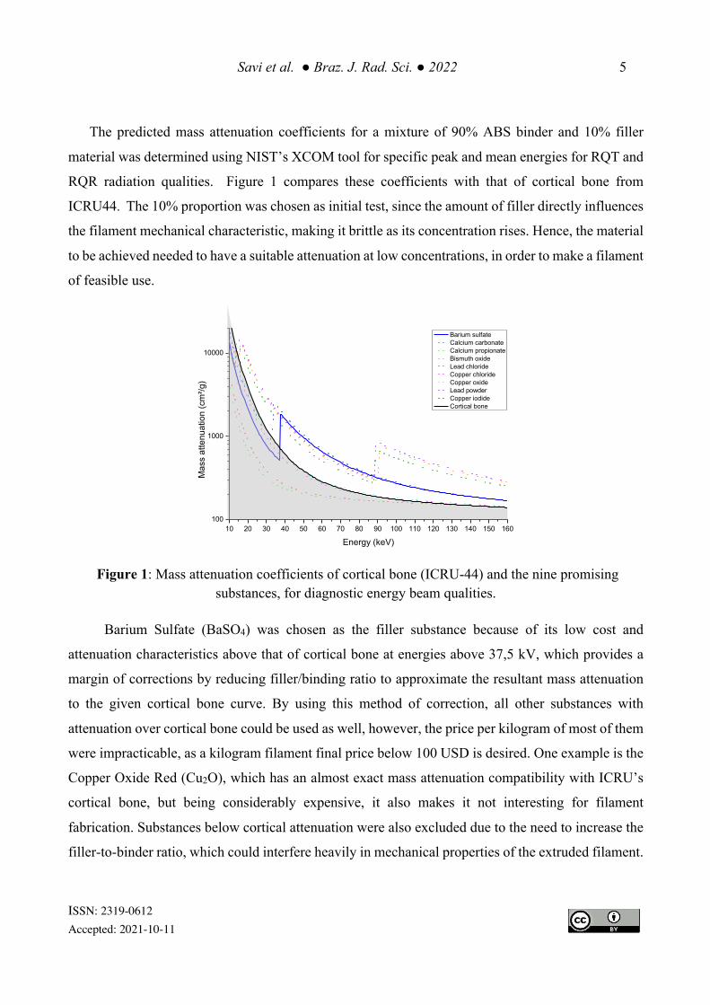
The predicted mass attenuation coefficients for a mixture of 90% ABS binder and 10% filler
material was determined using NIST’s XCOM tool for specific peak and mean energies for RQT and
RQR radiation qualities. Figure 1 compares these coefficients with that of cortical bone from
ICRU44. The 10% proportion was chosen as initial test, since the amount of filler directly influences
the filament mechanical characteristic, making it brittle as its concentration rises. Hence, the material
to be achieved needed to have a suitable attenuation at low concentrations, in order to make a filament
of feasible use.
Figure 1: Mass attenuation coefficients of cortical bone (ICRU-44) and the nine promising
substances, for diagnostic energy beam qualities.
Barium Sulfate (BaSO4) was chosen as the filler substance because of its low cost and
attenuation characteristics above that of cortical bone at energies above 37,5 kV, which provides a
margin of corrections by reducing filler/binding ratio to approximate the resultant mass attenuation
to the given cortical bone curve. By using this method of correction, all other substances with
attenuation over cortical bone could be used as well, however, the price per kilogram of most of them
were impracticable, as a kilogram filament final price below 100 USD is desired. One example is the
Copper Oxide Red (Cu2O), which has an almost exact mass attenuation compatibility with ICRU’s
cortical bone, but being considerably expensive, it also makes it not interesting for filament
fabrication. Substances below cortical attenuation were also excluded due to the need to increase the
filler-to-binder ratio, which could interfere heavily in mechanical properties of the extruded filament.
10 20 30 40 50 60 70 80 90 100 110 120 130 140 150 160100
1000
10000
Barium sulfate Calcium carbonate Calcium propionate Bismuth oxide Lead chloride Copper chloride Copper oxide Lead powder Copper iodide Cortical bone
Mas
s at
tenu
atio
n (c
m²/g
)
Energy (keV)

Savi et al. ● Braz. J. Rad. Sci. ● 2022 6
ISSN: 2319-0612 Accepted: 2021-10-11
2.2 Filament homogeneity
Filament homogeneity, or lack of filler agglomeration in the raw filament, is evaluated primarily
visually and later using a scanning electron microscope (SEM) and computed tomography scan
technologies (CT and µCT). During manufacture of filament samples, a dark color marked the binder
while a lighter color designated the filler. Whenever large agglomeration of filler was observed, the
batch of material was ground and re-extruded to eliminate defects. Homogeneity of each filament was
determined by 3D printing 20 x 20 x 20 mm3 samples using a GTMax 3D Core H4 desktop printer
with Simplify3D® software selected with 100% infill in a -45/+45º rectilinear pattern, 200 µm layer
height and 0.4 mm nozzle. Each cube was scanned in a Philips Brilliance CT with 6 detector rows
using 120 kVp, 200 mA, standard algorithm of volume, helicoidal acquisition and posterior standard
axial reconstruction in 2 mm slices.
2.3 SEM and µCT analysis
The distribution of filler in the produced filaments was evaluated using Scanning Electron
Microscopy (SEM) and Micro Computed Tomography (µCT). SEM was performed using a Hitachi
Tabletop TM3000 microscope equipped with Electron Density Spectrometry (EDS), to identify
which elements were present in the filament. The µCT was performed using a Zeiss Metroton 800
with 100kV, 21µm voxel size and gain of 2.5x. Images were visualized using VGL Studio Software.
2.4 Density measurements
To perform density measurements, each raw filament analyzed had the mass (𝑚) of 5 samples,
with 20 mm each, weighted in a precision scale and, to determine its volume (𝑣), the dimensions were
measured in relation to water with a Hubbard pycnometer (Figure 2). Mass and volume values were
acquired three times for each parameter. Each measurement was performed three times, to reduce
uncertainties. The means and standard deviations were then calculated.

Savi et al. ● Braz. J. Rad. Sci. ● 2022 7
ISSN: 2319-0612 Accepted: 2021-10-11
Figure 2: Samples, scale and Hubbard pycnometer (inside scale) used for density measurements.
The Equation 1 was used to determine the density (𝜌) of the samples, the uncertainties were
propagated and its result given in g/cm³, then compared with ICRP 110 Annex A [33].
𝜌 = !"
(1)
2.5 Hounsfield Unit (HU) equivalence
A CT scan provides a three-dimensional voxel map of the tissue being imaged. The value of each
voxel (CT number) in the image is determined by its gray scale representing the attenuation of x-rays
passing through the voxel volume. The CT number is expressed in Hounsfield units (HU) and is a
relation of the linear attenuation coefficient of the material (𝜇!#$%&'#() in relation to the water
(𝜇)#$%&), where the value for water is defined equal to zero (Equation2) [34].
𝐻𝑈 =!!"#$%&"'"!("#$%!("#$%
× 1000 (2)
In order to analyze how the radiopaque filament produced would perform in terms of HU, five
cubic samples with 100% infill were printed and scanned using the same set-up of topic 2.2. Each

Savi et al. ● Braz. J. Rad. Sci. ● 2022 8
ISSN: 2319-0612 Accepted: 2021-10-11
cube was evaluated using a region of interest (ROI) tool with ~123mm², the values of mean HU and
standard deviation (SD) were determined and a final HU mean with propagated uncertainties was
calculated to know the highest mean HU value that each filament can achieve.
3. RESULTS AND DISCUSSIONS
The process involved in combining the binder to the elected filler occurs in the filament extruder,
specifically in a molten section, where ABS pellets are melted between 215 ºC to 230 °C while the
BaSO4 is added as a fine powder and carried by a thread. Before being added to the system, the
powder has to be processed in a vibrating sieve to reduce the granulometry, dried for 24 hours and
then vacuum sealed. As to the ABS, it is important that the pellet size is small as possible, making it
easier to transport the BaSO4 powder homogeneously and increasing contact surface. The original
pellets are oval white/pearl colored, and to reduce its size, a colored pure ABS filament was extruded,
and later grounded until the pieces were smaller enough to better carry and mix with filler (Figure 3).
Figure 3: White/pearl ABS pellets (left) and the crushed colored ABS filament (right).
Figure 4-A illustrates the printed result of four extrusions of filament using 2.5% to 10% (in steps
of 2.5%) BaSO4 powder that produced an inhomogeneous material. The first problem was observed
inside the molten section, where the single screw extruder is capable of carrying and mixing binder
and filler until they become united, however, in an inhomogeneous way. Although a twin-screw
extrusion would like improve homogeneity [35], it was unavailable at the time. The second problem

Savi et al. ● Braz. J. Rad. Sci. ● 2022 9
ISSN: 2319-0612 Accepted: 2021-10-11
observed was that the filler powder, when inside the transport extrusion mechanism, tends to
agglomerate at the bottom of the pipe and in the thread carrying it, resulting in an inhomogeneous
delivery to the molten ABS section of the extruder, creating uncertainty about the amount of BaSO4
present in the filament (Figure 4-B).
Figure 4: (A) poor homogeneity observed in initial CT tests, (B) agglomeration inside the transport
pipe and in the screw.
To solve the lack of homogeneity due the single thread machinery, the filament was extruded,
grounded in a crusher and then extruded again. After the first attempt, homogeneity and color changes
could be visually detected, the filament color turned lighter as white specks of filler became scarcer
and well distributed along the filament. This process was then repeated once for each batch of filament
produced, in order to increase the effects observed. These steps provided a more homogeneous
filament.
To reduce the BaSO4 agglomeration in the screw, another substance was added as filler, the
Calcium Carbonate (CaCO3). The CaCO3 powder reduces aggregation, but represents a friction and
erosive problem, being a more abrasive substance to the extruder. By mixing both in a powdered form
before being introduced in the extruder, the composite filler could be more easily delivered to the hot-
end with the molten ABS.
To determine the optimal ratio between BaSO4 and CaCO3, more mass attenuation coefficient
data had to be taken in consideration. A new graph using the NIST XCOM Database, representing
the attenuation behavior for eleven possible mixtures, is shown in Figure 5. All mixtures used a 90%
Savi et al. ● Braz. J. Rad. Sci. ● 2022 10
ISSN: 2319-0612 Accepted: 2021-10-11
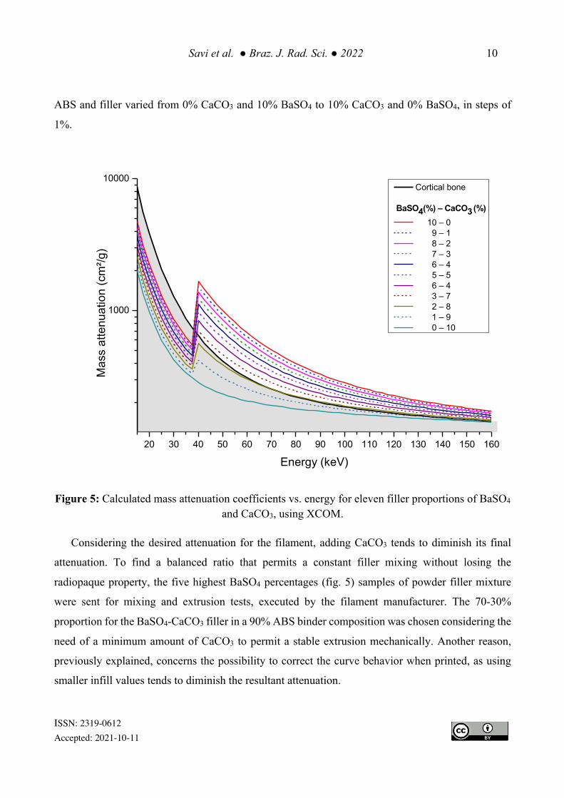
ABS and filler varied from 0% CaCO3 and 10% BaSO4 to 10% CaCO3 and 0% BaSO4, in steps of
1%.
Figure 5: Calculated mass attenuation coefficients vs. energy for eleven filler proportions of BaSO4
and CaCO3, using XCOM.
Considering the desired attenuation for the filament, adding CaCO3 tends to diminish its final
attenuation. To find a balanced ratio that permits a constant filler mixing without losing the
radiopaque property, the five highest BaSO4 percentages (fig. 5) samples of powder filler mixture
were sent for mixing and extrusion tests, executed by the filament manufacturer. The 70-30%
proportion for the BaSO4-CaCO3 filler in a 90% ABS binder composition was chosen considering the
need of a minimum amount of CaCO3 to permit a stable extrusion mechanically. Another reason,
previously explained, concerns the possibility to correct the curve behavior when printed, as using
smaller infill values tends to diminish the resultant attenuation.
20 30 40 50 60 70 80 90 100 110 120 130 140 150 160
1000
10000
Mas
s at
tenu
atio
n (c
m²/g
)
Energy (keV)
Cortical bone
BaSO4(%) – CaCO3 (%) 10 – 0 9 – 1 8 – 2 7 – 3 6 – 4 5 – 5 6 – 4 3 – 7 2 – 8 1 – 9 0 – 10

Savi et al. ● Braz. J. Rad. Sci. ● 2022 11
ISSN: 2319-0612 Accepted: 2021-10-11
After determining filler composition, one more batch of filament was extruded, homogenized and
identified as Filament A. An increase in filament homogeneity were visually detected when compared
with the early extrusions without CaCO3 addition, mainly due to a more consistent and stable
filler/binder ratio being transported and extruded. The filament became even lighter colored, and the
filler specks were scarcer. Still, the addition of powder filler changes the mechanical properties of the
ABS filament, making it more fragile on bending and breaking up more easily.
To confirm the internal homogeneity of Filament A, another set of samples with
20 × 20 × 20 mm³ were printed with 100% infill in a -45/+45 rectilinear pattern, at a 200 µm layer
height. Each cube was scanned in a Philips Brilliance CT 6 channel tomography using 120 kVp, 200
mA, standard algorithm to volume, helicoidal acquisition and posterior axial standard reconstruction
in 2 mm slices.
Analyzing the scan, it was possible to check the resultant mean of 1000±21 HU, compatible with
cortical bone, radiologically represented in CT scans with values above 250 HU. However, other
denser tissues, like tooth dentin and enamel, could not be represented with Filament A. Also, the
standard deviation found, ≅2% of the mean HU, can quantitatively evidence radiological
homogeneity.
Aiming to keep the first planned BaSO4 concentration of 10% and increase the HU range to
represent denser human tissues, the composition of Filament A was modified by increasing BaSO4
and CaCO3 as listed on Table 2 to produce three different filaments named XCT-A, XCT-B and XCT-
C. All filaments were extruded with a constant 1.75 mm diameter and the same homogenization
procedures previously described were applied. Proportionally, after each filler percentage increases,
the mechanical properties diminish, turning it more fragile as the powder inside the plastic contributes
to easily breaking up, when bending the filament in 90 degrees. However, the printing parameters
were equal for pure and filler added ABS. After production, the three XCT filaments and a filament
made of pure ABS were cut and evaluated in a precision scale and a Hubbard pycnometer. Results
are also in Table 2.
Considering the density values of human tissues and organs on ICRP 110 Annex A, high density
values of cortical bone (like humerus 1.92 g/cm³) and teeth (2.75 g/cm³) could not be achieved with
any of the produced filaments, resulting in a material with different mechanical properties and matter
interaction than the proposed mimicked tissues. On the other hand, pure ABS density is equal to

Savi et al. ● Braz. J. Rad. Sci. ● 2022 12
ISSN: 2319-0612 Accepted: 2021-10-11
breast gland and very close (0.01g/cm³ difference) of many tissues like gallbladder, lymphatic nodes,
prostate, salivary gland, among others.
Table 2: Radiopaque filaments created and its composition and density comparison in relation to ABS.
Filament
Composition (in mass)
Density (g/cm³) BaSO4
CaCO3
ABS
ABS 0% 0% 100% 1.020±0.000
XCT-A 10% 4% 86% 1.166±0.006
XCT-B 15% 6% 79% 1.211±0.002
XCT-C 20% 8% 72% 1.271±0.003
However, the fact that the filament mismatches density tissue is not an impediment, as it's
desirable but not excluding. The material supposed to mimic the human tissue has its density equal
or close to the one to be reproduced [36].
In an effort of know the attenuation theoretical behavior of the created filaments, NIST XCOM
was used once more to compare ABS, cortical bone and XCT filaments, as shown in Figure 6.
In order to verify the distribution of filler among the binder matrix, two imaging techniques were
used. Figure 7 (A)-(D) shows, respectively, ABS and XCT-A/B/C filaments in Scanning Electron
Microscopy, while (F)-(I) shows it in the µCT. It was possible to observe the absence of filler at the
pure ABS and a gradual increase in quantity of filler as its concentration increases from XCT-A to C,
especially observable in µCT. As can be seen on SEM images, the quantity of filler agglomeration
diminishes as the process of crushing filament and extruding it again occurs. Using Electron Density
Spectroscopy (EDS), it was observed that the agglomerations seen on XCT A and B are made mainly
by BaSO4 due its natural characteristic of agglomeration.
To evaluate Hounsfield unit equivalence and radiological tissue representation, five
20 × 20 × 20 mm samples were printed for each XCT filament, amounting 15 cubes. All samples
were printed with a 100% infill, in an angular -45º/+45º rectilinear pattern, and 200 µm layer height.
The XCT samples were then batch scanned in the CT (same setup described in section 2.1), having

Savi et al. ● Braz. J. Rad. Sci. ● 2022 13
ISSN: 2319-0612 Accepted: 2021-10-11
its mean HU values and standard deviation determined. These results can be found in Table 3 and the
radiological analysis is shown in Figure 8.
Figure 6: Theoretical attenuation behavior for ABS, XCT filaments and its comparison with ICRU-
44 cortical bone attenuation.
The three XCT filaments performed a long range of HU, opening a possibility of mimicking some
denser human tissues. Cortical bone may be mimicked by XCT-A as the mean HU value of the
filament is 1607 HU, with a propagated uncertainty of 23. Dentin can be also mimicked using XCT-
A or XCT-B, as its equivalence of mean HU value is 1965, with a propagated uncertainty of 30, is
compatible.
Enamel, the natural human tissue with highest radiation attenuation, can be mimicked using XCT-
C and its highest mean value found on this paper was 2624 HU, with a propagated uncertainty of 50.
The standard deviation also confirms a considerable radiological homogenous material, whilst the
20 30 40 50 60 70 80 90 100 110 120 130 140 150 160100
1000
10000
Mas
s at
tenu
atio
n (c
m²/g
)
Energy (keV)
Cortical Bone ABS XCT-A XCT-B XCT-C

Savi et al. ● Braz. J. Rad. Sci. ● 2022 14
ISSN: 2319-0612 Accepted: 2021-10-11
filler powder could be barely visually discernible, with low HU uncertainties, as its maximum
variation was ≅ 2%.
Figure 7: A-D scanning electron microscopy and (F-G) micro computed tomography of in
sequence ABS, XCT-A, XCT-B and XCT-C
Predicting an assembly, the use of XCT-A for jaw bone, XCT-B for dentin and XCT-C for enamel
would be realistic, as there will be three well discernible HU values mimicking three different
attenuation regions. Uniting pure ABS filament [17], these four filaments when printed together, may
enable the creation of 3D printed phantoms capable of mimicking a wide range of human tissues.
Still, it is possible to correct the attenuation behavior (and in consequence the HU value) by changing
the infill percentage of the 3D printed part and tests are needed to calibrate the printing parameters to
match a specific CT setup.

Savi et al. ● Braz. J. Rad. Sci. ● 2022 15
ISSN: 2319-0612 Accepted: 2021-10-11
Table 3: Mean HU values and standard deviation for each 100% infill XCT cubic sample.
Sample number
XCT-A XCT-B XCT-C
Mean SD Mean SD Mean SD
1 1613 20 1986 29 2644 57
2 1562 23 1909 25 2535 42
3 1615 29 1966 37 2636 44
4 1631 23 2000 30 2662 52
5 1613 20 1986 29 2644 57
Mean 1607 26 1970 36 2624 50
XCT-A HU mean values observed in table 3 are inside the cortical bone interval. Also, the
standard deviation at all samples were also acceptable, as for dentin and enamel representations,
XCT-B and C values are also inside the HU interval for those tissues, respectively.
The final price of the filament is a point of interest in this paper, which could diminish production
costs and turn phantoms more available for developing countries. The XCT filaments had an equal
or lower price of production, when compared with special 3D printing filaments. The cost of 1
kilogram of filament is estimated at 52 USD for XCT-A, 70 USD for XCT-B and 89 USD for XCT-
C. This considerable low price could aid the development of inexpensive radiological objects, as well
as anthropomorphic phantoms and patient simulators.
3.1 Research limitations
This research reaches a goal creating filaments for hard tissue representations in FFF 3D printing
technology. However, the final result may vary, as the filaments were tested in a single calibrated CT
machine. Different CT equipment and/or acquisition parameters can find distinct HU values, but not
enough to differ from a bone aspect.
High atomic number substances have a particular increase of attenuation behavior for low
energies due to the k-edge peak of BaSO4, which changes the resulting spectrum in theoretical
calculations. If one intends to use these filaments at energies other than CT, further testing is required

Savi et al. ● Braz. J. Rad. Sci. ● 2022 16
ISSN: 2319-0612 Accepted: 2021-10-11
to observe how the filament will respond as a result of the interaction of BaSO4 at these energies, the
proportion of photoelectric effect and Compton scattering should change and interact with the
detector differently.
At this moment, it’s possible to affirm that the XCT filaments are suitable for mimicking bone,
dentin and enamel for CT imaging phantoms. To consider the created filaments as tissue-equivalents,
a larger characterization should be made, as the information here presented are not enough to affirm
that property as valid.
5. CONCLUSIONS
Although there are other recent 3d printing methods for high density representations of the human
body, the idea needed to be improved to solve some problems reported in literature [10–13]. The
solution presented in this paper uses a different approach, by using inexpensive nonmetallic filler to
compose a filament that could print high density anatomies without double extrusion. ABS was used
as base with addition of BaSO4 and CaCO3 to increase radiopaque properties and to solve mechanical
problems due to inconsistent extrusion, respectively. Techniques like µCT, SEM, pycnometry and
CT were used to initially evaluate the filaments created and to determine quantities as density and
Hounsfield units, in order to verify which tissues could be mimicked using the XCT filament in FFF
3D printing. High density tissues, as the cortical portion of long bones, dentin and enamel, can be
reproduced in 3D printed phantoms, using any simple FFF 3d printing machines.
This product was tested, validated and should allow the development of custom cheaper
radiological imaging phantoms than the ones commercially available.
6. OTHER INFORMATION The XCT filaments are a property of Federal Institute of Santa Catarina – Brazil, with patent
pending registered under number BR1020190245077.

Savi et al. ● Braz. J. Rad. Sci. ● 2022 17
ISSN: 2319-0612 Accepted: 2021-10-11
ACKNOWLEDGMENT
The authors would like to thank Research Directorship of IFSC - Campus Florianopolis and Pro-
Rectory of Research and Innovation of IFSC for the funding. Also, a thanks to IPEN, CAPES,
FAPESP (PROJECT 2017/50332-0) and CNPq (PROJECT 312131/2016-0 and process number
42098/2017-5) for their support. An especial thanks must be given to Instituto SENAI de Inovação
em Manufatura Avançada e Microfabricação and the collaborators Cristiano Cardoso and Gleicy
Ribeiro for the µCT scan.
REFERENCES
[1] MANKOVICH, N. J.; LAMBERT, T.; ZRIMEC, T.; HILLER, J. B. Anatomic vascular phantom for the verification of MRA and XRA visualization and fusion. Med Imaging 1995: Physiol Funct from Multidimens Images, v. 2433, p.73, 1995. https://doi.org/10.1117/12.209724.
[2] ALSSABBAGH, M.; TAJUDDIN, A. B.; MANAP, M. A. Evaluation of nine 3D printing materials as tissue equivalent materials in terms of mass attenuation coefficient and mass density. Int J Adv Appl Sci, v. 4, p. 168–73, 2017. https://doi.org/10.21833/ijaas.2017.09.024.
[3] IVANOV, D.; BLIZNAKOVA, K.; BULIEV, I.; POPOV, P.; METTIVIER, G.; RUSSO, P.; DI LILLO, F.; SARNO, A.; VIGNERO, J.; BOSMANS, H.; BRAVIN, A.; BLISKANOV, Z. Suitability of low density materials for 3D printing of physical breast phantoms. Phys Med Biol, v.63, n. 175020, 2018. https://doi.org/10.1088/1361-6560/AAD315.
[4] GEAR, J. I.; CUMMINGS, C.; CRAIG, A. J.; DIVOLI, A.; LONG, C. D. C.; TAPNER, M.; FLUX, G. D. Abdo-Man: a 3D-printed anthropomorphic phantom for validating quantitative SIRT. EJNMMI Phys, v. 3, n.17, 2016. https://doi.org/10.1186/s40658-016-0151-6.
[5] NIEBUHR, N. I.; JOHNEN, W.; GÜLDAGLAR, T.; RUNZ, A.; ECHNER, G.; MANN, P.; MÖHLER, C.; PFAFFENBERGER, A.; JÄKEL, O.; GREILICH, S. Technical Note: Radiological properties of tissue surrogates used in a multimodality deformable pelvic phantom for MR-guided radiotherapy. Med Phys, v. 43, p. 908–916, 2016. https://doi.org/10.1118/1.4939874.
[6] SCULPTEO. The state of 3D printing. Ed 2018. p. 1–30. Avaliable at: https://info.sculpteo.com/the-state-of-3d-printing-2018.
[7] TAPPA, K.; JAMMALAMADAKA, U. Novel Biomaterials Used in Medical 3D Printing Techniques. J Funct Biomater, v. 9, n. 1, 2018. https://doi.org/10.3390/jfb9010017.
[8] KIARASHI, N.; RAVIN, C. E.; NOLTE, A.C.; STURGEON, G. M.; SEGARS, W. P.; NOLTE, L. W.; LO, J. Y. Development of realistic physical breast phantoms matched to virtual breast phantoms based on human subject data; Development of realistic physical breast phantoms matched to virtual breast phantoms based on human subject data. Med Phys, v. 42, n.7, p.4116–4126, 2015.

Savi et al. ● Braz. J. Rad. Sci. ● 2022 18
ISSN: 2319-0612 Accepted: 2021-10-11
https://doi.org/10.1118/1.4919771.
[9] SHEN, S.; ZHAO, Z.; WANG, H.; HAN, Y.; DONG, E.; LIU, B.; LIU, W.; CROMEENS, B.; ADLER, B.; BESNER, G.; RAY, W.; HOEHNE, B.; XU, R. Freeform fabrication of tissue-simulating phantoms by combining three-dimensional printing and casting. Des Qual Biomed Technol IX, v. 9700, p.970009-1– 97009-6, 2016 . https://doi.org/10.1117/12.2212305.
[10] BECKMANN, J.; POPOVIC, K. Assessment of the attenuation of metal-infused filaments for 3D printing a gamma camera calibration phantom. Med Eng Phys, v. 80, p. 60–64, 2020. https://doi.org/10.1016/J.MEDENGPHY.2020.04.003.
[11] TINO, R.; YEO, A.; BRANDT, M.; LEARY, M.; KRON, T. The interlace deposition method of bone equivalent material extrusion 3D printing for imaging in radiotherapy. Mater Des, v. 199, 2021. https://doi.org/10.1016/j.matdes.2020.109439.
[12] CEH, J.; YOUD, T.; MASTROVICH, Z.; PETERSON, C.; KHAN, S.; SASSER, T. A.; SANDER, I. M.; DONEY, J.; TURNER, C.; LEEVY, W. M. Bismuth infusion of ABS enables additive manufacturing of complex radiological phantoms and shielding equipment. Sensors (Switzerland), v. 17, p.1–11, 2017. https://doi.org/10.3390/s17030459.
[13] ALSSABBAGH, M.; TAJUDDIN, A. A.; ABDULMANAP, M.; ZAINON, R. Evaluation of 3D
printing materials for fabrication of a novel multi-functional 3D thyroid phantom for medical dosimetry and image quality. Radiat Phys Chem 2017, v. 135, p.106–112, 2017. https://doi.org/10.1016/j.radphyschem.2017.02.009.
[14] SHIN, J.; SANDHU, R. S.; SHIH, G. Imaging Properties of 3D Printed Materials: Multi-Energy CT of Filament Polymers. J Digit Imaging, v. 30, p. 572–575, 2017. https://doi.org/10.1007/s10278-017-9954-9.
[15] DANCEWICZ, O. L.; SYLVANDER, S. R.; MARKWELL, T. S.; CROWE, S.B.; TRAPP, J. V. Radiological properties of 3D printed materials in kilovoltage and megavoltage photon beams. Phys Medica, v. 38, p. 111–118, 2017. https://doi.org/10.1016/J.EJMP.2017.05.051.
[16] SAVI, M.; ANDRADE, M. A. B.; POTIENS, M. P. A. Commercial filament testing for use in 3D printed phantoms. Radiat Phys Chem, v. 174, 2020. https://doi.org/10.1016/J.RADPHYSCHEM. 2020.108906.
[17] RESNIK, R.; KIRCOS, L. T.; MISCH, C. Diagnostic Imaging and Techniques In: Contemporary Implant Dentistry. 3rd ed. St. Louis: Mosby Elsevier; 2007. p. 38-67.
[18] SAKUMA, A.; SAITOH, H.; MAKINO, Y.; INOKUCHI, G.; HAYAKAWA, M.; YAJIMA, D.; IWASE, H. Three-dimensional visualization of composite fillings for dental identification using CT images. Dentomaxillofacial Radiol, v. 41, p. 515–519, 2012. https://doi.org/10.1259/dmfr/13441277.
[19] BORGES, M. S.; MUCHA, J. N. Bone density assessment for mini-implants position. Dental Press J Orthod, v. 15, p. 58–60, 2010.
[20] HAZELAAR, C.; VAN EIJNATTEN, M.; DAHELE, M.; WOLFF, J.; FOROUZANFAR, T.; SLOTMAN, B.; VERBAKEL, W. F. A. R. Using 3D printing techniques to create an anthropomorphic thorax phantom for medical imaging purposes. Med Phys, v. 45, p. 92–100, 2018. https://doi.org/10.1002/mp.12644.
[21] LI, Y.; LI, Z.; AMMANUEL, S.; GILLAN, D.; SHAH, V. Efficacy of using a 3D printed lumbosacral
Savi et al. ● Braz. J. Rad. Sci. ● 2022 19
ISSN: 2319-0612 Accepted: 2021-10-11
spine phantom in improving trainee proficiency and confidence in CT-guided spine procedures. 3D Print Med, v. 4, 2018. https://doi.org/10.1186/s41205-018-0031-x.
[22] HAMEDANI, B. A.; MELVIN, A.; VAHEESAN, K.; GADANI, S.; PEREIRA, K.; HALL, A. F. Three-dimensional printing CT-derived objects with controllable radiopacity. J Appl Clin Med Phys, v. 19, p. 317–328, 2018. https://doi.org/10.1002/acm2.12278.
[23] PRICE, G.; BIGLIN, E. R.; COLLINS, S.; AITKINHEAD, A.; SUBIEL, A.; CHADWICK, A. L.; CULLEN, D. M.; KIRKBY, K. J.; SCHETTINO, G.; TIPPING, J.; ROBINSON, A. An open source heterogeneous 3D printed mouse phantom utilising a novel bone representative thermoplastic. Phys Med Biol, v. 65, 2020. https://doi.org/10.1088/1361-6560/ab8078.
[24] INEOS STYROLUTION. Technical datasheet: Terluran GP-35. 2016. Avaliabe at: https://www.ineos-styrolution.com/INTERSHOP/web/WFS/Styrolution-Portal-Site/en_US/-/USD/ViewPDF-Print.pdf?SKU=300600120829&RenderPageType=ProductDetail.
[25] THREEPOPNATKUL, P.; TEPPINTA, W.; SOMBATSOMPOP, N. Effect of co-monomer ratio in ABS and wood content on processing and properties in wood/ABS composites. Fibers Polym, v. 12, n. 8, p.1007–1013, 2011. https://doi.org/10.1007/S12221-011-1007-2.
[26] MCCULLOUGH, E.J.; YADAVALLI, V. K. Surface modification of fused deposition modeling ABS to enable rapid prototyping of biomedical microdevices. J Mater Process Technol, v. 213, n. 6, p. 947–954, 2013. https://doi.org/10.1016/j.jmatprotec.2012.12.015.
[27] BERGER, M. J.; HUBBELL, J. H.; SELTZER, S. M.; CHANG, J.; COURSEY, J. S.; SUKUMAR, R.; ZUCKER, D. S.; OLSEN, K. XCOM: Photon Cross Section Database (version 1.5), 2010.
[28] PHYSIKALISCH-TECHNISCHE BUNDESANSTALT. Radiation qualities used for studies in radiation protection, 2015.
[29] WHITE, D. R.; BOOZ, J.; GRIFFITH, R. V.; SPOKAS J. J.; WILSON, I. J. Report 44. J Int Comm Radiat Units Meas. v. os23, n. 1, 1989. https://doi.org/10.1093/jicru/os23.1.report44.
[30] KIM, S.; CHEN, J.; CHENG, T.; GINDULYTE, A.; HE, J.; HE, S.; LI, Q.; SHOEMAKER, B. A.; THIESSEN, P. A.; YU, B.; ZASLAVSKY, L.; ZHANG, J.; BOLTON, E. E. PubChem 2019 update: Improved access to chemical data. Nucleic Acids Res, v. 47, n. D1, p. 1102–1109, 2019. https://doi.org/10.1093/nar/gky1033.
[31] VALOR, A.; REGUERA, E.; SÁNCHEZ-SINENCIO, F. Synthesis and X-ray diffraction study of calcium salts of some carboxylic acids. Powder Diffr, v. 17, p. 13–18, 2002. https://doi.org/10.1154/ 1.1414011.
[32] HAYNES, W. M. CRC Handbook of Chemistry and Physics. 93rd ed. CRC Press; 2012.
[33] ICRP. International Comission on Radiation Protection, Report 110: Annexes A-D. v. 39, p. 47 –70, 2009. https://doi.org/10.1016/j.icrp.2009.07.005.
[34] HSIEH, J. Computed Tomography: Principles, Design, Artifacts, and Recent Advances. 2nd ed. Bellingham: Spie, 2009. Avaliable at: https://spie.org/Publications/Book/2049426?SSO=1.
[35] TODD, D. B. Improving incorporation of fillers in plastics. A special report. Adv Polym Technol, v. 19, p. 54–64, 2000. https://doi.org/10.1002/(SICI)1098-2329(20000117)19:1<54::AID-ADV6>3.0. CO;2-#.
Savi et al. ● Braz. J. Rad. Sci. ● 2022 20
ISSN: 2319-0612 Accepted: 2021-10-11
[36] XU, X. G.; ECKERMAN, K. F. Handbook of anatomical models for radiation dosimetry. 1st ed. Boca Raton: CRC Press, 2010.
Copyright © 2022 FDOKUMEN