Activation of the MKL1/actin signaling pathway induces ...
39
HAL Id: hal-01061363 https://hal-univ-rennes1.archives-ouvertes.fr/hal-01061363 Submitted on 5 Sep 2014 HAL is a multi-disciplinary open access archive for the deposit and dissemination of sci- entific research documents, whether they are pub- lished or not. The documents may come from teaching and research institutions in France or abroad, or from public or private research centers. L’archive ouverte pluridisciplinaire HAL, est destinée au dépôt et à la diffusion de documents scientifiques de niveau recherche, publiés ou non, émanant des établissements d’enseignement et de recherche français ou étrangers, des laboratoires publics ou privés. Activation of the MKL1/actin signaling pathway induces hormonal escape in estrogen-responsive breast cancer cell lines. Gwenneg Kerdivel, Antoine Boudot, Denis Habauzit, Frederic Percevault, Florence Demay, Farzad Pakdel, Gilles Flouriot To cite this version: Gwenneg Kerdivel, Antoine Boudot, Denis Habauzit, Frederic Percevault, Florence Demay, et al.. Activation of the MKL1/actin signaling pathway induces hormonal escape in estrogen-responsive breast cancer cell lines.. Molecular and Cellular Endocrinology, Elsevier, 2014, 390 (1-2), pp.34-44. 10.1016/j.mce.2014.03.009. hal-01061363
-
Upload
khangminh22 -
Category
Documents
-
view
0 -
download
0
Transcript of Activation of the MKL1/actin signaling pathway induces ...

HAL Id: hal-01061363https://hal-univ-rennes1.archives-ouvertes.fr/hal-01061363
Submitted on 5 Sep 2014
HAL is a multi-disciplinary open accessarchive for the deposit and dissemination of sci-entific research documents, whether they are pub-lished or not. The documents may come fromteaching and research institutions in France orabroad, or from public or private research centers.
L’archive ouverte pluridisciplinaire HAL, estdestinée au dépôt et à la diffusion de documentsscientifiques de niveau recherche, publiés ou non,émanant des établissements d’enseignement et derecherche français ou étrangers, des laboratoirespublics ou privés.
Activation of the MKL1/actin signaling pathway induceshormonal escape in estrogen-responsive breast cancer
cell lines.Gwenneg Kerdivel, Antoine Boudot, Denis Habauzit, Frederic Percevault,
Florence Demay, Farzad Pakdel, Gilles Flouriot
To cite this version:Gwenneg Kerdivel, Antoine Boudot, Denis Habauzit, Frederic Percevault, Florence Demay, et al..Activation of the MKL1/actin signaling pathway induces hormonal escape in estrogen-responsivebreast cancer cell lines.. Molecular and Cellular Endocrinology, Elsevier, 2014, 390 (1-2), pp.34-44.�10.1016/j.mce.2014.03.009�. �hal-01061363�

1
Title: Activation of the MKL1/actin signaling pathway induces hormonal escape in estrogen-1
responsive breast cancer cell lines. 2
3
Authors: Gwenneg Kerdivel1, Antoine Boudot1, Denis Habauzit1, Frederic Percevault1, Florence 4
Demay1, Farzad Pakdel1, Gilles Flouriot1* 5
6
1 University of Rennes 1, Institut de Recherche en Santé Environnement et Travail, IRSET, INSERM 7
U1085, Team TREC, Biosit, Rennes, France 8
9
*Corresponding author: 10
Dr. Gilles Flouriot 11
INSERM U1085, IRSET, University of Rennes 1, Beaulieu Campus, 35042 Rennes cedex, France. 12
Phone: +33-2 23 23 68 04 13
Fax: +33-2 23 23 67 94 14
E-mail: [email protected] 15
16
Disclosure Statement: The authors have nothing to disclose. 17
18
2
Abstract 1
2
Estrogen receptor alpha (ERα) is generally considered to be a good prognostic marker because 3
almost 70% of ERα-positive tumors respond to anti-hormone therapies. Unfortunately, during cancer 4
progression, mammary tumors can escape from estrogen control, resulting in resistance to treatment. 5
In this study, we demonstrate that activation of the actin/megakaryoblastic leukemia 1 (MKL1) 6
signaling pathway promotes the hormonal escape of estrogen-sensitive breast cancer cell lines. The 7
actin/MKL1 signaling pathway is silenced in differentiated ERα-positive breast cancer MCF-7 and 8
T47D cell lines and active in ERα-negative HMT-3522 T4-2 and MDA-MB-231 breast cancer cells, 9
which have undergone epithelial-mesenchymal transition. We showed that MKL1 activation in MCF-7 10
cells, either by modulating actin dynamics or using MKL1 mutants, down-regulates ERα expression 11
and abolishes E2-dependent cell growth. Interestingly, the constitutively active form of MKL1 12
represses PR and HER2 expression in these cells and increases the expression of HB-EGF, TGFβ, and 13
amphiregulin growth factors in an E2-independent manner. The resulting expression profile (ER-, PR-, 14
HER2-) typically corresponds to the triple-negative breast cancer expression profile. 15
16
Keywords: MKL1, hormone resistance, breast cancer, cell growth, estrogen receptor, tamoxifen 17

3
1. Introduction 1
Estrogens, especially 17β-estradiol (E2), control the proliferation and differentiation of the 2
epithelial cells of the mammary gland (Couse and Korach, 1999). The effects of E2 are principally 3
mediated by the estrogen receptors (ERs), which regulate the expression of specific target genes after 4
binding to regulatory regions. In addition to its crucial roles in physiology, ERα is also associated with 5
human pathological processes, such as in estrogen-dependent breast cancers (Deroo and Korach, 6
2006). Approximately 70% of diagnosed breast cancers express ERα, which mediates mitogenic 7
effects in a E2-dependent manner (Dahlman-Wright et al., 2006; Zhao et al., 2008; 2011), and breast 8
tumors expressing ERα often appear to be more differentiated and less invasive than ERα-negative 9
cancers (Rochefort et al., 1998; Platet et al., 2004). Furthermore, the proliferation of ERα-positive 10
cancer cells can be repressed by anti-estrogens, such as tamoxifen, which are already used in hormone 11
therapy (Wickerham et al., 2009; Kim et al., 2011). For these reasons, ERα expression is generally 12
associated with a good prognosis. Unfortunately, over time, ERα-positive breast tumors can escape 13
from hormonal control, becoming resistant to hormone therapy, and switch from a well-differentiated 14
epithelial phenotype to a metastatic and aggressive one (Rau et al., 2005). Hormone-resistant cells 15
frequently exhibit a loss in ERα activity or expression, while the activity of growth receptor signaling 16
increases (Barone et al., 2010; Murphy et al., 2011). For example, the up-regulation of EGFR 17
expression occurs in some tumors and serves as an alternative survival and proliferation pathway 18
(Newby et al., 1997). Despite numerous studies on the topic, the molecular mechanisms involved in 19
the loss of hormonal responses are still unclear. 20
Growing evidence suggests a role for the myocardin-related transcription factor MKL1 21
(megakaryoblastic leukemia 1, also termed MRTF-A, MAL, or BSAC) in cancer progression as a 22
tumor-promoting or tumor-suppressor factor, depending on the cellular context (Medjkane et al., 2009; 23
Scharenberg et al., 2010; Hu et al., 2011; Muehlich et al., 2011). MKL1 was first described as a 24
coactivator of serum response factor (SRF) in the control of motile or contractile cell functions, 25
especially during vascular smooth muscle cell and cardiac myocyte differentiation, neuronal 26
migration, or cancer metastasis (Cen et al., 2003; Scharenberg et al., 2010). MKL1 continuously 27
4
shuttles between the nucleus and cytoplasm via a process controlled by the cellular pool of globular actin 1
(G-actin) (Posern et al., 2002). The nuclear import of MKL1 is regulated by RhoA activity on actin 2
dynamics. Sequestered in an inactive form by G-actin in the cytosol, MKL1 is released following the 3
polymerization of G-actin into filamentous actin (F-actin), which is induced by RhoA (Busche et al., 4
2008, 2010). Nuclear G-actin has also been demonstrated to facilitate the nuclear export of MKL1. 5
Although the localization and activity of MKL1 are controlled by G-actin, the two events remain 6
independent from each other (Vartiainen et al., 2007; Muehlich et al., 2008). In epithelial-like cells, 7
the actin/MKL1 signaling pathway is notably activated by the loss of cell-cell junctions that occurs 8
during the epithelial-mesenchymal transition (EMT) (Micalizzi et al., 2010). 9
We have previously shown that the Rho/actin/MKL1 signaling pathway is also a main actor in 10
controlling ERα transcriptional activity (Huet et al., 2008, 2009). In transient transfection experiments, the 11
transactivation efficiency of ERα results in part from the respective contribution exerted by its two 12
activation functions, AF1 and AF2, the activities of which are tightly regulated in a cell differentiation 13
stage-dependent manner (Mérot et al., 2004a). Precisely, the more differentiated a cell is, the more that 14
cell mediates ERα signaling through its AF1. In contrast, AF2 is the only active AF in cells that have 15
achieved their EMT (Mérot et al., 2004a). Interestingly, tumor cell lines endogenously expressing ERα 16
exhibit an AF1-permissive cell context, whereas strictly AF-2-permissive tumor cells are always ERα 17
negative. Furthermore, the stable expression of a functional ERα in strictly AF2-permissive cells 18
restores an AF1-sensitive cell context. The transactivation efficiency of ERα was demonstrated to be 19
closely dependent upon the functional state of MKL1. The inhibition of MKL1 activity favors AF1 20
transactivation of ERα; in contrast, the activation of MKL1 silences AF1 activity, thus dramatically 21
reducing the transactivation efficiency of ERα, which then acts exclusively through AF2. Chromatin 22
immunoprecipitation (ChIP) experiments on the ERα-positive breast cancer MCF7 cell line showed that 23
MKL1 is present on the promoter of all tested E2-regulated genes (Huet et al., 2009). This presence 24
appears to be mainly E2-independent, and sequential ChIPs indicated a corecruitment with ERα onto 25
several promoters. 26
In the present report, we investigated the impact of the actin/MKL1 signaling pathway on 27
breast cancer cell responsiveness to estrogen. The results show that the actin/MKL1 signaling pathway 28

5
is involved in the higher proliferation of ERα-negative cell lines that have undergone EMT, in contrast 1
to differentiated ERα-positive MCF-7 and T47D cell lines. We further demonstrate that MKL1 2
activation in MCF-7, either by promoting the F/G actin ratio or using MKL1 mutants, induces the 3
hormonal escape of cell growth. This loss of E2 responsiveness is concomitant with the down-4
regulation of ERα and progesterone receptor (PR) and the enhancement of growth factor signaling. 5
6
2. Materials and methods 7
2.1. Plasmids and transient transfections 8
The reporter gene C3 (complement 3)-Luc, the pCR-ER-66, pCR-ERα-Δ79, and pCR-ERα-46 9
expression vectors, and the CMV-βgal internal control vector have been described previously (Mérot 10
et al., 2004b). The V159N and R62D β-actin mutant expression vectors were kindly provided by Pr. 11
MK. Vartiainen (University of Helsinki, Finland). A 100-ng sample of these expression vectors or 12
control vector (pCR3.1) was used in transient transfection using JetPEI® (Polyplus transfection) 13
according to the manufacturer’s protocol. p3Xflag-MKL1, p3Xflag-MKL1 ΔN200, and p3Xflag-14
MKL1 ΔC301 expression vectors, which were used to generate the pcDNA4/TO expression vectors, 15
were a gift from Pr. R. Prywes (Colombia University, USA). 16
17
2.2. Antibodies, siRNAs, and reagents 18
E2 and 4-hydroxy-tamoxifen (4-OHT) were purchased from Sigma-Aldrich and Alexa Fluor® 19
594 Phalloidin and DNaseI Alexa Fluor® 488 Conjugate from Invitrogen. 20
Nonspecific siRNA as a control (12935-300, Invitrogen) and an siRNA directed against 21
MKL1 (One-target plus smart pool L-015434-00-0005; Thermo Scientific Dharmacon) were 22
transfected using Lipofectamine™ 2000 (Invitrogen) in accordance with manufacturer’s 23
recommendations. 24
The primary antibodies used for western blotting and immunofluorescence analyses were as 25
follows: rabbit polyclonal (Rp) antibodies against c-Fos (sc-52), ERα (HC-20, sc-543), p-Akt1/2/3 26

6
(Ser473)-R (sc-7985-R), Akt1/2/3 (H-136, sc-8312), ERK1 (K-23, sc-94), and SRF (G-20, sc-335), a 1
goat polyclonal antibody directed against MKL1 (MRTF-A C-19, sc-21558), and a mouse monoclonal 2
(Mm) antibody against p-ERK (sc-7383) or against β-actin (AC-15, sc-69879) were acquired from 3
Santa Cruz; an Mm anti-flag (clone M2) and Mm anti-vimentin (clone V9) antibody were obtained 4
from Sigma-Aldrich; an Mm anti-PCNA antibody was obtained from Dako; Rp anti-E-cadherin 5
(ab15148), Rp anti-MKL1 (ab113264), and Rp anti-alpha smooth muscle actin (ab5694) antibodies 6
were obtained from Abcam; an Mm antibody directed against vinculin (MAB3574) was purchased 7
from Millipore; the secondary peroxidase-conjugated goat anti-rabbit antibody was purchased from 8
Pierce, and the bovine anti-goat and goat anti-mouse antibodies were purchased from Santa Cruz; 9
Alexa Fluor® dye-conjugated secondary antibodies were employed for immunofluorescence 10
(Invitrogen). 11
12
2.3. Cell culture and treatments 13
MCF-7, T47D, and MDA-MB-231 cells were routinely maintained in DMEM (GIBCO) 14
supplemented with 10 % fetal bovine serum (FBS; Biowest) and antibiotics (GIBCO) at 37°C in 5 % 15
CO2. HMT-3522 T4-2 cells (Briand et al., 1996) were routinely maintained in DMEM / hams F12 16
(GIBCO) supplemented with insulin, transferrin, and sodium selenite (Insulin-Transferrin-Selenium 17
100X, GIBCO), 10-10 M E2, 0.5 µg/mL hydrocortisone (Sigma), and 5 µg/mL ovine prolactin (Sigma). 18
Flasks coated with collagen IV (BD Bioscience) were used for culturing the HMT-3522 T4-2 cells. 19
MCF-7 clones stably transfected with the control or overexpressing the wild-type form of MKL1 20
(WT) or deleted forms of MKL1 (ΔN200 or ΔC301) tagged with a flag epitope were obtained by 21
transfecting cells with the pcDNA6/TR plasmid and the corresponding pcDNA4/TO expression 22
vectors (T-Rex system, Invitrogen) using JetPEI®. The clones were selected in a medium containing 5 23
µg/mL blasticidin and 100 µg/mL zeocin (Invitrogen). Individual clones were isolated and grown in a 24
medium containing selective antibiotics to maintain selection pressure. When treatments with steroids 25
were required, the cells were maintained 48 h in DMEM (GIBCO) supplemented with 2.5 % dextran / 26
charcoal-stripped FBS (dsFBS; Biowest) prior to the experiments. The induction of WT and mutant 27

7
MKL1 expression in MCF-7 clones was performed under the same condition with 1 µg/mL 1
tetracycline. 2
3
2.4. Immunofluorescence 4
Cells were plated on cover slides. After treatment, phosphate-buffered saline (PBS) containing 5
4 % paraformaldehyde was used to fix the cells for 10 min. The cells were permeabilized in PBS 6
containing 0.3 % Triton X-100 for 10 min, incubated in sodium citrate for 1 h, and then washed again. 7
The cells were incubated overnight at 4°C with primary antibodies; the next day, incubation with 8
secondary antibodies was performed for 2 h at room temperature. Finally, the cover slides were 9
mounted in Vestashield® medium with DAPI (Vector). Images were obtained with an Imager.Z1 10
ApoTome AxioCam (Zeiss) microscope. 11
12
2.5. Protein extraction and western blotting 13
Whole-cell extracts were directly prepared in 3X Laemmli buffer. Following sonication, the 14
protein extracts were denatured for 5 min at 95°C, separated on 10 % SDS polyacrylamide gels, and 15
transferred to polyvinylidene difluoride membrane (Millipore). The proteins were then probed with 16
specific antibodies. An enhanced chemiluminescence system (Immune-Star, Bio-Rad) was used to the 17
detect immunocomplexes. 18
19
2.6. RT-PCR assays 20
Trizol™ reagent (Invitrogen) was used for total RNA extraction according to the 21
manufacturer’s protocol. Retrotranscription was performed using MMLV reverse transcriptase. 22
Quantitative RT-PCRs were performed using the iQ™ SYBR® Green supermix from BioRad (Bio-23
Rad, Hercules, CA, USA). 24
25
2.7. Flow cytometry analyses 26

8
Cells were maintained for 48 h in DMEM without phenol red and supplemented with 2.5% 1
dsFBS for E2 treatments or in DMEM supplemented with 5 % FBS for 4-OHT treatments. After 2
treatment, the cells were trypsinized, fixed for 30 min on ice with 70 % ethanol, and then incubated for 3
20 min at 4°C in IFA buffer (10 mM HEPES, pH 7.4, 150 mM NaCl, 4 % SVF). A 30-min incubation 4
with RNaseA (100 µg/mL) at 37°C was then performed, and 25 µg/mL propidium iodide was finally 5
added prior to FACS analysis. A total of 104 cells from each sample were analyzed using the FACScan 6
apparatus (Becton-Dickinson). 7
8
2.8. Statistical analysis 9
Statistical analyses were performed using Student’s t-test. The values are provided as the mean 10
± standard error of the mean (SEM). 11
12
3. Results 13
3.1. Hormone-dependent and -independent breast cancer cells exhibit different MKL1 activities 14
Four breast cancer cell lines, MCF-7, T47D, HMT-3522 T4-2, and MDA-MB-231, were 15
selected for their differential status with respect to ERα, proliferation, and EMT (Fig. 1, panels A and 16
B). MCF-7 and T47D cells express ERα and their proliferation is controlled by estrogen. In contrast, 17
HMT-3522 T4-2 and MDA-MB-231 cells do not express ERα and their proliferation is hormone-18
independent. Furthermore, MCF-7 and T47D cells are well-differentiated epithelial cells with a 19
minimally aggressive behavior, whereas HMT-3522 T4-2 and MDA-MB-231 cells are invasive and 20
poorly differentiated. HMT-3522 T4-2 and MDA-MB-231 cell lines have undergone EMT, as shown 21
by the loss of the epithelial adhesion protein E-cadherin and the expression of vimentin, an intermediate 22
filament (Fig. 1, panel A). Changes in actin dynamics are a hallmark of EMT. Therefore, the F/G-actin 23
ratio was determined in the four cell lines by immunofluorescence staining using phalloidin and 24
DNase I, which bind specifically to F- and G-actin, respectively (Fig. 1, panel C). A quantitative 25
assessment of the fluorescence intensity revealed a 2-fold increase in the F/G-actin ratio in the 26

9
dedifferentiated ERα-negative cell lines versus the ERα-positive cell lines. Because an increased F/G-1
actin ratio activates MKL1 (Posern et al., 2002; Busche et al., 2008), the ERα-negative cell lines were 2
expected to have higher MKL1 activity. MKL1 was expressed in the four cell lines, as shown by 3
western blotting (Fig. 1, panel B). MKL1 activation is often associated with the massive nuclear 4
accumulation of the protein, though these events are independent from each other (Vartiainen et al., 5
2007). Immunofluorescence experiments showed differences in the sub-cellular localization of MKL1 6
between the ERα-positive and -negative cells, with a high percentage of cells with nuclear MKL1 in 7
the ERα-negative cell lines (Fig. 1, panels A, D). MKL1 is a coactivator of serum response factor 8
(SRF), but the SRF gene is also an important target of the Rho/actin/MKL1 signaling pathways 9
through the SRE in its promoter, providing positive feedback loop regulation. SRF expression was 10
therefore determined by western blotting in the four cell lines. The highest level of SRF expression 11
was detected in the HMT-3522 T4-2 and MDA-MB-231 cells, in agreement with the nuclear 12
localization of MKL1 (Fig. 1, panel B). Finally, corroborating the higher MKL1 activity in the ERα-13
negative cell lines, the respective contribution exerted by the AF1 and AF2 transactivation functions to 14
ERα transcriptional activity was clearly different in the ERα-positive and ERα-negative cell lines, 15
indicative of a different functional state of MKL1 in these cells (Fig. 1, panel E). Indeed, the failure of 16
ERα to transactivate E2-regulated reporter genes through the AF1 transactivation function was previously 17
demonstrated to be a consequence of MKL1 activation (Huet et al., 2009). In these experiments, cell 18
permissiveness to either ERα AFs was determined by comparing the transcriptional activity of ERα 19
with that of ERα Δ79 (deletion of AF1 box 1) and ERα CF (total deletion of AF1). 20
To identify the involvement of MKL1 in the proliferation of ERα-positive and ERα-negative cell lines, 21
the impact of MKL1 siRNA knockdown on the percentage of cells in S phase was determined through 22
nuclear PCNA staining in immunofluorescence experiments. As shown in Fig. 1 (panel F), the MCF-7 23
and T47D cells exhibited a lower percentage of cells in S phase than the HMT-3522 T4-2 and MDA-24
MB-231 cells, as expected. MKL1 silencing by siRNA had no impact on MCF-7 and T47D 25
proliferation, corroborating the fact that MKL1 is not or only weakly active in these cell lines but 26
relevantly reduces the percentage of HMT-3522 T4-2 and MDA-MB-231 cells in S phase. 27
Collectively, these results indicate that the higher percentage of cells in S phase in mesenchymal-like 28

10
ERα-negative cell lines is in part due to the activation of the actin/MKL1 signaling pathway. 1
Furthermore, these data suggest that hormone-dependent and -independent breast cancer cells might 2
differ with regard to their MKL1 activity. 3
4
3.2. Changes in the actin dynamic pathway modulate E2-dependent proliferation via MKL1 5
As previously indicated, differentiated hormone-dependent breast cancer cells exhibit no or 6
low MKL1 activity. EMT is characterized by an increase in the F/G-actin ratio and consequently 7
MKL1 activation (Morita et al., 2007). Hormonal escape is also a hallmark of the progression of 8
hormone-dependent breast cancer cells toward EMT. To verify whether a direct link exits between 9
these two events, we attempted to activate endogenous MKL1 in the hormone-dependent breast cancer 10
MCF-7 cell line by expressing mutant forms of actin that are known to modify the F/G-actin ratio. 11
R62D is a non-polymerizable actin mutant that represses MKL1 activity on SRF target genes. In 12
contrast, Actin-V159N favors F-actin formation and consequently leads to MKL1 activation (Posern et 13
al., 2002). Fig. 2 (panel A) shows the expected changes in the F/G-actin ratio induced by these actin 14
mutants. Interestingly, most of the cells were affected, suggesting that low amounts of transfected plasmids 15
are sufficient to induce these changes and that actin dynamics could be, as with other polymerization 16
phenomena, very sensitive to disrupting molecules. Indicative of cell fate, ERα localization was first 17
assayed by immunofluorescence (Fig. 2, panels B, C). Although the R62D mutant had no or little 18
effect, Actin-V159N induced a clear decrease in ER nuclear localization, slightly leaking to the 19
cytoplasm, which was comparable to the ERα profile observed during E2-induced cell proliferation. 20
Changes in the MKL1 distribution after transfection of the different actin constructs were observed. 21
Indeed, the V159N mutant increased the nuclear translocation of MKL1 (Fig. 2, panel D). The impact of 22
the actin mutants on the percentage of MCF-7 cells in S phase was then determined following PCNA 23
staining (Fig. 2, panel E, left). The results showed a slight but significant reduction in the number of 24
cells in S phase in the presence of the non-polymerizable R62D actin mutant (S=12.3% vs S=15.7% in 25
the control, p<0.05), whereas the expression of actin-V159N enhanced the number of cells (S=25% vs 26
S=15.7% in the control, p<0.05). As a consequence, the fold induction of MCF-7 cells in S phase 27

11
observed after E2 treatment was reduced in the cells expressing actin-V159N and increased in the cells 1
expressing actin-R62D (1.7 and 3.6, respectively, for these cells vs 2.7 for the control cells). To 2
evaluate MKL1 involvement in this process, similar experiments were performed in MCF-7 cells in 3
which MKL1 expression was silenced by siRNA. As depicted in Fig. 2 (panel E, right), MKL1 4
knockdown completely abolished the effect of the R62D (p<0.05) and V159N (p<0.001) actin 5
mutants. In contrast, MKL1 repression had no impact on E2-induced proliferation. Similar data were 6
obtained using T47D cells (suppl. 1). Collectively, these results indicate that the activation of the 7
actin/MKL1 pathway induces an enhancement in the percentage of cells in S phase that escape from 8
hormonal control. 9
10
3.3. Expression of a constitutively active form of MKL1 in MCF-7 cells represses ERα 11
expression and abolishes E2-induced transcription 12
To further investigate the impact of MKL1 on hormone-dependent breast cancer cells, we 13
generated MCF-7 sub-clones using a tetracycline-inducible vector system to stably express the MKL1-14
WT, -ΔN200, or -ΔC301 forms. Deleted of the N-terminal RPEL motifs, MKL1-ΔN200, is a 15
constitutively active form, whereas MKL1-ΔC301 behaves as a dominant negative form due to the 16
deletion of the transactivation domain (Cen et al., 2003). In addition to the first series of individual 17
MCF-7 sub-clones, two individual clones expressing mutant forms of MKL1 were selected (Clones 2 18
and 3). It should be noted that all the data obtained with these additional clones mostly confirm the 19
results obtained with the first series (clone 1) (suppl. 2). We first controlled MKL1 protein expression 20
in the different sub-clones using western blotting and immunofluorescence methods (Fig. 3, panel A, 21
B, and suppl. 2). Proteins of interest were produced after tetracycline treatment only (suppl. 2). The 22
nuclear localization of Flagged-MKL1-WT and MKL1-ΔC301 was observed in less than 10% of the 23
cells, whereas most of the cells showed MKL1-ΔN200 localized in the nucleus (Fig. 3, panel B). The 24
activity of the MKL1 mutants was confirmed by western blot monitoring of the expression level of 25
SRF, c-fos, or α-actin, three known targets of the actin/MKL1 signaling pathway (Fig. 3, panel A, and 26
suppl. 2). Intriguingly, a high expression level of MKL1-ΔN200 was shown to repress ERα 27

12
expression. Corroborating the western blotting analysis, ERα immunostaining performed on the entire 1
cell population revealed a global decrease in ERα expression in the cells expressing the constitutively 2
active form of MKL1 (Fig. 3, panel C). Regardless of the form of MKL1 (WT, MKL1-ΔN200, and 3
MKL1-ΔC301), ER was down-regulated when MKL1 was nuclear. Therefore, the nuclear localization 4
of MKL1 proteins and ERα are mutually exclusive (Fig. 3, panel B). Quantitative real-time PCR 5
experiments demonstrated that ERα expression was also repressed at the mRNA level in MCF-6
7/MKL1-ΔN200 cells (Fig. 3, panel D). Interestingly, MKL1-ΔN200 also exhibited down-regulated 7
GATA3, a hallmark of hormonal responsiveness. Therefore, the estrogenic responses of PR and pS2, 8
two main E2 target genes, were deeply impacted by MKL1 activities, as expected. Although the 9
expression of both genes was induced by E2 in the MCF-7/cont, MCF-7/MKL1-WT, and MCF-10
7/MKL1-ΔC301 cells, this regulation was almost completely abolished in the MCF-7/MKL1-ΔN200 11
cells. 12
13
3.4. Expression of a constitutively active form of MKL1 in MCF-7 cells enhances growth factor 14
signaling and induces hormonal escape 15
Our study was extended by implementing quantitative real-time PCR experiments targeting 16
the expression of genes encoding members of the growth factor signaling pathway (Fig. 4, panel A, 17
and suppl. 2). TGFα and amphiregulin, two main growth factors in MCF-7 cells, have been previously 18
shown to be up-regulated by E2, whereas the expression of the receptors HER1 and HER2 is repressed 19
(Normanno et al., 1993; Zhang et al., 1994; Dardes et al., 2002). As expected, the expression of TGFα 20
and amphiregulin was induced by E2 in the MCF-7/cont, MCF-7/MKL1, and MCF-7/MKL1-ΔC301 21
cells. HER1 and HER2 expression was also repressed by E2 in these three cell lines. Surprisingly, the 22
expression of the TGFα and amphiregulin genes was strongly enhanced in a hormone-independent 23
manner in MCF-7/MKL1-ΔN200. In the same way, HB-EGF, a membrane-bound growth factor over-24
expressed in malignant cells (Miyamoto et al., 2006), was dramatically up-regulated in these cells. In 25
parallel, the expression level of the receptor HER1 was no longer down-regulated by E2, remaining at 26
a high level in the presence of the steroid. Finally, HER2 expression was clearly down-regulated in a 27

13
hormone-independent manner in the MCF-7/MKL1-ΔN200 cells. Western blots probing the 1
MAPK/ERK and PI3K/Akt signaling pathways revealed an activation of these signaling pathways in 2
the MCF-7/MKL1-ΔN200 compared to control or MCF-7/MKL1 cells (Fig. 4, panel B, and suppl. 2). 3
Finally, regarded as one of the main axes involved in the control of proliferation/metastasis balance, 4
the chemokine CXCL12 and its receptor CXCR4 are both E2-regulated genes (Habauzit et al., 2010; 5
Boudot et al., 2011). The constitutively active form of MKL1 completely abolished the E2-dependent 6
regulation of this axis and induced an interesting profile characterized by low CXCL12 and a high 7
level of CXCR4. 8
A flow cytometry analysis was carried out on the different MCF-7 sub-clones to study the 9
relative proportion of cells in the different cell cycle phases (Fig. 4, panel C). Compared to the control 10
cells, WT MKL1 over-expression did not affect the percentage of cells in S phase, though the 11
expression of the dominant negative form (ΔC301) slightly reduced it. As expected, E2 treatment 12
enhanced the percentage of cells in S phase in these three MCF-7 sub-clones. The highest fold E2-13
induction was generally detected in the cells expressing MKL1-ΔC301. In parallel, the E2 induction of 14
cells in S phase was almost completely abolished in the MCF-7/MKL1-ΔN200 cells. The percentages 15
of cells in S phase were generally slightly enhanced in these cells, albeit with some variations between 16
clones. These variations might be related to changes in the balance of proliferation/migration between 17
MCF-7/MKL1-ΔN200 clones. The highest E2-independent increase in cells in S phase was observed 18
in clone 1 (Fig. 4, panel C) and was accompanied by an enhancement in cells in SubG1, a sign of 19
apoptosis or necrosis. The presence of blebbing cells with the loss of cell membrane integrity and the 20
absence of Poly (ADP-ribose) polymerase (PARP) cleavage was indicative of a necrotic process (data 21
not shown). A cell counting assay showed that changes in the different cell cycle phases impacted cell 22
growth, as expected (Fig. 4, panel D). 23
Altogether, these results indicate that the activation of MKL1 in MCF-7 cells induces the 24
hormonal escape of cell growth concomitant with an in increase in activity of the growth factor 25
signaling pathway. 26
27
3.5. MKL1 activation promotes resistance to tamoxifen 28

14
Because the deregulation of E2-induced proliferation is often associated with resistance to 1
tamoxifen, we then assessed the impact of tamoxifen treatment on cell growth. For that purpose, the 2
four MCF-7 sub-clones were cultivated in media containing 5% FBS and treated for 48 h with 4-OHT 3
or vehicle; the cell cycle was then analyzed by flow cytometry (Fig. 5, panel A). In the MCF-7/cont, 4
MCF-7/MKL1, and MCF-7/MKL1-ΔC301 clones, the percentage of cells in S phase was significantly 5
reduced by the 4-OHT treatment. The strongest effect was observed in the MCF-7/MKL1-ΔC301 6
cells, with an almost 3-fold reduction of cells in S phase. In contrast, the percentage of MCF-7/MKL1-7
ΔN200 cells in S phase was unaffected by the 4-OHT treatment (S=21% vs S=23%). This resistance 8
of MCF-7/MKL1-ΔN200 cells to tamoxifen was also confirmed in qPCR assays, with the most 9
obvious effect observed for the growth factors TGFα and amphiregulin (Fig. 4, panel B). High 10
expression was measured even in the presence of 4-OHT. Constitutively repressed in MCF-7/MKL1-11
ΔN200 cells, ER, PR, and CXCL12 gene expression was not further affected by the 4-OHT treatment. 12
Taken together, these results indicate that MKL1 activation promotes resistance to tamoxifen in breast 13
cancer cells. 14
15
4. Discussion 16
Identifying the mechanisms underlying endocrine resistance is the topic of intensive investigation. 17
In the case of hormone-dependent breast cancers, endocrine therapies target ERα function either 18
directly through the use of an antagonist specific ligand such as tamoxifen or fulvestrant or indirectly 19
by depriving cancer cells from estrogen using aromatase inhibitors. Thus, the loss of ERα activity or 20
expression and the onset of the hormonal escape of breast cancer proliferation has tragic consequences 21
on prognoses and treatments. 22
In the present study, we investigated the impact of MKL1, a transcriptional integrator of actin 23
dynamics originally identified in acute megakaryoblastic leukemia, on breast cancer cells with regard 24
to ERα status and estrogenic responsiveness. Growing evidence suggests both positive and negative 25
roles for MKL1 in tumorigenesis and cancer progression (Milyavsky et al., 2007; Medjkane et al., 26
2009; Scharenberg et al., 2010). First, it is known that changes in cell contacts and in cytoskeletal 27
15
conformation, which occur during EMT, result in the activation of MKL1 (Morita et al., 2007). 1
Second, the expression of SCAI (Suppressor of CAncer cell Invasion), a cofactor inhibiting MKL1 2
activity, has been shown to be inversely correlated with cancer aggressiveness and is down-regulated 3
in many human tumors, including breast tumors (Brandt et al., 2009). Finally, the activation of MKL1 4
was recently shown to be a pro-tumorigenic event in human hepatocarcinogenesis (Muehlich et al., 5
2011). Our results showed that MKL1 also mediates the loss of estrogenic responsiveness in breast 6
cancer cells, allowing them to become hormone resistant. The comparison of ERα-positive MCF-7 and 7
T47D breast cancer cells with ERα-negative HMT-3522 T4-2 and MDA-MB-231 cells demonstrated 8
that MKL1 activity is higher in hormone-independent breast cancer cell lines. In these latter cells, 9
MKL1 activity was at least in part responsible for their higher proliferation rate, as shown by siRNA 10
knockdown experiments. Furthermore, expression of the constitutively active form of MKL1 in MCF-11
7 cells abrogated E2 regulation and slightly increased the percentage of cells in S phase in an E2-12
independent manner. In contrast, expression of the dominant negative form of MKL1 favored cell 13
quiescence in E2-free medium, allowing an efficient control of the mitogenic effect of E2. In parallel, 14
ERα expression was almost completely repressed by the constitutively active form of MKL1. Similar 15
results were obtained by artificially changing the F/G-actin ratio with actin mutants, though in a less 16
efficient manner. Several mechanisms have been described to explain ERα silencing in breast cancer 17
cells (Musgrove and Sutherland, 2009), and MKL1 activation during tumor progression could be 18
another explanation. Expression of the constitutively active form of MKL1 in MCF-7 cells induced 19
resistance to tamoxifen, a selective ER modulator that abolishes tumor growth (Jordan, 2003), 20
suggesting that MKL1 activation during cancer progression could be a potential mechanism by which 21
breast cancer cells escape from hormonal control and become resistant to hormonal therapies. 22
With regard to our observations in breast cancer cell lines, it is conceivable that mutations 23
leading to constitutive or over-active MKL1 of could induce hormonal escape and the loss of ERα-24
expression in breast tumors. To our knowledge, no studies have described activating mutations of 25
MKL1 in tumors. Nevertheless, an MKL1 promoter single-nucleotide polymorphism (SNP) resulting 26
in MKL1 overexpression has been linked to atherosclerosis progression and coronary artery disease 27
(Hinohara et al., 2009). More recently, SNPs in the MKL1 locus were also shown to be associated 28

16
with triple-negative breast cancer (Purrington et al., 2013). The impact of these polymorphisms in 1
breast tumor progression could be suspected. Obviously, more investigations should be conducted to 2
study MKL1 activation and potential MKL1 mutations in tumor cells and to correlate them with ER- 3
and endocrine resistance-tumor status. However, it is likely that the activation of MKL1 in tumor cells 4
results more from modifications of the microenvironment occurring during cancer progression rather 5
than direct MKL1 mutation. 6
To mechanistically investigate MKL1 action, we analyzed the expression profile of several 7
E2-regulated and growth factor genes following MKL1 activation. First, confirming the repressive 8
effect of MKL1 on ERα expression, all of the tested genes lost their estrogenic regulation in the 9
presence of the constitutively active form of MKL1. This finding is substantiated by the repression of 10
GATA3, a pioneer factor essential for an E2 response and considered to be a marker of hormonal 11
response (Eeckhoute et al., 2007). Additionally, MKL1 activation enhanced TGFα, amphiregulin, and 12
HB-EGF expression. The over-expression of different components of growth factor pathways is 13
generally a hallmark of hormone-resistant breast cancer cells and is thought to explain the increased 14
proliferation rate of these cells (Massarweh and Schiff, 2006). Corroborating this observation, an 15
increase in ERK phosphorylation was detected in cells expressing the MKL1-ΔN200 form. Regulated 16
in part by growth factors, MAPK/ERK is a downstream signaling pathway mediating several cellular 17
responses, such as proliferation. Concordant with a growth escape from estrogenic control is the 18
repression of the chemokine CXCL12. This factor has been clearly demonstrated to be one of the key 19
mediators in the estrogen-dependent growth of breast cancer cells (Hall, 2003; Boudot et al., 2011). 20
Furthermore, CXCL12 repression is correlated with poor disease-free and overall survival in both 21
ERα-positive and ERα-negative breast cancers (Mirisola et al., 2009). Finally, it has been 22
demonstrated that the overexpression of the CXCL12 receptor, CXCR4, allows cancer cells to 23
maintain their growth ability when treated with anti-estrogens (Rhodes et al., 2010). 24
Further insights are now necessary to understand the mechanisms engaged by MKL1 to influence 25
estrogen signaling in breast cancer cells. MKL1 activation is often associated with the nuclear 26
accumulation of the protein, and indeed, we observed that the nuclear translocation of MKL1 was 27
higher in the ERα-negative EMT-like cell lines than the ERα-positive cell lines. However, it should be 28
17
mentioned that changes in the sub-cellular localization of MKL1 are not always detected, despite 1
differences in the transcriptional activity and expression of ERα (Huet et al., 2009). The cell support, 2
serum, or antibody used could result in changes in nuclear MKL1 just below the detection limit. We 3
have previously shown in ChIP experiments that MKL1 is recruited to the promoters of E2-responsive 4
genes in MCF-7 cells, though no massive nuclear translocation of MKL1 is observed (Huet et al., 2009). 5
Although MKL1 was co-recruited with ERα to the promoter regions of some E2-regulated genes, we 6
failed to detect a clear direct interaction between both proteins in in vitro GST-pull-down experiments 7
(data not shown). Furthermore, the association of MKL1 with E2-regulated promoters appeared to be 8
constitutive because it is also observed in the absence of estradiol and ERs. Similarly, the E2-independent 9
induction of some growth factors, such as TGFα and amphiregulin, measured in the presence of the 10
constitutively active form of MKL1 occurred with low or no ERα expression. Finally, we previously 11
reported changes in the transcriptional activity of ERE- and SRE-less reporter genes following the over-12
expression of WT and mutant forms of MKL1 (Huet et al., 2009). Therefore, it seems obvious that MKL1 13
acts through a mechanism that is independent of its interaction with ERα. The hormonal escape of 14
estrogen-responsive breast cancer cells might instead be the result of a more global role of MKL1 on the 15
differentiation/de-differentiation balance through a yet-unknown mechanism that allows global genetic 16
reprogramming. Notably, the existence of several similarities between the effects of the constitutively 17
active form of MKL1 and the effects of HDAC inhibitors on estrogen signaling in breast cancer cells 18
(data not shown) should prompt us to study more extensively the epigenetic regulations by MKL1. 19
MKL1 has also recently been implicated in tumor cell invasion and metastasis (Medjkane et 20
al., 2009). Despite their mitogenic role, TGFα, amphiregulin, and HB-EGF have also been associated 21
with enhanced cell motility and invasion (Willmarth and Ethier, 2006; Bos et al., 2009; Kikuchi et al., 22
2011). The increased expression of these growth factors when MKL1 is activated may corroborate a 23
pro-metastasis effect of MKL1. The CXCL12 axis is also involved in migration and metastasis, 24
especially in breast cancer. Indeed, metastatic breast cancer cells generally express high levels of 25
CXCR4 and preferentially migrate to and colonize organs that express high levels of CXCL12, such as 26
the brain, bone, liver, and lung (Luker and Luker, 2006; Kerdivel et al., 2013). Interestingly, MKL1 27
did repress the ligand CXCL12 and induced the expression of its receptor (CXCR4), likely promoting 28

18
the migration of cancer cells. Variations in the proliferation/migration balance might explain the 1
observed differences in cell growth between MCF-7 clones expressing the constitutively active form 2
of MKL1. 3
Finally, the expression profile in cells expressing the constitutively active form of MKL1 led 4
to an intriguing observation. Indeed, the activation of MKL1 repressed the expression of ER, PR, and 5
HER2, a typical profile of triple-negative breast cancer tumors (TNBCs). Interestingly, the MKL1 6
locus was recently identified as a susceptible risk factor for TNBCs (Purrington et al., 2013). TNBCs 7
represent approximately 15% of breast cancers and are a poor prognostic factor for survival. Indeed 8
TNBCs generally exhibit a more aggressive phenotype, and there is a lack of available effective 9
targeted therapies (Rastelli et al., 2010). In association with classical chemotherapies, targeting MKL1 10
in this cancer subtype could be promising to reduce their aggressiveness. Stably expressing the 11
inactive form MKL1-ΔC301 in ERα-negative MDA-MB 231 cells was not sufficient to restore 12
hormonal control (suppl. 3). A drug inhibiting MKL1-dependent transcription, CCG-1423 (Evelyn et 13
al., 2007), is already available, but more investigations should be performed to reveal a potential 14
relevance to breast cancer treatment. 15
In conclusion, our study highlights the impact of MKL1 activation on MCF-7 breast cancer 16
cell proliferation and its role in the acquisition of a hormone-independent phenotype. With regard to 17
its hormonal escape-promoting effect, the MKL1 pathway could represent an interesting target for 18
mammary tumor therapies. 19
20
Conflict of interest 21
The authors declare no conflict of interest. 22
23
Acknowledgments 24
We thank Pr. Vartiainen and Pr. Prywes for providing plasmids. This work was supported by 25
fellowships from The Région Bretagne, the CNRS, the Ligue Contre le Cancer, the University of 26
Rennes 1, and the European University of Brittany. 27

19
1
References 2
Barone, I., Brusco, L., Fuqua, S.A.W., 2010. Estrogen receptor mutations and changes in 3
downstream gene expression and signaling. Clin. Cancer Res. 16, 2702–2708. 4
Bos, P.D., Zhang, X.H.-F., Nadal, C., Shu, W., Gomis, R.R., Nguyen, D.X., Minn, A.J., van 5
de Vijver, M.J., Gerald, W.L., Foekens, J.A., Massagué, J., 2009. Genes that mediate breast 6
cancer metastasis to the brain. Nature 459, 1005–1009. 7
Boudot, A., Kerdivel, G., Habauzit, D., Eeckhoute, J., Le Dily, F., Flouriot, G., Samson, M., 8
Pakdel, F., 2011. Differential estrogen-regulation of CXCL12 chemokine receptors, CXCR4 9
and CXCR7, contributes to the growth effect of estrogens in breast cancer cells. PLoS ONE 6, 10
e20898. 11
Brandt, D.T., Xu, J., Steinbeisser, H., Grosse, R., 2009. Regulation of myocardin-related 12
transcriptional coactivators through cofactor interactions in differentiation and cancer. Cell 13
Cycle 8, 2523–2527. 14
Briand, P., Nielsen, K.V., Madsen, M.W., Petersen, O.W., 1996. Trisomy 7p and malignant 15
transformation of human breast epithelial cells following epidermal growth factor withdrawal. 16
Cancer Res. 56, 2039–2044. 17
Busche, S., Descot, A., Julien, S., Genth, H., Posern, G., 2008. Epithelial cell-cell contacts 18
regulate SRF-mediated transcription via Rac-actin-MAL signalling. J. Cell. Sci. 121, 1025–19
1035. 20
Busche, S., Kremmer, E., Posern, G., 2010. E-cadherin regulates MAL-SRF-mediated 21
transcription in epithelial cells. J. Cell. Sci. 123, 2803–2809. 22
Cen, B., Selvaraj, A., Burgess, R.C., Hitzler, J.K., Ma, Z., Morris, S.W., Prywes, R., 2003. 23
Megakaryoblastic leukemia 1, a potent transcriptional coactivator for serum response factor 24
(SRF), is required for serum induction of SRF target genes. Mol. Cell. Biol. 23, 6597–6608. 25
20
Couse, J.F., Korach, K.S., 1999. Estrogen receptor null mice: what have we learned and 1
where will they lead us? Endocr. Rev. 20, 358–417. 2
Dahlman-Wright, K., Cavailles, V., Fuqua, S.A., Jordan, V.C., Katzenellenbogen, J.A., 3
Korach, K.S., Maggi, A., Muramatsu, M., Parker, M.G., Gustafsson, J.-A., 2006. International 4
Union of Pharmacology. LXIV. Estrogen receptors. Pharmacol. Rev. 58, 773–781. 5
Dardes, R.C., Schafer, J.M., Pearce, S.T., Osipo, C., Chen, B., Jordan, V.C., 2002. Regulation 6
of estrogen target genes and growth by selective estrogen-receptor modulators in endometrial 7
cancer cells. Gynecol. Oncol. 85, 498–506. 8
Deroo, B.J., Korach, K.S., 2006. Estrogen receptors and human disease. J. Clin. Invest. 116, 9
561–570. 10
Eeckhoute, J., Keeton, E.K., Lupien, M., Krum, S.A., Carroll, J.S., Brown, M., 2007. Positive 11
cross-regulatory loop ties GATA-3 to estrogen receptor alpha expression in breast cancer. 12
Cancer Res. 67, 6477–6483. 13
Evelyn, C.R., Wade, S.M., Wang, Q., Wu, M., Iñiguez-Lluhí, J.A., Merajver, S.D., Neubig, 14
R.R., 2007. CCG-1423: a small-molecule inhibitor of RhoA transcriptional signaling. Mol. 15
Cancer Ther. 6, 2249–2260. 16
Habauzit, D., Boudot, A., Kerdivel, G., Flouriot, G., Pakdel, F., 2010. Development and 17
validation of a test for environmental estrogens: Checking xeno-estrogen activity by CXCL12 18
secretion in BREAST CANCER CELL LINES (CXCL-test). Environ. Toxicol. 25, 495–503. 19
Hall, J.M., 2003. Stromal Cell-Derived Factor 1, a Novel Target of Estrogen Receptor Action, 20
Mediates the Mitogenic Effects of Estradiol in Ovarian and Breast Cancer Cells. Molecular 21
Endocrinology 17, 792–803. 22
Hinohara, K., Nakajima, T., Yasunami, M., Houda, S., Sasaoka, T., Yamamoto, K., Lee, B.-23
S., Shibata, H., Tanaka-Takahashi, Y., Takahashi, M., Arimura, T., Sato, A., Naruse, T., Ban, 24
J., Inoko, H., Yamada, Y., Sawabe, M., Park, J.-E., Izumi, T., Kimura, A., 2009. 25

21
Megakaryoblastic leukemia factor-1 gene in the susceptibility to coronary artery disease. 1
Hum. Genet. 126, 539–547. 2
Hu, Q., Guo, C., Li, Y., Aronow, B.J., Zhang, J., 2011. LMO7 Mediates Cell-Specific 3
Activation of the Rho-Myocardin-Related Transcription Factor-Serum Response Factor 4
Pathway and Plays an Important Role in Breast Cancer Cell Migration. Molecular and 5
Cellular Biology 31, 3223–3240. 6
Huet, G., Mérot, Y., Le Dily, F., Kern, L., Ferrière, F., Saligaut, C., Boujrad, N., Pakdel, F., 7
Métivier, R., Flouriot, G., 2008. Loss of E-cadherin-mediated cell contacts reduces estrogen 8
receptor alpha (ER alpha) transcriptional efficiency by affecting the respective contribution 9
exerted by AF1 and AF2 transactivation functions. Biochem. Biophys. Res. Commun. 365, 10
304–309. 11
Huet, G., Mérot, Y., Percevault, F., Tiffoche, C., Arnal, J.-F., Boujrad, N., Pakdel, F., 12
Métivier, R., Flouriot, G., 2009. Repression of the estrogen receptor-alpha transcriptional 13
activity by the Rho/megakaryoblastic leukemia 1 signaling pathway. J. Biol. Chem. 284, 14
33729–33739. 15
Jordan, V.C., 2003. Antiestrogens and selective estrogen receptor modulators as 16
multifunctional medicines. 2. Clinical considerations and new agents. J. Med. Chem. 46, 17
1081–1111. 18
Kerdivel, G., Boudot, A., Pakdel, F., 2013. Estrogen represses CXCR7 gene expression by 19
inhibiting the recruitment of NFκB transcription factor at the CXCR7 promoter in breast 20
cancer cells. Biochem. Biophys. Res. Commun. 21
Kikuchi, K., Li, X., Zheng, Y., Takano, Y., 2011. Invasion of breast cancer cells into collagen 22
matrix requires TGF-α and Cdc42 signaling. FEBS Lett. 585, 286–290. 23
Kim, C., Tang, G., Pogue-Geile, K.L., Costantino, J.P., Baehner, F.L., Baker, J., Cronin, 24
M.T., Watson, D., Shak, S., Bohn, O.L., Fumagalli, D., Taniyama, Y., Lee, A., Reilly, M.L., 25

22
Vogel, V.G., McCaskill-Stevens, W., Ford, L.G., Geyer, C.E., Jr, Wickerham, D.L., 1
Wolmark, N., Paik, S., 2011. Estrogen receptor (ESR1) mRNA expression and benefit from 2
tamoxifen in the treatment and prevention of estrogen receptor-positive breast cancer. J. Clin. 3
Oncol. 29, 4160–4167. 4
Luker, K.E., Luker, G.D., 2006. Functions of CXCL12 and CXCR4 in breast cancer. Cancer 5
letters 238, 30–41. 6
Massarweh, S., Schiff, R., 2006. Resistance to endocrine therapy in breast cancer: exploiting 7
estrogen receptor/growth factor signaling crosstalk. Endocr. Relat. Cancer 13 Suppl 1, S15–8
24. 9
Medjkane, S., Perez-Sanchez, C., Gaggioli, C., Sahai, E., Treisman, R., 2009. Myocardin-10
related transcription factors and SRF are required for cytoskeletal dynamics and experimental 11
metastasis. Nat. Cell Biol. 11, 257–268. 12
Mérot, Y., Métivier, R., Penot, G., Manu, D., Saligaut, C., Gannon, F., Pakdel, F., Kah, O., 13
Flouriot, G., 2004a. The relative contribution exerted by AF-1 and AF-2 transactivation 14
functions in estrogen receptor alpha transcriptional activity depends upon the differentiation 15
stage of the cell. J. Biol. Chem. 279, 26184–26191. 16
Mérot, Y., Métivier, R., Penot, G., Manu, D., Saligaut, C., Gannon, F., Pakdel, F., Kah, O., 17
Flouriot, G., 2004b. The relative contribution exerted by AF-1 and AF-2 transactivation 18
functions in estrogen receptor alpha transcriptional activity depends upon the differentiation 19
stage of the cell. J. Biol. Chem. 279, 26184–26191. 20
Micalizzi, D.S., Farabaugh, S.M., Ford, H.L., 2010. Epithelial-mesenchymal transition in 21
cancer: parallels between normal development and tumor progression. J Mammary Gland 22
Biol Neoplasia 15, 117–134. 23
Milyavsky, M., Shats, I., Cholostoy, A., Brosh, R., Buganim, Y., Weisz, L., Kogan, I., Cohen, 24
M., Shatz, M., Madar, S., Kalo, E., Goldfinger, N., Yuan, J., Ron, S., MacKenzie, K., Eden, 25

23
A., Rotter, V., 2007. Inactivation of myocardin and p16 during malignant transformation 1
contributes to a differentiation defect. Cancer Cell 11, 133–146. 2
Mirisola, V., Zuccarino, A., Bachmeier, B.E., Sormani, M.P., Falter, J., Nerlich, A., Pfeffer, 3
U., 2009. CXCL12/SDF1 expression by breast cancers is an independent prognostic marker 4
of disease-free and overall survival. Eur. J. Cancer 45, 2579–2587. 5
Miyamoto, S., Yagi, H., Yotsumoto, F., Kawarabayashi, T., Mekada, E., 2006. Heparin-6
binding epidermal growth factor-like growth factor as a novel targeting molecule for cancer 7
therapy. Cancer Sci. 97, 341–347. 8
Morita, T., Mayanagi, T., Sobue, K., 2007. Dual roles of myocardin-related transcription 9
factors in epithelial mesenchymal transition via slug induction and actin remodeling. J. Cell 10
Biol. 179, 1027–1042. 11
Muehlich S, Hampl V, Khalid S, Singer S, Frank N, Breuhahn K, et al. (2011). The 12
transcriptional coactivators megakaryoblastic leukemia 1/2 mediate the effects of loss of the 13
tumor suppressor deleted in liver cancer 1. Oncogene; e-pub ahead of print 5 December 2011; 14
doi: 10.1038/onc.2011.560. 15
Muehlich, S., Wang, R., Lee, S.-M., Lewis, T.C., Dai, C., Prywes, R., 2008. Serum-induced 16
phosphorylation of the serum response factor coactivator MKL1 by the extracellular signal-17
regulated kinase 1/2 pathway inhibits its nuclear localization. Mol. Cell. Biol. 28, 6302–6313. 18
Murphy, L.C., Seekallu, S.V., Watson, P.H., 2011. Clinical significance of estrogen receptor 19
phosphorylation. Endocr. Relat. Cancer 18, R1–14. 20
Musgrove, E.A., Sutherland, R.L., 2009. Biological determinants of endocrine resistance in 21
breast cancer. Nat. Rev. Cancer 9, 631–643. 22
Newby, J.C., Johnston, S.R., Smith, I.E., Dowsett, M., 1997. Expression of epidermal growth 23
factor receptor and c-erbB2 during the development of tamoxifen resistance in human breast 24
cancer. Clin. Cancer Res. 3, 1643–1651. 25

24
Normanno, N., Qi, C., Gullick, W., Persico, G., Yarden, Y., Wen, D., Plowman, G., Kenney, 1
N., Johnson, G., Kim, N., Brandt, R., Martinezlacaci, I., Dickson, R., Salomon, D., 1993. 2
Expression of amphiregulin, cripto-1, and heregulin-alpha in human breast-cancer cells. Int. J. 3
Oncol. 2, 903–911. 4
Platet, N., Cathiard, A.M., Gleizes, M., Garcia, M., 2004. Estrogens and their receptors in 5
breast cancer progression: a dual role in cancer proliferation and invasion. Crit. Rev. Oncol. 6
Hematol. 51, 55–67. 7
Posern, G., Sotiropoulos, A., Treisman, R., 2002. Mutant actins demonstrate a role for 8
unpolymerized actin in control of transcription by serum response factor. Mol. Biol. Cell 13, 9
4167–4178. 10
Purrington, K.S., Slager, S., Eccles, D., Yannoukakos, D., Fasching, P.A., Miron, P., 11
Carpenter, J., et al. 2013. Genome-wide association study identifies 25 known breast cancer 12
susceptibility loci as risk factors for triple-negative breast cancer. Carcinogenesis (Epub ahead 13
of print). 14
Rastelli, F., Biancanelli, S., Falzetta, A., Martignetti, A., Casi, C., Bascioni, R., Giustini, L., 15
Crispino, S., 2010. Triple-negative breast cancer: current state of the art. Tumori 96, 875–888. 16
Rau, K.-M., Kang, H.-Y., Cha, T.-L., Miller, S.A., Hung, M.-C., 2005. The mechanisms and 17
managements of hormone-therapy resistance in breast and prostate cancers. Endocr. Relat. 18
Cancer 12, 511–532. 19
Rhodes, L.V., Short, S.P., Neel, N.F., Salvo, V.A., Zhu, Y., Elliott, S., Wei, Y., Yu, D., Sun, 20
M., Muir, S.E., Fonseca, J.P., Bratton, M.R., Segar, C., Tilghman, S.L., Sobolik-Delmaire, T., 21
Horton, L.W., Zaja-Milatovic, S., Collins-Burow, B.M., Wadsworth, S., Beckman, B.S., 22
Wood, C.E., Fuqua, S.A., Nephew, K.P., Dent, P., Worthylake, R.A., Curiel, T.J., Hung, M.-23
C., Richmond, A., Burow, M.E., 2010. Cytokine Receptor CXCR4 Mediates Estrogen-24

25
Independent Tumorigenesis, Metastasis, and Resistance to Endocrine Therapy in Human 1
Breast Cancer. Cancer Research 71, 603–613. 2
Rochefort, H., Platet, N., Hayashido, Y., Derocq, D., Lucas, A., Cunat, S., Garcia, M., 1998. 3
Estrogen receptor mediated inhibition of cancer cell invasion and motility: an overview. J. 4
Steroid Biochem. Mol. Biol. 65, 163–168. 5
Scharenberg, M.A., Chiquet-Ehrismann, R., Asparuhova, M.B., 2010. Megakaryoblastic 6
leukemia protein-1 (MKL1): Increasing evidence for an involvement in cancer progression 7
and metastasis. Int. J. Biochem. Cell Biol. 42, 1911–1914. 8
Vartiainen, M.K., Guettler, S., Larijani, B., Treisman, R., 2007. Nuclear actin regulates 9
dynamic subcellular localization and activity of the SRF cofactor MAL. Science 316, 1749–10
1752. 11
Wickerham, D.L., Costantino, J.P., Vogel, V.G., Cronin, W.M., Cecchini, R.S., Ford, L.G., 12
Wolmark, N., 2009. The use of tamoxifen and raloxifene for the prevention of breast cancer. 13
Recent Results Cancer Res. 181, 113–119. 14
Willmarth, N.E., Ethier, S.P., 2006. Autocrine and Juxtacrine Effects of Amphiregulin on the 15
Proliferative, Invasive, and Migratory Properties of Normal and Neoplastic Human Mammary 16
Epithelial Cells. J. Biol. Chem. 281, 37728–37737. 17
Zhang, Z., Funk, C., Roy, D., Glasser, S., Mulholland, J., 1994. Heparin-binding epidermal 18
growth factor-like growth factor is differentially regulated by progesterone and estradiol in rat 19
uterine epithelial and stromal cells. Endocrinology 134, 1089–1094. 20
Zhao, C., Dahlman-Wright, K., Gustafsson, J.-A., 2008. Estrogen receptor beta: an overview 21
and update. Nucl Recept Signal 6, e003. 22
23

26
Figure Legends: 1
Fig. 1: ER-negative MDA-MB-231 and HMT-3522 T4-2 cells differ from ER-positive MCF-7 and 2
T47D cells in the activation of the actin/MKL1 signaling pathway. MCF-7, T47D, HMT-3522 T4-2, 3
and MDA-MB-231 (MDA) cells were grown in 2.5% dextran-treated charcoal-stripped FBS. (panel 4
A) The expression and subcellular localization of E-cadherin, vimentin, ERα, and MKL1 were 5
analyzed by immunofluorescence assays in MCF-7, T47D, HMT-3522 T4-2, and MDA-MB-231 6
breast cancer cells. (panel B) Total protein samples isolated from MCF-7, T47D, HMT-3522 T4-2, 7
and MDA-MB-231 cells were immunoblotted for MKL1, ERα, and SRF; ERK was used as a control. 8
(panel C) F-actin and G-actin staining were analyzed by immunofluorescence using Alexa Fluor 594 9
phalloidin and Alexa Fluor 488 DNase I, respectively. The signal intensities were quantified using 10
Image-J software and are expressed as the percentage of the intensity measured in MCF-7 cells. The 11
relative F/G actin ratio was then determined. (panel D) Localization of MKL1 was analyzed by 12
immunofluorescence assays in MCF-7, T47D, HMT-3522 T4-2, and MDA-MB-231 cells, and the 13
percentage of cells with nuclear MKL1 staining was quantified. (panel E) Cells were transiently 14
transfected with a C3-LUC reporter gene (100 ng), 50 ng of ERα, ERα Δ79, or ERα CF expression 15
vector (gray histograms), or control vector (black histograms) and 50 ng of the internal control CMV-16
βgal. The cells were treated for 48 h with 10-8 M E2. Luciferase activities were normalized to β-17
galactosidase and are expressed as the fold increase above the levels measured in the absence of ERα 18
for each cell line. A schematic illustration of the sequence of ERα and of the two N-terminal truncated 19
forms, ERα Δ79 and ERα CF, is shown. (panel F) MCF-7, T47D, HMT-3522 T4-2, and MDA-MB-20
231 cells were transfected with siRNA targeting MKL1 or with a control siRNA 16 hours before 21
treatment. PCNA staining was visualized by immunofluorescence to determine the percentages of 22
cells in S phase. The western blot presented in the right panel verifies the efficiency of the siRNAs. 23
All the data are mean values from at least triplicate experiments ± SEM (***P<0.001). 24
25
Fig. 2: MCF-7 cell proliferation is regulated by actin dynamics through MKL1. MCF-7 cells were 26
transiently transfected with actin-R62D or -V159N mutant expression vectors or control vector and 27

27
treated with vehicle or 10-8 M E2. (panel A) The impact of actin-R62D or -V159N expression on the 1
F/G actin ratio was assayed. F-actin and G-actin staining was analyzed by immunofluorescence using 2
Alexa Fluor 594 phalloidin and Alexa Fluor 488 DNase I, respectively. The fluorescence intensities 3
were quantified using Image-J software and are expressed as percentage of the intensity measured in 4
the cells transfected with control vector. The relative F/G actin ratio then determined (n = 14; P<0.05). 5
(panel B) The expression and subcellular localization of ERα were analyzed by immunofluorescence 6
assays. (panel C) The fluorescence intensity of nuclear ERα was quantified using Image-J software 7
and normalized as a percentage of the control. Significant differences (P<0.05) are indicated by 8
different lowercase letters. (panel D) Localization of MKL1 was analyzed by immunofluorescence 9
assays in MCF-7 cells transfected with actin mutants or control expression vectors, and the percentage 10
of cells with nuclear MKL1 staining was quantified. (panel E) MCF-7 cells were transfected with an 11
siRNA targeting MKL1 or with a control siRNA and 24 hours later with actin mutant expression 12
vectors. After 16 hours, the cells were treated for 48 h with 10-8 M E2 or vehicle in the presence of 13
2.5% dextran-treated charcoal-stripped FBS. PCNA staining was visualized by immunofluorescence, 14
and the percentage of cells in S phase was determined. The mean values from at least three 15
experiments ± SEM are shown. Significant differences (P<0.05) are indicated by different lowercase 16
letters. Asterisks illustrate significant differences (*P<0.05, ***P<0.001) between the cells transfected 17
with control SiRNA or MKL-1-targeted siRNA. 18
19
Fig. 3: A constitutively active form of MKL1 impairs the estrogenic response in MCF-7 cells. MCF-7 20
sub-clones (Clone 1 series) expressing flagged WT and mutant forms of MKL1 were established: 21
MCF-7/cont, MCF-7/MKL1 WT, MCF-7/MKL1 ΔN200, and MCF-7/MKL1 ΔN301. (panel A) After 22
a 48-h treatment with tetracycline, total proteins from the MCF-7 sub-clones were immunoblotted for 23
MKL1, flag, SRF, c-Fos, ERα, and ERK. (panel B) The expression and subcellular localization of the 24
flagged MKL1 forms and ERα were analyzed by immunofluorescence assays. (panel C) The 25
fluorescence intensity of nuclear ERα was quantified on the entire population using Image-J software 26
and normalized as a percentage of the control. (panel D) MCF-7/cont, MCF-7/MKL1-WT, MCF-27
7/MKL1-ΔN200, and MCF-7/MKL1-ΔN301 cells were treated with 10 nM E2 or vehicle in the 28

28
presence of 2.5% dextran-treated charcoal-stripped FBS for 48 h. The expression of several E2-1
regulated genes after 10-8 M E2 treatment was quantified using real-time PCR. For all data, the mean 2
values from at least three experiments ± SEM are shown. Gray asterisks indicate significant 3
differences between the treated and untreated cells for each clone, whereas black asterisks indicate 4
significant differences between untreated clones (*P<0.05; **P<0.01; ***P<0.001). 5
6
Fig. 4: The expression of a constitutively active form of MKL1 in MCF-7 cells enhances growth 7
factor signaling and induces hormonal escape. (panel A) MCF-7/cont, MCF-7/MKL1-WT, MCF-8
7/MKL1-ΔN200, and MCF-7/MKL1-ΔN301 cells (Clone 1 series) were treated with 10-8 M E2 or 9
vehicle in the presence of 2.5% dextran-treated charcoal-stripped FBS for 48 h. The expression of 10
several E2-regulated genes after E2 treatment was quantified using real-time PCR. The mean values 11
from at least three experiments ± SEM are shown. Gray asterisks indicate significant differences 12
between treated and untreated cells for each clone whereas black asterisks indicate significant 13
differences between untreated clones (*P<0.05; **P<0.01; ***P<0.001). (panel B) Western blot 14
analysis of p-Akt, Akt, p-ERK, and ERK expression in MCF-7 sub-clones. (panel C) The MCF-7 sub-15
clones were treated with 10-8 M E2 or vehicle in the presence of 2.5% dextran-treated charcoal-16
stripped FBS for 48 h. Flow cytometry experiments were performed to determine the percentages of 17
cells in each cell cycle phase. The data are the mean values from triplicate experiments ± SEM. (panel 18
D) MCF-7 sub-clones were treated with 10-8 M E2 or vehicle in the presence of 2.5% dextran-treated 19
charcoal-stripped FBS for 3 or 5 days. The cells were then counted manually upon trypsinization. The 20
data are the mean values from triplicate experiments ± SEM. Asterisks indicate significant differences 21
in the fold increases in cell numbers upon E2 treatment between clones (*P<0.05; **P<0.01; 22
***P<0.001). 23
24
Fig. 5: MKL1 modulates the MCF-7 response to 4-hydoxytamoxifene (4OHT). MCF-7/cont, MCF-25
7/MKL1 WT, MCF-7/MKL1 ΔN200, and MCF-7/MKL1-ΔN301 cells (Clone 1 series) were treated or 26
not with 1 µM 4OHT in the presence of 5% FBS (containing estrogens) for 48 h. (panel A) Flow 27
cytometry experiments were carried out to determine the percentages of cells in each cell cycle phase. 28

29
(panel B) The expression of several E2-regulated genes after 4OHT treatment was quantified using 1
real-time PCR. The mean values from at least three experiments ± SEM are shown. Gray asterisks 2
indicate significant differences between the treated and untreated cells for each clone, whereas black 3
asterisks indicate significant differences between untreated clones. (*P<0.05; **P<0.01; ***P<0.001) 4
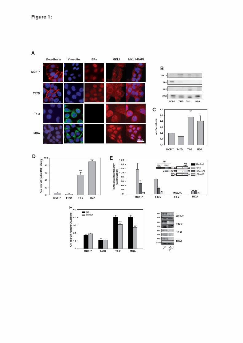
Figure 1:
E-cadherin Vimentin ERα
MCF-7
MDA
A
E
B
ERα
MCF-7 MDA
MKL1
ERK
C
D
T47D
T4-2
0
10
20
30
40
50
% of
cells
with
nucle
ar PC
NA st
aining
T4-2T47D
ratio
F-acti
n/G-ac
tin
***
0,0
0,5
1,0
1,5
2,0
2,5
3,0***
MCF-7 MDAT4-2T47D
Tran
sact
ivat
ion
effic
ienc
y(fo
ld in
duct
ion)
0
20
40
60
80
100
120
140
160
MCF-7 MDAT47D T4-2
ControlERα
ERα Δ79ERα CF
F
0
20
40
60
80
100
% of
cells
with
nucle
ar MK
L1 st
aining
MCF-7 MDAT4-2T47D
Mkl1
MCF-7
MDA
ERK
si
MKL1siC
Mkl1
β-actin
SiCSiMKL1
******
MCF-7 MDAT4-2T47D
T4-2
T47DMkl1
ERK
Mkl1
ERK
MKL1
***
***
BOX 1 BOX 2/3
AF1
C D E F
C D E F
AF2
A C D E FB
MKL1-DAPI
SRF
20 μm
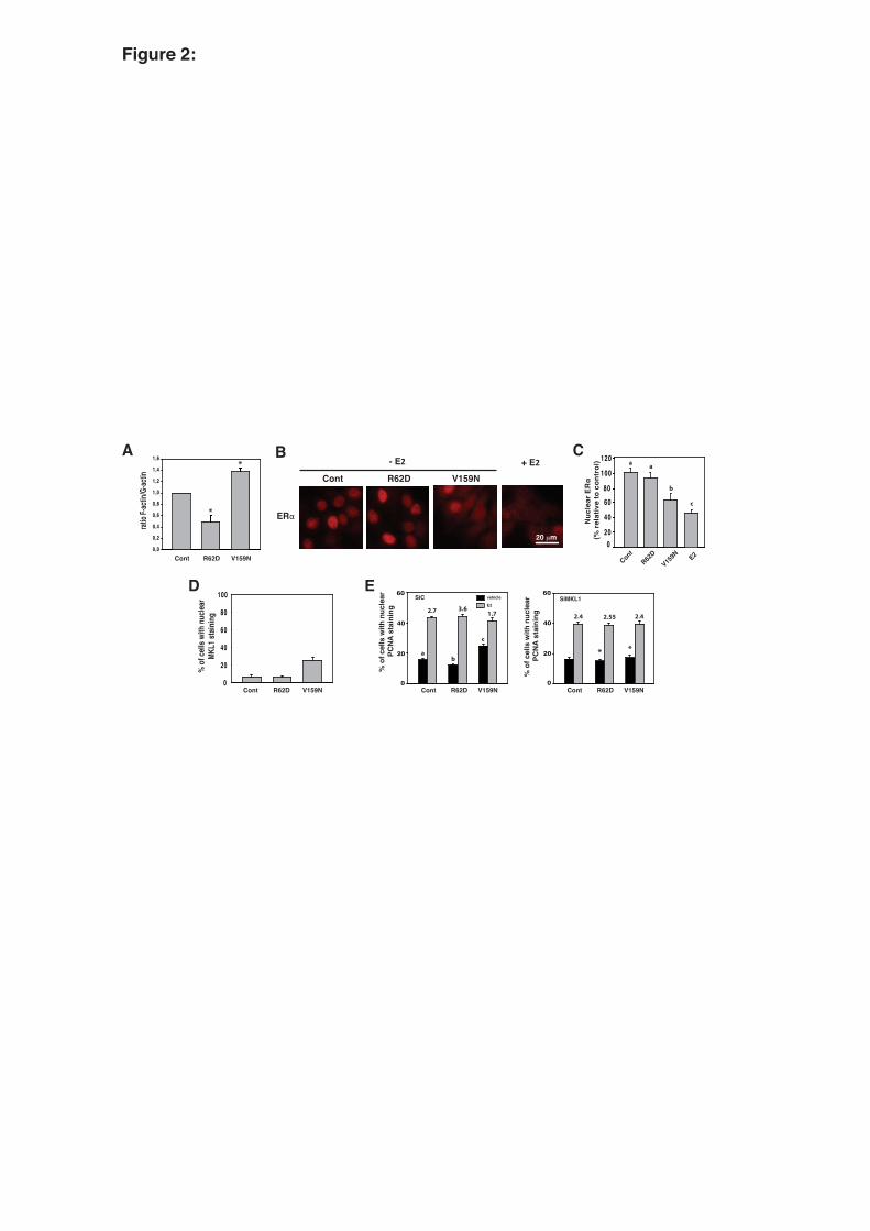
Figure 2:
ratio
F-acti
n/G-ac
tin
Cont R62D V159N
BCont R62D V159N
ERα
+ E2A
- E2
D E
aa a
SiC
0
20
40
60
% o
f cel
ls w
ith n
ucle
ar
PCN
A st
aini
ng
vehicle
E22.7 3.6
1.7
% o
f cel
ls w
ith n
ucle
ar
PCN
A st
aini
ng
0
20
40
60SiMKL1
Cont R62D V159N
2.552.4 2.4
c
ab
Cont R62D V159N0
20
40
60
80
100
% o
f cell
s with
nuc
lear
MKL1
stain
ing
Nuc
lear
ERα
(% re
lativ
e to
con
trol
)
ContR62
DV15
9N0
20
40
6080
100
120a a
b
C
E2
c
Cont R62D V159N
**
0,0
0,2
0,4
0,6
0,8
1,0
1,2
1,4
1,6
20 μm
*
*
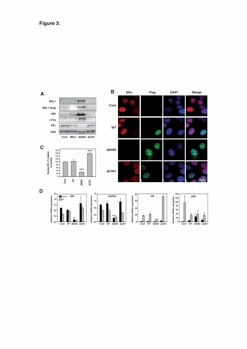
Figure 3:
D
Rel
ativ
e m
RN
A ex
pres
sion
0 .0
0.4
0.8
1.2
1.6
2.0
Cont WT ∆N200 ∆C301
ERαvehicle
E2
**
*
Cont WT ∆N200 ∆C301
Rel
ativ
e m
RN
A ex
pres
sion
0
20
40
60
80PR
* *
*
Cont WT ∆N200 ∆C301Rel
ativ
e m
RN
A ex
pres
sion
pS2
020
40
60
80100
120
140 *
**
Rel
ativ
e m
RN
A ex
pres
sion
Cont WT ∆N200 ∆C3010.0
0.4
0.8
1.2
1.6GATA3
* **
***
020406080
100120140160180
Cont WT
∆N200
∆C301
Nucle
ar E
Rα (%
relat
ive to
cont
rol)
***
***
C
B
Cont
WT
∆N200
∆C301
ERα Flag MergeDAPI
Cont MKL1 ∆N200 ∆C301
ERα
c-Fos
ERK
MKL1
MKL1 (flag)
A
SRF
10 μm

B
Rel
ativ
e m
RN
A ex
pres
sion
Cont WT ∆N200 ∆C301
CXCR4
0.0
1.0
2.0
3.0*
* *
Rel
ativ
e m
RN
A ex
pres
sion
Cont WT ∆N200 ∆C301
CXCL12
0
10
20
30
40
*
*
*
Cont WT ∆N200 ∆C301
Rel
ativ
e m
RN
A ex
pres
sion TGFα
0.0
1.0
2.0
3.0
* *
*
Cont WT ∆N200 ∆C301
Rel
ativ
e m
RN
A ex
pres
sion Amphiregulin
0
20
40
60
80
*
** *
Cont WT ∆N200 ∆C301
Rel
ativ
e m
RN
A ex
pres
sion HB-EGF
020406080
100120140160180 *
0
1
2
3
4
5
Rel
ativ
e m
RN
A ex
pres
sion
Cont WT ∆N200 ∆C301
HER1
* *
*
0.00.20 .40 .60 .81.01.21 .41 .6
Rel
ativ
e m
RN
A ex
pres
sion
Cont WT ∆N200 ∆C301
HER2
** *
A
D
0
20
40
60
80
100
- +Cont WT ∆N200 ∆C301
E2 - + - + - +
SubG1G0/G1 G2/M
S
13.5+/- 0.5
26.3+/- 1.5
15.5+/- 2 27.6
+/- 3
20.5+/- 2
22+/- 1.3
9.25+/- 0.7
23.3+/- 3.2
Perc
enta
ge o
f cel
ls
1.3+/- 0.1
3.1+/- 0.3
1.6+/- 0.2
3.1+/- 0.5
7+/- 0.5
7.9+/- 0.6 1.3
+/- 0.3
1.5+/- 0.3
vehicle
E2
Figure 4:
0
2
4
6
8
10
12
Cont WT
∆N20
0
∆C30
1
3 days 5 days
Cont WT
∆N20
0
∆C30
1
vehicle
E2
Relat
ive C
ell N
umbe
r
3x3x
2.5x
2.1x
5.8x
3.5x
1.5x
8.6x
*
*
***
*
Cont MKL1 ∆N200 ∆C301
p-ERK
ERK
p-Akt
Akt
C

B
0.0
0.5
1.0
1.5
2.0
2.5
Cont WT ∆N200 ∆C301Rel
ativ
e m
RN
A e
xpre
ssio
n
PR
** * *
0
1
2
3
4
5
6
Cont WT ∆N200 ∆C301Rel
ativ
e m
RN
A e
xpre
ssio
n TGFα
*
* *02468
10121416
Cont WT ∆N200 ∆C301
Rel
ativ
e m
RN
A e
xpre
ssio
n Amphiregulin*
** * * 0.0
0.4
0.8
1.2
1.6
Rel
ativ
e m
RN
A e
xpre
ssio
n
Cont WT ∆N200 ∆C301
CXCL12
***** * *
Rel
ativ
e m
RN
A e
xpre
ssio
n
0.0
0.4
0.8
1.2
1.6
Cont WT ∆N200 ∆C301
ERvehicle4-OHT
***
- +0
20
40
60
80
100
Cont WT ∆N200 ∆C3014OHT - + - + - +
SubG1G0/G1 G2/M
S
27+/- 0.6
15+/- 2.8
28+/- 3.5
18+/-2.3 23
+/- 4
21+/- 3
20+/- 2.5
7.5+/- 1.7
A
Per
cent
age
of c
ells
in e
ach
cel
l cyc
le p
hase
s
3+/-0.4
3.8+/-2
3.7+/- 0.8
5+/-1.7
9.7+/-
3
8.9+/-3.8
2.5+/-0.3
2.1+/-0.3
Figure 5:
Title: Activation of MKL1/actin signaling pathway induces hormonal escape of estrogen-responsive
breast cancer cell lines.
Authors: Gwenneg Kerdivel1, Antoine Boudot1, Denis Habauzit1, Frederic Percevault1, Florence
Demay1, Farzad Pakdel1, Gilles Flouriot1*
Supplementary Figure S1
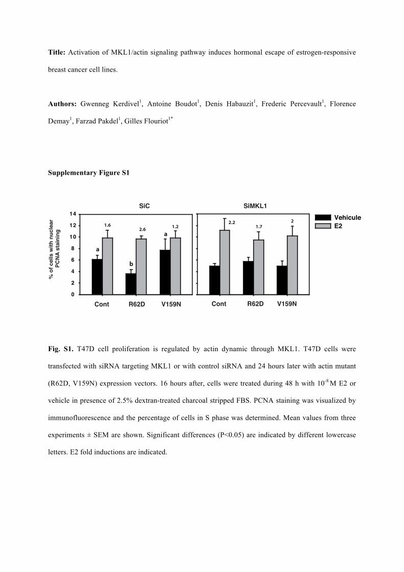
Fig. S1. T47D cell proliferation is regulated by actin dynamic through MKL1. T47D cells were
transfected with siRNA targeting MKL1 or with control siRNA and 24 hours later with actin mutant
(R62D, V159N) expression vectors. 16 hours after, cells were treated during 48 h with 10-8 M E2 or
vehicle in presence of 2.5% dextran-treated charcoal stripped FBS. PCNA staining was visualized by
immunofluorescence and the percentage of cells in S phase was determined. Mean values from three
experiments ± SEM are shown. Significant differences (P<0.05) are indicated by different lowercase
letters. E2 fold inductions are indicated.
% o
f cel
ls w
ith n
ucle
ar
PCN
A s
tain
ing
1.62.6
1.2
SiMKL1
Cont R62D V159N
1.72.2 2
a
a
b
Cont R62D V159N0
2
4
6
8
10
12
14 VehiculeE2
SiC

Supplementary Figure S2
0
20
40
60
80
100
SubG1
G1
S
G2/M
0
10
20
30
40
50
0
5
10
15
20
25
30
0,0
0,5
1,0
1,5
2,0
2,5
3,0
3,5
0
20
40
60
80
100
120
140
160
180
0
20
40
60
80
100
120
140
0,0
0,5
1,0
1,5
2,0
2,5
3,0
3,5
0,0
0,2
0,4
0,6
0,8
1,0
1,2
0,0
0,2
0,4
0,6
0,8
1,0
1,2
1,4
1,6
-
E2
Tet
Tet + E2
ER_
p-ERK
ERK
Mkl1
p-Akt
Akt
Mkl1 (flag)
A
SRF
_-actin
Cont
¨1��� ¨&���
Tet: + + + + +- - - - -
clone 2 clone 2clone 3 clone 3Cont
¨1��� ¨&���
clone 2 clone 2clone 3 clone 3
5HODWLYH�P51$�H[SUHVVLRQ
5HODWLYH�P51$�H[SUHVVLRQ
5HODWLYH�P51$�H[SUHVVLRQ
5HODWLYH�P51$�H[SUHVVLRQ
5HODWLYH�P51$�H[SUHVVLRQ
5HODWLYH�P51$�H[SUHVVLRQ
5HODWLYH�P51$�H[SUHVVLRQ
5HODWLYH�P51$�H[SUHVVLRQ
(5њ
Cont
¨1��� ¨&���
clone 2 clone 2clone 3 clone 3
PR
$PSKLUHJXOLQ HB-EGF
HER1 HER2
CXCL12 CXCR4
* * * * * * * *
* * * * * ** *
* * * *
* * * *
* * * *
* * * *
Tet:
++
+ +
--
- -+ +- -+ +- -+ +- -+ +- -
++ --++ --++ --++ --E2:
Cont
¨1��� ¨&���
clone 2 clone 2clone 3 clone 3
B
C
3HUFHQWDJH�RI�FHOOV�LQ�HDFK
�FHOO�F\FOH�SKDVHV
13
31
13
31
12
28
15
16
8
26
10
14 1
2
22
10
19
9
27
6
27

Fig. S2. Constitutively active form of MKL1 impairs estrogenic response in MCF-7 cells. Two other
series of clones (Clone 2 and 3) expressing flagged mutant forms of MKL1 were established. (panel
A) After a 48h treatment with tetracyclin, total proteins from MCF-7 sub-clones were immunoblotted
for MKL1, flag, SRF, !-actin, ER!, p-Akt, Akt, p-ERK and ERK. (panel B) MCF-7 sub-clones were
treated or not with 10-8 M E2 and tetracyclin in presence of 2.5% dextran-treated charcoal stripped
FBS for 48 h. Flow cytometry experiments were realized to determine percentages of cells in each cell
cycle phase. (panel C) Clone 2 and 3 series were treated or not with 10-8 M E2 and tetracyclin in
presence of 2.5% dextran-treated charcoal stripped FBS for 48 h. Expression of several E2-regulated
genes then was quantified using real-time PCR. Mean values from three independent experiments ±
SEM are shown. Asterisks indicate not determined values.
Supplementary Figure S3
Fig. S3. Expression of the dominant negative MKL1 C301 in ER-negative MDA-MB231 cells fails to
restore ER!, PR and HER-2 expression. MDA-MB231 sub-clones expressing flagged "C301 mutant
form of MKL1 were established: two MDA-MB231/cont clones (C1 and C2), and two MDA-
MB231/MKL1 "C301 clones (C1 and C2) were obtained. (panel A) After a 48h treatment with

tetracycline or vehicle, expression of ERα, PR and HER2 was quantified using real-time PCR. Mean
values from two experiments ± SEM are shown. (panel B)Total proteins from MDA-MB231 sub-
clones were immunoblotted for flag and ERK.