Identification of chromosome sequence motifs that mediate meiotic pairing and synapsis in C. elegans
20
Identification of Chromosome Sequence Motifs That Mediate Meiotic Pairing and Synapsis in C. elegans Carolyn M. Phillips 1,5 , Xiangdong Meng 2 , Lei Zhang 2 , Jacqueline H. Chretien 1,3 , Fyodor D. Urnov 2 , and Abby F. Dernburg 1,3,4,* 1 Department of Molecular and Cell Biology, University of California, Berkeley 2 Sangamo BioSciences, Inc., Pt. Richmond Tech Center, 501 Canal Blvd., Suite A100, Richmond, California 94804, USA 3 Howard Hughes Medical Institute 4 Life Sciences Division, Lawrence Berkeley National Laboratory Abstract C. elegans chromosomes contain specialized regions called pairing centers (PCs) that mediate homologous pairing and synapsis during meiosis. Four related proteins, ZIM-1, -2, -3, and HIM-8, associate with these sites and are required for their essential functions. Here we show that short sequence elements enriched in the corresponding chromosome regions selectively recruit these proteins in vivo. In vitro analysis using SELEX indicates that each protein’s binding specificity arises from a combination of two zinc fingers and an adjacent domain. Insertion of a cluster of recruiting motifs into a chromosome lacking its endogenous PC is sufficient to restore homologous pairing, synapsis, crossover recombination, and segregation. These findings help to illuminate how chromosome sites mediate essential aspects of meiotic chromosome dynamics. Studies of genome rearrangements in the nematode Caenorhabditis elegans have shown that particular regions near one end of each chromosome are required in cis for homologous recombination and segregation during meiosis. Translocations or deletions of these regions suppress genetic exchange across large chromosome regions 1-5 . These “pairing centers” (PCs) stabilize pairing and promote the assembly of the synaptonemal complex (SC) between homologous chromosomes 6, 7 . A family of four paralogous proteins, each containing two motifs resembling C2H2 zinc fingers (ZnFs), is required for PC function 8, 9 . Each protein localizes to the PCs of one or two pairs of chromosomes during early meiotic prophase: ZIM-1 on chromosomes II and III, ZIM-2 on V, ZIM-3 on I and IV, and HIM-8 on the X chromosome. Loss of any of these proteins results in defects in pairing, synapsis, recombination, and segregation of the corresponding chromosomes. * Author for correspondence: [email protected], 470 Stanley Hall, MC 3220, Berkeley, CA 94720, USA, 510-666-3615. 5 Current address: Department of Molecular Biology, Massachusetts General Hospital, 185 Cambridge St. CPZN-7250, Boston, MA 02114 NIH Public Access Author Manuscript Nat Cell Biol. Author manuscript; available in PMC 2014 April 28. Published in final edited form as: Nat Cell Biol. 2009 August ; 11(8): 934–942. doi:10.1038/ncb1904. NIH-PA Author Manuscript NIH-PA Author Manuscript NIH-PA Author Manuscript
-
Upload
independent -
Category
Documents
-
view
2 -
download
0
Transcript of Identification of chromosome sequence motifs that mediate meiotic pairing and synapsis in C. elegans

Identification of Chromosome Sequence Motifs That MediateMeiotic Pairing and Synapsis in C. elegans
Carolyn M. Phillips1,5, Xiangdong Meng2, Lei Zhang2, Jacqueline H. Chretien1,3, Fyodor D.Urnov2, and Abby F. Dernburg1,3,4,*
1Department of Molecular and Cell Biology, University of California, Berkeley
2Sangamo BioSciences, Inc., Pt. Richmond Tech Center, 501 Canal Blvd., Suite A100,Richmond, California 94804, USA
3Howard Hughes Medical Institute
4Life Sciences Division, Lawrence Berkeley National Laboratory
Abstract
C. elegans chromosomes contain specialized regions called pairing centers (PCs) that mediate
homologous pairing and synapsis during meiosis. Four related proteins, ZIM-1, -2, -3, and HIM-8,
associate with these sites and are required for their essential functions. Here we show that short
sequence elements enriched in the corresponding chromosome regions selectively recruit these
proteins in vivo. In vitro analysis using SELEX indicates that each protein’s binding specificity
arises from a combination of two zinc fingers and an adjacent domain. Insertion of a cluster of
recruiting motifs into a chromosome lacking its endogenous PC is sufficient to restore
homologous pairing, synapsis, crossover recombination, and segregation. These findings help to
illuminate how chromosome sites mediate essential aspects of meiotic chromosome dynamics.
Studies of genome rearrangements in the nematode Caenorhabditis elegans have shown that
particular regions near one end of each chromosome are required in cis for homologous
recombination and segregation during meiosis. Translocations or deletions of these regions
suppress genetic exchange across large chromosome regions1-5. These “pairing centers”
(PCs) stabilize pairing and promote the assembly of the synaptonemal complex (SC)
between homologous chromosomes6, 7.
A family of four paralogous proteins, each containing two motifs resembling C2H2 zinc
fingers (ZnFs), is required for PC function8, 9. Each protein localizes to the PCs of one or
two pairs of chromosomes during early meiotic prophase: ZIM-1 on chromosomes II and III,
ZIM-2 on V, ZIM-3 on I and IV, and HIM-8 on the X chromosome. Loss of any of these
proteins results in defects in pairing, synapsis, recombination, and segregation of the
corresponding chromosomes.
*Author for correspondence: [email protected], 470 Stanley Hall, MC 3220, Berkeley, CA 94720, USA, 510-666-3615.5Current address: Department of Molecular Biology, Massachusetts General Hospital, 185 Cambridge St. CPZN-7250, Boston, MA02114
NIH Public AccessAuthor ManuscriptNat Cell Biol. Author manuscript; available in PMC 2014 April 28.
Published in final edited form as:Nat Cell Biol. 2009 August ; 11(8): 934–942. doi:10.1038/ncb1904.
NIH
-PA
Author M
anuscriptN
IH-P
A A
uthor Manuscript
NIH
-PA
Author M
anuscript
cis-acting elements that underlie PC function have not yet been described. Here we identify
sequence motifs enriched on each chromosome that specifically recruit the cognate ZnF
protein required for that chromosome to undergo faithful meiotic segregation. Our
identification of sites that recruit each protein in vivo is corroborated by in vitro binding
experiments that illuminate the basis for their sequence specificity. Integration of these
sequences onto a chromosome deficient for PC activity is sufficient to restore meiotic
chromosome pairing and synapsis. Moreover, we demonstrate that these recruitment motifs
do not require a specific chromosome position, and that one ZnF protein can substitute for
another to promote meiotic chromosome interactions.
Results and Discussion
Identification of X Chromosome Pairing Center sequences
The X chromosome PC has been previously mapped to the region distal to (or left of, by C.
elegans convention) the dpy-3 locus, 2.15 Mb from the left telomere3, 10. X chromosomes
lacking this region usually fail to synapse or undergo exchange and consequently
missegregate, resulting in an elevated frequency of XO (male) progeny3, known as the High
incidence of males, or Him, phenotype11.
To delimit the region containing the X chromosome PC more precisely, deficiencies were
mapped using single nucleotide polymorphisms (SNPs)12, 13. We analyzed three
deficiencies that result in the loss of HIM-8 localization from the X chromosome and
remove all PC function (Fig. 1b-d) 3. Each of these three deficiencies lacked all markers
tested between 50 Kb and 1.46 Mb from the left end (see Methods; Fig. 1a), but did not
delete a marker at 2.07 Mb. In contrast, yDf19, an X chromosome deficiency that retains
HIM-8 staining (Fig. 1e) and undergoes normal meiotic segregation14 lacked the leftmost
markers scored, but its right breakpoint was found to lie between 1.06 and 1.17 Mb from the
left end (Fig. 1a). These data indicate that elements sufficient to recruit HIM-8 and confer
PC activity are contained within sequences between 1.06 and 2.07 Mb from the left end of
the X chromosome.
Candidate sequences within this 1 Mb region were injected into wild-type C. elegans to test
for HIM-8 binding. The resulting transgenic animals carried high copy extrachromosomal
arrays, which typically contain megabases of the injected DNA and are transmitted through
mitosis and meiosis (see Methods)15. Combining FISH with immunofluorescence, we tested
whether candidate arrays could recruit HIM-8 in germline nuclei. Although this approach is
unbiased with respect to candidate sequences, it does require that HIM-8 recognize a
sequence motif or other element within the chromatin context of an extrachromosomal array,
which undergoes transcriptional silencing and enriched H3K9 dimethylation in germline
nuclei16. While we did not know a priori whether HIM-8 would bind to arrays, we were
encouraged by the success of an analogous approach to identify sequence elements that
recruit C. elegans dosage compensation complex proteins in somatic nuclei17, 18.
From an initial pool of cosmids that recruited HIM-8, we narrowed the recruitment activity
to smaller fragments, ultimately to a 539 bp amplicon (Fig. 1f; Supplementary Information,
Phillips et al. Page 2
Nat Cell Biol. Author manuscript; available in PMC 2014 April 28.
NIH
-PA
Author M
anuscriptN
IH-P
A A
uthor Manuscript
NIH
-PA
Author M
anuscript
Table S1). Centered within this short sequence are five and a half copies of a 21 bp repeat,
and no other repetitive element, coding sequence, or other feature of obvious interest.
Computational analysis revealed that a 12 bp motif, (TTGGTCAGTGCT) contained within
the larger repeat is enriched on the X chromosome relative to the autosomes and in the PC
region relative to the entire X chromosome (Supplementary Information, Fig. S1a). When
degeneracy was allowed, we found that some closely related sequences were also enriched
in the PC region, and that TTGGTCAGTGCA, which differs at the 3’ nucleotide, is even
more abundant than the original motif (Fig. 2; Supplementary Information, Fig. S1).
Interestingly, a version of this sequence lacking the 5’ base was previously identified via
computational methods as the most overrepresented oligonucleotide on the X chromosome
and named CeRep5019.
To test whether TTGGTCAGTGCA could also recruit HIM-8, we amplified two short
regions from the left end of the X chromosome, each containing several copies of this motif
with different flanking sequences. Both amplicons recruited HIM-8 (Fig. 1g; Supplementary
Information, Table S1), as did a synthetic oligonucleotide containing four tandem copies of
this motif interspersed with flanking spacers. TTGGTCAGTGCA is therefore sufficient to
recruit HIM-8.
Both of these related HIM-8 recruiting motifs are highly enriched near the left end of the X
chromosome and are most often found in short, tandemly oriented clusters with a
predominant 21-base periodicity (Supplementary Information, Table S2)19. Many copies lie
within introns of known or predicted genes. Small clusters or isolated occurrences of these
motifs can also be found elsewhere on the X chromosome and occasionally on autosomes.
yDf19, the deficiency chromosome with a functional PC, retains about half of the motifs
normally found in the left 2 Mb of the X chromosome, while meDf2, meDf3, and meDf5
remove between 94-100% of the motifs found in this dense cluster.
Immunofluorescent detection of HIM-8 in germline nuclei reveals a single primary
association site in the genome, which corresponds to the region of the X chromosome
removed by meDf2, meDf3, and meDf5 (Fig. 1b-d)9. However, granular staining of HIM-8 is
detected elsewhere on the chromatin, which may reflect binding to related motifs elsewhere
in the genome. HIM-8 may weakly promote X chromosome segregation even in the absence
of the PC, since loss of him-8 function results in a more severe meiotic phenotype than X
chromosome PC deficiencies9. To assay the recruitment potential of clusters outside the PC,
we amplified clusters of motifs from the center and right end of the X chromosome, and also
from Chromosome III. Each of these amplicons recruits HIM-8 in the high-copy
extrachromosomal array assay (Fig. 1h; Supplementary Information, Table S1). While
HIM-8 recruitment motifs elsewhere on the X may contribute to segregation, they lack key
PC functions; in particular, they do not measurably stabilize homolog pairing in the absence
of synapsis7. This suggests that PCs require a minimal density of binding sites, or perhaps
other cis-acting components, for full function.
Phillips et al. Page 3
Nat Cell Biol. Author manuscript; available in PMC 2014 April 28.
NIH
-PA
Author M
anuscriptN
IH-P
A A
uthor Manuscript
NIH
-PA
Author M
anuscript

Identification of autosomal pairing center sequence motifs
Our evidence indicates that HIM-8 is recruited in vivo by a sequence previously identified in
silico as the most overrepresented short oligonucleotide on the X chromosome. Prior
computational analysis also identified short sequences that are most overrepresented on each
autosome relative to the other five chromosomes. Each of these sequences, designated as
CeRep45-50, was found to be asymmetrically enriched near one end of a single
chromosome19. These regions of enrichment roughly corresponded with PCs, which have
been mapped to varying precision on different chromosomes. However, these observations
were based entirely on computational analysis, and no functions have previously been
demonstrated for these abundant motifs.
We revisited and modified the previous analysis in light of the knowledge that PCs on
Chromosomes I and IV share a common ZnF protein (ZIM-3), as do II and III (ZIM-1)8. We
found that CeRep45, the most overrepresented sequence on Chromosome I19, is also highly
enriched within a 120-kb window within the PC region on IV (Fig. 2; Supplementary
Information, Fig. S2). Interestingly, while most repeats on I are clustered in alternating
orientation (or “inverted” clusters, Supplementary Information, Table S2) with a total period
of 68 bp, the copies on IV are mostly in tandemly oriented clusters with a 19 bp periodicity
(Supplementary Information, Table S2). Amplicons spanning clusters of this motif from
either Chromosome I or IV strongly recruited ZIM-3 to extrachromosomal arrays
(Supplementary Information, Fig. S5a, S5c, Table S1).
Chromosomes II and III both require ZIM-1 for meiotic pairing and synapsis8. While neither
CeRep45 nor CeRep46, the sequences most overrepresented on Chromosomes II and III,
respectively, is highly abundant on the other chromosome, we found that both repeats are
associated with a distinct motif, TG/TGGTCTGCTA, which is enriched on both
chromosomes (Fig. 2 Supplementary Information, Fig. S3). Amplicons containing clusters
of these elements showed specific recruitment of ZIM-1 (Supplementary Information, Fig.
S5b, Table S1). While this motif is predominantly in inverted clusters on both II and III, the
predominant spacing of these elements is different on the two chromosomes (Supplementary
Information, Table S2).
Recruitment of PC proteins to V was more enigmatic. The CeRep49 sequence identified by
Sanford and Perry ([T]TGGGCGCTGCT) seemed an excellent candidate for ZIM-2
recruitment, in that it is highly enriched on Chromosome V and also resembles in base
composition and length the motifs that recruit the other proteins (Fig. 2; Supplementary
Information, Fig. S4). Indeed, we did find that one cluster of this repeat specifically
recruited ZIM-2, although somewhat less robustly than other ZIM-recruiting arrays
(Supplementary Information, Fig. S5d, Table S1). However, a different cluster from
Chromosome V containing both this motif and the motif TTGGTCGCTGCT, which differs
at the underlined base, strongly recruited both ZIM-2 and HIM-8 (Supplementary
Information, Fig. S5e-f, Table S1). As shown previously19, the organization of CeRep49
repeats on Chromosome V is bimodal with respect to spacing, with prominent 18- and 32-
base periodicities. Clusters with 32 bp spacing contain CeRep49 alone, while clusters with
18 bp spacing always include this G–>T variant motif (Supplementary Information, Table
Phillips et al. Page 4
Nat Cell Biol. Author manuscript; available in PMC 2014 April 28.
NIH
-PA
Author M
anuscriptN
IH-P
A A
uthor Manuscript
NIH
-PA
Author M
anuscript
S2). It is unclear whether the co-recruitment of HIM-8 to arrays containing the second class
of cluster is due to the variant sequence, or to the distinct spacing, but it is suggestive that
the T variant is more similar to the X chromosome repeat, TTGGTCAGTGCA. No obvious
recruitment of HIM-8 is detected cytologically on Chromosome V, and genetic evidence
indicates that only ZIM-2 is required for efficient pairing and synapsis of this chromosome8.
Nevertheless, indirect evidence suggests that HIM-8 might contribute to pairing of
Chromosome V in the absence of ZIM-2, since appreciable crossing-over is detected
cytologically in a zim-2 mutant8.
In vitro analysis of Pairing Center sequences
HIM-8 and the ZIM proteins each contain two short domains resembling C2H2 ZnFs20, the
most common DNA binding motif in metazoa21. Data presented here and in previous
work8, 9 indicated that defined chromosomal sequences are able to recruit HIM-8 and the
ZIM proteins in vivo, and that missense mutations in the ZnF domains of HIM-8 disrupt
chromosome association9. However, the spacing between the Zn-coordinating Cys and His
residues in the first finger of the ZIM/HIM-8 family is distinct from the canonical C2H2
spacing seen in proteins such as Zif26822 or Sp123. Furthermore, a single canonical C2H2
ZnF usually specifies only a 3 or 4 bp subsite22, raising the question of how these unusual
two-ZnF proteins might recognize the ~12 bp non-palindromic sequences we identified.
To determine whether the C2H2 ZnFs in the ZIM/HIM-8 proteins can bind DNA sequence-
specifically, we used a SELEX assay, as previously described24. Briefly, protein fragments
expressed in vitro were incubated with a library of double-stranded DNA fragments carrying
a randomized 21 bp stretch of DNA. The protein-bound DNA was isolated and amplified,
and the cycle was reiterated three more times, after which the bound DNA fragments were
sequenced and analyzed to derive a consensus. Somewhat surprisingly, the assay produced
very similar results when this analysis was performed with the core ZnF domains of HIM-8,
ZIM-2, and ZIM-3. In all three cases, the pentamer motif “TTGGC” clearly emerged as the
preferred binding site (Fig. 3; Supplementary Information, Table S3). Interestingly, this
sequence is similar but not identical to one end of all of the binding motifs we identified in
vivo. We next expressed a longer fragment of HIM-8, including the region from the ZnF
domain to the C-terminus of the protein. As shown (Fig. 3b; Supplementary Information,
Table S3), this fragment bound specifically to oligonucleotides containing the
consensus T/ATGGTCAGTGC, which is identical to the full length HIM-8 recruiting repeat
we identified in vivo.
Taken together, the SELEX results indicate that HIM-8 uses a composite protein-DNA
interaction domain to recognize its full target site. We infer that the distinct recruitment
motifs of the ZIM proteins are likely recognized by the combined specificities of their ZnF
domain and a short adjacent C-terminal domain in each protein, and that the Zn fingers
likely specify one end of each binding sites containing “TTGG.” These findings suggest a
direct correspondence between each ZnF protein and the sequences we identified in vivo,
implying that no other cofactors are likely required to recruit these proteins to their cognate
chromosome sequences. Future work may elucidate how these novel composite DNA-
binding domains interact with their cognate binding sites.
Phillips et al. Page 5
Nat Cell Biol. Author manuscript; available in PMC 2014 April 28.
NIH
-PA
Author M
anuscriptN
IH-P
A A
uthor Manuscript
NIH
-PA
Author M
anuscript
ZIM/HIM-8 recruiting arrays interact with the nuclear envelope
Chromosomal PCs are physically associated with the nuclear envelope during early meiotic
prophase8, 9. We noticed that when ZIM/HIM-8 proteins are recruited to the
extrachromosomal arrays, they concentrate at the interface between the array and the nuclear
envelope (see Fig. 1f-h). This suggests that the ZnF proteins recruit their binding sites to the
periphery of the array and the nucleus. In work detailed elsewhere (A. Sato, CMP, AFD;
Manuscript in preparation), we have found that the sites of contact between endogenous PCs
and the nuclear envelope are enriched for several proteins that contribute to chromosome
segregation. These include the inner and outer nuclear membrane proteins SUN-1 and
ZYG-12, which concentrate at discrete patches along the nuclear surface during early
prophase25, 26. We therefore tested whether arrays that recruit ZIM/HIM-8 proteins also
associate with these nuclear envelope components. We compared the behavior of three
extrachromosomal arrays, two that recruit different ZnF proteins and a control, non-
recruiting array (Fig. 1g; Supplementary Information, Fig. S5c, Table S1). The ZIM/HIM-8
recruiting arrays clearly associated with patches of SUN-1 and ZYG-12 at the nuclear
envelope, in contrast to the non-recruiting array (Fig. 4; data not shown). Thus, ZIM/HIM-8
recruitment motifs are sufficient to link the arrays to a protein complex that likely tethers
these sequences to the nuclear envelope and mediates interactions with cytoplasmic dynein
and microtubules (A. Sato, CMP, AFD; Manuscript in preparation).
HIM-8 recruiting sequences are sufficient to restore PC activity to the X Chromosome
PCs contribute to meiotic chromosomal segregation by both stabilizing homolog pairing and
promoting synapsis7. To test whether a dense cluster of HIM-8 recruitment motifs is
sufficient to restore these PC functions to an X chromosome lacking its endogenous PC, a
HIM-8 recruiting extrachromosomal array was integrated onto the PC-deficient meDf2
chromosome (meDf2 ieIs5; see Methods; Fig. 5a). Whereas the X chromosomes in meDf2
homozygotes usually fail to synapse (Fig. 5b)7, in meDf2 ieIs5 nearly all nuclei showed
complete homologous synapsis (Fig. 5c), demonstrating that HIM-8 recruitment by the
inserted array can mediate proper synapsis of the X chromosomes. We also analyzed pairing
of the X chromosomes in the absence of synapsis by knocking down the expression of syp-2,
an essential SC component27, by RNA interference (RNAi). Throughout the early meiotic
region of the gonad, hybridization to the integrated array showed only a single region of
staining (Fig. 5d), indicating that these inserted sequences can stabilize pairing between
homologous X chromosomes in the absence of synapsis. By introducing the him-8 (tm611)
null allele9, we found that both the pairing and synapsis mediated by the integrated array
require HIM-8 function (Fig. 5e and data not shown), much as these events do when
mediated by the endogenous X chromosome PC.
Pairing and synapsis enable chromosomes to complete crossover recombination, which is
required for chiasma formation and homolog segregation at the first meiotic division. To
determine whether meDf2 ieIs5 chromosomes undergo crossing-over, we scored the
frequency of bivalents at diakinesis (Fig. 5f-h). As shown previously3, most oocytes (65.6%)
in meDf2 hermaphrodites displayed univalent X chromosomes. By contrast, 99.4% of oocyte
nuclei in meDf2 ieIs5 homozygotes exhibited bivalent (recombinant) X chromosomes. The
fidelity of X chromosome meiotic segregation can be quantified in C. elegans by the
Phillips et al. Page 6
Nat Cell Biol. Author manuscript; available in PMC 2014 April 28.
NIH
-PA
Author M
anuscriptN
IH-P
A A
uthor Manuscript
NIH
-PA
Author M
anuscript

frequency of male self-progeny produced by hermaphrodites. meDf2 ieIs5 hermaphrodites
produced 1.8% males, dramatically fewer than meDf2 homozygotes (33.1%) (Fig. 5i). We
conclude that the integration of many copies of a 539 bp segment that recruits HIM-8
rescues pairing, synapsis, crossing-over, and segregation defects arising from deletion of the
endogenous PC.
PC function does not require a specific chromosome position or ZnF protein
In our initial rescue experiment, HIM-8 motifs were fortuitously integrated towards the left
end of the meDf2 chromosome, near their natural location. We carried out additional
experiments to ask whether the position on the chromosome is critical for PC activity, and
also whether a particular chromosome requires a specific member of the ZIM/HIM-8 family.
Identification of sequences that recruit both HIM-8 and ZIM-2 to arrays allowed us to
address these questions about PC plasticity. We UV-irradiated animals carrying such an
array and screened for integration events onto either Chromosome V or X (Supplementary
Information, Fig. S5e-f). Two independent integrations into the left (non-PC) arm of
chromosome V were recovered (ieIs12 and ieIs13), as were two integrations on the right
(non-PC) arm of X (ieIs14 and ieIs15). The integrated arrays on Chromosome V were
homozygosed and crossed to zim-2 (tm574), which eliminates ZIM-2 activity8, while the X
chromosome integrants were crossed to him-8 (mn253) to abrogate HIM-8 function. In all
cases the presence of the array on Chromosome V or X resulted in dramatic restoration of
bivalent formation relative to zim-2 or him-8 mutants, respectively (Fig. 6a-e). Disjunction
of the X chromosome (as measured by the frequency of male self-progeny) was also rescued
in the absence of HIM-8, presumably by ZIM-2 association with the X-linked integrated
array (Fig. 6f). These results indicate that ZIM-2 can promote crossing-over on the X
chromosome, that HIM-8 can promote crossing-over on V, and that the chromosomal
position of the major ZIM/HIM-8 binding cluster is plastic.
Integration of the same extrachromosomal array onto two different chromosomes also
provided an opportunity to address whether such “artificial PCs” can promote pairing and
synapsis between non-homologous chromosomes. We generated trans-heterozygotes by
crossing worms with the ZIM-2/HIM-8 recruiting array integrated on V (ieIs12 or ieIs13) to
animals with the same array integrated on the X chromosomes (ieIs14 or ieIs15). We looked
at whether such “matching” PCs could stabilize pairing between non-homologous
chromosomes in the absence of synapsis, and/or promote non-homologous synapsis. To look
at pairing in the absence of synapsis, we visualized the integrated arrays in worms subjected
to syp-2 RNAi. In all four trans-heterozygous combinations, the ZIM-2/HIM-8 recruiting
arrays on X and V were consistently paired (99% of nuclei) throughout the normal leptotene/
zygotene and pachytene regions of the gonad (Fig. 6g-h; Supplementary Information, Table
S4), indicating that non-homologous chromosomes did indeed undergo robust, stable
pairing.
When synapsis was allowed to proceed in animals carrying the matching integrated arrays
on non-homologous chromosomes, the meiotic configuration of individual nuclei was more
variable, likely due to the competition between the endogenous PCs and the integrated
arrays (Fig. 6i; Supplementary Information, Table S4). In 71% of pachytene-stage nuclei,
Phillips et al. Page 7
Nat Cell Biol. Author manuscript; available in PMC 2014 April 28.
NIH
-PA
Author M
anuscriptN
IH-P
A A
uthor Manuscript
NIH
-PA
Author M
anuscript

the integrated arrays were together and the associated non-homologous chromosomes were
synapsed, while the true homologous partners of the two array-bearing chromosomes
remained unsynapsed. 10% of nuclei showed complete homologous synapsis with the
integrated arrays apart, indicating that the arrays occasionally failed to induce non-
homologous synapsis even if they had initially paired. An additional 6% of nuclei showed
complete synapsis, yet the integrated arrays were closely associated. This may reflect
physical association between two pairs of homologously synapsed chromosomes;
alternatively the normal copies of V and X may have synapsed with each other, or aberrantly
loaded SC without pairing.
Together, these data suggest that the artificial PCs created by integrated ZnF-binding arrays
have potent pairing activity and that they can sometimes, but not always, mediate synapsis
between non-homologous chromosomes even in the presence of homologous partners
carrying intact PCs. These results are consistent with the idea that the integrated arrays and
endogenous PCs compete to initiate synapsis. We emphasize, however, that the integrated
arrays have important differences from endogenous PCs. Not only are the repeats probably
present at both higher density and much higher copy number within the artificial PCs, but
within natural PCs the ZnF-recruiting motifs are distributed among hundreds of kilobases of
chromosome-specific sequences. We consider it likely that these interspersed sequences play
an important role in specifying partner choice, perhaps by stabilizing and/or destabilizing
pairing. This would explain why pairing between ZIM-1 or ZIM-3 binding PCs on different
chromosomes is not detected in wild-type animals8.
ZIM/HIM-8 recruiting arrays induce meiotic defects
Consistent with the aberrant interactions observed between arrays on non-homologous
chromosomes, animals carrying free HIM-8 and ZIM recruiting arrays exhibited meiotic
defects, including unsynapsed chromosomes, univalents, and male and inviable progeny
(Supplementary Information, Fig. S6). These defects may reflect aberrant pairing between
arrays and endogenous PCs dependent on the same ZnF protein or titration of the ZIM/
HIM-8 proteins off the endogenous PC onto the array. However, these are unlikely to be the
only explanations, since other chromosomes also showed segregation defects; for example,
ZIM-binding arrays induced a weak Him phenotype, indicating missegregation of the X
chromosome. This suggests that the arrays may titrate limiting components, such as nuclear
envelope proteins, away from endogenous PCs that do not share the same ZnF dependence.
These findings help to illuminate the mechanisms underlying homolog pairing, synapsis, and
segregation during meiosis. They demonstrate that PCs are likely defined by the binding of
ZIM/HIM-8 proteins, via a novel, composite DNA binding domain, to a concentrated locus
on each chromosome, and that consequent association with a complex of proteins at the
nuclear envelope is both necessary and sufficient to facilitate specific pairing and synapsis
between homologous chromosomes.
Phillips et al. Page 8
Nat Cell Biol. Author manuscript; available in PMC 2014 April 28.
NIH
-PA
Author M
anuscriptN
IH-P
A A
uthor Manuscript
NIH
-PA
Author M
anuscript
Online Methods
SNP Mapping
The boundaries of the deficiencies meDf2, meDf3, meDf5, and yDf19 were mapped using
“snip-SNPs,” polymorphisms that alter a restriction site between N2 Bristol and a Hawaiian
isolate of C. elegans12, 13. An unc-1 dpy-3 strain with Hawaiian-derived SNPs to the left of
dpy-3 was generated by extensive outcrossing to the Hawaiian strain CB4856. mnDp66; Df
males were then crossed to these Hawaiian unc-1 dpy-3 hermaphrodites. Cross progeny were
allowed to self-fertilize, and Unc nonDpy F2s (lacking mnDp66, which carries N2 SNP
alleles) were lysed and their DNA was genotyped for seven SNPs ranging from 0.05 to 2.07
Mb from the left end of the X chromosome. Detection of only the Hawaiian digest pattern
indicated that a SNP lies within the deletion, whereas both N2 and Hawaiian alleles were
expected for SNPs outside the deletion.
Progeny analysis
Hermaphrodites were picked to individual plates as L4s and moved daily to fresh plates until
they no longer laid eggs. All of their self-progeny were counted to determine the proportion
of unhatched (dead) eggs and adult males and hermaphrodites. The following strains were
analyzed: ieEx69 (ZIM-3 recruiting array derived from the PC region of Chromosome I;
n=1370), ieEx29 (ZIM-1 recruiting array derived from an amplicon in the PC region of
Chromosome II; n=1754), ieEx41 (HIM-8 recruiting array generated from PC region of X
chromosome; n=3170), wild-type (n=1954), mnDp66; meDf2 (n=2217), mnDp66;
unc-119(ed3); meDf2 ieIs5 (n=3637), ieIs14 (n=2370), ieIs15 (n=1663), him-8(mn253)
(n=1315), him-8(mn253); ieIs14 (n=1526), and him-8(mn253); ieIs15 (n=1111).
Extrachromosomal Arrays and Integration
Extrachromosomal arrays were generated by injecting DNA mixtures including a plasmid
carrying a phenotypic marker (rol6(su1006), unc-119(+), or myo-2∷GFP) at 10-100 μg/ml,
phage λ DNA (HindIII digested; New England Biolabs) at 50-100 μg/ml, and candidate
DNA sequences on cosmids, plasmids, or PCR products, at 10-50 μg/ml. PCR primers are
listed in Table S1.
A synthetic 84 bp oligonucleotide consisted of four tandem copies of the sequence
AATTTGTGTTGGTCAGTGCAA. Both DNA strands were synthesized, annealed and co-
injected with plasmids carrying phenotypic markers. Similar results were obtained when the
same sequence was cloned into a TOPO vector (Invitrogen).
To integrate a HIM-8 recruiting array containing a 539 bp amplicon from cosmid K06A9
(ieEx22), array-bearing animals were crossed to the X chromosome PC deficiency strain
mnDp66 (X;I); unc-119(ed3) III; meDf2 X. ieEx22 contains an unc-119 rescuing construct,
so the resulting array-bearing, meDf2 F2 animals were nonUnc and Him. The worms were
washed 4x in M9 and placed on a 10-cm unseeded NGM plate. Once the liquid had absorbed
into the agar, the plate was placed uncovered into a Stratalinker, and irradiated with 350
J/m2 of 254-nm light. The animals were allowed to recover for four hours on food. Four L4s
or young adults were then transferred to each of 40 plates. When their progeny reached the
Phillips et al. Page 9
Nat Cell Biol. Author manuscript; available in PMC 2014 April 28.
NIH
-PA
Author M
anuscriptN
IH-P
A A
uthor Manuscript
NIH
-PA
Author M
anuscript
L4 larval stage, 500 of these F1s were picked to individual plates. From each F1, three F2
animals were picked to individual plates and their progeny (F3s) were screened for stable
transmission of the nonUnc phenotype. A single integrant that mapped to the X chromosome
was recovered. This array-containing X chromosome was outcrossed three times to meDf2
animals to generate the mnDp66 (X;I); unc-119(ed3) III; meDf2 ieIs5 X strain used for
analysis.
Because a duplication of the X PC region, mnDp66, was present in the original irradiated
animals, we checked to make sure that the improvement in segregation was not a result of
this duplication recombining back onto the X chromosome. The unc-1 locus is present on
mnDp66 and absent from meDf2. The dpy-3 locus is present on meDf2 and absent on
mnDp66. We therefore crossed mnDp66; meDf2 ieIs5 hermaphrodites to N2 males, and
mated the resulting male cross progeny, mnDp66/+; meDf2 ieIs5, to unc-1 dpy-3
hermaphrodites. Unc nonDpy progeny (unc-1 dpy-3/meDf2 ieIs5) were recovered at the
expected frequency of 50%, indicating that the unc-1 region is still missing from the meDf2
ieIs5 X chromosome.
Chromosomal integration of a ZIM-2/HIM-8 recruiting array was performed similarly to the
integration of the HIM-8 recruiting array above, except that the extrachromosomal array was
derived from a region of sequence motifs from Chromosome V with 18 bp spacing and
carried a dominant rol-6(su1006) marker and except for the presence of the array (ieEx75),
the parental animals carried a normal karyotype. 230 F1s were individually plated after UV-
irradiation. Ten lines were identified that produced 100% Rol progeny, of which two
mapped to the left arm of Chromosome V (ieIs12 and ieIs13) and two mapped to the right
arm of the X chromosome (ieIs14 and ieIs15).
Immunofluorescence and FISH
Cytological methods were performed as previously described9. A FISH probe specific for
the extrachromosomal and integrated arrays was synthesized from λ-phage DNA as
previously described8. Chromosome-specific probes recognizing the middle and right ends
of the X chromosome and to the 5S rDNA on Chromosome V have been described
previously9, 28.
To quantify the frequency of recombinant chromosomes, hermaphrodites were picked as
L4s and maintained at 15°C for three days. Adults were dissected, fixed, and hybridized
with appropriate fluorescent probes to allow unambiguous identification of the relevant
chromosomes. 3D images of oocyte nuclei at diakinesis were recorded and chromosomes
were scored as bivalent (both homologs connected) or univalent (separate). The number of
nuclei scored for each experiment were as follows: X chromosomes in wildtype (n=162),
mnDp66; meDf2 (n=121), mnDp66; unc-119(ed3); meDf2 ieIs5 (n=174), ieIs14 (n=157),
ieIs15 (n=110), him-8(mn253) (n=150), him-8(mn253); ieIs14 (n=238), and him-8(mn253);
ieIs15 (n=300). Chromosome V in the following strains: wild-type (n=194), ieIs12 (n=105),
ieIs13 (n=115), zim-2(tm574) (n=140), zim-2(tm574); ieIs12 (n=155), and zim-2(tm574);
ieIs13 (n=127). For all experiments involving integrated arrays, an array-specific probe was
included to confirm that the chromosome being analyzed (i.e., X or V) contained an array.
Phillips et al. Page 10
Nat Cell Biol. Author manuscript; available in PMC 2014 April 28.
NIH
-PA
Author M
anuscriptN
IH-P
A A
uthor Manuscript
NIH
-PA
Author M
anuscript
Feeding RNAi
To examine chromosome pairing in the absence of synapsis, expression of syp-2, which
encodes essential component of the central element of SC, was eliminated by feeding RNAi
in mnDp66; unc-119(ed3); meDf2 ieIs5 and mnDp66; unc-119(ed3); him-8(tm611); meDf2
ieIs5 animals, and also in the trans-heterozygous progeny of [ieIs12 or ieIs13] and [ieIs14
or ieIs15]. L4 larvae were placed on plates containing IPTG with lawns of bacteria
containing RNAi clone sjj_C24G6.129. Their progeny were dissected and stained 20-24
hours post-L4. To examine cross-progeny of animals carrying arrays on different
chromosomes, matings were carried out on syp-2 RNAi lawns and the resulting
transheterozygotes were maintained on syp-2 RNAi plates until dissection, 20-24 hours post-
L4. Absence of synapsis was verified by SYP-1 immunofluorescence in all analyzed
animals.
Computational Identification of Motifs
To determine if sequences related to TTGGTCAGTGCT might also be enriched on the X
chromosome, a Perl script was written to search the genome, allowing variation at one
nucleotide within the sequence at a time. The script was run iteratively to analyze any
related sequences that were enriched at least 5-fold on the X chromosome relative to the
autosomes. All such derived, enriched X chromosome motifs are presented in
Supplementary Information, Fig. S1a. The same method was used to identify sequences
related to the ZIM recruitment motifs that are enriched on the autosomes (see
Supplementary Information, Fig. S2a, Fig. S3a, and Fig. S4a).
SELEX
In vitro site selection by SELEX was performed exactly as described24. Fragments of ZIM/
HIM-8 cDNA constructs were PCR-amplified, in a two-step scheme, to yield linear products
carrying HA-tags at the carboxy-terminus. A library of DNA fragments carrying a 21 bp
randomized sequence flanked by a constant region was used for experiments on ZIM-2 and
ZIM-3, whereas two libraries, one with a 21 bp and the other a 26 bp randomized portion,
was used for experiments on HIM-8 (data presented are collate findings from both). The
MEME server30 was used to identify motifs and generate the logograms presented.
Supplementary Material
Refer to Web version on PubMed Central for supplementary material.
Acknowledgments
This work was supported by an NSF Predoctoral Fellowship to CMP, and by Burroughs Welcome Career Award1000950 and NIH R01 GM065591 to AFD. We are grateful to Anne Villeneuve for SYP-1 antibodies, BarbaraMeyer, Kevin Corbett and Ed Rebar for valuable suggestions, to members of the Meyer lab for assistance with theextrachromosomal array assay, and to members of the Dernburg lab and anonymous referees for helpful commentson the manuscript.
Phillips et al. Page 11
Nat Cell Biol. Author manuscript; available in PMC 2014 April 28.
NIH
-PA
Author M
anuscriptN
IH-P
A A
uthor Manuscript
NIH
-PA
Author M
anuscript

References
1. McKim KS, Howell AM, Rose AM. The effects of translocations on recombination frequency inCaenorhabditis elegans. Genetics. 1988; 120:987–1001. [PubMed: 3224815]
2. McKim KS, Peters K, Rose AM. Two types of sites required for meiotic chromosome pairing inCaenorhabditis elegans. Genetics. 1993; 134:749–768. [PubMed: 8349107]
3. Villeneuve AM. A cis-acting locus that promotes crossing over between X chromosomes inCaenorhabditis elegans. Genetics. 1994; 136:887–902. [PubMed: 8005443]
4. Zetka M, Rose A. The genetics of meiosis in Caenorhabditis elegans. Trends Genet. 1995; 11:27–31. [PubMed: 7900192]
5. Zetka MC, Rose AM. The meiotic behavior of an inversion in Caenorhabditis elegans. Genetics.1992; 131:321–332. [PubMed: 1644275]
6. MacQueen AJ, Colaiacovo MP, McDonald K, Villeneuve AM. Synapsis-dependent and -independent mechanisms stabilize homolog pairing during meiotic prophase in C. elegans. GenesDev. 2002; 16:2428–2442. [PubMed: 12231631]
7. MacQueen AJ, et al. Chromosome sites play dual roles to establish homologous synapsis duringmeiosis in C. elegans. Cell. 2005; 123:1037–1050. [PubMed: 16360034]
8. Phillips CM, Dernburg AF. A family of zinc-finger proteins is required for chromosome-specificpairing and synapsis during meiosis in C. elegans. Dev Cell. 2006; 11:817–829. [PubMed:17141157]
9. Phillips CM, et al. HIM-8 binds to the X chromosome pairing center and mediates chromosome-specific meiotic synapsis. Cell. 2005; 123:1051–1063. [PubMed: 16360035]
10. Herman RK, Kari CK. Recombination between small X chromosome duplications and the Xchromosome in Caenorhabditis elegans. Genetics. 1989; 121:723–737. [PubMed: 2721932]
11. Hodgkin J, Horvitz HR, Brenner S. Nondisjunction mutants of the nematode Caenorhabditiselegans. Genetics. 1979; 91:67–94. [PubMed: 17248881]
12. Swan KA, et al. High-throughput gene mapping in Caenorhabditis elegans. Genome Res. 2002;12:1100–1105. [PubMed: 12097347]
13. Wicks SR, Yeh RT, Gish WR, Waterston RH, Plasterk RH. Rapid gene mapping in Caenorhabditiselegans using a high density polymorphism map. Nat Genet. 2001; 28:160–164. [PubMed:11381264]
14. Nicoll M, Akerib CC, Meyer BJ. X-chromosome-counting mechanisms that determine nematodesex. Nature. 1997; 388:200–204. [PubMed: 9217163]
15. Mello CC, Kramer JM, Stinchcomb D, Ambros V. Efficient gene transfer in C. elegans:extrachromosomal maintenance and integration of transforming sequences. Embo J. 1991;10:3959–3970. [PubMed: 1935914]
16. Kelly WG, et al. X-chromosome silencing in the germline of C. elegans. Development(Cambridge, England). 2002; 129:479–492.
17. Dawes HE, et al. Dosage compensation proteins targeted to X chromosomes by a determinant ofhermaphrodite fate. Science. 1999; 284:1800–1804. [PubMed: 10364546]
18. McDonel P, Jans J, Peterson BK, Meyer BJ. Clustered DNA motifs mark X chromosomes forrepression by a dosage compensation complex. Nature. 2006; 444:614–618. [PubMed: 17122774]
19. Sanford C, Perry MD. Asymmetrically distributed oligonucleotide repeats in the Caenorhabditiselegans genome sequence that map to regions important for meiotic chromosome segregation.Nucleic Acids Res. 2001; 29:2920–2926. [PubMed: 11452017]
20. Miller J, McLachlan AD, Klug A. Repetitive zinc-binding domains in the protein transcriptionfactor IIIA from Xenopus oocytes. Embo J. 1985; 4:1609–1614. [PubMed: 4040853]
21. Tupler R, Perini G, Green MR. Expressing the human genome. Nature. 2001; 409:832–833.[PubMed: 11237001]
22. Pavletich NP, Pabo CO. Zinc finger-DNA recognition: crystal structure of a Zif268-DNA complexat 2.1 A. Science. 1991; 252:809–817. [PubMed: 2028256]
Phillips et al. Page 12
Nat Cell Biol. Author manuscript; available in PMC 2014 April 28.
NIH
-PA
Author M
anuscriptN
IH-P
A A
uthor Manuscript
NIH
-PA
Author M
anuscript
23. Kadonaga JT, Carner KR, Masiarz FR, Tjian R. Isolation of cDNA encoding transcription factorSp1 and functional analysis of the DNA binding domain. Cell. 1987; 51:1079–1090. [PubMed:3319186]
24. Perez EE, et al. Establishment of HIV-1 resistance in CD4(+) T cells by genome editing usingzinc-finger nucleases. Nature biotechnology. 2008; 26:808–816.
25. Malone CJ, et al. The C. elegans hook protein, ZYG-12, mediates the essential attachment betweenthe centrosome and nucleus. Cell. 2003; 115:825–836. [PubMed: 14697201]
26. Penkner A, et al. The nuclear envelope protein Matefin/SUN-1 is required for homologous pairingin C. elegans meiosis. Dev Cell. 2007; 12:873–885. [PubMed: 17543861]
27. Colaiacovo MP, et al. Synaptonemal complex assembly in C. elegans is dispensable for loadingstrand-exchange proteins but critical for proper completion of recombination. Dev Cell. 2003;5:463–474. [PubMed: 12967565]
28. Dernburg AF, et al. Meiotic recombination in C. elegans initiates by a conserved mechanism and isdispensable for homologous chromosome synapsis. Cell. 1998; 94:387–398. [PubMed: 9708740]
29. Kamath RS, Ahringer J. Genome-wide RNAi screening in Caenorhabditis elegans. Methods. 2003;30:313–321. [PubMed: 12828945]
30. Bailey TL, Elkan C. Fitting a mixture model by expectation maximization to discover motifs inbiopolymers. Proc Int Conf Intell Syst Mol Biol. 1994; 2:28–36. [PubMed: 7584402]
Phillips et al. Page 13
Nat Cell Biol. Author manuscript; available in PMC 2014 April 28.
NIH
-PA
Author M
anuscriptN
IH-P
A A
uthor Manuscript
NIH
-PA
Author M
anuscript
Figure 1.The X chromosome PC region. (a) Left two megabases of the X chromosome. Genetic and physical markers used for mapping
are indicated. Three deficiencies that remove the PC (meDf2, meDf3, and meDf5) and one that does not (yDf19) were mapped.
All PC deficiencies remove pk6142 but not pk6143, indicating breakpoints between 1.46 and 2.07 Mb from the left end. yDf19
removes unc-1 but not pk6141, indicating a breakpoint between 1.06 and 1.17 Mb from the left end. (b-e) HIM-8
immunofluorescence (yellow) in meiotic nuclei from hermaphrodites of the indicated genotypes. Diagrams of the X
chromosomes and the mnDp66 duplication, which is required for viability in deficiency homozygotes, are shown. (f-h) HIM-8
immunofluorescence (yellow) was combined with FISH (red) to test for recruitment of HIM-8 to extrachromosomal arrays.
Diagram on right indicates the genomic location of the sequences tested in each panel: (f) 539 bp amplicon from cosmid K06A9
on XL. (g) Cluster of TTGGTCAGTGCA repeats from XL. (h) Cluster of 4 HIM-8 recruitment motifs from IIIL recruits HIM-8
(yellow) but not ZIM-1 (green). All images are maximum-intensity projections of deconvolved 3D stacks. Scale bars represent 5
μm.
Phillips et al. Page 14
Nat Cell Biol. Author manuscript; available in PMC 2014 April 28.
NIH
-PA
Author M
anuscriptN
IH-P
A A
uthor Manuscript
NIH
-PA
Author M
anuscript
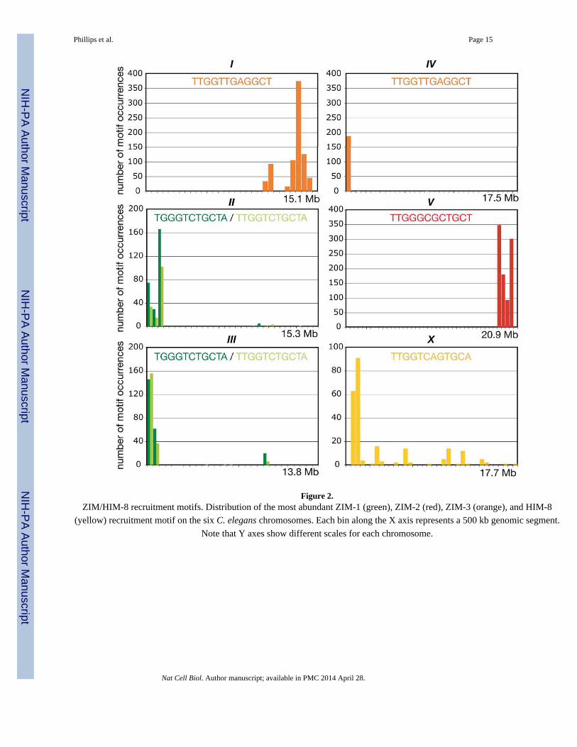
Figure 2.ZIM/HIM-8 recruitment motifs. Distribution of the most abundant ZIM-1 (green), ZIM-2 (red), ZIM-3 (orange), and HIM-8
(yellow) recruitment motif on the six C. elegans chromosomes. Each bin along the X axis represents a 500 kb genomic segment.
Note that Y axes show different scales for each chromosome.
Phillips et al. Page 15
Nat Cell Biol. Author manuscript; available in PMC 2014 April 28.
NIH
-PA
Author M
anuscriptN
IH-P
A A
uthor Manuscript
NIH
-PA
Author M
anuscript
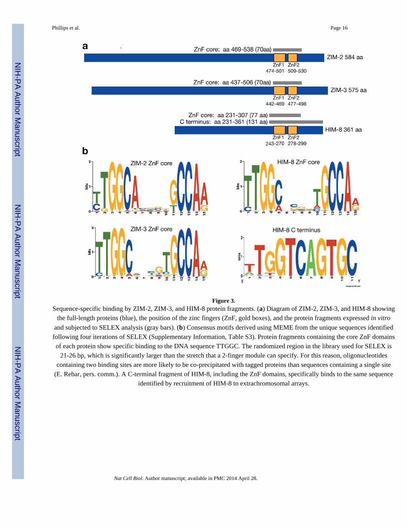
Figure 3.Sequence-specific binding by ZIM-2, ZIM-3, and HIM-8 protein fragments. (a) Diagram of ZIM-2, ZIM-3, and HIM-8 showing
the full-length proteins (blue), the position of the zinc fingers (ZnF, gold boxes), and the protein fragments expressed in vitro
and subjected to SELEX analysis (gray bars). (b) Consensus motifs derived using MEME from the unique sequences identified
following four iterations of SELEX (Supplementary Information, Table S3). Protein fragments containing the core ZnF domains
of each protein show specific binding to the DNA sequence TTGGC. The randomized region in the library used for SELEX is
21-26 bp, which is significantly larger than the stretch that a 2-finger module can specify. For this reason, oligonucleotides
containing two binding sites are more likely to be co-precipitated with tagged proteins than sequences containing a single site
(E. Rebar, pers. comm.). A C-terminal fragment of HIM-8, including the ZnF domains, specifically binds to the same sequence
identified by recruitment of HIM-8 to extrachromosomal arrays.
Phillips et al. Page 16
Nat Cell Biol. Author manuscript; available in PMC 2014 April 28.
NIH
-PA
Author M
anuscriptN
IH-P
A A
uthor Manuscript
NIH
-PA
Author M
anuscript
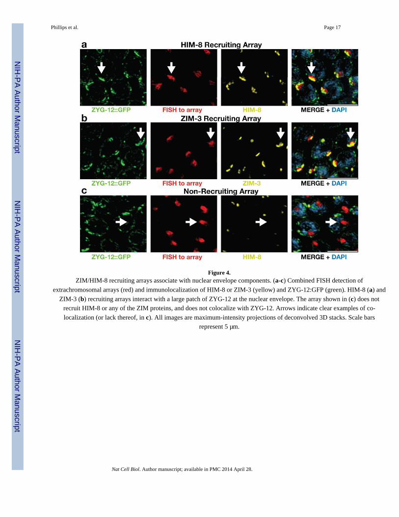
Figure 4.ZIM/HIM-8 recruiting arrays associate with nuclear envelope components. (a-c) Combined FISH detection of
extrachromosomal arrays (red) and immunolocalization of HIM-8 or ZIM-3 (yellow) and ZYG-12:GFP (green). HIM-8 (a) and
ZIM-3 (b) recruiting arrays interact with a large patch of ZYG-12 at the nuclear envelope. The array shown in (c) does not
recruit HIM-8 or any of the ZIM proteins, and does not colocalize with ZYG-12. Arrows indicate clear examples of co-
localization (or lack thereof, in c). All images are maximum-intensity projections of deconvolved 3D stacks. Scale bars
represent 5 μm.
Phillips et al. Page 17
Nat Cell Biol. Author manuscript; available in PMC 2014 April 28.
NIH
-PA
Author M
anuscriptN
IH-P
A A
uthor Manuscript
NIH
-PA
Author M
anuscript
Figure 5.HIM-8 recruitment motifs are sufficient for PC function. (a) Integration of a HIM-8 recruiting array (see Fig. 1f) onto meDf2, a
PC-deficient X chromosome. (b) Hermaphrodite homozygous for meDf2 has unsynapsed X chromosomes in most pachytene
nuclei, visualized as axial elements marked by HTP-3 (red) lacking the central element protein SYP-1 (green). Arrows indicate
examples of unsynapsed chromosomes. (c) Most meiotic nuclei in meDf2 ieIs5 hermaphrodites are fully synapsed. (d, e)
Stabilization of pairing in the absence of synapsis (syp-2 RNAi) is examined by performing FISH to the integrated array (red).
In meDf2 ieIs5 oocytes (d) the arrays are paired, as indicated by only a single region of FISH staining. In him-8; meDf2 ieIs5
oocytes (e) the arrays are unpaired, indicating that pairing between integrated arrays, like that seen between endogenous PCs9, is
him-8 dependent. (f, g) Oocytes at diakinesis in meDf2 and meDf2 ieIs5 hermaphrodites. FISH probes to the center (yellow) and
right end (red) identify the X chromosomes. Arrows indicate non-recombinant (univalent) and recombinant (bivalent) X
Phillips et al. Page 18
Nat Cell Biol. Author manuscript; available in PMC 2014 April 28.
NIH
-PA
Author M
anuscriptN
IH-P
A A
uthor Manuscript
NIH
-PA
Author M
anuscript
chromosomes in meDf2 and meDf2 ieIs5 hermaphrodites, respectively. (h) Quantification of recombinant X chromosomes. (i)Quantification of males produced by self-fertilizing hermaphrodites of the indicated genotypes. Scale bars represent 5 μm.
Phillips et al. Page 19
Nat Cell Biol. Author manuscript; available in PMC 2014 April 28.
NIH
-PA
Author M
anuscriptN
IH-P
A A
uthor Manuscript
NIH
-PA
Author M
anuscript

Figure 6.ZIM/HIM-8 proteins can interchangeably support PC function. (a-d) Oocytes at diakinesis from [zim-2], [zim-2; ieIs12],
[him-8], and [him-8; ieIs14] hermaphrodites. FISH probes to the 5S rDNA (red in a, b) and an X-chromosome repeat (red in c,d) were used to identify chromosomes V and X, respectively. In animals carrying chromosomal insertions of ZIM-2/HIM-8
binding sites ieIs12 (b) and ieIs14 (d), the chromosome-specific probes localize to a single bivalent, which is also marked by a
FISH probe to λ DNA (green), indicating that the insertion of binding sites restored crossover recombination on chromosome V
in zim-2 animals and the X chromosome in him-8 animals. (e) Quantification of bivalent X (red) and V (orange) chromosomes.
(f) Quantification of males produced by self-fertilizing hermaphrodites of the indicated genotypes. (g,h) Arrays of binding sites
(red) inserted into two different chromosomes (ieIs12/+; ieIs14/+ and ieIs13/+; ieIs15/+) were assayed for their ability to
promote stable pairing between non-homologous chromosomes in the absence of synapsis (syp-2 RNAi). (i) Synapsis was
analyzed in animals heterozygous for two different insertions (ieIs13/+; ieIs15/+) by immunostaining of SC components.
Nuclei containing unsynapsed chromosomes, visualized as segments positive for the axial element protein HTP-3, (red) but
lacking transverse filament proteins including SYP-1 (green), usually contain integrated arrays (blue) that are paired and
synapsed with each other, indicating non-homologous synapsis between chromosomes V and X (arrows). In contrast, nuclei with
fully synapsed chromosomes often contained unpaired arrays (blue), indicating that all chromosomes are likely synapsed with
their appropriate homologs (arrowheads). Scale bars represent 5 μm.
Phillips et al. Page 20
Nat Cell Biol. Author manuscript; available in PMC 2014 April 28.
NIH
-PA
Author M
anuscriptN
IH-P
A A
uthor Manuscript
NIH
-PA
Author M
anuscript