Chromosome distribution studies after inorganic lead exposure
Condensin I Reveals New Insights on Mouse MeioticChromosome Structure and DynamicsAlberto Viera1*, Rocıo Gomez1, Marıa T. Parra1, John A. Schmiesing2, Kyoko Yokomori2, Julio S. Rufas1, Jose A. Suja1
1 Departamento de Biologıa, Edificio de Biologicas, Universidad Autonoma de Madrid, Madrid, Spain, 2 Department of Biological Chemistry, Collegeof Medicine, University of California at Irvine, Irvine, California, United States of America
Chromosome shaping and individualization are necessary requisites to warrant the correct segregation of genomes in eithermitotic or meiotic cell divisions. These processes are mainly prompted in vertebrates by three multiprotein complexes termedcohesin and condensin I and II. In the present study we have analyzed by immunostaining the appearance and subcellulardistribution of condensin I in mouse mitotic and meiotic chromosomes. Our results demonstrate that in either mitotically ormeiotically dividing cells, condensin I is loaded onto chromosomes by prometaphase. Condensin I is detectable as a fuzzy axialstructure running inside chromatids of condensed chromosomes. The distribution of condensin I along the chromosome lengthis not uniform, since it preferentially accumulates close to the chromosome ends. Interestingly, these round accumulationsfound at the condensin I axes termini colocalized with telomere complexes. Additionally, we present the relative distribution ofthe condensin I and cohesin complexes in metaphase I bivalents. All these new data have allowed us to proposea comprehensive model for meiotic chromosome structure.
Citation: Viera A, Gomez R, Parra MT, Schmiesing JA, Yokomori K, et al (2007) Condensin I Reveals New Insights on Mouse Meiotic ChromosomeStructure and Dynamics. PLoS ONE 2(8): e783. doi:10.1371/journal.pone.0000783
INTRODUCTIONChromosome compaction and condensation are indispensable
requisites for the correct segregation of chromosomes during
anaphase in mitosis and meiosis ([1–3]). In this sense, duplicated
DNA molecules must progressively restructure and condense
during prophase in order to individualize, package into rod-shaped
structures, and resolve their chromatids prior to anaphase in order
to prompt a proper chromosome segregation (for review see [4,5]).
Although the knowledge on this topic is continuously increasing,
we are still far away from the complete understanding of the
molecular machinery and the interrelated processes that lead to
a correct chromosome compaction and condensation. In the past
years, a multiprotein complex termed condensin was characterized
in mitotic chromosomes from Xenopus egg extracts ([6,7]). This
condensin complex has revealed to be one of the main acting
factors involved in chromosome condensation, maintenance of
chromosome shaping, and proper sister chromatid segregation in
anaphase from yeast to humans ([6–9]). In Xenopus, the condensin
complex consists of five different subunits, SMC2 and SMC4, two
members of the SMC (Structural Maintenance of Chromosomes)
protein family, and three non-SMC proteins, CAP-G, CAP-D2
and the kleisin CAP-H ([1,10]). Analogous complexes have been
subsequently identified in a variety of species such as Caenorhabditis
elegans ([11,12]), Schizosaccharomyces pombe ([13]), Saccharomyces
cerevisiae ([14]), or human ([15,16]). It has been established that
the hindrance of chromosomal condensation by either the
blocking or depletion of any of the condensin subunits leads to
segregation abnormalities and therefore cellular crisis in both
mitotically ([2,17–23]) and meiotically ([22–25]) dividing cells.
Recent investigations have revealed the existence of another
holocomplex which shares the two SMC subunits in addition to
the non-SMC subunits CAP-G2, CAP-D3, and the kleisin CAP-
H2. This new complex has been termed condensin II ([26,27]),
and therefore the initially described complex has been renamed as
condensin I. Both complexes possess different specific dynamic
and functional characteristics (for review see [4,5]). At least in
vertebrate chromosomes, condensin I and II exhibit distinct
patterns of distribution along the mitotic chromosome axis
([26,28]), and whereas condensin II has been reported to locate
to chromosomes as early as interphase, condensin I loads to
chromosomes by early prometaphase ([28]). It has been recently
proposed that condensin II may act as a primer organizer of
chromatids along the axis, while condensin I would be responsible
for the chromosomal final shaping ([26,28]).
Despite all this background, little is known as regards to the
appearance, location and dynamics of condensin in meiotic cells,
being the initial studies reduced to A. thaliana ([23]), C. elegans
([22,25]), Xenopus ([29]), and S. cerevisiae ([24]). Therefore, the aim
of the present study was to determine the presence, loading,
dynamics and participation of the condensin I complex in the
structure of mammalian meiotic chromosomes. For this purpose
we have analyzed by immunofluorescence the presence, sub-
cellular distribution and cell cycle-regulated dynamics of con-
densin I in mitotic spermatogonia and spermatocytes during both
meiotic divisions in male mouse. Moreover, we have double
immunolabeled condensin I subunits with different telomere and
centromere/kinetochores proteins in order to precisely determine
the location of condensin I in condensed meiotic chromosomes.
Additionally, we show for the first time in mammals the relative
distribution of condensin and cohesin complexes in metaphase I
chromosomes. According to our results we propose and discuss
a comprehensive model for chromosome structure in meiosis.
Academic Editor: Mikhail Blagosklonny, Ordway Research Institute, United Statesof America
Received June 13, 2007; Accepted July 18, 2007; Published August 22, 2007
Copyright: � 2007 Viera et al. This is an open-access article distributed underthe terms of the Creative Commons Attribution License, which permitsunrestricted use, distribution, and reproduction in any medium, provided theoriginal author and source are credited.
Funding: This work was supported by grants BFU2006-06655/BCM and BFU2005-05668-C03-01/BCM from Ministerio de Educacion y Ciencia, and grant CCG06-UAM/SAL-0260 from Universidad Autonoma de Madrid and Comunidad deMadrid. Rocıo Gomez is supported by a Fundacion Francisco Cobos andMinisterio de Educacion y Ciencia predoctoral fellowships.
Competing Interests: The authors have declared that no competing interestsexist.
* To whom correspondence should be addressed. E-mail: [email protected]
PLoS ONE | www.plosone.org 1 August 2007 | Issue 8 | e783

RESULTS
ImmunoblottingTo test the specificity of the anti-hCAP-H antibody in mouse
meiotic cells we performed an immunoblot analysis with extracts
of mouse testis, using extracts of HeLa cultured cells as a control.
The antibody specifically recognized a single protein band, in both
HeLa and mouse testis extracts, that migrated at a same position
representing approximately 90 kDa (Supplementary Fig. S1),
which corresponds to the previously reported molecular weight
of this protein in human cells ([30]). Thus, the anti-hCAP-H
antibody also recognizes mouse CAP-H, indicating a high degree
of conservation for this protein in mammals.
Distribution of CAP-H during spermatogonial
mitosisWe obtained almost an identical pattern of labeling for all the
three non-SMC subunits of the condensin I complex during
spermatogonial mitosis and both meiotic divisions. Thus, although
we will only refer to those results obtained for CAP-H, identical
patterns of distribution were observed for CAP-G, and CAP-D2,
except where indicated, and are available as supplementary
information.
To study the distribution of the non-SMC condensin I subunits
we employed the squash procedure since it preserves chromosome
condensation and positioning in dividing spermatocytes, and also
allows the unambiguous determination of all mitotic and meiotic
stages ([31]). The distribution of CAP-H was first analyzed in
mitotically proliferating spermatogonial cells present in squashed
seminiferous tubules by using an anti-hCAP-H antibody. CAP-H
was not detected in prophase nuclei (Fig. 1A and B), however, this
antibody rendered a significant labeling on mitotic chromosomes
from late prophase/early prometaphase up to telophase (Fig. 1C–
P). In early prometaphase, a faint and diffuse CAP-H labeling was
observed on the arms of condensing chromosomes, and bright and
discrete CAP-H signals were additionally found at some
chromosome regions (Fig. 1C and D). In late prometaphase
chromosomes, a fuzzy CAP-H staining resembling a single coiled
axial structure was detected inside them (arrows in Fig. 1E).
Interestingly, the proximal and distal ends of those fuzzy CAP-H
axes appeared as brighter spots (arrowheads in Fig. 1E). Since
mouse chromosomes are telocentric ([32]), we will refer to
Figure 1. CAP-H distribution in spermatogonial mitosis. Mouse spermatogonia were stained for CAP-H (green) and counterstained with DAPI (blue).(A, B) CAP-H is not detected in early prophase. (C, D) In early prometaphase, condensing chromosomes show a faint and diffuse CAP-H labeling, butsome bright accumulations (arrowheads) are observed on them. (E, F) In late prometaphase, CAP-H is detected as a single axis (arrows) running alongand inside chromosomes. Note that the ends of these CAP-H axes (arrowheads) are brighter than the own axes. (G, H) Top view of a metaphase cellshowing a ‘rosette’-like chromosome distribution. In these views a single CAP-H axis is seen in each chromosome. The centromeric (red arrowheads)and distal (white arrowheads) axes ends are brightly stained. (I–L) Lateral views of metaphase cells. In these views each chromosome shows twoparallel CAP-H axes (arrows), one per sister chromatid. The centromeric (red arrowheads) and distal (white arrowheads) axes ends appear brightlystained. (M–P) Two different focal planes throughout an anaphase. A single CAP-H axis (arrows) is present inside each chromatid. In (F), (L) and (P) theCAP-H labeling on one chromosome/chromatid has been pseudocolored in red and superimposed on its corresponding DAPI image. Bar, 5 mm.doi:10.1371/journal.pone.0000783.g001
Condensin I: Mouse Chromosomes
PLoS ONE | www.plosone.org 2 August 2007 | Issue 8 | e783

proximal chromosome ends as those located at the centromere
end, and to distal ones as those located at the non-centromeric end
of the chromosome.
The observation of polar views of spermatogonial metaphases,
displaying the characteristic ‘‘rosette’’-like chromosome distribu-
tion (Fig. 1G and H), demonstrated that the chromosome
distribution of CAP-H was identical to that observed in
prometaphase. Each metaphase chromosome showed a single
fuzzy CAP-H axis with two brighter dots at their ends, a proximal
one (red arrowheads in Fig. 1G) close to the centromeric
heterochromatin (discernible by a brighter DAPI staining; Fig. 1H),
and a distal one (white arrowheads in Fig. 1G) near the non-
centromeric end of the chromosome. Nevertheless, the observation
of lateral views of spermatogonial metaphases (Fig. 1I–L) revealed
one CAP-H axis (arrows in Fig. 1K) inside each sister chromatid
(Fig. 1L). These sister axes appeared enlarged at both their
proximal (red arrowheads in Fig. 1I and K) and distal (white
arrowheads in Fig. 1K) ends, although the proximal dots were
brighter than the distal ones. This CAP-H labeling was also
observed in segregating chromatids during anaphase (Fig. 1M–P).
CAP-G (data not shown) and CAP-D2 (Supplementary Fig. S2)
showed the same pattern of chromosome distribution as CAP-H
during spermatogonial mitosis. Therefore, the distribution of
condensin I subunits in mouse spermatogonial mitotic chromo-
somes is similar to that previously reported for human mitotic
chromosomes ([26]), except for the preferential accumulations at
chromosome ends.
The phosphorylation of histone H3 at serine 10 (pH3) is
considered the earliest event defining the onset of mitotic
condensation ([33]). In order to establish the timing of association
of CAP-H relative to the initiation of chromosome condensation,
we made a double immunolabeling of CAP-H and pH3. Our
results showed that CAP-H was recruited to chromosomes after
the phosphorylation of histone H3 (Supplementary Fig. S3).
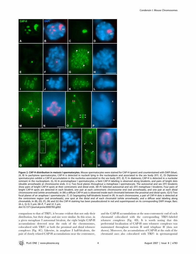
Distribution of CAP-H during meiosis IDuring meiosis I, CAP-H was first detected in pachytene
spermatocytes. In these spermatocytes, several large CAP-H
accumulations, about five, were observed scattered in the
nucleoplasm (Fig. 2A and B). One of these CAP-H accumulations
was always associated to the sex body (Fig. 2A and B). From late
pachytene up to diplotene, the number of nucleoplasmic CAP-H
accumulations decreased until only a single CAP-H accumulation
remained visible at the sex body periphery (Fig. 2C and D). In
diakinesis spermatocytes, a single small and round CAP-H
accumulation was visible in the nucleoplasm (Fig. 2E and F).
The dynamics of the CAP-H accumulations from pachytene up to
diakinesis strongly resembled that of nucleoli in prophase I mouse
spermatocytes ([34]). The double immunolabeling of CAP-H and
fibrillarin, a nucleolar protein mainly located at the dense fibrillar
component ([35]), demonstrated that CAP-H was present at
nucleoli from pachytene up to diakinesis (Supplementary Fig. S4).
It is worth mentioning that whereas CAP-H concentrated at
nucleoli in prophase I spermatocytes, CAP-D2 and CAP-G were
only detected on chromosomes from prometaphase I on (data not
shown). In these spermatocytes, CAP-H, as CAP-D2 (Supplemen-
tary Fig. S5) and CAP-G, were detected as a faint labeling on
bivalents (Fig. 2G and H), and as pairs of bright dots (double
arrowheads in Fig. 2G). During metaphase I, the sex and the
autosomal bivalents (Fig. 2I–P), presented fuzzy CAP-H axial
structures inside their chromatids, and brightly labeled pairs of
dots at their proximal and distal ends (Fig. 2I–L). In favorable
bivalents, the presence of four diffuse CAP-H axes was discerned
along the inner region of each chromatid between the proximal
and distal spots (Fig. 2M and N). As a rule, four pairs of bright
CAP-H spots were clearly discerned in each bivalent, even in the
sex one (Fig. 2M–P), a pair close to each homologous centromere
(red arrowheads in Fig. 2M and O), and another one near each
distal chromosome end (white arrowheads in Fig. 2M and O).
Segregating anaphase I half-bivalents displayed diffuse CAP-H
axes, and brighter spots at their ends (Fig. 2Q–S). Within a given
chromosome, a pair of closely related CAP-H dots was
distinguished at the centromeric ends of the axes, and a single
spot, located at the other axis end, near the distal tip of each
chromatid (Fig. 2T and U).
A double immunolabeling of CAP-H and pH3 on meiosis I
spermatocytes showed that pH3 was first detected at the
heterochromatic chromocentres in diplotene spermatocytes as
previously reported ([36]), while CAP-H was still concentrated at
nucleoli (Supplementary Fig. S6).
Distribution of CAP-H during meiosis IIIn late interkinesis nuclei, CAP-H appeared as nucleoplasmic
accumulations representing nucleoli, and as small spots (Fig. 3A
and B). Afterwards, in prophase II, condensing chromosomes
presented a diffuse CAP-H labeling and bright spots close to their
ends (Fig. 3C and D). Metaphase II chromosomes displayed
a blurred CAP-H staining inside their chromatids and four
brighter spots near their ends (Fig. 3E–H). Two separated
proximal CAP-H spots were positioned at the centromere region
(red arrowheads in Fig. 3I), and two additional ones at the distal
end of each chromatid (white arrowheads in Fig. 3I). At the onset
of anaphase II (Fig. 3K and L), segregating chromatids presented
a central CAP-H axis and one spot at either the proximal (red
arrowheads in Fig. 3K), and distal (white arrowheads in Fig. 3K)
axis ends. This distribution of CAP-H was similar in anaphase II
spermatocytes (Fig. 3M–O).
A double immunolabeling of CAP-H and pH3 on meiosis II
spermatocytes showed that CAP-H was detected at nucleoli and
small spots in late interkinesis nuclei, when their chromatin was
completely labeled by pH3 (Supplementary Fig. S7).
CAP-H preferentially localizes to telomere
complexesIn order to accurately determine the localization of CAP-H at the
ends of condensed meiotic chromosomes, we performed double
immunolabeling experiments using either an anti-centromere
ACA serum, revealing kinetochores, or an anti-TRF1 antibody
to detect the telomere complexes.
The ACA serum revealed a pair of closely associated signals,
representing sister kinetochores, at each homologous centromere
in metaphase I bivalents (Fig. 4A). The pair of centromeric CAP-H
spots was smaller, and appeared beneath the closely associated
sister kinetochores (Fig. 4A). This relative distribution was
maintained at centromere regions of anaphase I half-bivalents
(data not shown). In metaphase II chromosomes, sister kineto-
chores appeared as two round individualized signals facing
opposite poles (Fig. 4B). One proximal CAP-H signal appeared
below each kinetochore (Fig. 4B). Therefore, the proximal
accumulations of condensin I were located beneath the kineto-
chores in condensed chromosomes during both meiotic divisions.
Since the distribution of the bright accumulations of CAP-H at
the axes ends, from prometaphase I up to telophase II, presented
a similar distribution to that previously described for the telomere
complexes in condensed mouse meiotic chromosomes ([37]), we
performed a double immunolabeling of CAP-H and TRF1. When
the chromosomal distribution of CAP-H was analyzed in
Condensin I: Mouse Chromosomes
PLoS ONE | www.plosone.org 3 August 2007 | Issue 8 | e783
comparison to that of TRF1, it became evident that not only their
distribution, but their shape and size were similar. In this sense, in
a given metaphase I autosomal bivalent, the eight bright CAP-H
accumulations detected near the ends of the chromosomes,
colocalized with TRF1 at both the proximal and distal telomere
complexes (Fig. 4C). Likewise, in anaphase I half-bivalents, the
pair of closely related CAP-H accumulations near the centromere,
and the CAP-H accumulations at the non-centromeric end of each
chromatid colocalized with the corresponding TRF1-labeled
telomere complexes (Fig. 4D). It is worth noting that this
preferential localization of CAP-H onto telomere complexes was
maintained throughout meiosis II until telophase II (data not
shown). Moreover, the accumulations of CAP-H at the ends of the
chromatid axes also colocalized with TRF1 in spermatogonial
Figure 2. CAP-H distribution in meiosis I spermatocytes. Mouse spermatocytes were stained for CAP-H (green) and counterstained with DAPI (blue).(A, B) In pachytene spermatocytes, CAP-H is detected in nucleoli lying in the nucleoplasm and associated to the sex body (XY). (C, D) Diplotenespermatocytes exhibit a CAP-H accumulation in the nucleolus associated to the sex body (XY). (E, F) In diakinesis, CAP-H is detected in a nucleolarremnant in the nucleoplasm. (G, H) In prometaphase I spermatocytes, a faint CAP-H labeling is observed along bivalents, and pairs of bright dots(double arrowheads) at chromosome ends. (I–L) Two focal planes throughout a metaphase I spermatocyte. The autosomal and sex (XY) bivalentsshow pairs of bright CAP-H spots at their centromeric and distal ends. (M–P) Selected autosomal and sex (XY) metaphase I bivalents. Four pairs ofbright CAP-H spots are detected in each bivalent, one pair at each centromeric chromosome end (red arrowheads), and one pair at each distalchromosome end (white arrowheads). In (M) a diffuse CAP-H axis is observed inside each chromatid between the proximal and distal spots. (Q-S) Twofocal planes of an anaphase I spermatocyte. (T, U) Segregating half-bivalents boxed in (R). In each chromosome, a pair of CAP-H dots is detected atthe centromere region (red arrowheads), one spot at the distal end of each chromatid (white arrowheads), and a diffuse axial labeling alongchromatids. In (B), (D), (F), (N) and (U) the CAP-H staining has been pseudocolored in red and superimposed on its corresponding DAPI image. Bars:(A–L, Q–S) 5 mm; (M–P, T and U) 3 mm.doi:10.1371/journal.pone.0000783.g002
Condensin I: Mouse Chromosomes
PLoS ONE | www.plosone.org 4 August 2007 | Issue 8 | e783

metaphase chromosomes (Supplementary Fig. S8). Therefore, all
these observations strongly support that condensin I complexes are
preferentially recruited to the telomere complexes of condensed
chromosomes during male mouse meiosis and mitosis.
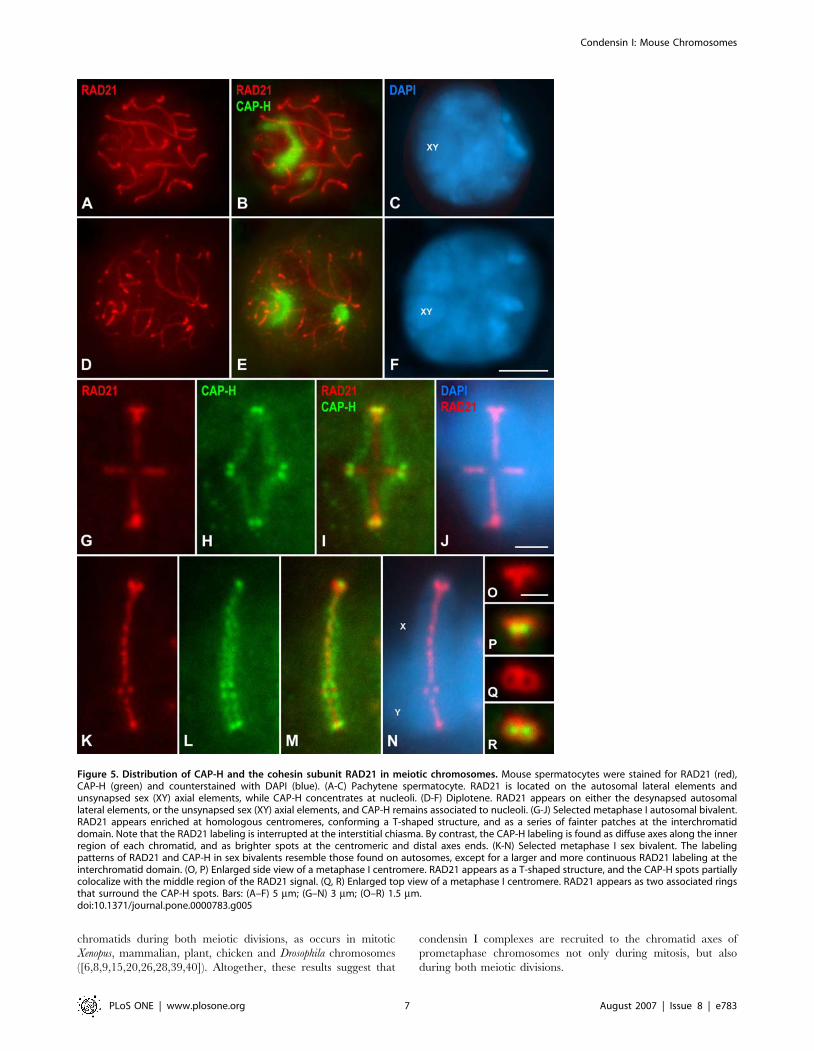
Relative distribution of condensin I and RAD21
cohesin complexes in meiosis I chromosomesIn order to precisely determine the relative distribution of
condensin I and cohesin complexes in mouse meiotic chromo-
somes we performed a double immunolabeling of CAP-H and the
cohesin subunit RAD21, that is expressed during male mouse
meiosis ([38]).
Pachytene spermatocytes evidenced RAD21 signals along
cohesin axes that are coincident with the fully synapsed autosomal
lateral elements (LEs) of the synaptonemal complex (SC), and the
unsynapsed sex axial elements (AEs) (Fig. 5A-C). By contrast,
CAP-H appeared at nucleoli lying in the nucleoplasm and at the
sex body periphery (Fig. 5B). In diplotene spermatocytes, RAD21
was detected along desynapsing autosomal LEs and at unsynapsed
sex AEs (Fig. 5D-F). In this stage, CAP-H remained located at
nucleoli (Fig. 5E). Afterwards, from diakinesis up to prometaphase
I, RAD21 faded away from desynapsed LEs, and CAP-H
disappeared from nucleoli.
In either the autosomal or sex metaphase I bivalents, RAD21
was observed at the so-called interchromatid domain, and at
centromeres (Fig. 5G–N). An accurate analysis of autosomal
metaphase I bivalents revealed a series of small RAD21 patches at
the interchromatid domain, from centromeres towards distal
telomeres (Fig. 5G). Interestingly, this labeling was usually brighter
and more continuous in the sex bivalent (Fig 5K). This
interchromatid domain RAD21 labeling was interrupted at
chiasma sites in both autosomal and sex metaphase I bivalents
(Fig. 5G and K). Despite the localization at the interchromatid
domain, RAD21 appeared concentrated onto T-shaped structures
at the centromeres of all autosomes (Fig. 5G) and the centromere
of the X chromosome (Fig. 5K). By contrast, the CAP-H labeling,
as previously described, was found as a fuzzy axial structure at the
inner region of each chromatid, with brighter accumulations at
their proximal and distal ends (Fig. 5H and L). The merged images
of RAD21 and CAP-H in metaphase I bivalents showed that
whereas RAD21 was located at the interchromatid domain
between the sister chromatids, CAP-H appeared at their inner
regions (Fig. 5I and M). A detailed examination of side-viewed
metaphase I centromeres showed that the proximal CAP-H
accumulations flanked the RAD21 T-shaped structure (Fig. 5O
and P). Interestingly, centromere top views demonstrated the
presence of two side-by-side associated RAD21 rings (Fig. 5Q),
with the proximal CAP-H spots located slightly beneath their
cavities (Fig. 5R). Consequently, proximal CAP-H spots, which
partially colocalized with the RAD21 T-like structure, appear at
the so-called inner centromere domain of metaphase I chromo-
somes ([36]), as previously described for proximal telomere
complexes in mouse condensed meiotic chromosomes ([37]).
DISCUSSION
Recruitment of condensin I to spermatogonial
mitotic chromosomesWe have found that the non-SMC subunits of the condensin I
complex CAP-G, CAP-D2, and CAP-H, become associated to
chromosomes in mouse spermatogonial cells during prometa-
Figure 3. CAP-H distribution in meiosis II spermatocytes. Mouse spermatocytes were stained for CAP-H (green) and counterstained with DAPI(blue). (A, B) Late interkinesis nucleus. CAP-H appears at several nucleoli (nu) and as small spots in the nucleoplasm. (C, D) Prophase II spermatocyte. Adiffuse CAP-H labeling, and bright spots, are observed along chromosomes. (E–H) Two different focal planes of a metaphase II spermatocyte. BrightCAP-H spots are present at chromosome ends. (I, J) Selected metaphase II chromosome. Each chromatid shows two CAP-H spots, one at thecentromeric end (red arrowheads) and one at its distal end (white arrowhead). (K, L) Selected segregating chromatids in early anaphase II. Bothchromatids present a CAP-H axis and one spot at each axis end. (M–O) Two focal planes of a late anaphase II spermatocyte. Each chromatid exhibitsa single diffuse CAP-H axis between the centromeric and distal ends spots. In (B) and (L) the CAP-H labeling has been pseudocolored in red andsuperimposed on its corresponding DAPI image. Bars: (A–H, M–O) 5 mm; (I–L) 3 mm.doi:10.1371/journal.pone.0000783.g003
Condensin I: Mouse Chromosomes
PLoS ONE | www.plosone.org 5 August 2007 | Issue 8 | e783

phase. Consistently, our results show that these subunits are
recruited after the phosphorylation of histone H3 at serine 10,
a landmark of the initiation of mitotic condensation ([33]). Thus,
our data support those obtained in mammalian and plant somatic
cells reporting the association of condensin I complexes to
chromosomes only after the nuclear envelope breakdown in
prometaphase ([9,28,30,39,40]). Therefore, our observations
revealed an identical loading pattern of condensin I in somatic
and male germ cells. This contrasts with condensin II complexes,
which are loaded to chromosomes during prophase ([28,39,40]).
Nucleolar localization of condensin IIn striking contrast with our observations in spermatogonial cells,
CAP-H was the only non-SMC subunit of condensin I detectable
inside prophase spermatocyte nuclei during both meiotic divisions.
CAP-H localized at nucleoli from pachytene up to diakinesis
during prophase I, and also in late interkinesis and prophase II
nuclei. This nucleolar localization of CAP-H, and also of CAP-C
and CAP-E, has been previously described in interphase human
HeLa cells ([30]). Likewise, some condensin I and II subunits have
been found at nucleoli in interphase Xenopus ([41]), Arabidopsis and
tobacco cultured cells ([40]), and also in Xenopus oocytes ([42]). It
has been suggested that in higher eukaryotes the condensin
complex may participate in the chromatin remodeling and
condensation-dependent silencing of rDNA loci ([30]). Alterna-
tively, it has been suggested that condensin may alter the
secondary structure of rRNAs or play a scaffolding role in spatial
organization of nucleoli ([41]), or may be sequestered at nucleoli to
facilitate chromosome decondensation during telophase ([30]).
However, the putative nucleolar function of condensin complexes,
or even condensin subunits, during mitosis and meiosis remains to
be explored.
Recruitment of condensin I to meiotic chromosomesOur results indicate that condensin I appears at chromatid axes by
prometaphase during both meiotic divisions and spermatogonial
mitosis. Thus, our results are similar to those previously reported
during C. elegans meiosis showing that MIX-1, a SMC2 homo-
logue, and HCP-6, a homologue of the condensin II subunit CAP-
D3, are recruited to chromosomes by late prophase I, during
diakinesis ([25]). The condensin I subunits whose distribution we
have analyzed are located at the inner region of the condensed
Figure 4. CAP-H accumulates preferentially at telomere complexes. Selected bivalents and chromosomes were stained for CAP-H (green),kinetochores revealed by an ACA serum (pink), TRF1 (red), and counterstained with DAPI (blue). (A) Metaphase I bivalent. A pair of CAP-H dots at thecentromere region of each chromosome is below the closely associated sister kinetochores. (B) Metaphase II chromosome. The separated CAP-Hspots at the centromere region appear below the kinetochores. (C) Metaphase I bivalent and (D) anaphase I half-bivalent. The CAP-H dots colocalizewith the TRF1 signals. Bar, 3 mm.doi:10.1371/journal.pone.0000783.g004
Condensin I: Mouse Chromosomes
PLoS ONE | www.plosone.org 6 August 2007 | Issue 8 | e783
chromatids during both meiotic divisions, as occurs in mitotic
Xenopus, mammalian, plant, chicken and Drosophila chromosomes
([6,8,9,15,20,26,28,39,40]). Altogether, these results suggest that
condensin I complexes are recruited to the chromatid axes of
prometaphase chromosomes not only during mitosis, but also
during both meiotic divisions.
Figure 5. Distribution of CAP-H and the cohesin subunit RAD21 in meiotic chromosomes. Mouse spermatocytes were stained for RAD21 (red),CAP-H (green) and counterstained with DAPI (blue). (A-C) Pachytene spermatocyte. RAD21 is located on the autosomal lateral elements andunsynapsed sex (XY) axial elements, while CAP-H concentrates at nucleoli. (D-F) Diplotene. RAD21 appears on either the desynapsed autosomallateral elements, or the unsynapsed sex (XY) axial elements, and CAP-H remains associated to nucleoli. (G-J) Selected metaphase I autosomal bivalent.RAD21 appears enriched at homologous centromeres, conforming a T-shaped structure, and as a series of fainter patches at the interchromatiddomain. Note that the RAD21 labeling is interrupted at the interstitial chiasma. By contrast, the CAP-H labeling is found as diffuse axes along the innerregion of each chromatid, and as brighter spots at the centromeric and distal axes ends. (K-N) Selected metaphase I sex bivalent. The labelingpatterns of RAD21 and CAP-H in sex bivalents resemble those found on autosomes, except for a larger and more continuous RAD21 labeling at theinterchromatid domain. (O, P) Enlarged side view of a metaphase I centromere. RAD21 appears as a T-shaped structure, and the CAP-H spots partiallycolocalize with the middle region of the RAD21 signal. (Q, R) Enlarged top view of a metaphase I centromere. RAD21 appears as two associated ringsthat surround the CAP-H spots. Bars: (A–F) 5 mm; (G–N) 3 mm; (O–R) 1.5 mm.doi:10.1371/journal.pone.0000783.g005
Condensin I: Mouse Chromosomes
PLoS ONE | www.plosone.org 7 August 2007 | Issue 8 | e783

Telomeric accumulation of condensin IOur results show that condensin I complexes are particularly
enriched at the ends of the chromatid axes in mouse mitotic and
meiotic chromosomes. Double immunolabelings with an anti-
centromere autoantibody revealing kinetochores and TRF1,
a telomeric protein, demonstrate that the proximal condensin I
accumulations are located below the kinetochores. Moreover, the
proximal and distal accumulations of condensin I colocalized with
the proximal and distal telomere complexes, respectively ([37]). As
far as we know, this is the first time that condensin I complexes are
shown to be preferentially accumulated at telomere complexes.
The localization of proximal telomere and condensin I com-
plexes below kinetochores is intriguing at first sight. All the
chromosomes from the C57BL/6 mouse strain, except the Y
chromosome, are telocentric since a short arm is not detected at
a cytogenetic level. Thus, the proximal telomere and condensin I
complexes observed below kinetochores do not correspond to
a short arm. Previously, we have reported that in pachytene and
diplotene mouse chromosomes the proximal telomeric DNA
repeats and specific telomeric proteins are associated to the
nuclear envelope, and that centromeres are internally located
relative to the telomere complexes ([37]). By contrast, from
metaphase I on, the proximal telomere complexes are located
below the kinetochores. These results led us to suggest that in
mouse meiotic chromosomes, there is a relocation of the proximal
telomere complexes and kinetochores from diakinesis up to
metaphase I, once the telomeres have detached from the nuclear
envelope, in order to protect the proximal telomeric repeats and
allow the interaction of microtubules with the kinetochores ([37]).
In this sense, the reorganization of the proximal telomere
complexes and kinetochores would be possible since it has been
reported that the proximal telomere repeats and the centromeric
minor satellite DNA repeats, that organize the kinetochores, are
relatively separated by other repetitive sequences ([32,43,44]).
The preferential accumulation of condensin I complexes at
telomere complexes in mouse mitotic and meiotic chromosomes
might be due to the ultra-long arrays of telomeric DNA repeats
found in mouse chromosomes ([45–47]). In this respect,
established mouse strains, such as C57BL/6, have long hypervari-
able telomeric DNA lengths if compared with strains recently
derived from wild mice ([48]). Most studies analyzing the
distribution of telomeric DNA repeats in mammalian chromo-
somes by FISH have revealed that these repeats do not appear as
large chromatin loops, as expected according to the scaffold/radial
loop model of chromosome structure ([49]). By contrast, the
telomeric repeats are detected at the cytological telomeres as
discrete spots surrounded by chromatin in both mitotic ([50,51])
and meiotic chromosomes ([52–56]). Our results suggest that the
large accumulations of condensin I at telomere complexes may
contribute to the specific organization of telomeric DNA in
condensed mouse chromosomes. In this sense, the high number of
telomeric repeats found in mouse could be organized as short
loops in condensed chromosomes, as in prophase I chromosomes
([53]), that are maintained in a condensed state by means of their
interaction with condensin I complexes. Since condensin I is
located at chromatid axes along the arms, i.e. at the putative bases
of the chromatin loops, the accumulation of condensin I at
telomere complexes indicates that the number of loop bases is
higher at telomeres than along the arms. Consequently, the
presence of a high number of short telomeric DNA loops would
explain the accumulation of condensin I complexes at their bases
in mouse chromosomes. Alternatively, the telomeric DNA repeats
are not organized as loops, and the condensin I complexes would
allow their compaction in an undetermined organization. In our
opinion these two alternatives are not mutually exclusive and
should be tested in the future.
Our results show that in mouse chromosomes the proximal
condensin I complexes do not colocalize with kinetochores. This is
consistent with results obtained in human chromosomes where
condensin I complexes are not present at kinetochores. However,
condensin II complexes are enriched near the inner kinetochore
plates, and depletion of condensin II subunits promotes defects in
kinetochore structure and function ([28]). Consequently, further
studies are needed to know whether condensin II complexes are
also needed to organize kinetochores in mouse mitotic and meiotic
chromosomes.
Change of chromosome structure during meiosis IThere are no data concerning the appearance and location of the
chromatid axes in condensed mammalian meiotic chromosomes
(for review see [57]). We have found that condensin I axes, as well
as silver-stained chromatid axes (Supplementary Fig. S9), are
located at the inner region of each metaphase I chromatid.
Consequently, and assuming the scaffold/radial loop model of
chromosome structure, mouse metaphase I chromosomes show an
organization similar to that found in mitotic metaphase chromo-
somes ([4,57]) (Fig. 6). This situation is similar to that found in
grasshopper metaphase I chromosomes. In these species, silver-
stained chromatid axes are peripherally located with respect to the
width of each chromatid by early metaphase I, so that sister axes
are close to each other ([58–60]). However, the sister axes
individualize all along their length to appear at the inner region of
each chromatid by late metaphase I ([61–63]).
The appearance of chromatid axes at the inner region of
metaphase I chromatids contrasts with their distribution in
prophase I pachytene chromosomes. In rooster pachytene
chromosomes, topo IIa, a component of the chromatid axes of
mitotic chromosomes together with condensin I and II complexes,
appears located on the outer edge of the synaptonemal complex
LEs ([64]). Similarly, condensin complexes are present at
pachytene LEs during budding yeast meiosis ([24]). These results
indicate that the chromatid axes from both sister chromatids are
intimately associated at the LE. Since, chromatin is organized as
loops whose bases attach to LEs, and LEs are peripherally located
in relation to the chromatin loops from both sister chromatids, it is
said that pachytene chromosomes show a ‘meiotic organization’
(for review see [57]). Thus, chromosome organization must change
from a meiotic one during pachytene to a mitotic one in
metaphase I. While in grasshoppers this change of chromosome
organization occurs throughout metaphase I, in male mouse this
must happen between diplotene and metaphase I. Thus, during
meiosis, different species change their chromosome structure at
different times. In this sense, what seems to be really crucial is
that chromosomes must show a mitotic organization previous to
the release of sister-chromatid arm cohesion at the onset of
anaphase I.
Redistribution of cohesin complexes during meiosis IThe different cohesin complexes found during mammalian meiosis
are located at cohesin axes coincident with the LEs during
prophase I (for review see [65]). Thus, cohesin complexes are
located at or near the bases of the chromatin loops from both sister
chromatids (for review see [57]). Our observations demonstrate
that cohesin complexes are distributed at the interchromatid
domain in metaphase I bivalents, and not embracing both sister
chromatids. Consequently, and according to the scaffold/radial
loop model, the cohesin complexes would embrace the distal end
Condensin I: Mouse Chromosomes
PLoS ONE | www.plosone.org 8 August 2007 | Issue 8 | e783

of chromatin loops from both sister chromatids (Fig. 6). These
observations suggest that, concomitantly with the remodeling of
chromosome structure from a meiotic to a mitotic organization,
i.e. from pachytene to metaphase I, cohesin complexes are
redistributed from the bases of the chromatin loops to their distal
ends. In this sense, it has been recently demonstrated in budding
yeast that there is a condensin-dependent removal of a subset of
cohesin complexes during prophase I that is dependent on the
phosphorylation of the cohesin subunit Rec8 by Cdc5, a Polo-like
kinase ([66]). A similar Polo-like kinase-dependent pathway to
remove most cohesin complexes from chromosome arms also
occurs during prophase in mitotic chromosomes ([67-69]). Like-
wise, it has been previously shown that there is a removal of
a subset of cohesin complexes from chromosome arms during late
diplotene and diakinesis in male mouse meiosis ([38,70]). Since
SGO2, a protector of centromeric cohesin complexes, appears
only at centromeres during late diplotene and diakinesis in mouse
spermatocytes ([71]), it is tempting to suggest that SGO2 would
not protect arm cohesin complexes against their removal by
a putative phosphorylation-dependent pathway during late pro-
phase I stages. Accordingly, we propose that during diplotene and
diakinesis, a partial loss of cohesin complexes from the arms could
facilitate the resolution of sister chromatids, concomitantly with
a change of chromosome structure from a meiotic to a mitotic
organization. In this scenario, the cohesin complexes remaining at
the arms during diakinesis might redistribute, for instance by
a sliding mechanism, from the bases of chromatin loops to their
ends in order to ensure sister-chromatid arm cohesion until the
onset of anaphase I. Alternatively, some cohesin complexes might
be removed from the arms during diakinesis and reloaded during
or after chromosome structure remodelling. Obviously, further
studies are needed to verify these hypotheses.
MATERIALS AND METHODS
Cell cultureHeLa cultured cells were grown in flasks in Dulbecco’s modified
Eagle’s medium (DMEM) containing 10% fetal calf serum,
50 units/ml penicillin, 50 mg/ml streptomycin and 1% 0.2 M L-
glutamine (all from Imperial Laboratories, Andover, United
Kingdom), at 37uC in a humidified atmosphere containing 5%
CO2.
Electrophoresis and immunoblottingHeLa cells extracts were prepared by harvesting cultured cells,
which were washed twice with PBS (137 mM NaCl, 2.7 mM KCl,
10.1 mM Na2HPO4, 1.7 mM KH2PO4, [pH 7.4]). Then, they
were lysed in boiling SDS sample buffer (50 mM Tris-ClH pH 6.8,
3% SDS, 2 mM EDTA, 15% sucrose, 9% b-mercaptoethanol,
0.005% bromophenol blue).
Testis from adult male C57BL/6 mice were removed and
placed in 2 ml of SDS solubilization solution (50 mM Tris-ClH
pH 6.8, 5 mM EDTA, 3% SDS, 1% protease inhibitor cocktail).
Then, testes were homogenized on ice in a Potter homogenizer.
The extract was placed in boiling water bath for 5 minutes. The
appropriate quantity of extract was diluted with 5X SDS-lysis
buffer (62.5 mM Tris-ClH pH 6.8, 2% SDS, 5% b-mercaptoetha-
nol, 10% glycerol, 0.005% bromophenol blue) and boiled for
5 minutes.
SDS-PAGE was carried out in 10% polyacrylamide gels. Gels
were electrically transferred to Trans-Blot sheets (Bio-Rad,
Hercules, California, United States) for 1.5 hour at 4uC and
310 mA. Sheets were blocked for 1 hour with 4% non-fat dry milk
in PBS, followed by an overnight incubation at 4uC with an anti-
hCAP-H antibody at a 1:10,000 dilution in 4% non-fat dry milk in
PBS. Immunoreactive bands were visualized by incubation for
1 hour at room temperature with horseradish peroxidase-conju-
gated donkey anti-rabbit Ig (Amersham Life Science, Little
Chalfont, United Kingdom) at a 1:5,000 dilution in PBS, and
subsequent development using an ECL (Enhanced Chemilumi-
nescence) detection system (Amersham Life Science) according to
the manufacturer instructions.
Squashing of spermatocytesAdult male C57BL/6 mice were used in the present study. Testis
were removed, detunicated, and seminiferous tubules then
processed for squashing. For squashing we followed the technique
previously described by Parra et al. ([31]). Briefly, seminiferous
tubules were fixed in freshly prepared 2% formaldehyde in PBS
containing 0.1% Triton X-100. After 5 minutes several seminif-
erous tubules fragments were placed on a clean slide coated with
1 mg/ml poly-L-lysine (Sigma, St. Louis, Missouri, United States)
with a small drop of fixative, and the tubules were gently minced
with tweezers. The tubules were then squashed and the coverslip
removed after freezing in liquid nitrogen.
Figure 6. Representation of a metaphase I bivalent showing therelative distributions of condensin I and RAD21-containing cohesincomplexes. One chromosome is depicted in light grey and itshomologue is darker grey. Chromosomes are telocentric, and themetaphase I bivalent shows a single interstitial chiasma. Kinetochoresare indicated in yellow, kMTs in light grey, condensin I in green, RAD21in red, and TRF1 in blue. The colocalisation of condensin I and TRF1 isshown in light blue. In (A), the condensin I complexes delineate a fuzzyaxis inside each chromatid, with prominent accumulations at theirproximal and distal ends which colocalize with TRF1. The two proximalcondensin I accumulations appear below the closely associated sisterkinetochores, and partially colocalize with the middle region of theT-shaped RAD21 structure at each centromere. RAD21-containingcohesin complexes are also depicted as patches at the interchromatiddomain. (B) Hypothetical model accounting for the distribution ofcondensin I and RAD21-containing cohesin complexes in relation toradial chromatin loops in a mouse metaphase I bivalent. Thelongitudinal and transverse sections of the arms correspond to areasindicated in (A).doi:10.1371/journal.pone.0000783.g006
Condensin I: Mouse Chromosomes
PLoS ONE | www.plosone.org 9 August 2007 | Issue 8 | e783

Immunofluorescence microscopyAfter fixation, the slides were rinsed three times for 5 minutes in
PBS, and incubated for 45 minutes at room temperature with
primary antibodies. Several subunits of the condensin I complex
were detected. CAP-H was detected with a rabbit polyclonal
antibody generated against a 211 aa amino-terminal fragment of
human CAP-H ([72]), at a 1:100 dilution in PBS. To detect CAP-
G a polyclonal rabbit serum raised against a recombinant
polypeptide from the middle region (amino acids 235 to 570) of
human CAP-G ([73]) was employed at a 1:100 dilution in PBS.
Two polyclonal rabbit antisera which respectively recognize the
amino-terminal (amino acids 997 to 1401) and the carboxyl-
terminal (amino acids 1 to 241) subdomains of human hCAP-D2
(CNAP1) ([15]) were employed to detect CAP-D2 at a 1:20
dilution in PBS. Kinetochores were detected with the human
autoimmune anti-centromere (ACA) serum GS, that recognizes
CENP-A, -B and -C (kindly provided by Dr. William Earnshaw;
[74]), at a 1:10,000 dilution in PBS. To detect the telomeric
protein TRF1 we used a polyclonal rabbit serum (#TRF12-S;
Alpha Diagnostic International, San Antonio, Texas, United
States), raised against mouse TRF1, at a 1:100 dilution in PBS
([37]). To detect the cohesin subunit RAD21 we used the
polyclonal rabbit serum K854 (kindly provided by Dr. Jose Luis
Barbero; [75]), at a 1:50 dilution in PBS. Fibrillarin was detected
with the S4 human anti-fibrillarin autoantibody at a 1:500 dilution
(kindly provided by Dr. Ricardo Benavente; [76]). Histone H3
phosphorylated at serine 10 (pH3) was detected with a polyclonal
rabbit serum (Upstate, Lake Placid, New York, United States) at
a 1:1,000 dilution. Following three washes in PBS for 5 minutes,
the slides were incubated for 30 minutes at room temperature with
secondary antibodies. A fluorescein isothiocyanate (FITC)-conju-
gated goat anti-rabbit IgG (Jackson, West Grove, Pennsylvania,
United States) at a 1:150 dilution in PBS, and a Texas Red-
conjugated goat anti-human IgG (Jackson) at a 1:150 dilution in
PBS, were used for simultaneous double immunolabeling. The
slides were subsequently rinsed in PBS, and counterstained for
3 minutes with 2 mg/ml DAPI (49, 6-diamidino-2-phenylindole).
After a final rinse in PBS, the slides were mounted in Vectashield
(Vector Laboratories, Burlingame, California, United States) and
sealed with nail varnish. In double immunolabeling experiments,
primary antibodies were incubated simultaneously except when
primary antibodies were generated in the same host species when
we proceeded as previously described ([77]). In those cases slides
were first incubated with the rabbit serum against hCAP-H for
1 hour at room temperature, rinsed in PBS and incubated
overnight at 4uC with an FITC-conjugated goat Fab’ fragment
anti-rabbit IgG (Jackson) at a 1:100 dilution in PBS. Afterwards,
slides were rinsed six times for 5 minutes in PBS, incubated with
the rabbit antibody against TRF1 or RAD21 for 1 hour, rinsed
three times for 5 minutes in PBS, and incubated with a Texas
Red-conjugated goat anti-rabbit IgG (Jackson) at a 1:150 dilution.
Observations were performed using an Olympus BH-2
microscope (Olympus, Hamburg, Germany) equipped with
epifluorescence optics and the images were recorded with an
Olympus DP50 digital camera. Digital images were then treated
using the Adobe PhotoShop 7.0 software.
SUPPORTING INFORMATION
Figure S1 Immunoblot of HeLa cell extracts (left lane) and
mouse testis extracts (right lane) probed with the anti-hCAP-H
antibody. The positions of two molecular mass markers are
indicated. The antibody specifically recognized a single protein
band of about 90 kDa in both extracts.
Found at: doi:10.1371/journal.pone.0000783.s001 (0.28 MB TIF)
Figure S2 CAP-D2 distribution in spermatogonial mitosis.
Mouse spermatogonia were stained for CAP-D2 (green), kineto-
chores with an ACA serum (red), and counterstained with DAPI
(blue). (A–C) Early prometaphase. Condensing chromosomes
present a faint and diffuse CAP-D2 staining, but some bright
spots are also observed. (D–F) Metaphase in top view, and (G–I)
anaphase. A single diffuse CAP-D2 axis is seen in each chromo-
some/chromatid, as well as bright accumulations. Bar, 5 mm.
Found at: doi:10.1371/journal.pone.0000783.s002 (5.93 MB TIF)
Figure S3 H3 phosphorylation precedes condensin I recruit-
ment to chromosomes in spermatogonial mitosis. Mouse sper-
matogonia were stained for histone H3 phosphorylated at serine
10 (pH3) (red), CAP-H (green), and counterstained with DAPI
(blue). (A–C) Middle prophase. Phosphorylated H3 is present in
condensing chromosomes but not CAP-H. (D–F) Late prophase.
CAP-H appears as small bright spots (arrowheads) and in the
nucleoplasm. (G–I) Metaphase in top view. A single CAP-H axis
(arrows) is seen in each chromosome. Bar, 5 mm.
Found at: doi:10.1371/journal.pone.0000783.s003 (5.91 MB TIF)
Figure S4 Relative distributions of CAP-H and fibrillarin in
prophase I spermatocytes. Mouse spermatocytes were stained for
CAP-H (green), fibrillarin (red), and counterstained with DAPI
(blue). (A–C) Zygotene spermatocyte. CAP-H is not detected, and
fibrillarin appears at nucleoli. (D–F) Early pachytene, (G–I) late
pachytene, and (J–L) diplotene spermatocytes. CAP-H and
fibrillarin colocalize at the nucleoplasmic nucleoli, and at the
nucleolus associated to the sex body (XY). Note that CAP-H and
fibrillarin are not present at the round body and/or the fibrillar
centre (arrows) inside the sex body-associated nucleolus during
pachytene, and the nucleolus in diplotene (Knibiehler et al., 1981).
Fibrillarin is additionally present in Cajal bodies (arrowheads)
lying in the nucleoplasm (E, H) or associated to the nucleolus (K).
Bar, 5 mm. Supplementary Reference: Knibiehler B, Mirre C,
Hartung M, Jean P, Stahl A (1981) Sex vesicle-associated
nucleolar orgnizers in mouse spermatocytes: localization, struc-
ture, and function. Cytogenet Cell Genet 31: 47–57.
Found at: doi:10.1371/journal.pone.0000783.s004 (10.16 MB
TIF)
Figure S5 CAP-D2 distribution in spermatocytes. Mouse
spermatocytes were stained for CAP-D2 (green), kinetochores
with an ACA serum (red in A–F, J–R), TRF1 (red in G–I), and
counterstained with DAPI (blue). (A–C) Metaphase I spermato-
cyte. The autosomal bivalents show pairs of bright CAP-D2 spots
at their centromeric and distal ends. (D–I) Two selected autosomal
metaphase I bivalents. The proximal pair of CAP-D2 signals
appears below the closely associated sister kinetochores (D–F). The
four pairs of CAP-D2 signals colocalize with the TRF1 signals (G–
I). (J–L) Anaphase I, (M–O) metaphase II, and (P–R) anaphase II
spermatocytes. Chromosomes/chromatids show a faint CAP-D2
labeling along them, and brighter CAP-D2 dots at their ends. The
insets in (J–L) show an enlarged anaphase I half-bivalent (arrows).
Bars: (A–C, J–R) 5 mm; (D–I) 3 mm.
Found at: doi:10.1371/journal.pone.0000783.s005 (1.14 MB JPG)
Figure S6 H3 phosphorylation at serine 10 precedes condensin I
recruitment to chromosomes in meiosis I. Mouse spermatocytes
were stained for histone H3 phosphorylated at serine 10 (pH3)
(red), CAP-H (green), and counterstained with DAPI blue). (A–C)
Diplotene spermatocyte. Phosphorylated H3 is enriched at
chromocentres, while CAP-H appears at nucleoli. (D–F) Late
diplotene. Phosphorylated H3 appears on all the chromatin, and
CAP-H at a nucleolus. (G–I) Prometaphase I spermatocyte. CAP-
Condensin I: Mouse Chromosomes
PLoS ONE | www.plosone.org 10 August 2007 | Issue 8 | e783

H appears on bivalents, and as pairs of bright dots (double
arrowheads). (J–L) Metaphase I spermatocyte. CAP-H is detected
as pairs of bright spots at the centromeric and distal chromosome
ends. (M–P) Two focal planes of an anaphase I spermatocyte. In
each chromosome, a pair of CAP-H dots is detected at the
centromere region (red arrowheads), one spot at the distal end of
each chromatid (white arrowheads), and a diffuse axial labeling
along chromatids. The inset shows the chromosome indicated in
(O), where the CAP-H staining has been pseudocolored in red and
superimposed on its corresponding DAPI image. Bar, 5 mm.
Found at: doi:10.1371/journal.pone.0000783.s006 (9.96 MB TIF)
Figure S7 H3 phosphorylation precedes condensin I recruit-
ment to chromosomes in meiosis II. Mouse spermatocytes were
stained for histone H3 phosphorylated at serine 10 (pH3) (red),
CAP-H (green), and counterstained with DAPI blue). (A–D) Two
focal planes of a late interkinesis nucleus. Phosphorylated H3 is
present on all the chromatin, whereas CAP-H appears at one
nucleolus (nu) and at small spots in the nucleoplasm. (E–G)
Metaphase II, and (H–K) two focal planes of an anaphase II
spermatocyte. Bright CAP-H spots are present at chromosome
ends. Bar, 5 mm.
Found at: doi:10.1371/journal.pone.0000783.s007 (5.26 MB TIF)
Figure S8 Relative distributions of CAP-H and TRF1 in
spermatogonial metaphase chromosomes. (A–D) Mouse meta-
phase spermatogonia stained for CAP-H (green), TRF1 (red), and
counterstained with DAPI (blue). A fuzzy CAP-H axis is visible
inside each sister chromatid. The bright CAP-H accumulations at
the axes ends colocalize with TRF1. Bar, 5 mm.
Found at: doi:10.1371/journal.pone.0000783.s008 (2.25 MB TIF)
Figure S9 Silver staining of metaphase I spermatocytes. (A)
Metaphase I spread spermatocyte where the sex bivalent is
indicated (XY), and selected autosomal bivalents (B, D) and sex (C)
bivalent. Two silver-stained structures representing sister kineto-
chores (arrowheads) are found at the centromeric region of each
homologue. Faint silver-stained axes (arrows) are observed along
the inner region of the chromatids. (E) Autosomal metaphase I
bivalent shown in Fig. 2N stained for CAP-H (pseudocolored in
red), and counterstained with DAPI (blue). The CAP-H axes are
located inside the chromatids as the silver-stained chromatid axes
(D). Bars: (A) 10 mm; (B–E) 3 mm.
Found at: doi:10.1371/journal.pone.0000783.s009 (5.41 MB TIF)
ACKNOWLEDGMENTSWe wish to express our sincere thanks to Bill Earnshaw for providing us the
GS human anti-centromere autoantibody, to Jose Luis Barbero for his
generous gift of the rabbit anti-RAD21 antibody, and to Ricardo
Benavente for providing the S4 human anti-fibrillarin autoantibody.
Author Contributions
Conceived and designed the experiments: AV JS JR. Performed the
experiments: MP RG AV JS JR. Analyzed the data: MP RG AV JS JR JS
KY. Contributed reagents/materials/analysis tools: AV JS JR JS KY.
Wrote the paper: AV JS JR.
REFERENCES1. Swedlow JR, Hirano T (2003) The making of the mitotic chromosome: modern
insights into classical questions. Mol Cell 11: 557–69.
2. Hirano T (2005) Condensins: organizing and segregating the genome. Curr Biol15: R265–75.
3. Belmont AS (2006) Mitotic chromosome structure and condensation. Curr Opin
Cell Biol 18: 632–8.
4. Losada A, Hirano T (2005) Dynamic molecular linkers of the genome: the first
decade of SMC proteins. Genes Dev 19: 1269–87.
5. Nasmyth K, Haering CH (2005) The structure and function of SMC and kleisin
complexes. Annu Rev Biochem 74: 595–648.
6. Hirano T, Mitchison TJ (1994) A heterodimeric coiled-coil protein required formitotic chromosome condensation in vitro. Cell 79: 449–58.
7. Hirano T, Kobayashi R, Hirano M (1997) Condensins, chromosomecondensation protein complexes containing XCAP-C, XCAP-E and a Xenopus
homolog of the Drosophila Barren protein. Cell 89: 511–21.
8. Saitoh N, Goldberg IG, Wood ER, Earnshaw WC (1994) ScII: an abundant
chromosome scaffold protein is a member of a family of putative ATPases with
an unusual predicted tertiary structure. J Cell Biol 127: 303–18.
9. Maeshima K, Laemmli UK (2003) A two-step scaffolding model for mitotic
chromosome assembly. Dev Cell 4: 467–80.
10. Neuwald AF, Hirano T (2000) HEAT repeats associated with condensins,
cohesins, and other complexes involved in chromosome-related functions.
Genome Res 10: 1445–52.
11. Chuang PT, Albertson DG, Meyer BJ (1994) DPY-27:a chromosome
condensation protein homolog that regulates C. elegans dosage compensationthrough association with the X chromosome. Cell 79: 459–74.
12. Lieb JD, Capowski EE, Meneely P, Meyer BJ (1996) DPY-26, a link between
dosage compensation and meiotic chromosome segregation in the nematode.Science 274: 1732–6.
13. Sutani T, Yuasa T, Tomonaga T, Dohmae N, Takio K, et al. (1999) Fissionyeast condensin complex: essential roles of non-SMC subunits for condensation
and Cdc2 phosphorylation of Cut3/SMC4. Genes Dev 13: 2271–83.
14. Freeman L, Aragon-Alcaide L, Strunnikov A (2000) The condensin complexgoverns chromosome condensation and mitotic transmission of rDNA. J Cell
Biol 149: 811–24.
15. Schmiesing JA, Gregson HC, Zhou S, Yokomori K (2000) A human
condensin complex containing hCAP-C-hCAP-E and CNAP1, a homolog
of Xenopus XCAP-D2, colocalizes with phosphorylated histone H3 duringthe early stage of mitotic chromosome condensation. Mol Cell Biol 20:
6996–7006.
16. Kimura K, Cuvier O, Hirano T (2001) Chromosome condensation by
a human condensin complex in Xenopus egg extracts. J Biol Chem 276:5417–20.
17. Saka Y, Sutani T, Yamashita Y, Saitoh S, Takeuchi M, et al. (1994) Fission yeast
cut3 and cut14, members of a ubiquitous protein family, are required for
chromosome condensation and segregation in mitosis. Embo J 13: 4938–52.
18. Strunnikov AV, Hogan E, Koshland D (1995) SMC2, a Saccharomyces
cerevisiae gene essential for chromosome segregation and condensation, defines
a subgroup within the SMC family. Genes Dev 9: 587–99.
19. Bhat MA, Philp AV, Glover DM, Bellen HJ (1996) Chromatid segregation at
anaphase requires the barren product, a novel chromosome-associated protein
that interacts with Topoisomerase II. Cell 87: 1103–14.
20. Steffensen S, Coelho PA, Cobbe N, Vass S, Costa M, et al. (2001) A role for
Drosophila SMC4 in the resolution of sister chromatids in mitosis. Curr Biol 11:
295–307.
21. Bhalla N, Biggins S, Murray AW (2002) Mutation of YCS4, a budding yeast
condensin subunit, affects mitotic and nonmitotic chromosome behavior. Mol
Biol Cell 13: 632–45.
22. Hagstrom KA, Holmes VF, Cozzarelli NR, Meyer BJ (2002) C. elegans
condensin promotes mitotic chromosome architecture, centromere organization,
and sister chromatid segregation during mitosis and meiosis. Genes Dev 16:
729–42.
23. Siddiqui NU, Stronghill PE, Dengler RE, Hasenkampf CA, Riggs CD (2003)
Mutations in Arabidopsis condensin genes disrupt embryogenesis, meristem
organization and segregation of homologous chromosomes during meiosis.
Development 130: 3283–95.
24. Yu HG, Koshland DE (2003) Meiotic condensin is required for proper
chromosome compaction, SC assembly, and resolution of recombination-
dependent chromosome linkages. J Cell Biol 163: 937–47.
25. Chan RC, Severson AF, Meyer BJ (2004) Condensin restructures chromosomes
in preparation for meiotic divisions. J Cell Biol 167: 613–25.
26. Ono T, Losada A, Hirano M, Myers MP, Neuwald AF, et al. (2003) Differential
contributions of condensin I and condensin II to mitotic chromosome
architecture in vertebrate cells. Cell 115: 109–21.
27. Yeong FM, Hombauer H, Wendt KS, Hirota T, Mudrak I, et al. (2003)
Identification of a subunit of a novel Kleisin-beta/SMC complex as a potential
substrate of protein phosphatase 2A. Curr Biol 13: 2058–64.
28. Ono T, Fang Y, Spector DL, Hirano T (2004) Spatial and temporal regulation
of Condensins I and II in mitotic chromosome assembly in human cells. Mol
Biol Cell 15: 3296–308.
29. Watrin E, Cubizolles F, Osborne HB, Le Guellec K, Legagneux V (2003)
Expression and functional dynamics of the XCAP-D2 condensin subunit in
Xenopus laevis oocytes. J Biol Chem 278: 25708–15.
30. Cabello OA, Eliseeva E, He WG, Youssoufian H, Plon SE, et al. (2001) Cell
cycle-dependent expression and nucleolar localization of hCAP-H. Mol Biol Cell
12: 3527–37.
Condensin I: Mouse Chromosomes
PLoS ONE | www.plosone.org 11 August 2007 | Issue 8 | e783
31. Parra MT, Page J, Yen TJ, He D, Valdeolmillos A, et al. (2002) Expression and
behaviour of CENP-E at kinetochores during mouse spermatogenesis.Chromosoma 111: 53–61.
32. Kalitsis P, Griffiths B, Choo KH (2006) Mouse telocentric sequences reveal
a high rate of homogenization and possible role in Robertsonian translocation.Proc Natl Acad Sci U S A 103: 8786–91.
33. Hendzel MJ, Wei Y, Mancini MA, Van Hooser A, Ranalli T, et al. (1997)Mitosis-specific phosphorylation of histone H3 initiates primarily within
pericentromeric heterochromatin during G2 and spreads in an ordered fashion
coincident with mitotic chromosome condensation. Chromosoma 106: 348–60.34. Tres LL (2005) XY chromosomal bivalent: nucleolar attraction. Mol Reprod
Dev 72: 1–6.35. Ochs RL, Lischwe MA, Spohn WH, Busch H (1985) Fibrillarin: a new protein of
the nucleolus identified by autoimmune sera. Biol Cell 54: 123–33.36. Parra MT, Viera A, Gomez R, Page J, Carmena M, et al. (2003) Dynamic
relocalization of the chromosomal passenger complex proteins inner centromere
protein (INCENP) and aurora-B kinase during male mouse meiosis. J Cell Sci116: 961–74.
37. Viera A, Parra MT, Page J, Santos JL, Rufas JS, et al. (2003) Dynamicrelocation of telomere complexes in mouse meiotic chromosomes. Chromosome
Res 11: 797–807.
38. Parra MT, Viera A, Gomez R, Page J, Benavente R, et al. (2004) Involvement ofthe cohesin Rad21 and SCP3 in monopolar attachment of sister kinetochores
during mouse meiosis I. J Cell Sci 117: 1221–34.39. Hirota T, Gerlich D, Koch B, Ellenberg J, Peters JM (2004) Distinct functions of
condensin I and II in mitotic chromosome assembly. J Cell Sci 117: 6435–45.40. Fujimoto S, Yonemura M, Matsunaga S, Nakagawa T, Uchiyama S, et al.
(2005) Characterization and dynamic analysis of Arabidopsis condensin
subunits, AtCAP-H and AtCAP-H2. Planta 222: 293–300.41. Uzbekov R, Timirbulatova E, Watrin E, Cubizolles F, Ogereau D, et al. (2003)
Nucleolar association of pEg7 and XCAP-E, two members of Xenopus laeviscondensin complex in interphase cells. J Cell Sci 116: 1667–78.
42. Beenders B, Watrin E, Legagneux V, Kireev I, Bellini M (2003) Distribution of
XCAP-E and XCAP-D2 in the Xenopus oocyte nucleus. Chromosome Res 11:549–64.
43. Kuznetsova IS, Prusov AN, Enukashvily NI, Podgornaya OI (2005) New typesof mouse centromeric satellite DNAs. Chromosome Res 13: 9–25.
44. Kuznetsova I, Podgornaya O, Ferguson-Smith MA (2006) High-resolutionorganization of mouse centromeric and pericentromeric DNA. Cytogenet
Genome Res 112: 248–55.
45. Kipling D, Cooke HJ (1990) Hypervariable ultra-long telomeres in mice. Nature347: 400–2.
46. Starling JA, Maule J, Hastie ND, Allshire RC (1990) Extensive telomere repeatarrays in mouse are hypervariable. Nucleic Acids Res 18: 6881–8.
47. Zijlmans JM, Martens UM, Poon SS, Raap AK, Tanke HJ, et al. (1997)
Telomeres in the mouse have large inter-chromosomal variations in the numberof T2AG3 repeats. Proc Natl Acad Sci U S A 94: 7423–8.
48. Hemann MT, Greider CW (2000) Wild-derived inbred mouse strains have shorttelomeres. Nucleic Acids Res 28: 4474–8.
49. Laemmli UK, Cheng SM, Adolph KW, Paulson JR, Brown JA, et al. (1978)Metaphase chromosome structure: the role of nonhistone proteins. Cold Spring
Harb Symp Quant Biol 42 Pt 1: 351–60.
50. Moyzis RK, Buckingham JM, Cram LS, Dani M, Deaven LL, et al. (1988) Ahighly conserved repetitive DNA sequence, (TTAGGG)n, present at the
telomeres of human chromosomes. Proc Natl Acad Sci U S A 85: 6622–6.51. Meyne J, Baker RJ, Hobart HH, Hsu TC, Ryder OA, et al. (1990) Distribution
of non-telomeric sites of the (TTAGGG)n telomeric sequence in vertebrate
chromosomes. Chromosoma 99: 3–10.52. Moens PB, Pearlman RE (1990) Telomere and centromere DNA are associated
with the cores of meiotic prophase chromosomes. Chromosoma 100: 8–14.53. Heng HH, Chamberlain JW, Shi XM, Spyropoulos B, Tsui LC, et al. (1996)
Regulation of meiotic chromatin loop size by chromosomal position. Proc Natl
Acad Sci U S A 93: 2795–800.54. Scherthan H, Weich S, Schwegler H, Heyting C, Harle M, et al. (1996)
Centromere and telomere movements during early meiotic prophase of mouseand man are associated with the onset of chromosome pairing. J Cell Biol 134:
1109–25.55. Scherthan H, Jerratsch M, Li B, Smith S, Hulten M, et al. (2000) Mammalian
meiotic telomeres: protein composition and redistribution in relation to nuclear
pores. Mol Biol Cell 11: 4189–203.
56. Viera A, Parra MT, Rufas JS, Suja JA (2002) Size heterogeneity of telomeric
DNA in mouse meiotic chromosomes. Cytogenet Genome Res 98: 221–4.
57. Suja JA, Rufas JS (2007) Chromatid cores in meiotic chromosome structure and
segregation. In: Lankenau DH, Egel R, eds. Genome Dynamics and Stability,
vol. 3 Recombination and Meiosis. Berlin: Springer-Verlag, DOI 10.1007/
7050_2006_023.
58. Rufas JS, Gimenez-Abian J, Suja JA, Garcıa de la Vega C (1987) Chromosome
organization in meiosis revealed by light microscope analysis of silver-stained
cores. Genome 29: 706–712.
59. Rufas JS, Santos JL, Diez M, Suja JA (1992) Meiotic chromosome structure:
relationship between the synaptonemal complex and the chromatid cores.
Genome 35: 1054–1061.
60. Suja JA, Antonio C, Rufas JS (1992) Involvement of chromatid cohesiveness at
the centromere and chromosome arms in meiotic chromosome segregation:
a cytological approach. Chromosoma 101: 493–501.
61. Suja JA, de la Torre J, Gimenez-Abian JF, Garcia de la Vega C, Rufas JS (1991)
Meiotic chromosome structure. Kinetochores and chromatid cores in standard
and B chromosomes of Arcyptera fusca (Orthoptera) revealed by silver staining.
Genome 34: 19–27.
62. Suja JA, Antonio C, Debec A, Rufas JS (1999) Phosphorylated proteins are
involved in sister-chromatid arm cohesion during meiosis I. J Cell Sci 112 ( Pt
17): 2957–69.
63. Rodriguez EM, Parra MT, Rufas JS, Suja JA (2001) Colchicine promotes
a change in chromosome structure without loss of sister chromatid cohesion in
prometaphase I-arrested bivalents. Chromosoma 110: 478–86.
64. Moens PB, Earnshaw WC (1989) Anti-topoisomerase II recognizes meiotic
chromosome cores. Chromosoma 98: 317–22.
65. Revenkova E, Jessberger R (2006) Shaping meiotic prophase chromosomes:
cohesins and synaptonemal complex proteins. Chromosoma 115: 235–40.
66. Yu HG, Koshland D (2005) Chromosome morphogenesis: condensin-dependent
cohesin removal during meiosis. Cell 123: 397–407.
67. Losada A, Hirano M, Hirano T (2002) Cohesin release is required for sister
chromatid resolution, but not for condensin-mediated compaction, at the onset
of mitosis. Genes Dev 16: 3004–16.
68. Sumara I, Vorlaufer E, Stukenberg PT, Kelm O, Redemann N, et al. (2002)
The dissociation of cohesin from chromosomes in prophase is regulated by Polo-
like kinase. Mol Cell 9: 515–25.
69. Hauf S, Roitinger E, Koch B, Dittrich CM, Mechtler K, et al. (2005)
Dissociation of cohesin from chromosome arms and loss of arm cohesion during
early mitosis depends on phosphorylation of SA2. PLoS Biol 3: e69.
70. Prieto I, Suja JA, Pezzi N, Kremer L, Martinez AC, et al. (2001) Mammalian
STAG3 is a cohesin specific to sister chromatid arms in meiosis I. Nat Cell Biol
3: 761–6.
71. Gomez R, Valdeolmillos A, Parra MT, Viera A, Carreiro C, et al. (2007)
Mammalian SGO2 appears at the inner centromere domain and redistributes
depending on tension across centromeres during meiosis II and mitosis. EMBO
Rep 8: 173–80.
72. Heale JT, Ball AR Jr, Schmiesing JA, Kim JS, Kong X, et al. (2006) Condensin I
interacts with the PARP-1-XRCC1 complex and functions in DNA single-strand
break repair. Mol Cell 21: 837–48.
73. Geiman TM, Sankpal UT, Robertson AK, Chen Y, Mazumdar M, et al. (2004)
Isolation and characterization of a novel DNA methyltransferase complex
linking DNMT3B with components of the mitotic chromosome condensation
machinery. Nucleic Acids Res 32: 2716–29.
74. Earnshaw WC, Cooke CA (1989) Proteins of the inner and outer centromere of
mitotic chromosomes. Genome 31: 541–52.
75. Prieto I, Pezzi N, Buesa JM, Kremer L, Barthelemy I, et al. (2002) STAG2 and
Rad21 mammalian mitotic cohesins are implicated in meiosis. EMBO Rep 3:
543–50.
76. Reimer G, Pollard KM, Penning CA, Ochs RL, Lischwe MA, et al. (1987)
Monoclonal autoantibody from a (New Zealand black x New Zealand white)F1
mouse and some human scleroderma sera target an Mr 34,000 nucleolar protein
of the U3 RNP particle. Arthritis Rheum 30: 793–800.
77. Page J, Berrios S, Rufas JS, Parra MT, Suja JA, et al. (2003) The pairing of X
and Y chromosomes during meiotic prophase in the marsupial species Thylamys
elegans is maintained by a dense plate developed from their axial elements. J Cell
Sci 116: 551–60.
Condensin I: Mouse Chromosomes
PLoS ONE | www.plosone.org 12 August 2007 | Issue 8 | e783
Copyright © 2022 FDOKUMEN