Mapping the Down Syndrome Chromosome Region - Nature
13
Original Paper 1610069 Eur J Hum Genet 1993; 1:51 -63 Mapping the Down Syndrome Chromosome Region N. Crétêa Ph. Gosseta D. Théophilea M. Duterque-Coquillaudh J.L. Blouina C. Fayssettes3 P.M AmeC N. Crêau-Goldberga a URA 1335 CNRS, Hôpital Necker-Enfants-Malades, Paris; b Laboratoire d’Oncologie Moléculaire, CNRS URA 1160, Institut Pasteur, Lille, France Establishment of a YAC Contig Spanning 1.2 Megabases Abstract The triplication of a region of chromosome 21 around D21S55 in 21q22.2-22.3 has been involved in the main fea- tures of Down syndrome including mental retardation (Down syndrome chromosome region: DCR). To improve the physi- cal map of this region, we screened yeast artificial chromo- some (YAC) libraries with ETS2 and ERG sequences. Five selected clones were analyzed by AluPCR, pulsed-field gel electrophoresis, and in situ hybridization. A 1.2-Mg contig, encompassing the protooncogenes ETS2 and ERG, was iden- tified, its restriction map established and compared to the genomic map. ERG is distal to D21S55 and proximal to ETS2. ERG and ETS2 genes are 400 kb apart and in opposite orientations. The contig contains the distal boundary and part of the DCR. Three putative HTF islands were identified. Key Words Down syndrome chromosome region Yeast artificial chromosomes Contig AluPCR In situ hybridization ever, the proximal part [3] and the distal part [4] of band 21q22 have independently been shown to be involved in the expression of Down syndrome. More recently, the molecular study of two different patients with partial trisomy (FG and IG) have highlighted a region, around D21S55, involved in the pathogenesis of short stature, various facial, hand and foot anoma- lies characteristic of Down syndrome, muscu- lar hypotonia and mental retardation [5, 6], This region has been named DCR for Down Introduction Down syndrome (trisomy 21) is the com- monest birth defect and afflicts 1 in 700 live- born infants. It is characterized by a specific phenotype and mental retardation. In most cases, it results from the presence in all cells of an extra copy of chromosome 21 [1]. Karyo- type analyses of rare partial trisomy 21 cases have indicated that only the distal part of chromosome 21, band 21q22, is involved in the pathogenesis of the syndrome [2], How- Received’April 17, 1992 Revision received: June 15,1992 Accepted: July 10,1992 Dr. N. Créau-Goldberg URA 1335 CNRS Hôpital Necker-Enfants Malades 149, rue de Sèvres F-75743 Paris Cedex 15 (France) ©1993 S. Karger AG, Basel 1018-4813/93/ 0011—0051S2-75/0
-
Upload
khangminh22 -
Category
Documents
-
view
1 -
download
0
Transcript of Mapping the Down Syndrome Chromosome Region - Nature

Original Paper 1610069
Eur J Hum Genet 1993; 1:51 -63
Mapping the Down Syndrome Chromosome Region
N. Crétêa Ph. Gosseta D. ThéophileaM. Duterque-Coquillaudh J.L. BlouinaC. Fayssettes3 P.M AmeC
N. Crêau-Goldberga
a URA 1335 CNRS, Hôpital Necker-Enfants-Malades, Paris;
b Laboratoire d’Oncologie Moléculaire, CNRS URA 1160, Institut Pasteur, Lille, France
Establishment of a YAC Contig Spanning 1.2 Megabases
AbstractThe triplication of a region of chromosome 21 around D21S55 in 21q22.2-22.3 has been involved in the main features of Down syndrome including mental retardation (Down syndrome chromosome region: DCR). To improve the physical map of this region, we screened yeast artificial chromosome (YAC) libraries with ETS2 and ERG sequences. Five selected clones were analyzed by AluPCR, pulsed-field gel electrophoresis, and in situ hybridization. A 1.2-Mg contig, encompassing the protooncogenes ETS2 and ERG, was identified, its restriction map established and compared to the genomic map. ERG is distal to D21S55 and proximal to ETS2. ERG and ETS2 genes are 400 kb apart and in opposite orientations. The contig contains the distal boundary and part of the DCR. Three putative HTF islands were identified.
Key WordsDown syndrome
chromosome region Yeast artificial chromosomes Contig AluPCRIn situ hybridization
ever, the proximal part [3] and the distal part [4] of band 21q22 have independently been shown to be involved in the expression of Down syndrome.
More recently, the molecular study of two different patients with partial trisomy (FG and IG) have highlighted a region, around D21S55, involved in the pathogenesis of short stature, various facial, hand and foot anomalies characteristic of Down syndrome, muscular hypotonia and mental retardation [5, 6], This region has been named DCR for Down
Introduction
Down syndrome (trisomy 21) is the commonest birth defect and afflicts 1 in 700 live- born infants. It is characterized by a specific phenotype and mental retardation. In most cases, it results from the presence in all cells of an extra copy of chromosome 21 [1]. Karyotype analyses of rare partial trisomy 21 cases have indicated that only the distal part of chromosome 21, band 21q22, is involved in the pathogenesis of the syndrome [2], How
Received’April 17, 1992 Revision received: June 15,1992 Accepted: July 10,1992
Dr. N. Créau-GoldbergURA 1335 CNRSHôpital Necker-Enfants Malades149, rue de SèvresF-75743 Paris Cedex 15 (France)
©1993S. Karger AG, Basel 1018-4813/93/0011—0051S2-75/0
syndrome chromosome region [7], Using patient IG, the proximal border of DCR has been mapped between D21S17 and D21S55 and using patient FG the distal border of this region has been mapped between D21S55 and ETS2. Analysis of the genotype-phenotype correlation in other patients with partial trisomy 21 has led to a more detailed Down syndrome phenotypic map of chromosome 21: the D21S55 region or DCR is associated with the expression of mental retardation, muscular hypotonia, joint hyperflexibility and 9 morphological features. A larger region from D21S55 to MX1, which includes the DCR, is associated to the DCR features plus 6 other morphological features [8]. These data are consistent with and improve the accuracy of the previously reported DS phenotype maps [9, 10],
The size of the DCR has been estimated to range between 400 and 3,000 kilobases (kb) [5, 6]. The physical map of distal chromosome 21 [11] suggests that the D21S55-MX1 region is between 4.5 and 6 megabases (Mb). Only a preliminary physical map of the DCR is available and no gene has yet been identified in this region.
To improve the physical map of this region, we screened yeast artificial chromosomes (YAC) libraries [12] which allow the study of large DNA fragments. Two YAC libraries were screened by polymerase chain reaction (PCR) and selected YAC clones were analysed by pulsed-field gel electrophoresis (PFGE), AluPCR and in situ hybridization. A 1.2-Mb contig, encompassing the protooncogenes ETS2 and ERG, was identified.
The genomic map of patient FG, which defines the distal boundary of the DCR [13], was compared with the normal genomic map and the YACs maps. The 1.2 Mb contig appears to contain the distal boundary and part of the DCR.
Materials and MethodsGenomic PFGEHigh-molecular-weight DNA from human lym
phocytes or fibroblasts was prepared in low-melting- point agarose plugs as in [14].
Digestion of the plugs, separation by PFGE and DNA transfer were performed as in [ 15].
Southern BlotWe used human DNA, YAC DNA or DNA from a
panel of somatic cell hybrids containing the totality (WA17) [16] or various segments of chromosome 21 (ACEM, GA9-3,1881c-l 3b and 9542-5c) [17] as single human chromosome. To study YAC DNA, the total yeast DNA was used.
DNAs were digested, separated on a 0.8 % agarose gel and blotted onto Zetabind (AMF Cuno) or Biodyne B (Pall) by the alkaline procedure.
Gene Dosage AssayUnique DNA sequence copy numbers were deter
mined by a slot blot method previously described[18].
PCR ConditionsPCR was carried out in a Hybrid TRI thermal cy
cler, in a volume of 50 pi containing 500 ng of DNA, 10 mM Tris-HCl pH 8.3, 50 mM KC1, 2 mM MgCl2, 0.01% gelatin, 200 pM of each dNTP, 0.5-1 pM of one of each primer and two units Taq polymerase (Per- kin-Elmer Cetus), with a 100-pl mineral oil overlay. Samples were first denatured 5 min at 95 °C in the thermal cycler and then submitted to 30 cycles of amplification.
YAC Libraries and Screening Two YAC libraries were screened by PCR for
ETS2 and ERG: the CEPH library [19] and the Chromosome 21 Joint YAC Effort (CHR21JYE) library[20].
For ETS2, the primers ETS2-1 (5'AGATTCT- G ACT GTGACTC AT GCCAC 3') and ETS2-2 (5'AGG- T GCTT G AGTCCAT GGCT GTTTG3 ') correspond to positions 201-225 and 501-525 of the promoter sequence [21]. The cycle of amplification leading to the 325-bp product was: annealing 30 s at 62 °C, elongation 30 s at 72 °C, and dénaturation 20 s at 92 °C. For the CHR21JYE library, other primers, determined by Brennan [22] were used.
For ERG, the primers Al85 (5'GAAGGCAC- CAACGGGGAGTTCAA3') and 68C (5'GACCTT- GGTCATGATGTTCTTGTC3') correspond to posi
Crété/Gosset/Théophile/Duterque-Coquillaud/Blouin/V ayssettes/Sinet/Créau-Goldberg
52 1,2-Mb YAC Contig in the DownSyndrome Chromosome Region
tions 1208-1232 and 1332-1355 of the cDNA sequence [23]. The cycle of amplification leading to the 148-bp product was: annealing 2 min at 60 °C, elongation 90 s at 700 C, and dénaturation 20 s at 92 ° C.
Restriction Mapping of the YACsFor total digestion, plugs were incubated overnight
with 50 units of enzyme according to the manufacturer’s recommendations.
For partial digestion, plugs were incubated for 2 h with 1 or 5 units of enzyme in the appropriate reaction buffer (Biolabs or Promega).
Digests were fractionated by PFGE [15]. Gels were blotted onto Zetabind (AMF Cuno) or Biodyne B (Pall) using the alkaline procedure. Filters were first hybridized with the pBR322-derivative DNA probes corresponding to each of the pYAC4 arms [12]. The left arm containing the yeast Trp gene was visualized by the 2.7-kb PvuII-BamHI restriction fragment of PBR322 (pBR2.7) and the right arm, containing the yeast Ura gene, was visualized by the 1.6-kb PvuII- BamHI restriction fragment of pBR322 (pBRl .6) [31]. Filters were then hybridized with each of the available internal probes.
Consensus restriction maps of each YAC were constructed from the resulting autoradiographs.
Preparation of YAC DNAYeast was grown in SD medium [24] at 30 °C for
30 h. High-molecular-weight yeast DNA plugs were prepared by the method described by Carle and Olson [25] with some modifications [15]. Each plug (22 pi) contained 7.5 X 10⁶ cells.
Total yeast DNA was obtained as follows: the cells were pelleted, incubated for 30 min at 37 °C in SCE ( 1 M sorbitol, 0.1 M sodium citrate, 0.06 M EDTA-cit- ric acid pH 7) containing 171 mM betamercaptoetha- nol and 25 U/ml of zymolyase. After centrifugation (10 min at 10,000 rpm) the pellet was resuspended in 50 mM Tris-HCl pH 8, 50 vaM EDTA, 1 % sarkosyl, 3 M urea, 500 pg/ml proteinase K and incubated for 1.5 h at 55 °C. DNA was then extracted once with phenol, once with phenol:chloroform:isoamylalcohol (24:24:1 [v/v]) and once with chloroform:isoamylalco- hol (24:1 [v/v]). The last upper phase was ethanol precipitated, and DNA was recovered by spooling and was dissolved in lOmM Tris-HCl, lmM EDTA, pH 7.5.
In situ HybridizationMetaphasic chromosomes were obtained from
blood lymphocytes from patient (FG) and from healthy donors. Cells were cultured by a standard protocol and synchronized using thymidine [32]. The probes were labelled by nick translation using biotin- 14 ATP (BRL), according to manufacturer’s protocol and purified by passage through G50 Sephadex, preci- pited with 3 M sodium acetate, in the presence of salmon sperm DNA (100 pg) and ethanol. The amount of labeled total yeast DNA depends on the size of the YAC. To obtain 50 ng of YAC labelled, 3 pg of yeast DNA were used for a YAC size from 500 to 800 kb. Probes were dissolved in 10 pi hybridization buffer with 300-fold human DNA as competitor, and denatured at 100 ° C for 10 min.
In situ hybridization was performed according to Zhang et al. [33], and antibiotin FITC-conjugated antibody (Sigma) was used for detection. Chromosome preparations were counterstained with propidium iodide (Sigma) and examined with a Zeiss Axiaphot microscope.
Isolation and Identification of YAC Ends Insert-terminal YAC segments were isolated by
vector AluPCR. The primers for the right arm (1091) or the left arm (1089) [26] were used in combination with the Alu 3' primer (AluIV) [27] or with the Alu 5' primer (PDJ33) [28]. The conditions of amplification are the same as with AluIV primer alone or PDJ33 primer alone, as described below. The YAC ends amplified by PCR were fractionated in a 1.5 % agarose gel, melted in H2O and frozen at -20 °C for 1 h; the mixture was heated to 65 °C for 5 min, then labelled by random priming [29] and used as probes.
AluPCRThis technique allows the amplification of unique
DNA sequences localized between two Alu sequences [27, 30], The PCR conditions for the AluIV primer were: annealing 2 min at 57 °C, elongation 4 min at 72 °C and dénaturation 1 min at 93 °C, and for the PDJ33 primer: annealing 2 min at 60 °C, elongation 4 min at 72 °C, and dénaturation 1 min at 93 °C. The PCR products were fractionated on an 1.5% agarose gel. The fragments shared by several YACs were prepared as described above and used as probes.
ProbesThe D21S55 probe used was the 3.9-kb EcoRI
genome fragment [34], The ETS2 cDNA probe was the 2.3-kb EcoRI fragment of the cDNA [35]. The ETS2 promoter probe was the 325-pb fragment amplified by PCR.
ERG cDNAs have been isolated by the screening of a human fetal liver cDNA library. These cDNAs corre-
53
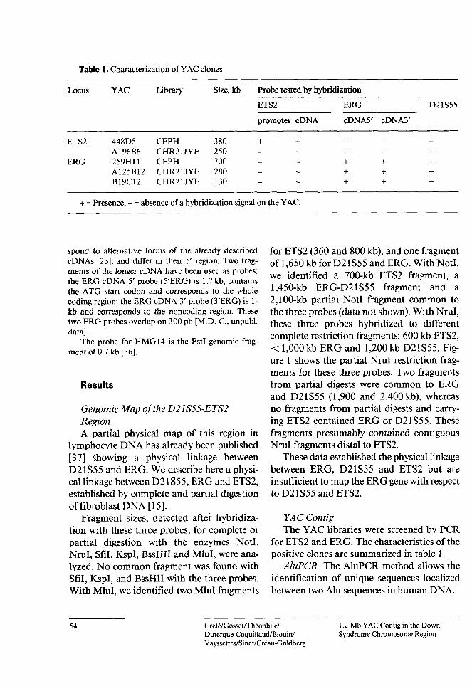
Table 1. Characterization of YAC clones
Size, kb Probe tested by hybridizationLibraryYACLocus
ETS2 ERG D21S55
cDNA5' cDNA3'promoter cDNA
380CEPH CHR21JYE 250 CEPH CHR21JYE 280 CHR21JYE 130
ETS2 448D5A196B6259H11A125B12B19C12
+++
700ERG + ++ ++ +
+ = Presence, - = absence of a hybridization signal on the YAC.
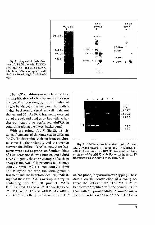
for ETS2 (360 and 800 kb), and one fragment of 1,650 kb for D21S55 and ERG. With Noti, we identified a 700-kb ETS2 fragment, a 1,450-kb ERG-D21S55 fragment and a 2,100-kb partial Noti fragment common to the three probes (data not shown). With Nrul, these three probes hybridized to different complete restriction fragments: 600 kb ETS2, < 1,000 kb ERG and 1,200 kb D21S55. Figure 1 shows the partial Nrul restriction fragments for these three probes. Two fragments from partial digests were common to ERG and D21S55 (1,900 and 2,400 kb), whereas no fragments from partial digests and carrying ETS2 contained ERG or D21S55. These fragments presumably contained contiguous Nrul fragments distal to ETS2.
These data established the physical linkage between ERG, D21S55 and ETS2 but are insufficient to map the ERG gene with respect to D21S55 and ETS2.
spond to alternative forms of the already described cDNAs [23], and differ in their 5' region. Two fragments of the longer cDNA have been used as probes: the ERG cDNA 5' probe (5'ERG) is 1.7 kb, contains the ATG start codon and corresponds to the whole coding region; the ERG cDNA 3' probe (3'ERG) is 1- kb and corresponds to the noncoding region. These two ERG probes overlap on 300 pb [M.D.-C., unpubl. data].
The probe for HMG14 is the PstI genomic fragment of 0.7 kb [36],
Results
Genomic Map of the D21S55-ETS2RegionA partial physical map of this region in
lymphocyte DNA has already been published [37] showing a physical linkage between D21S55 and ERG. We describe here a physical linkage between D21S55, ERG and ETS2, established by complete and partial digestion of fibroblast DNA [15].
Fragment sizes, detected after hybridization with these three probes, for complete or partial digestion with the enzymes Noti, Nrul, Sfil, KspI, BssHII and Mlul, were analyzed. No common fragment was found with Sfil, KspI, and BssHII with the three probes. With Mlul, we identified two Mlul fragments
YAC ContigThe YAC libraries were screened by PCR
for ETS2 and ERG. The characteristics of the positive clones are summarized in table 1.
AluPCR. The AluPCR method allows the identification of unique sequences localized between two Alu sequences in human DNA.
Crété/Gosset/Théophile/Duterque-Coquillaud/Blouin/V ayssettes/Sinet/Créau-Goldberg
1,2-Mb Y AC Contig in the DownSyndrome Chromosome Region
54
ERG C Disi A 5
2 1
ETS2 CDNA
2 1D21S55
2 1
WELLS—
#'2900—
2250—2400- .»f ,
1900—
2400—
190 0—
1450—
<1000- t -H1200 —
t<1000Fig. 1. Sequential hybridizations of a PFGEblot with D21S55, ERG cDNA5', and ETS2 cDNA. Fibroblast DNA was digested with Nrul. 1 = 10 mMMg2+; 2 = 0.5 mM Mg2+.
The PCR conditions were determined for the amplification of a few fragments. By varying the Mg2+ concentration, the number of visible bands could be increased but with a higher background signal as well [data not shown, and 37], As PCR fragments were cut out of the gels and used as probes with no further purification, we performed AluPCR in conditions giving the lowest background.
With the primer AluIV (fig. 2), we obtained fragments of the same size in different YACs. To determine their position on chromosome 21, their identity and the overlap between the different YAC clones, these fragments were used as probes on Southern blots of YAC (data not shown), human, and hybrid DNAs. Figure 3 shows an example of such an analysis: the two PCR products nl, namely AluIV: 1 from 259H11 and AluIV: 1 from 448D5 hybridized with the same genomic fragment and are therefore identical, indicating that these two YACs overlap in a region containing this AluPCR product. YACs B19C12,259H11 and A125B12 overlap as do 259H11, A125B12 and 448D5. As 448D5 and A196B6 both hybridize with the ETS2
t 2 3 4 5 6
PB
_2027 _1605 _11 98
n 1 _ 6 76 - 530
Fig. 2. Ethidium-bromide-stained gel of inter- AluIV PCR products. 1 = 259H11; 2 = A125B12; 3 = 448D5; 4 = A196B6; 5 = B19C12; 6 = yeast Saccharomyces cerevisiae AB972; nl indicates the inter-Alu IV fragments used as AluIV: 1 probes (fig. 3, 4).
cDNA probe, they are also overlapping. These data allow the construction of a contig between the ERG and the ETS2 YACs. More bands were amplified with the primer PDJ33 than with the primer AluIV. A similar analysis of the results with the primer PDJ33 con-
55

8 7 56 4 3 2 1 8 7 6
1ir.
W
ALU IV: (259Mil)A LU IV: (44905)
Fig. 3. Sequential hybridizations of EcoRI restricted DNAs. 1 = Human fibroblast; 2 = hybrid Acem; 3= hybrid GA9-3; 4= hybrid 1881c-l 3b; 5= hybrid 9542c-5a; 6= Chinese hamster; 7 = WA17, and 8 = mouse with AluIV: 1 probes from 448D5 and 259H11 identifying the same 15-kb fragment.
Table 2. Isolation and characterization of YAC ends
Hybridization on Southern blot (EcoRI fragments in kb)Primers PCR fragment size, bp
Matrix
hybrid 259H11 A125B12 B19CI2 448D5 A196B6WA17
Vector Alu human
R = 1091 L= 1089 R= 1091 L= 1089 R= 1091 L = 1089 R= 1091 L = 1089 R = 1091 L= 1089
PDJ33259H11 450 5.5 5.5 5.5ND
A125B12 ND 850 7 7 7 7AluIV
B19C12 2,700ND 8AluIV
448D5 2,900 2.4/2.7 2.4/2.7 2.4ND 2.7 2.4PDJ33PDJ33 1,900 2.5A196B6 2.5 2.5ND
ND = Not determined; - = absence of hybridization signal.
firmed the YAC overlaps identified with AluIV (data not shown).
YAC Ends. YAC ends were identified by vector-AluPCR and the results are summarized in table 2. Only 50% of the ends could be isolated by this method. The amplification
products were used as probes on Southern blots to determine their localizations on chromosome 21 and the overlap between YAC clones (table 2). The right arm of 259H11 and that of A196B6 did not hybridize with any other YACs, indicating that these ends map at
Crété/Gosset/Théophile/Duterque-Coquillaud/Blouin/Vayssettes/Sinet/Créau-Goldberg
1,2-Mb YAC Contig in the DownSyndrome Chromosome Region
56
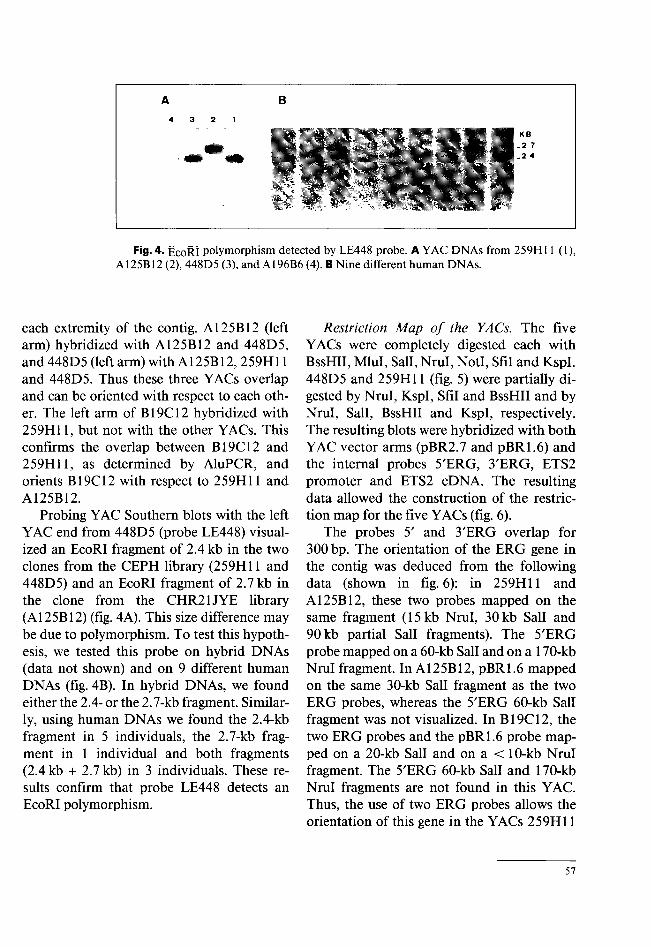
Fig. 4. EcoRI polymorphism detected by LE448 probe. A YAC DNAs from 259H11 (1), A125B12 (2), 448D5 (3), and A196B6 (4). B Nine different human DNAs.
each extremity of the contig. A125B12 (left arm) hybridized with A125B12 and 448D5, and 448D5 (left arm) with A125B12, 259H11 and 448D5. Thus these three YACs overlap and can be oriented with respect to each other. The left arm of B19C12 hybridized with 259H11, but not with the other YACs. This confirms the overlap between B19C12 and 259H11, as determined by AluPCR, and orients B19C12 with respect to 259H11 and A125B12.
Probing YAC Southern blots with the left YAC end from 448D5 (probe LE448) visualized an EcoRI fragment of 2.4 kb in the two clones from the CEPH library (259H11 and 448D5) and an EcoRI fragment of 2.7 kb in the clone from the CHR21JYE library (A125B12) (fig. 4A). This size difference may be due to polymorphism. To test this hypothesis, we tested this probe on hybrid DNAs (data not shown) and on 9 different human DNAs (fig. 4B). In hybrid DNAs, we found either the 2.4- or the 2.7-kb fragment. Similarly, using human DNAs we found the 2.4-kb fragment in 5 individuals, the 2.7-kb fragment in 1 individual and both fragments (2.4 kb + 2.7 kb) in 3 individuals. These results confirm that probe LE448 detects an EcoRI polymorphism.
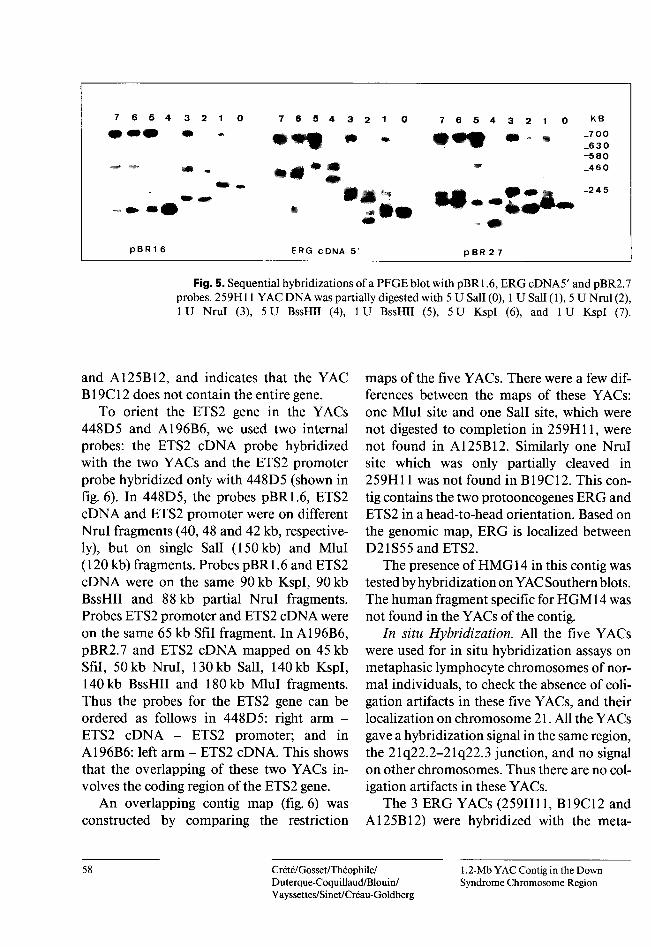
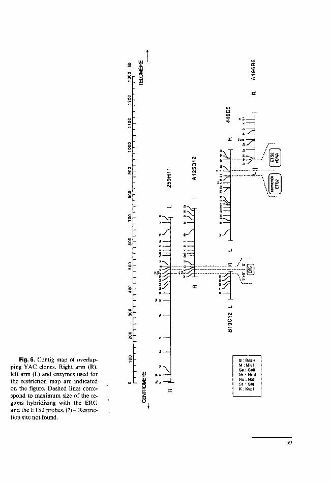
Restriction Map of the YACs. The five YACs were completely digested each with BssHII, Mlul, Sail, Nrul, Noti, Sfil and Kspl. 448D5 and 259H11 (fig. 5) were partially digested by Nrul, Kspl, Sfil and BssHII and by Nrul, Sail, BssHII and Kspl, respectively. The resulting blots were hybridized with both YAC vector arms (pBR2.7 and pBR1.6) and the internal probes 5'ERG, 3'ERG, ETS2 promoter and ETS2 cDNA. The resulting data allowed the construction of the restriction map for the five YACs (fig. 6).
The probes 5' and 3'ERG overlap for 300 bp. The orientation of the ERG gene in the contig was deduced from the following data (shown in fig. 6): in 259H11 and A125B12, these two probes mapped on the same fragment (15 kb Nrul, 30 kb Sail and 90 kb partial Sail fragments). The 5'ERG probe mapped on a 60-kb Sail and on a 170-kb Nrul fragment. In A125B12, pBR1.6 mapped on the same 30-kb Sail fragment as the two ERG probes, whereas the 5'ERG 60-kb Sail fragment was not visualized. In B19C12, the two ERG probes and the pBR1.6 probe mapped on a 20-kb Sail and on a < 10-kb Nrul fragment. The 5'ERG 60-kb Sail and 170-kb Nrul fragments are not found in this YAC. Thus, the use of two ERG probes allows the orientation of this gene in the YACs 259H11
57
7 6 5 4 3 2 1 O 7 6 5 4 3 2 1 0 KB7 6 5 4 3 2 1 0_7 00 _6 3 0 -580 _46 0
• Æk ^-2 4 5
teTO'
p B R 1 6 ERG c DNA 5 p BR 2 7
Fig. 5. Sequential hybridizations of a PFGE blot with pBR 1.6, ERGcDNA5' and pBR2.7 probes. 259H11 YAC DNA was partially digested with 5 U Sali (0), 1 U Sali (1), 5 U Nrul (2), 1 U Nrul (3), 5 U BssHII (4), 1U BssHII (5), 5 U KspI (6), and 1 U KspI (7).
and A125B12, and indicates that the YAC B19C12 does not contain the entire gene.
To orient the ETS2 gene in the YACs 448D5 and A196B6, we used two internal probes: the ETS2 cDNA probe hybridized with the two YACs and the ETS2 promoter probe hybridized only with 448D5 (shown in fig. 6). In 448D5, the probes pBR1.6, ETS2 cDNA and ETS2 promoter were on different Nrul fragments (40, 48 and 42 kb, respectively), but on single Sail (150 kb) and Mlul ( 120 kb) fragments. Probes pBR 1.6 and ETS2 cDNA were on the same 90 kb KspI, 90 kb BssHII and 88 kb partial Nrul fragments. Probes ETS2 promoter and ETS2 cDNA were on the same 65 kb Sfil fragment. In A196B6, pBR2.7 and ETS2 cDNA mapped on 45 kb Sfil, 50 kb Nrul, 130 kb Sail, 140 kb KspI, 140 kb BssHII and 180 kb Mlul fragments. Thus the probes for the ETS2 gene can be ordered as follows in 448D5: right arm - ETS2 cDNA - ETS2 promoter; and in A196B6: left arm - ETS2 cDNA. This shows that the overlapping of these two YACs involves the coding region of the ETS2 gene.
An overlapping contig map (fig. 6) was constructed by comparing the restriction
maps of the five YACs. There were a few differences between the maps of these YACs: one Mlul site and one Sail site, which were not digested to completion in 259H11, were not found in A125B12. Similarly one Nrul site which was only partially cleaved in 259H11 was not found in B19C12. This con- tig contains the two protooncogenes ERG and ETS2 in a head-to-head orientation. Based on the genomic map, ERG is localized between D21S55 and ETS2.
The presence of HMG14 in this contig was tested by hybridization on YAC Southern blots. The human fragment specific for HGM14 was not found in the YACs of the contig.
In situ Hybridization. All the five YACs were used for in situ hybridization assays on metaphasic lymphocyte chromosomes of normal individuals, to check the absence of coligation artifacts in these five YACs, and their localization on chromosome 21. All the YACs gave a hybridization signal in the same region, the 21q22.2-21q22.3 junction, and no signal on other chromosomes. Thus there are no coligation artifacts in these YACs.
The 3 ERG YACs (259H11, B19C12 and A125B12) were hybridized with the meta-
58 Crété/Gosset/Théophile/Duterque-Coquillaud/Blouin/V ay ssettes/Sinet/Créau-Goldberg
1.2-Mb YAC Contig in the DownSyndrome Chromosome Region
CDWja mCD
oo _ <CO - ßj
DCoo _CU
mQ00̂ 3 tö ---------Oo .
CE zxaO 3oo
/ 8CM.......z UjOCD
____o wo CM05
X tr< g «<J>in COzacCM
\T©O “à3 i----
«___« —o "A3- <Dohs z — 3
oo<£3 » ——
*1 _— 3--- iSS' 3- __ OC ./ iooo -:;gin
z3* •— ä«ac iniz \ +3$
CCoo3
oo CM«0O
CQooCU <B ' '
2 —■—o
Fig. 6. Contig map of overlapping YAC clones. Right arm (R), left arm (L) and enzymes used for the restriction map are indicated on the figure. Dashed lines correspond to maximum size of the regions hybridizing with the ERG and the ETS2 probes. (?) = Restriction site not found.
O B : BssHII M : Miul Sa: Sail Nr * Nrul No : Noti Sf : SM K : Kspl
"Xe sc ——
<3 2oX
t
59
<%
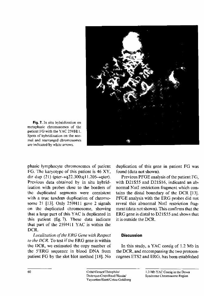
Fig. 7. In situ hybridization on metaphasic chromosomes of the patient FG with the YAC 259H11. Spots of hybridization on the normal and rearranged chromosomes are indicated by white arrows.
phasic lymphocyte chromosomes of patient FG. The karyotype of this patient is 46 XY, dir dup (21) (pter->q22.300:ql 1.205-»qter). Previous data obtained by in situ hybridization with probes close to the borders of the duplicated segments were consistent with a true tandem duplication of chromosome 21 [13]. Only 259H11 gave 2 signals on the duplicated chromosome, showing that a large part of this YAC is duplicated in this patient (fig. 7). These data indicate that part of the 259H11 YAC is within the DCR.
duplication of this gene in patient FG was found (data not shown).
Previous PFGE analysis of the patient FG, with D21S55 and D21S16, indicated an abnormal Noti restriction fragment which contains the distal boundary of the DCR [13]. PFGE analysis with the ERG probes did not reveal this abnormal Noti restriction fragment (data not shown). This confirms that the ERG gene is distal to D21S55 and shows that it is outside the DCR.
Localization of the ERG Gene with Respect to the DCR. To test if the ERG gene is within the DCR, we estimated the copy number of the 5'ERG sequence in blood DNA from patient FG by the slot blot method [18]. No
Discussion
In this study, a YAC contig of 1.2 Mb in the DCR, and encompassing the two protooncogenes ETS2 and ERG, has been established
60 Crété/Gosset/Théophile/Duterque-Coquillaud/Blouin/Vayssettes/Sinet/Créau-Goldberg
1.2-Mb YAC Contig in the DownSyndrome Chromosome Region
by studying five YACs positive with either ERG or ETS2. The overlaps between these YACs were first determined by AluPCR. The extent of the overlapping regions were assessed by AluPCR, ETS2 and ERG PCR and by restriction mapping. 50% of the YAC ends were isolated by vector AluPCR (table 2) and localized on chromosome 21, especially the ends of the contig (259H11 right arm and A196B6 right arm). In two cases, B19C12 right arm and A196B6 left arm, the extremities hybridized with ERG or ETS2 cDNAs respectively, indicating their localization on chromosome 21. YAC ends for 259H11 left arm, A125B12 right arm, and 448D5 right arm were not isolated, but showed identical restriction maps with at least one other YAC. For these three ends, a minor rearrangement cannot be excluded. The AluPCR fragments isolated from these YACs allowed the identification of new potentially polymorphic markers on chromosome 21 : the left YAC end of 448D5 (probe LE448) detects an EcoRI polymorphism. These new markers therefore constitute powerful tools for the systematic physical and genetic mapping of chromosome 21 and for the analysis of partial trisomy 21.
Previous PFGE mapping data reported a restriction fragment common to D21S55 and ERG [36]. Our genomic map shows a physical linkage between D21S55, ERG and ETS2, but whether ERG is distal or proximal to D21S 5 5 cannot be determined. Comparison of the genomic and YAC maps indicates that D21S55 is more proximal than ERG to ETS2. This was confirmed by the analysis of patient FG. The distance between ERG and ETS2 is about 400 kb. The genomic Noti map indicates that the distance between D21S55, which is not present in this contig, and ERG is about 1,000 kb. Two other sequences, HMG14 and D21S3, previously mapped in this region of the chromosome [17], can be excluded from the contig: HMG14 did not
hybridize on YAC Southern blots, and D21 S3 PCR screening [CHR21JYE and I. Chumakov, personal commun., for the CEPH library] was found negative with these YACs.
There are more rare cutter restriction sites in YAC DNA than appear on the genome map. This is likely due to differential DNA méthylation since DNA cloned in YACs does not undergo cytosine méthylation [38], Only Noti digestion did not reveal any additional sites. It has been suggested that multiple sites containing CpG identify HTF islands (Hpall tiny fragment islands) associated with many mammalian genes [39]. At least three putative islands (BssHII + KspI) were identified in the contig: one is located in the ETS2 promoter region [21], which is also identified on the genomic map [40]; two others, one distal to ETS2 and one proximal to ERG at the extremity of the contig (right end of 259H11) may be landmarks of unknown genes.
ERG (Ets-related gene) and ETS2 are two related protooncogenes that are 400 kb apart and in opposite orientations on the chromosome. They encode proteins which are specific DNA-binding molecules acting as sequence-specific transcriptional activators [41-43 and M.D.C., unpubl. data]. ETS2 is expressed in various adult cells, particularly those with dividing potentiality [44, 45], and the presence of a HTF island in its promoter region is consistent with the characteristics of an ubiquitous gene. Conversely, no HTF was found in the 5' region of the ERG gene, the expression of which was not observed in a variety of normal tissues [46], except in transformed cells [23], Thus, ERG might be expressed in a cell- or tissue-specific way during embryogenesis and play a role in development.
As tissue-specific genes lacking HTF islands have been described [39], the search for genes in this region will be carried out after subcloning in phages or cosmids by several
61
alternative approaches: conservation of sequences by hybridization on Zooblots indicating putative exonic sequences, exon trapping, and screening of cDNA libraries. Moreover, chromosome walking from the extremity of 259H11 is in progress to extend the con- tig towards D21S55, and to identify the gene content of the DCR.
J.M. Delabar for helpful discussion, to D. Le Paslier and D. Cohen for providing the YACs of the CEPH library, to I. Chumakov for providing D21 S3 screening data of the CEPH library, to D. Patterson for providing the YACs of the Chromosome 21 Joint YAC Screening Effort and the hybrids ACEM, GA9-3, 188 lc-13b and 9542-5c, to P. Watkins for the probe D21S55, to D. Stéhelin forthe ETS2 cDNA, to M. Bus- tin for the HMG14 probe and to M. Poissonnier who referred patient FG.
This work was financially supported by Centre National de la Recherche Scientifique, Institut National de la Santé et de la Recherche Médicale, Ministère de la Recherche et de la Technologie, Faculté Necker, Université Paris V, N.C. has been supported by the Association Française contre les Myopathies, and Association de Recherche sur le Cancer.
AcknowledgmentsWe are grateful to Françoise Le Dez for her contri
bution to this work, to Z. Chettouh for technical assistance, to M.L. Yaspo for sharing unpublished data, to
References1 Lejeune J, Gautier M, Turpin R:
Etudes chromosomiques somatiques de neuf enfants mongoliens. C R Hebd Séances Acad Sci 1959;248: 409-411.
2 Aula P, Leisti J, Von Koskull H: Partial trisomy 21. Clin Genet 1973; 4:241-251.
3 Poissonnier M, Saint-Paul B, Dutril- laux B, Chassaigne M, Gruyer P, de Blignières-Strouk G: Trisomie 21 partielle (21q21->21q22.2). Ann Génét (Paris) 1976;19:69-73.
4 Mattéi JF, Mattéi MG, Beateman MA, Giraud F: Trisomy 21 for the region 21q22.3: Identification by high resolution R banding patterns. Hum Genet 1981;56:409-411.
5 Rahmani Z, Blouin JL, Créau-Gold- berg N, Watkins PC, Mattei JF, Poissonnier M, Prieur M, Chettouh, Z, Nicole A, Aurias A, Sinet PM, Delabar JM: Critical role of the D21S55 region on chromosome 21 in the pathogenesis of Down syndrome. Proc Natl Acad Sci USA 1989;86:5958-5962.
6 Rahmani Z, Blouin JL, Créau-Gold- berg N, Watkins PC, Mattéi JF, Poissonnier M, Prieur M, Chettouh Z, Nicole A, Aurias A, Sinet PM, Delabar JM: Down syndrome critical region around D21S55 on proximal 21q22.3. Am J Med Genet 1990;suppl 7:98-103.
Carritt B, Litt M: Report of the committee on the genetic constitution of chromosomes 20 and 21. Cytogenet Cell Genet 1989;51:358-371.Sinet PM, Rahmani Z, Théophile D, Chettouh Z, Blouin JL, Prieur M, Noel B, Pangalos C, Mattei JF, Delabar JM: Molecular definition of 7 minimal regions for 23 features of Down syndrome on chromosome 21. Am J Hum Genet 1991;suppl 49:A1528.Korenberg JR, Bradley C, Disteche CM: Down syndrome: molecular mapping of the congenital heart disease and duodenal stenosis. Am J Hum Genet 1992;50:294-302. McCormick MK, Schinzel A, Petersen MB, Stetten G, Driscoll DJ, Cantu ES, Tranebjaerg L, Mikkelsen M, Watkins PC, Antonorakis SE: Molecular genetic approach to the characterization of the ‘Down syndrome Region’ of Chromosome 21. Genomics 1989;5:325-331. Burmeister M, Suwon K, Roydon Price E, de Lange T, Tantravahi U, Myers RM, Cox DR: A map of the distal region of the long arm of human chromosome 21 constructed by radiation hybrid mapping and pulsed-field gel electrophoresis. Genomics 1991;9:19-30.
7 12 Burke DT, Carle GF, Olson MV: Cloning of large segments of exogenous DNA into yeast by means of artificial chromosome vectors. Science 1987;236:806-812.
13 Blouin JL, Aurias A, Créau-Gold- berg N, Apiou F, Alcai'de-Loridan C, Bruel A, Prieur M, Kraus J, Delabar JM, Sinet PM: Cytogenetic and molecular analysis of a de novo tandem duplication of chromosome 21. Hum Genet 1991;88:167-174.
14 Gardiner K, Laas W, Patterson D: Fractionation of large mammalian DNA restriction fragments using vertical pulsed gradient gel electrophoresis. Somat Cell Mol Genet 1986;12:185-195.
15 Crété N, Delabar JM, Sinet PM, Créau-Goldberg N: Accurate evaluation of the sizes of DNA fragments (from 30 to 4700 kb) in pulsed field gel electrophoresis. Biotechniques 1991;11:711-717.
16 Kozak CA, Lawrence JB, Ruddle FH: A sequential staining technique for the chromosomal analysis of interspecific mouse/hamster and mouse/human somatic cell hybrids. Exp Cell Res 1977;105:109-117.
17 Cox DR, Shimizu N: Report of the committee on the genetic constitution of chromosome 21. Cytogenet Cell Genet 1991;55:235-244.
9
10
11
Crété/Gosset/Théophile/Duterque-Coquillaud/Blouin/Vayssettes/Sinet/Créau-Goldberg
1.2-Mb YAC Contig in the DownSyndrome Chromosome Region
62
28 Breukel C, Wijnen J, Tops C, Van der Klift H, Dauwerse H, Khan PM: Vector-Alu PCR: A rapid step in mapping cosmids and YACs. Nucleic Acids Res 1991 ; 18:3097.
29 Feinberg AP, Volgelstein B: A technique for radiolabeling DNA restriction endonuclease fragments to high specific activity. Anal Biochem 1983;132:6-13; Addendum: Anal Biochem 1984;137:266-267.
30 Nelson DL, Ledbetter SA, Corbo L, Victoria MF, Ramirez-Solis R, Webster TD, Ledbetter DH, Caskey CT: Alu polymerase chain reaction: a method for rapid isolation of human specific sequences from complex DNA sources. Proc Natl Acad Sci USA 1989;86:6686-6690.
31 Anand R, Riley JH, Butler R, Smith JC, Markham AF: A 3.5 genome equivalent multi-access YAC library: Construction, characterisation, screening and storage. Nucleic Acids Res 1990;18:1951-1956.
32 Viegas-Péquignot E, Dutrillaux B: Une méthode simple pour obtenir des prophases et des prométaphases. Ann Génét (Paris) 1978;21:122- 124.
33 Zhang FR, Heilig R, Thomas G, Au- rias A: A one-step efficient and specific non-radioactive non-fluorescent method for in situ hybridization of banded chromosomes. Chro- mosoma 1990;99:436-439.
34 Watkins PC, Watkins PA, Hoffman N, Stanislovitis P: Isolation of single-copy probes detecting DNA polymorphism from a cosmid library of chromosome 21. Cytogenet Cell Genet 1985;40:773-774.
35 Boulukos KE, Pognonec P, Begue A, Galibert F, Gesquières JC, Stehelin D, Ghysdael J: Identification in chickens of an evolutionarily conserved cellular ets-2 gene (c-ets-2) encoding molecular proteins related to the products of the c-ets protooncogene. EMBO J 1988;7:697- 705.
36 Landsman D, McBride OW, Soares N, Crippa MP, Srikantha' T, Bustin M: Chromosomal protein HMG14: Identification, characterization, and chromosome localisation of a functional gene from the large human multigene family. J Biol Chem 1989;264:3421-3427.
37 Gardiner K, Horisberger M, Kraus J, Tantravahi U, Korenberg J, Rao
V, Reddy S, Patterson D: Analysis of human chromosome 21 : correlation of physical and cytogenetic maps; gene and CpG island distributions. EMBO J 1990;9:25-34.
38 Butler R, Ogilvie DJ, Elvin P, Riley JH, Finniear RS, Slynn G, Morten JEN, Markham AF, Anand R: Walking, cloning, and mapping with Yeast Artificial Chromosomes: A contig encompassing D21S13 and D21S16. Genomics 1992; 12:42-51.
39 Bird AP: CpG-rich islands and the function of DNA méthylation. Nature 1986;321:209-213.
40 Yaspo M-L, Crété N, Chettouh Z, Blouin JL, Rahmani Z, Stehelin D, Sinet P-M, Créau-Goldberg N, De- labar J-M: New chromosomes 21 DNA markers isolated by pulsed field gel electrophoresis from an ETS2 containing Down syndrome chromosomal region. Hum Genet, in press.
41 Bosselut R, Duvall JF, Gégonne A, Bailly M, Hémar A, Brady J, Ghysdael J: The product of the c-ets-1 proto-oncogene and the related Ets- 2 protein act as transcriptional activators of the long terminal repeat of human T cell leukemia virus HTLV- l.EMBOJ 1990;9:3137-3144.
42 Wasylyk B, Wasylyk C, Flores P, Begue A, Leprince D, Stéhelin D: The c-ets proto-oncogenes encode transcription factors that cooperate with c-Fos and c-Jun for transcriptional activation. Nature 1990;346: 191-193.
43 Reddy ESP, Rao VN: Erg, an ets- related gene, codes for sequence- specific transcriptional activators. Oncogene 1991;6:2285-2289.
44 Bhat NK, Fisher RJ, Fujiwara S, As- cione R, Papas TS: Temporal and tissue-specific expression of mouse ets genes. Proc Natl Acad Sci USA 1987;84:3161-3165.
45 Amouyel Ph, Gégonne A, Dela- courte A, Défossez A, Stéhelin D: Expression of ETS protooncogenes in astrocytes in human cortex. Brain Res 1988;447:149-153.
46 SacchiN, Cheng SV, Tanzi RE, Gu- sella JF, Drabkin HA, Patterson, Haines JH, Papas TS: The ETS genes on chromosome 21 are distal to the breakpoint of the acute myelogenous leukemia translocation (8;21). Genomics 1988;3:110-116.
18 Blouin JL, Rahmani Z, Chettouh Z, Prieur M, Fermanian J, Poissonnier M, Leonard C, Nicole A, Mattéi JF, Sinet PM, Delabar JM: Slot blot method for the quantification of DNA sequences and mapping of chromosome rearrangements: Application to chromosome 21. Am J Hum Genet 1990;46:518-526.
19 Albertsen HM, Abderrahim H, Cann HM, Dausset J, Le Paslier D, Cohen D: Construction and characterization of a yeast artificial chromosome library containing seven haploid human genome equivalents. Proc Natl Acad Sci USA 1990;87: 4256-4260.
20 Browstein BH, Silverman GA, Little RD, Bluke DT, Korsmeyer SJ, Schlessinger D, Olson MV: Isolation of single-copy human genes from a library of yeast artificial chromosome clones. Science 1989;244: 1348-1351.
21 Mavrothalassitis GJ, Watson DK, Papas TS: Molecular and functional characterization of the promoter of ETS2, the human c-ets-2 gene. Proc Natl Acad Sci USA 1990;87:1047- 1051.
22 Patterson D: Report of the Second International Workshop on Human chromosome 21. Cytogenet Cell Genet 1991;57:167-174.
23 Rao VN, Papas TS, Reddy SP: ERG, a human ETS-related gene on the chromosome 21: alternative splicing, polyadenylation, and translation. Science 1987;237:635-639.
24 Sherman F, Fink GR, Hicks JB: Method in yeast genetics. Cold Spring Harbor Lab, Cold Spring Harbor NY 1986.
25 Carle GF, Olson MV: An electrophoretic karyotype for yeast. Proc Natl Acad Sci USA 1985;82:3756- 3760.
26 Riley J, Butler R, Ogilvie D, Finniear R, Jenner D, Powell S, AnandR, Smith JC, Markham AF: A novel, rapid method for the isolation of terminal sequences from yeast artificial chromosomes (YAC) clones. Nucleic Acids Res 1990; 18:2887- 2890.
27 Cotter FE, Hampton GM, NasipuriS, Bodmer WF, Young BD: Rapid isolation of human chromosome- specific DNA probes from a somatic cell hybrid. Genomics 1990;7:257- 263.
63