Epigallocatechin-3-gallate, a DYRK1A inhibitor, rescues cognitive deficits in Down syndrome mouse...
11
Mol. Nutr. Food Res. 2013, 00, 1–11 1 DOI 10.1002/mnfr.201300325 RESEARCH ARTICLE Epigallocatechin-3-gallate, a DYRK1A inhibitor, rescues cognitive deficits in Down syndrome mouse models and in humans Rafael De la Torre 1,2,3 , Susana De Sola 1 , Meritxell Pons 3,4 , Arnaud Duchon 5 , Mar´ ıa Mart´ ınez de Lagran 3,4 , Mag´ ı Farr ´ e 1,6 , Montserrat Fit ´ o 2 , Bessy Benejam 7 , Klaus Langohr 1,8 , Joan Rodriguez 1 , Mitona Pujadas 1,2 , Jean Charles Bizot 9 , A´ ıda Cuenca 1,10 , Nathalie Janel 11 , Silvina Catuara 4 , Maria Isabel Covas 2 , Henri Blehaut 12 , Yann Herault 5 , Jean Marie Delabar 10 and Mara Dierssen 3,4 1 Human Pharmacology and Clinical Neurosciences Research Group-Neurosciences Program, IMIM-Hospital del Mar Research Institute, Barcelona, Spain 2 Cardiovascular Risk and Nutrition Research Group-Inflammatory and Cardiovascular Disorders Program, IMIM-Hospital del Mar Research Institute, and CIBER of Physiopathology of Obesity and Nutrition (CIBEROBN), Barcelona, Spain 3 University Pompeu Fabra, CEXS-UPF, Barcelona, Spain 4 Center for Genomic Regulation (CRG), and CIBER de Enfermedades Raras (CIBERER), Barcelona, Spain 5 Institut de G ´ en ´ etique et de Biologie Mol ´ eculaire et Cellulaire, Translational Medicine and Neuroscience Program, IGBMC, CNRS, INSERM, Universit ´ e de Strasbourg, UMR7104, UMR964, and Institut Clinique de la Souris, ICS, GIE CERBM, Illkirch, France 6 Universitat Aut ` onoma de Barcelona (UDIMAS-UAB), Barcelona, Spain 7 Fundaci ´ o Catalana S´ ındrome de Down, Barcelona, Spain 8 Department of Statistics and Operations Research, Universitat Polit` ecnica de Catalunya, Barcelona, Spain 9 Key-Obs S.A., All ´ ee du Titane, Orl ´ eans, France 10 Epidemiology of Drugs of Abuse Research Group-Epidemiology and Public Health Program, IMIM-Hospital del Mar Research Institute, Barcelona, Spain 11 Adaptive Functional Biology, EAC CNRS, University Paris Diderot, Sorbonne Paris Cit´ e, Paris, France 12 J´ er ˆ ome Lejeune Foundation, Paris, France Scope: Trisomy for human chromosome 21 results in Down syndrome (DS), which is among the most complex genetic perturbations leading to intellectual disability. Accumulating data suggest that overexpression of the dual-specificity tyrosine-(Y)-phosphorylation regulated kinase 1A (DYRK1A), is a critical pathogenic mechanisms in the intellectual deficit. Methods and results: Here we show that the green tea flavonol epigallocatechin-gallate (EGCG), a DYRK1A inhibitor, rescues the cognitive deficits of both segmental trisomy 16 (Ts65Dn) and transgenic mice overexpressing Dyrk1A in a trisomic or disomic genetic background, respectively. It also significantly reverses cognitive deficits in a pilot study in DS individuals with effects on memory recognition, working memory and quality of life. We used the mouse models to ensure that EGCG was able to reduce DYRK1A kinase activity in the hippocampus and found that it also induced significant changes in plasma homocysteine levels, which were Received: May 3, 2013 Revised: July 4, 2013 Accepted: July 7, 2013 Correspondence: Dr. Mara Dierssen, Systems Biology Program, Genomic Regulation Center (CRG), and CIBER de Enfermedades Raras (CIBERER), Dr Aiguader 88, 08003 Barcelona, Spain E-mail: [email protected] Fax: +34-933160099 Abbreviations: DS, Down syndrome; DYRK1A, dual-specificity tyrosine-(Y)-phosphorylation regulated kinase 1A; EGCG, epigal- locatechin gallate; GSH-Px, glutathione peroxidase; Hcy, Homo- cysteine; HSA21, human chromosome 21; IQ, Intellectual quo- tient; oxLDL, Oxidized LDL; TgDyrk1A, Transgenic mice overex- pressing Dyrk1A C 2013 WILEY-VCH Verlag GmbH & Co. KGaA, Weinheim www.mnf-journal.com
Transcript of Epigallocatechin-3-gallate, a DYRK1A inhibitor, rescues cognitive deficits in Down syndrome mouse...
Mol. Nutr. Food Res. 2013, 00, 1–11 1DOI 10.1002/mnfr.201300325
RESEARCH ARTICLE
Epigallocatechin-3-gallate, a DYRK1A inhibitor, rescues
cognitive deficits in Down syndrome mouse models
and in humans
Rafael De la Torre1,2,3, Susana De Sola1, Meritxell Pons3,4, Arnaud Duchon5,Marıa Martınez de Lagran3,4, Magı Farre1,6, Montserrat Fito2, Bessy Benejam7,Klaus Langohr1,8, Joan Rodriguez1, Mitona Pujadas1,2, Jean Charles Bizot9, Aıda Cuenca1,10,Nathalie Janel11, Silvina Catuara4, Maria Isabel Covas2, Henri Blehaut12, Yann Herault5,Jean Marie Delabar10 and Mara Dierssen3,4
1 Human Pharmacology and Clinical Neurosciences Research Group-Neurosciences Program, IMIM-Hospital delMar Research Institute, Barcelona, Spain
2 Cardiovascular Risk and Nutrition Research Group-Inflammatory and Cardiovascular Disorders Program,IMIM-Hospital del Mar Research Institute, and CIBER of Physiopathology of Obesity and Nutrition (CIBEROBN),Barcelona, Spain
3 University Pompeu Fabra, CEXS-UPF, Barcelona, Spain4 Center for Genomic Regulation (CRG), and CIBER de Enfermedades Raras (CIBERER), Barcelona, Spain5 Institut de Genetique et de Biologie Moleculaire et Cellulaire, Translational Medicine and Neuroscience Program,IGBMC, CNRS, INSERM, Universite de Strasbourg, UMR7104, UMR964, and Institut Clinique de la Souris, ICS, GIECERBM, Illkirch, France
6 Universitat Autonoma de Barcelona (UDIMAS-UAB), Barcelona, Spain7 Fundacio Catalana Sındrome de Down, Barcelona, Spain8 Department of Statistics and Operations Research, Universitat Politecnica de Catalunya, Barcelona, Spain9 Key-Obs S.A., Allee du Titane, Orleans, France10 Epidemiology of Drugs of Abuse Research Group-Epidemiology and Public Health Program, IMIM-Hospital del
Mar Research Institute, Barcelona, Spain11 Adaptive Functional Biology, EAC CNRS, University Paris Diderot, Sorbonne Paris Cite, Paris, France12 Jerome Lejeune Foundation, Paris, France
Scope: Trisomy for human chromosome 21 results in Down syndrome (DS), which isamong the most complex genetic perturbations leading to intellectual disability. Accumulatingdata suggest that overexpression of the dual-specificity tyrosine-(Y)-phosphorylation regulatedkinase 1A (DYRK1A), is a critical pathogenic mechanisms in the intellectual deficit.Methods and results: Here we show that the green tea flavonol epigallocatechin-gallate (EGCG),a DYRK1A inhibitor, rescues the cognitive deficits of both segmental trisomy 16 (Ts65Dn)and transgenic mice overexpressing Dyrk1A in a trisomic or disomic genetic background,respectively. It also significantly reverses cognitive deficits in a pilot study in DS individualswith effects on memory recognition, working memory and quality of life. We used the mousemodels to ensure that EGCG was able to reduce DYRK1A kinase activity in the hippocampusand found that it also induced significant changes in plasma homocysteine levels, which were
Received: May 3, 2013Revised: July 4, 2013
Accepted: July 7, 2013
Correspondence: Dr. Mara Dierssen, Systems Biology Program,Genomic Regulation Center (CRG), and CIBER de EnfermedadesRaras (CIBERER), Dr Aiguader 88, 08003 Barcelona, SpainE-mail: [email protected]: +34-933160099
Abbreviations: DS, Down syndrome; DYRK1A, dual-specificitytyrosine-(Y)-phosphorylation regulated kinase 1A; EGCG, epigal-
locatechin gallate; GSH-Px, glutathione peroxidase; Hcy, Homo-cysteine; HSA21, human chromosome 21; IQ, Intellectual quo-tient; oxLDL, Oxidized LDL; TgDyrk1A, Transgenic mice overex-pressing Dyrk1A
C© 2013 WILEY-VCH Verlag GmbH & Co. KGaA, Weinheim www.mnf-journal.com

2 R. De la Torre et al. Mol. Nutr. Food Res. 2013, 00, 1–11
correlated with Dyrk1A expression levels. Thus, we could use plasma homocysteine levels asan efficacy biomarker in our human study.Conclusion: We conclude that EGCG is a promising therapeutic tool for cognitive enhancementin DS, and its efficacy may depend of Dyrk1A inhibition.
Keywords:
Cognition / Down syndrome / DYRK1A / Epigallocatechin gallate / Homocysteine
� Additional supporting information may be found in the online version of this article atthe publisher’s web-site
1 Introduction
Health benefits of the green tea catechins on the preventionof cardiovascular diseases [1], cancer chemoprevention [2],infective [3], and as neuroprotectors in neurodegenerativedisease [4] have been postulated. Nevertheless, their in vivoeffectiveness and molecular mechanisms are difficult to eluci-date and remain a challenging task. Nevertheless, the safety ofgreen tea extracts and of its main active cathechin, the flavonolepigallocatechin gallate (EGCG) should easy the translationfrom in vitro and animal models to human clinical trials. Re-cently, the effectiveness of EGCG on the promotion of adulthippocampal neurogenesis has been reported [5] and invitesits application in clinical neurodevelopmental and neurode-generative disorders. In this context a previous report alsosuggested the beneficial effects of EGCG in animal modelsof Down syndrome (DS) [6].
Trisomy for human chromosome 21 (HSA21) results inDS (OMIM 190685) and is one of the most common hu-man chromosomal disorders. While the trisomy affects everytissue, in DS reduced cognitive ability is among the most lim-iting features [7,8]. Even though there is certainly a contribu-tion of noncoding regions to the phenotype, it is likely thatthe dosage imbalance of some individual genes on HSA21directly contributes to some phenotypes. Thus, normalizingexpression levels or the function of critical genes could pre-vent or reverse the deleterious effects of gene overdose. Dual-specificity tyrosine-(Y)-phosphorylation regulated kinase 1A(Dyrk1A) is a plausible candidate gene to explain some DSphenotypes [reviewed in [9, 10]]. It is localized in the DS crit-ical region of the HSA21 [11, 12] and encodes for a dualkinase phosphorylating both threonine/serine and tyrosineresidues. In mice dosage imbalance of Dyrk1A in vivo affectsbrain structure and learning and memory [13, 14]. ReducedDyrk1A expression in heterozygous mice produces micro-cephaly [15], and compromises neuritogenesis [16] leading toreduced length and complexity of dendritic branches. Het-erozygous mice also show visuospatial learning and memorydeficits. Similarly, Dyrk1A overexpression in transgenic mice(TgDyrk1A) leads to cognitive impairment and less complexdendritic trees [17, 18]. Intriguingly, cognition impairmentand dendritic tree alteration in TgDyrk1A recapitulates thatof Ts65Dn mice, a DS mouse model bearing in trisomy 88 of161 classical protein coding genes present on HSA21, [19–21].
This suggests Dyrk1A as the underlying cause and points itsnormalization as potential DS therapy.
EGCG is a potent and selective inhibitor of DYRK1A ac-tivity [22]. Previous work showed that prenatal EGCG treat-ment could partially rescue brain alterations in transgenicpups overexpressing Dyrk1A and was able to normalize thelevels of some synaptic plasticity related proteins in the hip-pocampus of adult Dyrk1A transgenic mice suggesting pos-sible cognitive effects [6]. We here have explored for the firsttime if EGCG could rescue the cognitive alteration in adultDS mice and in a pilot study in DS subjects. We have usedtrisomic and transgenic mice to determine the possible ef-fects of short-term EGCG treatment in improving cognitivefunction, in particular on memory deficits in adults and toidentify biomarkers of EGCG efficacy to normalize Dyrk1Aactivity that could then be used in patients. Then, we havecarried out a randomized, double-blind, placebo-controlledpilot study evaluating the safety of EGCG and preliminarilyits clinical effects in young adults with DS. We conclude thatEGCG rescues cognitive deficits in trisomic (Ts65Dn) miceand DS individuals possibly through normalizing DYRK1Aactivity.
2 Materials and methods
2.1 Preclinical pharmacological studies
2.1.1 Mouse models
TgDyrk1A were obtained as previously described [13]. Fe-male B6EiC3Sn a/A-Ts (1716) 65Dn (Ts65Dn) mice were ob-tained from Jackson Laboratories (Bar Harbor, ME) and bredwith euploid B6C3BF1 males [23]. Young adult (3 monthsof age) male mice were randomly distributed in four groups(TgDyrk1A or Ts65Dn and their corresponding wild-type lit-termates, on placebo or EGCG). Of interest for the presentwork, Dyrk1A over dosage in TgDyrk1A yielded similar lev-els of overexpression than those detected in both the foetalDS brains and the partial trisomic Ts65Dn model [24]. Weused the following transgenic (TgDyrk1A) groups: WT non-treated n = 13; TG (TgDyrk1A)-nontreated n = 16; WT-EGCGn = 16; TG-EGCG n = 14. For the trisomic (Ts65Dn) we used:WT-nontreated n = 19; Ts65Dn nontreated n = 14; WT-EGCGn = 22; Ts65Dn-EGCG n = 14.
C© 2013 WILEY-VCH Verlag GmbH & Co. KGaA, Weinheim www.mnf-journal.com

Mol. Nutr. Food Res. 2013, 00, 1–11 3
2.1.2 Ethical statement
Animals were bred and housed under SPF standard envi-ronmental conditions, and all experimental procedures wereperformed in compliance with animal welfare policies andapproved by the local ethical committee (Comite Etico de Ex-perimentacion Animal del PRBB (CEEA-PRBB); procedurenumbers MDS-08–1060P1 and JMC-07–1001P1-MDS). Allmet local (Spanish law 5/1995 and decrees 214/97, 32/2007;French Ministry of Agriculture law 87/848) and European(EU directives 86/609 and 2001–486) regulations and theStandards for Use of Laboratory Animals n� A5388–01 (NIH).The CRG is authorized to work with genetically modifiedorganisms (A/ES/05/I-13 and A/ES/05/14). The researchpersonnel involved in the experiments were granted theofficial accreditation to perform the reported experiments.
2.1.3 EGCG administration
Mice were administered EGCG in drinking water for1 month. EGCG solution was prepared freshly from a greentea extract [Mega Green Tea Extract, Lightly Caffeinated R©
(0.8% caffeine) Life Extension R©, USA; EGCG content of326.25 mg per capsule] every 3 days (EGCG concentration:90 mg/mL for a dose of 2–3 mg per day). To test EGCG cog-nitive effects, TgDyrk1A or Ts65Dn and their correspondingwild-type littermates, on placebo or EGCG, were tested in ahippocampal-dependent learning using a water maze (WM;see [13, 25]) and a novel object recognition task.
2.1.4 Learning and memory tests
2.1.4.1 Water mazeTo test the effect of EGCG on hippocampal-dependent spa-tial cognition, mice were trained in a water maze (WM) afterbeing administered EGCG in drinking water during 30 days.TgDyrk1A or Ts65Dn and their corresponding wild-type lit-termates, on placebo or EGCG were tested. The water mazeconsisted of a circular pool filled with tepid water (19�C) opaci-fied by addition of white nontoxic paint. A white escape plat-form was located 1 cm below the water surface in a fixedposition. White curtains with affixed black patterns sur-rounded the maze to provide an arrangement of spatial cues.Mice learned the position of the hidden platform in four train-ing (acquisition) trials per day during 5 or 6 days, dependingon the strain [see [9]], until the animals reached the asymp-totic performance levels (best execution level). In each trial,mice were placed at one of the starting locations in randomorder and were allowed to swim until they located the plat-form. Mice failing to find the platform within 60 s were placedon it for 20 s and were returned to their home cage at the endof every trial. To assess the reference memory a probe ses-sion was performed 24 h after last acquisition session. In thissession, the platform was removed and mice were allowed to
swim for 60 s. All the trials were recorded and traced withan image tracking system (SMART, Panlab, Spain). Escapelatencies, swimming speed, percentage of time spent in eachquadrant of the pool were monitored and computed. Refer-ence memory was quantified in the probe session comparingthe amount of time mice spent in the target quadrant versusthe average of the three other quadrants of the pool.
2.1.4.2 Novel object recognitionMice were tested for learning and memory deficits in thenovel object recognition task [25]. This task is based on theinnate tendency of rodents to differentially explore novel ob-jects over familiar ones. TgDyrk1A or Ts65Dn and their cor-responding wild-type littermates, on placebo or EGCG, weregiven a habituation session in a circular arena for 30 min on asingle day. In the training session 24 h later, mice were placedin the centre of the arena and allowed to explore two iden-tical objects during 10 min. Following the training period,the rodent was removed from the environment for a delayperiod which ranged from 1 to 24 h, depending on the typeof memory being tested in each strain. After the delay, therodent is returned to the bin, where a new one replaced oneof the original objects. For each mouse, the objects were ran-domly assigned as either familiar or novel. Also the locationof the novel objects (left or right) is counterbalanced betweengroups. The exploration time for the familiar (TF) and thenew object (TN) during the test phase was recorded. Memorywas operationally defined by the percentage of explorationof the novel object (discrimination index DI) as the propor-tion of time that animals spent investigating the novel objectminus the proportion spent investigating the familiar one inthe testing period [Discrimination Index, DI = [(Novel Ob-ject Exploration Time – Familiar Object Exploration Time)/Total Exploration Time] × 100].
2.1.5. Biochemical analysis
2.1.5.1 DYRK1A activityKinase activity of DYRK1A protein was determined from hip-pocampus (6 mice per group) according to previously pub-lished protocol [26].
2.1.5.2 Plasma biomarkers of Dyrk1A activity:Homocysteine
The effect of EGCG on plasma homocysteine (Hcy) was eval-uated since Dyrk1A expression is correlated with plasmaHcy in DS [27] Plasma total Hcy, defined as the total con-centration of Hcy after quantitative reductive cleavage of alldisulfide bonds, was determined using a fluorimetric HPLCmethod [28]. We also checked if chronic EGCG administra-tion was able to significantly modify Dyrk1A activity in brainsamples of TgDyrk1A transgenic mice.
C© 2013 WILEY-VCH Verlag GmbH & Co. KGaA, Weinheim www.mnf-journal.com

4 R. De la Torre et al. Mol. Nutr. Food Res. 2013, 00, 1–11
3. Pilot clinical trial
3.1 Participants
Thirty-one young adults with DS aged 14 to 29 years old wereenrolled from a large cohort of outpatients of the “FundacioCatalana Sındrome de Down” (FCSD, Barcelona).
3.2 Exclusion criteria
Subjects with neurological disease other than DS, relevantmedical disease, comorbid mental disorder or currently tak-ing any treatment that could interfere with cognitive functionor alter any key biomarkers and biochemical parameters an-alyzed in the study were excluded. Other common exclusioncriteria applied to all the participants were: (i) having suf-fered from any major illness or undergoing major surgeryin the last 3 months before the study; (ii) regular medicationin the month preceding the study. Exceptions were made forsingle doses of symptomatic medication administered up tothe week preceding the trial; (iii) current ingestion of vitaminsupplements or catechins or AINE in the 2 weeks preced-ing the study; (iv) history of gastrointestinal, hepatic, renal orany other problems that may alter absorption, distribution,metabolism, or excretion of the drug; (v) subjects under veg-etarian diet; (vi) practice of physical exercise for more than2 h per day or energy consumption of more than 3000 kcalper week.
3.3 Randomization and masking
The study design was randomized, double blind and parallelgroups. Subjects were randomly assigned to EGCG (n = 15;same preparation as for animal models) or placebo (n = 16,identical hard gelatine capsules containing brown sugar).
3.4. Ethical statement
The study was approved by the local ethics committee (CEIC-Parc de Salut Mar, EGCG/DYRC1A/DS/IMIM/1, Clini-calTrials.gov Identifier: NCT01394796) and conducted inaccordance with the ethical standards of Helsinki Declara-tion. Participants, parents and/or legal guardians (in case oflegal incapacitation) were informed of the ensuing protocoland gave their written informed consent before enrolment.
3.5. EGCG administration
Participants assigned to the treatment group or placeboreceived one or two daily capsules depending on bodyweight, with a mean EGCG oral dose of 9 mg/kg/day (range6.9–12.7). During the enrolment visit, participants received
dietary recommendations to avoid food supplemented withfolic acid and raw green vegetables. Medical, biochemi-cal, and neuropsychological explorations were performed atthe Hospital del Mar Medical Research Institute (IMIM),and consisted of four evaluations during a 6 months pe-riod: baseline, after 1 and 3 months of treatment, and3 months after treatment discontinuation. Twenty-nine par-ticipants completed the 6 months trial: 13 of the EGCG group(two withdrawals) and 16 of the placebo group. Two par-ticipants experienced side effects: one in the EGCG grouppresented increased excitability that required withdrawal oftreatment and one in the placebo-treated group requireddose reduction due to abdominal pain (from 2 to 1 capsuleper day) but continued in the study. Another subject wasexcluded due to nonreliable compliance of EGCG admin-istration (Supporting Information Table 1; CONSORT flowdiagram; http://www.consort-statement.org/, supplementarymaterials).
3.6 Neuropsychological testing
A comprehensive neuropsychological battery was adminis-tered assessing psychomotor speed, attention, episodic mem-ory, executive functions, and visuomotor precision (see Sup-porting Information Table 3). Tests were presented in a fixedorder to allow adequate intertrial intervals on episodic mem-ory measurements. Parallel versions available for episodicmemory and executive function tests were used during thefollow-up assessments, to control for learning effects. Parentsand caregivers completed scales of functional ability in dailyliving, adaptive behavior and quality of life. Concerning com-puterized tests, only adult versions of the selected tests fromthe Cambridge Neuropsychological Test Automated Battery(CANTAB) were used.
3.7 Qualitative data on treatment effects
A brief semistructured self-made interview was conductedwith parents at the end of the 6 month trial with the aim toobtain feedback on the performance of the study and to col-lect qualitative data on their subjective impressions on severalparameters of interest related with treatment effects: notionof group assignment (treatment/placebo) before breaking theblind, presence or absence of relevant changes along the study(functional, cognitive, behavioral, clinical, or others) and spe-cific changes observed.
3.8 Biochemical analysis
Blood samples (25 mL) were collected after 10–14 h fasting forgeneral biochemical analyses. Total Hcy concentrations weredetermined by micro particle enzyme immunoassay (MEIA,Abbott Laboratories, Abbott Park, Ill, USA). Serum aspartate
C© 2013 WILEY-VCH Verlag GmbH & Co. KGaA, Weinheim www.mnf-journal.com

Mol. Nutr. Food Res. 2013, 00, 1–11 5
aminotransferase (AST/GOT) and alanine transaminase(ALT/GPT), glucose, total cholesterol and triglycerides wereanalyzed by standard enzymatic methods. LDL-cholesterolwas calculated by the Friedewald formula whenevertriglycerides were <300 mg/dL. Oxidized LDL (oxLDL) inplasma was measured by a sandwich ELISA procedure(oxLDL, Mercodia, Uppsala, Sweden). Plasma glutathioneperoxidase (GSH-Px) activity was measured using cumenehydroperoxide as glutathione oxidant (Ransel RS 505,Randox Laboratories, Crumlin, UK). Treatment complianceand bioavailability were analyzed measuring plasma EGCGby HPLC/MS.
3.9 Statistical analysis
For mice experiments, we assessed significant differencesusing two-way ANOVA followed by Bonferroni/Dunnet posthoc tests. Treatment effect on plasma Hcy was analyzedusing two-way (genotype*treatment) ANOVA followed byBonferroni/Dunnet post hoc test. Differences were consid-ered significant at p < 0.05. Spearman correlation analyses ofHcy levels and Dyrk1a levels were first performed separatelyon wild-type and Ts65Dn mice (p < 0.1 was considered sig-nificant); the two groups were therefore analyzed together.Data were expressed as mean ± SEM. The SPSS statisticalsoftware was used for the analysis.
For DS individuals, a descriptive analysis of the socio-demographic, clinical and biochemical parameters of bothgroups (EGCG and placebo) at baseline was performed bymeans of absolute and relative frequencies in the case ofcategorical variables, as well as mean and SD measures forquantitative ones. Treatment effect after 3 months was esti-mated for clinical, biochemical and cognitive variables withinthe framework of ANCOVA models. To avoid a possiblebias due to group differences at baseline, these models in-cluded both baseline values and gender as covariates. Addi-tionally, the intellectual disability level (mild/moderate ver-sus severe) was included in the models for the cognitivevariables. We calculated 95% confidence intervals for thetreatment effect. The statistical software package R (The RFoundation for Statistical Computing) was used for all theanalyses.
4 Results
4.1. EGCG rescues cognitive deficits in Ts65Dn and
TgDyrk1A mice
In the Morris water maze, although nontreated TgDyrk1Amice overexpressing Dyrk1A in an otherwise disomic ge-netic background showed a slight, though nonsignificant im-pairment in visuospatial learning and memory, their perfor-mance was significantly improved by EGCG (Fig. 1A, ANOVArepeated measures, treatment effect, p < 0.05). Poor learn-
ing strategies, assessed as percentage of time spent in theperiphery of the pool (thigmotaxia), were also observed innontreated TgDyrk1A (Supporting Information Fig. 1A,ANOVA with Bonferroni, p < 0.01), that were rescuedby EGCG (Supporting Information Fig. 1A, ANOVA withBonferroni, p < 0.01). Finally, nontreated TgDyrk1A showedimpaired reference memory (Fig. 1C, ANOVA followed bypost hoc Bonferroni analysis, p < 0.01) that was again im-proved by EGCG (Fig. 1C, ANOVA followed by post hocBonferroni analysis, p < 0.05). Conversely, EGCG did notmodify the performance of wild-type mice (Fig. 1A).
To ensure that the effect of EGCG was relevant for DS, wetested the treatment in the Ts65Dn partial trisomic mousemodel. Ts65Dn mice showed spatial learning deficits (Fig. 1B,ANOVA repeated measures, genotype effect: p < 0.0001),and a marked thigmotactic behavior during the acquisitionsessions (Supporting Information Fig. 1B, ANOVA repeatedmeasures, genotype effect: p < 0.0001), that were rescued byEGCG (Fig. 1B and Supporting Information Fig. 1B, ANOVArepeated measures treatment effect, p < 0.05) without affect-ing performance in wild-types. In the probe test, Ts65Dn miceshowed again a thigmotactic strategy (Fig. 1D, ANOVA withBonferroni p < 0.001) that was recovered by EGCG treatment(Fig. 1D, ANOVA with Bonferroni, p < 0.05).
EGCG also rescued the object recognition memory im-pairment exhibited by Ts65Dn and TgDyrk1A mice in thenovel object recognition test. Both Ts65Dn and TgDyrk1Amice showed a clear deficit in novelty recognition as shownby a significantly lower recognition index (Fig. 2A and2B, two-way ANOVA genotype × object interaction withBonferroni as post hoc: WT nontreated versus TgDyrk1A non-treated p < 0.01; WT nontreated versus Ts65Dn nontreatedp < 0.01). EGCG significantly restored novel object recogni-tion in both transgenic (Fig. 2A, two-way ANOVA genotype ×treatment interaction with Bonferroni as post hoc: TgDyrk1Anontreated versus TgDyrk1A EGCG p < 0.01) and Ts65Dn(Fig. 2B, p <0.01). Interestingly, EGCG impaired novel objectrecognition in wild-type mice (Fig. 2A, WT nontreated versusWT EGCG: two-way ANOVA genotype-treatment interactionwith Bonferroni as post hoc p < 0.01).
4.2 Dyrk1A activity and plasma biomarkers
DYRK1A kinase activity was increased in the hippocampusof TgDyrk1A mice (Fig. 3A, one-way ANOVA p < 0.01) due toDyrk1A overexpression. EGCG normalized DYRK1A kinaseactivity in transgenic animals but it did not induce significantmodification in wild-type mice (Fig. 3A, two-way ANOVAgenotype-treatment interaction with Bonferroni as post hocp < 0.01).
Hcy in plasma were correlated with Dyrk1A brain levels(Fig. 3D; Spearman correlation Rho = 0.73; p = 0.027). Inbasal conditions, a decrease of Hcy plasma levels was de-tected in TgDyrk1A and Ts65Dn mice (Fig. 3B and 3C), ashas also been described in DS patients [29]. One month of
C© 2013 WILEY-VCH Verlag GmbH & Co. KGaA, Weinheim www.mnf-journal.com

6 R. De la Torre et al. Mol. Nutr. Food Res. 2013, 00, 1–11
Figure 1. Effects of EGCG on performance in the visuospatial learning and memory task in TgDyrk1A and Ts65Dn mouse models. (A, B)Time to reach the platform (escape latency) during learning sessions in (A) TgDyrk1A and (B) Ts65Dn. (C, D) Percentage of time spent inthe periphery (thigmotaxis) of the pool during the probe session in (C) TgDyrk1A and (D) Ts65Dn. Data are represented as mean ± SEM.Genotype effect * p < 0.05, *** p < 0.001; treatment effect # p < 0.05.
treatment with EGCG normalized Hcy plasma levels of bothTgDyrk1A and Ts65Dn to untreated wild-type levels althoughthe effect only reached statistical significance in trisomic mice(Fig. 3B and 3C, two-way ANOVA genotype-treatment interac-tion with Bonferroni as post hoc: TgDyrk1A p = 0.06; Ts65Dnp < 0.01).
4.3 Pilot clinical trial
Twenty-nine participants (Table 1) balanced for gender(51.7% male and 48.2% female) and age (22.2 ± 4.2 years inEGCG and 20.6 ± 2.2 in placebo) between treatment groups,were enrolled. Both groups had similar school attendancein special or standard education centres (14.86 ± 2.6 versus15.38 ± 2.4 for EGCG and placebo group respectively). Av-erage intellectual quotient (IQ) was similar for both groups,
within the range of moderate intellectual disability (IQ =45.9 ± 7.8 in EGCG versus 42.4 ± 6.4 in placebo). EGCGgroup showed a higher rate of individuals with trisomy 21(76·9%) against placebo group (68·7%). In addition, one indi-vidual had translocation (7·6%) in the EGCG group whereasone individual was mosaic (6·2%) in the placebo group. DSgenetic variations of six individuals remained unknown, two(15·3%) in the EGCG group and four (25%) in the placebogroup.
The clinical and biochemical evaluation revealed no signif-icant differences between groups in AST (GOT) or ALT (GPT)values after 3 months of treatment (Supporting Informa-tion Table 2). Plasma concentrations of cholesterol (placebo173.7 ± 26.0 versus EGCG 153.8 ± 22.0 mg/dL, p = 0.02),and LDL-cholesterol (111.6 ± 24.8 versus 88.9 ± 22.1 mg/dL,p = 0.014) were significantly reduced after 3 months ofEGCG treatment. No treatment effects were observed on
C© 2013 WILEY-VCH Verlag GmbH & Co. KGaA, Weinheim www.mnf-journal.com

Mol. Nutr. Food Res. 2013, 00, 1–11 7
Figure 2. Effect of the EGCG treatmentin the novel object recognition test. (A,B) Discrimination index during the testsession in (A) TgDyrk1A and (B) Ts65Dnmice. Data are represented as mean ±SEM. Two-way ANOVA with Bonferroni* p < 0.05, ** p < 0.01.
triglycerides, HDL-cholesterol, glucose, and GSH-Px activity(Supporting Information Table 2).
Although plasma EGCG concentrations did not differ be-tween genders or time of evaluation (94.4 ± 45.2 versus
89.4 ± 49.3 ng/mL, first versus third month), a significantincrease of Hcy was detected after 1 month of treatment,(Treatment effect at 1 month p = 0.024, 9.7 ± 1.5 versus11.6 ± 2.6, paired Student’s t-test) remaining below the
Figure 3. Effect of the EGCG treatment on Dyrk1A activity and plasma Hcy levels. (A) DYRK1A kinase activity was normalized after 1 monthof EGCG treatment in the hippocampus of TgDyrk1A mice. Plasma Hcy levels were normalized to wild-type levels in TS65Dn (C) (Twoway ANOVA genotype × treatment, followed by Bonferroni/Dunnet post hoc p < 0.01) and the same tendency was detected in TgDyrk1A(B) mice, although it did not reach statistical significance (p = 0.06). (D) Brain DYRK1A protein level is negatively correlated with plasmaHcy level in Ts65Dn (p = 0.027).
C© 2013 WILEY-VCH Verlag GmbH & Co. KGaA, Weinheim www.mnf-journal.com
8 R. De la Torre et al. Mol. Nutr. Food Res. 2013, 00, 1–11
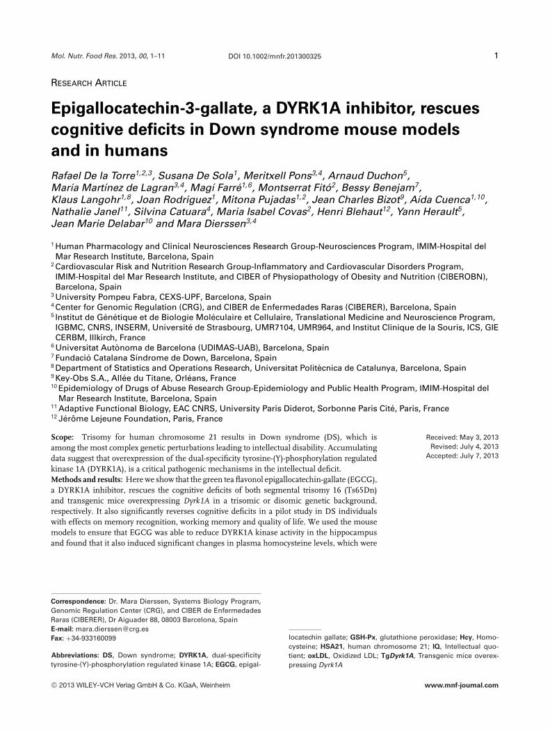
Table 1. Sociodemographic characteristics and clinical parame-ters at baseline
EGCG Placebo(n = 13) (n = 16)
Age 22.2 (4.2) 20.6 (2.2)SexFemale 6 (46.2%) 8 (50.0%)Male 7 (53.8%) 8 (50.0%)Education (years) 14.86 (2.6) 15.38 (2.4)
Intellectual disability levelMild/Moderate 8 (61.5%) 5 (31.2%)Severe 5 (38.5%) 11 (68.7%)
Intellectual quotient (IQ)K-BIT (standardized score) 45.9 (7.8) 42.4 (6.4)
DS genetic variationsTrisomy 21 10 (76.9%) 11 (68.7%)Mosaic 0 1 (6.2%)Translocation 1 (7.6%) 0Unknown 2 (15.3%) 4 (25.0%)Weight 60.1 (13.3) 56 (10.9)
Body Mass Index (BMI) 25.4 (4.0) 23.2 (3.8)
Results are presented as mean (SD) for continuous variablesand absolute frequency (relative frequency) for categoricalvariables.
critical value (15–20 �M) that was maintained along treat-ment, and returned to baseline after treatment discontinua-tion.
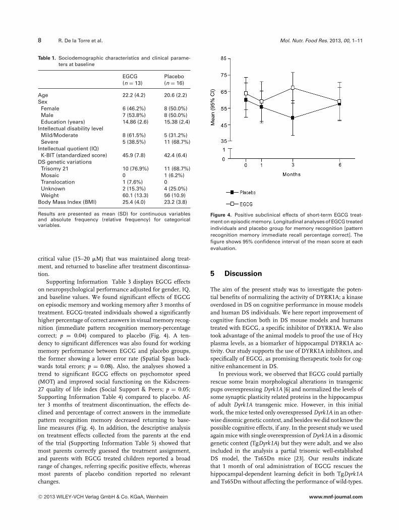
Supporting Information Table 3 displays EGCG effectson neuropsychological performance adjusted for gender, IQ,and baseline values. We found significant effects of EGCGon episodic memory and working memory after 3 months oftreatment. EGCG-treated individuals showed a significantlyhigher percentage of correct answers in visual memory recog-nition (immediate pattern recognition memory-percentagecorrect; p = 0.04) compared to placebo (Fig. 4). A ten-dency to significant differences was also found for workingmemory performance between EGCG and placebo groups,the former showing a lower error rate (Spatial Span back-wards total errors; p = 0.08). Also, the analyses showed atrend to significant EGCG effects on psychomotor speed(MOT) and improved social functioning on the Kidscreen-27 quality of life index (Social Support & Peers; p = 0.05;Supporting Information Table 4) compared to placebo. Af-ter 3 months of treatment discontinuation, the effects de-clined and percentage of correct answers in the immediatepattern recognition memory decreased returning to base-line measures (Fig. 4). In addition, the descriptive analysison treatment effects collected from the parents at the endof the trial (Supporting Information Table 5) showed thatmost parents correctly guessed the treatment assignment,and parents with EGCG treated children reported a broadrange of changes, referring specific positive effects, whereasmost parents of placebo condition reported no relevantchanges.
Figure 4. Positive subclinical effects of short-term EGCG treat-ment on episodic memory. Longitudinal analyses of EGCG treatedindividuals and placebo group for memory recognition [patternrecognition memory immediate recall percentage correct]. Thefigure shows 95% confidence interval of the mean score at eachevaluation.
5 Discussion
The aim of the present study was to investigate the poten-tial benefits of normalizing the activity of DYRK1A; a kinaseoverdosed in DS on cognitive performance in mouse modelsand human DS individuals. We here report improvement ofcognitive function both in DS mouse models and humanstreated with EGCG, a specific inhibitor of DYRK1A. We alsotook advantage of the animal models to proof the use of Hcyplasma levels, as a biomarker of hippocampal DYRK1A ac-tivity. Our study supports the use of DYRK1A inhibitors, andspecifically of EGCG, as promising therapeutic tools for cog-nitive enhancement in DS.
In previous work, we observed that EGCG could partiallyrescue some brain morphological alterations in transgenicpups overexpressing Dyrk1A [6] and normalized the levels ofsome synaptic plasticity related proteins in the hippocampusof adult Dyrk1A transgenic mice. However, in this initialwork, the mice tested only overexpressed Dyrk1A in an other-wise disomic genetic context, and besides we did not know thepossible cognitive effects, if any. In the present study we usedagain mice with single overexpression of Dyrk1A in a disomicgenetic context (TgDyrk1A) but they were adult, and we alsoincluded in the analysis a partial trisomic well-establishedDS model, the Ts65Dn mice [23]. Our results indicatethat 1 month of oral administration of EGCG rescues thehippocampal-dependent learning deficit in both TgDyrk1Aand Ts65Dn without affecting the performance of wild-types.
C© 2013 WILEY-VCH Verlag GmbH & Co. KGaA, Weinheim www.mnf-journal.com

Mol. Nutr. Food Res. 2013, 00, 1–11 9
Besides a general effect on hippocampal dependent learning,inadequate learning strategies (thigmotaxia) were reducedin EGCG-treated Ts65Dn and TgDyrk1A mice. Thus, wecan conclude that EGCG was able to rescue the phenotypesderived of Dyrk1A overexpression in transgenic mice andin the partial trisomy model, Ts65Dn. This suggested thatthe pathological effect driven by Dyrk1A overexpression isrelevant for DS, and that Dyrk1A is a strong candidate fortherapy. Our previous work [30] already demonstrated thatfunctional cortico-striatal defects could be corrected throughthe specific inhibition of Dyrk1A expression using adenoviralvectors (AAVshDyrk1A) in TgDyrk1A mice [31], but here weshow that also kinase activity normalization in adult micecan rescue the cognitive phenotypes. However, since Dyrk1Ais a dosage sensitive gene, the reduction of its kinase activitycould be deleterious in wild-type mice. It may be arguedthat the different brain regions involved in both tasks havedifferential sensitivity to DYRK1A kinase functionality. How-ever, although EGCG normalized DYRK1A kinase activityin the hippocampus in TgDyrk1A at the same dose regimethat improved hippocampal-dependent tests (Fig. 3A), butsurprisingly, the same doses did not modify kinase activityin wild-type mice. This would fit with the lack of effectsobserved in the Morris water maze in wild-type mice,and may suggest that the deleterious effects in the objectrecognition test could depend on changes of the Dyrk1Aactivity in cortical regions that we did not test or to othereffects of the polyphenol not measured in the present study.
Since we wanted to evaluate the effects of the treatmentin humans, the next step was to ensure that DYRK1A kinaseactivity normalization could be measured using a plasmabiomarker that could be then detected upon treatmentwith EGCG in humans. Our previous studies showed thatoverexpression of Dyrk1A is related to decreased Hcy plasmaconcentrations in TgDyrk1A and Ts65Dn mice suggestingthat Hcy could be a suitable biomarker correlated withDyrk1A in DS [27]. We here demonstrated a significantcorrelation between Hcy in plasma and brain levels ofDyrk1A (Fig. 3D) in Ts65Dn mice. Ts65Dn mice showeddecreased Hcy plasma levels similar to what has beendescribed in DS individuals [29]. One month of treatmentwith EGCG normalized the reduced levels of plasma Hcyof both TgDyrk1A and Ts65Dn to untreated wild-type levels.These experiments served as a proof of concept that thelearning deficits caused by Dyrk1A overexpression could berescued by Dyrk1A activity normalization and that plasmaHcy levels could be used as a biomarker.
We then explored the effects of EGCG on cognitive perfor-mance in young adults with DS, paying special attention tohippocampal function. Concretely, besides other measures(see Section 4) we used a visual recognition task that mea-sured visuoperceptual processing, the weakest component ofvisual memory in DS individuals. This measure has provento be sensitive to hippocampal functioning, in particular ofperirhinal cortex, which is critically involved in object recogni-tion memory [32–35] and is reduced in size in DS [36]. EGCG
treated individuals showed higher accuracy in visual memoryrecognition [37,38] and spatial working memory [39], suggest-ing a positive effect of this compound also on the prefrontalsystem, in particular ventromedial, ventrolateral and dorsolat-eral cortices. Overall, the results from this pilot study are con-sistent with EGCG effects found in TgDyrk1A and Ts65Dnmice and tentatively indicate that short-term EGCG treat-ment is able to induce beneficial effects on cognition in youngadults with DS, probably acting on distributed hippocampal-prefrontal functional networks supporting memory abilities.
EGCG administration also induced positive effects onquality of life and social functioning. This was detected byusing objective measures such as the Kidscreen battery, butalso in a parent/caregivers survey in which subjective positiveimpressions of mild cognitive enhancement and/or improvedbehavioral control over a wide array of cognitive/behavioralskills were reported in EGCG treated individuals. Interest-ingly, while parents reported improved verbal cognitive abil-ities, that are less preserved in DS, and considered one ofthe landmarks of global intellectual impairment in these in-dividuals [40, 41] and a significant impact of EGCG cogni-tive enhancement on everyday living, the neuropsychologicaltesting could not detect these effects. Such discrepancy couldrespond to a low sensitivity of the functional assessment toolsused.
One important aspect was safety and toxicity, since DSindividuals may be more sensitive than euploids to someadverse effect. EGCG has been demonstrated to be a safesubstance after repeated administration in humans. No al-teration of hepatic function markers (AST and ALT) was ob-served. Animal studies have reported EGCG to be involved inredox cycling and quinone formation having both antioxidantand pro-oxidative activities and being able to induce oxidativestress [42]. We found no increase of biomarkers of oxidativestress (oxLDL and GSH-Px activity), but a reduction of lipidoxidation in subjects treated with EGCG. The improvementon the oxidative/antioxidative status combined with a healthylipid profile, shown by the total cholesterol and LDL choles-terol concentrations can be considered beneficial.
Importantly, as for mice, in humans the biochemicalresults allowed to establish a clear relationship betweenmemory improvement and Hcy levels, suggesting a directdependence of cognitive improvement on Dyrk1A activity.Moreover, after 3 months of treatment discontinuationEGCG cognitive effects decline along with a parallel decreasein plasma Hcy levels, suggesting that Dyrk1A activitynormalization induced cognitive changes through temporaryneuronal changes. This is important since Dyrk1A has beenrelated to synaptic plasticity in the brain that may lead tostable structural modifications in neural circuits [15, 16, 18].This has functional consequences, since DS learning andmemory problems become considerably more noticeablethrough childhood and adolescence and appear to be relatedto an inability to “stabilize” information that is acquired,a problem in memory consolidation that is a function ofthe hippocampal system (see [10]). Consolidation of labile
C© 2013 WILEY-VCH Verlag GmbH & Co. KGaA, Weinheim www.mnf-journal.com

10 R. De la Torre et al. Mol. Nutr. Food Res. 2013, 00, 1–11
information storage requires the translation of short-lastingbiochemical synaptic events into more stable functionaland structural changes. This has been shown in trisomicmouse models of DS and also in TgDyrk1A and is proposedto underlie the reduced stability of learning in the humanphenotype (see [11] for review). By normalizing Dyrk1Aactivity, EGCG is possibly favoring learning through thesemechanisms, and thus it could be speculated that it couldbe especially beneficial if paired with interventions that alsoincrease plasticity, such as cognitive stimulation.
In conclusion, we here demonstrate the positive effects ofEGCG on learning and memory deficits of DS mouse modelsand humans, thus supporting further research on the useof EGCG as a potential therapeutic agent for improvingcognitive deficits in young DS adults. Hcy plasma concen-trations are proposed as a good biomarker of efficacy of thetreatment.
We wish to thank Life Extension and Dr. S. Hirsch for thegenerous gift of MGTE extracts. We thank Echevarne Laborato-ries for the generous provision of clinical analyses and FundacioCatalana Sındrome de Down (FCSD, Spain) for their assistancewith the recruitment of volunteers and for providing informa-tion and guidance along the study. Finally, last but not least,we would like to express very special thanks to all participantsand their families for their time and efforts. This work has beensupported by the Jerome Lejeune and Reina Sofia Foundation,the DURSI (2009SGR1313, 2009SGR718), SAF2010–16427,Koplowitz Foundation, FRAXA Foundation, CureFXS ERare-EU, FIS PS09102673 and ANR-MNP (DSTHER grant). Thiswork was funded in part by the Miguel Servet SNS contract(CP06/00100), Health Institute Carlos III. The CIBEROBNand CIBERER are initiatives of the Instituto de Salud Carlos III,Madrid, Spain.
The authors have declared no conflict of interest.
6 References
[1] Wolfram, S., Effects of green tea and EGCG on cardiovas-cular and metabolic health. J. Am. Coll. Nutr. 2007, 26,373S–388S.
[2] Shirakami, Y., Shimizu, M., Moriwaki, H., Cancer chemopre-vention with green tea catechins: from bench to bed. Curr.Drug. Targets 2012, 13, 1842–1857.
[3] Steinmann, J., Buer, J., Pietschmann, T., Steinmann, E., Anti-infective properties of epigallocatechin-3-gallate (EGCG),a component of green tea. Br. J. Pharmacol. 2013, 168,1059–1073.
[4] Kelsey, N. A., Wilkins, H. M., Linseman, D.A., Nutraceuti-cal antioxidants as novel neuroprotective agents. Molecules2010, 15, 7792–7814.
[5] Wang, Y., Li, M., Xu, X., Song, M. et al., Green teaepigallocatechin-3-gallate (EGCG) promotes neural progeni-tor cell proliferation and sonic hedgehog pathway activation
during adult hippocampal neurogenesis. Mol. Nutr. FoodRes. 2012, 56, 1292–1303.
[6] Guedj, F., Sebrie, C., Rivals, I., Ledru, A. et al., Green teapolyphenols rescue of brain defects induced by overexpres-sion of DYRK1A. PLoS One 2009, 4, e4606.
[7] Megarbane, A., Ravel, A., Mircher, C., Sturtz, F. et al., The 50thanniversary of the discovery of trisomy 21: the past, present,and future of research and treatment of Down syndrome.Genet. Med. 2009, 11, 611–616.
[8] Lott, I. T., Dierssen, M., Cognitive deficits and associatedneurological complications in individuals with Down’s syn-drome. Lancet Neurol. 2010, 9, 623–633.
[9] Dierssen, M., Herault, Y., Estivill, X., Aneuploidy: from aphysiological mechanism of variance to Down syndrome.Physiol. Rev. 2009, 89, 887–920.
[10] Dierssen, M., Down syndrome: the brain in trisomic mode.Nat. Rev. Neurosci. 2012, 13, 844–858.
[11] Rahmani, Z, Blouin, J. L., Creau-Goldberg, N., Watkins,P. C., Mattei, J. F. et al., Critical role of the D21S55region on chromosome 21 in the pathogenesis ofDown syndrome. Proc. Natl. Acad. Sci. USA 1989, 86,5958–5962.
[12] Dahmane, N., Ghezala, G. A., Gosset, P., Chamoun, Z. et al.,Transcriptional map of the 2.5-Mb CBR-ERG region of chro-mosome 21 involved in Down syndrome. Genomics 1998,48, 12–23.
[13] Altafaj, X., Dierssen, M., Baamonde, C., Martı, E. et al., Neu-rodevelopmental delay, motor abnormalities and cognitivedeficits in transgenic mice overexpressing Dyrk1A (mini-brain), a murine model of Down’s syndrome. Hum. Mol.Genet. 2001, 10, 1915–1923.
[14] Fotaki, V., Dierssen, M., Alcantara, S., Martınez, S. et al.,Dyrk1A haploinsufficiency affects viability and causes de-velopmental delay and abnormal brain morphology in mice.Mol. Cell. Biol. 2002, 22, 6636–6647.
[15] Benavides-Piccione, R., Dierssen, M., Ballesteros-Yanez, I.,Martınez de Lagran, M. et al., Alterations in the phenotypeof neocortical pyramidal cells in the Dyrk1A+/- mouse. Neu-robiol. Dis. 2005, 20, 115–122.
[16] Scales, T. M., Lin, S., Kraus, M., Goold, R. G. et al., Nonprimedand DYRK1A-primed GSK3 beta-phosphorylation sites onMAP1B regulate microtubule dynamics in growing axons.J. Cell. Sci. 2009, 122, 2424–2435.
[17] Lepagnol-Bestel, A. M., Zvara, A., Maussion, G., Quignon, F.et al., DYRK1A interacts with the REST/NRSF-SWI/SNF chro-matin remodelling complex to deregulate gene clusters in-volved in the neuronal phenotypic traits of Down syndrome.Hum. Mol. Genet. 2009, 18, 1405–1414.
[18] Martinez de Lagran, M., Benavides-Piccione, R., Ballesteros-Yanez, I., Calvo, M. et al., Dyrk1A influences neuronal mor-phogenesis through regulation of cytoskeletal dynamicsin mammalian cortical neurons. Cereb. Cortex 2012, 22,2867–2877.
[19] Escorihuela, R. M., Fernandez-Teruel, A., Vallina, I. F., Baa-monde, C. et al., A behavioral assessment of Ts65Dn mice:a putative Down syndrome model. Neurosci. Lett. 1995, 199,143–146.
C© 2013 WILEY-VCH Verlag GmbH & Co. KGaA, Weinheim www.mnf-journal.com

Mol. Nutr. Food Res. 2013, 00, 1–11 11
[20] Demas, G. E., Nelson, R. J., Krueger, B. K., Yarowsky, P.J., Impaired spatial working and reference memory in seg-mental trisomy (Ts65Dn) mice. Behav. Brain Res. 1998, 90,199–201.
[21] Dowdy-Sanders, N. C., Wenger, G. R., Working memory inthe Ts65Dn mouse, a model for Down syndrome. Behav.Brain Res. 2006, 168, 349–352.
[22] Bain, J., McLauchlan, H., Elliott, M., Cohen, P., The speci-ficities of protein kinase inhibitors: an update. Biochem. J.2003, 371, 199–204.
[23] Reeves, R. H., Irving, N. G., Moran, T. H., Wohn, A., Kitt, C.et al., A mouse model for Down syndrome exhibits learningand behaviour deficits. Nat. Genet. 1995, 11, 177–184.
[24] Toiber, D., Azkona, G., Ben-Ari, S., Toran, N. et al., En-gineering DYRK1A overdosage yields Down syndrome-characteristic cortical splicing aberrations. Neurobiol. Dis.2010, 40, 348–359.
[25] Braudeau, J., Delatour, B., Duchon, A., Pereira, P. L. et al.,Specific targeting of the GABA-A receptor alpha5 subtypeby a selective inverse agonist restores cognitive deficitsin Down syndrome mice. J. Psychopharmacol. 2011, 25,1030–1042.
[26] Papadopoulos, C., Arato, K., Lilienthal, E., Zerweck, J.et al., Splice variants of the dual specificity tyrosinephosphorylation-regulated kinase 4 (DYRK4) differ in theirsubcellular localization and catalytic activity. J. Biol. Chem.2011, 286, 5494–5505.
[27] Noll, C., Planque, C., Ripoll, C., Guedj, F. et al., DYRK1A,a novel determinant of the methionine-homocysteinecycle in different mouse models overexpressing thisDown-syndrome-associated kinase. PLoS One 2009, 4,e7540.
[28] Fortin, L. J., Genest, J, Jr., Measurement of homocyst(e)inein the prediction of arteriosclerosis. Clin. Biochem. 1995, 28,155–162.
[29] Varga P, V Olah A, Olah E., Biochemical alterations in patientswith Down syndrome. Orv. Hetil. 2008, 149, 1203–1213.
[30] Ortiz-Abalia, J., Sahun, I., Altafaj, X., Andreu, N. et al., Target-ing Dyrk1A with AAVshRNA attenuates motor alterations inTgDyrk1A, a mouse model of Down syndrome. Am. J. Hum.Genet. 2008, 83, 479–488.
[31] Altafaj, X., Martın, E. D., Ortiz-Abalia, J., Valderrama, A. et al.,Normalization of Dyrk1A expression by AAV2/1-shDyrk1Aattenuates hippocampal-dependent defects in the Ts65Dn
Down syndrome mouse model. Neurobiol. Dis. 2013, 52,117–27.
[32] Meunier, M., Bachevalier, J., Mishkin, M., Murray, E. A., Ef-fects on visual recognition of combined and separate abla-tions of the entorhinal and perirhinal cortex in rhesus mon-keys. J. Neurosci. 1993, 13, 5418–5432.
[33] Winters, B. D., Forwood, S. E., Cowell, R. A., Saksida, L.M. et al., Double dissociation between the effects of peri-postrhinal cortex and hippocampal lesions on tests of objectrecognition and spatial memory: heterogeneity of functionwithin the temporal lobe. J. Neurosci. 2004, 24, 5901–5908.
[34] Vicari, S., Bellucci, S., Carlesimo, G. A., Visual and spatiallong-term memory: differential pattern of impairments inWilliams and Down syndromes. Dev. Med. Child. Neurol.2005, 47, 305–311.
[35] Buffalo, E. A., Bellgowan, P. S., Martin, A., Distinct roles formedial temporal lobe structures in memory for objects andtheir locations. Learn. Mem. 2006, 13, 638–643.
[36] Pinter, J. D., Brown, W. E., Eliez, S., Schmitt, J. E. et al.,Amygdala and hippocampal volumes in children with Downsyndrome: a high-resolution MRI study. Neurology 2001, 56,972–974.
[37] Bachevalier, J., Mishkin, M., Visual recognition impairmentfollows ventromedial but not dorsolateral prefrontal lesionsin monkeys. Behav. Brain. Res. 1986, 20, 249–261.
[38] Turriziani P, Oliveri M, Salerno S, Costanzo F et al., Recogni-tion memory and prefrontal cortex: dissociating recollectionand familiarity processes using rTMS. Behav. Neurol. 2008,19, 23–27.
[39] Mottaghy, F. M., Gangitano, M., Sparing, R., Krause, B. J.et al., Segregation of areas related to visual working mem-ory in the prefrontal cortex revealed by rTMS. Cereb. Cortex2002, 12, 369–375.
[40] Brock, J., Jarrold, C., Serial order reconstruction in Downsyndrome: evidence for a selective deficit in verbal short-term memory. J. Child. Psychol. Psychiatry 2005, 46,304–316.
[41] Frenkel, S., Bourdin, B., Verbal, visual, and spatio-sequentialshort-term memory: assessment of the storage capacities ofchildren and teenagers with Down’s syndrome. J. Intellect.Disabil. Res. 2009, 53, 152–160.
[42] Sang, S., Hou, Z., Lambert, J. D., Yang, C. S., Redox prop-erties of tea polyphenols and related biological activities.Antioxid. Redox. Signal 2005, 7, 1704–1714.
C© 2013 WILEY-VCH Verlag GmbH & Co. KGaA, Weinheim www.mnf-journal.com