Transplanting Neural Progenitor Cells into a Chronic Dorsal ...
Upload
independentCategory
view
0download
0
This article appeared in a journal published by Elsevier. The attachedcopy is furnished to the author for internal non-commercial researchand education use, including for instruction at the authors institution
and sharing with colleagues.
Other uses, including reproduction and distribution, or selling orlicensing copies, or posting to personal, institutional or third party
websites are prohibited.
In most cases authors are permitted to post their version of thearticle (e.g. in Word or Tex form) to their personal website orinstitutional repository. Authors requiring further information
regarding Elsevier’s archiving and manuscript policies areencouraged to visit:
http://www.elsevier.com/authorsrights
Author's personal copy
Soluble amyloid precursor protein-a rescues age-linked declinein neural progenitor cell proliferation
Michael P. Demars, Carolyn Hollands, Kai Da (Tommy) Zhao, Orly Lazarov*
Department of Anatomy and Cell Biology, College of Medicine, The University of Illinois at Chicago, Chicago, IL, USA
a r t i c l e i n f o
Article history:Received 15 October 2012Received in revised form 7 March 2013Accepted 14 April 2013Available online 14 May 2013
Keywords:NeurogenesisAmyloid precursor proteinAdulthoodAgingCognitionLearning and memoryAlzheimer’s disease
a b s t r a c t
Neurogenesis is thought to play a role in cognitive function and hippocampal plasticity. Previous studiessuggest that neurogenesis declines with aging. However, the onset and mechanism of declined neuro-genesis are not fully elucidated. Here we show that the major decline in neurogenesis takes place duringadulthood, before aging. Decline in neurogenesis takes place in the subgranular layer of the dentate gyrusand in the subventricular zone, and is primarily due to a reduced number of fast-proliferating neuralprogenitor cells. Importantly, this decline can be rescued by intraventricular injection of recombinantsoluble amyloid precursor protein (sAPPa), which regulates neural progenitor cell proliferation in theadult brain. The counterpart, sAPPb, a product of the amyloidogenic cleavage pathway of amyloidprecursor protein, fails to exhibit a proliferative effect in vitro and in vivo, in equimolar concentrations tosAPPa. These observations suggest that adulthood is an appropriate time window for an interventionthat upregulates neurogenesis, such as enhancement of sAPPa levels, for the prevention of decliningbrain plasticity and cognitive function.
� 2013 Elsevier Inc. All rights reserved.
1. Introduction
Amyloid precursor protein (APP) can undergo metabolism viatwo distinct pathways leading to the production of a number ofintra- and extracellular metabolites. The amyloidogenic pathwayof APP cleavage, so termed for its involvement in the formationof beta amyloid, begins with cleavage by b-secretase, b-site APPcleaving enzyme 1, and releases soluble APPb (sAPPb) to thelumen (for review, see Vassar, 2012). In the non-amyloidogenicAPP cleavage pathway, initial cleavage occurs via a-secretaseenzymes, primarily composed of a disintegrin and matrix-metalloproteinase proteins, ADAMs, leading to the release ofsAPPa (Asai et al., 2003; Buxbaum et al., 1998; Jorissen et al.,2010; Lammich et al., 1999). It has been previously reportedthat sAPPa has trophic properties in a number of different celltypes including fibroblasts (Saitoh et al., 1989), thyroid epithelialcells (Pietrzik et al., 1998), embryonic stem cells (Ohsawa et al.,1999), and carcinoma cells (Ko et al., 2004). With respect toadult neural progenitor cells (NPCs), sAPPa has been shown to
bind to and stimulate the proliferation of NPCs in vitro andin vivo (Caille et al., 2004; Demars et al., 2011).
In the aging brain, there is a marked decline in neurogenesis inthe subventricular zone (SVZ) (Mirich et al., 2002; Shook et al.,2012) and subgranular layer (SGL) (Ben Abdallah et al., 2010;Bernal and Peterson, 2004; Bondolfi et al., 2004; Cameron andMcKay, 1999; Encinas et al., 2011; Kronenberg et al., 2006; Kuhnet al., 1996; Miranda et al., 2012). This decline in neurogenesis hasbeen shown to manifest in deficits in olfactory function andhippocampal-dependent learning and memory (Bizon et al., 2004;Dupret et al., 2008; Enwere et al., 2004). However, the cause ofneurogenic decline in the aging brain remains somewhat contro-versial. Some studies argue that the proliferation of NPCs and theirmaturation declines with age (Heine et al., 2004; Kuhn et al., 1996;Morgenstern et al., 2008; Rao et al., 2005). Olariu and colleaguessuggest that this decline is not because of alterations in the lengthof the cell cycle of NPCs in the SGL (Olariu et al., 2007). Encinas andcolleagues suggest that neurogenic decline is because of thedisappearance of neural stem cells (NSC) by their conversion intomature hippocampal astrocytes (Encinas et al., 2011). Bonaguidiet al. (2008) suggest that upregulation of bone morphogeneticprotein signaling or downregulation of its antagonists mightunderlie age-linked reduced self-renewal of NSC leading to thedecline in neurogenesis (Bonaguidi et al., 2008). Other groups haveargued that the rate of proliferation does not declinewith aging, but
* Corresponding author at: Anatomy and Cell Biology, College of Medicine, 808 SWood Street M/C 512, University of Illinois at Chicago, Chicago, IL 60612, USA.Tel.: þ1 312 355 0548; fax: þ1 312 413 0354.
E-mail address: [email protected] (O. Lazarov).
Contents lists available at SciVerse ScienceDirect
Neurobiology of Aging
journal homepage: www.elsevier .com/locate/neuaging
0197-4580/$ e see front matter � 2013 Elsevier Inc. All rights reserved.http://dx.doi.org/10.1016/j.neurobiolaging.2013.04.016
Neurobiology of Aging 34 (2013) 2431e2440
Author's personal copy
that cells might exhibit increased quiescence that might be attrib-uted to a decline in the vascular niche (Hattiangady and Shetty,2008). Although this debate is still ongoing, it is apparent that theneurogenic niche undergoes changes associated with aging thathave the potential to underlie neurogenic decline (Ahlenius et al.,2009; Miranda et al., 2012; Villeda et al., 2011). For instance, ithas been well established that the expression of many growthfactors and their receptors that are integral to neurogenic processespeak during development, and decline in expression thereafter (forreview, see Klempin and Kempermann, 2007). Neurogenesis in theadult central nervous system plays a role in hippocampus- andolfaction-dependent learning and memory (for review, see Bernaland Peterson, 2004; Kempermann and Gage, 2000; Lie et al.,2004). These findings raise the possibility that reduced neuro-genesis might, at least in part, account for impaired learning andmemory and cognitive deterioration in the elderly (Kempermannet al., 1998, 2002; Kuhn et al., 1996; Seki and Arai, 1995; Tropepeet al., 1997) and might enhance vulnerability to Alzheimer’sdisease (AD; for review, see Lazarov et al., 2010).
Aging remains the predominant risk factor for the develop-ment of the sporadic form of AD. Alterations in the cleavagepattern of APP are causative of familial forms of the disease (forreview, see Selkoe, 2001). We have previously reported thatdeficits in proliferation and neurogenesis occur long before theappearance of pathological hallmarks or cognitive impairment ina mouse model of familial AD (Demars et al., 2010). Takentogether with our observation that sAPPa is a proliferation factorof adult NPCs, this might suggest the hypothesis that increasinglevels of sAPPa in the neurogenic niches would rescue neuro-genic deficits.
Here we show that a dramatic decline in neurogenesis takesplace at 7e9 months of age, compared with mice at 2 months ofage, and only a minor further decrease takes place at 20 months.This suggests that the significant decline in neurogenesis takesplace during adulthood, rather than during aging. This decline ischaracterized by a reduced numbers of fast proliferating NPCs(type II cells in the SGL and type C cells in the SVZ), but there is nodecline in the number of NSC. Intracerebroventricular (ICV)injection of sAPPa ameliorates proliferation deficits in adult mice.In support of that, adding sAPPa to a neurosphere culture derivedfrom 7e9-month-old or 20-month-old mice enhances NPC pro-liferation. Additionally, we show that at a similar concentrationrange, sAPPb does not evoke the same proliferative effect as sAPPa.This study suggests that therapy aimed at rescuing decline inneurogenesis should be applied during adulthood rather thanaging, and that enhancement of sAPPa levels might rescue thisdecline.
2. Methods
2.1. Animals
Wild typemice, 2months, 7e9months, and 20months of age ona C57/Bl6XC3H background were maintained in our colony. Ourcolony is maintained via group housing (<5 mice per cage) ina barrier facility under a 14:10 light:dark cycle with free access tofood and water. Mice were euthanized using isofluorane andcervical dislocation. Animal care and procedures were conductedaccording to the National Institutes of Health Guide for the Care andUse of Laboratory Animals.
2.2. Recombinant sAPPa and sAPPb
sAPPa (Sigma-Aldrich, St. Louis, MO, USA) was used at 10 nMconcentrations unless otherwise indicated (dissolved in phosphate-
buffered saline [PBS]). sAPPb (Sigma-Aldrich) was also dissolved inPBS and was used in the indicated concentrations.
2.3. ICV injections
A PBS vehicle or recombinant sAPPa or sAPPb at a concentrationof 1 mM (1 mL per mouse; 0.25 mL/min) were stereotaxically injectedinto the left lateral ventricle of 7e9-month-old C57BL/6 mice usingthe following coordinates: anteroposterior, 0 mm; mediolate-ral, �0.8 mm; and dorsoventral, �2.0 mm from bregma. Mice wereanesthetized using a mixture of ketamine (100 mg/kg) and xylazine(10 mg/kg). The heads of the mice were then shaved and wipedwith 70% ethanol. Animals were placed into a stereotaxic frame anda centimeter incisionwas made in the midline of the scalp to revealthe bregma. The scalp was rinsed with 30% hydrogen peroxide anda small hole was drilled at the coordinate site measured frombregma according to the mouse atlas of Franklin and Paxinos(2008). Animals then received a unilateral injection of sAPP to thelateral ventricle. The sAPP was delivered through a 5-mL Hamiltonsyringe connected to a hydraulic injection system set to inject ata rate of 0.25 mL/min. The injection needle was then left in place foran additional minute to ensure distribution of the solution. Theneedle was slowly removed and the incision closed using EZ-clipsfrom Stoelting. After 6 hours of recovery, mice were given a singleintraperitoneal dose of 100 mg/kg BrdU solution. Twenty-fourhours after the BrdU injection, mice were transcardially perfusedand the brains processed for immunohistochemistry as describedon subsequent pages in this article.
2.4. Neural progenitor cell culture
Mice were euthanized and their brains were removed andplaced into sterile Dulbecco’s modified Eagle’s medium/F12 (Gibco,now part of Invitrogen Corporation, Carlsbad, CA, USA). A coronalslice (approximately 1 mm) was dissected starting 1e2 mm pos-terior to the olfactory bulb. The region occupying the lateral walland anterior horn of the lateral ventricles was removedwith the aidof a dissecting microscope and diced with a sterile scalpel. Neuro-sphere culture was prepared as previously described (Demars et al.,2011). Briefly, tissue pieces were collected in a mixture of Papainand DNase in Earl’s balanced salt solution and incubated at 37 �C for40 minutes. Then, tissue pieces were pelleted using centrifugationand dissociated to a single-cell suspension, and cells were plated inNPC media that included water, Dulbecco’s modified Eagle’smedium/F12, glucose (Sigma-Aldrich, St Louis, MO, USA), NaHCO3(Sigma-Aldrich), HEPES solution (N-(2-Hydroxyethyl)piperazine-N0-(2-ethanesulfonic acid)) (Sigma-Aldrich), L-glutamine (Invi-trogen Corporation), penicillin/streptomycin (Invitrogen Corpora-tion), putrescine (9.6 mg/mL; Sigma-Aldrich), apotransferrin (0.1mg/mL; Sigma-Aldrich), insulin (0.025 mg/mL; Roche, Indianapolis,IN, USA), selenium (5.2 ng/mL; Sigma-Aldrich), progesterone (6.3 ng/mL; Sigma-Aldrich), bovine serum albumin (2 mg/mL; Sigma-Aldrich), heparin (4 mg/mL; Sigma-Aldrich), epidermal growthfactor (EGF) (20 ng/mL; PeproTech Rocky Hill, NJ, USA), and basicfibroblast growth factor (bFGF) (10 ng/mL; Pepro-Tech), andpassaged after 10 days.
2.5. Clonogenic assay
Briefly, neurospheres were singly dissociated using mechanicaldissociation and plated at 1000 cells per well onto 96-well plates.Cells were then treated with the indicated molar concentration ofGM6001 or GM6001 negative control (Millipore Corporation,Billerica, MA, USA), b-secretase inhibitor IV (EMD/Millipore), andthe indicatedmolar concentrations of sAPP. Cells were treated every
M.P. Demars et al. / Neurobiology of Aging 34 (2013) 2431e24402432
Author's personal copy
day for 7 days. After 7 days in culture, neurospheres were countedusing an inverted light microscope, and the average neurospherediameter was calculated for each sphere by measuring 4 diametersfor each to control for irregularly shaped neurospheres using a ZeissAX10 microscope (Carl Zeiss Ltd, Hertfordshire, UK) and Stereo-Investigator software (StereoInvestigator version 8, MBF Bioscience,Williston, VT, USA). After sphere size determination, cells weresingly dissociated with a p200 pipette and counted with a hemo-cytometer. Each experiment was repeated 5 times using neuro-sphere cultures derived from 5 different animals.
2.6. Immunoprecipitation and Western blot analysis
The detection of sAPP in NPC culture was performed by condi-tioning media using 5 � 105 cells plated for 2 hours in fresh NPCmedia. Media was then spun (1000g for 10 minutes) to remove anycells and precleared with protein A agarose beads (Pierce, ThermoScientific, Rockford, IL, USA). Next, media was incubated overnightwith 22C11 antibodies against the N-terminus of APP. The next day,media was incubated for 30 minutes in protein A agarose beads,spun, and the pellet was resuspended in sample buffer. Sampleswere heated at 100 �C for 5 minutes and run on a 6% Tris-glycinegel. For the extraction of protein from the neurospheres used tocondition media, a lysis buffer containing 150 mM NaCl, 50 mMTris-Cl, 5 mM ethylenediaminetetraacetic acid (EDTA), 1% TritonX-100, 0.5% sodium deoxycholate, protease inhibitor cocktail(mammalian protease inhibitor cocktail; Sigma-Aldrich) and250 mM phenylmethylsulfonyl fluoride (PMSF) was used. Quantifi-cation of protein was performed using the bicinchoninic acid (BCA)method (Pierce) and equal amounts of protein were subjected todirect immunoblot analysis. For quantification, n � 3 was used.
2.7. Brain tissue processing
For in vivo immunohistochemical staining, all mice were anes-thetized using a mixture of ketamine (100 mg/kg) and xylazine(10 mg/kg) and transcardially perfused with 100mL of ice-cold PBS.The brains were then removed and halved on the sagittal plane. Theleft half was immediately placed into 4% paraformaldehyde on ice.
2.8. Immunohistochemistry
Left hemibrains from PBS-perfused mice were postfixed in 4%paraformaldehyde for 3 days and stored in 30% sucrose at 4 �C.Hemibrains were sectioned sagittally at 50 mm by using a freezingstage microtome and placed into cryopreservent (47.6% PBS, 28.57%ethylene glycol, and 25% glycerin [vol/vol]). Sections were blockedby using a solution containing 0.25% (vol/vol) Triton X-100 (Sigma-Aldrich) and 5% (vol/vol) Normal Donkey Serum (Jackson Immu-noResearch Laboratories, Inc, West Grove, PA, USA) in Tris-bufferedsaline solution (TBS). The following antibodies were used: BrdU(1:400; Accurate Chemicals, Westbury, NY, USA), nestin (1:100;Millipore Corporation), doublecortin (DCX, 1:400; Santa CruzBiotechnology, Inc, Santa Cruz, CA, USA) and glial fibrillary acidicprotein (GFAP; 1:500; Millipore Corporation). Floating sectionswere incubated in primary antibodies for 72 hours at 4 �C beforecontinuing with blocking, biotin conjugation (Jackson ImmunoR-esearch Laboratories, Inc), and secondary antibody incubation (cy2Streptavidin, anti-mouse cy3, anti-goat cy5, and anti-rabbit cy5;Jackson ImmunoResearch Laboratories, Inc).
2.9. Stereological quantification
The number of positively stained cells in sagittal brain sectionswas quantified using design-based stereology (StereoInvestigator
version 8, MBF Bioscience). For the analysis, every sixth section ofbrain tissue was quantified by applying the Nv � VRef method. Thefollowing parameters were used: for SVZ, sections were tracedusing a Zeiss AX10 microscope (Carl Zeiss Ltd, Hertfordshire,England) in lowmagnification (5�) and counting was performed athigh magnification (63�), counting frame¼ 100 mm � 100 mm, gridsize 100 mm� 100 mm, and all sections were counted using 12.5-mmtop and bottom guard zones. For the dentate gyrus (DG), thecounting frame was set equal to the grid size (100 mm � 100 mm) tocount the entirety of the DG because of the relative paucity of cells.All other parameters remained the same.
3. Results
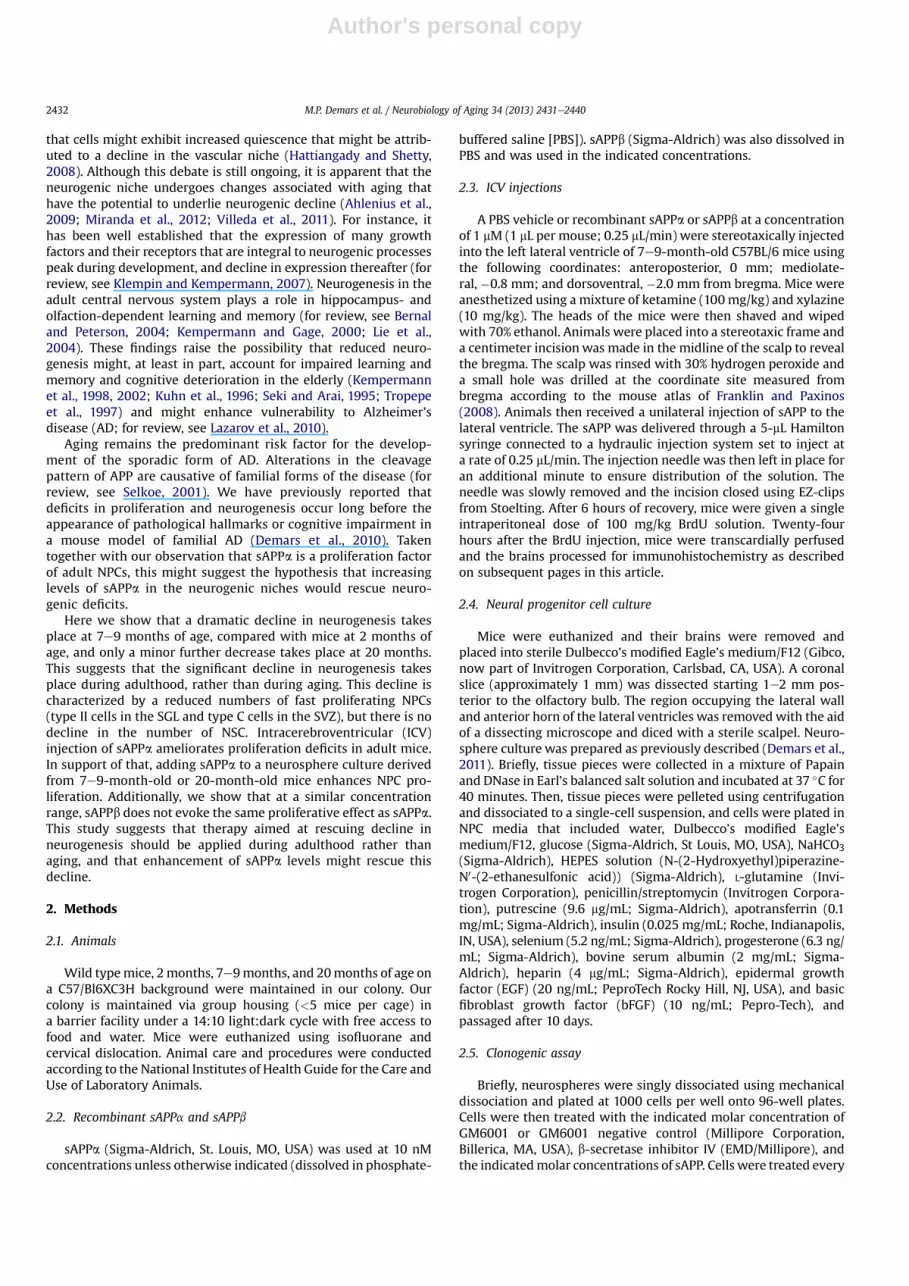
To examine the course of decline in neurogenesiswith agewefirstassessed the extent of neurogenesis in the SGL and SVZ of 2- (young),7e9- (adult), and 20- (aging) month-old mice (n ¼ 4). For thispurpose, we quantified the number of type B (nestinþGFAPþ), totalfast proliferating cells (BrdUþ), type C (BrdUþnestinþ, BrdUþDCXþ),and typeA (BrdU�DCXþ) cells.Unbiased stereology revealeda severereduction in the number of total proliferating cells (Fig. 1A), and thenumber of type C and A cells by 7e9 months of age in the SVZ(Fig. 1BeD). Notably, the number of fast proliferating cells (BrdUþ)and NPCs (nestinþBrdUþ) is significantly reduced by 7e9months ofage, and stays reduced without further significant reduction by 20months of age (Fig. 1A and B). This might suggest that the reducedproliferation of NPCs in the SVZ by 7e9 months of age might playa major role in reduced extent of neurogenesis. The number ofneuroblasts and immature neurons is significantly reduced by 7e9months, and this number is further reducedby20months (Fig.1C, D,and Kiiiev). The number of type B cells (GFAPþnestinþ) was notsignificantly changed in any of the age groups (Fig. 1E), suggestingthat the pool of NSC in the SVZ does not decline with age.
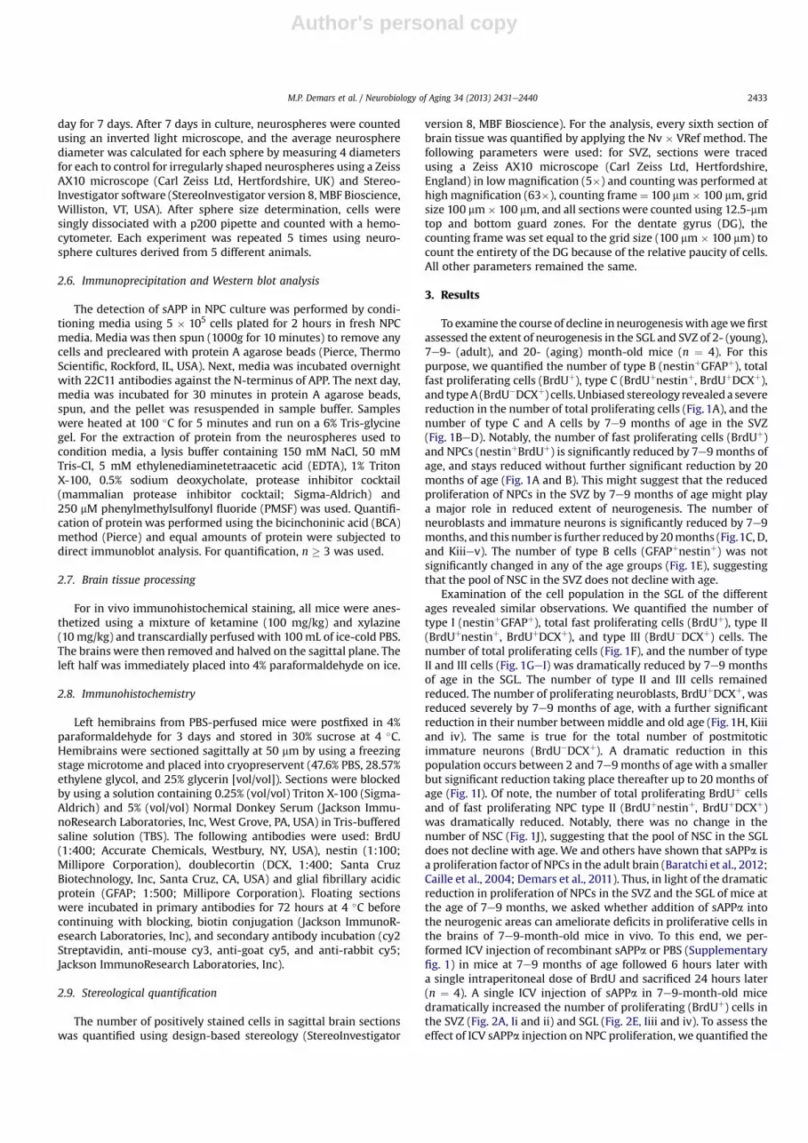
Examination of the cell population in the SGL of the differentages revealed similar observations. We quantified the number oftype I (nestinþGFAPþ), total fast proliferating cells (BrdUþ), type II(BrdUþnestinþ, BrdUþDCXþ), and type III (BrdU�DCXþ) cells. Thenumber of total proliferating cells (Fig. 1F), and the number of typeII and III cells (Fig. 1GeI) was dramatically reduced by 7e9 monthsof age in the SGL. The number of type II and III cells remainedreduced. The number of proliferating neuroblasts, BrdUþDCXþ, wasreduced severely by 7e9 months of age, with a further significantreduction in their number between middle and old age (Fig. 1H, Kiiiand iv). The same is true for the total number of postmitoticimmature neurons (BrdU�DCXþ). A dramatic reduction in thispopulation occurs between 2 and 7e9months of agewith a smallerbut significant reduction taking place thereafter up to 20 months ofage (Fig. 1I). Of note, the number of total proliferating BrdUþ cellsand of fast proliferating NPC type II (BrdUþnestinþ, BrdUþDCXþ)was dramatically reduced. Notably, there was no change in thenumber of NSC (Fig. 1J), suggesting that the pool of NSC in the SGLdoes not decline with age. We and others have shown that sAPPa isa proliferation factor of NPCs in the adult brain (Baratchi et al., 2012;Caille et al., 2004; Demars et al., 2011). Thus, in light of the dramaticreduction in proliferation of NPCs in the SVZ and the SGL of mice atthe age of 7e9 months, we asked whether addition of sAPPa intothe neurogenic areas can ameliorate deficits in proliferative cells inthe brains of 7e9-month-old mice in vivo. To this end, we per-formed ICV injection of recombinant sAPPa or PBS (Supplementaryfig. 1) in mice at 7e9 months of age followed 6 hours later witha single intraperitoneal dose of BrdU and sacrificed 24 hours later(n ¼ 4). A single ICV injection of sAPPa in 7e9-month-old micedramatically increased the number of proliferating (BrdUþ) cells inthe SVZ (Fig. 2A, Ii and ii) and SGL (Fig. 2E, Iiii and iv). To assess theeffect of ICV sAPPa injection on NPC proliferation, we quantified the
M.P. Demars et al. / Neurobiology of Aging 34 (2013) 2431e2440 2433
Author's personal copy
number of cells colabeled with BrdU and nestin. We show thatsAPPa injection rapidly and significantly increased the number ofproliferating NPCs in the SVZ (Fig. 2B and Ivi) and SGL (Fig. 2F) of
7e9-month-old mice. Notably, in the SVZ, the number of prolifer-ating neuroblasts was increased after sAPPa injection (Fig. 2C, Iv),suggesting that enhanced proliferation had a direct effect on the
Fig. 1. Age-linked decline in proliferating neural progenitor cells and immature neurons in the subventricular zone (SVZ) and subgranular layer (SGL). Quantification of proliferatingcells (BrdUþ [A and F]), proliferating neural progenitor cells (NPCs) (BrdUþ/nestinþ [B and G]), proliferating neuroblasts (BrdUþDCXþ [C and H]), immature neurons (BrdU�/DCXþ
[D and I]), and neural stem cells (NSC) (GFAPþ/nestinþ [E and J]) in the SVZ (AeE) and SGL (FeJ) of 2-, 7e9, and 20-month-old mice. (K) Representative images of (i) 2-month-old SVZ(BrdU, green; DCX, red), (ii) 20-month-old SVZ, (iii) 2-month-old SGL, (iv) 20-month-old SGL, (v) representative image of GFAP (green) and nestin (red) staining. n ¼ 4; * p < 0.05,analysis of variance with post hoc analysis. Abbreviations: BrdU, 5-bromo-2-deoxyuridine; DCX, doublecortin; GFAP, glial fibrillary acidic protein; NSC, neural stem cell.
Fig. 2. Intracerebroventricular injection of soluble amyloid precursor protein (sAPP)-a ameliorates aging-linked proliferation deficits in the subgranular layer (SGL) andsubventricular zone (SVZ). Quantification of proliferating cells (BrdUþ; SVZ [A] and SGL [E]), proliferating neural progenitor cells (NPCs) (BrdUþ/nestinþ; SVZ [B] and SGL [F]),neuroblasts (BrdUþDCXþ; SVZ [C] and SGL [G]), and NSC (GFAPþ/nestinþ; SVZ [D] and SGL [H]) in 7e9-month-old mice injected intracerebroventricularly with either phosphate-buffered saline (PBS) or 1 mL of 1 mM sAPPa. (I) Representative images of (i) PBS-injected SVZ (BrdU, green), (ii) sAPPa-injected SVZ (BrdU, green), (iii) PBS-injected SGL (BrdU, green),(iv) sAPPa-injected SGL (BrdU, green), (v) high magnification of BrdUþ (green)/DCXþ (red) staining, and (vi) representative image of BrdUþ (green)/nestinþ (red) staining. n ¼ 4;* p < 0.05, Student t test. Abbreviations: BrdU, 5-bromo-2-deoxyuridine; DCX, doublecortin; GFAP, glial fibrillary acidic protein; NSC, neural stem cell.
M.P. Demars et al. / Neurobiology of Aging 34 (2013) 2431e24402434

Author's personal copy
number of neuronally-committed NPCs. In the SGL, the number ofneuroblasts appeared slightly reduced but this change was notstatistically significant (Fig. 2G). Interestingly, there was a statisti-cally insignificant reduction in the number of NSC (GFAPþ/nestinþ)in the SVZ (Fig. 2D). In the SGL, reduction in the number of NSC wassignificant (Fig. 2H). This might raise the possibility that sAPPainduces NSC asymmetric division resulting in more NPCs in vivo.Alternatively, sAPPa might facilitate their commitment to neuronallineage. Taken together, these results suggest that sAPPa canenhance aging-linked reduced proliferation in the SVZ and SGL bystimulating NPC proliferation.
To further establish that the addition of sAPPa stimulates NPCproliferation, we isolated NPCs from the SVZ ofmice at the ages of 2,7e9, or 20 months, and treated the neurosphere cultures witheither recombinant sAPPa or vehicle. Consistent with previousreports suggesting proliferation deficits could be masked by cultureconditions (Ahlenius et al., 2009; Bouab et al., 2011), we saw nosignificant difference in neurosphere diameter or total number ofNPCs in neurosphere cultures derived frommice that are 2, 7e9, or20 months old (Fig. 3). However, we could stimulate proliferationwith the addition of 10 nM sAPPa in neurospheres derived from 2-,7e9, and even 20-month-old mice as indicated by increased neu-rosphere diameter (Fig. 3A and C), and increased total NPC numbers(Fig. 3B). This result suggests that NPCs derived from mice atvarious ages retain the ability to proliferate in response to sAPPain vitro.
The cleavage of APP can produce either sAPPa or sAPPb. There-fore, we sought to determine if NPCs respond differentially to thetwo sAPP species. We assayed sphere diameter of neurospheresderived from singly-dissociated NPCs treated with varying con-centrations of either sAPPa or sAPPb. Treatment with sAPPa in-creased neurosphere diameter at 10 nM concentrations witha trend toward significance at 1 nM. However, sAPPb treatmentfailed to significantly increase proliferation at any of the concen-trations tested (Fig. 4A; representative images in C). In our previous
work, we showed that sAPPa is able to ameliorate deficits in NPCproliferation caused by matrix-metalloproteinase (MPP) inhibitionthat inhibits enzymes exhibiting a-secretase activity that yieldssAPPa production (Demars et al., 2011). The use of the inhibitor ismeant to inhibit endogenous production of sAPPa, thus preventinga masking of the proliferative effect exerted by recombinant sAPPa.Therefore, we sought to determine whether sAPPb would enhanceproliferation in the absence of endogenous production of sAPPa. Aspreviously reported, 1 mM GM6001 MMP inhibitor treatmentimpairs proliferation, and sAPPa ameliorates these proliferationdeficits (Fig. 4B; Demars et al., 2011). At 1 nM concentrations, sAPPbenhances proliferation to the same extent as sAPPa. However, atconcentrations greater than 10 nM, sAPPb treatment does notrescue MMP inhibitor-induced proliferation deficits (Fig. 4B).
Treatment with MMP inhibitor likely exclusively reduces sAPPa.Thus, we next asked whether endogenous production of sAPPbmasks the proliferative effect of recombinant sAPPb. For thispurpose, we inhibited endogenous production of sAPPb by treatingNPC culture with b-secretase inhibitor IV. We showed that treat-ment of NPCs with b-secretase inhibitor reduces proliferationdramatically in assays of neurosphere diameter and total cellnumber (Fig. 5A). Though sAPPa ameliorates this deficit at 10-nMconcentrations, 1 nM sAPPb, the most effective dose in previousexperiments, does not enhance proliferation after b-secretaseinhibition (Fig. 5A and B). To further exclude the possibility that thepresence of either endogenous sAPPa or sAPPb mask the prolifer-ative effect of recombinant sAPPb, NPC cultures were treated withMMP and b-secretase inhibitor. Treatment of NPCs with GM6001 orb-secretase inhibitor IV reduced neurosphere diameter (Fig. 6A),and total cell number (Fig. 6B) to the same extent after 7 daysin vitro. To assess the ability of sAPPa or sAPPb to stimulateproliferation in conditions of minimal endogenous sAPP, we addedexogenous recombinant sAPPa or sAPPb to NPCs in dual secretaseinhibition. sAPPa rescued proliferation deficits to control levels.Conversely, sAPPb failed to ameliorate the proliferation deficits
Fig. 3. Neural progenitor cells (NPCs) from aged mice retain the ability to proliferate in response to soluble amyloid precursor protein (sAPP). (A) Neurosphere diameter after 7 daysin vitro. Singly-dissociated NPCs of 2-, 7e9, and 20-month-old mice were supplemented daily with 10 nM sAPPa or vehicle. (B) Total cell number of dissociated neurospheresquantified in (A). (C) Representative images of neurospheres in clonogenic analysis after 7 days in vitro with the indicated ages and treatments. n ¼ 5; * p < 0.05, Student t test oranalysis of variance with post hoc analysis.
M.P. Demars et al. / Neurobiology of Aging 34 (2013) 2431e2440 2435

Author's personal copy
incurred from dual secretase inhibition (Fig. 6A and B). Theseresults suggest that sAPPa is a more potent proliferation factor forNPCs than sAPPb.
Finally, we sought to address whether, similar to sAPPa, sAPPbwould rescue age-dependent proliferation deficits of NPCs. Becauseage-dependent proliferation deficits are not apparent in vitro, we
Fig. 4. Soluble amyloid precursor protein should be (sAPPa) and sAPPb differentially affect proliferation of neural progenitor cells (NPCs) and proliferation rescue aftermatrix-metalloproteinase inhibition by treatment with GM6001. (A) Clonogenic assay after treatment with varying concentrations of sAPPa (green) or sAPPb (purple) comparedwith phosphate-buffered saline-treated controls (NC; red). (B) Total cell number from dissociated neurospheres treated with GM6001 (blue), GM6001, and sAPPa (green) or GM6001and sAPPb (purple) compared with GM6001 negative control-treated controls (red). (C) Representative images of neurospheres from Fig. 4A, n ¼ 5; * p < 0.05, analysis of variancewith post-hoc analysis.
Fig. 5. b-secretase inhibitor (Inh)-induced proliferation deficits are ameliorated by soluble amyloid precursor protein should be (sAPPa) but not sAPPb. (A) Neurosphere diameter ina clonogenic assay after 7-day in vitro treatment with b-secretase inhibitor IV (blue), b-secretase inhibitor IV and sAPPa (green), or b-secretase inhibitor IV and sAPPb (purple),compared with DMSO-treated controls (NC) (red). (B) Neurosphere diameter in a cologenic assay after 7-day in vitro treatment with DMSO (NC) (red), 100 nM b-secretase inhibitorIV (blue), 100 nM b-secretase inhibitor IV and 10 nM sAPPa (green), and 100 nM b-secretase inhibitor IV and 1 nM sAPPb (purple) Fig. 5B. (C) Representative images of neurospheresin clonogenic assay. n ¼ 5; * p < 0.05, analysis of variance with post hoc analysis. Abbreviation: DMSO, dimethyl sulfoxide.
M.P. Demars et al. / Neurobiology of Aging 34 (2013) 2431e24402436

Author's personal copy
examined the effect of sAPPb administration on proliferation in7e9-month-old mice compared with the young mice in vivo(n ¼ 4). We showed that ICV injection of sAPPb led to a markedlyreduced population of proliferating cells in the SVZ (Fig. 7AeC) andSGL (Fig. 7DeF), including a dramatically reduced population ofBrdUþ/DCXþ proliferating neuroblasts (Fig. 7B [SVZ] and E [SGL]).Interestingly, similar to the sAPPa-injected cohorts, sAPPb-injectedanimals showed a decrease in GFAPþ/nestinþ NSCs in the SGL(Fig. 7F) and a trend in the SVZ, albeit not significant (Fig. 7C). Theseresults indicate that at equimolar concentrations, sAPPa and sAPPbhave significantly different effects on proliferating cells in thetwo neurogenic niches. Though sAPPa has the ability to amelioratedeficits in proliferation, sAPPb reduces the number of proliferatingcells in the already depleted brains of 7e9-month-old animals.
4. Discussion
This study reports several important observations. First, there isa dramatic decline in neurogenesis in adulthood, before aging. Thisis consistent with previous reports on neurogenesis in the agingbrain (Bernal and Peterson, 2004; Cameron and McKay, 1999;Kronenberg et al., 2006). Second, the number of proliferating cells isdramatically reduced by 7 months of age in the SVZ and SGL, withno further significant decline. We further showed that the declinein proliferation is caused, in large part, by a reduction in the numberof proliferating NPCs without any change in the number of NSCs ineither region. The molecular mechanism underlying this decline inproliferation is not fully elucidated. It is possible that the level ofessential proliferation factors declines with age. Notably, bindingsites for sAPP are localized to rapidly proliferating C cells in theadult brain (Caille et al., 2004). However, our results cannot rule outthe possibility that the age-linked decline in proliferating cells isbecause of increased quiescence of NSC and a reduced NPC pool.Because NSCs proliferate very slowly (Zheng et al., 2004), our
paradigm of single-pulse BrdU injection likely will not capture thispopulation. However, our results do indicate that the total numberof NSCs is steady across all ages and suggest that a waning numberof NSCs is not the cause of neurogenic decline.
Fig. 6. Soluble amyloid precursor protein should be (sAPPa), but not sAPPb, can rescue proliferation deficits incurred by dual secretase inhibition. (A) Neurosphere diameter ina clonogenic assay after 7 days in vitro; GM6001 (blue), b-secretase inhibitor IV (gold), dual secretase inhibition (light purple), dual secretase inhibition and sAPPa (green), or dualsecretase and sAPPb (dark purple) compared with DMSO (Veh)-treated (red) controls. (B) Total cell number of dissociated neurosphere from Fig. 6A. (C) Representative images ofneurospheres after 7 days in vitro with the indicated treatments. n ¼ 5; * p < 0.05, analysis of variance with post hoc analysis. Abbreviation: DMSO, dimethyl sulfoxide.
Fig. 7. Intracerebroventricular injection of soluble amyloid precursor protein (sAPP)-b exacerbates aging-linked deficits in proliferating cell numbers in the subventricularzone (SVZ) and subgranular layer (SGL). Quantification of proliferating cells (BrdUþ;[SVZ (A), SGL (D)]), proliferating neuroblasts (BrdUþDCXþ [SVZ (B), SGL (E)]), and NSC(GFAPþ/nestinþ [SVZ (C), SGL (F)]) in 7e9-month-old mice injected intra-cerebroventricularly with either phosphate-buffered saline (PBS) or 1 mL of 1 mMsAPPa. n ¼ 4; * p < 0.05, Student t test. Abbreviations: BrdU, 5-bromo-2-deoxyuridine;DCX, doublecortin; GFAP, glial fibrillary acidic protein; NSC, neural stem cell.
M.P. Demars et al. / Neurobiology of Aging 34 (2013) 2431e2440 2437

Author's personal copy
Third, we show that a single ICV injection of recombinantsAPPa is sufficient to significantly ameliorate age-linked deficits inthe number of proliferative NPCs. It should be noted that thoughthe final concentration of sAPP after injection is uncertain, it wasdesigned to be diluted approximately 100-fold in the cerebrospinalfluid of the ventricular space. Considering the injected dose of 1mM, this would result in a final concentration of approximately 10nM, the most effective concentration of sAPPa in our cultureconditions. The injection of sAPPa significantly increased thenumber of BrdU/nestin colabeled NPCs in the SGL and SVZ. Asingle injection of sAPPa into the lateral ventricle enhanced thenumber of neuroblasts in the SVZ but not in the SGL. One possi-bility is that because of the relatively low number of neuroblasts inthe SGL compared with the SVZ, a single injection is not sufficientto evoke an increase in neuroblasts within 24 hours. It is reason-able to assume that some of the detected BrdUþDCXþ started toproliferate before the stimulation of recombinant sAPPa. Thus,more studies are warranted to determine whether injection ofsAPPa leads to increased numbers of new neurons and to theirsurvival and functional incorporation in the olfactory bulb andgranular layer of the dentate gyrus.
Fourth, we showed that sAPPb is a less potent proliferationfactor than sAPPa in vitro and might only function to stimulateproliferation in a relatively small concentration range. Several of themutations in APP, that are causative of familial Alzheimer’s disease,lie in close proximity to the b-secretase cleavage site at theN-terminal portion of the beta amyloid region. These mutationscause a shift in the metabolism of APP toward the amyloidogenicpathway, increasing sAPPb production at the expense of sAPPa(Thinakaran et al., 1996). Familial Alzheimer’s disease-linkedtransgenic mice display impaired proliferation of NPCs before theonset of pathological hallmarks or the presentation of memorydeficits. In vitro, NPCs derived from these mice have impairedproliferation, suggesting a potential intrinsic mechanism caused bythe mutations (Demars et al., 2010). The differential activitybetween sAPPa and sAPPb has been previously reported withrespect to other trophic properties such as neuroprotection againstglutamatergic or beta amyloid toxicity (Furukawa et al., 1996),promotion of axonal elongation and primary dendritic length(Chasseigneaux et al., 2011), hippocampal long-term potentiation(Taylor et al., 2008), and rescue of prenatal lethality in an APP/APLP2 knockout mice (Li et al., 2010; Weyer et al., 2011). However,the cause of this functional divergence between the two peptideshas yet to be elucidated. The C-terminal portion of sAPPa alone isunable to stimulate proliferation in embryonic stem cells (Ohsawaet al., 1999). Whatever the cause of these differences, it is feasiblethat a shift in the homeostatic balance of APP processing favoringthe amyloidogenic pathway would impair neurogenesis. Not onlywould this result in the production of the less trophic sAPPb but itwould also lead to increases in the transcriptionally active form ofthe APP intracellular domain, which has been shown to be prefer-entially produced by this pathway (Belyaev et al., 2010; Goodgeret al., 2009), and to be a negative regulator of proliferation(Ghosal et al., 2010; Ma et al., 2008). Therefore, a shift in the balanceof these cleavage pathways could underlie, at least in part, neuro-genic deficits in aging and AD.
Finally, we showed that ICV injection of sAPPb at equimolarconcentrations to sAPPa does not enhance the number of prolifer-ating cells in either the SVZ or SGL of the aging brain. Conversely,sAPPb injection reduced the number of proliferating cells. There isevidence from peripheral neurons that during times of reducedgrowth factor support, sAPPb is released and binds to deathreceptor 6 (DR6), inducing neurodegeneration (Nikolaev et al.,2009). The mechanism underlying this suppression of prolifera-tion is not known. Future experiments will be aimed at unravelling
the significance of sAPPb-regulated decline in proliferation. Itshould be noted that the concentration of sAPPb used was designedto be an equimolar dose to sAPPa injections. As we have shown inour in vitro assays, the sAPPs might have different optimalconcentrations thus we might not have captured the optimal dosein our experiments. Alternatively, a difference in the half-life ofsAPPa versus sAPPb or their rate of metabolism might underlie thisoutcome.
In summary, this study shows that extent of neurogenesisdeclines well before aging, that the decline in neurogenesis asa function of age is largely because of a decline in the number ofproliferating NPCs, and that this reduction can be reversed bya single-dose ICV injection of sAPPa, and the same dose of sAPPbresulted in further impairment of proliferation. These findingshighlight the differential activity of the sAPPs with respect toproliferation of NPCs. Together with previous studies, this workprovides evidence that the regulation of APP processing playsa major role in the regulation of NPC proliferation in the adult brain.This paves the way for therapeutic intervention with an emphasison maintaining a homeostatic balance in APP processing in AD andphysiological aging. Though aging is the predominant risk factor forsporadic forms of AD, little is known about the expression of APPand APP metabolites during normal aging. One study of note didexamine APP maturation and processing during cellular aging ina human lung fibroblast cell line and showed a decreasing metab-olism with increasing cellular age including a decline in sAPPa(Kern et al., 2006). This group further showed that increasingmembrane cholesterol levels correlated with increasing cellularage. Intriguingly, APP, b-site APP cleaving enzyme 1, and presenilin1have been shown to associate increasingly with detergent-resistant, cholesterol-rich membranes or “lipid rafts” with in-creasing cellular age (Kang et al., 2006). Evidence suggests thatamyloidogenic processing of APP is sequestered in these detergent-resistant membrane microdomains (Schneider et al., 2008; Simonset al., 1998). Thus, the possibility exists that in physiological agingand AD there is a shift in the metabolic pathway or cleavage patternof APP favoring the amyloidogenic pathway over the seeminglymore trophic nonamyloidogenic pathway.
Disclosure statement
The authors declare no competing financial interests.Animal care and procedures were conducted according to the
National Institutes of Health Guide for the Care and Use of Labo-ratory Animals.
Acknowledgements
The work was supported by the NIA AG033570, NIA1RC1AG036208-01 ARRA, The Brain Research Foundation, and TheAlzheimer’s Association Young Investigator Award (OL).
Appendix A. Supplementary data
Supplementary data associated with this article can be found, inthe online version, at http://dx.doi.org/10.1016/j.neurobiolaging.2013.04.016.
References
Ahlenius, H., Visan, V., Kokaia, M., Lindvall, O., Kokaia, Z., 2009. Neural stem andprogenitor cells retain their potential for proliferation and differentiation intofunctional neurons despite lower number in aged brain. J. Neurosci. 29,4408e4419.
M.P. Demars et al. / Neurobiology of Aging 34 (2013) 2431e24402438

Author's personal copy
Asai, M., Hattori, C., Szabo, B., Sasagawa, N., Maruyama, K., Tanuma, S., Ishiura, S.,2003. Putative function of ADAM9, ADAM10, and ADAM17 as APP a-secretase. Biochem. Biophys. Res. Commun. 301, 231e235.
Baratchi, S., Evans, J., Tate, W.P., Abraham, W.C., Connor, B., 2012. Secreted amyloidprecursor proteins promote proliferation and glial differentiation of adulthippocampal neural progenitor cells. Hippocampus 22, 1517e1527.
Belyaev, N.D., Kellett, K.A., Beckett, C., Makova, N.Z., Revett, T.J., Nalivaeva, N.N.,Hooper, N.M., Turner, A.J., 2010. The transcriptionally active amyloid precursorprotein (APP) intracellular domain is preferentially produced from the 695isoform of APP in a {beta}-secretase-dependent pathway. J. Biol. Chem. 285,41443e41454.
Ben Abdallah, N.M., Slomianka, L., Vyssotski, A.L., Lipp, H.P., 2010. Early age-relatedchanges in adult hippocampal neurogenesis in C57 mice. Neurobiol. Aging 31,151e161.
Bernal, G.M., Peterson, D.A., 2004. Neural stem cells as therapeutic agents for age-related brain repair. Aging Cell 3, 345e351.
Bizon, J.L., Lee, H.J., Gallagher, M., 2004. Neurogenesis in a rat model of age-relatedcognitive decline. Aging Cell 3, 227e234.
Bonaguidi, M.A., Peng, C.Y., McGuire, T., Falciglia, G., Gobeske, K.T., Czeisler, C.,Kessler, J.A., 2008. Noggin expands neural stem cells in the adult hippocampus.J. Neurosci. 28, 9194e9204.
Bondolfi, L., Ermini, F., Long, J.M., Ingram, D.K., Jucker, M., 2004. Impact of age andcaloric restriction on neurogenesis in the dentate gyrus of C57BL/6 mice.Neurobiol. Aging 25, 333e340.
Bouab, M., Paliouras, G.N., Aumont, A., Forest-Berard, K., Fernandes, K.J., 2011. Aging ofthe subventricular zoneneural stemcell niche: evidence for quiescence-associatedchanges between early and mid-adulthood. Neuroscience 173, 135e149.
Buxbaum, J.D., Liu, K.N., Luo, Y., Slack, J.L., Stocking, K.L., Peschon, J.J., Johnson, R.S.,Castner, B.J., Cerretti, D.P., Black, R.A., 1998. Evidence that tumor necrosis factora converting enzyme is involved in regulated a-secretase cleavage of the Alz-heimer amyloid protein precursor. J. Biol. Chem. 273, 27765e27767.
Caille, I., Allinquant, B., Dupont, E., Bouillot, C., Langer, A., Muller, U., Prochiantz, A.,2004. Soluble form of amyloid precursor protein regulates proliferation ofprogenitors in the adult subventricular zone. Development 131, 2173e2181.
Cameron, H.A., McKay, R.D., 1999. Restoring production of hippocampal neurons inold age. Nat. Neurosci. 2, 894e897.
Chasseigneaux, S., Dinc, L., Rose, C., Chabret, C., Coulpier, F., Topilko, P., Mauger, G.,Allinquant, B., 2011. Secreted amyloid precursor protein beta and secretedamyloid precursor protein a induce axon outgrowth in vitro through Egr1signaling pathway. PLoS One 6, e16301.
Demars, M., Hu, Y.S., Gadadhar, A., Lazarov, O., 2010. Impaired neurogenesis is anearly event in the etiology of familial Alzheimer’s disease in transgenic mice.J. Neurosci. Res. 88, 2103e2117.
Demars, M.P., Bartholomew, A., Strakova, Z., Lazarov, O., 2011. Soluble amyloidprecursor protein: a novel proliferation factor of adult progenitor cells ofectodermal and mesodermal origin. Stem Cell Res. Ther. 2, 36.
Dupret, D., Revest, J.M., Koehl, M., Ichas, F., De Giorgi, F., Costet, P., Abrous, D.N.,Piazza, P.V., 2008. Spatial relational memory requires hippocampal adult neu-rogenesis. PLoS One 3, e1959.
Encinas, J.M., Michurina, T.V., Peunova, N., Park, J.H., Tordo, J., Peterson, D.A.,Fishell, G., Koulakov, A., Enikolopov, G., 2011. Division-coupled astrocyticdifferentiation and age-related depletion of neural stem cells in the adulthippocampus. Cell Stem Cell 8, 566e579.
Enwere, E., Shingo, T., Gregg, C., Fujikawa, H., Ohta, S., Weiss, S., 2004. Aging results inreduced epidermal growth factor receptor signaling, diminished olfactory neuro-genesis, and deficits in fine olfactory discrimination. J. Neurosci. 24, 8354e8365.
Franklin, K., Paxinos, G., 2008. The mouse brain in stereotaxic coordinates, third ed.Elsevier Academic Press, San Diego.
Furukawa, K., Sopher, B.L., Rydel, R.E., Begley, J.G., Pham, D.G., Martin, G.M., Fox, M.,Mattson, M.P., 1996. Increased activity-regulating and neuroprotective efficacyof a-secretase-derived secreted amyloid precursor protein conferred by a C-terminal heparin-binding domain. J. Neurochem. 67, 1882e1896.
Ghosal, K., Stathopoulos, A., Pimplikar, S.W., 2010. APP intracellular domain impairsadult neurogenesis in transgenic mice by inducing neuroinflammation. PLoSOne 5, e11866.
Goodger, Z.V., Rajendran, L., Trutzel, A., Kohli, B.M., Nitsch, R.M., Konietzko, U., 2009.Nuclear signaling by the APP intracellular domain occurs predominantlythrough the amyloidogenic processing pathway. J. Cell. Sci. 122, 3703e3714.
Hattiangady, B., Shetty, A.K., 2008. Aging does not alter the number or phenotype ofputative stem/progenitor cells in the neurogenic region of the hippocampus.Neurobiol. Aging 29, 129e147.
Heine, V.M., Maslam, S., Joels, M., Lucassen, P.J., 2004. Prominent decline of newborncell proliferation, differentiation, and apoptosis in the aging dentate gyrus, inabsence of an age-related hypothalamus-pituitary-adrenal axis activation.Neurobiol. Aging 25, 361e375.
Jorissen, E., Prox, J., Bernreuther, C., Weber, S., Schwanbeck, R., Serneels, L.,Snellinx, A., Craessaerts, K., Thathiah, A., Tesseur, I., Bartsch, U., Weskamp, G.,Blobel, C.P., Glatzel, M., De Strooper, B., Saftig, P., 2010. The disintegrin/metal-loproteinase ADAM10 is essential for the establishment of the brain cortex.J. Neurosci. 30, 4833e4844.
Kang, M.J., Chung, Y.H., Hwang, C.I., Murata, M., Fujimoto, T., Mook-Jung, I.H.,Cha, C.I., Park, W.Y., 2006. Caveolin-1 upregulation in senescent neurons altersamyloid precursor protein processing. Exp. Mol. Med. 38, 126e133.
Kempermann, G., Gage, F.H., 2000. Neurogenesis in the adult hippocampus. NovartisFound. Symp. 231, 220e235. discussion 235e241, 302e226.
Kempermann, G., Gast, D., Gage, F.H., 2002. Neuroplasticity in old age: sustainedfivefold induction of hippocampal neurogenesis by long-term environmentalenrichment. Ann. Neurol. 52, 135e143.
Kempermann, G., Kuhn, H.G., Gage, F.H., 1998. Experience-induced neurogenesis inthe senescent dentate gyrus. J. Neurosci. 18, 3206e3212.
Kern, A., Roempp, B., Prager, K., Walter, J., Behl, C., 2006. Down-regulation ofendogenous amyloid precursor protein processing due to cellular aging. J. Biol.Chem. 281, 2405e2413.
Klempin, F., Kempermann, G., 2007. Adult hippocampal neurogenesis and aging.Eur. Arch. Psychiatry Clin. Neurosci. 257, 271e280.
Ko, S.Y., Lin, S.C., Chang, K.W., Wong, Y.K., Liu, C.J., Chi, C.W., Liu, T.Y., 2004. Increasedexpression of amyloid precursor protein in oral squamous cell carcinoma. Int. J.Cancer 111, 727e732.
Kronenberg, G., Bick-Sander, A., Bunk, E., Wolf, C., Ehninger, D., Kempermann, G.,2006. Physical exercise prevents age-related decline in precursor cell activity inthe mouse dentate gyrus. Neurobiol. Aging 27, 1505e1513.
Kuhn, H.G., Dickinson-Anson, H., Gage, F.H., 1996. Neurogenesis in the dentate gyrusof the adult rat: age-related decrease of neuronal progenitor proliferation.J. Neurosci. 16, 2027e2033.
Lammich, S., Kojro, E., Postina, R., Gilbert, S., Pfeiffer, R., Jasionowski, M., Haass, C.,Fahrenholz, F., 1999. Constitutive and regulated a-secretase cleavage of Alz-heimer’s amyloid precursor protein by a disintegrin metalloprotease. Proc. Natl.Acad. Sci. U.S.A. 96, 3922e3927.
Lazarov, O., Mattson, M.P., Peterson, D.A., Pimplikar, S.W., van Praag, H., 2010.When neurogenesis encounters aging and disease. Trends Neurosci 33,569e579.
Li, H., Wang, B., Wang, Z., Guo, Q., Tabuchi, K., Hammer, R.E., Sudhof, T.C., Zheng, H.,2010. Soluble amyloid precursor protein (APP) regulates transthyretin andKlotho gene expression without rescuing the essential function of APP. Proc.Natl. Acad. Sci. U.S.A. 107, 17362e17367.
Lie, D.C., Song, H., Colamarino, S.A., Ming, G.L., Gage, F.H., 2004. Neurogenesis in theadult brain: new strategies for central nervous system diseases. Annu. Rev.Pharmacol. Toxicol. 44, 399e421.
Ma, Q.H., Futagawa, T., Yang, W.L., Jiang, X.D., Zeng, L., Takeda, Y., Xu, R.X.,Bagnard, D., Schachner, M., Furley, A.J., Karagogeos, D., Watanabe, K., Dawe, G.S.,Xiao, Z.C., 2008. A TAG1-APP signalling pathway through Fe65 negativelymodulates neurogenesis. Nat. Cell Biol. 10, 283e294.
Miranda, C.J., Braun, L., Jiang, Y., Hester, M.E., Zhang, L., Riolo, M., Wang, H., Rao, M.,Altura, R.A., Kaspar, B.K., 2012. Aging brain microenvironment decreaseshippocampal neurogenesis through Wnt-mediated survivin signaling. AgingCell 11, 542e552.
Mirich, J.M., Williams, N.C., Berlau, D.J., Brunjes, P.C., 2002. Comparative study ofaging in the mouse olfactory bulb. J. Comp. Neurol. 454, 361e372.
Morgenstern, N.A., Lombardi, G., Schinder, A.F., 2008. Newborn granule cells in theageing dentate gyrus. J. Physiol. 586, 3751e3757.
Nikolaev, A., McLaughlin, T., O’Leary, D.D., Tessier-Lavigne, M., 2009. APP binds DR6to trigger axon pruning and neuron death via distinct caspases. Nature 457,981e989.
Ohsawa, I., Takamura, C., Morimoto, T., Ishiguro, M., Kohsaka, S., 1999. Amino-terminal region of secreted form of amyloid precursor protein stimulatesproliferation of neural stem cells. Eur. J. Neurosci. 11, 1907e1913.
Olariu, A., Cleaver, K.M., Cameron, H.A., 2007. Decreased neurogenesis in aged ratsresults from loss of granule cell precursors without lengthening of the cell cycle.J. Comp. Neurol. 501, 659e667.
Pietrzik, C.U., Hoffmann, J., Stober, K., Chen, C.Y., Bauer, C., Otero, D.A., Roch, J.M.,Herzog, V., 1998. From differentiation to proliferation: the secretory amyloidprecursor protein as a local mediator of growth in thyroid epithelial cells. Proc.Natl. Acad. Sci. U.S.A. 95, 1770e1775.
Rao, M.S., Hattiangady, B., Abdel-Rahman, A., Stanley, D.P., Shetty, A.K., 2005. Newlyborn cells in the ageing dentate gyrus display normal migration, survival andneuronal fate choice but endure retarded early maturation. Eur. J. Neurosci. 21,464e476.
Saitoh, T., Sundsmo, M., Roch, J.M., Kimura, N., Cole, G., Schubert, D., Oltersdorf, T.,Schenk, D.B., 1989. Secreted form of amyloid beta protein precursor is involvedin the growth regulation of fibroblasts. Cell 58, 615e622.
Schneider, A., Rajendran, L., Honsho, M., Gralle, M., Donnert, G., Wouters, F.,Hell, S.W., Simons, M., 2008. Flotillin-dependent clustering of the amyloidprecursor protein regulates its endocytosis and amyloidogenic processing inneurons. J. Neurosci. 28, 2874e2882.
Seki, T., Arai, Y., 1995. Age-related production of new granule cells in the adultdentate gyrus. Neuroreport 6, 2479e2482.
Selkoe, D.J., 2001. Alzheimer’s disease: genes, proteins, and therapy. Physiol. Rev. 81,741e766.
Shook, B.A., Manz, D.H., Peters, J.J., Kang, S., Conover, J.C., 2012. Spatiotemporalchanges to the subventricular zone stem cell pool through aging. J. Neurosci. 32,6947e6956.
Simons, M., Keller, P., De Strooper, B., Beyreuther, K., Dotti, C.G., Simons, K., 1998.Cholesterol depletion inhibits the generation of beta-amyloid in hippocampalneurons. Proc. Natl. Acad. Sci. U.S.A. 95, 6460e6464.
Taylor, C.J., Ireland, D.R., Ballagh, I., Bourne, K., Marechal, N.M., Turner, P.R.,Bilkey, D.K., Tate, W.P., Abraham, W.C., 2008. Endogenous secreted amyloidprecursor protein-a regulates hippocampal NMDA receptor function, long-termpotentiation and spatial memory. Neurobiol. Dis. 31, 250e260.
Thinakaran, G., Teplow, D.B., Siman, R., Greenberg, B., Sisodia, S.S., 1996. Metabolismof the "Swedish" amyloid precursor protein variant in neuro2a (N2a) cells.
M.P. Demars et al. / Neurobiology of Aging 34 (2013) 2431e2440 2439

Author's personal copy
Evidence that cleavage at the "beta-secretase" site occurs in the golgi apparatus.J. Biol. Chem. 271, 9390e9397.
Tropepe, V., Craig, C.G., Morshead, C.M., van der Kooy, D., 1997. Transforminggrowth factor-a null and senescent mice show decreased neural progenitorcell proliferation in the forebrain subependyma. J. Neurosci. 17, 7850e7859.
Vassar, R., 2012. BACE1, the Alzheimer’s beta-secretase enzyme, in health anddisease. Mol. Neurodegener. 7 (suppl 1), L3.
Villeda, S.A., Luo, J., Mosher, K.I., Zou, B., Britschgi, M., Bieri, G., Stan, T.M.,Fainberg, N., Ding, Z., Eggel, A., Lucin, K.M., Czirr, E., Park, J.S., Couillard-Després, S., Aigner, L., Li, G., Peskind, E.R., Kaye, J.A., Quinn, J.F., Galasko, D.R.,
Xie, X.S., Rando, T.A., Wyss-Coray, T., 2011. The ageing systemic milieu nega-tively regulates neurogenesis and cognitive function. Nature 477, 90e94.
Weyer, S.W., Klevanski, M., Delekate, A., Voikar, V., Aydin, D., Hick, M., Filippov, M.,Drost, N., Schaller, K.L., Saar, M., Vogt, M.A., Gass, P., Samanta, A., Jaschke, A.,Korte, M., Wolfer, D.P., Caldwell, J.H., Muller, U.C., 2011. APP and APLP2 areessential at PNS and CNS synapses for transmission, spatial learning and LTP.EMBO J. 30, 2266e2280.
Zheng, W., Nowakowski, R.S., Vaccarino, F.M., 2004. Fibroblast growth factor 2 isrequired for maintaining the neural stem cell pool in the mouse brain sub-ventricular zone. Dev. Neurosci. 26, 181e196.
M.P. Demars et al. / Neurobiology of Aging 34 (2013) 2431e24402440
Copyright © 2022 FDOKUMEN