Canfam GSD: De novo chromosome-length genome ...
12
GigaScience, 9, 2020, 1–12 doi: 10.1093/gigascience/giaa027 Research RESEARCH Canfam GSD: De novo chromosome-length genome assembly of the German Shepherd Dog (Canis lupus familiaris) using a combination of long reads, optical mapping, and Hi-C Matt A. Field 1,2, † , Benjamin D. Rosen 3, † , Olga Dudchenko 4,5,6, † , Eva K.F. Chan 7,9 , Andre E. Minoche 7,8 , Richard J. Edwards 10 , Kirston Barton 7,9 , Ruth J. Lyons 7 , Daniel Enosi Tuipulotu 10 , Vanessa M. Hayes 7,9,11 , Arina D. Omer 4,5 , Zane Colaric 4,5 , Jens Keilwagen 12 , Ksenia Skvortsova 7 , Ozren Bogdanovic 7,10 , Martin A. Smith 7,9 , Erez Lieberman Aiden 4,5,6,13,14 , Timothy P.L. Smith 15 , Robert A. Zammit 16 and J. William O. Ballard 10, * 1 Centre for Tropical Bioinformatics and Molecular Biology, Australian Institute of Tropical Health and Medicine, James Cook University, Smithfield Road, Cairns, QLD 4878, Australia; 2 John Curtin School of Medical Research, Australian National University, Garran Rd, Canberra, ACT 2600, Australia; 3 Animal Genomics and Improvement Laboratory, Agricultural Research Service USDA, Baltimore Ave, Beltsville, MD 20705, USA; 4 The Center for Genome Architecture, Department of Molecular and Human Genetics, Baylor College of Medicine, Baylor Plaza, Houston, TX 77030, USA; 5 Department of Computer Science, Rice University, Main St, Houston, TX 77005, USA; 6 Center for Theoretical and Biological Physics, Rice University, Main St, Houston, TX 77005, USA; 7 Garvan Institute of Medical Research, Victoria Street, Darlinghurst, NSW 2010, Australia; 8 St Vincent’s Clinical School, University of New South Wales Sydney, Victoria Street, Darlinghurst NSW 2010, Australia ; 9 Faculty of Medicine, UNSW Sydney, High St, Kensington, NSW 2052, Australia; 10 School of Biotechnology and Biomolecular Sciences, UNSW Sydney, High St, Kensington, NSW 2052, Australia; 11 Central Clinical School, University of Sydney, Parramatta Road, Camperdown, NSW 2050, Australia; 12 Julius K ¨ uhn-Institut, Erwin-Baur-Str. 27, 06484 Quedlinburg, Germany; 13 Broad Institute of MIT and Harvard, Main St, Cambridge, MA 02142, USA; 14 Shanghai Institute for Advanced Immunochemical Studies, ShanghaiTech University, ShanghaiTech University, Huaxia Middle Rd, Pudong 201210, China; 15 US Meat Animal Research Center, Agricultural Research Service USDA, Rd 313, Clay Center, NE 68933, USA and 16 Vineyard Veterinary Hospital, Windsor Rd, Vineyard, NSW 2765, Australia Received: 21 October 2019; Revised: 29 January 2020; Accepted: 20 February 2020 C The Author(s) 2020. Published by Oxford University Press. This is an Open Access article distributed under the terms of the Creative Commons Attribution License (http://creativecommons.org/licenses/by/4.0/), which permits unrestricted reuse, distribution, and reproduction in any medium, provided the original work is properly cited. 1 Downloaded from https://academic.oup.com/gigascience/article/9/4/giaa027/5813919 by guest on 24 July 2022
-
Upload
khangminh22 -
Category
Documents
-
view
0 -
download
0
Transcript of Canfam GSD: De novo chromosome-length genome ...
GigaScience, 9, 2020, 1–12
doi: 10.1093/gigascience/giaa027Research
RESEARCH
Canfam GSD: De novo chromosome-length genomeassembly of the German Shepherd Dog (Canis lupusfamiliaris) using a combination of long reads, opticalmapping, and Hi-CMatt A. Field 1,2,†, Benjamin D. Rosen 3,†, Olga Dudchenko 4,5,6,†, EvaK.F. Chan 7,9, Andre E. Minoche7,8, Richard J. Edwards 10,Kirston Barton 7,9, Ruth J. Lyons7, Daniel Enosi Tuipulotu 10, VanessaM. Hayes 7,9,11, Arina D. Omer4,5, Zane Colaric4,5, Jens Keilwagen 12,Ksenia Skvortsova 7, Ozren Bogdanovic 7,10, Martin A. Smith7,9, ErezLieberman Aiden 4,5,6,13,14, Timothy P.L. Smith15, Robert A. Zammit 16 andJ. William O. Ballard 10,*
1Centre for Tropical Bioinformatics and Molecular Biology, Australian Institute of Tropical Health andMedicine, James Cook University, Smithfield Road, Cairns, QLD 4878, Australia; 2John Curtin School of MedicalResearch, Australian National University, Garran Rd, Canberra, ACT 2600, Australia; 3Animal Genomics andImprovement Laboratory, Agricultural Research Service USDA, Baltimore Ave, Beltsville, MD 20705, USA; 4TheCenter for Genome Architecture, Department of Molecular and Human Genetics, Baylor College of Medicine,Baylor Plaza, Houston, TX 77030, USA; 5Department of Computer Science, Rice University, Main St, Houston,TX 77005, USA; 6Center for Theoretical and Biological Physics, Rice University, Main St, Houston, TX 77005,USA; 7Garvan Institute of Medical Research, Victoria Street, Darlinghurst, NSW 2010, Australia; 8 St Vincent’sClinical School, University of New South Wales Sydney, Victoria Street, Darlinghurst NSW 2010, Australia ;9Faculty of Medicine, UNSW Sydney, High St, Kensington, NSW 2052, Australia; 10School of Biotechnology andBiomolecular Sciences, UNSW Sydney, High St, Kensington, NSW 2052, Australia; 11Central Clinical School,University of Sydney, Parramatta Road, Camperdown, NSW 2050, Australia; 12Julius Kuhn-Institut,Erwin-Baur-Str. 27, 06484 Quedlinburg, Germany; 13Broad Institute of MIT and Harvard, Main St, Cambridge,MA 02142, USA; 14Shanghai Institute for Advanced Immunochemical Studies, ShanghaiTech University,ShanghaiTech University, Huaxia Middle Rd, Pudong 201210, China; 15US Meat Animal Research Center,Agricultural Research Service USDA, Rd 313, Clay Center, NE 68933, USA and 16Vineyard Veterinary Hospital,Windsor Rd, Vineyard, NSW 2765, Australia
Received: 21 October 2019; Revised: 29 January 2020; Accepted: 20 February 2020
C© The Author(s) 2020. Published by Oxford University Press. This is an Open Access article distributed under the terms of the Creative CommonsAttribution License (http://creativecommons.org/licenses/by/4.0/), which permits unrestricted reuse, distribution, and reproduction in any medium,provided the original work is properly cited.
1
Dow
nloaded from https://academ
ic.oup.com/gigascience/article/9/4/giaa027/5813919 by guest on 24 July 2022
2 Canfam GSD: De novo chromosome-length genome assembly of the German shepherd dog
∗Correspondence address. Bill Ballard, School of Biotechnology and Biomolecular Sciences, University of New South Wales, Sydney, NSW 2052, Australia.Tel: +61-293853780/+61 2 93852021; Fax: +61 293851483; E-mail: [email protected] http://orcid.org/0000-0002-2358-6003†These authors contributed equally to this work.
Abstract
Background: The German Shepherd Dog (GSD) is one of the most common breeds on earth and has been bred for its utilityand intelligence. It is often first choice for police and military work, as well as protection, disability assistance, andsearch-and-rescue. Yet, GSDs are well known to be susceptible to a range of genetic diseases that can interfere with theirtraining. Such diseases are of particular concern when they occur later in life, and fully trained animals are not able tocontinue their duties. Findings: Here, we provide the draft genome sequence of a healthy German Shepherd female as areference for future disease and evolutionary studies. We generated this improved canid reference genome (CanFam GSD)utilizing a combination of Pacific Bioscience, Oxford Nanopore, 10X Genomics, Bionano, and Hi-C technologies. The GSDassembly is ∼80 times as contiguous as the current canid reference genome (20.9 vs 0.267 Mb contig N50), containing farfewer gaps (306 vs 23,876) and fewer scaffolds (429 vs 3,310) than the current canid reference genome CanFamv3.1. Twochromosomes (4 and 35) are assembled into single scaffolds with no gaps. BUSCO analyses of the genome assembly resultsshow that 93.0% of the conserved single-copy genes are complete in the GSD assembly compared with 92.2% for CanFamv3.1. Homology-based gene annotation increases this value to ∼99%. Detailed examination of the evolutionarily importantpancreatic amylase region reveals that there are most likely 7 copies of the gene, indicative of a duplication of 4 ancestralcopies and the disruption of 1 copy. Conclusions: GSD genome assembly and annotation were produced with majorimprovement in completeness, continuity, and quality over the existing canid reference. This resource will enable furtherresearch related to canine diseases, the evolutionary relationships of canids, and other aspects of canid biology.
Keywords: Hi-C; long-read sequencing; optical mapping; de novo genome assembly; canine hip dysplasia; DNA Zoo
Introduction
Arising from wild grey wolves on the Eurasian continent >15,000years ago, the dog (Canis lupus familiaris, NCBI:txid9615) was thefirst species to be domesticated [1–3]. Mitochondrial DNA ev-idence suggests that seats of canine domestication may havebeen China [3], Europe [4], and the Middle East [5]. Since domes-tication, canids have undergone thousands of years of selectivebreeding, giving rise to a myriad of phenotypic variants. How-ever, most modern breeds are <200 years old and are of Euro-pean ancestry [6, 7].
The German Shepherd Dog (GSD) is a medium to large work-ing dog and was developed from common livestock dogs late inthe 19th century in continental Europe [7]. In 1899, Captain Maxvon Stephanitz attended a dog exhibition event and was showna dog named ”Hektor Linksrhein.” Hektor satisfied what vonStephanitz believed a working dog should be, and he bought himimmediately. After purchasing the dog, von Stephanitz changedhis name to ”Horand von Grafrath” and founded the Verein furDeutsche Schaferhunde (Society for the German Shepherd Dog).Horand was declared to be the first GSD and was the first dogadded to the society’s breed register [8]. Von Stephanitz is re-ported to have kept a strong reign over the early developmentof the GSD, and this likely resulted in a degree of inbreeding.However, it also enabled the fixation of qualities that are nowfeatures of the breed.
Subsequent roles for the GSD, which included guarding andpolice work, contributed to selective breeding for larger andmore confident dogs [9]. Over recent decades, further selectiontowards characteristics deemed desirable in the show ring havefurther altered the GSD conformation [10]. Perhaps the best-known disease is canine hip dysplasia (CHD), which is a complexdisease combining genetic and environmental factors. Geneticfactors, such as shallow acetabulum, subluxation, and poorlyforming femoral heads will manifest early in a dog’s life if severe.Environmental factors such as overweight or poor exercise area(many stairs and much jumping in juvenile life) will manifest in
later life. Other common health problems include elbow dyspla-sia, bloat, degenerative myelopathy, epilepsy, haemophilia, dia-betes, inflammatory bowel disease, and a variety of cancers in-cluding osteosarcoma, lymphoma, and melanoma [11–16].
In Australia, early imports of GSDs were known to have ar-rived from 1904. In October 1928, the Federal Government ofAustralia placed an importation ban on the breed, which wasenforced in 1929. During the course of the import ban, whichwas to stretch for another 43 years, few imports were smuggledinto the country. The import ban was lifted in 1972, with somerestrictions remaining until 1976. With the lifting of the importban, German, New Zealand, English and some American dogswere imported into Australia, and the breed enjoyed a surge inpopularity. Currently, the GSD is the largest breed (purebred) dogpopulation in Australia [17].
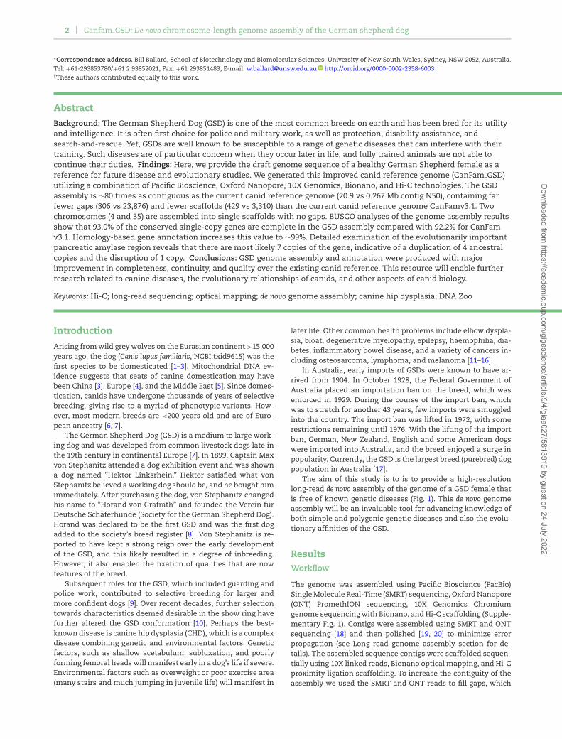
The aim of this study is to is to provide a high-resolutionlong-read de novo assembly of the genome of a GSD female thatis free of known genetic diseases (Fig. 1). This de novo genomeassembly will be an invaluable tool for advancing knowledge ofboth simple and polygenic genetic diseases and also the evolu-tionary affinities of the GSD.
ResultsWorkflow
The genome was assembled using Pacific Bioscience (PacBio)Single Molecule Real-Time (SMRT) sequencing, Oxford Nanopore(ONT) PromethION sequencing, 10X Genomics Chromiumgenome sequencing with Bionano, and Hi-C scaffolding (Supple-mentary Fig. 1). Contigs were assembled using SMRT and ONTsequencing [18] and then polished [19, 20] to minimize errorpropagation (see Long read genome assembly section for de-tails). The assembled sequence contigs were scaffolded sequen-tially using 10X linked reads, Bionano optical mapping, and Hi-Cproximity ligation scaffolding. To increase the contiguity of theassembly we used the SMRT and ONT reads to fill gaps, which
Dow
nloaded from https://academ
ic.oup.com/gigascience/article/9/4/giaa027/5813919 by guest on 24 July 2022
Field et al. 3
Figure 1: ”Nala” the female German Shepherd Dog. Nala, or formally “JonkahraNala” (Australian Registration #2100398550), was born in 2013 and is free of all
known genetic diseases. Her sire was imported from Germany, and her dam isfrom Australian lines.
was then followed by a final round of polishing. Homology-basedgene prediction was performed using C. lupus familiaris and 8 re-lated mammals. The resulting chromosome-length genome as-sembly and its gene annotation was deposited to NCBI with ac-cession number GCA 008641055.2. The mitochondrial genome(VSDE01000430) was subsequently added to the assembly. Fi-nally, comparisons with the canine genome of the boxer (Can-Fam3.1) were made [21].
Assembly statistics/completeness
The final submission contains 2,407,291,559 total bp(2,401,147,102 ungapped), 429 scaffolds with a contig N50length of 20.9 Mb, and a scaffold N50 length of 64.3 Mb. The full-length chromosome scaffolds in the assembly accounted for98.3% of the genome, with only 0.95% of all sequence not align-ing to a CanFam3.1 chromosome. Evaluation by BUSCO (BUSCOv3.0.2b [22], short mode, implementing BLAST+ v2.2.31 [23],HMMer v3.2.1 [24], AUGUSTUS v3.3.2 [25], and EMBOSS v6.6.0)against the Laurasiatheria ob9 dataset (n = 6,253) indicatedthat 93.0% of the conserved single-copy genes were complete(Table 1, Supplementary Tables 1, 2, Supplementary Fig. 2). Eachanalysis step in assembly, scaffolding, and polishing improvedscaffold N50 and/or BUSCO scores, consistent with improvingassembly quality (Supplementary Table 1, Supplementary Fig.2). BUSCO predictions are sensitive to changes in sequence andassembly size, with scaffolding and polishing causing losses aswell as gains (Supplementary Table 1). Compiling BUSCO resultsacross all assembly stages (BUSCOMP v0.8.0) reveals that ≥6,085(97.3%) are present and complete in the assembly, with only 118genes (1.9%) not found at any stage.
Additional k-mer analysis of the final assembly was per-formed using KAT v2.4.2 [26]. KAT comp was used to comparek-mer frequencies from the 10X reads (16 bp barcode trimmedfrom read 1) with their copy number in the assembly. This com-parison revealed no sign of missing data nor large duplications,including retention of haplotigs (Supplementary Fig. 3).
Comparison with CanFam3.1
The GSD assembly was compared with the current referencegenome CanFam3.1. Results are summarized in Table 1.
The GSD assembly offers improvements over CanFam3.1 us-ing a wide variety of metrics. The GSD assembly has a contig N50
that is almost 80 times greater than CanFam3.1 and contains 78times fewer gaps, and 2,881 fewer scaffolds. BUSCO results onthe genome also indicate an improvement in the GSD assembly,with 47 more complete genes (25 fewer fragmented genes and22 fewer missing genes).
On the basis of the existing CanFam3.1 annotation and theGSD annotation provided by GeMoMa [27], the longest full-length transcript per gene was selected to avoid an overestima-tion of duplicated genes by BUSCO v3.02. Comparing the BUSCOstatistics for the annotations, a clear improvement from 95.1%to 98.9% complete single-copy orthologs could be observed.
Variation relative to CanFam3.1All 39 full-length chromosomes in the final assembly werealigned to the corresponding chromosomes in CanFam3.1 usingMUMmer4 [28]. Single-nucleotide polymorphisms (SNPs) andsmall indels (deletions and insertions <50 bp) were called us-ing the MUMmer4 call-SNPs module. In total 3,137,227 SNVs and5,111,356 small indels were detected (Supplementary Table 3).Copy number variants (CNVs) and structural variants (SVs) werecalled using svmu (v0.2) [29]. Variants >100 bp were extracted,resulting in 66,673 total CNV/SVs. By variant type, this was bro-ken down into 39,742 CNVs, 13,552 insertions, 13,150 deletions,and 229 inversions (Supplementary Table 4).
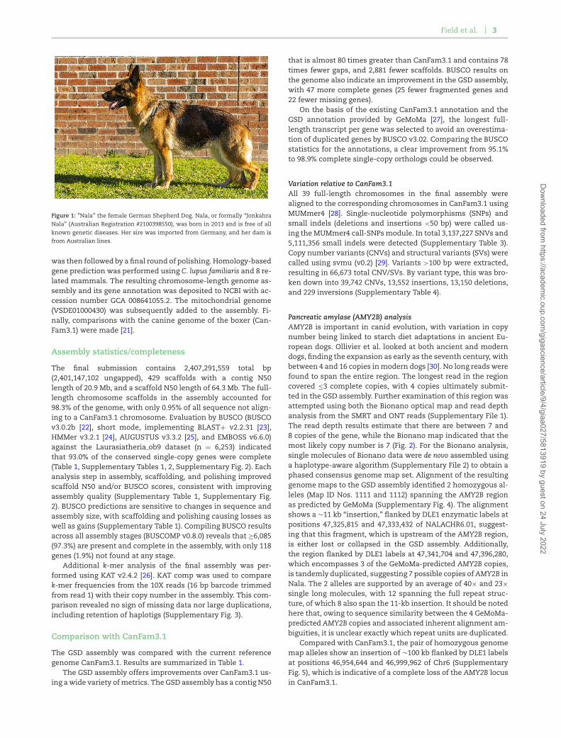
Pancreatic amylase (AMY2B) analysisAMY2B is important in canid evolution, with variation in copynumber being linked to starch diet adaptations in ancient Eu-ropean dogs. Ollivier et al. looked at both ancient and moderndogs, finding the expansion as early as the seventh century, withbetween 4 and 16 copies in modern dogs [30]. No long reads werefound to span the entire region. The longest read in the regioncovered ≤3 complete copies, with 4 copies ultimately submit-ted in the GSD assembly. Further examination of this region wasattempted using both the Bionano optical map and read depthanalysis from the SMRT and ONT reads (Supplementary File 1).The read depth results estimate that there are between 7 and8 copies of the gene, while the Bionano map indicated that themost likely copy number is 7 (Fig. 2). For the Bionano analysis,single molecules of Bionano data were de novo assembled usinga haplotype-aware algorithm (Supplementary File 2) to obtain aphased consensus genome map set. Alignment of the resultinggenome maps to the GSD assembly identified 2 homozygous al-leles (Map ID Nos. 1111 and 1112) spanning the AMY2B regionas predicted by GeMoMa (Supplementary Fig. 4). The alignmentshows a ∼11 kb “insertion,” flanked by DLE1 enzymatic labels atpositions 47,325,815 and 47,333,432 of NALACHR6.01, suggest-ing that this fragment, which is upstream of the AMY2B region,is either lost or collapsed in the GSD assembly. Additionally,the region flanked by DLE1 labels at 47,341,704 and 47,396,280,which encompasses 3 of the GeMoMa-predicted AMY2B copies,is tandemly duplicated, suggesting 7 possible copies of AMY2B inNala. The 2 alleles are supported by an average of 40× and 23×single long molecules, with 12 spanning the full repeat struc-ture, of which 8 also span the 11-kb insertion. It should be notedhere that, owing to sequence similarity between the 4 GeMoMa-predicted AMY2B copies and associated inherent alignment am-biguities, it is unclear exactly which repeat units are duplicated.
Compared with CanFam3.1, the pair of homozygous genomemap alleles show an insertion of ∼100 kb flanked by DLE1 labelsat positions 46,954,644 and 46,999,962 of Chr6 (SupplementaryFig. 5), which is indicative of a complete loss of the AMY2B locusin CanFam3.1.
Dow
nloaded from https://academ
ic.oup.com/gigascience/article/9/4/giaa027/5813919 by guest on 24 July 2022
4 Canfam GSD: De novo chromosome-length genome assembly of the German shepherd dog
Table 1: Genome assembly and annotation statistics for GSD assembly vs CanFam3.1
Statistic GSD CanFam3.1
Total sequence length 2,407,291,559 2,410,976,875Total ungapped length 2,401,147,102 2,392,715,236No. of contigs 735 27,106Contig N50 20,914,347 267,478Contig L50 37 2,436No. of scaffolds 410 3,268Scaffold N50 64,346,267 63,241,923Scaffold L50 15 15No. of gaps 306 23,876BUSCO complete (genome) 93.0% (91.6% single copy, 1.4% duplicate copy) 92.2% (91.1% single copy, 1.1% duplicate copy)BUSCO fragmented (genome) 3.6% 4.0%BUSCO missing (genome) 3.4% 3.8%BUSCO complete (annotation) 98.9% (96.5% single copy, 2.4% duplicate copy) 95.1% (94.1% single copy, 1.0% duplicate copy)BUSCO fragmented (annotation) 1.0% 1.9%BUSCO missing (annotation) 0.1% 3.0%
Figure 2: Bionano genome map alleles aligned to hypothetical sequence constructs. The hypothetical sequence constructs (green bars) contain either 7 (labelled”amy2b dom7copyext”) or 8 (labelled ”amy2b dom8copyext”) copies of the repeat unit (highlighted by coloured boxes within the green bar and numbered in whitefont). Dark blue and yellow vertical lines on the sequence contig and consensus map indicate matching and non-matching DLE1 enzymatic labels, respectively.
To better determine whether Bionano data support 7 or 8copies of the AMY2B repeat, we compared the 2 genome mapalleles against 2 synthetic sequence constructs containing ei-ther 7 (amy2b dom7copyext) or 8 (amy2b dom8copyext) copiesof the 14,862-bp AMY2B repeat with the highest read depth sup-port (namely, the third copy) from the GSD assembly, flankedby ∼401.5-kb sequences assembled from SMRT and ONT reads(Supplementary File 1). Alignment results confirmed the pres-ence of 7 repeat units, showing a perfect alignment to the 7-copysequence construct (Fig. 2A), but a “deletion” of 1 repeat unit rel-ative to the 8-copy construct (Fig. 2B).
Olfactory receptor and keratin cluster analysesCorrect annotation of the olfactory and keratin clusters in ca-nines has been problematic, but it is important for research oncanid health and evolution [21, 31, 32]. Dogs are macrosmaticanimals that rely highly on their sense of smell. Yet, the molec-ular basis of such prominent chemosensory capacities remainslargely unknown. The ability to detect and discriminate the mul-titude of odors in vertebrates is mediated by a superfamily ofG protein–coupled olfactory receptor (OR) proteins [33]. Based
on the description in the reference annotations, we filtered allmessenger RNAs (mRNAs) of the references that contain in theirdescription the regular expression “olfactory receptor.” We thenextracted the number of mRNAs and genes per reference organ-ism. Subsequently, we used the IDs to filter the GSD annotationand counted the number of predicted mRNAs and genes. Thisprocedure identifies 1,250 mRNAs and 933 genes in the GSD and849 mRNAs and 804 genes in the boxer. Quignon et al. [31] iden-tified 5 amino acid patterns characteristic of ORs in the caninegenome and retrieved 1,094 dog genes (872 genes and 222 pseu-dogenes).
Keratins are filament proteins of the epithelial cytoskeletonand are essential for normal skin homeostasis. Over time thegenes encoding keratins have undergone multiple rounds of du-plication, with high similarity between different keratin par-alogs [32]. Analogously to the olfactory receptor study, we fil-tered all mRNAs of the references that contain in their descrip-tion the keyword “keratin\d.” This procedure identifies 118 mR-NAs and 83 genes in the GSD and 73 mRNAs and 55 genes inthe boxer. Balmer et al. [32] investigated the NCBI (dog annota-tion release 103) gene predictions for the canine gene clusters
Dow
nloaded from https://academ
ic.oup.com/gigascience/article/9/4/giaa027/5813919 by guest on 24 July 2022
Field et al. 5
to RNA-sequencing data that were generated from adult skin of5 dogs and adult hair follicle tissue of 1 dog and annotated 61putatively functional keratin genes in the dog.
Discussion
Concerns for the health welfare of the GSD have been widelyaired [34, 35]. The GSD had the highest number of publishedpredispositions to inherited diseases overall among the 50 mostcommonly registered Kennel Club breeds and had the second-highest number of disorders exacerbated by conformation, ex-ceeded only by the Great Dane [36]. The British Kennel ClubBreed Watch system categorizes the GSD as a Category Threebreed “requiring particular monitoring and additional support”and considered to be more susceptible to developing specifichealth conditions associated with exaggerated conformation.Breed Watch points of concern include cow hocks, excessiveturn of stifle, nervous temperament, sickle hock, and weakhindquarters [37].
The high-quality genome assembly will advance knowledgeof breed-specific diseases such as CHD and extend to issuesrelated to canine personality. The severity of CHD depends onboth genetic and environmental factors. In GSDs, the heritabil-ity (h2) estimates have varied from 0.1 to 0.6 [38]. To date, differ-ent study populations and methods affect the results substan-tially because the reported quantitative trait locus associationand candidate genes are inconsistent between studies [39–41].While boxers are prone to CHD, the hip scores of ”Tasha” (usedfor CanFam) are unknown. Furthermore, GSD-specific SNPs aswell as significant CNVs and SVs are difficult to detect. Con-cerning canine personality, Saetre et al. [42] examined how traitsare transmitted between generations in a cohort containing>10,000 behaviorally tested GSD and Rottweiler dogs. In bothbreeds, the pattern of co-inheritance was found to be similar fora broad personality trait previously named shyness–boldness,with heritability estimated to be 0.25 in the 2 breeds. Cur-rently, the underlying genes involved in these behaviors are notknown.
The assembly is expected to enable the selection of GSDsfor particular duties including police work, where their sen-sitive nose is frequently used to discriminate odors. Robin etal. [43] analysed the nucleotide sequences of 109 OR genes(102 genes and 7 pseudogenes) in 6 different breeds includ-ing GSDs. In this study, they showed that OR genes are highlypolymorphic, with a mean of 1 SNP per 577 nucleotides. How-ever, the degree of polymorphism observed is highly variable,with some OR genes having few if any SNPs and others be-ing highly polymorphic (1 SNP/122 nucleotides). Yang et al. [44]conducted a preliminary study of 22 SNPs from the exonic re-gions of 12 OR genes in GSDs and found a significant correlationbetween SNP genotypes of OR genes and olfactory abilities ofdogs.
We envisage that these data will also facilitate understand-ing of the evolution of dog breeds and canids in general. Theevolutionary position of the GSD among extant breeds is notfirmly established. The Federation Cynologique Internationaleplaces it in Group 1 as part of the Herding group. Bigi et al.[45] hypothesized that the German Shepherd dog was closelyrelated to the Czechoslovakian wolfdog. More recently Parkeret al. [6] proposed that the GSD is distinct from other herd-ing breeds and in a clade along with the French Berger Pi-card, New Hampshire Chinook, Peruvian hairless, and Mexicanxoloitzcuinti.
Conclusions
This de novo genome assembly and annotation will be an invalu-able tool for advancing knowledge of breed-specific diseases andthe evolutionary affinities of the GSD. Here, we present an im-proved canid genome assembly and annotation relative to Can-Fam 3.1.
MethodsDNA extraction, sequencing, and scaffolding
Sampling: Nala the German Shepherd DogIn selecting an animal for the project, it was considered essen-tial to select a female that had been cleared, as much as possi-ble, of any recognizable inherited conditions. The animal neededto display all the hallmarks of a good quality representative ofthe breed but need not necessarily be a show-winning specimen.Nala is an easygoing and approachable 5.5-year-old female (born5 December 2013) and a treasured family pet that showed typicalappearance for a GSD. She has had no sign of the hip dysplasiathat appears in GSD (Supplementary Fig. 6) or any other knowngenetic diseases. Nala had a combined hip score of 3 (1 on theleft-hand side and 2 on the right-hand side) when the X-ray wastaken at 5 years of age: each hip was measured on a 0–53 scale,with a total of 106 being crippling. The score of 3 is well belowthe current Australian average of 9 for GSDs. She is registeredwith the Australian National Kennel Council (# 2100398550) withher dam from Australian bred lines and sire imported from Ger-many. Her dam and sire remain healthy aging adults withoutdisease. Nala’s dam has 7 progeny radiographed from 4 sireswith no failures. Her sire had 31 progeny radiographed from 13different dames resulting in 4 failures and 27 passes recorded forthe GSD National Council hip scheme. In the Australian 53-pointscoring scheme, a pass is ≤8 in any 1 hip, no point gets a 3, and≤16 in total.
Pacific Biosciences SMRT sequencingGenomic DNA was prepared from 1–2 mL of fresh blood using thegenomic-tip 100/G kit (Qiagen, Hilden, Germany). This was per-formed with supplemental RNase (Astral Scientific, Taren Point,Australia) and proteinase K (NEB, Ipswich, MA, USA) treatment,as per the manufacturer’s instructions. Isolated genomic DNAwas further purified using AMPure XP beads (Beckman Coulter,Brea, CA, USA) to eliminate sequencing inhibitors. DNA puritywas calculated using a Nanodrop spectrophotometer (ThermoFisher Scientific, Waltham, MA, USA), and molecular integritywas assessed using pulse-field gel electrophoresis. DNA in-tegrity was assessed by the Sage Science Pippin Pulse. A 0.75%KBB (Sage Science, Beverly, MA, USA) gel was run on the 9hr 10–48 kb (80 V) program. The DNA ladder used was the Invitrogen1 kb Extension DNA ladder (cat No. 10,511–012). A total of 150 ngof DNA was loaded on the gel.
We generated 2 libraries that were size selected on SageBluePippin gels (Sage Science, Beverly, MA, USA). Libraries weresequenced on Sequel machines with 2.0 chemistry recording10 h movies (PacBio Sequel System, RRID:SCR 017989). Sequenc-ing was conducted at the Ramaciotti Center for Comparative Ge-nomics at University of New South Wales (TOW5157A1, 15 SMRTcells with a total polymerase read length 108.48 Gb) and at theArizona Genomic Institute, University of Arizona (4 SMRT cellswith a total of 11 Gb of data; note: short-read lengths were dueto DNA shearing of the DNA during shipping from Australia toArizona).
Dow
nloaded from https://academ
ic.oup.com/gigascience/article/9/4/giaa027/5813919 by guest on 24 July 2022
6 Canfam GSD: De novo chromosome-length genome assembly of the German shepherd dog
ONT PromethION sequencingDNA (1 μg) was prepared for ONT sequencing using the 1D ge-nomic DNA by ligation kit (SQK-LSK109, ONT) according to thestandard protocol. Long fragment buffer was used for the fi-nal elution to exclude fragments shorter than 1,000 bp. In total,119 ng of adapted DNA was loaded onto a FLO-PRO002 Prome-thION flow cell and run on an ONT PromethION sequencing de-vice (PromethION, RRID:SCR 017987) using MinKNOW (18.08.2)with MinKNOW core (v1. 14.2).
Base-calling was performed after sequencing with the GPU-enabled guppy basecaller (v3.0.3) using the PromethION high-accuracy flip-flop model with config “dna r9.4.1 450bps hac.cfg.”
10X Genomics Chromium sequencingDNA was prepared following the protocol described above forSMRT sequencing. A 10X GEM library was barcoded from highmolecular weight (HMW) DNA according to the manufacturer’srecommended protocols. The protocol used was the ChromiumGenome Reagent Kits v2 User Guide, manual part No. CG00043Rev B [46]. Quality control was performed using LabChip GX(PerkinElmer, Waltham, MA, USA ) and Qubit 2.0 Flurometer(Life Technologies, Carlsbad, CA, USA) at the Kinghorn Cen-tre for Clinical Genomics. The library was run on a single laneof a v2 patterned flowcell. Paired-end sequencing with 150- bpread length was performed using the Illumina HiSeq X (IlluminaHiSeq X Ten, RRID:SCR 016385) within the Kinghorn Centre forClinical Genomics at the Garvan Institute of Medical Research,Sydney, Australia.
DNA methylomeTo explore the regulatory landscape of the GSD, we performedwhole-genome bisulfite sequencing [47] on genomic DNA ex-tracted from whole blood. In concordance with other adult ver-tebrates [48, 49], the GSD genome displays a typical bimodalDNA methylation pattern with >60% of CpG dinucleotides be-ing methylated at levels >80% (hypermethylated) and 12% ofCpG dinucleotides being methylated at ≤20% (hypomethylated).Next, to determine the number and genomic distribution of pu-tative regulatory regions, we segmented the methylome into un-methylated regions (UMRs) and low-methylated regions (LMRs),using the MethylSeekR algorithm [50]. UMRs are fully unmethy-lated and largely coincide with CpG island promoters whereasLMRs display partial DNA methylation, which is characteristicof distal regulatory elements such as enhancers in other mam-malian models [51]. These analyses resulted in the identifica-tion of ∼21,000 UMRs and ∼53,000 LMRs, in line with previouslyreported numbers of promoters and enhancers [50, 52] (Supple-mentary Fig. 7).
Bionano optical mappingHMW DNA was isolated from fresh blood (stored at 4◦C) us-ing the Bionano Prep Blood DNA Isolation Protocol (Bionano Ge-nomics [BNG], Document #30,033 revision C). Briefly, after lysingthe red blood cells, white blood cells were recovered and embed-ded in agarose plugs. These plugs were subjected to ProteinaseK (Qiagen Cat No. 158,920) digestion for 2 rounds (2 hours, thenovernight) at 50◦C. Following extensive washing as prescribed inthe protocol, the plugs were melted and treated with GELase en-zyme (Epicentre, Cat. No. G31200). The resulting HMW DNA wassubjected to drop dialysis, left to equilibrate at room tempera-ture for 4 days, and was then quantified using the Qubit BroadRange dsDNA Assay Kit (Thermo Fisher Scientific).
HMW DNA (∼190 ng/μL) was labelled (BNG, Part No. 20,351)at DLE-1 recognition sites, following the Bionano PrepTM Di-
rect Label and Stain Protocol (BNG, Document No. 30,206 revi-sion C). Labelled DNA was loaded directly onto Bionano SaphyrChips (BNG, Part No. 20,319), without further fragmentation oramplification, and imaged using a Saphyr instrument to gen-erate single-molecule optical maps (Saphyr, RRID:SCR 017992).Multiple cycles were performed to reach an average raw genomedepth of coverage of 190×.
Hi-C chromosome length scaffoldingThe Bionano assembly was further scaffolded to chromosomelength by the DNA Zoo following the prescribed methodology[53]. Briefly, an in situ Hi-C library was prepared [54] from a bloodsample of a purebred male individual named Tydus (AmericanKennel Club Registration DN5364660) provided by the CornellVeterinary Biobank and sequenced to 29× coverage (assuming2.4-Gb genome size).
Genome Assembly WorkflowLong-read genome assembly
The SMRT and ONT reads were corrected and assembled withthe Canu assembler (Canu, RRID:SCR 015880) v1.8.0 [18]. The re-sulting contigs were polished by aligning the raw reads to theassembly and correcting the sequencing errors using 2 roundsof Arrow polishing [19]. There were ∼10 million fixes in the firstround and ∼284,000 fixes in the second. The assembled GSDgenome, with a total length of 2.39 Gb, consisted of 1,389 con-tigs with an N50 length of 15.68 Mb. Following the Arrow pol-ishing there were 1,389 sequences, with a total length of 2.39Gb (including 111 repeats of total length 13,145,025 bp) with nobubbles. There were 2,560,498 unassembled sequences of totallength 17,998,063,955 bp.
10X Chromium linked reads
The Arrow-polished SMRT/ONT assembly was scaffolded us-ing GSD 10X linked reads as in ARCS [55]. The 10X data werealigned using the linked-read analysis software provided by 10XGenomics, Long Ranger, v2.1.6 [56]. Misaligned reads and readsnot mapping to contig ends were removed, and all possible con-nections between contigs were computed keeping best recipro-cal connections. Finally, contig sequences were joined, spacedby 10 kb with stretches of N’s, and if required reverse com-plemented (Supplementary File 3). In total 128 connections be-tween the SMRT/ONT contigs could be established, increasingthe assembly N50 length by 4.6 Mb (from 15.46 to 20.06 Mb; Sup-plementary File 3).
Polishing round 1
To further improve the assembly, another round of polishingwas performed by aligning the Illumina short reads from the10X Chromium sequencing to the assembly using minimap2 [57](v2.16) and correcting the sequencing errors using Racon (Racon,RRID:SCR 017642) v1.3.3 [58].
Optical mapping for super-scaffolding using Bionanodata
Single-molecule optical maps were filtered on minimummolecule length of 150 kb and minimum of 9 label sites permolecule. De novo assembly of single molecules into consensusmaps was performed using the Bionano Solve (v3.2.2 08022018)software with aligner RefAligner (7782.7865rel) [59, 60].
Dow
nloaded from https://academ
ic.oup.com/gigascience/article/9/4/giaa027/5813919 by guest on 24 July 2022
Field et al. 7
Assembly was “haplotype-unaware” such that heterozy-gous alleles were collapsed into haploid representation. In all,∼2 million single molecules with N50 of 220 kb were assembledinto 1,245 optical genome maps with N50 of 3.1 Mb. The finalassembly was in CMAP format (v0.2).
This genome map set was used to scaffold the sequence con-tigs using BNG’s Hybrid Scaffold pipeline (v10252018). In brief,the 1,261 sequence contigs were in silico digested on the basis ofthe DLE-1 motif (CTTAAG) creating sequence maps (CMAP). Se-quence maps were then aligned to the assembled optical mapsbased on DLE-1 labels using RefAligner. Discrete sequence mapsthat can be linked via a Bionano genome map were scaffolded.
Alignments indicating conflict between the sequence andoptical maps, and hence suggestive of misassembly, wereresolved. Specifically, optical maps supported by ≥10 singlemolecules at the conflict site were indicative of sequence mis-assembly, and so the sequence map would be “cut” (split) at theconflict point. In contrast, insufficient single-molecule supportfor the optical map was indicative of optical map assembly er-ror, and so the optical map would be “cut” at the conflict site.Details of the method are provided in the Bionano Solve Theoryof Operation: Hybrid Scaffold (Document No. 30,073). Followinghybrid scaffolding, 21 arbitrary 10-kb N-gaps (introduced duringthe sequence assembly process) were resized on the basis of es-timated inter-label distances from the optical maps. In all, 160sequence contigs were hybrid-scaffolded into 109 hybrid scaf-folds with N50 of ∼46.3 Mb. The remaining 1,004 sequence con-tigs with an N50 of ∼78.8 kb could not be scaffolded either be-cause they were too short (<100 kb) for hybrid scaffolding withBionano maps or because they did not align to any optical maps.
Chromosome-length assembly using Hi-C data
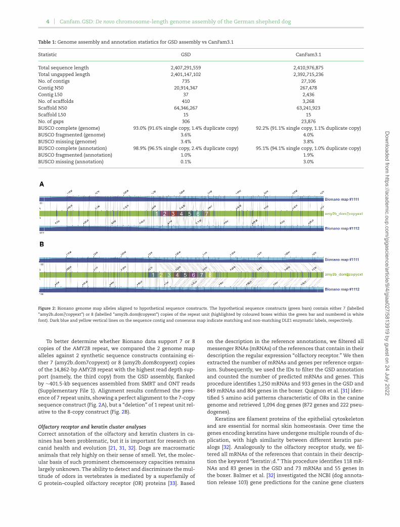
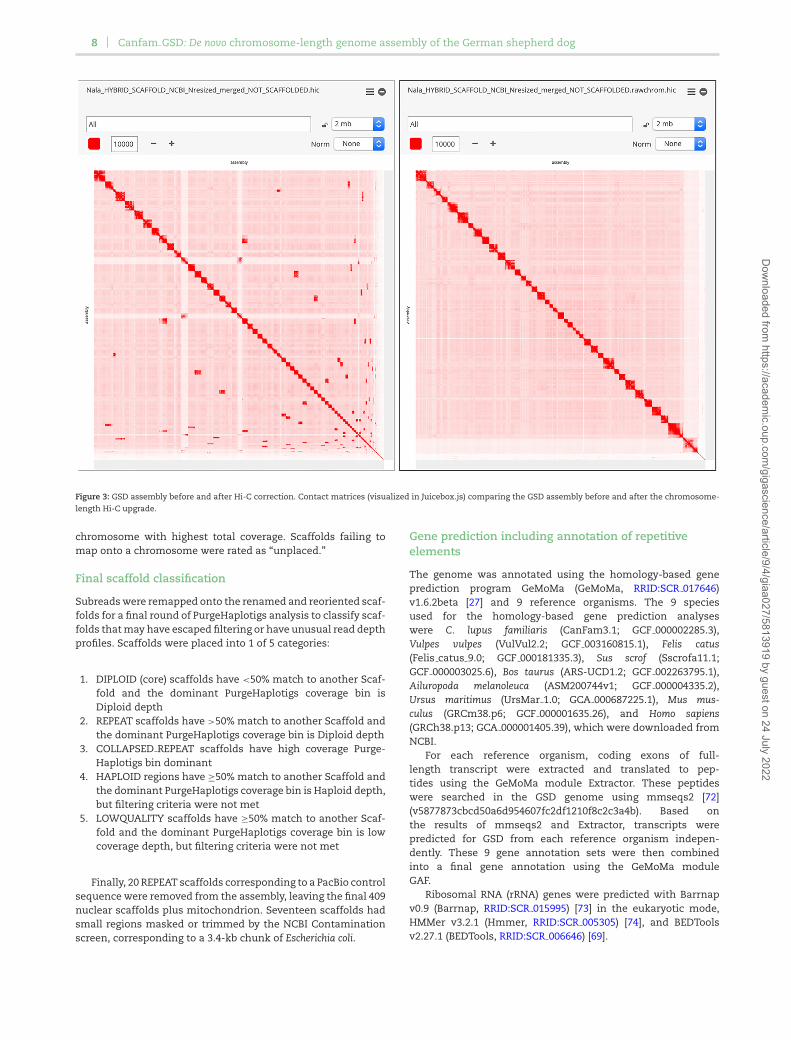
The Hi-C data were processed using Juicer (Juicer, RRID:SCR 017226) [61] and used as input into the 3D-DNA pipeline [62] toproduce a candidate chromosome-length genome assembly. Weperformed additional finishing on the scaffolds using JuiceboxAssembly Tools [63]. Fig. 3 shows the contact matrices generatedby aligning the Hi-C dataset to the genome assembly before theHi-C upgrade (left) and after Hi-C scaffolding (right). The matri-ces are visualized in Juicebox.js, a cloud-based visualization sys-tem for Hi-C data [64], and are available for browsing at multipleresolutions at DNA Zoo [65].
Gap filling
After scaffolding and correction, all raw SMRT and ONT readswere aligned to the assembly with Minimap2 (v2.16) (-ax map-pb/map-ont) and used by PBJelly (pbsuite v.15.8.24) [66] to fillgaps. It was able to completely close 210 gaps, increasing con-tig N50 to the final figure of 20.9 Mb.
Polishing round 2
Following scaffolding, another round of polishing was done tofurther improve the assembly. Polishing was performed by align-ing the Illumina short reads from the Chromium sequencing tothe assembly using Long Ranger v2.2.2 and correcting the SNPsand indels using Pilon (Pilon, RRID:SCR 014731) [20].
Final cleanup
The Pilon-polished genome underwent a final scaffold cleanupto generate a high-quality core assembly, remove low-coverage
artefacts and haplotig sequences, and annotate remaining scaf-folds with potential issues.
Low-coverage filter
The TOW5157A1 library PacBio subreads (12.5 Msubreads; 108 Gb) were mapped onto theNala canu arrow2 10x racon bionano HiC pbjelly pilon as-sembly using Minimap2 v2.16 (-ax map-pb –secondary = no)[57]. Initial read depth analysis was performed with BBMapv38.51 pileup.sh [67]. Any scaffolds with median coverage <3(e.g., <50% of the scaffold covered by ≥3 reads) were filtered outas low-coverage scaffolds. Of the 1,057 Pilon-polished scaffolds,220 scaffolds were removed in the initial low-coverage filter,leaving 837 scaffolds.
Purge Haplotigs analysis—round 1
Subreads were remapped on the remaining 837 scaffolds andprocessed with PurgeHaplotigs v20190612 [68] (implementingPerl v5.28.0, BEDTools v2.27.1 [69], R v3.5.3, and SAMTools v1.9[70]). Based on the PurgeHaplotigs depth histogram, low-, mid-and high-depth thresholds were set to 5×, 30×, and 80×. Anyscaffolds with <80% at diploid read depth were identified byPurgeHaplotigs for reassignment. Scaffolds with ≥80% bases inthe low/haploid coverage bins and ≥95% of their length mappedby PurgeHaplotigs onto another scaffold were filtered as hap-lotigs or assembly artefacts. Any other scaffolds with ≥80% low-coverage bases were filtered as ”low coverage.” This analysis re-sulted in a further 11 scaffolds filtered for low coverage and 268filtered as haplotigs or assembly artefacts, leaving 558 scaffolds.
Purge Haplotigs analysis—round 2
Subreads were remapped onto the remaining 558 scaffolds, re-sulting in a further 128 scaffolds filtered as haplotigs or as-sembly artefacts, leaving 430 scaffolds. No additional scaffoldswith ≥80% low-coverage bases were identified. Any scaffold with≥80% bases in the low/haploid coverage bins were filtered ashaplotigs or assembly artefacts. Scaffolds with ≥20% diploidcoverage were marked as retention as probable diploids. Scaf-folds with <20% diploid coverage and ≥50% high coverage weremarked as probable collapsed repeats. A single remaining scaf-fold marked as ”junk” by PurgeHaplotigs (>80% low/high cover-age) was also filtered as a probable artefact.
Purge Haplotigs analysis—round 3
Subreads were remapped onto the remaining 430 scaffolds fora third round of PurgeHaplotigs analysis. No further scaffoldswere identified for filtering.
CanFam3.1 chromosome mapping
The CanFam v3.1 reference genome was downloaded from En-sembl (Release 97, download date 5 August 2019). Full-lengthchromosomes were renamed with a CANFAMCHR prefix andused for reference mapping. The final Nala genome assem-bly was mapped onto the CanFam3.1 reference genome usingMinimap2 v2.16 [57] (-x asm5 –secondary = no –cs) to gener-ate PAF output. Scaffolds were assigned to CanFam3.1 chro-mosomes using PAFScaff v0.2.0 (PAFScaff, RRID:SCR 017976) [71]based on Minimap2-aligned assembly scaffold coverage againstthe reference chromosomes. Scaffolds were assigned to the
Dow
nloaded from https://academ
ic.oup.com/gigascience/article/9/4/giaa027/5813919 by guest on 24 July 2022
8 Canfam GSD: De novo chromosome-length genome assembly of the German shepherd dog
Figure 3: GSD assembly before and after Hi-C correction. Contact matrices (visualized in Juicebox.js) comparing the GSD assembly before and after the chromosome-length Hi-C upgrade.
chromosome with highest total coverage. Scaffolds failing tomap onto a chromosome were rated as “unplaced.”
Final scaffold classification
Subreads were remapped onto the renamed and reoriented scaf-folds for a final round of PurgeHaplotigs analysis to classify scaf-folds that may have escaped filtering or have unusual read depthprofiles. Scaffolds were placed into 1 of 5 categories:
1. DIPLOID (core) scaffolds have <50% match to another Scaf-fold and the dominant PurgeHaplotigs coverage bin isDiploid depth
2. REPEAT scaffolds have >50% match to another Scaffold andthe dominant PurgeHaplotigs coverage bin is Diploid depth
3. COLLAPSED REPEAT scaffolds have high coverage Purge-Haplotigs bin dominant
4. HAPLOID regions have ≥50% match to another Scaffold andthe dominant PurgeHaplotigs coverage bin is Haploid depth,but filtering criteria were not met
5. LOWQUALITY scaffolds have ≥50% match to another Scaf-fold and the dominant PurgeHaplotigs coverage bin is lowcoverage depth, but filtering criteria were not met
Finally, 20 REPEAT scaffolds corresponding to a PacBio controlsequence were removed from the assembly, leaving the final 409nuclear scaffolds plus mitochondrion. Seventeen scaffolds hadsmall regions masked or trimmed by the NCBI Contaminationscreen, corresponding to a 3.4-kb chunk of Escherichia coli.
Gene prediction including annotation of repetitiveelements
The genome was annotated using the homology-based geneprediction program GeMoMa (GeMoMa, RRID:SCR 017646)v1.6.2beta [27] and 9 reference organisms. The 9 speciesused for the homology-based gene prediction analyseswere C. lupus familiaris (CanFam3.1; GCF 000002285.3),Vulpes vulpes (VulVul2.2; GCF 003160815.1), Felis catus(Felis catus 9.0; GCF 000181335.3), Sus scrof (Sscrofa11.1;GCF 000003025.6), Bos taurus (ARS-UCD1.2; GCF 002263795.1),Ailuropoda melanoleuca (ASM200744v1; GCF 000004335.2),Ursus maritimus (UrsMar 1.0; GCA 000687225.1), Mus mus-culus (GRCm38.p6; GCF 000001635.26), and Homo sapiens(GRCh38.p13; GCA 000001405.39), which were downloaded fromNCBI.
For each reference organism, coding exons of full-length transcript were extracted and translated to pep-tides using the GeMoMa module Extractor. These peptideswere searched in the GSD genome using mmseqs2 [72](v5877873cbcd50a6d954607fc2df1210f8c2c3a4b). Based onthe results of mmseqs2 and Extractor, transcripts werepredicted for GSD from each reference organism indepen-dently. These 9 gene annotation sets were then combinedinto a final gene annotation using the GeMoMa moduleGAF.
Ribosomal RNA (rRNA) genes were predicted with Barrnapv0.9 (Barrnap, RRID:SCR 015995) [73] in the eukaryotic mode,HMMer v3.2.1 (Hmmer, RRID:SCR 005305) [74], and BEDToolsv2.27.1 (BEDTools, RRID:SCR 006646) [69].
Dow
nloaded from https://academ
ic.oup.com/gigascience/article/9/4/giaa027/5813919 by guest on 24 July 2022
Field et al. 9
Availability of Supporting Data and Materials
The complete genome build is available at NCBI (Gen-Bank accession No. GCA 008641055.2). DNA Methylationdata GEO accession is GSE136348. PAFScaff (PAFScaff,RRID:SCR 017976) is GPLv3 licensed and registered at bio.tools(biotools: PAFScaff Pairwise mApping Format reference-based scaffold anchoring and super-scaffolding.) [75]. Allsupporting data and materials are also available in the Giga-Science GigaDB database [76].
Additional Files
Supplementary File 1: Read depth analysis of Amy2B regionSupplementary File 2: Bionano AMY2B methodsSupplementary File 3: 10X chromium workflow detailsSupplementary Figure 1: Schematic overview of project work-flow. German Shepherd Dog (“Nala” or Jonkahra Nala) DNA wasderived from blood of a single female . Her dam, Jonkahra LetsElope, was from Australian breeding and the sire, CH ArkonVom Altenberger Land was imported from Germany. Sequenceswere generated on the Pacific Biosciences Sequel instrument(V2 chemistry) and Oxford Nanopore PromethION instrument(guppy basecaller Version 3.0.6 + 9999d81) to ∼30× genome cov-erage, each, based on a genome size estimate of 2.4 Gb (thisestimate is used for all coverage estimates). All long-read se-quences were assembled with the Canu v1.8 algorithm then er-ror corrected twice using the Arrow genomic consensus polish-ing module. The assembly was scaffolded with Chromium 10Xlinked reads (∼41× coverage excluding the barcode) using LongRanger v2.1.6 using DNA from the same animal. Polishing ofthe assembly for residual indels was done by aligning the Il-lumina data with Minimap2 and the Racon algorithm. Single-molecule Bionano data (∼57× effective coverage) were then usedto super-scaffold the sequence assembly using DNA extractedfrom the same canid. For this, single-molecule optical mapswere first de novo assembled into consensus maps, which werethan aligned to the sequence assembly in silico digested withthe same labelling enzyme for hybrid scaffolding, using Bio-nano Solve (v3.2.2 0 802 2018) with RefAligner (7782.7865rel).This assembly was further scaffolded to chromosome lengthby the DNA Zoo following the methodology described here: www.dnazoo.org/methods. Briefly, an in situ Hi-C library was pre-pared from a blood sample of a purebred GSD male named Ty-dus and sequenced to 29× coverage. The Hi-C data were pro-cessed using Juicer and used as input into the 3D-DNA pipelineto produce a candidate chromosome-length genome assembly.We performed additional finishing on the scaffolds using Juice-box Assembly Tools. The assembly was then long-read gap filledwith the PBJelly algorithm, and the additional data error cor-rected using Arrow. The Chromium data were mapped onto theassembly with the Long Ranger v2.1.6 program, and the final as-sembly was then polished using the Pilon algorithm. Of the 2.4-Gb assembled genome (German Shepherd breed-1.0), the totalassembly N50 contig and scaffold lengths are 23.1 and 64.3 Mb,respectively. The genome was annotated using the homology-based gene prediction program GeMoMa (version 1.6.2beta) and9 reference organisms. The assembled contigs were then alignedto CanFam3.1 for chromosome assignments.Supplementary Figure 2: BUSCO improvements in assemblyquality at each analysis step. a. BUSCO ratings for differentstages of Nala assembly, compared to CanFam 3.1. See Supple-mentary Table 1 for descriptions of assembly stages. C: com-plete; S: single copy; D: duplicated; F: fragmented; M: missing;
n: No. BUSCO genes. b. Missing BUSCO genes (%) vs scaffoldNG50 (2.41-Gb genome size). Purple: original assembly; black:scaffolding/polishing steps; blue: final assembly; red: CanFam3.1. Dashed red lines mark CanFam 3.1 statistics.Supplementary Figure 3: KAT k-mer analysis of Nala assembly.10X read k-mer frequency distributions for k-mers with differentassembly copy numbers derived from (A) Read 1 (16 bp barcodestrimmed) and (B) Read 2 (barcodes not trimmed).Supplementary Figure 4: Bionano consensus maps aligned toGSD contig NALACHR6.01. Overlay on the GSD contig (green bar)are 4 GeMoMa-predicted AMY2B transcripts, labelled R0–R3. Be-low and above the pair of Bionano consensus map alleles (bluebars) are single molecules (orange lines) supporting the genomemap assembly. Dark blue and yellow vertical lines on the se-quence contig and consensus map indicate matching and non-matching DLE1 enzymatic labels, respectively. DLE1 enzymaticlabels on single molecules are shown as dark blue or light orangedots for matching and non-matching labels, respectively. Bothgenome map alleles harbour a ∼11-kb “insertion” upstream ofthe AMY2B repeat (highlighted in teal) and a ∼54.6-kb tandemduplication marked by DLE1 labels at positions 47,341,704 and47,396,280 of the GSD NALACHR6.01 contig.Supplementary Figure 5: Bionano consensus maps aligned toCanFam3 Chr6. Relative to CanFam3, the homozygous Bionanogenome map alleles, Map Nos. 1111 and 1112, both containan ∼100-kb insertion flanked by DLE1 labels at 46,954,644 and46,999,962 on chromosome 6.Supplementary Figure 6: Hip X-ray of the German Shepherd DogNala. Her combined hip score of 3 (1 on left-hand side and 2 onright-hand side) when the X-ray was taken at 5 years of age: eachhip was measured on a 0–53 scale, with a total of 106 being crip-pling. This score is well below the current Australian average of9 for GSDs.Supplementary Figure 7: DNA methylation profiling of Ger-man Shepherd Dog Nala’s whole blood. (A) Percentage of CpGdinucleotides with different levels of methylation. High, 80–100%; medium, 20–80%; low, >0–20%; no, 0. (B) Segmentationof hypomethylated regions into CpG-rich unmethylated regions(UMRs) and CpG-poor low-methylated regions (LMRs). The num-ber of CpGs (log2) per region relative to its median methylation isshown. (C) Average DNA methylation profiles of UMRs and LMRs.(D) IGV browser track depicting mC profile and putative regula-tory elements.Supplementary Table 1: Summary assembly scaffold and BUSCOstatistics for different Nala assembly stages, CanFam 3.1, andcompiled best ratings.Supplementary Table 2: BUSCO gene rating for different Nalaassembly stages, CanFam 3.1, and compiled best ratings.Supplementary Table 3: GSD SNVs and small indels summaryby chromosome.Supplementary Table 4: GSD copy number and structural vari-ants (>100 bp) summary by chromosome.
Abbreviations
BLAST: Basic Local Alignment Search Tool; BMG: Bionano Ge-nomics; bp: base pairs; BUSCO: Benchmarking Universal Single-Copy Orthologs; CHD: canine hip dysplasia; CNV: copy num-ber variant; Gb: gigabase pairs; GPU: graphics processing unit;GSD: German Shepherd Dog; HMM: hidden Markov model; HMW:high molecular weight; kb: kilobase pairs; LMR: low-methylatedregion; Mb: megabase pairs; mRNA: messenger RNA; NCBI:National Center for Biotechnology Information; ONT: Oxford
Dow
nloaded from https://academ
ic.oup.com/gigascience/article/9/4/giaa027/5813919 by guest on 24 July 2022
10 Canfam GSD: De novo chromosome-length genome assembly of the German shepherd dog
Nanopore Technologies; OR: olfactory receptor; ORF: open read-ing frame; PacBio: Pacific Biosciences; qPCR: quantitative poly-merase chain reaction; rRNA: ribosomal RNA; SMRT: single-molecule real time; SNP: single-nucleotide polymorphism; SNV:single-nucleotide variant; SV: structural variant; UMR: unmethy-lated region.
Ethics Approval and Consent to Participate
All experimentation was performed under the approval of theUniversity of New South Wales Ethics Committee (ACEC ID:18/18B).
Competing Interests
The authors declare that they have no competing interests.
Funding
This work was supported by the Australian Health Foundationaward and by the Hip2Fit Crowdfunding initiative to J.W.O.B.and R.Z. Matching funds were provided by the University ofNew South Wales/School of Biotechnology and BiomolecularSciences Genomics Initiative. V.M.H. funded the Bionano datacollection. Hi-C scaffolding was performed and funded by theDNA Zoo Consortium. M.A.F. is funded by NHMRC APP5121190.E.L.A. was supported by an NSF Physics Frontiers Center Award(PHY1427654), the Welch Foundation (Q-1866), a USDA Agricul-ture and Food Research Initiative Grant (2017–05741), an NIH4D Nucleome Grant (U01HL130010), and an NIH Encyclopediaof DNA Elements Mapping Center Award (UM1HG009375). TheRamaciotti Centre for Genomics acknowledges infrastructurefunding from the Australian Research Council (LE150100031),the Australian Government NCRIS scheme administered by Bio-platforms Australia, and the New South Wales Government Re-search Attraction and Acceleration Program .
Authors’ Contributions
J.W.O.B. coordinated the project. M.A.F., B.D.R., T.P.L.S., andJ.W.O.B. designed the study. J.W.O.B funded the project. R.A.Z.provided the GSD samples. R.L. and D.T. performed genomicDNA extractions. K.B. and M.A.S. performed the ONT sequenc-ing, and R.L., the Bionano optical mapping. B.D.R. performed theinitial assembly and polishing, A.E.R. performed the chromiumscaffolding, and E.K.F.C and V.M.H. performed the Bionanosuper-scaffolding. O.D., A.O., and Z.C. performed the Hi-C experi-ment, and O.D. and E.L.A. conducted the Hi-C analyses. K.S. andO.B. conducted the DNA methylation analyses. M.A.F. and R.Eperformed all analyses of genome completeness. R.E. performedthe final polishing, final assembly cleanup, and KAT analysis.J.K. performed the genome annotation. R.E. performed the rRNAannotation. R.E., E.K.F.C., and B.D.R. performed the AMY2B anal-yses. M.A.F., B.D.R., O.D., R.E., A.E.M., E.K.F.C., O.B., and J.W.O.B.wrote the manuscript. All authors edited and approved the finalmanuscript.
Acknowledgements
Comments from 2 reviewers improved the manuscript. Wewould like to thank Helaya-Henderson Smith for providing fre-quent access to Nala. Staff at the Vineyard Veterinary Hospitalprovided constant encouragement. James Ferguson was instru-
mental in facilitating the ONT data collection. A whole-bloodsample for Hi-C library preparation was provided by Susan Gar-rison LVT, BT, Sample Collection Coordinator, Cornell Veteri-nary Biobank. SMRT sequencing was conducted at the Rama-ciotti Center for Comparative Genomics at University of NewSouth Wales and at the Arizona Genomic Institute, University ofArizona. The ONT, 10X Chromium, and Bionano genomics datawere collected at the Garvan Institute and the Hi-C data at BaylorCollege of Medicine.
References
1. Frantz LA, Mullin VE, Pionnier-Capitan M, et al. Genomic andarchaeological evidence suggest a dual origin of domesticdogs. Science 2016;352(6290):1228–31.
2. Freedman AH, Gronau I, Schweizer RM, et al. Genome se-quencing highlights the dynamic early history of dogs. PLosGenet 2014;10(1):e1004016.
3. Savolainen P, Zhang YP, Luo J, et al. Genetic evidencefor an East Asian origin of domestic dogs. Science2002;298(5598):1610–3.
4. Thalmann O, Shapiro B, Cui P, et al. Complete mitochondrialgenomes of ancient canids suggest a European origin of do-mestic dogs. Science 2013;342(6160):871–4.
5. Vonholdt BM, Pollinger JP, Lohmueller KE, et al. Genome-wide SNP and haplotype analyses reveal a rich history un-derlying dog domestication. Nature 2010;464(7290):898–902.
6. Parker HG, Dreger DL, Rimbault M, et al. Genomic analy-ses reveal the influence of geographic origin, migration, andhybridization on modern dog breed development. Cell Rep2017;19(4):697–708.
7. Talenti A, Dreger DL, Frattini S, et al. Studies of modern Ital-ian dog populations reveal multiple patterns for domesticbreed evolution. Ecol Evol 2018;8(5):2911–25.
8. Willis MB. The German Shepherd Dog: Its History, Develop-ment and Genetics. New York: Arco; 1977.
9. Samms S. German Shepherd Dog: A Comprehensive Guideto Owning and Caring for Your Dog. London: Kennel ClubBooks; 2003.
10. Benninger MI, Seiler GS, Robinson LE, et al. Three-dimensional motion pattern of the caudal lumbar and lum-bosacral portions of the vertebral column of dogs. Am J VetRes 2004;65(5):544–51.
11. Shaffer LG, Ramirez CJ, Phelps P, et al. An international ge-netic survey of breed-specific diseases in working dogs fromthe United States, Israel, and Poland. Cytogenet Genome Res2017;153(4):198–204.
12. Boge GS, Moldal ER, Dimopoulou M, et al. Breed susceptibil-ity for common surgically treated orthopaedic diseases in 12dog breeds. Acta Vet Scand 2019;61(1):19.
13. Peiravan A, Bertolini F, Rothschild MF, et al. Genome-wideassociation studies of inflammatory bowel disease in Ger-man shepherd dogs. PLoS One 2018;13(7):e0200685.
14. Soo M, Lopez-Villalobos N, Worth AJ. Heritabilities and ge-netic trends for elbow score as recorded by the New ZealandVeterinary Association Elbow Dysplasia Scheme (1992-2013)in four breeds of dog. N Z Vet J 2018;66(3):154–61.
15. Wah lJM, Herbst SM, Clark LA, et al. A review of hered-itary diseases of the German shepherd dog. J Vet Behav2008;3:255–65.
16. Christopherson PW, Bacek LM, King KB, et al. Two novel mis-sense mutations associated with hemophilia A in a fam-ily of Boxers, and a German shepherd dog. Vet Clin Pathol
Dow
nloaded from https://academ
ic.oup.com/gigascience/article/9/4/giaa027/5813919 by guest on 24 July 2022
Field et al. 11
2014;43(3):312–6.17. Shariflou MR, James JW, Nicholas FW, et al. A genealog-
ical survey of Australian registered dog breeds. Vet J2011;189(2):203–10.
18. Koren S, Walenz BP, Berlin K, et al. Canu: scalable and accu-rate long-read assembly via adaptive k-mer weighting andrepeat separation. Genome Res 2017;27(5):722–36.
19. Pacific Biosciences. Genomic Consensus. https://github.com/PacificBiosciences/GenomicConsensus.Accessed 1 Novem-ber 2019.
20. Walker BJ, Abeel T, Shea T, et al. Pilon: an integrated toolfor comprehensive microbial variant detection and genomeassembly improvement. PLoS One 2014;9(11):e112963.
21. Lindblad-Toh K, Wade CM, Mikkelsen TS, et al. Genome se-quence, comparative analysis and haplotype structure of thedomestic dog. Nature 2005;438(7069):803–19.
22. Simao FA, Waterhouse RM, Ioannidis P, et al. BUSCO: assess-ing genome assembly and annotation completeness withsingle-copy orthologs. Bioinformatics 2015;31(19):3210–2.
23. Altschul SF, Gish W, Miller W, et al. Basic Local AlignmentSearch Tool. J Mol Biol 1990;215(3):403–10.
24. Finn RD, Clements J, Eddy SR. HMMER web server: in-teractive sequence similarity searching. Nucleic Acids Res2011;39(Web Server issue):W29–37.
25. Stanke M, Morgenstern B. AUGUSTUS: a web server forgene prediction in eukaryotes that allows user-defined con-straints. Nucleic Acids Res 2005;33(Web Server issue):W465–7.
26. Mapleson D, Garcia Accinelli G, Kettleborough G, et al. KAT:a K-mer analysis toolkit to quality control NGS datasets andgenome assemblies. Bioinformatics 2017;33(4):574–6.
27. Keilwagen J, Hartung F, Grau J. GeMoMa: Homology-basedgene prediction utilizing intron position conservation andRNA-seq data. Methods Mol Biol 2019;1962:161–77.
28. Marcais G, Delcher AL, Phillippy AM, et al. MUMmer4: A fastand versatile genome alignment system. PLoS Comput Biol2018;14(1):e1005944.
29. Chakraborty M, Emerson JJ, Macdonald SJ, et al. Structuralvariants exhibit widespread allelic heterogeneity and shapevariation in complex traits. Nat Commun 2019;10(1):4872.
30. Ollivier M, Tresset A, Bastian F, et al. Amy2B copy numbervariation reveals starch diet adaptations in ancient Europeandogs. R Soc Open Sci 2016;3(11):160449.
31. Quignon P, Giraud M, Rimbault M, et al. The dog and rat ol-factory receptor repertoires. Genome Biol 2005;6(10):R83.
32. Balmer P, Bauer A, Pujar S, et al. A curated catalog of canineand equine keratin genes. PLoS One 2017;12(8):e0180359.
33. Olender T, Fuchs T, Linhart C, et al. The canine olfactorysubgenome. Genomics 2004;83(3):361–72.
34. Bateson P. Independent Inquiry into Dog Breeding. Cam-bridge: University of Cambridge; 2010.
35. Rooney N, Sargan D. Pedigree Dog Breeding in the UK: a MajorWelfare Concern? Horsham, West Sussex: RSPCA; 2008.
36. Asher L, Diesel G, Summers JF, et al. Inherited defects inpedigree dogs. Part 1: disorders related to breed standards.Vet J 2009;182(3):402–11.
37. Petazzoni M, Piras A, Jaeger GH, et al. Correction of rotationaldeformity of the pes with external skeletal fixation in fourdogs. Vet Surg 2009;38(4):506–14.
38. Hamann H, Kirchhoff T, Distl O. Bayesian analysis of heri-tability of canine hip dysplasia in German shepherd dogs. JAnim Breed Genet 2003;120:258–68.
39. Sanchez-Molano E, Woolliams JA, Pong-Wong R, et al. Quan-titative trait loci mapping for canine hip dysplasia and its
related traits in UK Labrador Retrievers. BMC Genomics2014;15:833.
40. Zhu L, Zhang Z, Friedenberg S, et al. The long (and wind-ing) road to gene discovery for canine hip dysplasia. Vet J2009;181(2):97–110.
41. Mikkola LI, Holopainen S, Lappalainen AK, et al. Novel pro-tective and risk loci in hip dysplasia in German shepherds.PLos Genet 2019;15(7):e1008197.
42. Saetre P, Strandberg E, Sundgren PE, et al. The genetic contri-bution to canine personality. Genes Brain Behav 2006;5:240–8.
43. Robin S, Tacher S, Rimbault M, et al. Genetic diversity of ca-nine olfactory receptors. BMC Genomics 2009;10:21.
44. Yang M, Geng GJ, Zhang W, et al. SNP genotypes of olfactoryreceptor genes associated with olfactory ability in Germanshepherd dogs. Anim Genet 2016;47(2):240–4.
45. Bigi D, Marelli SP, Randi E, et al. Genetic characterization offour native Italian shepherd dog breeds and analysis of theirrelationship to cosmopolitan dog breeds using microsatellitemarkers. Animal 2015;9(12):1921–8.
46. Chromium X: Chromium Genome Reagent Kit (v2 Chem-istry). https://support.10xgenomics.com/genome-exome/library-prep/doc/user-guide-chromium-genome-reagent-kit-v2-chemistry. Accessed 1 November 2019.
47. Urich MA, Nery JR, Lister R, et al. MethylC-seq library prepa-ration for base-resolution whole-genome bisulfite sequenc-ing. Nat Protoc 2015;10(3):475–83.
48. Meissner A, Mikkelsen TS, Gu H, et al. Genome-scale DNAmethylation maps of pluripotent and differentiated cells.Nature 2008;454(7205):766–70.
49. Bogdanovic O, Smits AH, de la Calle Mustienes E, et al. Ac-tive DNA demethylation at enhancers during the vertebratephylotypic period. Nat Genet 2016;48(4):417–26.
50. Burger L, Gaidatzis D, Schubeler D, et al. Identification of ac-tive regulatory regions from DNA methylation data. NucleicAcids Res 2013;41(16):e155.
51. Stadler MB, Murr R, Burger L, et al. DNA-binding factorsshape the mouse methylome at distal regulatory regions.Nature 2011;480(7378):490–5.
52. Mo A, Mukamel EA, Davis FP, et al. Epigenomic signa-tures of neuronal diversity in the mammalian brain. Neuron2015;86(6):1369–84.
53. DNA Zoo: Our Methods. www.dnazoo.org/methods. Ac-cessed 1 November 2019.
54. Rao SS, Huntley MH, Durand NC, et al. A 3D map of thehuman genome at kilobase resolution reveals principles ofchromatin looping. Cell 2014;159(7):1665–80.
55. Yeo S, Coombe L, Warren RL, et al. ARCS: scaffolding genomedrafts with linked reads. Bioinformatics 2018;34(5):725–31.
56. Chromium X: Long Ranger v2.1.6. https://support.10xgenomics.com/genome-exome/software/pipelines/latest/what-is-long-ranger. Accessed 1 November 2019.
57. Li H. Minimap2: pairwise alignment for nucleotide se-quences. Bioinformatics 2018;34(18):3094–100.
58. Vaser R, Sovic I, Nagarajan N, et al. Fast and accurate de novogenome assembly from long uncorrected reads. Genome Res2017;27(5):737–46.
59. Hastie AR, Dong L, Smith A, et al. Rapid genome mappingin nanochannel arrays for highly complete and accurate denovo sequence assembly of the complex Aegilops tauschiigenome. PLoS One 2013;8(2):e55864.
60. Lam ET, Hastie A, Lin C, et al. Genome mapping onnanochannel arrays for structural variation analysis and se-quence assembly. Nat Biotechnol 2012;30(8):771–6.
Dow
nloaded from https://academ
ic.oup.com/gigascience/article/9/4/giaa027/5813919 by guest on 24 July 2022
12 Canfam GSD: De novo chromosome-length genome assembly of the German shepherd dog
61. Durand NC, Robinson JT, Shamim MS, et al. Juicebox pro-vides a visualization system for Hi-C contact maps with un-limited zoom. Cell Syst 2016;3(1):99–101.
62. Dudchenko O, Batra SS, Omer AD, et al. De novo assemblyof the Aedes aegypti genome using Hi-C yields chromosome-length scaffolds. Science 2017;356(6333):92–5.
63. Dudchenko O, Shamim MS, Batra SS, et al. The Juicebox As-sembly Tools module facilitates de novo assembly of mam-malian genomes with chromosome-length scaffolds for un-der $1000. bioRxiv 2018, doi:10.1101/254797.
64. Robinson JT, Turner D, Durand NC, et al. Juicebox.js providesa cloud-based visualization system for Hi-C data. Cell Syst2018;6(2), doi:10.1016/j.cels.2018.01.001.
65. DNA Zoo. German Shepherd Assembly at DNAZoo. https://www.dnazoo.org/assemblies/Canis lupusfamiliaris German Shepherd. Accessed 1 November 2019.
66. English AC, Richards S, Han Y, et al. Mind the gap: upgradinggenomes with Pacific Biosciences RS long-read sequencingtechnology. PLoS One 2012;7(11):e47768.
67. BBMap. https://sourceforge.net/projects/bbmap/. Accessed 1November 2019.
68. Roach MJ, Schmidt SA, Borneman AR. Purge Haplotigs: al-lelic contig reassignment for third-gen diploid genome as-semblies. BMC Bioinformatics 2018;19(1):460.
69. Quinlan AR, Hall IM. BEDTools: a flexible suite of utilities forcomparing genomic features. Bioinformatics 2010;26(6):841–2.
70. Li H, Handsaker B, Wysoker A, et al. The sequence Align-ment/Map format and SAMtools. Bioinformatics 2009;25(16):2078–9.
71. Edwards RJ. PAFScaff. https://github.com/slimsuite/pafscaff.Accessed 17 January 2019.
72. Steinegger M, Soding J. MMseqs2 enables sensi-tive protein sequence searching for the analysisof massive data sets. Nat Biotechnol 2017;35(11):1026–8.
73. Seemann T. barnes 0.9. https://github.com/tseemann/barrnap. Accessed 1 November 2019.
74. Wheeler TJ, Eddy SR. nhmmer: DNA homologysearch with profile HMMs. Bioinformatics 2013;29(19):2487–9.
75. Edwards RJ. PAFScaff biotools. https://bio.tools/PAFScaff . Ac-cessed 17 January 2019.
76. Field MA, Rosen BD, Dudchenko O, et al. Supporting data for“Canfam GSD: De novo chromosome-length genome assem-bly of the German shepherd dog (Canis lupus familiaris) usinga combination of long reads, optical mapping, and Hi-C.” Gi-gaScience Database 2020. http://dx.doi.org/10.5524/100712.
Dow
nloaded from https://academ
ic.oup.com/gigascience/article/9/4/giaa027/5813919 by guest on 24 July 2022