“Full metal jacket” (stented length ≥64 mm) using drug-eluting stents for de novo coronary...
233
Long term follow-up after drug-eluting stent implantation and early experience with endothelial progenitor cell capture stent De resultaten van drug-eluting stent implantatie op lange termijn en vroegtijdige ervaringen met de endotheliale progenitor cell gecoate stent Jiro Aoki
-
Upload
independent -
Category
Documents
-
view
1 -
download
0
Transcript of “Full metal jacket” (stented length ≥64 mm) using drug-eluting stents for de novo coronary...
Long term follow-up after drug-eluting stent implantation
and early experience with endothelial progenitor cell
capture stent
De resultaten van drug-eluting stent implantatie op lange
termijn en vroegtijdige ervaringen met de endotheliale
progenitor cell gecoate stent
Jiro Aoki

Cover illustrations:
Front Cover: Photo of IJsselmeer from Afsluitdijk
Back Cover: Cover page of “Kaitai-shinsyo”
Kaitai-shinsho is the first medical book in Japan. The events leading up to the publication
of the work are described in detail in a later work by Gempaku, his Rangaku Kotohajime,
where he states that in March 1771. Gempaku, Ryotaku and others observed the dis-
section of the body of a criminal executed at Honegahara in the Senju district of Edo.
Comparing their findings with the Anatomische Tabellen, a Dutch translation of a work
on anatomy by the German Johann Adam Kulmus, they were astonished at its exactitude,
and undertook to do a Japanese translation, which they achieved after three and a half
years of indescribable labor. This translation was published under the title Kaitai-shinsho.
The Kaitai-shinsho not only contributed greatly to the advancement of medicine in Ja-
pan, it also stimulated a wider interest in Rangaku or Dutch studies, and in this sense too
it is a landmark work of classic translation.

Long term follow-up after drug-eluting stent
implantation and early experience with
endothelial progenitor cell capture stent
De resultaten van drug-eluting stent implantatie op lange termijn en
vroegtijdige ervaringen met de endotheliale progenitor cell gecoate stent
Thesis
to obtain the degree of Doctor from the
Erasmus University Rotterdam
by command of the
Rector Magnificus
Prof.dr. S.W.J. Lamberts
and in accordance with the decision of the Doctorate Board
The public defence shall be held on
Wednesday, June 14, 2006 at 15:45 hrs
by
Jiro Aoki
born in Tokyo, Japan

Doctoral Committee
Promotors
Prof.dr. P.W. Serruys
Prof.dr. W.J. van der Giessen
Other members
Prof.dr. P.J. de Feyter
Prof.dr. A.F.W. van der Steen
Dr. Eric Boersma

For Asato

Table of Contents
Long term follow-up after drug-eluting stent implantation and early experience
with EPC capture stent
Chapter 1
Introduction and Over view of the thesis
Part 1
Long term tissue growth inside and outside drug eluting stent in humans
Chapter 2
Evaluation of Four-year Coronary Artery Response After Sirolimus-Eluting Stent Implan-
tation by Using Serial Quantitative IVUS and Computer Assisted Grey-Scale Value Analysis
for Plaque Composition
Aoki J, Abizaid A, Serruys PW, Ong AT, Boersma E, Sousa E, Bruining N.
J Am Coll Cardiol. 2005;46:1670-1676
Chapter 3
Peri-stent remodeling and neointimal suppression two-years after polymer based pacli-
taxel eluting stents implantation- insight from serial IVUS analysis in the TAXUS II study
Aoki J, Colombo A, Dudek D, Banning A, Drzewiecki J, Zmudka K, Shiele F, Russell ME,
Koglin J, Serruys PW.
Circulation. 2005;112:3876-3883
Chapter 4
One-year clinical effect of various doses and pharmacokinetic releases of paclitaxel elut-
ed from an erodable polymer – Insights from the Paclitaxel In-Stent Controlled Elution
Study (PISCES)
Aoki J, Ong AT, Abizaid A, den Heijer P, Bonnier H, McClean D, Verheye S, Belardi G, Con-
dado J, Pieper M, Sousa E, Bressers M, Symons J, Litvack F, Sianos G, Serruys PW
Eurointervention. 2005;2:165-172
Chapter 5
Serial Assessment of Tissue Growth Inside and Outside the Stent after Implantation of
Drug-Eluting Stent in Clinical Trials. -Does delayed neointimal growth exist?
Aoki J, Abizaid A, Ong AT, Tsuchida K, Serruys PW
Eurointervention. 2005;3:253-255
11
17
21
29
39
59
Part 2
Efficacy of drug eluting stent for high risk patients
Chapter 6
"Full Metal Jacket" (stented length ≥ 64 mm) Using Drug-Eluting Stents for De Novo
Coronary Artery Lesions
Aoki J, Ong AT, Rodriguez-Granillo GA, Mc Fadden E, van Mieghem CA, Valgimigli M,
Tsuchida K, Sianos G, Regar E, de Jaegere P, van der Giessen W, de Feyter P, van Domburg
R, Serruys PW.
A Heart J. 2005;150:994-999
Chapter 7
The Efficacy of Sirolimus-Eluting Stents versus Bare Metal Stents for Diabetic Patients Un-
dergoing Elective Percutaneous Coronary Intervention
Aoki J, Ong AT, Rodriguez-Granillo GA, van Mieghem CA, Daemen J, Sonnenschein K, Mc
Fadden E, Sianos G, van der Giessen W, de Feyter P, van Domburg R, Serruys PW.
J Invasive Cardiol. 2005;17:344-348
Chapter 8
Outcomes of Unrestricted Utilization of Paclitaxel versus Sirolimus-Eluting Stents in 293
Unselected Consecutive Diabetic Patients
Ong AT, Aoki J, Hoye A, van Mieghem CA, Rodriguez Granillo GA, Valgimigli M, Tsuchida
K, Sonnenschein K, Regar E, van der Giessen WJ, de Jaegere PP, Sianos G, McFadden EP,
de Feyter PJ, van Domburg RT, Serruys PW.
Am J Cardiol. 2005;96:358-362
Chapter 9
Sirolimus-eluting stent implantation for chronic total occlusion of the left main coronary
artery
Aoki J, Hoye A, Saferov A, Alekyan B, Serruys P.
J Interv Cardiol. 2005;18:65-69
Chapter 10
Short-and long-term clinical outcome after drug-eluting stent implantation for the
percutaneous treatment of left main coronary artery disease: insight from the rapamycin-
eluting and taxus stent evaluated at Rotterdam cardiology hospital registries
Valgimigli M, van Mieghem CA, Ong AT, Aoki J, Rodriguez Granillo GA, McFadden EP,
Kappetein AP, de Feyter PJ, Smith PC, Regar E, van der Giessen WJ, Sianos G, de Jaegere
PP, van Domburg RT, Serruys PW.
Circulation. 2005;111:1383-1189
55
57
65
73
81
89
Chapter 11
Sirolimus- versus Paclitaxel-Eluting stent implantation for the percutaneous treatment
of left main coronary artery disease. A combined RESEARCH and TSEARCH Long-term
Analysis
Valgimigli M, Malagutti P, Aoki J, Garcia-Garcia HM, Rodriguez Granillo GA, van Mieghem
C, Loghart JML, Ong ATL, Sianos G, Regar E, van Domburg R, de Feyter P, de Jaegere P,
Serruys PW.
J Am Coll Cardiol. 2006;47:507-514
Chapter 12
Significant reduction in restenosis after the use of sirolimus-eluting stents in the treat-
ment of chronic total occlusions
Hoye A, Tanabe K, Lemos PA, Aoki J, Saia F, Arampatzis C, Degertekin M, Hofma SH, Sianos
G, McFadden E, van der Giessen WJ, Smits PC, de Feyter PJ, van Domburg RT, Serruys PW.
J Am Coll Cardiol. 2004;43:1954-1958
Chapter 13
Drug-Eluting Stent Implantation for Chronic Total Occlusions: Comparison between the
Sirolimus- and Paclitaxel-Eluting Stent
Hoye A, Ong AT, Aoki J, van Mieghem CA, Rodriguez Granillo GA, Valgimigli M, Sianos G,
McFadden EP, van der Giessen WJ, de Feyter PJ, van Domburg RT, Serruys PW.
Eurointervention. 2005;2:193-197
Chapter 14
Restenosis rates following bifurcation stenting with sirolimus-eluting stents for de novo
narrowings
Tanabe K, Hoye A, Lemos PA, Aoki J, Arampatzis CA, Saia F, Lee CH, Degertekin M, Hofma SH,
Sianos G, McFadden E, Smits PC, van der Giessen WJ, de Feyter P, van Domburg RT, Serruys PW.
Am J Cardiol. 2004;94:115-118
Chapter 15
Treatment of De novo bifurcation lesions; Comparison of Sirolimus- and Paclitaxel-elut-
ing stents
Hoye A, van Mieghem C, Ong ATL, Aoki J,Rodriguez Granillo GA, Valgimigli M, Tsuchida K,
Sianos G, McFadden EP, van der Giessen WJ, de Feyter P, van Domburg R, Serruys PW.
Eurointervention. 2005;1:24-30
Part 3
Endothelial progenitor cell – alternative to drug-eluting stents
99
109
117
125
131
141

Chapter 16
Thirty-day incidence and six-month clinical outcome of thrombotic stent occlusion after
bare-metal, sirolimus, or paclitaxel stent implantation
Ong AT, Hoye A, Aoki J, van Mieghem CA, Rodriguez Granillo GA,Sonnenschein K, Regar
E, McFadden EP, Sianos G, van der Giessen WJ, de Jaegere PP, de Feyter P, van Domburg
RT, Serruys PW.
J Am Coll Cardiol. 2005;45(6):947-953
Chapter 17
How to Accelerate the Re-endothelialization of Stents
Ong AT, Aoki J, Kutryk, MJ, Serruys PW.
Arch Mal Coeur Vaiss 2005; 98:123-126
Chapter 18
Endothelial Progenitor Cell Capture By Stents Coated With Antibody Against CD34-The
HEALING-FIM (Healthy Endothelial Accelerated Lining Inhibits Neointimal Growth-First
In Man) Registry.
Aoki J, Serruys PW, van Beusekom H, Ong AT, McFadden E, Sianos G, van der Giessen,
Regar E, de Feyter P, Davis R, Rowland S, Kutryk M.
J Am Coll Cardiol. 2005;45:1574-1579
Part 4
Future direction to the interventional cardiology
Chapter 19
Emergent strategies in the interventional cardiology.
Aoki J, Rodriguez-Granillo GA, Serruys PW.
Revista Espanola de Cardiologia. 2005;58:962-973
Summary and Conclusions
Samenvatting en Conclusies
Acknowledgements
Curriculum Vitae
List of Publications
Color figures
143
153
155
167
169
183
189
195
203
207
221

Chapter 1
Introduction and Overview of the Thesis


Introduction
13
INTRODUCTION AND OVERVIEW OF THE THESIS
Intracoronary stent replacement is being used increasingly for the treatment of athero-
sclerotic coronary artery disease and has gained widespread acceptance. Although stent
implantation itself has been shown to reduce restenosis compared to balloon angioplas-
ty, in-stent restenosis still occurs in 10-40% of patients.1,2 In-stent restenosis has long
been considered the main limitation hampering the long-term efficacy of coronary stent-
ing. Restenosis after stent occurs secondary to the accumulation of smooth muscle cells
and extracellular matrix which consists of proteoglycans, hyaluronan and collagen.3,4
To overcome this major limitation, drug-eluting stents were developed. Drug-elut-
ing stents consist of a drug (immunosuppressive, antiproliferative, or anti-inflammatory
drug), a polymer, and a stent platform. Several drugs with durable or erodable polymers
were tested in clinical trials and showed that drug-eluting stents significantly inhibit neo-
intimal growth compared with bare metal stents.5-7 Currently, drug-eluting stents have
been widely distributed all over the world and become main-stream of percutaneous
coronary intervention. However, (1) long-term efficacy and chronic vascular response af-
ter drug-eluting stents implantation in humans (Part 1 of this thesis) (2) effect of drug-
eluting stents for patients with high in-stent resteonsis risk factors, such as diffuse lesion,
diabetes mellitus, left main coronary artery lesion, chronic total occlusion or bifurcation
lesion (Part 2 of this thesis), have not been fully investigated. Furthermore, problem of
stent thrombosis is still observed in drug-eluting stent era. Drug-eluting stents interferes
with the natural healing response by preventing or significantly delaying the formation
of a functional endothelial lining over stent. The early establishment of a functional en-
dothelial layer after stent implantation may resolve this issue. Recently, the existence of
circulating endothelial progenitor cells has been identified as a key factor for re-endo-
thelialization.8,9 New concept stent using immobilized antibodies targeted at endothelial
progenitor cell surface antigens has been developed. (Part 3 of this thesis)
Part 1 Long term tissue growth inside and outside drug-eluting stent in humans
In animal models, the inhibition of neointimal hyperplasia after deployment of poly-
mer-coated sirolimus-eluting stents and gelatin coated paclitaxel-eluting stents was not
sustained at 90 days due to delayed cellular proliferation. Although long term follow-
up after drug-eluting stents implantation shows a sustained clinical benefit in several
randomized trials, little is known about neointimal growth and vessel reaction out of the
stent beyond the first 6 to 9 months. In chapter 2, four-year coronary artery response
inside and outside the stent after sirolimus-eluting stent implantation was evaluated by
Chapter 1
14
using serial quantitative IVUS (post-procedure, 4 months, 1 year, 2 years and 4 years) and
computer assisted grey-scale value analysis for plaque composition in 23 patients in the
Firs-in-man trial. In chapter 3, two-year coronary artery response inside and outside the
stent after bare metal stents and paclitaxel-eluting stent implantation were evaluated
by using serial quantitative IVUS (post-procedure, 6 months and 2 years) in the TAXUS
II Study. In chapter 4, one-year coronary artery response after various doses and phar-
macokinetic release of paclitaxel-eluting stents with an erodable polymer was evaluated
by using serial quantitative IVUS (post-procedure, 4 months and 1 year) in the Paclitaxel
In-Stent Controlled Elution Study (PISCES). In chapter 5, vessel reaction against several
kinds of drug-eluting stents (sirolimus, paclitaxel, everolimus and ABT 578-eluting stents
were summarized.
Part 2 Efficacy of drug-eluting stent for high risk patients
Many randomized trial showed that sirolimus-eluting stents and paclitaxel-eluting stents
are dramatically reduced in-stent restenosis and target lesion revascularization, com-
pared to bare metal stents. However, these trials had exclusion criteria. The efficacy of
drug-eluting stents for high risk patients and lesions for restenosis with conventional
bare metal stents has not been evaluated in detail. At the Thoraxcenter, Erasmus univer-
sity, the safety and efficacy of unrestricted utilization of sirolimus and paclitaxel-eluting
stents in the real world were analyzed. (RESEARCH: Rapamycin-Eluting Stent Evaluated
At Rotterdam Cardiology Hospital Registry and T-SEARCH: Taxus-Stent Evaluated At Rot-
terdam Cardiology Hospital Registry). In these registries, sub-analyses for “high risk pa-
tients” such as patients with diffuse lesion (chapter 6), diabetes mellitus (chapter 7 and
8), chronic total occlusion of left main bifurcation lesion (chapter 9), left main coronary
artery lesion (chapter 10 and 11), chronic total occlusion lesion (chapter 12 and 13) or
bifurcation lesion (chapter 4 and 15) were analyzed.
Part 3 Endothelial progenitor cell – alternative to drug-eluting stents
In chapter 16, stent thrombosis is still observed in daily practice, using drug-eluting
stents and it is associated with a high morbidity and mortality. EPC capture coating stent
using monoclonal anti-human CD34 antibodies has been developed and this device may
have the potential to reduce stent thrombosis and in-stent restenosis (chapter 17). In
chapter 18, the HEALING-FIM (Healthy Endothelial Accelerated Lining Inhibits Neointi-
mal Growth - First In Man) Registry which is the first clinical investigation using this tech-
nology, was reported.

Introduction
15
Part 4 Future direction to the interventional cardiology
Despite the advances in the treatment of patients with coronary artery disease, sudden
cardiac death is still unacceptably prevalent. Patients with ischemic heart disease usually
require a combination of therapies (drugs and coronary intervention) and may continue
to experience symptoms. Recently, numerous percutaneous interventional treatments
and diagnostic tools have been developed to diagnose the vulnerable plaque and to
treat the large number of patients with myocardial ischemia. In chapter 19, catheter
based bypass graft, therapeutic angiogenesis and myogenesis, and the catheter based
devices to detect the plaque vulnerability and composition were summarized.
References
1. de Feyter PJ, Kay P, Disco C, Serruys PW. Reference chart derived from post-stent-implantation intravas-cular ultrasound predictors of 6-month expected restenosis on quantitative coronary angiography. Circulation 1999;100:1777-83.
2. Serruys PW, Kay IP, Disco C, Deshpande NV, de Feyter PJ. Periprocedural quantitative coronary angiog-raphy after Palmaz-Schatz stent implantation predicts the restenosis rate at six months: results of a meta-analysis of the BElgian NEtherlands Stent study (BENESTENT) I, BENESTENT II Pilot, BENESTENT II and MUSIC trials. Multicenter Ultrasound Stent In Coronaries. J Am Coll Cardiol 1999;34:1067-74.
3. Farb A, Sangiorgi G, Carter AJ, Walley VM, Edwards WD, Schwartz RS, Virmani R. Pathology of acute and chronic coronary stenting in humans. Circulation 1999;99:44-52.
4. Farb A, Kolodgie FD, Hwang JY, Burke AP, Tefera K, Weber DK, Wight TN, Virmani R. Extracellular matrix changes in stented human coronary arteries. Circulation 2004;110:940-7.
5. Morice MC, Serruys PW, Sousa JE, Fajadet J, Ban Hayashi E, Perin M, Colombo A, Schuler G, Barragan P, Guagliumi G, Molnar F, Falotico R. A randomized comparison of a sirolimus-eluting stent with a stan-dard stent for coronary revascularization. N Engl J Med 2002;346:1773-80.
6. Moses JW, Leon MB, Popma JJ, Fitzgerald PJ, Holmes DR, O'Shaughnessy C, Caputo RP, Kereiakes DJ, Wil-liams DO, Teirstein PS, Jaeger JL, Kuntz RE. Sirolimus-eluting stents versus standard stents in patients with stenosis in a native coronary artery. N Engl J Med 2003;349:1315-23.
7. Stone GW, Ellis SG, Cox DA, Hermiller J, O'Shaughnessy C, Mann JT, Turco M, Caputo R, Bergin P, Green-berg J, Popma JJ, Russell ME. A polymer-based, paclitaxel-eluting stent in patients with coronary artery disease. N Engl J Med 2004;350:221-31.
8. Werner N, Junk S, Laufs U, Link A, Walenta K, Bohm M, Nickenig G. Intravenous transfusion of endothe-lial progenitor cells reduces neointima formation after vascular injury. Circ Res 2003;93:e17-24.
9. Kong D, Melo LG, Mangi AA, Zhang L, Lopez-Ilasaca M, Perrella MA, Liew CC, Pratt RE, Dzau VJ. Enhanced inhibition of neointimal hyperplasia by genetically engineered endothelial progenitor cells. Circula-tion 2004;109:1769-75.


Part 1
Long term tissue growth inside and outside
drug eluting stent in humans


Chapter 2
Evaluation of Four-year Coronary Artery Response After
Sirolimus-Eluting Stent Implantation by Using Serial
Quantitative IVUS and Computer Assisted Grey-Scale Value
Analysis for Plaque Composition
Aoki J, Abizaid A, Serruys PW, Ong AT,
Boersma E, Sousa E, Bruining N.
J Am Coll Cardiol. 2005;46:1670-1676


21
Chronic arterial response to SES
CLINICAL RESEARCH Interventional Cardiology
Evaluation of Four-Year CoronaryArtery Response After Sirolimus-Eluting StentImplantation Using Serial Quantitative IntravascularUltrasound and Computer-Assisted Grayscale ValueAnalysis for Plaque Composition in Event-Free PatientsJiro Aoki, MD,* Alexandre C. Abizaid, MD, PHD, FACC,† Patrick W. Serruys, MD, PHD, FACC,*Andrew T. L. Ong, MBBS, FRACP,* Eric Boersma, PHD,* J. Eduardo Sousa, MD, PHD, FACC,†Nico Bruining, PHD*Rotterdam, the Netherlands; and São Paulo, Brazil
OBJECTIVES This study sought to evaluate the long-term arterial response after sirolimus-eluting stentimplantation.
BACKGROUND Sirolimus-eluting stents are effective in inhibiting neointimal hyperplasia without affectingplaque volume behind the stent struts at six months.
METHODS Serial quantitative intravascular ultrasound and computer-assisted grayscale value analysisover four years were performed in 23 event-free patients treated with sirolimus-eluting stents.
RESULTS In the first two years, the mean plaque volume (155.5 � 42.8 mm3 post-procedure and156.8 � 57.7 mm3 at two years, p � 0.86) and plaque compositional change expressed asmean percent hypoechogenic tissue of the plaque behind the stent struts (78.9 � 8.6%post-procedure and 78.2 � 8.9% at two years, p � 0.67) did not significantly change.However, significant plaque shrinking (change in plaque volume � �18.4 mm3, p � 0.02)with an increase in plaque echogenicity (change in percent hypoechogenic tissue � �7.8%,p � 0.0001) was observed between two and four years. The mean neointimal volumeincreased over four years from 0 to 8.4 � 5.8 mm3 (p � 0.0001). However, no furtherstatistically significant change occurred between two and four years (7.0 � 6.7 mm3 vs.8.4 � 5.8 mm3, p � 0.25).
CONCLUSIONS Between two and four years after sirolimus-eluting stent implantation, peri-stent tissueshrank with a concomitant increase in echogenicity. These intravascular ultrasound findingssuggest that late chronic artery responses may evolve for up to four years after sirolimus-eluting stent implantation. In addition, the fact that the neointima does not significantlychange from two to four years may suggest that the biological phenomenon of a delayedhealing response has begun to subside. (J Am Coll Cardiol 2005;46:1670–6) © 2005 by theAmerican College of Cardiology Foundation
Polymer-based drug-eluting stents reduce in-stent neointi-mal hyperplasia in randomized trials and registries (1–3).However, concern exists that the non-erodable polymer, aswell as the presence of the drug within the polymer, mayexert long-term detrimental biological effects (4,5). Littledata are available on long-term arterial responses after eithersirolimus-eluting stent (SES) or paclitaxel-eluting stent(PES) implantation. A recent report showed that PESimplantation was associated with an increase in plaquevolume behind the stent struts at six months (6). AlthoughSES do not affect the plaque volume behind the stent strutsat six months, no data at later time points have beenreported (7,8). The goal of this study was to evaluate the lateprogression of the intra-stent neointima as well as thelong-term arterial remodeling process and changes in plaque
composition inside and outside the stent after SES implan-tation. To investigate these changes, serial quantitativeintravascular ultrasound (IVUS) and computer-assistedgrayscale value analyses for plaque compositional imagingover a four-year follow-up period were performed in event-free patients who were treated with SES.
METHODS
Study population. Thirty patients with de novo coronaryartery lesions were treated with a single 18-mm sirolimus-eluting Bx-Velocity stent (Cordis, Miami Lakes, Florida) inSão Paulo, Brazil, in the first human study of SES asdescribed elsewhere (9). After the procedure, proper riskfactor management was mandated for all patients. Of the 30patients, 26 patients underwent IVUS examination at fouryears by protocol, three patients had target vessel revascu-larization before scheduled four-year follow-up angiogra-phy, and one asymptomatic patient refused repeat angiog-
From the *Erasmus Medical Center, Rotterdam, the Netherlands; and the†Institute Dante Pazzanese of Cardiology, São Paulo, Brazil.
Manuscript received December 9, 2004; revised manuscript received June 22, 2005,accepted June 28, 2005.
Journal of the American College of Cardiology Vol. 46, No. 9, 2005© 2005 by the American College of Cardiology Foundation ISSN 0735-1097/05/$30.00Published by Elsevier Inc. doi:10.1016/j.jacc.2005.06.076
Chapter 2
22
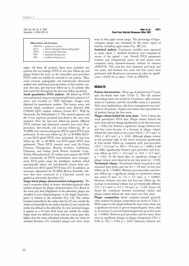
raphy. Of these 26 patients, three were excluded: twopatients did not undergo IVUS at two-year follow-up, andplaque behind the stent on the immediate post-procedureIVUS could not reliably be assessed in one patient. Thus,serial coronary angiography and intravascular ultrasoundanalysis were performed post-procedure, at four-month, one-year, two-year, and four-year follow-up in 23 patients whowere event-free throughout the four-year follow-up period.Serial quantitative IVUS analysis. All follow-up IVUSwere performed using an automated pull-back system at 0.5mm/s and recorded on VHS videotapes. Images weredigitized for quantitative analysis. The lumen, stent, andexternal elastic membrane contours were detected withCURAD QCU analysis software (Curad B.V., Wijk bijDuurstede, the Netherlands) (7). The stented segment andthe 5-mm segments proximal and distal to the stent wereanalyzed. Over the four-year follow-up period, differentIVUS catheters and ultrasound consoles were used. Post-procedure, four-month, and one-year follow-up, 20- or30-MHz non–electrocardiogram (ECG)-gated IVUS wereperformed. At two-year follow-up, 30- or 40-MHz ECG-or non–ECG-gated IVUS were performed. At four-yearfollow-up, 30- or 40-MHz non–ECG-gated IVUS wereperformed. Three IVUS consoles were used (In-Vision[Volcano Therapeutics, Rancho Cordova, California],Clearview, and Galaxy [both Boston Scientific Corp.,Natick, Massachusetts]). To analyze and compare the IVUSdata consistently, all IVUS examinations were retrospec-tively ECG-gated using the Intelligate method, whichautomatically selects the end-diastolic frames from pre-recorded non–ECG-gated IVUS data (10). In addition, anautomatic adjustment for 30-MHz Boston Scientific cath-eters that were connected to a Clearview console wasapplied as previously described (11).Image-based plaque characterization (echogenicity). Weused a computer-aided, in-house–developed grayscale valueanalysis program for plaque characterization (12). Based onthe mean gray level (brightness) of the adventitia, plaque wasclassified as more (hyperechogenic) or less bright (hypoecho-genic) in relation to the adventitia (Fig. 1A). The volumebounded externally by the surface that lies 0.5 mm outside themedia and internally by the surface that lies 0.2 mm outside themedia was defined as the adventitia. In cross-sectional images,it appears as a 0.3-mm thick band just outside the media.Upper tissue was defined as tissue that has a mean gray valuehigher than the mean adventitial intensity plus two times itsstandard deviation (12). Calcified plaque and stent struts
were in this upper tissue range. The percentage of hypo-echogenic plaque was calculated for the entire region ofinterest, excluding upper tissue (Fig. 1B) (12).Statistical analysis. Continuous variables were expressedas mean values � standard deviations and compared bymeans of the paired t test. Overall IVUS parameters(volume and echogenicity) across all time points werecompared using repeated-measure analysis of variance(ANOVA). Two post-hoc tests (between post-stent andtwo years, and between two years and four years) wereperformed with Bonferroni corrections (p value for signifi-cance �0.025) for p values �0.05 on ANOVA.
RESULTS
Patient characteristics. Mean age of patients was 57 years,and two-thirds were male (Table 1). The left anteriordescending artery was treated in 14 patients, right coronaryartery in 6 patients, and left circumflex artery in 3 patients.After stent implantation, risk factor management was insti-tuted in all patients. Aspirin and statins were prescribed inall patients during the four years.Plaque volume behind the stent struts. Table 2 shows theserial quantitative IVUS data. Plaque volume behind thestent struts did not change significantly in the first two years(p � 0.86), but showed a significant change between twoand four years because of a decrease in plaque volumebehind the stent struts at four years (156.8 � 57.7 mm3 vs.138.4 � 42.2 mm3, p � 0.02). Although plaque volumeat the proximal edge of the stents increased significantlyat four-month follow-up compared with post-procedure(32.5 � 12.2 mm3 vs. 39.8 � 15.8 mm3, p � 0.005), it didnot differ significantly between post-procedure and four-year follow-up (32.5 � 12.2 mm3 vs. 33.0 � 11.7 mm3,p � 0.81). At the distal edge, no significant changes inplaque volume were observed at any time point (p � 0.18).Neointimal volume. Neointimal volume increased at eachmeasured time point, and was 8.4 � 5.8 mm3 at four-yearfollow-up (p � 0.0001). Between post-procedure and two-year follow-up, a significant change in neointimal volumewas noted (0 mm3 vs. 7.0 � 6.7 mm3, p � 0.0001).However, between two-year and four-year follow-up, theincrease in neointimal volume was not statistically different(7.0 � 6.7 mm3 vs. 8.4 � 5.8 mm3, p � 0.25). Figure 2Ashows the correlation between neointimal volume andplaque volume behind the stent struts over four years.Plaque composition. Serial computer-assisted grayscalevalue analyses for plaque composition are shown in Table 3.With respect to the plaque behind the stent struts, there wasa significant decrease in percent hypoechogenic tissue, thatis, an increase in percent hyperechogenicity up to four years(p � 0.0001). Between post-procedure and two years, therewere no significant changes in plaque echogenicity (78.9 �8.6% vs. 78.2 � 8.9%, p � 0.67). However, between two
Abbreviations and AcronymsANOVA � analysis of varianceECG � electrocardiogram/electrocardiographicIVUS � intravascular ultrasoundPES � paclitaxel-eluting stentSES � sirolimus-eluting stent
1671JACC Vol. 46, No. 9, 2005 Aoki et al.November 1, 2005:1670–6 Four-Year Arterial Response After SES
color figures on page 222

23
Chronic arterial response to SES
and four years, significant changes were noted (78.2 � 8.9%vs. 70.4 � 10.6%, p � 0.0001). A similar pattern wasobserved for plaque at the proximal stent edge with asignificant reduction in percent hypoechogenic tissue atfour-year follow-up (p � 0.02). At the distal stent edge, nosignificant changes in plaque echogenicity were observedacross time points (p � 0.78). Figure 3 is a representative
example of a patient with plaque shrinkage and reduction ofhypoechogenic plaque composition from two-year to four-year follow-up.Neointimal echogenicity. The echogenicity of the neoin-tima increased over the four-year follow-up period (p �0.0001) (Fig. 2B). On post-hoc testing with Bonferronicorrections, the decrease in hypoechogenicity between four
Figure 1. (A) The adventitia is defined as tissue outside the external elastic membrane contour. For all non-shadowed adventitia pixels, the mean value andstandard deviation are calculated. To observe the suitability, a normal distribution curve based on the same mean and standard deviation histogram iscreated. (B) Cross-sectional image of echogenicity and distribution graph of plaque echogenicity behind the stent struts. Hyperechogenic areas are coloredgreen. Hypoechogenic areas are colored red. ROI � region of interest.
1672 Aoki et al. JACC Vol. 46, No. 9, 2005Four-Year Arterial Response After SES November 1, 2005:1670–6
color figures on page 222

Chapter 2
24
months and two years was not significant (74.9 � 12.7% vs.66.7 � 19.1%, p � 0.03), but was significant betweentwo-year and four-year follow-up (66.7 � 19.1% vs. 57.0 �22.6%, p � 0.01).
DISCUSSION
Previous reports have shown that SESs are effective ininhibiting neointimal hyperplasia without affecting totalvessel volume or plaque volume behind the stent struts at sixmonths (7,8). In the present study, this pattern of tissuegrowth inhibition inside and outside the SES was main-tained at two years. However, at four-year follow-up, asignificant negative remodeling (shrinking) of the plaquebehind the stent struts with an increase in hyperechogenic-ity was observed.
Several studies have shown that plaque echogenicity inthe carotid artery is related to the histologic components ofplaques and that echolucency (low echogenicity) can predictclinical events (13,14). For coronary plaque studies, plaqueechogenicity has also been related to the histologic compo-nents of plaque (15–17). Hyperechogenic tissue has beenshown to be associated with a predominance of densefibrous or elastic tissue, whereas hypoechogenic plaque wascorrelated with predominance of loose fibrous, lipid, ornecrotic tissue. Recently, other analysis systems such asIVUS elastography, IVUS palpography, IVUS radiofre-quency analysis (so-called virtual histology), and optical
coherence tomography have been used to evaluate plaquevulnerability and plaque composition (18–20). However,these methodologic approaches require prospective acquisi-tion of data, for which their respective devices were notavailable for human use at the time of initial assessment.Plaque echogenicity analysis by computer-assisted grayscalevalue is currently the only method that has the potential toanalyze plaque composition in a retrospective manner.
Table 1. Patient Characteristics
n � 23
Age, yrs (mean � SD) 57 � 9Male, % 65.2Hypertension, % 69.6Hyperlipidemia, % 47.8Diabetes mellitus, % 26.1Family history, % 30.4Current smoking, % 65.2Treated vessel
LAD, % 60.9LCX, % 13.0RCA, % 26.1
LAD � left anterior descending artery; LCX � left circumflex artery; RCA � rightcoronary artery.
Table 2. Serial Three-Dimensional Intravascular Ultrasound Analysis of Neointima and Plaque Volume
AfterProcedure 4 Mo 1 Yr 2 Yrs 4 Yrs
ANOVA*p Value
Post Hoc AfterProcedure to
2 YrsPost Hoc2 to 4 Yrs
Neointima, mm3 0 2.1 � 1.7 3.8 � 3.3 7.0 � 6.7 8.4 � 5.8 �0.0001 �0.0001 0.25Plaque (stented segment), mm3 155.5 � 42.8 160.4 � 60.4 153.5 � 61.6 156.8 � 57.7 138.4 � 42.2 0.04 0.86 0.02Vessel (stented segment), mm3 305.1 � 53.7 310.8 � 99.4 305.4 � 83.8 309.8 � 84.1 290.5 � 65.3 0.09 — —Plaque (proximal edge), mm3 32.5 � 12.2 39.8 � 15.8 36.5 � 11.9 37.4 � 13.0 33.0 � 11.7 0.006 0.03 0.05Vessel (proximal edge), mm3 82.6 � 21.9 88.6 � 23.8 80.1 � 17.2 82.6 � 22.1 78.3 � 21.6 0.001 0.98 0.10Plaque (distal edge), mm3 26.2 � 15.4 28.6 � 11.1 28.6 � 14.0 31.1 � 13.4 25.8 � 11.3 0.18 — —Vessel (distal edge), mm3 63.3 � 22.0 68.5 � 20.8 66.5 � 26.3 67.3 � 21.8 61.4 � 22.7 0.07 — —
*Repeated-measures analysis of variance (ANOVA) was performed among five periods. Post hoc analysis between after the procedure and two years, and between two and fouryears were performed with Bonferroni corrections (significant level of p value is 0.025).
Figure 2. (A) Correlation between neointimal volume and plaque volumebehind the stent struts over four years. (B) Correlation between neointimalpercent hypoechogenicity and plaque percent hypoechogenicity behind thestent struts over four years. Error bars indicate 95% confidence interval.
1673JACC Vol. 46, No. 9, 2005 Aoki et al.November 1, 2005:1670–6 Four-Year Arterial Response After SES
color figures on page 223

25
Chronic arterial response to SES
For SES, 80% of the drug load is eluted within 30 days ofstent implantation (1). However, concern has arisen aboutlong-term biological reactions to the non-biodegradablepolymer (5). Long-term exposure to these polymers maycause chronic inflammation. Complete natural healing (i.e.,re-endothelialization and a reduction in proteoglycan con-tent) may take three to six months after bare metal stentimplantation (21). In this present study, plaque volumebehind the stent struts decreased between two and fouryears, and this shrinkage was accompanied by a change inplaque echogenicity, suggestive of a change in plaquecomposition. This may indicate that the peri-stent area hasundergone a slower healing process after SES implantationthat results in a more fibrous content at four-year follow-up.
In animal experiments, it has been documented that theexuberant proliferative process observed after stenting islargely inhibited in the early stage with SES, but the normalhealing process is also delayed with persistence of fibrindeposition and inflammatory cells (22). In humans, onestudy reported the histologic findings of atherectomy spec-imens of restenotic lesions after implantation of a paclitaxelderivative-eluting polymer stent in which persistent fibrinaccumulation with smooth muscle cells and proteoglycanand collagen type III-rich matrix with inflammation was
observed at 12 months (4,5). An autopsy performed on apatient four years after SES implantation showed completehealing, indicating that the delayed healing response hadabated, concurring with our findings (23).
In this study, neointimal volume continued to increaseover the four-year period. However, the increase betweentwo and four years failed to reach statistical significance (7.0vs. 8.4 mm3, p � 0.25), and the change in neointimalvolume between two years and four years was significantlyless than between the post-procedure and two-year mea-surements (1.4 vs. 7.0 mm3, p � 0.04), which possiblyindicates that the delayed tissue proliferation has subsided.Carter et al. (24) reported long-term effects of SES in aporcine coronary model. They found that inhibition ofneointimal hyperplasia was not sustained at 90 days com-pared with the bare metal stent, and the mean neointimalarea was similar between 90 days and 180 days. Theseresults differ from observations seen in clinical trials, includ-ing this study. Several trials have reported low target vesselrevascularization rates up to three years (25). This discrep-ancy is likely explained by different monocyte and lympho-cyte responses to similar concentrations of sirolimus inhuman and porcine vessels, and the different anatomical
Table 3. Percent Hypoechogenic Tissue Component in the Plaque and Neointima
AfterProcedure 4 Mo 1 Yr 2 Yrs 4 Yrs
ANOVA*p Value
Post Hoc AfterProcedure to
2 YrsPost Hoc2 to 4 Yrs
Neointima, % — 74.9 � 12.7 68.9 � 17.1 66.7 � 19.1 57.0 � 22.6 0.0001† 0.03‡ 0.01Plaque (stented segment), % 78.9 � 8.6 79.4 � 6.7 81.3 � 7.4 78.2 � 8.9 70.4 � 10.6 �0.0001 0.67 �0.0001Plaque (proximal edge), % 88.7 � 6.9 87.2 � 6.5 87.1 � 6.0 87.7 � 7.9 81.7 � 9.3 0.02 0.67 0.008Plaque (distal edge), % 86.1 � 9.8 86.8 � 6.0 87.0 � 10.4 88.5 � 9.5 85.9 � 9.4 0.78 — —
*Repeated-measures analysis of variance (ANOVA) was performed among five periods. Post-hoc analysis between after the procedure and two years, and between two and fouryears were performed with Bonferroni corrections (significant level of p value is 0.025). †Repeated-measures ANOVA was performed among four periods, except after theprocedure. ‡Post-hoc analysis was performed between four months and two years.
Figure 3. A representative example of a patient with plaque compositional changes between two-year and four-year follow-up. Top row showscross-sectional vessel image at (A) post-procedure, (B) four months, (C) one year, (D) two years, and (E) four years. Red line indicates lumen. Light blueline indicates stent. Green line indicates media. Dense blue line indicates adventitia. Bottom row shows cross-sectional echogenicity images.Hyperechogenic areas are colored green. Hypoechogenic areas are colored red. (A’) Percent plaque behind stent hypoechogenicity was 84.0%post-procedure, (B’) 85.3% at four months, (C’) 88.4% at one year, (D’) 84.2% at two years, and (E’) 74.8% at four years.
1674 Aoki et al. JACC Vol. 46, No. 9, 2005Four-Year Arterial Response After SES November 1, 2005:1670–6
color figures on page 223

Chapter 2
26
features of normal porcine coronary arteries versus athero-sclerotic human coronary arteries (26).
Furthermore, the increase in neointimal echogenicity asobserved in this study would also suggest that a fibroticprocess is now operative and predominant. The rate ofgrowth of the neointima has slowed—both features suggestthat the completion of the healing process may be expectedin the near future.Study limitations. This study is limited by the samplesize of 23 patients. Secondly, patients without eventswere used for the serial IVUS analysis. Consequently,these results may not apply to all patients treated withSES. Different IVUS catheters and consoles were usedover the four-year period, and most data were acquiredwithout ECG gating. These differences may have ham-pered the consistency of volumetric and echogenic anal-ysis (27). For instance, BSC 30-MHz catheters con-nected to a Clearview console underestimates truedimensions (11). To correct for these discrepancies, theresults from the 30-MHz catheter were adjusted using apreviously reported mathematical model (11), and imageartifacts resulting from cardiac cycle motion observed innon–ECG-gated images were circumvented by the use ofthe Intelligate method, which converts non–ECG-gateddata to ECG-gated data retrospectively (10,28).
The adventitia was taken as a reference to determine thechange in echogenicity occurring in the peri-stent andintrastent tissue. Any alterations to the adventitia (e.g.,inflammatory and fibrotic reactions) after stent deploymentmay alter the echogenicity during the follow-up period.Because any potential increase in echogenicity of the adven-titia cannot be totally excluded or measured, the change inechogenicity of the peri-stent and intra-stent tissue may beunderestimated and the interference of stent struts withplaque echogenicity may not be completely excluded. Fi-nally, any remaining bias after correction from the use ofdifferent IVUS systems cannot be totally excluded in thepresent study. Despite the inherent limitations, plaqueechogenicity analysis by computer-assisted grayscale value isto date the only method with the potential to analyze plaquecomposition retrospectively.Conclusions. After SES implantation, negative plaqueremodeling and a decrease of hypoechogenic plaquecomposition were observed at four-year follow-up. Inaddition, the fact that the neointima does not signifi-cantly change from two to four years may suggest that thebiological phenomenon of a delayed healing response hasbegun to subside.
AcknowledgmentsWe thank Drs. Gaston A. Rodriguez-Granillo, EugeneMcFadden, and Brian Firth for their careful review of thismanuscript and for their constructive suggestions.
Reprint requests and correspondence: Dr. Patrick W. Serruys,Thoraxcenter, Bd 406, Erasmus Medical Center, Dr Molewater-plein 40, 3015 GD Rotterdam, the Netherlands. E-mail: [email protected].
REFERENCES
1. Morice MC, Serruys PW, Sousa JE, et al. A randomized comparisonof a sirolimus-eluting stent with a standard stent for coronary revas-cularization. N Engl J Med 2002;346:1773–80.
2. Moses JW, Leon MB, Popma JJ, et al. Sirolimus-eluting stents versusstandard stents in patients with stenosis in a native coronary artery.N Engl J Med 2003;349:1315–23.
3. Lemos PA, Serruys PW, van Domburg RT, et al. Unrestrictedutilization of sirolimus-eluting stents compared with conventional barestent implantation in the “real world:” the Rapamycin-Eluting StentEvaluated At Rotterdam Cardiology Hospital (RESEARCH) registry.Circulation 2004;109:190–5.
4. Virmani R, Liistro F, Stankovic G, et al. Mechanism of late in-stentrestenosis after implantation of a paclitaxel derivate-eluting polymerstent system in humans. Circulation 2002;106:2649–51.
5. Virmani R, Guagliumi G, Farb A, et al. Localized hypersensitivity andlate coronary thrombosis secondary to a sirolimus-eluting stent: shouldwe be cautious? Circulation 2004;109:701–5.
6. Tanabe K, Serruys PW, Degertekin M, et al. Chronic arterialresponses to polymer-controlled paclitaxel-eluting stents: comparisonwith bare metal stents by serial intravascular ultrasound analyses: datafrom the randomized TAXUS-II trial. Circulation 2004;109:196–200.
7. Serruys PW, Degertekin M, Tanabe K, et al. Intravascular ultrasoundfindings in the multicenter, randomized, double-blind RAVEL(RAndomized study with the sirolimus-eluting VElocity balloon-expandable stent in the treatment of patients with de novo nativecoronary artery Lesions) trial. Circulation 2002;106:798–803.
8. Degertekin M, Regar E, Tanabe K, et al. Evaluation of coronaryremodeling after sirolimus-eluting stent implantation by serial three-dimensional intravascular ultrasound. Am J Cardiol 2003;91:1046–50.
9. Sousa JE, Costa MA, Abizaid A, et al. Lack of neointimal prolifera-tion after implantation of sirolimus-coated stents in human coronaryarteries: a quantitative coronary angiography and three-dimensionalintravascular ultrasound study. Circulation 2001;103:192–5.
10. DeWinter SA, Hamers R, Degertekin M, et al. Retrospective image-based gating of intracoronary ultrasound images for improved quan-titative analysis: the Intelligate method. Catheter Cardiovasc Interv2004;61:84–94.
11. Bruining N, Hamers R, Teo TJ, deFeijter PJ, Serruys PW, RoelandtJR. Adjustment method for mechanical Boston scientific corporation30 MHz intravascular ultrasound catheters connected to a Clearviewconsole. Mechanical 30 MHz IVUS catheter adjustment. Int J Car-diovasc Imaging 2004;20:83–91.
12. de Winter SA, Heller I, Hamers R, de Feyter PJ, Serruys PW,Roelandt JR. Computer assisted three-dimensional plaque character-ization in intracoronary ultrasound studies. Comp Cardiol 2003;30:73–6.
13. Mathiesen EB, Bonaa KH, Joakimsen O. Echolucent plaques areassociated with high risk of ischemic cerebrovascular events in carotidstenosis: the Tromso study. Circulation 2001;103:2171–5.
14. Gronholdt ML, Nordestgaard BG, Schroeder TV, Vorstrup S,Sillesen H. Ultrasonic echolucent carotid plaques predict futurestrokes. Circulation 2001;104:68–73.
15. Schartl M, Bocksch W, Koschyk DH, et al. Use of intravascularultrasound to compare effects of different strategies of lipid-loweringtherapy on plaque volume and composition in patients with coronaryartery disease. Circulation 2001;104:387–92.
16. Prati F, Arbustini E, Labellarte A, et al. Correlation between highfrequency intravascular ultrasound and histomorphology in humancoronary arteries. Heart 2001;85:567–70.
17. Rasheed Q, Dhawale PJ, Anderson J, Hodgson JM. Intracoronaryultrasound-defined plaque composition: computer-aided plaque char-acterization and correlation with histologic samples obtained duringdirectional coronary atherectomy. Am Heart J 1995;129:631–7.
1675JACC Vol. 46, No. 9, 2005 Aoki et al.November 1, 2005:1670–6 Four-Year Arterial Response After SES

27
Chronic arterial response to SES
18. Nair A, Kuban BD, Tuzcu EM, Schoenhagen P, Nissen SE, VinceDG. Coronary plaque classification with intravascular ultrasoundradiofrequency data analysis. Circulation 2002;106:2200–6.
19. Schaar JA, De Korte CL, Mastik F, et al. Characterizing vulnerableplaque features with intravascular elastography. Circulation 2003;108:2636–41.
20. Jang IK, Bouma BE, Kang DH, et al. Visualization of coronaryatherosclerotic plaques in patients using optical coherence tomogra-phy: comparison with intravascular ultrasound. J Am Coll Cardiol2002;39:604–9.
21. Farb A, Sangiorgi G, Carter AJ, et al. Pathology of acute and chroniccoronary stenting in humans. Circulation 1999;99:44–52.
22. Suzuki T, Kopia G, Hayashi S, et al. Stent-based delivery of sirolimusreduces neointimal formation in a porcine coronary model. Circulation2001;104:1188–93.
23. Sousa JE, Costa MA, Farb A, et al. Images in cardiovascular medicine.Vascular healing four years after the implantation of sirolimus-eluting stent inhumans: a histopathological examination. Circulation 2004;110:e5–6.
24. Carter AJ, Aggarwal M, Kopia GA, et al. Long-term effects ofpolymer-based, slow-release, sirolimus-eluting stents in a porcinecoronary model. Cardiovasc Res 2004;63:617–24.
25. Fajadet J, Morice MC, Bode C, et al. Maintenance of long-termclinical benefit with sirolimus-eluting coronary stents: three-yearresults of the RAVEL trial. Circulation 2005;111:1040–4.
26. Wright DC, Deol HS, Tuch BE. A comparison of the sensitivity ofpig and human peripheral blood mononuclear cells to the antiprolif-erative effects of traditional and newer immunosuppressive agents.Transpl Immunol 1999;7:141–7.
27. Li Y, Honye J, Saito S, et al. Variability in quantitative measurementof the same segment with two different intravascular ultrasoundsystems: in vivo and in vitro studies. Catheter Cardiovasc Interv 2004;62:175–80.
28. Bruining N, von Birgelen C, de Feyter PJ, et al. ECG-gated versusnongated three-dimensional intracoronary ultrasound analysis: impli-cations for volumetric measurements. Cathet Cardiovasc Diagn 1998;43:254–60.
1676 Aoki et al. JACC Vol. 46, No. 9, 2005Four-Year Arterial Response After SES November 1, 2005:1670–6


Chapter 3
Peri-stent remodeling and neointimal suppression
two-years after polymer based paclitaxel eluting stents
implantation- insight from serial IVUS analysis in the
TAXUS II study
Aoki J, Colombo A, Dudek D, Banning A,
Drzewiecki J, Zmudka K, Shiele F, Russell
ME, Koglin J, Serruys PW.
Circulation. 2005;112:3876-3883

31
Chronic arterial response to PES
Peristent Remodeling and Neointimal Suppression 2 YearsAfter Polymer-Based, Paclitaxel-Eluting Stent Implantation
Insights From Serial Intravascular Ultrasound Analysis in theTAXUS II Study
Jiro Aoki, MD; Antonio Colombo, MD; Dariusz Dudek, MD; Adrian P. Banning, MD;Janusz Drzewiecki, MD; Krzysztof Zmudka, MD; Francois Schiele, MD; Mary E. Russell, MD;
Joerg Koglin, MD; Patrick W. Serruys, MD, PhD; for the TAXUS II Study Group
Background—The purpose of this study was to evaluate long-term vascular responses as long as 2 years after implantationof polymer-based, paclitaxel-eluting stents in contrast to uncoated stents.
Methods and Results—TAXUS II is a randomized, double-blind trial comparing slow-release (SR) and moderate-release(MR) TAXUS stents with bare-metal control stents (BMSs). One hundred sixty-one event-free patients (SR, 43; MR,41; and BMS, 77) underwent serial intravascular ultrasound (IVUS) analysis after the procedure and at 6 months and2 years. At 2 years, neointimal responses continued to be significantly suppressed in the SR and MR groups whencompared with the BMS group (BMS, 1.49�1.12 mm2; SR, 0.94�0.76 mm2 [P�0.004]; and MR, 1.06�0.90 mm2
[P�0.02]). Between 6 months and 2 years, the BMS group showed compaction of the neointima (�, �0.22�1.05 mm2
[P�0.08]). In contrast, both the SR and MR groups exhibited an increase (� SR, 0.30�0.76 mm2 (P�0.01); MR,0.41�0.94 mm2 [P�0.009]). Between 6 months and 2 years, the initial increase in plaque outside the stent regressedin the BMS and SR groups to levels comparable to those after the procedure, whereas expansive remodeling partiallyregressed in the MR group (� between after the procedure and 2 years BMS, �0.34�1.28 mm2 [P�0.05]; SR,�0.02�1.40 mm2 [P�0.93]; MR, 0.32�1.56 mm2 [P�0.27]).
Conclusions—The 2-year follow-up demonstrates that neointimal suppression was dose independent and that this effectwas still sustained at 2 years. However, the increase in area outside the stent seen at 6 months regressed to differentextents in a dose-dependent manner at 2 years. (Circulation. 2005;112:3876-3883.)
Key Words: stents � remodeling � restenosis
Both slow-release (SR) and moderate-release (MR)polymer-based, paclitaxel-eluting stents prevent in-stent
neointimal growth compared with bare-metal stents (BMSs).1
In the patient population studied in TAXUS II, these antir-estenotic effects were comparable for both dose formulations.However, at 6 months, this inhibition was associated withexpansive, peristent remodeling.2 The extent of peristentremodeling was more pronounced with the MR formulation,implying dose-dependent differences in vascular responsesoutside but not inside the stent.
Clinical Perspective p 3883
In animal studies, the mechanism of action of drug-elutingstents on neointimal proliferation after stent implantationseems to be partially explained by a delay in vascularresponses. For polymer-based, paclitaxel-eluting stents, inhi-
bition of in-stent neointimal growth is associated with a delayin intimal healing up to 28 days, as indicated by initiallyincreased fibrin deposition, inflammation, and delayed endo-thelialization.3 By 90 days, peristrut changes associated withpaclitaxel were resolving, but in-stent neointimal growthsuppression was no longer present.TAXUS II is an international study of 2 consecutive
cohorts designed to evaluate 2 formulations of a polymer-based, paclitaxel-eluting stent. The primary end point in theoriginal protocol was the percent net volume obstruction,providing a unique opportunity to evaluate serial intravascu-lar ultrasound system (IVUS) changes over time.The objective of this study was to evaluate long-term arterial
responses both inside and outside the stent as long as 2 yearsafter implantation of polymer-based, paclitaxel-eluting stents byusing serial quantitative IVUS analysis in event-free patients.
Received April 26, 2005; revision received October 16, 2005; accepted October 18, 2005.From the Thoraxcenter Erasmus MC (J.A., P.W.S.), Rotterdam, the Netherlands; Ospedale San Raffaele (A.C.), Milan, Italy; Jagiellonian University
(D.D., K.Z.), Krakow, Poland; John Radcliffe Hospital (A.P.B.), Oxford, England; PSK No. 7 Zaklad Kardiologii (J.D.), Katowice, Poland; the CentreHospitalier Universitarie Jean Minjoz (F.S.), Besancon, France; and Boston Scientific Corp (M.E.R., J.K.), Natick, Mass.Correspondence to P.W. Serruys, MD, PhD, Thoraxcenter, Ba 583, Erasmus MC, Dr Molewaterplein 40, 3015 GD Rotterdam, The Netherlands. E-mail
[email protected]© 2005 American Heart Association, Inc.
Circulation is available at http://www.circulationaha.org DOI: 10.1161/CIRCULATIONAHA.105.558601
3876

Chapter 3
32
MethodsPatient SelectionThe original study design, procedure, and IVUS results have beendescribed previously.1,2,4 The design of the TAXUS II study is arandomized, double-blind, controlled trial. In brief, 536 patients withsingle de novo coronary lesions (�50% stenosis on visual assess-ment) treatable with a single stent (3.0- or 3.5-mm diameter and15 mm long) were randomly assigned to 2 consecutive, independentcohorts. The first cohort of patients was randomized to be treatedwith either a control BMS or a TAXUS-NIRx SR formulation stent.A second cohort of patients was randomized to control BMSs versusa TAXUS-NIRx MR formation. The primary end point was met witha significant 66% (SR) and 62% (MR) reduction in 6-month percentnet volume obstruction as assessed by IVUS in both groups, withoutan apparent difference between the SR and MR groups.To further evaluate the natural history of vascular responses to
TAXUS, the study protocol was amended to include a 2-year IVUSlong-term follow-up. Of the initial 536 patients, 161 patients withoutany clinical events through the 2-year follow-up and with serial(postprocedure, 6 months, and 2 years) and analyzable IVUS datawere selected for this substudy (BMS, 77; SR, 43; and MR, 41). Thispatient population represents a highly selected subgroup of patientssuccessfully treated with either a BMS, a TAXUS SR stent, or aTAXUS MR stent. Because the 2-year quantitative coronary angiog-raphy (QCA) and IVUS substudy was added as an amendment to theoriginal study protocol, it required additional approval from the localregulatory agencies and ethics committees before being implementedat the sites. The 2-year substudy was not approved by the appropriateregulatory authorities or ethics committees at 9 of the 38 sites. As aresult, of the original 536 intent-to-treat patients, 154 were noteligible to participate in the 2-year substudy. Additionally, 122patients did not consent to the 2-year substudy, whereas 30 patientswere excluded for clinical reasons, resulting in 230 patients whowere eligible to participate in the substudy. In 34 of the 230 patients,IVUS either was not performed or was not of adequate quality to beincluded in the qualitative and quantitative analysis. An additional 23patients were excluded because they had either a target-vesselrevascularization before 6 months or had a target-vessel revascular-ization after 6 months but did not have IVUS before the target-vesselrevascularization. Finally, 12 patients were excluded from thisanalysis because paired IVUS data were not available at all 3 timepoints (postprocedure, 6-month, and 2-year data). Written, informedconsent was obtained from all patients.
TAXUS Stent SystemThe 15-mm NIR Conformer stent of 3.0- or 3.5-mm diameter wasused in this study (Boston Scientific Corp and Medinol Ltd). AllTAXUS NIRx stents were coated with 1.0 �g/mm2 paclitaxel in anSR or MR formulation as previously described.1 The SR and MRdose formulations are characterized by differences in the amount ofdrug released during the initial 48-hour burst phase as well as theamount of drug remaining embedded in the polymer at 30 days.
Study ProcedureStents were implanted after balloon predilatation as described in theinitial reports. Per study protocol, all patients were to receive 75mg/d aspirin indefinitely. Clopidogrel (300 mg) was administered,preferably 48 hours before the procedure, followed by 75 mg oncedaily for 6 months.
Quantitative IVUS and Angiographic AnalysisThe quantitative IVUS and QCA analyses were performed by anindependent core laboratory that remains blinded to treatmentallocation during the ongoing 5-year follow-up (Cardialysis). SerialIVUS (postprocedure, 6-month, and 2-year follow-up) procedureswere performed after administration of 200 �g intracoronary nitro-glycerin with an automated pullback at 0.5 mm/s. All IVUSprocedures were recorded on VHS videotape, and images weredigitized for analysis. A computer-based contour-detection program
was used for automated 3-dimensional reconstruction of the stentedsegment.5,6 Reconstruction and quantification of 3-dimensionalIVUS images have been validated previously.5 In brief, a series oftomographic images is continuously acquired during an IVUSpullback procedure. With use of a 40-MHz ultrasound probe, 25frames per second are available from a motorized pullback procedurerecorded on videotape. In this study, an average of 714 sections inthe stented lesion were obtained per patient. All acquired cross-sectional frames were analyzed by semiautomatic contour tracing inseveral reconstructed, longitudinally cut planes, as developed andtested in cooperation with Cuard B.V. The applied approach focuseson the tracing of contours in the reconstructed, longitudinally cutplanes (L-mode view). The number of L-mode contours to be tracedis independent of the number of frames in the analysis. As many as72 L-mode views at 5° intervals can be selected for display andsubsequent analysis. Contours of vessel, lumen, and stent structurescan then be traced. Instead of trying to find the structures completelyautomatically, the program uses starting points as defined by theuser, followed by autotracing of the contour segment in either of 2possible directions. Pieces of the contours can be retraced in asemiautomatic procedure to optimize the interaction between oper-ator and algorithm. In this way, all cross-sectional areas are ana-lyzed, and the 3-dimensional nature of the data set is fully used. Theinterobserver correlation coefficients for lumen, stent, and vesselvolumes resulted in R2 values of 0.96, 0.99, and 0.99, respectively.In the stented segment, mean peristent area and mean neointimal
area were calculated. Incomplete stent apposition (ISA) was definedas 1 or more stent struts clearly separated from the vessel wall withevidence of blood speckles behind the strut without overlapping sidebranches.6–8 ISA was classified into 3 groups. Resolved ISA wasdefined as ISA that disappeared during follow-up. Persistent ISAwas defined as ISA that was evident both after the procedure and atfollow-up. Late acquired ISA was defined as ISA that was absentafter the procedure but present at follow-up.
Statistical AnalysisThe BMS groups of the 2 cohorts were combined because thebaseline and 6-month follow-up data showed no significant differ-ences, as previously described.1 Therefore, 3 groups are reported inthis study: The combined BMS, the TAXUS-SR, and theTAXUS-MR groups. Discrete variables are displayed as percentagesand were tested with Fisher’s exact test. Continuous variables areexpressed as mean�SD. When 3 groups were compared, overallprobability values were derived from 1-way ANOVA or Fisher’sexact test. Comparisons between postprocedure and follow-up datawere performed with a 2-tailed, paired t test, whereas comparisonsbetween 2 groups were performed with Fisher’s least significantdifference test. Linear regression was performed to assess thecorrelation between different IVUS outcomes. A value of P�0.05was considered statistically significant.
ResultsPatients and Procedural CharacteristicsTable 1 presents the patient and procedural characteristics forthe group that had serial (after the procedure, at 6 months, andat 2 years) IVUS examinations compared with the entireTAXUS II study cohort. Comparable baseline demographicand angiographic data with the exception of the prevalence ofmales (P�0.03) indicate that the data in the serial IVUScohorts are representative of the overall randomized studypopulation.
Area Inside the Stent (Neointimal Area)As shown in Table 2, the neointimal area at 6 months wassignificantly suppressed in the SR and MR groups, asdetected by serial IVUS when compared with the BMS group(1.71�1.38 mm2 in the BMS group versus 0.64�0.81 mm2 in
Aoki et al Two-Year Arterial Response to TAXUS Stent 3877

33
Chronic arterial response to PES
the SR group, P�0.0001; 0.66�0.83 mm2 in the MR group,P�0.0001). This reduction relative to the BMS group wasalso present at 2 years (1.49�1.12 mm2 in the BMS groupversus 0.94�0.76 mm2 in the SR group, P�0.004;1.06�0.90 mm2 in the MR group, P�0.02). When relativechanges within the 3 groups were compared, the BMS groupshowed compaction of the neointima between 6 months and2 years, as demonstrated by a trend toward a decrease inneointimal area (�, �0.22�1.05 mm2, P�0.08). In contrast,
the SR and MR groups both exhibited a significant increase inneointimal area between 6 months and 2 years when com-pared with controls (SR �, 0.30�0.76 mm2, P�0.01 versusBMS; MR �, 0.41�0.94 mm2, P�0.009 versus BMS; Table2). However, according to QCA analyses, the average mini-mal lumen diameter (MLD) in the SR and MR groups did notdecrease from 6 months to 2 years (2.30�0.33 to2.36�0.28 mm in the SR group; 2.30�0.38 to2.31�0.35 mm in the MR group; Table 3).
TABLE 1. Baseline and Procedural Characteristics
BMS(n�77)
SR(n�43)
MR(n�41) P*
All RandomizedPatients (N�536)
Age, y 57.2�9.0 59.4�1.3 57.5�10.9 0.50 60.1�10.0
Male, % 85.7 65.1 73.2 0.03 75.6
Current smoker, % 29.9 14.0 24.4 0.14 25.0
Diabetes, % 13.0 9.3 9.8 0.85 14.7
Hypertension, % 46.8 62.8 51.2 0.24 61.4
Hypercholesterolemia, % 72.7 83.7 80.5 0.35 75.9
Unstable angina, % 27.3 27.9 14.6 0.26 34.3
Previous MI, % 50.7 41.9 51.2 0.63 39.7
Target vessel, %
LAD 52.0 41.9 39.0 0.34 44.6
LCX 11.7 20.9 19.5 0.31 19.6
RCA 36.4 37.2 41.5 0.89 35.8
RVD, mm 2.73�0.50 2.78�0.38 2.76�0.45 0.88 2.75�0.46
Target lesion length, mm 10.5�4.15 10.0�3.69 11.0�6.27 0.68 10.4�4.23
Stent size, mm 3.29�0.25 3.31�0.24 3.23�0.25 0.31 3.26�0.25
MI indicates myocardial infarction; LAD, left anterior descending coronary artery; LCX, left circumflex artery; RCA,right coronary artery; and RVD, reference vessel diameter. Other abbreviations are as defined in text. Values aremean�SD or percentages.*ANOVA among the 3 groups.
TABLE 2. Quantitative IVUS Data
BMS (n�77) SR (n�43) MR (n�41) P*
Neointimal area, mm2
6 Months 1.71�1.38 0.64�0.81 0.66�0.83 �0.0001
2 Years 1.49�1.12 0.94�0.76 1.06�0.90 0.006
Difference 2 years�6 months �0.22�1.05 0.30�0.76 0.41�0.94 0.001
Plaque area outside the stent(peristent area), mm2
After procedure 7.97�2.00 8.18�2.11 8.38�2.52 0.71
Difference 6 months�after 0.68�1.19 1.18�1.66 1.52�1.68 0.04
Difference 2 years�6 months �1.02�0.92 �1.21�1.39 �1.19�1.56 0.74
Difference 2 years�after �0.34�1.28 �0.02�1.40 0.32�1.56 0.11
Vessel area, mm2
After procedure 16.43�3.07 17.25�2.93 16.79�3.36 0.50
Difference 6 months�after 1.03�1.41 1.21�1.84 1.60�1.94 0.34
Difference 2 years�6 months �1.18�1.23 �1.33�1.74 �1.38�1.78 0.50
Difference 2 years�after �0.15�1.49 �0.12�1.76 0.21�1.80 0.60
Abbreviations are as defined in text. Values are mean�SD.*ANOVA among the 3 groups.
3878 Circulation December 20/27, 2005

Chapter 3
34
Area Outside the Stent (Peristent Area)Changes up to 6 MonthsIn the first 6 months, significant increases in mean peristentarea (calculated as mean vessel area minus mean stent area),consistent with expansive vessel remodeling, were observedin all 3 groups (Table 2). In the BMS group, the peristent areaincreased by 8.5%, from 7.97�2.00 to 8.65�1.91 mm2
(P�0.0001). This contrasted with an increase by 14.4% in theSR group (8.18�2.11 mm2 after the procedure versus9.37�2.79 mm2 at 6 months; P�0.0003) and by 18.1% in theMR group (8.38�2.52 mm2 after the procedure versus9.90�2.45 mm2 at 6 months; P�0.0001).
Changes From 6 Months to 2 YearsFrom 6 months to 2 years, a significant regression of theperistent area was observed in all 3 groups. In the BMSgroup, the peristent area decreased by 11.8%, from8.65�1.91 to 7.63�1.56 mm2 (P�0.0001). Similarly, theperistent area decreased by 12.9% in the SR group(9.37�2.79 mm2 versus 8.16�2.09 mm2; P�0.0001) and by12.0% in the MR group (9.90�2.45 mm2 versus8.70�2.20 mm2; P�0.0003).At 2 years, this regression in the BMS and SR groups
resulted in absolute peristent areas that were comparable tothose observed after the procedure (difference between post-procedure and 2-year measurements,�0.34�1.28 mm2 in theBMS group and �0.02�1.40 mm2 in the SR group). In theMR group, regression of the initial increase was incomplete,with a remaining net increase at 2 years with respect to thepostprocedure value of 0.32�1.56 mm2 (Table 2).Assessment of the peristent area showed that the expansive
vessel remodeling observed at 6 months regressed from 6months to 2 years in all 3 groups. The incidence of thisremodeling pattern was observed in the vast majority ofpatients (67.5% in the BMS, 74.4% in the SR, and 78.0% inthe MR groups; Figure 1).
Correlation Between Area Inside the Stent(Neointimal Area) and Area Outside the Stent(Peristent Area)Figure 2 presents the changes of the area inside the stentversus the correlating changes outside the stent for 2 years to
illustrate the natural history of vascular responses around andwithin the stent. This graph illustrates that for the BMS, therewas a reduction in neointimal area as well as a reduction inthe peristent area between 6 months and 2 years. For the SRgroup, the neointimal area increased, yet there was a reduc-tion in peristent area, similar to that seen with the BMS.Finally, for the MR group, the neointima significantly in-creased between 6 months and 2 years, with a partial decreasein peristent area.For all 3 groups, there were no correlations between
peristent area after the procedure and neointimal area at 6months (R��0.016, P�0.70 in the BMS group; R��0.021,P�0.55 in the SR group; and R��0.037, P�0.93 in the MRgroup) and 2 years (R��0.011, P�0.51 in the BMS group;R�0.11, P�0.037 in the SR group; and R��0.036, P�0.88in the MR group). In addition, there were no correlationsbetween relative changes in peristent area and changes in
TABLE 3. QCA Data
After Procedure 6 Months 2 Years
BMS (n�77)
RVD, mm 2.89�0.39 2.64�0.49 2.64�0.46
MLD, mm 2.61�0.35 1.91�0.45 2.05�0.40
DS, % 9.50�5.45 27.19�13.36 21.49�11.41
SR (n�43)
RVD, mm 2.87�0.30 2.81�0.40 2.78�0.33
MLD, mm 2.59�0.29 2.30�0.33 2.36�0.28
DS, % 9.53�5.44 17.63�8.78 14.64�6.96
MR (n�41)
RVD, mm 2.89�0.38 2.75�0.37 2.79�0.42
MLD, mm 2.58�0.33 2.30�0.38 2.31�0.35
DS, % 10.24�5.25 16.05�9.59 16.24�9.56
RVD indicates reference vessel diameter; DS, diameter stenosis. Otherabbreviations are as defined in text. Values are mean�SD.
Figure 1. The incidence of increasing (I) or decreasing (D) peris-tent area up to 2 years after stent implantation within eachgroup. I/D indicates that peristent area (PSA) increased duringthe first 6 months and decreased from 6 months to 2 years; D/Dindicates that PSA decreased up to 2 years; I/I indicates thatPSA increased up to 2 years; and D/I indicates that PSAdecreased during the first 6 months and increased from 6months to 2 years. Other abbreviations are as defined in text.
Figure 2. Correlation between neointimal area and peristentarea for 2 years. Probability values for peristent area from afterthe procedure to 6 months are 0.0001 for BMS, 0.0003 for SR,and �0.0001 for MR. Probability values for peristent area from 6months to 2 years are �0.0001 for BMS, �0.0001 for SR, and0.0003 for MR. The probability value for neointimal area fromafter the procedure to 6 months is �0.0001 for all 3 groups,whereas probability values for neointimal area from 6 months to2 years are 0.08 for BMS, 0.01 for SR, and 0.009 for MR.Abbreviations are as defined in text.
Aoki et al Two-Year Arterial Response to TAXUS Stent 3879

35
Chronic arterial response to PES
neointimal area over time at 6 months (R��0.013, P�0.57in the BMS group; R�0.028, P�0.18 in the SR group; andR��0.018, P�0.48 in the MR group) and 2 years(R��0.017, P�0.72 in the BMS group; R��0.021, P�0.54in the SR group; and R�0.021, P�0.22 in the MR group).
Incomplete Stent AppositionAs shown in Figure 3, the incidences of ISA at 2 years weresimilar among the 3 groups (10.4% in the BMS group, 7.0%in the SR group, and 7.3% in the MR group; P�0.82). Theincidences of late acquired ISA at 2 years in all groups werelower than at 6 months (2.6% versus 6.5% in the BMS group,0% versus 9.3% in the SR group, and 2.4% versus 9.8% in theMR group).
DiscussionThis article presents the first long-term (up to 2 years) serialIVUS analysis after deployment of BMSs or drug-elutingstents in a series of 161 patients. To assess long-term IVUSoutcomes in response to the original study stent implantedduring the index procedure, neointimal area analyses had toexclude patients with target-lesion revascularization beforethe follow-up IVUS examination. Thus, neointimal hyperpla-sia in the overall population might be underestimated whencompared with this analysis. The major findings of this studyare as follows: (1) Neointimal suppression is maintained foras long as 2 years in both the SR and MR groups whencompared with the BMS group. (2) Whereas the BMS groupshowed compaction of the neointima over time, there werevery modest increases in neointima in the SR and MR groupsbetween 6 months and 2 years, with significant reductionsrelative to the control. (3) The initial increase in peristent areathat coincides with neointimal suppression in both TAXUSgroups during the first 6 months regressed completely in the
SR group and partially in the MR group during the following18 months to 2 years.
Change in Plaque Area Outside the StentIn both paclitaxel groups, a significantly increased peristentarea was observed at 6 months. This expansive remodelingregressed at 2 years, resulting in comparable levels ofperistent area to the BMS group. One might hypothesize thatthis could be a result of the “drug effect” associated with apotentially delayed healing process. The mode of action ofpolymer-based, paclitaxel-eluting stents is believed to beassociated with a delay in cellular processes within the vesselwall. This effect is exemplified in animal models by laterendothelialization, reduced smooth muscle cell proliferation,and increased fibrin disposition around the stent struts.3
Long-term follow-up beyond 2 years may provide moreinsight regarding the possibility of a continued delay versus apersistent alteration of the remodeling processes outside thestent.Mean peristent area is driven by vessel area minus stent
area. Thus, ISA area influences peristent area. The occurrenceof late acquired ISA at 6 months that was resolved at 2 yearscan also explain the plaque remodeling pattern outside thestent in this study. However, the incidences of late acquiredISA at 6 months that were resolved at 2 years were low (1.3%in the BMS, 4.7% in the SR, and 4.9% in the MR groups). Inaddition, because the change in vessel area was similar to thechange in peristent area, plaque remodeling outside the stentcan be mainly accounted for by changes in plaque area, notISA.The current in vivo findings are limited to areal compari-
sons between groups and do not account for potentiallydifferent cellular compositions of the areas inside and outsidethe stent among the groups. New IVUS technologies, such ascomputer-assisted gray-scale value analysis and virtual his-
Figure 3. Incidence of ISA for as long as2 years after stent implantation. Abbrevi-ations are as defined in text.
3880 Circulation December 20/27, 2005

Chapter 3
36
tology, may offer advances in the detection of plaque com-position to help confirm this hypothesis in future studies.9,10
Long-Term Effect of Inhibition ofNeointimal GrowthBoth neointimal area from IVUS analyses and the change inMLD from QCA analyses are parameters of neointimalgrowth. In the present study, there are apparent discrepanciesbetween the change in neointimal area and the change inMLD from 6 months to 2 years, especially in the TAXUSgroups. Although average neointimal area increased in bothTAXUS groups from 6 months to 2 years, average MLD didnot decrease during the same time period. Neointimal area iscalculated by considering neointimal growth in the entirestented segment. The change in MLD is calculated byconsidering the worst region, regardless of axial location.Thus, MLD does not account for diffuse neointimal growthover the entire length of the stent. These differences demon-strate that neointimal area is a better index of the magnitudeand distribution of neointimal growth within the stent seg-ment and may account for some of the discrepancies betweenthe change in neointimal area and the change in MLD in thisstudy.Carter et al11 reported that the inhibition of neointimal
hyperplasia after deployment of polymer-coated, sirolimus-eluting stents was not sustained at 90 and 180 days owing todelayed cellular proliferation associated with increased levelsof proliferative cell nuclear antigen. Farb et al3 reported thatneointimal suppression after deployment of chondroitin sul-fate– and gelatin-coated, paclitaxel-eluting stents was also notmaintained at 90 days. In this human IVUS study, from 6months to 2 years, there was a small but significant increasein neointima of unclear clinical significance observed in theTAXUS group. At variance with the animal studies men-tioned earlier, the present study of a distinctly differentpolymer and dose-release formulation shows that a signifi-cant effect on neointimal suppression was still present at the2-year follow-up when compared with the BMS.In this large IVUS 2-year substudy, neointimal regression
in the BMS group confirmed findings previously reportedfrom other long-term follow-up assessments of the naturalhealing process after stent implantation. Kimura et al12
reported that mean in-stent luminal diameter as measured byserial QCA analysis was improved from 1.95 to 2.09 mmbetween 6 months and 3 years. This observation is inagreement with a postmortem human coronary artery analy-sis.13 A hypercellular neointima, rich in type III collagen,versican, and hyaluronan but relatively little type I collagen,was observed as long as 18 months after BMS implantation inhumans. After 18 months, neointimal tissue regressed be-cause of the replacement of water-trapping proteoglycans(hyaluronan and versican) by decorin and type I collagen.After drug-eluting stent implantation, this pathologicalchange may be delayed owing to chronic vessel responsesinduced by the presence of a durable polymer that stillcontains the drug. The histological findings of atherectomyspecimens of neointimal tissue after implantation of a pacli-taxel derivative–eluting polymer stent showed persistentfibrin accumulation with smooth muscle cells and a
proteoglycan- and type III collagen–rich matrix associatedwith inflammation at 12 months.14
Although long-term follow-up after drug-eluting stent im-plantation has shown a sustained clinical benefit in severalrandomized trials, little is known about neointimal growthbeyond the first 6 to 9 months.15,16 The longest availableangiographic follow-up after drug-eluting stent implantation(sirolimus-eluting stents) is 4 years. From 2 to 4 years,neointimal growth was still observed.16 When neointimalgrowth after drug-eluting stent implantation begins to subsideis unknown. The issue of a “late catch-up phenomenon”(delayed restenosis), which was observed after brachyther-apy, has not been fully investigated with drug-eluting-stents.17–20 Longer follow-up with serial angiographic andIVUS analyses are needed to resolve this issue.Interestingly enough, this phenomenon of long-term com-
paction of the neointima was not observed in the TAXUS armof this substudy up to 2 years. One might argue that this canalso be attributed to the mode of action of paclitaxel, asdiscussed earlier, resulting in a general delay in vascularresponses. In contrast to the findings outside the stent, nopotential dose-dependent differences could be identified in-side the stent. This raises the interesting question of differentdose thresholds and time kinetics for paclitaxel-inducedeffects on different cellular compartments. Longer-termfollow-up studies will be needed to further understand thesephenomena.
Change of Plaque Area Outside the Stent andNeointimal GrowthThe relation between peristent remodeling and neointimalgrowth after BMS implantation has been a controversialtopic. Nakamura et al21 reported an inverse correlationbetween percent peristent volume change and percent intras-tent neointimal volume change (r��0.517, P�0.0001),whereas Hoffman et al22 reported a weak positive correlation(r�0.282, P�0.058)). However, the remodeling of the peris-tent area observed in the present study had no relation toneointimal growth inside the stent in all groups.
Incomplete Stent AppositionRegional expansive remodeling has been established as thecause of late ISA after BMS implantation.23 In this study,plaque outside the stent shrank from 6 months to 2 years. Asa result, the incidence of late acquired ISA at 2 years waslower than at 6 months. After drug-eluting stent implantation,the multiple effects of the eluted drugs also influence thephenomenon of ISA; the antiproliferative effect may precludethe growth of tissue in the void between the struts, and theantimetabolic effect may induce either necrosis or apoptosis,which may generate a new empty space between the struts.6,7
However, the incidences of ISA in both the SR and MRpaclitaxel stent groups for 2 years were similar to thoseobserved in the BMS group. In other words, the antiprolif-erative and antimetabolic effects of the drug did not affect theincidence of ISA at 2 years, and the clinical relevance of ISAis dubious.8 Previously, one case of ISA at 6 months that
Aoki et al Two-Year Arterial Response to TAXUS Stent 3881

37
Chronic arterial response to PES
evolved into a coronary aneurysm at 18 months aftersirolimus-eluting stent implantation was reported.7 However,such a phenomenon was not observed in this study.
LimitationsThe virtual impossibility of analyzing exactly the sameultrasonic cross sections is a major limitation in serialultrasound studies. To minimize this limitation, the3-dimensional IVUS reconstruction with semiautomatic con-tour tracing in several reconstructed, longitudinally cut planeswas adapted for this study.In addition, this study is a substudy of the TAXUS II trial,
which is analyzing outcomes in a highly selected subgroup ofevent-free patients with focal, de novo lesions, and serial2-year data were obtained in approximately half of theoriginal 6-month cohort of 314 patients. Although the serialdata at 6 months were comparable to the original 6-monthcohort data, this selection might theoretically limit transfer-ability to the overall patient cohort.
ConclusionsIncreased plaque outside the stent 6 months after paclitaxel-eluting stent implantation regressed completely in the SRgroup and partially in the MR group by 2 years. Neointimalsuppression with both SR and MR paclitaxel-eluting stentswas sustained for as long as 2 years. Whereas neointimadecreased in the BMS group between 6 months and 2 years,neointima continued to increase in the SR and MR groupswithout effect on clinical events. Although plaque outside thestent began to shrink by 2 years, longer-term follow-up willbe required to establish the natural history of local paclitaxeldelivery.
AcknowledgmentThe TAXUS II Study was supported by Boston Scientific Corp.
DisclosureDr Russell is a full-time employee of and stockholder in BostonScientific Corp. Dr Koglin is a full-time employee of BostonScientific Corp. Dr Dudek has a consultant/advisory board positionwith Boston Scientific Corp and has received fees for speaking atinterventional cardiology meetings from Boston Scientific Corp. DrBanning has received a research grant and other research supportfrom Boston Scientific Corp. Dr Drzewiecki has received a researchgrant from Boston Scientific Corp. The other authors report noconflicts.
References1. Colombo A, Drzewiecki J, Banning A, Grube E, Hauptmann K, Silber S,Dudek D, Fort S, Schiele F, Zmudka K, Guagliumi G, Russell ME.Randomized study to assess the effectiveness of slow- and moderate-release polymer-based paclitaxel-eluting stents for coronary arterylesions. Circulation. 2003;108:788–794.
2. Tanabe K, Serruys PW, Degertekin M, Guagliumi G, Grube E, Chan C,Munzel T, Belardi J, Ruzyllo W, Bilodeau L, Kelbaek H, Ormiston J,Dawkins K, Roy L, Strauss BH, Disco C, Koglin J, Russell ME, ColomboA. Chronic arterial responses to polymer-controlled paclitaxel-elutingstents: comparison with bare metal stents by serial intravascularultrasound analyses: data from the randomized TAXUS-II trial. Circu-lation. 2004;109:196–200.
3. Farb A, Heller PF, Shroff S, Cheng L, Kolodgie FD, Carter AJ, Scott DS,Froehlich J, Virmani R. Pathological analysis of local delivery of pac-litaxel via a polymer-coated stent. Circulation. 2001;104:473–479.
4. Serruys PW, Degertekin M, Tanabe K, Russell ME, Guagliumi G, WebbJ, Hamburger J, Rutsch W, Kaiser C, Whitbourn R, Camenzind E,Meredith I, Reeves F, Nienaber C, Benit E, Disco C, Koglin J, ColomboA. Vascular responses at proximal and distal edges of paclitaxel-elutingstents: serial intravascular ultrasound analysis from the TAXUS II trial.Circulation. 2004;109:627–633.
5. Hamers R, Bruining N, Knook M, Sabate M, Roelandt JRTC. A novelapproach to quantitative analysis of intra vascular ultrasound images.Comput Cardiol. 2001;28:588–592.
6. Serruys PW, Degertekin M, Tanabe K, Abizaid A, Sousa JE, Colombo A,Guagliumi G, Wijns W, Lindeboom WK, Ligthart J, de Feyter PJ, MoriceMC. Intravascular ultrasound findings in the multicenter, randomized,double-blind RAVEL (RAndomized study with the sirolimus-elutingVElocity balloon-expandable stent in the treatment of patients with denovo native coronary artery Lesions) trial. Circulation. 2002;106:798–803.
7. Degertekin M, Serruys PW, Tanabe K, Lee CH, Sousa JE, Colombo A,Morice MC, Ligthart JM, de Feyter PJ. Long-term follow-up ofincomplete stent apposition in patients who received sirolimus-elutingstent for de novo coronary lesions: an intravascular ultrasound analysis.Circulation. 2003;108:2747–2750.
8. Tanabe K, Serruys PW, Degertekin M, Grube E, Guagliumi G, UrbaszekW, Bonnier J, Lablanche JM, Siminiak T, Nordrehaug J, Figulla H,Drzewiecki J, Banning A, Hauptmann K, Dudek D, Bruining N, HamersR, Hoye A, Ligthart JM, Disco C, Koglin J, Russell ME, Colombo A.Incomplete stent apposition after implantation of paclitaxel-eluting stentsor bare metal stents: insights from the randomized TAXUS II trial.Circulation. 2005;111:900–905.
9. Schartl M, Bocksch W, Koschyk DH, Voelker W, Karsch KR, Kreuzer J,Hausmann D, Beckmann S, Gross M. Use of intravascular ultrasound tocompare effects of different strategies of lipid-lowering therapy on plaquevolume and composition in patients with coronary artery disease. Circu-lation. 2001;104:387–392.
10. Nair A, Kuban BD, Tuzcu EM, Schoenhagen P, Nissen SE, Vince DG.Coronary plaque classification with intravascular ultrasound radiofre-quency data analysis. Circulation. 2002;106:2200–2206.
11. Carter AJ, Aggarwal M, Kopia GA, Tio F, Tsao PS, Kolata R, Yeung AC,Llanos G, Dooley J, Falotico R. Long-term effects of polymer-based,slow-release, sirolimus-eluting stents in a porcine coronary model. Car-diovasc Res. 2004;63:617–624.
12. Kimura T, Yokoi H, Nakagawa Y, Tamura T, Kaburagi S, Sawada Y,Sato Y, Hamasaki N, Nosaka H, Nobuyoshi M. Three-year follow-upafter implantation of metallic coronary-artery stents. N Engl J Med.1996;334:561–566.
13. Farb A, Kolodgie FD, Hwang JY, Burke AP, Tefera K, Weber DK, WightTN, Virmani R. Extracellular matrix changes in stented human coronaryarteries. Circulation. 2004;110:940–947.
14. Virmani R, Liistro F, Stankovic G, Di Mario C, Montorfano M, Farb A,Kolodgie FD, Colombo A. Mechanism of late in-stent restenosis afterimplantation of a paclitaxel derivate–eluting polymer stent system inhumans. Circulation. 2002;106:2649–2651.
15. Fajadet J, Morice MC, Bode C, Barragan P, Serruys PW, Wijns W,Constantini CR, Guermonprez JL, Eltchaninoff H, Blanchard D,Bartorelli A, Laarman GJ, Perin M, Sousa JE, Schuler G, Molnar F,Guagliumi G, Colombo A, Ban Hayashi E, Wulfert E. Maintenance oflong-term clinical benefit with sirolimus-eluting coronary stents:three-year results of the RAVEL Trial. Circulation. 2005;111:1040–1044.
16. Sousa JE, Costa MA, Abizaid A, Feres F, Seixas AC, Tanajura LF,Mattos LA, Falotico R, Jaeger J, Popma JJ, Serruys PW, Sousa AG.Four-year angiographic and intravascular ultrasound follow-up of patientstreated with sirolimus-eluting stents. Circulation. 2005;111:2326–2329.
17. Meerkin D, Joyal M, Tardif JC, Lesperance J, Arsenault A, Lucier G,Bonan R. Two-year angiographic follow-up of intracoronary Sr-90therapy for restenosis prevention after balloon angioplasty. Circulation.2002;106:539–543.
18. Teirstein PS, Massullo V, Jani S, Popma JJ, Russo RJ, Schatz RA,Guarneri EM, Steuterman S, Sirkin K, Cloutier DA, Leon MB,Tripuraneni P. Three-year clinical and angiographic follow-up after intra-coronary radiation: results of a randomized clinical trial. Circulation.2000;101:360–365.
3882 Circulation December 20/27, 2005

Chapter 3
38
19. Grise MA, Massullo V, Jani S, Popma JJ, Russo RJ, Schatz RA, GuarneriEM, Steuterman S, Cloutier DA, Leon MB, Tripuraneni P, Teirstein PS.Five-year clinical follow-up after intracoronary radiation: results of arandomized clinical trial. Circulation. 2002;105:2737–2740.
20. Waksman R, Ajani AE, White RL, Chan R, Bass B, Pichard AD, SatlerLF, Kent KM, Torguson R, Deible R, Pinnow E, Lindsay J. Five-yearfollow-up after intracoronary gamma radiation therapy for in-stent reste-nosis. Circulation. 2004;109:340–344.
21. Nakamura M, Yock PG, Bonneau HN, Kitamura K, Aizawa T, Tamai H,Fitzgerald PJ, Honda Y. Impact of peri-stent remodeling on restenosis: a
volumetric intravascular ultrasound study. Circulation. 2001;103:2130–2132.
22. Hoffmann R, Mintz GS, Popma JJ, Satler LF, Pichard AD, Kent KM,Walsh C, Mackell P, Leon MB. Chronic arterial responses to stentimplantation: a serial intravascular ultrasound analysis of Palmaz-Schatzstents in native coronary arteries. J Am Coll Cardiol. 1996;28:1134–1139.
23. Mintz GS, Shah VM, Weissman NJ. Regional remodeling as the cause oflate stent malapposition. Circulation. 2003;107:2660–2663.
CLINICAL PERSPECTIVEAlthough long-term follow-up after drug-eluting stent implantation has shown a sustained clinical benefit in severalrandomized trials, little is known about neointimal and plaque growth beyond the first 6 to 9 months. In the IVUS analysesof the TAXUS II study, neointimal suppression with both SR and MR paclitaxel-eluting stents was sustained for as longas 2 years. Whereas neointima decreased in the BMS group between 6 months and 2 years, neointima continued to increasein the SR and MR groups without affecting clinical events. Although plaque outside the stent began to shrink by 2 years,a longer-term follow-up will be required to establish the natural history of local paclitaxel delivery. The issue of delayedrestenosis, which has been observed after brachytherapy, has not been thoroughly evaluated with drug-eluting stent.
Aoki et al Two-Year Arterial Response to TAXUS Stent 3883
Chapter 4
One-year clinical eff ect of various doses and
pharmacokinetic releases of paclitaxel eluted from an
erodable polymer – Insights from the Paclitaxel In-Stent
Controlled Elution Study (PISCES)
Aoki J, Ong AT, Abizaid A, den Heijer P,
Bonnier H, McClean D, Verheye S, Belardi G,
Condado J, Pieper M, Sousa E, Bressers M,
Symons J, Litvack F, Sianos G, Serruys PW
Eurointervention. 2005;2:165-172


41
Chronic arterial response to PES with erodable polymer
- 165 -
AbstractAims: The one year clinical benefit of various doses and release durations of paclitaxel eluted from an erod-
able polymer has not been evaluated so far.
Methods and results: Conor paclitaxel-eluting stents have intra-stent wells in which drug and polymer are
deposited. Stents with six different release formulations (dose: 10 µg or 30 µg, duration: 5, 10 or 30 days,
direction: mural or bidirectional) were implanted in 6 patient cohorts. Clinical follow-up was conducted at
4 and 12 months. Quantitative angiography and IVUS were performed at 4 months, and additional angio-
graphic and IVUS follow-up were performed for groups D5 (10µg/30days/mural) and D6
(30µg/30days/mural), as they had shown the most favorable results at 4 months. At one year, the lowest
major adverse cardiac event rates were observed in the slow release (30 day) group (5.1% in D5 and 6.9%
in D6). One-year in-stent late loss was 0.52±0.34 mm in D5 and 0.36±0.50mm in D6 (p=0.20) while
neointimal area was 0.99±0.54 mm2 in D5 and 0.77±0.92 mm2 in D6 (p=0.42). Corresponding in-stent
binary restenosis at one year was 0% and 5.6% respectively (p=0.36).
Conclusions: Patients who received the slow release formulation stent had better clinical outcome at one
year than those who received the fast release formulation. However, the effect on neointimal suppression
requires investigation in a larger population to determine whether the high dose formulation confers an
additional clinical benefit.
KEYWORDSStent, Paclitaxel,Elution release kinetics
EuroInterv.2005;1:165-172
* Corresponding author: Head of Interventional Cardiology, Ba 583, Thoraxcenter, Erasmus Medical Center, Dr. Molewaterplein 40, 3015 GD,
Rotterdam, The Netherlands
E-mail: [email protected]
© Europa Edition 2005. All rights reserved.
One-year clinical outcome of various doses andpharmacokinetic release formulations of paclitaxel elutedfrom an erodable polymer - Insight in the Paclitaxel In-Stent Controlled Elution Study (PISCES) Jiro Aoki1, MD; Andrew T.L. Ong1, MBBS, FRACP; Alexandre Abizaid2, MD; Peter den Heijer3, MD, PhD;Hans Bonnier4, MD, PhD; Dougal R. McClean5, MD; Stefan Verheye6, MD, PhD; Jorge Belardi7, MD;Jose A. Condado8, MD; Michel Pieper9, MD; J. Eduardo Sousa2, MD; Marco Bressers10, MSc; Janette Symons10; Frank Litvack11, MD; Georgios Sianos1, MD, PhD; Patrick W. Serruys1*, MD, PhD
1. Erasmus Medical Center, Rotterdam, The Netherlands - 2. Instituto Dante Pazzanese de Cardiologia, Sao Paulo, Brazil
3. Amphia Ziekenhuis, Breda, The Netherlands - 4. Catharina Ziekenhuis, Eindhoven, The Netherlands - 5. Christchurch
Hospital, Christchurch, New Zealand - 6. Academisch Ziekenhuis Middelheim, Antwerp, Belgium - 7. Instituto Cardiovascular
de Buenos Aires, Buenos Aires, Argentina - 8. Hospital Miguel Perez Carreno, Caracas, Venezuela - 9. Herzzentrum Bodensee,
Kreuzlingen, Switzerland - 10. Cardialysis, Rotterdam, The Netherlands - 11. Conor Medsystems, Menlo Park, California, USA
F. Litvack is an employee of Conor Medsystems. No other authors have a conflict of interest. Funding sources: Conor Medsystems
Clinical research

Chapter 4
42
- 166 -
One-year results of PISCES
IntroductionDrug-eluting stents consist of a drug, a polymer, and a stent plat-
form. Several drugs with durable or erodable polymers have been
tested in clinical trials and show that drug-eluting stents significant-
ly inhibit neointimal growth compared to bare metal stents1-4.
However, the most effective drug dose and pharmacokinetic release
formulation have not been evaluated thoroughly in humans.
The Paclitaxel In-Stent Controlled Elution Study (PISCES) has
demonstrated that kinetic variations play a key role in the efficacy of
a drug-eluting system5. At 4 months, the inhibition of in-stent neoin-
timal hyperplasia was better in the slow release groups compared to
the fast release groups. The present study evaluates (1) the one-
year clinical outcome in all 6 groups and (2) neointimal growth in
the two slow release groups, using serial quantitative coronary
angiography (QCA) and intravascular ultrasound (IVUS) analysis; in
order to understand the long-term impact of drug dose and phar-
macokinetic release.
Methods
Patient selection
The PISCES trial was a prospective, multi-center, sequentially
enrolled, non-randomized, open-label trial in which patients were
treated with a Conor paclitaxel-eluting stent in one of six different
release formulations, and the results of each group was compared
(Table 1). The study device and protocol have been described previ-
ously5,6. In brief, 191 patients with single de novo lesions with a ref-
erence diameter of 2.5-3.5 mm and a lesion length that could be cov-
ered by a single 17mm stent were enrolled. Conor drug-eluting stents
were loaded with 10 or 30 µg of paclitaxel within a bioresorbable poly-
lactide-co-glycolide (PLGA) matrix. The drug and polymer were
deposited in the wells. The in-vitro drug release period was either 10
or 30 days. The PLGA polymer is fully erodable and neither polymer
nor drug is retained in the stent after several months of implantation.
Follow-up and endpoints
The study protocol required all patients to have follow-up clinic vis-
its with an electrocardiogram (ECG) at one, four and twelve months.
An independent clinical event committee adjudicated clinical
events and ECGs. Quantitative angiography and IVUS were per-
formed at 4 months. Clopidogrel was discontinued per protocol at 6
months following stent implantation.
Additional angiographic and IVUS follow-up was performed at
12 months in groups D5 and D6 which showed the best results at
4 months (Figure 1)5,6.
The safety endpoint of the present study is a composite of major
adverse cardiac events (MACE) defined as cardiac death, Q-wave
or non-Q-wave myocardial infarction, and target lesion revascular-
ization (TLR) at 12 months. If the cause of death was undeter-
mined, it was categorized as cardiac death. Myocardial infarction
(MI) was diagnosed by a rise in the creatine kinase level to more
than twice the upper normal limit with an increased creatine kinase-
MB accompanied by new abnormal Q-waves in the surface electro-
cardiogram (Q-wave MI) or not (non-Q-wave MI). TLR was defined
as revascularization of the stented and the peri-stent segments
(5mm proximal and distal). Target vessel revascularization (TVR)
was defined as revascularization due to narrowing (>50% diameter
stenosis) of any portion of the target vessel outside the peri-stent
segment but was not included as an event in the MACE rate.
The efficacy endpoints included the in-stent and peri-stent (in-stent
+ 5 mm proximal edge + 5 mm distal edge) angiographic late loss
and binary restenosis rate as well as percent in-stent volume
obstruction as determined by quantitative intravascular ultrasound
(IVUS).
Quantitative Coronary Angiography (QCA)evaluation
The quantitative ultrasound and coronary angiographic (QCA)
analyses were performed by an independent core laboratory that
remained blinded to treatment allocation (Cardialysis, Rotterdam,
The Netherlands). Quantitative coronary angiography was per-
formed by means of the CAAS II analysis system (Pie Medical BV,
Maastricht, The Netherlands). In each patient, the in-stent and peri-
stent segments were analyzed. Binary restenosis was defined in
every segment as diameter stenosis >50% at follow-up. Late loss
was defined as the difference between MLD post-procedure and
MLD at follow-up
Quantitative Intravascular Ultrasound (IVUS)
Post-procedure and follow-up stented vessel segments were exam-
ined with intravascular ultrasound (Cardio Vascular Imaging
System, CVIS, Sunnyvale CA, U.S.A.) using an automated pullback
at 0.5 mm per second. A computer-based contour detection pro-
gram was applied using CUARD QCU analysis software (Cuard BV,
Wijk Bij Duurstede, The Netherlands) for 3-D reconstruction of the
stented and adjacent segments7,8. The intrastent neointimal area
was calculated as the stent area minus lumen area, and plaque
area outside the stent was calculated as the vessel area minus stent
area. The percentage in-stent volume obstruction was calculated as
intrastent neointimal volume/stent volume*100.
Table 1. Release formulations
D1 D2 D3 D4 D5 D6
Paclitaxel dose 10 10 10 30 10 30(µg/17mm stent)
Duration of elution (days) 5 10 10 10 30 30
Direction of elution Abluminal and luminal Abluminal and luminal Abluminal Abluminal and luminal Abluminal Abluminal(bidirectional) (bidirectional) (mural) (bidirectional) (mural) (mural)
Key 10/5/b 10/10/b 10/10/m 30/10/b 10/30/m 30/30/m
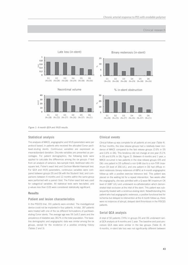
43
Chronic arterial response to PES with erodable polymer
- 167 -
Clinical research
Statistical analysisThe analyses of MACE, angiographic and IVUS parameters were per
protocol based, in patients who received the allocated Conor pacli-
taxel-eluting stents. Continuous variables are expressed as
mean±standard deviation. Discrete variables are presented as per-
centages. For patient demographics, the following tests were
applied to calculate the differences among the six groups: F-test
from an analysis of variance, two-sample t-test, likelihood ratio chi-
square test, Fisher’s exact test and Cochran-Mantel-Haenszel test.
For QCA and IVUS parameters, continuous variables were com-
pared between groups D5 and D6 with the Student t test, and com-
parisons between 4 months and 12 months within the same group
were performed with a paired t test. The Fisher exact test was used
for categorical variables. All statistical tests were two-tailed, and
p values less than 0.05 were considered statistically significant.
Results
Patient and lesion characteristicsIn the PISCES trial, 191 patients were enrolled. The investigational
device could not be implanted in four patients. In total, 187 patients
were treated with one of the six different formulations of paclitaxel-
eluting Conor stents. The average age was 59.1±9.2 years and the
prevalence of diabetes was 18.2% in the total population. The base-
line demographic and angiographic data was similar among the six
groups, except for the incidence of a positive smoking history
(Tables 2 and 3).
Clinical events
Clinical follow-up was complete for all patients at one year (Table 4).
At four months, the slow release groups had a relatively lower inci-
dence of MACE compared to the fast release groups (2.6% in D5
and 3.4% in D6). This tendency did not change at one year (5.1%
in D5 and 6.9% in D6; Figure 2). Between 4 months and 1 year, a
MACE occurred in two patients in the slow release groups (D5 and
D6): one patient in D5 suffered a non Q-MI due to a non-TVR (max-
imum CK level of 356 U/L), and one patient in D6 had diffuse in-
stent restenosis (binary restenosis of 68%) at 4-month angiographic
follow-up with a positive exercise tolerance test. This patient was
placed on the waiting list for a repeat intervention. Two weeks after
the angiography, she was admitted with a Q-wave MI (maximum CK
level of 1687 U/L) and underwent re-catheterization which demon-
strated total occlusion at the inlet of the stent. This patient was sub-
sequently treated with a sirolimus-eluting stent. Notwithstanding this
patient who had angiographic restenosis, a positive functional test for
ischemia but delayed re-intervention at the 4 month follow-up, there
were no instances of abrupt, delayed stent thrombosis in the PISCES
patients.
Serial QCA analysis
A total of 50 patients (74%) in groups D5 and D6 underwent seri-
al QCA analysis at 4 months and 1 year. The baseline and post-pro-
cedure QCA data were similar in the two groups (Table 4). At
4 months, in-stent late loss was not significantly different between
Figure 1: 4-month QCA and IVUS results.
D1(N=29)
D2(N=28)
D3(N=28)
D4(N=29)
D5(N=38)
D6(N=26)
1
0,8
0,6
0,4
0,2
0
Late loss (in-stent)
mm
D1(N=29)
D2(N=28)
D3(N=28)
D4(N=29)
D5(N=38)
D6(N=26)
15
10
5
0
Binary restenosis (in-stent)
%
D1(N=28)
D2(N=23)
D3(N=27)
D4(N=26)
D5(N=37)
D6(N=21)
40
30
20
10
0
Neointimal volume
mm
3
D1(N=28)
D2(N=23)
D3(N=27)
D4(N=26)
D5(N=37)
D6(N=21)
30
20
10
0
% in-stent obstruction
%

Chapter 4
44
- 168 -
One-year results of PISCES
D5 and D6, although in-stent late loss was lower in D6 than in D5
(0.32±0.40 mm versus 0.40±0.32 mm respectively, p=0.43).
From 4 months to 1 year, the late loss increased in both groups but
the trend remained in favor of D6; no statistical difference between
the two groups could be established although D6 showed a lower
in-stent late loss at 1 year (0.52±0.34 mm in D5 and 0.36±0.50 mm
in D6, p=0.20). Overall peri-stent binary restenosis at 1 year was
observed in one patient in each group (3.1% in D5 and 5.6% in
D6, p=1.00).
Serial IVUS analysis
A total of 45 patients (66%) underwent serial IVUS analysis at
4 months and 1 year. The IVUS results showed no statistical differ-
ences between groups D5 and D6 (Table 5). The percent in-stent
obstruction at 1 year was 12.46±7.60 in D5 and 8.37±9.10 in D6
(p=0.12). However, in D5, the neointimal area increased significant-
ly from 4 months to 1 year (delta=0.38mm2, p=0.0003) whereas
the difference between 4 months and 1 year in D6 failed to be sig-
nificant (delta=0.21 mm2, p=0.36, Figure 3).
At 4 months, significant expansive remodeling (an increase in the
plaque area outside the stent) was observed in both groups (7.75±1.93
mm2 vs 9.09±2.45 mm2, p<0.0001 in D5; 8.04±1.76 mm2 vs
8.95±1.67 mm2, p=0.0015 in D6). Between 4 months and 1 year, a
significant regression of the expansive plaque area outside the stent
was observed in both groups (9.09±2.45 mm2 vs 8.46±2.15 mm2,
p=0.045 in D5; 8.95±1.67 mm2 vs 8.37±1.74 mm2, p=0.0023 in D6).
DiscussionThe main findings of this study are the following: first, the slow
release (30 day) groups have better clinical outcomes at 1 year
compared to the fast release (5 or 10 day) groups. Second, com-
pared to the low dose (10 µg) group, the high dose (30 µg) group
had lower late loss and neointimal volume at one year without sta-
Table 2. Patient characteristics (per protocol)D-1 D-2 D-3 D-4 D-5 D-6 P-Value P-Value
10/5/b 10/10/b 10/10m 30/10/b 10/30/m 30/30/m comparing betweenN=30 N=29 N=30 N=30 N=39 N=29 6 groups D5 and D6
Age (mean±SD) 57.4±9.90 61.8±8.9 59.7±9.6 60.2±8.8 56.7±7.6 58.5±10.5 0.23 0.41
Male, % 60.0 (18/30) 72.4 (21/29) 76.7 (23/30) 60.0 (18/30) 82.1 (32/39) 69.0 (20/29) 0.28 0.21
Smoking, % 53.3(16/30) 86.2 (25/29) 76.7 (23/30) 73.3 (22/30) 89.7 (35/39) 72.4 (21/29) 0.01 0.06
Diabetes, % 16.7 (5/30) 17.2 (5/29) 23.3 (7/30) 13.3 (4/30) 10.3 (4/39) 31.0 (9/29) 0.31 0.03
Hypertension, % 40.0 (12/30) 62.1 (18/29) 63.3 (19/30) 56.7 (17/30) 35.9 (14/39) 62.1 (18/29) 0.08 0.03
Dyslipidemia, % 66.7 (20/30) 62.1 (18/29) 73.3 (22/30) 63.3 (19/30) 61.5 (24/39) 65.5 (19/29) 0.93 0.74
Prior MI, % 40.0 (12/30) 44.8 (13/29) 33.3 (10/30) 30.0 (9/30) 41.0 (16/39) 41.4 (12/29) 0.85 0.98
Prior CABG, % 3.3 (1/30) 3.5 (1/29) 0.0 (0/30) 0.0 (0/30) 2.6 (1/39) 6.9 (2/29) 0.59 0.39
Prior PCI 6.7 (2/30) 6.9 (2/29) 10.0 (3/30) 13.3 (4/30) 15.4 (6/39) 17.2 (5/29) 0.71 0.84
Table 3. Lesion and procedural characteristics (per protocol)D-1 D-2 D-3 D-4 D-5 D-6 P-Value P-Value
10/5/b 10/10/b 10/10m 30/10/b 10/30/m 30/30/m comparing betweenN=30 N=29 N=30 N=30 N=39 N=29 6 groups D5 and D6
Treated vessel
LAD 50.0% 41.4% 56.7% 50.0% 48.7% 27.6% 0.30 0.08
LCX 16.7% 20.7% 13.3% 23.3% 23.1% 37.9% 0.30 0.28
RCA 33.3% 37.9% 30.0% 26.7% 28.2% 34.5% 0.94 0.58
ACC/AHA classification
A/B1/B2 100.0% 100.0% 100.0% 100.0% 94.9% 96.6% 0.34 0.74
C 0.0% 0.0% 0.0% 0.0% 5.1% 3.4% 0.34 0.74
Angiographic features
Reference vessel diameter, mm 2.76±0.40 2.70±0.52 2.82±0.43 2.64±0.43 2.73±0.41 2.70±0.41 0.71 0.79
Lesion length, mm 9.73±3.68 9.08±3.60 10.60±3.83 10.62±3.09 9.35±3.24 10.31±3.36 0.37 0.24
Minimal lumen diameter, mm 1.10±0.35 1.06±0.38 0.97±0.37 1.05±0.25 1.03±0.28 1.00±0.31 0.70 0.64
Diameter stenosis, % 60.32±9.57 61.02±11.54 65.72±11.77 59.89±7.77 62.05±8.19 63.17±9.63 0.21 0.61
Procedural characteristics
Stent/patient 1.2±0.38 1.1±0.35 1.1±0.43 1.1±0.25 1.2±0.43 1.0±0.00 0.41 0.13
Stent length, mm 19.03±4.90 18.80±4.80 18.20±3.90 17.60±2.28 18.38±3.88 17.0±0.0 0 0.39 0.13
Stent diameter, mm 3.18±0.24 3.18±0.24 3.25±0.25 3.21±0.25 3.31±0.24 3.25±0.25 0.26 0.44
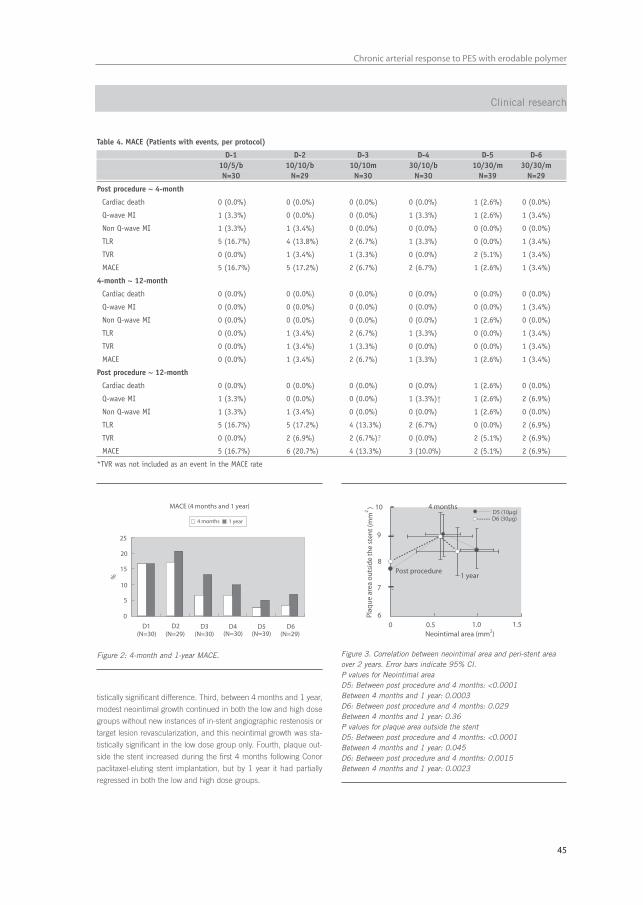
45
Chronic arterial response to PES with erodable polymer
- 169 -
Clinical research
Table 4. MACE (Patients with events, per protocol)
D-1 D-2 D-3 D-4 D-5 D-610/5/b 10/10/b 10/10m 30/10/b 10/30/m 30/30/mN=30 N=29 N=30 N=30 N=39 N=29
Post procedure ~ 4-month
Cardiac death 0 (0.0%) 0 (0.0%) 0 (0.0%) 0 (0.0%) 1 (2.6%) 0 (0.0%)
Q-wave MI 1 (3.3%) 0 (0.0%) 0 (0.0%) 1 (3.3%) 1 (2.6%) 1 (3.4%)
Non Q-wave MI 1 (3.3%) 1 (3.4%) 0 (0.0%) 0 (0.0%) 0 (0.0%) 0 (0.0%)
TLR 5 (16.7%) 4 (13.8%) 2 (6.7%) 1 (3.3%) 0 (0.0%) 1 (3.4%)
TVR 0 (0.0%) 1 (3.4%) 1 (3.3%) 0 (0.0%) 2 (5.1%) 1 (3.4%)
MACE 5 (16.7%) 5 (17.2%) 2 (6.7%) 2 (6.7%) 1 (2.6%) 1 (3.4%)
4-month ~ 12-month
Cardiac death 0 (0.0%) 0 (0.0%) 0 (0.0%) 0 (0.0%) 0 (0.0%) 0 (0.0%)
Q-wave MI 0 (0.0%) 0 (0.0%) 0 (0.0%) 0 (0.0%) 0 (0.0%) 1 (3.4%)
Non Q-wave MI 0 (0.0%) 0 (0.0%) 0 (0.0%) 0 (0.0%) 1 (2.6%) 0 (0.0%)
TLR 0 (0.0%) 1 (3.4%) 2 (6.7%) 1 (3.3%) 0 (0.0%) 1 (3.4%)
TVR 0 (0.0%) 1 (3.4%) 1 (3.3%) 0 (0.0%) 0 (0.0%) 1 (3.4%)
MACE 0 (0.0%) 1 (3.4%) 2 (6.7%) 1 (3.3%) 1 (2.6%) 1 (3.4%)
Post procedure ~ 12-month
Cardiac death 0 (0.0%) 0 (0.0%) 0 (0.0%) 0 (0.0%) 1 (2.6%) 0 (0.0%)
Q-wave MI 1 (3.3%) 0 (0.0%) 0 (0.0%) 1 (3.3%)† 1 (2.6%) 2 (6.9%)
Non Q-wave MI 1 (3.3%) 1 (3.4%) 0 (0.0%) 0 (0.0%) 1 (2.6%) 0 (0.0%)
TLR 5 (16.7%) 5 (17.2%) 4 (13.3%) 2 (6.7%) 0 (0.0%) 2 (6.9%)
TVR 0 (0.0%) 2 (6.9%) 2 (6.7%)? 0 (0.0%) 2 (5.1%) 2 (6.9%)
MACE 5 (16.7%) 6 (20.7%) 4 (13.3%) 3 (10.0%) 2 (5.1%) 2 (6.9%)
*TVR was not included as an event in the MACE rate
Figure 2: 4-month and 1-year MACE.
MACE (4 months and 1 year)
0
5
10
15
20
25
%
D1(N=30)
D2(N=29) (N=30) (N=30) (N=29)(N=39)
D3 D4 D5 D6
4 months 1 year
Figure 3. Correlation between neointimal area and peri-stent areaover 2 years. Error bars indicate 95% CI.P values for Neointimal area D5: Between post procedure and 4 months: <0.0001Between 4 months and 1 year: 0.0003D6: Between post procedure and 4 months: 0.029Between 4 months and 1 year: 0.36P values for plaque area outside the stentD5: Between post procedure and 4 months: <0.0001Between 4 months and 1 year: 0.045D6: Between post procedure and 4 months: 0.0015Between 4 months and 1 year: 0.0023
0 0.5 1.0 1.56
7
8
9
10 4 months
Post procedure1 year
Plaq
ue
area
ou
tsid
e th
e st
ent
(mm
2)
D5 (10µg)D6 (30µg)
Neointimal area (mm2)
tistically significant difference. Third, between 4 months and 1 year,
modest neointimal growth continued in both the low and high dose
groups without new instances of in-stent angiographic restenosis or
target lesion revascularization, and this neointimal growth was sta-
tistically significant in the low dose group only. Fourth, plaque out-
side the stent increased during the first 4 months following Conor
paclitaxel-eluting stent implantation, but by 1 year it had partially
regressed in both the low and high dose groups.

Chapter 4
46
- 170 -
One-year results of PISCES
Various drug elution processes have reported conflicting clinical
results3,9-12. In the SCORE trial, QuaDDS stents with a total of
4000 µg paclitaxel and a durable acrylate polymer were found to
have an unacceptable safety profile9. In the DELIVER trial, the Multi-
Link PENTA stent with a coating of 45-150µg paclitaxel (a dose den-
sity of 3.0µg/mm2 stent surface area) without a polymer showed no
impact in reducing clinical revascularization or restenosis compared
to bare metal stents, presumably due to the rapid elution of the
drug10. In the ELUTES and ASPECT trials, the high-dose non poly-
mer paclitaxel-eluting stent has the better outcomes compared to
the low dose group11,12. In the TAXUS IV trial, a slow-release (7.5%
at 30 days), polymer-based paclitaxel eluting stent with a total of
106 µg paclitaxel (a dose density of 1.0 µg/mm2 stent surface area)
eluted from a durable polymer showed significantly better inhibition
of neointimal growth and clinical outcomes when compared to bare
metal stents3. In the light of this study and other paclitaxel–eluting
stent trials, the drug release profile is a key factor in the clinical effi-
cacy, and drug-eluting stents with a slow release formulation seem
to be more efficient. In the present study, the slow release groups
demonstrate the best 1-year clinical outcomes, mainly due to better
outcomes in the first 4 months. The drug and polymer are com-
pletely removed from the wells after several months, thus this novel
drug-eluting stent may potentially preclude the chronic vessel reac-
tion usually observed with a durable polymer and persistent drug on
the stent13,14. Further, there were no instances of late stent throm-
bosis. Though more data from larger studies is required to draw
definitive conclusions, these results support the hypothesis that
complete drug elution and polymer resorbtion may confer safety
benefits with respect to delayed thrombosis.
In this study, the expansive vessel remodeling observed at 4 months
seems similar to the remodeling observed after implantation of
Taxus polymeric paclitaxel-eluting stents15. The expansive vessel
shrinkage observed at 1 year also suggests that the chronic vessel
reaction to mechanical injury and biological reaction to the drug and
polymer have subsided in that period of time.
However, the chronic vessel reaction inside the stent differs from
the reaction observed behind the stent struts. Although this study
showed a regression of tissue growth outside the stent between
4 months and 1 year, compaction of neointima was not observed in
either the low dose or the high dose group over the same time peri-
od. The precise reason for this phenomenon is unclear. One might
hypothesize that this could be a result of different tissue composi-
tion inside and outside the stent. The tissue growth inside the stent
is composed of smooth muscle cells in a proteoglycan rich matrix,
whereas the tissue growth behind the stent struts consists of sever-
al components: 1) intracellular matrix and cell proliferation such as
smooth muscle cells and lymphocyte cells, 2) oedema due to
mechanical injury and biological reaction against the drug, polymer
and stent, and 3) growth or regression of existing atherosclerotic
plaque. Thus, the direction of volumetric change (regression or
expansion) from 4 months to 1 year may not be similar inside and
outside the stent.
In this study, the actual late loss and neointimal area were smaller
in the 30 µg group than in the 10 µg group. These differences were
not statistically different and did not influence the clinical outcomes.
The 10 µg paclitaxel dose may be sufficient to suppress neointimal
growth in humans at least for a period of one year. It may also be
argued that the sample size is too small to detect a biological differ-
ence between the low and the high dose.
In animal studies, paclitaxel polymer coated stents have been found
to inhibit in-stent neointimal growth but with signs of delayed intimal
healing at 28 days, such as fibrin deposition, inflammation and
increased cellular proliferation. By 90 days, local toxicity associated
with paclitaxel resolves but in-stent neointimal growth suppression
is no longer present16. In humans following bare metal stent implan-
tation, the neointima does not keep growing beyond 6 months and
Table 5. Serial QCA analysis (post procedure, 4 months and 12 months)
D5 D6 P-value(10/30/m) (30/30/m)
N=32 N=18
Pre
Lesion length 9.39±3.37 10.04±2.80 0.49
RVD, mm 2.71±0.43 2.69±0.42 0.84
MLD, mm 1.05±0.29 1.02±0.33 0.73
DS, % 61.5±7.7 62.5±9.2 0.67
Post stenting
In-stent
MLD, mm 2.68±0.35 2.51±0.38 0.12
DS, % 12.4±6.5 13.6±6.2 0.56
In-peristent*
MLD, mm 2.31±0.41 2.18±0.38 0.29
DS, % 22.3±9.1 23.5±8.9 0.65
4-month
In-stent
MLD, mm 2.28±0.32 2.19± 0.53 0.53
DS, % 19.9±9.3 21.7±9.7 0.63
Late loss, mm 0.40±0.32 0.32±0.40 0.43
Binary restenosis, % 0.0 5.6 0.36
In-peristent*
MLD, mm 2.10±0.36 1.97±0.48 0.27
DS, % 25.3±8.7 29.9±13.6 0.20
Late loss, mm 0.21±0.29 0.21±0.39 0.95
Binary restenosis, % 0.0 5.6 0.36
12-month
In-stent
MLD, mm 2.16±0.34 2.15±0.65 0.93
DS, % 21.3±10.3 23.2±20.6 0.72
Late loss, mm 0.52±0.34 0.36±0.50 0.20
Binary restenosis, % 0.0 5.6 0.36
In-peristent*
MLD, mm 2.01±0.35 1.94±0.61 0.69
DS, % 27.5±9.67 29.3±20.8 0.73
Late loss, mm 0.30±0.26 0.24±0.50 0.62
Binary restenosis, % 3.1 5.6 1.00
* In-peristent = In-stent + 5 mm proximal + 5 mm distal

47
Chronic arterial response to PES with erodable polymer
- 171 -
instead begins to regress due to the replacement of water-trapping
proteoglycans (hyaluronan and versican) by decorin and type I col-
lagen17. In this study, compaction of neointima was not observed
and it is unknown whether neointimal tissue will keep growing or
stop beyond one year. Further follow-up is warranted to evaluate the
long-term efficacy of these devices and to find the best elution peri-
od and drug dose.
LimitationsAt one year, 26% and 34% of the patients in the groups of D5 and
D6 respectively did not undergo serial invasive QCA or IVUS follow-up
evaluation. Following completion of patient enrolment, the protocol
was subsequently amended to allow one year angiographic and IVUS
follow-up, necessitating a new informed consent. Patients who did not
undergo one-year angiography reported no anginal symptoms at one
year. The sample sizes for groups D5 and D6 were insufficient to
detect a difference in outcome between the low and high doses in the
slow release formulation. However, they served as the basis for the
development of a large randomized trial (the EuroSTAR trial) which is
evaluating both doses (10 µg and 30 µg per 17mm stent) of slow-
release paclitaxel using the reservoir-based technology on an ultra-
thin cobalt-chromium stent in 270 patients.
ConclusionsThe PISCES trial suggests that the pharmacokinetics of drug-eluting
stents is important for both neointimal suppression and for clinical
outcomes at 1 year. The slow release (30 day) formulation had bet-
ter clinical outcomes compared to the fast release (5 or 10 day) for-
mulation. The drug dose (10 µg or 30 µg) did not seem to influence
the amount of neointimal suppression but the sample sizes in this
pilot dose-finding study were insufficient to detect a beneficial dif-
ference in dose.
References1. Morice MC, Serruys PW, Sousa JE, Fajadet J, Ban Hayashi E, Perin M,
Colombo A, Schuler G, Barragan P, Guagliumi G, Molnar F, Falotico R. A randomized comparison of a sirolimus-eluting stent with a standardstent for coronary revascularization. N Engl J Med 2002;346:1773-80.
2. Moses JW, Leon MB, Popma JJ, Fitzgerald PJ, Holmes DR,O'Shaughnessy C, Caputo RP, Kereiakes DJ, Williams DO, Teirstein PS,Jaeger JL, Kuntz RE. Sirolimus-eluting stents versus standard stents inpatients with stenosis in a native coronary artery. N Engl J Med2003;349:1315-23.
3. Stone GW, Ellis SG, Cox DA, Hermiller J, O'Shaughnessy C, Mann JT,Turco M, Caputo R, Bergin P, Greenberg J, Popma JJ, Russell ME. A poly-mer-based, paclitaxel-eluting stent in patients with coronary artery dis-ease. N Engl J Med 2004;350:221-31.
4. Grube E, Sonoda S, Ikeno F, Honda Y, Kar S, Chan C, Gerckens U,Lansky AJ, Fitzgerald PJ. Six- and twelve-month results from first humanexperience using everolimus-eluting stents with bioabsorbable polymer.Circulation 2004;109:2168-71.
5. Serruys PW, Sianos G, Abizaid A, Aoki J, den Heijer P, Bonnier H,Smits P, McClean P, Verheye s, Belardi G, Condado J, Pieper M, Gambone L,Bressers M, Symons J, Sousa JE, Litvack F. The Effect of Variable Doseand Release Kinetics on Neointimal Hyperplasia Using a Novel Paclitaxel-Eluting Stent Platform - Paclitaxel In-Stent Controlled Elution Study(PISCES). JACC 2005;46:253-60
6. Finkelstein A, McClean D, Kar S, Takizawa K, Varghese K, Baek N,Park K, Fishbein MC, Makkar R, Litvack F, Eigler NL. Local drug deliveryvia a coronary stent with programmable release pharmacokinetics.Circulation 2003;107:777-84.
7. von Birgelen C, Di Mario C, Li W, Schuurbiers JC, Slager CJ, deFeyter PJ, Roelandt JR, Serruys PW. Morphometric analysis in three-dimensional intracoronary ultrasound: an in vitro and in vivo study per-formed with a novel system for the contour detection of lumen andplaque. Am Heart J 1996;132:516-27.
8. Serruys PW, Degertekin M, Tanabe K, Abizaid A, Sousa JE,Colombo A, Guagliumi G, Wijns W, Lindeboom WK, Ligthart J, de Feyter PJ,Morice MC. Intravascular ultrasound findings in the multicenter, random-ized, double-blind RAVEL (RAndomized study with the sirolimus-elutingVElocity balloon-expandable stent in the treatment of patients with denovo native coronary artery Lesions) trial. Circulation 2002;106:798-803.
9. Grube E, Lansky A, Hauptmann KE, Di Mario C, Di Sciascio G,Colombo A, Silber S, Stumpf J, Reifart N, Fajadet J, Marzocchi A, Schofer J,Dumas P, Hoffmann R, Guagliumi G, Pitney M, Russell ME. High-dose 7-hexanoyltaxol-eluting stent with polymer sleeves for coronary revascular-ization: one-year results from the SCORE randomized trial. J Am CollCardiol 2004;44:1368-72.
10. Lansky AJ, Costa RA, Mintz GS, Tsuchiya Y, Midei M, Cox DA,O'Shaughnessy C, Applegate RA, Cannon LA, Mooney M, Farah A,Tannenbaum MA, Yakubov S, Kereiakes DJ, Wong SC, Kaplan B, Cristea E,Stone GW, Leon MB, Knopf WD, O'Neill WW. Non-polymer-based pacli-taxel-coated coronary stents for the treatment of patients with de novocoronary lesions: angiographic follow-up of the DELIVER clinical trial.Circulation 2004;109:1948-54.
Clinical research
Table 6. Serial IVUS analysis (post procedure, 4 months and 12 months)
D5 D6 P-value(10/30/m) (30/30/m)
N=30 N=15
Post stenting
Vessel area, mm2 15.84±3.36 16.00 ±2.80 0.88
Stent area, mm2 8.09±1.81 7.95±1.77 0.81
Plaque area outside 7.75±1.93 8.04±1.76 0.63the stent, mm2
4-month
Vessel area, mm2 17.31±3.56 17.33±2.74 0.99
Stent area, mm2 8.22±1.86 8.64±1.92 0.49
Lumen area, mm2 7.61±1.87 8.08±1.82 0.43
Neointimal area, mm2 0.61±0.57 0.57±0.74 0.81
Plaque area outside 9.09±2.45 8.95±1.67 0.85the stent, mm2
% in-stent obstruction 7.49±6.92 6.21±7.39 0.57
12-month
Vessel area, mm2 16.74±3.56 16.73±3.02 0.99
Stent area, mm2 8.21±1.84 8.36±1.83 0.80
Lumen area, mm2 7.23±1.87 7.59±1.46 0.52
Neointimal area, mm2 0.99±0.54 0.77±0.92 0.42
Plaque area outside the stent, mm2 8.46±2.15 8.37±1.74 0.89
% in-stent obstruction 12.46±7.60 8.37 ±9.10 0.12

Chapter 4
48
- 172 -
One-year results of PISCES
11. Hong MK, Mintz GS, Lee CW, Song JM, Han KH, Kang DH, Song JK,Kim JJ, Weissman NJ, Fearnot NE, Park SW, Park SJ. Paclitaxel coatingreduces in-stent intimal hyperplasia in human coronary arteries: a serialvolumetric intravascular ultrasound analysis from the Asian Paclitaxel-Eluting Stent Clinical Trial (ASPECT). Circulation 2003;107:517-20.
12. Gershlick A, De Scheerder I, Chevalier B, Stephens-Lloyd A,Camenzind E, Vrints C, Reifart N, Missault L, Goy JJ, Brinker JA, Raizner AE,Urban P, Heldman AW. Inhibition of restenosis with a paclitaxel-eluting,polymer-free coronary stent: the European evaLUation of pacliTaxelEluting Stent (ELUTES) trial. Circulation 2004;109:487-93.
13. Virmani R, Liistro F, Stankovic G, Di Mario C, Montorfano M, Farb A,Kolodgie FD, Colombo A. Mechanism of late in-stent restenosis afterimplantation of a paclitaxel derivate-eluting polymer stent system inhumans. Circulation 2002;106:2649-51.
14. Virmani R, Guagliumi G, Farb A, Musumeci G, Grieco N, Motta T,Mihalcsik L, Tespili M, Valsecchi O, Kolodgie FD. Localized hypersensitiv-ity and late coronary thrombosis secondary to a sirolimus-eluting stent:should we be cautious? Circulation 2004;109:701-5.
15. Tanabe K, Serruys PW, Degertekin M, Guagliumi G, Grube E,Chan C, Munzel T, Belardi J, Ruzyllo W, Bilodeau L, Kelbaek H,Ormiston J, Dawkins K, Roy L, Strauss BH, Disco C, Koglin J, Russell ME,Colombo A. Chronic arterial responses to polymer-controlled paclitaxel-eluting stents: comparison with bare metal stents by serial intravascularultrasound analyses: data from the randomized TAXUS-II trial. Circulation2004;109:196-200.
16. Farb A, Heller PF, Shroff S, Cheng L, Kolodgie FD, Carter AJ,Scott DS, Froehlich J, Virmani R. Pathological analysis of local delivery ofpaclitaxel via a polymer-coated stent. Circulation 2001;104:473-9.
17. Farb A, Kolodgie FD, Hwang JY, Burke AP, Tefera K, Weber DK,Wight TN, Virmani R. Extracellular matrix changes in stented human coro-nary arteries. Circulation 2004;110:940-7.
AcknowledgementThe authors thank Patty Hevey and Louise Gambone for data
management and careful review of the manuscript.

Chapter 5
Serial Assessment of Tissue Growth Inside and Outside the
Stent after Implantation of Drug-Eluting Stent in Clinical
Trials. -Does delayed neointimal growth exist?
Aoki J, Abizaid A, Ong AT, Tsuchida K, Serruys PW
Eurointervention. 2005;3:253-255


51
Vascular response after DES
- 253 -
Serial assessment of tissue growth inside and outside the stent after implantation of drug-eluting stent in clinical trials. – Does delayed neointimal growth exist?
Although long term follow-up after drug-eluting stent (DES)
implantation shows a sustained clinical benefit in several random-
ized trials, delayed neointimal growth beyond the first 6 to 9 months
has been reported in serial intravascular ultrasound (IVUS) analy-
ses in some trials. The issue of a delayed restenosis which was
observed after brachytherapy has not been thoroughly evaluated
with DES.
Tissue growth inside the stent (Neointima)Drug-eluting stents (DES) dramatically reduce neointimal growth at
6 or 9 months compared to bare metal stents (BMS).1-3 Although
long term follow-up after DES implantation shows a sustained clini-
cal benefit in several randomised trials,4,5 little is known about neoin-
timal growth beyond the first 6 to 9 months. The issue of a “late
catch up phenomenon” (delayed restenosis) which was observed
after brachytherapy has not been fully investigated with DES.6
In porcine models, the inhibition of neointimal hyperplasia after
deployment of polymer-coated sirolimus-eluting stents was not
sustained at 90 and 180 days due to delayed cellular proliferation,
and neointimal suppression after deployment of chondroitin sul-
fate and gelatin coated paclitaxel-eluting stents was also not main-
tained at 90 days.7,8
In humans, neointimal tissue does not keep growing after BMS
implantation. During long-term angiographic follow-up, com-
paction of neointima has been described in several reports.9-11
Histological analyses of post-mortem coronary arteries demon-
strate that compaction of neointima occurs due to the replacement
of water-trapping proteoglycans by decorin and type I collagen.11
Following DES implantation, neointima continues to grow during
the follow-up period in some trials in which serial IVUS analyses
were performed (Table 1). A chronic arterial response towards the
durable polymer and to the remaining drug within the polymer has
been imputed to explain this phenomenon. However, this was also
observed with Conor paclitaxel-eluting stents in which neither
polymer nor drug is retained at the end of the programmed
release period.12 The precise reason for this observation is thus
still unclear, but may be related to the delayed healing response
and persistent biological reaction caused by the drug soon after
the implantation of DES. In view of the results of animal studies
and clinical studies, DES may delay restenosis, instead of halting
definitively the process of neointimal hyperplasia. Further follow-up
is warranted to evaluate the long-term efficacy of DES.
Tissue growth outside the stent (Vessel remodeling)Polymer based sirolimus-eluting stents (Cypher) and polymer
based paclitaxel-eluting stents (Taxus) are used in daily practice,
and several types of drugs and durable or erodable polymers have
been tested in clinical trials.1,3,13,14 After BMS implantation, expan-
sive vessel remodeling (i.e. increasing plaque outside the stent) was
reported.15 In the TAXUS II trial, the increased plaque outside the
stent 6 months after BMS implantation had regressed completely at
2 years (Table 2).16 Thus, it is likely that mechanical injury and bio-
logical reaction against the stainless steel stent that may have
Jiro Aoki1, MD; Alexandre C. Abizaid2, MD, PhD; Andrew.T.L. Ong1, MBBS, FRACP; Keiichi Tsuchida1, MD, PhD; Patrick W. Serruys*1, MD, PhD.
1. Thoraxcenter Erasmus MC, Rotterdam, the Netherlands
2. Institute Dante Pazzanese of Cardiology, São Paulo, Brazil
Editorial
* Corresponding author: Thoraxcenter, Ba 583, Erasmus MC, Dr Molewaterplein 40, 3015 GD Rotterdam, The Netherlands
E-mail: [email protected]
© Europa Edition 2005. All rights reserved.
EuroInterv.2005;1:253-255
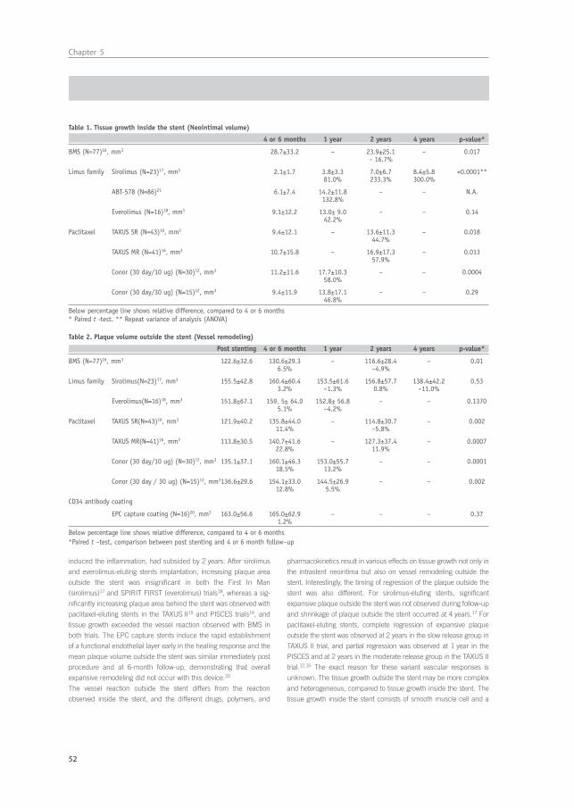
Chapter 5
52
- 254 -
induced the inflammation, had subsided by 2 years. After sirolimus
and everolimus-eluting stents implantation, increasing plaque area
outside the stent was insignificant in both the First In Man
(sirolimus)17 and SPIRIT FIRST (everolimus) trials18, whereas a sig-
nificantly increasing plaque area behind the stent was observed with
paclitaxel-eluting stents in the TAXUS II15 and PISCES trials19, and
tissue growth exceeded the vessel reaction observed with BMS in
both trials. The EPC capture stents induce the rapid establishment
of a functional endothelial layer early in the healing response and the
mean plaque volume outside the stent was similar immediately post
procedure and at 6-month follow-up, demonstrating that overall
expansive remodeling did not occur with this device.20
The vessel reaction outside the stent differs from the reaction
observed inside the stent, and the different drugs, polymers, and
pharmacokinetics result in various effects on tissue growth not only in
the intrastent neointima but also on vessel remodeling outside the
stent. Interestingly, the timing of regression of the plaque outside the
stent was also different. For sirolimus-eluting stents, significant
expansive plaque outside the stent was not observed during follow-up
and shrinkage of plaque outside the stent occurred at 4 years.17 For
paclitaxel-eluting stents, complete regression of expansive plaque
outside the stent was observed at 2 years in the slow release group in
TAXUS II trial, and partial regression was observed at 1 year in the
PISCES and at 2 years in the moderate release group in the TAXUS II
trial.12,16 The exact reason for these variant vascular responses is
unknown. The tissue growth outside the stent may be more complex
and heterogeneous, compared to tissue growth inside the stent. The
tissue growth inside the stent consists of smooth muscle cell and a
Table 1. Tissue growth inside the stent (Neointimal volume)
4 or 6 months 1 year 2 years 4 years p-value*
BMS (N=77)16, mm3 28.7±33.2 – 23.9±25.1 – 0.017– 16.7%
Limus family Sirolimus (N=23)17, mm3 2.1±1.7 3.8±3.3 7.0±6.7 8.4±5.8 <0.0001**81.0% 233.3% 300.0%
ABT-578 (N=86)21 6.1±7.4 14.2±11.8 – – N.A.132.8%
Everolimus (N=16)18, mm3 9.1±12.2 13.0± 9.0 – – 0.1442.2%
Paclitaxel TAXUS SR (N=43)16, mm3 9.4±12.1 – 13.6±11.3 – 0.01844.7%
TAXUS MR (N=41)16, mm3 10.7±15.8 – 16.9±17.3 – 0.01357.9%
Conor (30 day/10 ug) (N=30)12, mm3 11.2±11.6 17.7±10.3 – – 0.000458.0%
Conor (30 day/30 ug) (N=15)12, mm3 9.4±11.9 13.8±17.1 – – 0.2946.8%
Below percentage line shows relative difference, compared to 4 or 6 months* Paired t -test. ** Repeat variance of analysis (ANOVA)
Table 2. Plaque volume outside the stent (Vessel remodeling)
Post stenting 4 or 6 months 1 year 2 years 4 years p-value*
BMS (N=77)16, mm3 122.6±32.6 130.6±29.3 – 116.6±28.4 – 0.016.5% –4.9%
Limus family Sirolimus(N=23)17, mm3 155.5±42.8 160.4±60.4 153.5±61.6 156.8±57.7 138.4±42.2 0.533.2% –1.3% 0.8% –11.0%
Everolimus(N=16)18, mm3 151.8±67.1 159. 5± 64.0 152.8± 56.8 – – 0.13705.1% –4.2%
Paclitaxel TAXUS SR(N=43)16, mm3 121.9±40.2 135.8±44.0 – 114.8±30.7 – 0.00211.4% –5.8%
TAXUS MR(N=41)16, mm3 113.8±30.5 140.7±41.6 – 127.3±37.4 – 0.000722.8% 11.9%
Conor (30 day/10 ug) (N=30)12, mm3 135.1±37.1 160.1±46.3 153.0±55.7 – – 0.000118.5% 13.2%
Conor (30 day / 30 ug) (N=15)12, mm3136.6±29.6 154.1±33.0 144.5±26.9 – – 0.00212.8% 5.5%
CD34 antibody coating
EPC capture coating (N=16)20, mm3 163.0±56.6 165.0±62.9 – – – 0.371.2%
Below percentage line shows relative difference, compared to 4 or 6 months*Paired t –test, comparison between post stenting and 4 or 6 month follow–up

53
Vascular response after DES
- 255 -
proteoglycan rich matrix, whereas the tissue growth outside the stent
consists of several components; 1) cell proliferations and intracellular
matrix, 2) oedema caused by mechanical injury and chemical injury
due to drug, polymer and stent, 3) growth or regression of existing
atherosclerotic plaque. Studies involving larger sample sizes and
more detailed analyses with novel in vivo techniques of tissue charac-
terisation may be necessary to assess and better understand the
process of vessel remodeling after DES implantation.
References1. Morice MC, Serruys PW, Sousa JE, Fajadet J, Ban Hayashi E,
Perin M, Colombo A, Schuler G, Barragan P, Guagliumi G, Molnar F,Falotico R. A randomized comparison of a sirolimus-eluting stent with a stan-dard stent for coronary revascularization. N Engl J Med 2002;346:1773-1780.
2. Moses JW, Leon MB, Popma JJ, Fitzgerald PJ, Holmes DR,O’Shaughnessy C, Caputo RP, Kereiakes DJ, Williams DO, Teirstein PS, JaegerJL, Kuntz RE. Sirolimus-eluting stents versus standard stents in patients withstenosis in a native coronary artery. N Engl J Med 2003;349:1315-1323.
3. Stone GW, Ellis SG, Cox DA, Hermiller J, O’Shaughnessy C, Mann JT,Turco M, Caputo RP, Bergin P, Greenberg J, Popma JJ, Russell ME.A polymer-based, paclitaxel-eluting stent in patients with coronary arterydisease. N Engl J Med 2004;350:221-231.
4. Fajadet J, Morice MC, Bode C, Barragan P, Serruys PW, Wijns W,Constantini CR, Guermonprez JL, Eltchaninoff H, Blanchard D,Bartorelli A, Laarman GJ, Perin M, Sousa JE, Schuler G, Molnar F,Guagliumi G, Colombo A, Ban Hayashi E, Wulfert E. Maintenance of long-term clinical benefit with sirolimus-eluting coronary stents: three-yearresults of the RAVEL trial. Circulation 2005;111:1040-1044.
5. Sousa JE, Costa MA, Abizaid A, Feres F, Seixas AC, Tanajura LF,Mattos LA, Falotico R, Jaeger J, Popma JJ, Serruys PW, Sousa AG. Four-Year Angiographic and Intravascular Ultrasound Follow-Up of PatientsTreated With Sirolimus-Eluting Stents. Circulation 2005;111:2326-2329.
6. Serruys PW, Wijns W, Sianos G, de Scheerder I, van den Heuvel PA,Rutsch W, Glogar HD, Macaya C, Materne PH, Veldhof S, Vonhausen H,Otto-Terlouw PC, van der Giessen WJ. Direct stenting versus direct stent-ing followed by centered beta-radiation with intravascular ultrasound-guided dosimetry and long-term anti-platelet treatment: results of a ran-domized trial: Beta-Radiation Investigation with Direct Stenting and Galileoin Europe (BRIDGE). J Am Coll Cardiol 2004;44:528-537.
7. Farb A, Heller PF, Shroff S, Cheng L, Kolodgie FD, Carter AJ,Scott DS, Froehlich J, Virmani R. Pathological analysis of local delivery ofpaclitaxel via a polymer-coated stent. Circulation 2001;104:473-479.
8. Carter AJ, Aggarwal M, Kopia GA, Tio F, Tsao PS, Kolata R,Yeung AC, Llanos G, Dooley J, Falotico R. Long-term effects of polymer-based, slow-release, sirolimus-eluting stents in a porcine coronary model.Cardiovasc Res 2004;63:617-624.
9. Kimura T, Abe K, Shizuta S, Odashiro K, Yoshida Y, Sakai K,Kaitani K, Inoue K, Nakagawa Y, Yokoi H, Iwabuchi M, Hamasaki N,Nosaka H, Nobuyoshi M. Long-term clinical and angiographic follow-upafter coronary stent placement in native coronary arteries. Circulation2002;105:2986-2991.
10. Asakura M, Ueda Y, Nanto S, Hirayama A, Adachi T, Kitakaze M,Hori M, Kodama K. Remodeling of in-stent neointima, which became thin-ner and transparent over 3 years: serial angiographic and angioscopic fol-low-up. Circulation 1998;97:2003-2006.
11. Farb A, Kolodgie FD, Hwang JY, Burke AP, Tefera K, Weber DK,Wight TN, Virmani R. Extracellular matrix changes in stented human coro-nary arteries. Circulation 2004;110:940-947.
12. Aoki J, Ong AT, Abizaid A, den Heijer P, Bonnier H, McClean D,Verheye S, Belardi G, Condado J, Pieper M, Sousa E, Bressers M,Symons J, Sianos G, Serruys PW. One-year clinical effect of various dosesand pharmacokinetic releases of paclitaxel eluted from an erodable poly-mer-insights from paclitaxel in-stent controlled elution study (PISCES).Eurointervention 2005:1;165-172.
13. Serruys PW, Ormiston JA, Sianos G, Sousa JE, Grube E, den Heijer P,de Feyter P, Buszman P, Schomig A, Marco J, Polonski L, Thuesen L,Zeiher AM, Bett JH, Suttorp MJ, Glogar HD, Pitney M, Wilkins GT,Whitbourn R, Veldhof S, Miquel K, Johnson R, Coleman L, Virmani R.Actinomycin-eluting stent for coronary revascularization: a randomizedfeasibility and safety study: the ACTION trial. J Am Coll Cardiol2004;44:1363-1367.
14. Grube E, Lansky A, Hauptmann KE, Di Mario C, Di Sciascio G,Colombo A, Silber S, Stumpf J, Reifart N, Fajadet J, Marzocchi A,Schofer J, Dumas P, Hoffmann R, Guagliumi G, Pitney M, Russell ME.High-dose 7-hexanoyltaxol-eluting stent with polymer sleeves for coronaryrevascularization: one-year results from the SCORE randomized trial. J AmColl Cardiol 2004;44:1368-1372.
15. Tanabe K, Serruys PW, Degertekin M, Guagliumi G, Grube E, Chan C,Munzel T, Belardi J, Ruzyllo W, Bilodeau L, Kelbaek H, Ormiston J,Dawkins K, Roy L, Strauss BH, Disco C, Koglin J, Russell ME, Colombo A.Chronic arterial responses to polymer-controlled paclitaxel-eluting stents:comparison with bare metal stents by serial intravascular ultrasoundanalyses: data from the randomized TAXUS-II trial. Circulation2004;109:196-200.
16. Aoki J, Colombo A, Dudek D, Banning AP, Drzewiecki J, Zmudka K,Schiele F, Russell ME, Koglin J. Peri-Stent Remodeling and NeointimalSuppression Two-Years after Polymer-Based, Paclitaxel-Eluting StentImplantation - Insights from Serial IVUS Analysis in the TAXUS II Study.Circulation 2005, In press.
17. Aoki J, Abizaid A, Serruys PW, Ong AT, Boersma E, Sousa JE,Bruining N. Evaluation of four-year coronary artery response aftersirolimus-eluting stent implantation by using serial quantitative IVUS andcomputer assisted grey-scale value analysis for plaque composition inevent-free patients. J Am Coll Cardiol 2005;46:1670-1676.
18. Tsuchida K, Piek JJ, Neumann F, van der Giessen WJ, Wiemer M,Zeiher A, Grube E, Haase J, Thuesen L, Hamm C, Veldhof D, Fink S,Serruys PW. One-year results of a durable polymer everolimus-elutingstents in de novo coronary narrowings (The SPIRIT FIRST Trial).Eurointervention 2005, In press.
19. Serruys PW, Sianos G, Abizaid A, Aoki J, den Heijer P, Bonnier H,Smiths P, McClean D, Verheye S, Belardi G, Condado J, Pieper M,Gambone L, Bressers M, Symons J, Sousa E, Litvack F. The effect of vari-able dose and release kinetics on neointimal hyperplasia using a novelpaclitaxel-eluting stent platform –Paclitaxel In-Stent Controlled ElutionStudy (PISCES). J Am Coll Cardiol 2005;46:253-260.
20. Aoki J, Serruys PW, van Beusekom H, Ong AT, McFadden EP,Sianos G, van der Giessen WJ, Regar E, de Feyter P, Davis H, Rowland S,Kutryk MJ. Endothelial progenitor cell capture by stents coated with anti-body against CD34 - The HEALING-FIM (Healthy Endothelial acceleratedlinig inhibits neointimal growth-first in man) registry. J Am Coll Cardiol2005;45:1574-1579.
21. Meredith IT, Ormiston J, Whitbourn R, Kay P, Muller D, Bonan R,Popma JJ, Cutlip DE, Fitzgerald P, Prpic R, Kuntz RE. First-in-humanstudy of the Endeavor ABT-578-eluting phosphorylcholine-encapsulatedstent system in denovo native coronary artery lesions: Endeavor I Trial.Eurointervention 2005;1:157-164
Editorial


Part 2
Effi cacy of drug eluting stent for high risk patients


Chapter 6
“Full Metal Jacket” (stented length ≥ 64 mm) Using Drug-
Eluting Stents for De Novo Coronary Artery Lesions
Aoki J, Ong AT, Rodriguez-Granillo GA, Mc
Fadden E, van Mieghem CA, Valgimigli M,
Tsuchida K, Sianos G, Regar E, de Jaegere
P, van der Giessen W, de Feyter P, van
Domburg R, Serruys PW.
A Heart J. 2005;150:994-999


59
Full metal jacket using DES
‘‘Full metal jacket’’ (stented length _>64 mm) usingdrug-eluting stents for de novo coronary artery lesionsJiro Aoki, MD, Andrew T.L. Ong, MBBS, FRACP, Gaston A. Rodriguez Granillo, MD,Eugene P. McFadden, MD, FRCPI, Carlos A.G. van Mieghem, MD, Marco Valgimigli, MD,Keiichi Tsuchida, MD, Georgios Sianos, MD, PhD, Evelyn Regar, MD, PhD, Peter P.T. de Jaegere, MD, PhD,Willem J. van der Giessen, MD, PhD, Pim J. de Feyter, MD, PhD, Ron T. van Domburg, PhD, andPatrick W. Serruys, MD, PhD Rotterdam, The Netherlands
Background Stented segment length was a predictive factor for restenosis in the bare metal stent era. Theobjective of the study was to evaluate the medium-term clinical outcome and the potential for adverse effects whenvery long segments (ie, z64 mm of stented length) are treated by drug-eluting stent (DES) implantation, an approachcolloquially referred to as a ’’full metal jacket.’’
Methods Since April 2002, we have used DES as the default stent for all percutaneous coronary interventions.From our prospective institutional database we identified 122 consecutive patients, with de novo coronary lesions, inwhom a coronary artery was treated with at least 64 mm of overlapping DES: 81 patients were treated withsirolimus-eluting stents and 41 with paclitaxel-eluting stents.
Results The mean number of stents per lesion was 3.3 F 1.1, and the median stented length was 79 mm (range64-168 mm). Periprocedural Q-wave myocardial infarction (MI) occurred in 2 patients (1.6%) and subacute stentthrombosis in 1 patient (0.8%). During 1-year follow-up, 5 patients (4.1%), including 3 patients treated for acute MI withcardiogenic shock, died and 10 patients (8.2%) had nonfatal MI (creatine kinase–MB N3 times). The 1-year targetvessel revascularization rate was 7.5% and the overall incidence of major adverse cardiac events was 18%.Outcomes in sirolimus-eluting stents and paclitaxel-eluting stents groups did not differ statistically.
Conclusions The use of DES for the treatment of diffuse lesions was associated with a low rate of repeatrevascularization, irrespective of stent type. No safety concerns were raised at medium-term follow-up. (Am Heart J2005;150:994-9.)
In the bare metal stent era, the length of a stented
segment was an independent predictor of in-stent
restenosis.1-3 Recent randomized trials, in low-risk
patient/lesion cohorts, showed that drug-eluting stents
(DES) reduce the need for repeat intervention com-
pared with bare metal stents.4-7 Drug-eluting stents are
rapidly replacing bare metal stents and there has been
a tendency toward longer stented segment lengths,
given the full lesion coverage from a proximal to a
distal bangiographically normalQ segment to avoid stent
gaps and the incomplete coverage of lesions which
have been associated with restenosis after DES im-
plantation.5,8 However, the clinical effect of very long
and overlapping DES implantation in the so-called full
metal jacket approach and the potential effects of
increased metal and local drug exposures are un-
known. In this report, we investigate the clinical
outcome after very long sirolimus-eluting stent (SES)
and paclitaxel-eluting stent (PES) implantations in a
consecutive group of 122 patients (124 lesions) who
were treated with at least 64 mm of DES without any
gap in the same vessel.
MethodsPatient selection and procedureSince April 2002, we have adopted a policy of universal
DES implantation for all percutaneous coronary interventions
requiring stents at our center, irrespective of clinical
presentation or lesion morphology. Sirolimus-eluting stents
were exclusively used until March 2003. Since March 2003,
PES has been exclusively used. In total, 122 consecutive
From the Erasmus Medical Center, Rotterdam, Thoraxcenter, The Netherlands.
No author has any conflict of interest. All authors have read and approved submission of
the manuscript and the manuscript has not been published and is not being considered for
publication elsewhere in whole or part in any language except as an abstract.
Submitted November 26, 2004; accepted January 26, 2005.
Reprint requests: Patrick W. Serruys, MD, PhD, Thoraxcenter, Bd406, Erasmus MC, Dr
Molewaterplein 40, 3015-GD Rotterdam, The Netherlands.
E-mail: [email protected]
0002-8703/$ - see front matter
n 2005, Mosby, Inc. All rights reserved.
doi:10.1016/j.ahj.2005.01.050

Chapter 6
60
patients were treated with at least 64 mm of DES without any
gap in the same vessel for diffuse coronary lesions, chronic
total occlusion, or extensive dissections; this represents 7% of
our patients treated exclusively with DES in the same period.
The longest available individual stent length was 33 mm in
SES and 32 mm in PES. We defined a full metal jacket as
64 mm of continuous stent, calculated as the total of the
2 longest PES lengths.
All interventions were performed using standard techniques.
Written informed consent was obtained from all patients as
part of the prospective consecutive database. Periprocedural
use of glycoprotein IIb/IIIa and intravascular ultrasound (IVUS)
was at the discretion of the operator. All patients received a
loading dose of 300-mg clopidogrel followed by a daily dosage
of 75 mg for 6 months, in addition to life-long aspirin therapy.
End point definition and clinical follow-upPatients were followed up prospectively and major
adverse cardiac events (MACEs) were evaluated. Major
adverse cardiac event was defined as death, nonfatal
myocardial infarction (MI), or target vessel revascularization
(TVR). Myocardial infarction was defined as a creatine
kinase–MB (CK-MB) level that was N3 times the upper limit
of normal, based on the recommendations in the American
College of Cardiology/American Heart Association guide-
lines.9 For patients who presented with an acute coronary
syndrome and elevated baseline enzyme, a diagnosis of
periprocedural MI required a fall and rise of CK-MB of 50%
above the previous level.10 Target vessel revascularization
was defined as a reintervention in the treated vessel. Stent
thrombosis was angiographically documented as a complete
occlusion or a flow-limiting thrombus of a successfully
stented segment. Information regarding repeat interventions
was prospectively collected by means of an electronic
database. Survival status was assessed by written inquiries to
the Civil Registry. Questionnaires to assess clinical status
were sent to all living patients. The patient, referring
physician, and peripheral hospitals were directly approached
whenever necessary for additional information. Follow-up
angiography was planned only in patients who were
enrolled during the first 6 months (n = 38).
Statistical analysisContinuous variables were expressed as mean F SD.
Discrete variables were presented as percentages. Continuous
variables were compared with Student t test or Wilcoxon
ranked scores when applicable. The Fisher exact test was used
for categorical variables. The cumulative incidence of adverse
events was calculated according to the Kaplan-Meier method
and differences were assessed using the log-rank test. All
statistical tests were 2-tailed, and P b .05 was considered
statistically significant. Patient, lesion, and procedural charac-
teristics associated with 1-year MACE on univariate analysis
(P value for selection b.2) were tested for their multivariate
predictive value (tested values were cardiogenic shock, female
sex, multivessel disease, bifurcation stenting, and use of IIb/IIIa
inhibitors). The final model was built by backward stepwise
variable selection with entry and exit criteria set at the P = .05
and P = .1 levels, respectively.
ResultsPatient characteristicsOf the 122 consecutive patients, 81 patients (82
lesions) were treated with SES (SES group) and 41
patients (42 lesions) were treated with PES (PES group).
Patient characteristics are reported in Table I; 19% had
diabetes and 39% underwent the index procedure for an
acute coronary syndrome. There were no statistical
differences between groups, apart from a higher
proportion of men in the PES group. Figure 1 presents
the distribution of stented length in this study.
Procedural characteristicsLesion and procedural characteristics are presented in
Table II. The mean number of stents implanted per
lesion was 3.3 F 1.1 (range 2-7 stents), and the median
Table I. Patient characteristics
All(n = 122)
SES(n = 81)
PES(n = 41)
Age (y) 63 F 11 62 F 12 63 F 11Male (%) 75 69 88THypertension (%) 42 43 39Hyperlipidemia (%) 67 62 76Current smoking (%) 25 26 24Diabetes mellitus (%) 19 20 17Prior MI (%) 44 43 46Prior CABG (%) 5 5 5Prior PCI (%) 19 17 22Multivessel disease (%) 70 70 68Unstable angina (%) 26 31 17AMI (%) 11 11 12AMI with shock (%) 3 2 5
CABG, Coronary artery bypass graft surgery; PCI, percutaneous coronaryintervention; AMI, acute MI.TP b .05 for the comparison between SES and PES groups.
Figure 1
0
10
20
30
40
50
60
64-79 80-99 100-119 120-139 140-
Stented length (mm)
%
The distribution of stented length.
American Heart Journal
Volume 150, Number 5Aoki et al 995
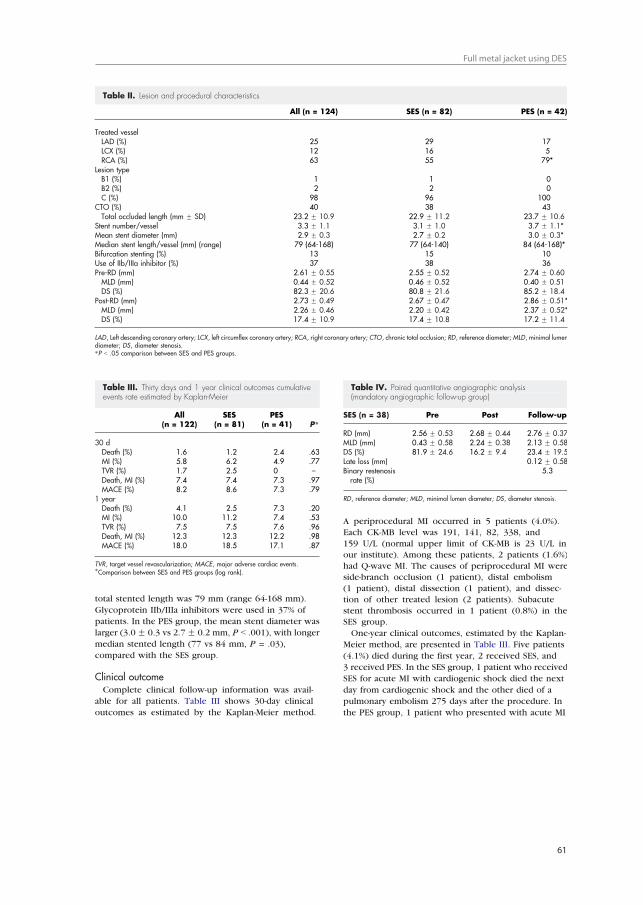
61
Full metal jacket using DES
total stented length was 79 mm (range 64-168 mm).
Glycoprotein IIb/IIIa inhibitors were used in 37% of
patients. In the PES group, the mean stent diameter was
larger (3.0F 0.3 vs 2.7F 0.2 mm, P b .001), with longer
median stented length (77 vs 84 mm, P = .03),
compared with the SES group.
Clinical outcomeComplete clinical follow-up information was avail-
able for all patients. Table III shows 30-day clinical
outcomes as estimated by the Kaplan-Meier method.
A periprocedural MI occurred in 5 patients (4.0%).Each CK-MB level was 191, 141, 82, 338, and
159 U/L (normal upper limit of CK-MB is 23 U/L inour institute). Among these patients, 2 patients (1.6%)
had Q-wave MI. The causes of periprocedural MI wereside-branch occlusion (1 patient), distal embolism
(1 patient), distal dissection (1 patient), and dissec-tion of other treated lesion (2 patients). Subacute
stent thrombosis occurred in 1 patient (0.8%) in theSES group.
One-year clinical outcomes, estimated by the Kaplan-
Meier method, are presented in Table III. Five patients
(4.1%) died during the first year, 2 received SES, and
3 received PES. In the SES group, 1 patient who received
SES for acute MI with cardiogenic shock died the next
day from cardiogenic shock and the other died of a
pulmonary embolism 275 days after the procedure. In
the PES group, 1 patient who presented with acute MI
Table III. Thirty days and 1 year clinical outcomes cumulativeevents rate estimated by Kaplan-Meier
All(n = 122)
SES(n = 81)
PES(n = 41) PT
30 dDeath (%) 1.6 1.2 2.4 .63MI (%) 5.8 6.2 4.9 .77TVR (%) 1.7 2.5 0 –Death, MI (%) 7.4 7.4 7.3 .97MACE (%) 8.2 8.6 7.3 .79
1 yearDeath (%) 4.1 2.5 7.3 .20MI (%) 10.0 11.2 7.4 .53TVR (%) 7.5 7.5 7.6 .96Death, MI (%) 12.3 12.3 12.2 .98MACE (%) 18.0 18.5 17.1 .87
TVR, target vessel revascularization; MACE, major adverse cardiac events.TComparison between SES and PES groups (log rank).
Table IV. Paired quantitative angiographic analysis(mandatory angiographic follow-up group)
SES (n = 38) Pre Post Follow-up
RD (mm) 2.56 F 0.53 2.68 F 0.44 2.76 F 0.37MLD (mm) 0.43 F 0.58 2.24 F 0.38 2.13 F 0.58DS (%) 81.9 F 24.6 16.2 F 9.4 23.4 F 19.5Late loss (mm) 0.12 F 0.58Binary restenosisrate (%)
5.3
RD, reference diameter; MLD, minimal lumen diameter; DS, diameter stenosis.
Table II. Lesion and procedural characteristics
All (n = 124) SES (n = 82) PES (n = 42)
Treated vesselLAD (%) 25 29 17LCX (%) 12 16 5RCA (%) 63 55 79*
Lesion typeB1 (%) 1 1 0B2 (%) 2 2 0C (%) 98 96 100
CTO (%) 40 38 43Total occluded length (mm F SD) 23.2 F 10.9 22.9 F 11.2 23.7 F 10.6
Stent number/vessel 3.3 F 1.1 3.1 F 1.0 3.7 F 1.1*Mean stent diameter (mm) 2.9 F 0.3 2.7 F 0.2 3.0 F 0.3*Median stent length/vessel (mm) (range) 79 (64-168) 77 (64-140) 84 (64-168)*Bifurcation stenting (%) 13 15 10Use of IIb/IIIa inhibitor (%) 37 38 36Pre-RD (mm) 2.61 F 0.55 2.55 F 0.52 2.74 F 0.60MLD (mm) 0.44 F 0.52 0.46 F 0.52 0.40 F 0.51DS (%) 82.3 F 20.6 80.8 F 21.6 85.2 F 18.4
Post-RD (mm) 2.73 F 0.49 2.67 F 0.47 2.86 F 0.51*MLD (mm) 2.26 F 0.46 2.20 F 0.42 2.37 F 0.52*DS (%) 17.4 F 10.9 17.4 F 10.8 17.2 F 11.4
LAD, Left descending coronary artery; LCX, left circumflex coronary artery; RCA, right coronary artery; CTO, chronic total occlusion; RD, reference diameter;MLD, minimal lumendiameter; DS, diameter stenosis.TP b .05 comparison between SES and PES groups.
American Heart Journal
November 2005996 Aoki et al
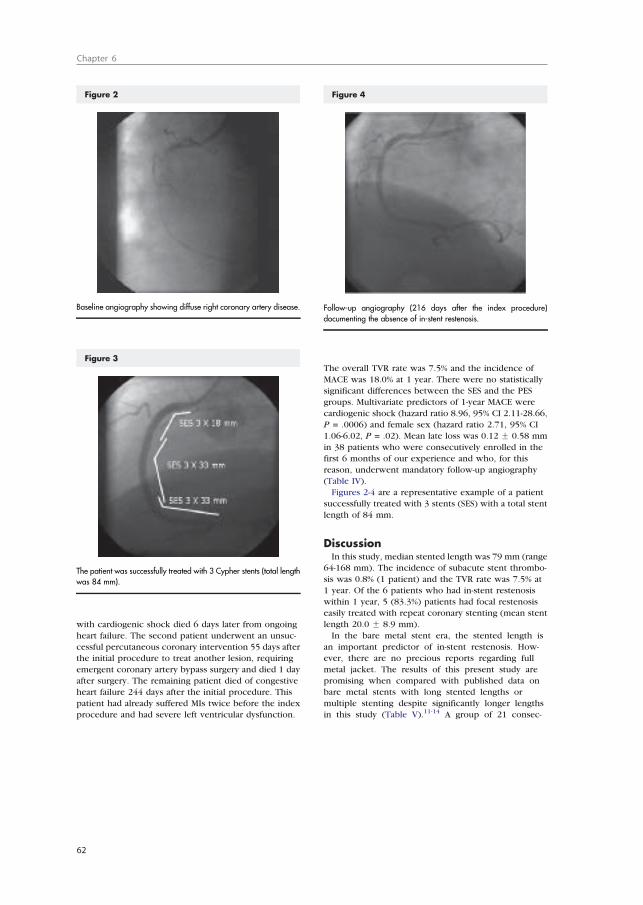
Chapter 6
62
with cardiogenic shock died 6 days later from ongoing
heart failure. The second patient underwent an unsuc-
cessful percutaneous coronary intervention 55 days after
the initial procedure to treat another lesion, requiring
emergent coronary artery bypass surgery and died 1 day
after surgery. The remaining patient died of congestive
heart failure 244 days after the initial procedure. This
patient had already suffered MIs twice before the index
procedure and had severe left ventricular dysfunction.
The overall TVR rate was 7.5% and the incidence of
MACE was 18.0% at 1 year. There were no statistically
significant differences between the SES and the PES
groups. Multivariate predictors of 1-year MACE were
cardiogenic shock (hazard ratio 8.96, 95% CI 2.11-28.66,
P = .0006) and female sex (hazard ratio 2.71, 95% CI
1.06-6.02, P = .02). Mean late loss was 0.12 F 0.58 mm
in 38 patients who were consecutively enrolled in the
first 6 months of our experience and who, for this
reason, underwent mandatory follow-up angiography
(Table IV).
Figures 2-4 are a representative example of a patient
successfully treated with 3 stents (SES) with a total stent
length of 84 mm.
DiscussionIn this study, median stented length was 79 mm (range
64-168 mm). The incidence of subacute stent thrombo-
sis was 0.8% (1 patient) and the TVR rate was 7.5% at
1 year. Of the 6 patients who had in-stent restenosis
within 1 year, 5 (83.3%) patients had focal restenosis
easily treated with repeat coronary stenting (mean stent
length 20.0 F 8.9 mm).
In the bare metal stent era, the stented length is
an important predictor of in-stent restenosis. How-
ever, there are no precious reports regarding full
metal jacket. The results of this present study are
promising when compared with published data on
bare metal stents with long stented lengths or
multiple stenting despite significantly longer lengths
in this study (Table V).11-14 A group of 21 consec-
Figure 2
Baseline angiography showing diffuse right coronary artery disease.
Figure 3
The patient was successfully treated with 3 Cypher stents (total lengthwas 84 mm).
Figure 4
Follow-up angiography (216 days after the index procedure)documenting the absence of in-stent restenosis.
American Heart Journal
Volume 150, Number 5Aoki et al 997
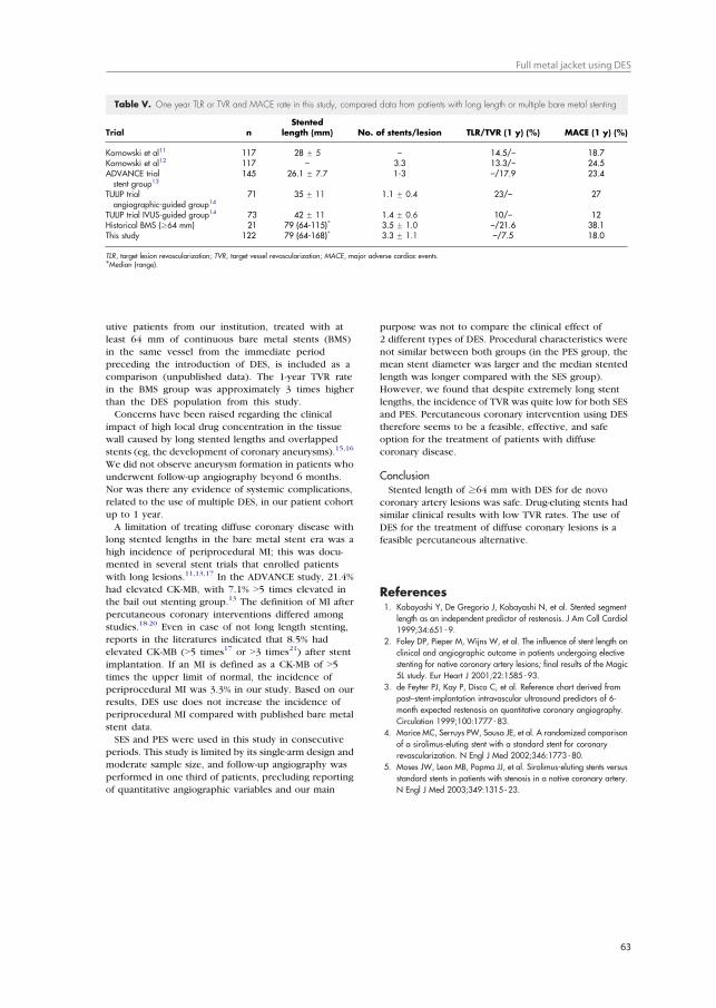
63
Full metal jacket using DES
utive patients from our institution, treated with at
least 64 mm of continuous bare metal stents (BMS)
in the same vessel from the immediate period
preceding the introduction of DES, is included as a
comparison (unpublished data). The 1-year TVR rate
in the BMS group was approximately 3 times higher
than the DES population from this study.
Concerns have been raised regarding the clinical
impact of high local drug concentration in the tissue
wall caused by long stented lengths and overlapped
stents (eg, the development of coronary aneurysms).15,16
We did not observe aneurysm formation in patients who
underwent follow-up angiography beyond 6 months.
Nor was there any evidence of systemic complications,
related to the use of multiple DES, in our patient cohort
up to 1 year.
A limitation of treating diffuse coronary disease with
long stented lengths in the bare metal stent era was a
high incidence of periprocedural MI; this was docu-
mented in several stent trials that enrolled patients
with long lesions.11,13,17 In the ADVANCE study, 21.4%
had elevated CK-MB, with 7.1% N5 times elevated in
the bail out stenting group.13 The definition of MI after
percutaneous coronary interventions differed among
studies.18-20 Even in case of not long length stenting,
reports in the literatures indicated that 8.5% had
elevated CK-MB (N5 times17 or N3 times21) after stent
implantation. If an MI is defined as a CK-MB of N5
times the upper limit of normal, the incidence of
periprocedural MI was 3.3% in our study. Based on our
results, DES use does not increase the incidence of
periprocedural MI compared with published bare metal
stent data.
SES and PES were used in this study in consecutive
periods. This study is limited by its single-arm design and
moderate sample size, and follow-up angiography was
performed in one third of patients, precluding reporting
of quantitative angiographic variables and our main
purpose was not to compare the clinical effect of
2 different types of DES. Procedural characteristics were
not similar between both groups (in the PES group, the
mean stent diameter was larger and the median stented
length was longer compared with the SES group).
However, we found that despite extremely long stent
lengths, the incidence of TVR was quite low for both SES
and PES. Percutaneous coronary intervention using DES
therefore seems to be a feasible, effective, and safe
option for the treatment of patients with diffuse
coronary disease.
ConclusionStented length of z64 mm with DES for de novo
coronary artery lesions was safe. Drug-eluting stents had
similar clinical results with low TVR rates. The use of
DES for the treatment of diffuse coronary lesions is a
feasible percutaneous alternative.
References1. Kobayashi Y, De Gregorio J, Kobayashi N, et al. Stented segment
length as an independent predictor of restenosis. J Am Coll Cardiol1999;34:651 -9.
2. Foley DP, Pieper M, Wijns W, et al. The influence of stent length onclinical and angiographic outcome in patients undergoing electivestenting for native coronary artery lesions; final results of the Magic5L study. Eur Heart J 2001;22:1585-93.
3. de Feyter PJ, Kay P, Disco C, et al. Reference chart derived frompost–stent-implantation intravascular ultrasound predictors of 6-month expected restenosis on quantitative coronary angiography.Circulation 1999;100:1777 -83.
4. Morice MC, Serruys PW, Sousa JE, et al. A randomized comparisonof a sirolimus-eluting stent with a standard stent for coronaryrevascularization. N Engl J Med 2002;346:1773 -80.
5. Moses JW, Leon MB, Popma JJ, et al. Sirolimus-eluting stents versusstandard stents in patients with stenosis in a native coronary artery.N Engl J Med 2003;349:1315-23.
Table V. One year TLR or TVR and MACE rate in this study, compared data from patients with long length or multiple bare metal stenting
Trial nStented
length (mm) No. of stents/lesion TLR/TVR (1 y) (%) MACE (1 y) (%)
Kornowski et al11 117 28 F 5 – 14.5/– 18.7Kornowski et al12 117 – 3.3 13.3/– 24.5ADVANCE trialstent group13
145 26.1 F 7.7 1-3 –/17.9 23.4
TULIP trialangiographic-guided group14
71 35 F 11 1.1 F 0.4 23/– 27
TULIP trial IVUS-guided group14 73 42 F 11 1.4 F 0.6 10/– 12Historical BMS (z64 mm) 21 79 (64-115)T 3.5 F 1.0 –/21.6 38.1This study 122 79 (64-168)T 3.3 F 1.1 –/7.5 18.0
TLR, target lesion revascularization; TVR, target vessel revascularization; MACE, major adverse cardiac events.TMedian (range).
American Heart Journal
November 2005998 Aoki et al

Chapter 6
64
6. Stone GW, Ellis SG, Cox DA, et al. A polymer-based, paclitaxel-eluting stent in patients with coronary artery disease. N Engl J Med2004;350:221 -31.
7. Lemos PA, Serruys PW, van Domburg RT, et al. Unrestrictedutilization of sirolimus-eluting stents compared with conventionalbare stent implantation in the breal worldQ: the rapamycin-elutingstent evaluated at Rotterdam cardiology hospital (RESEARCH)registry. Circulation 2004;109:190 -5.
8. Lemos PA, Saia F, Ligthart JM, et al. Coronary restenosis aftersirolimus-eluting stent implantation: morphological description andmechanistic analysis from a consecutive series of cases. Circulation2003;108:257 -60.
9. Smith Jr SC, Dove JT, Jacobs AK, et al. ACC/AHA guidelines forpercutaneous coronary intervention (revision of the 1993 PTCAguidelines)—executive summary: a report of the American Collegeof Cardiology/American Heart Association task force on practiceguidelines (Committee to revise the 1993 guidelines for percutane-ous transluminal coronary angioplasty) endorsed by the Society forCardiac Angiography and Interventions. Circulation 2001;103:3019 -41.
10. Stone GW, Grines CL, Cox DA, et al. Comparison of angioplastywith stenting, with or without abciximab, in acute myocardialinfarction. N Engl J Med 2002;346:957 -66.
11. Kornowski R, Mehran R, Hong MK, et al. Procedural results and lateclinical outcomes after placement of three or more stents in singlecoronary lesions. Circulation 1998;97:1355 -61.
12. Kornowski R, Bhargava B, Fuchs S, et al. Procedural results andlate clinical outcomes after percutaneous interventions using long(N or =25 mm) versus short (b20 mm) stents. J Am Coll Cardiol2000;35:612 -8.
13. Serruys PW, Foley DP, Suttorp MJ, et al. A randomized comparisonof the value of additional stenting after optimal balloon angioplasty
for long coronary lesions: final results of the additional value of NIRstents for treatment of long coronary lesions (ADVANCE) study.J Am Coll Cardiol 2002;39:393 -9.
14. Oemrawsingh PV, Mintz GS, Schalij MJ, et al. Intravascularultrasound guidance improves angiographic and clinical outcome ofstent implantation for long coronary artery stenoses: final results of arandomized comparison with angiographic guidance (TULIP study).Circulation 2003;107:62 -7.
15. Degertekin M, Serruys PW, Tanabe K, et al. Long-term follow-up ofincomplete stent apposition in patients who received sirolimus-eluting stent for de novo coronary lesions: an intravascularultrasound analysis. Circulation 2003;108:2747 -50.
16. Virmani R, Liistro F, Stankovic G, et al. Mechanism of late in-stentrestenosis after implantation of a paclitaxel derivate–eluting polymerstent system in humans. Circulation 2002;106:2649 -51.
17. Jeremias A, Baim DS, Ho KK, et al. Differential mortality risk ofpostprocedural creatine kinase–MB elevation following successfulversus unsuccessful stent procedures. J Am Coll Cardiol 2004;44:1210 -4.
18. Abdelmeguid AE, Topol EJ. The myth of the myocardial dinfarctletTduring percutaneous coronary revascularization proceduresCirculation 1996;94:3369 -75.
19. Califf RM, Abdelmeguid AE, Kuntz RE, et al. Myonecrosis afterrevascularization procedures. J Am Coll Cardiol 1998;31:241 -51.
20. Serruys PW, Unger F, Sousa JE, et al. Comparison of coronary-artery bypass surgery and stenting for the treatment of multivesseldisease. N Engl J Med 2001;344:1117 -24.
21. Saucedo JF, Mehran R, Dangas G, et al. Long-term clinical eventsfollowing creatine kinase–myocardial band isoenzyme elevationafter successful coronary stenting. J Am Coll Cardiol 2000;35:1134 -41.
American Heart Journal
Volume 150, Number 5Aoki et al 999

Chapter 7
The Effi cacy of Sirolimus-Eluting Stents versus Bare
Metal Stents for Diabetic Patients Undergoing Elective
Percutaneous Coronary Intervention
Aoki J, Ong AT, Rodriguez-Granillo
GA, van Mieghem CA, Daemen J,
Sonnenschein K, Mc Fadden E, Sianos
G, van der Giessen W, de Feyter P, van
Domburg R, Serruys PW.
J Invasive Cardiol. 2005;17:344-348

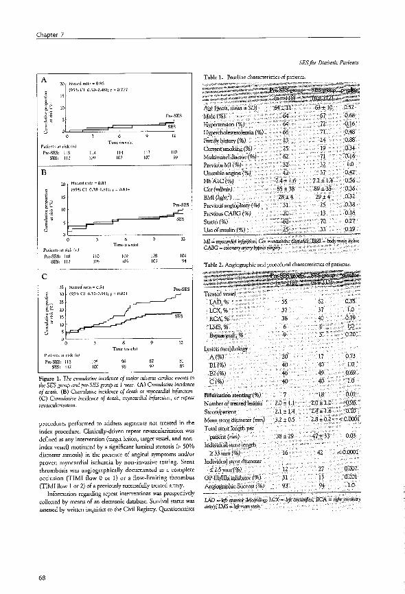

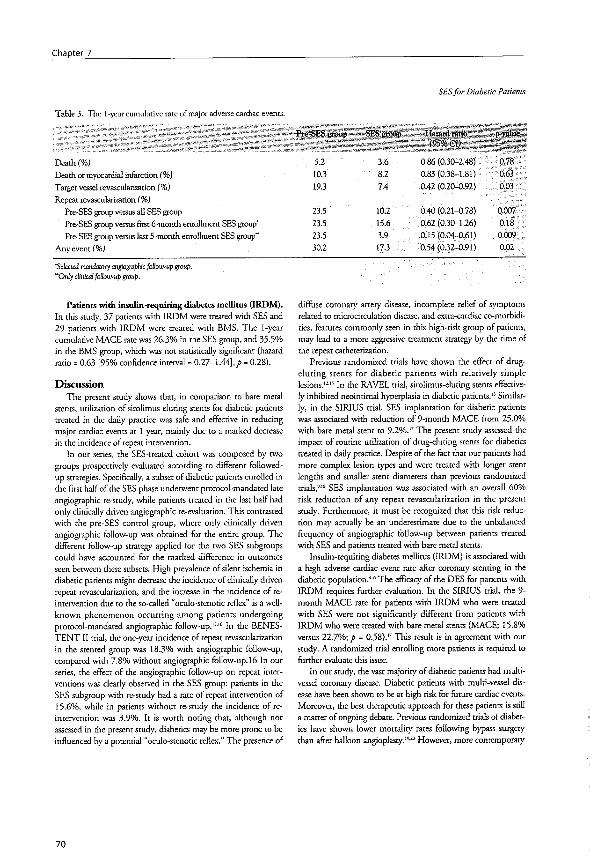


Chapter 8
Outcomes of Unrestricted Utilization of Paclitaxel versus
Sirolimus-Eluting Stents in 293 Unselected Consecutive
Diabetic Patients
Ong AT, Aoki J, Hoye A, van Mieghem
CA, Rodriguez Granillo GA, Valgimigli M,
Tsuchida K, Sonnenschein K, Regar E, van
der Giessen WJ, de Jaegere PP, Sianos G,
McFadden EP, de Feyter PJ, van Domburg
RT, Serruys PW.
Am J Cardiol. 2005;96:358-362

75
SES and PES for diabetic patients
Comparison of Short- (One Month) and Long- (Twelve Months) Term Outcomesof Sirolimus- Versus Paclitaxel-Eluting Stents in 293 Consecutive Patients With
Diabetes Mellitus (from the RESEARCH and T-SEARCH Registries)
Andrew T. L. Ong, MBBS, Jiro Aoki, MD, Carlos A. G. van Mieghem, MD,Gaston A. Rodriguez Granillo, MD, Marco Valgimigli, MD, Keiichi Tsuchida, MD,Karel Sonnenschein, Evelyn Regar, MD, PhD, Willem J. van der Giessen, MD, PhD,
Peter P. T. de Jaegere, MD, PhD, Georgios Sianos, MD, PhD,Eugene P. McFadden, MBChB, MD, Pim J. de Feyter, MD, PhD,Ron T. van Domburg, PhD, and Patrick W. Serruys, MD, PhD*
This study evaluated and compared the efficacy of sirolimus-eluting stents (n � 145patients) with that of paclitaxel-eluting stents (n � 148 patients) in 293 consecutiveunselected patients who had diabetes mellitus. Baseline clinical characteristics andpresentations were similar: mean age of 64 years, 50% presented with unstable anginaor myocardial infarction, and 66% had multivessel disease. Angiographic and pro-cedural characteristics differed, with more complex lesions and more vein graftsmanaged in the paclitaxel-eluting stent group. Overall mean stented length was 46 �32 mm. There were no differences in unadjusted outcomes by stent type (1-year majoradverse cardiac event rates of 20.4% for sirolimus-eluting stents vs 15.6% for pacli-taxel-eluting stents, p � 0.12) or when adjusted for multivariate predictors (adjustedhazard ratio 0.68, 95% confidence interval 0.37 to 1.24, p � 0.21). Independentpredictors of outcome in patients who had diabetes mellitus were stenting of the leftmain artery, stenting of the left anterior descending artery, creatinine clearance, andfemale gender. Patients who required insulin had a significantly higher, crude majoradverse cardiac event rate at 1 year compared with those who used oral agents, butthis rate became nonsignificant when adjusted for independent predictors ofoutcome. © 2005 Elsevier Inc. All rights reserved. (Am J Cardiol 2005;96:358–362)
The Rapamycin-Eluting Stent Evaluated At Rotterdam Car-diology Hospital (RESEARCH) and Taxus-Stent EvaluatedAt Rotterdam Cardiology Hospital (T-SEARCH) registriesare identical sequential prospective registries that were spe-cifically set up to evaluate the universal utilization of siroli-mus- and paclitaxel-eluting stents (SESs and PESs, respec-tively) in an unrestricted population.1,2 The present studyevaluated short- (1 month) and long-term (12 months) effica-cies of SESs and PESs in patients with diabetes mellitus (DM).
• • •We performed a prospective cohort study to investigate theoutcomes of drug-eluting stent implantation in patients whohad DM. Since April 2002, our institution has adopted apolicy of universal drug-eluting stent implantation for allpatients who undergo percutaneous coronary intervention
that requires stenting. All patients are prospectively enteredinto a dedicated database. The initial results of this approachhave been published elsewhere.1,2 Until February 2003,SESs (Cypher, Cordis, a Johnson & Johnson Company,Miami Lakes, Florida) were exclusively used; subsequently,PESs (Taxus, Boston Scientific Corp., Natick, Massachu-setts) became the default stent.Follow-up was complete for 98% of patients. Survival
status was obtained from municipal civil registries at 1, 6,and 12 months. All repeat interventions (surgical and per-cutaneous) and rehospitalizations were prospectively col-lected during follow-up. Questionnaires concerning anginalstatus and medication use were sent to all living patients at6 and 12 months. Referring physicians and institutions werecontacted for additional information, if required. Writteninformed consent was obtained from every patient.From April 2002 to December 2003, 293 unselected
consecutive patients who had DM and de novo coronaryartery disease were treated exclusively with drug-elutingstents; 145 patients received SESs and 148 received PESs. The2 groups were sequential and are part of the RESEARCH andT-SEARCH prospective registries, respectively. Patientswho have DM constitute 18% of the patient populationtreated percutaneously at our institution and were defined by
Thoraxcenter, Erasmus Medical Center, Rotterdam, The Netherlands.Manuscript received December 28, 2004; revised manuscript received andaccepted March 25, 2005.
This study was supported by the Erasmus MC, Rotterdam, TheNetherlands, and by unrestricted institutional grants from Boston ScientificCorp., Natick, Massachusetts, and Cordis, a Johnson & Johnson Company,Miami Lakes, Florida.
* Corresponding author. Tel.: 31-10-463-5260; fax: 31-10-436-9154.E-mail address: [email protected] (P.W. Serruys).
0002-9149/05/$ – see front matter © 2005 Elsevier Inc. All rights reserved. www.AJConline.orgdoi:10.1016/j.amjcard.2005.03.075

Chapter 8
76
therapy; those on oral medications were classified as requir-ing noninsulin and those on insulin therapy as requiringinsulin.All interventions were performed according to current
standard procedures using routine high-pressure balloon in-flations, with the final interventional strategy (includingdirect stenting, postdilatation, use of periprocedural glyco-protein IIb/IIIa inhibitors, and use of intravascular ultra-sound) at the operator’s discretion.3 Angiographic successwas defined as a residual stenosis �30% by visual analysisin the presence of Thrombolysis In Myocardial Infarctiongrade 3 flow. All patients were advised to maintain lifelonguse of aspirin (�80 mg/day). All patients were pretreatedwith 300 mg of clopidogrel. Postprocedurally, patients whoreceived PESs were prescribed �6 months of clopidogrel(75 mg/day),4 and those who received SESs were prescribedclopidogrel for �3 or 6 months depending on the complex-ity of the procedure.1
The primary outcome was the occurrence of major ad-verse cardiac events, defined as a composite of all-causedeath, nonfatal myocardial infarction, or target vessel re-vascularization. Myocardial infarction was diagnosed by anincrease in creatine kinase-MB fraction of �3 times theupper limit of normal.5 In patients who underwent coronaryartery bypass surgery during follow-up, periproceduralmyocardial infarction was diagnosed by an increase in cre-atine kinase-MB level of 5 times the upper limit of normal.6
For patients who presented with an acute myocardial infarc-tion, a diagnosis of repeat myocardial infarction in the acutephase required a decrease and then increase in creatinekinase-MB of 50% above the previous level.7 Target lesionrevascularization was defined as a repeat intervention (sur-gical or percutaneous) to control a luminal stenosis withinthe stent or in the 5-mm distal or proximal segments adja-cent to the stent. Target vessel revascularization was definedas a reintervention that was influenced by any lesion in thesame epicardial vessel. Angiographic stent thrombosis wasdefined as an angiographically documented complete occlu-sion (Thrombolysis In Myocardial Infarction grade 0 or 1flow) or a flow-limiting thrombus (Thrombolysis In Myo-cardial Infarction grade 1 or 2 flow) in a previously suc-cessfully stented artery in the first 30 days. Suspected stentthrombosis was defined as unexplained sudden death ornonfatal myocardial infarction that was unrelated to aperiprocedural complication without repeat angiography.Two independent cardiologists (AO and JA) reviewed allmajor adverse cardiac events.Continuous variables are presented as mean � SD and
were compared with Student’s unpaired t test. Categoricalvariables are presented as counts and percentages and werecompared by Fisher’s exact test. All statistical tests were2-tailed. Cox’s proportional hazards analysis was performedto identify independent predictors of major adverse cardiacevents using significant univariate variables and clinicallyimportant variables listed in Tables 1 and 2 (tested variableswere age, gender, insulin requirement, creatinine clearance,
Table 1Baseline clinical characteristics
Variable SES Group PES Group p Value(n � 145) (n � 148)
Men 66% 67% 0.8Age (yrs) 62.6� 10.2 64.6� 10.3 0.08Non–insulin-requiring DM 73% 77% 0.4Insulin-requiring DM 27% 23% 0.4Hypertension 68% 70% 0.7Hypercholesterolemia* 66% 85% �0.001Current smoking 23% 20% 0.7Previous myocardial infarction 37% 42% 0.5Previous coronary angioplasty 23% 21% 0.8Previous coronary bypassgrafting
10% 14% 0.5
Single-vessel coronary disease 34% 31% 0.7Multivessel coronary disease 66% 69% 0.7Clinical presentation 0.4Stable angina pectoris 49% 56%Unstable angina pectoris 38% 34%Acute myocardial infarction 13% 10%Hemoglobin-A1c (%) 7.3� 1.3 7.6� 1.5 0.09Creatinine clearance (ml/min) 87.6� 33.0 80.8� 33.4 0.10Body mass index (kg/m2) 28.4 � 4.1 27.9� 3.8 0.3
Values are means � SD or percentages.* Defined as a fasting cholesterol level �5.5mmol/L or use of lipid-
lowering therapy.
Table 2Angiographic and procedural characteristics
Variable SES Group PES Group p Value(n � 145) (n � 148)
Treated coronary vessel*Left anterior descending artery 60% 48% 0.05Left circumflex artery 40% 34% 0.3Right artery 32% 42% 0.11Left main artery 8% 8% 1.0Bypass graft 3% 10% 0.02Lesion type†
A 17% 8% 0.02B1 37% 24% 0.02B2 45% 47% 0.7C 47% 56% 0.13No. of coronary vessels treated 0.51 61% 64%2 35% 28%3 4% 8%Multivessel treatment 39% 36% 0.6Bifurcation stenting 19% 14% 0.4No. of stented vessels 1.4� 0.6 1.4 � 0.7 0.9No. of implanted stents 2.4� 1.5 2.3 � 1.4 0.6Total stented length per patient(mm)
45.3 � 32.1 48.5 � 33.8 0.4
Nominal stent diameter �2.5 mm 40% 47% 0.2Chronic total occlusion (�3 mos) 14% 12% 0.6Glycoprotein IIb/IIIa inhibitor use 18% 28% 0.04Angiographic success of alllesions
94% 97% 0.4
Values are means � SD or percentages.* Expressed as percentage of patients with vessel type treated. Total
exceeds 100%.† Expressed as percentage of patients with lesion type. Total exceeds
100%.
359Coronary Artery Disease/Sirolimus and Paclitaxel Stents in Diabetics

77
SES and PES for diabetic patients
presenting symptoms, lesion type, multivessel disease, bi-furcation stenting, stenting of the left main artery, stentingof the left anterior descending artery, stent type, number ofstents, total stent length, and minimum stent diameter).Stent type and requirement for insulin were forced into themodel, whereas other variables were entered in a forwardstepwise method (entry and removal criteria of 0.05 and0.10, respectively). The cumulative incidence of adverseevents was estimated according to the Kaplan-Meier method.Baseline clinical characteristics were similar for the 2
groups with the exception of hypercholesterolemia (Table 1).Sixty-six percent were men and 25% required insulin. Mul-tivessel coronary disease was present in 66% of patients,and 50% presented with an acute coronary syndrome (un-stable angina or acute myocardial infarction). Mean hemo-globin A1c levels were 7.6 � 1.5% in the PES group and7.3 � 1.3% in the SES group. More patients in the PESgroup were classified as having hypercholesterolemia (de-fined as a fasting serum cholesterol level �5.5mmol/L oruse of lipid-lowering therapy at the time of the procedure)due to the more widespread use of lipid-lowering agents inthe latter period.Significant differences were noted in terms of angio-
graphic and procedural characteristics (Table 2). In the PESgroup, more patients received treatment in a bypass graft(10% vs 3% in the SES group, p � 0.02) and fewer patientsreceived treatment in the left anterior descending artery(48% vs 60% respectively, p � 0.05). The use of glyco-protein IIb/IIIa inhibitors was greater in the PES group (28%vs 18%, p � 0.04). More complex lesions were treated in thePES group, with fewer type A or B1 lesions treated (p� 0.02).Multivessel treatment was performed in 40% of patients. Totalstented length was similar in the 2 groups (48.5 � 33.8 mm inthe PES group and 45.3 � 32.1 mm in the SES group, p �0.4), as was the number of stents implanted (2.3 � 1.3 vs 2.4� 1.5 stents, respectively, p � 0.6).In the first month after stent implantation, there were 10
deaths that were equally divided between groups (Table 3).Three deaths were clinically suspected to be due to stentthrombosis (Table 3). There were 8 myocardial infarctions,
5 in the SES group and 3 in the PES group. Of the 6 targetlesion revascularizations, 4 were for stent thrombosis and 2were the result of a procedural complication that requiredurgent coronary surgery. In total, 4 patients (2.8%) in theSES group and 3 (2.0%) in the PES group had suspected or
Figure 1. Event curves at 1 year for cumulative risks of (A) death, (B) deathor myocardial infarction, and (C) death, myocardial infarction, or targetvessel revascularization.
Table 3Major adverse cardiac events in the first month after stent implantation
Patients With Events at 0–1month
SES Group PES Group p Value*(n � 143) (n � 148)
Death 5 (3.4%) 5 (3.4%) 1.0Nonfatal myocardial infarction 5 (4.1%) 3 (2.0%) 0.3Target lesion revascularization 5 (3.4%) 1 (0.7%) 0.1Target vessel revascularization† 5 (3.4%) 3 (2.0%) 0.5Any event 14 (9.7%) 9 (6.1%) 0.3Angiographically proved stentthrombosis
3 (2.1%) 1 (0.7%) 0.4
Suspected stent thrombosis 1 (0.7%) 2 (1.4%) 1.0Total stent thrombosis 4 (2.8%) 3 (2.0%) 0.7
* By Fisher’s exact test.† Includes target lesion revascularization.
360 The American Journal of Cardiology (www.AJConline.org)

Chapter 8
78
proved stent thrombosis (p � 0.7). A major adverse cardiacevent occurred in 9.7% of patients in the SES group and6.1% of patients in the PES group within the first 30 days.At 1 year, there were no differences in the incidence of
death between groups (SES 7.7%, PES 7.2%, p � 0.9;Figure 1). The incidence of death or myocardial infarctionwas also similar (SES 14.1%, PES 10.0%, p � 0.3). Thecomposite end point of death, myocardial infarction, ortarget vessel revascularization was also nonsignificantly dif-ferent (SES 20.4%, PES 15.6%, p � 0.12), with a trendfavoring PES. Incidences of target lesion revascularizationwere 8.8% in the SES group and 5.7% in the PES group(p � 0.08; Figure 2When patients were classified by insulin requirement,
patients who required insulin (n � 72) developed moreevents compared with those who did not require insulin(n � 221, crude major adverse cardiac events 27.4% vs14.6%, respectively, p� 0.008; Figure 3). Hemoglobin A1clevels were higher in the insulin-requiring DM group than inthe non–insulin-requiring DM group, although not signifi-cantly (7.6% vs 7.2%, p � 0.4).
Stenting of the left anterior descending artery, stenting ofthe left main coronary artery, and female gender were in-dependently associated with worse outcomes, whereas bet-ter renal function (defined as milliliter-per-minute incre-
Figure 2. One-year cumulative risks of (A) target lesion revascularizationand (B) target vessel revascularization.
Table 4Multivariate predictors of major adverse cardiac events at one year(Cox’s proportional hazards model)
Variable Adjusted HR 95% CI p Value
Stenting of left anteriordescending artery
2.79 1.44–5.39 0.002
Stenting of left main artery 2.78 1.24–6.25 0.013Creatinine clearance (/ml/minincrement)
0.99 0.98–1.00 0.03
Women 1.90 1.02–3.53 0.04Use of PES 0.68 0.37–1.24 0.21Use of insulin 1.48 0.80–2.74 0.22
CI � confidence interval; HR � hazard ratio.
Figure 3. Cumulative risks of (A) death and (B) major adverse cardiacevents stratified by diabetic type. IRDM � insulin-requiring DM; NIRDM �non–insulin-requiring DM.
361Coronary Artery Disease/Sirolimus and Paclitaxel Stents in Diabetics

79
SES and PES for diabetic patients
ment in creatinine clearance) was associated with improvedoutcomes (Table 4). Insulin requirement, significant in uni-variate analysis, became nonsignificant in the multivariatemodel (adjusted hazard ratio 1.48, 95% confidence interval0.80 to 2.74, p � 0.22), and no significant differences werenoted with stent type (adjusted hazard ratio 0.68, 95%confidence interval 0.37 to 1.24, p � 0.21).
• • •The major finding of this study is that unrestricted use ofPESs in a universal drug-eluting stent environment is asso-ciated with a nonsignificantly lower incidence of majoradverse cardiac events at 1 year compared with SESs (ad-justed hazard ratio 0.68, 95% confidence interval 0.37 to1.24, p � 0.21). Patients with DM and who required insulinhad a significantly higher crude incidence of major adversecardiac events compared with those who did not requireinsulin in a combined drug-eluting population; this signifi-cance became nonsignificant after adjustment for multivar-iate predictors.Mortality rate at 1 year between groups in our study was
similar (7.7% in SES group and 7.2% in PES group, p �0.9). It is difficult to compare between studies; however, asa guide, this result lies between that reported in the stent armof the randomized Arterial Revascularization TherapyStudy (6.3%)8 and a multivessel report from the database ofthe Cardiovascular Research Foundation (14% to 15%),9
with the caveat that baseline demographics were different.For a population that had DM, the use of glycoproteinIIb/IIIa inhibitors in this study was very low; more frequentuse may have improved mortality rates.10
This study has described the experience of a single-center registry of drug-eluting stents in a moderate numberof patients. Routine angiographic follow-up was not per-formed, thus precluding an assessment of restenotic rates.The low use of glycoprotein IIb/IIIa inhibitors is a limitationbut was a reflection of the “real-world” practice of theoperators. The results of this study should be viewed as anexploratory analysis that reported outcomes after the unre-stricted use of drug-eluting stents in the real world in pa-tients who had DM.
1. Lemos PA, Serruys PW, van Domburg RT, Saia F, Arampatzis CA,Hoye A, Degertekin M, Tanabe K, Daemen J, Liu TK, et al. Unre-
stricted utilization of sirolimus-eluting stents compared withconventional bare stent implantation in the “real world”: theRapamycin-Eluting Stent Evaluated At Rotterdam Cardiology Hospi-tal (RESEARCH) registry. Circulation 2004;109:190–195.
2. Ong AT, Serruys PW, Aoki J, Hoye A, Van Mieghem CA, RodriguezGranillo GA, Valgimigli M, Sonnenschein K, Regar E, van der Ent M,et al. The unrestricted use of paclitaxel versus sirolimus-eluting stentsfor coronary artery disease in an unselected population—one yearresults of the Taxus-Stent Evaluated At Rotterdam Cardiology Hospi-tal (T-SEARCH) Registry. J Am Coll Cardiol 2005;45:1135–1141.
3. Lemos PA, Lee CH, Degertekin M, Saia F, Tanabe K, Arampatzis CA,Hoye A, van Duuren M, Sianos G, Smits PC, et al. Early outcome aftersirolimus-eluting stent implantation in patients with acute coronarysyndromes: insights from the Rapamycin-Eluting Stent Evaluated AtRotterdam Cardiology Hospital (RESEARCH) registry. J Am CollCardiol 2003;41:2093–2099.
4. Colombo A, Drzewiecki J, Banning A, Grube E, Hauptmann K, SilberS, Dudek D, Fort S, Schiele F, Zmudka K, et al. Randomized study toassess the effectiveness of slow- and moderate-release polymer-basedpaclitaxel-eluting stents for coronary artery lesions. Circulation 2003;108:788–1794.
5. Smith SC Jr, Dove JT, Jacobs AK, Kennedy JW, Kereiakes D, KernMJ, Kuntz RE, Popma JJ, Schaff HV, Williams DO, et al. ACC/AHAguidelines of percutaneous coronary interventions (revision of the1993 PTCA guidelines. A report of the American College of Cardi-ology/American Heart Association Task Force on Practice Guidelines(committee to revise the 1993 guidelines for percutaneous transluminalcoronary angioplasty). J Am Coll Cardiol 2001;37:2239i–lxvi.
6. Serruys PW, Unger F, Sousa JE, Jatene A, Bonnier HJ, SchonbergerJP, Buller N, Bonser R, van den Brand MJ, van Herwerden LA, etal. Comparison of coronary-artery bypass surgery and stenting forthe treatment of multivessel disease. N Engl J Med 2001;344:1117–1124.
7. Stone GW, Grines CL, Cox DA, Garcia E, Tcheng JE, Griffin JJ,Guagliumi G, Stuckey T, Turco M, Carroll JD, et al. Comparison ofangioplasty with stenting, with or without abciximab, in acute myo-cardial infarction. N Engl J Med 2002;346:957–966.
8. Abizaid A, Costa MA, Centemero M, Abizaid AS, Legrand VM,Limet RV, Schuler G, Mohr FW, Lindeboom W, Sousa AG, et al.Clinical and economic impact of diabetes mellitus on percutaneous andsurgical treatment of multivessel coronary disease patients: insightsfrom the Arterial Revascularization Therapy Study (ARTS) trial. Cir-culation 2001;104:533–538.
9. Mehran R, Dangas GD, Kobayashi Y, Lansky AJ, Mintz GS, AymongED, Fahy M, Moses JW, Stone GW, Leon MB. Short- and long-termresults after multivessel stenting in diabetic patients. J Am Coll Car-diol 2004;43:1348–1354.
10. Lincoff AM. Important triad in cardiovascular medicine: diabetes,coronary intervention, and platelet glycoprotein IIb/IIIa receptorblockade. Circulation 2003;107:1556–1559.
362 The American Journal of Cardiology (www.AJConline.org)

Chapter 9
Sirolimus-eluting stent implantation for chronic total
occlusion of the left main coronary artery
Aoki J, Hoye A, Saferov A, Alekyan B,
Serruys P.
J Interv Cardiol. 2005;18:65-69

IT FITS! (Intelligence Transfer: From Images to Solutions)Edited by Steven B.H. Timmis
Sirolimus-Eluting Stent Implantation for Chronic Total Occlusion of theLeft Main Coronary Artery
JIRO AOKI, M.D.∗, ANGELA HOYE, MB CHB∗, ANTON V. STAFEROV, M.D.∗∗,
BAGRAT G. ALEKYAN, M.D.∗∗, and PATRICK W. SERRUYS, M.D., PH.D.∗
From the ∗Department of Interventional Cardiology, Erasmus MC, Rotterdam, the Netherlands and ∗∗Department of InterventionalCardiology and Angioplasty, Bakoulev Scientific Center for Cardiovascular Surgery, Russian Academy of Medical Sciences, Moscow, Russia
Chronic total occlusion of the left main coronary artery (LMCA) is rare. Recently, percutaneous coronary in-tervention has been increasingly applied to unprotected LMCA lesions. We describe a patient with chronic to-tal occlusion of the LMCA who was successfully treated with bifurcation stenting with sirolimus-eluting stents.(J Interven Cardiol 2005;18:65–69)
Introduction
Chronic total occlusion of the left main coronaryartery (LMCA) is an unusual manifestation of coro-nary atherosclerotic disease in clinical practice.1–4 Therarity of this lesion may be accounted for by the rela-tively high incidence of death in these patients. Coro-nary artery bypass graft surgery (CABG) has been thestandard of care for LMCA disease, though recently,percutaneous coronary intervention (PCI) has been in-creasingly applied to unprotected LMCA lesions.5–12
However, the development of restenosis remains a ma-jor limitation of late outcomes after PCI, with the oc-currence of restenosis particularly associated with haz-ardous clinicalmanifestations. Sirolimus-eluting stents(SES) have been shown to dramatically reduce therestenosis rate in selected patients with relatively sim-ple lesions.13–15 We report a patient with chronic total
Address for reprints: Patrick W. Serruys, M.D., Ph.D., F.E.S.C.,F.A.C.C. Department of Interventional Cardiology, Thoraxcen-tre Bd 404 Dr. Molewaterplein 40 NL-3015 GD Rotterdam, theNetherlands. Tel: +31-10-4635260; Fax: +31-10-4639154; e-mail:[email protected]
All authors have read and approved submission of the manuscript.There is no conflict of interest for any author.
occlusion of the LMCA who was successfully treatedpercutaneously with SES implantation.
Case Report
A 35-year-old male presented with an acute anteriormyocardial infarction that was managed medically. Hesubsequently complained of on-going chest pain (CCSclass II–III angina16) and underwent coronary angiog-raphy 9 months later. His resting 12-ECG revealedevidence of a previousQ-wave anteroseptalmyocardialinfarction, and echocardiography demonstrated hy-pokinesis of the anteroseptal wall without left ventricleaneurysm. He was referred for coronary angiography,which showed a total occlusion of the LMCA (Fig. 1);the left anterior descending coronary artery (LAD)and left circumflex coronary artery (LCX) were retro-gradely filled via Rentrop grade III collaterals17 fromthe RCA, which was itself not significantly stenosed(Fig. 2). The patient rejected coronary artery bypassgrafting, but consented to undergo attempted revascu-larization with percutaneous coronary intervention.
A 6F introducer sheath was inserted in the rightfemoral artery. In addition, a 5F introducer sheath wasinserted in the left femoral artery to enable simultane-ous right and left coronary injections. In the absence
Vol. 18, No. 1, 2005 Journal of Interventional Cardiology 65

83
SES for CTO of LMCA
IT FITS! (Intelligence Transfer: From Images to Solutions)Edited by Steven B.H. Timmis
Sirolimus-Eluting Stent Implantation for Chronic Total Occlusion of theLeft Main Coronary Artery
JIRO AOKI, M.D.∗, ANGELA HOYE, MB CHB∗, ANTON V. STAFEROV, M.D.∗∗,
BAGRAT G. ALEKYAN, M.D.∗∗, and PATRICK W. SERRUYS, M.D., PH.D.∗
From the ∗Department of Interventional Cardiology, Erasmus MC, Rotterdam, the Netherlands and ∗∗Department of InterventionalCardiology and Angioplasty, Bakoulev Scientific Center for Cardiovascular Surgery, Russian Academy of Medical Sciences, Moscow, Russia
Chronic total occlusion of the left main coronary artery (LMCA) is rare. Recently, percutaneous coronary in-tervention has been increasingly applied to unprotected LMCA lesions. We describe a patient with chronic to-tal occlusion of the LMCA who was successfully treated with bifurcation stenting with sirolimus-eluting stents.(J Interven Cardiol 2005;18:65–69)
Introduction
Chronic total occlusion of the left main coronaryartery (LMCA) is an unusual manifestation of coro-nary atherosclerotic disease in clinical practice.1–4 Therarity of this lesion may be accounted for by the rela-tively high incidence of death in these patients. Coro-nary artery bypass graft surgery (CABG) has been thestandard of care for LMCA disease, though recently,percutaneous coronary intervention (PCI) has been in-creasingly applied to unprotected LMCA lesions.5–12
However, the development of restenosis remains a ma-jor limitation of late outcomes after PCI, with the oc-currence of restenosis particularly associated with haz-ardous clinicalmanifestations. Sirolimus-eluting stents(SES) have been shown to dramatically reduce therestenosis rate in selected patients with relatively sim-ple lesions.13–15 We report a patient with chronic total
Address for reprints: Patrick W. Serruys, M.D., Ph.D., F.E.S.C.,F.A.C.C. Department of Interventional Cardiology, Thoraxcen-tre Bd 404 Dr. Molewaterplein 40 NL-3015 GD Rotterdam, theNetherlands. Tel: +31-10-4635260; Fax: +31-10-4639154; e-mail:[email protected]
All authors have read and approved submission of the manuscript.There is no conflict of interest for any author.
occlusion of the LMCA who was successfully treatedpercutaneously with SES implantation.
Case Report
A 35-year-old male presented with an acute anteriormyocardial infarction that was managed medically. Hesubsequently complained of on-going chest pain (CCSclass II–III angina16) and underwent coronary angiog-raphy 9 months later. His resting 12-ECG revealedevidence of a previousQ-wave anteroseptalmyocardialinfarction, and echocardiography demonstrated hy-pokinesis of the anteroseptal wall without left ventricleaneurysm. He was referred for coronary angiography,which showed a total occlusion of the LMCA (Fig. 1);the left anterior descending coronary artery (LAD)and left circumflex coronary artery (LCX) were retro-gradely filled via Rentrop grade III collaterals17 fromthe RCA, which was itself not significantly stenosed(Fig. 2). The patient rejected coronary artery bypassgrafting, but consented to undergo attempted revascu-larization with percutaneous coronary intervention.
A 6F introducer sheath was inserted in the rightfemoral artery. In addition, a 5F introducer sheath wasinserted in the left femoral artery to enable simultane-ous right and left coronary injections. In the absence
Vol. 18, No. 1, 2005 Journal of Interventional Cardiology 65

Chapter 9
84
AOKI, ET AL.
Figure 1. Left coronary artery angiogram, revealing total occlusionof the left main stem artery.
of antegrade flow through the occlusion, such a dualinjection technique allows visualization of the distalvessels (LAD and LCX) via the collateral filling fromthe RCA, thereby facilitating correct positioning ofthe wire. A 6F XB 3.5 guiding catheter (Cordis) wasplaced in the left main coronary segment, and success-
Figure 2. Right coronary angiogram demonstrating grade III retro-grade collaterals arising from the right coronary artery to both theleft anterior descending and the left circumflex arteries.
Figure 3. Left coronary angiogram showing the dissection of theleft main stem artery to the mid-circumflex artery.
ful recanalization was achieved with a 0.0014�� Shi-nobi wire (Cordis), which was advanced into the dis-tal LAD. A second 0.0014�� Shinobi wire was taken,and passage into the LCX was attempted. Unfortu-nately, this was complicated by catheter/wire-induceddissection from the LMCA to the mid-LCX (Fig. 3),and the wire was withdrawn. The LAD/distal LMCAwas stabilized through stent implantation with a 3.0 ×23 mm SES (Cypher, Cordis), which was then post-dilated with a 3.5 mm balloon (U-pass, Cordis) givinga good result (Fig. 4). After several attempts, a 0.0014��
Shinobi wire was eventually successfully crossed, viathe SES struts, into the true lumen of the LCX. Af-ter sequential predilatation using a 2.0 mm balloon(Stomer, Medtronic), both the lesion and the mid-LCXdissection (Fig. 5) were treated with implantation of a2.5 × 33 mm SES deployed from LCX ostium. Thefinal angiogram showed good result with TIMI III flowin both the LAD and the LCX (Fig. 6).18
The patient made an unremarkable recovery andwasallowed to go home. There were no major adversecardiac events during the in-hospital period, and at9-month clinical follow-up he remained well with norecurrence of angina.
Discussion
Chronic total occlusion (CTO) of the LMCA israre. In patients who are investigated in the catheter
66 Journal of Interventional Cardiology Vol. 18, No. 1, 2005

85
SES for CTO of LMCA
SES FOR CTO OF LMCA
Figure 4. Left coronary angiogram following stent deploymentwitha 3.0 × 23 mm sirolimus-eluting stent in the left main stem/leftanterior descending artery.
laboratory, its prevalencevaries from0.04% to0.4%.1–4
CABG has been considered to be the treatment ofchoice in LMCA disease, especially for chronic totalocclusion of the LMCA. There are a few published re-
Figure 5. Left coronary angiogram showing that a wire has success-fully crossed through the stent struts and into the true lumen of thedistal left circumflex artery. The region of dissection can clearly beseen in the proximal and mid parts of the vessel (contrast staining).
Figure 6. The final angiogram following additional stent implanta-tion with a 2.5 × 33 mm sirolimus-eluting stent up to the ostium ofthe circumflex artery, showing a good result in both the left anteriordescending and the left circumflex arteries.
ports showing that the results of CABG for this groupof CTO`s are beneficial compared to medical ther-apy.1,2 However, there are no studies comparing PCIwith CABG for such patients. The advent of improvedPCI equipment including stents and atherectomy de-vices has been shown to be safe and effective in selectedpatients with unprotected elective LMCA stenosis,5–12
and recently Trehan et al. have reported a single caseof successful percutaneous stenting of a CTO of anunprotected LMCA.19 There are two major problemsassociated with PCI for chronic total occlusion of theLMCA. The first relates to the initial procedural diffi-culty of crossing the occlusion with a wire; publishedprocedural success rates for CTO`s are generally in therange of 40–81%.20,21 The second relates to resteno-sis; both bifurcation lesions and CTO`s are subject toa higher rate of restenosis compared with simpler le-sions.22–25 Importantly, the occurrence of restenosis inthe LMCAmay be associated with a significant rate ofmortality. In particular, PCI for bifurcation lesions ofthe distal LMCA, whereby both the LAD and the LCXarteries are stented, is both technically demanding andat high risk of restenosis.26–28 Drug eluting, stents havebeen shown to dramatically reduce the restenosis ratein elective patients with simple de novo lesions.13–15
The development and more widespread application of
Vol. 18, No. 1, 2005 Journal of Interventional Cardiology 67

Chapter 9
86
AOKI, ET AL.
drug-eluting stents hold the promise of a significantreduction in restenosis and the need for repeat revas-cularization. Arampatzis et al. have reported the ef-fectiveness of SES for the treatment of LMCA. A to-tal of 31 consecutive patients were treated solely withSESs either electively, for acute myocardial infarction,or due to procedural complication-related LMCA dis-section. In this study, the rate of out-of-hospital clinicalevents was extremely low, with zero percent mortal-ity and 4% target vessel revascularization.29 In addi-tion, low subsequent binary restenosis rates followingSES implantationhavebeendocumentedboth inCTO’s(9.1% at 6 months) and bifurcation lesions (22.7% at6 months).30,31 The technique of bifurcation stenting(T, culotte, kissing, or crush stenting) with drug-elutingstents is still controversial.30 Those techniques result-ing in overlapping stent struts lead to an increase inthe local concentration of drug, which may induce en-dothelial function impairment and thus be associatedwith an increased rate of stent thrombosis. In the currentreport, we present a patient who underwent successfulrecanalization of an LMCA CTO without the need fora cardiac support device, and underwent bifurcationstenting, with SESs. There were no major adverse car-diac events either in-hospital or over the subsequent 9months. Further data are needed to fully evaluate theuse of this strategy in such an unusual patient pop-ulation; however, SES-supported angioplasty may bea reasonable alternative to CABG in the treatment ofLMCA chronic total occlusion.
References
1. Zimmern SH, Rogers WJ, Bream PR, et al. Total occlusion ofthe left main coronary artery: The Coronary Artery SurgeryStudy (CASS) experience. Am J Cardiol 1982;49(8):2003–2010.
2. Ward DE, Valantine H, Hui W. Occluded left main stem coro-nary artery. Report of five patients and review of publishedreports. Br Heart J 1983;49(3):276–279.
3. Herregods MC, Piessens J, Vanhaecke J, et al. Complete oc-clusion of the main left coronary artery. A clinical study. ActaCardiol 1987;42(1):23–35.
4. de Feyter PJ, Serruys PW. Thrombolysis of acute total occlu-sion of the left main coronary artery in evolving myocardialinfarction. Am J Cardiol 1984;53(11):1727–1728.
5. Kosuga K, Tamai H, Ueda K, et al. Initial and long-term resultsof angioplasty in unprotected left main coronary artery. Am JCardiol 1999;83(1):32–37.
6. Silvestri M, Barragan P, Sainsous J, et al. Unprotected leftmain coronary artery stenting: Immediate and medium-termoutcomes of 140 elective procedures. J Am Coll Cardiol2000;35(6):1543–1550.
7. Tan WA, Tamai H, Park SJ, et al. Long-term clinical outcomes
after unprotected left main trunk percutaneous revasculariza-tion in 279 patients. Circulation 2001;104(14):1609–1614.
8. Park SJ, Hong MK, Lee CW, et al. Elective stenting of unpro-tected left main coronary artery stenosis: Effect of debulkingbefore stenting and intravascular ultrasound guidance. J AmColl Cardiol 2001;38(4):1054–1060.
9. Black A, Cortina R, Bossi I, et al. Unprotected left main coro-nary artery stenting: Correlates of midterm survival and im-pact of patient selection. J Am Coll Cardiol 2001;37(3):832–838.
10. Takagi T, Stankovic G, Finci L, et al. Results and long-termpredictors of adverse clinical events after elective percutaneousinterventions on unprotected left main coronary artery. Circu-lation 2002;106(6):698–702.
11. Park SJ, Park SW, Hong MK, et al. Long-term (three-year)outcomes after stenting of unprotected leftmain coronary arterystenosis in patients with normal left ventricular function. Am JCardiol 2003;91(1):12–16.
12. Brueren BR, Ernst JM, Suttorp MJ, et al. Long term followup after elective percutaneous coronary intervention for unpro-tected non-bifurcational left main stenosis: Is it time to changethe guidelines? Heart 2003;89(11):1336–1339.
13. Holmes DR, Jr., Leon MB, Moses JW, et al. Analysis of 1-yearclinical outcomes in the SIRIUS trial: A randomized trial ofa sirolimus-eluting stent versus a standard stent in patients athigh risk for coronary restenosis. Circulation 2004;109(5):634–640.
14. Stone GW, Ellis SG, Cox DA, et al. A polymer-based,paclitaxel-eluting stent in patients with coronary artery disease.N Engl J Med 2004;350(3):221–231.
15. Morice MC, Serruys PW, Sousa JE, et al. A randomized com-parison of a sirolimus-eluting stent with a standard stent forcoronary revascularization. N Engl J Med 2002;346(23):1773–1780.
16. Campeau L. Letter: Grading of angina pectoris. Circulation1976;54(3):522–523.
17. Cohen M, Rentrop KP. Limitation of myocardial ischemia bycollateral circulation during sudden controlled coronary arteryocclusion in human subjects: A prospective study. Circulation1986;74(3):469–476.
18. Anderson JL, Karagounis LA, Becker LC, et al. TIMI perfu-sion grade 3 but not grade 2 results in improved outcome afterthrombolysis for myocardial infarction. Ventriculographic, en-zymatic, and electrocardiographic evidence from the TEAM-3Study. Circulation 1993;87(6):1829–1839.
19. TrehanV,MehtaV,MukhopadhyayS, et al. Percutaneous stent-ing of chronic total occlusion of unprotected left main coronaryartery. Indian Heart J 2003;55(2):172–174.
20. Puma JA, SketchMH, Jr., Tcheng JE, et al. Percutaneous revas-cularization of chronic coronary occlusions:An overview. JAmColl Cardiol 1995;26(1):1–11.
21. Serruys PW, Hamburger JN, Koolen JJ, et al. Total occlusiontrial with angioplasty by using laser guidewire. The TOTALtrial. Eur Heart J 2000;21(21):1797–1805.
22. Rubartelli P, Niccoli L, Verna E, et al. Stent implantationversus balloon angioplasty in chronic coronary occlusions:Results from the GISSOC trial. Gruppo Italiano di Studiosullo Stent nelle Occlusioni Coronariche. J Am Coll Cardiol1998;32(1):90–96.
23. Buller CE, Dzavik V, Carere RG, et al. Primary stentingversus balloon angioplasty in occluded coronary arteries:The Total Occlusion Study of Canada (TOSCA). Circulation1999;100(3):236–242.
24. Al Suwaidi J, Berger PB, Rihal CS, et al. Immediate and long-term outcome of intracoronary stent implantation for true bi-furcation lesions. J Am Coll Cardiol 2000;35(4):929–936.
68 Journal of Interventional Cardiology Vol. 18, No. 1, 2005
87
SES for CTO of LMCA
SES FOR CTO OF LMCA
25. Yamashita T, Nishida T, Adamian MG, et al. Bifurcation le-sions: Two stents versus one stent—immediate and follow-upresults. J Am Coll Cardiol 2000;35(5):1145–1151.
26. Park SJ, Lee CW, Kim YH, et al. Technical feasibility,safety, and clinical outcome of stenting of unprotected leftmain coronary artery bifurcation narrowing. Am J Cardiol2002;90(4):374–378.
27. Lee CH, Degertekin M, van Domburg RT, et al. Impact of dif-ferent anatomical patterns of left main coronary stenting onlong-term survival. Am J Cardiol 2003;92(6):718–720.
28. Hu FB, Tamai H, Kosuga K, et al. Intravascular ultrasound-guided directional coronary atherectomy for unprotected leftmain coronary stenoses with distal bifurcation involvement.Am J Cardiol 2003;92(8):936–940.
29. Arampatzis CA, Lemos PA, Tanabe K, et al. Effectiveness ofsirolimus-eluting stent for treatment of leftmain coronary arterydisease. Am J Cardiol 2003;92(3):327–329.
30. Tanabe K, Hoye A, Lemos PA, et al. Restenosis rates followingbifurcation stenting with sirolimus-eluting stents for de novonarrowings. Am J Cardiol 2004;94(1):115–118.
31. Hoye A, Tanabe K, Lemos PA, et al. Significant reductionin restenosis following the use of sirolimus-eluting stents inthe treatment of chronic total occlusions. J Am Coll Cardiol2004;43(11):1954–1958.
EDITOR’S RESPONSE
In the current case presentation, Dr. Aoki offers aglimpse at one of the still untapped frontiers in in-terventional cardiology. Percutaneous management ofleft main coronary lesions has remained undesirablebecause of the immediate technical difficulties andthe subsequent risk of potentially fatal restenosis. Thepresent case was particularly difficult because the leftmain was chronically occluded into both the left an-terior descending and the left circumflex coronary ar-teries. The double catheter approachwith simultaneousinjection into the right coronary artery and the left maingreatly facilitated distal guidewire placement. Achiev-ing distal guidewire access was the greatest technicalchallenge of this particular case. Once distal accesswas attained, angiographic success was all but assured.While I believe that the angiographic results shouldhave been confirmed with intravascular ultrasound toensure adequate stent apposition within this criticalarea, this case represents a therapeutic victory over ex-treme lesion complexity with excellent angiographic
and clinical results. Such cases should only be per-formed in high-volumecenterswith highly experiencedcoronary interventionalists.
However, the acute results in left main coronarystenting are only part of the battle. The largest hurdle inthe management of these lesions is avoiding restenosisand its risk for death. The use of drug-eluting stentsin the left main coronary artery may offer a solution.Arampatzis et al. studied the use of sirolimus-coatedstents in 16 consecutive patients undergoing electiveleftmain stenting in a single center.1 Half of the patientshad lesions involving the left anterior descending/leftcircumflex bifurcation. Nine patients had unprotectedleft main stenoses. There were no deaths reported dur-ing 1 year of follow-up. Peri-procedural non-Q-wavemyocardial infarction occurred in one patient. Of the12 patients who underwent angiographic follow-up at6 months, only one demonstrated restenosis requiringrepeat percutaneous intervention. No other major ad-verse coronary events occurred in the entire cohortduring the year following left main intervention. Al-though the patient in the current case presentation didnot undergo angiographic follow-up, he remained clin-ically stable and free of angina for 9 months. This case,therefore, succeeded in illustrating the potential of theleft main coronary stenting in the drug-eluting stentera. I agree with the authors that this is an area ofinterventional cardiology that requires immediate in-vestigation to fully understand its applications and itslimitations.
STEVEN B.H. TIMMIS M.D.Division of Cardiology
William Beaumont HospitalRoyal Oak, MI 48073
Reference
1. Arampatzis CA, Lemos PA, Hoye A, et al. Elective sirolimus-eluting stent implantation for left main coronary artery disease:Six-month angiographic follow-up and1-year clinical outcome.Catheter Cardiovasc Interv 2004;62:292–296.
Vol. 18, No. 1, 2005 Journal of Interventional Cardiology 69


Chapter 10
Short-and long-term clinical outcome after drug-eluting
stent implantation for the percutaneous treatment of left
main coronary artery disease: insight from the rapamycin-
eluting and taxus stent evaluated at Rotterdam cardiology
hospital registries
Valgimigli M, van Mieghem CA, Ong AT,
Aoki J, Rodriguez Granillo GA, McFadden
EP, Kappetein AP, de Feyter PJ, Smith PC,
Regar E, van der Giessen WJ, Sianos G, de
Jaegere PP, van Domburg RT, Serruys PW.
Circulation. 2005;111:1383-1189


91
Clinical outcomes after DES for LMCA
Short- and Long-Term Clinical Outcome After Drug-ElutingStent Implantation for the Percutaneous Treatment of Left
Main Coronary Artery DiseaseInsights From the Rapamycin-Eluting and Taxus Stent Evaluated At
Rotterdam Cardiology Hospital Registries (RESEARCH and T-SEARCH)
Marco Valgimigli, MD; Carlos A.G. van Mieghem, MD; Andrew T.L. Ong, MBBS, FRACP;Jiro Aoki, MD; Gaston A. Rodriguez Granillo, MD; Eugene P. McFadden, MD, FRCPI;Arie Pieter Kappetein, MD, PhD; Pim J. de Feyter, MD, PhD; Pieter C. Smits, MD, PhD;Evelyn Regar, MD, PhD; Willem J. Van der Giessen, MD, PhD; George Sianos, MD, PhD;
Peter de Jaegere, MD, PhD; Ron T. Van Domburg, PhD; Patrick W. Serruys, MD, PhD
Background—The impact of drug-eluting stent (DES) implantation on the incidence of major adverse cardiovascularevents in patients undergoing percutaneous intervention for left main (LM) coronary disease is largely unknown.
Methods and Results—From April 2001 to December 2003, 181 patients underwent percutaneous coronary interventionfor LM stenosis at our institution. The first cohort consisted of 86 patients (19 protected LM) treated with bare metalstents (pre-DES group); the second cohort comprised 95 patients (15 protected LM) treated exclusively with DES. The2 cohorts were well balanced for all baseline characteristics. At a median follow-up of 503 days (range, 331 to 873 days),the cumulative incidence of major adverse cardiovascular events was lower in the DES cohort than in patients in thepre-DES group (24% versus 45%, respectively; hazard ratio [HR], 0.52 [95% CI, 0.31 to 0.88]; P�0.01). Total mortalitydid not differ between cohorts; however, there were significantly lower rates of both myocardial infarction (4% versus12%, respectively; HR, 0.22 [95% CI, 0.07 to 0.65]; P�0.006) and target vessel revascularization (6% versus 23%,respectively; HR, 0.26 [95% CI, 0.10 to 0.65]; P�0.004) in the DES group. On multivariate analysis, use of DES,Parsonnet classification, troponin elevation at entry, distal LM location, and reference vessel diameter were independentpredictors of major adverse cardiovascular events.
Conclusions—When percutaneous coronary intervention is undertaken at LM lesions, routine DES implantation, whichreduces the cumulative incidence of myocardial infarction and the need for target vessel revascularization comparedwith bare metal stents, should currently be the preferred strategy. (Circulation. 2005;111:1383-1389.)
Key Words: stents � angioplasty � arteries
Despite the recognition that coronary revascularization,in selected patients with multivessel disease, can
presently be accomplished by either a surgical or apercutaneous approach with no significant difference inlong-term mortality,1,2 coronary artery bypass grafting(CABG) is still considered the treatment of choice inpatients with left main (LM) disease.3 Several trials havereported on the safety and feasibility of stent implantationto treat LM stenosis.4,5 However, particularly in this subsetof patients, restenosis remains a major, and potentiallyfatal, complication, precluding more widespread use ofpercutaneous coronary intervention (PCI).4,6 In the first
observational report of patients treated with a sirolimus-eluting stent (SES) for LM disease, a low rate of binaryrestenosis and a favorable clinical outcome were reported.7
However, the benefit of drug-eluting stents (DES) on theshort- and long-term incidence of major adverse cardio-vascular events in this setting, compared with bare metalstents (BMS), remains largely unknown.
The purpose of the present study was to investigate, in thissubset of patients undergoing revascularization in a tertiaryreferral center, the differential impact of DES as opposed toconventional BMS on the occurrence of short- and long-termmajor cardiovascular events.
Received August 3, 2004; revision received December 7, 2004; accepted December 21, 2004.From Erasmus Medical Center (M.V., C.A.G.v.M., A.T.L.O., J.A., G.A.R.G., E.P.M., P.J.d.F., E.R., W.J.V.d.G., G.S., P.d.J., R.T.V.D., P.W.S.), and
Department of Cardiothoracic Surgery (A.P.K.), Thoraxcenter, and Department of Cardiology, Medical Center Rijnmond Zuid (P.C.S.), Rotterdam, theNetherlands.
Correspondence to P.W. Serruys, MD, PhD, Thoraxcenter, Bd-406, Dr Molewaterplein 40, 3015-GD Rotterdam, The Netherlands. [email protected]
© 2005 American Heart Association, Inc.
Circulation is available at http://www.circulationaha.org DOI: 10.1161/01.CIR.0000158486.20865.8B
1383
Interventional Cardiology
Chapter 10
92
MethodsStudy Design and Patient PopulationSince April 16, 2002, SES (Cypher, Johnson & Johnson, Cordis unit)have been used as a default strategy for every PCI at our institutionas part of the Rapamycin-Eluting Stent Evaluated At RotterdamCardiology Hospital (RESEARCH) registry. From the first quarter of2003, paclitaxel-eluting stents (PES) (Taxus, Boston ScientificCorporation) became commercially available, replacing SES as thestrategy of choice in every PCI because of cost-effectivenessconsiderations, as part of the Taxus Stent Evaluated At RotterdamCardiology Hospital (T-SEARCH) registry. As a policy, all electivepatients presenting with significant (�50% by visual estimation) LMdisease, referred to our institution for coronary revascularization, areevaluated by both interventional cardiologists and cardiac surgeons,and the decision to opt for PCI or surgery is reached by consensus onthe basis of a comprehensive evaluation of the following items:suitable anatomy and lesion characteristics for stenting and size andquality of vessels distal to the disease and of arterial and/or venousconduits for grafting. Finally, patient and/or referring physicianpreferences for a percutaneous approach, with both aware of theprocedural risks and contraindications to surgery on the basis of thepresence of comorbidity as evaluated by a cardiac surgeon, are alsoconsidered.
From April 16, 2002, to December 31, 2003, a total of 95consecutive patients were treated exclusively with �1 DES in theLM as part of an elective or nonelective revascularization procedureand constitute the DES group of the present report. Fifty-two patientsin the first cohort (of whom procedural details and medium-termfollow-up were previously reported for 317), received SES exclu-sively (available, at that time, in diameters from 2.25 to 3.00 mm),whereas in the following group of 43 patients, PES (available indiameters from 2.25 to 3.5 mm) were implanted. A control group forcomparison was composed of 86 consecutive patients who receivedconventional BMS (available in diameters from 2.5 to 5.00 mm) forLM treatment in the period immediately before the introduction ofSES. The following BMS were used: BX Sonic or BX Velocity in35% (Cordis, Johnson & Johnson Company), R-Stent in 29% (OrbusMedical Technologies), Multi-Link Penta in 28% (Guidant Corp),Multi-Link Tetra in 8% (Guidant Corp), and other stents in 4%.Therefore, the total study population comprised all 181 consecutivepatients who underwent percutaneous LM treatment from April 2001to December 2003 with either BMS or DES in the 2 study phases,respectively. To stratify the study population into high– and low–surgical risk groups, the Parsonnet surgical risk score was calculatedfor each patient.8 A score �15 was used to identify patients at highrisk, as previously suggested.6,9 Protected LM segment was definedas the presence of at least 1 patent arterial or venous conduit to atleast 1 left coronary segment. Nonelective treatment was defined asa procedure performed on referral before the beginning of the nextworking day.10
This protocol was approved by the hospital ethics committee andis in accordance with the Declaration of Helsinki. Written informedconsent was obtained from every patient.
Procedures and Postintervention MedicationsAll interventions were performed according to current standardguidelines, and the final interventional strategy, including the use ofglycoprotein IIb/IIIa inhibitors, was left entirely to the discretion ofthe operator, except for the stent utilization. Angiographic successwas defined as residual stenosis �30% by visual analysis in thepresence of Thrombolysis in Myocardial Infarction (TIMI) 3 flowgrade. All patients were advised to maintain the use of aspirinlifelong. One-month clopidogrel treatment (75 mg/d) was recom-mended for patients treated in the pre-DES phase. For patientstreated with either SES or PES, clopidogrel was prescribed for 6months.
End Point Definitions and Clinical Follow-UpThe primary outcome was the occurrence of major adverse cardiacevents, defined as (1) death, (2) nonfatal myocardial infarction (MI),
or (3) target vessel revascularization. Patients with �1 event havebeen assigned the highest ranked event, according to the previouslist. All deaths were considered to be of cardiac origin unless anoncardiac origin was established clinically or at autopsy. MI wasdiagnosed by a rise in the creatine kinase level to more than twice theupper normal limit with an increased creatine kinase-MB fraction.Target vessel revascularization was defined as a repeated interven-tion (surgical or percutaneous) to treat a luminal stenosis within thestent or in the 5-mm distal or proximal segments adjacent to thestent, including the ostium of the left anterior descending artery(LAD) and/or circumflex artery. Information about in-hospital out-comes was obtained from an electronic clinical database for patientsmaintained at our institution and by review of hospital records forthose discharged to referring hospitals (patients were referred from atotal of 14 local hospitals). Postdischarge survival status wasobtained from the Municipal Civil Registries. Information on occur-rence of MI or repeated interventions at follow-up was collected byconsulting our institutional electronic database and by contactingreferring physicians and institutions and all living patients.
Statistical AnalysisContinuous variables are shown as mean�SD and were compared byStudent unpaired t test. Categorical variables are presented as countsand percentages and were compared with the Fisher exact test.Survival curves were generated by the Kaplan-Meier method, andsurvival among groups was compared with the log-rank test. Coxproportional hazards models were used to assess risk reduction ofadverse events. Patients lost to follow-up were considered at riskuntil the date of last contact, at which point they were censored.Univariate analysis was performed with the consideration of allvariables reported in Tables 1 and 2. Multivariate analyses, withconsideration of all variables with a value of P�0.10, were per-formed to identify independent predictors of adverse events. Proba-bility was significant at a level of �0.05. All statistical tests were2-tailed. Statistical analysis was performed with the use of Statistica6.1 (Statsoft Inc).
Results
Baseline and Procedural CharacteristicsBaseline and procedural characteristics are shown in Table 1and Table 2. The 2 groups were well matched for all baselinecharacteristics, including comorbidities. Overall, the averageleft ventricular ejection fraction was slightly �40%, andapproximately half of the patients in both groups wereadmitted with acute coronary syndromes. Acute MI was theindication to the procedure in 19%; 10% of the patientspresented with severe hemodynamic compromise at entry.The distal LM was involved in two thirds of cases in bothgroups, whereas patients treated with DES had significantlymore 3-vessel disease, more bifurcation stenting, a highernumber of stents, and greater total stent length per patients.The nominal stent diameter, as a result of limited sizeavailability, was on average smaller in the DES group, whichexplains the more common practice of postdilatation in thisgroup of patients. Procedural success was 99% in patientsreceiving DES: in 1 patient who presented with acute MI andshock, a final TIMI 1 flow grade was obtained, and the patientdied 3 hours after the procedure. The procedural success was98% in patients treated in the pre-DES phase: in 2 patientswith acute MI and TIMI 0 flow grade in the left coronaryartery, the LM and proximal LAD were stented, and subse-quently CABG was performed because of residual criticalstenosis in the left circumflex artery.
1384 Circulation March 22, 2005
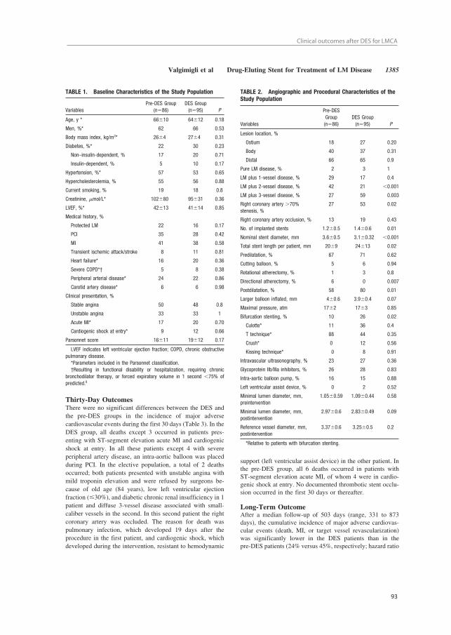
93
Clinical outcomes after DES for LMCA
Thirty-Day OutcomesThere were no significant differences between the DES andthe pre-DES groups in the incidence of major adversecardiovascular events during the first 30 days (Table 3). In theDES group, all deaths except 3 occurred in patients pres-enting with ST-segment elevation acute MI and cardiogenicshock at entry. In all these patients except 4 with severeperipheral artery disease, an intra-aortic balloon was placedduring PCI. In the elective population, a total of 2 deathsoccurred; both patients presented with unstable angina withmild troponin elevation and were refused by surgeons be-cause of old age (84 years), low left ventricular ejectionfraction (�30%), and diabetic chronic renal insufficiency in 1patient and diffuse 3-vessel disease associated with small-caliber vessels in the second. In this second patient the rightcoronary artery was occluded. The reason for death waspulmonary infection, which developed 19 days after theprocedure in the first patient, and cardiogenic shock, whichdeveloped during the intervention, resistant to hemodynamic
support (left ventricular assist device) in the other patient. Inthe pre-DES group, all 6 deaths occurred in patients withST-segment elevation acute MI, of whom 4 were in cardio-genic shock at entry. No documented thrombotic stent occlu-sion occurred in the first 30 days or thereafter.
Long-Term OutcomeAfter a median follow-up of 503 days (range, 331 to 873days), the cumulative incidence of major adverse cardiovas-cular events (death, MI, or target vessel revascularization)was significantly lower in the DES patients than in thepre-DES patients (24% versus 45%, respectively; hazard ratio
TABLE 1. Baseline Characteristics of the Study Population
VariablesPre-DES Group
(n�86)DES Group(n�95) P
Age, y * 66�10 64�12 0.18
Men, %* 62 66 0.53
Body mass index, kg/m2* 26�4 27�4 0.31
Diabetes, %* 22 30 0.23
Non–insulin-dependent, % 17 20 0.71
Insulin-dependent, % 5 10 0.17
Hypertension, %* 57 53 0.65
Hypercholesterolemia, % 55 56 0.88
Current smoking, % 19 18 0.8
Creatinine, �mol/L* 102�80 95�31 0.36
LVEF, %* 42�13 41�14 0.85
Medical history, %
Protected LM 22 16 0.17
PCI 35 28 0.42
MI 41 38 0.58
Transient ischemic attack/stroke 8 11 0.81
Heart failure* 16 20 0.36
Severe COPD*† 5 8 0.38
Peripheral arterial disease* 24 22 0.86
Carotid artery disease* 6 6 0.98
Clinical presentation, %
Stable angina 50 48 0.8
Unstable angina 33 33 1
Acute MI* 17 20 0.70
Cardiogenic shock at entry* 9 12 0.66
Parsonnet score 16�11 19�12 0.17
LVEF indicates left ventricular ejection fraction; COPD, chronic obstructivepulmonary disease.*Parameters included in the Parsonnet classification.†Resulting in functional disability or hospitalization, requiring chronic
bronchodilator therapy, or forced expiratory volume in 1 second �75% ofpredicted.8
TABLE 2. Angiographic and Procedural Characteristics of theStudy Population
Variables
Pre-DESGroup(n�86)
DES Group(n�95) P
Lesion location, %
Ostium 18 27 0.20
Body 40 37 0.31
Distal 66 65 0.9
Pure LM disease, % 2 3 1
LM plus 1-vessel disease, % 29 17 0.4
LM plus 2-vessel disease, % 42 21 �0.001
LM plus 3-vessel disease, % 27 59 0.003
Right coronary artery �70%stenosis, %
27 53 0.02
Right coronary artery occlusion, % 13 19 0.43
No. of implanted stents 1.2�0.5 1.4�0.6 0.01
Nominal stent diameter, mm 3.6�0.5 3.1�0.32 �0.001
Total stent length per patient, mm 20�9 24�13 0.02
Predilatation, % 67 71 0.62
Cutting balloon, % 5 6 0.94
Rotational atherectomy, % 1 3 0.8
Directional atherectomy, % 6 0 0.007
Postdilatation, % 58 80 0.01
Larger balloon inflated, mm 4�0.6 3.9�0.4 0.07
Maximal pressure, atm 17�2 17�3 0.85
Bifurcation stenting, % 10 26 0.02
Culotte* 11 36 0.4
T technique* 88 44 0.35
Crush* 0 12 0.56
Kissing technique* 0 8 0.91
Intravascular ultrasonography, % 23 27 0.36
Glycoprotein IIb/IIIa inhibitors, % 26 28 0.83
Intra-aortic balloon pump, % 16 15 0.88
Left ventricular assist device, % 0 2 0.52
Minimal lumen diameter, mm,preintervention
1.05�0.59 1.09�0.44 0.58
Minimal lumen diameter, mm,postintervention
2.97�0.6 2.83�0.49 0.09
Reference vessel diameter, mm,postintervention
3.37�0.6 3.25�0.5 0.2
*Relative to patients with bifurcation stenting.
Valgimigli et al Drug-Eluting Stent for Treatment of LM Disease 1385

Chapter 10
94
[HR], 0.52 [95% CI, 0.31 to 0.88]; P�0.01) (Figure, A).Mortality was similar in the DES (14%) and pre-DES cohort(16%; HR, 0.79 [95% CI, 0.38 to 1.66]; P�0.54) (Figure, B),whereas there was a significant reduction in both the rate ofMI (4% versus 12%, respectively; HR, 0.22 [95% CI, 0.07 to0.65]; P�0.006) and composite death/MI (Figure, C) as wellas in the need for target vessel revascularization (6% versus23%, respectively; HR, 0.26 [95% CI, 0.10 to 0.65];
P�0.004) (Figure, D) in the DES group. Seventy-four per-cent of the deaths were cardiac, whereas 3 of 13 in the DESgroup and 4 of 14 in the pre-DES phase were attributed toextracardiac reasons. In Table 4, the baseline and proceduralcharacteristics of those patients in the DES group whounderwent target vessel revascularization during follow-upare reported. In all cases, the lesion was located in the distalLM, in 50% of cases diabetes was present, and all except 1were women. In 3 cases, in-stent restenosis occurred; in 2patients intimal hyperplasia developed at the distal edge ofthe stent, whereas in 1 patient severe ostial side branchrestenosis (circumflex artery) necessitated reintervention. Inall cases, restenosis was focal (�10 mm in length) and wassuccessfully treated with repeated PCI. In the pre-DES group,13 cases of pure in-stent restenosis, of which 3 were focal,were treated with PCI (9 patients) or CABG (4 patients). In 2patients, diffuse intimal hyperplasia associated with progres-sion of atherosclerotic disease in other vessels was treatedwith CABG, and in 5 patients (3 with ST-segment elevationacute MI as the indication for LM intervention), stagedreintervention with CABG (in 4 patients) and PCI (in 1patient) was performed because revascularization remainedincomplete at the time of the index procedure.
One-year adverse events in patients treated with BMS before the introduction of DES (pre-DES group) and in patients treated exclusively withDES implantation (DES group). Cumulative risk of major adverse cardiovascular events (MACE) (A), death (B), death or MI (C), and target ves-sel revascularization (TVR) (D) is shown.
TABLE 3. Thirty-Day Outcomes
VariablesPre-DES Group
(n�86)DES Group(n�95) P*
Death, n (%) 6 (7) 10 (11) 0.60
Nonfatal MI, n (%) 8 (9) 4 (4) 0.24
Death or nonfatal MI, n (%) 14 (16) 14 (15) 0.84
Target vessel revascularization, n (%) 2 (2) 0 (0) 0.22
Repeated PCI 1 (1) 0 (0)
CABG 1 (1) 0 (0)
Any event, n (%) 16 (19) 14 (15) 0.56
Stent thrombosis, n (%)† 0 (0) 0 (0) 1
*By Fisher exact test.†Angiographically documented.
1386 Circulation March 22, 2005
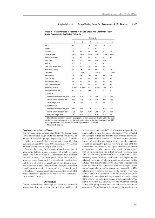
95
Clinical outcomes after DES for LMCA
Predictors of Adverse EventsThe Parsonnet score, ranging from 2.5 to 55.5 (mean value,18�2; interquartile range, 16.5) was 16�11 and 19�12 inthe pre-DES and DES groups, respectively (P�0.17) (Table1), with a trend toward a higher rate of patients considered athigh surgical risk (58% versus 46%, respectively; P�0.13) inthe DES compared with the pre-DES cohort.
On univariate analysis, Parsonnet classification, use ofintra-aortic balloon pump, presence of shock at entry,lesion located in the distal LM, nonelective PCI, troponinelevation at entry, TIMI flow grade before and after PCI,reference vessel diameter, left ventricular ejection fraction,and the use of DES were identified as significant predic-tors of adverse events. On multivariate analysis, Parsonnetclassification, troponin elevation at entry, lesions locatedat distal site, reference vessel diameter, and the use of DESwere independent predictors of major adverse cardiovas-cular events (Table 5).
DiscussionDespite the feasibility and the high procedural success rate ofpercutaneous LM intervention, the long-term incidence of
adverse events in the pre-DES “era” was often reported to beunacceptably high in this subset of patients.4,6 This reflectedthe inclusion of high-risk patients, such as those not consid-ered “good surgical candidates,” as well as the dramaticimpact of treated vessel failure in this specific anatomiccontext. In consecutive patients receiving elective BMS forunprotected LM treatment, the 3-year cumulative incidenceof death was recently reported to be �16%.6 In that series,28% of the population was at high surgical risk. More than50% of our study population was at high surgical riskaccording to the Parsonnet classification, thus explaining therelatively high rate of adverse events we observed. In thissetting, when patients treated with DES were compared withthose treated with BMS, a marked benefit with respect to therate of major adverse cardiac events, as evidenced by a 47%relative risk reduction, emerged in the former. This wasmainly due to the difference in the incidence of MI (67%relative risk reduction) and target vessel revascularization(65% relative risk reduction), with no effect on mortality. Thehigher prevalence of 3-vessel disease and bifurcation stentingin the DES group makes the observed benefit even moreconvincing. The difference in the incidence of events between
TABLE 4. Characteristics of Patients in the DES Group Who Underwent TargetVessel Revascularization During Follow-Up
Patient No.
1 2 3 4 5 6
Age, y 66 77 36 70 52 56
Gender F F F M F F
Diabetes Yes No No Yes No Yes
Lesion location Distal Distal Distal Distal Distal Distal
Severe calcification Yes No No No No No
Stent type SES SES PES PES PES PES
Stent No. 2 2 1 2 1 2
Total stent length, mm 16 36 20 48 16 36
Bifurcation stenting No Yes No Yes No Yes
Technique � � � Crush � � � Culotte � � � Culotte
Postdilatation Yes Yes Yes Yes Yes Yes
Final kissing No No No Yes No No
Gap between stents No No No No No No
Stent underexpansion Yes No No No No No
Restenosis location In-stent In-stent* RS In-stent DER DER*
Revascularization type PCI PCI PCI PCI PCI PCI
QCA after PCI
Reference vessel diameter, mm 3.74 3.27 3.53 2.65 2.44 2.76
Minimal lumen diameter, mm 2.12 1.06 3.34 2.49 1.94 2.32
Lesion length, mm 13.4 19.7 13.5 21.3 8.9 18.9
QCA at follow-up
Reference vessel diameter, mm 3.87 3.43 3.21 2.32 1.82 2.36
Minimal lumen diameter, mm 1.23 0.57 0.98 0.99 0.6 0.71
Restenosis length, mm 5.8 9.06 3.6 5.48 7.72 9.5
QCA indicates quantitative coronary angiography; In-stent, restenosis located within the stentmargins; RS, restenosis located in the side branch (the ostium of the circumflex artery); and DER,distal edge restenosis located within the 5-mm segment distal to the stent.*More than 1 focal site.
Valgimigli et al Drug-Eluting Stent for Treatment of LM Disease 1387

Chapter 10
96
the 2 groups emerged slowly after the procedure, with noclear advantage at 30 days, possibly reflecting the specificmechanism of action of DES on intimal hyperplasia.
The overall advantage of DES remained significant afteradjustment for the Parsonnet score, the anatomic site ofobstruction, and troponin status at entry. Therefore, our datasuggest that when percutaneous treatment of LM coronaryartery disease is undertaken, DES should be used as thedefault strategy.
The LM bifurcation was frequently involved (�60%) inour series, and even when the obstruction was more proxi-mally located and did not directly involve the LAD or leftcircumflex artery ostia, its treatment often required themanagement of LM bifurcation. To date, the results of SESimplantation to treat bifurcated lesions have been relatively
disappointing, with high rates of restenosis in the sidebranch.11 Our present findings are in keeping with theseprevious observations, confirming that in the DES era distalLM location is an independent predictor of adverse events atfollow-up. Furthermore, because the strategy and technicalaspects of bifurcation management were left entirely to thepreference of treating physicians, no clear conclusions can bedrawn in this regard.
Inconsistent findings have been reported thus far withregard to the effect of DES on long-term cumulativeincidence of MI. In the first randomized clinical trialscomparing SES or PES with BMS, no difference in theincidence of MI was observed.12,13 Second-generationrandomized trials assessing the benefit of DES in patientsselected to be at intermediate risk for in-stent restenosis orall-inclusive registries reported trends toward MI reductionin the DES group, but none of them reached statisticalsignificance.14,15 Recently, a clear reduction in the cumu-lative incidence of MI in the DES group was reported inthe SES-SMART trial, in which a selected group ofhigh-risk patients has been evaluated.16 Similarly, in ourpatient population, a reduced incidence of MI was ob-served in the DES group. Of note, 2 and 1 cases of MI inthe pre-DES phase were related to target vessel revascu-larization and not related to target vessel revascularization,respectively. Whether this difference between studies isthe reflection of a type II error in studies enrolling patientsat low or intermediate risk remains unclear, but when theretrospective nature of our investigation is considered, datafrom prospective studies are needed to confirm ourfindings.
Limitations of the StudyThe present study is a single-center experience from a tertiaryreferral center and lacks the clear advantages of a multicenterrandomized study. In keeping with the aim of our investiga-tion, an “all-comers” population has been enrolled, clearlyresulting in a heterogeneous group of patients. Further stud-ies, with larger sample sizes, are required to investigate thedifferential impact of DES versus BMS in prespecifiedsubgroups, stratified according to clinical presentation (stableversus unstable) or protected versus unprotected type oftreatment.
Despite the fact that the study was conducted over a relativelyshort period, we cannot exclude the possibility that improve-ments in technique or differences in drug prescription could havepartially accounted for the difference observed in terms of majoradverse cardiovascular events between groups. However, con-ducting randomized trials that seek to assess the efficacy of DESversus BMS in this specific subset of patients seems unlikely,and our understanding of the benefit of drug-coated stents totreat this group of patients will probably also rely in the nearfuture on well-conducted registries that are able to record andmonitor our daily clinical practice.
ConclusionsThe use of DES as a default strategy to treat LM disease wasassociated with a significant reduction in adverse events. Theeffectiveness of DES persisted even after adjustment forclinical and procedural variables, including the Parsonnet
TABLE 5. Univariate and Multivariate Cox ProportionalHazards Analysis
Variables PHazard Ratio(95% CI) �2
Univariate analysis
Distal LM disease 0.003 2.7 (4.8–1.53) 13.3
DES use 0.019 0.54 (0.9–0.32) 5.48
Nonelective PCI 0.0047 2.1 (3.5–1.3) 8
Intra-aortic balloon pumpuse
0.0002 2.9 (4.9–1.7) 14
LVEF, % 0.00001 0.95 (0.97–0.93) 20
Parsonnet score �0.00001 1.07 (1.09–1.05) 44
Reference vessel diameter 0.00001 0.36 (0.58–0.32) 19
Shock at entry �0.00001 4.48 (7.9–2.5) 21
TIMI flow before PCI 0.03 0.75 (0.96–0.58) 4.3
TIMI flow after PCI 0.03 0.58 (0.85–0.39) 4.7
Troponin T �0.02 �g/Lat entry
0.0002 3.15 (5.26–1.9) 18
Multivariate analysis 1
Distal LM disease 0.0007 2.94 (5.5–1.57) 76
DES use 0.00009 0.33 (0.57–0.19)
LVEF, % 0.09 0.98 (1.001–0.95)
Parsonnet score 0.0009 1.04 (1.07–1.01)
Reference vessel diameter 0.005 0.51 (0.79–0.33)
Troponin T �0.02 �g/L atentry
0.02 2.3 (4.4–1.2)
Multivariate analysis 2
Distal LM disease 0.00017 3.3 (6.1–1.7) 68
DES use 0.00018 0.35 (0.6–0.20)
LVEF, % 0.00013 0.95 (0.98–0.94)
Reference vessel diameter 0.0011 0.48 (0.74–0.30)
Shock at entry 0.006 3.49 (8.6–1.4)
Troponin T �0.02 �g/L atentry
0.016 2.27 (4.2–1.17)
Multivariate analysis model 1 was performed with all major adversecardiovascular event predictors on univariate analysis; in multivariate analysismodel 2, the Parsonnet score was removed because of colinearity between thevariables included in the model and those used in the calculation of the score,such as left ventricular ejection fraction (LVEF), use of intra-aortic balloonpump, and presence of shock.
1388 Circulation March 22, 2005

97
Clinical outcomes after DES for LMCA
surgical risk score. Our findings apply to a selected group ofpatients referred for percutaneous LM treatment and suggestthat in this setting routine DES implantation, by reducing thecumulative incidence of major adverse cardiovascular events,should be currently regarded as the strategy of choice. Untilnew evidence is provided by randomized clinical trialsdirectly comparing the surgical and percutaneous approaches,CABG should remain the preferred revascularization treat-ment in good surgical candidates presenting with LM coro-nary artery disease.
AcknowledgmentsThe authors express their gratitude to Dr Pedro A. Lemos for hismajor contribution to the RESEARCH Registry and acknowledge theexpert statistical assistance of Dr Eric Boersma.
References1. Serruys PW, Unger F, Sousa JE, Jatene A, Bonnier HJ, Schonberger JP,
Buller N, Bonser R, van den Brand MJ, van Herwerden LA, Morel MA,van Hout BA. Comparison of coronary-artery bypass surgery and stentingfor the treatment of multivessel disease. N Engl J Med. 2001;344:1117–1124.
2. Legrand VM, Serruys PW, Unger F, van Hout BA, Vrolix MC, FransenGM, Nielsen TT, Paulsen PK, Gomes RS, deq Ueiroz e Melo JM, NevesJP, Lindeboom W, Backx B. Three-year outcome after coronary stentingversus bypass surgery for the treatment of multivessel disease. Circu-lation. 2004;109:1114–1120.
3. Caracciolo EA, Davis KB, Sopko G, Kaiser GC, Corley SD, Schaff H,Taylor HA, Chaitman BR. Comparison of surgical and medical groupsurvival in patients with left main coronary artery disease: long-termCASS experience. Circulation. 1995;91:2325–2334.
4. Tan WA, Tamai H, Park SJ, Plokker HW, Nobuyoshi M, Suzuki T,Colombo A, Macaya C, Holmes DR Jr, Cohen DJ, Whitlow PL, Ellis SG.Long-term clinical outcomes after unprotected left main trunk percuta-neous revascularization in 279 patients. Circulation. 2001;104:1609–1614.
5. Park SJ, Hong MK, Lee CW, Kim JJ, Song JK, Kang DH, Park SW,Mintz GS. Elective stenting of unprotected left main coronary arterystenosis: effect of debulking before stenting and intravascular ultrasoundguidance. J Am Coll Cardiol. 2001;38:1054–1060.
6. Takagi T, Stankovic G, Finci L, Toutouzas K, Chieffo A, Spanos V,Liistro F, Briguori C, Corvaja N, Albero R, Sivieri G, Paloschi R, Di
Mario C, Colombo A. Results and long-term predictors of adverse clinicalevents after elective percutaneous interventions on unprotected left maincoronary artery. Circulation. 2002;106:698–702.
7. Arampatzis CA, Lemos PA, Tanabe K, Hoye A, Degertekin M, Saia F,Lee CH, Ruiter A, McFadden E, Sianos G, Smits PC, van der GiessenWJ, de Feijter P, van Domburg R, Serruys PW. Effectiveness ofsirolimus-eluting stent for treatment of left main coronary artery disease.Am J Cardiol. 2003;92:327–329.
8. Parsonnet V, Dean D, Bernstein AD. A method of uniform stratificationof risk for evaluating the results of surgery in acquired adult heart disease.Circulation. 1989;79:I-3–I-12.
9. Bernstein AD, Parsonnet V. Bedside estimation of risk as an aid fordecision-making in cardiac surgery. Ann Thorac Surg. 2000;69:823–828.
10. Roques F, Nashef SA, Michel P, Gauducheau E, de Vincentiis C, BaudetE, Cortina J, David M, Faichney A, Gabrielle F, Gams E, Harjula A,Jones MT, Pintor PP, Salamon R, Thulin L. Risk factors and outcome inEuropean cardiac surgery: analysis of the EuroSCORE multinationaldatabase of 19030 patients. Eur J Cardiothorac Surg. 1999;15:816–822;discussion 822–823.
11. Colombo A, Moses JW, Morice MC, Ludwig J, Holmes DR Jr, Spanos V,Louvard Y, Desmedt B, Di Mario C, Leon MB. Randomized study toevaluate sirolimus-eluting stents implanted at coronary bifurcationlesions. Circulation. 2004;109:1244–1249.
12. Morice MC, Serruys PW, Sousa JE, Fajadet J, Ban Hayashi E, Perin M,Colombo A, Schuler G, Barragan P, Guagliumi G, Molnar F, Falotico R.A randomized comparison of a sirolimus-eluting stent with a standardstent for coronary revascularization. N Engl J Med. 2002;346:1773–1780.
13. Stone GW, Ellis SG, Cox DA, Hermiller J, O’Shaughnessy C, Mann JT,Turco M, Caputo R, Bergin P, Greenberg J, Popma JJ, Russell ME. Apolymer-based, paclitaxel-eluting stent in patients with coronary arterydisease. N Engl J Med. 2004;350:221–231.
14. Lemos PA, Serruys PW, van Domburg RT, Saia F, Arampatzis CA, HoyeA, Degertekin M, Tanabe K, Daemen J, Liu TK, McFadden E, Sianos G,Hofma SH, Smits PC, van der Giessen WJ, de Feyter PJ. Unrestrictedutilization of sirolimus-eluting stents compared with conventional barestent implantation in the “real world”: the Rapamycin-Eluting StentEvaluated At Rotterdam Cardiology Hospital (RESEARCH) registry.Circulation. 2004;109:190–195.
15. Schampaert E, Cohen EA, Schluter M, Reeves F, Traboulsi M, Title LM,Kuntz RE, Popma JJ. The Canadian study of the sirolimus-eluting stent inthe treatment of patients with long de novo lesions in small nativecoronary arteries (C-SIRIUS). J Am Coll Cardiol. 2004;43:1110–1115.
16. Ardissino D, Cavallini C, Bramucci E, Indolfi C, Marzocchi A, Manari A,Angeloni C, Carosio G, Bonizzoni E, Colusso S, Repetto M, Merlini PA.Sirolimus-eluting vs uncoated stents for prevention of restenosis in smallcoronary areries: a randomized trial. JAMA. 2004;292:2727–2734.
Valgimigli et al Drug-Eluting Stent for Treatment of LM Disease 1389


Chapter 11
Sirolimus- versus Paclitaxel-Eluting stent implantation for
the percutaneous treatment of left main coronary artery
disease. A combined RESEARCH and TSEARCH
Long-term Analysis
Valgimigli M, Malagutti P, Aoki J, Garcia-
Garcia HM, Rodriguez Granillo GA, van
Mieghem C, Loghart JML, Ong ATL,
Sianos G, Regar E, van Domburg R, de
Feyter P, de Jaegere P, Serruys PW.
J Am Coll Cardiol. 2006;47:507-514


101
Angiographic outcomes after DES for LMCA
CLINICAL RESEARCH Interventional Cardiology
Sirolimus-Eluting Versus Paclitaxel-ElutingStent Implantation for the PercutaneousTreatment of Left Main Coronary Artery DiseaseA Combined RESEARCH and T-SEARCH Long-Term AnalysisMarco Valgimigli, MD, Patrizia Malagutti, MD, Jiro Aoki, MD, Héctor M. Garcia-Garcia, MD,Gaston A. Rodriguez Granillo, MD, Carlos A. G. van Mieghem, MD, Jurgen M. Ligthart, BSC,Andrew T. L. Ong, MBBS, FRACP, George Sianos, MD, PHD, Evelyn Regar, MD, PHD,Ron T. Van Domburg, PHD, Pim De Feyter, MD, PHD, Peter de Jaegere, MD, PHD,Patrick W. Serruys, MD, PHDRotterdam, the Netherlands
OBJECTIVES The purpose of this study was to investigate the long-term clinical and angiographic profileof sirolimus-eluting stent (SES) versus paclitaxel-eluting stent (PES) in patients undergoingpercutaneous intervention for left main (LM) coronary disease.
BACKGROUND The long-term clinical and angiographic impact of SES as opposed to PES implantation inthis subset of patients is unknown.
METHODS From April 2002 to March 2004, 110 patients underwent percutaneous intervention for LMstenosis at our institution; 55 patients were treated with SES and 55 with PES. The twogroups were well balanced for all baseline characteristics.
RESULTS At a median follow-up of 660 days (range 428 to 885), the cumulative incidence of majoradverse cardiovascular events was similar (25% in the SES group vs. 29%, in the PES group;hazard ratio 0.88 [95% confidence interval 0.43 to 1.82]; p � 0.74), reflecting similarities inboth the composite death/myocardial infarction (16% in the SES group and 18% in the PESgroup) and target vessel revascularization (9% in the SES group and 11% in the PES group).Angiographic in-stent late loss (mm), evaluated in 73% of the SES group and in 77% of thePES group, was 0.32 � 74 in the main and 0.36 � 0.59 in the side branch in the SES groupvs. 0.46 � 0.57 (p � 0.36) and 0.52 � 0.42 (p � 0.41) in the PES group, respectively.
CONCLUSIONS In consecutive patients undergoing percutaneous LM intervention, PES may perform closelyto SES both in terms of angiographic and long-term clinical outcome. (J Am Coll Cardiol2006;47:507–14) © 2006 by the American College of Cardiology Foundation
Routine drug-eluting stent (DES) implantation, by reducingthe need for target vessel revascularization (TVR) and angio-graphic restenosis, has been recently proposed as the preferredstrategy in poor surgical candidates undergoing percutane-ous left main coronary artery (LM) intervention (1–3).The longest average follow-up available for this treatment
is currently one year, and whether sirolimus-eluting stent(SES) or paclitaxel-eluting stent (PES) is performing betterin these patients is unknown (1–3).Similarly, even for more conventional lesions, the differ-
ential safety and efficacy profile of these two DES options islargely debated (4–8). When taken together, current evi-dence possibly suggests that in more complex lesion/patientsubsets, SES performs better and more safely than PES(4–7,9).
The percutaneous management of LM lesions is a chal-lenging intervention, where bifurcated vessels, extensivewall calcification, and poor hemodynamic tolerance oftencoexist during treatment.The purpose of the present study was to investigate, in a
high-risk subset of patients undergoing revascularization ina tertiary referral center, the differential long-term impact ofSES compared with PES in terms of clinical and angio-graphic outcome. Intravascular ultrasound analysis has alsobeen carried out at follow-up to quantity neointimal hyper-plasia volume.
METHODS
Study design and patient population. Since April 16,2002, SES (Cypher; Johnson & Johnson-Cordis, Warren,New Jersey) has been used as a default strategy for every PCIat our institution as part of the Rapamycin-Eluting StentEvaluated at Rotterdam Cardiology Hospital (RESEARCH)registry. From the first quarter of 2003, PES (Taxus; BostonScientific Corp., Natick, Massachusetts) became commerciallyavailable, replacing SES as the strategy of choice in every PCI,
From the Erasmus Medical Center, Thoraxcenter, Rotterdam, the Netherlands.This study was supported by the Erasmus Medical Center, Rotterdam, and byunrestricted institutional grants from Boston Scientific Corporation (Natick, Massa-chusetts) and Cordis, a Johnson & Johnson company (Warren, New Jersey).Manuscript received June 7, 2005; revised manuscript received August 24, 2005,
accepted September 8, 2005.
Journal of the American College of Cardiology Vol. 47, No. 3, 2006© 2006 by the American College of Cardiology Foundation ISSN 0735-1097/06/$32.00Published by Elsevier Inc. doi:10.1016/j.jacc.2005.09.040

Chapter 11
102
as part of the Taxus-Stent Evaluated at Rotterdam CardiologyHospital (T-SEARCH) registry. As a policy, all electivepatients presenting with significant (�50% by visual estima-tion) LM disease, referred to our institution for coronaryrevascularization, are evaluated both by interventional cardiol-ogists and by cardiac surgeons, and the decision to opt for PCIor surgery is reached by consensus as previously described (1).From April 16, 2002, to March 6, 2004, a total of 110
consecutive patients were treated exclusively with one ormore DES in the LM as part of an elective or nonelectiverevascularization procedure and constitute the patient pop-ulation of the present report. Fifty-five patients first receivedexclusively SES, available at that time in diameters from2.25 to 3.00 mm, and then 55 patients received PES,available in diameters from 2.25 to 3.5 mm. To ensurecomparability between the two study groups, the Parsonnetsurgical risk score, based on both clinical presentation profileand comorbidities, and the William Beaumont Hospitalsimplified scoring system were calculated for each patient(10,11). Nonelective treatment was defined as a procedurecarried out on referral before the beginning of the nextworking day (12).The protocol was approved by the hospital ethics com-
mittee and is in accordance with the Declaration of Hel-sinki. Written informed consent was obtained from everypatient.Procedures and postintervention medications. All inter-ventions were performed according to current standardguidelines, and, except for the stent utilization, the finalinterventional strategy was left entirely to the discretion ofthe operator. Angiographic success was defined as residualstenosis �30% by visual analysis in the presence of Throm-bolysis In Myocardial Infarction (TIMI) flow grade 3. Allpatients were advised to maintain aspirin lifelong, andclopidogrel was prescribed for 6 months in both groups.End point definitions and clinical follow-up. The occur-rence of major adverse cardiac events, defined as death,nonfatal myocardial infarction, or target vessel revascular-ization, was recorded. Patients with more than one event
were assigned the highest ranked event, according to theprevious list. End point definitions were previously reported(1). In order to make the clinical follow-up of the twosequential cohorts of patients comparable, clinical outcomeof the SES cohort was censored at two years.Quantitative angiographic and intravenous ultrasoundanalysis. Quantitative analyses of all angiographic datawere performed with the use of edge-detection techniques(CAAS II; Pie Medical, Maastricht, the Netherlands). Avalue of 0 mm was assigned for the minimal luminaldiameter (MLD) in cases of total occlusion at baseline orfollow-up. Binary restenosis (BR) was defined as stenosis of�50% of the luminal diameter in the target lesion. Acutegain was defined as the MLD after the index procedureminus the MLD at baseline angiography. Late loss (LL)was defined as the MLD immediately after the indexprocedure minus the MLD at angiographic follow-up.Quantitative angiographic measurements of the target le-sion were obtained in the in-lesion zone (including thestented segment as well as the margins 5 mm proximal anddistal to the stent). Intravascular ultrasound (IVUS) analysiswas performed after administration of 200 �g of intracoro-nary nitroglycerin, with an automated pullback at 0.5 mm/s.All IVUS procedures were recorded on VHS videotape, and
Table 1. Baseline Characteristics of the Study Population
VariablesSES Group(n � 55)
PES Group(n � 55)
pValue
Age (yrs)* 64 � 12 63 � 12 0.54Males (%)* 64 58 0.84Body mass index (kg/m2)* 23 � 4 25 � 5 0.31Diabetes (%)* 34 24 0.29Hypertension (%)* 54 53 �0.99Hypercholesterolemia (%) 58 56 �0.99Current smokers (%) 16 20 0.8Creatinine (�mol/l)* 96 � 32 100 � 80 0.68LV ejection fraction (%)* 44 � 16 44 � 12 0.95Medical history (%)Protected left main 18 15 0.80PCI 31 33 �0.99Myocardial infarction 38 47 0.44TIA/stroke 9 11 0.74Heart failure* 16 16 �0.99COPD severe*† 4 5 �0.99Peripheral arterial disease* 22 16 0.63Carotid artery disease* 9 5 0.71
Clinical presentation (%)Stable angina 49 45 0.86Silent ischemia 0 4 0.12Unstable angina 33 33 �0.99Acute myocardial infarction* 15 18 0.72Cardiogenic shock at entry* 9 9 �0.99
Parsonnet score 20 � 13 17 � 11 0.27William Beaumont Hospital
score‡7.7 � 4.28 7.36 � 4.7 0.73
*Parameter included in the Parsonnet classification. †Resulting in functional disabil-ity, hospitalization, requiring chronic bronchodilatator therapy, or FEV1 �75% ofpredicted. ‡Based on age �65 years, creatinine elevation, multivessel disease, andoccurrence of myocardial infarction within the previous 14 days.COPD � chronic obstructive pulmonary disease; LV � left ventricular; PCI �
percutaneous coronary intervention; PES � paclitaxel-eluting stent; SES �sirolimus-eluting stent; TIA � transient ischemic attack.
Abbreviations and AcronymsBMS � bare-metal stentBR � binary restenosisCX � left circumflex coronary arteryDES � drug-eluting stentIVUS � intravascular ultrasoundLAD � left anterior descending coronary arteryLL � late lossLM � left main coronary arteryMACE � major adverse cardiac eventsMI � myocardial infarctionMLD � minimal luminal diameterPES � paclitaxel-eluting stentRCA � right coronary arterySES � sirolimus-eluting stentTVR � target vessel revascularization
508 Valgimigli et al. JACC Vol. 47, No. 3, 2006Drug-Eluting Stent for LM Treatment February 7, 2006:507–14

103
Angiographic outcomes after DES for LMCA
images were digitized for analysis. A computer-based con-tour detection was performed with Qurad QCU analysissoftware (Curad, Wijk Bij Duurstede, the Netherlands) aspreviously described (13). Intimal hyperplasia volume wascalculated as stent volume minus luminal volume. Percent-age intimal hyperplasia was defined as intimal hyperplasiavolume divided by stent volume.Statistical analysis. The sample size was calculated on theassumption that average late loss in the SES and PES groupwould be around 0.15 mm and 0.35 mm, respectively, basedon previous findings. To detect this effect size with a sigmavalue of 0.3, 85% power, and a type I error (alpha) of 0.05,32 patients per group were required. Continuous variablesare shown as mean � standard deviation (SD) and werecompared using Student unpaired t test. Categorical vari-ables are presented as counts and percentages and comparedwith the Fisher exact test. Survival curves were generated bythe Kaplan-Meier method, and survival among groups wascompared using the log rank test. Cox proportional hazardsmodels were used to assess risk reduction of adverse events.Multivariable analysis, considering all variables reported inTables 1 and 2 with a p value of less than 0.10, wasperformed to adjust for possible confounders and identifywhether the stent received was an independent predictor ofadverse events. Probability was significant at a level of
�0.05. All statistical tests were two-tailed. Statistical anal-ysis was performed with Statistica 6.1 software (Statsoft,Tulsa, Oklahoma).
RESULTS
Baseline and procedural characteristics. Baseline and pro-cedural characteristics are shown in Tables 1 and 2. The twogroups were well matched for all baseline characteristics. Inboth cohorts, half of the patients presented with a leftventricular ejection fraction of 45% or less or with acutecoronary syndrome as indication to treatment, and 9% ofthe patients presented with severe hemodynamic compro-mise at entry. The distal LM was overall involved intwo-thirds of cases. In the SES group, nominal stentdiameter was smaller—reflecting the unavailability of stentbigger than 3.0 mm during the study period—and cumula-tive stent length tended to be shorter than in the PESgroup. Bifurcation stenting was equally employed in bothgroups with a clear preference for T-stenting and for culottetechnique in the SES and PES groups, respectively.Thirty-one patients (56%) in the SES and 25 (45%) in
the PES group received intervention in one or more non-LMlesion(s) during index procedure (p � 0.18). Complete
Table 2. Angiographic and Procedural Characteristics of the Study Population
VariablesSES Group(n � 55)
PES Group(n � 55) p Value
Lesion location (%)*Ostium 27 22 0.51Body 29 24 0.54Distal 64 76 0.28
Pure left main disease (%) 5 5 �0.99LM plus 1-vessel disease (%) 18 18 �0.99LM plus 2-vessel disease (%) 20 24 0.82LM plus 3-vessel disease (%) 57 53 0.87Right coronary artery �70% stenosis (%) 69 64 0.88Right coronary artery occlusion (%) 24 20 0.82Number of implanted stents 1.43 � 0.53 1.50 � 0.6 0.49Nominal stent diameter (mm) 3.00 � 0.25 3.29 � 0.28 �0.001Total stent length per patient (mm) 23 � 11 27 � 13 0.07Predilation (%) 71 60 0.15Cutting balloon (%) 7 7 �0.99Rotational atherectomy (%) 2 4 �0.99Directional atherectomy (%) 0 0 �0.99Postdilatation (%) 82 72 0.15Bigger balloon inflated (mm) 3.75 � 0.48 3.70 � 0.5 0.48Maximal pressure (atm) 18 � 5 19 � 3 0.55Bifurcation stenting (%) 29 33 0.84Culotte 0 20 0.009T-technique 20 5 0.05Crush 7 4 0.67Kissing technique 2 4 �0.99
Intravascular ultrasonography (%) 33 27 0.53IIb/IIIa inhibitors (%) 33 27 0.68Intra-aortic balloon pump (%) 20 18 �0.99Left ventricle assist device (%) 5 0 0.24Temporary pacing during procedure (%) 7 9 �0.99
*Location of disease was not mutually exclusive among LM segments.LM � left main coronary artery; other abbreviations as in Table 1.
509JACC Vol. 47, No. 3, 2006 Valgimigli et al.February 7, 2006:507–14 Drug-Eluting Stent for LM Treatment

Chapter 11
104
revascularization was achieved in 27 (49%) patients in theSES and 36 (65%) in the PES group (p � 0.17).Overall procedural success was 98%. In one patient
receiving SES, presenting with acute MI and shock, a finalTIMI flow grade 1 was obtained. An abrupt irreversibleocclusion of the circumflex occurred in one PES patientafter deployment of a stent in the left main and proximal leftanterior descending coronary artery (LAD).Thirty-day outcomes. There were no significant differ-ences between the SES and PES groups in the incidence ofmajor adverse cardiac events (MACE) (death, target vesselrevascularization, or myocardial infarction [MI]) during thefirst 30 days (Table 3). Eight deaths occurred in 10 patientspresenting with ST-segment elevation acute myocardialinfarction and cardiogenic shock at entry. One electivepatient, undergoing LM treatment under left ventricularassist device due to end-stage heart disease, died 2 days afterfor cardiogenic shock, while the second elective patientsdied for non-cardiovascular reasons after 19 days. Nodocumented thrombotic stent occlusion occurred in the first30 days or thereafter.Long-term clinical outcome. After a median follow-up of660 days (range 428 to 885 days), the cumulative incidence
Figure 1. Adverse events in patients treated with sirolimus-eluting stent(SES group) and in patients treated exclusively with paclitaxel-eluting stentimplantation (PES group). (A) Cumulative risk of major adverse events(MACE); (B) death or myocardial infarction (MI); and (C) target vesselrevascularization (TVR). CI � confidence interval.
Table 3. 30-Day and Long-Term Outcomes
VariablesSES
GroupPESGroup
pValue*
30-day outcomeWhole population (n � 55) (n � 55)Death, n (%) 7 (13) 3 (5) 0.32Nonfatal MI, n (%) 2 (4) 2 (4) �0.99Death or nonfatal MI, n (%) 9 (16) 5 (9) 0.40TVR, n (%) 1 (2) 0 (0) �0.99Any event, n (%) 10 (18) 5 (9) 0.27Stent thrombosis, n (%)† 0 (0) 0 (0)
Elective population (n � 43) (n � 43)Death, n (%) 2 (5) 0 (0) 0.49Nonfatal MI, n (%) 2 (5) 2 (5) �0.99Death or nonfatal MI, n (%) 4 (9) 2 (5) 0.68TVR, n (%) 1 (2) 0 (0) �0.99Any event, n (%) 5 (12) 2 (5) 0.44Stent thrombosis, n (%)† 0 (0) 0 (0) �0.99
Long-term outcomeWhole population (n � 55) (n � 55)Death, n (%) 6 (11) 7 (13) 0.70Nonfatal MI, n (%) 2 (4) 4 (7) 0.25Death or nonfatal MI, n (%) 9 (16) 10 (18) 0.90TVR, n (%) 5 (9) 6 (11) 0.67Any event, n (%) 14 (25) 16 (29) 0.74Stent thrombosis, n (%)† 0 (0) 0 (0) �0.99
Elective population (n � 43) (n � 43)Death, n (%) 2 (5) 2 (5) 0.98Nonfatal MI, n (%) 2 (5) 4 (9) 0.22Death or nonfatal MI, n (%) 4 (9) 6 (14) 0.51TVR, n (%) 5 (12) 5 (12) 0.82Any event, n (%) 9 (21) 11 (25) 0.55Stent thrombosis, n (%)† 0 (0) 0 (0) �0.99
*By Fischer exact test for 30-day outcome and by log rank test for long-term outcome.†Angiographically documented.MI � myocardial infarction; TVR � target vessel revascularization; other
abbreviations as in Table 1.
510 Valgimigli et al. JACC Vol. 47, No. 3, 2006Drug-Eluting Stent for LM Treatment February 7, 2006:507–14

105
Angiographic outcomes after DES for LMCA
of MACE did not significantly differ in the SES comparedto the PES patients (25% vs. 29%, respectively; hazard ratio[HR] 0.88 [95% confidence interval (CI): 0.43 to 1.82]; p�0.74) (Fig. 1A). The composite death/MI was 16% in theSES and 18% in the PES group (HR 0.95 [95% CI: 0.38 to2.33]; p � 0.90) (Fig. 1B), and cumulative incidence ofTVR was 9% in the SES and 11% in the PES group (HR0.77 [95% CI: 0.23 to 2.56]; p � 0.66) (Fig. 1C).In the elective patient population (43 patients in each
group), the cumulative incidence of MACE was similar inthe SES (21%) and PES groups (25%; HR 0.77 [95% CI0.32 to 1.8]; p� 0.55). The composite of death/MI was 9%in the SES and 14% in the PES group (HR 0.66 [95% CI0.19 to 2.3]; p � 0.52), and the need for TVR was 12% inboth groups (HR 0.87 [95% CI 0.24 to 3]; p � 0.8).The cumulative incidence of MACE was similar in
patients receiving single-vessel stenting (24% in the SESand 27% in the PES group) and those treated with bifur-cation stenting (31%, HR 1.38 [95% CI 0.47 to 4]; p �0.55; and 33%, HR 1.22 [95% CI 0.45 to 3.3]; p � 0.69;respectively.After adjustment for nominal stent diameter and total
stent length at multivariable Cox regression analysis, SESimplantation as opposed to PES failed to emerge as anindependent predictor of MACE (HR 0.79 [95% CI 0.5 to1.5]; p � 0.66). The same remained true after forcing theParsonnet score into the model.Angiographic outcome. Thirty-five patients in the SES(73% of eligible patients) and 38 patients in the PES group(77% of those eligible) underwent angiographic follow-up.Data regarding the quantitative coronary angiography formain and side branches are presented in Tables 4 and 5,respectively.
MAIN BRANCH. In the SES and PES groups, the maintreated branch circumflex was LM-LAD in 22 (63%) and21 (55%) patients, respectively followed by LM-circumflexin 6 (17%) and 8 (21%), LM alone in 5 (14%) and 6 (16%),and LM-intermediate branch in 2 (6%) and 3 (8%), respec-tively. In unprotected patients in the SES and PES groups,LM-LAD was the main treated branch in 77% and in 63%,respectively, followed by LM alone in 15% and 17%,LM-circumflex in 8% and 14%, and LM-intermediatebranch in 0% and 6%, respectively.Baseline and follow-up angiographic variables did not
differ in the two study groups. No difference was noted interms of BR in the two groups as the result of a similarin-stent and in-lesion LL (Table 4).
SIDE BRANCH. All baseline angiographic variables were wellmatched between SES and PES groups in the side branchesreceiving stent. At follow-up, both LL and BR were similarin the two study groups. For those side branches that did notundergo stenting as part of LM treatment, despite a biggerreference vessel diameter in the SES than in the PES group,the pattern of LL was around zero in both groups (Table 5).
COMBINED ANALYSIS. When both main and side brancheswere evaluated on a patient basis, in-lesion BR occurred in7 (20%) and 12 (32%) patients in the SES and PES groups,respectively (p � 0.44). All cases of nonocclusive restenosiswere focal (length �10 mm).IVUS analysis. Overall, 46 patients (18 in the SES and 28in the PES group) underwent IVUS investigation at follow-up. Their baseline and procedural characteristics did notdiffer from those receiving angiographic examination with-out IVUS (data not shown). In 11 patients in the SES and 16in the PES group, LM-LAD was evaluated; the study vesselwas LM-CX in 4 SES and 7 PES and LM alone in 3 SES and5 PES patients. In two patients per group, stent malappositionwas noted. As shown in Table 6 the degree of neointimalhyperplasia did not differ between the two study groups.
DISCUSSION
Since their introduction to the market, DES use has steadilyincreased. Depending on the health care system, they are
Table 4. Quantitative Coronary Angiography Analysis of theMain Branch
Variables
SESGroup(n � 35)
PESGroup(n � 38)
pValue
Before procedureRVD (mm) 3.20 � 0.57 3.2 � 0.73 0.82MLD (mm) 1.26 � 0.74 1.38 � 0.49 0.62Diameter stenosis (%) 60 � 24 59 � 16 0.85Lesion length (mm) 9.5 � 3.34 10 � 4.44 0.56
After procedureIn-stentRVD (mm) 3.10 � 0.5 3.2 � 0.6 0.43MLD (mm) 2.76 � 0.5 2.81 � 0.5 0.32Acute luminal gain (mm)* 1.50 � 0.78 1.43 � 0.75 0.66Diameter stenosis (%) 10.7 � 10 10.8 � 9 0.97
In-lesionRVD (mm) 2.86 � 0.6 2.77 � 0.66 0.54MLD (mm) 2.47 � 0.54 2.46 � 0.58 0.61Acute luminal gain (mm)* 1.21 � 0.84 1.10 � 0.83 0.54Diameter stenosis (%) 13 � 10 11 � 9 0.45
Follow-upIn-stentRVD (mm) 3.19 � 0.6 3.01 � 0.6 0.31MLD (mm) 2.44 � 0.85 2.35 � 0.6 0.60Diameter stenosis (%) 21.3 � 25 22.8 � 19 0.76Late loss (mm)† 0.32 � 74 0.46 � 0.57 0.36Binary restenosis, n (%)‡ 3 (9) 5 (13) 0.71Reocclusion, n (%)§ 2 (6) 0 (0) 0.24
In-lesionRVD (mm) 3.00 � 0.66 2.89 � 0.66 0.41MLD (mm) 2.24 � 0.83 2.2 � 0.63 0.77Diameter stenosis (%) 22 � 24 22 � 19 0.97Late loss (mm)† 0.22 � 0.73 0.25 � 0.46 0.86Binary restenosis, n (%)‡ 3 (9) 4 (11) �0.99Reocclusion, n (%) 2 (6) 0 (0) 1
*Difference between MLD after procedure and MLD before procedure. †Differencebetween MLD at follow-up and MLD after procedure. ‡All non-occlusive restenosiswere focal (length �10mm). §All two restenotic reocclusions occurred in protectedLM lesions.MLD � minimal lumen diameter; PES � paclitaxel-eluting stent; RVD �
reference vessel diameter; SES � sirolimus-eluting stent.
511JACC Vol. 47, No. 3, 2006 Valgimigli et al.February 7, 2006:507–14 Drug-Eluting Stent for LM Treatment
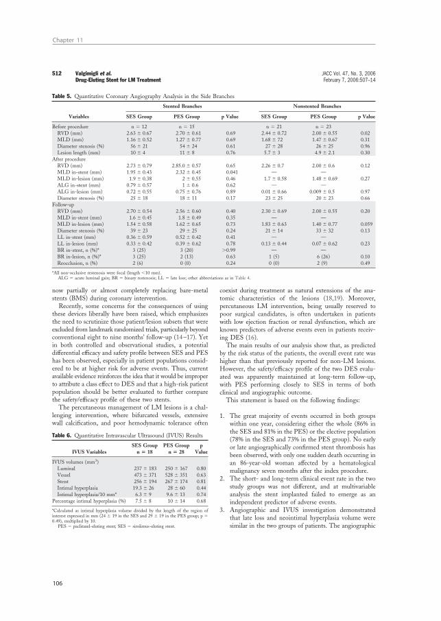
Chapter 11
106
now partially or almost completely replacing bare-metalstents (BMS) during coronary intervention.Recently, some concerns for the consequences of using
these devices liberally have been raised, which emphasizesthe need to scrutinize those patient/lesion subsets that wereexcluded from landmark randomized trials, particularly beyondconventional eight to nine months’ follow-up (14–17). Yetin both controlled and observational studies, a potentialdifferential efficacy and safety profile between SES and PEShas been observed, especially in patient populations consid-ered to be at higher risk for adverse events. Thus, currentavailable evidence reinforces the idea that it would be improperto attribute a class effect to DES and that a high-risk patientpopulation should be better evaluated to further comparethe safety/efficacy profile of these two stents.The percutaneous management of LM lesions is a chal-
lenging intervention, where bifurcated vessels, extensivewall calcification, and poor hemodynamic tolerance often
coexist during treatment as natural extensions of the ana-tomic characteristics of the lesions (18,19). Moreover,percutaneous LM intervention, being usually reserved topoor surgical candidates, is often undertaken in patientswith low ejection fraction or renal dysfunction, which areknown predictors of adverse events even in patients receiv-ing DES (16).The main results of our analysis show that, as predicted
by the risk status of the patients, the overall event rate washigher than that previously reported for non-LM lesions.However, the safety/efficacy profile of the two DES evalu-ated was apparently maintained at long-term follow-up,with PES performing closely to SES in terms of bothclinical and angiographic outcome.This statement is based on the following findings:
1. The great majority of events occurred in both groupswithin one year, considering either the whole (86% inthe SES and 81% in the PES) or the elective population(78% in the SES and 73% in the PES group). No earlyor late angiographically confirmed stent thrombosis hasbeen observed, with only one sudden death occurring inan 86-year-old woman affected by a hematologicalmalignancy seven months after the index procedure.
2. The short- and long-term clinical event rate in the twostudy groups was not different, and at multivariableanalysis the stent implanted failed to emerge as anindependent predictor of adverse events.
3. Angiographic and IVUS investigation demonstratedthat late loss and neointimal hyperplasia volume weresimilar in the two groups of patients. The angiographic
Table 5. Quantitative Coronary Angiography Analysis in the Side Branches
Stented Branches Nonstented Branches
Variables SES Group PES Group p Value SES Group PES Group p Value
Before procedure n � 12 n � 15 n � 21 n � 23RVD (mm) 2.63 � 0.67 2.70 � 0.61 0.69 2.44 � 0.72 2.00 � 0.55 0.02MLD (mm) 1.16 � 0.52 1.27 � 0.77 0.69 1.68 � 72 1.47 � 0.67 0.31Diameter stenosis (%) 56 � 21 54 � 24 0.61 27 � 28 26 � 25 0.96Lesion length (mm) 10 � 4 11 � 8 0.76 5.7 � 3 4.9 � 2.1 0.30
After procedureRVD (mm) 2.73 � 0.79 2.85.0 � 0.57 0.65 2.26 � 0.7 2.00 � 0.6 0.12MLD in-stent (mm) 1.95 � 0.43 2.32 � 0.45 0.041 — —MLD in-lesion (mm) 1.9 � 0.38 2 � 0.55 0.46 1.7 � 0.58 1.48 � 0.69 0.27ALG in-stent (mm) 0.79 � 0.57 1 � 0.6 0.62 — —ALG in-lesion (mm) 0.72 � 0.55 0.75 � 0.76 0.89 0.01 � 0.66 0.009 � 0.5 0.97Diameter stenosis (%) 25 � 18 18 � 11 0.17 23 � 25 20 � 23 0.66
Follow-upRVD (mm) 2.70 � 0.54 2.56 � 0.60 0.40 2.30 � 0.69 2.00 � 0.55 0.20MLD in-stent (mm) 1.6 � 0.45 1.8 � 0.49 0.35 — —MLD in-lesion (mm) 1.54 � 0.58 1.62 � 0.65 0.73 1.83 � 0.63 1.40 � 0.77 0.059Diameter stenosis (%) 39 � 23 29 � 25 0.24 21 � 14 33 � 32 0.13LL in-stent (mm) 0.36 � 0.59 0.52 � 0.42 0.41 — —LL in-lesion (mm) 0.33 � 0.42 0.39 � 0.62 0.78 0.13 � 0.44 0.07 � 0.62 0.23BR in-stent, n (%)* 3 (25) 3 (20) �0.99 — —BR in-lesion, n (%)* 3 (25) 2 (13) 0.63 1 (5) 6 (26) 0.10Reocclusion, n (%) 2 (6) 0 (0) 0.24 0 (0) 2 (9) 0.49
*All non-occlusive restenosis were focal (length �10 mm).ALG � acute luminal gain; BR � binary restenosis; LL � late loss; other abbreviations as in Table 4.
Table 6. Quantitative Intravascular Ultrasound (IVUS) Results
IVUS VariablesSES Group
n � 18PES Group
n � 28p
Value
IVUS volumes (mm3)Luminal 237 � 183 250 � 167 0.80Vessel 473 � 371 528 � 351 0.63Stent 256 � 194 267 � 174 0.81Intimal hyperplasia 19.3 � 26 28 � 60 0.44Intimal hyperplasia/10 mm* 6.3 � 9 9.6 � 13 0.74
Percentage intimal hyperplasia (%) 7.5 � 8 10 � 14 0.68
*Calculated as intimal hyperplasia volume divided by the length of the region ofinterest expressed in mm (24 � 19 in the SES and 29 � 19 in the PES group; p �0.49), multiplied by 10.PES � paclitaxel-eluting stent; SES � sirolimus-eluting stent.
512 Valgimigli et al. JACC Vol. 47, No. 3, 2006Drug-Eluting Stent for LM Treatment February 7, 2006:507–14
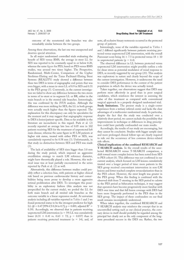
107
Angiographic outcomes after DES for LMCA
outcome of the nonstented side branches was alsoremarkably similar between the two groups.
Among these observations, the last one was unexpected anddeserves special attention.In all major randomized controlled trials evaluating the
benefit of SES versus BMS, the average in-stent LL forSES was reported to be constantly equal to or below 0.20,whereas the same figure for PES, based on PES versus BMSstudies, was around two times higher. The Prospective,Randomized, Multi-Center, Comparison of the CypherSirolimus-Eluting and the Taxus Paclitaxel-Eluting StentSystems (REALITY) study showed a difference betweenthese two DES in terms of angiographic end points that waseven bigger, with a mean in-stent LL of 0.9 in SES and 0.31in the PES group (5). Conversely, in the current investiga-tion we failed to show any difference between the two stentsin terms of in-stent or in-segment LL or BR, either in themain branch or in the stented side branches. Interestingly,this was confirmed by the IVUS analysis. Although thedifference was more striking for SES, the LL in both groupswere actually much higher than the figures expected. Theexplanation for this discrepancy can only be speculative forthe moment and it may suggest that angiographic responseto DES is lesion/patient-specific. Data so far available in theliterature are inconclusive in this regard. Park et al. (2)recently reported an average late loss of 0.05 mm in 102patients receiving SES for the treatment of unprotected leftmain disease, whereas the same figure in 85 LM patients athigher risk status, treated with either PES or SES, wascumulatively reported to be 0.58 mm (3). Unfortunately, inthat study no distinction between SES and PES was made(3).The lack of availability of SES sizes bigger than 3.0 mm
during the study period, which imposed an aggressiveoverdilation strategy to match LM reference diameter,might have theoretically played a role. However, this tech-nical issue was at least partially encountered in the seriesreported by Park et al. (2) as well.Alternatively, this difference between studies could pos-
sibly reflect a selection bias, with patients at higher clinicalrisk based on previous cardiovascular history and comor-bidities being more prone to develop a more aggressiveintimal proliferation after DES. To investigate this possi-bility in an exploratory fashion (this analysis was notprespecified for the current study), we pooled the LL forboth main branch and all stented side branches as theoutcome variable in a linear regression model. At univariateanalysis including all variables reported in Tables 1 and 2 wefound protected status to be the strongest predictor for highLL (� � 0.47 [95% CI 0.24 to 0.7]; p� 0.001; adjusted R2 �0.39). Accordingly, we observed that in patients receivingunprotected LM intervention (n� 59) LL was cumulativelylower (0.31 � 0.41 vs. 0.63 � 72; p � 0.037) than inpatients receiving protected treatment (n � 14), and, of
note, all occlusive binary restenosis occurred in the protectedgroup of patients.Interestingly, none of the variables reported in Tables 1
and 2 differed significantly between patients receiving pro-tected versus unprotected LM intervention, with the overallParsonnet score being 16 � 7.5 in protected versus 18 � 10in unprotected patients (p � 0.4).The observed difference in LL between protected versus
unprotected LM intervention might possibly outline a rolefor shear stress as potential modulator of vessel response toDES, as recently suggested by our group (20). This analysiswas exploratory in nature and clearly beyond the scope ofthe current investigation. However, it underscores the needto consider DES performance in the context of the patientpopulation in which the device was actually tested.Taken together, our observations suggest that DES may
perform more effectively in good than in poor surgicalcandidates, which reinforces the interest in assessing thevalue of this treatment as compared with conventionalsurgical approach in a properly designed randomized trial.Study limitations. The present study is a single-centerexperience from a tertiary referral center and lacks the clearadvantages of a multicenter randomized study. In particular,despite the fact that the study was conducted over arelatively short period, we cannot exclude the possibility thatimprovements in technique or differences in drug prescrip-tion could have partially confounded our main results.Accordingly, the results of our study are encouraging, but
they cannot be conclusive. Studies with bigger sample sizesand more prolonged clinical follow-ups are clearly requiredto rule out the occurrence of less common device-relatedside effects.Clinical implications of the combined RESEARCH andT-SEARCH analysis. In the overall results of the unse-lected RESEARCH versus T-SEARCH comparison, ashift toward more complex lesions has been noted from SESto PES cohort (8). This difference was not confirmed in ourcurrent analysis, which focused on LM lesions cumulativelytreated over a longer period of time: more patients in theSES group received concomitant intervention in non-LMlesions and fewer reached complete revascularization than inthe PES cohort. However, the stent length was greater inthe PES group. When this finding is combined with theobserved shift from T-stenting in the SES period to culottein the PES period as bifurcation technique, it may suggestthat operators have become progressively more familiar withDES over time and that full lesion coverage with DES hadbeen more frequently performed in the PES than in theSES group. The impact of these confounders on our finalresult remains incompletely understood.When taken together, the combined RESEARCH and
T-SEARCH analysis may reinforce the concept that in anuncontrolled setting such as our clinical practice, the coro-nary device in itself should probably be regarded among theprincipal but clearly not as the only component of the long-term procedural success in the DES era. Rather, the two
513JACC Vol. 47, No. 3, 2006 Valgimigli et al.February 7, 2006:507–14 Drug-Eluting Stent for LM Treatment
Chapter 11
108
drug-eluting stents available on the market should always beput in the context of the characteristics of the treatedpatients and the operator’s experience to better forecast theireffect on short- and long-term outcome.Conclusions. After a median follow-up of two years, nolate serious adverse events, possibly suggesting a time-dependent change in the therapeutic profile of the investi-gated devices, were observed.In a consecutive group of patients undergoing percutane-
ous LM intervention, PES may perform closely to SES interms of both clinical and angiographic outcome.A multinational multicenter randomized study is currently
ongoing to estimate the clinical value of PES-supported LMintervention with respect to conventional surgical treatment.
Reprint requests and correspondence: Prof. Patrick W. Serruys,Thoraxcenter, Bd-406, Dr Molewaterplein 40, 3015-GD Rotter-dam, the Netherlands. E-mail: [email protected].
REFERENCES
1. Valgimigli M, van Mieghem CA, Ong AT, et al. Short- andlong-term clinical outcome after drug-eluting stent implantation forthe percutaneous treatment of left main coronary artery disease:insights from the Rapamycin-Eluting and Taxus-Stent Evaluated atRotterdam Cardiology Hospital (RESEARCH and T-SEARCH)registries. Circulation 2005;111:1383–9.
2. Park SJ, Kim YH, Lee BK, et al. Sirolimus-eluting stent implantationfor unprotected left main coronary artery stenosis: comparison withbare metal stent implantation. J Am Coll Cardiol 2005;45:351–6.
3. Chieffo A, Stankovic G, Bonizzoni E, et al. Early and mid-term resultsof drug-eluting stent implantation in unprotected left main. Circula-tion 2005;111:791–5.
4. De Lezo J, Medina A, Pan M. Drug eluting stent for complex lesions:a randomized rapamycin versus paclitaxel (CORPORAL) study. J AmColl Cardiol 2005;45:75A.
5. Morice M. Eight-month outcome of the REALITY study: a prospec-tive, randomized, multicenter head-to-head comparison of the siroli-mus eluting stent (Cypher) and the paclitaxel eluting stent (Taxus).Paper presented at: 2005 Scientific Session of the American College ofCardiology; March 6, 2005; Orlando, FL.
6. Kastrati A, Mehilli J, von Beckerath N, et al. Sirolimus-eluting stentor paclitaxel-eluting stent vs balloon angioplasty for prevention of
recurrences in patients with coronary in-stent restenosis: a randomizedcontrolled trial. JAMA 2005;293:165–71.
7. Dibra A, Kastrati A, Mehilli J, et al. Paclitaxel-eluting or sirolimus-eluting stents to prevent restenosis in diabetic patients. N Engl J Med2005;353:663–70.
8. Ong AT, Serruys PW, Aoki J, et al. The unrestricted use of paclitaxel-versus sirolimus-eluting stents for coronary artery disease in an unselectedpopulation: one-year results of the Taxus-Stent Evaluated at Rotter-dam Cardiology Hospital (T-SEARCH) registry. J Am Coll Cardiol2005;45:1135–41.
9. Windecker S, Remondino A, Eberli FR, et al. Sirolimus-eluting andpaclitaxel-eluting stents for coronary revascularization. N Engl J Med2005;353:653–62.
10. Parsonnet V, Dean D, Bernstein AD. A method of uniform stratifi-cation of risk for evaluating the results of surgery in acquired adultheart disease. Circulation 1989;79:I3–12.
11. Qureshi MA, Safian RD, Grines CL, et al. Simplified scoring systemfor predicting mortality after percutaneous coronary intervention.J Am Coll Cardiol 2003;42:1890–5.
12. Roques F, Nashef SA, Michel P, et al. Risk factors and outcome inEuropean cardiac surgery: analysis of the EuroSCORE multinationaldatabase of 19030 patients. Eur J Cardiothorac Surg 1999;15:816–22;discussion 822–3.
13. Hamers R, Knook M, Roelandt J. A novel approach to quantitativeanalysis of intravascular ultrasound images. Comput Cardiol 2001;28:589–92.
14. McFadden EP, Stabile E, Regar E, et al. Late thrombosis indrug-eluting coronary stents after discontinuation of antiplatelet ther-apy. Lancet 2004;364:1519–21.
15. Waksman R, Kuchulakanti P, Kinnaird T, et al. Impact of off-labelusage on immediate and long-term outcomes in patients undergoingpercutaneous coronary intervention with sirolimus-eluting stents. Cir-culation 2004;110:2473A.
16. Iakovou I, Schmidt T, Bonizzoni E, et al. Incidence, predictors, andoutcome of thrombosis after successful implantation of drug-elutingstents. JAMA 2005;293:2126–30.
17. Alfonso F, Moreno R, Vergas J. Fatal infection after rapamycin elutingcoronary stent implantation. Heart 2005;91:e51.
18. Tan WA, Tamai H, Park SJ, et al. Long-term clinical outcomes afterunprotected left main trunk percutaneous revascularization in 279patients. Circulation 2001;104:1609–14.
19. Takagi T, Stankovic G, Finci L, et al. Results and long-termpredictors of adverse clinical events after elective percutaneous inter-ventions on unprotected left main coronary artery. Circulation 2002;106:698–702.
20. Gijsen FJ, Oortman RM, Wentzel JJ, et al. Usefulness of shear stresspattern in predicting neointima distribution in sirolimus-eluting stentsin coronary arteries. Am J Cardiol 2003;92:1325–8.
514 Valgimigli et al. JACC Vol. 47, No. 3, 2006Drug-Eluting Stent for LM Treatment February 7, 2006:507–14

Chapter 12
Signifi cant reduction in restenosis after the use of
sirolimus-eluting stents in the treatment of
chronic total occlusions
Hoye A, Tanabe K, Lemos PA, Aoki J, Saia
F, Arampatzis C, Degertekin M, Hofma SH,
Sianos G, McFadden E, van der Giessen
WJ, Smits PC, de Feyter PJ, van Domburg
RT, Serruys PW.
J Am Coll Cardiol. 2004;43:1954-1958

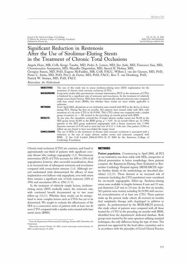
111
SES for CTO
Significant Reduction in RestenosisAfter the Use of Sirolimus-Eluting Stentsin the Treatment of Chronic Total OcclusionsAngela Hoye, MB, CHB, Kengo Tanabe, MD, Pedro A. Lemos, MD, Jiro Aoki, MD, Francesco Saia, MD,Chourmouzios Arampatzis, MD, Muzaffer Degertekin, MD, Sjoerd H. Hofma, MD,Georgios Sianos, MD, PHD, Eugene McFadden, MB, CHB, FACC, Willem J. van der Giessen, MD, PHD,Pieter C. Smits, MD, PHD, Pim J. de Feyter, MD, PHD, FACC, Ron T. van Domburg, PHD,Patrick W. Serruys, MD, PHD, FACCRotterdam, the Netherlands
OBJECTIVES The aim of this study was to assess sirolimus-eluting stent (SES) implantation for thetreatment of chronic total coronary occlusions (CTO).
BACKGROUND Long-term results after percutaneous coronary intervention (PCI) in the treatment of CTOsis hindered by a significant rate of restenosis and reocclusion. In the treatment of relativelysimple nonocclusive lesions, SESs have shown dramatically reduced restenosis rates comparedwith bare metal stents (BMS), but whether these results are more widely applicable isunknown.
METHODS From April 2002, all patients at our institution were treated with SES as the device of choiceduring PCI. During the first six months, 563 patients were treated solely with SES, withtreatment of a de novo CTO in 56 (9.9%). This CTO cohort was compared with a similargroup of patients (n � 28) treated in the preceding six-month period with BMS.
RESULTS At one year, the cumulative survival-free of major adverse cardiac events was 96.4% in theSES group versus 82.8% in the BMS group, p � 0.05. At six-month follow-up, 33 (59%)patients in the SES group underwent angiography with a binary restenosis rate (�50%diameter stenosis) of 9.1% and in-stent late loss of 0.13 � 0.46 mm. One patient (3.0%) atfollow-up was found to have reoccluded the target vessel.
CONCLUSIONS The use of SESs in the treatment of chronic total coronary occlusions is associated with areduction in the rate of major adverse cardiac events and restenosis compared withBMS. (J Am Coll Cardiol 2004;43:1954–8) © 2004 by the American College ofCardiology Foundation
Chronic total occlusions (CTO) are common, and found inapproximately one-third of patients with significant coro-nary disease who undergo angiography (1,2). Percutaneousintervention (PCI) of CTOs accounts for 10% to 15% of allangioplasties; however, after successful recanalization, thereis an increased rate of subsequent restenosis and reocclusioncompared with nonocclusive stenoses (3,4). Although sev-eral randomized trials demonstrated the efficacy of stentimplantation over balloon-only angioplasty, even with stentsthere remains a significant rate of both restenosis (32% to55%) and reocclusion (8% to 12%) (5–9).
In the treatment of relatively simple lesions, sirolimus-eluting stents (SES) markedly reduce the restenosis rate,with continued benefit documented up to two yearsfollow-up (10,11). Whether these results can be extrapo-lated to more complex lesions such as CTOs has yet to bedetermined. We sought to evaluate the effectiveness of theSES in a consecutive series of patients with at least one denovo CTO compared with a similar series treated with baremetal stents (BMS).
METHODS
Patient population. Commencing in April 2002, all PCIat our institution was done solely with SESs, irrespective ofclinical presentation or lesion morphology; these patientscomprise the Rapamycin-Eluting Stent Evaluated at Rot-terdam Cardiology Hospital registry (RESEARCH) regis-try (further details of the methodology are described else-where) (12,13). Those deemed at an increased risk ofrestenosis (including the CTO population) were consideredfor six-month angiographic follow-up. Sirolimus-elutingstents were available in lengths between 8 mm and 33 mm,and diameters 2.25 mm to 3.0 mm. In the first six months,563 patients were treated, including 56 (9.9%) with success-ful revascularization of at least one CTO. These patientsmake up the present study cohort; all received six monthsdual antiplatelet therapy with clopidogrel in addition toaspirin. As predetermined by the RESEARCH protocol,this study cohort of patients were compared with all thosetreated for a CTO in the preceding six months with BMS,identified from the departments’ dedicated database. Bothgroups were treated by the same operators utilizing standardtechniques, the only difference being the type of stent. Theprotocol was approved by the local ethics committee and isin accordance with the principles of Good Clinical Practice
From the Department of Interventional Cardiology, Erasmus MC, Rotterdam, theNetherlands.
Manuscript received October 29, 2003; revised manuscript received January 15,2004, accepted January 19, 2004.
Journal of the American College of Cardiology Vol. 43, No. 11, 2004© 2004 by the American College of Cardiology Foundation ISSN 0735-1097/04/$30.00Published by Elsevier Inc. doi:10.1016/j.jacc.2004.01.045
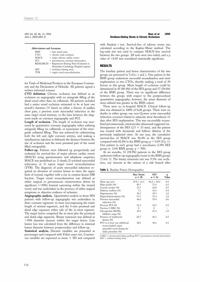
Chapter 12
112
for Trials of Medicinal Products in the European Commu-nity and the Declaration of Helsinki. All patients signed awritten informed consentCTO definition. Chronic occlusion was defined as anocclusion on angiography with no antegrade filling of thedistal vessel other than via collaterals. All patients includedhad a native vessel occlusion estimated to be at least onemonth’s duration (9) based on either a history of suddenchest pain, a previous acute myocardial infarction in thesame target vessel territory, or the time between the diag-nosis made on coronary angiography and PCI.Length of occlusion. The length of occlusion was mea-sured by quantitative coronary angiography either utilizingantegrade filling via collaterals, or assessment of the retro-grade collateral filling. This was achieved by catheterizingboth the left and right coronary arteries, and making asimultaneous injection to delineate the distance between thesite of occlusion and the most proximal part of the vesselfilled retrogradely.Follow-up. Patients were followed up prospectively andevaluated for survival-free of major adverse cardiac events(MACE) using questionnaires and telephone enquiries;MACE was predefined as: 1) death; 2) nonfatal myocardialinfarction; or 3) repeat target vessel revascularization(TVR). The diagnosis of acute myocardial infarction re-quired an elevation of creatine kinase to twice the upperlimit of normal, together with a rise in creatine kinase-MBfraction. Target vessel revascularization was defined aseither surgical or percutaneous reintervention driven bysignificant (�50%) luminal narrowing within the treatedvessel, and was undertaken in the presence of either anginalsymptoms or objective evidence of ischemia.Angiographic analysis. Quantitative analysis in those SESpatients with follow-up angiography was undertaken inthree coronary segments: in-stent (encompassing the entirelength of stented segment), and the 5-mm proximal anddistal edge segments either side of the in-stent segment.The target lesion comprised the in-stent plus the proximaland distal edge segments. Binary restenosis was defined as�50% diameter stenosis within the target lesion. Latelumen loss was calculated from the difference in minimallumen diameter between postprocedure and follow-up.Statistical analysis. Discrete variables are presented aspercentages and compared with Fisher exact test. Continu-ous variables are expressed as mean � SD and compared
with Student t test. Survival-free of adverse events wascalculated according to the Kaplan-Meier method. Thelog-rank test was used to compare MACE-free survivalbetween the two groups. All tests were two-tailed, and a pvalue of �0.05 was considered statistically significant.
RESULTS
The baseline patient and lesion characteristics of the twogroups are presented in Tables 1 and 2. One patient in theBMS group underwent successful recanalization and stentimplantation in two CTOs, thereby making a total of 29lesions in this group. Mean length of occlusion could bedetermined in 45 (80.4%) of the SES group and 17 (58.6%)of the BMS group. There was no significant differencebetween the groups with respect to the postproceduralquantitative angiography; however, the mean diameter ofstent utilized was greater in the BMS cohort.
There were no in-hospital MACE. Clinical follow-updata was obtained in 100% of both groups. There were nodeaths in either group; one non–Q-wave acute myocardialinfarction occurred related to subacute stent thrombosis 11days after SES implantation. This was successfully recana-lized percutaneously; intravascular ultrasound suggested un-derexpansion of the SES (2.5 � 33 mm), and the patientwas treated with abciximab and balloon dilation of thepreviously implanted stent. At one year, the cumulativesurvival-free of MACE was 96.4% in the SES groupcompared with 82.8% in the BMS group, p � 0.05 (Fig. 1).One patient in each group had a reocclusion (1.8% SESgroup vs. 3.6% BMS group, p � NS).
At six months, 33 (58.9%) patients in the SES groupunderwent follow-up angiography (none in the BMS group)(Table 3). The binary restenosis rate was 9.1%: one occlu-sion, one stenosis at the ostium of a side branch after
Table 1. Baseline Patient Demographics
Bare Stentsn � 28
SESn � 56
pValue
Mean age (yrs) 59.8 � 11.1 60.2 � 10.0 0.9Male gender (%) 85.7 71.4 0.2Current smoker (%) 35.7 26.8 0.5Diabetes mellitus (%) 7.1 14.3 0.4Hypertension (%) 39.3 39.3 1.0Hypercholesterolemia (%) 57.1 55.4 1.0Previous myocardial
infarction (%)46.4 55.4 0.6
Previous PCI (%) 21.4 12.5 0.3Previous CABG (%) 0 0 –Glycoprotein IIb/IIIa
inhibitor usage (%)25.0 21.4 1.0
Presence of multivesseldisease (%)
60.7 46.3 0.3
PCI in at least one additional(nonoccluded) majorepicardial vessel during theindex procedure (%)
28.6 42.6 0.2
CABG � coronary artery bypass grafting; PCI � percutaneous coronary intervention;SES � sirolimus-eluting stents.
Abbreviations and AcronymsBMS � bare metal stentCTO � chronic total occlusionMACE � major adverse cardiac eventsPCI � percutaneous coronary interventionRESEARCH � Rapamycin-Eluting Stent Evaluated at
Rotterdam Cardiology Hospital registrySES � sirolimus-eluting stentTVR � target vessel revascularization
1955JACC Vol. 43, No. 11, 2004 Hoye et al.June 2, 2004:1954–8 Sirolimus-Eluting Stents in Chronic Occlusions
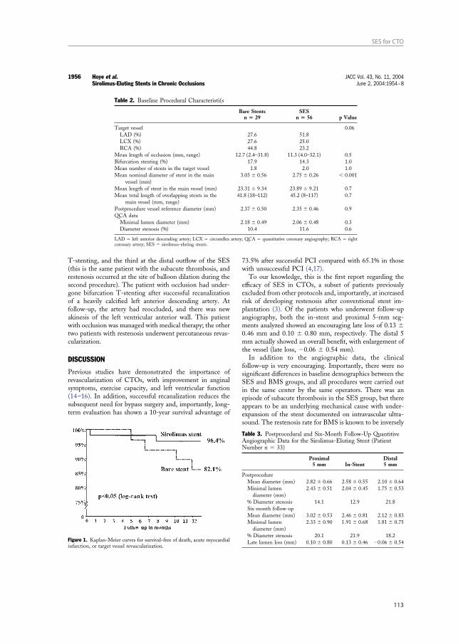
113
SES for CTO
T-stenting, and the third at the distal outflow of the SES(this is the same patient with the subacute thrombosis, andrestenosis occurred at the site of balloon dilation during thesecond procedure). The patient with occlusion had under-gone bifurcation T-stenting after successful recanalizationof a heavily calcified left anterior descending artery. Atfollow-up, the artery had reoccluded, and there was newakinesis of the left ventricular anterior wall. This patientwith occlusion was managed with medical therapy; the othertwo patients with restenosis underwent percutaneous revas-cularization.
DISCUSSION
Previous studies have demonstrated the importance ofrevascularization of CTOs, with improvement in anginalsymptoms, exercise capacity, and left ventricular function(14–16). In addition, successful recanalization reduces thesubsequent need for bypass surgery and, importantly, long-term evaluation has shown a 10-year survival advantage of
73.5% after successful PCI compared with 65.1% in thosewith unsuccessful PCI (4,17).
To our knowledge, this is the first report regarding theefficacy of SES in CTOs, a subset of patients previouslyexcluded from other protocols and, importantly, at increasedrisk of developing restenosis after conventional stent im-plantation (3). Of the patients who underwent follow-upangiography, both the in-stent and proximal 5-mm seg-ments analyzed showed an encouraging late loss of 0.13 �0.46 mm and 0.10 � 0.80 mm, respectively. The distal 5mm actually showed an overall benefit, with enlargement ofthe vessel (late loss, �0.06 � 0.54 mm).
In addition to the angiographic data, the clinicalfollow-up is very encouraging. Importantly, there were nosignificant differences in baseline demographics between theSES and BMS groups, and all procedures were carried outin the same center by the same operators. There was anepisode of subacute thrombosis in the SES group, but thereappears to be an underlying mechanical cause with under-expansion of the stent documented on intravascular ultra-sound. The restenosis rate for BMS is known to be inversely
Table 2. Baseline Procedural Characteristics
Bare Stentsn � 29
SESn � 56 p Value
Target vessel 0.06LAD (%) 27.6 51.8LCX (%) 27.6 25.0RCA (%) 44.8 23.2
Mean length of occlusion (mm, range) 12.7 (2.4–31.8) 11.3 (4.0–32.1) 0.5Bifurcation stenting (%) 17.9 14.3 1.0Mean number of stents in the target vessel 1.8 2.0 1.0Mean nominal diameter of stent in the main
vessel (mm)3.03 � 0.56 2.75 � 0.26 � 0.001
Mean length of stent in the main vessel (mm) 23.31 � 9.34 23.89 � 9.21 0.7Mean total length of overlapping stents in the
main vessel (mm, range)41.8 (18–112) 45.2 (8–117) 0.7
Postprocedure vessel reference diameter (mm) 2.37 � 0.50 2.35 � 0.46 0.9QCA data
Minimal lumen diameter (mm) 2.18 � 0.49 2.06 � 0.48 0.3Diameter stenosis (%) 10.4 11.6 0.6
LAD � left anterior descending artery; LCX � circumflex artery; QCA � quantitative coronary angiography; RCA � rightcoronary artery; SES � sirolimus-eluting stents.
Table 3. Postprocedural and Six-Month Follow-Up QuantitiveAngiographic Data for the Sirolimus-Eluting Stent (PatientNumber n � 33)
Proximal5 mm In-Stent
Distal5 mm
PostprocedureMean diameter (mm) 2.82 � 0.66 2.58 � 0.55 2.10 � 0.64Minimal lumen
diameter (mm)2.43 � 0.51 2.04 � 0.45 1.75 � 0.53
% Diameter stenosis 14.1 12.9 21.8Six-month follow-upMean diameter (mm) 3.02 � 0.53 2.46 � 0.81 2.12 � 0.83Minimal lumen
diameter (mm)2.33 � 0.90 1.91 � 0.68 1.81 � 0.75
% Diameter stenosis 20.1 21.9 18.2Late lumen loss (mm) 0.10 � 0.80 0.13 � 0.46 �0.06 � 0.54Figure 1. Kaplan-Meier curves for survival-free of death, acute myocardial
infarction, or target vessel revascularization.
1956 Hoye et al. JACC Vol. 43, No. 11, 2004Sirolimus-Eluting Stents in Chronic Occlusions June 2, 2004:1954–8

Chapter 12
114
related to the postprocedural minimal lumen diameter andthe number of stents utilized (18). In the current study,although the mean diameter of stent used was significantlygreater in the BMS cohort (related to a maximum availableSES diameter of 3.0 mm) with free utilization of postdila-tion, the postprocedural minimal lumen diameter was notsignificantly different between the two groups. The majorityof events related to TVR, with, at one year, a significantlyhigher rate of survival free of MACE of 96.4% in the SESgroup versus 82.8% in the BMS group.
Four major randomized trials have demonstrated theefficacy of stent implantation over balloon-only angioplastyin the treatment of CTOs, reducing the six-month reste-nosis rate from 68% to 74%, to 32% to 55% (5–8).Compared with this historical data, our study suggests thatthe SES confers a marked further advantage with a signif-icantly lower binary restenosis rate of 9.1% (p � 0.05) (Fig.2). In addition, we had only one patient (3.0%) with vesselreocclusion, compared with rates of between 8% to 12% inthe same published trials utilizing BMS. A recent study ofthe clinical results of 376 patients discharged from hospitalwithout an adverse event after successful intervention of aCTO showed, at one-year follow-up, a MACE rate of12.2% (19); our results are, therefore, quite remarkable, witha MACE-free survival rate of 96.4%.Study limitations. This study evaluated only a small cohortof patients, and angiographic follow-up was not obtained inall, so additional patients with silent reocclusion cannot beexcluded. However, those who did not undergo repeatangiography were all symptomatically well at follow-up. Inaddition, despite the discrepancy in follow-up angiographyrates between the two groups, which might have biased theresults towards more revascularization in the SES group, theMACE rate remained statistically significant with a bene-
ficial effect in favor of the SES. The study was notrandomized, and used a retrospective comparitive popula-tion; however, the same operators and interventional tech-niques were utilized.Conclusions. The use of SESs in the treatment of complexpatients with CTOs is associated with a reduction in therate of MACE and restenosis compared with BMS.
Reprint requests and correspondence: Dr. Patrick W. Serruys,Department of Interventional Cardiology, Thoraxcentre Bd 404,Dr Molewaterplein 40, NL-3015 GD Rotterdam, the Nether-lands. E-mail: [email protected].
REFERENCES
1. Kahn JK. Angiographic suitability for catheter revascularization oftotal coronary occlusions in patients from a community hospitalsetting. Am Heart J 1993;26:561–4.
2. Ruygrok PN, De Jaegere PP, Verploegh JJ, Van Domburg RT, DeFeyter PJ. Immediate outcome following coronary angioplasty: acontemporary single centre audit. Eur Heart J 1995;16 Suppl L:24–9.
3. Stone GW, Rutherford BD, McConahay DR, et al. Proceduraloutcome of angioplasty for total coronary artery occlusion: an analysisof 971 lesions in 905 patients. J Am Coll Cardiol 1990;15:849–56.
4. Ivanhoe RJ, Weintraub WS, Douglas JS Jr., et al. Percutaneoustransluminal coronary angioplasty of chronic total occlusions: primarysuccess, restenosis, and long-term clinical follow-up. Circulation1992;85:106–15.
5. Sirnes PA, Golf S, Myreng Y, et al. Stenting in Chronic CoronaryOcclusion (SICCO): a randomized, controlled trial of adding stentimplantation after successful angioplasty. J Am Coll Cardiol 1996;28:1444–51.
6. Rubartelli P, Niccoli L, Verna E, et al. Stent implantation versusballoon angioplasty in chronic coronary occlusions: results from theGISSOC trial (Gruppo Italiano di Studio sullo Stent nelle OcclusioniCoronariche). J Am Coll Cardiol 1998;32:90–6.
7. Lotan C, Rozenman Y, Hendler A, et al. Stents in total occlusion forrestenosis prevention: the multicentre randomized STOP study. TheIsraeli Working Group for Interventional Cardiology. Eur Heart J2000;21:1960–6.
8. Buller CE, Dzavik V, Carere RG, et al. Primary stenting versusballoon angioplasty in occluded coronary arteries: the Total OcclusionStudy of Canada (TOSCA). Circulation 1999;100:236–42.
9. Serruys PW, Hamburger JN, Koolen JJ, et al. Total occlusion trial withangioplasty by using laser guidewire: the TOTAL trial. Eur Heart J2000;21:1797–805.
10. Degertekin M, Serruys PW, Foley DP, et al. Persistent inhibition ofneointimal hyperplasia after sirolimus-eluting stent implantation:long-term (up to 2 years) clinical, angiographic, and intravascularultrasound follow-up. Circulation 2002;106:1610–3.
11. Morice MC, Serruys PW, Sousa JE, et al. A randomized comparisonof a sirolimus-eluting stent with a standard stent for coronary revas-cularization. N Engl J Med 2002;346:1773–80.
12. Lemos P, Lee C, Degertekin M, et al. Early outcome after sirolimus-eluting stent implantation in patients with acute coronary syndromes:insights from the Rapamycin-Eluting Stent Evaluated At RotterdamCardiology Hospital (RESEARCH) registry. J Am Coll Cardiol2003;41:2093–9.
13. Lemos P, Serruys PW, van Domburg RT, et al. Unrestricted utiliza-tion of sirolimus-eluting stents compared to conventional bare stentimplantation in the “real world.” The Rapamycin-Eluting StentEvaluated At Rotterdam Cardiology Hospital (RESEARCH) registry.Circulation. In Press.
14. Finci L, Meier B, Favre J, Righetti A, Rutishauser W. Long-termresults of successful and failed angioplasty for chronic total coronaryarterial occlusion. Am J Cardiol 1990;66:660–2.
15. Puma JA, Sketch MH Jr., Tcheng JE, et al. Percutaneous revascular-ization of chronic coronary occlusions: an overview. J Am Coll Cardiol1995;26:1–11.
Figure 2. The percentage binary restenosis rate (�50% diameter stenosis)and reocclusion rate of Rapamycin-Eluting Stent Evaluated at RotterdamCardiology Hospital registry (RESEARCH) compared with publisheddata from the patients treated with stent implantation in the randomizedtrials Stenting in Chronic Coronary Occlusion (SICCO) (5), GruppoItaliano di Studio sullo Stent nelle Occlusioni Coronariche (GISSOC) (6),Stents in Total Occlusion for Restenosis Prevention (STOP) (7), and theTotal Occlusion Study of Canada (TOSCA) (8). DS � diameter stenosis.*p � 0.05 compared with the results of RESEARCH; †p � 0.01 comparedwith the results of RESEARCH.
1957JACC Vol. 43, No. 11, 2004 Hoye et al.June 2, 2004:1954–8 Sirolimus-Eluting Stents in Chronic Occlusions

115
SES for CTO
16. Rambaldi R, Hamburger JN, Geleijnse ML, et al. Early recovery ofwall motion abnormalities after recanalization of chronic totallyoccluded coronary arteries: a dobutamine echocardiographic, prospec-tive, single-center experience. Am Heart J 1998;136:831–6.
17. Suero JA, Marso SP, Jones PG, et al. Procedural outcomes andlong-term survival among patients undergoing percutaneous coronaryintervention of a chronic total occlusion in native coronary arteries: a20-year experience. J Am Coll Cardiol 2001;38:409–14.
18. Kastrati A, Schomig A, Elezi S, et al. Predictive factors ofrestenosis after coronary stent placement. J Am Coll Cardiol1997;30:1428–36.
19. Olivari Z, Rubartelli P, Piscione F, et al. Immediate results andone-year clinical outcome after percutaneous coronary interventions inchronic total occlusions: data from a multicenter, prospective, oBM-Servational study (TOAST-GISE). J Am Coll Cardiol 2003;41:1672–8.
1958 Hoye et al. JACC Vol. 43, No. 11, 2004Sirolimus-Eluting Stents in Chronic Occlusions June 2, 2004:1954–8


Chapter 13
Drug-Eluting Stent Implantation for Chronic Total
Occlusions: Comparison between the Sirolimus- and
Paclitaxel-Eluting Stent
Hoye A, Ong AT, Aoki J, van Mieghem
CA, Rodriguez Granillo GA, Valgimigli M,
Sianos G, McFadden EP, van der Giessen
WJ, de Feyter PJ, van Domburg RT,
Serruys PW.
Eurointervention. 2005;2:193-197


119
SES and PES for CTO
EuroInterv.2005;1:193-197 - 193 -
Drug-eluting stent implantation for chronic total occlusions: comparison between the Sirolimus- and Paclitaxel-eluting stent
AbstractAims: Long-term results following percutaneous coronary intervention (PCI) with bare metal stents in the
treatment of chronic total occlusions (CTOs) is hindered by a significant rate of restenosis and re-occlu-
sion. Drug-eluting stents have shown dramatically reduced restenosis rates for the treatment of relatively
simple non-occlusive lesions, though there is only limited data as to the efficacy in CTO’s. We evaluated
the long-term results of the sirolimus-eluting stent (SES) and paclitaxel-eluting stent (PES) for the treatment
of CTOs.
Methods and results: From April 2002, all patients at our institution were treated with SES as the device of
choice during PCI. During the first quarter of 2003 the default strategy changed to the use of PES. Drug-
eluting stent implantation was carried out in CTOs (defined as >3 months’ duration) in 9% of de novo PCI
procedures. A total of 76 consecutive patients were treated with SES implantation, followed by a consecu-
tive series of 57 patients treated with PES implantation. These patients were compared with a similar group
of patients (n=26) treated with BMS in the 6-month period preceding April 2002.
At 400 days, the cumulative survival-free of target vessel revascularization was 80.8% in the BMS group
versus 97.4% and 96.4% in the SES and PES groups respectively (p=0.01).
Conclusions: The use of both the SES and PES in the treatment of chronic total coronary occlusions
reduces the need for target vessel revascularization compared to bare metal stents.
KEYWORDSOcclusion, stents,restenosis
Angela Hoye, MB, ChB; Andrew TL Ong, MD; Jiro Aoki, MD; Carlos AG van Mieghem, MD; Gaston A. Rodriguez Granillo, MD; Marco Valgimigli, MD; Georgios Sianos, MD, PhD; Eugene McFadden, MB, ChB, FACC; Willem J. van der Giessen, MD, PhD; Pim J. de Feyter, MD, PhD,FACC; Ron T. van Domburg, PhD; Patrick W Serruys*, MD, PhD, FACC
Department of Interventional Cardiology, Erasmus MC, Rotterdam, The Netherlands
The study was supported by unrestricted Institutional grants from Cordis (Johnson & Johnson), and Boston Scientific Corporation.
* Corresponding author: Prof. PW Serruys, Head of Interventional Cardiology, Ba 583, Thoraxcenter, Erasmus Medical Center, Dr. Molewaterplein40, 3015 GD, Rotterdam, The Netherlands E-mail: [email protected]
© Europa Edition 2005. All rights reserved.
Clinical research
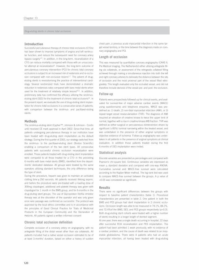
Chapter 13
120
- 194 -
Drug-eluting stents in chronic total occlusions
IntroductionSuccessful percutaneous therapy of chronic total occlusions (CTOs)
has been shown to improve symptoms of angina and left ventricu-
lar function, and reduce the subsequent need for coronary artery
bypass surgery1-5. In addition, in the long-term, recanalization of a
CTO can reduce mortality compared with those with an unsuccess-
ful attempt at recanalization6. However, the long-term outcome of
percutaneous coronary intervention (PCI) for chronic total coronary
occlusions is subject to an increased risk of restenosis and re-occlu-
sion compared with non-occlusive lesions1,7. The advent of drug-
eluting stents is revolutionising the practice of interventional cardi-
ology. Several randomized trials have demonstrated a dramatic
reduction in restenosis rates compared with bare metal stents when
used for the treatment of relatively simple lesions8-11. In addition,
preliminary data has confirmed the efficacy utilizing the sirolimus-
eluting stent (SES) for the treatment of chronic total occlusions12. In
the present report, we evaluate the use of drug-eluting stent implan-
tation for chronic total occlusions in a consecutive series of patients,
with comparison between the sirolimus- and paclitaxel-eluting
stents.
MethodsThe sirolimus-eluting stent (Cypher™, Johnson & Johnson - Cordis
unit) received CE mark approval in April 2002. Since that time, all
patients undergoing percutaneous therapy in our institution have
been treated with drug-eluting stent implantation as the default
strategy. During the first quarter of 2003, our strategy switched from
the sirolimus- to the paclitaxel-eluting stent (Boston Scientific)
enabling a comparison of the two stent types. All consecutive
patients with successful chronic occlusion recanalization were
enrolled. Those patients treated with drug-eluting stent implantation
were compared to all those treated for a CTO in the preceding
6-months with bare metal stents (BMS), identified from the depart-
ments’ dedicated database. All groups were treated by the same
operators utilizing standard techniques; the only difference being
the type of stent.
During the procedure, heparin was given to maintain an activated
clotting time ≥ 250 seconds. All patients received lifelong aspirin,
and before the procedure were pre-treated with a loading dose of
300mg clopidogrel, additional anti-platelet therapy was given with
clopidogrel for 1 month in the BMS group, and for 6-months in the
drug-eluting stent groups. The use of Glycoprotein IIb/IIIa inhibitor
therapy was at the discretion of the operator and was only given
once wire passage was confirmed as successful. The protocol was
approved by the local ethics committee and is in accordance with
the principles of Good Clinical Practice for Trials of Medicinal
Products in the European Community and the Declaration of
Helsinki. All patients signed a written informed consent
Chronic total occlusion definition
Complete occlusion of a coronary artery on angiography, with no
antegrade filling of the distal vessel other than via collaterals. All
patients included had a native vessel occlusion estimated to be of
at least 3-months’ duration, based on either a history of sudden
chest pain, a previous acute myocardial infarction in the same tar-
get vessel territory, or the time between the diagnosis made on coro-
nary angiography and PCI.
Length of occlusion
This was measured by quantitative coronary angiography (CAAS II;
Pie Medical Imaging, The Netherlands) either utilizing antegrade fill-
ing via collaterals, or assessment of the retrograde collateral filling
achieved through making a simultaneous injection into both the left
and right coronary arteries to delineate the distance between the site
of occlusion and the most proximal part of the vessel filled retro-
gradely. This length evaluated only the occluded vessel, and did not
therefore include stenosis of the vessel pre- and post- the occlusion.
Follow-up
Patients were prospectively followed-up for clinical events, and eval-
uated for survival-free of major adverse cardiac events (MACE)
using questionnaires and telephone enquiries. MACE was pre-
defined as: 1) death, 2) non-fatal myocardial infarction (AMI), or 3)
repeat target vessel revascularization (TVR). The diagnosis of AMI
required an elevation of creatine kinase to twice the upper limit of
normal, together with a rise in creatine kinase-MB fraction. TVR was
defined as either surgical or percutaneous reintervention driven by
significant (>50%) luminal narrowing within the treated vessel, and
was undertaken in the presence of either anginal symptoms or
objective evidence of ischemia. Follow-up angiography was under-
taken in all patients in the presence of anginal symptoms at clinical
evaluation; in addition those patients treated during the first
6-months of DES implantation were invited.
Statistical analysis
Discrete variables are presented as percentages and compared with
Pearson’s chi-square test. Continuous variables are expressed as
mean ± standard deviation and compared with one-way ANOVA.
Cumulative survival and MACE-free survival were calculated
according to the Kaplan-Meier method. The log-rank test was used
to compare MACE-free survival between the groups. A p value of
<0.05 was considered as significant.
ResultsThere were no significant differences between the groups with
respect to baseline patient characteristics (table 1). Procedural
characteristics are presented in table 2. One patient in both the
BMS and PES groups had stent implantation in 2 chronic occlu-
sions. Occlusion length was able to be measured in 74.1%, 84.2%,
and 72.4%of the BMS, SES, and PES groups respectively (p=0.3).
Both drug-eluting stent cohorts were treated with a higher number
of stents resulting in a longer length of stented segment.
At one year, there was a single death occurring in hospital, 22 days
after successful RCA recanalization and PES implantation. The
patient had been admitted 1 week previously, with no evidence of
a cardiac problem, and the cause of death was related to an inop-
erable glioblastoma. There were 4 patients who had an acute
myocardial infarction, all having been treated with drug-eluting
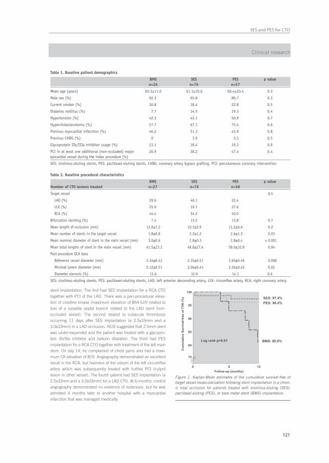
121
SES and PES for CTO
- 195 -
stent implantation. The first had SES implantation for a RCA CTO
together with PCI of the LAD. There was a peri-procedural eleva-
tion of creatine kinase (maximum elevation of 854 IU/l) related to
loss of a sizeable septal branch related to the LAD stent (non-
occluded vessel). The second related to subacute thrombosis
occurring 11 days after SES implantation (a 2.5x33mm and a
3.0x33mm) in a LAD occlusion. IVUS suggested that 2.5mm stent
was under-expanded and the patient was treated with a glycopro-
tein IIb/IIIa inhibitor and balloon dilatation. The third had PES
implantation for a RCA CTO together with treatment of the left main
stem. On day 14, he complained of chest pains and had a maxi-
mum CK elevation of 819. Angiography demonstrated an excellent
result in the RCA, but haziness of the ostium of the left circumflex
artery which was subsequently treated with further PCI (culprit
lesion in other vessel). The fourth patient had SES implantation (a
2.5x33mm and a 3.0x33mm) for a LAD CTO. At 6-months, control
angiography demonstrated no evidence of restenosis, but he was
admitted 4 months later to another hospital with a myocardial
infarction that was managed medically.
Clinical research
Table 1. Baseline patient demographics
BMS SES PES p valuen=26 n=76 n=57
Mean age (years) 60.3±11.0 61.1±10.6 58.4±10.4 0.3
Male sex (%) 92.3 65.8 80.7 0.3
Current smoker (%) 30.8 18.4 22.8 0.5
Diabetes mellitus (%) 7.7 14.5 19.3 0.4
Hypertension (%) 42.3 42.1 50.9 0.7
Hypercholesterolemia (%) 57.7 67.1 75.4 0.6
Previous myocardial infarction (%) 46.2 51.3 43.9 0.8
Previous CABG (%) 0 3.9 5.3 0.5
Glycoprotein IIb/IIIa inhibitor usage (%) 23.1 18.4 19.3 0.9
PCI in at least one additional (non-occluded) major 26.9 38.2 47.4 0.4epicardial vessel during the index procedure (%)
SES: sirolimus-eluting stents, PES: paclitaxel-eluting stents, CABG: coronary artery bypass grafting, PCI: percutaneous coronary intervention
Figure 1. Kaplan-Meier estimates of the cumulative survival-free oftarget vessel revascularization following stent implantation in a chron-ic total occlusion for patients treated with sirolimus-eluting (SES),paclitaxel-eluting (PES), or bare metal stent (BMS) implantation.
Table 2. Baseline procedural characteristics
BMS SES PES p valueNumber of CTO lesions treated n=27 n=76 n=58
Target vessel 0.5
LAD (%) 29.6 46.1 22.4
LCX (%) 25.9 19.7 27.6
RCA (%) 44.4 34.2 50.0
Bifurcation stenting (%) 7.4 13.2 13.8 0.7
Mean length of occlusion (mm) 13.0±7.2 10.3±5.9 11.2±6.6 0.2
Mean number of stents in the target vessel 1.8±0.8 2.2±1.2 2.6±1.3 0.03
Mean nominal diameter of stent in the main vessel (mm) 3.0±0.6 2.8±0.3 2.8±0.4 < 0.001
Mean total lengths of stent in the main vessel (mm) 41.5±23.3 48.8±27.4 58.0±32.8 0.04
Post-procedure QCA data
Reference vessel diameter (mm) 2.34±0.43 2.35±0.51 2.60±0.49 0.008
Minimal lumen diameter (mm) 2.12±0.51 2.04±0.43 2.26±0.42 0.02
Diameter stenosis (%) 11.6 12.9 14.1 0.6
SES: sirolimus-eluting stents, PES: paclitaxel-eluting stents, LAD: left anterior descending artery, LCX: circumflex artery, RCA: right coronary artery

Chapter 13
122
- 196 -
Drug-eluting stents in chronic total occlusions
All events in the bare stent group related to the need for target ves-
sel revascularization. At one year, the survival-free of target vessel
revascularization was significantly higher in the SES and PES
groups compared with the BMS group (97.4% and 96.4% versus
80.8% respectively, p=0.01) Figure 1.
DiscussionIn the present report we have demonstrated the efficacy of drug-
eluting stent implantation for the percutaneous treatment of chron-
ic total occlusions when compared to bare metal stents. In addition,
we have shown that both the sirolimus- and paclitaxel-eluting stent
are associated with a low rate of target vessel revascularization at
6 months.
There have been several randomized trials that have demonstrated
the efficacy of stent implantation over balloon-only angioplasty for
the percutaneous treatment of CTOs, reducing the 6-month
restenosis rate from 68-74% to 32-55%13-17. Initial randomized
studies of drug-eluting stent implantation, demonstrated efficacy in
reducing restenosis compared to conventional stent implantation,
but excluded patients with CTOs8-11. However, recent preliminary
data from our own group have shown that the efficacy of the SES is
applicable in the treatment of CTOs (defined as >1 months’ dura-
tion), with a one year cumulative survival-free of major adverse car-
diac events of 96.4%12. In the present study, we evaluate a larger
series of consecutive patients treated for a truly chronic total occlu-
sion (>3 months in duration) with drug-eluting stent implantation.
We have shown that both the SES and PES significantly reduce the
need for TVR, with a cumulative survival-free of TVR of 80.8% in the
BMS group versus 97.4% and 96.4% in the SES and PES groups
respectively (p=0.01) Figure 1.
Importantly, there were no significant differences in baseline demo-
graphics between the groups, and all procedures were carried out
in the same centre by the same operators. Restenosis following
BMS implantation is known to be inversely related to the post-pro-
cedural MLD and the number of stents utilized18. In the current
study, the mean nominal diameter of stent used was significantly
greater in the BMS cohort, related to a maximum available SES and
PES diameter of 3.0mm and 3.5mm respectively. In addition,
despite utilizing a greater number of stents, both the SES and PES
demonstrated efficacy over the BMS. Furthermore, the beneficial
effect of the SES occurred despite a smaller post-procedural MLD.
All major adverse cardiac events in the BMS group related to the
need for TVR, including 1 patient who required coronary artery
bypass surgery. Within the drug-eluting stent groups there were
5 additional non-TVR events. One patient had a subacute thrombo-
sis, but this might have been avoidable with evidence from IVUS
demonstrating a possible underlying mechanism of inadequate
stent expansion. In addition, there is good evidence in a further 3 of
these cases that the event was unrelated to treatment of the occlud-
ed vessel. One patient died of non-cardiac causes, and 2 of the
myocardial infarctions were thought to be related to intervention
carried out in another (non-occluded) vessel. The fifth patient pre-
sented with an AMI in the territory of the target vessel, 4 months
after control angiography demonstrated patent stents. Clopidogrel
medication had been stopped at the time of the follow-up
angiogram, such that the patient was on aspirin therapy alone. The
duration of dual anti-platelet therapy needed to reduce / abolish the
risk of late stent thrombosis in patients treated with DES, particular-
ly for complex disease, is still unclear. Recently, Ong et al. reported
on late (>30 days) stent thrombosis following DES implantation in a
consecutive cohort of >2000 patients, they found a low incidence of
0.35% (95% confidence limits 0.17% to 0.72%)19. Importantly,
there were no episodes in patients continuing on dual anti-platelet
therapy. However, whether there is a true benefit in continuing
clopidogrel in addition to aspirin, over and above the possible dis-
advantages, requires further large scale evaluation.
In patients with significant coronary artery disease, although a CTO
is found in at least one third, the majority are treated with either
medical therapy or are referred for coronary artery bypass surgery,
with percutaneous treatment of CTOs accounting for only 10-15%
PCI procedures20. The major limitation of PCI for CTOs is the inabil-
ity to cross the lesion with a wire, however great advancements have
been made in the manufacture of specialized wires, and there are
additionally, promising novel technologies such as the Intraluminal™
wire and Frontrunner catheter21-23. The current report has demon-
strated the efficacy of drug-eluting stent implantation in CTOs and,
together with improvements in recanalization rates, a strategy of
percutaneous therapy of CTOs will become more widely applicable.
Study limitationsThe study was not randomized, and angiographic follow-up data
was not routinely obtained in all patients, so additional events such
as silent re-occlusion cannot be excluded. However, clinical follow-
up was obtained in >99% patients (all but one patient), and assess-
ment of symptomatic status in those that did not require re-inter-
vention, showed that all were symptomatically well at follow-up. The
study was not randomized, and used a retrospective comparative
population; however the same operators and interventional tech-
niques were utilised.
References1. Ivanhoe RJ, Weintraub WS, Douglas JS, Jr., Lembo NJ, Furman M,
Gershony G, Cohen CL, King SB, 3rd. Percutaneous transluminal coro-nary angioplasty of chronic total occlusions. Primary success, restenosis,and long-term clinical follow-up. Circulation. 1992;85:106-15.
2. Warren RJ, Black AJ, Valentine PA, Manolas EG, Hunt D. Coronaryangioplasty for chronic total occlusion reduces the need for subsequentcoronary bypass surgery. Am Heart J. 1990;120:270-4.
3. Finci L, Meier B, Favre J, Righetti A, Rutishauser W. Long-termresults of successful and failed angioplasty for chronic total coronary arte-rial occlusion. Am J Cardiol. 1990;66:660-2.
4. Puma JA, Sketch MH, Jr., Tcheng JE, Harrington RA, Phillips HR,Stack RS, Califf RM. Percutaneous revascularization of chronic coronaryocclusions: an overview. J Am Coll Cardiol. 1995;26:1-11.
5. Rambaldi R, Hamburger JN, Geleijnse ML, Poldermans D,Kimman GJ, Aiazian AA, Fioretti PM, Ten Cate FJ, Roelandt JR, Serruys PW.Early recovery of wall motion abnormalities after recanalization of chronictotally occluded coronary arteries: a dobutamine echocardiographic,prospective, single-center experience. Am Heart J. 1998;136:831-6.
6. Suero JA, Marso SP, Jones PG, Laster SB, Huber KC, Giorgi LV,

123
SES and PES for CTO
- 197 -
Johnson WL, Rutherford BD. Procedural outcomes and long-term survivalamong patients undergoing percutaneous coronary intervention of achronic total occlusion in native coronary arteries: a 20-year experience.J Am Coll Cardiol. 2001;38:409-14.
7. Stone GW, Rutherford BD, McConahay DR, Johnson WL, Jr., Giorgi LV, Ligon RW, Hartzler GO. Procedural outcome of angioplasty fortotal coronary artery occlusion: an analysis of 971 lesions in 905 patients.J Am Coll Cardiol. 1990;15:849-56.
8. Stone GW, Ellis SG, Cox DA, Hermiller J, O’Shaughnessy C, Mann JT,Turco M, Caputo R, Bergin P, Greenberg J, Popma JJ, Russell ME.A polymer-based, paclitaxel-eluting stent in patients with coronary arterydisease. N Engl J Med. 2004;350:221-31.
9. Morice MC, Serruys PW, Sousa JE, Fajadet J, Ban Hayashi E, Perin M,Colombo A, Schuler G, Barragan P, Guagliumi G, Molnar F, Falotico R. A randomized comparison of a sirolimus-eluting stent with a standardstent for coronary revascularization. N Engl J Med. 2002;346:1773-80.
10. Moses JW, Leon MB, Popma JJ, Fitzgerald PJ, Holmes DR,O’Shaughnessy C, Caputo RP, Kereiakes DJ, Williams DO, Teirstein PS,Jaeger JL, Kuntz RE. Sirolimus-eluting stents versus standard stents inpatients with stenosis in a native coronary artery. N Engl J Med.2003;349:1315-23.
11. Grube E, Silber S, Hauptmann KE, Mueller R, Buellesfeld L,Gerckens U, Russell ME. TAXUS I: six- and twelve-month results from arandomized, double-blind trial on a slow-release paclitaxel-eluting stentfor de novo coronary lesions. Circulation. 2003;107:38-42.
12. Hoye A, Tanabe K, Lemos PA, Aoki J, Saia F, Arampatzis C,Degertekin M, Hofma SH, Sianos G, McFadden E, Van Der Giessen WJ,Smits PC, De Feyter PJ, Van Domburg RT, Serruys PW. Significant reduc-tion in restenosis after the use of sirolimus-eluting stents in the treatmentof chronic total occlusions. J Am Coll Cardiol. 2004;43:1954-8.
13. Sirnes PA, Golf S, Myreng Y, Molstad P, Emanuelsson H, Albertsson P,Brekke M, Mangschau A, Endresen K, Kjekshus J. Stenting in ChronicCoronary Occlusion (SICCO): a randomized, controlled trial of adding stentimplantation after successful angioplasty. J Am Coll Cardiol.1996;28:1444-51.
14. Rubartelli P, Niccoli L, Verna E, Giachero C, Zimarino M, Fontanelli A,Vassanelli C, Campolo L, Martuscelli E, Tommasini G. Stent implantationversus balloon angioplasty in chronic coronary occlusions: results fromthe GISSOC trial. Gruppo Italiano di Studio sullo Stent nelle Occlusioni
Coronariche. J Am Coll Cardiol. 1998;32:90-6.
15. Lotan C, Rozenman Y, Hendler A, Turgeman Y, Ayzenberg O, Beyar R,Krakover R, Rosenfeld T, Gotsman MS. Stents in total occlusion forrestenosis prevention. The multicentre randomized STOP study. TheIsraeli Working Group for Interventional Cardiology. Eur Heart J.2000;21:1960-6.
16. Buller CE, Dzavik V, Carere RG, Mancini GB, Barbeau G, Lazzam C,Anderson TJ, Knudtson ML, Marquis JF, Suzuki T, Cohen EA, Fox RS, TeoKK. Primary stenting versus balloon angioplasty in occluded coronaryarteries: the Total Occlusion Study of Canada (TOSCA). Circulation.1999;100:236-42.
17. Hoher M, Wohrle J, Grebe OC, Kochs M, Osterhues HH, Hombach V,Buchwald AB. A randomized trial of elective stenting after balloon recanaliza-tion of chronic total occlusions. J Am Coll Cardiol. 1999;34:722-9.
18. Kastrati A, Schomig A, Elezi S, Schuhlen H, Dirschinger J,Hadamitzky M, Wehinger A, Hausleiter J, Walter H, Neumann FJ.Predictive factors of restenosis after coronary stent placement. J Am CollCardiol. 1997;30:1428-36.
19. Ong AT, McFadden EP, Regar E, de Jaegere PP, van Domburg RT,Serruys PW. Late angiographic stent thrombosis (LAST) events with drug-eluting stents. J Am Coll Cardiol. 2005;45:2088-92.
20. Kahn JK. Angiographic suitability for catheter revascularization oftotal coronary occlusions in patients from a community hospital setting.Am Heart J. 1993;126:561-4.
21. Orlic D CA, Stankovic G, Sangiori G, Airoldi F, Corvaja N,Montorfano M, Michev I, Vitrella G, Colombo A. Preliminary experiencewith the Frontrunner coronary catheter, novel device dedicated tomechanical revascularization of chronic total occlusions. J Am CollCardiol. 2004;43:56A.
22. Ng W, Chen WH, Lee PY, Lau CP. Initial experience and safety inthe treatment of chronic total coronary occlusions with a new opticalcoherent reflectometry-guided radiofrequency ablation guidewire. Am JCardiol. 2003;92:732-4.
23. Hoye A, Onderwater, E. Cummins, P. Sianos, G. Serruys, PW.Improved Recanalization of Chronic Total Coronary Occlusions Using anOptical Coherence Reflectometry-Guided Guidewire. Cathet CardiovascInterv. 2004;63:158-63.
Clinical research


Chapter 14
Restenosis rates following bifurcation stenting with
sirolimus-eluting stents for de novo narrowings
Tanabe K, Hoye A, Lemos PA, Aoki J,
Arampatzis CA, Saia F, Lee CH, Degertekin
M, Hofma SH, Sianos G, McFadden E,
Smits PC, van der Giessen WJ, de Feyter P,
van Domburg RT, Serruys PW.
Am J Cardiol. 2004;94:115-118


127
SES for bifurcation lesions
Restenosis Rates Following Bifurcation Stenting WithSirolimus-Eluting Stents for De Novo Narrowings
Kengo Tanabe, MD, Angela Hoye, MB ChB, Pedro A. Lemos, MD, Jiro Aoki, MD,Chourmouzios A. Arampatzis, MD, Francesco Saia, MD, Chi-hang Lee, MBBS,Muzzafer Degertekin, MD, Sjoerd H. Hofma, MD, Georgios Sianos, MD, PhD,
Eugene McFadden, MB ChB, Pieter C. Smits, MD, PhD,Willem J. van der Giessen, MD, PhD, Pim de Feyter, MD, PhD, Ron T. van Domburg, PhD,
and Patrick W. Serruys, MD, PhD
The percutaneous treatment of coronary bifurcationstenoses is hampered by an increased rate of subse-quent restenosis. The present study reports on theoutcomes of a consecutive series of 58 patients with65 de novo bifurcation stenoses treated with siroli-mus-eluting stent implantation in both the main vesseland side branch. At 6 months, the incidence of majoradverse cardiac events was 10.3% (1 death and 5target lesion revascularizations) with no episodes ofacute myocardial infarction or stent thrombosis. �2004 by Excerpta Medica, Inc.
(Am J Cardiol 2004;91:115–118)
Percutaneous coronary intervention of bifurcationlesions is associated with lower procedural suc-
cess rates1 and an increased subsequent rate of majoradverse cardiac events (MACEs) and restenosis. Var-ious techniques and strategies have been applied in anattempt to improve outcomes, including kissing bal-loon dilatation and the use of stent implantation inboth branches.2 The use of adjunctive atherectomywas found to be disadvantageous in the CoronaryAngioplasty Versus Excisional Atherectomy Trial(CAVEAT-I) trial.3 Although there was an improvedinitial angiographic result with less residual stenosis,this was at the expense of a higher rate of side branchocclusion and acute myocardial infarction. In thelong-term, results of angioplasty in bifurcations havebeen hampered by problems of restenosis, particularlyafter stent implantation within the side branch.4,5 Re-cently, sirolimus-eluting stents (SESs) have demon-strated dramatically reduced restenosis rates in pa-tients with relatively simple lesions.6,7 We sought toinvestigate the safety and efficacy of SESs in a con-secutive series of unselected patients with de novobifurcation lesions enrolled in the Rapamycin-ElutingStent Evaluation At Rotterdam Cardiology Hospital(RESEARCH) registry.8
• • •Since April 2002, SES implantation (Cypher, John-
son & Johnson–Cordis, Miami, Florida) has been usedas the default strategy for all patients treated in ourinstitution, as part of the RESEARCH registry.8Briefly, this single-center registry aims to evaluate theefficacy of SES implantation in the “real world” ofinterventional cardiology. All consecutive patientswere enrolled, irrespective of clinical presentation andlesion characteristics, and the incidence of MACEswas prospectively evaluated during follow-up. At 6months, a total of 563 consecutive patients weretreated solely with SESs. Of these, 58 patients(10.3%) with de novo bifurcation lesions were treatedwith SES implantation in both the main and sidebranches; these patients comprise the present studypopulation. The patients’ informed written consentwas obtained in accordance with the rules of theinstitutional ethics committee, which approved thestudy.
All procedures were performed with standard in-terventional techniques, except with the use of theSES as the device of choice. The strategy of bifurca-tion stenting employed and the use of kissing balloondilatation after procedure was at the operators’ discre-tion. One of 4 methods of stenting was used: T-stenting, culotte stenting, kissing stents, or the “crush”technique. T-stenting and culotte stenting have beenpreviously described.5,9 Kissing stents involved simul-taneous implantation of the stents within bothbranches, with the proximal edges alongside eachother, thereby bringing forward the point of diver-gence. The crush technique involves positioning bothstents, with the proximal part of the side branch stentlying well within the main vessel, while ensuring thatthe edge of the stent in the main vessel is moreproximal than the side branch stent. The side branchstent is deployed first, and the balloon and wire arecarefully withdrawn. The main vessel stent is thendeployed, thereby crushing the proximal part of theside branch stent.10 SESs were available in diametersfrom 2.25 to 3.00 mm and lengths from 8 to 33 mm.During the procedure, intravenous heparin was givento maintain an activated clotting time of �250 sec-onds. All patients were prescribed lifelong aspirin andclopidogrel for 6 months. The use of glycoproteinIIb/IIIa inhibitors was at the discretion of the operator.
Clinical and angiographic follow-up was per-formed at 6 months. MACEs were predefined asdeath, myocardial infarction, or target lesion revascu-
From the Thoraxcenter, Erasmus Medical Center, Rotterdam, The Neth-erlands. This study was supported by a grant from Cordis Corporation,a Johnson & Johnson Company. Dr. Serruys’ address is: Thoraxcenter,Bd 406, Erasmus MC, Dr Molewaterplein 40, 3015 GD Rotterdam,The Netherlands. E-mail: [email protected]. Manuscriptreceived December 2, 2003; revised manuscript received and ac-cepted March 19, 2004.
115©2004 by Excerpta Medica, Inc. All rights reserved. 0002-9149/04/$–see front matterThe American Journal of Cardiology Vol. 91 July 1, 2004 doi:10.1016/j.amjcard.2004.03.040
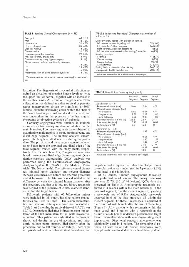
Chapter 14
128
larization. The diagnosis of myocardial infarction re-quired an elevation of creatine kinase levels to twicethe upper limit of normal, together with an increase inthe creatine kinase-MB fraction. Target lesion revas-cularization was defined as either surgical or percuta-neous reintervention driven by significant (�50%)luminal diameter narrowing either within the stent orthe 5-mm borders proximal and distal to the stent, andwas undertaken in the presence of either anginalsymptoms or objective evidence of ischemia.
Coronary angiograms were obtained in multipleviews after intracoronary injection of nitrates. For themain branches, 3 coronary segments were subjected toquantitative angiography: in-stent, proximal edge, anddistal edge segment. The in-stent analysis encom-passed the length of all stents used during the proce-dure. The proximal and distal edge segment includedup to 5 mm from the proximal and distal edge of thetotal segment treated with the study stents, respec-tively. For the side branches, 2 segments were ana-lyzed: in-stent and distal edge 5-mm segment. Quan-titative coronary angiographic (QCA) analysis wasperformed using the Cardiovascular AngiographyAnalysis System II (CAAS II; Pie Medical, Maas-tricht, The Netherlands). The reference vessel diame-ter, minimal lumen diameter, and percent diameterstenosis were measured before and after the procedureand at follow-up. The late loss was calculated as thedifference between the minimal lumen diameter afterthe procedure and that at follow-up. Binary restenosiswas defined as the presence of �50% diameter steno-sis within the target lesion.
Fifty-eight patients with 65 bifurcation lesionswere included in this study. Baseline patient charac-teristics are listed in Table 1. The lesion characteris-tics and stenting technique utilized are presented inTable 2. At 6 months, the survival-free of MACEs was89.7%. One patient died after bifurcation stent implan-tation of the left main stem for an acute myocardialinfarction. This patient was admitted in cardiogenicshock, and despite the use of abciximab and intra-aortic balloon pump support, died shortly after theprocedure due to left ventricular failure. There wereno episodes of acute or subacute stent thrombosis, and
no patient had a myocardial infarction. Target lesionrevascularization was undertaken in 5 patients (8.6%)as outlined in the following.
Of 65 lesions, 6-month angiographic follow-upwas performed in 44 lesions. The binary restenosisrate was 22.7% (10 of 44 lesions). QCA data arepresented in Table 3. Angiographic restenosis oc-curred in 4 lesions within the main branch (1 in theproximal segment; 3 in the in-stent segment), yieldinga restenosis rate of 9.1%. Angiographic restenosisoccurred in 6 of the side branches, all within thein-stent segment. Of these 6 restenoses, 5 occurred atthe ostium of side branch after the use of T-stenting(Figure 1). All 4 patients with a restenosis within themain vessel and 1 patient with a restenosis at theostium of a side branch underwent percutaneous targetlesion revascularization with new drug-eluting stentimplantation. Directional coronary atherectomy wasadditionally used in 1 patient. The remaining 5 pa-tients, all with ostial side branch restenoses, wereasymptomatic and treated with medical therapy alone.
TABLE 3 Quantitative Coronary Angiography
ProximalSegment
In-stentSegment
DistalSegment
Main branch (n � 44)Reference diameter (mm) N/A 2.64 N/AMinimal lumen diameter (mm)
Preprocedure N/A 0.64 N/APostprocedure 2.39 2.19 1.866-mo follow-up 2.26 2.07 1.85
Diameter stenosis at 6 mo (%) 28.3 22.9 25.4Late lumen loss (mm) 0.12 0.12 0.01Restenosis rate (%) 2.3 6.8 0
Side branch (n � 44)Reference diameter (mm) 1.99 N/AMinimal lumen diameter (mm)
Preprocedure 0.61 N/APostprocedure 1.80 1.576-mo follow-up 1.49 1.47
Diameter stenosis at 6 mo (%) 31.0 21.9Late lumen loss (mm) 0.31 0.09Restenosis rate (%) 13.6 0
Values are presented as mean values or relative percentages.
TABLE 1 Baseline Clinical Characteristics (n � 58)
Age (yrs) 63 � 10Men 42 (72%)Hypertension 26 (45%)Hypercholesterolemia 35 (60%)Diabetes mellitus 16 (28%)Current smoker 16 (28%)Previous myocardial infarction 22 (38%)Previous coronary angioplasty 5 (9%)Previous coronary artery bypass surgery 3 (5%)No. of coronary arteries significantly narrowed
1 15 (26%)2 28 (48%)3 15 (26%)
Presentation with an acute coronary syndrome 18 (31%)
Values are presented as the numbers (relative percentages) or mean value �
SD.
TABLE 2 Lesion and Procedural Characteristics (number oflesions � 65)
Coronary artery treated with bifurcation stentingLeft anterior descending/diagonal 39 (60%)Left circumflex/obtuse marginal 16 (25%)Right coronary/posterior descending 4 (6%)Left main stem—left anterior descending/circumflex 6 (9%)
Stenting techniqueT-stenting 41 (63%)Culotte stenting 5 (8%)Kissing stenting 2 (3%)Crush stenting 17 (26%)
Kissing balloon dilatation after stenting 20 (31%)Glycoprotein IIb/IIIa inhibitor use 20 (31%)
Values are presented as the numbers (relative percentages).
116 THE AMERICAN JOURNAL OF CARDIOLOGY� VOL. 91 JULY 1, 2004

129
SES for bifurcation lesions
• • •The major findings of this study of bifurcation
stenting include the following. (1) SES implantationin both the main and side branches is feasible andassociated with a low procedural complication rateand no episodes of stent thrombosis. (2) The targetlesion revascularization rate of 8.6% is seeminglydiminished compared with historical controls. (3) An-giographic restenosis rates of the main and sidebranches are 9.1% and 13.6%, respectively, with anoverall restenosis rate of 22.7%. (4) Five of the 6restenoses occurring in the side branch were located atthe ostium after using the T-stenting technique.
Drug-eluting stent deployment in both vessels totreat bifurcation lesions may raise theoretical concernsthat it could result in a propensity to stent thrombosis.When we treat bifurcation lesions with SESs using theculotte, kissing, or crush stenting techniques, there aresome overlapping stent struts, where the higher con-centration of sirolimus may induce endothelial func-tion impairment and thus be associated with an in-creased rate of stent thrombosis. Although thesestenting techniques were applied in 37% of the lesionstreated, no stent thrombosis was reported during fol-low-up, implying that sirolimus has a wide safetymargin.
Several strategies have been advocated to treatbifurcation lesions with percutaneous coronary inter-vention, such as deployment of stents in both vessels,stenting in 1 branch with balloon angioplasty in theother, and mechanical debulking. The published re-ports regarding the subsequent need for target lesionrevascularization utilizing bare stents range from 17%to 53%5,11,12; thus, the rate of 8.6% in our study isvery favorable. In addition, the rate observed in thepresent study may underestimate the true beneficialtreatment effect of SES as explained in the following.
Five of the 6 restenoses in the side branch occurredat the ostium after T-stenting. When we apply T-stenting, stent positioning must be extremely accurate
to ensure complete coverage of the side branch os-tium. This is particularly difficult and/or impossible toachieve when the angle between the 2 branches ismuch �90°. Restenosis at this site may therefore bemainly a reflection of incomplete coverage. The reste-nosis rate in the side branch following T-stenting was16.7% (5 of 30 lesions), whereas that following theother stent techniques was 7.1% (1 of 14 lesions). Thepresent study is limited because the choice of strategywas nonrandomized, and there is no comparison withalternative strategies, such as the use of stent implan-tation in the main vessel alone, with balloon-onlyangioplasty of the side branch. In addition, the samplesize was relatively small, and any difference betweenthe different techniques was not statistically signifi-cant. However, our results suggest that it seems wiseto ensure the complete coverage of the ostium withSESs using stenting techniques other than T-stenting.The crush technique is technically easier and quickerto do than a culotte, but further data with longerfollow-up from a larger population are needed to fullydetermine the efficacy of these techniques.
1. Al Suwaidi J, Yeh W, Cohen HA, Detre KM, Williams DO, Holmes DR Jr.Immediate and one-year outcome in patients with coronary bifurcation lesions inthe modern era (NHLBI dynamic registry). Am J Cardiol 2001;87:1139–1144.2. Lefevre T, Louvard Y, Morice MC, Loubeyre C, Piechaud JF, Dumas P.Stenting of bifurcation lesions: a rational approach. J Interv Cardiol 2001;14:573–585.3. Brener SJ, Leya FS, Apperson-Hansen C, Cowley MJ, Califf RM, Topol EJ.A comparison of debulking versus dilatation of bifurcation coronary arterialnarrowings (from the CAVEAT I Trial). Coronary Angioplasty Versus ExcisionalAtherectomy Trial-I. Am J Cardiol 1996;78:1039–1041.4. Anzuini A, Briguori C, Rosanio S, Tocchi M, Pagnotta P, Bonnier H, GimelliG, Airoldi F, Margonato A, Legrand V, Colombo A. Immediate and long-termclinical and angiographic results from Wiktor stent treatment for true bifurcationnarrowings. Am J Cardiol 2001;88:1246–1250.5. Pan M, Suarez de Lezo J, Medina A, Romero M, Hernandez E, Segura J,Castroviejo JR, Pavlovic D, Melian F, Ramirez A, Castillo JC. Simple andcomplex stent strategies for bifurcated coronary arterial stenosis involving theside branch origin. Am J Cardiol 1999;83:1320–1325.6. Degertekin M, Serruys PW, Foley DP, Tanabe K, Regar E, Vos J, Smits PC,van der Giessen WJ, van den Brand M, de Feyter P, Popma JJ. Persistent
FIGURE 1. A 3.0 � 33 mmSES was implanted in thecircumflex artery, and a2.25 � 8 mm SES wasimplanted in the sidebranch (obtuse marginal)with T-stenting technique(A). At 6-month angio-graphic follow-up, resteno-sis occurred at the ostiumof the side branch (arrow-head) (B).
BRIEF REPORTS 117

Chapter 14
130
inhibition of neointimal hyperplasia after sirolimus-eluting stent implantation:long-term (up to 2 years) clinical, angiographic, and intravascular ultrasoundfollow-up. Circulation 2002;106:1610–1613.7. Morice MC, Serruys PW, Sousa JE, Fajadet J, Ban Hayashi E, Perin M,Colombo A, Schuler G, Barragan P, Guagliumi G, Molnar F, Falotico R. Arandomized comparison of a sirolimus-eluting stent with a standard stent forcoronary revascularization. N Engl J Med 2002;346:1773–1780.8. Lemos PA, Lee CH, Degertekin M, Saia F, Tanabe K, Arampatzis CA, HoyeA, van Duuren M, Sianos G, Smits PC, et al. Early outcome after sirolimus-eluting stent implantation in patients with acute coronary syndromes: insightsfrom the Rapamycin-Eluting Stent Evaluated At Rotterdam Cardiology Hospital(RESEARCH) registry. J Am Coll Cardiol 2003;41:2093–2099.9. Chevalier B, Glatt B, Royer T, Guyon P. Placement of coronary stents in
bifurcation lesions by the “culotte” technique. Am J Cardiol 1998;82:943–949.10. Colombo A, Stankovic G, Orlic D, Corvaja N, Liistro F, Airoldi F, ChieffoA, Spanos V, Montorfano M, Di Mario C. Modified T-stenting technique withcrushing for bifurcation lesions: immediate results and 30-day outcome. CatheterCardiovasc Interv 2003;60:145–151.11. Al Suwaidi J, Berger PB, Rihal CS, Garratt KN, Bell MR, Ting HH,Bresnahan JF, Grill DE, Holmes DR Jr. Immediate and long-term outcome ofintracoronary stent implantation for true bifurcation lesions. J Am Coll Cardiol2000;35:929–936.12. Yamashita T, Nishida T, Adamian MG, Briguori C, Vaghetti M, Corvaja N,Albiero R, Finci L, Di Mario C, Tobis JM, Colombo A. Bifurcation lesions: twostents versus one stent–immediate and follow-up results. J Am Coll Cardiol 2000;35:1145–1151.
Outcome of Percutaneous Coronary InterventionVersus Coronary Bypass Grafting for Patients With LowLeft Ventricular Ejection Fractions, Unstable AnginaPectoris, and Risk Factors for Adverse Outcomes WithBypass (the AWESOME Randomized Trial and Registry)
Steven P. Sedlis, MD, Kodangudi B. Ramanathan, MD, Douglass A. Morrison, MD,Gulshan Sethi, MD, Jerome Sacks, PhD, and William Henderson, PhD, for the
Investigators of the Department of Veterans Affairs Cooperative Study #385, theAngina With Extremely Serious Operative Mortality Evaluation (AWESOME)
The recently concluded Angina With Extremely Seri-ous Operative Mortality Evaluation (AWESOME) wasa randomized clinical trial of percutaneous coronaryintervention versus coronary bypass graft surgeryamong patients with medically refractory ischemiawho were at high risk for coronary bypass graftsurgery because of >1 risk factors that included se-verely reduced left ventricular (LV) function, definedas LV ejection fraction <35%. This study reports theoutcome of patients with LV ejection fraction<35% inthe randomized clinical trial and the physician-di-rected and patient choice registries of the AWESOMEstudy. �2004 by Excerpta Medica, Inc.
(Am J Cardiol 2004;94:118–120)
Angina With Extremely Serious Operative Mortal-ity Evaluation (AWESOME) was a nationwide,
prospective, randomized, Department of Veterans Af-fairs clinical trial designed to compare the long-termsurvival with coronary bypass graft surgery (CABG)
versus percutaneous coronary intervention (PCI) forpatients with medically refractory myocardial isch-emia and increased risk of adverse outcomes withCABG. Patients were enrolled only if they had �1 of5 risk factors for adverse outcomes with CABG (priorCABG; myocardial infarction within 7 days; left ven-tricular ejection fraction [LVEF] �35%; age �70years; or intra-aortic balloon required to stabilize).Eligible patients who were deemed by their physiciansand by study investigators to be suitable for CABGand PCI were asked to participate in the randomizedtrial. Eligible patients who were acceptable for eitherCABG or PCI but who refused randomization wereentered into a prospective patient choice registry. El-igible patients who were unacceptable for eitherCABG or PCI were entered into a physician-directedregistry. The study protocol and the 3-year survivaland quality of life of patients enrolled in the random-ized study and registry have been previously report-ed.1–3 EF was reported by the investigator at each siteand estimated from contrast ventriculography or echo-cardiography or calculated by radionuclide ventricu-lography. Patients with EFs �35% were a prespeci-fied subgroup for analysis.
• • •Differences in baseline variable frequencies were
tested by chi-square tests for proportions. Long-termsurvival was measured by Kaplan-Meier survival es-timates, which were plotted. The statistical signifi-cance of global differences between survival curveswas judged by a log-rank test.
There were 2,431 patients enrolled in the AWE-SOME study between 1995 and 2000, including 446patients with EFs �35% (94 randomized and 352
From the New York VA Medical Center and New York UniversitySchool of Medicine, New York, New York; Memphis VA MedicalCenter, Memphis, Tennessee; Tucson VA Medical Center and Univer-sity of Arizona, Tucson, Arizona; Veterans Affairs Cooperative StudiesProgram Coordinating Center, Hines, Illinois; and University of Colo-rado Health Sciences Center, Denver, Colorado. This study wasfunded by the Cooperative Studies Program of the United StatesDepartment of Veterans Affairs Research and Development Service,Washington, DC. Dr. Sedlis’ address is: Section of Cardiology,12W, VA Medical Center, 423 East 23rd Street, New York, NewYork 10010. E-mail: [email protected]. Manuscript receivedNovember 26, 2003; revised manuscript received and acceptedMarch 15, 2004.
118 ©2004 by Excerpta Medica, Inc. All rights reserved. 0002-9149/04/$–see front matterThe American Journal of Cardiology Vol. 94 July 1, 2004 doi:10.1016/j.amjcard.2004.03.041

Chapter 15
Treatment of De novo bifurcation lesions; Comparison of
Sirolimus- and Paclitaxel-eluting stents
Hoye A, van Mieghem C, Ong ATL, Aoki
J,Rodriguez Granillo GA, Valgimigli M,
Tsuchida K, Sianos G, McFadden EP, van
der Giessen WJ, de Feyter P, van Domburg
R, Serruys PW.
Eurointervention. 2005;1:24-30

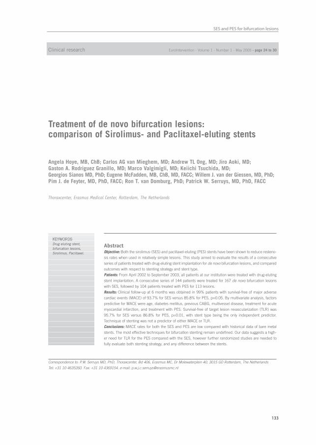
133
SES and PES for bifurcation lesions
- 24 -
EuroIntervention - Volume 1 - Number 1 - May 2005 - page 24 to 30
Treatment of de novo bifurcation lesions: comparison of Sirolimus- and Paclitaxel-eluting stents
AbstractObjective: Both the sirolimus-(SES) and paclitaxel-eluting (PES) stents have been shown to reduce resteno-
sis rates when used in relatively simple lesions. This study aimed to evaluate the results of a consecutive
series of patients treated with drug-eluting stent implantation for de novo bifurcation lesions, and compared
outcomes with respect to stenting strategy and stent type.
Patients: From April 2002 to September 2003, all patients at our institution were treated with drug-eluting
stent implantation. A consecutive series of 144 patients were treated for 167 de novo bifurcation lesions
with SES, followed by 104 patients treated with PES for 113 lesions.
Results: Clinical follow-up at 6 months was obtained in 99% patients with survival-free of major adverse
cardiac events (MACE) of 93.7% for SES versus 85.8% for PES, p=0.05. By multivariate analysis, factors
predictive for MACE were age, diabetes mellitus, previous CABG, multivessel disease, treatment for acute
myocardial infarction, and treatment with PES. Survival-free of target lesion revascularization (TLR) was
95.7% for SES versus 86.8% for PES, p=0.01, with stent type being the only independent predictor.
Technique of stenting was not a predictor of either MACE or TLR.
Conclusions: MACE rates for both the SES and PES are low compared with historical data of bare metal
stents. The most effective techniques for bifurcation stenting remain undefined. Our data suggests a high-
er need for TLR for the PES compared with the SES, however further randomized studies are needed to
fully evaluate both stenting strategy, and any difference between the stents.
KEYWORDSDrug eluting stent,bifurcation lesions,Sirolimus, Paclitaxel.
Angela Hoye, MB, ChB; Carlos AG van Mieghem, MD; Andrew TL Ong, MD; Jiro Aoki, MD;Gaston A. Rodriguez Granillo, MD; Marco Valgimigli, MD; Keiichi Tsuchida, MD; Georgios Sianos MD, PhD; Eugene McFadden, MB, ChB, MD, FACC; Willem J. van der Giessen, MD, PhD;Pim J. de Feyter, MD, PhD, FACC; Ron T. van Domburg, PhD; Patrick W. Serruys, MD, PhD, FACC
Thoraxcenter, Erasmus Medical Center, Rotterdam, The Netherlands
Correspondence to: P.W. Serruys MD, PhD, Thoraxcenter, Bd 406, Erasmus MC, Dr Molewaterplein 40, 3015 GD Rotterdam, The Netherlands
Tel: +31 10 4635260. Fax: +31 10 4369154. e-mail: [email protected]
This study was supported by institutional grants from the Cordis Corporation (Johnson & Johnson) and Boston Scientific Corp.
Clinical research
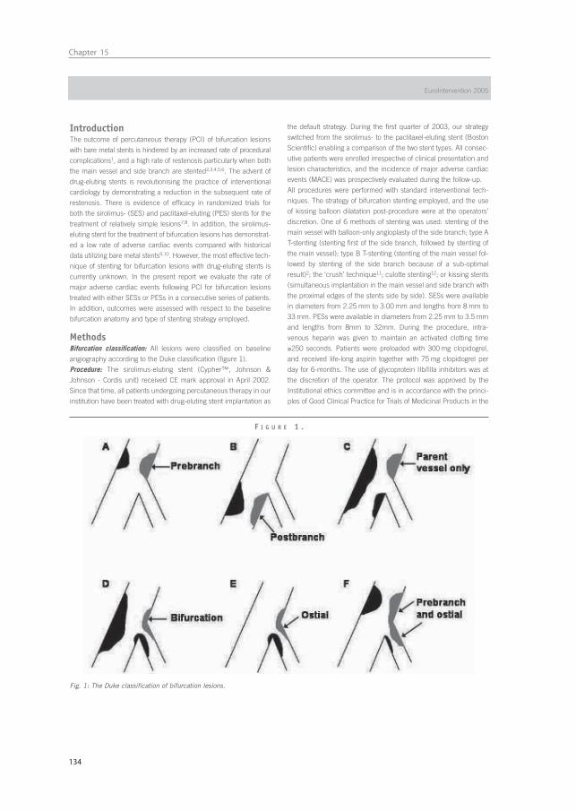
Chapter 15
134
- 25 -
IntroductionThe outcome of percutaneous therapy (PCI) of bifurcation lesions
with bare metal stents is hindered by an increased rate of procedural
complications1, and a high rate of restenosis particularly when both
the main vessel and side branch are stented2,3,4,5,6. The advent of
drug-eluting stents is revolutionising the practice of interventional
cardiology by demonstrating a reduction in the subsequent rate of
restenosis. There is evidence of efficacy in randomized trials for
both the sirolimus- (SES) and paclitaxel-eluting (PES) stents for the
treatment of relatively simple lesions7,8. In addition, the sirolimus-
eluting stent for the treatment of bifurcation lesions has demonstrat-
ed a low rate of adverse cardiac events compared with historical
data utilizing bare metal stents9,10. However, the most effective tech-
nique of stenting for bifurcation lesions with drug-eluting stents is
currently unknown. In the present report we evaluate the rate of
major adverse cardiac events following PCI for bifurcation lesions
treated with either SESs or PESs in a consecutive series of patients.
In addition, outcomes were assessed with respect to the baseline
bifurcation anatomy and type of stenting strategy employed.
MethodsBifurcation classification: All lesions were classified on baseline
angiography according to the Duke classification (figure 1).
Procedure: The sirolimus-eluting stent (Cypher™, Johnson &
Johnson - Cordis unit) received CE mark approval in April 2002.
Since that time, all patients undergoing percutaneous therapy in our
institution have been treated with drug-eluting stent implantation as
the default strategy. During the first quarter of 2003, our strategy
switched from the sirolimus- to the paclitaxel-eluting stent (Boston
Scientific) enabling a comparison of the two stent types. All consec-
utive patients were enrolled irrespective of clinical presentation and
lesion characteristics, and the incidence of major adverse cardiac
events (MACE) was prospectively evaluated during the follow-up.
All procedures were performed with standard interventional tech-
niques. The strategy of bifurcation stenting employed, and the use
of kissing balloon dilatation post-procedure were at the operators’
discretion. One of 6 methods of stenting was used: stenting of the
main vessel with balloon-only angioplasty of the side branch; type A
T-stenting (stenting first of the side branch, followed by stenting of
the main vessel); type B T-stenting (stenting of the main vessel fol-
lowed by stenting of the side branch because of a sub-optimal
result)2; the ‘crush’ technique11; culotte stenting12; or kissing stents
(simultaneous implantation in the main vessel and side branch with
the proximal edges of the stents side by side). SESs were available
in diameters from 2.25 mm to 3.00 mm and lengths from 8 mm to
33 mm. PESs were available in diameters from 2.25 mm to 3.5 mm
and lengths from 8mm to 32mm. During the procedure, intra-
venous heparin was given to maintain an activated clotting time
≥250 seconds. Patients were preloaded with 300 mg clopidogrel,
and received life-long aspirin together with 75 mg clopidogrel per
day for 6-months. The use of glycoprotein IIb/IIIa inhibitors was at
the discretion of the operator. The protocol was approved by the
Institutional ethics committee and is in accordance with the princi-
ples of Good Clinical Practice for Trials of Medicinal Products in the
F I G U R E 1 .
Fig. 1: The Duke classification of bifurcation lesions.
EuroIntervention 2005

135
SES and PES for bifurcation lesions
- 26 -
Treatment of de novo bifurcation lesions: comparison of Sirolimus- and Paclitaxel-eluting stents
European Community and the Declaration of Helsinki. All patients
signed a written informed consent
Follow-up: Clinical follow-up was obtained using telephone calls and
questionnaires, and evaluated the rate of major adverse cardiac
events (MACE) which were pre-defined as death, acute myocardial
infarction (AMI), or target vessel revascularization (TVR). The diag-
nosis of AMI required an elevation of creatine kinase levels to twice
the upper limit of normal, together with a rise in creatine kinase-MB
fraction. Target lesion revascularization was defined as either surgi-
cal or percutaneous reintervention driven by significant (>50%)
luminal diameter narrowing either within the stent or the 5mm bor-
ders proximal and distal to the stent, and was undertaken in the
presence of either anginal symptoms or objective evidence of
ischemia. Target vessel revascularization was defined as revascular-
ization within the target vessel including encompassing the target
lesion. The definition of stent thrombosis was the presence of intra-
stent thrombosis, with or without stent occlusion, documented on
angiography, and was categorized as acute if occurring within
24 hours or subacute if within 30 days after stent implantation.
Statistical analysis: Discrete variables are presented as percentages
and compared with Fisher exact test. Continuous variables are
expressed as mean ± standard deviation and compared with
Student’s t test. Cumulative survival and MACE-free survival were
calculated according to the Kaplan-Meier method. The log-rank test
was used to compare MACE-free survival between the two groups.
All tests were two-tailed, and a p value of <0.05 was considered as
significant. Logistic regression models were established to investi-
gate independent predictors of MACE (death, AMI, or TVR), and tar-
get lesion revascularization. Variables entered were age, gender,
diabetes mellitus, hypertension, hypercholesterolemia, smoking,
multivessel disease, prior AMI, prior CABG, clinical presentation,
use of a glycoprotein IIb/IIIa inhibitor, target vessel, bifurcation
anatomy, stent type, stenting technique, diameter of stent, total
length of stents, and use of kissing balloon post-dilatation. Odds
ratio with corresponding 95% confidence intervals are reported.
ResultsThe baseline patient and procedural characteristics for the SES and
PES cohorts are presented in tables 1 and 2 respectively. There
were no significant differences between the 2 groups with respect
to baseline patient characteristics, though there was a trend
towards an increased usage of glycoprotein IIb/IIIa inhibitors in the
PES group (38.5% versus 27.8% in the SES group, p=0.07). There
was no significant difference in the number of stents used, howev-
er, the mean nominal diameter of stent used in the main vessel was
greater with the PES (2.93 ± 0.34mm versus 2.85 ± 0.23 for the
SES, p=0.007). For those patients treated with stent implantation in
the side branch, though there was no significant difference in the
number of stents used, the total length of stented segment in the
side branch was longer for the PES-treated patients (18.8 ±
10.5mm versus 14.1 ± 7.6mm, p=0.0001). The choice of stenting
strategy during the 2 treatment periods is presented in figure 2.
The total number of lesions treated with each stenting technique
was single stent utilization in 55 (19.6%), type A T-stenting in
47 (16.8%), type B T-stenting in 46 (16.4%), crush stenting in
88 (31.4%), culotte stenting in 24 (8.6%), and kissing stents in
20 (7.1%). There was no difference with respect to the use of kiss-
ing balloon post-dilatation between the SES and PES cohorts.
Clinical follow-up was obtained in 99.2% patients. Angiographically
documented stent thrombosis occurred in 2 patients treated with
SES (1.4%) and 3 patients treated with PES (2.9%), p=0.4 (Table 3).
All episodes of stent thrombosis were subacute (within 30 days fol-
lowing stent implantation), and were treated percutaneously, all
patients survived. The cumulative incidence of major adverse car-
diac events at 6-months for the SES and PES groups are presented
in Table 4, and the survival-free of MACE at 6-months is illustrated
in figure 3. The independent predictors for MACE and TLR by mul-
tivariate analysis are shown in Table 5. The only factor found to be
predictive for TLR was stent type. Neither the baseline bifurcation
anatomy, nor the type of stenting strategy utilized, were predictive of
events.
At 6-months, survival-free of TLR was 95.7% for SES versus 86.8%
for PES, p=0.01 (figure 4). TLR was for subacute thrombosis in
5 patients (see above), was for restenosis of the main vessel in
4 lesions treated with SES (2.4%) and 6 lesions treated with PES
(5.3%), for restenosis of the side branch in 3 lesions treated with
SES (1.8%) and 3 treated with PES (2.7%), and for restenosis of
both branches in 2 lesions treated with SES (1.2%) and 2 treated
with PES (1.8%).
DiscussionIn the present report we have demonstrated low rates of major
adverse cardiac events at 6-months for both the sirolimus- and
paclitaxel-eluting stents when used for the treatment of de novobifurcation lesions. Independent predictors for MACE were age, dia-
betes mellitus, multivessel disease, previous CABG, treatment in the
setting of acute myocardial infarction, and therapy with PES. Target
lesion revascularization (TLR) at 6-months was higher in the PES
group than the SES group, with a survival-free of TLR of 86.8% ver-
sus 95.7% respectively, p=0.01. By multivariate analysis, the use of
PES was the only factor predictive for TLR.
The most effective strategy for the treatment of bifurcation lesions
with drug-eluting stents is currently unknown. In the present study,
the choice of stenting strategy was at the operators’ discretion.
Previous data from our group following bifurcation stenting with the
SES, demonstrated an overall restenosis rate of 23%9. The majority
of restenoses of the side branch occurred at the ostium following
T-stenting. Indeed, the restenosis rate in the side branch following
T-stenting was 16.7% whilst that following other stenting techniques
was 7.1%. We hypothesised that these restenoses might relate to
inadequate / incomplete coverage of the ostium of the side branch
thereby reducing the efficacy of the drug-eluting stent. This led to a
shift away from a strategy of T-stenting, towards methods which
ensure complete coverage - the crush and culotte techniques of
stenting (figure 2). One potential disadvantage of these strategies
however, is that they lead to an area of double or triple layer of stent
struts raising theoretical concerns that the increased dosage of drug
at this site might induce endothelial dysfunction and potentiate the
risk of thrombosis. Despite the change in stenting technique in the
present study, the choice of strategy was not an independent pre-

Chapter 15
136
Table 1. Baseline patient demographics
SES PES p valuen=144 n=104
Mean age (years) 62.4 ± 10.5 60.3 ± 11.8 0.1
Male sex (%) 74.3 73.1 1
Current smoker (%) 27.1 27.9 1
Diabetes mellitus (%) 18.8 17.3 1
Hypertension (%) 43.1 46.2 0.7
Hypercholesterolemia (%) 56.9 62.5 0.3
Previous myocardial infarction (%) 35.4 38.5 0.2
Previous CABG (%) 4.9 3.8 0.9
Clinical presentation 0.4
Stable angina (%) 65.3 67.3
Unstable angina (%) 21.5 17.3
Acute ST-elevation myocardial infarction (%) 13.2 16.3
Glycoprotein IIb/IIIa inhibitor usage (%) 27.8 38.5 0.07
PCI in at least one additional major epicardial vessel during the index procedure (%) 40.3 39.4 1
SES: Sirolimus-eluting stents, PES: Paclitaxel-eluting stents, CABG: coronary artery bypass grafting, PCI: percutaneous coronary intervention.
Table 2. Baseline procedural characteristics
SES PES p valueTotal number of bifurcation lesions treated 167 113
Target vessel 0.3
LAD / diagonal (%) 61.1 56.6
LCX / obtuse marginal (%) 19.2 17.7
RCA bifurcation (%) 9.6 8.0
LMS (%) 10.2 17.7
Bifurcation classification 0.4
A (%) 4.8 3.5
B (%) 7.2 5.3
C (%) 8.4 6.2
D (%) 17.5 20.4
E (%) 8.4 3.5
F (%) 44.0 50.4
Total occlusion (TIMI 0 flow) (%) 9.6 10.6
Pre-dilatation of main vessel (%) 59.3 54.0 0.4
Pre-dilatation of the side branch (%) 42.5 31.9 0.07
Pre-dilatation with kissing balloons (%) 15.0 13.3 0.9
Mean number of stents in the main vessel 1.56 ± 0.84 1.48 ± 0.67 0.4
Mean nominal diameter of stent in the main vessel (mm) 2.85 ± 0.23 2.93 ± 0.34 0.007
Mean total lengths of stent in the main vessel (mm) 30.4 ± 17.7 30.3 ± 17.8 1.0
Mean number of stents in side branch 1.11 ± 0.36 1.13 ± 0.39 0.8
Mean nominal diameter of stent in the side branch (mm) 2.53 ± 0.29 2.60 ± 0.35 0.06
Mean total lengths of stent in the side branch (mm) 14.1 ± 7.6 18.8 ± 10.5 0.0001
Nominal diameter of balloon in side branch for POBA 2.28 ± 0.44 2.19 ± 0.49 0.5
Post-dilatation with kissing balloons (%) 47.3 45.1 0.9
SES: Sirolimus-eluting stents, PES: Paclitaxel-eluting stents, LAD: left anterior descending artery, LCX: circumflex artery, RCA: right coronary artery,LMS: left main stem, POBA: plain old balloon angioplasty.
- 27 -
EuroIntervention 2005

137
SES and PES for bifurcation lesions
- 28 -
Table 4. Cumulative incidence of major adverse cardiac events at6-months for the Sirolimus- and Paclitaxel-eluting stents
SES PES p value n=144 n=104 (log rank)
Death (%) 1.4 3.2 0.4Death or AMI (%) 4.9 7.1 0.5Death, AMI, or TLR (%) 6.3 13.2 0.08Death, AMI, or TVR (%) 6.3 14.2 0.05
SES: Sirolimus-eluting stents, PES: Paclitaxel-eluting stents, AMI: acute myocardial infarction, TLR: target lesion revascularization,TVR: target vessel revascularization.
Table 5. Independent predictors of major adverse cardiac eventsand target lesion revascularization at 6 months
Odds ratio 95% confidence intervals
MACEAge 1.02 1.01 to 1.05Prior CABG 2.75 1.1 to 7.2Diabetes mellitus 2.15 1.2 to 4.0Multivessel disease 1.36 1.0 to 1.9Presentation with acute myocardial infarction 2.35 1.1 to 5.0Therapy with Sirolimus-eluting stent 0.71 0.4 to 1.0
TLRTherapy with Sirolimus-eluting stent 0.45 0.19 to 0.95
MACE: major adverse cardiac event; CABG: coronory artery bypass graftsurgery; TLR: target lesion revascularization
F I G U R E 4 .
Fig. 4: Kaplan-Meier curves for survival-free of target lesion revascu-larization (TLR) for the Sirolimus-eluting (SES) and Paclitaxel-elutingstent (PES).
F I G U R E 2 .
Fig. 2: The type of stenting strategy employed for the Sirolimus-eluting (SES) and Paclitaxel-eluting stent (PES).
50
45
40
35
30
25
20
15
10
SESPES
5
0
Single
stent
Typa A
T ste
nting
Typa B
T ste
nting
Crush
Culotte
Kissing st
ents
F I G U R E 3 .
Fig. 3: Kaplan-Meier curves for survival-free of major adverse cardiacevents (MACE) for the Sirolimus-eluting (SES) and Paclitaxel-elutingstent (PES).
Table 3. Demographic of the 5 patients angiographically documented stent thrombosis
Age, Stent Target Time to Diabetes Use of Clinical Stenting Kissingsex type vessel thrombosis, mellitus GP IIb/IIIa presentation strategy balloon
days inhibitor at index post-dilatation74yr F SES LAD 1 N N SA Crush Y57yr M SES LAD 18 Y Y AMI Type B “T” N66yr M PES LCx 7 N N UA Crush N46yr F PES LAD 6 N N AMI Crush N51yr F PES LCx 4 N Y AMI Type B “T” Y
SES: Sirolimus-eluting stent; PES: Paclitaxel-eluting stent; LAD: left anterior descending; LCx: left circumflex; SA: stable angina; AMI: acute myocar-dial infarction; UA: unstable angina.
Treatment of de novo bifurcation lesions: comparison of Sirolimus- and Paclitaxel-eluting stents

Chapter 15
138
dictor for either MACE or the need for TLR. The current study is lim-
ited by the lack of angiographic follow up, so cannot fully evaluate
restenosis which, particularly when occurring in the side branch,
may be clinically silent.
Currently, there is only one published randomized evaluation of
drug-eluting stents for bifurcation lesions10. This randomized
85 patients to a single SES with balloon-angioplasty of the side
branch, versus implantation of 2 SESs. The overall rate of resteno-
sis at 6 months was 26% (19% in the single stent group versus
28% in the double stent group, p=NS). However, the study was lim-
ited by the high crossover rate with 51% of the patients in the sin-
gle stent group crossing to the double stent group because of a sub-
optimal result in the side branch. In addition, the approach to stent-
ing technique was not uniform. However, both this randomized
study, and the registry data from our group demonstrate an
improvement in the restenosis rates compared with historical data
of bare metal stenting.
Restenosis following bare stent implantation is related to the length
of stent, and inversely related to the diameter13. The majority of
TLRs were for restenosis within the main vessel stent, yet the nom-
inal stent diameter was actually bigger for the PES. This probably
related to a larger available diameter of PES (3.5mm versus 3.0mm
for the SES), and throughout the study, post-dilatation was carried
out whenever necessary. The mean total length of stent used in the
side branch of the PES group was significantly longer than the SES
group. However, neither stent diameter nor length was an inde-
pendent predictor for subsequent MACE or need for TLR.
Previous data of bare metal stent implantation in bifurcation
lesions, demonstrate rates of target lesion revascularization of
between 16% and 38%2,3,4,5,6. Compared with this historical data,
in the current study, TLR was certainly lower for the SES (survival-
free of TLR of 95.7% at 6 months). However, multivariate analysis
demonstrated a significantly higher need for TLR following stenting
with the PES compared with the SES, with the majority of TLRs in
the main vessel. This might reflect a difference in the efficacy of
the 2 drugs, at least at the current dosages, or relate to differences
in stent design14. The SES is a closed-design stent whereby each
cell is bound on all sides with the junction of each strut pair joined
to another strut pair junction. The PES however, is an open-cell
design meaning that some of the junction nodes are unattached
within the stent structure. A previous of 54 patients undergoing
elective stenting showed that platelet activation was lower in those
receiving a closed versus open-cell designed stent15. In the pres-
ent study, though not significantly different between the 2 groups,
subacute thrombosis did occur in a higher percentage of the PES
patients (2.9% versus 1.4%, p=0.4). The same authors15 exam-
ined stent implantation in the pig model and found that more tis-
sue prolapse occurred following implantation of a stent with an
open cell design. Both the SES and PES have been evaluated in
large randomized studies and compared with their respective bare
stents (Bx Velocity™ and Express™)16,17. Though the inclusion cri-
teria in these studies were not absolutely identical, both studies
were very similar and included patients with stable or unstable
angina and single de novo lesions; bifurcation lesions were exclud-
ed. Both the mean lesion length, and reference vessel diameter
were similar. Evaluation of the angiographic follow-up of those
treated with bare stents, showed a mean in-stent lumen loss of
1.00 ± 0.70mm in SIRIUS (Bx Velocity™), and 0.92 ± 0.58mm in
TAXUS-IV (Express™). The higher late lumen loss in the Bx
Velocity™ stent conflicts with the suggestion that the lower TLR
rate with SES in the present study might relate to the difference in
stent design. Both the SES and PES are covered by polymer coat-
ings to facilitate drug-elution. Previous evaluation of other polymers
has suggested that these can in themselves promote varying
degrees of an inflammatory response and restenosis18. In the same
randomized studies, evaluation of the drug-eluting stent cohorts
showed a mean in-stent late loss of 0.17 ± 0.45mm in SIRIUS, and
0.39 ± 0.50mm in TAXUS-IV, perhaps suggesting the SES is more
efficacious at inhibiting the development of neointimal hyperplasia
than the PES.
Interpretation of the results of the present study with respect to stent
type is limited by the lack of randomization. The REALITY study is a
multicenter evaluation of more than 1300 patients with multivessel
disease, randomized to either SES or PES implantation. Initial results
were recently presented at the American College of Cardiology meet-
ing in 200519. There was no significant difference with respect to the
overall rates of MACE between the stent types (9.2% for SES versus
10.6% for PES, p=0.41). However, in keeping with the difference in
the degree of platelet activation related to stent design15, the rate of
stent thrombosis was higher for the PES group (1.8% versus 0.4%,
p=0.0196). Furthermore, all angiographic parameters with respect
to efficacy of suppression of neointimal growth were better following
SES implantation. The in-stent late loss was 0.09 ± 0.43mm for the
SES, versus 0.31 ± 0.44mm for the PES, p<0.001. Such a difference
may potentially be clinically relevant when treating complex lesions
such as bifurcations, particularly when vessels with a small diameter
are stented. Patients with bifurcation lesions were not excluded from
this study, and a more detailed analysis of subgroups such as those
treated for a bifurcation lesion is awaited.
The most effective strategy for percutaneous therapy of bifurcation
lesions with drug-eluting stents needs to be carefully evaluated in
future studies. Interpretation of future randomized studies should
take into account baseline anatomical differences of bifurcation
lesions as the best strategy for a true bifurcation lesion (involving
both the main vessel and side branch) may not necessarily be the
same as that for lesions affecting only one of the branches. In addi-
tion, restenosis particularly at the side branch may not always lead
to a recurrence in symptoms and follow-up angiography should be
carried out to fully evaluate the results.
Study limitationsThe major limitations of this study are that it is a single centre reg-
istry and is non-randomized, with the choice of stenting strategy left
entirely at the operators’ discretion. In addition, routine angiograph-
ic follow-up data was not obtained, and additional restenoses giving
rise to minimal / no symptoms, particularly at the ostium of the side
branch, cannot be excluded. However, clinical follow-up data was
available for >99% providing an accurate reflection of the rate of
clinically important adverse events following therapy of bifurcation
lesions in a consecutive series of patients without exclusion.
- 29 -
EuroIntervention 2005

139
SES and PES for bifurcation lesions
- 30 -
Treatment of de novo bifurcation lesions: comparison of Sirolimus- and Paclitaxel-eluting stents
ConclusionsThe use of both the sirolimus- and paclitaxel-eluting stents for the
treatment of de novo bifurcation lesions appears feasible and safe,
both demonstrating low rates of major adverse cardiac events at
6-months. The increased rate of target lesion revascularization fol-
lowing PES implantation needs to be further evaluated in a random-
ized fashion, and at present, the most appropriate technique for
bifurcation stenting with drug-eluting stents remains unclear.
References1. Al Suwaidi J, Yeh W, Cohen HA, et al. Immediate and one-year out-
come in patients with coronary bifurcation lesions in the modern era(NHLBI dynamic registry). Am J Cardiol. 2001;87:1139-44.
2. Lefevre T, Louvard Y, Morice MC, et al. Stenting of bifurcationlesions: classification, treatments, and results. Catheter Cardiovasc Interv.2000;49:274-83.
3. Anzuini A, Briguori C, Rosanio S, et al. Immediate and long-termclinical and angiographic results from Wiktor stent treatment for true bifur-cation narrowings. Am J Cardiol. 2001;88:1246-50.
4. Pan M, Suarez de Lezo J, Medina A, et al. Simple and complexstent strategies for bifurcated coronary arterial stenosis involving the sidebranch origin. Am J Cardiol. 1999;83:1320-5.
5. Yamashita T, Nishida T, Adamian MG, et al. Bifurcation lesions: twostents versus one stent—immediate and follow-up results. J Am CollCardiol. 2000;35:1145-51.
6. Al Suwaidi J, Berger PB, Rihal CS, et al. Immediate and long-termoutcome of intracoronary stent implantation for true bifurcation lesions. J Am Coll Cardiol. 2000;35:929-36.
7. Morice MC, Serruys PW, Sousa JE, et al. A randomized comparisonof a sirolimus-eluting stent with a standard stent for coronary revascular-ization. N Engl J Med. 2002;346:1773-80.
8. Grube E, Silber S, Hauptmann KE, et al. TAXUS I: six- and twelve-month results from a randomized, double-blind trial on a slow-releasepaclitaxel-eluting stent for de novo coronary lesions. Circulation.2003;107:38-42.
9. Tanabe K, Hoye A, Lemos PA, et al. Restenosis rates following bifur-cation stenting with sirolimus-eluting stents for de novo narrowings.Am J Cardiol. 2004;94:115-8.
10. Colombo A, Moses JW, Morice MC, et al. Randomized study toevaluate sirolimus-eluting stents implanted at coronary bifurcation lesions.Circulation. 2004;109:1244-9.
11. Colombo A, Stankovic G, Orlic D, et al. Modified T-stenting tech-nique with crushing for bifurcation lesions: immediate results and 30-dayoutcome. Catheter Cardiovasc Interv. 2003;60:145-51.
12. Chevalier B, Glatt B, Royer T, et al. Placement of coronary stents in bifurcation lesions by the “culotte” technique. Am J Cardiol.1998;82:943-9.
13. de Feyter PJ, Kay P, Disco C, et al. Reference chart derived frompost-stent-implantation intravascular ultrasound predictors of 6-monthexpected restenosis on quantitative coronary angiography. Circulation.1999;100:1777-83.
14. Morton AC, Crossman D, Gunn J. The influence of physical stentparameters upon restenosis. Pathol Biol (Paris). 2004;52:196-205.
15. Gurbel PA, Callahan KP, Malinin AI, et al. Could stent design affectplatelet activation? Results of the Platelet Activation in Stenting (PAST)Study. J Invasive Cardiol. 2002;14:584-9.
16. Moses JW, Leon MB, Popma JJ, et al. Sirolimus-eluting stents ver-sus standard stents in patients with stenosis in a native coronary artery. N Engl J Med. 2003;349:1315-23.
17. Stone GW, Ellis SG, Cox DA, et al. A polymer-based, paclitaxel-elut-ing stent in patients with coronary artery disease. N Engl J Med.2004;350:221-31.
18. van der Giessen WJ, Lincoff AM, Schwartz RS, et al. Markedinflammatory sequelae to implantation of biodegradable and non-biodegradable polymers in porcine coronary arteries. Circulation.1996;94:1690-7.
19. Morice M. REALITY: A Prospective, Randomized, Multi-CenterComparison Study of the CYPHER Sirolimus-eluting and TAXUSPaclitaxel-eluting Stent Systems. Presented at The American College ofCardiology 2005


Part 3
Endothelial progenitor cell – alternative
to drug-eluting stents


Chapter 16
Thirty-day incidence and six-month clinical outcome of
thrombotic stent occlusion after bare-metal, sirolimus, or
paclitaxel stent implantation
Ong AT, Hoye A, Aoki J, van Mieghem CA,
Rodriguez Granillo GA, Sonnenschein K,
Regar E, McFadden EP, Sianos G, van der
Giessen WJ, de Jaegere PP, de Feyter P,
van Domburg RT, Serruys PW.
J Am Coll Cardiol. 2005;45(6):947-953


145
SAT after DES
Thirty-Day Incidence and Six-Month ClinicalOutcome of Thrombotic Stent Occlusion AfterBare-Metal, Sirolimus, or Paclitaxel Stent ImplantationAndrew T. L. Ong, MBBS, FRACP, Angela Hoye, MBCHB, MRCP, Jiro Aoki, MD,Carlos A. G. van Mieghem, MD, Gaston A. Rodriguez Granillo, MD, Karel Sonnenschein,Evelyn Regar, MD, PHD, Eugene P. McFadden, MBCHB, MD, FRCPI, FACC,Georgios Sianos, MD, PHD, Willem J. van der Giessen, MD, PHD, Peter P. T. de Jaegere, MD, PHD,Pim de Feyter, MD, PHD, FACC, Ron T. van Domburg, PHD, Patrick W. Serruys, MD, PHD, FACCRotterdam, The Netherlands
OBJECTIVES We sought to determine the real-world incidence of angiographically confirmed and possiblestent thrombosis (ST) in an unrestricted population during the first 30 days after bare-metalstent (BMS), sirolimus-eluting stent (SES), and paclitaxel-eluting stent (PES) implantation.
BACKGROUND Current data on ST in drug-eluting stents (DES) have come from randomized trials withstrict entry criteria, which limits their generalizability to daily practice.
METHODS The study population comprised three sequential cohorts of 506 consecutive patients withBMS, 1,017 consecutive patients with SES, and 989 consecutive patients treated with PES.
RESULTS In the first 30 days after stent implantation, 6 BMS (1.2%, 95% confidence interval [CI] 0.5%to 2.6%; p � 0.9), 10 SES (1.0%, 95% CI 0.5% to 1.8%), and 10 PES (1.0%, 95% CI 0.6%to 1.9%) patients developed angiographically proven ST. Multiple potential risk factors wereidentified in most patients with ST. Bifurcation stenting in the setting of acute myocardialinfarction was an independent risk factor for angiographic ST in the entire population (oddsratio [OR] 12.9, 95% CI 4.7 to 35.8, p� 0.001). In patients with DES who had angiographicST, 30-day mortality was 15%, whereas another 60% suffered a nonfatal myocardialinfarction; no further deaths occurred during six months of follow-up. Including possiblecases, 7 BMS (1.4%, 95% CI 0.7% to 2.8%), 15 SES (1.5%, 95% CI 0.9% to 2.4%), and 16PES (1.6%, 95% CI 1.0% to 2.6%) patients had ST.
CONCLUSIONS The unrestricted use of SES or PES is associated with ST rates in the range expected forBMS. Stent thrombosis was associated with a high morbidity and mortality. Bifurcationstenting, when performed in patients with acute myocardial infarction, was associated with anincreased risk of ST. (J Am Coll Cardiol 2005;45:947–53) © 2005 by the AmericanCollege of Cardiology Foundation
Drug-eluting stents (DES) reduce clinical events related torestenosis. Concerns have been raised regarding the inci-dence of stent thrombosis (ST) with the unrestricted use ofthese stents. Data from the bare-metal stent (BMS) erareport a high morbidity and mortality with ST (1,2).Evidence for ST in DES has come from randomizedcontrolled trials with strict entry criteria for the treatment ofsingle lesions, limiting conclusions that are applicable to thereal-world setting (3–6). Other information has come fromelectronic registries with inherent biases that preclude gen-eralization of the findings. A single-center registry recentlyreported its results with sirolimus-eluting stents (SES) (7).The aim of this present study is to describe the incidence ofST (both angiographically proven and including possiblecases) in three consecutive populations while analyzing theunrestricted use of a control BMS group, SES, andpaclitaxel-eluting stents (PES).
METHODS
Study design and patient population. Since April 2002,SES (Cypher; Cordis Corp., Miami Lakes, Florida, aJohnson & Johnson Company) have been the stents ofchoice for all percutaneous coronary interventions irrespec-tive of their clinical presentation or clinical outcome (8). Inthe first quarter of 2003, PES (Taxus; Boston ScientificCorp., Natick, Massachusetts) replaced SES as the defaultstent.This present study comprises three sequential cohorts: a
control group of the last 506 consecutive patients treatedwith BMS before April 2002; 1,017 consecutive patientswith SES treated between April 2002 and February 2003;and 989 consecutive patients with PES treated betweenFebruary 2003 and December 2003.Procedure and antiplatelet management. All interven-tions were performed according to current standard guide-lines, and the final interventional strategy including peripro-cedural glycoprotein IIb/IIIa and intravascular ultrasounduse, was left to the discretion of the operator. Patients werepretreated with aspirin and a loading dose of 300 mg ofclopidogrel. After their procedure, all patients were pre-scribed a lifelong aspirin regimen. Clopidogrel was pre-
From the Thoraxcenter, Erasmus Medical Center, Rotterdam, The Netherlands.Supported by Erasmus Medical Center, Rotterdam, and by an unrestricted institu-tional grant from Cordis a Johnson and Johnson Company and Boston ScientificCorporation.Manuscript received August 26, 2004; revised manuscript received September 23,
2004, accepted September 28, 2004.
Journal of the American College of Cardiology Vol. 45, No. 6, 2005© 2005 by the American College of Cardiology Foundation ISSN 0735-1097/05/$30.00Published by Elsevier Inc. doi:10.1016/j.jacc.2004.09.079
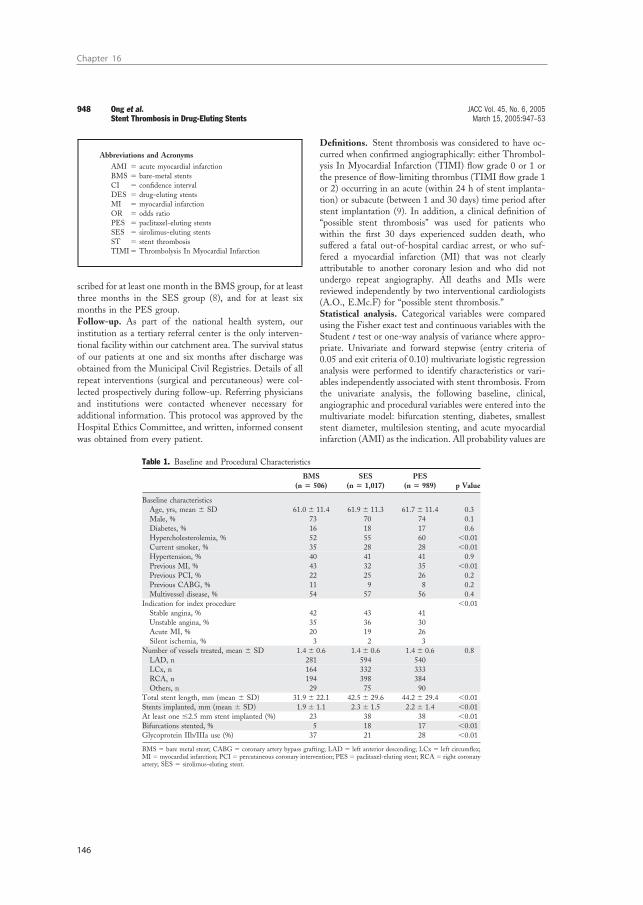
Chapter 16
146
scribed for at least one month in the BMS group, for at leastthree months in the SES group (8), and for at least sixmonths in the PES group.Follow-up. As part of the national health system, ourinstitution as a tertiary referral center is the only interven-tional facility within our catchment area. The survival statusof our patients at one and six months after discharge wasobtained from the Municipal Civil Registries. Details of allrepeat interventions (surgical and percutaneous) were col-lected prospectively during follow-up. Referring physiciansand institutions were contacted whenever necessary foradditional information. This protocol was approved by theHospital Ethics Committee, and written, informed consentwas obtained from every patient.
Definitions. Stent thrombosis was considered to have oc-curred when confirmed angiographically: either Thrombol-ysis In Myocardial Infarction (TIMI) flow grade 0 or 1 orthe presence of flow-limiting thrombus (TIMI flow grade 1or 2) occurring in an acute (within 24 h of stent implanta-tion) or subacute (between 1 and 30 days) time period afterstent implantation (9). In addition, a clinical definition of“possible stent thrombosis” was used for patients whowithin the first 30 days experienced sudden death, whosuffered a fatal out-of-hospital cardiac arrest, or who suf-fered a myocardial infarction (MI) that was not clearlyattributable to another coronary lesion and who did notundergo repeat angiography. All deaths and MIs werereviewed independently by two interventional cardiologists(A.O., E.Mc.F) for “possible stent thrombosis.”Statistical analysis. Categorical variables were comparedusing the Fisher exact test and continuous variables with theStudent t test or one-way analysis of variance where appro-priate. Univariate and forward stepwise (entry criteria of0.05 and exit criteria of 0.10) multivariate logistic regressionanalysis were performed to identify characteristics or vari-ables independently associated with stent thrombosis. Fromthe univariate analysis, the following baseline, clinical,angiographic and procedural variables were entered into themultivariate model: bifurcation stenting, diabetes, smalleststent diameter, multilesion stenting, and acute myocardialinfarction (AMI) as the indication. All probability values are
Table 1. Baseline and Procedural Characteristics
BMS(n � 506)
SES(n � 1,017)
PES(n � 989) p Value
Baseline characteristicsAge, yrs, mean � SD 61.0 � 11.4 61.9 � 11.3 61.7 � 11.4 0.3Male, % 73 70 74 0.1Diabetes, % 16 18 17 0.6Hypercholesterolemia, % 52 55 60 �0.01Current smoker, % 35 28 28 �0.01Hypertension, % 40 41 41 0.9Previous MI, % 43 32 35 �0.01Previous PCI, % 22 25 26 0.2Previous CABG, % 11 9 8 0.2Multivessel disease, % 54 57 56 0.4
Indication for index procedure �0.01Stable angina, % 42 43 41Unstable angina, % 35 36 30Acute MI, % 20 19 26Silent ischemia, % 3 2 3
Number of vessels treated, mean � SD 1.4 � 0.6 1.4 � 0.6 1.4 � 0.6 0.8LAD, n 281 594 540LCx, n 164 332 333RCA, n 194 398 384Others, n 29 75 90
Total stent length, mm (mean � SD) 31.9 � 22.1 42.5 � 29.6 44.2 � 29.4 �0.01Stents implanted, mm (mean � SD) 1.9 � 1.1 2.3 � 1.5 2.2 � 1.4 �0.01At least one �2.5 mm stent implanted (%) 23 38 38 �0.01Bifurcations stented, % 5 18 17 �0.01Glycoprotein IIb/IIIa use (%) 37 21 28 �0.01
BMS � bare metal stent; CABG � coronary artery bypass grafting; LAD � left anterior descending; LCx � left circumflex;MI� myocardial infarction; PCI� percutaneous coronary intervention; PES� paclitaxel-eluting stent; RCA� right coronaryartery; SES � sirolimus-eluting stent.
Abbreviations and AcronymsAMI � acute myocardial infarctionBMS � bare-metal stentsCI � confidence intervalDES � drug-eluting stentsMI � myocardial infarctionOR � odds ratioPES � paclitaxel-eluting stentsSES � sirolimus-eluting stentsST � stent thrombosisTIMI� Thrombolysis In Myocardial Infarction
948 Ong et al. JACC Vol. 45, No. 6, 2005Stent Thrombosis in Drug-Eluting Stents March 15, 2005:947–53

147
SAT after DES
two-sided, and statistical significance was set at the 0.05level. A cumulative event graph consisting of patients withangiographic stent thrombosis was generated plotting theproportion of patients with stent thrombosis (Y-axis)against time (X-axis) stratified by stent type. Incidences ofstent thrombosis are reported as a percentage with associ-ated 95% confidence intervals (CIs).
RESULTS
Baseline and procedural characteristics. The patients inour cohort were at high risk, with unstable angina or AMIbeing the indication in more than one-half of the cases(Table 1). Multivessel disease was present in more thanone-half of the population. One-third of the population hada previous AMI, whereas one-quarter had previous coronaryinterventions. Glycoprotein IIb/IIIa use was lower in theSES and PES groups compared with the BMS group.Clinical outcome. Angiographic ST was documented in26 of 2,512 patients (Table 2). Six cases occurred in theBMS group (1.2%, 95% CI 0.5% to 2.6%), 10 casesoccurred in the SES group (1.0%, 95% CI 0.5% to 1.8%),and 10 cases occurred in the PES group (1.0%, 95% CI0.6% to 1.9%). The first two SES patients with ST havebeen reported previously (10). Most stent thromboses oc-
curred in the first 11 days, regardless of stent type, with amean time to event of 5.8 � 5.4 days (Fig. 1).In the BMS population, there were two acute stent
thromboses and four subacute stent thromboses. Among thesix patients, ST presented as AMI in five patients. Nonedied during the six months of follow-up (Table 2). In thecombined group of SES and PES (2,006 patients), therewere 2 cases of acute ST and 18 cases of subacute ST (Fig.1). A detailed description of these patients is given in Table3. Analysis via intravascular ultrasonography was performedin four patients. In most patients, at least one recognizedrisk factor for ST (i.e., long stented length, use of smallstents, use of multiple stents, and residual dissection afterstent implantation) was present. Importantly, 2 of the 20patients had not taken clopidogrel.Mortality and morbidity. Overall, 20 of 26 patients (77%)re-presented with an AMI, whereas the other 6 re-presented with angina pectoris (Table 2). Of these 26patients, 3 (Patients #12, #18, and #20 from Table 3—all inthe DES population) died at days 11, 5, and 3, respectively.Two patients died during reintervention from intractableventricular fibrillation, whereas the third underwent emer-gency surgery after a suboptimal reintervention and couldnot be weaned from bypass. The incidence of death at 30days was 12%, whereas another 65% suffered a nonfatal MI.Among the survivors of ST, there were no further deaths inthe six months after reintervention.Possible ST. Thirty-day survival data was complete for98% of patients (Table 4). There were 12 patients who werejudged with “possible stent thrombosis,” of which 9 diedand 3 had nonfatal MIs. Of the nine deaths, four wereout-of-hospital sudden deaths, three occurred in hospitalwith ventricular tachycardia as the initiating preterminalrhythm, and two had ST-segment elevation and died beforethey could undergo reangiography. Among those with MIs,one patient developed a postprocedural enzyme leak, andanother developed ventricular fibrillation requiring multiplecardioversions the day after the procedure. Repeat coronaryangiography six months later demonstrated occluded stentsin both of these patients; whereas a third underwentcoronary angiography 14 days after stent implantationbecause of an increase in cardiac enzyme levels, whichdemonstrated an in-stent filling defect which was treated
Figure 1. Cumulative incidence of angiographic stent thrombosis stratifiedby groups against time. Vertical line � day 11 on horizontal axis. BMS �bare-metal stents; PES � paclitaxel-eluting stents; SES � sirolimus-eluting stents.
Table 2. Outcome Following Angiographic Stent Thrombosis
BMS SES PES p Value
Angiographic stent thrombosis, n (%) 6 (1.2%) 10 (1.0%) 10 (1.0%) 0.9Clinical presentationAcute MI, n 5 7 8Angina, n 1 3 2
Maximum total CK, mean � SD 4,983 � 2,570 1,268 � 476 3,361 � 1,404 �0.01Maximum CK-MB, mean � SD 397 � 186 171 � 80 322 � 166 �0.01Outcome30-day mortality, n 0 0 36-month mortality, n 0 0 3
CK � creatine kinase; other abbreviations as in Table 1.
949JACC Vol. 45, No. 6, 2005 Ong et al.March 15, 2005:947–53 Stent Thrombosis in Drug-Eluting Stents

Chapter 16
148
with abciximab, and subsequently underwent repeat percu-taneous coronary intervention two weeks later. Includingthe suspected cases, the combined incidence of angiographicand possible ST was 1.4% (95% CI 0.7% to 2.8%) in theBMS control group, 1.5% (95% CI 0.9% to 2.4%) in theSES group, and 1.6% (95% CI 1.0% to 2.6%) in the PESgroup. In the combined total of 38 documented and possibleST, there were 12 deaths (32%) and 20 nonfatal MIs (53%)in the first 30 days.Multivariate analysis. By univariate analysis, bifurcationstenting was the only significant factor (p � 0.01). Multi-variate analysis was performed with the following covariatesbased on their significance on univariate analysis as well astheir potential clinical impact: diabetes (p � 0.07), smalleststent diameter (p � 0.13), multilesion stenting (p � 0.17),
AMI as the indication (p � 0.3), and bifurcation stenting.By multivariate analysis, bifurcation stenting was the onlyindependent predictor of ST (odds ratio [OR] 3.0, 95% CI1.3 to 6.8, p � 0.01). When the interaction of bifurcationstenting by AMI was entered as a covariate, it was highlysignificant (OR 12.9, 95% CI 4.7 to 35.8, p � 0.001), andbifurcation stenting as a covariate was no longer significant.
DISCUSSION
The main findings in this study can be summarized asfollows: 1) the incidence of angiographic ST in an un-selected, complex DES population was low (�1.0%), withinthe same range as the corresponding BMS population andconcordant with previously published results from the BMS
Table 3. Detailed Description of Drug-Eluting Stent Patients With Angiographic Stent Thrombosis
Patient 1 2 3 4 5 6 7 8 9 10Type of DES SES SES SES SES SES SES SES SES SES SESTime to Thrombosis (days) 0.125 11 7 10 1.08 6 3 7 17 3
Baseline characteristicsAge (yrs) 72 61 86 57 75 55 53 58 58 74Gender F F F M F F M M M MDiabetes � � � � � � � � � �Current smoker � � � � � � � � � �Previous MI � � � � � � � � � �Previous intervention � � � � � � � � � �
Index procedureIndication for procedure UAP AP UAP AMI, ST AP AP, ISR UAP post-AMI AP AMI UAP post-AMIGlycoprotein IIb/IIIa use � � � Y � � � Y Y �
Angiographic features of index procedureCulprit vessel LAD LAD LAD LAD LAD/DIAG LAD/DIAG RCA LAD DIAG LADLesion type (AHA) B1 C C C B2 C B2 B2 B2 B2Bifurcation technique (whereperformed)
� � � � crush t-stent � � t-stent �
Final kissing balloons in bifurcationstenting
� � � � Y N N
Minimum stent diameter (mm) 2.25 2.5 3 3 3 2.5 3 2.75 3 2.75Total stent length (mm) 26 66 41 26 36 41 41 18 31 36Total stents implanted 2 2 3 2 2 2 2 1 2 2
ReinterventionClinical presentation AMI AP AMI AP AMI AMI AMI AP AMI AMIAdditional stent implanted Y � Y Y � � Y � �IVUS findings (where performed) RD UD � RD � � � � UDSite of thrombosis in bifurcation lesions � � � � MB�SB SB � � SB �Incomplete oral anti-platelet therapy � � � � � � � � Y �Successful procedural outcome Y Y Y Y Y Y Y Y Y Y
AMI� acute myocardial infarction; AP� angina pectoris; DIAG� diagonal branch; IM� intermediate branch; LAD� left anterior descending artery; LCx� left circumflexartery; MB � mainbranch; N � no; OMCx � obtuse marginal branch; RCA � right coronary artery; RD � residual dissection; SB � sidebranch; ST � stent thrombosis; UAP� unstable angina pectoris; UD � underdeployment; Y � yes; other abbreviations as in Table 1.
Table 4. Incidence of Stent Thrombosis Classified by Definition
Stent TypeNumber ofPatients
Angiographically ProvenStent Thrombosisn (% [95% CI])
Possible StentThrombosis
n (% [95% CI])All Stent Thrombosisn (% [95% CI])
BMS 506 6 1 7(1.2% [0.5%–2.6%]) (0.2% [0.0%–1.1%]) (1.4% [0.7%–2.8%])
SES 1,017 10 5 15(1.0% [0.5%–1.8%]) (0.5% [0.2%–1.1%]) (1.5% [0.9%–2.4%])
PES 989 10 6 16(1.0% [0.6%–1.9%]) (0.6% [0.3%–1.3%]) (1.6% [1.0%–2.6%])
CI � confidence interval; other abbreviations as in Table 1.
950 Ong et al. JACC Vol. 45, No. 6, 2005Stent Thrombosis in Drug-Eluting Stents March 15, 2005:947–53
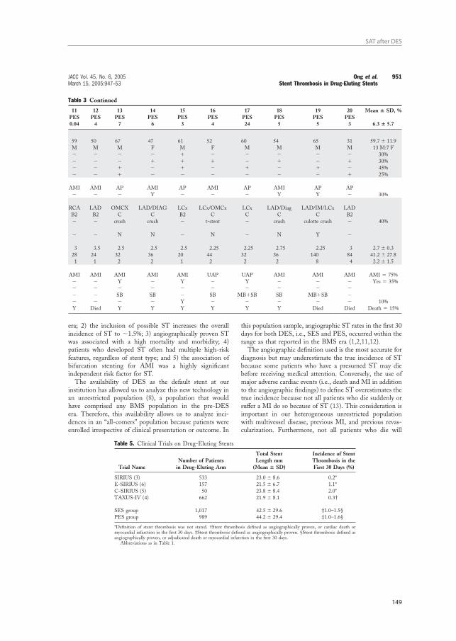
149
SAT after DES
era; 2) the inclusion of possible ST increases the overallincidence of ST to �1.5%; 3) angiographically proven STwas associated with a high mortality and morbidity; 4)patients who developed ST often had multiple high-riskfeatures, regardless of stent type; and 5) the association ofbifurcation stenting for AMI was a highly significantindependent risk factor for ST.The availability of DES as the default stent at our
institution has allowed us to analyze this new technology inan unrestricted population (8), a population that wouldhave comprised any BMS population in the pre-DESera. Therefore, this availability allows us to analyze inci-dences in an “all-comers” population because patients wereenrolled irrespective of clinical presentation or outcome. In
this population sample, angiographic ST rates in the first 30days for both DES, i.e., SES and PES, occurred within therange as that reported in the BMS era (1,2,11,12).The angiographic definition used is the most accurate for
diagnosis but may underestimate the true incidence of STbecause some patients who have a presumed ST may diebefore receiving medical attention. Conversely, the use ofmajor adverse cardiac events (i.e., death and MI in additionto the angiographic findings) to define ST overestimates thetrue incidence because not all patients who die suddenly orsuffer a MI do so because of ST (13). This consideration isimportant in our heterogeneous unrestricted populationwith multivessel disease, previous MI, and previous revas-cularization. Furthermore, not all patients who die will
Table 3 Continued
11 12 13 14 15 16 17 18 19 20 Mean � SD, %PES PES PES PES PES PES PES PES PES PES0.04 4 7 6 3 4 24 5 5 3 6.3 � 5.7
59 50 67 47 61 52 60 54 65 31 59.7 � 11.9M M M F M F M M M M 13 M:7 F� � � � � � � � � � 30%� � � � � � � � � � 30%� � � � � � � � � � 45%� � � � � � � � � � 25%
AMI AMI AP AMI AP AMI AP AMI AP AP� � � Y � � � Y Y � 30%
RCA LAD OMCX LAD/DIAG LCx LCx/OMCx LCx LAD/Diag LAD/IM/LCx LADB2 B2 C C B2 C C C C B2� � crush crush � t-stent � crush culotte crush � 40%
� � N N � N � N Y �
3 3.5 2.5 2.5 2.5 2.25 2.25 2.75 2.25 3 2.7 � 0.328 24 32 36 20 44 32 36 140 84 41.2 � 27.81 1 2 2 1 2 2 2 8 4 2.2 � 1.5
AMI AMI AMI AMI AMI UAP UAP AMI AMI AMI AMI � 75%� � Y � Y � Y � � � Yes � 35%� � � � � � � � � �� � SB SB � SB MB�SB SB MB�SB �� � � � Y � � � � � 10%Y Died Y Y Y Y Y Y Died Died Death � 15%
Table 5. Clinical Trials on Drug-Eluting Stents
Trial NameNumber of Patientsin Drug-Eluting Arm
Total StentLength mm(Mean � SD)
Incidence of StentThrombosis in theFirst 30 Days (%)
SIRIUS (3) 533 23.0 � 8.6 0.2*E-SIRIUS (6) 157 21.5 � 6.7 1.1*C-SIRIUS (5) 50 23.8 � 8.4 2.0*TAXUS-IV (4) 662 21.9 � 8.1 0.3†
SES group 1,017 42.5 � 29.6 ‡1.0–1.5§PES group 989 44.2 � 29.4 ‡1.0–1.6§
*Definition of stent thrombosis was not stated. †Stent thrombosis defined as angiographically proven, or cardiac death ormyocardial infarction in the first 30 days. ‡Stent thrombosis defined as angiographically proven. §Stent thrombosis defined asangiographically proven, or adjudicated death or myocardial infarction in the first 30 days.Abbreviations as in Table 1.
951JACC Vol. 45, No. 6, 2005 Ong et al.March 15, 2005:947–53 Stent Thrombosis in Drug-Eluting Stents

Chapter 16
150
undergo autopsy studies to determine the cause of death. Toattenuate this overestimation and to provide an accuratefigure, we have adjudicated all deaths and noncatheterized,nonfatal MIs within the first 30 days in the three groups andincluded them with the angiographically proven patients toprovide an overall incidence for each group.The incidences of ST for both groups of DES are within
the range reported in the larger randomized clinical trials ofDES (3–6) despite longer total stent length, multivesseltreatment, and a heterogeneous population (Table 5). Thisincidence complements information already available fromthe randomized trials regarding the safety of these newdevices.Angiographic ST was associated with a high mortality
and morbidity in our study. Within the DES population, 15patients (75%) experienced a MI as their diagnosis at thesecond presentation, and 3 (15%) died during the reinter-vention procedure. The inclusion of possible ST patientsincreased the mortality to 32%. Given the small number ofevents, the fact that no deaths occurred in the BMS groupwas most likely due to chance. These results are in concor-dance with the results of a large BMS registry (2).Previous studies have demonstrated that residual dissec-
tion (1,11), long stents (1), small final lumen diameter (1),and use of multiple stents (2) are risk factors for thedevelopment of ST. In our series, multiple risk factors wereidentified in most patients who developed ST. Patients withST had more multiple lesions treated, smaller minimumstent diameters, and longer stent lengths compared withthose without ST; however, these factors were not signifi-cant on univariate analysis. What did emerge and which hasnot been previously reported is that patients undergoingbifurcation stenting had a higher incidence of ST comparedwith those without bifurcation stenting. A recent study onbifurcations reported a 3.5% incidence of ST, which ishigher than the overall incidence in this population (14).Although stent implantation for AMI was not significant
on univariate analysis, the interaction of AMI and bifurca-tion stenting when entered as a covariate for ST onmultivariate analysis emerged as a highly significant inde-pendent predictor, and bifurcation stenting as a covariatewas no longer significant. This result confirms a clinicalsuspicion in our department regarding the increased risk ofST in patients treated with bifurcation stenting in thesetting of AMI.Mechanical reasons that predispose to ST can be modi-
fied by interventional technique. Optimizing stent place-ment including, if necessary, intravascular ultrasound-guided postdilation, kissing balloon postdilation withbifurcation stenting, and careful inspection for residualdissection after stent implantation, may further reduce theincidence of ST.Pharmacologic reasons for ST, i.e., inadequate antiplate-
let therapy, are patient-specific factors. Recent researchliterature has focused on “resistance” to either aspirin (15) orto clopidogrel (16). Currently, most laboratories do not
routinely test for antiplatelet resistance. In our series, twopatients who had not taken their prescribed clopidogrel afterthe procedure developed ST.This report covers ST occurring in the first 30 days after
stent implantation only, during which all patients receiveddual antiplatelet therapy. The duration of clopidogrel ther-apy differed among the three groups; in part, it reflectsuncertainty with regards to re-endothelialization after DESimplantation. Late ST has been reported to occur withBMS (17) and with DES (18), including a reported fatality(19) after clopidogrel discontinuation. At this stage, theincidence of late ST in the DES era is unknown, and furtherstudies are required to clarify this potential late complica-tion.Comment on sample size and statistical comparisons.Because ST occurs at a low incidence (�1.0 to 1.5%), asmall sample size may underestimate or overestimate thetrue incidence. In a previously published report from ourinstitution, we reported an angiographic incidence of 0.4%in 508 patients (8). In the present study we extended thepopulation to incorporate the entire period of DES used todate at our institution (n � 2,006) to allow a more accurateanalysis of the true incidence of ST in the DES population.Despite having 2,512 patients, the low and small/negligibleabsolute difference in incidence precludes formal statisticalcomparisons of ST rates among the three groups because itlacks sufficient statistical power. To achieve adequate powerwould require sample sizes in the order of �100,000patients. To date, this study is the largest series of patientsreported on in the DES era.Study limitations. These single-center registry data com-plement available randomized data, as they reflect the resultsof unrestricted DES use.Conclusions. Despite having a more complex cohort withhigh-risk inclusion criteria, longer stent lengths, and morecomplex procedural features, the incidence of ST with DESare in the same range as the BMS population observed inour present study. They also are in agreement with previ-ously reported data by others from the BMS era and withthose results reported on in the earlier randomized DEStrials. Furthermore, the two groups of DES, i.e., SES andPES, share an incidence of �1.0% to 1.5%. Stent throm-bosis is associated with a high morbidity and mortality.As extensively documented in previous reports with BMS,
mechanical reasons were observed to be frequent associationsfor ST with DES. In this study, bifurcation stenting in thesetting of AMI was a highly significant independent predictorfor angiographic ST.
AcknowledgmentsThe authors would like to thank Pedro A. Lemos, MD,PhD, Chourmouzios A. Arampatzis, MD, PhD, and PieterC. Smits, MD, PhD, for their contribution to this work.
952 Ong et al. JACC Vol. 45, No. 6, 2005Stent Thrombosis in Drug-Eluting Stents March 15, 2005:947–53
151
SAT after DES
Reprint requests and correspondence: Dr. Patrick W. Serruys,Thoraxcenter, Bd-406, Dr. Molewaterplein 40, 3015-GD Rotter-dam, Netherlands. E-mail: [email protected].
REFERENCES
1. Cutlip DE, Baim DS, Ho KK, et al. Stent thrombosis in the modernera: a pooled analysis of multicenter coronary stent clinical trials.Circulation 2001;103:1967–71.
2. Orford JL, Lennon R, Melby S, et al. Frequency and correlates ofcoronary stent thrombosis in the modern era: analysis of a single centerregistry. J Am Coll Cardiol 2002;40:1567–72.
3. Moses JW, Leon MB, Popma JJ, et al. Sirolimus-eluting stents versusstandard stents in patients with stenosis in a native coronary artery.N Engl J Med 2003;349:1315–23.
4. Stone GW, Ellis SG, Cox DA, et al. A polymer-based, paclitaxel-eluting stent in patients with coronary artery disease. N Engl J Med2004;350:221–31.
5. Schampaert E, Cohen EA, Schluter M, et al. The Canadian study ofthe sirolimus-eluting stent in the treatment of patients with long denovo lesions in small native coronary arteries (C-SIRIUS). J Am CollCardiol 2004;43:1110–5.
6. Schofer J, Schluter M, Gershlick AH, et al. Sirolimus-eluting stentsfor treatment of patients with long atherosclerotic lesions in smallcoronary arteries: double-blind, randomised controlled trial (E-SIRIUS). Lancet 2003;362:1093–9.
7. Jeremias A, Sylvia B, Bridges J, et al. Stent thrombosis after successfulsirolimus-eluting stent implantation. Circulation 2004;109:1930–2.
8. Lemos PA, Serruys PW, van Domburg RT, et al. Unrestrictedutilization of sirolimus-eluting stents compared with conventional barestent implantation in the “real world”: the Rapamycin-Eluting StentEvaluated At Rotterdam Cardiology Hospital (RESEARCH) registry.Circulation 2004;109:190–5.
9. Lemos PA, Lee CH, Degertekin M, et al. Early outcome aftersirolimus-eluting stent implantation in patients with acute coronary
syndromes: insights from the Rapamycin-Eluting Stent Evaluated AtRotterdam Cardiology Hospital (RESEARCH) registry. J Am CollCardiol 2003;41:2093–9.
10. Regar E, Lemos PA, Saia F, et al. Incidence of thrombotic stentocclusion during the first three months after sirolimus-eluting stentimplantation in 500 consecutive patients. Am J Cardiol 2004;93:1271–5.
11. Cheneau E, Leborgne L, Mintz GS, et al. Predictors of subacute stentthrombosis: results of a systematic intravascular ultrasound study.Circulation 2003;108:43–7.
12. Tolleson TR, Newby LK, Harrington RA, et al. Frequency of stentthrombosis after acute coronary syndromes (from the SYMPHONYand 2nd SYMPHONY trials). Am J Cardiol 2003;92:330–3.
13. Honda Y, Fitzgerald PJ. Stent thrombosis: an issue revisited in achanging world. Circulation 2003;108:2–5.
14. Colombo A, Moses JW, Morice MC, et al. Randomized study toevaluate sirolimus-eluting stents implanted at coronary bifurcationlesions. Circulation 2004;109:1244–9.
15. Gum PA, Kottke-Marchant K, Welsh PA, White J, Topol EJ. Aprospective, blinded determination of the natural history of aspirinresistance among stable patients with cardiovascular disease. J Am CollCardiol 2003;41:961–5.
16. Lau WC, Gurbel PA, Watkins PB, et al. Contribution of hepaticcytochrome P450 3A4 metabolic activity to the phenomenon ofclopidogrel resistance. Circulation 2004;109:166–71.
17. Wang F, Stouffer GA, Waxman S, Uretsky BF. Late coronary stentthrombosis: early vs. late stent thrombosis in the stent era. CatheterCardiovasc Interv 2002;55:142–7.
18. Colombo A, Drzewiecki J, Banning A, et al. Randomized study toassess the effectiveness of slow- and moderate-release polymer-basedpaclitaxel-eluting stents for coronary artery lesions. Circulation 2003;108:788–94.
19. Virmani R, Guagliumi G, Farb A, et al. Localized hypersensitivity andlate coronary thrombosis secondary to a sirolimus-eluting stent: shouldwe be cautious? Circulation 2004;109:701–5.
953JACC Vol. 45, No. 6, 2005 Ong et al.March 15, 2005:947–53 Stent Thrombosis in Drug-Eluting Stents


Chapter 17
How to Accelerate the Re-endothelialization of Stents
Ong AT, Aoki J, Kutryk, MJ, Serruys PW.
Arch Mal Coeur Vaiss 2005; 98:123-126


Chapter 18
Endothelial Progenitor Cell Capture By Stents Coated
With Antibody Against CD34-The HEALING-FIM (Healthy
Endothelial Accelerated Lining Inhibits Neointimal Growth-
First In Man) Registry
Aoki J, Serruys PW, van Beusekom H,
Ong AT, McFadden E, Sianos G, van der
Giessen, Regar E, de Feyter P, Davis R,
Rowland S, Kutryk M.
J Am Coll Cardiol. 2005;45:1574-1579


161
First human experience of EPC capture stent
Endothelial Progenitor Cell Capture byStents Coated With Antibody Against CD34The HEALING-FIM (Healthy Endothelial AcceleratedLining Inhibits Neointimal Growth-First In Man) RegistryJiro Aoki, MD,* Patrick W. Serruys, MD, PHD, FACC,* Heleen van Beusekom, MD, PHD,* Andrew T. L.Ong, MBBS, FRACP,* Eugene P. McFadden, MBCHB, MD, FRCPI, FACC,*Georgios Sianos, MD, PHD,* Willem J. van der Giessen, MD, PHD,* Evelyn Regar, MD, PHD,*Pim J. de Feyter, MD, PHD, FACC,* H. Richard Davis, MSC,† Stephen Rowland, PHD,†Michael J. B. Kutryk, MD, PHD‡Rotterdam, the Netherlands; Fort Lauderdale, Florida; and Toronto, Canada
OBJECTIVES This study was designed to evaluate whether rapid endothelialization of stainless steel stentswith a functional endothelium prevents stent thrombosis and reduces the restenotic process.
BACKGROUND A “pro-healing” approach for prevention of post-stenting restenosis is theoretically favoredover the use of cytotoxic or cytostatic local pharmacologic therapies. It is believed that thecentral role of the vascular endothelium is to maintain quiescence of the underlying media andadventitia.
METHODS Sixteen patients with de novo coronary artery disease were successfully treated withimplantation of endothelial progenitor cell (EPC) capture stents.
RESULTS Complete procedural and angiographic success was achieved in all 16 patients. Thenine-month composite major adverse cardiac and cerebrovascular events (MACCE) rate was6.3% as a result of a symptom-driven target vessel revascularization in a single patient. Therewere no other MACCE despite only one month of clopidogrel treatment. At six-monthfollow-up, mean angiographic late luminal loss was 0.63 � 0.52 mm, and percent stentvolume obstruction by intravascular ultrasound analysis was 27.2 � 20.9%.
CONCLUSIONS This first human clinical investigation of this technology demonstrates that the EPC capturecoronary stent is safe and feasible for the treatment of de novo coronary artery disease. Furtherdevelopments in this technology are warranted to evaluate the efficacy of this device for thetreatment of coronary artery disease. (J Am Coll Cardiol 2005;45:1574–9) © 2005 by theAmerican College of Cardiology Foundation
The emergence of drug-eluting stents has dramaticallyreduced the incidence of in-stent restenosis (1,2). Thistherapy interferes with the natural healing response bypreventing or significantly delaying the formation of afunctional endothelial lining over the stent (3).Recently, the existence of circulating endothelial progen-
itor cells (EPCs) has been identified as a key factor forre-endothelialization (4). The early establishment of afunctional endothelial layer after vascular injury has beenshown to assist in the prevention of neointimal proliferationand thrombus formation (5,6). The EPC capture stentshave been developed using immobilized antibodies targetedat EPC surface antigens. The HEALING-FIM (HealthyEndothelial Accelerated Lining Inhibits NeointimalGrowth-First In Man) registry is the first clinical investi-gation using this technology.
METHODS
Patient selection. The HEALING-FIM registry is asingle-center, prospective, non-randomized registry trial.Patients were eligible if they were between ages 18 and 85years and had a diagnosis of stable or unstable angina orsilent ischemia. Additional eligibility criteria were the pres-ence of a single primary target lesion in a native coronaryartery that was 2.5 to 3.5 mm in diameter that could becovered by a single trial stent (13 mm or 18 mm length), astenosis of 51% to 99% of the luminal diameter as estimatedvisually, and a flow rate of grade 1 or higher according to theclassification of the Thrombolysis In Myocardial Infarction(TIMI) trial. Patients were not eligible for enrollment ifthey had an evolving myocardial infarction; stenosis of theleft main coronary artery; a lesion located at an ostiallocation; a calcified lesion that could not be completelydilated before stenting; angiographically visible thrombuswithin the target lesion; a left ventricular ejection fraction of�30%; or an intolerance of aspirin, clopidogrel, ticlopidine,heparin, stainless steel, or contrast material. The trial wasreviewed and approved by the ethics review committee, andwritten informed consent was obtained from all patients.Study device: EPC capture stent. The EPC antibodysurface consists of a covalently coupled polysaccharide
From the *Thoraxcenter, Erasmus Medical Center, Rotterdam, the Netherlands;†OrbusNeich, Fort Lauderdale, Florida; and ‡Terrence Donnelly Heart Centre, St.Michael’s Hospital, University of Toronto, Toronto, Canada. This study wassupported by OrbusNeich, Fort Lauderdale, Florida. Mr. Davis and Dr. Rowland arefull-time employees of OrbusNeich, and Dr. Kutryk is a consultant of Orbus MedicalTechnologies.Manuscript received December 9, 2004; revised manuscript received January 22,
2005, accepted January 25, 2005.
Journal of the American College of Cardiology Vol. 45, No. 10, 2005© 2005 by the American College of Cardiology Foundation ISSN 0735-1097/05/$30.00Published by Elsevier Inc. doi:10.1016/j.jacc.2005.01.048

Chapter 18
162
intermediate coating with murine monoclonal anti-humanCD34 antibodies, attached to a stainless steel stent (R stent,OrbusNeich, Fort Lauderdale, Florida) (Fig. 1). This anti-body specifically targets CD34� cells (endothelial pro-genitor cells are CD34 positive) in the vascular circula-tion. This device was supplied in aqueous sodium azidesolution as a preservative to maintain bioactivity andrequired hand crimping by the operator onto a percuta-neous transluminal coronary angioplasty balloon catheterbefore implantation.Study procedures. Lesions were treated according to localstandard interventional techniques. Specifically, the decisionto predilate or direct stent was at the investigator’s discre-tion, and post-dilation was performed as required to ensurethat the residual stenosis was �20% by visual assessment,with a TIMI flow grade rate 3. In case of a dissection or
incomplete coverage of the lesion, implantation of addi-tional EPC capture R stents was permitted.Intravenous boluses of heparin were administered to
maintain an activated clotting time �300 s during theimplantation. Treatment with aspirin, at a dose of at least 80mg/day, was initiated at least 12 h before the procedure andcontinued for one month. In addition, a loading dose of 300mg of clopidogrel was administered before the procedure,followed by 75 mg daily for 28 days. Glycoprotein IIb/IIIainhibitors were used at the operator’s discretion. Angio-graphic success was defined as the successful implantation ofthe study device, with a stenosis of �20% of the vesseldiameter with TIMI flow grade 3.Follow-up. All patients were scheduled for a clinicalfollow-up at one, six, and nine months following theimplantation procedure to assess the anginal status andthe occurrence of major adverse cardiac and cerebrovas-cular events (MACCE). An electrocardiogram was ob-tained at each visit, and an angiographic and intravascularultrasound (IVUS) study was performed at a mean of 185� 14 days.Quantitative angiographic and IVUS analysis. Coronaryangiograms were obtained in multiple views after an intra-coronary injection of nitrates. Offline quantitative analysesof preprocedural, postprocedural, and six-month follow-upangiographic data were performed. Restenosis was definedas a reduction of 50% or more of the luminal diameter. Lateluminal loss was defined as the difference between theminimal luminal diameter after procedure and at sixmonths. The target lesion was defined as the stented
Abbreviations and AcronymsEPC � endothelial progenitor cellHAMA � human antimurine antibodyHEALING-FIM � Healthy Endothelial Accelerated
Lining Inhibits NeointimalGrowth-First In Man
IVUS � intravascular ultrasoundMACCE � major adverse cardiac and
cerebrovascular eventsMI � myocardial infarctionSMC � smooth muscle cellTIMI � Thrombolysis In Myocardial
Infarction
Figure 1. Endothelial progenitor cell (EPC) capture coating technology.
1575JACC Vol. 45, No. 10, 2005 Aoki et al.May 17, 2005:1574–9 First Human Experience of EPC Capture Stent
color figures on page 224

163
First human experience of EPC capture stent
segment plus the 5-mm segments proximal and distal to thestented segment.Intravascular ultrasound was performed with an auto-
mated pullback at 0.5 mm/s to examine the target lesion atpostprocedure and six-month follow-up. Lumen, stent, andexternal elastic membrane contours were detected with theuse of CUARD QCU analysis software (CUARD BV,Wijk Bij Duurstede, the Netherlands), applying a three-dimensional reconstruction as described elsewhere (7).Study end points. The primary safety end point of thisstudy was the absence of stent thrombosis up to six months.The second end point was a composite of MACCE, definedas cardiac death, stroke, Q-wave or non–Q-wave myocardialinfarction (MI), and target vessel revascularization. Strokewas defined as a focal neurologic deficit resulting from avascular cause involving the central nervous system. Q-waveMI was defined as development of new abnormal Q wavesnot present on the patient’s baseline. Non–Q-wave MI wasdefined as a creatine kinase of more than twice the upperlimit of normal with an abnormal level of the MB isoen-zyme of creatine kinase. The efficacy end point was lateluminal loss as determined by quantitative coronary angiog-raphy and % stent volume obstruction by IVUS at sixmonths. Stent thrombosis was angiographically docu-mented as a complete occlusion (TIMI flow grade 0 or 1) ora flow-limiting thrombus (TIMI flow grade 1 or 2) of apreviously successfully treated artery.Detection of human antimurine antibody (HAMA).Human antimurine antibody testing was not added to thestudy until several patients had already returned for follow-up. Testing with baseline data was conducted on 4 of 16patients. The HAMA was determined by a commerciallyavailable enzyme-linked immunosorbent assay kit (ME-DAC, Hamburg, Germany) (8). A positive assay wasdefined as �10 ng/ml. Significant levels of HAMA weredefined as �150 ng/ml.Histology. One specimen was retrieved by directionalatherectomy (Flexi-cut, Guidant Europe SA, Diegem, Bel-gium), fixed in 4% buffered formaldehyde with metal stentfragments removed and embedded in paraffin.Sections were stained with hematoxylin eosin and an
elastin stain (Resorcin Fuchsin) for general assessment of thetissue. Cellular characterization was performed with immu-nocytochemistry, using antibodies against smooth muscle(specific alpha-actin), leukocytes (CD45), macrophages(mac 387), proliferation cells (Mib 1), and EPC (CD34).All antibodies except anti-CD34 were obtained fromDAKO (DakoCytomation, Produktionsvej, Denmark).Statistical analysis. Because of the size of the patientpopulation in this nonrandomized registry, no formal sta-tistical analysis was conducted to determine the efficacy ofthe device. Continuous variables are expressed as mean �SD. Comparisons between postprocedure and six-monthfollow-up values were performed with a two-tailed paired ttest. A p value�0.05 was considered statistically significant.
RESULTS
Baseline characteristics and procedural outcome. Over-all, 16 patients were enrolled in the HEALING-FIMregistry between May and November 2003. Table 1 presentsthe baseline characteristics and procedural outcomes for thispatient population. A second overlapping stent was im-planted in two patients to treat a dissection followingimplantation of the first stent.Clinical outcome. Table 2 provides an overview of theMACCE at one and nine months. There was no subacutethrombosis or MACCE in the first month after implanta-tion. During the first nine months, MACCE occurred in apatient with insulin-dependent diabetes with diabetic ne-phropathy. This patient (#12) was admitted with a non–ST-segment elevation MI (maximum creatinine kinase levelwas 524 U/l) at six months after the index procedure andunderwent recatheterization that demonstrated focal in-stent restenosis with TIMI flow grade 3 and no visiblecontrast detect, which could have been caused by stentthrombosis. No MACCE was reported in other patients.Six-month angiographic and IVUS results in stentedsegment. Scheduled coronary angiography at six monthswas performed in 15 patients (93.8%). The patient whorefused angiography was asymptomatic. At six months,mean late luminal loss was 0.63 mm on quantitative
Table 1. Baseline Characteristics and Procedural Outcomes,Patients (n � 16)
Men 11 (69%)Age (yrs) 68 � 10Diabetes 5 (31%)Hypertension 12 (75%)Hypercholesterolemia 12 (75%)Family history 6 (38%)Current smoking 4 (25%)Unstable angina 8 (50%)Target coronary arteryLAD 8 (50%)RCA 5 (31%)LCX 3 (19%)
Lesion typeA 8 (50%)B1 6 (38%)B2 2 (13%)C 0 (0%)
Angiographic featuresLesion length (mm) 10.6 � 4.0Reference vessel diameter (mm) 2.63 � 0.30Minimal luminal diameter (mm) 0.95 � 0.33Diameter stenosis (%) 63.7 � 11.9
Procedural outcomesMean stent diameter (mm) 3.3 � 0.3Mean stent length (mm) 18.4 � 6.4Overlapping stenting 2 (13%)Post dilation 8 (50%)Direct stenting 2 (13%)Use of glycoprotein IIb/IIIa inhibitor 3 (19%)Angiographic success 16 (100%)
LAD � left anterior descending; LCX � left circumflex; RCA � right coronaryartery.
1576 Aoki et al. JACC Vol. 45, No. 10, 2005First Human Experience of EPC Capture Stent May 17, 2005:1574–9
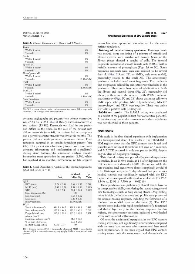
Chapter 18
164
coronary angiography and percent stent volume obstructionwas 27.2% on IVUS (Table 3). Binary restenosis occurred intwo patients (13.3%). Restenosis was focal in one patientand diffuse in the other. In the case of the patient withdiffuse restenosis (case #4), the patient had no symptomsand a percent diameter stenosis was 58% at six months. Thispatient did not undergo repeat revascularization. Focalrestenosis occurred in an insulin-dependent patient (case#12). This patient was subsequently treated with directionalcoronary atherectomy and implantation of a paclitaxel-eluting stent. Intravascular ultrasound analysis revealedincomplete stent apposition in one patient (6.3%), whichhad resolved at six months. Furthermore, no late-acquired
incomplete stent apposition was observed for the entirepatient population.Histology of the atherectomy specimen. Histologic anal-ysis showed tissue consisting of a mixture of myxoid andfibrous material with variable cell density. Some of thefibrous pieces showed a paucity of cells. The myxoidfragments consisted of smooth muscle cells (SMCs) withinvariable amounts of proteoglycan (Figs. 2A to 2C). Somethrombus remnants were seen and assessed to be severaldays old (Figs. 2D and 2E, no SMCs, only some nuclei),presumably related to the small MI. The atherectomyspecimens included metal stent fragments. That indicatesthat the plaques behind the stent struts were included in thespecimens. There were large areas of calcification in boththe fibrous and myxoid tissue (Fig. 2F), presumably oldplaque, as these were also observed with IVUS. Immuno-cytochemistry (Figs. 2C and 2E) shows that most cells wereSMC-alpha-actin positive. Mib-1 (proliferation), Mac387(macrophages), and CD34 were negative. There were only afew CD45-positive cells (leukocytes).HAMA test results. The HAMA testing was conductedon a subset of the population (last four consecutive patients).A positive assay due to the treatment with the study devicewas not observed in these patients.
DISCUSSION
This study is the first clinical experience with implantationof a bioengineered stent. The results of the HEALING-FIM registry show that the EPC capture stent is safe andfeasible: with no stent thrombosis (30 days or 6 months),and MACCE occurred in only one patient (6.3%), despiteonly 30 days of clopidogrel therapy.This clinical registry was preceded by several experimen-
tal studies. In an in vivo study, at 1 h after deployment theEPC capture stent showed a �90% cell coverage, while thebare stainless steel stents were almost completely devoid ofcells. Histologic analysis at 31 days showed that percent arealuminal stenosis was significantly reduced with the EPCcapture stents compared with stainless steel stents (15.49 �4.54% vs. 23.96 � 7.70%, p � 0.01) (9).These preclinical and preliminary clinical results have to
be interpreted carefully, considering the recent emergence ofnew technologies such as drug-eluting stents. Drug-elutingstents inhibit the inflammatory and proliferative process ofthe normal healing response, including the formation of aconfluent endothelial layer on the stent (3). The EPCcapture stents induce the rapid establishment of a functionalendothelial layer early in the healing response. In thisregistry, the atherectomy specimen indicated a well-healedartery with minimal inflammation.Of note, the neointimal hyperplasia in the EPC capture
coating stents was not significantly reduced when comparedwith the usual late loss seen after conventional bare metalstent implantation. It has been argued that EPC capturecoating covers only the stent struts, and theoretically no
Table 2. Clinical Outcomes at 1 Month and 9 Months
DeathWithin 1 month 0%9 months 0%
StrokeWithin 1 month 0%9 months 0%
Q-wave MIWithin 1 month 0%9 months 0%
Non–Q-wave MIWithin 1 month 0%9 months 6.3% (1/16)
TVRWithin 1 month 0%9 months 6.3% (1/16)
MACCEWithin 1 month 0%9 months 6.3% (1/16)
Stent thrombosisWithin 1 month 0%9 months 0%
MACCE � major adverse cardiac and cerebrovascular events; MI � myocardialinfarction; TVR � target vessel revascularization.
Table 3. Serial Quantitative Analysis of the Stented Segment byQCA and IVUS (n � 15)
Post6-Month
Follow-Upp
Value
QCARVD (mm) 2.71 � 0.31 2.69 � 0.32 0.738MLD (mm) 2.47 � 0.29 1.84 � 0.56 0.0004%DS 8.5 � 3.4 32.1 � 16.7 0.0001Stent thrombosis (%) 0%Late loss (mm) 0.63 � 0.52Loss index 0.45 � 0.39Binary restenosis 13.3% (2/15)
IVUSVessel volume (mm3) 336.5 � 86.7 339.9 � 88.9 0.583Stent volume (mm3) 173.5 � 44.9 175.0 � 42.6 0.715Plaque behind stentvolume (mm3)
163.0 � 56.6 165.0 � 62.9 0.371
Neointima volume (mm3) 51.7 � 47.8% in-stent obstruction 27.2 � 20.9In-stent malapposition 6.7% (1/15) 0%
DS � diameter stenosis; IVUS � intravascular ultrasound; MLD � mean luminaldiameter; QCA � quantitative coronary angiography; RVD � revascularized vesseldiameter.
1577JACC Vol. 45, No. 10, 2005 Aoki et al.May 17, 2005:1574–9 First Human Experience of EPC Capture Stent

165
First human experience of EPC capture stent
early functional endothelial lining can be expected in theinterstrut space, although the interstrut area in the animalmodel (healthy coronary artery undergoing direct stenting)was covered with functional endothelium within 48 h; thissituation differs substantially from a human pathologicatherosclerosis vessel after balloon injury. In addition, thebioactivity of these prototype EPC capture stents used inHEALING-FIM registry was unstable and was easilyreduced by sterilization with gamma irradiation. It wassubsequently discovered through the use of a bioassay thatgamma irradiation lessened the immunoaffinity of EPCcapture prototype stents used in this trial. Therefore, it islikely that the reduced bioactivity of EPC capture stents inthe HEALING-FIM registry may not have been enough toinhibit neointimal hyperplasia after stent implantation.The technology behind the creation of an EPC affinity
surface is achieved by attaching murine monoclonal antihu-man CD 34 antibodies to the stent. An immunoreactionagainst the murine monoclonal antibody may occur inpatients who have human ant-murine antibody (HAMA).In addition, production of HAMA may result in theneutralization of the EPC capture surface. In this registry,
increased HAMA levels were not observed in the fourpatients who underwent serial HAMA testing, and nopatients exhibited systemic symptoms of immunoreaction.This safety issue was in agreement with other trials, evalu-ating immunologic treatment using murine antibody forovarian cancer (10).Because of the small sample size and single-center en-
Table 4. Evolution of Endothelial Progenitor Cell CaptureStent: Improvement From HEALING-FIM (H1) toHEALING II (H2)
DeviceH1 - Wet, hand-crimpedH2 - Dry, premounted
StabilityH1 - Wet; supplied in sodium azide preservative; required rinsingbefore use
H2 - Dry, formulated to preserve antibody structure & activitySterilizationH1 - Gamma irradiation; 15 to 25 kGyH2 - Gamma irradiation; �15 kGy
BioactivityH1 - Significant reduction in activity with sterilizationH2 - Stable with sterilization
Figure 2. Overview (A) and details (left: B, C; mid:D, E; right: F) of the atherectomy specimen obtained six months following stent placement. Histologyshows a mixture of myxoid (B, C) and fibrous material (F) consisting of smooth muscle cells (C, E) within variable amounts of extracellular matrix. Somefragments contained thrombus remnants (Th) (D, E) and calcifications (Ca) (F). A � elastin stain; B, D, F� hematoxylin eosin; C, E �immunocytochemistry for smooth muscle cell-specific alpha actin.
1578 Aoki et al. JACC Vol. 45, No. 10, 2005First Human Experience of EPC Capture Stent May 17, 2005:1574–9
color figures on page 224

Chapter 18
166
rollment, the present study did not fully evaluate the efficacyof this device. However, further developments in thistechnology, such a providing a dry stent premounted on adelivery system and maintaining good activity post-sterilization, are currently being evaluated in theHEALING-II registry (Table 4). The technology for pre-serving antibody structure and bioactivity has advanced,resulting in a higher capture of EPCs (Fig. 3). As a result ofthese improvements, further clinical investigation of thistechnology is warranted to evaluate the efficacy of this devicefor the treatment of coronary artery disease.Conclusions. The present study demonstrates that theEPC capture stent is safe and feasible. Further study and
development of this promising technology are needed toconfirm the clinical efficacy of this bio-engineered stent.
Reprint requests and correspondence: Dr. Patrick W. Serruys,Thoraxcenter, Bd 406, Erasmus Medical Center, Dr. Molewater-plein 40, 3015 GD Rotterdam, the Netherlands. E-mail: [email protected].
REFERENCES
1. Morice MC, Serruys PW, Sousa JE, et al. A randomized comparisonof a sirolimus-eluting stent with a standard stent for coronary revas-cularization. N Engl J Med 2002;346:1773–80.
2. Stone GW, Ellis SG, Cox DA, et al. A polymer-based, paclitaxel-eluting stent in patients with coronary artery disease. N Engl J Med2004;350:221–31.
3. Carter AJ, Aggarwal M, Kopia GA, et al. Long-term effects ofpolymer-based, slow-release, sirolimus-eluting stents in a porcinecoronary model. Cardiovasc Res 2004;63:617–24.
4. Asahara T, Murohara T, Sullivan A, et al. Isolation of putativeprogenitor endothelial cells for angiogenesis. Science 1997;275:964–7.
5. Werner N, Junk S, Laufs U, et al. Intravenous transfusion ofendothelial progenitor cells reduces neointima formation after vascularinjury. Circ Res 2003;93:e17–24.
6. Kong D, Melo LG, Mangi AA, et al. Enhanced inhibition ofneointimal hyperplasia by genetically engineered endothelial progeni-tor cells. Circulation 2004;109:1769–75.
7. von Birgelen C, Di Mario C, Li W, et al. Morphometric analysis inthree-dimensional intracoronary ultrasound: an in vitro and in vivostudy performed with a novel system for the contour detection oflumen and plaque. Am Heart J 1996;132:516–27.
8. Konig T, Baum RP, Radtke M, Franke D. HAMA-ELISA medac: anew assay for the quantitation of human anti-mouse antibodies. ClinLab 2002;48:207–10.
9. Kutryk MJ, Kuliszewski MA. In vivo endothelial progenitor cellseeding for the accelerated endothelialization of endovascular devices.Am J Cardiol 2003;92:94L.
10. Miotti S, Negri DR, Valota O, et al. Level of anti-mouse-antibodyresponse induced by bi-specific monoclonal antibody OC/TR inovarian-carcinoma patients is associated with longer survival. Int JCancer 1999;84:62–8.
Figure 3. Endothelial progenitor cell capture stent bioactivity (in vitro).The cell-binding activity of the HEALING-FIM and HEALING-IIstents was compared with negative controls (bare stents). In the bioassay,stents were exposed to a suspension of human CD34� cells (kg-1a cells,American Type Culture Collection) for 1 h with agitation. Excess cells areremoved and bound cells are tagged with a fluorescent nuclear stain (DAPI,4=6-diamidino-2-phenylindole). After rinsing, the stents were placed in amicroplate for analysis in a fluorescent plate reader. The number of cellsbound was calculated by interpolation from standard curve on the sameplate.
1579JACC Vol. 45, No. 10, 2005 Aoki et al.May 17, 2005:1574–9 First Human Experience of EPC Capture Stent

Part 4
Future direction to the interventional cardiology


Chapter 19
Emergent strategies in the interventional cardiology
Aoki J, Rodriguez-Granillo GA, Serruys
PW. Revista Espanola de Cardiologia.
2005;58:962-973


171
Emergent strategies in interventional cardiology
962 Rev Esp Cardiol. 2005;58(8):962-73 88
Despite the advances in the treatment of patients withcoronary artery disease, sudden cardiac death is stillunacceptably prevalent. Patients with ischemic heart disease usually require a combination of therapies (drugsand coronary intervention) and may continue to experien-ce symptoms.
Recently, numerous percutaneous interventional treat-ments and diagnostic tools have been developed to diag-nose the vulnerable plaque and to treat the large numberof patients with myocardial ischemia. Ongoing researchon the use of drug eluting stents, catheter based bypassgraft (percutaneous approaches that use the adjacent ve-nous circulation to bypass an obstructed artery and stent-based approach for ventricle to coronary artery bypass),therapeutic angiogenesis and myogenesis, and the cathe-ter based devices to detect the plaque vulnerability andcomposition (lipid-rich atheromatous core, thin fibrouscap, and expansive vessel remodeling) may result in ad-ditional diagnostic and therapeutic options for patientswith coronary artery disease.
Key words: Percutaneous coronary bypass. Angioge-nesis. Myogenesis. Vulnerable plaque.
INTRODUCTION
In-stent restenosis has long been considered themain limitation hampering the long-term efficacy of
coronary stenting. However, use of drug eluting stents,such as sirolimus eluting stents (Cypher) and paclita-xel eluting stents (TAXUS), are associated withmarkedly reduced restenosis rates in several random-ized trials and registries.1-4 Drug eluting stents are nowused in a majority of intracoronary stenting proce-dures. Nevertheless, in-stent restenosis still occurrs insome patients and some specific problems, such aslate thrombosis after discontinuation of antiplatelettherapy has been reported.5
Recently, other approaches to treat the coronaryartery disease have been introduced. Percutaneous ap-
UP DAT E
Myocardial Revascularization (VII)
Emergent Strategies in Interventional CardiologyJiro Aoki, Gastón A. Rodríguez-Granillo, and Patrick W. Serruys
Thoraxcenter, Erasmus Medical Center, Rotterdam, The Netherlands.
Correspondence: P.W. Serruys MD, PhD.Thoraxcenter, Bd 406. Erasmus MC, Dr Molewaterplein 40. 3015 GDRotterdam. Países Bajos.E-mail: [email protected]
Estrategias emergentes en cardiologíaintervencionista
A pesar de los avances que se han efectuado en el tra-tamiento de los pacientes con enfermedad coronaria, loscuadros de muerte súbita por causa cardíaca tienen toda-vía una prevalencia inaceptable. Los pacientes con car-diopatía isquémica requieren habitualmente la combina-ción de distintas formas de tratamiento (fármacos eintervención coronaria), a pesar de lo cual pueden seguirpresentando sintomatología.
Recientemente, se han desarrollado numerosas formasde tratamiento mediante intervención percutánea y distin-tas herramientas diagnósticas que permiten la detecciónde las placas vulnerables y el tratamiento del elevado nú-mero de pacientes que presentan isquemia miocárdica.La investigación actual respecto al uso de endoprótesiscon capacidad de liberación de fármacos, bypass corona-rio efectuado con catéteres (abordajes percutáneos enlos que se utiliza la circulación venosa adyacente para re-vascularizar la arteria obstruida, así como el abordajemediante endoprótesis para la conexión entre el ventrícu-lo y la arteria coronaria), los abordajes terapéuticos deangiogénesis y miogénesis, y las técnicas intracoronariaspara la detección de la vulnerabilidad y la composición delas placas (placas ateromatosas con abundante core lipí-dico, cubierta fibrosa fina y remodelado positivo), puedefacilitar opciones diagnósticas y terapéuticas adicionalesen los pacientes con enfermedad coronaria.
Palabras clave: Bypass coronario percutáneo. Angio-génesis. Miogénesis. Placas vulnerables.
SECTION SPONSORED BY LABORATORIO DR ESTEVE
Document dowloaded from http://www.revespcardiol.org, day 11/01/2006. This copy is for personal use. Any transmission of this document by any media or format is strictly prohibited.

Chapter 19
172
tics of TCFA lesions: large lipid core, thin cap (≤65µm) and positive remodeling. Detection of thesenon-obstructive, lipid rich, high-risk plaques maypotentially have an important impact in the preven-tion of acute myocardial infarction and suddendeath.
CATHETER-BASED CORONARY BYPASS
Percutaneous in Situ Coronary VenousArterialization
Catheter based coronary bypass has evolved since1995.14,15 Percutaneous approaches that use the adja-cent venous circulation to bypass an obstructed arteryare being tested. In percutaneous in situ coronary ve-nous arterialization (PICVA), a coronary artery isconnected to the adjacent vein at one site upstreamfrom the lesion, directing oxygenated blood flow intothe vein (Figure 2). The oxygenated blood then tra-vels through the venous system in the reverse direc-tion to perfuse the myocardium. In a percutaneous insitu coronary artery bypass (PICAB), 2 channels arecreated between the coronary artery and the adjacentvein, one upstream and the other downstream fromthe lesion (Figure 3). The blood enters the upstreamchannel, flows through the isolated vein to bypass thelesion, and re-enters the healthy segments of theartery through the downstream channel. Oesterle et alreported the initial clinical experience with PICVA.15
PICVA was attempted in eleven patients with severeangina and no reasonable option for either angioplas-ty or surgical revascularization. In 6 patients, theadjacent vein could not be adequately targeted forsuccessful needle and wire delivery. PICVA wassuccessfully completed in 5 patients. Two of the 5cases had catastrophic complications and died within48 hr of the procedure. Of the remaining 3 cases, allpatients experienced an improvement in their anginalsymptoms. However, 3-month follow-up angiographyrevealed a closed PICVA channel in 2 patients. Afterthis initial clinical experience, the system of devicesis currently undergoing significant modification (thenew imaging strategy and modified connecting andblocking devices), and a further clinical study is un-derway.
Stent-Based Approach for Ventricle to Coronary Artery Bypass
Alternative approach, also performed via sternoto-my, is a ventricle to coronary artery bypass (VCAB).In this procedure, a stent-based device (VSTENT) isused to create a conduit between the left ventricle andthe left coronary artery, thereby increasing flow in thecoronary artery. The advantage of this approach is thatno grafting is necessary and it can be performed rapid-
Aoki J, et al. Emergent Strategies in the Interventional Cardiology
89 Rev Esp Cardiol. 2005;58(8):962-73 963
proaches that use the adjacent venous circulation tobypass an obstructed artery and stent-based approachfor ventricle to coronary artery bypass are being tes-ted. Therapeutic angiogenesis and myogenesis are alsobeing tested. Ongoing research on angiogenetic genetherapies and stem-cell transfer may result in additio-nal therapeutic options benefiting patients with myo-cardial ischemia.
Despite the advances in the treatment of patientswith coronary artery disease, sudden cardiac death isstill prevalent. Sudden cardiac death occurs in half ofall cardiac deaths and plaque rupture is the cause inmore than 70% of such deaths.6,7 In addition, silentplaque rupture and its consequent wound healing ac-celerate plaque growth and is a more frequent featurein arteries with less severe luminal narrowing.8
It has been established that atherosclerotic plaquecomposition is a key determinant of the fate of suchlesions.9 Lipid-rich necrotic cores occupying a largepercentage of the plaque are considered highly throm-bogenic and prone to rupture.9,10 In addition, high le-vels of free-cholesterol and cholesterol clefts are com-mon features of ruptured plaques and its mostprevalent predecessor, the thin-cap fibro atheroma(TCFA) (Figure 1).6,11
A previous study of a large series of sudden deathpatients showed the presence of ruptured TCFA asthe underlying cause of 60% of acute thrombi. Fur-thermore, 70% of those patients presented additionalTCFAs without overlying rupture.12 Several novel in-vasive imaging techniques have been developed withthe intention to identify one of more features ofTCFA.13 Such techniques target the main characteris-
Figure 1. Thin-cap fibro atheroma (TCFA). TC indicates thin fibrouscap; NC, Large necrotic lipid core.6(Reprinted with permission of Lippincott Williams & Wilkins.)
Document dowloaded from http://www.revespcardiol.org, day 11/01/2006. This copy is for personal use. Any transmission of this document by any media or format is strictly prohibited.

173
Emergent strategies in interventional cardiology
964 Rev Esp Cardiol. 2005;58(8):962-73 90
Aoki J, et al. Emergent Strategies in the Interventional Cardiology
ly. Only one experimental revascularization procedurehas been reported.16
RETROGRADE PERFUSION
Synchronized retroinfusion with arterial blood tocoronary veins was able to partially reduce myocar-dial ischemia in patients undergoing percutaneouscoronary angioplasty (PTCA).17 In general, patients
with left main stenosis or left main-equivalent steno-sis are considered to be at high risk for PTCA. Pohlet al reported on the 1-year results of a prospectiverandomized single center study (Myoprotect I) in 44patients with symptomatic left main or left main-equivalent lesions, who were randomly assigned tothe stent group (n=23) or the bypass group (n=21).18
In all patients randomized to percutaneous treatment,selective pressure-regulated retroperfusion of arterialblood into the anterior cardiac vein was applied du-ring ischemia. Twenty-eight-day mortality and 1-yearmortality rate as well as quality of life scores were
Figure 2. Percutaneous in situ coronary venous arterialization (PIC-VA).15
(Reprinted with permission of John Wiley & Sons Ltd. All Rights Re-served.)
Figure 3. Percutaneous in situ coronary artery bypass (PICAB).15
(Reprinted with permission of John Wiley & Sons Ltd. All Rights Re-served.)
Document dowloaded from http://www.revespcardiol.org, day 11/01/2006. This copy is for personal use. Any transmission of this document by any media or format is strictly prohibited.
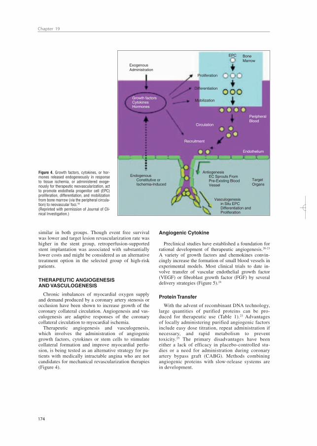
Chapter 19
174
Aoki J, et al. Emergent Strategies in the Interventional Cardiology
91 Rev Esp Cardiol. 2005;58(8):962-73 965
similar in both groups. Though event free survivalwas lower and target lesion revascularization rate washigher in the stent group, retroperfusion-supportedstent implantation was associated with substantiallylower costs and might be considered as an alternativetreatment option in the selected group of high-riskpatients.
THERAPEUTIC ANGIOGENESIS AND VASCULOGENESIS
Chronic imbalances of myocardial oxygen supplyand demand produced by a coronary artery stenosis orocclusion have been shown to increase growth of thecoronary collateral circulation. Angiogenesis and vas-culogenesis are adaptive responses of the coronarycollateral circulation to myocardial ischemia.
Therapeutic angiogenesis and vasculogenesis,which involves the administration of angiogenicgrowth factors, cytokines or stem cells to stimulatecollateral formation and improve myocardial perfu-sion, is being tested as an alternative strategy for pa-tients with medically intractable angina who are notcandidates for mechanical revascularization therapies(Figure 4).
Angiogenic Cytokines
Preclinical studies have established a foundation forrational development of therapeutic angiogenesis.20-23
A variety of growth factors and chemokines convin-cingly increase the formation of small blood vessels inexperimental models. Most clinical trials to date in-volve transfer of vascular endothelial growth factor(VEGF) or fibroblast growth factor (FGF) by severaldelivery strategies (Figure 5).24
Protein Transfer
With the advent of recombinant DNA technology,large quantities of purified proteins can be pro-duced for therapeutic use (Table 1).25 Advantagesof locally administering purified angiogenic factorsinclude easy dose titration, repeat administration ifnecessary, and rapid metabolism to preventtoxicity.25 The primary disadvantages have beeneither a lack of efficacy in placebo-controlled stu-dies or a need for administration during coronaryartery bypass graft (CABG). Methods combiningangiogenic proteins with slow-release systems arein development.
ExogenousAdministration
EPC BoneMarrow
PeripheralBlood
Endothelium
TargetOrgans
Proliferation
Differentiation
Mobilization
Circulation
Recruitment
Growth factorsCytokinesHormones
EndogenousConstitutive orIschemia-Induced
Vasculogenesisin Situ EPCDifferentiation andProliferation
AntiogenesisEC Sprouts FromPre-Existing Blood Vessel
Figure 4. Growth factors, cytokines, or hor-mones released endogeneously in responseto tissue ischemia, or administered exoge-nously for therapeutic neovascularization, actto promote endothelia progenitor cell (EPC)proliferation, differentiation, and mobilizationfrom bone marrow (via the peripheral circula-tion) to neovascular foci.19
(Reprinted with permission of Journal of Cli-nical Investigation.)
Document dowloaded from http://www.revespcardiol.org, day 11/01/2006. This copy is for personal use. Any transmission of this document by any media or format is strictly prohibited.

175
Emergent strategies in interventional cardiology
Gene Therapy
Gene therapy is an approach in which the geneticmaterial directing production of a protein is trans-ferred in place of the protein itself. A number of ve-hicles are used for transferring DNA to heart tissue,including purified DNA, DNA/lipid complexes, ade-no-associated viruses, or adenovirus.24,25 An advan-tage of this approach appears to be localized, sus-tained but not indefinite production of angiogenicfactors.26 Apparent disadvantages are the possibili-ties of vector toxicity, an immune response to thegene therapy vector, or inappropriately localizedgene transfer resulting in angiogenesis in a tumouror the retina.
The efficacy of gene transfer approaches to thera-peutic angiogenesis is now being tested in clinicaltrials (Table 2).25,27-33 Early uncontrolled open-labelclinical trials generally gave positive results, al-though the possibility of a placebo effect was not ex-cluded. Controlled Phase II trials are providing posi-tive but not definitive results. This is promising, sincethe patient population being studied has failed allother therapies and is likely to be refractory to inter-vention. However, most of the efficacy measuresstudied to date are surrogate endpoints such as exer-cise tolerance time (ETT), angina, or perfusion.While these measures are useful in suggesting clini-cal efficacy, hard clinical endpoints such as mortality,myocardial infarction (MI) and the need for revascu-larization should be studied. Long-term follow updata are also necessary. An important observation isthat the safety results of these trials indicate no majorproblems. Potential side-effects such as worsening ofatherosclerosis, retinopathy or cancer, have not beenobserved in clinical trials.
Two large Phase III clinical trials (AGENT 3 and 4)were designed to evaluate further the safety and effica-cy of Ad5FGF4.31 Both trials were designed as ran-domized, double blind and placebo controlled trials. Ineach trial, a recruitment goal was 450 patients andthese patients would be randomized to 3 groups(placebo group, Ad5FGF-4 at a dose of 109 vp group,and Ad5FGF-4 at a dose of 1010 vp group). In January2004, enrollment was stopped (416 patients in AGENT3 and 115 patients in AGENT 4) because interim dataanalysis of AGENT 3 indicates that the studies willprovide insufficient evidence of efficacy. However, en-rolled patients’ follow-up continues and final data willbe presented in the near future.
Cell Based Therapy
Another alternative method for increasing coronaryvascularization is the transplantation of stem or pro-genitor cells.34,35 These cells not only produce a varietyof growth factors and cytokines, but participate struc-
966 Rev Esp Cardiol. 2005;58(8):962-73 92
Aoki J, et al. Emergent Strategies in the Interventional Cardiology
turally in the formation of new vascular tissue andmyocyte.
The promising results from experimental studiespromoted the initiation of clinical trials.36,38 Stem andprogenitor cells are being tested in patients with bothacute myocardial infarction and chronic ischemicheart failure (Table 3 and 4).35,39-46 Improved wall mo-tions or increased perfusion was demonstrated inmost of study patients. However, BOOST is the solerandomized controlled clinical trial.42 Sixty patientswith ST-segment elevation myocardial infarction
Figure 5. Clinical feasible gene delivery routes.25
(Reprinted with permission of Nature Publishing Group.)
Intracoronary Inyection
Angioplasty
EpicardialInyection
EpicardialInyection
IntraventricularInyection
Adventitial Delivery.Cell Grafting orex Vivo Treatament ofBypass Grafts
TABLE 1. Proteins That Stimulate Angiogenesis inPreclinical Studies*,25
Growth factorsVEGF-A, VEGF-B, VEGF-C, VEGF-D, VEGF-E, PLGFFGF-1, FGF-2, FGF-4, FGF-5Angiopoietin-1, Angiopoietin-2HGF, PDGF-BBGM-CSF, neurotropinIGF-1, IGF-2ChemokinesMCP-1Transcription factorsHIF-1, Egr-1, Prox-1OthersDel-1,Cyr 61, PR39Tissue kallikrein, Secreted frizzled-related proteineNOS, iNOS
*VEGF indicates vascular endothelial growth factors. PLGF, placental growthfactors; FGF, fibroblast growth factors; HGF, hepatocyte growth factors;PDGF, platelet derived growth factors; GM-CSF, granulocyte macrophage co-lony stimulating factors; IGF, insulin like growth factors; MCP, monocyte che-motactic protein; HIF, hypoxia inducible transcription factors; Egr-1, earlygrowth response factor-1; Prox-1, prospero-related homeobox-1; Del-1, deve-lopmentally regulated endothelial cell locus-1; Cyr 61, cysteine-rich angioge-nic inducer 61; PR 39, praline and arginine-rich peptide; eNOS, endothelial ni-tric oxide synthetase; iNOS, inducible nitric oxide synthetase.
Document dowloaded from http://www.revespcardiol.org, day 11/01/2006. This copy is for personal use. Any transmission of this document by any media or format is strictly prohibited.

Chapter 19
176
Aoki J, et al. Emergent Strategies in the Interventional Cardiology
93 Rev Esp Cardiol. 2005;58(8):962-73 967
were randomly assigned to either a control group thatreceived optimum post infarction medical treatment,or a bone-marrow-cell group that received optimummedical treatment and intracoronary transfer of au-tologous bone marrow cells 4.8 days after percuta-neous coronary intervention (PCI). After 6 months,mean global left ventricular ejection fraction (LVEF)had significantly increased in the bone-marrow-cellgroup, compared to the control group. However, moststudies at present are limited by the small patient en-rolment, and recently some papers reported thatbone-marrow-derived haematopoietic stem cells donot transdifferentiate into cardiac myocytes in is-chemic myocardium.47,48
PERCUTANEOUS MYOBLASTTRANSPLANTATION
Most preclinical experience has been reported ontransplantation of skeletal myoblasts in infarcted myo-cardium.49-52 These studies demonstrated that trans-planted skeletal myoblasts in damaged myocardiumare capable of cellular engraftment, myotube forma-
tion, and long-term graft survival. Several clinical per-cutaneous myoblast transplantation trials, using boththe transendocardial and the transvenous approaches,have been reported.
Transendocardial Approach
A pilot safety and feasibility study on percutaneoustransplantation of autologous skeletal myoblasts bytransendocardial injection in five patients with is-chemic heart failure was reported by the group of Rot-terdam.50 Autologous skeletal myoblasts were obtainedfrom the quadriceps muscle and cultured in vitro forcell expansion. With a NOGA®-guided catheter sys-tem, 196±105 million cells were transendocardially in-jected into the infarcted area. After 3 months, LV ejec-tion fraction increased and regional wall analysis bymagnetic resonance image (MRI) showed significantincreased wall thickening at the target area. In the lightof these preliminary favourable results, a multicenterEuropean uncontrolled investigation has been started.Overall, 15 patients have been enrolled and treatedwith transendocardial skeletal myoblasts injection.
TABLE 2. The Result of Controlled Phase II Clinical Trials Testing Angiogenic Factors to Treat Coronary HeartDisease*,25
Trial Therapeutic Agent No. End Point Results
VIVA27 Recombinant VEGF protein 178 ETT (60 days) NegativeFIRST28 Recombinant FGF-2 protein 337 ETT (90 days) NegativeGM-CSF29 Recombinant GM-CSF protein 21 Invasive collateral flow index
(2 weeks) PositiveAGENT30 Adenovirus-FGF-4 79 ETT (4 weeks) PositiveAGENT II 31 Adenovirus-FGF-4 52 MPI (8 weeks) PositiveKAT 32 Adenovirus-VEGF 165 103 MPI (6 months) Positive
Plasmid/liposomeVEGF165 (Adeno only)
Euroinject One 33 Plasmid VEGF165 74 MPI (3 months) Negative
*VEGF indicates vascular endothelial growth factors; FGF, fibroblast growth factors; ETT, exercise tolerance time; MPI, myocardial perfusion improvement.
TABLE 3. Stem/Progenitor Cell Therapy (Intracoronary Application) in Patients With Acute MyocardialInfarction*,35
Reference Cell Type Patients Results
Strauer et al39 BMC (40 mL) versus control n=10 Hypokinetic area (LVA) ↓, contractility infarct region ↑, end-systolic volume ↓,perfusion (tallium scintigraphy) ↑
TOPCARE-AMI40,41 BMC (50 mL) versus CPC (250 mL) n=20 Global and regional EF (LVA, echo, MRI) ↑,end-systolic volume , viability (PET, MRI) ↑, coronary flow reserve ↑
BOOST (prospective BMC (120 mL) versus CONTROL n=30 EF (MRI) ↑and randomised)42
*BMC indicates bone marrow cell; CPC, circulating progenitor cell; LVA, left ventricular angiogram; EF, ejection fraction; MRI, magnetic resonance image; PET, po-sitron emission tomography.
Document dowloaded from http://www.revespcardiol.org, day 11/01/2006. This copy is for personal use. Any transmission of this document by any media or format is strictly prohibited.

177
Emergent strategies in interventional cardiology
968 Rev Esp Cardiol. 2005;58(8):962-73 94
Aoki J, et al. Emergent Strategies in the Interventional Cardiology
The results of this study will be presented in the nearfuture.
Transvenous Approach
A novel catheter-based endovascular system for di-rect myocardial injection using IVUS guide needlepunctures via the coronary venous system (theTRANSAccessTM) has been developed (Figure 6).53 Inthe POZAN trial, 10 patients underwent intramyocar-dial injection by using the TRANSAccess.54 The pro-cedure was not successful in one patient but NYHAclass improved in all patients and ejection fraction in-creased 3%-8% in 6 out of 9 cases during 6 month fol-low-up.
Ventricular Arrythmia
After intramyocardial injection of skeletal myo-blasts, ventricular arrhythmia was reported. Menascheet al52 reported that implantable cardioverter-defibrilla-tor (ICD) implantation for ventricular arrythmias wasrequired in 4 out of 10 patients after open chest autolo-gus myoblast transplantation.50,52 One of the 5 patientswho were enrolled in the study on percutaneoustransplantation of autologous skeletal myoblasts bytransendocardial approach had also sustained episodesof ventricular tachycardia and required ICD. This isprobably related to: a) heterogenecity of action poten-tials between the native and the transplanted stem cells;b) intrinsic arrhythmia potential of injected cells; c) in-creased nerve sprouting induced by stem cell injection;and d) local injury or edema induced by myocardialpuncture and inflammatory response.
FUTURE PERSPECTIVES TOWARDSINVASIVE DETECTION OF VULNERABLEPLAQUE
Lipid-Rich Atheromatous Core
IVUS provides an accurate tomographic view of thecoronary arteries and has shown a high correlation
with histology samples in in vitro validation studies.55-57
However, accurate plaque characterization with visualinterpretation of gray-scale IVUS, particularly of lipidrich plaques, remains an unresolved issue.56 On thecontrary, spectral analysis of IVUS radiofrequencydata (IVUS-Virtual HistologyTM [IVUS-VH]) hasdemonstrated potential to provide detailed quantitativeinformation on plaque composition and has been vali-dated in studies of explanted human coronary seg-ments (Figure 7).58
Angioscopic yellow plaques have been related toatheromatous plaques in previous validation studies.59
However, this technique evaluates only the luminalsurface of the intima thus quantifiable values of lipidcore size are out of its scope.
Raman spectroscopy is a technique that can cha-racterize the chemical composition of tissues.60 Invitro studies have demonstrated that diagnostic algo-rithms allow the discrimination of coronary arterialtissue in 3 categories: non-atherosclerotic, non-calci-fied and calcified plaques.61 Nevertheless, this tech-nique also lacks the ability to provide geometrical in-formation of the vessel and has a shallow penetrationdepth.
Intravascular ultrasound elastography and palpogra-phy are techniques that allow the assessment of localmechanical tissue properties (Figure 8).62,63 At a de-fined pressure, soft tissue (lipid-rich) components willdeform more than hard components (fibrous-calci-fied).64 However, they are unable to provide quantifia-ble measurements of plaque components.
Finally, intravascular magnetic resonance (IVM-RI) has recently emerged as a potential tool to iden-tify TCFA since it can accurately determine the pre-sence of lipid within the arterial wall.65,66 In vivofeasibility remains to be proven and acquisition timeand extent of the scan analysis requires impro-vement.
Thin-Fibrous Cap
The threshold to define the cap as “thin” has pre-viously been set at <65 µm.67 However, it is well
TABLE 4. Stem/Progenitor Cell Therapy in Patients With Chronic Heart Failure*,35
Reference Cell Type Application Patients Results
Stamm et al43 CD133+BMC (85-195 mL) Injection during CABG n=6 EF in 4 patients (LVA) ↑, perfusion in 5 patients (SPECT) ↑
Tse et al44 BMC (50 mL) Intramyocardial/NOGA n=8 Wall motion and thickening ↑Fuchs et al45 BMC Intramyocardial/NOGA n=10 Angina score and stress-induced
ischemia improvedPerin et46 BMC (50 mL) Intramyocardial/NOGA n=14 EF ↑, end-systolic volume ↓
*CD indicates cluster of differentiation; BMC, bone marrow cell; CABG, coronary artery bypass grafting; EF, ejection fraction; LVA, left ventricular angiogram;SPECT, single photon emission computed tomography; EF, ejection fraction.
en
T: tomo-
giogram;
Document dowloaded from http://www.revespcardiol.org, day 11/01/2006. This copy is for personal use. Any transmission of this document by any media or format is strictly prohibited.

Chapter 19
178
Aoki J, et al. Emergent Strategies in the Interventional Cardiology
95 Rev Esp Cardiol. 2005;58(8):962-73 969
established that general tissue shrinkage can not beavoided during dehydration.68 Shrinkage of up to60%, 15% and 80% can occur during critical-point-drying, free-drying, and air-drying respectively.69
Furthermore, post-mortem contraction of arteries fur-ther contributes to deteriorate the pathological quan-tification of atherosclerosis.70 Accordingly, we be-lieve that the threshold should be higher than 65 µm.Since the axial resolution of IVUS is between 100and 150 µm, techniques such as IVUS-VH with thecapacity to quantify each plaque component and toidentify the location of the lipid core in relation tothe lumen have the potential to recognize all the 3features of TCFA lesions.
Optical coherence tomography (OCT) is an imagingtechnique that allows high-resolution (axial resolutionof 15 µm) imaging in biological systems (Figure 9).71
Accordingly, OCT has the capacity to allow in-vivo,real time visualization of a thin fibrous cap. Recent invivo data suggest the possibility of detection ofmacrophages in atherosclerotic plaques.72
The sensitivity and specificity of palpography to de-tect vulnerable plaques has recently been assessed inpost-mortem human coronary arteries where vulnera-ble plaques were detected with a sensitivity of 88%and a specificity of 89%.63
Positive Remodeling
The expansive compensatory enlargement of thecoronary arteries in response to an increase in plaquearea is called positive or expansive remodeling.73
Several studies showed an increase in inflammatorymarker levels, larger lipid cores, and pronounced me-dial thinning in positive remodeled vessels.74-76 Re-cently, the relationship between vascular remodelingand plaque composition was assessed using IVUS.77-79
In these studies, the remodeling index for soft lesions
Figure 6. Direct myocardial injection using IVUS guide needle punctu-res via the coronary venous system.
Figure 7. Virtual histology image. Red: lipid core. White: calcium. Ye-llow: fibrofatty. Green: fibrosis. Gray: media.
PericardialSpace
Vein
Artery
SeptumVentricularCavity
Anterior Wall
currentPointerPositionNeedleTrajectory
Figure 8. Palpography image. Yellow,high strain spot.
Document dowloaded from http://www.revespcardiol.org, day 11/01/2006. This copy is for personal use. Any transmission of this document by any media or format is strictly prohibited.
color figures on page 225
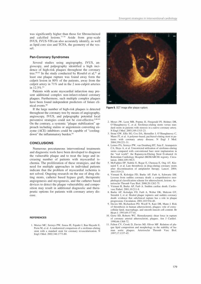
179
Emergent strategies in interventional cardiology
was significantly higher than those for fibrous/mixedand calcified lesions.77-79 Aside from gray-scaleIVUS, IVUS-VH can also accurately identify, as wellas lipid core size and TCFA, the geometry of the ves-sel.
Pan-Coronary Syndromes
Several studies using angiography, IVUS, an-gioscopy, and palpography identified a high inci-dence of high-risk plaques throughout the coronarytree.80-83 In the study conducted by Rioufol et al,81 atleast one plaque rupture was found away form theculprit lesion in 80% of the patients, away from theculprit artery in 71% and in the 2 non-culprit arteriesin 12.5%.81
Patients with acute myocardial infarction may pre-sent additional complex non-infarct-related coronaryplaques. Furthermore, such multiple complex plaqueshave been found independent predictors of future cli-nical events.80
If the large number of high-risk plaques is detectedthroughout the coronary tree by means of angiography,angioscopy, IVUS, and palpography potential localpreventive strategies could not be cost-effective.8,80-83
On the contrary, a systemic “plaque stabilization” ap-proach including statins or angiotensin converting en-zyme (ACE) inhibitors could be capable of “cooling-down” the inflammatory burden.84
CONCLUSIONS
Numerous percutaneous interventional treatmentsand diagnostic tools have been developed to diagnosethe vulnerable plaque and to treat the large and in-creasing number of patients with myocardial is-chemia. The proliferation of these strategies, and theneed for multiple approaches in individual patientsindicate that the problem of myocardial ischemia isnot solved. Ongoing research on the use of drug elu-ting stents, catheter based bypass graft, therapeuticangiogenesis and myogenesis, and the catheter baseddevices to detect the plaque vulnerability and compo-sition may result in additional diagnostic and thera-peutic options for patients with coronary artery dis-ease.
REFERENCES
1. Morice MC, Serruys PW, Sousa JE, Fajadet J, Ban Hayashi E,Perin M, et al. A randomized comparison of a sirolimus-elutingstent with a standard stent for coronary revascularization. NEngl J Med. 2002;346:1773-80.
2. Moses JW, Leon MB, Popma JJ, Fitzgerald PJ, Holmes DR,O’Shaughnessy C, et al. Sirolimus-eluting stents versus stan-dard stents in patients with stenosis in a native coronary artery.N Engl J Med. 2003;349:1315-23.
3. Stone GW, Ellis SG, Cox DA, Hermiller J, O’Shaughnessy C,Mann JT, et al. A polymer-based, paclitaxel-eluting stent in pa-tients with coronary artery disease. N Engl J Med.2004;350:221-31.
4. Lemos PA, Serruys PW, van Domburg RT, Saia F, ArampatzisCA, Hoye A, et al. Unrestricted utilization of sirolimus-elutingstents compared with conventional bare stent implantation inthe “real world”: the Rapamycin-Eluting Stent Evaluated AtRotterdam Cardiology Hospital (RESEARCH) registry. Circu-lation. 2004;109:190-5.
5. McFadden EP, Stabile E, Regar E, Cheneau E, Ong AT, Kin-naird T, et al. Late thrombosis in drug-eluting coronary stentsafter discontinuation of antiplatelet therapy. Lancet. 2004;364:1519-21.
6. Virmani R, Kolodgie FD, Burke AP, Farb A, Schwartz SM.Lessons from sudden coronary death: a comprehensive mor-phological classification scheme for atherosclerotic lesions. Ar-terioscler Thromb Vasc Biol. 2000;20:1262-75.
7. Virmani R, Burke AP, Farb A. Sudden cardiac death. Cardio-vasc Pathol. 2001;10:211-8.
8. Burke AP, Kolodgie FD, Farb A, Weber DK, Malcom GT,Smialek J, et al. Healed plaque ruptures and sudden coronarydeath: evidence that subclinical rupture has a role in plaqueprogression. Circulation. 2001;103:934-40.
9. Davies MJ, Richardson PD, Woolf N, Katz DR, Mann J. Riskof thrombosis in human atherosclerotic plaques: role of extra-cellular lipid, macrophage, and smooth muscle cell content. BrHeart J. 1993;69:377-81.
10. Gertz SD, Roberts WC. Hemodynamic shear force in ruptureof coronary arterial atherosclerotic plaques. Am J Cardiol.1990;66:1368-72.
11. Felton CV, Crook D, Davies MJ, Oliver MF. Relation of pla-que lipid composition and morphology to the stability of hu-man aortic plaques. Arterioscler Thromb Vasc Biol.1997;17:1337-45.
970 Rev Esp Cardiol. 2005;58(8):962-73 96
Aoki J, et al. Emergent Strategies in the Interventional Cardiology
Figure 9. OCT image after plaque rupture.
Document dowloaded from http://www.revespcardiol.org, day 11/01/2006. This copy is for personal use. Any transmission of this document by any media or format is strictly prohibited.

Chapter 19
180
12. Farb A, Burke AP, Tang AL, Liang TY, Mannan P, Smialek J,et al. Coronary plaque erosion without rupture into a lipid core.A frequent cause of coronary thrombosis in sudden coronarydeath. Circulation. 1996;93:1354-63.
13. Kolodgie FD, Burke AP, Farb A, Gold HK, Yuan J, Narula J,et al. The thin-cap fibroatheroma: a type of vulnerable plaque:the major precursor lesion to acute coronary syndromes. CurrOpin Cardiol. 2001;16:285-92.
14. Oesterle SN, Reifart N, Hauptmann E, Hayase M, Yeung AC.Percutaneous in situ coronary venous arterialization: report ofthe first human catheter-based coronary artery bypass. Circula-tion. 2001;103:2539-43.
15. Oesterle SN, Reifart N, Hayase M, Hauptmann E, Low R, Er-bel R, et al. Catheter-based coronary bypass: a developmentupdate. Catheter Cardiovasc Interv. 2003;58:212-8.
16. Boekstegers P, Raake P, Al Ghobainy R, Horstkotte J, HinkelR, Sandner T, et al. Stent-based approach for ventricle-to-coro-nary artery bypass. Circulation. 2002;106:1000-6.
17. Boekstegers P, Giehrl W, von Degenfeld G, Steinbeck G. Se-lective suction and pressure-regulated retroinfusion: an effecti-ve and safe approach to retrograde protection against myocar-dial ischemia in patients undergoing normal and high riskpercutaneous transluminal coronary angioplasty. J Am CollCardiol. 1998;31:1525-33.
18. Pohl T, Giehrl W, Reichart B, Kupatt C, Raake P, Paul S, et al.Retroinfusion-supported stenting in high-risk patients for per-cutaneous intervention and bypass surgery: results of the pros-pective randomized myoprotect I study. Catheter CardiovascInterv. 2004;62:323-30.
19. Isner JM, Asahara T. Angiogenesis and vasculogenesis as the-rapeutic strategies for postnatal neovascularization. J Clin In-vest. 1999;103:1231-6.
20. Romano di Peppe S, Mangoni A, Zambruno G, Spinetti G, Me-lillo G, et al. Adenovirus-mediated VEGF(165) gene transferenhances wound healing by promoting angiogenesis in CD1diabetic mice. Gene Ther. 2002;9:1271-7.
21. Deodato B, Arsic N, Zentilin L, Galeano M, Santoro D, TorreV, et al. Recombinant AAV vector encoding human VEGF165enhances wound healing. Gene Ther. 2002;9:777-85.
22. Wright MJ, Wightman LM, Latchman DS, Marber MS. Invivo myocardial gene transfer: optimization and evaluationof intracoronary gene delivery in vivo. Gene Ther. 2001;8:1833-9.
23. Vitadello M, Schiaffino MV, Picard A, Scarpa M, Schiaffino S.Gene transfer in regenerating muscle. Hum Gene Ther. 1994;5:11-8.
24. Losordo DW, Dimmeler S. Therapeutic angiogenesis and vas-culogenesis for ischemic disease. Part I: angiogenic cytokines.Circulation. 2004;109:2487-91.
25. Yla-Herttuala S, Alitalo K. Gene transfer as a tool to inducetherapeutic vascular growth. Nat Med. 2003;9:694-701.
26. Simons M, Bonow RO, Chronos NA, Cohen DJ, Giordano FJ,Hammond HK, et al. Clinical trials in coronary angiogenesis:issues, problems, consensus: An expert panel summary. Circu-lation. 2000;102:E73-86.
27. Henry TD, Annex BH, McKendall GR, Azrin MA, López JJ,Giordano FJ, et al. The VIVA trial: Vascular endothelialgrowth factor in Ischemia for Vascular Angiogenesis. Circula-tion. 2003;107:1359-65.
28. Simons M, Annex BH, Laham RJ, Kleiman N, Henry T,Dauerman H, et al. Pharmacological treatment of coronary ar-tery disease with recombinant fibroblast growth factor-2: dou-ble-blind, randomized, controlled clinical trial. Circulation.2002;105:788-93.
29. Seiler C, Pohl T, Wustmann K, Hutter D, Nicolet PA, Windec-ker S, et al. Promotion of collateral growth by granulocyte-ma-crophage colony-stimulating factor in patients with coronaryartery disease: a randomized, double-blind, placebo-controlledstudy. Circulation. 2001;104:2012-7.
Aoki J, et al. Emergent Strategies in the Interventional Cardiology
97 Rev Esp Cardiol. 2005;58(8):962-73 971
30. Grines CL, Watkins MW, Helmer G, Penny W, Brinker J, Mar-mur JD, et al. Angiogenic Gene Therapy (AGENT) trial in pa-tients with stable angina pectoris. Circulation. 2002;105:1291-7.
31. Grines C. The AGENT clinical trials programme. Eur Heart JSupplement. 2004;6:18-23.
32. Hedman M, Hartikainen J, Syvanne M, Stjernvall J, HedmanA, Kivela A, et al. Safety and feasibility of catheter-based localintracoronary vascular endothelial growth factor gene transferin the prevention of postangioplasty and in-stent restenosis andin the treatment of chronic myocardial ischemia: phase II re-sults of the Kuopio Angiogenesis Trial (KAT). Circulation.2003;107:2677-83.
33. Kastrup J. Euroinject One trial. Late breaking clinical trialssession. J Am Coll Cardiol. 2003;41:1603.
34. Rafii S, Lyden D. Therapeutic stem and progenitor cell trans-plantation for organ vascularization and regeneration. Nat Med.2003;9:702-12.
35. Losordo DW, Dimmeler S. Therapeutic angiogenesis and vas-culogenesis for ischemic disease: part II: cell-based therapies.Circulation. 2004;109:2692-7.
36. Kawamoto A, Gwon HC, Iwaguro H, Yamaguchi JI, Uchida S,Masuda H, et al. Therapeutic potential of ex vivo expanded en-dothelial progenitor cells for myocardial ischemia. Circulation.2001;103:634-7.
37. Fuchs S, Baffour R, Zhou YF, Shou M, Pierre A, Tio FO, et al.Transendocardial delivery of autologous bone marrow enhan-ces collateral perfusion and regional function in pigs with chro-nic experimental myocardial ischemia. J Am Coll Cardiol.2001;37:1726-32.
38. Kawamoto A, Tkebuchava T, Yamaguchi J, Nishimura H,Yoon YS, Milliken C, et al. Intramyocardial transplantation ofautologous endothelial progenitor cells for therapeutic neovas-cularization of myocardial ischemia. Circulation. 2003;107:461-8.
39. Strauer BE, Brehm M, Zeus T, Kostering M, Hernandez A,Sorg RV, et al. Repair of infarcted myocardium by autologousintracoronary mononuclear bone marrow cell transplantation inhumans. Circulation. 2002;106:1913-8.
40. Assmus B, Schachinger V, Teupe C, Britten M, Lehmann R,Dobert N, et al. Transplantation of Progenitor Cells and Rege-neration Enhancement in Acute Myocardial Infarction (TOP-CARE-AMI). Circulation. 2002;106:3009-17.
41. Britten MB, Abolmaali ND, Assmus B, Lehmann R, Honold J,Schmitt J, et al. Infarct remodeling after intracoronary progeni-tor cell treatment in patients with acute myocardial infarction(TOPCARE-AMI): mechanistic insights from serial contrast-enhanced magnetic resonance imaging. Circulation. 2003;108:2212-8.
42. Wollert KC, Meyer GP, Lotz J, Ringes-Lichtenberg S, Lip-polt P, Breidenbach C, et al. Intracoronary autologous bone-marrow cell transfer after myocardial infarction: the BOOSTrandomised controlled clinical trial. Lancet. 2004;364:141-8.
43. Stamm C, Westphal B, Kleine HD, Petzsch M, Kittner C, Klin-ge H, et al. Autologous bone-marrow stem-cell transplantationfor myocardial regeneration. Lancet. 2003;361:45-6.
44. Tse HF, Kwong YL, Chan JK, Lo G, Ho CL, Lau CP. Angio-genesis in ischaemic myocardium by intramyocardial autolo-gous bone marrow mononuclear cell implantation. Lancet.2003;361:47-9.
45. Fuchs S, Satler LF, Kornowski R, Okubagzi P, Weisz G, Baf-four R, et al. Catheter-based autologous bone marrow myocar-dial injection in no-option patients with advanced coronary ar-tery disease: a feasibility study. J Am Coll Cardiol. 2003;41:1721-4.
46. Perin EC, Dohmann HF, Borojevic R, Silva SA, Sousa AL,Mesquita CT, et al. Transendocardial, autologous bone marrowcell transplantation for severe, chronic ischemic heart failure.Circulation. 2003;107:2294-302.
Document dowloaded from http://www.revespcardiol.org, day 11/01/2006. This copy is for personal use. Any transmission of this document by any media or format is strictly prohibited.

181
Emergent strategies in interventional cardiology
972 Rev Esp Cardiol. 2005;58(8):962-73 98
Aoki J, et al. Emergent Strategies in the Interventional Cardiology
47. Balsam LB, Wagers AJ, Christensen JL, Kofidis T, WeissmanIL, Robbins RC. Haematopoietic stem cells adopt mature hae-matopoietic fates in ischaemic myocardium. Nature. 2004;428:668-73.
48. Murry CE, Soonpaa MH, Reinecke H, Nakajima H, NakajimaHO, Rubart M, et al. Haematopoietic stem cells do not transdif-ferentiate into cardiac myocytes in myocardial infarcts. Nature.2004;428:664-8.
49. Herreros J, Prosper F, Pérez A, Gavira JJ, García-VellosoMJ, Barba J, et al. Autologous intramyocardial injection ofcultured skeletal muscle-derived stem cells in patients withnon-acute myocardial infarction. Eur Heart J. 2003;24:2012-20.
50. Smits PC, van Geuns RJ, Poldermans D, Bountioukos M, On-derwater EE, Lee CH, et al. Catheter-based intramyocardial in-jection of autologous skeletal myoblasts as a primary treatmentof ischemic heart failure: clinical experience with six-monthfollow-up. J Am Coll Cardiol. 2003;42:2063-9.
51. Menasche P, Hagege AA, Scorsin M, Pouzet B, Desnos M,Duboc D, et al. Myoblast transplantation for heart failure. Lan-cet. 2001;357:279-80.
52. Menasche P, Hagege AA, Vilquin JT, Desnos M, Abergel E,Pouzet B, et al. Autologous skeletal myoblast transplantationfor severe postinfarction left ventricular dysfunction. J AmColl Cardiol. 2003;41:1078-83.
53. Thompson CA, Nasseri BA, Makower J, Houser S, McGarryM, Lamson T, et al. Percutaneous transvenous cellular car-diomyoplasty. A novel nonsurgical approach for myocardialcell transplantation. J Am Coll Cardiol. 2003;41:1964-71.
54. Siminiak T, Fiszer D, Jerzykowska O, Rozwadowska N, Gry-gielska B, Majewski M, et al. Percutaneous transvenous trans-plantation of autologus myoblasts in the treatment of postin-farction heart failure-the Pozan trial. Eur Heart J. 2004;25:264.
55. Tobis JM, Mallery JA, Gessert J, Griffith J, Mahon D, BessenM, et al. Intravascular ultrasound cross-sectional arterial ima-ging before and after balloon angioplasty in vitro. Circulation.1989;80:873-82.
56. Potkin BN, Bartorelli AL, Gessert JM, Neville RF, Almagor Y,Roberts WC, et al. Coronary artery imaging with intravascularhigh-frequency ultrasound. Circulation. 1990;81:1575-85.
57. Nishimura RA, Edwards WD, Warnes CA, Reeder GS, HolmesDR Jr, Tajik AJ, et al. Intravascular ultrasound imaging: in vi-tro validation and pathologic correlation. J Am Coll Cardiol.1990;16:145-54.
58. Nair A, Kuban BD, Tuzcu EM, Schoenhagen P, Nissen SE,Vince DG. Coronary plaque classification with intravascularultrasound radiofrequency data analysis. Circulation. 2002;106:2200-6.
59. Thieme T, Wernecke KD, Meyer R, Brandenstein E, HabedankD, Hinz A, et al. Angioscopic evaluation of atherosclerotic pla-ques: validation by histomorphologic analysis and associationwith stable and unstable coronary syndromes. J Am Coll Car-diol. 1996;28:1-6.
60. Baraga JJ, Feld MS, Rava RP. In situ optical histochemistryof human artery using near infrared Fourier transform Ra-man spectroscopy. Proc Natl Acad Sci U S A. 1992;89:3473-7.
61. Romer TJ, Brennan JF 3rd, Fitzmaurice M, Feldstein ML, Dei-num G, Myles JL, et al. Histopathology of human coronary at-herosclerosis by quantifying its chemical composition with Ra-man spectroscopy. Circulation. 1998;97:878-85.
62. Schaar JA, Regar E, Mastik F, McFadden EP, Saia F, Disco C,et al. Incidence of high-strain patterns in human coronary arte-ries: assessment with three-dimensional intravascular palpo-graphy and correlation with clinical presentation. Circulation.2004;109:2716-9.
63. Schaar JA, de Korte CL, Mastik F, Strijder C, Pasterkamp G,Boersma E, et al. Characterizing vulnerable plaque features
with intravascular elastography. Circulation. 2003;108:2636-41.
64. de Korte CL, van der Steen AF, Cespedes EI, Pasterkamp G.Intravascular ultrasound elastography in human arteries: ini-tial experience in vitro. Ultrasound Med Biol. 1998;24:401-8.
65. Schneiderman JWR, Weiss A, Smouha E, Muchnik L, Chen-Zion M, Golan E, et al. Detection of vulnerable plaques in ex-vivo human aortas with novel intravascular magnetic resonancecatheter(abstract). Circulation. 2002;657.
66. Schneiderman JWR, Weiss A, Smouha E, Muchnik L, Alexan-drowicz G, Chen-Zion M, et al. Vulnerable plaque diagnosis bya self-contained intravascular magnetic resonance imaging pro-be in ex-vivo human in-situ coronary arteries. J Am Coll Car-diol. 2003;41:59A.
67. Burke AP, Farb A, Malcom GT, Liang YH, Smialek J, VirmaniR. Coronary risk factors and plaque morphology in men withcoronary disease who died suddenly. N Engl J Med. 1997;336:1276-82.
68. MKW L. A critical appraise of the effects of fixation, dehy-dration and embedding of cell volume. In: Revel JP, BarnardT, Haggis GH, editors. The Science of Biological SpecimenPreparation for Microscopy and Microanalysis. ScanningElectron Microscopy, AMF O’Hare, IL 60666 1984; pp.61-70.
69. Boyde A JS, Tamarin A. Dimensional changes during speci-men preparation for scanning electron microscopy. Scan Elec-tron Microsc.1977:507-18.
70. Fishbein MC, Siegel RJ. How big are coronary atheroscleroticplaques that rupture? Circulation. 1996;94:2662-6.
71. Hrynchak P, Simpson T. Optical coherence tomography: an in-troduction to the technique and its use. Optom Vis Sci.2000;77:347-56.
72. MacNeill BD, Jang IK, Bouma BE, Iftimia N, Takano M, Ya-bushita H, et al. Focal and multi-focal plaque macrophage dis-tributions in patients with acute and stable presentations of co-ronary artery disease. J Am Coll Cardiol. 2004;44:972-9.
73. Glagov S, Weisenberg E, Zarins CK, Stankunavicius R, Kolet-tis GJ. Compensatory enlargement of human atheroscleroticcoronary arteries. N Engl J Med. 1987;316:1371-5.
74. Pasterkamp G, Schoneveld AH, van der Wal AC, Haudens-child CC, Clarijs RJ, Becker AE, et al. Relation of arterial geo-metry to luminal narrowing and histologic markers for plaquevulnerability: the remodeling paradox. J Am Coll Cardiol.1998;32:655-62.
75. Varnava AM, Mills PG, Davies MJ. Relationship between co-ronary artery remodeling and plaque vulnerability. Circulation.2002;105:939-43.
76. Burke AP, Kolodgie FD, Farb A, Weber D, Virmani R. Morp-hological predictors of arterial remodeling in coronary atheros-clerosis. Circulation. 2002;105:297-303.
77. Tauth J, Pinnow E, Sullebarger JT, Basta L, Gursoy S, LindsayJ Jr, et al. Predictors of coronary arterial remodeling patterns inpatients with myocardial ischemia. Am J Cardiol. 1997;80:1352-5.
78. Sabate M, Kay IP, de Feyter PJ, van Domburg RT, DeshpandeNV, Ligthart JM, et al. Remodeling of atherosclerotic coronaryarteries varies in relation to location and composition of pla-que. Am J Cardiol. 1999;84:135-40.
79. Fuessl RT, Kranenberg E, Kiausch U, Baer FM, Sechtem U,Hopp HW. Vascular remodeling in atherosclerotic coronary ar-teries is affected by plaque composition. Coron Artery Dis.2001;12:91-7.
80. Goldstein JA, Demetriou D, Grines CL, Pica M, Shoukfeh M,O’Neill WW. Multiple complex coronary plaques in patientswith acute myocardial infarction. N Engl J Med. 2000;343:915-22.
81. Rioufol G, Finet G, Ginon I, Andre-Fouet X, Rossi R, Vialle E,et al. Multiple atherosclerotic plaque rupture in acute coronary
Document dowloaded from http://www.revespcardiol.org, day 11/01/2006. This copy is for personal use. Any transmission of this document by any media or format is strictly prohibited.

Chapter 19
182
83. Schaar JA, Muller JE, Falk E, Virmani R, Fuster V, SerruysPW, et al. Terminology for high-risk and vulnerable coronaryartery plaques. Report of a meeting on the vulnerable plaque,June 17 and 18, 2003, Santorini, Greece. Eur Heart J.2004;25:1077-82.
84. Ambrose JA, Martínez EE. A new paradigm for plaque stabili-zation. Circulation. 2002;105:2000-4.
Aoki J, et al. Emergent Strategies in the Interventional Cardiology
99 Rev Esp Cardiol. 2005;58(8):962-73 973
syndrome: a three-vessel intravascular ultrasound study. Circu-lation. 2002;106:804-8.
82. Asakura M, Ueda Y, Yamaguchi O, Adachi T, Hirayama A,Hori M, et al. Extensive development of vulnerable plaquesas a pan-coronary process in patients with myocardial infarc-tion: an angioscopic study. J Am Coll Cardiol. 2001;37:1284-8.
Document dowloaded from http://www.revespcardiol.org, day 11/01/2006. This copy is for personal use. Any transmission of this document by any media or format is strictly prohibited.

Summary and Conclusions


185
SUMMARY AND CONCLUSIONS
Part 1 Long term tissue growth inside and outside drug-eluting stent in humans
After drug-eluting stents (DES) implantation, vessel reaction inside and outside the stent
is not identical. Chapter 2 presented vessel reaction after sirolimus-eluting stent (SES)
implantation. In the first 2 years, the mean plaque volume and plaque compositional
change expressed as mean percent (%) hypoechogenic tissue of the plaque behind the
stent struts did not significantly change. However, significant plaque shrinking with an
increase in plaque echogenicity was observed between 2 years and 4 years. The mean
neointimal volume increased over 4 years. However no further statistically significant
change occurred between 2 and 4 years. Chapter 3 showed that vessel reaction after
paclitaxel-eluting stent (PES) implantation. Increased plaque outside the stent 6 months
after PES implantation regressed completely in the slow release (SR) group and partially
in the middle release (MR) group by 2 years. Neointimal suppression with both SR and MR
PES is sustained up to 2 years. Whereas neointima decreased in the bare metal stent (BMS)
group between 6 months and 2 years, neointima continued to increase in the SR and MR
groups without impact on clinical events. Chapter 4 examined that vessel reaction after
various doses and pharmacokinetic release of PES with an erodable polymer. Although
type of polymer was different, vessel reaction inside and outside the stent was similar as
PES with a durable polymer. Chapter 5 summarized vessel reaction after DES, including
everolimus and ABT 578-eluting stents. Vessel reaction outside the stent was different
between limus family (sirolimus, and everolimus) and paclitaxel. Significant expansive
remodeling was observed only after paclitaxel release stents, but vessel regressed later
time point. Neointima, in other words, vessel reaction inside the stent, regressed after
implantation of BMS. However, neointima keeps growing after implantation of all kinds
of DES. Although long term follow-up after DES implantation shows a sustained clinical
benefit in several randomized trials, delayed neointimal growth beyond the first 6 to 9
months has been reported in serial intravascular ultrasound system (IVUS) analyses in
some trials. The issue of a delayed restenosis which was observed after brachytherapy
has not been thoroughly evaluated with DES.
Part 2 Efficacy of drug-eluting stent for high risk patients
In the BMS era, long lesion, diabetes mellitus, let main coronary artery disease, chronic
total occlusion, and bifurcation lesion are high risk for in-stent restenosis. Clinical effects
of SES and PES on patients with these high risk factors were analyzed. In Chapter 6,
Summary and Conclusions

186
the medium-term clinical outcome and the potential for adverse effects when very long
segments (i.e.≥64mm of stented length) were treated by DES implantation, an approach
colloquially referred to as a “full metal jacket” were evaluated. The one-year target ves-
sel revascularization rate was 7.5% and the overall incidence of major adverse cardiac
events was 18.0%. These results seemed better than full metal jacket, using BMS (The
one-year target vessel revascularization rate was 21.6% and the overall incidence of ma-
jor adverse cardiac events was 38.0% in the historical control.) Outcomes in SES and PES
groups did not differ statistically. In Chapter 7 and 8, patients with diabetes mellitus
were evaluated. Routine utilization of SES for diabetic patients significantly reduced the
rate of adverse cardiac events at 1 year compared to bare metal stents. (17.3% in the SES
group versus 30.2% in the pre-SES group (hazard ratio, 0.54 [95% confidence interval,
0.32-0.91]; p=0.02)) PES were associated with a nonsignificantly lower incidence of major
adverse cardiac events at 1 year compared with SES (adjusted hazard ratio 0.68, 95%
confidence interval 0.37 to 1.24, p = 0.21). Chapter 9 illustrated a patient with chronic
total occlusion of left main coronary artery who was successfully treated with bifurcation
stenting with SES. Chapter 10 and 11 analyzed patients with left main coronary artery
disease, the cumulative incidence of major adverse cardiovascular events was lower in
the DES cohort than in patients in the pre-DES group (24% versus 45%, respectively; haz-
ard ratio [HR], 0.52 [95% CI, 0.31 to 0.88]; P=0.01). Total mortality did not differ between
cohorts; however, there were significantly lower rates of both myocardial infarction (4%
versus 12%, respectively; HR, 0.22 [95% CI, 0.07 to 0.65]; P=0.006) and target vessel re-
vascularization (6% versus 23%, respectively; HR, 0.26 [95% CI, 0.10 to 0.65]; P=0.004) in
the DES group. PES may perform closely to SES both in terms of angiographic and long-
term clinical outcome, the cumulative incidence of major adverse cardiovascular events
was comparable (25% in the SES vs. 29%, in the PES group; HR 0.88 [95% CI: 0.43-1.82];
p=0.74), reflecting similarities in both the composite death/MI (16% in the SES and 18%
in the PES group) and target vessel revascularization (9% in the SES and 11% in the PES
group). Angiographic in-stent late loss (mm) was0.32±74 in the main and 0.36±0.59 in
the side-branch the SES vs. 0.46±0.57 and 0.52±0.42 in the PES, respectively (p=0.36 and
p=0.41).In Chapter 12 and 13, we demonstrated the efficacy of DES implantation for the
percutaneous treatment of chronic total occlusions when compared to BMS. Both SES
and PES were associated with a low rate of target vessel revascularization. At one year,
the survival-free of target vessel revascularization was significantly higher in the SES and
PES groups compared with the BMS group (97.4% and 96.4% versus 80.8% respectively,
p=0.01). Clinical and angiographic effects for patient with bifurcation lesions were ana-
lyzed in Chapter 14 and 15. Major adverse cardiac events (MACE) rates for both SES and
PES were low compared with historical data of BMS. Our data suggests a higher need for
target lesion revascularization for the PES compared with the SES, however further ran-
domized studies are needed to fully evaluate both stenting strategy, and any difference
Summary and Conclusions

187
between the stents (Survival-free of target lesion revascularization was 95.7% for SES
versus 86.8% for PES, p=0.01.). Technique of stenting was not a predictor of either MACE
or TLR. The most effective techniques for bifurcation stenting remain undefined.
Both SES and PES have almost similar clinical effect to reduce MACE compared to BMS,
even if patients have high risk restenosis factors.
Part 3 Endothelial progenitor cell – alternative to drug-eluting stents
DES have dramatically reduced the incidence of in-stent restenosis. However, the inci-
dence of angiographic stent thrombosis in an unselected, complex DES population was
1.0%, within the same range as the corresponding BMS population and concordant with
previously published results from the BMS era. (Chapter 16) Other technology of re-
ducing the incidence of stent thrombosis is necessary to improve clinical outcome of
coronary stenting. The acceleration of the endothelialization of stent is a key factor to
reduce stent thrombosis. (Chapter 17) Recently, the existence of circulating endothelial
progenitor cells (EPC) has been identified as a key factor for re-endothelialization. HEAL-
ING-FIM registry is the first clinical experience with implantation of a bio-engineered
stent which is anti-CD34 coated stents, designed to capture circulating endothelial pro-
genitor cells. The results of the HEALING-FIM (Healthy Endothelial Accelerated Lining
Inhibits Neointimal Growth - First In Man) registry showed that the EPC capture stent is
safe and feasible: with no stent thrombosis (30 days or 6 months), MACE occurred in only
one patient (6.3%), despite only 30 days of clopidogrel therapy. Further developments in
this technology are warranted to evaluate the efficacy of this device for the treatment of
coronary artery disease. (Chapter 18)
Part 4 Future direction to the interventional cardiology
Chapter 19 summarized that other approach for coronary artery disease, except DES.
Ongoing research on catheter based bypass graft, therapeutic angiogenesis and myo-
genesis, and the catheter based devices to detect the plaque vulnerability and composi-
tion may result in additional diagnostic and therapeutic options for patients with coro-
nary artery disease.
In conclusion
Both SES and PES have significant effect to reduce major accident cardiac events, mainly
due to reduce target lesion revascularization, even if patients have high risk factors for
restenosis. Current issues of DES are long term effects of these devices and stent throm-
Summary and Conclusions
188
bosis. Long-term follow-up after DES implantation shows a sustained clinical benefit in
several randomized trials. In the intravascular ultrasound analyses of DES, neointima con-
tinued to increase without impact on clinical events. Although plaque outside the stent
began to shrink beyond 1 year, longer term follow-up will be required to establish the
natural history of local drug delivery by DES. In addition, the EPC capture stent which can
rapidly endothelialize stainless steel stents has a possibility to reduce stent thrombosis in
the first human study. Further developments in this technology are warranted to evalu-
ate the efficacy of this device and reduce the incidence of stent thrombosis.
Chapter 20Summary and Conclusions

Samenvatting en conclusies


191
Samenvatting en conclusies
SAMENVATTING EN CONCLUSIES
Deel 1 Lange termijn weefselgroei aan binnen-en buitenzijde van drug eluting stents in de mens
Na drug-eluting stent (DES) plaatsing, is de vaatreaktie aan binnen-en buitenzijde van de
stent niet identiek. Hoofstuk 2 toonde de vaatreaktie volgend op sirolimus-eluting stent
(SES) implantatie. Gedurende de eerste twee jaar toonde de gemiddelde hoeveelheid
plaque en de veranderende plaque samenstelling uitgedrukt als gemiddeld percent-
age (%) echo-arm weefsel van de plaque achter de stent draden geen verandering van
betekenis. Echter, plaque krimp van betekenis met een toename van plaque echo genic-
iteit werd gezien tussen twee en vier jaar. De gemiddelde neointimale hoeveelheid nam
gedurende vier jaar toe. Echter, statistisch gezien werden geen belangrijke veranderin-
gen waargenomen tussen twee en vier jaar. Hoofdstuk 3 toonde vaatreaktie volgend
op paclitaxel-eluting stent (PES) implantatie. Toegenomen plaque, welke zes maanden
na PES implantatie aanwezig was aan de buitenzijde van de stent , verminderde volledig
in de groep met de langzame afgifte (SR) en gedeeltelijk in de groep met de middel-
matige afgifte (MR) gedurende de volgende twee jaar. Neointimale onderdrukking via
zowel SR als MR PES duurde tot twee jaar voort. Daar waar neointima in de Bare Metal
Stent (BMS) groep afnam tussen 6 maanden en twee jaar, bleef neointima toenemen in
de SR en MR groepen zonder van invloed te zijn op klinische gebeurtenissen. Hoofdstuk
4 onderzocht de vaatreaktie op diverse doses- en farmacokinetische afgifte van PES met
een erodeerbaar polymeer. Alhoewel sprake was van verschillende typen polymeer, de
vaatreaktie aan binnen-en buitenzijde van de stent was gelijk aan PES met een stabiele
polymeer.
Hoofdstuk 5 vatte vaatreaktie na DES samen, inclusief everolimus en ABT 578-eluting
stents. De vaatreaktie aan de buitenzijde van de stent was verschillend bij de limus fami-
lie (sirolimus en everolimus) en paclitaxel. Belangrijk positieve remodeling werd alleen
bij paclitaxel afgevende stents waargenomen maar nam later weer af.
Neointima, met andere woorden, vaatreaktie aan de binnenzijde van de stent, vermin-
derde na BMS implantatie.
Echter, bij alle soorten DES implantatie blijft neointima groeien. Hoewel lange termijn
follow-up na DES implantatie een voortdurend klinisch voordeel aantoont in verschei-
dene gerandomiseerde studies, is vertraagde neointimale groei na de eerste zes tot ne-
gen maanden gerapporteerd in opeenvolgende intravasculair utrasound system (IVUS)
analyses in enkele onderzoeken. De kwestie van de vertraagde restenosis, welk na bra-
chytherapie werd geobserveerd, is nog niet grondig geëvalueerd met behulp van DES.

192
Samenvatting en conclusies
Deel 2 Doeltreffendheid van drug-eluting stents for hoog-risico patienten
In het BMS-tijdperk, lange lesie, diabetes mellitus, linker coronair ziekte, chronische totaal
occlusie en bifurcatie lesies zijn risicovol voor in-stent restenose. Bij patienten met deze
hoog risico factoren werden de klinische effecten van SES en PES geanaliseerd. Hoofd-
stuk 6 Klinische resultaten op middellange termijn en het potentieel voor ongunstige
resultaten werden geëvalueerd bij patienten waarbij lange segmenten ( dat wil zeggen
≥ 64mm aan stent lengte ) werden behandeld met DES implantatie (populair “full metal
jacket” genoemd). Het éénjarig coronaire revascularisatie percentage was 7,5% en het
optreden van belangrijke cardiale eindpunten was 18%. Deze resultaten bleken beter
dan “full metal jacket” met BMS (het éénjarig percentage revasculatisatie was 21,6% en
het optreden van cardiale gebeurtenissen 38% in de historische controle). Resultaten
in SES-en PES groepen toonden statistisch geen verschil. In hoofdstuk 7 en 8 werden
patienten met diabetes mellitus geëvalueerd. In vergelijking met BMS reduceerde rou-
tinematig gebruik van SES voor diabetes patienten cardiale gebeurtenissen binnen een
jaar aanzienlijk. (17.3% in de SES groep versus 30.2% in de pre-SES groep (risico percent-
age, 0.54 [95% vertrouwens interval, 0.32-0.91]; p=0.02)). PES werd geassocieerd met
een non-significant optreden van cardiale gebeurtenissen binnen een jaar vergeleken
met SES adjusted risico percentage 0.68, 95% vertrouwens interval 0.37 to 1.24, p=0.21).
Hoofdstuk 9 illustreerde een patient met een chronische totale occlusie van de linker
coronair welke succesvol behandeld werd met bifurcatie stenting met SES. Hoofdstuk
10 en 11 analyseerde patienten met linker coronair ziekte, het cumulatieve vóórkomen
van belangrijke cardiale eindpunten was lager in het DES cohort dan in de pre-DES pati-
enten groep (24% versus 45%, respectievelijk; risico percentage [HR] , 0.52 [95% CI, 0.31
tot 0.88]; P=0.01). De totale mortaliteit tussen cohorten verschilde niet, echter, er waren
aanzienlijk lagere percentages van zowel myocardinfarct (4% versus 12%, respectieveli-
jk; HR, 0.22 [95% CI, 0.07 tot 0.65]; P=0.006) als revascularisatie (6% versus 23%, respec-
tievelijk; HR, 0.26 [95% CI, 0.10 tot 0.65]; P=0.004) in de DES groep. PES benadert de werk-
ing van SES in termen van angiografische en lange termijn resultaten. Het cumulatieve
vóórkomen van cardiale gebeurtenissen was vergelijkbaar (25% in de SES versus 29% in
de PES groep; HR 0.88 [95% CI: 0.43 – 1.82]; p=0.74, overeenkomsten weergevend in het
samengestelde geheel dood/MI (16% in de SES en 18% in de PES groep). Angiografisch
in-stent late-loss (mm) was 0.32 ± 74 in het hoofd vat en 0.36 ± 0.59 in de zijtak de SES vs.
0.46 ± 0.57 en 0.52 ± 0.42 in de PES, respectievelijk (p=0.36 en p=0.41). In hoofdstuk 12
en 13 demonstreerden we de doeltreffendheid van DES implantatie voor de percutane
behandeling van chronische totale occlusie in vergelijking met BMS. Zowel SES als PES
werden geassocieerd met een laag percentage revascularisatie. Na één jaar was de over-
leving zonder noodzaak tot revascularisatie aanzienlijk hoger in de SES en PES groepen
dan in de BMS groep (97.4% en 96.4% versus 80.8% respectievelijk, p=0.01). Klinische

193
Samenvatting en conclusies
en angiografische effecten voor patienten met bifurcatie lesies werden geanalyseerd in
hoofdstuk 14 en 15. Cardiale eindpunten (MACE) percentages voor zowel SES als PES
waren laag in vergelijking met historische BMS data. Onze bevindingen suggereren een
grotere noodzaak voor revascularisatie voor de PES vergeleken met de SES, echter meer
gerandomiseerd onderzoek is nodig om zowel stenting strategie als mogelijke verschil-
len tussen de stents (overleving vrij van revascularisatie was 95.7% voor SES versus 86.8%
voor PES, p=0.01) te vergelijken. De stenting techniek was geen voorspeller van MACE
of TLR. De meest effectieve technieken voor bifurcatie stenting blijven ongedefinieerd.
Zowel SES als PES hebben bijna gelijke klinische effecten om MACE te reduceren in
vergelijking met BMS, zelfs als patienten beschikken over hoog-risico factoren.
Deel 3 Endotheliale voorlopercellen – alternatief voor drug-eluting stents
DES heeft het optreden van in-stent restenose spectaculair verminderd. Echter, het
optreden van angiografische stent thrombose in een ongeselekteerde, complexe DES
populatie was 1% en overeenstemmend met eerder gepubliceerde resultaten van het
BMS tijdperk. Hoofdstuk 16 Andere technieken om het optreden van stent thrombose
te verminderen zijn nodig om de klinische resultaten van coronair stenten te verbeteren.
De versnelling van stent endothelialisatie is een belangrijke factor om stent thrombose
te verminderen. Hoofdstuk 17 Recentelijk, het bestaan van circulerende endotheliale
voorloper cellen (EPC) is ontdekt als een belangrijke factor voor re-endothelialisatie. De
HEALING-FIM registratie is de eerste klinische ervaring met implantatie van een bio-en-
gineering stent (anti CD-34 gecoate stent) welke ontworpen is om circulerende endothe-
liale voorloper cellen te vangen. De resultaten van de HEALING-FIM (Healthy Endothelial
Accelerated Linning Inhibits Neointimal Growth – First in Man) studie toonden aan dat
de EPC stent veilig en bruikbaar is: geen stent thrombose deed zich voor (30 dagen of 6
maanden), MACE trad enkel bij één patient op (6.3%), ondanks clopidogrel-therapie van
slechts 30 dagen. Verdere ontwikkelingen in deze technologie zijn gewenst om de doel-
matigheid van deze stent te vergroten voor de behandeling van coronair lijden. Hoofd-
stuk 18.
Deel 4 Toekomstige richting van de Interventie Cardiologie Hoofdstuk 19 geeft
een samenvatting van alternatieve behandelingen van coronair lijden, behalve DES. Na-
der onderzoek naar met catheters te plaatsen bypass grafts, therapeutische angio-en
myogenese, en de catheter gebaseerde instrumenten om hoge risico-plaque gevoelig-
heid en opbouw aan te tonen kunnen resulteren in aanvullende diagnostische en thera-
peutische opties voor patienten met coronair lijden.

194
Samenvatting en conclusies
In conclusie: Zowel SES als DES hebben grote invloed op de reductie van Cardiale eind-
punten (MACE), voornamelijk toe te schrijven aan de vermindering van revasculariza-
tie, zelfs als patienten hoge risico factoren voor restenose hebben. Huidige discussie-
punten van DES zijn lange termijn effecten van deze devices en stent thrombose. Lange
termijn follow-up na stent implantatie toont een aanhoudend klinisch voordeel aan in
diverse gerandomiseerde onderzoeken. In de intravasculaire ultrasound analyses van
DES, bleef neointima groeien zonder dat het invloed had op klinische gebeurtenissen,
hoewel plaque aan de buitenzijde van de stent na één jaar begon af te nemen. Langere
termijn follow-up is nodig om het natuurlijke beloop van lokale drug afgifte door DES
vast te stellen. Daarbij, de EPC capture stent heeft de mogelijkheid stent thrombose te
reduceren in een eerste humane studie. Verdere ontwikkelingen in deze technologie zijn
gewenst om de doelmatigheid van dit device te evalueren en het optreden van stent
thrombose te reduceren.

Acknowledgements

Chapter 20

197
Acknowledgements
ACKNOWLEDGEMENTS
Beginning
June 2002, Prof Serruys and Dr Kengo Tanabe (my Japanese predecessor) came to Japan
to attend the congress. One night during the congress, I had a dinner with Prof Serruys,
Prof Yamaguchi (boss of our group), previous Japanese fellows at the Thoraxcenter (Dr
Yukio Ozaki, Dr Ken Kozuma) and Dr Kengo Tanabe. At that dinner, I realized that I was a
next candidate for Japanese fellow at the Thoraxcenter. That was a great chance for me,
since Prof Serruys is a something god and ex-fellows are very famous in the field of inter-
ventional cardiology in Japan. After that dinner, Dr Kazuhiro Hara (my boss) and Prof Yuji
Ikari (member of my committee) gave me a financial supports. Owing to all their warm
supports, I was able to get an opportunity to learn at the Thoraxcenter, one of the famous
institutions all over the world. I would like to express my gratitude for them.
May 2003, SIRS was widely distributed in Asia. I came to Rotterdam with bad flu at the
end of May. I thought that everybody would avoid me, since a strange Asian guy with
runny nose spoke extremely terrible English. Five days after my arrival at Rotterdam, I
had a chance to meet Professor Serruys. Dr Tanabe who was very busy in his last minutes
at the Thoraxcenter, took me to Professor Serruys’s house (At this moment, I could not
imagine that I would go to his house more than hundred times.). Professor Serruys told
me that you should know my PC and prepare the presentation on angiogenesis for ESC.
Although I did not know anything about angiogenesis, this job was the first work for
me at the Thoraxcenter. My room at the Thoraxcenter was Kamer 120 in the Z building.
It took 10 minutes to go to Cathlab. My room-mates, Dirk and Evelyn were not in the
room. Evelyn worked in CCU and Dirk was in the long vacation. I studied and worked at
Z120 alone more than one month. Senior fellows, Francesco, Pedro, Angela, Akis were
very kind but they were very busy due to their own works and their rooms were far from
Z120. In addition, I was so called “a typical Japanese” taciturn person. It was very difficult
for me to take part in the Thoraxcenter. I lost 7kg during first month (from 62 kg to 55kg).
However, I realized that the Thoraxcenter was a wonderful place for fellows after a while.
I have many forgettable friends from all over the world.

198
Colleagues at Thoraxcenter
In July 2003, new fellow came to the Thoraxcenter. Andrew Ong, brilliant native English
speaker, father of T-SEARCH. I greatly appreciate his wonderful supports. He has checked
my English and gave advices for almost all of my manuscripts. We spent one year in the
same room. May and their pretty daughter, Natasha invited me to their house several
times. I have never forgotten their smiles.
After a while, Carlos van Mieghem, a big clinical fellow and mediator between MSCT
and interventional cardiology came. I realized that he is a true European gentleman. His
Dutch translation helped me many times.
In August 2003, Gaston Rodriguez, a cheerful young guy, Got of IVUS-VH and my best
friend, came from Argentina. He was the first room-mate and spent 2 years in the same
room at the Thoraxcenter. We went to drink many times and discussed our own manu-
scripts, Japanese and Argentinean cultures, private problems, and dreams etc. His beau-
tiful wife, Ines is always smiling and I learned Argentinean custom and spirit from her.
“Mate and Bombilla” are one of my treasures.
Shortly, Maniyal Vijayakumar, new room Z 120 resident, came from India. I have never
met such a delightful person. One day, Gaston, Vijay and I talked about music. Vijay knows
Spice girls, but he did not know Michael Jackson and the Beatles. We laughed so much. I
want to go to Cochin to hear “Vijay’s tac-tac-tac”.
Marco Valgimigli, a genius manuscript writer, Karate master. I understood why Japanese
girls like Italian men and brands. Italian man is so gentle especially for ladies. I am very
proud that I hold thesis defense on the same day with you. Some day, I will go to Sardinia
to meet you and Patrizia.
Keiichi san, a hard worker and new Japanese fellow came one year later. We have gone
to Professor’s house many times to work together. I appreciate so much his cooperation.
After Asato went back Japan, by Trans-Siberian train, he and his family (Jyunko, Yoshi,
and Kohei) supported me very much. Let’s go drink Sake and eat sushi after you go back
Japan.
Hector Garcia-Garcia, a crazy Mexican, master of statistics. When we talked with Gaston,
you kindly spoke in English, not Spanish every times. I know that he is tender-hearted.
Please calculate your body mass index and eat non fat Japanese food.
Acknowledgements

199
Shuzo Tanimoto, my successor. Thank you very much for your assistance in proceeding in
my thesis. I hope that you have unforgettable experiences with your wife and daughter
in Rotterdam.
I met many other international fellows. (Sophia from Greece, Pierfrancesco from Italy,
Jose and Raquel from Spain, Adrian from Peru, many Dutch clinical fellows and students,
Joost and Karel etc)
Instructors at the Thoraxcenter
First of all, I would like to express my deepest gratitude for Professor Serruys and his fam-
ily. Although professor was always very busy, he checked my manuscripts late evening
and weekend. His wonderful family (Dannielle, Michael, Gregory, and Olivia) were always
kind for me. At the end of 2003, I and Asato, my wife were invited to Professr’s house to
celebrate the new-year eve. We enjoyed fireworks. This is a popular custom in Western
countries, but not popular in Japan. Some of the fireworks went up to sky. However, some
fireworks hit the tree and returned to the ground and some unstable fireworks acciden-
tally fired to professor’s car and face. I have to apologize for this accident. Dear Professor,
please take care fire works at the new-year eve.
I would like to thank Professor Pim de Feyter, member of my committee, for teaching me
the insightful MSCT.
Professor Willem J. van der Giessen, co-promoter of my thesis, is a great investigator in
the not only clinical but also experimental field. Many Japanese companies came to learn
how to proceed in animal experiments. I hope that I can work with you to test new medi-
cal devices made in Japan near future.
I also wish to acknowledge other instructors, Prof Anton van der Steen, Professor P.M.T.
Pattynama, Dr Peter de Jaegere, Dr Eugene McFadden, Dr Georgios Sianos, Dr Heleen van
Beusekom, Dr Eric Boersma, Dr Sjoerd Hofma, Dr Ron van Domburg, Dr HJ Duckers, and
Dr Jan Willem de Jong for their supports.
My topics are related to IVUS. In the Thoraxcenter, there are many IVUS masters. Jurgen
Ligthart, God of IVUS, one of my paranymphs. When I had trouble with IVUS, I always
asked him. I want to drive his classic car near future.
Acknowledgements
200
Nico Bruining and Sebastian, brilliant IVUS masters. Their developing soft wares are
genius. In Japan (Far east), we use this excellent soft ware. Without them, I could not have
written many chapters. Please up date the software in Japan.
I would like to thank all staffs at the Thoraxcenter.
Excellent study coordinators, Paul Cummins, a great Irish man. Please do not speak Eng-
lish too fast. According to your suggestions, I watched the bloody movies, Kill Bill and
Zatoichi. Arno Ruiter, a marathon runner, fun of Feyenoord. We watched the foot ball
game in Feburary. Score was 7 vs 0. Of course, Feyenoord was won!
Kind secretaries, Anja, I understood some European jokes due to your lessons. Elles, thank
you very much for translating the conclusions in this thesis from English into Dutch. Titia,
Edith, Sara and Annet always help me.
Technicians, and nurses, Gio, Emile, Elco, Anne-Marie, Max, John, Maaike, Dick, Dennis,
Marjo, Elza and others always supported me in the angio-rooms.
Jan and Paula, master of pictures and movies. Before congresses, I always asked them
something difficult jobs related to movies for professor Serruys. They always made the
complete movies and pictures. I was extremely indebted to them.
Cardialysis
Many big projects were born in Cardialysis. I think that Cardialysis is the best place to
learn how to create the trials, how to analyze QCA and QCU, and how to interpret data. It
was very interesting and useful for me to join some meetings at the Cardialysis. I greatly
thank to all staffs at Cardialysis, Marco, (great statistician and SAS master, I asked many
statistic problems to you), Janetta, (very kind lady, I usually heard your laughing voice at
the corridor and the entrance of Cardialysis. I miss your vigor.), Ellen (study coordinator
of HEALING trial), Ron, Marie-Angela, Mr Van Es, Bianca, Yvonne, Peter, Connie, Alli and
others.
Family
My parent (Yoshio and Kazuko), me brother (Daisuke) and my grandparent (Kazushige
and Kayoko) always support me and sent Japanese foods, books, and energy to me. I can
do what I want to do, due to their understandings.
Finally, I have to most deeply thank to my wife Asato. We spent unforgettable time in
Europe. When I depressed, you always encouraged me. We went to many European coun-
tries during 2 years, Netherlands, Belgium, Germany, France, England, Spain, Portugal,
Acknowledgements

201
Acknowledgements
Austria, Italy, San Marino, Liechtenstein, Switzerland, Luxembourg, Sweden, Poland and
Greece (both), Denmark, Norway, Finland, Russia, Romania, Czech, Estonia, Latvia, Lithu-
ania (only Asato). Future, I want to go to the remaining European countries together.
At the end,
Thank you very much for wonderful supports to me.


Curriculum Vitae

205
Curriculum Vitae
CURRICULUM VITAE
Name: Jiro Aoki
Sex: Male
Date of Birth: March 17, 1973
Place of Birth: Tokyo, Japan
Citizenship: Japan
E-mail address [email protected]
Education:
1997 M.D. Tsukuba University school of Medicine
1991 Graduated from High School
Professional Training and Employment:
2005 Staff in Cardiology, Mitsui Memorial Hospital, Toky
2003-2005 Fellow in the department of interventional cardiology, Thoraxcenter,
Erasmus MC, Rotterdam
2002-2003 Chief resident in Cardiology, Mitsui Memorial Hospital, Tokyo
1999-2002 Resident in Cardiology, Mitsui Memorial Hospital, Tokyo
1997-1999 Resident in Internal Medicine, Mitsui Memorial Hospital, Tokyo
Qualification:
Fellow of the Japanese Society of Internal Medicine
Broad certificated member of the Japanese Circulation Society
Fellow of the Japanese Society of Interventional Cardiology


List of Publications

209
List of Publications
LIST OF PUBLICATIONS
Manuscripts (First author)
1. Coronary revascularization improves long-term prognosis in diabetic and nondiabetic
end-stage renal disease
Aoki J, Ikari Y, Nakajima H, Sugimoto T, Hara K.
Circ J. 2002;66:595-599
2. Clinical outcome of percutaneous transluminal coronary rotational atherectomy in pa-
tients with end-stage renal disease
Aoki J, Ikari Y, Sugimoto T, Fukuda S, Hara K.
Circ J. 2003;67:617-621
3. Comparison of three-year outcomes after coronary stenting versus coronary artery
bypass grafting in patients with multivessel coronary disease, including involvement of
the left anterior descending coronary artery proximally (a subanalysis of the arterial re-
vascularization therapies study trial)
Aoki J, Ong AT, Arampatzis CA, Vijaykumar M, Rodriguez Granillo GA, Disco CM, Serruys
PW.
Am J Cardiol. 2004;94:627-631
4. Clinical and pathologic characteristics of dilated cardiomyopathy in hemodialysis pa-
tients
Aoki J, Ikari Y, Nakajima H, Mori M, Sugimoto T, Hatori M, Tanimoto S, Amiya E, Hara K.
Kidney Int. 2005;67:333-340
5. Sirolimus-eluting stent implantation for chronic total occlusion of the left main coro-
nary artery
Aoki J, Hoye A, Saferov A, Alekyan B, Serruys P.
J Interv Cardiol. 2005;18:65-69
6. Endothelial Progenitor Cell Capture By Stents Coated With Antibody Against CD34-The
HEALING-FIM (Healthy Endothelial Accelerated Lining Inhibits Neointimal Growth-First
In Man) Registry
Aoki J, Serruys PW, van Beusekom H, Ong AT, McFadden E, Sianos G, van der Giessen,
Regar E, de Feyter P, Davis R, Rowland S, Kutryk M.
J Am Coll Cardiol. 2005;45:1574-1579

210
List of Publications
7. The Efficacy of Sirolimus-Eluting Stents versus Bare Metal Stents for Diabetic Patients
Undergoing Elective Percutaneous Coronary Intervention
Aoki J, Ong AT, Rodriguez-Granillo GA, van Mieghem CA, Daemen J, Sonnenschein K, Mc
Fadden E, Sianos G, van der Giessen W, de Feyter P, van Domburg R, Serruys PW.
J Invasive Cardiol. 2005;17:344-348
8. 5-Year Clinical Effect of Coronary Stenting and Coronary Artery Bypass Grafting in Re-
nal Insufficient Patients With Multivessel Coronary Artery Disease -Insights from ARTS
Trial
Aoki J, Ong AT, van Herwerden LA, Sousa JE, Jatene A, Bonnier J, Schönberger J, Buller N,
Bonser R, Lindeboom W, Unger F, Serruys PW.
E Heart J. 2005;26:1488-1493
9. “Full Metal Jacket” (stented length ≥ 64 mm) Using Drug-Eluting Stents for De Novo
Coronary Artery Lesions
Aoki J, Ong AT, Rodriguez-Granillo GA, Mc Fadden E, van Mieghem CA, Valgimigli M, Tsu-
chida K, Sianos G, Regar E, de Jaegere P, van der Giessen W, de Feyter P, van Domburg R,
Serruys PW.
A Heart J. 2005;150:994-999
10. Evaluation of Four-year Coronary Artery Response After Sirolimus-Eluting Stent Im-
plantation by Using Serial Quantitative IVUS and Computer Assisted Grey-Scale Value
Analysis for Plaque Composition
Aoki J, Abizaid A, Serruys PW, Ong AT, Boersma E, Sousa E, Bruining N.
J Am Coll Cardiol. 2005;46:1670-1676
11. Peri-stent remodeling and neointimal suppression two-years after polymer based
paclitaxel eluting stents implantation- insight from serial IVUS analysis in the TAXUS II
study
Aoki J, Colombo A, Dudek D, Banning A, Drzewiecki J, Zmudka K, Shiele F, Russell ME,
Koglin J, Serruys PW.
Circulation. 2005;112:3876-3883
12. One-year clinical effect of various doses and pharmacokinetic releases of paclitaxel
eluted from an erodable polymer – Insights from the Paclitaxel In-Stent Controlled Elu-
tion Study (PISCES)
Aoki J, Ong AT, Abizaid A, den Heijer P, Bonnier H, McClean D, Verheye S, Belardi G, Con-
dado J, Pieper M, Sousa E, Bressers M, Symons J, Litvack F, Sianos G, Serruys PW.
Eurointervention. 2005;2:165-172

211
List of Publications
13. Serial Assessment of Tissue Growth Inside and Outside the Stent after Implantation
of Drug-Eluting Stent in Clinical Trials. -Does delayed neointimal growth exist?
Aoki J, Abizaid A, Ong AT, Tsuchida K, Serruys PW.
Eurointervention. 2005;3: 253-255
14. Emergent strategies in the interventional cardiology.
Aoki J, Rodriguez-Granillo GA, Serruys PW.
Revista Espanola de Cardiologia. 2005;58:962-973
15. Detection of Pattern of Myocardial Fibrosis by Contrast Enhanced MRI: Is Redefinition
of Uremic Cardiomyopathy Necessary for Management of Patients?
Aoki J, Hara K
Kidney International. 2006;69:1171-1172
16. Acute Myocardial Infarction Due To Spontaneous Coronary Artery Dissection Detect-
ed By 64-Slice Spiral Computed Tomography Three Days Previously
Aoki J, Nakazawa G, Onuma Y, Tanabe K, Nakajima H, Hara K
Eurointervention. 2006, In press
17. Incidence and Clinical Impact of Coronary Stent Fracture After Sirolimus-Eluting
Stent Implantation
Aoki J, Nakazawa G, Tanabe K, Yamamoto H, Nakayama T, Onuma Y, Higashikuni Y, Otsuki
Y, Yagishita A, Nakajima H, Hara K
Submitted for publication
Manuscripts (co-author)
1. Short-and long-term clinical outcome after drug-eluting stent implantation for the
percutaneous treatment of left main coronary artery disease: insight from the rapamy-
cin-eluting and taxus stent evaluated at Rotterdam cardiology hospital registries
Valgimigli M, van Mieghem CA, Ong AT, Aoki J, Rodriguez Granillo GA, McFadden EP, Kap-
petein AP, de Feyter PJ, Smith PC, Regar E, van der Giessen WJ, Sianos G, de Jaegere PP,
van Domburg RT, Serruys PW.
Circulation. 2005;111:1383-1189
2. Significant reduction in restenosis after the use of sirolimus-eluting stents in the treat-
ment of chronic total occlusions
212
List of Publications
Hoye A, Tanabe K, Lemos PA, Aoki J, Saia F, Arampatzis C, Degertekin M, Hofma SH, Sianos
G, McFadden E, van der Giessen WJ, Smits PC, de Feyter PJ, van Domburg RT, Serruys PW. J Am Coll Cardiol. 2004;43:1954-1958
3. The Unrestricted Utilization of Paclitaxel versus Sirolimus-Eluting Stents. One year re-
sults from the Taxus-Stent Evaluated At Rotterdam Cardiology Hospital (T-SEARCH) Reg-
istry
Ong AT, Serruys PW, Aoki J, Hoye A, van Mieghem CA, Rodriguez Granillo GA, Valgimigli
M, Sonnenschein K, Regar E, de Jaegere PP, McFadden EP, Sianos G, van der Giessen WJ,
de Feyter PJ, van Domburg RT.
J Am Coll Cardiol 2005; 45:1135-1141
4. Thirty-day incidence and six-month clinical outcome of thrombotic stent occlusion
after bare-metal, sirolimus, or paclitaxel stent implantation
Ong AT, Hoye A, Aoki J, van Mieghem CA, Rodriguez Granillo GA, Sonnenschein K, Regar
E, McFadden EP, Sianos G, van der Giessen WJ, de Jaegere PP, de Feyter P, van Domburg
RT, Serruys PW.
J Am Coll Cardiol. 2005;45(6):947-953
5. The Effect of Variable Dose and Release Kinetics on Neointimal Hyperplasia Using a
Novel Paclitaxel-Eluting Stent Platform The Paclitaxel In-Stent Controlled Elution Study
(PISCES)
Serruys PW, Sianos G, Abizaid A, Aoki J, den Heijer P, Bonnier H, Smits P, McClean D, Verh-
eye S, Belardi J, Condado J, Pieper M, Gambone L, Bressers M, Symons J, Sousa E, Litvack
F.
J Am Coll Cardiol. 2005;46:253-260
6. Sirolimus-Eluting Stents Remain Superior to Bare Metal Stents at 2 years – Medium
Term Results From the RESEARCH Registry
Ong ATL, van Domvurg R, Aoki J, Sonnenschein, Lemos PA, Serruys PW.
J Am Coll Cardiol.2006;47:1356-1360
7. Vulnerable plaque detection using intravascular ultrasound radiofrequency data anal-
ysis
Rodriguez Granillo GA, García-García HM, McFadden EP, Valgimigli M, Aoki J, de Feyter P.
Serruys PW.
J Am Coll Cardiol. 2005;46:2038-2042

213
List of Publications
8. Sirolimus- versus Paclitaxel-Eluting stent implantation for the percutaneous treatment
of left main coronary artery disease. A combined RESEARCH and TSEARCH Long-term
Analysis
Valgimigli M, Malagutti P, Aoki J, Garcia-Garcia HM, Rodriguez Granillo GA, van Mieghem
C, Loghart JML, Ong ATL, Sianos G, Regar E, van Domburg R, de Feyter P, de Jaegere P,
Serruys PW.
J Am Coll Cardiol. 2006;47:507-514
9. Non-cardiac Findings in Cardiac Imaging With Multi-director Computed Tomography
Onuma Y, Tanabe K, Nakazawa G, Aoki J, Nakajima H, Ibukuro K, Hara K.
J Am Coll Cardiol. 2006 In press.
10. Long-term outcomes following stenting of bifurcation lesions utilizing the crush
technique: kissing balloon post-dilatation is mandatory to reduce restenosis of the side
branch
Hoye A, Ialovou I, Ge L, van Mieghem C, Ong ATL, Cosgrave J, Sangiorgi GM, Airlodi F,
Montorfano M, Michev I, Chieffo A, Carlino M, Corvaja N, Aoki J, Rodriguez Granillo GA,
Valgimigli M, Sianos G, McFadden EP, van der Giessen WJ, de Feyter P, van Domburg R,
Serruys PW, Colombo A.
J Am Coll Cardiol.2006;47:1949-1958
11. Restenosis rates following bifurcation stenting with sirolimus-eluting stents for de
novo narrowings
Tanabe K, Hoye A, Lemos PA, Aoki J, Arampatzis CA, Saia F, Lee CH, Degertekin M, Hofma SH,
Sianos G, McFadden E, Smits PC, van der Giessen WJ, de Feyter P, van Domburg RT, Serruys PW.
Am J Cardiol. 2004;94:115-118
12. Effectiveness of sirolimus-eluting stent implantation for the treatment of coronary
artery disease in octogenarians
Vijayakumar M, Lemos PA, Hoye A, Ong AT, Aoki J, Granillo GR, McFadden EP, Sianos G, Hof-
ma SH, Smits PC, van der Giessen WJ, de Feyter P, van Domburg RT, Cummins PA, Serruys PW.
Am J Cardiol. 2004;94:909-913
13. Comparison of early outcome of percutaneous coronary intervention for unprotect-
ed left main coronary artery disease in the drug-eluting stent era with versus without
intravascular ultrasonic guidance
Agostoni P, Valgimigli M, Van Mieghem CA, Rodriguez-Granillo GA, Aoki J, Ong AT, Tsu-
chida K, McFadden EP, Ligthart JM, Smits PC, de Jaegere P, Sianos G, Van der Giessen WJ,
De Feyter P, Serruys PW.

214
List of Publications
Am J Cardiol. 2005;95:644-647
14. Outcomes of Unrestricted Utilization of Paclitaxel versus Sirolimus-Eluting Stents in
293 Unselected Consecutive Diabetic Patients
Ong AT, Aoki J, Hoye A, van Mieghem CA, Rodriguez Granillo GA, Valgimigli M, Tsuchida
K, Sonnenschein K, Regar E, van der Giessen WJ, de Jaegere PP, Sianos G, McFadden EP,
de Feyter PJ, van Domburg RT, Serruys PW.
Am J Cardiol. 2005;96:358-362
15. Immediate and one-year outcome of percutaneous intervention of saphenous vein
graft disease with paclitaxel-eluting stent
Tsuchida K, Ong, ATL, Aoki J, van Mieghem C, Rodriguez-Granillo GA, Valgimigli M,
Sianos G, Regar E, McFadden EP, van der Giessen WJ, de Feyter PJ, de Jaegere PRT, van
Domburg,RT, Serruys PW.
Am J Cardiol. 2005;96:395-398
16. Coronary artery remodeling is related to plaque composition.
Rodriguez Granillo GA, Serruys PW, García-García HM, Aoki J, Valgimigli M, van Mieghem
C, McFadden E, de Jaegere P, de Feyter P.
Heart.2006;92:388-391
17. One Year clinical follow-up of Paclitaxel-eluting stents for Acute Myocardial Infarction
compared to Sirolimus-eluting stents.
Hofma SH, Ong AT, Aoki J, van Mieghem CA, Rodriguez Granillo GA, Valgimigli M, Regar
E, de Jaegere PP, McFadden EP, Sianos G,van der Giessen WJ, de Feyter PJ, van Domburg
RT, Serruys PW.
Heart. 2005;91:1176-1180
18. Geometrical validation of intravascular ultrasound radiofrequency data analysis (Vir-
tual HistologyTM) acquired with a 30 MHz Boston Scientific Corporation imaging catheter
Rodriguez Granillo GA, Bruining N, McFadden E, Ligthart J, Aoki J, Regar E, de Feyter P,
Serruys PW.
Catheter Cardiovasc Interv. 2005;66:514-518
19. Revisiting late loss and neointimal volumetric measurements in a drug-eluting stent
trial: Analysis from the SPIRIT FIRST trial.
Tsuchida K, Garcia-Garcia HM, Ong AT, Valgimigli M, Aoki J, Rademaker TA, Morel MA, van
Es GA, Bruining N, Serruys PW.
Catheter Cardiovasc Interv. 2006;67:188-197

215
List of Publications
20. Initial characterization of Ikari Guide catheter for transradial coronary intervention
Ikari Y, Nakajima H, Iijima R, Aoki J, Tanabe K, Nakayama T, Miyazawa A, Hatori M, Kyouno
H, Tanimoto S, Amiya E, Nakazawa G, Onuma Y, Hara K.
J Invasive Cardiol. 2004;16:65-68
21. Treatment of coronary artery disease in dialysis patients with sirolimus-eluting stents:
1-year clinical follow-up of a consecutive series of cases.
Daemen J, Lemos P, Aoki J, Arampatzis C, Hoye A, McFadden E, Serruys P.
J Invasive Cardiol. 2004;16:685-687
22. Sirolimus-eluting stents for the treatment of atherosclerotic ostial lesions.
Vijayakumar M, Rodriguez Granillo GA, Lemos PA, Aoki J, Hoye A, Ong AT, McFadden EP,
Sianos G, Hofma SH, Smits PC, van der Giessen WJ, de Feyter P, van Domburg RT, Serruys
PW.
J Invasive Cardiol. 2005;17:10-12
23. One year clinical outcome after coronary stenting of very small vessels using 2.25
mm sirolimus and paclitaxel eluting stents: a comparison between the RESEARCH and
T-SEARCH registries.
Rodriguez Granillo GA, Valgimigli M, Garcia-Garcia HM, Ong ATL, Aoki J, van Mieghem C,
Tsuchida K, McFadden EP, de Feyter P, Serruys PW.
J Invasive Cardiol. 2005;17:409-412
24. Percutaneous therapy of bifurcation lesions with drug-eluting stent implantation:
the Culotte technique revisited. Long-term outcomes following stenting of bifurcation
lesions utilizing the crush technique: kissing balloon post-dilatation is mandatory to re-
duce restenosis of the side branch
Hoye A, van Mieghem C, Ong ATL, Aoki J, Rodriguez Granillo GA, Valgimigli M, Tsuchida
K, Sianos G, McFadden EP, van der Giessen WJ, de Feyter P, van Domburg R, Serruys PW,
Colombo A.
International Journal of Cardiovascular Interventions. 2005;7:36-40
25. Methodological Considerations and Approach to Cross-Technique Comparisons us-
ing In Vivo Coronary Plaque Characterization Based on Intravascular Ultrasound Radiofre-
quency Data Analysis: Insights From the Integrated Biomarker and Imaging Study (IBIS)
Rodriguez Granillo GA, Aoki J, Ong ATL, Valgimigli M, van Mieghem C, Regar E, McFadden
E, de Feyter P, Serruys PW.
International Journal of Cardiovascular Interventions. 2005;7:52-58

216
List of Publications
26. Paclitaxel eluting stents for the treatment of angiographically non significant athero-
sclerotic lesions
Rodriguez Granillo GA, Valgimigli M, Ong ATL, Aoki J, van Mieghem C, Hoye A, Tsuchida
K, McFadden EP, de Feyter P, Serruys PW.
International Journal of Cardiovascular Interventions. 2005;7:68-71
27. In vivo Variability in Quantitative Coronary Ultrasound and Tissue Characterization
Measurements with Mechanical and Phased-array Catheters
Rodriguez Granillo GA, McFadden EP, Aoki J, van Mieghem C, Regar E, Bruining N, Serruys
PW.
International Journal of Cardiovascular Imaging. 2006;22:47-53
28. Treatment of De novo bifurcation lesions; Comparison of Sirolimus- and Paclitaxel-
eluting stents
Hoye A, van Mieghem C, Ong ATL, Aoki J,Rodriguez Granillo GA, Valgimigli M, Tsuchida K,
Sianos G, McFadden EP, van der Giessen WJ, de Feyter P, van Domburg R, Serruys PW.
Eurointervention, 2005;1:24-30
29. Drug-Eluting Stent Implantation for Chronic Total Occlusions: Comparison between
the Sirolimus- and Paclitaxel-Eluting Stent.
Hoye A, Ong AT, Aoki J, van Mieghem CA, Rodriguez Granillo GA, Valgimigli M, Sianos G,
McFadden EP, van der Giessen WJ, de Feyter PJ, van Domburg RT, Serruys PW.
Eurointervention. 2005;2:193-197
30. Paclitaxel-eluting stent restenosis shows three-layer appearance by optical coher-
ence tomography
Tanimoto S, Aoki J, Serruys PW, Regar E
Eurointervention. 2006;4:484
31. No change in endothelial – dependent vasomotion late after coronary irradiation
Hofma AH, van Dalen BM, Lemos PA, Ligthart JMR, Aoki J, McFadden EP, Sianos G, van
Essen D, de Feyter P, Serruys PW, van der Giessen W.
Cardiovascular Radiation Medicine. 2004;5:156-161
32. Classification and current treatment options of in-stent restenosis. Present status and
future perspectives
Ong AT, Aoki J, McFadden EP, Serruys PW.
Herz. 2004;29:187-194.

217
List of Publications
33. Therapeutic options for patients with chronic myocardial ischemia
Serruys PW, Aoki J.
Eur Heart J supplement. 2004;6:E2-11
34. How to Accelerate the Re-endothelialization of Stents
Ong AT, Aoki J, Kutryk, MJ, Serruys PW.
Arch Mal Coeur Vaiss 2005; 98:123-126
35. The impact of metallic allergy on stent implantation Metal allergy and recurrence of
in-stent restenosis
Iijima R, Ikari Y, Amiya E, Tanimoto S, Nakazawa G, Kyono H, Hatori M, Miyazawa A, Na-
kayama T, Aoki J, Nakajima H, Hara K.
Int J Cardiol. 2005;104:319-25
Abstracts (International Congress, first author)
1. Clinical and pathologic characteristics of dilated cardiomyopathy in hemodialysis pa-
tients
Aoki J, Ikari Y, Nakajima H, Mori M, Sugimoto T, Hara K.
ACC 2004, New Orleans
2. Comparison of three-year outcomes after coronary stenting versus coronary artery
bypass grafting in patients with multivessel coronary disease, including involvement of
the left anterior descending coronary artery proximally (a subanalysis of the arterial re-
vascularization therapies study trial)
Aoki J, Ong AT, Arampatzis CA, Vijaykumar M, Rodriguez Granillo GA, Disco CM, Serruys
PW.
ESC 2004, Munich
3. Comparison of unrestricted utilization of paclitaxel and sirolimus eluting stents in con-
secutive unselected diabetic patients
Aoki J, Ong AT, , Hoye A, van Mieghem CA, Rodriguez Granillo GA, Valgimigli M, Tsuchida
K, Sonnenschein K, Regar E, van der Giessen WJ, de Jaegere PP, Sianos G, McFadden EP,
de Feyter PJ, van Domburg RT, Serruys PW.
ESC 2004, Munich
4. The efficacy of Sirolimus eluting stents versus bare metal stents for diabetic patients
undergoing elective percutaneous coronary intervention

218
List of Publications
Aoki J, Ong AT, Rodriguez-Granillo GA, van Mieghem CA, Daemen J, Mc Fadden E, Sianos
G, van der Giessen W, de Feyter P, van Domburg R, Serruys PW
AHA 2004, New Orleans
5. Full Metal Jacket Using Drug-Eluting Stents for De Novo Coronary Artery Lesions
Aoki J, Ong AT, Rodriguez-Granillo GA, Mc Fadden E, van Mieghem CA, Valgimigli M, Tsu-
chida K, Sianos G, Regar E, de Jaegere P, van der Giessen W, de Feyter P, van Domburg R,
Serruys PW
ACC 2005, Orlando
6. 5-Year Clinical Effect of Coronary Stenting and Coronary Artery Bypass Grafting in Re-
nal Insufficient Patients With Multivessel Coronary Artery Disease -Insights from ARTS
Trial
Aoki J, Ong AT, van Herwerden LA, Sousa JE, Jatene A, Bonnier J, Schönberger J, Buller N,
Bonser R, Lindeboom W, Unger F, Serruys PW
ACC 2005, Orlando
7. Endothelial Progenitor Cell Capture By Stents Coated With Antibody Against CD34-The
HEALING-FIM (Healthy Endothelial Accelerated Lining Inhibits Neointimal Growth-First
In Man) Registry
Aoki J, van Beusekom H, Ong AT, McFadden E, Sianos G, van der Giessen, Regar E, de
Feyter P, Kutryk M, Serruys PW
ACC 2005, Orlando
8. Chronic total occlusion – EuroPCR case
Aoki J
Joint session EuroPCR & TCT: DES in complex lesion. A technical case review, Euro PCR
2005, Paris
9. Evaluation of Four-year Coronary Artery Response After Sirolimus-Eluting Stent Im-
plantation by Using Serial Quantitative IVUS and Computer Assisted Grey-Scale Value
Analysis for Plaque Composition
Aoki J, Abizaid A, Serruys PW, Ong AT, Boersma E, Sousa E, Bruining N.
ESC 2005, Stockholm
10. Peri-stent remodeling and neointimal suppression two-years after polymer based
paclitaxel eluting stents implantation- insight from serial IVUS analysis in the TAXUS II
study

219
List of Publications
Aoki J, Colombo A, Dudek D, Banning A, Drzewiecki J, Zmudka K, Shiele F, Russell ME,
Koglin J, Serruys PW.
ESC 2005, Stockholm

Financial contribution of this thesis is greatly acknowledged from the following compa-
nies.
Cordis, Johnson & Johnson, Japan
OrbusNeich
Cardialysis
Terumo
Boston Scientific, Japan

Color fi gures

222
Color fi gures
and four years, significant changes were noted (78.2 � 8.9%vs. 70.4 � 10.6%, p � 0.0001). A similar pattern wasobserved for plaque at the proximal stent edge with asignificant reduction in percent hypoechogenic tissue atfour-year follow-up (p � 0.02). At the distal stent edge, nosignificant changes in plaque echogenicity were observedacross time points (p � 0.78). Figure 3 is a representative
example of a patient with plaque shrinkage and reduction ofhypoechogenic plaque composition from two-year to four-year follow-up.Neointimal echogenicity. The echogenicity of the neoin-tima increased over the four-year follow-up period (p �0.0001) (Fig. 2B). On post-hoc testing with Bonferronicorrections, the decrease in hypoechogenicity between four
Figure 1. (A) The adventitia is defined as tissue outside the external elastic membrane contour. For all non-shadowed adventitia pixels, the mean value andstandard deviation are calculated. To observe the suitability, a normal distribution curve based on the same mean and standard deviation histogram iscreated. (B) Cross-sectional image of echogenicity and distribution graph of plaque echogenicity behind the stent struts. Hyperechogenic areas are coloredgreen. Hypoechogenic areas are colored red. ROI � region of interest.
1672 Aoki et al. JACC Vol. 46, No. 9, 2005Four-Year Arterial Response After SES November 1, 2005:1670–6
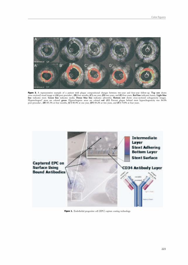
223
Color fi gures
For SES, 80% of the drug load is eluted within 30 days ofstent implantation (1). However, concern has arisen aboutlong-term biological reactions to the non-biodegradablepolymer (5). Long-term exposure to these polymers maycause chronic inflammation. Complete natural healing (i.e.,re-endothelialization and a reduction in proteoglycan con-tent) may take three to six months after bare metal stentimplantation (21). In this present study, plaque volumebehind the stent struts decreased between two and fouryears, and this shrinkage was accompanied by a change inplaque echogenicity, suggestive of a change in plaquecomposition. This may indicate that the peri-stent area hasundergone a slower healing process after SES implantationthat results in a more fibrous content at four-year follow-up.
In animal experiments, it has been documented that theexuberant proliferative process observed after stenting islargely inhibited in the early stage with SES, but the normalhealing process is also delayed with persistence of fibrindeposition and inflammatory cells (22). In humans, onestudy reported the histologic findings of atherectomy spec-imens of restenotic lesions after implantation of a paclitaxelderivative-eluting polymer stent in which persistent fibrinaccumulation with smooth muscle cells and proteoglycanand collagen type III-rich matrix with inflammation was
observed at 12 months (4,5). An autopsy performed on apatient four years after SES implantation showed completehealing, indicating that the delayed healing response hadabated, concurring with our findings (23).
In this study, neointimal volume continued to increaseover the four-year period. However, the increase betweentwo and four years failed to reach statistical significance (7.0vs. 8.4 mm3, p � 0.25), and the change in neointimalvolume between two years and four years was significantlyless than between the post-procedure and two-year mea-surements (1.4 vs. 7.0 mm3, p � 0.04), which possiblyindicates that the delayed tissue proliferation has subsided.Carter et al. (24) reported long-term effects of SES in aporcine coronary model. They found that inhibition ofneointimal hyperplasia was not sustained at 90 days com-pared with the bare metal stent, and the mean neointimalarea was similar between 90 days and 180 days. Theseresults differ from observations seen in clinical trials, includ-ing this study. Several trials have reported low target vesselrevascularization rates up to three years (25). This discrep-ancy is likely explained by different monocyte and lympho-cyte responses to similar concentrations of sirolimus inhuman and porcine vessels, and the different anatomical
Table 3. Percent Hypoechogenic Tissue Component in the Plaque and Neointima
AfterProcedure 4 Mo 1 Yr 2 Yrs 4 Yrs
ANOVA*p Value
Post Hoc AfterProcedure to
2 YrsPost Hoc2 to 4 Yrs
Neointima, % — 74.9 � 12.7 68.9 � 17.1 66.7 � 19.1 57.0 � 22.6 0.0001† 0.03‡ 0.01Plaque (stented segment), % 78.9 � 8.6 79.4 � 6.7 81.3 � 7.4 78.2 � 8.9 70.4 � 10.6 �0.0001 0.67 �0.0001Plaque (proximal edge), % 88.7 � 6.9 87.2 � 6.5 87.1 � 6.0 87.7 � 7.9 81.7 � 9.3 0.02 0.67 0.008Plaque (distal edge), % 86.1 � 9.8 86.8 � 6.0 87.0 � 10.4 88.5 � 9.5 85.9 � 9.4 0.78 — —
*Repeated-measures analysis of variance (ANOVA) was performed among five periods. Post-hoc analysis between after the procedure and two years, and between two and fouryears were performed with Bonferroni corrections (significant level of p value is 0.025). †Repeated-measures ANOVA was performed among four periods, except after theprocedure. ‡Post-hoc analysis was performed between four months and two years.
Figure 3. A representative example of a patient with plaque compositional changes between two-year and four-year follow-up. Top row showscross-sectional vessel image at (A) post-procedure, (B) four months, (C) one year, (D) two years, and (E) four years. Red line indicates lumen. Light blueline indicates stent. Green line indicates media. Dense blue line indicates adventitia. Bottom row shows cross-sectional echogenicity images.Hyperechogenic areas are colored green. Hypoechogenic areas are colored red. (A’) Percent plaque behind stent hypoechogenicity was 84.0%post-procedure, (B’) 85.3% at four months, (C’) 88.4% at one year, (D’) 84.2% at two years, and (E’) 74.8% at four years.
1674 Aoki et al. JACC Vol. 46, No. 9, 2005Four-Year Arterial Response After SES November 1, 2005:1670–6
intermediate coating with murine monoclonal anti-humanCD34 antibodies, attached to a stainless steel stent (R stent,OrbusNeich, Fort Lauderdale, Florida) (Fig. 1). This anti-body specifically targets CD34� cells (endothelial pro-genitor cells are CD34 positive) in the vascular circula-tion. This device was supplied in aqueous sodium azidesolution as a preservative to maintain bioactivity andrequired hand crimping by the operator onto a percuta-neous transluminal coronary angioplasty balloon catheterbefore implantation.Study procedures. Lesions were treated according to localstandard interventional techniques. Specifically, the decisionto predilate or direct stent was at the investigator’s discre-tion, and post-dilation was performed as required to ensurethat the residual stenosis was �20% by visual assessment,with a TIMI flow grade rate 3. In case of a dissection or
incomplete coverage of the lesion, implantation of addi-tional EPC capture R stents was permitted.Intravenous boluses of heparin were administered to
maintain an activated clotting time �300 s during theimplantation. Treatment with aspirin, at a dose of at least 80mg/day, was initiated at least 12 h before the procedure andcontinued for one month. In addition, a loading dose of 300mg of clopidogrel was administered before the procedure,followed by 75 mg daily for 28 days. Glycoprotein IIb/IIIainhibitors were used at the operator’s discretion. Angio-graphic success was defined as the successful implantation ofthe study device, with a stenosis of �20% of the vesseldiameter with TIMI flow grade 3.Follow-up. All patients were scheduled for a clinicalfollow-up at one, six, and nine months following theimplantation procedure to assess the anginal status andthe occurrence of major adverse cardiac and cerebrovas-cular events (MACCE). An electrocardiogram was ob-tained at each visit, and an angiographic and intravascularultrasound (IVUS) study was performed at a mean of 185� 14 days.Quantitative angiographic and IVUS analysis. Coronaryangiograms were obtained in multiple views after an intra-coronary injection of nitrates. Offline quantitative analysesof preprocedural, postprocedural, and six-month follow-upangiographic data were performed. Restenosis was definedas a reduction of 50% or more of the luminal diameter. Lateluminal loss was defined as the difference between theminimal luminal diameter after procedure and at sixmonths. The target lesion was defined as the stented
Abbreviations and AcronymsEPC � endothelial progenitor cellHAMA � human antimurine antibodyHEALING-FIM � Healthy Endothelial Accelerated
Lining Inhibits NeointimalGrowth-First In Man
IVUS � intravascular ultrasoundMACCE � major adverse cardiac and
cerebrovascular eventsMI � myocardial infarctionSMC � smooth muscle cellTIMI � Thrombolysis In Myocardial
Infarction
Figure 1. Endothelial progenitor cell (EPC) capture coating technology.
1575JACC Vol. 45, No. 10, 2005 Aoki et al.May 17, 2005:1574–9 First Human Experience of EPC Capture Stent

224
Color fi gures
early functional endothelial lining can be expected in theinterstrut space, although the interstrut area in the animalmodel (healthy coronary artery undergoing direct stenting)was covered with functional endothelium within 48 h; thissituation differs substantially from a human pathologicatherosclerosis vessel after balloon injury. In addition, thebioactivity of these prototype EPC capture stents used inHEALING-FIM registry was unstable and was easilyreduced by sterilization with gamma irradiation. It wassubsequently discovered through the use of a bioassay thatgamma irradiation lessened the immunoaffinity of EPCcapture prototype stents used in this trial. Therefore, it islikely that the reduced bioactivity of EPC capture stents inthe HEALING-FIM registry may not have been enough toinhibit neointimal hyperplasia after stent implantation.The technology behind the creation of an EPC affinity
surface is achieved by attaching murine monoclonal antihu-man CD 34 antibodies to the stent. An immunoreactionagainst the murine monoclonal antibody may occur inpatients who have human ant-murine antibody (HAMA).In addition, production of HAMA may result in theneutralization of the EPC capture surface. In this registry,
increased HAMA levels were not observed in the fourpatients who underwent serial HAMA testing, and nopatients exhibited systemic symptoms of immunoreaction.This safety issue was in agreement with other trials, evalu-ating immunologic treatment using murine antibody forovarian cancer (10).Because of the small sample size and single-center en-
Table 4. Evolution of Endothelial Progenitor Cell CaptureStent: Improvement From HEALING-FIM (H1) toHEALING II (H2)
DeviceH1 - Wet, hand-crimpedH2 - Dry, premounted
StabilityH1 - Wet; supplied in sodium azide preservative; required rinsingbefore use
H2 - Dry, formulated to preserve antibody structure & activitySterilizationH1 - Gamma irradiation; 15 to 25 kGyH2 - Gamma irradiation; �15 kGy
BioactivityH1 - Significant reduction in activity with sterilizationH2 - Stable with sterilization
Figure 2. Overview (A) and details (left: B, C; mid:D, E; right: F) of the atherectomy specimen obtained six months following stent placement. Histologyshows a mixture of myxoid (B, C) and fibrous material (F) consisting of smooth muscle cells (C, E) within variable amounts of extracellular matrix. Somefragments contained thrombus remnants (Th) (D, E) and calcifications (Ca) (F). A � elastin stain; B, D, F� hematoxylin eosin; C, E �immunocytochemistry for smooth muscle cell-specific alpha actin.
1578 Aoki et al. JACC Vol. 45, No. 10, 2005First Human Experience of EPC Capture Stent May 17, 2005:1574–9

225
Color fi gures
Aoki J, et al. Emergent Strategies in the Interventional Cardiology
95 Rev Esp Cardiol. 2005;58(8):962-73 969
established that general tissue shrinkage can not beavoided during dehydration.68 Shrinkage of up to60%, 15% and 80% can occur during critical-point-drying, free-drying, and air-drying respectively.69
Furthermore, post-mortem contraction of arteries fur-ther contributes to deteriorate the pathological quan-tification of atherosclerosis.70 Accordingly, we be-lieve that the threshold should be higher than 65 µm.Since the axial resolution of IVUS is between 100and 150 µm, techniques such as IVUS-VH with thecapacity to quantify each plaque component and toidentify the location of the lipid core in relation tothe lumen have the potential to recognize all the 3features of TCFA lesions.
Optical coherence tomography (OCT) is an imagingtechnique that allows high-resolution (axial resolutionof 15 µm) imaging in biological systems (Figure 9).71
Accordingly, OCT has the capacity to allow in-vivo,real time visualization of a thin fibrous cap. Recent invivo data suggest the possibility of detection ofmacrophages in atherosclerotic plaques.72
The sensitivity and specificity of palpography to de-tect vulnerable plaques has recently been assessed inpost-mortem human coronary arteries where vulnera-ble plaques were detected with a sensitivity of 88%and a specificity of 89%.63
Positive Remodeling
The expansive compensatory enlargement of thecoronary arteries in response to an increase in plaquearea is called positive or expansive remodeling.73
Several studies showed an increase in inflammatorymarker levels, larger lipid cores, and pronounced me-dial thinning in positive remodeled vessels.74-76 Re-cently, the relationship between vascular remodelingand plaque composition was assessed using IVUS.77-79
In these studies, the remodeling index for soft lesions
Figure 6. Direct myocardial injection using IVUS guide needle punctu-res via the coronary venous system.
Figure 7. Virtual histology image. Red: lipid core. White: calcium. Ye-llow: fibrofatty. Green: fibrosis. Gray: media.
PericardialSpace
Vein
Artery
SeptumVentricularCavity
Anterior Wall
currentPointerPositionNeedleTrajectory
Figure 8. Palpography image. Yellow,high strain spot.
Document dowloaded from http://www.revespcardiol.org, day 11/01/2006. This copy is for personal use. Any transmission of this document by any media or format is strictly prohibited.