Differential healing response attributed to culprit lesions of patients with acute coronary...
9
Differential healing response attributed to culprit lesions of patients with acute coronary syndromes and stable coronary artery after implantation of drug-eluting stents: An optical coherence tomography study ☆ , ☆☆ Lorenz Räber a,1,2 , Thomas Zanchin a,1,2 , Sandro Baumgartner a,2 , Masanori Taniwaki a,2 , Bindu Kalesan b,2 , Aris Moschovitis a,2 , Hector M. Garcia-Garcia c,2 , Jörn Justiz d,2 , Thomas Pilgrim a,2 , Peter Wenaweser a,2 , Bernhard Meier a,2 , Peter Jüni b,2 , Stephan Windecker a, ⁎ ,2 a Department of Cardiology, Bern University Hospital, Bern, Switzerland b Institute of Social and Preventive Medicine, University of Bern, Switzerland c Thoraxcenter, Erasmus Medical Center, Rotterdam, The Netherlands d Human-Centered Engineering Institute of Applied Sciences, Biel, Switzerland abstract article info Article history: Received 12 July 2013 Received in revised form 26 January 2014 Accepted 22 February 2014 Available online xxxx Keywords: Optical coherence tomography Acute coronary syndrome Drug-eluting stent Arterial healing Background: Pathology studies have shown delayed arterial healing in culprit lesions of patients with acute cor- onary syndrome (ACS) compared with stable coronary artery disease (CAD) after placement of drug-eluting stents (DES). It is unknown whether similar differences exist in-vivo during long-term follow-up. Using optical coherence tomography (OCT), we assessed differences in arterial healing between patients with ACS and stable CAD five years after DES implantation. Methods and results: A total of 88 patients comprised of 53 ACS lesions with 7864 struts and 35 stable lesions with 5298 struts were suitable for final OCT analysis five years after DES implantation. The analytical approach was based on a hierarchical Bayesian random-effects model. OCT endpoints were strut coverage, malapposition, protrusion, evaginations and cluster formation. Uncovered (1.7% vs. 0.7%, adjusted p = 0.041) or protruding struts (0.50% vs. 0.13%, adjusted p = 0.038) were more frequent among ACS compared with stable CAD lesions. A similar trend was observed for malapposed struts (1.33% vs. 0.45%, adj. p = 0.072). Clusters of uncovered or malapposed/protruding struts were present in 34.0% of ACS and 14.1% of stable patients (adj. p = 0.041). Coronary evaginations were more frequent in patients with ST-elevation myocardial infarction compared with stable CAD patients (0.16 vs. 0.13 per cross section, p = 0.027). Conclusion: Uncovered, malapposed, and protruding stent struts as well as clusters of delayed healing may be more frequent in culprit lesions of ACS compared with stable CAD patients late after DES implantation. Our ob- servational findings suggest a differential healing response attributable to lesion characteristics of patients with ACS compared with stable CAD in-vivo. © 2014 Elsevier Ireland Ltd. All rights reserved. 1. Introduction The long-term risk for recurrent events is higher among patients with acute coronary syndromes (ACS) compared to those with stable coronary artery disease (CAD) after placement of drug-eluting stent (DES). Aside from non-device related factors, differences in arterial healing have been suggested as a potential explanation with a higher frequency of uncovered struts, fibrin deposition and inflammation observed in autopsy specimen [1]. Few studies using intravascular opti- cal coherence tomography imaging also observed a higher rate of un- covered and malapposed stent struts among ACS patients but were limited to one year follow-up after DES implantation [2–4]. Owing to a possible association of uncovered and malapposed struts with the risk of late stent thrombosis [5] and the prevailing uncertainty with respect International Journal of Cardiology xxx (2014) xxx–xxx ☆ Grant support: This study was supported by research grants from the Bern University Hospital and the Swiss National Science Foundation (Grant Nos. 33CM30_140336 and 310030-118353 to Dr. Windecker). Dr. Räber is a recipient of a research fellowship Special Program University Medicine (SPUM) funded by the Swiss National Science Foundation. ☆☆ Conflict of interest statement: Dr. Wenaweser has a relationship with Cordis and Johnson & Johnson. Dr. Meier has received research and speaker fees from Johnson & Johnson and Cordis. Dr. Windecker has received research grants from Abbott, Boston Scientific, Biotronik, Biosensors, Cordis, and Medtronic. All other authors have reported that they have no relationships relevant to the contents of this paper to disclose. ⁎ Corresponding author at: Department of Cardiology, Bern University Hospital, 3010 Bern, Switzerland. E-mail address: [email protected] (S. Windecker). 1 Equally contributing first authors. 2 All authors take responsibility for all aspects of the reliability and freedom from bias of the data presented and their discussed interpretation. IJCA-17684; No of Pages 9 http://dx.doi.org/10.1016/j.ijcard.2014.02.036 0167-5273/© 2014 Elsevier Ireland Ltd. All rights reserved. Contents lists available at ScienceDirect International Journal of Cardiology journal homepage: www.elsevier.com/locate/ijcard Please cite this article as: Räber L, et al, Differential healing response attributed to culprit lesions of patients with acute coronary syndromes and stable coronary artery a..., Int J Cardiol (2014), http://dx.doi.org/10.1016/j.ijcard.2014.02.036
Transcript of Differential healing response attributed to culprit lesions of patients with acute coronary...

International Journal of Cardiology xxx (2014) xxx–xxx
IJCA-17684; No of Pages 9
Contents lists available at ScienceDirect
International Journal of Cardiology
j ourna l homepage: www.e lsev ie r .com/ locate / i j ca rd
Differential healing response attributed to culprit lesions of patients withacute coronary syndromes and stable coronary artery after implantationof drug-eluting stents: An optical coherence tomography study☆,☆☆
Lorenz Räber a,1,2, Thomas Zanchin a,1,2, Sandro Baumgartner a,2, Masanori Taniwaki a,2, Bindu Kalesan b,2,Aris Moschovitis a,2, Hector M. Garcia-Garcia c,2, Jörn Justiz d,2, Thomas Pilgrim a,2, Peter Wenaweser a,2,Bernhard Meier a,2, Peter Jüni b,2, Stephan Windecker a,⁎,2
a Department of Cardiology, Bern University Hospital, Bern, Switzerlandb Institute of Social and Preventive Medicine, University of Bern, Switzerlandc Thoraxcenter, Erasmus Medical Center, Rotterdam, The Netherlandsd Human-Centered Engineering Institute of Applied Sciences, Biel, Switzerland
☆ Grant support: This studywas supported by researchHospital and the Swiss National Science Foundation (Gr310030-118353 to Dr. Windecker). Dr. Räber is a reciSpecial Program University Medicine (SPUM) fundedFoundation.☆☆ Conflict of interest statement: Dr. Wenaweser hasJohnson & Johnson. Dr. Meier has received research anJohnson and Cordis. Dr. Windecker has received researScientific, Biotronik, Biosensors, Cordis, and Medtronic. Athat they have no relationships relevant to the contents o
⁎ Corresponding author at: Department of CardiologyBern, Switzerland.
E-mail address: [email protected] (S. Wind1 Equally contributing first authors.2 All authors take responsibility for all aspects of the reli
the data presented and their discussed interpretation.
http://dx.doi.org/10.1016/j.ijcard.2014.02.0360167-5273/© 2014 Elsevier Ireland Ltd. All rights reserved
Please cite this article as: Räber L, et al, Differstable coronary artery a..., Int J Cardiol (2014
a b s t r a c t
a r t i c l e i n f oArticle history:
Received 12 July 2013Received in revised form 26 January 2014Accepted 22 February 2014Available online xxxxKeywords:Optical coherence tomographyAcute coronary syndromeDrug-eluting stentArterial healing
Background: Pathology studies have shown delayed arterial healing in culprit lesions of patients with acute cor-onary syndrome (ACS) compared with stable coronary artery disease (CAD) after placement of drug-elutingstents (DES). It is unknown whether similar differences exist in-vivo during long-term follow-up. Using opticalcoherence tomography (OCT), we assessed differences in arterial healing between patients with ACS and stableCAD five years after DES implantation.Methods and results:A total of 88 patients comprised of 53 ACS lesionswith 7864 struts and 35 stable lesionswith5298 struts were suitable for final OCT analysis five years after DES implantation. The analytical approach wasbased on a hierarchical Bayesian random-effects model. OCT endpoints were strut coverage, malapposition,protrusion, evaginations and cluster formation. Uncovered (1.7% vs. 0.7%, adjusted p = 0.041) or protrudingstruts (0.50% vs. 0.13%, adjusted p = 0.038) were more frequent among ACS compared with stable CAD lesions.
A similar trend was observed for malapposed struts (1.33% vs. 0.45%, adj. p = 0.072). Clusters of uncoveredor malapposed/protruding struts were present in 34.0% of ACS and 14.1% of stable patients (adj. p = 0.041).Coronary evaginations were more frequent in patients with ST-elevation myocardial infarction compared withstable CAD patients (0.16 vs. 0.13 per cross section, p = 0.027).Conclusion: Uncovered, malapposed, and protruding stent struts as well as clusters of delayed healing may bemore frequent in culprit lesions of ACS compared with stable CAD patients late after DES implantation. Our ob-servational findings suggest a differential healing response attributable to lesion characteristics of patientswith ACS compared with stable CAD in-vivo.© 2014 Elsevier Ireland Ltd. All rights reserved.
grants from the BernUniversityant Nos. 33CM30_140336 andpient of a research fellowshipby the Swiss National Science
a relationship with Cordis andd speaker fees from Johnson &ch grants from Abbott, Bostonll other authors have reportedf this paper to disclose., Bern University Hospital, 3010
ecker).
ability and freedom from bias of
.
ential healing response attrib), http://dx.doi.org/10.1016/j
1. Introduction
The long-term risk for recurrent events is higher among patientswith acute coronary syndromes (ACS) compared to those with stablecoronary artery disease (CAD) after placement of drug-eluting stent(DES). Aside from non-device related factors, differences in arterialhealing have been suggested as a potential explanation with a higherfrequency of uncovered struts, fibrin deposition and inflammationobserved in autopsy specimen [1]. Few studies using intravascular opti-cal coherence tomography imaging also observed a higher rate of un-covered and malapposed stent struts among ACS patients but werelimited to one year follow-up after DES implantation [2–4]. Owing to apossible association of uncovered and malapposed struts with the riskof late stent thrombosis [5] and the prevailing uncertainty with respect
uted to culprit lesions of patients with acute coronary syndromes and.ijcard.2014.02.036

2 L. Räber et al. / International Journal of Cardiology xxx (2014) xxx–xxx
to the optimal duration of dual antiplatelet therapy, differences in arte-rial healing between patients with ACS and stable CAD after placementof DES remain clinically relevant. We therefore compared markers ofarterial healing including strut coverage, protrusion, malapposition,and coronary evaginations among patients with ACS and stable CADusing OCT five years after implantation of early generation DES.
2. Methods
2.1. Patient population and lesion selection
The design and results of SIRTAX (Sirolimus-Eluting Stent ComparedWith Paclitaxel-Eluting Stent for Coronary Revascularization) and SIRTAX LATE (Sirolimus-Eluting StentCompared With Paclitaxel-Eluting Stent for Coronary Revascularization—Late) havebeen previously reported [6]. For the purpose of the present study, all consecutive pa-tients undergoing angiographic follow-up at five years between December 2009 andJuly 2010 (n = 145) were eligible for OCT imaging. The flow of patients and reasons forexclusion are reported in the diagram (Fig. 1). In patients withmore than one study lesion(n= 19), all lesions were randomly allocated a numerical code of 1, 2 or 3 by an indepen-dent statistician. OCT was routinely performed in the lesion with the lowest number. Innone of these patients, the second or third lesion underwent OCT to respect the randomselection. In four patients with multiple lesions, OCT was technically not feasible. Thus,in 15 patients suitable for final analysis, lesion selection was performed in a randommanner. Among these patients, a total of 8wereACS patients and only in twoACS patients,the imaging was done in the non-culprit lesion at baseline (culprit lesion defined ac-cording to ECG and ventriculography findings).
The study complied with the Declaration of Helsinki regarding investigation inhumans and was approved by the institutional ethics committees at Bern UniversityHospital, Switzerland. All patients provided written informed consent.
2.2. OCT imaging and analysis
OCT was performed with a time domain M2 system (Lightlab Imaging, Westford,Massachusetts) using a pullback speed of 2 mm/s and the non-occlusive flushing
Fig. 1. Flow chart showing stud
Please cite this article as: Räber L, et al, Differential healing response attribstable coronary artery a..., Int J Cardiol (2014), http://dx.doi.org/10.1016/j
technique. After the diagnostic angiography and administration of 5000 IU unfractionatedheparin, the ImageWire (Lightlab Imaging) was carefully advanced distal to the studylesion. Following administration of 200 μg of i.c. nitroglycerin, the target vessel wasflushed through the guiding catheter with non-ionic, isosmolar contrast agent (Iodixanol320, Visipaque, GE Healthcare, Cork, Ireland) using a power injector with flush rates be-tween 3 and 4 ml/s. OCT pullbacks were assessed offline using a proprietary software(Lightlab Imaging, St. Jude Medical). Lesions were analyzed at cross sectional level withan interval of at 1 mm and assessed for strut coverage, malapposition, and protrusion bya single analyst blinded to patient and lesion presentation. All frames were reviewed bya second analyst, who in case of disagreement consultedwith a third referee,withfinal de-cision based on consensus. Pullbacks were excluded in case N30% of the total stent lengthwas not analyzable. Frames were considered not analyzable when N25% of the circumfer-ence was not visible due to insufficient flush or out of zoom. A strut was defined as asignal-intense bright spot with a typical dorsal shadowing. Thickness of strut coveragewas measured as the distance between the endoluminal side of the strut in the midpointof its long axis and the intersection of the lumen contour with the straight line betweenthe endoluminal side of the strut and the gravitational center of the vessel. Strutswere considered uncovered in case of a partial or complete absence of tissue coverage.Protrusion was defined as strut extension into the lumen for more than 160 μm butwith no obvious separation from the vessel wall [7]. Apposition was assessed by mea-suring the distance between the center of the endoluminal strut surface and the inter-section between lumen contour and the line connecting the center of the endoluminalstrut side and the gravitational center of the vessel. Strut malapposition was definedas a distance ≥160 μm based on the consensus derived from the strut thickness ofSES (153 μm) and PES (148 μm) plus the minimal axial resolution of OCT (10 μm).This consensus allowed a blinded assessment. Representative examples of uncovered,protruding or malapposed stent struts are presented in Fig. 2. Geographic mapswere created displaying struts using color codes for strut characteristics, includingstrut coverage, apposition, and protrusion (Fig. 3). The resultant map represented thestented vessel cut longitudinally along the reference angle 0° (corresponding to the 12o'clock position in the respective OCT cross section) and spread out on an area [7].The stent maps of all lesions are depicted in Fig. 4.
Coronary evaginations (Fig. 2, example E) were suspected whenever the luminalvessel contour extended in a pouchlike fashion beyond the line connecting all stent struts(stent contour). Under these circumstances, the maximal radial distance between thecircular line connecting all struts and the luminal vessel wall was evaluated using the
y design and patient flow.
uted to culprit lesions of patients with acute coronary syndromes and.ijcard.2014.02.036
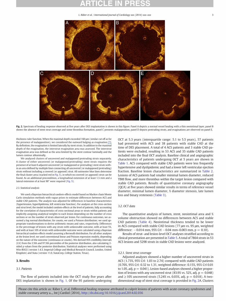
Fig. 2. Spectrum of healing response observed at five years after DES implantation is shown in this figure. Panel A depicts a normal vessel healing with a thin neointimal layer, panel Bshows the absence of stent strut coverage and some thrombus formation, panel C presents malapposition, panel D depicts protruding struts, and evaginations are observed on panel E.
3L. Räber et al. / International Journal of Cardiology xxx (2014) xxx–xxx
thickness ruler function.When themaximal depth exceeded 160 μm(similar cut off as forthe presence of malapposition), we considered the outward bulging as evagination [7].By definition, the evagination is limited laterally by stent struts. In addition to themaximaldepth of the evagination, the interstrut evagination area was assessed. The interstrutevagination area was defined as the area limited by the stent contour luminally and thelumen contour abluminally.
We analyzed clusters of uncovered and malapposed/protruding struts separately.A cluster of either uncovered (or malapposed/protruding) stent struts requires thepresence of at least 6 adjacent uncovered (ormalapposed or protruding) stent strutswith-in an area defined bymultiple lines connecting all uncovered (ormalapposed/protruding)struts without including a covered (or apposed) strut. All outmoster blue lines determinethe final cluster area (tracked red in Fig. 5), in which no covered (or apposed) strut can befound. As an additional precondition, a longitudinal extension of at least 1.5 mm and alateral extension of at least 90° were required (Fig. 5).
2.3. Statistical analysis
Weused a Bayesian hierarchical random-effects model based onMarkov chainMonteCarlo simulation methods with vague priors to estimate differences between ACS andstable CAD patients. The analysis was adjusted for differences in baseline characteristics(hypertension, hyperlipidemia, left ventricular function). For analyses at the cross-sectionand strut level, themodel included random-effects at the level of patients, fully accountingfor the correlation of characteristics of cross-sectional areas or struts within patients andimplicitly assigning analytical weights to each lesion depending on the number of crosssections or on the number of struts observed per lesion. For continuous outcomes, we as-sumed a log normal distribution; for counts, we used a Poisson distribution; we used ap-propriate transformations to derive arithmetic means and rates, respectively. Differencesin the percentage of lesions with any struts with unfavorable outcome, with at least 5%,andwith at least 10% of struts with unfavorable outcomewere calculated using a Bayesianhierarchical random-effects model assuming a Bernoulli distribution. For all other analysesat the lesion level, we used conventional linear and Poisson regression models, dependingon the nature of the outcome (continuous or counts). We derived 95% credibility intervals(CrI) from the 2.5th and 97.5th percentiles of the posterior distribution, also calculating 2-sided p values from the posterior distribution. Statistical analyses were performed usingWin-BUGS (version 1.4.3, Imperial College and Medical Research Council, London, UnitedKingdom) and Stata (version 11.0, StataCorp, College Station, Texas).
3. Results
3.1. Patients
The flow of patients included into the OCT study five years afterDES implantation is shown in Fig. 1. Of the 95 patients undergoing
Please cite this article as: Räber L, et al, Differential healing response attribstable coronary artery a..., Int J Cardiol (2014), http://dx.doi.org/10.1016/j
OCT at 5.3 years (interquartile range: 5.1 to 5.5 years), 57 patientshad presented with ACS and 38 patients with stable CAD at thetime of DES placement. A total of 4 ACS patients and 3 stable CAD pa-tients were excluded, resulting in 53 ACS and 35 stable CAD patientsincluded into the final OCT analysis. Baseline clinical and angiographiccharacteristics of patients undergoing OCT at 5 years are shown inTable 1. ACS compared with stable CAD patients were less frequentlyhypertensive and dyslipidemic and had a lower left ventricular ejectionfraction. Baseline lesion characteristics are summarized in Table 2.Lesions of ACS patients had smaller minimal lumen diameter, reducedTIMI flow, and more thrombus within the target lesion compared withstable CAD patients. Results of quantitative coronary angiography(QCA) at five years showed similar results in terms of reference vesseldiameter, minimal lumen diameter, % diameter stenosis, late lumenloss and binary restenosis (Table 3).
3.2. OCT data
The quantitative analysis of lumen, stent, neointimal area and %volume obstruction showed no differences between ACS and stableCAD patients (Table 4). Neointimal thickness tended to be lowerin ACS compared with stable CAD lesions (77 μm vs. 95 μm, weighteddifference: −0.014 mm, 95% CrI −0.04 mm–0.003 mm, p = 0.10).
Results of strut- and lesion-level OCT analyses stratified according toclinical presentation are presented in Table 5. A total of 7864 struts in 53ACS lesions and 5298 struts in stable CAD lesions were analyzed.
3.2.1. Stent strut coverageAdjusted analyses showed a higher number of uncovered struts in
ACS (1.73%, 95% CrI: 1.03 to 2.74) compared with stable CAD patients(0.70%, 95% CrI: 0.32 to 1.31, weighted difference: 0.15%, 95% CrI 0.01to 1.05, adj. p= 0.041). Lesion-based analyses showed a higher propor-tion of lesions with any uncovered strut (83.9% vs. 52%, adj. p = 0.048)and ≥10% uncovered struts (5.24% vs. 0.03%, adj. p = 0.018). A two-dimensional map of stent strut coverage is provided in Fig. 2A. Clusters
uted to culprit lesions of patients with acute coronary syndromes and.ijcard.2014.02.036

Fig. 3. The concept of the creation of geographic stent strut maps is illustrated. Lesions are presented as areas assuming a cylindrical geometry of the stent. Struts are color coded accordingto coverage, apposition, andprotrusion. The x axis represents the length of the stent (mm),whereas the y axis indicates the position of the strut in the individual cross section ranging from0° to 360°.
4 L. Räber et al. / International Journal of Cardiology xxx (2014) xxx–xxx
of uncovered struts were documented in 6 (17%) ACS lesions (#14, #18,#19, #42, #46, #49) and 2 (#1) stable CAD lesions (5.7%, adj. p= 0.24).
3.2.2. MalappositionAmong the ACS lesions, there was a trend towards a higher rate
of malapposed struts (1.33%, 95% CrI 0.72%–2.30 vs. 0.45%, 95 CrI0.18–1.02, weighted difference: 0.20%, 95% CrI −0.02%–1.81%, adj.p = 0.072). Neither the mean ISA area (0.55 mm2 vs. 0.50 mm2, adj.p = 0.80), nor the mean ISA distance (0.27 mm vs. 0.26 mm, adj.p = 0.72) differed between ACS and stable CAD lesions.
Lesion-level analysis of malapposition showed a trend towardsmore lesions with at least one malapposed strut among the ACSlesions compared with stable CAD lesions (77.7% vs. 39.5%, adj.p = 0.066), no difference however was found in the proportion oflesions with at least ≥10% malapposed struts (2.75% vs. 1.80%, adj.p = 0.89) (Table 5).
Clusters of malapposed or protruding stent struts were found in 15(28.3%) ACS lesions (#1, #4, #5, #8, #14, #16, #17, #19, #23, #24,#29, #37, #45, #52) and 4 (#6, #7, #15, #17) non-ACS lesions (11.4%,adj. p = 0.069) (Fig. 4A). Clusters of uncovered or malapposed/protruding stent struts were found in 18 (34%) ACS lesions (#1, #4,#5, #8, #14, #16, #17, #18, #19, #23, #24, #29, #37, #42, #45, #46,#49, #52) and 5 (#1, #6, #7, #15, #17) non-ACS lesions (14.3%, adj.p = 0.049).
Please cite this article as: Räber L, et al, Differential healing response attribstable coronary artery a..., Int J Cardiol (2014), http://dx.doi.org/10.1016/j
3.2.3. ProtrusionProtruding struts were more frequent in ACS (0.50%, 95% CrI: 0.25–
0.91) compared with stable CAD lesions (0.13%, 95% CrI: 0.04–0.32;weighted difference: 0.06%, 95% CrI: 0.001%–0.56%; adj. p = 0.038).The percentage of lesions with at least one protruding strut was higheramong the ACS lesions (54.3% vs. 13.9%, adj. p = 0.012), whereas theproportion of lesions with ≥10% protruding struts (0.40% vs. 0.01%;adj. p = 0.98) was not different.
3.2.4. Coronary evaginationsThe number of coronary evaginations tended to be higher in ACS
lesions (0.16 vs. 0.13 per cross sections; adj. p = 0.10) in the absenceof differences in mean area and depth of individual evaginations(Table 4). While no difference was observed between NSTEMI and sta-ble patients (0.14 vs. 0.13 per cross section, adj. p = 0.70), a significantdifference was noted between STEMI and stable patients (0.16 vs. 0.13,adj. p = 0.027) (Table 6, Fig. 6).
3.2.5. Type of ACSThe results of OCT analyses according to ACS type – STEMI and
NSTEMI – are presented in Table 6 and Fig. 6. Uncovered (1.68% vs.0.76%, weighted difference 0.89 (−0.09–2.25), p = 0.077), protruding(0.52% vs. 0.14%, weighted difference 0.38 (0.08–0.93), p = 0.012)and malapposed struts (1.46% vs. 0.48%, weighted difference 0.95
uted to culprit lesions of patients with acute coronary syndromes and.ijcard.2014.02.036

Fig. 4. Geographical stent strut maps of all 53 ACS (upper panel) and 35 non-ACS lesions (lower) are shown. Each lesion is assigned with a lesion number (e.g. #1–#53 in ACS lesion,#1–#35 in non-ACS lesions). Lesions are presented as areas assuming a cylindrical geometry of the stent. Struts are color coded according to coverage, apposition and protrusion. Strutcoverage is presented in panel A using a green color for covered struts and red color for uncovered struts. In panel B, strut apposition is presented. Apposed struts are shown in green,protruding struts in yellow, and malapposed struts in red. The x-axis represents the length of the stent (mm) whereas the y-axis indicates the position of the strut in the individualcross section ranging from 0 to 360°. Zones of stent overlap are marked with blue lines. The cluster analysis indicates that clusters of uncovered or malapposed/protruding stent strutswere found in 18 (34%) ACS lesions (#1, #4, #5, #8, #14, #16, #17, #18, #19, #23, #24, #29, #37, #42, #45, #46, #49, #52) and 5 (#1, #6, #7, #15, #17) non-ACS lesions (14.3%, adj.p = 0.049).
5L. Räber et al. / International Journal of Cardiology xxx (2014) xxx–xxx
(0.12–2.28), adj. p = 0.022) were more prevalent in STEMI comparedwith stable CAD lesions. Similarly, all three characteristics were morefrequent among NSTEMI than stable CAD lesions, although less pro-nouncedwithout reaching conventional levels of statistical significance.
STEMI lesions had a significantly higher number of coronary evagi-nations compared with stable CAD lesions (0.16% vs. 0.13%, weighteddifference 0.03%, 95% CrI 0.004%–0.06%, adj. p = 0.027) with a largermean evagination area (0.21 mm2 vs. 0.16 mm2, weighted difference0.06 mm2, 95% CrI 0.01 mm2–0.10 mm2, adj. p = 0.024) and depth(0.25 mm vs. 0.22 mm, weighted difference 0.03 mm, 95% CrI0.003 mm–0.07 mm, adj. p = 0.035). Conversely, no significant differ-ences were observed between NSTEMI and stable CAD lesions.
4. Discussion
This is the first in-vivo OCT study, which compares the arterialhealing response during long-term follow-up between ACS and
Please cite this article as: Räber L, et al, Differential healing response attribstable coronary artery a..., Int J Cardiol (2014), http://dx.doi.org/10.1016/j
stable CAD patients after DES placement with the following princi-pal findings:
1. Uncovered (adj. p = 0.041, adj. difference 0.15, 95% CI 0.01–1.05)and protruding struts (adj. p = 0.038, adj. difference 0.06, 95%CI 0.001–0.56) were more frequent in ACS than stable CAD pa-tients, with the highest incidence among STEMI patients. Similarly,there was a trend towards a higher frequency of malapposedstent struts in ACS lesions (adj. p = 0.072, adj. difference 0.20,95% CI −0.02–1.81).
2. Clusters of adverse strut characteristics (uncovered, malapposedor protruding stent struts) are more common among ACS patients(adj. p = 0.049)
3. Coronary evaginations are most frequently observed in lesionsof STEMI (adj. p = 0.027) compared to stable CAD lesions. Nodifference, however, was observed between NSTEMI and stableCAD patients (adj. p = 0.70).
uted to culprit lesions of patients with acute coronary syndromes and.ijcard.2014.02.036

Fig. 5. Two examples of lesions with clusters according to the definition outlined in theMethods section are shown. The red line reflects the border within which no singlecovered or malapposed/protruding strut can be seen.
Table 2Baseline characteristics of lesions undergoing OCT analysis.
ACS No ACS p-Value
Number of lesions 53 35Target lesion coronary artery (n [%])Left main 0 (0) 2 (5.7) 0.24Left anterior descending 25 (47.2) 17 (48.6)Left circumflex 11 (20.7) 9 (25.7)Right 17 (32.1) 7 (20.0)
ACC-AHA lesion class (n [%])A 5 (9.4) 8 (22.9) 0.20B1 23 (43.4) 14 (40.0)B2 17 (32.1) 6 (17.1)C 8 (15.1) 7 (20.0)
Angiographic measurementsLesion length (mm ± SD) 16.4 (5.9) 16.2 (9.5) 0.93Reference vessel diameter (mm ± SD) 2.9 (0.4) 2.8 (0.4) 0.73Minimal lumen diameter (mm ± SD) 0.35 (0.4) 0.59 (0.3) 0.003Stenosis (% lumen diameter ± SD) 87.9 (12.8) 79.3 (10.8) 0.002Pre-procedure TIMI flow b .001Grade 0 21 (39.6) 0 (0)Grade I 3 (5.7) 0 (0)Grade II 6 (11.3) 2 (5.7)Grade III 23 (43.4) 33 (94.3)
Thrombus not present 25 (48.1) 35 (100) b .001Calcification 17 (32.1) 14 (40.0) 0.50Procedures
No. of study stents per lesion (n ± SD) 1.2 (0.4) 1.1 (0.4) 0.76Stent diameter (mm ± SD) 3.0 (0.4) 2.9 (0.4) 0.37Total stent length per lesion (mm ± SD) 19.6 (7.6) 19.3 (9.4) 0.88Maximal pressure (atm ± SD) 15.3 (3.5) 14.1 (3.1) 0.10Direct stenting (n ± SD) 0.26 (0.4) 0.26 (0.4) 0.94
Angiographic resultsReference vessel diameter (mm ± SD) 2.9 (0.4) 2.8 (0.5) 0.23Final minimal lumen diameter (mm ± SD)In-stent 2.7 (0.4) 2.7 (0.4) 0.42In-segment 2.7 (0.4) 2.6 (0.5) 0.47
Final stenosis (% of lumen diameter ± SD)In-stent 6.9 (4.8) 5.9 (4.1) 0.32In-segment 8.4 (6.9) 7.5 (6.5) 0.63
Acute gain (mm ± SD)In-stent 2.4 (0.5) 2.1 (0.5) 0.004In-segment 2.4 (0.5) 2.1 (0.6) 0.031
6 L. Räber et al. / International Journal of Cardiology xxx (2014) xxx–xxx
Previous investigations of arterial healing in response to stent im-plantation have largely focused on differences between DES and BMS.Only few intravascular imaging studies addressed differences in vesselwall healing between ACS and stable CAD patients. Gonzalo et al. [2]
Table 1Baseline clinical characteristics.
ACS No ACS p-Value
Number of patients (n) 53 35Age N60 years (%) 11 (20.8) 12 (34.3) 0.22Males (n [%]) 44 (83.0) 26 (74.3) 0.42Diabetes mellitus (n [%]) 9 (17.0) 28 (80.0) 0.78Insulin dependent 3 (5.7) 2 (5.7) 1.00Hypertension (n [%]) 26 (49.1) 25 (71.4) 0.048Hyperlipidemia (n [%]) 25 (47.2) 25 (71.4) 0.029Current smoking (n [%]) 25 (47.2) 10 (28.6) 0.12Previous myocardial infarction (n [%]) 16 (30.2) 9 (25.7) 0.81Multivessel disease (n [%]) 35 (66.0) 25 (71.4) 0.65SYNTAX score 14.0 ± 9.0 10.4 ± 5.5 0.64Lesion per patientOne 43 (81.1) 29 (82.9) 0.65Two 9 (17.0) 4 (11.4)Three 1 (1.9) 2 (5.7)
Left ventricular ejection fraction (%), mean (SD) 55.9 (9.9) 60.5 (10.9) 0.04Stent typeSES 27 (50.9) 14 (40.0) 0.38PES 26 (49.1) 21 (60.0)
p value based on Fishers' exact test for categorical variables and student t test forcontinuous variables
Please cite this article as: Räber L, et al, Differential healing response attribstable coronary artery a..., Int J Cardiol (2014), http://dx.doi.org/10.1016/j
found a higher rate of incomplete stent apposition and uncoveredstent struts in STEMI compared with stable CAD patients 6 monthsafter DES implantation. Similarly, Kubo et al. [3] reported a higher rateof malapposed and uncovered struts among patients with unstable an-gina at 9 months. None of these studies addressed thequestionwhetherdifferences in healing persist or continue to accrue over time dependingon lesion type. We observed a higher proportion of uncovered struts(adj. p = 0.041, adj. difference 0.15, 95% CI 0.01–1.05) and protrudingstruts (adj. p = 0.038, adj. difference 0.06, 95% CI 0.001–0.56) and atrend towards more malapposed struts (adj. p = 0.072, adj. difference0.20, 95% CI −0.02–1.81) among event-free ACS patients. Differenceswere observed both on a strut and lesion level and further substantiatedby two-dimensional analysis of clusters of adverse characteristics.The latter observation is of importance as a spatial accumulation ofuncovered or malapposed/protruding struts may be clinically morerelevant than isolated stent strut findings. Clusters of uncovered andmalapposed/protruding struts were more common in ACS lesions (adj.p= 0.049) and require further analysis with respect to recurrent ische-mic events. To test whether the type of ACS influences the healingpattern, we stratified our analysis according to STEMI, NSTEMI and sta-ble CAD and observed a gradient of risk with the highest frequencyof uncovered, malapposed and protruding struts among the STEMI pa-tients followed by NSTEMI and stable CAD patients (Fig. 6).
The present analysis was not limited to stent strut characteristicsbut also incorporated the vessel wall between and behind struts,namely the assessment of coronary evaginations [7]. Evaginationswere more frequent among STEMI as compared to stable CAD patients(adj. p = 0.027), notably after adjustment for stent type. Coronary
uted to culprit lesions of patients with acute coronary syndromes and.ijcard.2014.02.036

Table 3Angiographic follow-up results at five years of lesions undergoing OCT analysis.
ACS No ACS Difference (95% CI) p-Value
Number of lesions 53 35Reference vessel diameter (mm ± SD) 2.90 (0.57) 2.75 (0.61) 0.15 (−0.02 to 0.32) 0.09Minimal lumen diameter (mm ± SD)In-stent 2.48 (0.72) 2.33 (0.77) 0.15 (−0.07 to 0.37) 0.19In-segment 2.39 (0.73) 2.22 (0.78) 0.17 (−0.05 to 0.39) 0.13
% diameter stenosisIn-stent 15.02 (17.56) 15.65 (18.80) −0.63 (−6.00 to 4.75) 0.82In-segment 17.73 (18.25) 19.24 (19.54) −1.50 (−7.09 to 4.08) 0.6
Late loss (mm ± SD)In-stent 0.27 (0.44) 0.30 (0.47) −0.03 (−0.16 to 0.10) 0.66In-segment 0.25 (0.40) 0.31 (0.43) −0.07 (−0.19 to 0.06) 0.28
Binary restenosis (n [%])In-stent 2.00 (3.77) 2.00 (5.71) −1.94 (−19.19 to 15.30) 0.83In-segment 3.00 (5.66) 2.00 (5.71) −0.05 (−9.85 to 9.74) 0.99
Row percent (%) values are predicted probabilities derived frommixed maximum logistic regression models. Mean and standard deviation (SD) are predicted values derived frommixedmaximum likelihood regressionmodels. Mixedmaximum likelihood regressionmodels were used for continuous andmixedmaximum logistic regressionmodels for binary outcomes toderive the differences between females and males. p-Values relate to the difference between two stent types.
7L. Räber et al. / International Journal of Cardiology xxx (2014) xxx–xxx
evaginations refer to an outward bulging of the vessel wall betweenstent struts and are a phenomenon, which has been described system-atically for the first time in the SIRTAX LATE OCT study [8]. In alarge pooled database, we observed a higher frequency of evaginationsin early generation DES (with preference of SES over PES) as comparedto newer generation DES. In a serial imaging substudy using OCTand IVUS, intra-stent dissections, protrusion, and thrombus were iden-tified as a potential cause of evaginations [9]. Both protrusion andthrombus have been shown to be more frequent in STEMI patients[10], explaining our findings of a higher frequency of evaginations inSTEMI compared with stable angina patients. Similar to uncoveredand malapposed stent struts, the clinical relevance of evaginations hasnot been determined in a prospective study. However, lesions withvery late stent thrombosis share morphological features observed inlesions with evaginations [11,12], suggesting a potential link betweenthe two entities.
ACS lesions substantially differ from those of stable CAD in termsof the underlying tissue composition. While plaques underlying ACSare frequently characterized by large lipid pools with necrotic coresand thrombus, stable CAD lesions often consist of fibrous or calcific tis-sue [13]. Although it remains unknown how and to which degree theunderlying plaque morphology affects arterial healing after DES place-ment, several hypotheses have been put forward. Limus derivatives
Table 4Results of OCT analysis— continuous outcomes.
ACS (95% CI) No ACS (95% CI)
Analysis at lesion levelNumber of lesions analyzed 53 35Cross sections analyzed per lesion 17.1 (15.5–18.8) 16.5 (14.7–18.6)Struts analyzed per lesion 136.7 (121.1–154.2) 137.2 (118.0–161.7)Minimal luminal area (mm2) 6.21 (5.57–6.91) 5.26 (4.63–6.00)Minimal stent area (mm2) 7.08 (6.47–7.75) 6.29 (5.62–7.01)Percent volume obstruction (%) 7.50 (5.02–12.5) 8.47 (5.05–16.8)
Analysis at cross section levelNumber of cross section analyzed 955 620Number of struts per cross section 7.51 (7.10–7.94) 7.76 (7.24–8.32)Luminal area (mm2) 6.34 (5.76–6.97) 5.35 (4.76–6.05)Stent area (mm2) 7.18 (6.60–7.77) 6.47 (5.89–7.21)Neointimal thickness (mm) 0.077 (0.069–0.087) 0.095 (0.082–0.109)Neointimal area (mm2) 0.82 (0.73–0.91) 0.91 (0.80–1.05)Mean area ISA (mm2) 0.55 (0.42–0.71) 0.50 (0.33–0.75)Mean malapposition distance (mm) 0.27 (0.24–0.30) 0.26 (0.22–0.31)Number of evaginations 0.16 (0.14–0.17) 0.13 (0.11–0.15)Mean evagination area (mm2) 0.20 (0.17–0.23) 0.16 (0.3–0.20)Mean evagination depth (mm) 0.25 (0.23–0.27) 0.22 (0.20–0.25)
Presented are means or percentages with 95% confidence intervals or 95% credible intervals. A
Please cite this article as: Räber L, et al, Differential healing response attribstable coronary artery a..., Int J Cardiol (2014), http://dx.doi.org/10.1016/j
and paclitaxel are lipophilic drugs with high affinity for lipid-rich/necrotic core plaques, residing for extended periods of time comparedwith fibrous tissue, which is more frequent in stable CAD lesions [14].Similarly, an increased thrombus load may reduce systemic washoutand preserve drug in the arterial wall [15]. Both observationsmay resultin higher drug content and may influence healing by slowing smoothmuscle cell proliferation and endothelial regrowth, thus preventingendothelial coverage of stent struts. In addition, necrotic cores areavascular compared to fibrous tissue of stable CAD plaques with alower density of smooth muscle and endothelial cells. In-vivo studiescorrelating tissue composition with the arterial healing response arerelatively scarce. Recently, Hong and coworkers found the extent of ne-crotic core to be related to late acquired malapposition at one year [16].Likewise, intravascular ultrasound studies of patients undergoing treat-ment for STEMI found late acquired vessel wall malapposition to bemore common in DES treated attenuated than non-attenuated plaques[17]. Both studies suggest that the presence of necrotic core is associatedwith an adverse healing pattern. Also thrombus is more prevalent inACS lesions. Thrombus may undergo maturation or is being dissolved/embolized. The latter has been suspected to result in late acquiredmalapposition. However, to date, no single serial intravascular imagingstudy has proven a solid association between thrombus and the occur-rence of late acquired malapposition [18,19].
Crude difference (95% CI) p-Value Adj. difference (95% CI) p-Value
0.52 (−2.11–3.15) 0.68 0.20 (−1.97–2.66) 0.87−0.70 (28.7–25.4) 0.96 −1.99 (−30.7–27.0) 0.890.94 (0.002–1.89) 0.049 0.52 (−0.14–1.36) 0.120.80 (−0.16–1.74) 0.10 0.43 (−0.22–1.19) 0.19
−0.95 (−9.43–5.01) 0.75 −0.77 (−66.4–47.8) 0.75
−0.25 (−0.96–0.44) 0.46 −0.19 (−1.06–0.70) 0.650.99 (0.07–1.84) 0.031 0.50 (−0.04–1.20) 0.07
0.72 (−0.31–1.49) 0.14 0.40 (−0.17–1.12) 0.18−0.018 (−0.034–0.002) 0.029 −0.014 (−0.04–0.003) 0.10
−0.10 (−0.25–0.07) 0.23 −0.07 (−0.23–0.09) 0.380.05 (−0.23–0.28) 0.71 0.03 (−0.32–0.48) 0.80
0.004 (−0.05–0.06) 0.88 0.014 (−0.06–0.10) 0.720.02 (−0.01–0.05) 0.063 0.02 (−0.004–0.053) 0.10
0.04 (−0.003–0.08) 0.07 0.02 (−0.01–0.06) 0.190.03 (−0.001–0.06) 0.06 0.02 (−0.01–0.05) 0.17
djusted for stent type, hypertension, hyperlipidemia, LVEF, and maximal pressure.
uted to culprit lesions of patients with acute coronary syndromes and.ijcard.2014.02.036

Table 6Stratification according to STEMI, NSTEMI/unstable angina and stable CAD.
STEMI (95% CI) NSTEMI (95% CI) Stable (95% CI) Diff. (95% CI)STEMI vs. stable
p-Value Diff. (95% CI)NSTEMI vs. stable
p-Value p-Trend
Results of OCT analysis— countsAnalysis at strut levelTotal number of struts analyzed 4784 3102 5298Uncovered struts (%) 1.68 (0.91–2.97) 1.48 (0.57–3.45) 0.76 (0.37–1.45) 0.89 (−0.09–2.25) 0.077 0.69 (−0.37–2.69) 0.23 0.18Protruding struts (%) 0.52 (0.23–1.08) 0.41 (0.12–1.18) 0.14 (0.04–0.31) 0.38 (0.08–0.93) 0.012 0.26 (−0.05–1.04) 0.12 0.07Malapposed struts (%) 1.46 (0.70–2.78) 0.91 (0.28–2.55) 0.48 (0.22–1.02) 0.95 (0.12–2.28) 0.022 0.41 (−0.39–2.08) 0.33 0.17
Analysis at lesion levelUncovered struts, lesions withAny uncovered struts 86.9 (68.1–96.6) 77.2 (42.1–95.5) 54.1 (29.7–77.5) 31.6 (4.49–60.1) 0.023 21.9 (−18.2–55.1) 0.26 0.11
Protruding struts, lesions withAny protruding struts 54.1 (29.8–77.3) 49.7 (17.2–82.6) 16.3 (4.91–36.2) 36.7 (7.50–65.1) 0.014 32.0 (−3.44–69.4) 0.08 0.03
Malapposed struts, lesions withAny malapposed struts 87.1 (68.4–96.6) 59.6 (23.9–88.3) 37.8 (16.4–62.3) 48.1 (18.8–74.2) 0.001 20.9 (−20.7–59.5) 0.33 0.09
Results of OCT analysis— continuous outcomesAnalysis at cross section levelNumber of evaginations 0.16 (0.14–0.19) 0.14 (0.11–0.17) 0.13 (0.11–0.15) 0.03 (0.004–0.06) 0.027 0.01 (−0.03–0.04) 0.70 0.35Mean evagination area (mm2) 0.21 (0.18–0.25) 0.17 (0.13–0.22) 0.16 (0.13–0.20) 0.06 (0.01–0.10) 0.024 0.01 (−0.04–0.06) 0.75 0.44Mean evagination depth (mm) 0.25 (0.23–0.28) 0.24 (0.21–0.28) 0.22 (0.20–0.24) 0.03 (0.003–0.07) 0.035 0.02 (−0.2–0.06) 0.26 0.17
Table 5Results of OCT analysis— counts.
ACS (95% CI) No ACS (95% CI) Crude difference (95% CI) p-Value Adj. difference (95% CI) p-Value
Analysis at strut levelTotal number of struts analyzed 7864 5298Uncovered struts (%) 1.73 (1.03–2.74) 0.70 (0.32–1.31) 1.01 (0.16–2.07) 0.020 0.15 (0.01–1.05) 0.041Protruding struts (%) 0.50 (0.25–0.91) 0.13 (0.04–0.32) 0.36 (0.09–0.77) 0.011 0.06 (0.001–0.56) 0.038Malapposed struts (%) 1.33 (0.72–2.30) 0.45 (0.18–1.02) 0.85 (0.06–1.86) 0.035 0.20 (−0.02–1.81) 0.072
Analysis at lesion levelUncovered struts, lesions withAny uncovered struts 83.9 (67.7–94.3) 52.0 (28.4–75.5) 30.9 (4.89–58.6) 0.019 20.4 (0.06–59.7) 0.048At least 10% uncovered struts 5.24 (1.08–15.6) 0.03 (0.0002–1.97) 5.02 (0.87–15.3) 0.006 0.99 (0.001–82.5) 0.018
Protruding struts, lesions withAny protruding struts 54.3 (34.1–73.7) 13.9 (3.71–32.8) 39.3 (13.6–63.8) 0.004 4.01 (0.04–48.6) 0.0196At least 10% protruding struts 0.40 (0.01–3.75) 0.01 (0.0001–1.65) 0.31 (−0.92–3.56) 0.27 −0.16 (−31.3–60.3) 0.98
Malapposed struts, lesions withAny malapposed struts 77.7 (59.8–91.0) 39.5 (17.5–64.4) 37.3 (8.01–65.6) 0.012 20.3 (0.96–58.5) 0.066At least 10% malapposed struts 2.75 (0.43–10.7) 1.80 (0.15–9.89) 0.75 (−5.88–7.71) 0.70 −0.003 (−3.96–3.42) 0.89
Presented are means or percentages with 95% confidence intervals or 95% credible intervals. Adjusted for stent type, hypertension, hyperlipidemia, LVEF, and maximal pressure.
8 L. Räber et al. / International Journal of Cardiology xxx (2014) xxx–xxx
Observational studies identified ACS as an independent predictorfor the occurrence of stent thrombosis for up to 4 years [20,21]. Ratesof definite or probable very late ST (between years 1 and 3) were 50%higher in the STEMI population of HORIZONS-AMI [22] compared withstable CAD in the SPIRIT II/III pooled analysis treated with PES [23].While a higher thrombus load [24] may explain early differences in
Fig. 6. Frequency of adverse stent strut characteristics and evaginations as assessed5 years after DES implantation and stratified according to the clinical presentation ispresented in this bar graph.
Please cite this article as: Räber L, et al, Differential healing response attribstable coronary artery a..., Int J Cardiol (2014), http://dx.doi.org/10.1016/j
clinical outcomes, differences in long-term clinical events are not fullyunderstood. Interestingly, our findings in ACS lesions were identifiedas pathological substrate underlying cases of late ST, offering a potentialexplanation for the increased rate of ST during long-term follow-up [1].Despite the lack of a prospective evaluation of the impact of uncoveredandmalapposed/protruding struts on stent thrombosis, Guagliumi et al.[5] observed in a case control study using OCT that patients sufferinglate ST had a higher proportion of uncovered and malapposed stentstruts as compared to control patients.
5. Limitations
Results need to be carefully interpreted in light of several limita-tions. First, this is a post-hoc subanalysis of an OCT study designed toevaluate long-term healing differences between a sirolimus-elutingand a paclitaxel-eluting stent, thus the presented results can only beconsidered as hypothesis generating. Second, the absence of a post-procedural baseline OCT imaging prevents the differentiation betweenpersistent and late acquired malapposition. As a consequence, wecannot precisely determine whether malapposition at follow-up iscaused by inadequate stent expansion or due to differences of theunderlying plaque, therefore relating to true healing differences. Ofnote, Gutierrez-Chico et al. [25] have previously demonstrated thatmalapposed struts with a distance of less than 270 μm undergo healingin 100% of cases. We measured a median malapposition distance of
uted to culprit lesions of patients with acute coronary syndromes and.ijcard.2014.02.036

9L. Räber et al. / International Journal of Cardiology xxx (2014) xxx–xxx
270 μm, suggesting that a certain proportion of malapposed strutswere indeed late acquired and reflect differences in healing pattern ac-cording to clinical presentation. Third, patients undergoing follow-upimaging at five years were free of any study lesion related events andthus present a highly selected subcohort of patients at a relativelylow risk for adverse events, explaining why the absolute difference innumbers between groups was relatively low. Along the same line, rel-atively low neointimal thickness values were measured, which is a di-rect consequence of the exclusion of all patients who underwent targetlesion revascularization within 5 years.
The present results are based on findings with early generationdrug-eluting stents. Whether they are applicable to newer generationDES remains unclear and needs further investigation.
References
[1] Nakazawa G, Finn AV, JonerM, et al. Delayed arterial healing and increased late stentthrombosis at culprit sites after drug-eluting stent placement for acute myocardialinfarction patients: an autopsy study. Circulation 2008;118:1138–45.
[2] Gonzalo N, Barlis P, Serruys PW, et al. Incomplete stent apposition and delayed tis-sue coverage are more frequent in drug-eluting stents implanted during primarypercutaneous coronary intervention for ST-segment elevation myocardial infarctionthan in drug-eluting stents implanted for stable/unstable angina: insights fromoptical coherence tomography. JACC Cardiovasc Interv 2009;2:445–52.
[3] Kubo T, Imanishi T, Kitabata H, et al. Comparison of vascular response aftersirolimus-eluting stent implantation between patients with unstable and stableangina pectoris: a serial optical coherence tomography study. JACC CardiovascImaging 2008;1:475–84.
[4] Takano M, Inami S, Jang IK, et al. Evaluation by optical coherence tomography ofneointimal coverage of sirolimus-eluting stent three months after implantation.Am J Cardiol 2007;99:1033–8.
[5] Guagliumi G, Sirbu V, Musumeci G, et al. Examination of the in vivo mechanisms oflate drug-eluting stent thrombosis: findings from optical coherence tomographyand intravascular ultrasound imaging. JACC Cardiovasc Interv 2012;5:12–20.
[6] Raber L, Wohlwend L, Wigger M, et al. Five-year clinical and angiographic outcomesof a randomized comparison of sirolimus-eluting and paclitaxel-eluting stents:results of the Sirolimus-Eluting Versus Paclitaxel-Eluting Stents for Coronary Revas-cularization LATE trial. Circulation 2011;123:2819–28.
[7] Raber L, Baumgartner S, Garcia HM, et al. Long-term vascular healing in response tosirolimus- and paclitaxel-eluting stents: an optical coherence tomography study.JACC Cardiovasc Interv 2012;5:946–57.
[8] Cook S, Eshtehardi P, Kalesan B, et al. Impact of incomplete stent apposition on long-term clinical outcome after drug-eluting stent implantation. Eur Heart J 2012;33:1334–43.
[9] Radu MD, Raber L, Kalesan B, et al. Coronary evaginations are associated withpositive vessel remodelling and are nearly absent following implantation of
Please cite this article as: Räber L, et al, Differential healing response attribstable coronary artery a..., Int J Cardiol (2014), http://dx.doi.org/10.1016/j
newer-generation drug-eluting stents: an optical coherence tomography and intra-vascular ultrasound study. Eur Heart J 2013 (Epub ahead of print).
[10] KuboT, Imanishi T, Takarada S, et al. Assessment of culprit lesionmorphology in acutemyocardial infarction: ability of optical coherence tomography compared with intra-vascular ultrasound and coronary angioscopy. J Am Coll Cardiol 2007;50:933–9.
[11] Cook S,Wenaweser P, Togni M, et al. Incomplete stent apposition and very late stentthrombosis after drug-eluting stent implantation. Circulation 2007;115:2426–34.
[12] Alfonso F, Perez-Vizcayno MJ, Ruiz M, et al. Coronary aneurysms after drug-elutingstent implantation: clinical, angiographic, and intravascular ultrasound findings. JAm Coll Cardiol 2009;53:2053–60.
[13] Narula J, NakanoM,Virmani R, et al. Histopathologic characteristics of atheroscleroticcoronary disease and implications of the findings for the invasive and noninvasivedetection of vulnerable plaques. J Am Coll Cardiol 2013;61:1041–51.
[14] Levin ADVN, Hwang CW, Edelman ER. Specific binding to intracellular proteinsdetermines arterial transport properties for rapamycin and paclitaxel. Proc NatlAcad Sci U S A 2004;101:9436–67.
[15] Hwang CW, Levin AD, Jonas M, Li PH, Edelman ER. Thrombosis modulates arterialdrug distribution for drug-eluting stents. Circulation 2005;111:1619–26.
[16] Hong YJ, Jeong MH, Choi YH, et al. Relation between poststenting peristent plaquecomponents and late stent malapposition after drug-eluting stent implantation:virtual histology-intravascular ultrasound analysis. Int J Cardiol 2012;167:1848–53.
[17] Xu K, Mintz GS, Kubo T, et al. Long-term follow-up of attenuated plaques in patientswith acute myocardial infarction: an intravascular ultrasound substudy of theHORIZONS-AMI trial. Circ Cardiovasc Interv 2012;5:185–92.
[18] Ozaki Y, Okumura M, Ismail TF, et al. The fate of incomplete stent apposition withdrug-eluting stents: an optical coherence tomography-based natural history study.Eur Heart J 2010;31:1470–6.
[19] Kawamori H, Shite J, Shinke T, et al. Natural consequence of post-interventionstent malapposition, thrombus, tissue prolapse, and dissection assessed by opticalcoherence tomography at mid-t.erm follow-up. Eur Heart J Cardiovasc Imaging2013;14:865–75.
[20] Wenaweser P, Daemen J, Zwahlen M, et al. Incidence and correlates of drug-eluting stent thrombosis in routine clinical practice. 4-Year results from a large2-institutional cohort study. J Am Coll Cardiol 2008;52:1134–40.
[21] Leibundgut G, Nietlispach F, Pittl U, Brunner-La Rocca H, Kaiser CA, Pfisterer ME.Stent thrombosis up to 3 years after stenting for ST-segment elevation myocardialinfarction versus for stable angina—comparison of the effects of drug-eluting versusbare-metal stents. Am Heart J 2009;158:271–6.
[22] Dangas GD, Caixeta A, Mehran R, et al. Frequency and predictors of stent thrombosisafter percutaneous coronary intervention in acute myocardial infarction. Circulation2011;123:1745–56.
[23] Stone GW, Rizvi A, Sudhir K, et al. Randomized comparison of everolimus- andpaclitaxel-eluting stents. 2-Year follow-up from the SPIRIT (Clinical Evaluation ofthe XIENCE V Everolimus Eluting Coronary Stent System) IV trial. J Am Coll Cardiol2011;58:19–25.
[24] Sianos G, Papafaklis MI, Daemen J, et al. Angiographic stent thrombosis after routineuse of drug-eluting stents in ST-segment elevationmyocardial infarction: the impor-tance of thrombus burden. J Am Coll Cardiol 2007;50:573–83.
[25] Gutierrez-Chico JL,Wykrzykowska J, Nuesch E, et al. Vascular tissue reaction to acutemalapposition in human coronary arteries: sequential assessment with opticalcoherence tomography. Circ Cardiovasc Interv 2012;5:20–9.
uted to culprit lesions of patients with acute coronary syndromes and.ijcard.2014.02.036