Dirofilarial human cases in the Old World, attributed to Dirofilaria immitis : a critical analysis
13
Dirofilarial human cases in the Old World, attributed to Dirofilaria immitis : a critical analysis Silvio Pampiglione, Francesco Rivasi 1 & Andrea Gustinelli Department of Veterinary Public Health and Animal Pathology, University of Bologna, Ozzano Emilia, Bologna, and 1 Department of Pathologic Anatomy and Forensic Medicine, Section of Pathological Anatomy, University of Modena and Reggio Emilia, Modena, Italy Date of submission 6 May 2008 Accepted for publication 15 August 2008 Pampiglione S, Rivasi F & Gustinelli A (2009) Histopathology 54, 192–204 Dirofilarial human cases in the Old World, attributed to Dirofilaria immitis : a critical analysis Aims: To review 28 cases of human dirofilariasis reported in the last 30 years in the Old World and attributed, by their respective authors, to Dirofilaria immitis or a species of Dirofilaria other than D. repens. Methods and results: Each case was analysed by exam- ining the published accounts or by discussions with the authors, who were interviewed whenever possible. Conclusions: On the basis of these analyses we conclude that there is as yet no proof demonstrating with certainty that Old World D. immitis plays a pathogenic role in humans. It remains to be explained why D. immitis causes pulmonary infections in humans in the Americas while, in the Old World, this location appears, instead, to be always associated with D. repens, even though the former species is at times more frequent than the latter both in dogs and in the vectors. To explain this apparently different pathogenic power, two hypotheses are proposed: (i) there are perhaps twin populations with different genotypes on the two sides of the Atlantic, with different infective capacity for man and dog; (ii) the infective capacity to humans of the parasite could be modified, only in the Old World, by some unidentified factor, possibly inherent to the vector, that affects the complex mechanism of the vector–parasite relationship, affecting the survival of the larvae. Keywords: case analysis, Dirofilaria immitis, Dirofilaria repens, dirofilariasis, humans, lung, subconjunctival tissue, subcutaneous tissue Abbreviations: ECR, external cuticular ridge; ELISA, enzyme-linked immunosorbent assay; PCR, polymerase chain reaction Introduction Dirofilarial human infections in the Americas in pulmonary locations are generally assumed to be due to Dirofilaria immitis, 1,2 a nematode of dogs located in the host inside the blood vessels, in particular the pulmonary arterial network, widespread in the Old and the New World. Subcutaneous locations in the Amer- icas are attributed to another nematode, D. tenuis, parasite of the racoon, an American mammal restricted to the New World. In the Old World dirofilarial human cases, whether subcutaneous or in other locations, have, with the exception of Japan, 3 been identified mostly as due to D. repens, 4–6 another habitual parasitic nematode of the subcutaneous tissue of dogs, endemic in many parts of the world but absent from the Americas. Nevertheless, from the literature it can be seen that some authors are of different opinions, considering in fact that in almost 28 cases (20 pulmonary, five subcutaneous, one subconjunctival and one in the retroperitoneal fat tissue) D. repens is not the causal agent of the infections. They: (i) attribute the infection for the most part (23 cases) to D. immitis; (ii) in five Address for correspondence: Professor Francesco Rivasi, Department of Pathologic Anatomy and Forensic Medicine, Section of Pathological Anatomy, University of Modena and Reggio Emilia, Modena, via del Pozzo, 71, 4100 Modena, Italy. e-mail: [email protected] Ó 2009 The Authors. Journal compilation Ó 2009 Blackwell Publishing Limited. Histopathology 2009, 54, 192–204. DOI: 10.1111/j.1365-2559.2008.03197_a.x
-
Upload
independent -
Category
Documents
-
view
0 -
download
0
Transcript of Dirofilarial human cases in the Old World, attributed to Dirofilaria immitis : a critical analysis

Dirofilarial human cases in the Old World, attributed toDirofilaria immitis : a critical analysis
Silvio Pampiglione, Francesco Rivasi1 & Andrea GustinelliDepartment of Veterinary Public Health and Animal Pathology, University of Bologna, Ozzano Emilia, Bologna, and1Department of Pathologic Anatomy and Forensic Medicine, Section of Pathological Anatomy, University of Modena and
Reggio Emilia, Modena, Italy
Date of submission 6 May 2008Accepted for publication 15 August 2008
Pampiglione S, Rivasi F & Gustinelli A
(2009) Histopathology 54, 192–204
Dirofilarial human cases in the Old World, attributed to Dirofilaria immitis : a critical analysis
Aims: To review 28 cases of human dirofilariasisreported in the last 30 years in the Old World andattributed, by their respective authors, to Dirofilariaimmitis or a species of Dirofilaria other than D. repens.Methods and results: Each case was analysed by exam-ining the published accounts or by discussions withthe authors, who were interviewed whenever possible.Conclusions: On the basis of these analyses we concludethat there is as yet no proof demonstrating withcertainty that Old World D. immitis plays a pathogenicrole in humans. It remains to be explained whyD. immitis causes pulmonary infections in humans inthe Americas while, in the Old World, this location
appears, instead, to be always associated with D. repens,even though the former species is at times morefrequent than the latter both in dogs and in the vectors.To explain this apparently different pathogenic power,two hypotheses are proposed: (i) there are perhaps twinpopulations with different genotypes on the two sides ofthe Atlantic, with different infective capacity for manand dog; (ii) the infective capacity to humans of theparasite could be modified, only in the Old World,by some unidentified factor, possibly inherent to thevector, that affects the complex mechanism of thevector–parasite relationship, affecting the survival ofthe larvae.
Keywords: case analysis, Dirofilaria immitis, Dirofilaria repens, dirofilariasis, humans, lung, subconjunctival tissue,subcutaneous tissue
Abbreviations: ECR, external cuticular ridge; ELISA, enzyme-linked immunosorbent assay; PCR, polymerase chainreaction
Introduction
Dirofilarial human infections in the Americas inpulmonary locations are generally assumed to be dueto Dirofilaria immitis,1,2 a nematode of dogs located inthe host inside the blood vessels, in particular thepulmonary arterial network, widespread in the Old andthe New World. Subcutaneous locations in the Amer-icas are attributed to another nematode, D. tenuis,
parasite of the racoon, an American mammal restrictedto the New World.
In the Old World dirofilarial human cases, whethersubcutaneous or in other locations, have, with theexception of Japan,3 been identified mostly as due toD. repens,4–6 another habitual parasitic nematode ofthe subcutaneous tissue of dogs, endemic in many partsof the world but absent from the Americas.
Nevertheless, from the literature it can be seen thatsome authors are of different opinions, consideringin fact that in almost 28 cases (20 pulmonary, fivesubcutaneous, one subconjunctival and one in theretroperitoneal fat tissue) D. repens is not the causalagent of the infections. They: (i) attribute the infectionfor the most part (23 cases) to D. immitis; (ii) in five
Address for correspondence: Professor Francesco Rivasi,
Department of Pathologic Anatomy and Forensic Medicine, Section
of Pathological Anatomy, University of Modena and Reggio Emilia,
Modena, via del Pozzo, 71, 4100 Modena, Italy.
e-mail: [email protected]
� 2009 The Authors. Journal compilation � 2009 Blackwell Publishing Limited.
Histopathology 2009, 54, 192–204. DOI: 10.1111/j.1365-2559.2008.03197_a.x

cases identified only the genus, Dirofilaria, but not thespecies; (iii) considered the causal agent in one case tobe D. tenuis.
Since the documentation presented by the variousauthors did not completely convince us by the mor-phological criteria commonly accepted, and since itseemed that the precise identification of the speciesconcerned was of undoubted parasitological, histopath-ological and epidemiological interest, we re-examinedthese cases, analysing the criteria upon which thediagnosis was based. In some cases, we were also ableto contact the authors and make an in-depth study ofthe subject with them, re-examining the histologicalspecimens. The conclusions of those analyses arereported here.
Materials and methods
The analysed 28 human cases of dirofilariasis reportedin the Old World and attributed to D. immitis or otherdirofilariae, with the exception of D. repens, are listed inTable 1. For each case, we noted the date of publica-tion, the name of the author, the sex and age of thepatient, the place where the patient was treated and theeventual locality of endemic areas to which the patienthad travelled some months or years before the onset ofsymptomatology (geographical anamnesis), the loca-tion of the parasite in the patient’s body and theauthors’ diagnosis. To these cases should be added allthose attributed to D. immitis only on a serological andradiological basis reported by Simon et al.,7 Corderoet al.8–10 and Muro et al.11 on adult subjects living inSpain. However, since the authors were not able topresent parasitological confirmation with an actualspecimen of the nematode, we excluded them from ouranalysis mainly based on the morphological data.
For all the cases listed, we analysed the factors usedby the authors for the identification of the species. Ofthe cases where identification was reached principallyon morphology, we were able to check the histologicalspecimens in six cases (cases 3, 6, 10, 11, 17 and 28),examining new sections, taking new photographs anddrawings with a camera lucida. In others, we were ableto find essential data enabling us to recognize thespecies from the published photographs, or from newones sent to us by the authors, by analysing themorphological elements in detail, often enlarging themor strongly contrasting the images. Finally, in a fewcases, we traced and interviewed the patients with thecollaboration of the authors and, from them, verifiedthe anamnestic details of their travels in the periodbefore the surgical removal of the parasite, informationthat had not been previously reported.
Results
Table 2 summarizes the results of the analyses. Welisted what were, in our opinion, the criteria invalidat-ing the diagnoses of the authors, such as the absenceof: (i) correct description of the worm, (ii) adequatephotographic documentation, or (iii) the anamnesticreport of movements of the patient in the monthspreceding the onset of symptoms. Table 2 also includesresults of eventual serological examinations and,finally, our diagnosis for each case.
In case 1, treated in Boston (MA, USA),12 thepatient’s stay in Uzbekistan had been reported in theclinical history but the importance of this fact (ende-mic zone for D. repens) had not been considered. Norwere the external cuticular ridges (ECRs) noted in themorphological description, even though they wereevident both in the photograph published in the paperand in those kindly sent to us from Boston (Figure 1).The presence of ECRs is a crucial element in differen-tiating between subgenus Dirofilaria, which presents asmooth cuticle (with the exception of the ventral side ofthe caudal end in males) and subgenus Nochtiella.Dirofilaria immitis belongs to the former subgenus andD. repens to the latter. In addition to the presence orabsence of ECRs, the differential diagnosis betweenD. repens and D. immitis in microscopic sections isbased on the diameter of the parasites (D. repens $ 450–650 lm and # 370–450 lm; D. immitis $ 125–300 lm and # 140–200 lm), the thickness of thecuticle, the number and distribution of the fibres in themuscular layer and a few small other morphologicaldetails.
The same remarks apply to case 2, also treated inBoston,13 in which the patient had visited Italy4 months previously, and ECRs were clearly visible inthe attached photographs (Figure 2).
In cases 314 and 615 from Italy, we were able toexamine new sections kindly sent to us both by thehistopathologists in Siena and by one of the authors,and can confirm the presence of ECRs16,17 (Figures 3and 4). In the latter case, marked degeneration of theworm permitted us to make only a presumptivediagnosis of D. repens.
In case 4, observed in Germany,18 the diagnosis wasbased on serology [enzyme-linked immunosorbentassay (ELISA)] for D. immitis (not being tested forD. repens). On the other hand, this case, without eitherparasitological or a radiological report of a coin lesion,makes us doubt that it was pulmonary dirofilariasis.
In the morphological description of cases 519 and23,20 both from India, the authors, without photo-graphic support, mentioned the presence of ECRs in the
Human dirofilariasis analysis 193
� 2009 The Authors. Journal compilation � 2009 Blackwell Publishing Ltd, Histopathology, 54, 192–204.

Table 1. Human dirofilarial cases reported in the Old World, not attributed to D. repens
No. Date Authors Sex ⁄ age LocalityLocation ofthe worm
Diagnosis accordingto the authors
1 1979 Anonymous12 M ⁄ 53 (Boston, MA, USA)Tashkent, Uzbekistan
Lung Necrotic D.i.
2 1981 Darrow and Lack13 M ⁄ 66 (Boston, MA, USA) Italy Lung Male D.i.
3 1982 Luzi et al.14 F ⁄ 38 Siena province, Italy Subcutaneous Female D.i.
4 1987 Langer et al.18 M ⁄ 45 (Hannover, Germany) Greece Lung? Supposed D.i.
5 1989 Badhe and Sane19 F ⁄ 10 Madhya Pradesh, India Lung Female D.i.
6 1990 Fabbretti et al.15 M ⁄ 58 Bologna province, Italy Lung D.i. or D.r.?
7 1990 Tornieporth et al.21 F ⁄ 39 (Hannover, Germany)Corsica, France
Lung Female D.i.
8 1990 Mobedi et al.22 ? East Azerbaijan, Iran Lung Supposed D.i.
9 1990 Mobedi et al.22 ? East Azerbaijan, Iran Lung Supposed D.i.
10 1993 Monchy et al.23 F ⁄ 75 Noumea, New Caledonia Lung Necrotic D.i.
11 1993 Wockel et al.24 F ⁄ 45 (Gauting, Germany)Corsica, France
Lung Necrotic D.i.
12 1994 Elnazer25 M ⁄ adult Assiut, Egypt Retroperiton.fat tissue
Female D.i.
13 1995 Santamaria et al.43 F ⁄ 35 Milan province, Italy Subcutaneous Barely recognizableworm remains. D.i.
14 1995 Santamaria et al.43 F ⁄ 48 Pavia province, Italy Subcutaneous Barely recognizableworm remains. D.i.
15 1996 Jelinek et al.26 F ⁄ 46 (Munich, Germany)Corsica, France
Lung D.i.
16 1996 Jelinek et al.26 F ⁄ 30 (Munich, Germany)Piedmont, Tuscany, Italy
Lung Necrotic D. sp.
17 1997 Fueter and Gebbers27 M ⁄ 42 (Luzern, Switzerland) Italy,USA, Japan, etc.
Lung D.i.
18 1998 Breitenbucher et al.28 F ⁄ 64 (Berne, Switzerland)Corsica, Italy, Greece
Lung Necrotic D.i.
19 1998 Awadalla et al.29 M ⁄ 50 Alexandria, Egypt Subcutaneous FemaleNochtiella spp.
20 1998 Sukpanichnant et al.34 M ⁄ 20 Bangkok, Thailand Lung Larval stageof D.i.
21 2000 Lee et al.35 M ⁄ 47 Chungju, Korea Lung D.i.
22 2002 Gomez-Merino et al.36 M ⁄ 30 Alicante, Spain Lung Necrotic andfragmented D.i.
23 2002 Kim et al.37 M ⁄ 39 Seoul, Korea Liver Male D.i. withdegener. changes
194 S Pampiglione et al.
� 2009 The Authors. Journal compilation � 2009 Blackwell Publishing Ltd, Histopathology, 54, 192–204.

text, but strangely decided that it was D. immitis in thefirst case and D. tenuis in the second. In our opinion,both cases are attributable to D. repens, a species thathas already been reported with increasing frequency onthe Indian subcontinent.
In case 7,21 reported from Germany but probablyacquired in Corsica, the cuticle was smooth in thephotographs published in the paper, whereas in thosesent to us by the authors, some ECRs could be seen(Figure 5), demonstrating that we were dealing withthe subgenus Nochtiella.
Cases 822 and 922 from Iran were only provisionallydiagnosed as being due to D. immitis by the radiologicalreport of a coin lesion, but without any definite proofthat they were dealing with an image of a helminth.
In case 1023 from New Caledonia, thought to be anautochthonous D. immitis infection, in-depth study ofthe anamnesis made clear that we were dealing with apatient who, a few months before surgery, had stayeda number of times in Provence (Monchy, personalcommunication, 1994). Successive sections of theworms sent to us by Dr Monchy demonstrated thepresence of ECRs (Figure 6).
In case 11,24 from Germany but acquired in Corsica,ECRs were present in a photograph published in thepaper and were even more evident in one sent to us bythe authors (Figure 7).
The attribution to D. immitis of the Egyptian case1225 was based by the author on the morphologicaldescription of the worm, but which omits the structureof the cuticle, whether smooth or with ECRs, so wecannot judge if the worm belongs either to Notchiella orDirofilaria subgenera.
In cases 1526 and 16,26 also from Germany, theauthors’ diagnosis was based more on the serologicalreaction positive for D. immitis (not tested, however, for
D. repens) than on the morphology of the nematode, adescription of which was not given. The stay of thepatients, one in Corsica and the other in North Italy,regions where both species are endemic, suggests thatthey could also dealing with D. repens. However,without morphological proof, our diagnosis is limitedonly to the genus in both cases.
In case 1727 (Figure 8), a globetrotter travellingfrom the USA to Italy, Japan and Australia, there isno description of the nematode. From a study of thespecimens sent to us by the authors, the presence ofminute ECRs, also visible in a photograph in the paper,was evident. Nevertheless, since it could not beexcluded that the infection had been acquired in theUSA or other regions of the world, it also cannot beexcluded that they were dealing with D. tenuis insteadof D. repens; or perhaps with another exotic species ofDirofilaria.
In case 18,28 ECRs were visible in both a publishedphotograph and in another sent to us by the authors(Figure 9). Nevertheless, the authors diagnosedD. immitis.
The photograph of case 1929 from Egypt showsevident ECRs in some sections (Figure 10). The wormwas tentatively identified by the authors only as afemale of Nochtiella spp. but the morphological traitstestified clearly that it was a female of D. repens. Fromthe literature consulted, it would be the first report ofD. repens in humans in Egypt: another report by Bonuand Zina,30 erroneously cited by some authors asEgyptian,31–33 concerned, in fact, a patient from thecity of Alessandria in Piedmont, Italy, and not fromAlexandria in Egypt.
Case 2034 from Thailand concerned a young manwho died in hospital in a marasmic condition. Autopsydiscovered an incidental pulmonary lesion attributed
Table 1. (Continued)
No. Date Authors Sex ⁄ age LocalityLocation ofthe worm
Diagnosis accordingto the authors
24 2003 Bhat20 F ⁄ 70 Mangalore, India Subcutaneous D. tenuis
25 2003 Tsung and Liu39 M ⁄ 69 Taipei, Taiwan Lung Necrotic D.i.
26 2005 Foroulis et al.40 M ⁄ 55 Larissa, Greece Lung D. sp or D.i.
27 2005 Hemmersbach et al.41 F ⁄ 72 (Canarias, Spain) Provence,Southern France
Lung D. sp.
28 2005 Ziadi et al.42 M ⁄ 8 Sousse, Tunisia Subconjunct. Female D.i.
In parentheses are the localities where the patients were treated, when different from the infection site.
D.i., Dirofilaria immitis; D.r., Dirofilaria repens; D.sp., Dirofilaria sp.
Human dirofilariasis analysis 195
� 2009 The Authors. Journal compilation � 2009 Blackwell Publishing Ltd, Histopathology, 54, 192–204.

Tab
le2.
The
sam
eca
ses
asTab
le1:
criter
iain
valid
atin
gth
eorigin
aldia
gnosi
s
No.
Worm
des
crip
tion
Photo
sof
the
his
tolo
gic
alse
ctio
nG
eogra
phic
alan
amnes
isSe
rolo
gy
Dia
gnosi
saf
ter
anal
ysis
1In
suffi
cien
tR
eport
ed.
ECR
spre
sent
Rep
ort
ed:
Uzb
ekis
tan
Not
per
form
edD
.r.
2In
suffi
cien
tR
eport
ed.
ECR
spre
sent
Rep
ort
ed:
Ital
yN
ot
per
form
edD
.r.
3C
orr
ect
but
insu
ffici
ent
Rep
ort
ed.
ECR
spre
sent
Rep
ort
ed:
Ital
yN
ot
per
form
edD
.r.
4N
ot
report
edN
ot
report
edR
eport
ed:
Gre
ece
ELIS
A,
test
edonly
for
D.i
.N
ow
orm
found
5C
orr
ect
Not
report
edN
ot
report
ed:
pro
bab
lyIn
dia
Not
per
form
edD
.r.
6C
orr
ect
but
insu
ffici
ent
Rep
ort
ed.
ECR
spre
sent
Rep
ort
ed:
Ital
yN
ot
per
form
edPoss
ible
D.r
.
7C
orr
ect
but
insu
ffici
ent
Rep
ort
ed.
ECR
spre
sent
Rep
ort
ed:
Cors
ica,
Fran
ceEL
ISA
,te
sted
only
for
D.i
.Poss
ible
D.r
.
8N
ot
report
edN
ot
report
edN
ot
report
ed:
Iran
?N
ot
per
form
edN
ow
orm
found
9N
ot
report
edN
ot
report
edN
ot
report
ed:
Iran
?N
ot
per
form
edN
ow
orm
found
10
Insu
ffici
ent
Rep
ort
ed.
ECR
spre
sent
Not
report
ed.
Aft
ernew
inte
rvie
w:
Fran
ceN
ot
per
form
edD
.r.
11
Corr
ect
but
insu
ffici
ent
Rep
ort
ed.
ECR
spre
sent
Rep
ort
ed:
Cors
ica,
Fran
ceEL
ISA
test
edonly
for
D.i
.Poss
ible
D.r
.
12
Insu
ffici
ent
Not
report
edN
ot
report
edN
ot
per
form
edD
.sp
.
13
Not
report
edN
ot
report
edR
eport
ed:
Ital
yEL
ISA
,Im
m.B
D.s
p.
?
14
Not
report
edN
ot
report
edR
eport
ed:
Ital
yEL
ISA
,Im
m.B
D.s
p.
?
15
Not
report
edN
ot
report
edR
eport
ed:
Cors
ica,
Fran
ceEL
ISA
,te
sted
only
for
D.i
.D
.sp.
16
Not
report
edN
ot
report
edR
eport
ed:
Ital
yEL
ISA
,te
sted
only
for
D.i
.D
.sp.
17
Not
report
edR
eport
ed.
ECR
spre
sent
Rep
ort
ed:
USA
,Ja
pan
,It
aly,
Aust
ralia
Not
per
form
edPoss
ible
D.r
.or
D.t
.or
D.s
p.
196 S Pampiglione et al.
� 2009 The Authors. Journal compilation � 2009 Blackwell Publishing Ltd, Histopathology, 54, 192–204.

Tab
le2.
(Conti
nued)
No.
Worm
des
crip
tion
Photo
sof
the
his
tolo
gic
alse
ctio
nG
eogra
phic
alan
amnes
isSe
rolo
gy
Dia
gnosi
saf
ter
anal
ysis
18
Corr
ect
but
insu
ffici
ent
Rep
ort
ed.
ECR
spre
sent
Rep
ort
ed:
Cors
ica,
Ital
y,G
reec
eEL
ISA
test
edonly
for
fila
riae
D.r
.
19
Corr
ect
Rep
ort
ed.
ECR
spre
sent
Not
report
ed:
pro
bab
lyEg
ypt
Not
per
form
edD
.r.
20
Insu
ffici
ent
Rep
ort
ed.
ECR
snot
pre
sent?
Not
report
edN
ot
per
form
edPoss
ible
D.r
.
21
Insu
ffici
ent
Rep
ort
ed.
ECR
spre
sent
Not
report
edN
ot
per
form
edPoss
ible
D.r
.
22
Insu
ffici
ent
Not
report
edN
ot
report
ed:
pro
bab
lySp
ain
ELIS
Ate
sted
only
for
D.i
.Poss
ible
D.r
:
23
Inco
rrec
tR
eport
edbut
not
acco
rdin
gw
ith
des
crip
tion
Not
report
edEL
ISA
test
edonly
for
D.i
.C
apil
lari
ahepati
ca
24
Insu
ffici
ent
(EC
Rre
port
ed)
Not
report
edN
ot
report
ed:
pro
bab
lyIn
dia
Not
per
form
edPoss
ible
D.r
.
25
Insu
ffici
ent
Rep
ort
ed.
ECR
spre
sent
Not
report
ed:
pro
bab
lyTai
wan
Not
per
form
edPoss
ible
D.r
.
26
Not
report
edR
eport
ed.
ECR
spre
sent
Not
report
ed:
pro
bab
lyG
reec
eN
ot
per
form
edD
.r.
27
Not
report
edR
eport
edbut
too
smal
lR
eport
ed:
Fran
ceEL
ISA
neg
ativ
efo
rD
.i.
D.s
p.
28
Insu
ffici
ent
Rep
ort
ed:
ECR
spre
sent
Rep
ort
ed:
Tunis
iaN
ot
per
form
edO
nch
oce
rca
sp.
The
num
ber
sin
the
firs
tco
lum
nco
rres
pond
toth
ose
of
Tab
le1.
D.i
.,D
irofila
ria
imm
itis
;D
.r.,
Dir
ofila
ria
repens;
D.s
p.,
Dir
ofila
ria
sp.;
D.t
.,D
irofila
ria
tenuis
;EC
Rs,
exte
rnal
cuticu
lar
ridges
.
Human dirofilariasis analysis 197
� 2009 The Authors. Journal compilation � 2009 Blackwell Publishing Ltd, Histopathology, 54, 192–204.

by the authors to D. immitis. The parasite description,very short and incomplete, referred to a larval stage ofthe worm, partly degenerate, with ‘a thick and smoothcuticle’ and abundant muscle masses. From the photo-graphs attached, however, it is difficult to identifythe cuticle structure. No geographical anamnesis isreported, so the actual locality where the infectionoccurred is unknown. An attempt to contact theauthors for the purpose of examining the histological
sections being unsuccessful, it is impossible for usto determine the species of the worm and we prefera cautious diagnosis of Dirofilaria sp. In Thailand,two human cases associated with D. repens, one sub-cutaneous and another subconjunctival, have beendescribed in recent years.3
Case 21,35 from Korea, was attributed to D. immitis.The morphological description was insufficient, andno ECRs were described, notwithstanding that they
1
25 µm 40 µm 50 µm
20 µm
35 µm 35 µm 30 µm
25 µm30 µm
2 3
4 5 6
7 8 9
Plate I: Figure 1. Case N. 1 courtesy by Dr E.J. Mark, 1994 (Trichromic Stain); Figure 2. Case N. 2 courtesy by J. Surg. Oncol., 1981 (H&E);
Figure 3. Case N. 3 courtesy by Prof. Pampilione, 1982 (H&E); Figure 4. Case N. 6 courtesy by Parassitologia, 1991 (H&E); Figure 5. Case N. 7
courtesy by Prof. N. Tornieporth, 1994 (H&E); Figure 6. Case N. 10 courtesy by Dr D. Monchy, 1994 (Trichromic stain); Figure 7. Case 11,
courtesy by Prof. W. Wockel, 1992 (originally H&E); Figure 8. Case 17, courtesy by Prof. J.O. Gebbers., 1997 (Silver-methenamin);
Figure 9. Case 18, courtesy by Dr R. Gayer, 1998 (H&E).
198 S Pampiglione et al.
� 2009 The Authors. Journal compilation � 2009 Blackwell Publishing Ltd, Histopathology, 54, 192–204.

appeared in a tract of the cuticle of the attachedphotograph (Figure 11). No geographical anamnesiswas reported, invalidating the actual locality where theinfection occurred. In any case, the worm seems to be afemale of Dirofilaria sp. We prefer a cautious diagnosisof possible D. repens.
The diagnosis of case 22,36 from Spain, withoutpublished photographs of the worm sections, was based
on an ELISA positive for IgG and IgM antifilariaantibodies, but it is not specified if it was tested forthe two species or only for D. immitis. Furthermore, thereport of a ‘necrotic and fragmented dead nematode’does not allow identification of more than the genus.
Case 2337 concerns a Korean patient with ‘adegenerated D. immitis’ located in the liver. Accordingto the authors it would be ‘the first human hepatic
10 11 12
13 14 15
16 17 18
20 µm 50 µm 15 µm
500 µm
30 µm20 µm40 µm
40 µm 35 µm
Plate II: Figure 10. Case 19, courtesy by J. Egypt. Soc. Parasitol., 1998 (H&E); Figure 11. Case 21, courtesy by Yonsei Med. J. (Methenamine silver
stain), 2000; Figure 12. Case 23, courtesy by J. Korean Med. Sci., 2002 (Methenamine silver stain) (BB, Bacillary bands; C, Cuticle; GT, Genital
tube; I, Intestine); Figure 13. Case 25, courtesy by J. Formos. Med. Assoc., 2003 (H&E); Figure 14. Case 26, courtesy by Thorac. Cardiov. Surg.,
2005 (H&E); Figure 15. Case 28, courtesy by Dr M. Trimech, 2007 (H&E); Figure 16. Case of Pampiglione et al., 1984 courtesy by Pathologica
(Trichromic stain); Figure 17. Case of Pampiglione et al., courtesy by Histopathology, 1996; Figure 18. Case of Pampiglione et al., courtesy by
It. J. Chest Dis., 1994.
Human dirofilariasis analysis 199
� 2009 The Authors. Journal compilation � 2009 Blackwell Publishing Ltd, Histopathology, 54, 192–204.

D. immitis infection in the world’. Diagnosis was basedon the morphological findings and positivity forD. immitis of an ELISA test (not being tested forD. repens). Nevertheless, the morphological details seenfrom the photographs attached (Figure 12) do notcorrespond to the description of the claimed presence ofa male of D. immitis reported by the authors. On thecontrary, the photographs show some elements thatcould be the sections of adult fragments of Capillaria sp.Moreover, no geographical anamnesis was reported, soin this case the actual locality where the infectionoccurred is also unknown. For these reasons, besidesthe exceptional localization for a species of Dirofilaria,we cannot share the authors’ diagnosis, and suggestthat of a possible Capillaria hepatica38.
In cases 2539 and 26,40 ECRs are recognizable(Figures 13 and 14), even if, in the former, the cuticleappears oedematous and greatly deteriorated.
In case 27,41 a tiny photograph and lack of anydescription of the parasite, together with a negativeELISA for D. immitis, did not permit a definite diagnosis,not even as to the genus.
Finally, in case 2842 from Tunisia of a subconjunc-tival infection (Figure 15), the authors diagnosedD. immitis, as they considered the cuticle of the wormto be smooth; but in both the published figures and insome slides kindly sent to us by one of the authors, wehave noticed small dome-shaped ECRs spaced anaverage of 40 lm apart, whereas in deeper cuticularlayers some transverse striae per inter-ridge were alsopresent. Furthermore, the nematode appears so tightlycoiled in a single nodule that, when cut by themicrotome, almost 40 of its sections were so stronglypacked together that no tissue material appearedbetween them, reminiscent of an Onchocerca nodule.All these morphological characters exclude D. immitisand lean towards Onchocerca sp. Research is underwaywith the aim of diagnosing this species.
As regards the two Italian subcutaneous cases (cases1343 and 1443) and the numerous Spanish casesattributed to D. immitis with a pulmonary location byvarious authors,7–11 they are all based exclusively onserological data, and we cannot agree with thisdiagnosis until it is confirmed by the morphology ofthe parasite. In particular, in the two cases of Santa-maria et al.,43 the histological picture of a ‘barelyrecognizable worm’ did not permit diagnosis, not onlyof the species but even of the genus or family; and itcannot be said that D. immitis was the cause of thenodules. Serological positivity is difficult to interpretand gives no indication of the species of Dirofilaria.44–47
On the whole, however, it can be noted that thedescription of the parasite was correct in seven cases
only, even if in five of these it was partially incomplete.In one case the description was totally incorrect, notcorresponding to the morphological details evidentfrom the photographs. In the two cases in which ECRswere described, the diagnosis did not go furtherthan the subgenus in one (case 19) and, in the other(case 24), where no photographs were published, theworm was attributed to an improbable species knownonly from the Western hemisphere. In 10 cases, thedescriptions were only outlined and were thereforeinsufficient, whereas in 10 other cases the nematodewas not described at all. In 12 cases, photographs ofsections of the worms were missing or inadequate. In15 cases, ECRs were visible in the photographs but, inspite of this, in 11 they were not mentioned in themorphological description. This feature (ECRs), typicalof the subgenus Nochtiella, invalidates the identificationof the species immitis, which belongs in the subgenusDirofilaria. In all cases, the patients had passed a periodbefore symptoms in a zone where not only D. immitis isendemic but also D. repens. In the clinical histories of13 cases, the dates of the movements of the patientbefore presentation were not noted, fundamentalinformation for understanding where the infectionwas acquired. Overall, it could be deduced that, atleast in nine cases, it was most likely that we weredealing with D. repens; in another six cases thediagnosis of D. repens was highly probable; in anotherseven only the diagnosis of the genus was possible duethe lack of a complete description of the morphology;in two the diagnosis was erroneous for the genus also:one was Capillaria hepatica and one Onchocerca sp. Inthree cases it was impossible to be certain of even thepresence of a helminth. However, in no case were weable to find features confirming that D. immitis was thepathogenic agent. Concerning case 2034 from Thai-land, due to the lack of suitable information from theauthors, we cannot honestly totally exclude thepossibility that we are dealing with D. immitis. This isthe only case in our analysis that represents a possibleexception.
Discussion
In the cases reviewed here, the diagnosis of D. immitiswas reached by the authors almost always withoutvalid morphological evidence. It is likely that thediagnosis was often suggested, especially for thepulmonary cases, by analogy with cases from America,where D. repens is not present and where almost allhuman pulmonary cases are due to D. immitis.Furthermore, the fact that, in dogs, D. immitis is inthe pulmonary arterial network, together with the
200 S Pampiglione et al.
� 2009 The Authors. Journal compilation � 2009 Blackwell Publishing Ltd, Histopathology, 54, 192–204.
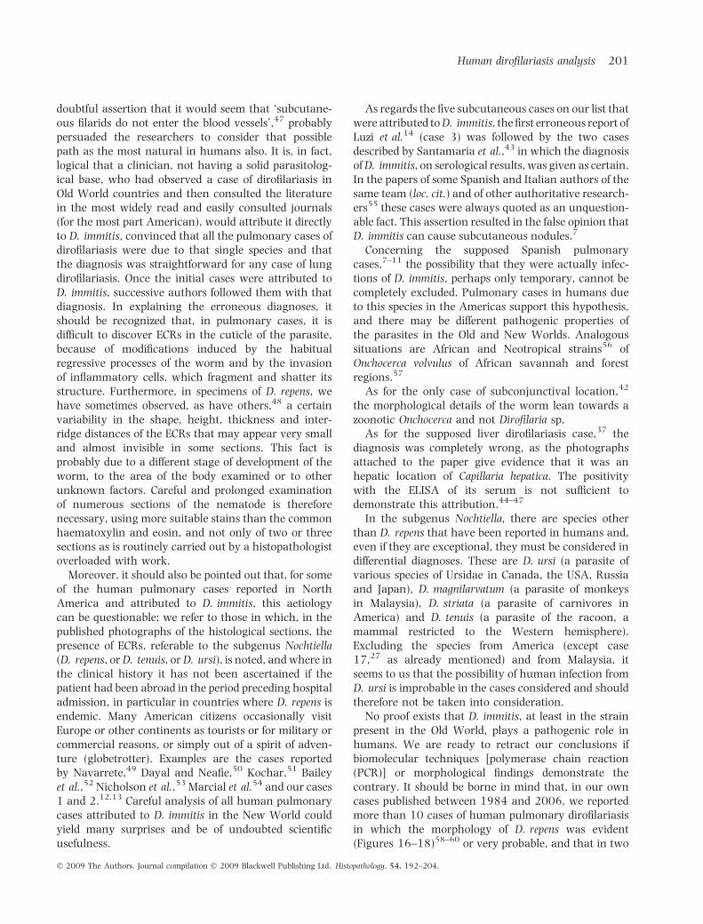
doubtful assertion that it would seem that ‘subcutane-ous filarids do not enter the blood vessels’,47 probablypersuaded the researchers to consider that possiblepath as the most natural in humans also. It is, in fact,logical that a clinician, not having a solid parasitolog-ical base, who had observed a case of dirofilariasis inOld World countries and then consulted the literaturein the most widely read and easily consulted journals(for the most part American), would attribute it directlyto D. immitis, convinced that all the pulmonary cases ofdirofilariasis were due to that single species and thatthe diagnosis was straightforward for any case of lungdirofilariasis. Once the initial cases were attributed toD. immitis, successive authors followed them with thatdiagnosis. In explaining the erroneous diagnoses, itshould be recognized that, in pulmonary cases, it isdifficult to discover ECRs in the cuticle of the parasite,because of modifications induced by the habitualregressive processes of the worm and by the invasionof inflammatory cells, which fragment and shatter itsstructure. Furthermore, in specimens of D. repens, wehave sometimes observed, as have others,48 a certainvariability in the shape, height, thickness and inter-ridge distances of the ECRs that may appear very smalland almost invisible in some sections. This fact isprobably due to a different stage of development of theworm, to the area of the body examined or to otherunknown factors. Careful and prolonged examinationof numerous sections of the nematode is thereforenecessary, using more suitable stains than the commonhaematoxylin and eosin, and not only of two or threesections as is routinely carried out by a histopathologistoverloaded with work.
Moreover, it should also be pointed out that, for someof the human pulmonary cases reported in NorthAmerica and attributed to D. immitis, this aetiologycan be questionable; we refer to those in which, in thepublished photographs of the histological sections, thepresence of ECRs, referable to the subgenus Nochtiella(D. repens, or D. tenuis, or D. ursi), is noted, and where inthe clinical history it has not been ascertained if thepatient had been abroad in the period preceding hospitaladmission, in particular in countries where D. repens isendemic. Many American citizens occasionally visitEurope or other continents as tourists or for military orcommercial reasons, or simply out of a spirit of adven-ture (globetrotter). Examples are the cases reportedby Navarrete,49 Dayal and Neafie,50 Kochar,51 Baileyet al.,52 Nicholson et al.,53 Marcial et al.54 and our cases1 and 2.12,13 Careful analysis of all human pulmonarycases attributed to D. immitis in the New World couldyield many surprises and be of undoubted scientificusefulness.
As regards the five subcutaneous cases on our list thatwere attributed to D. immitis, the first erroneous report ofLuzi et al.14 (case 3) was followed by the two casesdescribed by Santamaria et al.,43 in which the diagnosisof D. immitis, on serological results, was given as certain.In the papers of some Spanish and Italian authors of thesame team (loc. cit.) and of other authoritative research-ers55 these cases were always quoted as an unquestion-able fact. This assertion resulted in the false opinion thatD. immitis can cause subcutaneous nodules.7
Concerning the supposed Spanish pulmonarycases,7–11 the possibility that they were actually infec-tions of D. immitis, perhaps only temporary, cannot becompletely excluded. Pulmonary cases in humans dueto this species in the Americas support this hypothesis,and there may be different pathogenic properties ofthe parasites in the Old and New Worlds. Analogoussituations are African and Neotropical strains56 ofOnchocerca volvulus of African savannah and forestregions.57
As for the only case of subconjunctival location,42
the morphological details of the worm lean towards azoonotic Onchocerca and not Dirofilaria sp.
As for the supposed liver dirofilariasis case,37 thediagnosis was completely wrong, as the photographsattached to the paper give evidence that it was anhepatic location of Capillaria hepatica. The positivitywith the ELISA of its serum is not sufficient todemonstrate this attribution.44–47
In the subgenus Nochtiella, there are species otherthan D. repens that have been reported in humans and,even if they are exceptional, they must be considered indifferential diagnoses. These are D. ursi (a parasite ofvarious species of Ursidae in Canada, the USA, Russiaand Japan), D. magnilarvatum (a parasite of monkeysin Malaysia), D. striata (a parasite of carnivores inAmerica) and D. tenuis (a parasite of the racoon, amammal restricted to the Western hemisphere).Excluding the species from America (except case17,27 as already mentioned) and from Malaysia, itseems to us that the possibility of human infection fromD. ursi is improbable in the cases considered and shouldtherefore not be taken into consideration.
No proof exists that D. immitis, at least in the strainpresent in the Old World, plays a pathogenic role inhumans. We are ready to retract our conclusions ifbiomolecular techniques [polymerase chain reaction(PCR)] or morphological findings demonstrate thecontrary. It should be borne in mind that, in our owncases published between 1984 and 2006, we reportedmore than 10 cases of human pulmonary dirofilariasisin which the morphology of D. repens was evident(Figures 16–18)58–60 or very probable, and that in two
Human dirofilariasis analysis 201
� 2009 The Authors. Journal compilation � 2009 Blackwell Publishing Ltd, Histopathology, 54, 192–204.

other recent case,61 confirmation was possible usingPCR. We are also inclined to recognize that humancases of D. immitis involving the lung in the Old Worldenvironment may occur in future, if only rarely.
It remains to be shown why, in the Old World, whereD. immitis is present in dogs with a prevalencesometimes greater than that of D. repens, comparableoften to the prevalence found in the USA, cases ofpulmonary dirofilariasis associated with that speciesseem non-existent, in spite of the probable numerousexposures to the larvae of the nematode by frequentbites of the vector. This fact can be compared with thesimilar apparent paradox concerning Culex pipiensfatigans and Wuchereria bancrofti in Rangoon,62 wherethere were a large number of infected mosquitoes witha high biting rate, but few people actually becameinfected, with positive skin tests but without micro-filariaemia. While waiting for molecular biology tothrow further light on the problem, we can onlysuggest that the difference between the Old Worldhuman pulmonary cases and the Western hemispheremight be explained by two different hypotheses:(i) there are perhaps twin populations of D. immitiswith different genotypes on the two sides of theAtlantic, with different infective capacity for man anddog; (ii) the infective capacity to humans of the parasitecould be modified only in the Old World by someunidentified factor, possibly inherent to the vector, thataffects the complex vector–parasite relationship,63,64
decreasing the survival of the larvae.In conclusion, whatever reason, as far as the Old
World strain is concerned, the human organism seemsunsuitable for the development of D. immitis larvaeand, consequently, if they are carried by an appropriatevector, their vitality and infecting capacity are greatlyaffected. Furthermore, our observations demonstratehow unfounded are the statements of some authors47
who claim that the pulmonary cases in Italy are dueto D. immitis. Even more unfounded and misleading isthe statement that all subcutaneous cases in Europe,other than the pulmonary ones, are due to D. immitis,leaving the identification of D. repens only applicableto subconjunctival cases65 and that ‘the presentlyaccepted attributions of subcutaneous dirofilariasis toD. repens infection is false’.43
We hope that the results of our analysis willstimulate other researchers to bring new evidence tobear on this important subject.
Acknowledgements
The authors thank their colleagues F. Fedeli, R. Gayer,J. O. Gebbers, T. Jelinek, L. Khaldi, E. J. Mark, D. Monchy,
A. T. Morresi, N. Tornieporth, M. Trimech, S. H. Tsungand W. Wockel, who collaborated in the research,sending histological specimens of the cases underexamination, their original photographs or anamnestichistories of their patients, which they re-checked indetail. Thanks are also due to the scientific journals thatgave permission to reproduce some copyrighted photosin the plates I and II: Histopathology, Journal of EgyptianSociety of Parasitology, Journal of Formosan MedicalAssociation, Journal of Korean Medical Sciences, Journal ofSurgical Oncology, Italian Journal of Chest Diseases, Para-ssitologia, Pathologica, Thoracic Cardiovascular Surgery,Yonsei Medical Journal. The authors are pleased toacknowledge Professors Thomas Orihel (Tulane Univer-sity, New Orleans, USA) and Robert Killick Kendrick(Imperial College of Science, London, UK) for criticallyreviewing the manuscript. Mrs Eva Prati and MsAnnalisa Maiorano of the Ercolani Library, BolognaUniversity, are also sincerely acknowledged for theirkind help with the bibliographical research.
References
1. Neafie RC, Connor DH, Meyers WM. Dirofilariasis. In Binford CH,
Connor DH eds. Pathology of tropical and extraordinary diseases,
Vol. 2. Washington DC: Armed Forces Institute of Pathology,
American Registry of Pathology, 1976; 391–396.
2. Ro JY, Tsakalakis PJ, White VA et al. Pulmonary dirofilariasis:
the great imitator of primary or metastatic lung tumor. Hum.
Pathol. 1989; 20; 69–76.
3. Miyoshi T, Tsubouchi H, Iwasaki A et al. Human pulmonary
dirofilariasis: a case report and review of the recent Japanese
literature. Respirology 2006; 11; 343–347.
4. Pampiglione S, Canestri Trotti G, Rivasi F. Human dirofilariasis
due to Dirofilaria (Nochtiella) repens: a review of world literature.
Parassitologia 1995; 37; 149–193.
5. Dissanaike AS, Abeyewickreme W, Wijesundera MD, Weera-
sooriya MV, Ismail MM. Human dirofilariasis caused by
Dirofilaria (Nochtiella) repens in Sri Lanka. Parassitologia 1997;
39; 375–382.
6. Pampiglione S, Rivasi F. Human dirofilariasis due to Dirofilaria
(Nochtiella) repens: an update of world literature from 1995 to
2000. Parassitologia 2000; 42; 231–254.
7. Simon F, Muro A, Cordero M, Martın J. A seroepidemiological
survey of human dirofilariosis in Western Spain. Trop. Med.
Parasitol. 1991; 42; 106–108.
8. Cordero M, Munoz MR, Muro A, Simon F. Transient pulmonary
nodule caused by Dirofilaria immitis. Eur. Respir. J. 1990; 3;
1070–1071.
9. Cordero M, Munoz MR, Muro A, Simon F, Perera L. Small calcified
nodules. An undescribed radiologic manifestation of human
pulmonary dirofilariasis. J. Infect. Dis. 1992; 165; 398–399.
10. Cordero M, Muro A, Simon F, Tapia J, Espinoza E. Are transient
pulmonary solitary nodules a common event in human diro-
filariasis? Clin. Investig. 1992; 70; 437–440.
11. Muro Alvarez A, Cordero Sanchez M, Martın Martın J, Simon
Martın F. Seroepidemiological studies on human dirofilariosis in
Spain. Ann. Trop. Med. Parasitol. 1990; 84; 209–213.
202 S Pampiglione et al.
� 2009 The Authors. Journal compilation � 2009 Blackwell Publishing Ltd, Histopathology, 54, 192–204.

12. Anonymous. Case 13-1979. Presentation of case: case records.
Massachussetts Gen. Hosp. 1979; 300; 723–729.
13. Darrow JC, Lack EE. Solitary lung nodule due to Dirofilaria
immitis (dog ‘‘Heartworm’’). J. Surg. Oncol. 1981; 16; 219–224.
14. Luzi P, Leoncini L, Lungarella G. Human subcutaneous dirofil-
ariasis in Italy. A case report. Riv. Parassitol. 1982; 43; 259–
263.
15. Fabbretti G, Fedeli F, Alessi A et al. Human pulmonary dirofil-
ariasis: report of a new European case. Histol. Histopathol. 1990;
5; 311–313.
16. Pampiglione S, Canestri Trotti G, Squadrini F. Human subcuta-
neous dirofilariasis.2. A report of 5 new cases of Dirofilaria repens
in Central and Northern Italy and of a sixth case with uncertain
parasitological diagnosis. Parassitologia 1982; 24; 167–176.
17. Pampiglione S, Fedeli F. Dirofilariasi polmonare umana: aspetti
parassitologici del secondo caso segnalato in Italia. Parassitologia
1991; 33; 153–157.
18. Langer HE, Bialek R, Mielke H, Klose J. Human dirofilariasis
with reactive arthritis. Case report and review of the literature.
Klin. Wochenschr. 1987; 65; 746–751.
19. Badhe BP, Sane SY. Human pulmonary dirofilariasis in India: a
case report. J. Trop. Med. Hyg. 1989; 92; 425–426.
20. Bhat KG. Human dirofilariasis. Ind J Med Microbiol. 2003; 21; 65.
21. Tornieporth N, Brandis A, Vogel B, Disko R. Autochthonous
pulmonale dirofilariose in Europa. Dtsch. Med. Wschr. 1990; 115;
15–19.
22. Mobedi I, Javadian E, Abai MR. Two Pulmonary Cases of Dirofilaria
immitis (?) Based on Radiographic Coin Lesion in Iran. The First
Congress of Parasitic Diseases in Iran, December 11–13. Rasht,
Iran: Guilan University of Medical Sciences, 1990; 78. (abstract,
in Persian).
23. Monchy D, Levenes H, Guegan H, Poey C, Dubourdieu D.
Dirofilariose pulmonaire. Med. Trop. (Mars) 1993; 53; 367–371.
24. Wockel W, Eckert J, Loscher T, Haussinger K, Morresi A.
Autochthone europaische Lungen-dirofilariose. Pneumologie
1993; 47; 227–231.
25. Elnazer MM. Human ectopic infection with Dirofilaria immitis
(Leydi, 1865) representing first record from Egypt. Egypt Med. J.
1994; 11; 435–438.
26. Jelinek T, Schulte-Hillen J, Loscher T. Human dirofilariasis. Int. J.
Dermatol. 1996; 35; 872–875.
27. Fueter R, Gebbers J-O. Hunderherzwurm. Schweiz. Med. Woc-
henschr. 1997; 127; 2014–2015.
28. Breitenbucher A, Gayer R, Giachino D, Mordasini C. Multiple
pulmonary nodules. Respiration 1998; 65; 91–94.
29. Awadalla HN, Bayoumi DM, Ibrahim IR. The first case report of
suspected human dirofilariasis in the eyelid of a patient from
Alexandria. J. Egypt. Soc. Parasitol. 1998; 28; 941–944.
30. Bonu G, Zina G. Une rare affection chez l’homme: le granulome
souscutane par Dirofilaria. Bull. Soc. Fr. Dermatol. Syphilgr. 1972;
79; 248–250.
31. Romano A, Sachs R, Lengy J. Human ocular dirofilariasis in
Israel. Isr. J. Med. Sci. 1976; 12; 208–214.
32. Bruijning CFA. Human dirofilariasis. A report of the first case of
ocular dirofilariasis in the Netherlands and a review of the
literature. Trop. Geogr. Med. 1981; 33; 295–305.
33. Chauve CM. Dirofilaria repens (Railliet & Henry, 1911), Dipetalo-
nema reconditum (Grassi, 1840), Dipetalonema dracunculoides
(Cobbold, 1870), Dipetalonema grassii (Noe, 1907): quatre filaires
meconnues du chien. Prat. Med. Chir. Anim. Comp. 1990; 25;
293–304.
34. Sukpanichnant S, Leenuttapong V, Dejsomritrutai W et al.
Pulmonary dirofilariasis in a patient with multisystem Langer-
hans cell histiocytosis: the first reported case in Thailand. J. Med.
Assoc. Thai. 1998; 81; 722–727.
35. Lee KJ, Park GB, Yong TS et al. The first Korean case of human
pulmonary dirofilariasis. Yonsei Med. J. 2000; 41; 285–288.
36. Gomez-Merino E, Chiner E, Signes-Costa J et al. Pulmonary
dirofilariasis mimicking lung cancer. Monaldi Arch. Chest Dis.
2002; 57; 33–34.
37. Kim MK, Kim CH, Yeom BW et al. The first human case of
hepatic dirofilariasis. J. Korean Med. Sci. 2002; 17; 686–690.
38. Pampiglione S, Gustinelli G. Human hepatic capillariasis: a
second case occurred in Korea. J. Korean Med. Sci. 2008; 23;
560–561.
39. Tsung SH, Liu CC. Human pulmonary dirofilariasis in Taiwan.
J. Formos. Med. Assoc. 2003; 102; 42–44.
40. Foroulis CN, Khaldi L, Desimonas N, Kalafati G. Pulmonary
dirofilariasis mimicking lung tumor with chest wall and
mediastinal invasion. Thorac. Cardiovasc. Surg. 2005; 53; 173–
175.
41. Hemmersbach-Miller M, Delmont J, Hours C, Brouqui P. Pseudo-
tumor of the lung, a rare clinical presentation of dirofilariasis.
Presse Med. 2005; 34; 109–110.
42. Ziadi S, Trimeche M, Mestiri S et al. La dirofilariose sous-
conjonctivale humaine. A propos de deux cas tunisiens. J. Fr.
Ophtalmol. 2005; 28 e2; 773–775.
43. Santamaria B, Di Sacco B, Muro A et al. Serological diagnosis of
subcutaneous dirofilariosis. Clin. Exp. Dermatol. 1995; 20; 19–
21.
44. Glickman L, Grieve R, Schantz P. Serologic diagnosis of zoo-
notic pulmonary dirofilariasis. Am. J. Med. 1986; 80; 161–
164.
45. Beaver PC. Intraocular filariasis: a brief review. Am. J. Trop. Med.
Hyg. 1989; 40; 40–45.
46. Shah MK. Human pulmonary dirofilariasis: review of the
literature. South. Med. J. 1999; 92; 276–279.
47. Gutierrez Y, Misselevich I, Fradis M, Podoshin L, Boss JH.
Dirofilaria repens infection in Northern Israel. Am. J. Surg. Pathol.
1995; 19; 1088–1091.
48. Orihel TC, Eberhard ML. Zoonotic filariasis. Clin. Microbiol. Rev.
1998; 11; 366–381.
49. Navarrete AR. Pulmonary dirofilariasis. Chest 1972; 61; 51–55.
50. Dayal Y, Neafie RC. Human pulmonary dirofilariasis. Am. Rev.
Respir. Dis. 1975; 112; 437–443.
51. Kochar AS. Human pulmonary dirofilariasis. Am. J. Clin. Pathol.
1985; 84; 19–83.
52. Bailey TS, Sohrabi A, Roberts SS. Pulmonary coin lesions caused
by Dirofilaria immitis. J. Surg. Oncol. 1990; 44; 268–272.
53. Nicholson CP, Allen MS, Trastek VF, Tazelaar HD, Pairolero PC.
Dirofilaria immitis: a rare, increasing cause of pulmonary
nodules. Mayo Clin. Proc. 1992; 67; 646–650.
54. Marcial MA, File S, Fernandez A, Defendini E. Pulmonary
dirofilariasis: first reported case in Puerto Rico. PR Health Sci. J.
1995; 14; 17–19.
55. Walther M, Muller R. Diagnosis of human filariases (except
onchocerciasis). Adv. Parasitol. 2003; 53; 149–193.
56. Bradley JE, Unnasch TR. Molecular approaches to the diagnosis
of onchocerciasis. Adv. Parasitol. 1996; 37; 57–106.
57. Zimmerman PA, Katholi CR, Wooten MC, Lung UN, Unnasch
TR. Recent evolutionary history of American Onchocerca volvulus,
based on analysis of a tandemly reported DNA sequence family.
Mol. Biol. Evol. 1994; 11; 384–392.
58. Pampiglione S, Bosi F, Maconi AG et al. Pulmonary dirofilariasis:
clinical and parasitologic findings of a new human case. G. Ital.
Mal. Torace 1994; 48; 1–4.
Human dirofilariasis analysis 203
� 2009 The Authors. Journal compilation � 2009 Blackwell Publishing Ltd, Histopathology, 54, 192–204.

59. Pampiglione S, Rivasi F, Canestri Trotti G. Dirofilariosi polmon-
are umana: un caso in Italia. Pathologica 1984; 76; 565–572.
60. Pampiglione S, Rivasi F, Paolino S. Human pulmonary diro-
filariasis. Hystopathology 1996; 29; 69–72.
61. Rivasi F, Boldorini R, Criante P, Leutner M, Pampiglione S.
Detection of Dirofilaria (Nochtiella) repens DNA by polymerase
chain reaction in embedded paraffin tissues from two human
pulmonary locations. APMIS 2006; 114; 66–73.
62. Hairston NG, De Meilon B. On the inefficiency of transmission of
Wuchereria bancrofti from mosquito to human host. Bull. World
Health Org. 1968; 38; 935–941.
63. Mac Greevy PB, Theis JH, Lavoirpierre MMJ, Clarke J. Studies on
filariasis III. Dirofilaria immitis: emergence of the infective larvae
from the mouth part of Aedes aegypti. J. Helmithol. 1974; 48;
221–223.
64. Kettle DS. Medical and Veterinary Entomology. Lymphatic
filariasis: (Wuchereria bancrofti, Brugia malayi). CAB Int. Wall-
ingford, U.K. 1990; 588–604.
65. Simon F, Lopez-Belmonte J, Marcos-Atxutegi C, Morchon R,
Martın-Pacho JR. What is happening outside North America
regarding human dirofilariasis? Vet. Parasitol. 2005; 133;
181–189.
204 S Pampiglione et al.
� 2009 The Authors. Journal compilation � 2009 Blackwell Publishing Ltd, Histopathology, 54, 192–204.