
Material analyses of ‘Christ with singing and music-making Angels’, a late 15th-C panel painting...
14
Material analyses of ‘Christ with singing and music-making Angels’, a late 15th-C panel painting attributed to Hans Memling and assistants: Part I. non-invasive in situ investigations† Geert Van der Snickt, * a Costanza Miliani, bc Koen Janssens, a Brunetto G. Brunetti, b Aldo Romani, bc Francesca Rosi, bc Philippe Walter, d Jacques Castaing, d Wout De Nolf, a Lizet Klaassen, e Ineke Labarque e and Regine Wittermann f Received 25th February 2011, Accepted 6th September 2011 DOI: 10.1039/c1ja10073d In cultural heritage science, compositional data is traditionally obtained from works of art through the analysis of samples by means of various bench-top instruments (scanning electron microscope, Raman spectrometer, etc.). Alternatively, the object can be transported to a laboratory where it may be examined, usually by spectroscopic methods working in reflection mode. However, this paper describes how a complementary set of mobile and portable instruments was deployed in situ to gain a comprehensive view on the materials and related ageing compounds of an (almost) unmovable 15th-C polyptych, prior to and in preparation of the extraction of a limited number of samples. In line with the methodological approach discussed, PXRF was first employed as an efficient screening tool. The ensuing elemental data was supplemented by more specific information on both organic as inorganic materials supplied by reflection near- and mid-FTIR spectroscopy and fluorimetry. In completion, a limited number of diffraction patterns were collected with a mobile XRD instrument in order to identify the constituent crystalline phases in pigments, grounding materials and degradation products. In this way, it could be demonstrated how a rich array of colours was obtained by means of a limited palette of pigments: lead white, lead tin yellow, azurite, natural ultramarine, bone black, vermillion, madder lake, and a green copper-organo complex were detected and situated on the panels. Remarkably, next to chalk also gypsum was found in the ground layer(s) of this Western European easel painting. The relatively large surface of the background was covered with gold leaf; the analyses seem to point towards the labour-intensive water gilding technique. The versatility of this combination of analytical techniques was further illustrated by the accurate characterisation of degradation products affecting the readability and conservation of the painting: the overall presence of a calcium oxalate-based film of variable thickness was established. Nevertheless, further analysis of cross- sectioned samples was considered desirable in order to study the stratigraphy, to gain direct access to altered and sub-imposed layers and to allow highly detailed analysis of micrometric degradation products by state-of-the art techniques (i.e. synchrotron radiation). Introduction The identification and study of painting materials is an aspect of conservation science which received considerable attention in the recent literature 1–4 . The study of the materials and techniques used by the creative artists is of great interest, not only to investigate the (material) history of these objects and the modus operandi of the artists of that time, but also in view of the conservation treatment itself. A thorough technical study provides decisive information for selection of the appropriate conservation strategy and enhances the overall knowledge on the complex deterioration processes associated with ancient works of art 4 . During the last decade, two divergent instrumental trends were responsible for a significant progress in this a Department of Chemistry, University of Antwerp, Universiteitsplein 1, B-2610 Wilrijk, Belgium. E-mail: [email protected]; Fax: +32 3 265 23 76; Tel: +32 3 265 23 63 b Centre SMAArt, Department of Chemistry, University of Perugia, Via Elce di Sotto 8, 06123 Perugia, Italy c CNR-ISTM (Istituto di Scienze e Tecnologie Molecolari) c/o Department of Chemistry, University of Perugia, Via Elce di Sotto 8, 06123 Perugia, Italy d Centre de Recherche et de Restauration des Mus ees de France, CNRS UMR171, Palais du Louvre, Porte des Lions, 14 Quai Franc ¸ois Mitterand, 75001 Paris, France e Koninklijk Museum voor Schone Kunsten Antwerpen, Leopold De Waelplaats 2, B-2000 Antwerpen, Belgium f Private conservator, Hof van Uythem, Remerstraat 143, B-3130 Begijnendijk, Belgium † This article is part of a themed issue highlighting the latest research in the area of synchrotron radiation in art and archaeometry. 2216 | J. Anal. At. Spectrom., 2011, 26, 2216–2229 This journal is ª The Royal Society of Chemistry 2011 Dynamic Article Links C < JAAS Cite this: J. Anal. At. Spectrom., 2011, 26, 2216 www.rsc.org/jaas PAPER Downloaded by CNR Bologna on 11 November 2011 Published on 23 September 2011 on http://pubs.rsc.org | doi:10.1039/C1JA10073D View Online / Journal Homepage / Table of Contents for this issue
Transcript of Material analyses of ‘Christ with singing and music-making Angels’, a late 15th-C panel painting...

Dynamic Article LinksC<JAAS
Cite this: J. Anal. At. Spectrom., 2011, 26, 2216
www.rsc.org/jaas PAPER
Dow
nloa
ded
by C
NR
Bol
ogna
on
11 N
ovem
ber
2011
Publ
ishe
d on
23
Sept
embe
r 20
11 o
n ht
tp://
pubs
.rsc
.org
| do
i:10.
1039
/C1J
A10
073D
View Online / Journal Homepage / Table of Contents for this issue
Material analyses of ‘Christ with singing and music-making Angels’, a late15th-C panel painting attributed to Hans Memling and assistants: Part I.non-invasive in situ investigations†
Geert Van der Snickt,*a Costanza Miliani,bc Koen Janssens,a Brunetto G. Brunetti,b Aldo Romani,bc
Francesca Rosi,bc Philippe Walter,d Jacques Castaing,d Wout De Nolf,a Lizet Klaassen,e Ineke Labarquee
and Regine Wittermannf
Received 25th February 2011, Accepted 6th September 2011
DOI: 10.1039/c1ja10073d
In cultural heritage science, compositional data is traditionally obtained from works of art through the
analysis of samples by means of various bench-top instruments (scanning electron microscope, Raman
spectrometer, etc.). Alternatively, the object can be transported to a laboratory where it may be
examined, usually by spectroscopic methods working in reflection mode. However, this paper describes
how a complementary set of mobile and portable instruments was deployed in situ to gain
a comprehensive view on the materials and related ageing compounds of an (almost) unmovable 15th-C
polyptych, prior to and in preparation of the extraction of a limited number of samples. In line with the
methodological approach discussed, PXRF was first employed as an efficient screening tool. The
ensuing elemental data was supplemented by more specific information on both organic as inorganic
materials supplied by reflection near- and mid-FTIR spectroscopy and fluorimetry. In completion,
a limited number of diffraction patterns were collected with a mobile XRD instrument in order to
identify the constituent crystalline phases in pigments, grounding materials and degradation products.
In this way, it could be demonstrated how a rich array of colours was obtained by means of a limited
palette of pigments: lead white, lead tin yellow, azurite, natural ultramarine, bone black, vermillion,
madder lake, and a green copper-organo complex were detected and situated on the panels.
Remarkably, next to chalk also gypsum was found in the ground layer(s) of this Western European
easel painting. The relatively large surface of the background was covered with gold leaf; the analyses
seem to point towards the labour-intensive water gilding technique. The versatility of this combination
of analytical techniques was further illustrated by the accurate characterisation of degradation
products affecting the readability and conservation of the painting: the overall presence of a calcium
oxalate-based film of variable thickness was established. Nevertheless, further analysis of cross-
sectioned samples was considered desirable in order to study the stratigraphy, to gain direct access to
altered and sub-imposed layers and to allow highly detailed analysis of micrometric degradation
products by state-of-the art techniques (i.e. synchrotron radiation).
aDepartment of Chemistry, University of Antwerp, Universiteitsplein1, B-2610 Wilrijk, Belgium. E-mail: [email protected]; Fax:+32 3 265 23 76; Tel: +32 3 265 23 63bCentre SMAArt, Department of Chemistry, University of Perugia, ViaElce di Sotto 8, 06123 Perugia, ItalycCNR-ISTM (Istituto di Scienze e Tecnologie Molecolari) c/o Departmentof Chemistry, University of Perugia, Via Elce di Sotto 8, 06123 Perugia,ItalydCentre de Recherche et de Restauration des Mus�ees de France, CNRSUMR171, Palais du Louvre, Porte des Lions, 14 Quai FrancoisMitterand, 75001 Paris, FranceeKoninklijk Museum voor Schone Kunsten Antwerpen, Leopold DeWaelplaats 2, B-2000 Antwerpen, BelgiumfPrivate conservator, Hof van Uythem, Remerstraat 143, B-3130Begijnendijk, Belgium
† This article is part of a themed issue highlighting the latest research inthe area of synchrotron radiation in art and archaeometry.
2216 | J. Anal. At. Spectrom., 2011, 26, 2216–2229
Introduction
The identification and study of painting materials is an aspect of
conservation science which received considerable attention in the
recent literature1–4. The study of the materials and techniques
used by the creative artists is of great interest, not only to
investigate the (material) history of these objects and the modus
operandi of the artists of that time, but also in view of the
conservation treatment itself. A thorough technical study
provides decisive information for selection of the appropriate
conservation strategy and enhances the overall knowledge on
the complex deterioration processes associated with ancient
works of art4. During the last decade, two divergent instrumental
trends were responsible for a significant progress in this
This journal is ª The Royal Society of Chemistry 2011

Dow
nloa
ded
by C
NR
Bol
ogna
on
11 N
ovem
ber
2011
Publ
ishe
d on
23
Sept
embe
r 20
11 o
n ht
tp://
pubs
.rsc
.org
| do
i:10.
1039
/C1J
A10
073D
View Online
field: the implementation of mobile non-invasive equipment on
one hand and research with synchrotron radiation (SR) on the
other hand. The first development brought spectroscopic anal-
ysis literally to the conservators, making it more tangible and
comprehensible for them. The advent of mobile instruments
with analytical performances close to or equal to the conven-
tional lab-based devices made in situ analysis feasible and rele-
vant5,6. Conservators and scientists benefited greatly from this
breakthrough as in the past, transportation of fragile and
precious works of art to a laboratory or extraction of samples
often restrained them from an extensive analytical campaign.
On the other hand, synchrotron light-based analysis
transfers research to large-scale facilities, rendering spectroscopic
analysis rather abstract and inaccessible for conservators.
Nevertheless, different authors demonstrated how the elevated
spatial resolution, brightness and energy-selectivity of the
primary beam makes SR techniques indispensable for gaining
new insights in the composition of painting materials and their
degradation phenomena7–15. The authors are convinced that
a combination of both instrumental advances (mobile equipment
and synchrotron radiation) will become essential for the
conservation treatment of important works of art, such as Hans
Memling’s (ca.1435–1494) ‘Christ with singing and music-
making Angels’ (see Fig. 1).
Since 2001, conservators of the Royal Museum of Fine Arts in
Antwerp are engaged with the examination and conservation
treatment of this masterpiece, dated ca. 1487-900. From an
analytical point of view, the conservation treatment period
provided a unique time window during which the paint layers
were accessible for direct analysis, without the disturbing inter-
ference of non-original, superimposed layers (e.g. old varnish,
restoration retouchings). Additionally, the removal of the frame
and the retouchings allowed taking samples at positions which
are supportable from a deontological point of view, e.g. at the
edge of the panels or at an existing lacuna. Therefore, an
extensive analytical campaign was initiated in the framework of
collaboration between the Royal Museum of Fine Arts in Ant-
werp and the Antwerp X-ray Instrumentation and Imaging
Laboratory (AXI2L) research group16 of the University of
Antwerp.
As it is generally known that each analytical technique offers
specific prospects and limitations17, a wide range of comple-
mentary techniques were employed. In order to supply a clear
survey of this diversified sequence of investigations, a schematic
Fig. 1 Picture of ‘Christ with singing and music-making Angels’ from the col
with numbering of the angels. Pictures by Ren�e Gerritsen, copyright KMSK
This journal is ª The Royal Society of Chemistry 2011
overview of the applied methodology is presented in Fig. 2. The
scheme demonstrates how the analytical campaign was divided
into two main phases: (1) research on the painting and (2)
research on samples. During the first phase, a large number of
analytical measurements were performed directly on the panels
by means of mobile and portable equipment. As Fig. 2 illustrates,
during the second phase, untreated samples were first studied by
means of various synchrotron radiation-based methods. In this
way, the broadly-based information obtained during the first
phase was supplemented with highly detailed and species-selec-
tive data collected on a few samples. The samples were subse-
quently embedded in cross-section and examined with
conventional lab-based instruments for a systematic study of the
spatial distribution of the identified materials over the different
paint layers. The analysis of embedded cross-sectioned samples
at synchrotron end stations is relevant as well, as it allows col-
lecting layer-specific information. However, due to the limited
access to synchrotron facilities, it was not possible to probe each
available sample with all relevant SR techniques, before and after
embedding. In view of the large amount of data resulting from
these analytical measurements, we were prompted to divide the
study into several parts. This article is confined to the analytical
measurements on the painting by means of portable and mobile
equipment (phase 1, see 1.3 in Fig. 2). The results of the
measurements on samples before embedding (see 2.1 in Fig. 2)
and after embedding (see 2.2 in Fig. 2), will be discussed in sequel
articles.
A first explorative and fairly extensive series of analyses was
performed by means of Portable X-Ray Fluorescence (PXRF)
spectrometry. After processing and interpretation of the
resulting data, some of the instrumentation of MOLAB was
employed in a second phase to complement the ensuing find-
ings. MOLAB is a mobile laboratory composed of a unique
collection of portable equipment which is available to art-
historians, conservators, and conservation scientists through
respectively the Eu-ARTECH and CHARISMA projects,
funded by the 6th and 7th FP18,19. During the last decade,
MOLAB developed a well-established expertise concerning in
situ, non-invasive characterization of works of art making use
of e.g., fibre-optic mid-20–22 and near-FTIR23 spectrometry,
UV fluorescence spectrometry in steady state24–26 and time
resolved27 and micro-Raman28 for local molecular finger-
printing of artwork materials. In summary, it is expected that
the intensive in situ and non-invasive campaign discussed in
lection of the Royal Museum of Fine Arts in Antwerp (inv. nrs. 778–780),
A.
J. Anal. At. Spectrom., 2011, 26, 2216–2229 | 2217

Fig. 2 Scheme presenting an overview of the research methodology applied toMemling’s ‘Christ with singing and music-making Angels’. In this article,
only the results of ‘1.3: analysis on the painting’ are discussed.
Dow
nloa
ded
by C
NR
Bol
ogna
on
11 N
ovem
ber
2011
Publ
ishe
d on
23
Sept
embe
r 20
11 o
n ht
tp://
pubs
.rsc
.org
| do
i:10.
1039
/C1J
A10
073D
View Online
this paper, will enable to extrapolate the highly specific
information obtained from a limited number of minute
samples to the whole surface of the sizeable panels. In this
way, our objective is to obtain a maximum of information with
a minimum of sampling.
2218 | J. Anal. At. Spectrom., 2011, 26, 2216–2229
The paintings
The Royal Museum of Fine Arts in Antwerp houses over 7600
works of art illustrative for the artistic evolution in the Southern
Netherlands from the 13th- until the 20th-C. The museum is well
This journal is ª The Royal Society of Chemistry 2011

Dow
nloa
ded
by C
NR
Bol
ogna
on
11 N
ovem
ber
2011
Publ
ishe
d on
23
Sept
embe
r 20
11 o
n ht
tp://
pubs
.rsc
.org
| do
i:10.
1039
/C1J
A10
073D
View Online
known for its rich collection of Mediaeval and Northern
Renaissance paintings, containing several important works of
Flemish Primitives (e.g. Rogier Van der Weyden, Jan Van Eyck,
Gerard David, etc.). One of the key masterpieces of this period is
‘Christ with singing and music-making Angels’ dated ca. 1487-
900. This sizeable work, attributed to Hans Memling and assis-
tants, depicts Christ, flanked by sixteen angels singing and/or
playing different musical instruments. The scene is presented
against a golden background and runs on three monumental
panel paintings with inventory numbers 778, 779 and 780,
measuring 167.7 � 212.7 cm, 170 � 231.5 cm and 170 � 231 cm
respectively. Most probably, this trinity represents only the top
register of what was once an even larger polyptych, probably
originally commissioned for the convent church of Santa Maria
la Real in N�ajera, Spain29. However, since 1895, this masterwork
is part of the collection of the Royal Museum of Fine Arts in
Antwerp. In what follows, throughout the three panels, each
angel has been assigned a code number from 1 to 16 (see Fig. 1).
Experimental
As Fig. 3 illustrates, the analyses during phase 1 were carried out
in situ by means of non-invasive and non-contact techniques:
Portable X-Ray Fluorescence (PXRF), Mobile X-Ray Diffrac-
tion (MXRD), mobile reflection mid-Fourier Transform
Infrared (mid-FTIR), mobile reflection near-Fourier Transform
Infrared (near-FTIR) and mobile reflection UV-vis spectroscopy
in absorption and emission.
PXRF analyses were carried out with a TRACeR III-V device,
a commercial system manufactured by Keymaster Technologies
(currently Bruker, Karlsruhe, Germany). This compact device is
equipped with a Rh-tube with a maximal acceleration voltage of
40 kV and a beam current of 2 to 25 mA. Its maximum irradiation
power is therefore 1 W. The measuring spot has a circular shape
with a surface of approximately 1 cm2. Spectra were collected by
means of a Peltier cooled Si-PIN diode detector with a resolution
of 169.4 eV, FWHM at Mn (5.9 keV). Detector and source are
orientated in 45� geometry. All measurements were performed in
air, at a voltage of 40 kV, a current of 2.3 mA and an acquisition
time of 200s. A limited amount of spots were analysed at 15kV
and 15mA to probe for low Z elements. After measurement, all
spectra were evaluated using the software package AXIL30. As
Fig. 3 demonstrates, the apparatus was mounted on a solid
studio camera stand, permitting controlled and accurate move-
ment of the device along the X-Y-Z axes. Consequently, the
device could be levered safely to the desired position, leaving
a few mm between its snout and the object. Technical features
(e.g.minimum detection limits) and the hands-on drawbacks and
benefits for the analysis of paintings, of the analytical method in
general and this instrument in particular are discussed
elsewhere31.
Mobile X-Ray Diffraction (MXRD) combined with XRF:
a single source is used to ascertain that the X-ray fluorescence is
collected from exactly the same spot where the diffracted beams
stem from. An air-cooled iMOXS-MFR (IFG, Adlershof, Ber-
lin, Germany) X-ray tube is used. The X-ray source provides
a maximum power of 30 W; during the experiments, a voltage of
40 kV was used with a current of 700 mA. In order to increase
the useful photon flux, the source is equipped with
This journal is ª The Royal Society of Chemistry 2011
a polycapillary semi-lens that provides a 4 mm (3.8–4.4 mm)
diameter parallel X-ray beam (total exit divergence of 0.25� or
4.36 mrad). The copper anode provides, through a 0.1 mm
beryllium window, polychromatic X-rays necessary for XRF
measurements. The source is equipped with a 15-mm Ni filter to
strongly attenuate the Cu Kb-line and avoid the presence of
secondary diffraction peaks in addition to the main peaks due
to Cu-Ka. XRD is therefore performed with the usual mono-
chromatic radiation (Cu-Ka; E ¼ 8.047 keV; ¼ 0.154 nm); the
bremstrahlung giving rise to some background. With the
MXRD instrument, diffractograms are collected in reflection
mode using an incident angle u of 10� to the specimen surface
allowing to reach 2q values larger than 10�. Fine-grain alumina
samples were used for the calibration. The FIT 2D software32
was used to transform the two-dimensional diffraction images
into 2q spectra. Based on the latter and a database of X-ray
powder diffraction patterns, the EVA33 and XRDUA34 software
programme were used to determine the crystalline phases that
were present. The components of the MXRD system are
assembled on a frame that can be moved along the surface of
the object to be analysed. Two laser pointers intersect at the
analysis position, where the X-ray beam impinges the surface of
the object. The relatively prolonged acquisition times,
amounting up to 30 min, limited the number of measurements.
The technical features and analytical performance of this
instrument are discussed elsewhere35.
Reflection mid-infrared (mid-FTIR) spectrometry: a portable
JASCO VIR 9500 spectrophotometer equipped with a Remspec
mid-infrared fibre optic sampling probe was used. It is made up
of a Midac Illuminator IR radiation source, a Michelson inter-
ferometer and a liquid nitrogen cooledMCT-detector. The probe
was a bifurcated cable containing 19 chalcogenide glass fibres
that allowed the collection of spectra in the range 4000–900 cm�1
at a resolution of 4 cm�1. The probe diameter was about 4 mm.
The non-contact probe is kept perpendicular to the painting
surface (0�/0� geometry) at a distance of about 3 mm. The total
reflectivity, R, due to the combined diffuse and specular
components, was collected over 400 scans using the spectrum
from an aluminium mirror plate for background correction.
reflection near-infrared (near-FTIR) spectrometry: Near
infrared spectra were recorded using a portable JASCO VIR
9600 spectrophotometer made up of a halogen lamp as the
source, a Michelson interferometer equipped with a CaF2 beam
splitter,and room temperature InGaAs detector. The spectral
range is 12500–4000 cm�1 with an energy resolution of 4 cm�1.
The spectrophotometer is equipped with a silica glass fibre optic
sampling probe (2 m-long, 200/300 mm of core) which can
remotely measure the reflection of a variety of solid surfaces with
a spatial resolution of about 10 mm2. Both for mid and near-IR,
the spectrum intensity was defined as the pseudo absorbance A0
where A0 ¼ log (1/R).
Reflectance UV-vis fluorimetry (RF): Fluorescence spectra
were collected using a portable fluorimeter, assembled as
a prototype from individual components at the University of
Perugia, and already described in a previous paper25. The exci-
tation was performed at 480 nm using a suitable couple of short-
(FWHM 10 nm, 30% transmittance at 480 nm) and long-
bandpass filters (constant transmittance above 520 nm). The
spectral resolution was about 25 nm. The emission spectra
J. Anal. At. Spectrom., 2011, 26, 2216–2229 | 2219

Fig. 3 Composite image showing the different mobile, analytical techniques during measurement: A: PXRF, B: reflection near-FTIR, C: reflection mid-
FTIR, D: reflection UV-vis Fluorimetry and E: MXRD.
Dow
nloa
ded
by C
NR
Bol
ogna
on
11 N
ovem
ber
2011
Publ
ishe
d on
23
Sept
embe
r 20
11 o
n ht
tp://
pubs
.rsc
.org
| do
i:10.
1039
/C1J
A10
073D
View Online
obtained were then corrected for the detector sensitivity using
a suitable elaborated correction file. Reflection measurements
were undertaken using a portable spectrophotometer composed
of Avantes parts that is a deuterium-halogen lamp (AvaLight-
DH-2000-FHS) as a light source, an integrating sphere with
2220 | J. Anal. At. Spectrom., 2011, 26, 2216–2229
a 6 mm diameter viewing aperture (ISP-30-6) used to collect and
transfer the reflectance signals via a quartz fibre optic system
(diameter 600 mm) and an AvaSpec–2048 CCD detector. The
AvaSoft software controls the acquisition of the spectra in the
200–1100 nm range.
This journal is ª The Royal Society of Chemistry 2011

Dow
nloa
ded
by C
NR
Bol
ogna
on
11 N
ovem
ber
2011
Publ
ishe
d on
23
Sept
embe
r 20
11 o
n ht
tp://
pubs
.rsc
.org
| do
i:10.
1039
/C1J
A10
073D
View Online
Time resolved fluorescence measurements were carried out by
a portable single photon counting apparatus described else-
where27. The instrument is composed by a pulsed source with
inter-changeable diodes and LEDs a photocathode detector
working in the 350–850 nm range a FluoroHub electronic device
containing the TAC (time-amplitude converter) and a PC that
fully controls the system of data acquisition and elaboration. It
allows a spatial resolution of about 12 mm2 and a time resolution
of about 0.4–0.5 ns or 0.1 ns using LEDs or laser diodes
respectively.
Results and discussion
The non-invasive and non-contact characteristics of mobile
analytical techniques allowed carrying out a large number of
measurements in a relatively short amount of time. Although the
different analytical sessions were spread over several years in
accordance with the progression of the conservation treatment,
the actual analyses were completed in a total of ca. 12 working
days. The sizeable panel paintings were measured in the
conservation studio and stayed on the solid, metal easels that
were especially designed for the lengthy and elaborate conser-
vation treatment. In this way, more than 200 analyses were
performed using five different diagnostic methods without any
manipulation of the panels or surface preparation. In addition to
efficiency and flexibility, another important advantage of these
in situ techniques was the direct interaction between the conser-
vators of the panels and the (cultural heritage) scientists per-
forming the analyses. The operators of the analytical instrument
could assess the results immediately after each measurement,
enabling them to confer with the conservators for interpretation
of the results before continuing with another position or per-
forming another type of measurement on the current one. In the
next paragraphs, the results of the multimodal measurements are
discussed with respect to the materials and the pigments used for
realising the specific colour groups.
White
Substantial amounts of Pb were noticeable in all PXRF spectra
recorded on all three of the panels. MXRD and reflection mid-
FTIR complemented this outcome by identifying hydrocerussite
(2PbCO3$Pb(OH)2) and/or cerussite (PbCO3) in a number of
spots.
Although a variety of lead-based materials was used in
paintings for different purposes (pigments, fillers, siccatives .),
it was nevertheless expected that the detected Pb-L XRF signals
were mainly due to the presence of lead white. This lead
carbonate hydroxide was the only white pigment used in oil
painting until the 19th-C. Consequently, it has been intensively
used by artists since ancient times to realise white areas or to
lighten the shade of other colours by intermixture. Additionally,
this pigment was commonly added to paint with the aim of
compensating the drawbacks of other pigments, e.g. to enhance
the drying properties or the opacity of the paint36.
MXRD allowed identifying hydrocerussite, the basic form of
lead carbonate in several paint areas of the Memling panels,
including white zones (e.g. in the white dress of angel #2), while
both hydrocerussite and cerussite were found in the blue
This journal is ª The Royal Society of Chemistry 2011
undergarment of angel #6. Welcomme et al. demonstrated by
means of Synchrotron Radiation m-XRD that the 16th-C lead
white employed by Matthias Gr€unewald, consisted of a mixture
of hydrocerussite and cerussite in variable ratios12. These
changeable proportions of the different types of lead carbonate
are probably due to deviating recipes and/or the inadequate
control of the atmospheric conditions (temperature, carbon
dioxide, moisture, etc.) during the production process36. In
addition, mid-FTIR identified hydrocerussite in the white dress
of angel #4 and hydrocerussite or cerussite on a number of
differently coloured spots, based on the characteristic shape of
the n1 + n3 combination band of carbonate moiety at 2410 cm�1
and on the presence of the OH stretching fundamental band at
3537cm�16. Still, further analysis of cross-sectioned samples is
desirable to obtain a detailed specification of the mineral phases
of lead white and to situate the detected Pb-compound(s) in the
paint stratigraphy.
Ground layer(s)
After removal of the varnish and retouchings, the ground layer(s)
surfaced in small areas where the paint was damaged. PXRF
detected Ca all over the panels while MXRD and reflection mid-
FTIR were able to specify that calcite and gypsum are present in
the ground.
PXRF alone did not permit to analyse selectively the ground
layer(s), due to the relatively large diameter of the primary beam
(spot size ca. 1 cm2). Therefore, initially, the source of the
observed calcium compound was ambiguous as the incidence of
Ca can have various causes: a chalk- or gypsum-based ground
layer, the use of chalk as filler in paint or a component of
a pigment (e.g. bone or ivory black, a substrate for an organic
dye, etc.). Fortunately, the elevated lateral resolution of the
MXRD and mid-FTIR instrument (ca. 4 mm) permitted char-
acterising the materials in the ground layer(s) without interfer-
ence of the surface paint (see Fig. 4). Clear XRD patterns were
collected in these areas, demonstrating the presence of both
calcite (CaCO3) and gypsum (CaSO4$2H2O). The incidence of
both gypsum and calcite in the ground layers was confirmed by
means of mid-FTIR. Absorptions corresponding to the combi-
nation and overtone bands at circa 2200 cm�1 and at circa
2500 cm�1, indicative of the presence of gypsum37 and calcite20
respectively, were observed in areas with emerging ground layers
but also in several undamaged areas through the painting layer.
The discovery of gypsum was rather unexpected, since it is
a characteristic grounding material for Southern European easel
painting, while the use of chalk is usually associated with
Western and Northern European artists38,39. It remained unclear
whether the Spanish destination of the paintings is in some way
related with the occurrence of gypsum in the ground layers.
Surprisingly, gypsum was not found in all analysed spots. In
a number of areas, such as the gilded area above angel #15 and
the blue dress and brown instrument of angel #5 (panel 778),
only calcite was traced. However, it remained unclear if this
discrepancy is due to the fact that gypsum is inconsistently
distributed over the surface of the panels or whether it is asso-
ciated with technical limitations of the employed equipment. For
instance, it is not impossible that in some zones, the stack of
superimposed paint layers prevents the primary 8 keV photons of
J. Anal. At. Spectrom., 2011, 26, 2216–2229 | 2221

Fig. 4 Middle: detail of the paint surface showing a reserve/defect where the ground layer(s) emerge. The lower rectangle (01) indicates a spot where the
preparation seems largely preserved, while the upper rectangle (02) indicates a spot where part of the ground layer(s) is missing. A + B: reflection mid-
FTIR spectra establishing the presence of calcite and gypsum in area (01) and (02). C + D: MXRD patterns confirming the presence of calcite and
gypsum; however, the presence of gypsum is less apparent in pattern C (damaged area). This analytical result suggests the presence of multiple ground
layers with a difference in composition.
Dow
nloa
ded
by C
NR
Bol
ogna
on
11 N
ovem
ber
2011
Publ
ishe
d on
23
Sept
embe
r 20
11 o
n ht
tp://
pubs
.rsc
.org
| do
i:10.
1039
/C1J
A10
073D
View Online
the MXRD instrument to access the gypsum-based ground layer
and/or to escape from the paint. In view of the relatively low
energy of the primary beam and the high Z value of the elements
in the paint, a substantial absorption of both the primary and
secondary radiation can be anticipated. In addition, the low
angle of both the incident as the diffracted beams with respect to
the panel surface lengthens their path through the set of paint
layers; hence the absorption of the radiation by the materials
present is increased. As a result, it was also not completely clear if
gypsum and calcite occur in separate layers or intermixed. The
FTIR and MXRD measurements seem to suggest a layered
structure, but also here, the impending analysis of embedded
samples promises complementary information on the build-up of
the preparation layer(s).
Blue to purple
The investigations established that a wide variety of blue shades,
ranging from light blue to deep purple, was achieved by
combining the blue pigment azurite, 2CuCO3$Cu(OH)2, with an
assortment of other pigments such as bone black, lead white, lead
tin yellow (Pb2SnO4) and/or an organic red lake. In addition, the
expensive pigment ultramarine, Na7Al6Si6O24S3, was employed
exclusively to apply highlights on the blue gems of Christ.
All PXRF spectra were dominated by substantial copper and
lead peaks. In particular, the copper content was detected in the
light to dark blue garment of angels #5, #15, the wings of the
angels #2, #4, #6, #12, #13, #15 and in the sleeve and caudae
2222 | J. Anal. At. Spectrom., 2011, 26, 2216–2229
(the ribbons on the tiara) of Christ. This outcome indicated an
intensive use of azurite.
Reflection mid-FTIR analyses substantiated the identification
of azurite as the combination bands of both the copper carbonate
(structured signal at 2500 cm�1) and copper hydroxide (doublet
at 4244 and 4373 cm�1) moiety21 were clearly visible (see Fig. 5).
MXRD ascertained the presence of the blue mineral in the wings
of angel #6 and the robe of angel #5. In the latter area, MXRD
established the presence of graphite as well, most probably
intermixed with the blue paint to produce a darker shade.
Accordingly, mid-FTIR spectra of dark blue areas showed
a small sharp signal at 2010 cm�1 that is typical of bone black6,
although its vibrational assignment is still lacking. The combined
use of PXRF and reflection mid-FTIR indicated that the blue-
green feathers in the rainbow-coloured wings of angels #4 and #8
were obtained by combining azurite with lead tin yellow, as
shown in Fig. 6.
Light blue paint was obtained by combining azurite with
variable ratios of lead white. The fact that no other relevant
elements than Cu were detected in the blue-purple to deep purple
areas, suggests that this colour was obtained by combining
azurite with a red organic pigment (laminated and/or inter-
mixed). Reflectance UV-vis emission and absorption spectros-
copy succeeded in validating this hypothesis, specifying the
vegetal origin of the anthraquinone dyestuff, representative for
madder lake. The emission spectrum is shown in Fig. 7; madder
lake is identified by the shape of absorption spectrum, structured
at 510 and 540 nm, and the maximum of emission feature at
600 nm40. The identification was also confirmed by the time
This journal is ª The Royal Society of Chemistry 2011
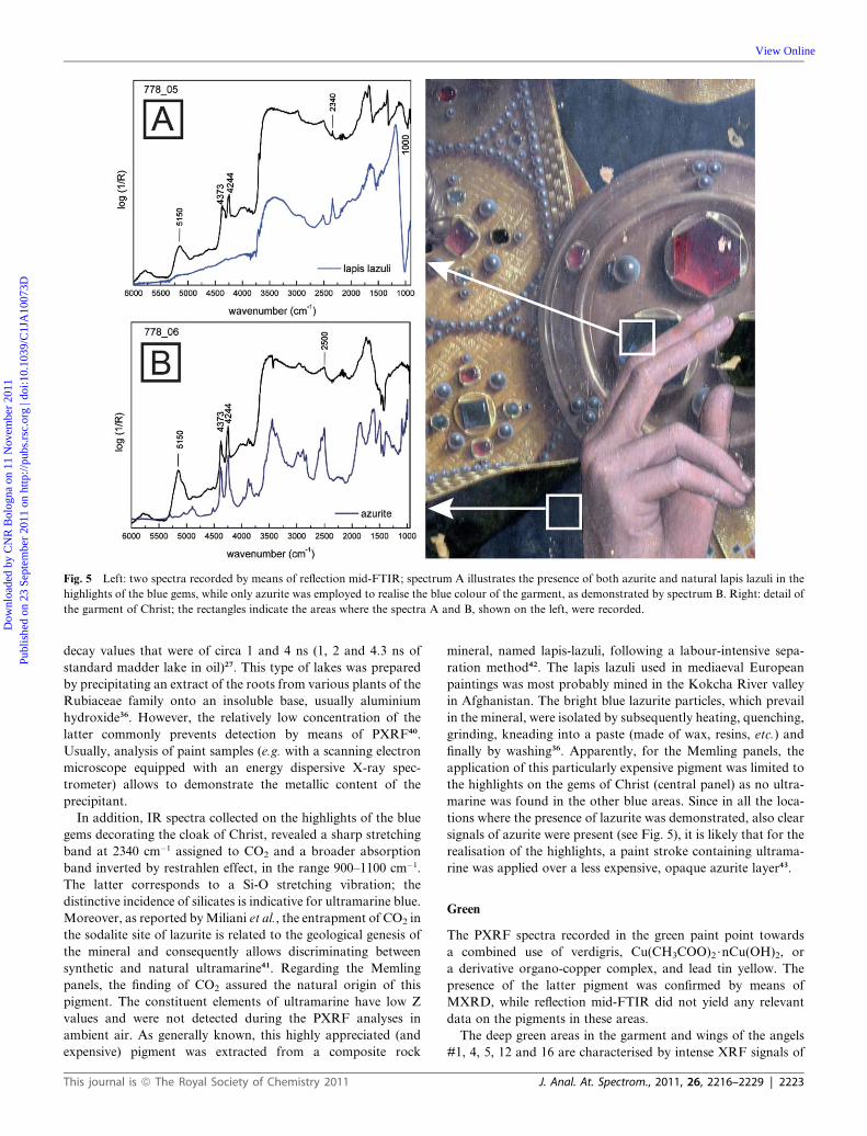
Fig. 5 Left: two spectra recorded by means of reflection mid-FTIR; spectrum A illustrates the presence of both azurite and natural lapis lazuli in the
highlights of the blue gems, while only azurite was employed to realise the blue colour of the garment, as demonstrated by spectrum B. Right: detail of
the garment of Christ; the rectangles indicate the areas where the spectra A and B, shown on the left, were recorded.
Dow
nloa
ded
by C
NR
Bol
ogna
on
11 N
ovem
ber
2011
Publ
ishe
d on
23
Sept
embe
r 20
11 o
n ht
tp://
pubs
.rsc
.org
| do
i:10.
1039
/C1J
A10
073D
View Online
decay values that were of circa 1 and 4 ns (1, 2 and 4.3 ns of
standard madder lake in oil)27. This type of lakes was prepared
by precipitating an extract of the roots from various plants of the
Rubiaceae family onto an insoluble base, usually aluminium
hydroxide36. However, the relatively low concentration of the
latter commonly prevents detection by means of PXRF40.
Usually, analysis of paint samples (e.g. with a scanning electron
microscope equipped with an energy dispersive X-ray spec-
trometer) allows to demonstrate the metallic content of the
precipitant.
In addition, IR spectra collected on the highlights of the blue
gems decorating the cloak of Christ, revealed a sharp stretching
band at 2340 cm�1 assigned to CO2 and a broader absorption
band inverted by restrahlen effect, in the range 900–1100 cm�1.
The latter corresponds to a Si-O stretching vibration; the
distinctive incidence of silicates is indicative for ultramarine blue.
Moreover, as reported byMiliani et al., the entrapment of CO2 in
the sodalite site of lazurite is related to the geological genesis of
the mineral and consequently allows discriminating between
synthetic and natural ultramarine41. Regarding the Memling
panels, the finding of CO2 assured the natural origin of this
pigment. The constituent elements of ultramarine have low Z
values and were not detected during the PXRF analyses in
ambient air. As generally known, this highly appreciated (and
expensive) pigment was extracted from a composite rock
This journal is ª The Royal Society of Chemistry 2011
mineral, named lapis-lazuli, following a labour-intensive sepa-
ration method42. The lapis lazuli used in mediaeval European
paintings was most probably mined in the Kokcha River valley
in Afghanistan. The bright blue lazurite particles, which prevail
in the mineral, were isolated by subsequently heating, quenching,
grinding, kneading into a paste (made of wax, resins, etc.) and
finally by washing36. Apparently, for the Memling panels, the
application of this particularly expensive pigment was limited to
the highlights on the gems of Christ (central panel) as no ultra-
marine was found in the other blue areas. Since in all the loca-
tions where the presence of lazurite was demonstrated, also clear
signals of azurite were present (see Fig. 5), it is likely that for the
realisation of the highlights, a paint stroke containing ultrama-
rine was applied over a less expensive, opaque azurite layer43.
Green
The PXRF spectra recorded in the green paint point towards
a combined use of verdigris, Cu(CH3COO)2$nCu(OH)2, or
a derivative organo-copper complex, and lead tin yellow. The
presence of the latter pigment was confirmed by means of
MXRD, while reflection mid-FTIR did not yield any relevant
data on the pigments in these areas.
The deep green areas in the garment and wings of the angels
#1, 4, 5, 12 and 16 are characterised by intense XRF signals of
J. Anal. At. Spectrom., 2011, 26, 2216–2229 | 2223

Fig. 6 The pigments identified in angel #8 demonstrate how the artists achieved a particular rich colour palette using a limited amount of colouring
substances. The PXRF spectra A to D exemplify how an array of blue to purple tints was obtained by combining a blue copper-based pigment, i.e.
azurite, with other pigments (organic lakes, lead tin yellow, carbon black and lead white).
Dow
nloa
ded
by C
NR
Bol
ogna
on
11 N
ovem
ber
2011
Publ
ishe
d on
23
Sept
embe
r 20
11 o
n ht
tp://
pubs
.rsc
.org
| do
i:10.
1039
/C1J
A10
073D
View Online
Cu, Sn and Pb. This is typical for a well-known green paint
stratigraphy used by mediaeval painters to realise the intense
green colour of foliage or fabrics. In this context, various authors
reported paint layers containing lead-tin yellow, lead white and
a green copper-based pigment in paintings of the 15th-
2224 | J. Anal. At. Spectrom., 2011, 26, 2216–2229
16th-C13,44. In line with this theory, the green copper-based
pigment would be verdigris or a derivative organo-copper
complex as, according to K€uhn, no other pigment (e.g.malachite
or green earth) or combination of pigments available at that time
could have created an equal intense green colour45.
This journal is ª The Royal Society of Chemistry 2011

Fig. 7 UV-vis emission and absorption spectra illustrating the combined
use of vermillion and madder lake to realise the red gems. UV-vis features
of a reddish area: absorption (pink line) and emission (red dotted line)
spectra. Insert: Fluorescence decay curves (red dots), excitation source
time profile (black dots), fitting curve (light grey line) and distribution of
residuals (bottom plot), lexc ¼ 455 nm, lem ¼ 620 nm.
Dow
nloa
ded
by C
NR
Bol
ogna
on
11 N
ovem
ber
2011
Publ
ishe
d on
23
Sept
embe
r 20
11 o
n ht
tp://
pubs
.rsc
.org
| do
i:10.
1039
/C1J
A10
073D
View Online
Verdigris was produced by reactions of metallic copper in the
presence of acetic acid. The above mentioned derivative organo-
copper complex was usually manufactured by dissolution of
verdigris in drying oil and/or resin, resulting in an amorphous
pigment containing copper carboxylate complexes of Cu(II) with
acetic, oleaginous and/or resinous acids15. In addition, analogous
compounds are also known to exist between copper and proteins
and from the reaction between copper compounds and
beeswax45. In older literature, this group of pigments is often
incorrectly referred to as ‘copper resinate’. As this type of
pigment does not necessarily contain a resin, the term ‘organo-
copper complex’ was proposed36. To complicate things even
more, it is not always clear to what extent mediaeval artists
employed this organo-copper complex intentionally or if it was
formed by the spontaneous dissolution of green copper pigments
in their organic binding medium, over time. For instance, Gunn
et al. revealed that when verdigris is mixed with an oleaginous
and/or resinous binder, the pigment starts to transform imme-
diately by the exchange of the acetato ligands of the pigment with
the carboxylic groups of the medium46.
MXRD identified lead stannate (lead-tin yellow, type I) and
hydrocerussite (lead white) in the green paint (green dress angel
#12) but failed to pinpoint the nature of the green pigment.
Nevertheless, this negative outcome is consistent with the
assumed presence of an amorphous organo-copper complex as
XRD is only sensitive towards crystalline substances. Lipid
signals from carbonyl (1745 cm�1) and C–H stretching (2845 &
2920 cm�1), both indicative of natural fats and oils, dominate the
FTIR spectra collected in green areas where conservators
removed the superimposed degradation (see further) and varnish
layers hampering any possible signals from the copper resinate
and/or acetate. The function of the detected lead-tin yellow and
lead white consists in increasing both the drying properties and
the covering power of the paint, as this type of green pigment is
known to produce a (semi-) transparent paint layer. Moreover,
lead-tin yellow compensates the bluish shade of verdigris45.
Additional measurements on cross-sectioned samples will be
This journal is ª The Royal Society of Chemistry 2011
necessary to clarify the chemical nature of the Cu-compounds
observed and to situate these elements in the stratigraphy.
Degradation phenomena
A number of the above mentioned green areas were of particular
interest to the conservators, not only in view of the unclear
nature of the green pigment, but especially considering the local
occurrence of a dark, opaque crust that could only be mechan-
ically removed with the aid of a scalpel and a microscope. During
cleaning tests, all of the conventional solvents failed in elimi-
nating this solid degradation product. Fig. 8 exemplifies how the
green colour of the maniple of angel #4 is concealed by the
greyish crust. In addition, a set of brownish, semi-transparent
layers seemed to be situated in between the crust and the green
paint. At first sight, this stack resembled a series of super-
imposed, old varnish layers. Nevertheless, the conservators were
questioning the originality of these layers as in the past, authors
had reported the presence of original, brownish layers on top of
green copper paint. In some cases, the green paint itself was
partly discoloured, but now and again the discoloration was
present as a separate layer resembling a glaze47,49. In addition,
Gunn et al. proved that copper(II) ions can diffuse from copper-
based paint into superimposed, organic (varnish) layers,
rendering it extremely difficult to differentiate the original paint
layers from later varnishes47. In spite of various scientific inves-
tigations, the exact nature of this type of degradation mechanism
of the green copper paint is still open for discussion15,45,47,48.
In this crust, it was possible to identify two related Ca-based
oxalate salts by means of MXRD: whewellite (CaC2O4$H2O)
and weddellite (C2CaO4$2H2O). This analytical outcome
explains the rigid and opaque aspect of the crusts and confirmed
earlier, preliminary FTIR analyses performed on surface scrap-
ings by Catherine Higgitt49. Surprisingly, the in situ mid-FTIR
analyses evidenced the widespread presence of traces of oxalates
all over the three panels, even in areas where no disturbing crust
was observed by the conservators. The characterization of
calcium oxalates by reflectance infrared is possible through the
derivative bands at 1315 cm�1 and 1620 cm�1 corresponding to ns
(C–O) + d (O–C]O) and n(C]O) respectively50. In the past,
calcium oxalates have been encountered commonly on weathered
surfaces such as outdoor stone materials of various natures
(marble, sandstone, granite, etc.)51,52, exterior wall paintings4,7,
rock art53 and even stained glass windows54. The importance of
this problem and the omnipresence of these compounds are
reflected by the large number of papers addressing this issue and
the fact that a dedicated conference was held in 199655. Never-
theless, the cause for the development of these particularly stable,
inorganic crystals is still subject of debate. In general, the
different opinions can be condensed into a chemical and a bio-
logical mechanism, both resulting from the reaction of oxalic
acid with a calcium-rich substrate: (a) the oxalate film is formed
by an oxidative degradation of organic material, or (b) the oxalic
acid is produced as a metabolic by-product of micro-biological
organisms (lichens, algae, bacteria, etc.), cultivating on organic
material. According to the conservators, the proposed prove-
nance of the organic material is most probably a non-original,
organic layer, or a stack of organic layers, applied in the course
of time with the aim of improving the appearance of the painting
J. Anal. At. Spectrom., 2011, 26, 2216–2229 | 2225

Fig. 8 A: detail of a green paint surface in the maniple of angel #4. The white dotted line indicates the interface between an area where the oxalate crust
was mechanically removed by the conservators (lower part) and an area prior to cleaning (upper part). The white circle indicates the area where spectra
B, C andDwere collected. B: PXRF spectrum showing intense Cu (green copper pigment), Pb + Sn (lead tin yellow), Fe (earth pigment) and Ca (calcium
oxalate) emission lines. C: MXRD pattern establishing the presence of (among others) weddellite and whewellite. D: reflection mid-FTIR spectrum
confirming the presence of calcium oxalates.
Dow
nloa
ded
by C
NR
Bol
ogna
on
11 N
ovem
ber
2011
Publ
ishe
d on
23
Sept
embe
r 20
11 o
n ht
tp://
pubs
.rsc
.org
| do
i:10.
1039
/C1J
A10
073D
View Online
by saturating the deep and dark colours. Application of layers of
oil, animal fat and/or egg white with the aim of ‘reviving’ the
paint surface was a common practice at the end of the 19th-C [Y].
This would explain why the crust is especially present on the deep
green paint as well, as these areas benefit the most from satura-
tion. Apart from that, additional factors such as the deposition
of dead organisms, animal excrements or other protective
conservation treatments cannot be excluded.52,53
Nevertheless, the occurrence of calcium oxalates on easel
paintings has been rarely reported in literature. The calcium
source is less apparent and microbiological attack is less expected
due to the relatively protected indoor environment (as opposed
to open-air monuments). However, recently, degradation prod-
ucts similar to the oxalates found on the Memling panels were
encountered on 15th-C paintings from the Museu Nacional
d’Art de Catalunya in Barcelona. Salvad�o et al. employed SR
m-XRD and SR m-FTIR to establish the presence of weddellite
and whewellite on embedded samples14. In addition, Kahrim
et al. documented the removal of a calcium oxalate crust from
a 20th-C. oil painting56. In both cases, the authors indicated that
the oxalic acid was predominantly stemming from varnish layers,
a finding that corroborated with the observations on the Mem-
ling panels. In addition, the authors suggest that the calcium
2226 | J. Anal. At. Spectrom., 2011, 26, 2216–2229
compound was caused by deposition of gypsum particles from
the (museum) atmosphere. Still, the origin of calcium on the
surface of theMemling panels remains uncertain, both accidental
(e.g. environmental deposition15, application of lime wash or
other protective coatings in the church57, etc.) or deliberate
deposit of calcium compounds (ancient conservation treatment
with milk or casein products, calcium phosphate; etc.) should be
considered. The organic breeding ground could be the above
mentioned set of old varnish layers, but also here, the possible
presence of old protective coatings or consolidation treatments
(oil, resin, glue, etc.) should not be overlooked. In addition, the
imminent analysis of paint samples will be required in order to
supply information on the stratigraphical distribution of these
alteration products.
Yellow
PXRF measurements in the yellow paint of the robe of the third
angel revealed a combination of Pb and Sn, usually associated
with the pigment lead-tin yellow. This deduction was ascertained
by means ofMXRD, as the presence of a crystalline phase of lead
tin oxide (lead-tin yellow type 1) was demonstrated. On the other
hand, the artist(s) clearly preferred a yellow earth pigment
This journal is ª The Royal Society of Chemistry 2011

Dow
nloa
ded
by C
NR
Bol
ogna
on
11 N
ovem
ber
2011
Publ
ishe
d on
23
Sept
embe
r 20
11 o
n ht
tp://
pubs
.rsc
.org
| do
i:10.
1039
/C1J
A10
073D
View Online
(ochre) to paint the blond hair of the angels as all corresponding
PXRF spectra were typified by substantial iron X-ray emission
lines. Yellow earth deposits are found in numerous localities all
over Europe. These naturally occurring, stable pigments all
contain the iron oxide hydroxide goethite (FeOOH) as the key
colouring matter and are accompanied by various contaminants
such as clays, silicates, carbonates and other phases dependent of
their provenance36. However, the latter often consist of low Z
elements and/or are present in small quantities. Therefore, these
compounds were not detected by means of PXRF in ambient air.
Still, mid-FTIR corroborated the terrestrial source of supply as
kaolin, Al4[Si4O10](OH)8, was discovered in these zones. Kaolin
is a white compound with a very fine grain (<2 mm) usually
associated with art from the Middle and Far East36, but here it is
most probably present as an impurity, related to the clay
compound of the yellow earth.
Brown
According to the PXRFmeasurements, the brown paint contains
a significant concentration of iron. Consequently, an earth
pigment was employed to realise the brown areas such as the hair
of Christ or the wood of the musical instruments. The absence of
Mn-XRF signals appears to exclude the presence of (burnt)
sienna’s or umbers. The latter are both iron oxides and
hydroxides similar to ochre but discriminated by a distinct
content of manganese oxides. An alternative for sienna and
umber would be yellow ochres that can be turned in to brown or
red pigments by a thermal transformation. Basically Fe(II)
hydroxides (goethite: FeOOH) are converted into Fe(III)oxides
(such as hematite, Fe2O3) by burning them at temperatures
around 300 �C. Burnt ochres demonstrate a disordered crystal
structure, different from naturally occurring hematite, due to the
roasting [36, X]. Unfortunately, apart from quartz, MXRD did
not yield any identifiable patterns in these areas possibly due to
the significant distortion of the lattice during roasting, which
resulted in a poorly crystalline material.
Red
The flesh tones and the bright red paint areas in the wings and
garments of the angels and Christ feature intense XRF signals of
mercury. This finding points towards the use of HgS, a pigment
that was either obtained as a mineral (cinnabar) or manufactured
(vermillion) from sulphur and liquid mercury. The presence of
minium (Pb3O4), another red pigment known at that time, could
not be confirmed nor denied by PXRF. The lead emission lines of
minium would be concealed by the presence of the above-
mentioned (see title ‘white’) intense Pb-peaks lines, observed in
all spectra. However, the use of minium was not anticipated, as it
is a highly instable pigment. Colours tending to orange, such as
the feathers in the wing of the angel #9, were obtained by
a combination of vermillion with lead-tin yellow, as demon-
strated by MXRD and PXRF.
The conservators also observed a reddish glaze on top of the
vermillion-based paint. In addition, certain areas, such as the
lining of the cloak of the first angel or the clouds, present pink to
burgundy tints. Here, no relevant elements were observed in the
PXRF spectra, excluding the application of a red, inorganic
This journal is ª The Royal Society of Chemistry 2011
pigment. In both cases, the use of a red lake pigment was
assumed. Lake pigments are pigments manufactured by precip-
itating dyestuffs, derived from various vegetable and animal
sources, on an inert substrate (e.g. alum). Usually, lake
compounds were used for glazing purposes, modifying the colour
or adding depth to the underlying paint. Analogous with the
purple tints (see Fig. 7) the pigment was characterised as madder
lake by means of reflection UV-vis fluorimetry27,41. However, no
fluorescence was detected in the more red-brownish clouds,
possibly owing to a degraded madder lake pigment.
Black
The panels present a limited number of black areas, such as the
black trumpet played by the fourth angel. As no relevant
elements were detected by PXRF, it was deduced that the black
colour was produced by means of a carbon-based pigment
obtained by burning organic material (e.g. oil, bones, wood,
etc.). Carbon blacks of vegetal origin can be distinguished from
carbon black of animal origin as the latter are characterised by
the presence of calcium and phosphor (stemming from the burnt
bones or ivory). PXRF spectra show the emission lines of
calcium but it remained unclear whether this is due to the
underlying gypsum/chalk ground layer(s) or to the employment
of a Ca-rich carbon black pigment. The concentration and
atomic number of phosphor is usually too low for detection with
ambient air PXRF. Nevertheless, mid-FTIR spectra display
a small peak at 2010 cm�1, indicative for carbon black of animal
origin, in several black and blue areas such as the black trumpet
of angel #13 and the blue garment of Christ.
Gilding
One of the most striking features of this work of art is the large
gilded surface. Apart from lead, PXRF did not detect any metals
indicative for a gold alloy (Ag, Cu, etc.). Usually, relatively pure
gold was used for manufacturing the very thin gold leaf.
Consequently, it was assumed that the detected lead was asso-
ciated with an underlying adhesive layer. Based on the binding
medium used for the adhesive layer, historic gilding techniques
can be divided into two main procedures: (a) water gilding and
(b) mordant gilding. For the latter, the gold leaf is applied over
a thin layer containing siccative oils and/or resins. Lead-based
materials are commonly added to speed up the drying process
and to obtain the desired colour. Usually, a red-brown colour is
preferred as the underlying layer shows through the micrometric
gold leaf, giving it a warm glow and masking possible defects.
Water gilding (a) involves adhesion of the gold leaf on a so-called
bole, a fine-grained clay coloured by iron oxide pigments and
bound by water-based glue11. This clay provides a mouldable,
soft material that permits polishing of the gilded surface with an
agate stone. Consequently, water gilding leads to a shinier
surface than the mordant technique58. Bearing in mind the
dimensions of the gilt surface and considering the particularly
labour-intensive aspect of the water gilding technique, employ-
ment of a mordant (oil technique) appears to be the most plau-
sible here. The fact that PXRF detected Pb in the gilded area
seems to corroborate this theory. As already mentioned, lead-
based materials have siccative properties and are therefore
J. Anal. At. Spectrom., 2011, 26, 2216–2229 | 2227

Dow
nloa
ded
by C
NR
Bol
ogna
on
11 N
ovem
ber
2011
Publ
ishe
d on
23
Sept
embe
r 20
11 o
n ht
tp://
pubs
.rsc
.org
| do
i:10.
1039
/C1J
A10
073D
View Online
related to oil-based, mordant gilding. Nevertheless, apart from
gold, MXRD documented the presence of goethite and quartz.
The later mineral is indicative for clay; an outcome which is
rather consistent with the presence of a bole layer (water gilding)
below the gold. Also here, it is expected that the forthcoming
analyses of samples will pinpoint the exact build-up of the gilding
and the nature of the constituting materials.
Binding media and varnish
The near-FTIR investigations registered C–H combination
bands at 4260 (nsCH2 + dCH2) and 4340 (naCH2 + dCH2) cm�1,
most probably due to lipids23, on all of the points measured on
the three panels. The presence of lipids points towards the use of
an oil and/or egg yolk as binding medium. Weak protein signals
(4595 cm�1, first overtone nCO amide I + amide II, 4880 cm�1,
nsNH + dNH)23 were exclusively registered in some small,
damaged paint areas where the ground layer emerged. As
a result, the material in the ground layers was probably bound by
glue and/or egg white. Apart from that, mid-FTIR measure-
ments demonstrated that the old organic varnish (partially
removed at the time of the measurements) contained a natural
terpenic resin. In addition, the collected spectra established the
presence of wax, mainly on the central panel 780 and to a lesser
extent on the other panels.
Conclusions
The combination of a variety of mobile instruments leads to
a far-reaching insight in the materials of a 15th-C polyptych
‘Christ surrounded by Angels playing Music’ by Hans Memling.
The advantages of these non-destructive, non-contact instru-
ments were fully exploited as a large number of spots were
analysed, allowing a systematic study of the full surface of the
sizeable panels. In addition, the in situ measurements assured
a direct interaction between conservators and (conservation)
scientists, resulting in a more effective campaign of analyses and
material identification. The supplementation of the elemental
data, gained by means of PXRF, with the species-selective
information derived from MXRD and FTIR analysis, is the key
to a more comprehensive identification of both organic and
inorganic materials used by the artists and of their current state.
In addition, the relatively short acquisition times of PXRF and
FTIR spectrometry allowed their use as swift screening tools.
The ensuing results were completed with a limited number of
MXRD measurements, which, though more time-consuming,
provided highly specific crystallographic data.
In particular, the interpretation of the analytical results
permitted to document the presence of the pigments lead tin
yellow, azurite, ultramarine, lead white, a green organo-copper
complex, brown and yellow earth, vermillion and madder lake.
Using this relatively limited pallet, Memling succeeded in
creating a wide scale of colours and optical effects. In addition,
the measurements demonstrated that the blue gems on the cloak
of Christ were worked with a combination of high-priced ultra-
marine with less expensive azurite. This set of complementary
diagnostic techniques supplied unexpected information on the
ground layers as well: both chalk and gypsum were detected in
various locations on all panels; these two materials were
2228 | J. Anal. At. Spectrom., 2011, 26, 2216–2229
probably applied in separate layers. The use of gypsum was
unexpected as this is a grounding material rather associated with
Southern European easel painting than with the Northern
European painting tradition of Memling’s era. Another note-
worthy outcome was the finding of quartz and goethite in the
gilding, suggesting the use of labour intensive water gilding as
application technique.
The capabilities of the analytical instrumentation that was
employed became particularly clear during the characterisation of
the degradation products and related phenomena. MXRD did not
only identify calcium-oxalate in the observed, opaque crust, but
also permitted detecting whewellite and weddellite separately.
Additionally, mid-FTIR revealed the distribution of these oxalates
over the entire surface of the panels, even in areas where no crust
was observed. In this way, this study did not only contribute to the
technical knowledge on early-Netherlandish painting but also
supported the ongoing conservation treatment. Moreover, the
omnipresence of alteration products on ancient paintings and the
need for better understanding their genesis and formation mech-
anisms was demonstrated once more by this study.
In conclusion, the implementation of this array of comple-
mentary and completely non-invasive analyses provided
a detailed overview of the materials used and the techniques
employed by Memling and provided some insights into the
ageing phenomena that have taken place as well. Usually this
information can only be achieved by (destructive) analysis of
samples, extracted from the panels.
Nevertheless, analysis of cross-sectioned samples remains
desirable to pinpoint the nature of all materials and their
distribution over the various layers. In this framework, the
above-mentioned investigations allowed defining relevant
regions of interests and related questions for further research in
a substantiated manner while minimising the number of samples
required for these studies. Especially, sampling of the green, blue-
purple, gilded and altered areas is considered, in order to allow
a deconvolution of the paint stratigraphy in these areas and to
better understand the development of degradation products.
Therefore, in the second phase of the study, the results obtained
in this first phase will be correlated to, confronted with and
supplemented by findings obtained via laboratory and synchro-
tron radiation based microanalysis of untreated and cross-
sectioned samples.
Acknowledgements
This research was supported by the Interuniversity Attraction
Poles Programme - Belgian Science Policy (IUAP VI/16). The
text also presents results of GOA ‘‘XANES meets ELNES’’
(Research Fund University of Antwerp, Belgium) and from
FWO (Brussels, Belgium) projects no. G.0103.04, G.0689.06 and
G.0704.08. The staff of the Royal Museum of Fine Arts Antwerp
is acknowledged for this pleasant cooperation and the author-
isation for the publication of the images in this article. Therefore,
a word of gratitude to Paul Huvenne, Yolande Deckers, Stef
Antonissen and Gwen Borms. In addition, the authors would
like to thank the MOLAB’s team operators Chiari Anselmi and
Federica Presciutti. MOLAB analyses have been carried out
through the support of the EU within the 6th Framework Pro-
gramme (Contract Eu-ARTECH, RII3-CT-2004-506171).
This journal is ª The Royal Society of Chemistry 2011

Dow
nloa
ded
by C
NR
Bol
ogna
on
11 N
ovem
ber
2011
Publ
ishe
d on
23
Sept
embe
r 20
11 o
n ht
tp://
pubs
.rsc
.org
| do
i:10.
1039
/C1J
A10
073D
View Online
Notes and references
1 L. de Viguerie, V. A. Sole and Ph.Walter, Anal. Bioanal. Chem., 2009,395, 2015–2020.
2 M. Cotte, J. Susini, J. Dik and K. Janssens,Acc. Chem. Res., 2010, 43,705–714.
3 C. Aib�eo, S. Goffin, O. Schalm, G. Van der Snickt, N. Laquiere,P. Eyskens and K. Janssens, J. Raman Spectrosc., 2008, 39, 1091–1098.
4 A. Nevin, J. L. Melia, I. Osticioli, G. Gautier and M. P. Colombini,J. Cult. Heritage, 2008, 9, 154–161.
5 C. Miliani, F. Rosi, B. G. Brunetti and A. Sgamellotti, Acc. Chem.Res., 2010, 43, 728–738.
6 C. Miliani, F. Rosi and A. Burnstock, Appl. Phys. A: Mater. Sci.Process., 2007, 89, 849–856.
7 K. Janssens, J. Dik, M. Cotte and J. Susini,Acc. Chem. Res., 2010, 43,814–825.
8 M. Cotte, J. Susini, V. A. Sole, Y. Taniguchi, J. Chillida, E. Checrounand Ph. Walter, J. Anal. At. Spectrom., 2008, 23, 820–828.
9 J. Dik, K. Janssens, G. Van der Snickt, L. Van der Loeff, K. Rickersand M. Cotte, Anal. Chem., 2008, 80, 6436–6442.
10 G. Van der Snickt, J. Dik and M. Cotte, Anal. Chem., 2009, 81, 2600–2610.
11 A. Lluveras, S. Boularand, J. Roque, M. Cotte, P. Giraldez andM. Vendrell-Saz, Appl. Phys. A: Mater. Sci. Process., 2008, 90,23–33.
12 E. Welcomme, Ph. Walter, P. Bleuet, J. L. Hodeau, E. Dooryhee,P. Martinetto and M. Menu, Appl. Phys. A: Mater. Sci. Process.,2007, 89, 825–832.
13 N. Salvado, T. Pradell, E. Pantos, M. Z. Papiz, J. Molera, M. Secoand M. Vendrell-Saz, J. Synchrotron Radiat., 2002, 9, 215–222.
14 N. Salvadoa, S. Butir, J. Nicholson, H. Emerich, A. Labrador andT. Pradelle, Talanta, 2009, 79, 419–428.
15 L. Cartechini, C. Miliani, B. G. Brunetti, A. Sgamellotti, C. Altavilla,E. Ciliberto and F. D’Acapito, Appl. Phys. A: Mater. Sci. Process.,2008, 92, 243–250.
16 http://webhost.ua.ac.be/mitac4/.17 G. Van der Snickt, W. De Nolf, B. Vekemans and K. Janssens, Appl.
Phys. A: Mater. Sci. Process., 2008, 92, 59–68.18 http://www.eu-artech.org/[last accessed: 14th January 2010].19 http://www.charismaproject.eu/[last accessed: 14th January 2010].20 C. Ricci, C. Miliani, B. G. Brunetti and A. Sgamellotti, Talanta, 2006,
29, 1221–1226.21 C.Miliani, F. Rosi, I. Borgia, B. G. Brunetti and A. Sgamellotti,Appl.
Spectrosc., 2007, 61, 293–299.22 F. Rosi, A. Daveri, C. Miliani, G. Verri, P. Benedetti, F. Piqu�e,
B. G. Brunetti and A. Sgamellotti, Anal. Bioanal. Chem., 2009, 395,2097–2106.
23 M. Vagnini, C. Miliani, L. Cartechini, B. G. Brunetti andA. Sgamellotti, Anal. Bioanal. Chem., 2009, 395, 2107–2118.
24 A. Romani, C. Clementi, C. Miliani and G. Favaro, Acc. Chem. Res.,2010, 43, 837–846.
25 C. Clementi, C. Miliani, A. Romani and G. Favaro, Spectrochim.Acta, Part A, 2006, 64, 906–912.
26 C. Clementi, C. Miliani, A. Romani and G. Favaro, Spectrochim.Acta, Part A, 2009, 71, 2057–2062.
27 A. Romani, C. Clementi, C. Miliani, B. G. Brunetti, A. Sgamellottiand G. Favaro, Appl. Spectrosc., 2008, 62, 1395–1399.
28 F. Rosi, V. Manuali, C. Miliani, B. G. Brunetti, A. Sgamellotti,T. Grygar and D. Hradil, J. Raman Spectrosc., 2009, 40, 107–111.
This journal is ª The Royal Society of Chemistry 2011
29 D. De Vos, Hans Memling. Het volledige Œuvre. Mercatorfonds,Antwerp, 1994, 259–293.
30 B. Vekemans, K. Janssens, L. Vincze, L, F Adams and P. Van Espen,X-Ray Spectrom., 1994, 23, 278–285.
31 G. Van der Snickt, K. Janssens, O. Schalm, C. Aib�eo, H. Klost andM. Alfeld, X-Ray Spectrom., 2009, 39, 103–111.
32 http://www.esrf.eu/computing/scientific/FIT2D/.33 http://www.bruker-axs.de/eva.html.34 http://xrdua.ua.ac.be/.35 A. Gianoncelli, J. Castaing, L. Ortega, E. Dooryh�ee, J. Salomon,
Ph. Walter, J. L. Hodeau and P. Bordet, X-Ray Spectrom., 2008,37, 418–423.
36 N. Eastaugh, V. Walsh, T. Chaplin, R. Siddall, The PigmentCompendium, CD-ROM Elsevier, 2004.
37 F. Rosi, A. Daveri, B. Doherty, S. Nazzareni, B. G. Brunetti,A. Sgamellotti and C. Miliani, Appl. Spectrosc., 2010, 64, 956–963.
38 R. J. Gettens and M. E. Mrose, Stud. Conserv., 1954, 1, 174–189.39 E. Martin, N. Sonoda and A. R. Duval, Stud. Conserv., 1992, 37, 82–
92.40 C. Clementi, B. Doherty, P. L. Gentili, C. Miliani, A. Romani,
B. G. Brunetti and A. Sgamellotti, Appl. Phys. A: Mater. Sci.Process., 2008, 92, 25–33.
41 C. Miliani, A. Daveri, B. G. Brunetti and A. Sgamellotti, Chem. Phys.Lett., 2008, 466, 148–151.
42 S. Bruni, F. Cariati, F. Casadio and L. Tonioli, Vib. Spectrosc., 1999,20, 15–25.
43 A. Kurella and I. Strauss, Restauro, 1983, 89, 34–54.44 H. K€uhn, Stud. Conserv., 1970, 15, 12–36.45 D. A. Scott, Copper and Bronze: Corrosion, Colorants, Conservation,
Getty Conservation Institute, Los Angeles, 2002.46 M. Gunn, G. Chottard, E. Rivi�ere, J. J. Girerd and J. C. Chottard,
Stud. Conserv., 2002, 47, 12–23.47 C. M. Groen, ICOM meeting, Venice, 1975.48 L. Kockaert, Stud. Conserv., 1979, 24, 69–74.49 C. Higgitt, M. Spring. Preliminary EDX and FTIR microscopic
examination of samples, National Gallery, unpublished researchreport.
50 C. Miliani, B. Doherty, A. Daveri, A. Loesch, H. Ulbricht,B. G. Brunetti and A. Sgamellotti, Spectrochim. Acta, Part A, 2009,73, 587–592.
51 L. Rampazzi, A. Andreotti, I. Bonaduce, M. P. Colombini,C. Colombo and L. Toniolo, Talanta, 2004, 63, 967–977.
52 F. Cariati, L. Rampazzi, L. Toniolo and A. Pozzi, Stud. Conserv.,2000, 45, 180–188.
53 J. Russ, W. D. Kaluarachchi, L. Drummond and H. G. M. Edwards,Stud. Conserv., 1999, 44, 91–103.
54 M. Aulinas, M. Garcia-Valles, D. Gimeno, J. L. Fernandez-Turiel,F. Ruggieri and M. Puges, Environ. Sci. Pollut. Res., 2009, 16, 443–452.
55 M. Realini, L. Toniolo, Oxalate Films in the Conservation of Works ofArt, Castello d’Argile (Italy), 1996.
56 K. Kahrim, A. Daveri, P. Rocchi, G. de Cesare, L. Cartechini,C. Miliani, B. G. Brunetti and A. Sgamellotti, Spectrochim. Acta,Part A, 2009, 74, 1182–1188.
57 M. Alvarez de buergo and R. Fort Gonzalez, Constr. Build. Mater.,2003, 17, 83–89.
58 P. MacTaggart, A. MacTaggart, Practical Guilding, ArchetypePublications, London, 2002; X. K. Helwig, Stud. Conserv., 1997, 42,181–188; Y. R. Whithe and J. Kirby, National Gallery TechnicalBulletin, 2001, 22, 64–84.
J. Anal. At. Spectrom., 2011, 26, 2216–2229 | 2229