Canine and human Dirofilaria infections in the Balkan Peninsula
6
Veterinary Parasitology 209 (2015) 151–156 Contents lists available at ScienceDirect Veterinary Parasitology jo u r nal homep age: www.elsevier.com/locate/vetpar Review Canine and human Dirofilaria infections in the Balkan Peninsula Suzana A. Tasi ´ c-Otaˇ sevi ´ c a,b , Marija S. Trenki ´ c Boˇ zinovi ´ c c , Simona V. Gabrielli d , Claudio Genchi e,∗ a Department of Microbiology and Immunology, Faculty of Medicine, University of Nis, 81, Bul. Dr. Zorana Djindjica, 18000 Niˇ s, Serbia b Center of Microbiology and Parasitology, Institute of Public health Nis, 50, Bulvd. Dr. Zorana Djindjica, 18000 Niˇ s, Serbia c Clinic of Ophthalmology, Clinical Center of Niˇ s, Bulvd. Dr Zorana Djindjica 48, 18000 Niˇ s, Serbia d Department of Public Health and Infectious Diseases, “Sapienza” University of Rome, Piazza le Aldo Moro 5, 00185 Rome, Italy e Department of Animal Science and Public Health, Università degli Studi di Milano, Via Celoria 10, 20133 Milan, Italy a r t i c l e i n f o Article history: Received 6 November 2014 Received in revised form 11 February 2015 Accepted 15 February 2015 Keywords: Dirofilariosis Dirofilaria immitis Dirofilaria repens Canine infection Human infection Balkan Peninsula a b s t r a c t Dirofilaria immitis and Dirofilaria repens infections are mosquito-borne diseases, mainly of dogs. Both parasites are zoonotic, and they sometimes cause serious infections in humans. The aim of this short review was to examine the situation in the Balkan Peninsula, from where it is not always easy to obtain suitable data, often reported in journals and other publications difficult to be retrieved and with poor or no visibility. The review included data from international and regional literature, doctoral theses, and conference proceedings. © 2015 Elsevier B.V. All rights reserved. Contents 1. Introduction . . . . . . . . . . . . . . . . . . . . . . . . . . . . . . . . . . . . . . . . . . . . . . . . . . . . . . . . . . . . . . . . . . . . . . . . . . . . . . . . . . . . . . . . . . . . . . . . . . . . . . . . . . . . . . . . . . . . . . . . 151 2. Canine Dirofilaria infections . . . . . . . . . . . . . . . . . . . . . . . . . . . . . . . . . . . . . . . . . . . . . . . . . . . . . . . . . . . . . . . . . . . . . . . . . . . . . . . . . . . . . . . . . . . . . . . . . . . . . . . 152 3. Human Dirofilaria infections . . . . . . . . . . . . . . . . . . . . . . . . . . . . . . . . . . . . . . . . . . . . . . . . . . . . . . . . . . . . . . . . . . . . . . . . . . . . . . . . . . . . . . . . . . . . . . . . . . . . . . . 152 4. Conclusions . . . . . . . . . . . . . . . . . . . . . . . . . . . . . . . . . . . . . . . . . . . . . . . . . . . . . . . . . . . . . . . . . . . . . . . . . . . . . . . . . . . . . . . . . . . . . . . . . . . . . . . . . . . . . . . . . . . . . . . . 154 Conflict of interest . . . . . . . . . . . . . . . . . . . . . . . . . . . . . . . . . . . . . . . . . . . . . . . . . . . . . . . . . . . . . . . . . . . . . . . . . . . . . . . . . . . . . . . . . . . . . . . . . . . . . . . . . . . . . . . . . 154 References . . . . . . . . . . . . . . . . . . . . . . . . . . . . . . . . . . . . . . . . . . . . . . . . . . . . . . . . . . . . . . . . . . . . . . . . . . . . . . . . . . . . . . . . . . . . . . . . . . . . . . . . . . . . . . . . . . . . . . . . . 154 1. Introduction Dating back more than a century, Dirofilaria immitis and Dirofilaria repens canine infections (Leidy, 1856; Raillet and Henry, 1911) are mosquito-borne diseases. Both parasites ∗ Corresponding author. Tel.: +39 0250318101; fax: +39 0250318095. E-mail address: [email protected] (C. Genchi). are of concern in veterinary medicine and still prevalent in dogs in spite of the available diagnostic methods, effective prevention (McCall et al., 2008), and increased awareness and perception in the veterinary field (Genchi et al., 2014). In dogs, D. immitis causes a severe/very severe condition (canine heartworm disease) and the parasite is distributed worldwide. D. repens is the causative agent of subcuta- neous dirofilariosis, and the condition occurs very often http://dx.doi.org/10.1016/j.vetpar.2015.02.016 0304-4017/© 2015 Elsevier B.V. All rights reserved.
-
Upload
independent -
Category
Documents
-
view
1 -
download
0
Transcript of Canine and human Dirofilaria infections in the Balkan Peninsula

R
CP
SCa
b
c
d
e
a
ARRA
KDDDCHB
C
1
DH
0
Veterinary Parasitology 209 (2015) 151–156
Contents lists available at ScienceDirect
Veterinary Parasitology
jo u r nal homep age: www.elsev ier .com/ locate /vetpar
eview
anine and human Dirofilaria infections in the Balkaneninsula
uzana A. Tasic-Otasevic a,b, Marija S. Trenkic Bozinovic c, Simona V. Gabrielli d,laudio Genchie,∗
Department of Microbiology and Immunology, Faculty of Medicine, University of Nis, 81, Bul. Dr. Zorana Djindjica, 18000 Nis, SerbiaCenter of Microbiology and Parasitology, Institute of Public health Nis, 50, Bulvd. Dr. Zorana Djindjica, 18000 Nis, SerbiaClinic of Ophthalmology, Clinical Center of Nis, Bulvd. Dr Zorana Djindjica 48, 18000 Nis, SerbiaDepartment of Public Health and Infectious Diseases, “Sapienza” University of Rome, Piazza le Aldo Moro 5, 00185 Rome, ItalyDepartment of Animal Science and Public Health, Università degli Studi di Milano, Via Celoria 10, 20133 Milan, Italy
r t i c l e i n f o
rticle history:eceived 6 November 2014eceived in revised form 11 February 2015ccepted 15 February 2015
a b s t r a c t
Dirofilaria immitis and Dirofilaria repens infections are mosquito-borne diseases, mainly ofdogs. Both parasites are zoonotic, and they sometimes cause serious infections in humans.The aim of this short review was to examine the situation in the Balkan Peninsula, fromwhere it is not always easy to obtain suitable data, often reported in journals and otherpublications difficult to be retrieved and with poor or no visibility. The review included data
eywords:irofilariosisirofilaria immitisirofilaria repensanine infectionuman infection
from international and regional literature, doctoral theses, and conference proceedings.© 2015 Elsevier B.V. All rights reserved.
alkan Peninsula
ontents
1. Introduction . . . . . . . . . . . . . . . . . . . . . . . . . . . . . . . . . . . . . . . . . . . . . . . . . . . . . . . . . . . . . . . . . . . . . . . . . . . . . . . . . . . . . . . . . . . . . . . . . . . . . . . . . . . . . . . . . . . . . . . . 1512. Canine Dirofilaria infections . . . . . . . . . . . . . . . . . . . . . . . . . . . . . . . . . . . . . . . . . . . . . . . . . . . . . . . . . . . . . . . . . . . . . . . . . . . . . . . . . . . . . . . . . . . . . . . . . . . . . . . 1523. Human Dirofilaria infections . . . . . . . . . . . . . . . . . . . . . . . . . . . . . . . . . . . . . . . . . . . . . . . . . . . . . . . . . . . . . . . . . . . . . . . . . . . . . . . . . . . . . . . . . . . . . . . . . . . . . . . 1524. Conclusions . . . . . . . . . . . . . . . . . . . . . . . . . . . . . . . . . . . . . . . . . . . . . . . . . . . . . . . . . . . . . . . . . . . . . . . . . . . . . . . . . . . . . . . . . . . . . . . . . . . . . . . . . . . . . . . . . . . . . . . . 154
Conflict of interest . . . . . . . . . . . . . . . . . . . . . . . . . . . . . . . . . . . . . . . . . . . . . . . . . . . . . . . . . . . . . . . . . . . . . . . . . . . . . . . . . . . . . . . . . . . . . . . . . . . . . . . . . . . . . . . . . 154References . . . . . . . . . . . . . . . . . . . . . . . . . . . . . . . . . . . . . . . . . . . . . . . . . . . . . . . . . . . . . . . . . . . . . . . . . . . . . . . . . . . . . . . . . . . . . . . . . . . . . . . . . . . . . . . . . . . . . . . . . 154
. Introductionare of concern in veterinary medicine and still prevalent in
Dating back more than a century, Dirofilaria immitis andirofilaria repens canine infections (Leidy, 1856; Raillet andenry, 1911) are mosquito-borne diseases. Both parasites
∗ Corresponding author. Tel.: +39 0250318101; fax: +39 0250318095.E-mail address: [email protected] (C. Genchi).
http://dx.doi.org/10.1016/j.vetpar.2015.02.016304-4017/© 2015 Elsevier B.V. All rights reserved.
dogs in spite of the available diagnostic methods, effectiveprevention (McCall et al., 2008), and increased awarenessand perception in the veterinary field (Genchi et al., 2014).In dogs, D. immitis causes a severe/very severe condition
(canine heartworm disease) and the parasite is distributedworldwide. D. repens is the causative agent of subcuta-neous dirofilariosis, and the condition occurs very often
inary Pa
152 S.A. Tasic-Otasevic et al. / Veterwithout clinical symptoms, found in Europe, Asia, andAfrica only (McCall et al., 2008). Both species are zoonoticand cause benign to severe conditions in humans (Oriheland Eberhard, 1998; Pampiglione and Rivasi, 2000; Genchiet al., 2011a; Simón et al., 2012). In Europe, human Dirofi-laria infections are caused mainly by D. repens (Pampiglioneand Rivasi, 2000; Genchi et al., 2011a; Simón et al., 2012;Sałamatin et al., 2013), although recently an autochthonouscase of D. immitis conjunctival infection was diagnosed inItaly (Avellis et al., 2011), one subcutaneous infection inFrance (Foissac et al., 2013), and four ocular infections inUkraine (Rossi et al., 2015), all confirmed by molecularmethods.
In the VII European Multicolloquium of Parasitology(EMOP VII, 1996), a workshop was dedicated to humanand animal dirofilariosis in the Old World. At that time, theprevalence of Dirofilaria in dogs ranged from 3% to 35% incountries such as Bulgaria, Greece, and Romania. Olteanu(1996) reported that canine dirofilariosis has been reportedin Romania since the beginning of the 1900s (Motas , 1903),and in a survey carried out in 1935, the prevalence of heart-worm in dogs from Bucharest and southern areas of thecountry was 65% (Kanev et al., 1996). The first case of canineheartworm infection in Macedonia was reported by Jezicand Simic (1929). In spite of the data that proved the pres-ence of the infection in canine population, further studiesare available only from the beginning of the 2000s. It is dif-ficult to establish if this was a consequence of an actualspread of the infection in dogs and the emergence of clin-ical cases, or because of more frequent reports of humancases, mainly caused by D. repens. In fact, in some countries,human infections have been diagnosed before the availabil-ity of data in the canine population. For instance, 14 cases ofautochthonous human D. repens infection were diagnosedand confirmed by molecular methods from 2003 through2004 in the Rostov region (Russia) (Kramer et al., 2007),when no data were available on canine infection. Similarly,in Croatia, the first case of human infection was publishedin 1996 (Bujger et al., 1996), and at least 10 cases of D.repens have been reported until 2008 (Bezic, 2009), but datafrom dogs were limited to the Istria Peninsula (Holler et al.,2010).
In the last decades, several factors including climatechanges, the introduction of new, competent vectors suchas Aedes albopictus, and the movement and relocation ofinfected, microfilaremic dogs, which are the main reser-voirs of infection, have allowed the spread of both canineand human infections from the endemic/hyperendemicareas of southern Europe towards the northern and north-eastern regions. It is worth noting that the spread ofdirofilarial infection has been predicted since the begin-ning of the 2000s by forecast models (Genchi et al., 2005,2011b), and later confirmed by empirical data (Genchi et al.,2011a; Kartashev et al., 2011; Simón et al., 2012; Sałamatinet al., 2013; Ermakova et al., 2014).
Because data on Dirofilaria infection from the BalkanPeninsula are often scanty and sometimes not easily found
in the international literature, the aim of this review wasto overview and update the current data about the epi-demiological scenario of canine and human Dirofilariainfections in these countries. The review included datarasitology 209 (2015) 151–156
from international and regional literature, doctoral theses,and conference proceedings.
2. Canine Dirofilaria infections
In Table 1, data of canine Dirofilaria infections reportedfrom 2000 to 2014 in the Balkan Peninsula are shown.The more recent data show a men prevalence of 7% forD. immitis and 11% for D. repens in Kosovo and in a sur-vey carried out in 1995–1996, Rapti and Rehbein (2010)reported a heartworm prevalence of 83% in dogs presentingcardiopulmonary signs and 10% in “inconspicuous” dogs.In Serbia, two extensive, recent surveys showed a preva-lence of 17–49% for D. repens microfilaremic dogs and of1.6–7% for D. immitis microfilaremic dogs, depending onthe surveyed area (Tasic et al., 2008, 2012); including occultinfections, the actual prevalence of heartworm was 12%(Tasic et al., 2012). Results obtained throughout parasito-logical and serological surveys in Bosnia and Herzegovinashowed that Dirofilaria infections are quite frequent in thiscountry (overall prevalence 5%) with the highest valuesin West Herzegovina (13%) (Zahirovic, 2010). Most micro-filaremic dogs were infected with D. immitis (Zahirovic,2010). Recently, Mircean et al. (2012) carried out a sero-logical survey on D. immitis in Romania, showing an overallprevalence of 3.3% (3–31%, depending on the regions) andan extension of positive areas of the infection towards thesouthern limit of the Carpathian Arch. Data on the preva-lence of D. repens are scanty, although the parasite seemsconsiderably prevalent in some southwestern areas of thecountry, where its prevalence (20.5%) is higher than thatof D. immitis (4.8%) (Ilie et al., 2012). However, the spreadof both Dirofilaria infections throughout the country wasrecently confirmed by Ciocan et al. (2013) and Ionica et al.(2014). Both D. immitis and D. repens have been reported inCroatia, mainly in Northern Istria where D. repens is preva-lent (Holler et al., 2010). Both parasites have been reportedin Macedonia (Jurhar-Pavlova et al., 2012), although thelow number of examined dogs does not allow to assess theactual prevalence of the infection. In Greece, the prevalenceof D. repens and D. immitis was found to be between 8% and22%, and 5% and 11%, respectively (Vakalis and Himonas,1997). In a recent study, Diakou and Kapantaidakis (2014)confirmed the endemicity of heartworm infection in dogsin northern regions (prevalence 13%) and a lower preva-lence (0.6–3%) in the southern part of the country.
Several of the above data were from sporadic reportsand most of the surveys were carried out with a non-probabilistic sampling procedure, thus biasing the results.However, even agreeing that the geographic range ofthe disease has generally expanded (see also the recentdata from Austria, Germany, and Poland: Demiaszkiewicz,2014; Sassnau et al., 2014; Silbermayr et al., 2014), it is dif-ficult to say that the countries in the Balkan Peninsula haverecently changed status from non-endemic to endemic, asstated by Morchón et al. (2012).
3. Human Dirofilaria infections
In two reviews of human D. repens infection from 1918to 1995 and from 1995 to 2000, Pampiglione et al. (1995)
S.A. Tasic-Otasevic et al. / Veterinary Parasitology 209 (2015) 151–156 153
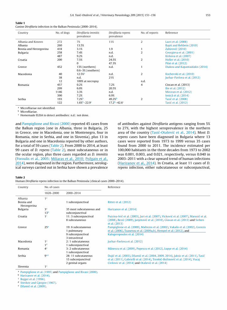
Table 1Canine Dirofilaria infection in the Balkan Peninsula (2000–2014).
Country No. of dogs Dirofilaria immitisprevalence
Dirofilaria repensprevalence
No. of reports Reference
Albania and Kosovo 272 7% 11% 2 Lazri et al. (2008)Albania 260 13.5% Rapti and Rehbein (2010)Bosnia and Herzegovina 418 3.1% 1.9 1 Zahirovic (2010)Bulgaria 258 7.4% n.d. 2 Georgieva et al. (2001)
487 9.2% n.d. Kirkova et al. (2007)Croatia 200 7.5% 24.5% 2 Holler et al. (2010)
0 47.3% Pilat et al. (2012)Greece 452 13% (northern) n.d. 1 Diakou and Kapantaidakis (2014)
0.6–3% (southern)Macedonia 40 12.5%a n.d. 2 Kochevski et al. (2010)
38 n.d. 21% Jurhar-Pavlova et al. (2012)12 100% at necropsy n.d.
Romania 457 0.2% 7.2% 4 Ciocan et al. (2013)299 6.0% 20.5% Ilie et al. (2012)
1146 3.3% n.d. Mircean et al. (2012)390 7.2% 6.9% Ionica et al. (2014)
Serbia 193 7.2%b 49.2%b 2 Tasic et al. (2008)122 1.6%b–22.9c 17.2b–42.6c Tasic et al. (2012)
atiRBf9t(2i
TH
a Microfilariae not identified.b Microfilariae.c Homemade ELISA to detect antibodies: n.d.: not done.
nd Pampiglione and Rivasi (2000) reported 45 cases fromhe Balkan region (one in Albania, three in Bulgaria, 25n Greece, one in Macedonia, one in Montenegro, four inomania, nine in Serbia, and one in Slovenia), plus 13 inulgaria and one in Macedonia reported by other authors,
or a total of 59 cases (Table 2). From 2000 to 2014, at least9 cases of D. repens (Table 2), most subcutaneous or in
he ocular region, plus three cases regarded as D. immitisForoulis et al., 2005; Miliaras et al., 2010; Pozgain et al.,014), were diagnosed in the region. Furthermore, serolog-cal surveys carried out in Serbia have shown a prevalence
able 2uman Dirofilaria repens infection in the Balkan Peninsula (clinical cases 2000–20
Country No. of cases Referenc
1828–2000 2000–2014
Albania 1a
Bosnia andHerzegovina
1 subconjunctival Ritter et
Bulgaria 3a
13b35 most subcutaneous andsubconjunctival
Harizano
Croatia 1c 11: 3 subconjunctival8 subcutaneous
Puizina-(2008), Bet al. (20
Greece 25a 19: 6 subcutaneous1 pulmonary9 subconjunctival3 intravitreal
Pampiglet al. (20Kalogero
Macedonia 1a
1d2: 1 subcutaneous1 subconjunctival
Jurhar-P
Romania 4a 3: 2 subcutaneous1 subconjunctival
Manescu
Serbia 9a,e 28: 11 subcutaneous15 subconjunctival2 genital organs
Dujic et
et al. (20Cirilovic
Slovenia 1a
a Pampiglione et al. (1995) and Pampiglione and Rivasi (2000).b Harizanov et al. (2014).c Bujger et al. (1996).d Stevkov and Gjorgov (1967).e Dzamic et al. (2009).
of antibodies against Dirofilaria antigens ranging from 5%to 27%, with the highest seroprevalence in the northernarea of the country (Tasic-Otasevic et al., 2014). Most D.repens cases have been diagnosed in Bulgaria where 13cases were reported from 1973 to 1999 versus 35 casesfound from 2000 to 2011. The incidence estimated per100,000 habitants in the three decades from 1973 to 2002
was 0.001, 0.003, and 0.021, respectively, versus 0.040 in2003–2011 with a clear upward trend of human infections(Harizanov et al., 2014). In Croatia, at least 11 cases of D.repens infection, either subcutaneous or subconjunctival,14).
e
al. (2012)
v et al. (2014)
Ivic et al. (2003), Juri et al. (2007), Vickovic et al. (2007), Marusic et al.ezic (2009), Janjetovic et al. (2010), Glavan et al. (2013) and Sviben13)ione et al. (2000), Maltezos et al. (2002), Vakalis et al. (2002), Gorezis06), Tzanetou et al. (2009a,b), Hempel et al. (2012), andpoulos et al. (2014)
avlova et al. (2012)
et al. (2009), Popescu et al. (2012), Lups e et al. (2014)
al. (2003), Dzamic et al. (2004, 2009, 2014), Jaksic et al. (2011), Tasic11), Gabrielli et al. (2014), Trenkic-Bozinovic et al. (2014), Vucaj
et al. (2014) and Otasevic et al. (2014)

inary Pa
154 S.A. Tasic-Otasevic et al. / Veterwere diagnosed in humans from 2003 to 2013 (Puizina-Ivic et al., 2003; Juri et al., 2007; Vickovic et al., 2007;Marusic et al., 2008; Bezic, 2009; Janjetovic et al., 2010;Glavan et al., 2013; Sviben et al., 2013). In Greece, fol-lowing the 25 cases of Dirofilaria infection published upto 2000 (Pampiglione et al., 1995; Pampiglione and Rivasi,2000; Vakalis and Himonas, 1997), a further 19 D. repensinfections have been reported from 2000 to 2014: six sub-cutaneous, one pulmonary, three subconjunctival, and oneintravitreal (Pampiglione et al., 2000; Maltezos et al., 2002;Vakalis et al., 2002; Gorezis et al., 2006; Tzanetou et al.,2009a,b; Hempel et al., 2012; Kalogeropoulos et al., 2014).Interestingly, one case of human microfilaremic infectionwas reported in 1998 (Petrocheilou et al., 1998). Over-all, five cases have been reported from Macedonia andRomania (two and three, respectively). In particular, oneinfection with an atypical presentation, mimicking condi-tions such as cellulitis associated with impaired walkingability, was reported in Romania (Popescu et al., 2012).Recently, a recurrent case was diagnosed in the north-western part of the country (Lups e et al., 2014). After theremoval of the first subcutaneous nodule, a second noduledeveloped 4 months later. In both cases, the worms wereidentified as D. repens by polymerase chain reaction (PCR).In Serbia, 28 cases of D. repens infection have recently beendiagnosed: 11 subcutaneous, 15 subconjunctival and twoscrotal nodules containing worms were reported (in threeof the cases, species identification was uncertain: Dujicet al., 2003; Dzamic et al., 2004, 2009, 2014; Jaksic et al.,2011; Tasic et al., 2011; Ignjatovic et al., 2013; Gabrielliet al., 2014; Trenkic-Bozinovic et al., 2014; Vucaj Cirilovicet al., 2014).
Among the cases of suspected human infection with D.immitis (Foroulis et al., 2005; Miliaras et al., 2010; Pozgainet al., 2014), the finding of a living worm in the thoraciccavity of a patient during coronary artery bypass surgeryis worth noting (Pozgain et al., 2014). None of the sus-pected cases of D. immitis human infection was confirmedby molecular methods, although three D. immitis infectionshave been observed and confirmed by molecular methodsin Europe (Italy: Avellis et al., 2011; France: Foissac et al.,2013; and Ukraine: Rossi et al., 2015), suggesting that thisspecies, although less frequently, is of zoonotic concern notonly in the America (Orihel and Eberhard, 1998).
4. Conclusions
The presented data confirm the presence of Dirofilariainfections in the Balkan Peninsula, although from the pub-lished data it is difficult to obtain a clear idea where theinfection is endemic in the canine population. In fact, fewstudies have mapped the risk areas such as those car-ried out in Romania (Mircean et al., 2012), in Serbia (Tasicet al., 2008, 2011), and along the Mediterranean coast ofAlbania (Rapti and Rehbein, 2010). Nevertheless, althoughdata on Dirofilaria infection in mosquitoes are scanty from
the region, it seems that environmental factors and themosquito abundance are suitable for the transmission ofthe infection. In fact, the number of human reports from theBalkan Peninsula has been increasing in the last decades,rasitology 209 (2015) 151–156
and most are due to D. repens, often confirmed by molecularmethods.
The published reports of human Dirofilaria infectionswere based on clinically manifest disease. It is likely that theinfections are much more frequent considering the sero-logical data (Tasic-Otasevic et al., 2014) and that viscerallocalization of the worms is only diagnosed accidentallyduring thorax radiography of clinically healthy people(Muro et al., 1999). Considering that the main reservoiris represented by infected, microfilaremic dogs, furtherdata are needed to better define the prevalence in dogsin the Balkan Peninsula in order to assess the risk areasof the infection. Infected, microfilaremic dogs must betreated and the efficacy of the treatment should be checkedby blood examination (Knott or filter tests). Finally, theuncertainty of data suggests the need to extend preven-tive treatment against Dirofilaria in dogs living in the areaswhere the presence of the parasites has been confirmed,regardless of the actual prevalence.
Conflict of interest
The authors declare no conflict of interest.
References
Avellis, F.O., Kramer, L.H., Mora, P., Bartolino, A., Benedetti, P., Rivasi, F.,2011. A case of human conjunctival dirofilariosis by Dirofilaria immitisin Italy. Vector Borne Zoonotic Dis. 11, 451–452.
Bezic, J., 2009. Human dirofiliariasis in Croatia. Acta Dermatovenerol.Croat. 17, 82–83.
Bujger, Z., Ekert, M., Tojagic, M., Cacic, M., Granic, J., 1996. Dirofilariaconjunctivae. Ophtalmol. Croat. 5, 63–66.
Ciocan, R., Mederle, N., Jacsó, O., Tánczos, B., Fok, É., 2013. Autochthonouscases of Dirofilaria in dogs from Timis Country (Western Part) Roma-nia. Global J. Med. Res. 13, 29–34.
Demiaszkiewicz, A.W., 2014. Dirofilaria repens Railliet et Henry, 1911 – anew parasite acclimatized in Poland. Ann. Parasitol. 60, 31–35.
Diakou, A., Kapantaidakis, E., 2014. Epidemiology of dirofilariosis in dogsin Greece: previous and latest information. In: Fourth European Diro-filaria and Angiostrongylus days (FEDAD), 2–4 June 2014, Budapest, p.44.
Dujic, M.P., Mitrovic, B.S., Kranjcic-Zec, I.F., 2003. Orbital swelling as a signof live Dirofilaria repens in subconjunctival tissue. Scand. J. Inf. Dis. 35,430–431.
Dzamic, A.M., 2014. Personal communication.Dzamic, A.M., Arsic-Arsenijevic, V., Radonjic, I., Mitrovic, S., Marty, P.,
Kranjcic-Zec, I.F., 2004. Subcutaneous Dirofilaria repens infection ofthe eyelid in Serbia and Montenegro. Parasite 11, 239–240.
Dzamic, A.M., Colovic, I.V., Arsic-Arsenijevic, V.S., Stepanovic, S., Boricic,I., Dzamic, Z., Mitrovic, S.M., Rasic, D.M., Stefanovic, I., Latkovic, Z.,Kranjcic-Zec, I.F., 2009. Human Dirofilaria repens infection in Serbia. J.Helminthol. 83, 129–137.
Dzamic, M.A., Pantelic, J., Kovacevic, I., Karadzic, J., Dakic, Z., ColovicCalovski, I., 2014. Human dirofilariosis: four new Serbian cases causedby Dirofilaria repens. In: Fourth European Dirofilaria and Angiostrongy-lus days (FEDAD), 2–4 June 2014, Budapest, p. 82.
EMOP VII European Multicolloquium of Parasitology, 1996. Parassitologia38, 354–368.
Ermakova, L.A., Nagorny, S.A., Krivorotova, E.Y., Pshenichnaya, N.Y.,Matina, O.N., 2014. Dirofilaria repens in the Russian Federation: cur-rent epidemiology, diagnosis, and treatment from a federal referencecenter perspective. Int. J. Infect. Dis. 23, 47–52.
Foissac, M., Million, M., Mary, C., Dales, J.P., Souraud, J.B., Piarroux, R.,Parola, P., 2013. Subcutaneous infection with Dirofilaria immitis nema-tode in human, France. Emerg. Infect. Dis. 19, 171–172.
Foroulis, C.N., Khaldi, L., Desimonas, N., Kalafati, G., 2005. Pulmonarydirofilariosis mimicking lung tumor with chest wall and mediastinalinvasion. Thorac. Cardiovasc. Surg. 53, 173–175.
Gabrielli, S., Tasic, A., Trenkic-Bozinovic, M., Ignjatovic, M., Otasevic, S.,Cancrini, G., 2014. Further cases of human dirofilariosis diagnosed in

inary Pa
G
G
G
G
G
G
G
H
H
H
I
I
I
J
J
J
J
J
K
K
K
K
S.A. Tasic-Otasevic et al. / Veter
South-East Serbia. In: Fourth European Dirofilaria and Angiostrongylusdays (FEDAD), 2–4 June 2014, Budapest, p. 81.
enchi, C., Bowman, D., Drake, J., 2014. Canine heartworm dis-ease (Dirofilaria immitis) in Western Europe: survey of veterinaryawareness and perceptions. Parasites Vectors 7, 206 http://www.parasitesandvectors.com/content/7/1/206
enchi, C., Kramer, L.H., Rivasi, F., 2011a. Dirofilarial infections in Europe.Vector Borne Zoonotic Dis. 11, 1307–1317.
enchi, C., Mortarino, M., Rinaldi, L., Cringoli, G., Traldi, G., Genchi, M.,2011b. Changing climate and changing vector-borne disease distribu-tion: the example of Dirofilaria in Europe. Vet. Parasitol. 176, 295–299.
enchi, C., Rinaldi, L., Cascone, C., Mortarino, M., Cringoli, G., 2005. Isheartworm really spreading in Europe? Vet. Parasitol. 133, 137–148.
eorgieva, D., Kirkova, Z., Ivanov, A., 2001. A study on the prevalence anddiagnostics of dirofilariosis (heartworm disease) in carnivores. Bulg.J. Vet. Med. 4, 231–236.
lavan, N., Pecanic, S., Bosak, A., Gacanin, L., Abram, M., Jonjic, N., 2013.Dirofilaria repens infection in a ten-year-old boy from the Istria Penin-sula: case report. Acta Clin. Croat. 52, 533–536.
orezis, S., Psilla, M., Asproudis, I., Peschos, D., Papadopoulou, C., Stefan-iotou, M., 2006. Intravitreal dirofilariosis: a rare ocular infection. Orbit25, 57–59.
arizanov, R.N., Jordanova, D.P., Bikov, I.S., 2014. Some aspects of theepidemiology, clinical manifestations, and diagnosis of human dirofi-lariosis caused by Dirofilaria repens. Parasitol. Res. 113, 1571–1579.
empel, F., Poppert, S., Böker, T., Schaaf, B., 2012. 75-year-old woman withsymptoms of the eye. Dtsch. Med. Wochenschr. 137, 1641–1642.
oller, D., Racz, A., Bosnir, Petrak, O., 2010. The prevalence of dirofilariosisin the hinterland of the Istrian peninsula. Med. Jadert. 40, 67–74 (inCroatian with English summary).
gnjatovic, M., Velickovic, L., Mihailovic, D., Tasic, A., Miladinovic Tasic, N.,Otasevic, S., 2013. The human dirofilariosis-one more case in South-eastern Serbia. In: International Congress-XLVII Days of PreventiveMedicine, Nis http://www.izjz-nis.org.rs/index.html
lie, M.S., Imre, K., Hotea, I., Darabus , G., 2012. Survey of canine dirofilar-iosis from south-western Romania – preliminary results. In: Grandi,G., Kramer, L., Genchi, G. (Eds.), 3rd European Dirofilaria Days. June21–22, Parma, Italy, p. 68, ISBN-13: 978-88-903582-6-5.
onica, A.M., Matei, I.A., Mircean, V., Dumitrache, M.O., D’Amico, G., Gyorke,A., Panchev, N., Annoscia, G., Albrechová, K., Otranto, D., Modry, D.,Mihalca, A.D., 2014. Current survey on the prevalence and distribu-tion of Dirofilaria spp. and Acanthocheilonema reconditum infectionsin dogs in Romania. Parasitol. Res., http://dx.doi.org/10.1007/s00436-014-4263-4.
aksic, V., Mitic, N., Pavlovic, I., Vitosevic, Z., Mirkovic, M., Zoric, L., Sta-menkovic, D., Vuksa, D., Ðokic, O., 2011. Discrete eyelid swellingcaused by live subconjunctival Dirofilaria repens. Cent. Eur. J. Med. 6,177–180.
anjetovic, Z., Arar, Z.V., Paradzik, M.T., Sapina, L., Bitunjac, M., Lojen, G.,Marinculic, A., 2010. Ocular dirofilariosis: a case report. Acta Med.Croat. 64, 41–45 (in Croatian).
ezic, J., Simic, C., 1929. Prilog poznavanju parazitarne invazije pasa uvarosi Skoplju. Jugoslov. Vet. Glasnik. 9, 383–384 (in Serbian).
urhar-Pavlova, M., Cvetkovic, D., Duma, H., Kochevski, Z., Jankoska, G.,Petrovska, M., 2012. Dirofilariosis in R. Macedonia, an overview ofliterature data and our experience. In: Proceeding of XLIV Day ofPreventive Medicine, Nis, pp. 130–132 http://www.izjz-nis.org.rs/index.html
uri, J., Kuzman, T., Stiglmayer, N., Tojagic, M., 2007. A case of lacrimalgland dirofilariosis. Ophthalmologica 221, 204–206.
alogeropoulos, C.D., Stefaniotou, M.I., Gorgoli, K.E., Papadopoulou, C.V.,Pappa, C.N., Paschidis, C.A., 2014. Ocular dirofilariosis: a case series of8 patients. Middle East Afr. J. Ophthalmol. 21, 312–316.
anev, I., Kamenov, I., Ganchev, G., Prelezov, P., Tzvetkov, Y., Tomcheva,V., Halacheva, M., Georgieva, D., Vuchev, D., Tanchev, T., Slavova, G.,1996. Dirofilaria repens and Dirofilaria immitis in animals and humansin Bulgaria. Abstracts VII European Multicolloquium of Parasitology.Parassitologia 38, 358.
artashev, V., Batashova, I., Kartashov, S., Ermakov, A., Mironova, A.,Kuleshova, Y., Ilyasov, B., Kolodiy, I., Klyuchnikov, A., Ryabikina, E.,Babicheva, M., Levchenko, Y., Pavlova, R., Pantchev, N., Morchón, R.,Simón, F., 2011. Canine and human dirofilariosis in the Rostov region(southern Russia). Vet. Med. Int., http://dx.doi.org/10.4061/2011/685713 (Article ID 685713).
irkova, Z., Ivanov, A., Geogieva, D., 2007. Dirofilariosis in dogs and wildcarnivores in Bulgaria. In: Genchi, C., Rinaldi, L., Cringpoli, G. (Eds.),Dirofilaria immitis and D. repens in Dog and Cat and Human Infec-tions; Abstracts of the First European Dirofilaria Days. Ronaldo Editore,Naples, p. 204.
rasitology 209 (2015) 151–156 155
Kochevski, Z., Atanskova, E., Nikolovski, G., Stefanovska, J., 2010. Presenceof filariasis in police dogs in the region of Skopje. In: Days of VeterinaryMedicine, 28–30 October 2010, Ohrid, R Macedonia, p. 10.
Kramer, L.H., Kartashev, V.V., Grandi, G., Morchón, R., Nagornii, S.A., Kara-nis, P., Simón, F., 2007. Human subcutaneous dirofilariosis, Russia.Emerg. Infect. Dis. 13, 150–152.
Lazri, T., Duscher, G., Edelhofer, R., Bytyci, B., Gjino, P., Joachim, A.,2008. Arthropod-borne parasites of dogs, especially Leishmania, in theKosovo and Albania. Wien. Klin. Wochenschr. 120, 54–58 (in German).
Leidy, J., 1856. A synopsis of entozoa and some other ecto-congenersobserved by the author. Filaria immitis Leidy. Proc. Acad. Nat. Sci. Phila.8, 55.
Lupse, M., Mircean, V., Cavasi, A., Mihalca, A.D., 2014. Recurrent subcu-taneous human dirofilariosis due to Dirofilaria repens after surgicalremoval of the worm and anthelmintic treatment. Parasites Vectors 7(Suppl. 1), P3 http://www.parasitesandvectors.com/7/51/P3
Maltezos, E.S., Sivridis, E.L., Giatromanolaki, A.N., Simopoulos, C.E., 2002.Human subcutaneous dirofilariosis: a report of three cases manifes-ting as breast or axillary nodules. Scott. Med. J. 47, 86–88.
Manescu, R., Barascu, D., Mocanu, C., Pîrvanescu, H., Mîndria, I., Balas oiu,M., Turculeanu, A., 2009. Subconjunctival nodule with Dirofilariarepens. Chirurgia (Bucur.) 109, 95–97 (in Romanian).
Marusic, Z., Stastny, T., Kirac, I., Stojcevic, D., Kruslin, B., Tomas, D., 2008.Subcutaneous dirofilariosis caused by Dirofilaria repens diagnosed byhistopathologic and polymerase chain reaction analysis. Acta Derma-tovenerol. Croat. 16, 222–225.
McCall, J.W., Genchi, C., Kramer, L.H., Guerrero, J., Venco, L., 2008. Heart-worm disease in animals and humans. Adv. Parasitol. 66, 193–285.
Miliaras, D., Meditskou, S., Kelekis, A., Papachristos, I., 2010. Human pul-monary dirofilariosis: one more case in Greece suggests that Dirofilariais a rather common cause of coin lesions in the lungs in endemic areasof Europe. Int. J. Immunopathol. Pharmacol. 23, 345–348.
Mircean, V., Dumitrache, M.O., Györke, A., Pantchev, N., Jodies, R., Mihalca,A.D., Cozma, V., 2012. Seroprevalence and geographic distributionof Dirofilaria immitis and tick-borne infections (Anaplasma phagocy-tophilum, Borrelia burgdorferi sensu lato, and Ehrlichia canis) in dogsfrom Romania. Vector Borne Zoonotic Dis. 12, 595–604.
Morchón, R., Carretón, E., González-Miguel, J., Mellado-Hernández, I.,2012. Heartworm disease (Dirofilaria immitis) and their vectors inEurope, new distribution trends. Front. Physiol. 3, http://dx.doi.org/10.3389/fphys.2012.00196.
Motas , C., 1903. cited by Olteanu, 1996.Muro, A., Genchi, C., Cordero, M., Simón, F., 1999. Human dirofilariosis in
the European Union. Parasitol. Today 15, 386–389.Olteanu, G., 1996. Dirofilariosis in man and animals in Romania. Abstracts
VII European Multicolloquium of Parasitology. Parassitologia 38, 360.Orihel, T.C., Eberhard, M.L., 1998. Zoonotic filariasis. Clin. Microbiol. Rev.
11, 366–381.Otasevic, S., Tasic, A., Gabrielli, S., Trenkic Bozinovic, M., Cancrini, G., 2014.
Canine and human Dirofilaria infections: what is new in the BalkanPeninsula. In: Fourth European Dirofilaria and Angiostrongylus days(FEDAD), 2–4 June 2014, Budapest, pp. 33–37.
Pampiglione, S., Canestri Trotti, G., Rivasi, F., 1995. Human dirofilario-sis due to Dirofilaria (Nochtiella) repens: a review of world literature.Parassitologia 37, 149–193.
Pampiglione, S., Rivasi, F., 2000. Human dirofilariosis due to Dirofilaria(Nochtiella) repens: an update of world literature from 1995 to 2000.Parassitologia 42, 235–254.
Pampiglione, S., Rivasi, F., Vakalis, N., 2000. Human pulmonary dirofilar-iosis: the first case observed in Greece. Ann. Pathol. 20, 626–628 (inFrench).
Petrocheilou, V., Theodorakis, M., Williams, H., Prifti, H., Georgilis,K., Apostolopoulou, I., Mavrikakis, M., 1998. Microfilaremia fromDirofilaria-like parasite in Greece. APMIS 106, 315–318.
Pilat, M., Kobas, M., Beck, R., Mihalievic, Z, Marinculic, A., 2012. Studyon the prevalence of Dirofilaria repens in native dogs from a previ-ously unknown foci in Slavonia, Croatia. In: Grandi, G., Kramer, L.,Genchi, C. (Eds.), Proceedings of Third European Dirofilaria Days. ,p. 54.
Popescu, I., Tudose, I., Racz, P., Muntau, B., Giurcaneanu, C., Poppert, S.,2012. Human Dirofilaria repens infection in Romania: a case report.Case Rep. Infect. Dis. (Article ID 472976) http://dx.doi.org/10.1155/2012/472976
Pozgain, Z., Dulic, G., Sego, K., Blazekovic, R., 2014. Live Dirofilaria immi-
tis found during coronary artery bypass grafting procedure. Eur. J.Cardiothorac. Surg. 46, 134–1366.Puizina-Ivic, N., Dzakula, N., Bezic, J., Punda-Polic, V., Sardelic, S., Kuzmic-Prusac, I., 2003. First two cases of human dirofilariosis recorded inCroatia. Parasite 10, 382–384.

inary Pa
156 S.A. Tasic-Otasevic et al. / VeterRaillet, A., Henry, A., 1911. Remarques sur sujet de deux notes de MM.Bouche et Bernard. Bull. Soc. Pathol. Exot. 4, 485–488.
Rapti, D., Rehbein, S., 2010. Seroprevalence of canine heartworm(Dirofilaria immitis) infection in Albania. Parasitol. Res. 107,481–485.
Ritter, A., Egger, S., Emesz, M., 2012. Dirofilariosis: subconjunctivalinfection with Dirofilaria repens. Ophthalmologe 109, 788–790 (inGerman).
Rossi, A., Peix, Á., Pavlikovskaya, T., Sagach, O., Nikolaenko, S., Chizh,N., Kartashev, V., Simón, S., Siles-Lucas, M., 2015. Genetic diversityof Dirofilaria spp. isolated from subcutaneous and ocular lesions ofhuman patients in Ukraine based on the 12S rRNA sequence. ActaTrop. 142, 1–4.
Sałamatin, R.V., Pavlikovska, T.M., Sagach, O.S., Nikolayenko, S.M.,Kornyushin, V.V., Kharchenko, V.O., Masny, A., Cielecka, D., Konieczna-Sałamatin, J., Conn, D.B., Golab, E., 2013. Human dirofilariosis due toDirofilaria repens in Ukraine, an emergent zoonosis: epidemiologicalreport of 1465 cases. Acta Parasitol. 58, 592–598.
Sassnau, R., Czajka, C., Kronefeld, M., Werner, D., Genchi, C., Tannich,E., Kampen, H., 2014. Dirofilaria repens and Dirofilaria immi-tis DNA findings in mosquitoes in Germany: temperature dataallow autochthonous extrinsic development. Parasitol. Res. 113,3057–3061.
Silbermayr, K., Eigner, B., Joachim, A., Duscher, G.G., Seidel, B.,Allerberger, F., Indra, A., Hufnagl, P., Fuehrer, H.P., 2014.Autochthonous Dirofilaria repens in Austria. Parasites Vectors14 (7), 226, http://dx.doi.org/10.1186/1756-3305-7-226 http://www.parasitesandvectors.com/content/7/1/226
Simón, F., Siles-Lucas, M., Morchón, R., González-Miguel, J., Mellado, I.,Carretón, E., Montoya-Alonso, J.A., 2012. Human and animal dirofi-lariosis: the emergence of a zoonotic mosaic. Clin. Microbiol. Rev. 25,507–544.
Stevkov, S., Gjorgov, A., 1967. A case of subcutaneous nodular dirofilariosis
in the Macedonia Socialist Republic. Lijec. Vjesn. 89, 641–646.Sviben, M., Mestrovic, T., Nemer, K., Bartulovic, K.P., Skara, R., Galinovic,G.M., 2013. Dirofilaria repens as a cause of subconjunctival infectionin a 77-years old female patient from Croatia, a case report. Coll.Antropol. 37, 995–997.
rasitology 209 (2015) 151–156
Tasic, A., Rossi, L., Tasic, S., Miladinovic-Tasic, N., Ilic, T., Dimitrijevic, S.,2008. Survey of canine dirofilariosis in Vojvodina, Serbia. Parasitol.Res. 103, 1297–1302.
Tasic, S., Stoiljkovic, N., Miladinovic Tasic, N., Tasic, A., Mihailovic, D., Rossi,L., Gabrielli, S., Cancrini, G., 2011. Subcutaneous dirofilariosis in SouthEast Serbia – case report. Zoonoses Public Health 5, 318–322.
Tasic, A., Tasic-Otasevic, S., Gabrielli, S., Miladinovic-Tasic, N., Ignjatovic,A., Ðor –devic, J., Dimitrijevic, S., Cancrini, G., 2012. Canine Dirofilariainfections in two uninvestigated areas of Serbia: epidemiological andgenetic aspects. Vector Borne Zoonotic Dis. 12, 1031–1035.
Tasic-Otasevic, S.A., Gabrielli, S.V., Tasic, A.V., Miladinovictasic, N.L., Kostic,J.T., Ignjatovic, A.M., Popovic Dragonjic, L.D., Milosevic, Z.G., Arsic-Arsenijevic, V.S., Cancrini, G.A., 2014. Seroreactivity to Dirofilariaantigens in people from different areas of Serbia. BMC Infect. Dis. 14,68 http://www.biomedcentral.com/1471-2334/14/68
Trenkic-Bozinovic, M., Tomasevic, B., Gabrielli, S., Otasevic, S., Petrovic, A.,Cancrini, G., Tasic, A., Trenkic, M., 2014. Subconjunctival infection dueto Dirofilaria repens – case report. Acta Fac. Med. Nis. 31, 133–138.
Tzanetou, K., Gasteratos, S., Pantazopoulou, A., Gogou, C., Konidaris, D., Fra-gia, K., 2009a. Subcutaneous dirofilariosis caused by Dirofilaria repensin Greece: a case report. J. Cutan. Pathol. 36, 892–895.
Tzanetou, K., Gogou, C., Giannoulopoulos, A., Patralexis, C., Fragia, K.,2009b. Fibrous subcutaneous nodule caused by Dirofilaria repens.Travel Med. Infect. Dis. 7, 318–322.
Vakalis, N.C., Himonas, C.A., 1997. Human and canine dirofilariosis inGreece. Parassitologia 39, 389–391.
Vakalis, N., Vougioukas, N., Patsoula, E., Spanakos, G., Sioutopoulou, D.O.,Vamvakopoulos, N.C., 2002. Genotypic assignment of infection byDirofilaria repens. Parasitol. Int. 51, 163–169.
Vickovic, N., Granic, J., Desnica, B., Makek, N., Balen-Topic, M., 2007. Subcu-taneous dirofilariosis – a case report. Infektoloski Glasnik 27, 135–137(in Croatian).
Vucaj Cirilovic, V., Dobrosavljev, M., Niciforovic, D., Donat, D., Bogdanovic-
Stojanovic, D., Jukovic, M., 2014. Dirofilariosis of the breast:sonographic appearance. J. Clin. Ultrasound 42, 433–435.Zahirovic, A., PhD thesis 2010. Investigation of Dirofilariosis in the Ter-ritory of Bosnia and Herzegovina. University of Sarajevo, VeterinaryFaculty (in Bosnian).