Chromosomal Protein HMGN1 Modulates Histone H3 Phosphorylation
Upload
independentCategory
view
2download
0
Histone hyperacetylation affects meiotic recombination andchromosome segregation in Arabidopsis
Giorgio Perrella1,†, M. Federica Consiglio1,†, Riccardo Aiese-Cigliano1, Gaetana Cremona1, Eugenio Sanchez-Moran2,
Lucia Barra1, Angela Errico3, Ray A. Bressan4, F. Christopher H. Franklin2 and Clara Conicella1,*
1CNR-IGV, Research Institute of Plant Genetics, Research Division, Portici, Via Universita 133, 80055 Portici, Italy,2School of Biosciences, University of Birmingham, Birmingham B15 2TT, UK,3Department of Soil, Plant, Environmental and Animal Sciences, University of Naples ‘Federico II,’ Via Universita 100, 80055
Portici, Italy, and4Department of Horticulture and Landscape Architecture, Purdue University, West Lafayette, IN, USA
Received 6 November 2009; revised 15 February 2010; accepted 17 February 2010; published online 8 April 2010.*For correspondence (fax +39 81 775 3579; e-mail [email protected]).†These authors contributed equally to this work.
SUMMARY
In this study, the meiotic role of MEIOTIC CONTROL OF CROSSOVERS1 (MCC1), a GCN5-related histone
N-acetyltransferase, is described in Arabidopsis. Analysis of the over-expression mutant obtained by enhancer
activation tagging revealed that acetylation of histone H3 increased in male prophase I. MCC1 appeared to
be required in meiosis for normal chiasma number and distribution and for chromosome segregation. Overall,
elevated MCC1 did not affect crossover number per cell, but has a differential effect on individual
chromosomes elevating COs for chromosome 4, in which there is also a shift in chiasma distribution, and
reducing COs for chromosome 1 and 2. For the latter there is a loss of the obligate CO/chiasma in 8% of the
male meiocytes. The meiotic defects led to abortion in about half of the male and female gametes in the
mutant. In wild type, the treatment with trichostatin A, an inhibitor of histone deacetylases, phenocopies
MCC1 over-expression in meiosis. Our results provide evidence that histone hyperacetylation has a significant
impact on the plant meiosis.
Keywords: histone acetylation, meiosis, chromatin organization, chiasma, Arabidopsis.
INTRODUCTION
Meiosis is a modified cell division programme essential to
all the sexually reproducing eukaryotes. Starting with DNA
replication and followed by two successive nuclear divi-
sions, the major consequences of meiosis are a halving of
chromosome number and the reassortment and segregation
of genetic information guaranteed by homologous recom-
bination (Zickler and Kleckner, 1998).
Histone modifications are increasingly recognized as
playing important roles in meiotic recombination (Borde
et al., 2009; Buard et al., 2009). In particular, histone acety-
lation was shown to be involved in meiotic recombination
either by local analysis at hotspots in fission and budding
yeast (Yamada et al., 2004; Merker et al., 2008) or by
genome-wide analysis in Saccharomyces cerevisiae (Miecz-
kowski et al., 2007). In fact, histones around the ade6–M26
locus and HIS4 locus, meiotic recombination hotspots in
Schizosaccharomyces pombe and S. cerevisiae respec-
tively, were highly acetylated during meiosis. Deletion of a
histone acetyltransferase (HAT), SpGcn5, required for ade6–
M26 hotspot-specific hyperacetylation, led to a partial
decrease in DNA double-stranded breaks (DSBs) at this
locus (Yamada et al., 2004). Vice versa, loss-of-function of a
histone deacetylase (HDAC), ScRpd3p, increases HIS4 hot-
spot activity (Merker et al., 2008). A mutation of the SIR2
gene encoding a HDAC changes the genomic distribution of
meiosis-specific DSBs in S. cerevisiae, elevating DSBs for
5% of yeast genes and reducing DSBs for 7% of the genes
(Mieczkowski et al., 2007). H3 and H4 acetylation enrichment
was reported at the active mouse Psmb9 hotspot. Interest-
ingly, H3 acetylation was different in Psmb9 and another
hotspot, Hlx1, indicating distinct features between hotspots
(Buard et al., 2009). The function of histone acetylation
might be direct in recruiting factors of meiotic recombina-
tion machinery and/or indirect by regulating chromatin
structure at local and at higher-order levels (Hirota et al.,
2008). Indeed, histone acetylation has been demonstrated
796 ª 2010 The AuthorsJournal compilation ª 2010 Blackwell Publishing Ltd
The Plant Journal (2010) 62, 796–806 doi: 10.1111/j.1365-313X.2010.04191.x

in vitro to directly influence higher-order chromatin struc-
ture by inhibiting the formation of compact 30-nm chroma-
tin fibres (Shogren-Knaak et al., 2006). The observation that
in budding yeast over 80% of histone H4 is acetylated at
lysine 16 and most of the genome exists in a decondensed
state is consistent with a role for histone acetylation in
chromatin decompaction also in vivo (Lohr et al., 1977;
Smith et al., 2003). In meiosis, a requirement for histone
deacetylation in the condensation of metaphase chromo-
somes was reported in Xenopus laevis oocyte (Magnaghi-
Jaulin and Jaulin, 2006). Histone acetylation can affect
chromatin structure by altering the interactions of histones
between adjacent nucleosomes or with the DNA (Kouza-
rides, 2007) and/or by collectively establishing a code
recognized by downstream effector proteins and complexes
(Strahl and Allis, 2000).
Histone deacetylation has been shown to be critical for
chromosome segregation in TSA-treated mammalian
oocytes. Indeed, histone hyperacetylation produced lagging
chromosomes in mouse and pig (De La Fuente et al., 2004;
Akiyama et al., 2006; Wang et al., 2006).
Studies on histone acetylation dynamics in plant meiosis
are sketchy. To date, histone acetylation patterns have been
described in Arabidopsis mutants, ask1 (Yang et al., 2006)
and dsy10 (Boateng et al., 2008). Different loss-of-function
mutations in genes for individual HATs/HDACs have been
identified in Arabidopsis thaliana whose genome has 18
genes encoding potentially functional HDACs and 12 puta-
tive HAT genes (Pandey et al., 2002; see also http://
www.chromdb.org). Analyses of HDAC T-DNA knockout
and antisense lines, which in most cases displayed histone
hyperacetylation, revealed a role for the HDACs in repro-
ductive development. In particular, down-regulation of
AtHD1, which is a global regulator of different developmen-
tal processes, induced delayed flowering, flower abnormal-
ities, partial sterility and consequent seed set dropping (Tian
et al., 2003). Silencing of AtHD2A, a plant-specific HDAC,
severely disturbed reproductive development causing seed
abortion (Wu et al., 2000). An AtHDA6 mutant was reported
to exhibit reduced fertility, but only in the first flowers
(Aufsatz et al., 2002). However, AtHDA6 is reported to be
involved in the silencing of transgenes, DNA repeats, and
rDNA loci (Probst et al., 2004) as well in jasmonate response,
senescence, and flowering (Wu et al., 2008). Among HATs,
AtGCN5 (GNAT family), AtHAG4/HAM1 and HAG5/HAM2
(MYST family) have been functionally characterized in
Arabidopsis. Mutation of AtGCN5 induced many defects
on plant development including sterility or partial fertility
with production of few seeds (Bertrand et al., 2003; Vlacho-
nasios et al., 2003). Loss-of-function of either HAM1 or
HAM2 did not display any abnormal phenotype while no
double mutant could be recovered. Analysis of HAM1/ham1;
ham2/ham2 and ham1/ham1; HAM2/ham2 plants indicate
that these MYST members are required for gametophyte
development (Latrasse et al., 2008). In all the aforemen-
tioned mutants except the last, the mechanism underlying
the reduction of plant fertility remained unclear and, in
particular, meiosis was not directly investigated.
In this study we have used an over-expression mutant of a
GCN5-related histone N-acetyltransferase gene, MEIOTIC
CONTROL OF CROSSOVERS1 (MCC1), that was obtained by
enhancer activation tagging (Perrella et al., 2006) to inves-
tigate the interrelationship between histone hyperacetyla-
tion and meiotic events in Arabidopsis. Analysis of mcc1
meiocytes revealed hyperacetylation of histone H3 during
male meiosis that was accompanied by defects in chromo-
some segregation and changes in crossover number and
distribution in different chromosomes.
RESULTS
Phenotype of the mcc1 mutant
We isolated the b16 line following T-DNA mutagenesis by
enhancer activation tagging in a screen for mutants with low
seed-set (Perrella et al., 2006). The line exhibited a signifi-
cant reduction in fertility, producing on average 68% fewer
seeds per silique (n = 40; P £ 0.01) than wild-type plants.
Based on our subsequent cytological characterization,
b16 was re-named meiotic control of crossovers1 (mcc1).
Analysis of F1 plants from a wild type · mcc1 cross con-
firmed that the mutation is dominant.
During vegetative development the mutant showed some
changes respect to wild type. The rosette leaves did appear
narrower and elongated (Figure S1a). Additionally, mcc1
plants exhibited faster stem elongation (Figure S1b) and
flowered about 6 days earlier than wild type (P £ 0.001).
Analysis of mcc1 anthers revealed a strong reduction in
pollen viability (Figure S1d). Inspection of mcc1 siliques
indicated failure of seed development (Figure S1e). On
average mcc1 siliques were 17% shorter than wild type
(Figure S1f). Prior to the onset of female meiosis ovule
development in mcc1 resembled that in wild type. Histolog-
ical examination of mcc1 gynoecia showed that following
megasporogenesis the embryo sacs fail to differentiate in
�50% of mcc1 ovules (n > 100 ovules). Such that, at
anthesis, the estimated number of fully developed ovules
per gynoecium was significantly less than in wild type
(11.3 � 2.8 versus 22.6 � 4.8; P £ 0.005) (Figure S2). How-
ever, the female lethality is not a gametophytic event
because MCC1 mutation inheritance was previously dem-
onstrated to be sporophytic (Perrella et al., 2006).
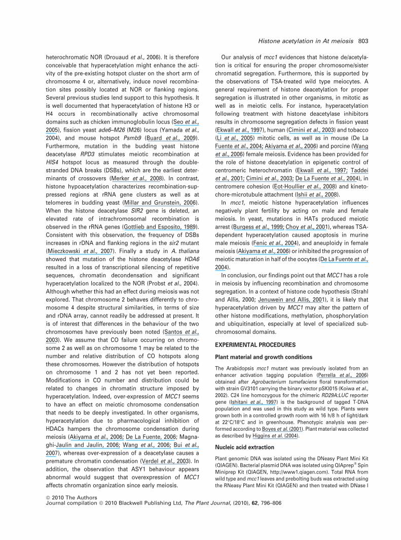
Molecular characterization and expression of MCC1
Previously, we showed that the mcc1 mutation cosegregat-
ed with a single T-DNA associated herbicide-resistance
marker demonstrating tight genetic linkage of the two loci
(Perrella et al., 2006). A DNA fragment flanking the T-DNA
insert in mcc1 plants was obtained by thermal asymmetric
Histone acetylation in At meiosis 797
ª 2010 The AuthorsJournal compilation ª 2010 Blackwell Publishing Ltd, The Plant Journal, (2010), 62, 796–806

interlaced-PCR (TAIL PCR) (Liu et al., 1995). This sequence
was found to match the last exon of the predicted Arabid-
opsis gene At3g02960. However, the insertion of the T-DNA
into the coding sequence of At3g02960 (Figure 1a) provoked
no gene deregulation (Figure S3) making unlikely that the
low seed set phenotype could be associated with it. As the
T-DNA included multiple copies of the CaMV 35S enhancer
(Weigel et al., 2000), we tested whether the insertion caused
an enhancement of the expression of the adjacent genes
(Figure S3). RT-PCR analysis, performed on leaves and
flower buds, indicated that only the expression of gene
At3g02980 (MCC1), located 9.6 kb from the CaMV 35S enh-
ancers, was elevated in the mutant compared with wild type
(Figure 1b). This finding was confirmed by quantitative real-
time RT-PCR, which revealed that the transcript level of
MCC1 was higher in mutant flower buds and leaf tissue
compared with wild type by a factor of 3- and 1.5-fold
respectively (Figure 1b).
MCC1 has five exons and four introns (Genebank acces-
sion AC018363; F13E7.7) and encodes a predicted 247 amino
acid protein containing a GCN5-related N-acetyltransferase
domain (GenBank accession no. NM_111168). Our RT-PCR
experiments, loaded on 2% agarose gel, revealed two
transcripts (Figure S4a). The smallest transcript (GenBank
accession EU598462) contains the full predicted ORF while
the longest (GenBank accession no. EU598461) retains 30 nt
of the second intron that causes a premature termination
codon truncating the coding sequence at the 97th aminoacid
(Figure S4b). We found one MCC1 homologue (At5g16800,
78% identical) in the Arabidopsis genome using a BLASTP
search using the predicted MCC1 sequence as the query.
To confirm that the low seed set phenotype of mcc1 was
due to enhanced expression of MCC1, we generated trans-
genic Arabidopsis plants expressing a genomic DNA frag-
ment of At3g02980 under the control of a CaMV 35S
promoter. Ninety-nine transformants were obtained that
on visual inspection were found to have a large variability in
seed set/silique. Eight independent transgenic lines that
displayed a strong reduction in seed set were analysed
further. The presence of the transgene with CaMV 35S
promoter and kanamycin marker was confirmed by PCR and
cytological examination revealed a reduction in pollen
viability and male meiotic abnormalities similar to those
described below in mcc1 (Figure S5). When the C24 ecotype
(a)
(b)
Figure 1. Chromosomal location of the T-DNA
insertion and enhanced expression of the acti-
vated gene in mcc1 mutant.
(a) Schematic representation of the T-DNA
insertion site in the mcc1 mutant. T-DNA is
inserted within At3g02960 locus (exons are
represented by black boxes), encoding a copper
binding related protein. The four predicted
genes, indicated by a black arrow, around
T-DNA are At3g02950 encoding an unknown
protein, MYB107 (At3g02940) encoding a puta-
tive transcription factor, EXL6 (At3g02970)
encoding an exordium protein and At3g02980
encoding a GCN5-related N-acetyltransferase
(GNAT) protein. BAR, Basta resistance. 4 · 35S
denotes four copies of the 35S enhancer.
(b) Expression analyses of MCC1. (a) RT-PCR
expression analysis of MCC1 in wild type (WT)
and mcc1 bud (B) and leaf (L) tissue. The 18S
RNA is the control for MCC1 expression. (b) Real
time RT-PCR of relative expression of MCC1 in
wild type (WT) and mcc1 in reproductive (B,
bud) and somatic (L, leaf) tissue. The actin
(ACT2) gene is used as the normalization con-
trol. Values are mean � SE (n = 3 experiments).
798 Giorgio Perrella et al.
ª 2010 The AuthorsJournal compilation ª 2010 Blackwell Publishing Ltd, The Plant Journal, (2010), 62, 796–806
was transformed with the empty vector no effect on the
phenotype was detected.
To perform loss-of-function of MCC1, T-DNA insertion
lines SALK_092359, SALK_008970, and SALK_135591 were
analysed. The locus At3g02980 was tagged at the promoter
(SALK_092359, SALK_008970) and at the third intron
(SALK_135591) (Figure S6a). Plants homozygous for the
three independent mutant alleles had a phenotype resem-
bling that of wild type. No difference in the expression level
was seen for the MCC1 gene between both lines
SALK_092359, and SALK_008970 and the wild type with
primers downstream of the insertion site (Figure S6b)
revealing that MCC1 is not disrupted in these two lines. In
the case of line SALK_135591, we failed to detect a transcript
using primers spanning the insertion, although we detected
a transcript using primers that were upstream of the
insertion (Figure S6b). We hypothesized that either the
truncated MCC1 cDNA expressed in SALK_135591 is func-
tional or, alternatively, MCC1 acts redundantly.
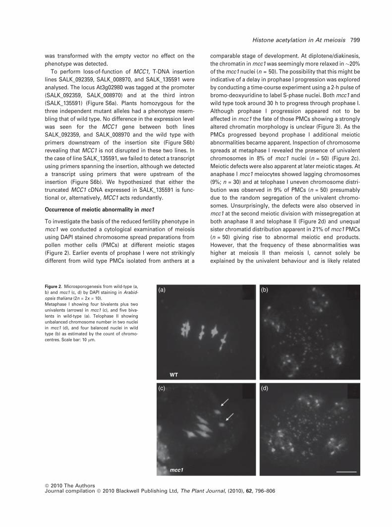
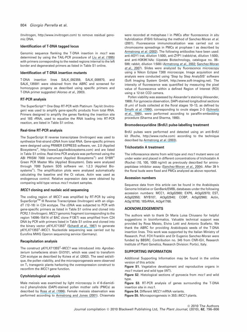
Occurrence of meiotic abnormality in mcc1
To investigate the basis of the reduced fertility phenotype in
mcc1 we conducted a cytological examination of meiosis
using DAPI stained chromosome spread preparations from
pollen mother cells (PMCs) at different meiotic stages
(Figure 2). Earlier events of prophase I were not strikingly
different from wild type PMCs isolated from anthers at a
comparable stage of development. At diplotene/diakinesis,
the chromatin in mcc1 was seemingly more relaxed in�20%
of the mcc1 nuclei (n = 50). The possibility that this might be
indicative of a delay in prophase I progression was explored
by conducting a time-course experiment using a 2-h pulse of
bromo-deoxyuridine to label S-phase nuclei. Both mcc1 and
wild type took around 30 h to progress through prophase I.
Although prophase I progression appeared not to be
affected in mcc1 the fate of those PMCs showing a strongly
altered chromatin morphology is unclear (Figure 3). As the
PMCs progressed beyond prophase I additional meiotic
abnormalities became apparent. Inspection of chromosome
spreads at metaphase I revealed the presence of univalent
chromosomes in 8% of mcc1 nuclei (n = 50) (Figure 2c).
Meiotic defects were also apparent at later meiotic stages. At
anaphase I mcc1 meiocytes showed lagging chromosomes
(9%; n = 30) and at telophase I uneven chromosome distri-
bution was observed in 9% of PMCs (n = 50) presumably
due to the random segregation of the univalent chromo-
somes. Unsurprisingly, the defects were also observed in
mcc1 at the second meiotic division with missegregation at
both anaphase II and telophase II (Figure 2d) and unequal
sister chromatid distribution apparent in 21% of mcc1 PMCs
(n = 50) giving rise to abnormal meiotic end products.
However, that the frequency of these abnormalities was
higher at meiosis II than meiosis I, cannot solely be
explained by the univalent behaviour and is likely related
WT
mcc1
(a) (b)
(c) (d)
Figure 2. Microsporogenesis from wild-type (a,
b) and mcc1 (c, d) by DAPI staining in Arabid-
opsis thaliana (2n = 2x = 10).
Metaphase I showing four bivalents plus two
univalents (arrows) in mcc1 (c), and five biva-
lents in wild-type (a). Telophase II showing
unbalanced chromosome number in two nuclei
in mcc1 (d), and four balanced nuclei in wild
type (b) as estimated by the count of chromo-
centres. Scale bar: 10 lm.
Histone acetylation in At meiosis 799
ª 2010 The AuthorsJournal compilation ª 2010 Blackwell Publishing Ltd, The Plant Journal, (2010), 62, 796–806

to aberrant centromere function in sister chromatid
segregation.
Female meiosis was also investigated in mcc1. This study
revealed univalents at metaphase I (Figure S7) and chromo-
some segregation defects similar to those observed in male
meiosis.
MCC1 over-expression changes chiasma number per
chromosome and chiasma distribution
To establish whether MCC1 affected meiotic recombination,
chiasma frequency and distribution were evaluated in male
meiosis (Table 1) at metaphase I after FISH with 45S and 5S
rDNA probes (Figure 4) that allow the five chromosome
pairs to be individually recognized as previously described
by Sanchez-Moran et al. (2001). Interestingly, the univalent
chromosomes that occurred in 8% of mcc1 nuclei was
apparently not random, as all FISH observations revealed
that this only involved the nucleolar acrocentric chromo-
some 2 (Figure 4). This finding was not seen in corre-
sponding wild type nuclei (n = 50), which is in complete
agreement with previous results indicating that the mecha-
nism that imposes the formation of a minimum of a single
obligate CO/chiasma in Arabidopsis is very robust. Rather
surprisingly, given the observation of the univalents, the
mean number of chiasmata per cell was 7.9 in both mcc1 and
wild type (n = 50 PMCs). Analysis of bivalent configuration,
that is, ‘rod versus ring,’ where the chiasmata are restricted
to a single arm or present in both arms respectively,
revealed a significant decrease of ‘ring’ bivalents in mcc1 in
respect to wild type for chromosome 1 (P = 0.037) and a
significant increase for chromosome 4 (P = 0.033). No sig-
nificant changes were apparent on the remaining chromo-
somes. Analysis of chiasma localization in terms of distal
versus proximal revealed an apparent increase of proximal
COs on short arm of chromosome 3, but the increase was
not statistically significant (P = 0.094). For chromosome 4 in
mcc1 a significant increase (P = 0.002) in the proportion of
distal versus proximal chiasmata, 43 versus 23 respectively,
was observed relative to wild type (29 versus 32). This was
accompanied by a significant increase (P = 0.041) in the
relative proportion of chiasmata in the short arm. As men-
tioned above, the effect of this was to increase the overall
occurrence of ring bivalents by 12% relative to wild type.
Overall, it appears that elevated MCC1 did not affect cross-
over number per cell, but has a differential effect on indi-
vidual chromosomes elevating COs for chromosome 4, in
which there is also a shift in chiasma distribution, and
Figure 3. Pollen mother cell (PMC) in mcc1 at mid-prophase I showing a
strongly altered morphology of the chromosomes. Scale bar: 2 lm.
Table 1 Average of pairing configurationsper cell (rod versus ring), chiasmata percell (distal versus proximal) and per biva-lent arm (short versus long) in wild typeand mcc1 mutant (n = 50 male meiocytes)
Genotype
Configurations Chiasmata
Bivalents,ring rod Univalents Distal Proximal Short arm Long arm
Wild typechr1 0.92, 0.08 – 1.48 0.58 0.92 1.14chr2 0.26, 0.74 – 0.78 0.48 0.34 0.92chr3 0.54, 0.46 – 1.08 0.46 0.54 1.00chr4 0.20, 0.80 – 0.58 0.64 0.24 0.98chr5 0.78, 0.22 – 1.36 0.46 0.78 1.04
Total 2.70, 2.30 – 5.28 2.62 2.82 5.08mcc1
chr1 0.84a, 0.16a – 1.40 0.52 0.84 1.08chr2 0.22, 0.70 0.08 0.72 0.42 0.30 0.84chr3 0.68, 0.32 – 1.10 0.62 0.68 1.04chr4 0.32a, 0.68a – 0.86b 0.46b 0.38a 0.94chr5 0.84, 0.16 – 1.44 0.42 0.84 1.02
Total 2.88, 2.02 0.08 5.52 2.44 3.04 4.92
Data was analysed by chi-squared test.aP £ 0.05; bP £ 0.01.
800 Giorgio Perrella et al.
ª 2010 The AuthorsJournal compilation ª 2010 Blackwell Publishing Ltd, The Plant Journal, (2010), 62, 796–806

reducing COs for chromosome 1 and 2. In the latter this leads
to the loss of the obligate CO/chiasma in some nuclei.
Synaptonemal complex (SC) appears normal in mcc1
Based on our observation that recombination was affected in
mcc1 we investigated whether SC was normal in the mutant.
Immunolocalization studies were carried out on spread
preparation of PMCs from the mutant at various stages
throughout prophase I using Abs recognizing the axis-
associated protein, ASY1 (Armstrong et al., 2002) and the
SC transverse element protein, ZYP1 (Higgins et al., 2005). In
wild type, immunolocalization of ASY1 indicates that the
protein associates with the chromatin in G2 as numerous
punctate foci. At the onset of leptotene there is a rapid,
co-ordinated change in the distribution to a linear chromo-
some axis associated signal (Sanchez-Moran et al., 2007).
This finding coincides with the elaboration of the chromo-
some axes (Figures S8 and S9). In mcc1 initial localization
of ASY1 is indistinguishable from that in wild type
(Figure S10a–c), but subsequently the axis transition ap-
peared less coordinated (Figure S10d–i). This situation was
manifested in the presence of nuclei at zygotene that
exhibited stretches of linear ASY1 signal set against a diffuse
chromatin-associated signal (Figure S10g–i). We have never
observed this distribution of ASY1 in wild type material
(Figure S9d–i). As prophase I progressed through zygotene
to pachytene the ASY1 assumed a more typical linear signal
running the length of the chromosome axes and there
appears no substantive effect on the rate of progression of
the PMCs through to pachytene (Figure S10j–l). At pachy-
tene, ASY1 behaviour was accompanied by apparently nor-
mal SC formation based on immunolocalization of ZYP1
(Figure S11).
Hyperacetylation of histone H3 in mcc1 male meiosis
Based on our findings that meiosis was defective in the
mutant we surmised that if the prediction that MCC1 pos-
sesses histone acetyltransferase activity was correct, then
evidence of histone hyperacetylation should be apparent in
mcc1 PMCs. The level of histone acetylation in mcc1 and
wild type PMC spread preparations was quantified at
pachytene by measuring the fluorescence signal intensity
following dual-immunolocalization using an antibody (Ab)
recognizing the lysine residues K9 and K14 on the tail of
histone H3 (H3K9K14Ac) (Figure 5) and an anti-ASY1 Ab or,
alternatively, anti-ZYP1 Ab (Figure S8). ASY1 and ZYP1
being well characterized for their spatial and temporal dis-
tribution during prophase I allow to identify unambiguously
the PMC stage. The fluorescence signal arising from the anti-
H3K9K14Ac Ab was consistently stronger in mcc1 by a factor
of 30% (n = 85 nuclei) (Figure 5). This finding is consistent
with mcc1 PMCs exhibiting an elevated level of histone H3
acetylation.
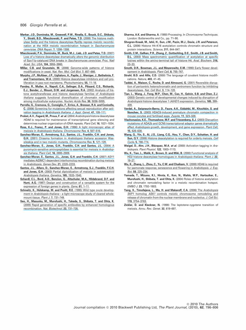
Trichostatin A treatment phenocopies MCC1
over-expression
To obtain additional evidence that the meiotic defects
described in mcc1 were induced by the histone hyperacet-
ylation, we treated wild type plants with trichostatin A (TSA),
an inhibitor of histone deacetylases (Murphy et al., 2000).
Immunolocalization with anti-H3K9K14Ac Ab confirmed that
the PMCs from the treated plants exhibited hyperacetylation
Chr.4
Chr.4
mcc1
Chr.3Chr.2
Chr.2
Chr.1Chr.5 Chr.5
WT
Figure 4. FISH from wild type (WT) and mcc1 at
metaphase I in male meiosis.
FISH was performed with the 5S rDNA (red) and
45S rDNA (green) probes. In WT, nucleolar
chromosome 2 is a rod bivalent recognizable
by 45S probes located on short arm; nucleolar
chromosome 4 is a rod bivalent, as well, and
shows 45S and 5S signals, both located on short
arm. Chromosome 5 is a ring bivalent carrying
only 5S. Chromosome 1 and 3 are ring bivalents
without any signal and are distinguishable by
size. In mcc1, the two univalents belong to
chromosome 2 while chromosome 4 is config-
ured as a ring bivalent. Scale bar: 5 lm.
Figure 5. Level of histone acetylation in wild type and mcc1 microsporocytes
at pachytene (n = 85 nuclei).
It was measured by fluorescence intensity according to ‘Step by Step
AnalySIS’ software. Data are means and SD.
Histone acetylation in At meiosis 801
ª 2010 The AuthorsJournal compilation ª 2010 Blackwell Publishing Ltd, The Plant Journal, (2010), 62, 796–806

(Figure 6) indicating that deacetylation was inhibited start-
ing at the lowest TSA concentration (10 ng/ml). The obser-
vation of DAPI-stained chromosome spreads in PMCs
revealed that TSA treatment was able to phenocopy the
meiotic defects of mcc1 mutant. This factor included the
presence of univalents at diakinesis (Figure 7c) leading to
missegregation and unbalanced nuclei at the end of meiosis
(Figure 7d). No meiotic irregularities were detected in
untreated plants (Figure 7a,b). Thus, these results indicate
that the effect of TSA treatment during meiosis is substan-
tially the same as over-expression of MCC1.
DISCUSSION
Our analysis of the A. thaliana mcc1 mutant where
enhanced activation of a putative histone acetylase results in
hyperacetylation of histone H3 in male meiosis has revealed
compelling evidence of a link between histone de/acetyla-
tion and normal meiotic progression.
In mcc1, hyperacetylation has a clear effect on chiasma
behaviour although not univocal for all the chromosomes.
In particular, chromosome 1 shows a tendency (P < 0.05) to
form a single CO per bivalent instead of double COs typical
of wild-type. CO assurance on chromosome 2 is defective,
such that a proportion (8%) fails to form the obligate CO
which is the minimum requirement to ensure accurate
chromosome segregation at the first meiotic division (Lam
et al., 2005). Chromosome 4 formed a significantly greater
proportion (P < 0.05) of double COs per bivalent compared
with wild type. Indeed, in this chromosome we observed a
significant increase (P < 0.05) in the proportion of chias-
mata in the short arm (s.a.) relative to the long arm (l.a.) in
mcc1 (0.38 s.a/0.94 l.a.) compared with wild type (0.24 s.a./
0.94 l.a.). CO increase was accompanied by a significant
shift (P < 0.01) toward distal chiasmata in the long arm of
the chromosome 4. We propose that different chromosome
features may underlie the differential response to hyper-
acetylation observed for chromosome 4 in mcc1. Apart
from its smaller size, which may well be relevant, the key
feature of chromosomes 4 is the presence of a large array of
rDNA in the short arm (Copenhaver and Pikaard, 1996).
Additionally, a cluster of CO hotspots has been mapped
to the short arm of chromosome 4 adjacent to the
+TSA
–TSA
(a) (b) (c)
(d) (e) (f)
Figure 6. Dual immunolocalization of ASY1
(green; b, e), and H3K9K14Ac (red; c, f) to PMCs
at zygonema with trichostatin A-treatment
(+TSA, d–f) and without ()TSA, a–c).
DNA was counterstained with DAPI (a, d). Higher
level of histone acetylation is shown in TSA-
treated sample (f) respect to untreated (c) at the
same stage when ASY1 is visible as a continu-
ous signal along chromosome axes (b, e). Scale
bar: 5 lm.
–TSA
(a) (b)
(c) (d)
+TSA
Figure 7. Meiosis from representative PMCs of wild type after the treatment
with histone deacetylase inhibitor trichostatin A (+TSA, c, d) as compared with
untreated PMCs ()TSA, a, b).
In TSA-treated PMCs (10 ng/ml TSA), 2 univalents at diakinesis (c), and
unbalanced nuclei at telophase II (d). Regular behaviour is shown by
untreated PMCs (a, b). Scale bar: 10 lm.
802 Giorgio Perrella et al.
ª 2010 The AuthorsJournal compilation ª 2010 Blackwell Publishing Ltd, The Plant Journal, (2010), 62, 796–806
heterochromatic NOR (Drouaud et al., 2006). It is therefore
conceivable that hyperacetylation might enhance the acti-
vity of the pre-existing hotspot cluster on the short arm of
chromosome 4 or, alternatively, induce novel recombina-
tion sites possibly located at NOR or flanking regions.
Several previous studies lend support to this hypothesis. It
is well documented that hyperacetylation of histone H3 or
H4 occurs in recombinationally active chromosomal
domains such as chicken immunoglobulin locus (Seo et al.,
2005), fission yeast ade6–M26 (M26) locus (Yamada et al.,
2004), and mouse hotspot Psmb9 (Buard et al., 2009).
Furthermore, mutation in the budding yeast histone
deacetylase RPD3 stimulates meiotic recombination at
HIS4 hotspot locus as measured through the double-
stranded DNA breaks (DSBs), which are the earliest deter-
minants of crossovers (Merker et al., 2008). In contrast,
histone hypoacetylation characterizes recombination-sup-
pressed regions at rRNA gene clusters as well as at
telomeres in budding yeast (Millar and Grunstein, 2006).
When the histone deacetylase SIR2 gene is deleted, an
elevated rate of intrachromosomal recombination is
observed in the rRNA genes (Gottlieb and Esposito, 1989).
Consistent with this observation, the frequency of DSBs
increases in rDNA and flanking regions in the sir2 mutant
(Mieczkowski et al., 2007). Finally a study in A. thaliana
showed that mutation of the histone deacetylase HDA6
resulted in a loss of transcriptional silencing of repetitive
sequences, chromatin decondensation and significant
hyperacetylation localized to the NOR (Probst et al., 2004).
Although whether this had an effect during meiosis was not
explored. That chromosome 2 behaves differently to chro-
mosome 4 despite structural similarities, in terms of size
and rDNA array, cannot readily be addressed at present. It
is of interest that differences in the behaviour of the two
chromosomes have previously been noted (Santos et al.,
2003). We assume that CO failure occurring on chromo-
some 2 as well as on chromosome 1 may be related to the
number and relative distribution of CO hotspots along
these chromosomes. However the distribution of hotspots
on chromosome 1 and 2 has not yet been reported.
Modifications in CO number and distribution could be
related to changes in chromatin structure imposed by
hyperacetylation. Indeed, over-expression of MCC1 seems
to have an effect on meiotic chromosome condensation
that needs to be deeply investigated. In other organisms,
hyperacetylation due to pharmacological inhibition of
HDACs hampers the chromosome condensation during
meiosis (Akiyama et al., 2006; De La Fuente, 2006; Magna-
ghi-Jaulin and Jaulin, 2006; Wang et al., 2006; Bui et al.,
2007), whereas over-expression of a deacetylase causes a
premature chromatin condensation (Verdel et al., 2003). In
addition, the observation that ASY1 behaviour appears
abnormal would suggest that overexpression of MCC1
affects chromatin organization since early meiosis.
Our analysis of mcc1 evidences that histone de/acetyla-
tion is critical for ensuring the proper chromosome/sister
chromatid segregation. Furthermore, this is supported by
the observations of TSA-treated wild type meiocytes. A
general requirement of histone deacetylation for proper
segregation is illustrated in other organisms, in mitotic as
well as in meiotic cells. For instance, hyperacetylation
following treatment with histone deacetylase inhibitors
results in chromosome segregation defects in fission yeast
(Ekwall et al., 1997), human (Cimini et al., 2003) and tobacco
(Li et al., 2005) mitotic cells, as well as in mouse (De La
Fuente et al., 2004; Akiyama et al., 2006) and porcine (Wang
et al., 2006) female meiosis. Evidence has been provided for
the role of histone deacetylation in epigenetic control of
centromeric heterochromatin (Ekwall et al., 1997; Taddei
et al., 2001; Cimini et al., 2003; De La Fuente et al., 2004), in
centromere cohesion (Eot-Houllier et al., 2008) and kineto-
chore-microtubule attachment (Ishii et al., 2008).
In mcc1, meiotic histone hyperacetylation influences
negatively plant fertility by acting on male and female
meiosis. In yeast, mutations in HATs produced meiotic
arrest (Burgess et al., 1999; Choy et al., 2001), whereas TSA-
dependent hyperacetylation caused apoptosis in murine
male meiosis (Fenic et al., 2004), and aneuploidy in female
meiosis (Akiyama et al., 2006) or inhibited the progression of
meiotic maturation in half of the oocytes (De La Fuente et al.,
2004).
In conclusion, our findings point out that MCC1 has a role
in meiosis by influencing recombination and chromosome
segregation. In a context of histone code hypothesis (Strahl
and Allis, 2000; Jenuwein and Allis, 2001), it is likely that
hyperacetylation driven by MCC1 may alter the pattern of
other histone modifications, methylation, phosphorylation
and ubiquitination, especially at level of specialized sub-
chromosomal domains.
EXPERIMENTAL PROCEDURES
Plant material and growth conditions
The Arabidopsis mcc1 mutant was previously isolated from anenhancer activation tagging population (Perrella et al., 2006)obtained after Agrobacterium tumefaciens floral transformationwith strain GV3101 carrying the binary vector pSKI015 (Koiwa et al.,2002). C24 line homozygous for the chimeric RD29A:LUC reportergene (Ishitani et al., 1997) is the background of tagged T-DNApopulation and was used in this study as wild type. Plants weregrown both in a controlled growth room with 16 h/8 h of light/darkat 22�C/18�C and in greenhouse. Phenotypic analysis was per-formed according to Boyes et al. (2001). Plant material was collectedas described by Higgins et al. (2004).
Nucleic acid extraction
Plant genomic DNA was isolated using the DNeasy Plant Mini Kit(QIAGEN). Bacterial plasmid DNA was isolated using QIAprep� SpinMiniprep Kit (QIAGEN, http://www1.qiagen.com). Total RNA fromwild type and mcc1 leaves and prebolting buds was extracted usingthe RNeasy Plant Mini Kit (QIAGEN) and then treated with DNase I
Histone acetylation in At meiosis 803
ª 2010 The AuthorsJournal compilation ª 2010 Blackwell Publishing Ltd, The Plant Journal, (2010), 62, 796–806
(Invitrogen, http://www.invitrogen.com) to remove residual geno-mic DNA.
Identification of T-DNA tagged locus
Genomic sequence flanking the T-DNA insertion in mcc1 wasdetermined by using the TAIL-PCR procedure of Liu et al. (1995)with primers corresponding to the nested regions internal to the leftborder and degenerated primers as listed in Table S1 online.
Identification of T-DNA insertion mutants
T-DNA insertion lines SALK_092359, SALK_008970, andSALK_135591 were obtained from the ABRC and screened forhomozygous progeny as described using specific primers andT-DNA primer suggested (Alonso et al., 2003).
RT-PCR analysis
The SuperScript� One-Step RT-PCR with Platinum Taq kit (Invitro-gen) was used to amplify gene-specific products from total RNA.Primers designed to amplify the genes flanking the insertion siteand 18S rRNA, used to equalize the RNA loading into RT-PCRreaction, are listed in Table S1 online.
Real-time RT-PCR analysis
The SuperScript III reverse transcriptase (Invitrogen) was used tosynthesize first-strand cDNA from total RNA. Gene-specific primerswere designed using PRIMER EXPRESS software, ver. 2.0 (AppliedBiosystems�, http://www3.appliedbiosystems.com) and are listedin Table S1 online. Real-time PCR analysis was performed using theABI PRISM 7000 instrument (Applied Biosystems�) and SYBR�
Green PCR Master Mix (Applied Biosystem). Data were analysedthrough 7000 System SDS software ver. 1.2.3 (Applied Bio-systems�). The amplification plots were analysed automaticallycalculating the baseline and the Ct values. Actin was used asendogenous control. Relative expression data were obtained bycomparing wild type versus mcc1 mutant samples.
MCC1 cloning and nucleic acid sequencing
The coding region of MCC1 was amplified by RT-PCR by usingSuperScriptTM III Reverse Transcriptase (Invitrogen) with an oligo-dT (12–18) in C24 ecotype. The cDNA was subjected to PCR withgene-specific primers as listed in Table S1 online and cloned intoPCR2.1 (Invitrogen). MCC1 genomic fragment (corresponding to theregion 14096–15414 of BAC clone F13E7) was amplified from C24DNA by PCR with primers listed in Table S1 online and cloned intothe binary vector pKYLX7135S2 (Schardl et al., 1987) to generatepKYLX7135S2–MCC1. Nucleotide sequencing was carried out byEurofins MWG Operon sequencing service (Germany).
Recapitulation analysis
The construct pKYLX7135S2–MCC1 was introduced into Agrobac-terium tumefaciens strain GV3101, which was used to transformC24 ecotype as described by Koiwa et al. (2002). The seed set/sili-que, the pollen viability, and the microsporogenesis were observedon T1 transgenic plants harboring the overexpression construct toreconfirm the MCC1 gene function.
Cytohistological analysis
Male meiosis was examined by light microscopy in 4¢-6-diamidi-no-2 phenylindole (DAPI)-stained pollen mother cells (PMCs) asdescribed by Ross et al. (1996). Female meiosis observation wasperformed according to Armstrong and Jones (2001). Chiasmata
were recorded at metaphase I in PMCs after fluorescence in situhybridization (FISH) following the method of Sanchez-Moran et al.(2001). Fluorescence immunolocalization was carried out onchromosome spreadings in PMCs at prophase I as described byArmstrong et al. (2002). The following antibodies have been used:anti-ASY1 (rat, dilution 1:500), anti-ZYP1 (rabbit/rat, dilution 1:500),and anti-H3K9K14Ac (Upstate Biotechnology, catalogue no. 06–599; rabbit, dilution 1:500) (Armstrong et al., 2002; Sanchez-Moranet al., 2007). Slides were analyzed by fluorescence microscopyusing a Nikon Eclipse T300 microscope. Image acquisition andanalysis were conducted using ‘Step by Step AnalySIS’ software(Soft Imaging System GmbH, http://www.soft-imaging.net). Theintensity of fluorescence was quantified by measuring the pixelvalue of fluorescence within a defined Region of Interest (ROI)using a 12-bit CCD camera.
Pollen viability was assessed by Alexander’s staining (Alexander,1969). For gynoecia observation, DAPI-stained longitudinal sections(8 lm) of buds collected at the floral stages 10–13, as defined bySmyth et al. (1990), corresponding to ovule stages 2–3 (Schneitzet al., 1995), were performed according to paraffin-embeddingprocedure (Sharma and Sharma, 1980).
Bromodeoxyuridine (BrdU) pulse-labelling treatment
BrdU pulses were performed and detected using an anti-BrdUkit (Roche, http://www.roche.com) according to the techniquedescribed by Armstrong et al. (2003).
Trichostatin A treatment
The inflorescence stems from wild type and mcc1 mutant were cutunder water and placed in different concentrations of trichostatin A(Roche) (10, 100, 1000 ng/ml) as previously described for amino-peptidase inhibitor assay (Sanchez-Moran et al., 2004). After 39 hthe floral buds were fixed and PMCs analyzed as above reported.
Accession numbers
Sequence data from this article can be found in the ArabidopsisGenome Initiative or GenBank/EMBL databases under the followingaccession numbers: MCC1, At3g02980; EXP6, At3g02970; EST,At3g02950; MYB107, At3g02940; COBP, At3g02960; Actin,At3g18780; 18SrRNA, At3g41768.
ACKNOWLEDGEMENTS
The authors wish to thank Dr Maria Luisa Chiusano for helpfulsuggestions in bioinformatics. Valuable technical support wasprovided by Rosa Maisto, Elvira Lotti and Antonio Scafarto. Wethank the ABRC for providing Arabidopsis seeds of the T-DNAinsertion lines. This work was supported by the Italian Ministry ofResearch. Prof. FCH Franklin and Dr Eugenio Sanchez-Moran werefunded by BBSRC. Contribution no. 345 from CNR-IGV, ResearchInstitute of Plant Genetics, Research Division: Portici, Italy.
SUPPORTING INFORMATION
Additional Supporting Information may be found in the onlineversion of this article:Figure S1. Vegetative development and reproductive organs inmcc1 mutant and wild type (WT).Figure S2. Histological sections of gynoecia from mcc1 and wildtype.Figure S3. RT-PCR analysis of genes surrounding the T-DNAinsertion site in mcc1.Figure S4. Different MCC1 mRNA variants.Figure S5. Microsporogenesis in 35S::MCC1 plants.
804 Giorgio Perrella et al.
ª 2010 The AuthorsJournal compilation ª 2010 Blackwell Publishing Ltd, The Plant Journal, (2010), 62, 796–806
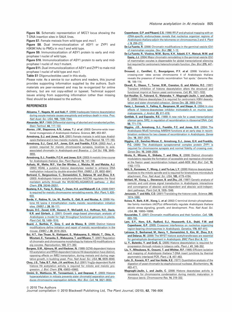
Figure S6. Schematic representation of MCC1 locus showing theT-DNA insertion sites in SALK lines.Figure S7. Female meiosis from wild-type and mcc1.Figure S8. Dual immunolocalization of ASY1 or ZYP1 andH3K9K14Ac to PMCs in mcc1 and wild type.Figure S9. Immunolocalization of ASY1 protein to early and mid-prophase I nuclei of wild type.Figure S10. Immunolocalization of ASY1 protein to early and mid-prophase I nuclei of mcc1 mutant.Figure S11. Dual immunolocalization of ASY1 and ZYP1 to male midprophase I nuclei of wild type and mcc1.Table S1 Oligonucleotides used in this study.Please note: As a service to our authors and readers, this journalprovides supporting information supplied by the authors. Suchmaterials are peer-reviewed and may be re-organized for onlinedelivery, but are not copy-edited or typeset. Technical supportissues arising from supporting information (other than missingfiles) should be addressed to the authors.
REFERENCES
Akiyama, T., Nagata, M. and Aoki, F. (2006) Inadequate histone deacetylation
during oocyte meiosis causes aneuploidy and embryo death in mice. Proc.
Natl Acad. Sci. USA, 103, 7339–7344.
Alexander, M.P. (1969) Differential staining of aborted and nonaborted pollen.
Stain Technol. 44, 117–122.
Alonso, J.M., Stepanova, A.N., Leisse, T.J. et al. (2003) Genome-wide inser-
tional mutagenesis of Arabidopsis thaliana. Science, 301, 653–657.
Armstrong, S.J. and Jones, G.H. (2001) Female meiosis in wild type Arabid-
opsis thaliana and in two meiotic mutants. Sex. Plant Reprod. 13, 177–183.
Armstrong, S.J., Caryl, A.P., Jones, G.H. and Franklin, F.C.H. (2002) Asy1, a
protein required for meiotic chromosome synapsis, localizes to axis-
associated chromatin in Arabidopsis and Brassica. J. Cell Sci. 115, 3645–
3655.
Armstrong, S.J., Franklin, F.C.H. and Jones, G.H. (2003) A meiotic time-course
for Arabidopsis thaliana. Sex. Plant Reprod. 16, 141–149.
Aufsatz, W., Mette, M.F., Van Der Winden, J., Matzke, M. and Matzke, A.J.
(2002) HDA6, a putative histone deacetylase needed to enhance DNA
methylation induced by double-stranded RNA. EMBO J. 21, 6832–6841.
Bertrand, C., Bergounioux, C., Domenichini, S., Delarue, M. and Zhou, D.X.
(2003) Arabidopsis histone acetyltransferase AtGCN5 regulates the floral
meristem activity through the WUSCHEL/AGAMOUS pathway. J. Biol.
Chem. 278, 28246–28251.
Boateng, K.A., Yang, X., Dong, F., Owen, H.A. and Makaroff, C.A. (2008) SWI1
is required for meiotic chromosome remodeling events. Mol. Plant, 1, 620–
633.
Borde, V., Robine, N., Lin, W., Bonfils, S., Geli, B. and Nicolas, A. (2009) His-
tone H3 lysine 4 trimethylation marks meiotic recombination initiation
sites. EMBO J. 28, 99–111.
Boyes, D.C., Zaved, A.M., Ascenzi, R., McCaskill, A.J., Hoffman, N.E., Davis,
K.R. and Gorlach, J. (2001) Growth stage-based phenotypic analysis of
Arabidopsis: a model for high throughput functional genomics in plants.
Plant Cell, 13, 1499–1510.
Buard, J., Barthes, P., Grey, C. and de Massy, B. (2009) Distinct histone
modifications define initiation and repair of meiotic recombination in the
mouse. EMBO J. 28, 2616–2624.
Bui, H.T., Van Thuan, N., Kishigami, S., Wakayama, S., Hikichi, T., Ohta, H.,
Mizutani, E., Yamaoka, E., Wakayama, T. and Miyano, T. (2007) Regulation
of chromatin and chromosome morphology by histone H3 modifications in
pig oocytes. Reproduction, 133, 371–382.
Burgess, S.M., Ajimura, M. and Kleckner, N. (1999) GCN5-dependent histone
H3 acetylation and RPD3-dependent histone H4 deacetylation have distinct,
opposing effects on IME2 transcription, during meiosis and during vege-
tative growth, in budding yeast. Proc. Natl Acad. Sci. USA, 96, 6835–6840.
Choy, J.S., Tobe, B.T., Huh, J.H. and Kron, S.J. (2001) Yng2p-dependent NuA4
histone H4 acetylation activity is required for mitotic and meiotic pro-
gression. J. Biol. Chem. 276, 43653–43662.
Cimini, D., Mattiuzzo, M., Torosantucci, L. and Degrassi, F. (2003) Histone
hyperacetylation in mitosis prevents sister chromatid separation and pro-
duces chromosome segregation defects. Mol. Biol. Cell, 14, 3821–3833.
Copenhaver, G.P. and Pikaard, C.S. (1996) RFLP and physical mapping with an
rDNA-specific endonuclease reveals that nucleolus organizer regions of
Arabidopsis thaliana adjoin the telomeres on chromosomes 2 and 4. Plant
J. 9, 259–272.
De La Fuente, R. (2006) Chromatin modifications in the germinal vesicle (GV)
of mammalian oocytes. Dev. Biol. 292, 1–12.
De La Fuente, R., Viveiros, M.M., Burns, K.H., Adashi, E.Y., Matzuk, M.M. and
Eppig, J.J. (2004) Major chromatin remodeling in the germinal vesicle (GV)
of mammalian oocytes is dispensable for global transcriptional silencing
but required for centromeric heterochromatin function. Dev. Biol. 275, 447–
458.
Drouaud, J., Camilleri, C., Bourguignon, P.Y. et al. (2006) Variation in
crossing-over rates across chromosome 4 of Arabidopsis thaliana
reveals the presence of meiotic recombination ‘hot spots.’ Genome Res.
16, 106–114.
Ekwall, K., Olsson, T., Turner, B.M., Cranston, G. and Allshire, R.C. (1997)
Transient inhibition of histone deacetylation alters the structural and
functional imprint at fission yeast centromeres. Cell, 91, 1021–1032.
Eot-Houllier, G., Fulcrand, G., Watanabe, Y., Magnaghi-Jaulin, L. and Jaulin,
C. (2008) Histone deacetylase 3 is required for centromeric H3K4 deacety-
lation and sister chromatid cohesion. Genes Dev. 22, 2693–2744.
Fenic, I., Sonnack, V., Failing, K., Bergmann, M. and Steger, K. (2004) In vivo
effects of histone-deacetylase inhibitor trichostatin-A on murine sper-
matogenesis. J. Androl. 25, 811–818.
Gottlieb, S. and Esposito, R.E. (1989) A new role for a yeast transcriptional
silencer gene, SIR2, in regulation of recombination in ribosomal DNA. Cell,
56, 771–776.
Higgins, J.D., Armstrong, S.J., Franklin, F.C. and Jones, G.H. (2004) The
Arabidopsis MutS homolog AtMSH4 functions at an early step in recom-
bination: evidence for two classes of recombination in Arabidopsis. Genes
Dev. 18, 2557–2570.
Higgins, J.D., Sanchez-Moran, E., Armstrong, S.J., Jones, G.H. and Franklin,
F.C. (2005) The Arabidopsis synaptonemal complex protein ZYP1 is
required for chromosome synapsis and normal fidelity of crossing over.
Genes Dev. 15, 2488–2500.
Hirota, K., Mizuno, K., Shibata, T. and Ohta, K. (2008) Distinct chromatin
modulators regulate the formation of accessible and repressive chromatin
at the fission yeast recombination hotspot ade6–M26. Mol. Biol. Cell, 19,
1162–1173.
Ishii, S., Kurasawa, Y., Wong, J. and Yu-Lee, L.Y. (2008) Histone deacetylase 3
localizes to the mitotic spindle and is required for kinetochore-microtubule
attachment. Proc. Natl Acad. Sci. USA, 105, 4179–4184.
Ishitani, M., Xiong, L., Stevenson, B. and Zhu, J.K. (1997) Genetic analysis of
osmotic and cold stress signal transduction in Arabidopsis: interactions
and convergence of abscisic acid-dependent and abscisic acid-indepen-
dent pathways. Plant Cell, 9, 1935–1949.
Jenuwein, T. and Allis, C.D. (2001) Translating the histone code. Science, 293,
1074–1080.
Koiwa, H., Barb, A.W., Xiong, L. et al. (2002) C-terminal domain phosphatase-
like family members (AtCPLs) differentially regulate Arabidopsis thaliana
abiotic stress signaling, growth, and development. Proc. Natl Acad. Sci.
USA, 99, 10893–10898.
Kouzarides, T. (2007) Chromatin modifications and their function. Cell, 128,
693–705.
Lam, S.Y., Horn, S.R., Radford, S.J., Housworth, E.A., Stahl, F.W. and
Copenhaver, G.P. (2005) Crossover interference on nucleolus organizing
region-bearing chromosomes in Arabidopsis. Genetics, 170, 807–812.
Latrasse, D., Benhamed, M., Henry, Y., Domenichini, S., Kim, W., Zhou, D.X.
and Delarue, M. (2008) The MYST histone acetyltransferases are essential
for gametophyte development in Arabidopsis. BMC Plant Biol. 8, 121.
Li, Y., Butenko, Y. and Grafi, G. (2005) Histone deacetylation is required for
progression through mitosis in tobacco cells. Plant J. 41, 346–352.
Liu, Y., Mitsukawa, N., Oosumi, T. and Whitter, R.F. (1995) Efficient isolation
and mapping of Arabidopsis thaliana T-DNA insert junctions by thermal
asymmetric interlaced PCR. Plant J. 8, 457–463.
Lohr, D., Kovacic, R.T. and Van Holde, K.E. (1977) Quantitative analysis of the
digestion of yeast chromatin by staphylococcal nuclease. Biochemistry, 16,
463–471.
Magnaghi-Jaulin, L. and Jaulin, C. (2006) Histone deacetylase activity is
necessary for chromosome condensation during meiotic maturation in
Xenopus laevis. Chromosome Res. 14, 319–332.
Histone acetylation in At meiosis 805
ª 2010 The AuthorsJournal compilation ª 2010 Blackwell Publishing Ltd, The Plant Journal, (2010), 62, 796–806
Merker, J.D., Dominska, M., Greewell, P.W., Rinella, E., Bouck, D.C., Shibata,
Y., Strahl, B.D., Mieczkowski, P. and Petes, T.D. (2008) The histone meth-
ylase Set2p and the histone deacetylase Rpd3p repress meiotic recombi-
nation at the HIS4 meiotic recombination hotspot in Saccharomyces
cerevisiae. DNA Repair, 7, 1298–1308.
Mieczkowski, P.A., Dominska, M., Buck, M.J., Lieb, J.D. and Petes, T.D. (2007)
Loss of a histone deacetylase dramatically alters the genomic distribution
of Spo11p-catalyzed DNA breaks in Saccharomyces cerevisiae. Proc. Natl
Acad. Sci. USA, 104, 3955–3960.
Millar, C.B. and Grunstein, M. (2006) Genome-wide patterns of histone
modifications in yeast. Nat. Rev. Mol. Cell Biol. 7, 657–666.
Murphy, J.P., McAleer, J.P., Uglialoro, A., Papile, J., Weniger, J., Bethelmie, F.
and Tramontano, W.A. (2000) Histone deacetylase inhibitors and cell pro-
liferation in pea root meristems. Phytochemistry, 55, 11–18.
Pandey, R., Muller, A., Napoli, C.A., Selinger, D.A., Pikaard, C.S., Richards,
E.J., Bender, J., Mount, D.W. and Jorgensen, R.A. (2002) Analysis of his-
tone acetyltransferase and histone deacetylase families of Arabidopsis
thaliana suggests functional diversification of chromatin modification
among multicellular eukaryotes. Nucleic Acids Res. 30, 5036–5055.
Perrella, G., Cremona, G., Consiglio, F., Errico, A., Bressan, R.A. and Conicella,
C. (2006) Screening for mutations affecting sexual reproduction after acti-
vation tagging in Arabidopsis thaliana. J. Appl. Genet. 47, 109–111.
Probst, A.V., Fagard, M., Proux, F. et al. (2004) Arabidopsis histone deacetylase
HDA6 is required for maintenance of transcriptional gene silencing and
determines nuclear organization of rDNA repeats. Plant Cell, 16, 1021–1034.
Ross, K.J., Fransz, P. and Jones, G.H. (1996) A light microscopic atlas of
meiosis in Arabidopsis thaliana. Chromosome Res. 4, 507–516.
Sanchez-Moran, E., Armstrong, S.J., Santos, J.L., Franklin, C.H. and Jones,
G.H. (2001) Chiasma formation in Arabidopsis thaliana accession Was-
sileskija and in two meiotic mutants. Chromosome Res. 9, 121–128.
Sanchez-Moran, E., Jones, G.H., Franklin, C.H. and Santos, J.L. (2004) A
puromycin-sensitive aminopeptidase is essential for meiosis in Arabidop-
sis thaliana. Plant Cell, 16, 2895–2909.
Sanchez-Moran, E., Santos, J.L., Jones, G.H. and Franklin, C.H. (2007) ASY1
mediates AtDMC1-dependent interhomolog recombination during meiosis
in Arabidopsis. Genes Dev. 21, 2220–2233.
Santos, J.L., Alfaro, D., Sanchez-Moran, E., Armstrong, S.J., Franklin, F.C.H.
and Jones, G.H. (2003) Partial diploidization of meiosis in autotetraploid
Arabidopsis thaliana. Genetics, 165, 1533–1540.
Schardl, C.L., Bvrd, A.D., Benzion, G., Altschuler, M.A., Hildebrand, D.F. and
Hunt, A.G. (1987) Design and construction of a versatile system for the
expression of foreign genes in plants. Gene, 61, 1–11.
Schneitz, K., Hulskamp, M. and Pruitt, R.E. (1995) Wild type ovule develop-
ment in Arabidopsis thaliana – a light microscope study of cleared whole-
mount tissue. Plant J. 7, 731–749.
Seo, H., Masuoka, M., Murofushi, H., Takeda, S., Shibata, T. and Ohta, K.
(2005) Rapid generation of specific antibodies by enhanced homologous
recombination. Nat. Biotechnol. 23, 731–735.
Sharma, A.K. and Sharma, A. (1980) Processing. In Chromosome Techniques.
London: Butterworths and Co., pp. 71–90.
Shogren-Knaak, M., Ishii, H., Sun, J.M., Pazin, M.J., Davie, J.R. and Peterson,
C.L. (2006) Histone H4–K16 acetylation controls chromatin structure and
protein interactions. Science, 311, 844–847.
Smith, C.M., Gafken, P.R., Zhang, Z., Gottschling, D.E., Smith, J.B. and Smith,
D.L. (2003) Mass spectrometric quantification of acetylation at specific
lysines within the amino-terminal tail of histone H4. Anal. Biochem. 316,
23–33.
Smyth, D.R., Bowman, J.L. and Meyerowitz, E.M. (1990) Early flower devel-
opment in Arabidopsis. Plant Cell, 8, 755–767.
Strahl, B.D. and Allis, C.D. (2000) The language of covalent histone modifi-
cations. Nature, 403, 41–45.
Taddei, A., Maison, C., Roche, D. and Almouzni, G. (2001) Reversible disrup-
tion of pericentric heterochromatin and centromere function by inhibiting
deacetylases. Nat. Cell Biol. 3, 114–120.
Tian, L., Wang, J., Fong, M.P., Chen, M., Cao, H., Gelvin, S.B. and Chen, Z.J.
(2003) Genetic control of developmental changes induced by disruption of
Arabidopsis histone deacetylase 1 (AtHD1) expression. Genetics, 165, 399–
409.
Verdel, A., Seigneurin-Berny, D., Faure, A.K., Eddahbi, M., Khochbin, S. and
Nonchev, S. (2003) HDAC6-induced premature chromatin compaction in
mouse oocytes and fertilised eggs. Zygote, 11, 323–328.
Vlachonasios, K.E., Thomashow, M.F. and Triezenberg, S.J. (2003) Disruption
mutations of ADA2b and GCN5 transcriptional adaptor genes dramatically
affect Arabidopsis growth, development, and gene expression. Plant Cell,
15, 626–638.
Wang, Q., Yin, S., Ai, J.S., Liang, C.G., Hou, Y., Chen, D.Y., Schatten, H. and
Sun, Q.Y. (2006) Histone deacetylation is required for orderly meiosis. Cell
Cycle, 5, 766–774.
Weigel, D., Ahn, J.H., Blazquez, M.A. et al. (2000) Activation tagging in Ara-
bidopsis. Plant Physiol. 122, 1003–1113.
Wu, K., Tian, L., Malik, K., Brown, D. and Miki, B. (2000) Functional analysis of
HD2 histone deacetylase homologues in Arabidopsis thaliana. Plant J. 22,
19–27.
Wu, K., Zhang, L., Zhou, C., Yu, C.W. and Chaikam, V. (2008) HDA6 is required
for jasmonate response, senescence and flowering in Arabidopsis. J. Exp.
Bot. 59, 225–234.
Yamada, T., Mizuno, K.I., Hirota, K., Kon, N., Wahls, W.P., Hartsuiker, E.,
Murofushi, H., Shibata, T. and Ohta, K. (2004) Roles of histone acetylation
and chromatin remodeling factor in a meiotic recombination hotspot.
EMBO J. 23, 1792–1803.
Yang, X., Timofejeva, L., Ma, H. and Makaroff, C.A. (2006) The Arabidopsis
SKP1 homolog ASK1 controls meiotic chromosome remodeling and
release of chromatin from the nuclear membrane and nucleolus. J. Cell Sci.
119, 3754–3763.
Zickler, D. and Kleckner, N. (1998) The leptotene–zygotene transition of
meiosis. Annu. Rev. Genet. 32, 619–697.
806 Giorgio Perrella et al.
ª 2010 The AuthorsJournal compilation ª 2010 Blackwell Publishing Ltd, The Plant Journal, (2010), 62, 796–806
Copyright © 2022 FDOKUMEN