Histone Deacetylase Inhibitors as Multitarget-Directed ... - MDPI
27
International Journal of Molecular Sciences Review Histone Deacetylase Inhibitors as Multitarget-Directed Epi-Drugs in Blocking PI3K Oncogenic Signaling: A Polypharmacology Approach Kasturi Ranganna *, Chelliah Selvam, Amruthesh Shivachar and Zivar Yousefipour Department of Pharmaceutical Science, College of Pharmacy and Health Sciences, Texas Southern University, 3100 Cleburne St., Houston, TX 77004, USA; [email protected] (C.S.); [email protected] (A.S.); Zivar.Yousefi[email protected] (Z.Y.) * Correspondence: [email protected]; Tel.: +1-713-313-1886 Received: 20 September 2020; Accepted: 29 October 2020; Published: 2 November 2020 Abstract: Genetic mutations and aberrant epigenetic alterations are the triggers for carcinogenesis. The emergence of the drugs targeting epigenetic aberrations has provided a better outlook for cancer treatment. Histone deacetylases (HDACs) are epigenetic modifiers playing critical roles in numerous key biological functions. Inappropriate expression of HDACs and dysregulation of PI3K signaling pathway are common aberrations observed in human diseases, particularly in cancers. Histone deacetylase inhibitors (HDACIs) are a class of epigenetic small-molecular therapeutics exhibiting promising applications in the treatment of hematological and solid malignancies, and in non-neoplastic diseases. Although HDACIs as single agents exhibit synergy by inhibiting HDAC and the PI3K pathway, resistance to HDACIs is frequently encountered due to activation of compensatory survival pathway. Targeted simultaneous inhibition of both HDACs and PI3Ks with their respective inhibitors in combination displayed synergistic therapeutic efficacy and encouraged the development of a single HDAC-PI3K hybrid molecule via polypharmacology strategy. This review provides an overview of HDACs and the evolution of HDACs-based epigenetic therapeutic approaches targeting the PI3K pathway. Keywords: epigenetics; histone deacetylase; histone deacetylase inhibitors; phosphatidylinositol kinase; HDAC-PI3K hybrid molecule; CUDC-907 1. Introduction Phosphatidylinositol 3-kinases (PI3Ks) are a family of intracellular signaling lipid kinases that regulate a wide array of cellular processes essentially in all normal tissues. Members of the PI3Ks family are categorized into three distinct classes (class I-III) based on their structure, expression, regulation, and substrate specificity. Class I PI3K family includes well-known four different PI3Ks named PI3Kα, PI3Kβ, PI3Kγ, and PI3Kδ and are implicated in remarkably diverse pathways [1,2]. An extensive amount of research knowledge related to the PI3Ks reveals that they transduce a host of cellular signals and regulate a wide range of essential cellular processes and physiological functions, including cell growth, proliferation, differentiation, survival, migration, death, and metabolism [1–3]. Naturally, the PI3K pathway is highly regulated by multiple mechanisms, often involving crosstalk with other signaling pathways in various physiological and pathophysiological situations. Importantly, the major outcome of intense investigations of PI3K signaling is the realization that aberrant activation of signaling is strongly linked to human diseases particularly in various types of cancers [4–7]. In recent years, a number of PI3K pathway inhibitors have been developed using the PI3K pathway as a drug target in human cancers, and many of them are being used in preclinical and clinical trials. However, Int. J. Mol. Sci. 2020, 21, 8198; doi:10.3390/ijms21218198 www.mdpi.com/journal/ijms
-
Upload
khangminh22 -
Category
Documents
-
view
0 -
download
0
Transcript of Histone Deacetylase Inhibitors as Multitarget-Directed ... - MDPI

International Journal of
Molecular Sciences
Review
Histone Deacetylase Inhibitors asMultitarget-Directed Epi-Drugs in Blocking PI3KOncogenic Signaling: A Polypharmacology Approach
Kasturi Ranganna *, Chelliah Selvam, Amruthesh Shivachar and Zivar Yousefipour
Department of Pharmaceutical Science, College of Pharmacy and Health Sciences, Texas Southern University,3100 Cleburne St., Houston, TX 77004, USA; [email protected] (C.S.);[email protected] (A.S.); [email protected] (Z.Y.)* Correspondence: [email protected]; Tel.: +1-713-313-1886
Received: 20 September 2020; Accepted: 29 October 2020; Published: 2 November 2020�����������������
Abstract: Genetic mutations and aberrant epigenetic alterations are the triggers for carcinogenesis.The emergence of the drugs targeting epigenetic aberrations has provided a better outlook forcancer treatment. Histone deacetylases (HDACs) are epigenetic modifiers playing critical roles innumerous key biological functions. Inappropriate expression of HDACs and dysregulation of PI3Ksignaling pathway are common aberrations observed in human diseases, particularly in cancers.Histone deacetylase inhibitors (HDACIs) are a class of epigenetic small-molecular therapeuticsexhibiting promising applications in the treatment of hematological and solid malignancies, and innon-neoplastic diseases. Although HDACIs as single agents exhibit synergy by inhibiting HDAC andthe PI3K pathway, resistance to HDACIs is frequently encountered due to activation of compensatorysurvival pathway. Targeted simultaneous inhibition of both HDACs and PI3Ks with their respectiveinhibitors in combination displayed synergistic therapeutic efficacy and encouraged the developmentof a single HDAC-PI3K hybrid molecule via polypharmacology strategy. This review provides anoverview of HDACs and the evolution of HDACs-based epigenetic therapeutic approaches targetingthe PI3K pathway.
Keywords: epigenetics; histone deacetylase; histone deacetylase inhibitors; phosphatidylinositolkinase; HDAC-PI3K hybrid molecule; CUDC-907
1. Introduction
Phosphatidylinositol 3-kinases (PI3Ks) are a family of intracellular signaling lipid kinases thatregulate a wide array of cellular processes essentially in all normal tissues. Members of the PI3Ksfamily are categorized into three distinct classes (class I-III) based on their structure, expression,regulation, and substrate specificity. Class I PI3K family includes well-known four different PI3Ksnamed PI3Kα, PI3Kβ, PI3Kγ, and PI3Kδ and are implicated in remarkably diverse pathways [1,2].An extensive amount of research knowledge related to the PI3Ks reveals that they transduce a host ofcellular signals and regulate a wide range of essential cellular processes and physiological functions,including cell growth, proliferation, differentiation, survival, migration, death, and metabolism [1–3].Naturally, the PI3K pathway is highly regulated by multiple mechanisms, often involving crosstalkwith other signaling pathways in various physiological and pathophysiological situations. Importantly,the major outcome of intense investigations of PI3K signaling is the realization that aberrant activationof signaling is strongly linked to human diseases particularly in various types of cancers [4–7]. In recentyears, a number of PI3K pathway inhibitors have been developed using the PI3K pathway as a drugtarget in human cancers, and many of them are being used in preclinical and clinical trials. However,
Int. J. Mol. Sci. 2020, 21, 8198; doi:10.3390/ijms21218198 www.mdpi.com/journal/ijms
Int. J. Mol. Sci. 2020, 21, 8198 2 of 27
clinical trials with early versions of PI3K inhibitors (PI3KIs) used as single-target drugs in monotherapyhave exhibited limited efficacy, due to compensatory or concurrent activation of other survival andgrowth-related signaling pathways [8,9].
To overcome the limitations of a single-target approach, alternative strategies are being developedto block tumor growth and progression. Among these, inhibition of histone deacetylase (HDAC) activityappears to be a promising approach. By inducing growth-inhibitory cellular effects and by regulatingboth histones and non-histones substrates via regulating signaling pathways, histone deacetylaseinhibitors (HDACIs) exhibit the potential to induce synergistic antitumor activity [10–13]. Moreover,the latest polypharmacology approach that is designed to overcome the limitations of a single-targetapproach appears to have a superior therapeutic effect with an affiliated reduction in adverse reactionsand diminished potential drug resistance. Specifically, the epigenetic polypharmacology approach,using a dual-acting HDAC and PI3K inhibitor, by incorporating HDACI functionality into a PI3KIpharmacophore, is capable of potent anticancer activity [14–16]. Simultaneous inhibition of HDACsand PI3K activity by the dual HDAC and PI3K inhibitor can disrupt the oncogenic signaling networkin cancer cells [14–16]. This review will focus on the role of HDACs and PI3K signaling in malignancy.Extend this information to understand the effectiveness of polypharmacology-based strategy inblocking oncogenic signaling networks.
2. Phosphatidylinositol 3-Kinases (PI3Ks)
2.1. Class I Phospatidylinositol 3-Kinases
Class I PI3Ks are involved in diverse pathways activated by an array of signals including growthfactors, mitogenic factors, and hormones [1,2]. Class I PI3Ks are different in their tissue distribution andfunctions. While PI3Kα and PI3Kβ isoforms are ubiquitously expressed, PI3Kγ and PI3Kδ isoforms areenriched in hematopoietic cells such as leukocytes [17]. Class I PI3Ks are further subdivided into classIA (PI3Kα, PI3Kβ, and PI3Kδ) and class IB (PI3Kγ) and are heterodimers composed of a regulatorysubunit (p85 or p101 family) and a catalytic subunit (p110α, p110β, p110γ or p110δ). All four classI catalytic subunits, p110α, p110β, p110γ, and p110δ are encoded by PIK3CA, PIK3CB, PIK3CG,and PIK3CD genes, respectively. The p85 regulatory subunit consists of p85α (p85α, p55α, or p50αsplice variants), p85β and p85γ and is encoded by PIK3R1, PIk34R2, and PIKR3 genes, respectively.The catalytic subunits p110α, p110β, and p110δ associate with regulatory subunit p85 to form class IAPI3Kα, PI3Kβ, and PI3Kδ. The catalytic subunit p110γ and regulatory subunit p101 together formclass IB PI3Kγ. All four class I PI3Ks catalyze the conversion of phosphatidylinositol 4,5-biphosphate[PI(4,5)P2] to phosphatidylinositol 3,4,5-triphosphate [PI(3,4,5,)P3]. PI(3,4,5,)P3 (PIP3) is a secondmessenger involved in the activation of a number of downstream molecules of the PI3K signalingpathway [1,2,18].
PI3Ks transduce a host of cellular signals and regulate a wide range of essential cellularprocesses and physiological functions, including cell growth, proliferation, differentiation, survival,migration, death, and metabolism [1–3]. Obviously, the PI3K pathway is highly regulated by multiplemechanisms, often involving crosstalk with other signaling pathways in various physiological andpathophysiological situations. Considering that the PI3K signaling is fundamental to normal cellularprocesses and physiological functions, glitches in the regulation of the PI3K pathway can lead to thepathogenesis of diseases. Overwhelming information indicates aberrant activation of the PI3K signalingis strongly linked to human cancers [4–7]. Non-cancerous neurological disorders [19,20], cardiovasculardiseases [21–23], diabetes [24], immune disorders [17], and CLOVES syndrome characterized bylocalized tissue outgrowths [25] also share a common etiology with oncogenesis.
Abnormal mutational activation or amplification of the PIK3CA gene encoding p110α catalyticsubunit of PI3Kα is the commonly observed event that is associated with a dysfunctional PI3K signalingcascade in different human solid and hematological malignancies [4–7]. The PI3K pathway is alsodysregulated through other mechanisms including loss or inactivation of phosphatase and tensinhomolog on chromosome 10 (PTEN), a tumor suppressor, and upregulation of tyrosine kinase growth

Int. J. Mol. Sci. 2020, 21, 8198 3 of 27
factor receptors (RTKs) and oncogenes such as ras [6,7,26–29]. Recently, activation of PIK3CB geneencoding catalytic subunit p110β of PI3Kβ appears to be involved in the development of prostate andbreast cancer [30,31]. Studies also indicate p85 regulatory isoforms also contribute to tumorigenesis bydifferent mechanisms. Alterations in PIK3R1 and PIK3R2 genes encoding p85α and p85β, respectively,are reported to be associated with tumorigenic activity [32–34]. Due to these alterations in class IPI3Ks, the PI3K pathway is overactivated in many malignancies leading to activation of Akt/PIKB anddownstream effector molecules promoting competitive growth and survival advantage, metastaticability, and resistance to drug therapy. More than 50% of breast, prostate, ovarian, glioblastomamultiforme, uterine, and lung cancer cases are linked to the deregulation of the PI3K pathway [35–37].
2.2. Activation of Phosphatidylinositol 3-Kinase Signaling
In the absence of activating signals, the kinase activity of the p110 catalytic subunit of classIA PI3K is inhibited by its dimerization with the regulatory p85 subunit. Upon RTK or G-proteincoupled receptor (GPCR) activation in response to extracellular signals, class IA p110/p85 heterodimeris recruited to the plasma membrane, where p85 separates from p110 catalytic subunit activating p110kinase activity [38,39]. Activated p110 phosphorylates phosphatidylinositol 4,5-biphosphate (PIP2) toproduce lipid second messenger phosphatidylinositol 3,4,5-triphosphate (PIP3). The PIP3 then bindsand recruits a subset of pleckstrin-homology (PH), FYVE, Phox (PX), C1, C2, or other lipid domains ofdownstream targets to the cell surface membrane. Among these downstream targets, Akt and PDKI arethe PH domain-containing proteins that are crucial for activating cell growth and survival pathways.Akt, one of the PI3K effector residing in the cytoplasm in an inactive form, translocates to the plasmamembrane upon receptor activation of PI3K and binds to PIP3. Similarly, PDK1, also containingPH domain, translocates to the plasma membrane and binds to PIP3 upon PI3K activation. Upontranslocation to the cell surface membrane, PDK1 partially activates Akt by phosphorylating Thr307.Full activation of Akt is achieved through phosphorylation of Ser473 by mTOR complex 2 (mTORC2),DNA-dependent kinase, or PDK2. Akt acts downstream to PI3K controlling multiple pathwaysthat regulate cell proliferation, growth, survival, death, and metabolism through its downstreamtargets [1–3,40]. The other essential downstream target of PI3K is mTOR, which regulates proteinsynthesis essential for cell growth, proliferation, angiogenesis, and other cellular activities. It acts bothdownstream and upstream of Akt and it is active in two different multiprotein complexes, the target ofrapamycin complex 1 (TORC1) and 2 (TORC2). The central role of PIP3 formed by the class I PI3Ksin all these cellular processes is negatively regulated by PTEN. PTEN downregulates class I PI3Kactivated pathways and PIP3 levels by converting PIP3 to PIP2 (Figure 1) [1–3,8,40,41].
Int. J. Mol. Sci. 2020, 21, x 3 of 27
prostate and breast cancer [30,31]. Studies also indicate p85 regulatory isoforms also contribute to tumorigenesis by different mechanisms. Alterations in PIK3R1 and PIK3R2 genes encoding p85α and p85β, respectively, are reported to be associated with tumorigenic activity [32–34]. Due to these alterations in class I PI3Ks, the PI3K pathway is overactivated in many malignancies leading to activation of Akt/PIKB and downstream effector molecules promoting competitive growth and survival advantage, metastatic ability, and resistance to drug therapy. More than 50% of breast, prostate, ovarian, glioblastoma multiforme, uterine, and lung cancer cases are linked to the deregulation of the PI3K pathway [35–37].
2.2. Activation of Phosphatidylinositol 3-Kinase Signaling
In the absence of activating signals, the kinase activity of the p110 catalytic subunit of class IA PI3K is inhibited by its dimerization with the regulatory p85 subunit. Upon RTK or G-protein coupled receptor (GPCR) activation in response to extracellular signals, class IA p110/p85 heterodimer is recruited to the plasma membrane, where p85 separates from p110 catalytic subunit activating p110 kinase activity [38,39]. Activated p110 phosphorylates phosphatidylinositol 4,5-biphosphate (PIP2) to produce lipid second messenger phosphatidylinositol 3,4,5-triphosphate (PIP3). The PIP3 then binds and recruits a subset of pleckstrin-homology (PH), FYVE, Phox (PX), C1, C2, or other lipid domains of downstream targets to the cell surface membrane. Among these downstream targets, Akt and PDKI are the PH domain-containing proteins that are crucial for activating cell growth and survival pathways. Akt, one of the PI3K effector residing in the cytoplasm in an inactive form, translocates to the plasma membrane upon receptor activation of PI3K and binds to PIP3. Similarly, PDK1, also containing PH domain, translocates to the plasma membrane and binds to PIP3 upon PI3K activation. Upon translocation to the cell surface membrane, PDK1 partially activates Akt by phosphorylating Thr307. Full activation of Akt is achieved through phosphorylation of Ser473 by mTOR complex 2 (mTORC2), DNA-dependent kinase, or PDK2. Akt acts downstream to PI3K controlling multiple pathways that regulate cell proliferation, growth, survival, death, and metabolism through its downstream targets [1–3,40]. The other essential downstream target of PI3K is mTOR, which regulates protein synthesis essential for cell growth, proliferation, angiogenesis, and other cellular activities. It acts both downstream and upstream of Akt and it is active in two different multiprotein complexes, the target of rapamycin complex 1 (TORC1) and 2 (TORC2). The central role of PIP3
formed by the class I PI3Ks in all these cellular processes is negatively regulated by PTEN. PTEN downregulates class I PI3K activated pathways and PIP3 levels by converting PIP3 to PIP2 (Figure 1) [1–3,8,40,41].
Figure 1. Schematic diagram of generic PI3K signaling displaying the signaling pathways of proteins leading to different cellular effects. In the figure, arrows indicate activation, and blunt end lines indicate inhibition.
Figure 1. Schematic diagram of generic PI3K signaling displaying the signaling pathways of proteinsleading to different cellular effects. In the figure, arrows indicate activation, and blunt end linesindicate inhibition.
Int. J. Mol. Sci. 2020, 21, 8198 4 of 27
2.3. Phosphatidylinositol 3-Kinase Inhibitors
PI3KIs are subdivided into pan-PI3K inhibitors, isoform-specific PI3K inhibitors, dual PI3K/mTORinhibitors, mTOR, and Akt inhibitors [8,9,42]. Many of the PI3KIs are being used in preclinical andclinical trials to treat cancers. The pan-PI3KIs such as pictilisib (Taselisib, GDC-0941), apitolisib(GDC-0980), and buparlisb (BKM120) that target all class I isoforms exhibit similar efficacy. SincePIK3CA is frequently mutated in solid tumors, isoform-specific PI3KIs such as alpelisib (BYL719) andtaselisib (GDC0032) that selectively target the PIK3CA protein product p100α are under preclinical andclinical testing. On the other hand, B cell signaling is upregulated in chronic lymphocytic leukemiathrough the hyperactivation of the PI3K pathway due to increased expression of PIK3CD proteinproduct p110δ in hematopoietic cells and lymphoid tissue. Idelalisib, a specific inhibitor for p110δ,is the first PI3KI approved for the treatment of relapsed chronic lymphocytic leukemia in combinationwith CD-20 antibody rituximab [37]. However, clinical trials with early versions of PI3KIs used assingle-target drugs in monotherapy have limited efficacy potentially due to compensatory or concurrentactivation of other survival and growth-related signaling pathways [8,9]. Different and alternativestrategies are being developed to block tumor growth and progression to overcome the limitationsof a single target approach. Among these, the latest polypharmacology approach that is designed toovercome the limitations of single-target therapy appears to have a superior therapeutic effect alongwith reduced adverse reactions and diminished potential drug resistance. Specifically, the epigeneticpolypharmacology approach which uses a dual-acting HDAC and PI3K inhibition by incorporatingHDAC inhibitor functionality into a PI3K inhibitor pharmacophore is capable of potent anticanceractivity by disrupting the oncogenic signaling network through simultaneous inhibition of HDACsand PI3K in cancer cells [15,16].
3. Histone Deacetylases (HDACs)
HDACs are a family of enzymes that play crucial roles in a number of biological processesessentially through silencing gene transcription. Gene expression is regulated by the contrastingaction of HDACs and histone acetyltransferases (HATs). The HATs mediate the acetylation of histonespromoting gene transcription, whereas HDACs suppress gene expression by deacetylating histones.Besides histones, functions of several nonhistone proteins that are essential for cell proliferation,differentiation, and apoptosis are regulated by acetylation/deacetylation.
3.1. Role of HDACs in Epigenetic Alterations
Apart from genetic mutations, alterations in epigenetic regulations are the main trigger for cancerinitiation, progression, and metastasis [11]. Chromatin is a dynamic structure subject to remodelingthrough reversible epigenetic alterations in response to environmental cues. The architecture ofchromatin is subject to dynamic remodeling from a highly condensed transcriptionally repressivestate to a more relaxed transcriptionally permissible state in response to external and internal cues.Thus, permitting accessibility of DNA to the transcriptional machinery. Such chromatin remodelingis facilitated by epigenetic factors through various covalent posttranslational modifications (PTMs)to the N-terminal histone tails, including acetylation, methylation, phosphorylation, ubiquitination,and ADP-ribosylation. Among these epigenetic histone modifications, acetylation of histones playsa crucial role in the epigenetic regulation of gene expression. Histone acetyltransferases (HATs)and histone deacetylases (HDACs) represent two enzyme families that are crucial to epigeneticregulation of gene expression. Acetylation of lysine ε- amino group of lysine side-chain in theN-terminal tails of core histones (H2A, H2B, H3, and H4) is a reversible dynamic PTM regulated bycontrasting activities of HATs and HDACs [10–13]. Transfer of acetyl group to the lysine residues ofhistones by HATs neutralizes their positive charges causing decondensed chromatin structure that isaccessible to transcriptional activation [43]. Additionally, multiple specific sites are acetylated on corehistones. These sites are recognized by bromodomain components found in certain proteins that are

Int. J. Mol. Sci. 2020, 21, 8198 5 of 27
part of the chromatin-remodeling coactivator complexes involved in transcriptional activation [44].In contrast, HDACs stimulate condensed and transcriptionally repressed chromatin structure bydeacetylating histones and are associated with corepressor complexes that takes part in transcriptionalsuppression [10–13,45–49]. Besides acetylation, the lysine side chain of histones is a target of other PTMssuch as methylation, sumoylation, and ubiquitination [50–57]. However, these PTMs are mutuallyexclusive on the same lysine residue, promoting a great potential for cross-regulation. Crosstalkbetween these different PTMs at independent sites of histone forms a ”histone code” that is translatedto a specific biological outcome crucial to normal development, and disease pathogenesis [10,12,51–57].
Acetylation of lysine is a major PTM modification of histones and crossregulation betweenacetylation and another PTM is crucial in modulating chromatin-based transcriptional control [50–57].It is now recognized that thousands of human proteins are subject to reversible acetylation anddeacetylation [47,51,58]. Enrichment of acetylated proteins by immunoprecipitation with anti-acetyl-lysine antibodies, combined with high-resolution mass spectrometry-based proteomics, revealslysine acetylation is a widespread and important epigenetic PTM that affects histones and somenuclear proteins, along with thousands of other human proteins [59,60]. Some of the acetylatednonhistone proteins include transcription factors, various other nuclear regulators, and cytoplasmicproteins, suggesting that lysine acetylation is not only crucial to the nuclear functions, but alsoessential for regulating various cytoplasmic processes, including cytoskeletal dynamics, energymetabolism, endocytosis, and signal transduction [47,51,52,61]. Moreover, several acetylatednonhistone proteins are key factors in different signaling pathways. In some nonhistone proteins, lysineacetylation occurs at multiple sites and crosstalk with phosphorylation, methylation, ubiquitination,and other PTMs, leading to a code-like multisite modification program that is crucial for the controlof cellular signaling specific to a particular scenario. Considering that histones are not the onlyproteins that are subject to reversible acetylation and deacetylation, it is conceivable that HATs,HDACs, and bromodomains, the epigenetic players that add, erase, and read the acetyl group,respectively, have a broader impact on signaling pathways. Accordingly, different HATs and HDACsare now referred to as histone/lysine acetyltransferases (HATs/KATs) and histone deacetylase/lysinedeacetylases (HDACs/KDACs), respectively. These enzymes play a critical role in nonhistone proteinacetylation regulating signaling events essential for a range of cellular processes and physiologicaland pathophysiological functions [62–64]. Epigenetic-driven alterations in lysine acetylation due todysregulated activities of several HATs/KATs, HDACs/KDACs, and bromodomain-containing proteinshave been linked to various diseases, notably in cancer, but also in cardiovascular disease, neurologicaland immunological disorders [38–41,60,64–67]. A variety of HATs/KATs, HDACs/KDACs, andbromodomain inhibitors have been developed, studied, and used as anticancer agents in clinical trials.
3.2. Types of HDACs
Enzymatic deacetylation was first detected in 1977 when Friend erythroleukemic cells (FEC) weretreated with n-butyrate [68]. Butyrate stimulated differentiation of FEC into hemoglobin-synthesizingnormal-like cells. This phenotypic change was linked to histone hyperacetylation, indicating thepresence of HDAC, and butyrate is recognized as HDACI. There are 18 different mammalian HDACsthat have been identified. HDACs are classified into four classes I, II, III, and IV [6,10,12,45]. Class I, II,and IV are referred to as classical HDACs and class III enzymes are known as sirtuins. Class I, II, and IVHDACs are dependent on Zn2+ for the deacetylase activity. Class I HDACs include HDAC1, HDAC2,HDAC3, and HDAC8 that have sequence similarity to yeast counterpart RPD3. Class II HDACs arerelated to yeast enzyme HDAI and are divided into subclasses IIa and IIb. The IIa includes HDAC4,HDAC5 HDAC7, and HDAC9, whereas IIb includes HDAC6 and HDAC10. Class IV consists of onlyHDAC11. Class III includes seven NAD+-dependent sirtuins that are related to yeast counterpart Sir2.
HDACs have an important role in cytoplasmic functions besides their epigenetic functionsregulating gene expression. A number of cytoplasmic nonhistone proteins are acetylated by HDACs.These proteins include transcription factors, transcription regulators, DNA repair enzymes, chaperone
Int. J. Mol. Sci. 2020, 21, 8198 6 of 27
proteins, signal transduction mediators, cytoskeletal proteins, and inflammation mediators [12,13,69–71].Accordingly, histone deacetylases/lysine deacetylases (HDACs/KDACs) are directly or indirectlyinvolved in several cellular functions such as gene expression, signaling pathways, protein stability,protein-protein interaction, eventually affecting cell proliferation, growth, differentiation, apoptosis,migration, and angiogenesis [12,13,69–71]. Moreover, altered expression of HDACs has been observedin many cancerous and noncancerous diseases, particularly in cancer, advocating HDACs are importanttherapeutic targets for anticancer therapies [72–74]. Therefore, the development of HDAC inhibitors isessential to the therapeutic intervention of epigenetic diseases, cancer in particular.
4. Histone Deacetylase Inhibitors (HDACIs)
The cancer cells harbor the epigenetic abnormalities required for oncogenesis by altering theepigenetic landscape of chromatin. Especially acetylation, one of the predominant modifications inepigenetics, serves as a key regulatory mechanism for gene expression. The global dysregulationof epigenetic histone acetylation modifications results in altered gene expression contributing tothe establishment of the disease state. Aberrant epigenetic alterations are the important driversin cancer pathogenesis often due to the silencing of tumor suppressor genes or overexpression ofoncogenes. Moreover, the cancer cells with a unique portfolio of epigenetic changes are differentfrom their normal counterparts. Accumulation of these altered epigenetic changes allows cancercells to solidify their exclusive phenotype through dysregulated gene expression changes [75–77].Furthermore, the accumulation of altered epigenetic changes can also promote resistance to therapythrough the acquisition of pro-survival signaling [75,78]. The epigenetic histone acetylation alterationsinduced by aberrant expression of HDACs can be reversed by pharmacological inhibition of HDACswith HDACIs. Although HDACIs affect gene expression on a global level, their actions are specificto the malignant phenotype and have been recognized that only about 8–20% of genes are affectedby HDACIs [79]. Additionally, HDACIs also indirectly affect gene expression by inhibiting HDACsinteractions with nonhistones.
The HDACIs are a class of small-molecules that inhibit the activity of Zn2+-dependent classicalHDACs (class I, II, and class IV HDACs) and promote acetylation of histone and nonhistone proteinsubstrates. Originally, HDACIs were known for their varied cellular effects on cancer cells, such asinhibiting cell proliferation and stimulating differentiation or apoptosis, advocating their usefulnessin anticancer drug discovery, and development [80,81]. Now, it is recognized HDACIs induce anarray of cellular effects [69,82,83]. They stimulate cell cycle arrest by upregulating p21Cip1 anddownregulating cyclins [69,84,85]. They also do the following: 1) Induce intrinsic and extrinsicapoptosis pathways causing apoptosis [75,86,87]. 2) Induce autophagy [88]. 3) Promote anti-angiogeniceffect by altering Hif-1α function and downregulation of VEGF [89]. 4) Alter expression of nonhistoneproteins including transcription factors and regulators, inflammation/immune response mediators,chaperones, DNA repair enzymes, and structural proteins [69,82,83,90]. These features of HDACIsare exploited as promising anticancer epi-drugs and several HDACIs have entered clinical trials inboth hematologic and solid tumors [70,72–75,81–83,90]. As such, HDACIs are investigated mainlyas anticancer epi-drugs, even though HDAC enzymes also play a crucial role in other diseasessuch as cardiovascular disease [91], neurological disorders [92], immunological processes and viralinfections [93], and other disorders [94].
4.1. Types of Histone Deacetylase Inhibitors (HDACIs)
Several HDACIs have been either isolated from natural sources or synthesized with differingefficacy, potency, and pharmacokinetics [81,95]. The active site of Zn2+-dependent HDACs consistsof a tubular pocket and a Zn2+ ion at the bottom of the pocket. The canonical pharmacophoreof the HDACIs is composed of three different parts. A cap structure (CAP) that interacts withthe rim of the tubular catalytic pocket of HDACs, a zinc ion binding group (ZBG), and a linkerspanning the length of the tubular pocket responsible for connecting the cap and the ZBG [12,13,96,97].

Int. J. Mol. Sci. 2020, 21, 8198 7 of 27
Based on their Zn2+-binding structure, HDACIs are classified into different classes. These includehydroxamates, carboxylic acids, benzamides, and cyclic peptides, with differing potency, selectivity,and toxicity [48,69,98]. Hydroximic acid based HDACIs include trichostatin A (TSA), vorinostat(SAHA, suberoylanilide hydroxamic acid), panobinostat, belinostat, and several others. While TSA isused only in laboratory studies because of its toxicity, vorinostat, panobinostat, belinostat, and severalothers are approved for hematologic malignancies. Most of the hydroximic acid-based HDACIs arepan-HDACIs. The carboxylic acid type of HDACIs includes short-chain fatty acids such as butyricacid, phenylbutyrate, and valproic acid, which are weak inhibitors of HDAC class I and II. Butyrateand phenylbutyrate acids are in several phase II and phase I trials, respectively. Valproic acid isapproved for epilepsy, bipolar disorders, and migraine. Among the benzamides, entinostat andchidamide (HBI-8000) are HDAC class I inhibitors. Entinostat is in phase II trials for several differenthematologic and solid cancers. The cyclic tetrapeptides include the bicyclic depsipeptide romidepsin(FK228, FR901228). It is an HDAC class I inhibitor that has been approved for treating cutaneousT-cell lymphoma.
Most HDACIs are considered pan-inhibitors because they display a similar mechanism of actionon different classical HDACs except for class IIa HDACs. HDACIs bind to the zinc atom in the catalyticpocket in a non-competitive manner but lacks selectivity. Thus, it is difficult to discern which classicalHDAC enzyme inhibition is responsible for the therapeutic or toxic effects observed in clinical trials.To achieve desirable clinical outcomes, it would be appropriate to know which HDAC is contributingto the pathogenesis so that HDAC-specific inhibitors can be developed in the future.
4.2. Histone Deacetylase Inhibitors in Clinical Trials
The early drugs like butyrate targeting HDACs are used based on phenotypic effects in cancermodels without knowing the specific targets. The observations that HDACIs exhibit varied cellulareffects on cancer cells such as inhibiting cell proliferation and stimulating differentiation or apoptosis,point to their usefulness for anticancer drug discovery and development [80]. During the past tenplus years, several promising HDACIs have surfaced as effective anticancer epi-drugs. Because of thetransient and reversible nature of epigenetic modifications and also based on their antiproliferativeand proapoptotic effects in cancer cells, several HDACIs are approved for cancer treatment by theUnited States Food and Drug Administration (US FDA) [72–74,99]. Vorinostat, also (suberoylanilidehydroxamic acid, SAHA), a hydroxamic acid, is the first pan-HDACI approved in 2006 for thetreatment of rare refractory cutaneous T-cell lymphoma (CTCL). Romidepsin (FK-288, FR901228, cyclicdepsipeptide), a natural prodrug having inhibitory activity against class I HDACs, is approved in2009 for the treatment of rare refractory CTCL. Belinostat (PXD101), and panobinostat (LBH589) aretwo other approved hydroxamate-containing pan-HDAC inhibitors. Belinostat is approved for thetreatment of peripheral T-cell lymphoma in 2014 and panobinostat for the treatment of refractorymultiple myeloma in 2015 [99]. Benzamide-based HDACI chidamide (HBI-8000) which inhibitsHDAC1, 2, 3 and 10 has been approved for the treatment of refractory peripheral T-cell lymphoma inChina [72–74,99]. Class I-selective benzamide containing entinostat and mocetinostat are in clinicaltrials for several solid tumors. Even though the HDAC-specific epi-drugs display potent anticanceractivities on hematological malignancies, they exhibit limited effectiveness in supporting persistentsuppression of solid tumors in monotherapies [100–102]. The single epi-drug therapeutic strategyis frequently ineffective in providing enduring tumor suppression. One of the main reasons forineffectiveness is, cancer cells can evade drug-induced effects by eliciting other compensatory survivalpathways due to the emergence of pharmacological resistance. Interestingly, HDACIs also exhibitanticancer effect by disrupting pro-survival signaling through epigenetic drug-induced sensitizationmechanisms [75].

Int. J. Mol. Sci. 2020, 21, 8198 8 of 27
4.3. Disruption of Pro-Survival Signaling by HDACIs
Epigenetic inhibitors can disrupt pro-survival signaling in cancers that have developed resistanceto therapy. During oncogenesis, epigenetic alterations can dysregulate the expression of certainproteins such as growth factor receptors or proteins associated with apoptosis that provide survivaladvantage causing resistance to therapy [75,103]. Increased expression of growth factor receptorsin cancer cells can allow them to become resistant to therapy by over-activating the downstreampathways; for example, the PI3K/Akt pathway that leads to inhibition of cell death [104]. Despite theefforts to alleviate the effects of growth factor receptors by targeted therapies against it, such therapiesare inadequate because of the rapid development of resistance. The use of HDACIs to control theexpression of growth factor receptors appears to be a promising approach; accordingly, in breastcancer dacinostat, an HDACI disrupted EGF-mediated signaling that is linked to increased metastasisand cell survival [105]. This effect was achieved by downregulating the expression ofHER2 (humanEGF-receptor-2) through two independent epigenetic mechanisms: by reducing HER2 mRNA level, andby increasing proteasomal degradation due to dissociation from its chaperone protein HSP90 throughenhanced acetylation. Additionally, a combination of HDACI entinostat and Her2/EGF-receptor kinaseinhibitor, lapatinib synergistically inhibited Akt signaling to promote apoptosis in HER2-overexpressingbreast cancer cells [106].
HDACIs can also disrupt pro-survival signaling by enhancing apoptosis signaling pathways.The binding of tumor necrosis factor-related apoptosis-inducing ligand (TRAIL) to its receptorsdeath domain-containing receptor (DR) initiates pro-death signaling receptor expression stimulatingapoptosis through caspase cascade [107]. Cancer cells are frequently resistant to apoptosis due toreduced DR receptor expression. These cells can be sensitized to overcome resistance to undergo celldeath by treating them with HDACIs. Exposure of breast cancer cells to vorinostat sensitized thecancer cells to TRAIL-induced apoptosis by upregulating DR5 receptor expression [108,109].
Additionally, HDACIs can also sensitize cancer cells to overcome their dysregulated cellcycle control and diminished activities of DNA damage repair pathways through their ability toreverse dysregulated gene expression. Entinostat, an HDACI can sensitize breast cancer cells todoxorubicin-induced growth arrest by downregulating the expression of myc, E2F, and other G2Mcell cycle genes. [75]. Entinostat in combination with decitabine, a DNA methyltransferase inhibitor,is able to restore cell cycle control in pancreatic cancer through upregulation of p21 [75].
4.4. Mechanisms of Resistance to HDACIs
Cancer cells are highly resilient and adaptable in their ability to respond and to evade growthinhibitory and toxic factors to survive and resist death. They constantly adjusting to overcomethe effects of damaging agents and environmental constraints to exist independently free from theinfluence of external proliferative/survival stimuli. These acquired stable alterations may hinder cancercells response to the effects of HDACIs through developing different mechanisms to survive andachieve resistance. A thorough understanding of molecular determinants of resistance to HDACIsmay provide the basis for therapeutic options with enhanced efficacy. Resistance to HDACIs is oftenseen and involves different mechanisms [75,110,111]. Some of the resistance mechanisms to HDACIsinvolves cell cycle proteins, thioredoxin expression, apoptosis-related proteins, signaling proteins,and NF-kB expression.
Cell cycle proteins: It has been implicated that the induction of p21Cip1 in response to HDACIs isresponsible for cell cycle arrest and serves to play a protective role. Several studies have shown thatupregulated expression of p21Cip1 in response to HDACIs mediates cell cycle arrest and differentiationor apoptosis [112]. When p21-deficient HCT116 cells are treated with romidepsin, cells get arrested inthe G2 phase compared to wild type cells [113]. The p21 is considered as a negative regulator of the cellcycle and a cyclin-dependent kinase inhibitor implicated in apoptosis [114]. Studies in U937 leukemiacells demonstrated that HDACI-induced p21Cip1 protected the cells from apoptosis, and blocking itsexpression resulted in apoptosis [115].
Int. J. Mol. Sci. 2020, 21, 8198 9 of 27
Thioredoxin expression: Resistance to HDACIs can be linked to an increased capacity of cancercells to resist oxidative stress. HDACIs increase ROS production and appear to be an important factorin the proapoptotic effects of HDACIs. Increased expression of thioredoxins, peroxiredoxins, and redoxproteins that protect cells from ROS may acquire resistance to HDACIs. Treatment of cancer cells withHDACIs causes oxidative stress by increasing ROS levels [116]. Cell death induced by HDACIs can bepartially rescued by preincubating with free radical scavenging N-acetylcysteine.
Apoptosis-related proteins: Increased expression of antiapoptotic proteins has been shownto prevent HDACIs-mediated cell death. Increased levels of Bcl-2 and Bcl-x cause resistance tothe treatment of vorinostat, dacinostat, panobinostat, or oxamflatin [117,118]. The knockdownof proapoptotic Bax in panobinostat-sensitive T-cell lymphoma cell lines resulted in diminishedtoxicity [119]. On the other hand, knockdown of antiapoptotic Mcl-1 enhances HDACI-mediatedapoptosis in primary chronic lymphocytic leukemia cells and K562 cells [120]. These reports indicatesilencing of proapoptotic proteins blunts HDACIs-induced cell death.
Signaling proteins: Resistance to HDACIs is significantly associated with activation of PI3K andMAPK pathways. A combination of panobinostat with compounds that abolish PI3K and MAPKsignaling result in synergistic cytotoxicity may be due to increased ROS [121]. A combination ofromidepsin with inhibitors of Akt resulted in synergistic cytotoxicity indicating phosphorylated Akt isan important resistance mechanism to romidepsin. These and other combination studies targetingdifferent signaling molecules appear to cause resistance. Signal transducer and activator of transcription(STAT) pathway also contributes to resistance to HDACIs. In a group of almost 40 lymphoma celllines, activation and expression of STAT-1, -3 and -5 was higher in cell lines that were more resistant tovorinostat than in sensitive cell lines [122]. Analysis of a series of skin biopsy for the nuclear stainingof phosphorylated STAT3 recognized that patients with strong staining were more likely resistant tovorinostat resistance [122]. The combination treatment with vorinostat and compounds that targetphosphorylated STAT3 may increase the efficacy.
NF-kB activation: The activation of the NF-kB pathway occurs in a number of cancers and leads tothe deactivation of the apoptotic pathway and increased cell survival [123]. It has been identified as amediator of resistance to HDACI treatment. Inhibition of HDAC by HDACIs enhances transcriptionalactivation of NF-kB through acetylation of RelA/p65 subunit leading to the induction of an array ofgenes involved in protection against cell death [124]. The activation of NF-kB by HDACIs interfereswith the triggering of cell death. In non-small lung cancer cell lines and leukemia cells, activationof NF-kB by HDACIs triggers cell death [125,126]. Inhibition of NF-kB by its inhibitor Bay-11-7085sensitizes the cancer cells to death in response to inhibition of HDAC.
Additionally, HDACIs can also sensitize cancer cells to overcome their dysregulated cellcycle control and diminished activities of DNA damage repair pathways through their ability toreverse dysregulated gene expression. Entinostat, an HDACI can sensitize breast cancer cells todoxorubicin-induced growth arrest by downregulating the expression of myc, E2F and other G2M cellcycle genes. [75]. Entinostat in combination with decitabine, a DNA methyltransferase inhibitor, is ableto restore cell cycle control in pancreatic cancer through upregulation of p21 [75].
5. Therapeutic Strategies
In cancer, genetic, epigenetic, and metabolic factors all participate in neoplasia by altering themolecular networks that control cell proliferation, growth, differentiation, migration, and cell death.Deregulation/misregulation of different molecular networks and misunderstanding in cross-talksbetween epigenetic and non-epigenetic players that are linked to various cellular processes playimportant role in the etiology of cancer. Supporting these effects, the aberrant expressions of HDACspromote altered epigenetic modifications that endorse abnormal gene expression profiles [72–74,127].The knockdown of several HDACs in some of these cancer cells stimulated cell cycle arrest and apoptosissupporting the aberrant expression of HDAC activity is linked to cancer [97]. Relevantly, HDACIshave a range of anticancer activities through the collaboration of their primary chromatin-associated
Int. J. Mol. Sci. 2020, 21, 8198 10 of 27
effects on gene transcription and their cellular effects [68,128]. These features of HDACIs supportstargeting epigenetic aberrations to prevent carcinogenesis, and open opportunities for HDACIs as theclass of anticancer agents. Several of these HDACIs have already been approved by the US FDA forcancer treatment [see Section 4.1] and are in preclinical and clinical testing.
Although the HDACIs, when used as single agents, display potent anticancer activities onhematological malignancies, they exhibit limited effectiveness in the suppression of solid tumors inmonotherapies [101,129]. The single HDAC therapeutic approach is frequently ineffective in providingsustained tumor suppression because cancer cells can dodge drug-induced effect due to pharmacologicalresistance, and also because of the toxicity and off-target effects of epi-drugs [130–133]. This letdownhas encouraged new drug design and therapeutic approaches to provide lasting tumor suppression.The goal of drug discovery in the epi-drug research is no longer just the design of highly potentand selective drugs acting on a specific epi-target, but advancing towards designing network-activecompounds with multitargeting capability through a polypharmacology approach [14–16,131–134].
5.1. Alternate Strategies Using Network-Active Compounds
The objective of the polypharmacology approach is to simultaneously hit all different targetslinked to the onset and development of a particularly complex disease at different levels so thatimproved therapeutic effect is achieved [14–16,131–134]. The goal is to achieve a synergistic effectby hitting a cellular pathway at different levels by using multitarget drugs. Synergistic therapeuticeffects can be achieved by three alternative strategies even though they exhibit striking differences.These include multi-medication therapy (MMT, drug combination), multi-compound medication(MCM, co-formulation of multiple active compounds), and multitarget-directed ligands (MTDLs,single multitargeting compounds) [15,16]. Both MMT and MCM strategies have their own specifictargets but are based on the combination or association of two or more active principles, whereasMTDLs strategy is designed to create a single molecule with an ability to simultaneously interactwith different targets. Specifically, a combination of epi-drugs with other anticancer agents includingkinase inhibitors, chemotherapeutic agents, hormonal inhibitors, and other epi-drugs for whicha rationale has been defined to benefit from synergistic effects of drug combinations have beendesigned [15,16,131–134]. Some of them are under preclinical and clinical testing. Importantly, amongthe several hybrid drugs produced, CUDC-907 is a promising orally bioavailable small-moleculedual HDAC-PI3K inhibitor [15,16,134–144]. CUDC-907 simultaneously inhibits HDACs and PI3Kcausing a synergistic effect. It is designed by introducing hydroxamic acid moiety as the zincbinding functional group to a morpholinothienopyrimidine-based PI3K pharmacophore throughan appropriate linker [135]. Recently, another dual HDAC-PI3K hybrid drug is reported, whichis designed by introducing hydroxamic acid moiety as the zinc binding functional group to aquinazoline-based PI3K pharmacophore through an applicable linker [143]. Different therapeuticapproaches involving HDAC-specific epi-drugs and PI3KIs directed against neoplastic diseases arenarrated in the following section.
5.1.1. HDACs-Specific Epi-Drugs Targeting HDACs
HDACIs induce multiple cellular effects specific to cell types through the mobilization of variousmolecular pathways. Among these, the PI3K pathway is the most prominent pathway affectedby HDACIs. Since, dysregulation of PI3Ks and their downstream effector molecules forming thePI3K/Akt/mTOR axis contribute to cancer initiation, progression, and growth, HDACIs are used assingle drugs to test whether their anticancer effect involves inhibition of PI3Ks in different cancercells and in animal xenograft cancer models. Moreover, determining the efficacy of single drugs is aprerequisite for designing a multidrug combinational approach.
Sodium butyrate, an HDACI, inhibits cell proliferation, induces differentiation, or promotesapoptosis in a variety of tumor cells. Gastric BGC823 cells treated with butyrate resulted in inhibitionof cell proliferation, altered cellular morphology, and increased expression of PTEN and mucosal factor

Int. J. Mol. Sci. 2020, 21, 8198 11 of 27
MUC2, but decreased expression of PI3K [145]. These effects are heightened by intervention with PI3Kinhibitors and lessened with PTEN siRNA indicating sodium butyrate induced upregulation of PTENand MUC2 expression and differentiation of gastric cancer cells are mediated through the PTEN/PI3Ksignaling pathway [145].
Esophageal squamous cell carcinoma (ESCC) is a highly malignant and lethal disease. Treatmentof ESCC cells with Trichostatin A (TSA), an HDACI, inhibited proliferation of ESCC cells and arrestedthe cells in the G1 phase by upregulating p21Cip1 and p27Kip1 cell cycle inhibitors [146]. TSA alsoinduced apoptosis by upregulating pro-apoptotic protein Bax and downregulating anti-apoptoticBcl-2. Furthermore, TSA inhibited the expression of PI3K and reduced the phosphorylation of Aktand ERK1/2 along with an increase in acetylated histone H4. These results indicate that TSA impedesESCC cell proliferation and promote apoptosis by inhibiting HDAC activity and activation of PI3k/Aktand ERK1/2 pathways and (Figure 2).
Int. J. Mol. Sci. 2020, 21, x 11 of 27
ESCC cell proliferation and promote apoptosis by inhibiting HDAC activity and activation of PI3k/Akt and ERK1/2 pathways and (Figure 2).
PI3K
Akt
ERK1/2P21 and p27
Acetylated Histone
Bax Bcl-2
Apoptosis
HDAC Inhibitor
Inhibition of cell proliferation
Figure 2. Schematic representation of HDACI Trichostatin A (TSA) effects on Esophageal squamous cell carcinoma (ESCC). TSA inhibits ESCC proliferation, and arrests cells in G1 phase by upregulating by p21Cip1 and p27Kip1. It inhibits cell proliferation by inhibiting PI3K expression, and reducing activation of Akt and ERK1/2. Promotes apoptosis by altering expression of pro- and anti-apoptotic proteins, and increases histone acetylation [146]. (Blunt end lines indicate inhibition, upward arrows indicate upregulation and downward arrows indicate downregulation of proteins).
Panobinostat, approved for the treatment of multiple myeloma patients, was tested for its efficacy on NB4 cells and primary acute promyelocytic leukemia (APL) patient cells. The results indicate panobinostat not only effectively reduces the survival rate of both NB4 and APL primary cells, but also eliminated the stimulatory survival effect of microenvironment signals transduced by mesenchymal stem cells (MSC) on APL cells by incorporating PI3K inhibitor when APL cells are co-cultured with MSC [147].
Romidepsin (depsipeptide, FK228) approved for the treatment of cutaneous and peripheral T-cell lymphoma treatment directly inhibits PI3K activity and strongly promotes apoptosis through its HDAC/PI3K dual inhibition [148]. Furthermore, FK-A11, an analog of depsipeptide, is reported to be the most potent HDAC/PI3K dual inhibitor exhibiting antitumor activity in HT1080 fibrosarcoma and PC3 prostate cancer cell xenograft mouse models [149].
Vorinostat (SAHA, Zolinza) is a good efficacious, well-tolerated drug that has been used in the treatment against CTCL. It also exhibits anti-solid tumor effects for other types of cancer in monotherapy and also in combination therapy. To understand the mechanism of Vorinostat anti-solid tumor activity, its effects on cervical cancer were investigated [150]. The outcome of these studies revealed that Vorinostat inhibits cell proliferation, migration, and invasion of cervical cancer cells, arrest cells in S phase, and induces apoptosis. Furthermore, Vorinostat inhibited PI3K (p110α), p-PI3K p55, and p-Akt protein expression and upregulated major histocompatibility class I-related chain A (MICA) expression in vitro and in vivo, which promoted natural killer (NK) cell-mediated cervical cancer cell lysis. These studies indicate that vorinostat exhibits anti-solid tumor activity by upregulating MICA expression in cervical cancer cells through the mediation of the PI3K/Akt pathway, which enhances the susceptibility of cervical cancer cells for NK cell-mediated cytolysis.
5.1.2. Multi-Drug Combination Targeting HDACs Activity and PI3K Pathway
Simultaneous use of two or more single-target inhibitors that are previously validated as effective in monotherapy can be used for combination therapy to stimulate a synergistic effect. Blocking multiple signaling nodes arising from HDACs and PI3K pathways simultaneously with
Figure 2. Schematic representation of HDACI Trichostatin A (TSA) effects on Esophageal squamouscell carcinoma (ESCC). TSA inhibits ESCC proliferation, and arrests cells in G1 phase by upregulatingby p21Cip1 and p27Kip1. It inhibits cell proliferation by inhibiting PI3K expression, and reducingactivation of Akt and ERK1/2. Promotes apoptosis by altering expression of pro- and anti-apoptoticproteins, and increases histone acetylation [146]. (Blunt end lines indicate inhibition, upward arrowsindicate upregulation and downward arrows indicate downregulation of proteins).
Panobinostat, approved for the treatment of multiple myeloma patients, was tested for its efficacyon NB4 cells and primary acute promyelocytic leukemia (APL) patient cells. The results indicatepanobinostat not only effectively reduces the survival rate of both NB4 and APL primary cells, but alsoeliminated the stimulatory survival effect of microenvironment signals transduced by mesenchymalstem cells (MSC) on APL cells by incorporating PI3K inhibitor when APL cells are co-cultured withMSC [147].
Romidepsin (depsipeptide, FK228) approved for the treatment of cutaneous and peripheral T-celllymphoma treatment directly inhibits PI3K activity and strongly promotes apoptosis through itsHDAC/PI3K dual inhibition [148]. Furthermore, FK-A11, an analog of depsipeptide, is reported to bethe most potent HDAC/PI3K dual inhibitor exhibiting antitumor activity in HT1080 fibrosarcoma andPC3 prostate cancer cell xenograft mouse models [149].
Vorinostat (SAHA, Zolinza) is a good efficacious, well-tolerated drug that has been used in thetreatment against CTCL. It also exhibits anti-solid tumor effects for other types of cancer in monotherapyand also in combination therapy. To understand the mechanism of Vorinostat anti-solid tumor activity,its effects on cervical cancer were investigated [150]. The outcome of these studies revealed thatVorinostat inhibits cell proliferation, migration, and invasion of cervical cancer cells, arrest cells in S

Int. J. Mol. Sci. 2020, 21, 8198 12 of 27
phase, and induces apoptosis. Furthermore, Vorinostat inhibited PI3K (p110α), p-PI3K p55, and p-Aktprotein expression and upregulated major histocompatibility class I-related chain A (MICA) expressionin vitro and in vivo, which promoted natural killer (NK) cell-mediated cervical cancer cell lysis. Thesestudies indicate that vorinostat exhibits anti-solid tumor activity by upregulating MICA expression incervical cancer cells through the mediation of the PI3K/Akt pathway, which enhances the susceptibilityof cervical cancer cells for NK cell-mediated cytolysis.
5.1.2. Multi-Drug Combination Targeting HDACs Activity and PI3K Pathway
Simultaneous use of two or more single-target inhibitors that are previously validated as effectivein monotherapy can be used for combination therapy to stimulate a synergistic effect. Blockingmultiple signaling nodes arising from HDACs and PI3K pathways simultaneously with HDACI andPI3KI can overcome the inadequacies of single therapeutic agents. A combination of HDACI/PI3KI,and HDACI/dual PI3K-mTOR inhibitors are currently widely used in preclinical studies and in theclinics for the treatment of both hematologic and solid malignancy [15,16,133].
A combination of TSA and PI3K-mTOR dual inhibitor BEZ235 was used to test their effectivenesson breast cancer cells [151]. Their combination synergistically inhibited the growth of multiplebreast cancer cell lines. Mechanistic studies revealed a combination of TSA and BEZ235 reducedphosphorylation of downstream targets of PI3K including Akt, mTOR, S6, and 4EBP-1 accountingfor inhibition of cell proliferation and apoptosis. Further analysis revealed an increase in cleavedcaspase-3, caspase-8, caspase-9, and poly-ADP ribose polymerase-1 (PARP-1), along with the reducedexpression of Bcl-2 and increased expression of Bax indicating the combination of TSA and BEZ235causes an anti-breast cancer effect through both mitochondrial pathway and the death receptor pathway(Figure 3). Co-treatment of breast cancer cells with TSA and BEZ235 also caused autophagy basedon LC3B-II and Beclin-1 increased levels. In-vivo studies further revealed a combination of TSA andBEZ235 blocked tumor growth without any noticeable side effects. Taken together, TSA and BEZ235drug combination approach has significant application in the treatment of breast cancer patients [151].
Int. J. Mol. Sci. 2020, 21, x 12 of 27
HDACI and PI3KI can overcome the inadequacies of single therapeutic agents. A combination of HDACI/PI3KI, and HDACI/dual PI3K-mTOR inhibitors are currently widely used in preclinical studies and in the clinics for the treatment of both hematologic and solid malignancy [15,16,133].
A combination of TSA and PI3K-mTOR dual inhibitor BEZ235 was used to test their effectiveness on breast cancer cells [151]. Their combination synergistically inhibited the growth of multiple breast cancer cell lines. Mechanistic studies revealed a combination of TSA and BEZ235 reduced phosphorylation of downstream targets of PI3K including Akt, mTOR, S6, and 4EBP-1 accounting for inhibition of cell proliferation and apoptosis. Further analysis revealed an increase in cleaved caspase-3, caspase-8, caspase-9, and poly-ADP ribose polymerase-1 (PARP-1), along with the reduced expression of Bcl-2 and increased expression of Bax indicating the combination of TSA and BEZ235 causes an anti-breast cancer effect through both mitochondrial pathway and the death receptor pathway (Figure 3). Co-treatment of breast cancer cells with TSA and BEZ235 also caused autophagy based on LC3B-II and Beclin-1 increased levels. In-vivo studies further revealed a combination of TSA and BEZ235 blocked tumor growth without any noticeable side effects. Taken together, TSA and BEZ235 drug combination approach has significant application in the treatment of breast cancer patients [151].
TSABEZ
Cleaved: Caspase-3Caspase-8Caspase-9PARP-1
BaxBcl-2
Apoptosis Inhibition of cell
proliferationAutophagy
LC3B-11Beclin-1
P
P
P
P
P
Figure 3. Schematic illustration of activity of TSA/BEZ235 combination on breast cancer cells. Combination treatment of TSA/BEZ235 results in synergistic suppression of breast cancer cells [152]. Through reduced activation of PI3K/Akt/mTOR and their downstream targets, TSA/BEZ235 drug combination accounts for inhibition of cell proliferation and apoptosis. The anti-breast cancer effect of drug combination is through both the mitochondrial and the death receptor pathway. TSA/BEZ235 combination also causes autophagy. (blunt end lines indicate inhibition, upward arrows indicate upregulation and downward arrows indicate downregulation of proteins).
Recent evidence indicates frequent mutation in histone modifying proteins and dysregulation of PI3K pathway in non-Hodgkin lymphoma (NHL), particularly in diffuse large B-cell lymphoma (DLBCL). This observation prompted exploration of the effectiveness and an assessment of the mechanisms of action of co-administration of panobinostat and BEZ235 in DLBCL cells [152]. Panobinostat and BEZ235 interact synergistically in ABC-, GC-, and double-hit DLBCL, but not in normal CD34+ cells. The synergistic effect of panobinostat and BEZ325 was displayed by significant Akt dephosphorylation and GSK3 dephosphorylation/activation. Drug combination promoted apoptosis as revealed by abrogation of p21Cip1 induction, Mcl-1 downregulation, Bim upregulation, and increased Bcl2/Bcl-xl binding and reduced Bax/Bak binding to Bcl/Bcl-xl/Mcl-1. Additionally, co-administration of panobinostat and BEZ325 increased H2A.X phosphorylation and histoneH3/H4 acetylation. These effects were collectively linked to the synergism of panobinostat and BEZ235. The
Figure 3. Schematic illustration of activity of TSA/BEZ235 combination on breast cancer cells.Combination treatment of TSA/BEZ235 results in synergistic suppression of breast cancer cells [152].Through reduced activation of PI3K/Akt/mTOR and their downstream targets, TSA/BEZ235 drugcombination accounts for inhibition of cell proliferation and apoptosis. The anti-breast cancer effect ofdrug combination is through both the mitochondrial and the death receptor pathway. TSA/BEZ235combination also causes autophagy. (blunt end lines indicate inhibition, upward arrows indicateupregulation and downward arrows indicate downregulation of proteins).

Int. J. Mol. Sci. 2020, 21, 8198 13 of 27
Recent evidence indicates frequent mutation in histone modifying proteins and dysregulationof PI3K pathway in non-Hodgkin lymphoma (NHL), particularly in diffuse large B-cell lymphoma(DLBCL). This observation prompted exploration of the effectiveness and an assessment of themechanisms of action of co-administration of panobinostat and BEZ235 in DLBCL cells [152].Panobinostat and BEZ235 interact synergistically in ABC-, GC-, and double-hit DLBCL, but not innormal CD34+ cells. The synergistic effect of panobinostat and BEZ325 was displayed by significant Aktdephosphorylation and GSK3 dephosphorylation/activation. Drug combination promoted apoptosis asrevealed by abrogation of p21Cip1 induction, Mcl-1 downregulation, Bim upregulation, and increasedBcl2/Bcl-xl binding and reduced Bax/Bak binding to Bcl/Bcl-xl/Mcl-1. Additionally, co-administrationof panobinostat and BEZ325 increased H2A.X phosphorylation and histoneH3/H4 acetylation. Theseeffects were collectively linked to the synergism of panobinostat and BEZ235. The synergisticeffect of a combination of panobinostat and BEZ235 underscores the therapeutic potential of a drugcombinational approach.
Sodium butyrate exhibited inhibitory effects on an aggressive metastatic human colon cancer cellline, KM20 [153]. Sodium butyrate-mediated effect on these cells was enhanced by the inhibition ofthe PI3K pathway resulting in apoptosis and reduced viability of KM20 cells. Co-incubation of KM20cells with butyrate and PI3K inhibitor LY294002 or wortmannin enhanced colon cancer cell apoptosisby activating caspase 9 and caspase 3. This finding indicates the anticancer effect of HDACI can besynergized with a PI3KI, and effective in colon cancer treatment.
In tumorigenic non-small cell lung cancer, (NSCLC) resistance to HDACIs is mediated throughthe activation of nuclear factor-kB (NF-kB) via PI3K/Akt-dependent pathway. Whether inhibitionof the PI3K/Akt pathway will allow the NSCLC cells to overcome the resistance to HDACI-inducedapoptosis was investigated using butyrate [154]. Treatment of NSCLC cell lines with butyrateactivated NF-kB-dependent transcription and this effect was inhibited by LY294002, a PI3KI. Combinedtreatment of NSCLC with butyrate and LY294002 increased apoptosis by increasing caspase-3 leveland DNA fragmentation. In vivo, combined treatment of butyrate and LY294002 was tumoristaticwith decreased tumor growth and increased apoptosis. Decreased phospho-Akt level and increasedacetylated histone H3 level in tumor tissues treated with butyrate and LY294002 indicating NSCLCxenografts can be sensitized to undergo apoptosis with combined treatment with HDACI and PI3K/Aktpathway inhibitors.
Another study supports the superior efficiency of HDACI and BEZ235 combination treatmentagainst NSCLC [155]. Heightened activities of HDACs and PI3K/Akt signaling pathways have beenassociated with NSCLC development and progression. Co-treatment of NSCLC cells with TSA andBEZ235 reveals a synergistic effect on the inhibition of NSCLC cell proliferation and stimulationof apoptosis. This combination also synergistically inhibited NSCLC migration, invasion, and theNSCLC epithelial-mesenchymal transition in vitro. Furthermore, reduction in xenograft growth andmetastasis rates and ki-67 protein expression in vivo in response to co-treatment with TSA and BEZ235also provides evidence for synergistic efficacy of the co-treatment, warranting further evaluation ofcombination treatment approach for NSCLC.
Head and neck squamous cell carcinoma (HNSCC) is the sixth most common cancer in thedeveloped world. Despite improved surgical and radiological treatment of HNSCC, it continues tobe a serious disease and needs new treatment approaches. This situation brings HDACIs into thepicture for the treatment option. Studies have revealed that a variety of HDACIs exhibit anticancereffects on squamous cell carcinoma (SCC) in vitro and in recent patient trials. However, the patienttrials have shown that HDACIs have limited therapeutic potential in monotherapies for the treatmentof HNSCC [156]. Since it is possible to boost anticancer effects of HDACIs by selective targeting ofthe PI3K/Akt pathway, the effect of PI3K, Akt, and dual PI3K/mTOR inhibitors either alone or incombination on the therapeutic potential of HDACIs in HNSCC cell lines and xenograft HNSCCmodels was investigated [157]. Treatment of HNSCC cell lines with PI3K, Akt, and dual PI3K/mTORinhibitors demonstrate increased in vitro cytotoxicity induced by HDACIs. Furthermore, HDACIs

Int. J. Mol. Sci. 2020, 21, 8198 14 of 27
and PI3KIs inhibited tumor growth of HNSCC xenograft models. In agreement with this effect,intra-tumoral HDAC and PI3K inhibition was observed as evaluated by histone H3 acetylation statusand phospho-Akt staining, respectively.
Glioblastoma multiforme (GBM) is a common malignant brain tumor. GBM cells treated withPanobinostat and BEZ235 alone or in combination were assessed for cell viability, proliferation, andapoptosis, and mechanisms of cytotoxicity were evaluated [158]. The combination approach withHDACI and PI3KI synergistically inhibited cell viability, significantly inhibited cell proliferation, andstimulated apoptosis. Additionally, co-treatment also increased caspase 3/7 activity, suppressed markersof cell proliferation and anti-apoptotic markers, and Akt signaling. These effects of a combinationapproach with HDACI and PI3K/mTOR inhibitor are promising and necessitate further evaluation inGBM therapy.
Medulloblastoma (MB) is a common malignant pediatric brain tumor. Based on genetic studiesthere are four different molecular groups of MB. The most aggressive MB among these groups exhibitsamplification or overexpression of myc oncogene. Patients with myc-driven MB reveal poor prognosisand need more effective treatment. Using an animal model of myc-driven MB, the effectiveness ofHDACIs was tested for their ability to decrease the viability of tumor cells [159]. HDACIs not onlypotently inhibit the survival of myc-driven MB cells in vitro partly due to the upregulation of theFOXO1 tumor suppressor gene, but also synergize inhibition of tumor growth in vivo in combinationwith PI3KIs. These effects recognize the effectiveness of combination therapy for the most aggressiveform of MB.
5.1.3. Polypharmacology-Based Approach Targeting HDAC Activity and PI3K Pathway
While the traditional multi-drug combination therapy approach is based on a combination of twoor more drugs, each with its own specific target, the polypharmacology approach is designed to developa single-hybrid drug to simultaneously interact with multitargets that are responsible for the onset of adisease. The hybrid drugs are created by conjugating two or more active molecules that individuallyexhibit a confirmed activity against specific targets. Such a multitarget hybrid drug strategy hasbeen used to develop a dual HDAC-PI3K inhibitor hybrid drug that simultaneously inhibits HDACsand PI3K pathway in cancer cells. Prior to developing the multitarget hybrid drug, to confirm thepotential synergistic response of HDAC and PI3K inhibition, individual effects of HDACI vorinostat(SAHA) and PI3K inhibitor pictilisib (GDC-0941) on growth inhibition of human PC-3 prostate cancercells were determined [135]. Then the combined effect of vorinostat and pictilisib was compared totheir individual effect and analyzed using the median effect analysis [160]. The analysis indicatedsignificant combination index that was less than 1, which provided a rationale for the development ofa single dual HDAC and PI3KI hybrid drug. Based on this information, multitarget inhibitors weredesigned and synthesized by integrating HDAC inhibitory hydroxamic acid functionality (vorinostat,panobinostat, JNJ-16241199) into a core structure of morpholinothienopyrimidine pharmacophorethat is shared by several PI3KIs such as apitolisib (GDC-0980), pictilisib (GDC-0941), PI-103 andBKM120 (Buparlisib [135]. One of these hybrid drugs identified is CUDC-907 (Figure 4). It was testedto confirm whether it displays potent pan-inhibitor activity against HDAC class I and II enzymes,and pan-inhibition against class I PI3Ks. Its potency against class I HDAC was found to be like that ofpanobinostat and greater than vorinostat [135]. Similarly, CUDC-907 revealed potent inhibition of classI PI3Ks with an IC50 of 19, 54, and 39 nmol/L for PI3Kα, P13Kβ, and PI3Kδ, respectively. This activityis comparable to that of known PI3KI pictilisib (GDC-0941) [135]. CUDC-907 through its integratedHDAC inhibitory activity induces lasting inhibition of PI3K-Akt-mTOR pathway and compensatorysignaling molecules such as Raf, MEK, MAPK, and STAT-3, and upstream receptor tyrosine kinasesas assessed by western blotting. Dose-dependent sustained downregulated phosphorylation of Aktand its downstream targets, 4EBP-1, and p70S by CUDC-907 in H460 cells not only confirms the PI3Kpathway inhibitory activity of CUDC-907 but also exhibits the potential to evade resistance stemmingfrom downstream activation of Akt. CUDC-907 also induced the accumulation of acetylated histone
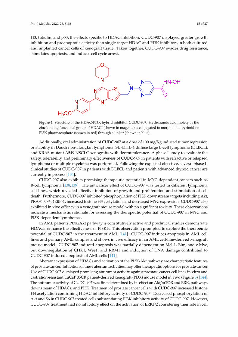
Int. J. Mol. Sci. 2020, 21, 8198 15 of 27
H3, tubulin, and p53, the effects specific to HDAC inhibition. CUDC-907 displayed greater growthinhibition and proapoptotic activity than single-target HDAC and PI3K inhibitors in both culturedand implanted cancer cells of xenograft tissue. Taken together, CUDC-907 evades drug resistance,stimulates apoptosis, and induces cell cycle arrest.Int. J. Mol. Sci. 2020, 21, x 15 of 27
Figure 4. Structure of the HDAC/PI3K hybrid inhibitor CUDC-907. Hydroxamic acid moiety as the zinc binding functional group of HDACI (shown in magenta) is conjugated to morpholino- pyrimidine PI3K pharmacophore (shown in red) through a linker (shown in blue).
Additionally, oral administration of CUDC-907 at a dose of 100 mg/Kg induced tumor regression or stability in Daudi non-Hodgkin lymphoma, SU-DHL-4 diffuse large B-cell lymphoma (DLBCL), and KRAS-mutant A549 NSCLC xenografts with decent tolerance. A phase I study to evaluate the safety, tolerability, and preliminary effectiveness of CUDC-907 in patients with refractive or relapsed lymphoma or multiple myeloma was performed. Following the expected objective, several phase II clinical studies of CUDC-907 in patients with DLBCL and patients with advanced thyroid cancer are currently in process [134].
CUDC-907 also exhibits promising therapeutic potential in MYC-dependent cancers such as B-cell lymphoma [138,139]. The anticancer effect of CUDC-907 was tested in different lymphoma cell lines, which revealed effective inhibition of growth and proliferation and stimulation of cell death. Furthermore, CUDC-907 inhibited phosphorylation of PI3K downstream targets including Akt, PRAS40, S6, 4EBP-1, increased histone H3 acetylation, and decreased MYC expression. CUDC-907 also exhibited in vivo efficacy in a xenograft mouse model with no significant toxicity. These observations indicate a mechanistic rationale for assessing the therapeutic potential of CUDC-907 in MYC and PI3K-dependent lymphomas.
In AML patients PI3K/Akt pathway is constitutively active and preclinical studies demonstrate HDACIs enhance the effectiveness of PI3KIs. This observation prompted to explore the therapeutic potential of CUDC-907 in the treatment of AML [141]. CUDC-907 induces apoptosis in AML cell lines and primary AML samples and shows in vivo efficacy in an AML cell-line-derived xenograft mouse model. CUDC-907-induced apoptosis was partially dependent on Mcl-1, Bim, and c-Myc, but downregulation of CHK1, Wee1, and RRM1 and induction of DNA damage contributed to CUDC-907-induced apoptosis of AML cells [141].
Aberrant expression of HDACs and activation of the PI3K/Akt pathway are characteristic features of prostate cancer. Inhibition of these aberrant activities may offer therapeutic options for prostate cancer. Use of CUDC-907 displayed promising antitumor activity against prostate cancer cell lines in vitro and castration-resistant LuCaP 35CR patient-derived xenograft (PDX) mouse model in vivo (Figure 5) [144]. The antitumor activity of CUDC-907 was first determined by its effect on Akt/mTOR and ERK, pathways downstream of HDACs, and PI3K. Treatment of prostate cancer cells with CUDC-907 increased histone H4 acetylation confirming HDAC inhibitory activity of CUDC-907. Decreased phosphorylation of Akt and S6 in CUDC-907 treated cells substantiating PI3K inhibitory activity of CUDC-907. However, CUDC-907 treatment had no inhibitory effect on the activation of ERK1/2 considering their role in cell proliferation. This response may represent resistance to CUDC-907. Furthermore, an assessment of the molecular mechanism linked to the antitumor activity of CUDC-907 demonstrates that it involves induction of apoptosis and DNA damage. CUDC-907 treatment-induced decreased antiapoptotic Mcl-1 and Bcl-xL levels, increased proapoptotic Bim and decreased c-myc contribute to the onset of apoptosis. CUDC-907 also induced DNA damage and apoptosis by downregulating DNA damage response proteins like Wee1, CHK1, RRM1, and RRM2 (Figure 5). Moreover, the treatment of LuCaP 35CR patient-derived xenograft with
Figure 4. Structure of the HDAC/PI3K hybrid inhibitor CUDC-907. Hydroxamic acid moiety as thezinc binding functional group of HDACI (shown in magenta) is conjugated to morpholino- pyrimidinePI3K pharmacophore (shown in red) through a linker (shown in blue).
Additionally, oral administration of CUDC-907 at a dose of 100 mg/Kg induced tumor regressionor stability in Daudi non-Hodgkin lymphoma, SU-DHL-4 diffuse large B-cell lymphoma (DLBCL),and KRAS-mutant A549 NSCLC xenografts with decent tolerance. A phase I study to evaluate thesafety, tolerability, and preliminary effectiveness of CUDC-907 in patients with refractive or relapsedlymphoma or multiple myeloma was performed. Following the expected objective, several phase IIclinical studies of CUDC-907 in patients with DLBCL and patients with advanced thyroid cancer arecurrently in process [134].
CUDC-907 also exhibits promising therapeutic potential in MYC-dependent cancers such asB-cell lymphoma [138,139]. The anticancer effect of CUDC-907 was tested in different lymphomacell lines, which revealed effective inhibition of growth and proliferation and stimulation of celldeath. Furthermore, CUDC-907 inhibited phosphorylation of PI3K downstream targets including Akt,PRAS40, S6, 4EBP-1, increased histone H3 acetylation, and decreased MYC expression. CUDC-907 alsoexhibited in vivo efficacy in a xenograft mouse model with no significant toxicity. These observationsindicate a mechanistic rationale for assessing the therapeutic potential of CUDC-907 in MYC andPI3K-dependent lymphomas.
In AML patients PI3K/Akt pathway is constitutively active and preclinical studies demonstrateHDACIs enhance the effectiveness of PI3KIs. This observation prompted to explore the therapeuticpotential of CUDC-907 in the treatment of AML [141]. CUDC-907 induces apoptosis in AML celllines and primary AML samples and shows in vivo efficacy in an AML cell-line-derived xenograftmouse model. CUDC-907-induced apoptosis was partially dependent on Mcl-1, Bim, and c-Myc,but downregulation of CHK1, Wee1, and RRM1 and induction of DNA damage contributed toCUDC-907-induced apoptosis of AML cells [141].
Aberrant expression of HDACs and activation of the PI3K/Akt pathway are characteristic featuresof prostate cancer. Inhibition of these aberrant activities may offer therapeutic options for prostate cancer.Use of CUDC-907 displayed promising antitumor activity against prostate cancer cell lines in vitro andcastration-resistant LuCaP 35CR patient-derived xenograft (PDX) mouse model in vivo (Figure 5) [144].The antitumor activity of CUDC-907 was first determined by its effect on Akt/mTOR and ERK, pathwaysdownstream of HDACs, and PI3K. Treatment of prostate cancer cells with CUDC-907 increased histoneH4 acetylation confirming HDAC inhibitory activity of CUDC-907. Decreased phosphorylation ofAkt and S6 in CUDC-907 treated cells substantiating PI3K inhibitory activity of CUDC-907. However,CUDC-907 treatment had no inhibitory effect on the activation of ERK1/2 considering their role in cell

Int. J. Mol. Sci. 2020, 21, 8198 16 of 27
proliferation. This response may represent resistance to CUDC-907. Furthermore, an assessment ofthe molecular mechanism linked to the antitumor activity of CUDC-907 demonstrates that it involvesinduction of apoptosis and DNA damage. CUDC-907 treatment-induced decreased antiapoptoticMcl-1 and Bcl-xL levels, increased proapoptotic Bim and decreased c-myc contribute to the onset ofapoptosis. CUDC-907 also induced DNA damage and apoptosis by downregulating DNA damageresponse proteins like Wee1, CHK1, RRM1, and RRM2 (Figure 5). Moreover, the treatment of LuCaP35CR patient-derived xenograft with CUDC-907 resulted in significant inhibition of tumor growthencouraging further clinical development of CUDC-907 for prostate cancer treatment [144].
Int. J. Mol. Sci. 2020, 21, x 16 of 27
CUDC-907 resulted in significant inhibition of tumor growth encouraging further clinical development of CUDC-907 for prostate cancer treatment [144].
Upregulation of class I HDACs is a common feature in hepatocellular carcinoma (HCC), which sponsors spreading, aggressiveness, and increased mortality. Motivated by the efficacious activity of CUDC-907, efforts were made to design novel HDAC/PI3K dual hybrid drugs via bio-isosterism, by replacing morpholinothienopyrimidine moiety (Figure 6A) in CUDC-907 with a morpholinopurine (Figure 6B,C) [137]. All compounds generated displayed strong anti-HDAC1 and anti-PI3Kα activities. CUDC-907 exhibited IC50 values of 1.7 and 19 nM for HDAC1 and PI3Kα, respectively (Figure 6A). Among the several analogs obtained, the compounds having a pyrimidine (Figure 6B) or aminopyrimidine (Figure 6C) at the C-2 position of the morpholinopurine moiety, demonstrated expected dual PI3Kα and HDAC1 inhibitory activities. The IC50 values for the analog in Figure 6B against HDAC1 and PI3Kα were 1.14 and 28.06 nM, respectively. Analog in Figure 6C displayed lower and balanced IC50 values for HDAC1 and PI3Kα, 1.04, and 1.33 nM, respectively.
Figure 5. Schematic display of antitumor activity of CUDC-907 hybrid drug against prostate cancer cells. Synergistic anti-cancer activity of CUDC-907 is illustrated by its inhibition of HDACs activity and PI3K/Akt pathway [144]. Increase in histone 4 acetylation and reduced activation of PI3K/Akt confirms its dual action on HDAC and PI3K. Outcome of these effects resulted in inhibition of cell proliferation, increase in DNA damage due to downregulation of DNA damage repair response proteins, and apoptosis because of downregulation of c-myc and antiapoptotic Mcl-1 and Bcl-xL proteins, and upregulation of pro-apoptosis protein Bim (blunt end lines indicate inhibition, upward arrows indicate upregulation and downward arrows indicate downregulation of proteins).
CUDC-907
Akt
S6
Inhibition of cell
proliferation
HDACs/PI3K
Bim
c-Myc
Mcl-1Bcl-xL
Apoptosis
CHK1Wee1
RRM1/M2
DNA repair
Histone 4acetylation
P
P
Figure 5. Schematic display of antitumor activity of CUDC-907 hybrid drug against prostate cancercells. Synergistic anti-cancer activity of CUDC-907 is illustrated by its inhibition of HDACs activityand PI3K/Akt pathway [144]. Increase in histone 4 acetylation and reduced activation of PI3K/Aktconfirms its dual action on HDAC and PI3K. Outcome of these effects resulted in inhibition of cellproliferation, increase in DNA damage due to downregulation of DNA damage repair response proteins,and apoptosis because of downregulation of c-myc and antiapoptotic Mcl-1 and Bcl-xL proteins, andupregulation of pro-apoptosis protein Bim (blunt end lines indicate inhibition, upward arrows indicateupregulation and downward arrows indicate downregulation of proteins).
Upregulation of class I HDACs is a common feature in hepatocellular carcinoma (HCC), whichsponsors spreading, aggressiveness, and increased mortality. Motivated by the efficacious activityof CUDC-907, efforts were made to design novel HDAC/PI3K dual hybrid drugs via bio-isosterism,by replacing morpholinothienopyrimidine moiety (Figure 6A) in CUDC-907 with a morpholinopurine(Figure 6B,C) [137]. All compounds generated displayed strong anti-HDAC1 and anti-PI3Kαactivities. CUDC-907 exhibited IC50 values of 1.7 and 19 nM for HDAC1 and PI3Kα, respectively(Figure 6A). Among the several analogs obtained, the compounds having a pyrimidine (Figure 6B)or aminopyrimidine (Figure 6C) at the C-2 position of the morpholinopurine moiety, demonstratedexpected dual PI3Kα and HDAC1 inhibitory activities. The IC50 values for the analog in Figure 6Bagainst HDAC1 and PI3Kαwere 1.14 and 28.06 nM, respectively. Analog in Figure 6C displayed lowerand balanced IC50 values for HDAC1 and PI3Kα, 1.04, and 1.33 nM, respectively.
Using the polypharmacology-based paradigm, two other dual HDAC-PI3K inhibitors wererecently reported [143]. These inhibitors were designed by introducing hydroxamic acid moiety as thezinc binding functional group to a quinazoline-based PI3K pharmacophore through an appropriatelinker. Nanomolar concentrations of both these lead inhibitors simultaneously inhibit PI3K and HDACactivity. These inhibitors inhibited proliferation, arrested the cell cycle, and induced apoptosis ofHCT116 cancer cells. Furthermore, they also altered p-Akt expression and acetylated histone H3 inHCT116. Pharmacokinetic studies in HCT116 and HGC-27 xenograft models further demonstratethat they have significant anticancer efficacies. Taken together, this study further supports the dualHDAC-PI3K hybrid drugs that have potential anticancer activity.
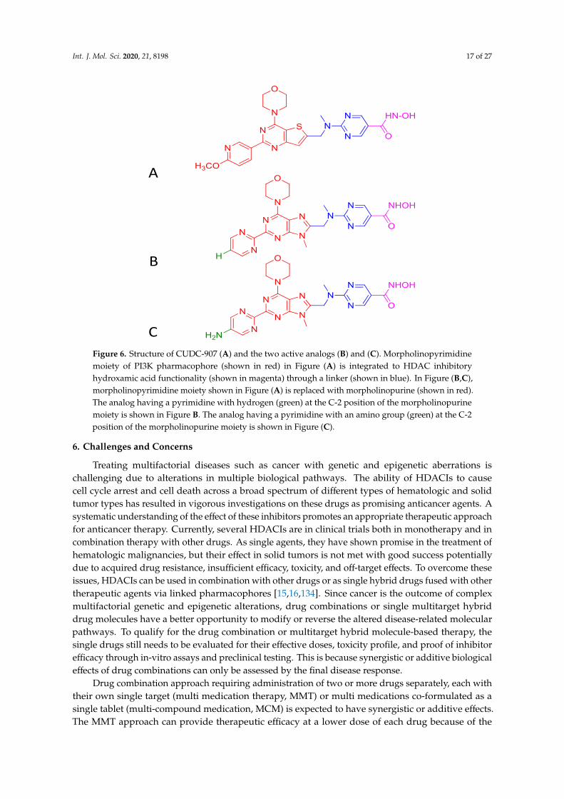
Int. J. Mol. Sci. 2020, 21, 8198 17 of 27
Int. J. Mol. Sci. 2020, 21, x 17 of 27
Figure 6. Structure of CUDC-907 (A) and the two active analogs (B) and (C). Morpholinopyrimidine moiety of PI3K pharmacophore (shown in red) in Figure (A) is integrated to HDAC inhibitory hydroxamic acid functionality (shown in magenta) through a linker (shown in blue). In Figure (B,C), morpholinopyrimidine moiety shown in Figure (A) is replaced with morpholinopurine (shown in red). The analog having a pyrimidine with hydrogen (green) at the C-2 position of the morpholinopurine moiety is shown in Figure B. The analog having a pyrimidine with an amino group (green) at the C-2 position of the morpholinopurine moiety is shown in Figure (C).
Using the polypharmacology-based paradigm, two other dual HDAC-PI3K inhibitors were recently reported [143]. These inhibitors were designed by introducing hydroxamic acid moiety as the zinc binding functional group to a quinazoline-based PI3K pharmacophore through an appropriate linker. Nanomolar concentrations of both these lead inhibitors simultaneously inhibit PI3K and HDAC activity. These inhibitors inhibited proliferation, arrested the cell cycle, and induced apoptosis of HCT116 cancer cells. Furthermore, they also altered p-Akt expression and acetylated histone H3 in HCT116. Pharmacokinetic studies in HCT116 and HGC-27 xenograft models further demonstrate that they have significant anticancer efficacies. Taken together, this study further supports the dual HDAC-PI3K hybrid drugs that have potential anticancer activity.
6. Challenges and Concerns
Treating multifactorial diseases such as cancer with genetic and epigenetic aberrations is challenging due to alterations in multiple biological pathways. The ability of HDACIs to cause cell cycle arrest and cell death across a broad spectrum of different types of hematologic and solid tumor types has resulted in vigorous investigations on these drugs as promising anticancer agents. A systematic understanding of the effect of these inhibitors promotes an appropriate therapeutic approach for anticancer therapy. Currently, several HDACIs are in clinical trials both in monotherapy and in combination therapy with other drugs. As single agents, they have shown promise in the treatment of hematologic malignancies, but their effect in solid tumors is not met with good success potentially due to acquired drug resistance, insufficient efficacy, toxicity, and off-target effects. To overcome these issues, HDACIs can be used in combination with other drugs or as single hybrid drugs fused with other therapeutic agents via linked pharmacophores [15,16,134]. Since cancer is the outcome of complex multifactorial genetic and epigenetic alterations, drug combinations or single multitarget hybrid drug molecules have a better opportunity to modify or reverse the altered disease-
A
B
CFigure 6. Structure of CUDC-907 (A) and the two active analogs (B) and (C). Morpholinopyrimidinemoiety of PI3K pharmacophore (shown in red) in Figure (A) is integrated to HDAC inhibitoryhydroxamic acid functionality (shown in magenta) through a linker (shown in blue). In Figure (B,C),morpholinopyrimidine moiety shown in Figure (A) is replaced with morpholinopurine (shown in red).The analog having a pyrimidine with hydrogen (green) at the C-2 position of the morpholinopurinemoiety is shown in Figure B. The analog having a pyrimidine with an amino group (green) at the C-2position of the morpholinopurine moiety is shown in Figure (C).
6. Challenges and Concerns
Treating multifactorial diseases such as cancer with genetic and epigenetic aberrations ischallenging due to alterations in multiple biological pathways. The ability of HDACIs to causecell cycle arrest and cell death across a broad spectrum of different types of hematologic and solidtumor types has resulted in vigorous investigations on these drugs as promising anticancer agents. Asystematic understanding of the effect of these inhibitors promotes an appropriate therapeutic approachfor anticancer therapy. Currently, several HDACIs are in clinical trials both in monotherapy and incombination therapy with other drugs. As single agents, they have shown promise in the treatment ofhematologic malignancies, but their effect in solid tumors is not met with good success potentiallydue to acquired drug resistance, insufficient efficacy, toxicity, and off-target effects. To overcome theseissues, HDACIs can be used in combination with other drugs or as single hybrid drugs fused with othertherapeutic agents via linked pharmacophores [15,16,134]. Since cancer is the outcome of complexmultifactorial genetic and epigenetic alterations, drug combinations or single multitarget hybriddrug molecules have a better opportunity to modify or reverse the altered disease-related molecularpathways. To qualify for the drug combination or multitarget hybrid molecule-based therapy, thesingle drugs still needs to be evaluated for their effective doses, toxicity profile, and proof of inhibitorefficacy through in-vitro assays and preclinical testing. This is because synergistic or additive biologicaleffects of drug combinations can only be assessed by the final disease response.
Drug combination approach requiring administration of two or more drugs separately, each withtheir own single target (multi medication therapy, MMT) or multi medications co-formulated as asingle tablet (multi-compound medication, MCM) is expected to have synergistic or additive effects.The MMT approach can provide therapeutic efficacy at a lower dose of each drug because of the
Int. J. Mol. Sci. 2020, 21, 8198 18 of 27
synergistic effects of the drug combination. As a result, the MMT approach can reduce the chancesof drug resistance and adverse reactions compared to single-drug treatment [15,16]. While the MMTapproach provides flexibility to choose different dosages and therapeutic schedules, it also causesproblems with patient compliance. The MCM approach can offset the negative impact on patientcompliance by providing multi-medications in the form of a co-formulated single tablet [16]. However,both MMT and MCM approaches present several challenges. The use of drugs with very differentchemical structures in the same therapeutic drug combination complicates pharmacodynamic andpharmacokinetic requirements. Other problems include drug–drug interactions and potential solubilityissues of the active substances that can modify the bloodstream uptake. Similarly, the simultaneouspresence of drugs in the target tissues is not guaranteed to promote a synergistic response. Additionalcomplications include each tumor and the therapeutic response of each patient is different.
Unlike drug combination approaches, the use of single multitarget hybrid drugs created based onthe multitarget-directed ligands (MTDLs) approach exhibits promising outcomes for solid tumors anddrug-resistant cancers. The single multitarget hybrid drugs created by using the polypharmacologyprinciple can simultaneously hit two or more targets. Importantly, single multitarget hybrid drugshave several advantages over the drug combinations. They display better pharmacokinetics andpharmacodynamics such as no solubility/bioavailability issues, greater therapeutic efficacy, lowertoxicities, the simultaneous presence of the chemical entities in multiple tissues, and improved patientcompliance [15,16]. Specifically, single multitarget hybrid drugs that can simultaneously hit two ormore targets in which one (or more) is (are) epigenetic and the other (s) belongs to a non-epigeneticcategory(ies) have greater potential for cancer therapy. This concept has resulted in the design anddevelopment of multitarget epigenetic hybrid drugs. Among the several epigenetic hybrid drugsproduced, CUDC-907 is a promising single dual HDAC-PI3K inhibitor molecule with an abilityto simultaneously inhibit HDACs and PI3K [15,16,134–144]. CUDC- 907 is an orally bioavailablesmall -molecule dual HDAC and PI3K inhibitor targeting class I and II HDACs and PI3Kα, β, andδ isoforms [135]. Its extensive antitumor activities in hematologic and solid tumors show promiseas an HDAC-PI3K inhibitor [134–144]. Considering the pleiotropic effects of HDAC inhibition andthe multiple downstream effectors of PI3K, it is not surprising CUDC-907 has a better opportunity toreverse the altered molecular pathways linked to oncogenesis. Even with the frequent difficulty instructural optimization to effectively merge the pharmacophoric portions of HDAC and PI3K entitiesto generate dual HDAC-PI3K inhibitor, it was possible to generate CUDC-907. Importantly, preclinicalfindings indicate the use of CUDC-907 has several advantages over the drug combination treatmentwith single-target HDAC and PI3K inhibitors (MMT and MCM) [15,16,135]. CUDC-907 exhibitsbetter pharmacokinetics and pharmacodynamics such as no solubility/bioavailability issues, greatertherapeutic efficacy, lower toxicities, the simultaneous presence of the chemical entities in multipletissues, and improved patient compliance [15,16,135]. Moreover, CUDC-907 is well tolerated withmanageable gastrointestinal and hematologic events compared to a combination of single HDAC andPI3K inhibitors without any infections, hepatotoxicity, or cardiotoxicity [136]. Adjusting dosing issimpler and pharmacokinetics and toxicity profiles are predictable. Based on these preclinical outcomes,CUDC907 was entered into phase I study to determine the safety, tolerability, and pharmacokinetics inpatients with advanced/relapsed solid tumors (NCT02307240). Based on encouraging results from thephase I study, a biomarker-driven phase II (NCT02674750) study has been initiated in patients withmyc mutation.
7. Conclusions and Perspectives
Epigenetic drug discovery is a rapidly evolving field, and the use of epigenetic drugs both inclinical and preclinical trials forecast a promising future for the treatment of multifactorial diseasessuch as cancer. The first dual inhibitor targeted at an epigenetic target was reported in 2007 and sincethen several dual inhibitors aimed at epigenetic targets have been reported. Importantly, most of theseinhibitors include HDAC inhibition as one of their mechanisms of action. This is attributed to the
Int. J. Mol. Sci. 2020, 21, 8198 19 of 27
high resilience offered by the HDAC pharmacophore. Moreover, the characteristics of HDACs andstrategies for their inhibition are better understood compared to other epigenetic targets. CUDC-907is one of the successes of the combination of HDAC inhibition with PI3K inhibition. Several otherepigenetic drugs are undergoing clinical trials in combination with other therapeutic agents via linkedpharmacophores that can provide synergistic effects both for solid and hematologic cancers. CUDC-907inhibits classical HDACs and all four class I PI3K isoforms in nanomolar amounts, and is currentlyin phase II cancer trials. Despite the promising efficacy in hematologic cancers, there is still a poorprognosis for solid tumors due to poor cancer-targeting specificity. A recent report indicates that ananoparticle-based pre-targeted delivery system improves the therapeutic window of small-moleculeinhibitors [161]. This approach increases the number of nanoparticles retained on the target tumorcells, improves the drug delivery, and shows the antitumor activity of BEZ235 through the inhibitionof PI3K/mTOR. The use of nanoparticle-based drug delivery technology may improve the delivery ofCUDC-907 to target cancer tissues, and at the same time, normal tissues can be protected. The futurelooks bright for dual inhibitors directed at epigenetic targets and their full potential in the treatment ofcomplex diseases is yet to be explored.
Author Contributions: Conceptualization, K.R.; writing, original draft preparation, K.R.; writing, review andediting, K.R., C.S., A.S. and Z.Y.; funding acquisition, K.R. All authors have read and agreed to the publishedversion of the manuscript.
Funding: This research was made possible, in part, by the support from grant number 5G12MD007605 from theNIMHD/NIH.
Conflicts of Interest: The authors declare no conflict of interest. The funders had no role in the design of thestudy; in the collection, analyses, or interpretation of data; in the writing of the manuscript, or in the decision topublish the results.
References
1. Vanhaesebroeck, B.; Guillermet-Guibert, J.; Graupera, M.; Bilanges, B. The emerging mechanism ofisoform-specific PI3K signaling. Nat. Rev. Mol. Cell Biol. 2010, 11, 329–341. [CrossRef] [PubMed]
2. Vanhaesebroeck, B.; Stephens, L.; Hawkins, P. PI3K signaling: The path to discovery and understanding.Nat. Rev. Mol. Cell Biol. 2012, 13, 195–203. [CrossRef] [PubMed]
3. Engelman, J.A.; Luo, J.; Cantley, L.C. The evolution of phosphatidylinositol 3-kinases as regulators of growthand metabolism. Nat. Rev. Genet. 2006, 7, 606–619. [CrossRef] [PubMed]
4. Lawrence, M.S.; Stojanov, P.; Mermel, C.H.; Robinson, J.T.; Garrqway, L.A.; Golub, T.R.; Meyerson, M.L.;Gabriel, S.B.; Lander, E.S.; Getz, G. Discovery and saturation analysis of cancer genes across 21 tumour types.Nature 2014, 505, 495–501. [CrossRef] [PubMed]
5. Gyori, D.; Chessa, T.; Hawkins, P.T.; Stephens, L.R. Class (I) phosphoinositide 3-kinases in the tumormicroenvironment. Cancers 2017, 9, 24. [CrossRef]
6. Thorpe, L.M.; Yuzugullu, H.; Zhao, J.J. PI3K in cancer: Divergent roles of isoforms, modes of activation andtherapeutic targeting. Nat. Rev. Cancer 2015, 15, 7–24. [CrossRef]
7. Janku, F.; Yap, T.; Meric-Bernstam, F. Targeting the PI3K pathway in cancer: Are we making head way?Nat. Rev. Clin. Oncol. 2018, 15, 273–291. [CrossRef]
8. McCubery, J.A.; Steelman, L.S.; Chappell, W.H.; Abrams, S.L. Franklin, Ras/Raf/MEK/ERK andPI3K/PTEN/Akt/mTOR cascade inhibitors: How mutations can result in therapy resistance and howto overcome resistance. Oncotarget 2012, 3, 1068–1111. [CrossRef]
9. Wee, S.; Jagani, Z.; Xiang, K.X.; Loo, A. PI3k pathway activation mediates resistance to MEK inhibitors inKRAS mutant cancers. Cancer Res. 2009, 69, 4286–4293. [CrossRef]
10. Yang, X.J.; Seto, E. HATs and HDACs: From structure, function and regulation to novel strategies for therapyand prevention. Oncogene 2007, 26, 5310–5318. [CrossRef]
11. Hagelkruys, A.; Sawicka, A.; Rennmayr, M.; Seiser, C. The biology of HDAC in cancer: The nuclear andepigenetic components. Handb. Exp. Pharmacol. 2011, 206, 13–37.
12. Delcuve, G.P.; Khan, D.H.; Davie, J.R. Roles of histone deacetylases in epigenetic regulation: Emergingparadigms from studies with inhibitors. Clin. Epigenet. 2012, 4, 5. [CrossRef]
Int. J. Mol. Sci. 2020, 21, 8198 20 of 27
13. Zwergel, C.; Stazi, G.; Valente, S.; Mai, A. Histone Deacetylase Inhibitors: Updated Studies in VariousEpigenetic-Related Diseases. J. Clin. Epigenet. 2016, 2, 7. [CrossRef]
14. Benedetti, R.; Conte, M.; Iside, C.; Atlcci, L. Epigenetic-based therapy: From single- to multi-target approaches.Int. J. Biochem. Cell Biol. 2015, 69, 121–131. [CrossRef] [PubMed]
15. deLera, A.R.; Ganesan, A. Epigenetic polypharmacology: From combination therapy to multitarget drugs.Clin. Epigenet. 2016, 8, 1–21.
16. Tomaselli, D.; Lucidi, A.; Rotili, D.; Mai, A. Epigenetic polypharmacology: A new frontier for epi-drugdiscovery. Med. Res. Rev. 2020, 40, 190–244. [CrossRef] [PubMed]
17. Okkenhaug, K. Signaling by the phosphoinositide 3-kinase family in immune cells. Annu. Rev. Immunol.2013, 31, 675–704. [CrossRef]
18. Cantley, L.C. The phosphoinositide 3-kinase pathway. Science 2002, 296, 1655–1657. [CrossRef]19. Rai, S.N.; Dilnashin, H.; Birla, H.; Singh, S.S.; Zahra, W.; Rathore, A.S.; Singh, B.K.; Singh, S.P. The role of
PI3K/Akt and ERK in neurodegenerative disorders. Neurotox Res. 2019, 35, 775–795. [CrossRef]20. Gross, C.; Bassell, G.J. Neuron-specific regulation of class I PI3K catalytic subunits and their dysfunction in
brain disorders. Front. Mol. Neurosci. 2014, 7, 12. [CrossRef]21. Oudit, G.Y.; Sun, H.; Kerfant, B.-G.; Crackower, M.A.; Penninger, J.M.; Backk, P.H. The role of
phoisphoinositide-3 kinase and PTEN in cardiovascular physiology and disease. J. Mol. Cell. Cardiol.2004, 37, 449–471. [CrossRef] [PubMed]
22. Eisenreich, A.; Rauch, U. PI3K inhibitors in cardiovascular disease. Cardiovasc. Ther. 2011, 29, 29–36.[CrossRef] [PubMed]
23. Durrant, T.N.; Hers, I. PI3K inhibitors in thrombosis and cardiovascular disease. Clin. Trans. Med. 2020, 9, 8.[CrossRef] [PubMed]
24. Maffei, A.; Lembo, G.; Carnevale, D. PI3Kinases in diabetes mellitus and its related complications. Int. J.Mol. Sci. 2018, 19, 4098. [CrossRef]
25. Kurek, K.C.; Luks, V.L.; Ayturk, U.M.; Alomari, A.I.; Fishman, S.J.; Spencer, S.A.; Mulliken, J.B.; Bowen, M.E.;Yamamoto, G.L.; Kozakewich, H.P.W.; et al. Somatic mosaic activating mutations in PIK3CA cause CLOVESsyndrome. Am. J. Hum. Genet. 2012, 90, 1108–1115. [CrossRef]
26. Parsons, R. Human cancer, PTEN, and the PI-3 kinase pathway. Semin. Cell Dev. Biol. 2004, 15, 171–176.[CrossRef]
27. Song, M.S.; Salmena, L.; Pandolfi, P.P. The functions and regulation of the PTEN tumour suppressor. Nat.Rev. Mol. Cell Biol. 2012, 13, 283–296. [CrossRef]
28. Aziz, S.A.; Davies, M.; Pick, E.; Zito, C.; Jilaveanu, L.; Camp, R.L.; Rimm, D.L.; Kluger, Y.; Kluger, H.M.Phosphatidylinositol 3-kinase as a therapeutic target in melanoma. Clin. Cancer Res. 2009, 15, 3029–3036.[CrossRef]
29. Zhou, B.P.; Hu, M.C.; Miller, S.A.; Yu, Z.; Xia, W.; Lin, S.Y.; Hung, M.C. Her-2/neu blocks tumor necrosisfactor-induced apoptosis via the Akt/NF-kappaB pathway. J. Biol. Chem. 2000, 275, 8027–8031. [CrossRef]
30. Hill, K.; Kalifa, S.; Das, J.R.; Bhatti, T.; Gay, M.; Williams, D.; Taliferro-Smith, L.; De Marzo, A.M. The role ofPI 3-kinase p100beta in Akt signaling, cell survival, and proliferation in human prostate cancer cells. Prostate2010, 70, 755–764. [CrossRef]
31. Dbouk, H.; Khalil, B.D.; Wu, H.; Shymanets, A.; Nurnberg, B.; Backer, J.M. Characterization oftumor-associated activating mutation of the p110β PI 3-K. PLoS ONE 2013, 8, e63833. [CrossRef] [PubMed]
32. Urick, M.E.; Rudd, M.L.; Godwin, A.K.; Sgroi, D.; Merino, M.; Bell, D.W. PIK3R1 (p85alpha) is somaticallymutated at high frequency in primary endometrial cancer. Cancer Res. 2011, 71, 4061–4067. [CrossRef][PubMed]
33. Wu, H.; Shekar, S.C.; Flinn, R.J.; El-Sibai, M.; Jaiswal, B.S.; Sen, K.L.; Janakiraman, V.; Seshagiri, S.; Gerfer, G.J.;Girvin, M.E.; et al. Regulation of class IA PI3-kinases: C2 domain- iSH2 domain contacts inhibit p85/p110alphaand are disrupted in oncogenic p85 mutants. Proc. Natl. Acad. Sci. USA 2009, 106, 20258–20263. [CrossRef][PubMed]
34. Cortes, I.; Sanchez-Ruiz, J.; Zuluaga, S.; Calvanese, V.; Marques, M. p85beta phosphoinositide 3-kinasesubunit regulates tumor progression. Proc. Natl. Acad. Sci. USA 2012, 109, 11318–11323. [CrossRef]
35. Engelman, J.A. Targeting PI3K signaling in cancer: Opportunities, challenges and limitations. Nat. Rev.Cancer 2009, 9, 550–562. [CrossRef]

Int. J. Mol. Sci. 2020, 21, 8198 21 of 27
36. Spangle, J.M.; Roberts, T.M.; Zhao, J.J. The emerging role of PI3K/Akt-mediated regulation in cancer.Biochem. Biophys. Acta 2017, 1868, 123–1131. [CrossRef]
37. Furman, R.R.; Sharman, J.P.; Coutre, S.E.; Cheson, B.D.; Pagel, J.M.; Hillmen, P.; Barrientos, J.C.; Zelenetz, A.D.;Kipps, T.J.; Flinn, I.; et al. Idelalisib and rituximab in relapsed chronic lymphocytic leukemia. N. Engl. J. Med.2014, 370, 997–1007. [CrossRef]
38. Ramesh, L.E.; Chen, C.S.; Cantley, L.C. Phosphatidylinositol (3,4,5)P3 interacts with SH2 domains andmodulates PI 3-kinase association with tyrosine-phosphorylated proteins. Cell 1995, 83, 821–830. [CrossRef]
39. Yu, J.; Wjasow, C.; Backer, J.M. Regulation of p85/p110alpha phosphatidylinositol 3-kinase. Distinct roles forthe n-terminal and c-terminal SH2 domains. J. Biol. Chem. 1998, 273, 30199–30203. [CrossRef]
40. Manning, B.D.; Toker, A. Akt/PKB signaling: Navigating the network. Cell 2017, 169, 381–405. [CrossRef]41. Milella, M.; Falcone, I.; Conciatori, F.; Incani, U.C.; Curatolo, A.D.; Inzerilli, N.; Nuzzo, C.M.A.; Vaccaro, N.;
Vari, S.; Cognetti, F.; et al. PTEN: Multiple functions in human malignant tumors. Front. Oncol. 2015, 5, 24.[CrossRef]
42. Ong, P.S.; Wang, L.Z.; Dai, X.; Tseng, S.H.; Loo, S.J.; Sethi, G. Judicious toggling of mTOR activity to combatinsulin resistance and cancer: Current evidence and perspectives. Front. Pharmacol. 2016, 7, 395. [CrossRef][PubMed]
43. Shahbazian, M.D.; Grunstein, M. Functions of site-specific histone acetylation and deacetylation. Annu. Rev.Biochem. 2007, 76, 75–100. [CrossRef] [PubMed]
44. Lee, K.K.; Workman, J.L. Histone acetyltransferase complexes: One size doesn’t fit all. Nat. Rev. Mol. CellBiol. 2007, 8, 284–295. [CrossRef]
45. de Ruijter, A.J.; van Gennip, A.H.; Caron, H.N.; Kemp, S.; van Kuilenburg, A.B. Histone deacetylases(HDACs): Characterization of the classical HDAC family. Biochem. J. 2003, 370, 737–749. [CrossRef][PubMed]
46. Clayton, A.L.; Hazzalin, C.A.; Mahadevan, L.C. Enhanced histone acetylation and transcription: A dynamicperspective. Mol. Cell 2006, 23, 289–296. [CrossRef]
47. Choudhary, C.; Kumar, C.; Gnad, F.; Nielsen, M.L.; Rehman, M.; Walther, T.C.; Olsen, J.V.; Mann, M. Lysineacetylation targets protein complexes and co-regulates major cellular functions. Science 2009, 325, 834–840.[CrossRef]
48. Ranganna, K.; Yatsu, F.M.; Mathew, O.P. Emerging epigenetic therapy for vascular proliferative diseases.In Atherogenesis; Parthasarathy, S., Ed.; InTech: London, UK, 2012; pp. 79–104.
49. Ranganna, K.; Mathew, O.P.; Milton, S.G. Regulation of cellular processes by epigenetic mechanisms ofbutyrate. In Butyrate: Food Sources, Functions and Health Benefits. Biochemistry Research Trends Series; Li, C., Ed.;Bovine Functional Genomics Laboratory, ARS, USDA: Beltsville, MD, USA; Nova Science Publisher, Inc.:Hauppauge, NY, USA, 2014; pp. 219–232. ISBN 978-1-63117-657-9.
50. Latham, J.A.; Dent, S.Y. Cross-regulation of histone modifications. Nat. Struct. Mol. Biol. 2007, 14, 1017–1024.[CrossRef]
51. Yang, X.J.; Seto, E. Lysine acetylation: Codified crosstalk with other posttranslational modifications. Mol. Cell2008, 31, 449–461. [CrossRef]
52. Wang, Q.; Zhang, Y.; Yang, C.; Xiong, H.; Lin, Y.; Yao, J.; Li, H.; Xie, L.; Zhao, W.; Yao, Y.; et al. Acetylation ofmetabolic enzymes coordinates carbon source utilization and metabolic flux. Science 2010, 327, 1004–1007.[CrossRef]
53. Patel, J.; Pathak, R.R.; Mujtaba, S. The biology of lysine acetylation integrates transcriptional programmingand metabolism. Nutr. Metab. 2011, 8, 12. [CrossRef] [PubMed]
54. Cheung, P.; Allis, C.D.; Sassone-Corsi, P. Signaling to chromatin through histone modifications. Cell 2000,103, 263–271. [CrossRef]
55. Strahl, B.D.; Allis, C.D. The language of covalent histone modifications. Nature 2000, 403, 41–45. [CrossRef]56. Kouzarides, T. Chromatin modifications and their function. Cell 2007, 128, 693–705. [CrossRef] [PubMed]57. Mathew, O.P.; Ranganna, K.; Yatsu, F.M. Butyrate, a histone deacetylase inhibitor, stimulates interplay
between different posttranslational modifications of histone H3 and differently alters G1-specific cell cycleproteins in vascular smooth muscle cells. Biomed. Pharmacother. 2010, 64, 733–740. [CrossRef]
58. Gil, J.; Ramirez-Torres, A.; Encarnacion-Guevara, S. Lysine acetylation and cancer: A proteomics perspective.J. Proteom. 2017, 150, 297–309. [CrossRef]

Int. J. Mol. Sci. 2020, 21, 8198 22 of 27
59. Grimes, M.; Benjamin, H.; Foltz, L.; Levy, T.; Rikova, K.; Gaiser, J.; Cook, W.; Smirnova, E.; Wheeler, T.;Clark, N.R.; et al. Integration of protein phosphorylation, acetylation and methylation data sets to outlinelung cancer signaling network. Sci. Signal. 2018, 11, eaaq1087. [CrossRef]
60. Alonso-Bastda, R.; Encarnacion-Guevara, S. Proteomic insights into lysine acetylation and the implicationsfor medical research. Exptert Rev. Proteom. 2019, 16, 1–3. [CrossRef]
61. Okumura, K.; Mendoza, M.; Bachoo, R.M.; DePinho, R.A.; Cavenee, W.K.; Fumari, F.B. PCAF modulatesPTEN activity. J. Biol. Chem. 2006, 281, 26562–26568. [CrossRef]
62. Buuh, Z.Y.; Lyu, Z.; Wang, R.E. Interrogating the roles of post-translational modifications of non-histoneproteins. J. Med. Chem. 2017, 61, 3239–3252. [CrossRef]
63. Filippakopoulos, P.; Knapp, S. Targeting bromodomains: Epigenetic readers of lysine acetylation. Nat. Rev.Drug Discov. 2014, 13, 337–356. [CrossRef]
64. Perez-Salvia, M.; Esteller, M. Bromodomain inhibitors and cancer therapy: From structures to applications.Epigenetics 2017, 12, 323–339. [CrossRef] [PubMed]
65. Di Cerbo, V.; Schneider, R. Cancers with wrong HATs: The impact of acetylation. Briefings Funct. Genom.2013, 12, 231–243. [CrossRef] [PubMed]
66. Sambataro, F.; Pennuto, M. Post-translational modifications and protein quality control in motor neuron andpolyglutamine diseases. Front. Mol. Neurosci. 2017, 10, 82. [CrossRef]
67. Liu, J.; Qian, C.; Cao, X. Post-translational modification control of innate immunity. Immunity 2016, 45, 15–30.[CrossRef] [PubMed]
68. Riggs, M.G.; Whittaker, R.G.; Neumann, J.R.; Ingram, V.M.N. Butyrate causes histone modification in Helaand Friend erythroleukemia cells. Nature 1977, 268, 462–464. [CrossRef]
69. Xu, W.S.; Parmigiani, R.B.; Marks, P.A. Histone deacetylase inhibitors: Molecular mechanism of action.Oncogene 2007, 26, 5541–5552. [CrossRef]
70. Garmpi, A.; Garmpis, N.; Damaskos, C.; Valsami, S.; Spartalis, E.; Lavaris, A.; Patelis, N.; Margonis, G.-A.;Apostolou, K.G.; Sparatalis, M.; et al. Histone deacetylase inhibitors as a new anticancer option: How far canwe go with expectations? J. BUON 2018, 23, 846–861.
71. Chen, C.-S.; Weng, S.-C.; Tseng, P.-H.; Chen, C.-S. Histone acetylation-independent effect of histonedeacetylase inhibitors on Akt through the reshuffling of protein phosphatase 1 complexes. J. Biol. Chem.2005, 280, 38879–38887. [CrossRef]
72. Wang, J.C.; Kafeel, M.I.; Avezbakiyev, B.; Chen, C.; Sun, Y.; Rathnasabapathy, C.; Kalavar, M.; He, Z.; Barton, J.;Lichter, S. Histone deacetylase in chronic lymphocytic leukemia. Oncology 2011, 81, 325–329. [CrossRef]
73. Zhang, H.; Shand, Y.P.; Chen, H.Y.; Li, J. Histone deacetylases function as novel potential therapeutic targetsfor cancer. Hepatol. Res. 2017, 47, 149–159. [CrossRef] [PubMed]
74. Zhang, Q.; Wang, S.; Chen, J.; Yu, Z. Histone deacetylases (HDACs) guided novel therapies for T-celllymphomas. Int. J. Med. Sci. 2019, 16, 424–442. [CrossRef]
75. Quagliano, A.; Gopalakrihnapillai, A.; Barwe, S.P. Understanding the mechanisms by which epigeneticmodifiers avert therapy resistance in cancer. Front. Oncol. 2020, 10, 992. [CrossRef] [PubMed]
76. Baylin, S.B.; Jones, P.A. A decade of exploring the cancer epigenome biological and translational implication.Nat. Rev. Cancer 2011, 11, 726–734. [CrossRef] [PubMed]
77. Fraga, M.F.; Ballestar, E.; Villar-Garea, A.; Boix-Chornet, M.; Espada, J.; Scotta, G.; Bonaldi, T.; Haydon, C.;Ropero, S.; Petrie, K.; et al. Loss of acetylation at lys16 and trimethylation of lys20 of histone H4 is a commonhallmark of human cancer. Nat. Genet. 2005, 37, 391–400. [CrossRef] [PubMed]
78. Zeller, C.; Brown, R. Therapeutic modulation of epigenetic drivers of drug resistance in ovarian cancer.Ther. Adv. Med. Oncol. 2010, 2, 319–329. [CrossRef] [PubMed]
79. Peart, M.J.; Smyth, G.K.; Van Laaar, R.K.; Bowtell, D.D.; Richon, V.M.; Marks, P.A.; Holloway, A.J.;Johnstone, R.W. Identification and functional significance of genes regulated by structurally different histonedeacetylase inhibitors. Proc. Natl. Acad. Sci. USA 2005, 102, 3697–3702. [CrossRef]
80. Smith, K.T.; Workman, J.L. Histone deacetylase inhibitors: Anticancer compounds. Int. J. Biochem. Cell. Biol.2009, 41, 21–25. [CrossRef]
81. Verza, F.A.; Das, U.; Fachin, A.L.; Dimmock, J.R.; Marins, M. roles of histone deacetylases and inhibitors inanticancer therapy. Cancers 2020, 12, 1664. [CrossRef]
82. Bolden, J.E.; Peart, M.J.; Johnstone, R.W. Anticancer activities of histone deacetylase inhibitors. Nat. Rev.Drug Discov. 2006, 5, 769–784. [CrossRef]

Int. J. Mol. Sci. 2020, 21, 8198 23 of 27
83. Stazi, G.; Fioravanti, R.; Mai, A.; Mattevi, A.; Valente, S. Histone deacetylases as an epigenetic pillar for thedevelopment of hybrid inhibitors in cancer. Curr. Opin. Chem. Biol. 2019, 50, 89–100. [CrossRef]
84. Blagosklonny, M.V.; Robey, R.; Sackett, D.L.; Du, L.; Traganos, F.; Darzynkiewicz, Z.; Fojo, T.; Bates, S.E.Histone deacetylase inhibitors all induce p21 but differently cause tubulin acetylation, mitotic arrest, andcytotoxicity. Mol. Cancer Ther. 2002, 1, 937–941. [PubMed]
85. Susanto, J.M.; Colvin, E.K.; Pinese, M.; Chang, D.K.; Pajic, M.; Mawson, A.; Caldon, C.E.; Musgrove, E.A.;Henshall, S.M.; Sutherland, R.L.; et al. The epigenetic agents suberoylanilide hydroxamic acid and 5AZA2′
deoxycytidine decrease cell proliferation, induce cell death and delay the growth of MiaPaCa2 pancreaticcancer cells in vivo. Int. J. Oncol. 2015, 46, 2223–2230. [CrossRef] [PubMed]
86. Bali, P.; Pranpat, M.; Swaby, R.; Fiskus, W.; Yamaguchi, H.; Balasis, M.; Rocha, K.; Wang, H.-G.; Richon, V.;Bhalla, K. Activity od suberoylanilide hydroxamic acid against human breast cancer cells with amplificationof her-2. Clin. Cancer Res. 2005, 11, 6382–6389. [CrossRef] [PubMed]
87. Yeh, C.C.; Deng, Y.T.; Sha, D.Y.; Hsiao, M.; Kuo, M.Y. Suberoylanilide hydroxamic acid sensitizes humanoral cancer cells to TRAIL-induced apoptosis through increase DR5 expression. Mol. Cancer Ther. 2009, 8,2718–2725. [CrossRef]
88. Mrakovcic, M.; Frohlich, L.F. Molecular determinants of cancer therapy resistance to HDAC inhibitor-inducedautophagy. Cancers 2020, 12, 109. [CrossRef]
89. Deng, B.; Luo, Q.; Halim, A.; Liu, Q.; Zhang, B.; Song, G. The antiangiogenesis role of histone deacetylasesinhibitors: Their potential application to tumor therapy and tissue repair. DNA Cell Biol. 2020, 39, 167–176.[CrossRef]
90. Zhang, J.; Zhong, Q. Histone deacetylase inhibitors and cell death. Cell. Mol. Life Sci. 2014, 71, 3885–3901.[CrossRef]
91. Schiattarella, G.G.; Sannino, A.; Toscano, E.; Cattanco, F.; Trimarco, B.; Esposito, G.; Perrino, C. Cardiovasculareffects of histone deacetylase inhibitors epigenetic therapies: Systemic review of 62 studies and new hypothesisfor future research. Int. J. Cardiol. 2016, 219, 396–403. [CrossRef]
92. Meng, J.; Li, Y.; Camarillo, C.; Yao, Y.; Zhang, Y.; Xu, C.; Jiang, L. The anti-tumor histone deacetylase inhibitorSAHA and the natural flavonoid curcumin exhibit synergistic neuroprotection against amyloid-beta toxicity.PLoS ONE 2014, 9, e85570. [CrossRef]
93. Jonsson, K.L.; Tolstrup, M.; Vad-Nielsen, J.; Kjaer, K.; Laustsen, A.; Andersen, M.N.; Rasmussen, T.A.;Sogaard, O.S.; Ostergaard, L.; Denton, P.W.; et al. Histone deacetylase inhibitor romidepsin inhibits de novoHIV-1 infections. Antmicrob. Agents Chemther. 2015, 59, 3984–3994. [CrossRef] [PubMed]
94. Wiech, N.I.; Fisher, J.F.; Helquist, P.; Wiest, O. Inhibition of histone deacetylases: A pharmacological approachto the treatment of non-cancer disorders. Curr. Top. Med. Chem. 2009, 9, 257–271. [CrossRef] [PubMed]
95. Merarchi, M.; Sethi, G.; Shanmugam, M.K.; Fan, L.; Arfuso, F.; Ahn, K.S. Role of natural products inmodulating histone deacetylase in cancer. Molecules 2019, 24, 1047. [CrossRef] [PubMed]
96. Marson, C.M. Histone deacetylase inhibitors: Design, structure-activity relationships and therapeuticimplications for cancer. Anti-Cancer Agents Med. Chem. 2009, 9, 661–692. [CrossRef] [PubMed]
97. West, A.C.; Johnstone, R.W. New and emerging HDAC inhibitors for cancer treatment. J. Clin. Invest. 2014,124, 30–39. [CrossRef]
98. Drummond, D.C.; Noble, C.O.; Kirpotin, D.B.; Guo, Z.; Scott, G.K.; Benz, C.C. Clinical development of histonedeacetylase inhibitors as anticancer agents. Annu. Rev. Pharmacol. Toxicol. 2005, 45, 495–528. [CrossRef]
99. Stenzel, K.; Hamacher, A.; Hansen, F.K.; Gertzen, C.G.W.; Senger, J.; Marquart, V.; Marek, L.; Marek, M.;Romier, C.; Remke, M.; et al. Alkoxyurea-based histone deacetylase inhibitors increase cisplatin potency inchemoresistant cancer cell lines. J. Med. Chem. 2017, 60, 5334–5348. [CrossRef]
100. Coiffier, B.; Pro, B.; Prince, H.M.; Foss, F.; Sokol, L.; Greenwood, M.; Caballero, D.; Borchmann, P.;Morschhauser, F.; Wilhelm, M.; et al. Results from a pivotal, open-label, phase II study of romidepsin inrelapsed or refractory peripheral T-cell lymphoma after prior systemic therapy. J. Clin. Oncol. 2012, 30,631–636. [CrossRef]
101. Qiu, T.; Zhou, L.; Zhu, W.; Wang, T.; Wang, J.; Shu, Y.; Liu, P. Effects of treatment with histone deacetylaseinhibitors in solid tumors: A review based on 30 clinical trials. Future Oncol. 2013, 9, 255–269. [CrossRef]
102. Rosik, L.; Niegish, G.; Fischer, U.; Jung, M.; Schulz, W.A.; Hoffmann, M.J. Limited efficacy of specific HDAC6inhibition in urothelial cancer cells. Cancer Biol. Ther. 2014, 15, 742–757. [CrossRef]
Int. J. Mol. Sci. 2020, 21, 8198 24 of 27
103. Tiash, S.; Chowdhary, E. Growth factor receptors: Promising drug targets in cancer. J. Cancer Metastasis Treat.2015, 1, 190. [CrossRef]
104. Yang, J.; Nie, J.; Ma, X.; Wei, Y.; Peng, Y.; Wei, X. Targeting PI3K in cancer: Mechanisms and advances inclinical trials. Mol. Cancer 2019, 18, 26. [CrossRef]
105. Fuino, L.; Bali, P.; Whttmann, S.; Donapaty, S.; Guo, F.; Yamaguchi, H.; Wang, H.-G.; Atadja, P.; Bhalla, K.Histone deacetylase inhibitor LAQ824 down-regulates Her-2 and sensitizes human breast cancer cells totrastuzumab, taxotere, gemcitabine, and epothilone B. Mol. Cancer Ther. 2003, 2, 971–984. [PubMed]
106. Lee, J.; Bartholomeusz, C.; Mansour, O.; Hhmphries, J.; Hortobagyi, G.N.; Ordentlich, P.; Ueno, N.T. A class Ihistone deacetylase inhibitor, entinostat, enhances lapatinib efficacy in HER2-overexpressing breast cancercells through FOXO3-mediated Bim 1 expression. Breast Cancer Res. Treat. 2014, 146, 259–272. [CrossRef][PubMed]
107. von Karstedt, S.; Montinaro, A.; Walczak, H. Exploring the TRAILs less traveled: TRAIL in cancer biologyand therapy. Nat. Rev. Cancer 2017, 17, 352–366. [CrossRef] [PubMed]
108. Butler, L.M.; Liapis, V.; Bouralexis, S.; Welldon, R.; Hay, S.; Thai, L.M.; Labrinidis, A.; Tilley, W.D.; Findlay, D.M.;Evdokiou, A. The histone deacetylase inhibitor, suberoylanilide hydroxamic acid, overcomes resistance ofhuman breast cancer cells to Apo2L/TRAIL. Int. J. Cancer 2006, 119, 944–954. [CrossRef] [PubMed]
109. Frew, A.J.; Lindaemann, R.K.; Martin, B.P.; Clarke, C.J.P.; Sharkey, J.; Anthony, D.A.; Banks, K.-M.;Haynes, N.M.; Gangatirkar, P.; Stanley, K.; et al. Combination therapy of established cancer using ahistone deacetylase inhibitor and a TRAIL receptor agonist. Proc. Natl. Acad. Sci. USA 2008, 105,11317–11322. [CrossRef]
110. Suraweera, A.; O’Byrne, K.J.; Richard, D.J. Combination therapy with histone deacetylase inhibitors (HDACi)for the treatment of cancer: Achieving the full therapeutic potential of HDACi. Front. Oncol. 2018, 8, 92.[CrossRef]
111. Fantin, V.R.; Richon, V.M. Mechanisms of resistance to histone deacetylase inhibitors and their therapeuticimplications. Clin. Cancer Res. 2007, 13, 7237–7242. [CrossRef]
112. Richon, V.M.; Sandhoff, T.W.; Rifkind, R.A.; Marks, P.A. Histone deacetylase inhibitor selectively inducesp21Waf1 expression and gene-associated histone deacetylation. Proc. Natl. Acad. Sci. USA 2000, 97,10014–10019. [CrossRef]
113. Sandor, V.; Senderowicz, A.; Mertins, S.; Sackett, D.; Sausville, E.; Blagosklonny, M.V.; Bates, S.E. P21-deficientG(1) arrest with downregulation of cyclin D1 and upregulation of cyclin E by the histone deacetylase inhibitorFR901228. Br. J. Cancer 2000, 83, 817–825. [CrossRef] [PubMed]
114. Gartel, A.L. The conflicting roles of the cdk inhibitor p21(WAF1) in apoptosis. Leuk. Res. 2005, 29, 1237–1248.[CrossRef] [PubMed]
115. Vrana, J.A.; Decker, R.H.; Johnson, C.R. Induction of apoptosis in U937 human leukemia cells bysuberoylanilide hydroxamic acid (SAHA) proceeds through pathways that are regulated by Bcl-2/Bcl-XL,c-jun and p21Cip1, but independent of p53. Oncogene 1999, 18, 7016–7025. [CrossRef]
116. Louis, M.; Rosato, R.R.; Brault, L.; Osbid, S.; Battaglia, E.; Yang, X.-H.; Grant, S.; Bagrel, D. The histonedeacetylase inhibitor sodium butyrate induces breast cancer cell apoptosis through diverse cytotoxic actionsincluding glutathione depletion and oxidative stress. Int. J. Oncol. 2004, 25, 1701–1711. [CrossRef]
117. Ellis, L.; Bots, M.; Lindemann, R.K.; Bolden, J.E.; Newbold, A.; Cluse, L.A.; Scott, C.L.; Strasser, A.; Atadja, P.;Lowe, S.W.; et al. The histone deacetylase inhibitors LAQ824 and LBH589 do not require death receptorsignaling or a functional apoptosome to mediate tumor cell death or therapeutic efficacy. Blood 2009, 114,380–393. [CrossRef]
118. Newbold, A.; Lindemann, R.K.; Cluse, L.A.; Whitecross, K.F.; Dear, A.E.; Johnstone, R.W. Characterizationof the novel apoptotic and therapeutic activities of the histone deacetylase inhibitor romidepsin.Mol. Cancer Ther. 2008, 7, 1066–1079. [CrossRef]
119. Shao, W.; Growney, J.D.; Feng, Y.; O’Connor, G.; Pu, M.; Zhu, W.; Yao, Y.-M.; Kwon, P.; Fawell, S.; Atadja, P.Activity of deacetylase inhibitor panobinostat (LBH589) in cutaneous T-cell lymphoma models: Definingmolecular mechanisms of resistance. Int. J. Cancer 2010, 127, 2199–2208. [CrossRef]
120. Inoue, S.; Walewska, R.; Dyer, M.J.; Cohen, G.M. Downregulation of Mcl-1 potentiates HDACI-mediatedapoptosis in leukemic cells. Leukemia 2008, 22, 819–825. [CrossRef]

Int. J. Mol. Sci. 2020, 21, 8198 25 of 27
121. Yu, C.; Friday, B.B.; Lai, J.P.; McCollum, A.; Atadja, P. Abrogation of MAPK and Akt signaling by AEE788synergistically potentiates histone deacetylase inhibitor induced apoptosis through reactive oxygen speciesgeneration. Clin. Cancer Res. 2007, 13, 1140–1148. [CrossRef] [PubMed]
122. Fantin, V.R.; Loboda, A.; Paweletz, C.P.; Hendrickson, R.C.; Pierce, J.W.; Roth, J.A.; Li, L.; Gooden, F.;Korenchuk, S.; Hou, X.S.; et al. Constitutive activation of signal transducer and activators of transcriptionpredicts vorinostat resistance in cutaneous T-cell lymphoma. Cancer Res. 2008, 68, 3785–3794. [CrossRef]
123. Staudt, L.M. oncogenic activation of NF-kappaB. Cold Spring Harb. Perspect. Biol. 2010, 2, a000109. [CrossRef]124. Mayo, M.W.; Denlinger, C.E.; Broad, R.M.; Yeung, F.; Reilly, E.T.; Shi, Y.; Jones, D.R. Ineffectiveness of histone
deacetylase inhibitors to induce apoptosis involves the transcriptional activation of NF-kB through the Aktactivity. J. Biol. Chem. 2003, 278, 18980–18989. [CrossRef] [PubMed]
125. Rundall, B.K.; Delinger, C.E.; Jones, D.R. Combined histone deacetylase and NF-kB inhibition sensitizesnon-small cell lung cancer to cell death. Surgery 2004, 136, 416–425. [CrossRef]
126. Dai, Y.; Rahmani, M.; Dent, P.; Grant, S. Blockade of histone deacetylase inhibitor-induced RelA/p65acetylation and NF-kB activation potentiates apoptosis in leukemia cells through a process mediated byoxidative damage, XIAP down-regulation, and c-Jun N-terminal kinase 1 activation. Mol. Cell. Biol. 2005, 25,5429–5444. [CrossRef]
127. Morin, R.D.; Mendez-Lago, M.; Mungall, A.J.; Goya, R.; Mungall, K.L.; Corbett, R.D.; Johnson, N.A.;Severson, T.M.; Chiu, R.; Field, M.; et al. Frequent mutation of histone- modifying genes in non-Hodgkinlymphoma. Nature 2011, 476, 298–303. [CrossRef]
128. Chen, J.; Ghazawi, F.M.; Bakker, W.; Li, Q. Valproic acid and butyrate induce apoptosis in human cancer cellsthrough inhibition of gene expression of Akt/protein kinase B. Mol. Cancer 2006, 5, 71. [CrossRef] [PubMed]
129. Luu, T.H.; Morgan, R.J.; Leong, L.; Lim, D.; McNamara, M.; Portnow, J.; Frankel, P.; Smith, D.D.; Doroshow, J.H.;Gandara, D.R.; et al. A phase II trial of Vorinostat (suberoylanilide hydroxamic acid) in metastatic breastcancer: A California cancer consortium study. Clin. Cancer Res. 2008, 14, 7138–7142. [CrossRef]
130. Valdespino, V.; Valdespino, P.M. Potential of epigenetic therapies in the management of solid tumors.Cancer Manag. Res. 2015, 7, 241–251. [CrossRef] [PubMed]
131. Ma, X.; Lv, X.; Zhang, J. Exploring polypharmacology for improving outcome of kinase inhibitors (KIs):An update of recent medicinal chemistry. Eur, J. Med. Chem. 2018, 143, 449–463. [CrossRef]
132. Ganesan, A. Multitarget drugs: An epigenetic epiphany. Chem. Med. Chem. 2016, 11, 1227–1241. [CrossRef]133. Thum, K.T.; Thomas, S.; Moore, A.; Munster, P.N. Rational therapeutic combinations with histone deacetylase
inhibitors for the treatment of cancer. Future Oncol. 2011, 7, 263–283.134. Luan, Y.; Li, J.; Bernatchez, J.A.; Li, R. Kinase and histone deacetylase hybrid inhibitors for cancer therapy.
J. Med. Chem. 2019, 62, 3171–3183. [CrossRef] [PubMed]135. Qian, C.; Lai, C.-J.; Bao, R.; Wang, D.-G.; Wang, J.; Xu, G.-X.; Atoyan, R.; Qu, H.; Yin, L.; Samson, M.; et al.
Cancer network disruption by a single molecule inhibitor targeting both histone deacetylase activity andphosphatidylinositol 3-kinase signaling. Clin. Cancer Res. 2012, 18, 4104–4113. [CrossRef] [PubMed]
136. Younes, A.; Berdeja, J.G.; Patel, M.R.; Flinn, I.; Gerecitano, J.F.; Neelapu, S.; Kelly, K.R.; Copeland, A.R.;Akins, A.; Clancy, M.S.; et al. Safety, tolerability, and preliminary activity of CUDC-907, a first-in-class, oral,dual inhibitor of HDAC andPI3K, in patients with relapsed or refractory lymphoma or multiple myeloma:An open-label, dose escalation, phase I trial. Lancet Oncol. 2016, 17, 622–631. [CrossRef]
137. Chen, Y.; Wang, X.; Xiang, W.; He, L.; Tang, M.; Wang, F.; Wang, T.; Yang, Z.; Yi, Y.; Wang, H.; et al.Development of purine-based hydroxamic acid derivatives potent histone deacetylase inhibitors withmarked in vitro and in vivo antitumor activities. J. Med. Chem. 2016, 59, 5488–5504. [CrossRef]
138. Sun, K.; Atoyan, R.; Borek, M.A.; Dellarocca, S.; Samson, M.E.S.; Ma, A.W.; Xu, G.-X.; Patterson, T.; Tuck, D.P.;Viner, J.L.; et al. Dual HDAC and PI3K inhibitor CUDC-907 downregulates MYC and suppresses growth ofMYC-dependent cancers. Mol. Cancer Ther. 2016, 16, 285–299. [CrossRef]
139. Modello, P.; Derenzini, E.; Asgari, Z.; Philip, J.; Brea, E.J.; Seshan, V.; Henderickson, R.C.; de Stanchina, E.;Scheinberg, D.A.; Younes, A. Dual inhibition of histone deacetylase and phosphoinositide 3-kinase enhancestherapeutic activity against B-cell lymphoma. Oncotarget 2017, 8, 14017–14028. [CrossRef]
140. Oki, Y.; Kelly, K.R.; Flinn, I.; Patel, M.R.; Gharavi, R.; Ma, A.; Parker, J.; Hafeez, A.; Tuck, D.; Younes, A.CUDC-907 in relapsed/refractory diffuse large B-cell lymphoma, including patients with Myc-alterations:Results from an expanded phase I trial. Haematologica 2017, 102, 1923–1930. [CrossRef]

Int. J. Mol. Sci. 2020, 21, 8198 26 of 27
141. Li, X.; Su, Y.; Madlambayan, G.; Edwards, H.; Polin, L.; Kushner, J.; Dzinic, S.H.; White, K.; Ma, J.; Knight, T.;et al. Antileukemic activity and mechanism of action of the novel PI3K and histone deacetylase dual inhibitorCUDC-907 in acute myeloid leukemia. Haemtologica 2019, 104, 2225–2240. [CrossRef]
142. Chen, Y.; Peubez, C.; Smith, V.; Xiong, S.; Kocsis-Fodor, G.; Kennedy, B.; Wagner, S.; Balotis, C.; Jayne, S.;Dyer, M.J.S.; et al. CUDC-907 blocks multiple pro-survival signals and abrogates microenvironmentprotection in CLL. J. Cell. Mol. Med. 2019, 23, 340–348. [CrossRef]
143. Zhang, K.; Lai, F.; Lin, S.; Ji, M.; Zhang, J.; Zhang, Y.; Jin, J.; Fu, R.; Wu, D.; Tian, H.; et al. Design, synthesis,and biological evaluation of 4-methyl quinazoline derivatives as anticancer agents simultaneously targetingphosphoinositide 3-kinases and histone deacetylases. J. Med. Chem. 2019, 62, 6992–7014. [CrossRef][PubMed]
144. Hu, C.; Xia, H.; Bai, S.; Zhao, J.; Edwards, H.; Li, X.; Yang, Y.; Lyu, J.; Wang, G.; Zhan, Y.; et al. CUDC-907,a novel dual PI3K and HDAC inhibitor, in prostate cancer: Antitumor activity and molecular mechanism ofaction. J. Cell. Mol. Med. 2020, 24, 7239–7253. [CrossRef]
145. Bai, Z.; Zhang, Z.; Ye, Y.; Wang, S. Sodium butyrate induces differentiation of gastric cancer cells to intestinalcells via PTEN/phosphoinositide 3-kinase pathway. Cell Biol. Int. 2010, 34, 1141–1145. [CrossRef]
146. Ma, J.; Guo, X.; Zhang, S.; Liu, H.; Lu, J.; Dong, Z.; Liu, K.; Ming, L. Trichostatin A, a histone deacetylaseinhibitor, suppresses proliferation and promotes apoptosis of esophageal squamous cell lines. Mol. Med. Rep.2015, 11, 4525–4531. [CrossRef]
147. Mosleh, M.; Safaroghli-Azar, A.; Bashash, D. Pan-HDAC inhibitor panobinostat, as a single agent or incombination with PI3K inhibitor, induces apoptosis in APL cells: An emerging approach to overcomeMSC-induced resistance. Int. J. Biochem. Cell Biol. 2020, 122. [CrossRef] [PubMed]
148. Saijo, K.; Katoh, T.; Shimodaira, H.; Oda, A.; Takahashi, O.; Ishioka, C. Romidepsin (FK228) and itsanalog directly inhibit phosphatidylinositol 3-kinase activity and potently induce apoptosis as histonedeacetylase/phosphatidylinositol 3-kinase dual inhibitors. Cancer Sci. 2012, 103, 1994–2001. [CrossRef]
149. Saijo, K.; Imai, H.; Chikamatsu, S.; Narita, K.; Katoh, T.; Ishioka, C. Antitumor activity and pharmacologiccharacterization of the depsipeptide analog as a novel histone deacetylase/phosphatidylinositol 3-kinasedual inhibitor. Cancer Sci. 2017, 108, 1469–1475. [CrossRef] [PubMed]
150. Xia, C.; He, Z.; Cai, Y.; Liang, S. Vorinostat upregulates MICA via the PI3K/Akt pathway to enhance theability of natural killer cells kill tumor cells. Eur. J. Pharmacol. 2020, 875, 173057. [CrossRef]
151. Chen, L.; Jin, T.; Zhu, K.; Piao, Y.; Quan, T.; Quan, C.; Lin, Z. PI3K/mTOR dual inhibitor BEZ235 and histonedeacetylase inhibitor trichostatin A synergistically exert anti-tumor activity in breast cancer. Oncotarget 2017,8, 11937–11949. [CrossRef]
152. Rahamani, M.; Aust, M.M.; Benson, E.C.; Wallace, L.; Friedberg, J.; Grant, S. PI3K/mTOR inhibition markedlypotentiates HDAC inhibitor activity in NHL cells through BIM- and MCL-1-dependent mechanisms in vitroand in vivo. Clin. Cancer Res. 2014, 20, 4849–4860. [CrossRef]
153. Wang, Q.; Li, N.; Wang, X.; Kim, M.M.; Evers, B.M. Augmentation of sodium butyrate-induced apoptosis byphosphatidylinositol 3-kinase inhibition in the KM20 human colon cancer cell line. Clin. Cancer Res. 2002, 8,1940–1947. [PubMed]
154. Denlinger, C.E.; Rundall, B.K.; Jones, D.R. Inhibition of phosphatidylinositol 3-kinase/Akt and histonedeacetylase activity induces apoptosis in non-small lung cancer in vitro and in vivo. J. Thorac. Cardiovasc. Surg.2005, 130, 1422–1429. [CrossRef] [PubMed]
155. Piao, J.; Chen, L.; Quan, T.; Li, L.; Quan, C.; Piao, Y.; Jin, T.; Lin, Z. Superior efficacy of co-treatment withthe dual PI3K/mTOR inhibitor BEZ235 and histone deacetylase inhibitor Trichostatin A against NSCLC.Oncotarget 2016, 7, 60169–60180. [CrossRef] [PubMed]
156. Blumenschein, G.R., Jr.; Kies, M.S.; Papadimitrakopoulou, V.A.; Lu, C.; Kumar, A.J.; Ricker, J.L.; Chiao, J.H.;Chen, C.; Frankel, S.R. Phase II trial of the histone deacetylase inhibitor Vorinostat (Zolinza, suberoylanilidehydroxamic acid, SAHA) in patients with recurrent and/or metastatic head and neck cancer. Investig. NewDrugs 2008, 26, 81–87. [CrossRef]
157. Erlich, R.B.; Kherrouche, Z.; Rickwood, D.; Endo-Munoz, L.; Cameron, S.; Dahler, A.; Hazar-Rethinam, M.;de Long, L.M.; Wooley, K.; Guminski, A.; et al. Preclinical evaluation of dual PI3K-mTOR inhibitors andhistone deacetylase inhibitors in head and neck squamous cell carcinoma. Br. J. Cancer 2012, 106, 107–115.[CrossRef] [PubMed]

Int. J. Mol. Sci. 2020, 21, 8198 27 of 27
158. Meng, W.; Wang, B.; Mao, W.; Wang, J.; Zhao, Y.; Li, Q.; Zhang, C.; Ma, J. Enhanced efficacy of histonedeacetylase inhibitor panobinostat combined with dual PI3K/ mTOR inhibitor BEZ235 against glioblastoma.J. Med. Sci. 2019, 81, 93–102.
159. Pel, Y.; Liu, K.-W.; Wang, J.; Garancher, A.; Tao, R.; Esparza, L.A.; Maier, D.L.; Udaka, Y.T.; Murad, N.;Morrissy, S.; et al. HDAC and PI3K antagonists cooperate to inhibit growth of MYC-driven Medulloblastoma.Cancer Cell 2016, 29, 311–323.
160. Chou, T.C.; Talalay, P. Quantitative analysis of dose-effect relationships: The combined effects of multipledrugs or enzyme inhibitors. Adv. Enzym. Regul. 1984, 22, 27–55. [CrossRef]
161. Alu, K.M.; Wang, A.Z.; Park, S.I. Pretargeted delivery of PI3K/mTOR small-molecule inhibitor-loadednanoparticles for treatment of non-Hodgkin’s lymphoma. Sci. Adv. 2020, 6, eaaz9798.
Publisher’s Note: MDPI stays neutral with regard to jurisdictional claims in published maps and institutionalaffiliations.
© 2020 by the authors. Licensee MDPI, Basel, Switzerland. This article is an open accessarticle distributed under the terms and conditions of the Creative Commons Attribution(CC BY) license (http://creativecommons.org/licenses/by/4.0/).