Targeting Histone Deacetylases: Opportunities for Cancer ...
35
Citation: Ruzic, D.; Djokovi´ c, N.; Srdi´ c-Raji´ c, T.; Echeverria, C.; Nikolic, K.; Santibanez, J.F. Targeting Histone Deacetylases: Opportunities for Cancer Treatment and Chemoprevention. Pharmaceutics 2022, 14, 209. https://doi.org/ 10.3390/pharmaceutics14010209 Academic Editor: Tihomir Tomašiˇ c Received: 29 November 2021 Accepted: 12 January 2022 Published: 16 January 2022 Publisher’s Note: MDPI stays neutral with regard to jurisdictional claims in published maps and institutional affil- iations. Copyright: © 2022 by the authors. Licensee MDPI, Basel, Switzerland. This article is an open access article distributed under the terms and conditions of the Creative Commons Attribution (CC BY) license (https:// creativecommons.org/licenses/by/ 4.0/). pharmaceutics Review Targeting Histone Deacetylases: Opportunities for Cancer Treatment and Chemoprevention Dusan Ruzic 1 , Nemanja Djokovi´ c 1 , Tatjana Srdi´ c-Raji´ c 2 , Cesar Echeverria 3 , Katarina Nikolic 1 and Juan F. Santibanez 4,5, * 1 Department of Pharmaceutical Chemistry, Faculty of Pharmacy, University of Belgrade, Vojvode Stepe 450, 11221 Belgrade, Serbia; [email protected] (D.R.); [email protected] (N.D.); [email protected] (K.N.) 2 Department of Experimental Oncology, Institute for Oncology and Radiology of Serbia, Pasterova 14, 11000 Belgrade, Serbia; [email protected] 3 Facultad de Medicina, Universidad de Atacama, Copayapu 485, Copiapo 1531772, Chile; [email protected] 4 Group for Molecular Oncology, Institute for Medical Research, National Institute of the Republic of Serbia, University of Belgrade, Dr. Subotica 4, POB 102, 11129 Belgrade, Serbia 5 Centro Integrativo de Biología y Química Aplicada (CIBQA), Universidad Bernardo O’Higgins, Santiago 8370854, Chile * Correspondence: [email protected]; Tel.: +381-11-2685-788; Fax: +381-11-2643-691 Abstract: The dysregulation of gene expression is a critical event involved in all steps of tumorigenesis. Aberrant histone and non-histone acetylation modifications of gene expression due to the abnormal activation of histone deacetylases (HDAC) have been reported in hematologic and solid types of cancer. In this sense, the cancer-associated epigenetic alterations are promising targets for anticancer therapy and chemoprevention. HDAC inhibitors (HDACi) induce histone hyperacetylation within target proteins, altering cell cycle and proliferation, cell differentiation, and the regulation of cell death programs. Over the last three decades, an increasing number of synthetic and naturally derived compounds, such as dietary-derived products, have been demonstrated to act as HDACi and have provided biological and molecular insights with regard to the role of HDAC in cancer. The first part of this review is focused on the biological roles of the Zinc-dependent HDAC family in malignant diseases. Accordingly, the small-molecules and natural products such as HDACi are described in terms of cancer therapy and chemoprevention. Furthermore, structural considerations are included to improve the HDACi selectivity and combinatory potential with other specific targeting agents in bifunctional inhibitors and proteolysis targeting chimeras. Additionally, clinical trials that combine HDACi with current therapies are discussed, which may open new avenues in terms of the feasibility of HDACi’s future clinical applications in precision cancer therapies. Keywords: histone deacetylases; cancer; epigenetic; chemoprevention; HDAC inhibitors; dietary-derived inhibitors; bifunctional inhibitors; PROTAC; clinical trials 1. Introduction Numerous epigenetic alterations are recognized as hallmarks of cancer biology [1]. Changes in methylation, such as global hypomethylation and CpG island hypermethylation, along with acetylation, including hypoacetylation of histones H3 and H4 [2–5], have been identified in the early stages of carcinogenesis. Specifically, histone proteins are core packaged with the eukaryotic DNA in a chro- matin unit called the nucleosome. Nucleosomes comprise 146 base pairs of DNA wrapped by a histone octamer containing two H2A, H2B, H3, and H4 histones [6]. Chromatin can be in a condensed state related to transcriptional gene repression, while the decondensation of chromatin or open configuration permits the access of regulatory transcription factors to Pharmaceutics 2022, 14, 209. https://doi.org/10.3390/pharmaceutics14010209 https://www.mdpi.com/journal/pharmaceutics
-
Upload
khangminh22 -
Category
Documents
-
view
1 -
download
0
Transcript of Targeting Histone Deacetylases: Opportunities for Cancer ...
�����������������
Citation: Ruzic, D.; Djokovic, N.;
Srdic-Rajic, T.; Echeverria, C.;
Nikolic, K.; Santibanez, J.F. Targeting
Histone Deacetylases: Opportunities
for Cancer Treatment and
Chemoprevention. Pharmaceutics
2022, 14, 209. https://doi.org/
10.3390/pharmaceutics14010209
Academic Editor: Tihomir Tomašic
Received: 29 November 2021
Accepted: 12 January 2022
Published: 16 January 2022
Publisher’s Note: MDPI stays neutral
with regard to jurisdictional claims in
published maps and institutional affil-
iations.
Copyright: © 2022 by the authors.
Licensee MDPI, Basel, Switzerland.
This article is an open access article
distributed under the terms and
conditions of the Creative Commons
Attribution (CC BY) license (https://
creativecommons.org/licenses/by/
4.0/).
pharmaceutics
Review
Targeting Histone Deacetylases: Opportunities for CancerTreatment and ChemopreventionDusan Ruzic 1 , Nemanja Djokovic 1 , Tatjana Srdic-Rajic 2, Cesar Echeverria 3, Katarina Nikolic 1
and Juan F. Santibanez 4,5,*
1 Department of Pharmaceutical Chemistry, Faculty of Pharmacy, University of Belgrade, Vojvode Stepe 450,11221 Belgrade, Serbia; [email protected] (D.R.); [email protected] (N.D.);[email protected] (K.N.)
2 Department of Experimental Oncology, Institute for Oncology and Radiology of Serbia, Pasterova 14,11000 Belgrade, Serbia; [email protected]
3 Facultad de Medicina, Universidad de Atacama, Copayapu 485, Copiapo 1531772, Chile;[email protected]
4 Group for Molecular Oncology, Institute for Medical Research, National Institute of the Republic of Serbia,University of Belgrade, Dr. Subotica 4, POB 102, 11129 Belgrade, Serbia
5 Centro Integrativo de Biología y Química Aplicada (CIBQA), Universidad Bernardo O’Higgins,Santiago 8370854, Chile
* Correspondence: [email protected]; Tel.: +381-11-2685-788; Fax: +381-11-2643-691
Abstract: The dysregulation of gene expression is a critical event involved in all steps of tumorigenesis.Aberrant histone and non-histone acetylation modifications of gene expression due to the abnormalactivation of histone deacetylases (HDAC) have been reported in hematologic and solid types ofcancer. In this sense, the cancer-associated epigenetic alterations are promising targets for anticancertherapy and chemoprevention. HDAC inhibitors (HDACi) induce histone hyperacetylation withintarget proteins, altering cell cycle and proliferation, cell differentiation, and the regulation of celldeath programs. Over the last three decades, an increasing number of synthetic and naturally derivedcompounds, such as dietary-derived products, have been demonstrated to act as HDACi and haveprovided biological and molecular insights with regard to the role of HDAC in cancer. The first partof this review is focused on the biological roles of the Zinc-dependent HDAC family in malignantdiseases. Accordingly, the small-molecules and natural products such as HDACi are described interms of cancer therapy and chemoprevention. Furthermore, structural considerations are includedto improve the HDACi selectivity and combinatory potential with other specific targeting agents inbifunctional inhibitors and proteolysis targeting chimeras. Additionally, clinical trials that combineHDACi with current therapies are discussed, which may open new avenues in terms of the feasibilityof HDACi’s future clinical applications in precision cancer therapies.
Keywords: histone deacetylases; cancer; epigenetic; chemoprevention; HDAC inhibitors; dietary-derivedinhibitors; bifunctional inhibitors; PROTAC; clinical trials
1. Introduction
Numerous epigenetic alterations are recognized as hallmarks of cancer biology [1].Changes in methylation, such as global hypomethylation and CpG island hypermethylation,along with acetylation, including hypoacetylation of histones H3 and H4 [2–5], have beenidentified in the early stages of carcinogenesis.
Specifically, histone proteins are core packaged with the eukaryotic DNA in a chro-matin unit called the nucleosome. Nucleosomes comprise 146 base pairs of DNA wrappedby a histone octamer containing two H2A, H2B, H3, and H4 histones [6]. Chromatin can bein a condensed state related to transcriptional gene repression, while the decondensation ofchromatin or open configuration permits the access of regulatory transcription factors to
Pharmaceutics 2022, 14, 209. https://doi.org/10.3390/pharmaceutics14010209 https://www.mdpi.com/journal/pharmaceutics
Pharmaceutics 2022, 14, 209 2 of 35
DNA and the control of RNA synthesis [7,8]. The regulation of chromatin configurations inits different active states is controlled through posttranslational modifications (PTMs) thatprimarily target amino acids within the N-terminal tail of the core histone proteins. PTMsencompass a wide range of chemical reactions, including ubiquitination, SUMOylation,GlcNAcylation, phosphorylation, methylation, and acetylation [9,10].
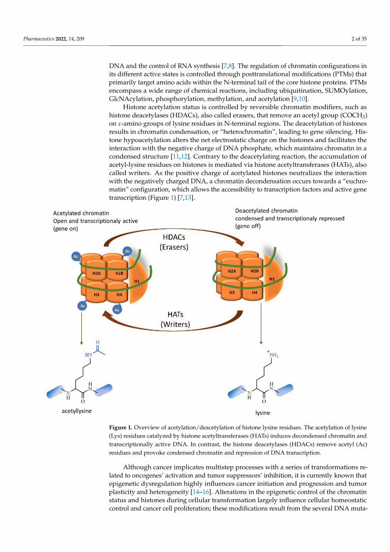
Histone acetylation status is controlled by reversible chromatin modifiers, such ashistone deacetylases (HDACs), also called erasers, that remove an acetyl group (COCH3)on ε-amino groups of lysine residues in N-terminal regions. The deacetylation of histonesresults in chromatin condensation, or “heterochromatin”, leading to gene silencing. His-tone hypoacetylation alters the net electrostatic charge on the histones and facilitates theinteraction with the negative charge of DNA phosphate, which maintains chromatin in acondensed structure [11,12]. Contrary to the deacetylating reaction, the accumulation ofacetyl-lysine residues on histones is mediated via histone acetyltransferases (HATs), alsocalled writers. As the positive charge of acetylated histones neutralizes the interactionwith the negatively charged DNA, a chromatin decondensation occurs towards a “euchro-matin” configuration, which allows the accessibility to transcription factors and active genetranscription (Figure 1) [7,13].
Pharmaceutics 2022, 14, 209 2 of 36
tion of chromatin or open configuration permits the access of regulatory transcription fac-tors to DNA and the control of RNA synthesis [7,8]. The regulation of chromatin configu-rations in its different active states is controlled through posttranslational modifications (PTMs) that primarily target amino acids within the N-terminal tail of the core histone proteins. PTMs encompass a wide range of chemical reactions, including ubiquitination, SUMOylation, GlcNAcylation, phosphorylation, methylation, and acetylation [9,10].
Histone acetylation status is controlled by reversible chromatin modifiers, such as histone deacetylases (HDACs), also called erasers, that remove an acetyl group (COCH3) on ε-amino groups of lysine residues in N-terminal regions. The deacetylation of histones results in chromatin condensation, or “heterochromatin”, leading to gene silencing. His-tone hypoacetylation alters the net electrostatic charge on the histones and facilitates the interaction with the negative charge of DNA phosphate, which maintains chromatin in a condensed structure [11,12]. Contrary to the deacetylating reaction, the accumulation of acetyl-lysine residues on histones is mediated via histone acetyltransferases (HATs), also called writers. As the positive charge of acetylated histones neutralizes the interaction with the negatively charged DNA, a chromatin decondensation occurs towards a “euchro-matin” configuration, which allows the accessibility to transcription factors and active gene transcription (Figure 1) [7,13].
Figure 1. Overview of acetylation/deacetylation of histone lysine residues. The acetylation of lysine (Lys) residues catalyzed by histone acetyltransferases (HATs) induces decondensed chromatin and transcriptionally active DNA. In contrast, the histone deacetylases (HDACs) remove acetyl (Ac) res-idues and provoke condensed chromatin and repression of DNA transcription.
Although cancer implicates multistep processes with a series of transformations re-lated to oncogenes’ activation and tumor suppressors’ inhibition, it is currently known that epigenetic dysregulation highly influences cancer initiation and progression and tu-mor plasticity and heterogeneity [14–16]. Alterations in the epigenetic control of the chro-
Figure 1. Overview of acetylation/deacetylation of histone lysine residues. The acetylation of lysine(Lys) residues catalyzed by histone acetyltransferases (HATs) induces decondensed chromatin andtranscriptionally active DNA. In contrast, the histone deacetylases (HDACs) remove acetyl (Ac)residues and provoke condensed chromatin and repression of DNA transcription.
Although cancer implicates multistep processes with a series of transformations re-lated to oncogenes’ activation and tumor suppressors’ inhibition, it is currently known thatepigenetic dysregulation highly influences cancer initiation and progression and tumorplasticity and heterogeneity [14–16]. Alterations in the epigenetic control of the chromatinstatus and histones during cellular transformation largely influence cellular homeostaticcontrol and cancer cell proliferation; these modifications result from the several DNA muta-

Pharmaceutics 2022, 14, 209 3 of 35
genic events occurring during tumor progression [17,18]. Among epigenetic dysregulationevents in cancer progression, dysfunctions of the HDACs and the related modifications ofthe protein acetylation levels highly influence cancer cells’ malignancy [19]. In this sense,HDACs, due to their importance in epigenetic abnormalities, have been developed asimportant therapeutic targets to control cancer progression and malignancy.
Aberrant HDAC expressions are involved in multiple different stages of cancer andbecome one of the hallmarks in hematological malignancies and solid tumors [20–22].Additionally, the increased expression of HDACs may be associated with poor outcomesand advanced disease in cancer patients, such as gastric, ovarian, neuroblastoma cancer,and multiple myeloma, among others [22]. As crucial players in cancer, HDACs areinvolved in the regulation of several cellular and molecular events (Figure 2). HDACsinfluence cell cycle and cellular proliferation, and HDAC inhibition induces cell cycle arrestat the G1 phase by reducing the expression of cyclins and cyclin-dependent kinases (CDK)or inducing the expression of CDK inhibitors [23,24]; HDACs inhibition also regulatescancer cell apoptosis via the regulation of the expression of pro- and antiapoptotic factors,such as cell surface death receptors and/or ligands including FAS/APO1-FASL, TNF-TNFreceptors and TRAIL-TRAIL receptors, reduction in the expression levels of cytoplasmicFLICE-like inhibitory protein (c-FLIP), Bax, and Bcl2 family members [25,26]. Likewise,DNA-damage repair (DDR) is critically regulated by HDACs due to its role in modulatingchromatin reorganization, maintaining the dynamic equilibrium of acetylation of DNA-damage repair proteins, as well as influencing almost all events in DNA repair such asbase excision repair, nucleotide excision repair, and mismatch repair [27]; Autophagy, withdemonstrated involvement in the development, maintenance, and progression of cancer,is susceptible to regulation by HDACs. In cancer cells, HDACs deacetylate cytoplasmicproteins involved in the regulation of crucial autophagy proteins such as LC3-II and Beclin1,as well as the intracellular signaling proteins mTor, apoptosis-inducing factor (AIF), andp53 [28–33]. Epithelial-to-mesenchymal transition (EMT) is a crucial process in cancercell invasion and metastasis. The EMT implicates an extensive range of the silencing ofepithelial genes and the activation of mesenchymal genes via transcriptional mechanismsthat directly implicate histone modifications. For instance, transcriptional repressors of theE-cadherin (CDH1), such as Snail or ZEB1, which recruit HDACs to CDH1 promoter forhistone deacetylation, result in gene silencing and potentiate the EMT [34,35]. As tumormass reaches 1–2 mm in size, the angiogenic process is activated for the supply of oxygenand nutrients and the disposal of carbon dioxide and waste [36]. Angiogenesis is tightlyregulated by the hypoxic microenvironment and the transcription factor hypoxia-induciblefactors-1 α (HIF-1α). HDACs either directly deacetylate HIF-1α and inhibit the proteindegradation or indirectly facilitate HIF-1α stabilization by deacetylating its chaperones,HSP70 and HSP90 [37,38].
This review will focus on the first generation of classic small-molecule and dietary-derived HDAC inhibitors with demonstrated anticancer and chemopreventive activities.Additionally, we will discuss some perspectives of addressing new ways of improving theHDACi selectivity and the combinatory potential of HDACi, and other specific targets thatcould be included in future clinical applications.

Pharmaceutics 2022, 14, 209 4 of 35Pharmaceutics 2022, 14, 209 4 of 36
Figure 2. The hallmarks of histone deacetylases in cancer biology. HDACs enzymes mediate cancer cell malignancy by promoting cell proliferation, autophagy, DNA-damage repair, and tumor angi-ogenesis while inhibiting apoptosis. Furthermore, HDACs contribute to the expression of metastatic phenotypes by triggering the epithelial to the mesenchymal transition program. Arrows indicate stimulation, and the bar represents inhibition.
2. Histone Deacetylases Overview The discovery of this first mammalian histone deacetylase (HDAC1) marked the be-
ginning of the HDACs family. HDAC research dates from 1964, when a relationship be-tween the acetylation status of histones was linked to RNA synthesis, followed by the description of a putative enzyme with histone deacetylase activity in 1969 [39,40]. None-theless, the first HDAC was discovered in 1999 using an affinity matrix based on the co-valent HDAC inhibitor trapoxin B. The sequencing of an isolated 46 kDa protein with deacetylase enzymatic activity revealed a resemblance with the yeast transcriptional reg-ulator encoded by the RPD3 gene [41].
Eleven HDAC isoforms have been described and grouped into four classes based on their homology with yeast deacetylases (Table 1) [42,43]. Class I encompasses HDAC1, 2, 3, and 8 with sequence homology to yeast deacetylase reduced potassium dependency-3 (Rpd3), which are mainly found in the nucleus and are part of multiprotein complexes that are included in the epigenetic control of gene expression, cell proliferation, and sur-vival. Meanwhile, class II comprises HDAC4, 5, 6, 7, 9, and 10, sharing sequence homolo-gies with the yeast histone deacetylases-1 (Hda1). HDAC4, 5, 7, and 9 are classified into a class IIa group and are found shuttling between the nucleus and cytoplasm. HDACs 6 and 10 are categorized as class IIb due to a second catalytic domain and are primarily found in the cytoplasm. Moreover, class IIa enzymes form multiprotein complexes with rela-tively weak deacetylase activity, while class IIb HDACs are active deacetylases. Class IV includes a unique and the smallest member, HDAC11 isoform, mainly found in the nu-cleus, which shares sequence identity with Rpd3 and Hda1 yeast deacetylases [12,44–47]. Additionally, these 11 HDACs are characterized by a Zn2+ in their active sites, which is fundamental for hydrolase activity, and known as the zinc-dependent “classical” HDACs [48]. The class III group encompasses seven NAD+-dependent histone deacetylase Sirtuins that are not in the scope of this review [42].
Figure 2. The hallmarks of histone deacetylases in cancer biology. HDACs enzymes mediate cancercell malignancy by promoting cell proliferation, autophagy, DNA-damage repair, and tumor angio-genesis while inhibiting apoptosis. Furthermore, HDACs contribute to the expression of metastaticphenotypes by triggering the epithelial to the mesenchymal transition program. Arrows indicatestimulation, and the bar represents inhibition.
2. Histone Deacetylases Overview
The discovery of this first mammalian histone deacetylase (HDAC1) marked thebeginning of the HDACs family. HDAC research dates from 1964, when a relationshipbetween the acetylation status of histones was linked to RNA synthesis, followed bythe description of a putative enzyme with histone deacetylase activity in 1969 [39,40].Nonetheless, the first HDAC was discovered in 1999 using an affinity matrix based onthe covalent HDAC inhibitor trapoxin B. The sequencing of an isolated 46 kDa proteinwith deacetylase enzymatic activity revealed a resemblance with the yeast transcriptionalregulator encoded by the RPD3 gene [41].
Eleven HDAC isoforms have been described and grouped into four classes based ontheir homology with yeast deacetylases (Table 1) [42,43]. Class I encompasses HDAC1, 2,3, and 8 with sequence homology to yeast deacetylase reduced potassium dependency-3(Rpd3), which are mainly found in the nucleus and are part of multiprotein complexes thatare included in the epigenetic control of gene expression, cell proliferation, and survival.Meanwhile, class II comprises HDAC4, 5, 6, 7, 9, and 10, sharing sequence homologies withthe yeast histone deacetylases-1 (Hda1). HDAC4, 5, 7, and 9 are classified into a class IIagroup and are found shuttling between the nucleus and cytoplasm. HDACs 6 and 10 arecategorized as class IIb due to a second catalytic domain and are primarily found in thecytoplasm. Moreover, class IIa enzymes form multiprotein complexes with relatively weakdeacetylase activity, while class IIb HDACs are active deacetylases. Class IV includes aunique and the smallest member, HDAC11 isoform, mainly found in the nucleus, whichshares sequence identity with Rpd3 and Hda1 yeast deacetylases [12,44–47]. Additionally,these 11 HDACs are characterized by a Zn2+ in their active sites, which is fundamental forhydrolase activity, and known as the zinc-dependent “classical” HDACs [48]. The class IIIgroup encompasses seven NAD+-dependent histone deacetylase Sirtuins that are not in thescope of this review [42].

Pharmaceutics 2022, 14, 209 5 of 35
Table 1. Main cellular localization and basic molecular features of HDACs are indicated.
Histone Deacetylases
Class Member CellularLocalization
ChromosomePosition
AminoacidsNo
(MolecularWeight, kD)
Basic Structure
I
HDAC1
Nucleus
1p35-p35.1 483 (51)
Pharmaceutics 2022, 14, 209 5 of 36
Table 1. Main cellular localization and basic molecular features of HDACs are indicated.
Histone Deacetylases
Class Member Cellular Localization
Chromosome Position
Aminoacids No
(Molecular Weight, kD)
Basic Structure
I HDAC1 Nucleus 1p35-p35.1 483 (51)
HDAC2 6q21 488 (55)
HDAC3 5q31.3 428 (49)
HDAC8 Xq13.1 377 (42)
IIa HDAC4 Nucleus/
cytoplasm
2q37.3 1084 (119)
HDAC5 17q21.31 1122 (122)
HDAC7 12q13.11 912 (103)
HDAC9 7p21 1069 (118)
IIb HDAC6 Cytoplasm Xp11.23 1215 (131)
HDAC10 2q13.33 669 (71)
IV HDAC11 Nucleus 3p25.1 343 (39)
N and C terminal regions, Catalytic domains, Nuclear localization sequence, Nu-clear export sequence, cytoplasmic anchoring motif, Zinc finger motif.
HDAcs are promising targets for cancer chemoprevention and chemotherapy de-pending on their involvement in gene expression and the variety of cellular functions in-volved in carcinogenesis, cell growth, survival, and homologous recombination [49–51]. Moreover, the manipulation of histone acetylation through HDAC inhibition has been proposed as a mechanism for derepressing genes dysregulated in chronic conditions such as neurodegenerative diseases, psychological disorders, and cancer [52,53].
3. Histone Deacetylases Inhibitors in the Treatment of Cancer For decades cancer has been defined as an autonomous disease governed by the mu-
tational activation of oncogenes and/or the inactivation of tumor suppressor genes, which triggers a sequence of molecular events that in time favor tumorigenesis [54]. Reasonably, chemotherapy’s primary and ultimate aim is to eliminate malignant cells and restore can-cer patients’ cellular and physiological homeostasis. Historically, chemotherapy is based on cytotoxic drugs to target the highly proliferating cancer cells potentially vulnerable due to the aberrant molecular mechanisms associated with malignant transformation [54]. Without a doubt, chemotherapy is one of the most significant advances to treat cancer
HDAC2 6q21 488 (55)
Pharmaceutics 2022, 14, 209 5 of 36
Table 1. Main cellular localization and basic molecular features of HDACs are indicated.
Histone Deacetylases
Class Member Cellular Localization
Chromosome Position
Aminoacids No
(Molecular Weight, kD)
Basic Structure
I HDAC1 Nucleus 1p35-p35.1 483 (51)
HDAC2 6q21 488 (55)
HDAC3 5q31.3 428 (49)
HDAC8 Xq13.1 377 (42)
IIa HDAC4 Nucleus/
cytoplasm
2q37.3 1084 (119)
HDAC5 17q21.31 1122 (122)
HDAC7 12q13.11 912 (103)
HDAC9 7p21 1069 (118)
IIb HDAC6 Cytoplasm Xp11.23 1215 (131)
HDAC10 2q13.33 669 (71)
IV HDAC11 Nucleus 3p25.1 343 (39)
N and C terminal regions, Catalytic domains, Nuclear localization sequence, Nu-clear export sequence, cytoplasmic anchoring motif, Zinc finger motif.
HDAcs are promising targets for cancer chemoprevention and chemotherapy de-pending on their involvement in gene expression and the variety of cellular functions in-volved in carcinogenesis, cell growth, survival, and homologous recombination [49–51]. Moreover, the manipulation of histone acetylation through HDAC inhibition has been proposed as a mechanism for derepressing genes dysregulated in chronic conditions such as neurodegenerative diseases, psychological disorders, and cancer [52,53].
3. Histone Deacetylases Inhibitors in the Treatment of Cancer For decades cancer has been defined as an autonomous disease governed by the mu-
tational activation of oncogenes and/or the inactivation of tumor suppressor genes, which triggers a sequence of molecular events that in time favor tumorigenesis [54]. Reasonably, chemotherapy’s primary and ultimate aim is to eliminate malignant cells and restore can-cer patients’ cellular and physiological homeostasis. Historically, chemotherapy is based on cytotoxic drugs to target the highly proliferating cancer cells potentially vulnerable due to the aberrant molecular mechanisms associated with malignant transformation [54]. Without a doubt, chemotherapy is one of the most significant advances to treat cancer
HDAC3 5q31.3 428 (49)
Pharmaceutics 2022, 14, 209 5 of 36
Table 1. Main cellular localization and basic molecular features of HDACs are indicated.
Histone Deacetylases
Class Member Cellular Localization
Chromosome Position
Aminoacids No
(Molecular Weight, kD)
Basic Structure
I HDAC1 Nucleus 1p35-p35.1 483 (51)
HDAC2 6q21 488 (55)
HDAC3 5q31.3 428 (49)
HDAC8 Xq13.1 377 (42)
IIa HDAC4 Nucleus/
cytoplasm
2q37.3 1084 (119)
HDAC5 17q21.31 1122 (122)
HDAC7 12q13.11 912 (103)
HDAC9 7p21 1069 (118)
IIb HDAC6 Cytoplasm Xp11.23 1215 (131)
HDAC10 2q13.33 669 (71)
IV HDAC11 Nucleus 3p25.1 343 (39)
N and C terminal regions, Catalytic domains, Nuclear localization sequence, Nu-clear export sequence, cytoplasmic anchoring motif, Zinc finger motif.
HDAcs are promising targets for cancer chemoprevention and chemotherapy de-pending on their involvement in gene expression and the variety of cellular functions in-volved in carcinogenesis, cell growth, survival, and homologous recombination [49–51]. Moreover, the manipulation of histone acetylation through HDAC inhibition has been proposed as a mechanism for derepressing genes dysregulated in chronic conditions such as neurodegenerative diseases, psychological disorders, and cancer [52,53].
3. Histone Deacetylases Inhibitors in the Treatment of Cancer For decades cancer has been defined as an autonomous disease governed by the mu-
tational activation of oncogenes and/or the inactivation of tumor suppressor genes, which triggers a sequence of molecular events that in time favor tumorigenesis [54]. Reasonably, chemotherapy’s primary and ultimate aim is to eliminate malignant cells and restore can-cer patients’ cellular and physiological homeostasis. Historically, chemotherapy is based on cytotoxic drugs to target the highly proliferating cancer cells potentially vulnerable due to the aberrant molecular mechanisms associated with malignant transformation [54]. Without a doubt, chemotherapy is one of the most significant advances to treat cancer
HDAC8 Xq13.1 377 (42)
Pharmaceutics 2022, 14, 209 5 of 36
Table 1. Main cellular localization and basic molecular features of HDACs are indicated.
Histone Deacetylases
Class Member Cellular Localization
Chromosome Position
Aminoacids No
(Molecular Weight, kD)
Basic Structure
I HDAC1 Nucleus 1p35-p35.1 483 (51)
HDAC2 6q21 488 (55)
HDAC3 5q31.3 428 (49)
HDAC8 Xq13.1 377 (42)
IIa HDAC4 Nucleus/
cytoplasm
2q37.3 1084 (119)
HDAC5 17q21.31 1122 (122)
HDAC7 12q13.11 912 (103)
HDAC9 7p21 1069 (118)
IIb HDAC6 Cytoplasm Xp11.23 1215 (131)
HDAC10 2q13.33 669 (71)
IV HDAC11 Nucleus 3p25.1 343 (39)
N and C terminal regions, Catalytic domains, Nuclear localization sequence, Nu-clear export sequence, cytoplasmic anchoring motif, Zinc finger motif.
HDAcs are promising targets for cancer chemoprevention and chemotherapy de-pending on their involvement in gene expression and the variety of cellular functions in-volved in carcinogenesis, cell growth, survival, and homologous recombination [49–51]. Moreover, the manipulation of histone acetylation through HDAC inhibition has been proposed as a mechanism for derepressing genes dysregulated in chronic conditions such as neurodegenerative diseases, psychological disorders, and cancer [52,53].
3. Histone Deacetylases Inhibitors in the Treatment of Cancer For decades cancer has been defined as an autonomous disease governed by the mu-
tational activation of oncogenes and/or the inactivation of tumor suppressor genes, which triggers a sequence of molecular events that in time favor tumorigenesis [54]. Reasonably, chemotherapy’s primary and ultimate aim is to eliminate malignant cells and restore can-cer patients’ cellular and physiological homeostasis. Historically, chemotherapy is based on cytotoxic drugs to target the highly proliferating cancer cells potentially vulnerable due to the aberrant molecular mechanisms associated with malignant transformation [54]. Without a doubt, chemotherapy is one of the most significant advances to treat cancer
IIa
HDAC4
Nucleus/cytoplasm
2q37.3 1084 (119)
Pharmaceutics 2022, 14, 209 5 of 36
Table 1. Main cellular localization and basic molecular features of HDACs are indicated.
Histone Deacetylases
Class Member Cellular Localization
Chromosome Position
Aminoacids No
(Molecular Weight, kD)
Basic Structure
I HDAC1 Nucleus 1p35-p35.1 483 (51)
HDAC2 6q21 488 (55)
HDAC3 5q31.3 428 (49)
HDAC8 Xq13.1 377 (42)
IIa HDAC4 Nucleus/
cytoplasm
2q37.3 1084 (119)
HDAC5 17q21.31 1122 (122)
HDAC7 12q13.11 912 (103)
HDAC9 7p21 1069 (118)
IIb HDAC6 Cytoplasm Xp11.23 1215 (131)
HDAC10 2q13.33 669 (71)
IV HDAC11 Nucleus 3p25.1 343 (39)
N and C terminal regions, Catalytic domains, Nuclear localization sequence, Nu-clear export sequence, cytoplasmic anchoring motif, Zinc finger motif.
HDAcs are promising targets for cancer chemoprevention and chemotherapy de-pending on their involvement in gene expression and the variety of cellular functions in-volved in carcinogenesis, cell growth, survival, and homologous recombination [49–51]. Moreover, the manipulation of histone acetylation through HDAC inhibition has been proposed as a mechanism for derepressing genes dysregulated in chronic conditions such as neurodegenerative diseases, psychological disorders, and cancer [52,53].
3. Histone Deacetylases Inhibitors in the Treatment of Cancer For decades cancer has been defined as an autonomous disease governed by the mu-
tational activation of oncogenes and/or the inactivation of tumor suppressor genes, which triggers a sequence of molecular events that in time favor tumorigenesis [54]. Reasonably, chemotherapy’s primary and ultimate aim is to eliminate malignant cells and restore can-cer patients’ cellular and physiological homeostasis. Historically, chemotherapy is based on cytotoxic drugs to target the highly proliferating cancer cells potentially vulnerable due to the aberrant molecular mechanisms associated with malignant transformation [54]. Without a doubt, chemotherapy is one of the most significant advances to treat cancer
HDAC5 17q21.31 1122 (122)
Pharmaceutics 2022, 14, 209 5 of 36
Table 1. Main cellular localization and basic molecular features of HDACs are indicated.
Histone Deacetylases
Class Member Cellular Localization
Chromosome Position
Aminoacids No
(Molecular Weight, kD)
Basic Structure
I HDAC1 Nucleus 1p35-p35.1 483 (51)
HDAC2 6q21 488 (55)
HDAC3 5q31.3 428 (49)
HDAC8 Xq13.1 377 (42)
IIa HDAC4 Nucleus/
cytoplasm
2q37.3 1084 (119)
HDAC5 17q21.31 1122 (122)
HDAC7 12q13.11 912 (103)
HDAC9 7p21 1069 (118)
IIb HDAC6 Cytoplasm Xp11.23 1215 (131)
HDAC10 2q13.33 669 (71)
IV HDAC11 Nucleus 3p25.1 343 (39)
N and C terminal regions, Catalytic domains, Nuclear localization sequence, Nu-clear export sequence, cytoplasmic anchoring motif, Zinc finger motif.
HDAcs are promising targets for cancer chemoprevention and chemotherapy de-pending on their involvement in gene expression and the variety of cellular functions in-volved in carcinogenesis, cell growth, survival, and homologous recombination [49–51]. Moreover, the manipulation of histone acetylation through HDAC inhibition has been proposed as a mechanism for derepressing genes dysregulated in chronic conditions such as neurodegenerative diseases, psychological disorders, and cancer [52,53].
3. Histone Deacetylases Inhibitors in the Treatment of Cancer For decades cancer has been defined as an autonomous disease governed by the mu-
tational activation of oncogenes and/or the inactivation of tumor suppressor genes, which triggers a sequence of molecular events that in time favor tumorigenesis [54]. Reasonably, chemotherapy’s primary and ultimate aim is to eliminate malignant cells and restore can-cer patients’ cellular and physiological homeostasis. Historically, chemotherapy is based on cytotoxic drugs to target the highly proliferating cancer cells potentially vulnerable due to the aberrant molecular mechanisms associated with malignant transformation [54]. Without a doubt, chemotherapy is one of the most significant advances to treat cancer
HDAC7 12q13.11 912 (103)
Pharmaceutics 2022, 14, 209 5 of 36
Table 1. Main cellular localization and basic molecular features of HDACs are indicated.
Histone Deacetylases
Class Member Cellular Localization
Chromosome Position
Aminoacids No
(Molecular Weight, kD)
Basic Structure
I HDAC1 Nucleus 1p35-p35.1 483 (51)
HDAC2 6q21 488 (55)
HDAC3 5q31.3 428 (49)
HDAC8 Xq13.1 377 (42)
IIa HDAC4 Nucleus/
cytoplasm
2q37.3 1084 (119)
HDAC5 17q21.31 1122 (122)
HDAC7 12q13.11 912 (103)
HDAC9 7p21 1069 (118)
IIb HDAC6 Cytoplasm Xp11.23 1215 (131)
HDAC10 2q13.33 669 (71)
IV HDAC11 Nucleus 3p25.1 343 (39)
N and C terminal regions, Catalytic domains, Nuclear localization sequence, Nu-clear export sequence, cytoplasmic anchoring motif, Zinc finger motif.
HDAcs are promising targets for cancer chemoprevention and chemotherapy de-pending on their involvement in gene expression and the variety of cellular functions in-volved in carcinogenesis, cell growth, survival, and homologous recombination [49–51]. Moreover, the manipulation of histone acetylation through HDAC inhibition has been proposed as a mechanism for derepressing genes dysregulated in chronic conditions such as neurodegenerative diseases, psychological disorders, and cancer [52,53].
3. Histone Deacetylases Inhibitors in the Treatment of Cancer For decades cancer has been defined as an autonomous disease governed by the mu-
tational activation of oncogenes and/or the inactivation of tumor suppressor genes, which triggers a sequence of molecular events that in time favor tumorigenesis [54]. Reasonably, chemotherapy’s primary and ultimate aim is to eliminate malignant cells and restore can-cer patients’ cellular and physiological homeostasis. Historically, chemotherapy is based on cytotoxic drugs to target the highly proliferating cancer cells potentially vulnerable due to the aberrant molecular mechanisms associated with malignant transformation [54]. Without a doubt, chemotherapy is one of the most significant advances to treat cancer
HDAC9 7p21 1069 (118)
Pharmaceutics 2022, 14, 209 5 of 36
Table 1. Main cellular localization and basic molecular features of HDACs are indicated.
Histone Deacetylases
Class Member Cellular Localization
Chromosome Position
Aminoacids No
(Molecular Weight, kD)
Basic Structure
I HDAC1 Nucleus 1p35-p35.1 483 (51)
HDAC2 6q21 488 (55)
HDAC3 5q31.3 428 (49)
HDAC8 Xq13.1 377 (42)
IIa HDAC4 Nucleus/
cytoplasm
2q37.3 1084 (119)
HDAC5 17q21.31 1122 (122)
HDAC7 12q13.11 912 (103)
HDAC9 7p21 1069 (118)
IIb HDAC6 Cytoplasm Xp11.23 1215 (131)
HDAC10 2q13.33 669 (71)
IV HDAC11 Nucleus 3p25.1 343 (39)
N and C terminal regions, Catalytic domains, Nuclear localization sequence, Nu-clear export sequence, cytoplasmic anchoring motif, Zinc finger motif.
HDAcs are promising targets for cancer chemoprevention and chemotherapy de-pending on their involvement in gene expression and the variety of cellular functions in-volved in carcinogenesis, cell growth, survival, and homologous recombination [49–51]. Moreover, the manipulation of histone acetylation through HDAC inhibition has been proposed as a mechanism for derepressing genes dysregulated in chronic conditions such as neurodegenerative diseases, psychological disorders, and cancer [52,53].
3. Histone Deacetylases Inhibitors in the Treatment of Cancer For decades cancer has been defined as an autonomous disease governed by the mu-
tational activation of oncogenes and/or the inactivation of tumor suppressor genes, which triggers a sequence of molecular events that in time favor tumorigenesis [54]. Reasonably, chemotherapy’s primary and ultimate aim is to eliminate malignant cells and restore can-cer patients’ cellular and physiological homeostasis. Historically, chemotherapy is based on cytotoxic drugs to target the highly proliferating cancer cells potentially vulnerable due to the aberrant molecular mechanisms associated with malignant transformation [54]. Without a doubt, chemotherapy is one of the most significant advances to treat cancer
IIbHDAC6
CytoplasmXp11.23 1215 (131)
Pharmaceutics 2022, 14, 209 5 of 36
Table 1. Main cellular localization and basic molecular features of HDACs are indicated.
Histone Deacetylases
Class Member Cellular Localization
Chromosome Position
Aminoacids No
(Molecular Weight, kD)
Basic Structure
I HDAC1 Nucleus 1p35-p35.1 483 (51)
HDAC2 6q21 488 (55)
HDAC3 5q31.3 428 (49)
HDAC8 Xq13.1 377 (42)
IIa HDAC4 Nucleus/
cytoplasm
2q37.3 1084 (119)
HDAC5 17q21.31 1122 (122)
HDAC7 12q13.11 912 (103)
HDAC9 7p21 1069 (118)
IIb HDAC6 Cytoplasm Xp11.23 1215 (131)
HDAC10 2q13.33 669 (71)
IV HDAC11 Nucleus 3p25.1 343 (39)
N and C terminal regions, Catalytic domains, Nuclear localization sequence, Nu-clear export sequence, cytoplasmic anchoring motif, Zinc finger motif.
HDAcs are promising targets for cancer chemoprevention and chemotherapy de-pending on their involvement in gene expression and the variety of cellular functions in-volved in carcinogenesis, cell growth, survival, and homologous recombination [49–51]. Moreover, the manipulation of histone acetylation through HDAC inhibition has been proposed as a mechanism for derepressing genes dysregulated in chronic conditions such as neurodegenerative diseases, psychological disorders, and cancer [52,53].
3. Histone Deacetylases Inhibitors in the Treatment of Cancer For decades cancer has been defined as an autonomous disease governed by the mu-
tational activation of oncogenes and/or the inactivation of tumor suppressor genes, which triggers a sequence of molecular events that in time favor tumorigenesis [54]. Reasonably, chemotherapy’s primary and ultimate aim is to eliminate malignant cells and restore can-cer patients’ cellular and physiological homeostasis. Historically, chemotherapy is based on cytotoxic drugs to target the highly proliferating cancer cells potentially vulnerable due to the aberrant molecular mechanisms associated with malignant transformation [54]. Without a doubt, chemotherapy is one of the most significant advances to treat cancer
HDAC10 2q13.33 669 (71)
Pharmaceutics 2022, 14, 209 5 of 36
Table 1. Main cellular localization and basic molecular features of HDACs are indicated.
Histone Deacetylases
Class Member Cellular Localization
Chromosome Position
Aminoacids No
(Molecular Weight, kD)
Basic Structure
I HDAC1 Nucleus 1p35-p35.1 483 (51)
HDAC2 6q21 488 (55)
HDAC3 5q31.3 428 (49)
HDAC8 Xq13.1 377 (42)
IIa HDAC4 Nucleus/
cytoplasm
2q37.3 1084 (119)
HDAC5 17q21.31 1122 (122)
HDAC7 12q13.11 912 (103)
HDAC9 7p21 1069 (118)
IIb HDAC6 Cytoplasm Xp11.23 1215 (131)
HDAC10 2q13.33 669 (71)
IV HDAC11 Nucleus 3p25.1 343 (39)
N and C terminal regions, Catalytic domains, Nuclear localization sequence, Nu-clear export sequence, cytoplasmic anchoring motif, Zinc finger motif.
HDAcs are promising targets for cancer chemoprevention and chemotherapy de-pending on their involvement in gene expression and the variety of cellular functions in-volved in carcinogenesis, cell growth, survival, and homologous recombination [49–51]. Moreover, the manipulation of histone acetylation through HDAC inhibition has been proposed as a mechanism for derepressing genes dysregulated in chronic conditions such as neurodegenerative diseases, psychological disorders, and cancer [52,53].
3. Histone Deacetylases Inhibitors in the Treatment of Cancer For decades cancer has been defined as an autonomous disease governed by the mu-
tational activation of oncogenes and/or the inactivation of tumor suppressor genes, which triggers a sequence of molecular events that in time favor tumorigenesis [54]. Reasonably, chemotherapy’s primary and ultimate aim is to eliminate malignant cells and restore can-cer patients’ cellular and physiological homeostasis. Historically, chemotherapy is based on cytotoxic drugs to target the highly proliferating cancer cells potentially vulnerable due to the aberrant molecular mechanisms associated with malignant transformation [54]. Without a doubt, chemotherapy is one of the most significant advances to treat cancer
IV HDAC11 Nucleus 3p25.1 343 (39)
Pharmaceutics 2022, 14, 209 5 of 36
Table 1. Main cellular localization and basic molecular features of HDACs are indicated.
Histone Deacetylases
Class Member Cellular Localization
Chromosome Position
Aminoacids No
(Molecular Weight, kD)
Basic Structure
I HDAC1 Nucleus 1p35-p35.1 483 (51)
HDAC2 6q21 488 (55)
HDAC3 5q31.3 428 (49)
HDAC8 Xq13.1 377 (42)
IIa HDAC4 Nucleus/
cytoplasm
2q37.3 1084 (119)
HDAC5 17q21.31 1122 (122)
HDAC7 12q13.11 912 (103)
HDAC9 7p21 1069 (118)
IIb HDAC6 Cytoplasm Xp11.23 1215 (131)
HDAC10 2q13.33 669 (71)
IV HDAC11 Nucleus 3p25.1 343 (39)
N and C terminal regions, Catalytic domains, Nuclear localization sequence, Nu-clear export sequence, cytoplasmic anchoring motif, Zinc finger motif.
HDAcs are promising targets for cancer chemoprevention and chemotherapy de-pending on their involvement in gene expression and the variety of cellular functions in-volved in carcinogenesis, cell growth, survival, and homologous recombination [49–51]. Moreover, the manipulation of histone acetylation through HDAC inhibition has been proposed as a mechanism for derepressing genes dysregulated in chronic conditions such as neurodegenerative diseases, psychological disorders, and cancer [52,53].
3. Histone Deacetylases Inhibitors in the Treatment of Cancer For decades cancer has been defined as an autonomous disease governed by the mu-
tational activation of oncogenes and/or the inactivation of tumor suppressor genes, which triggers a sequence of molecular events that in time favor tumorigenesis [54]. Reasonably, chemotherapy’s primary and ultimate aim is to eliminate malignant cells and restore can-cer patients’ cellular and physiological homeostasis. Historically, chemotherapy is based on cytotoxic drugs to target the highly proliferating cancer cells potentially vulnerable due to the aberrant molecular mechanisms associated with malignant transformation [54]. Without a doubt, chemotherapy is one of the most significant advances to treat cancer
N and C terminal regions,
Pharmaceutics 2022, 14, 209 5 of 37
Table 1. Main cellular localization and basic molecular features of HDACs are indicated.
Histone Deacetylases
Class Member Cellular
Localization
Chromosome
Position
Aminoacids
No
(Molecular
Weight, kD)
Basic Structure
I HDAC1 Nucleus 1p35-p35.1 483 (51)
HDAC2 6q21 488 (55)
HDAC3 5q31.3 428 (49)
HDAC8 Xq13.1 377 (42)
IIa HDAC4 Nucleus/
cytoplasm
2q37.3 1084 (119)
HDAC5 17q21.31 1122 (122)
HDAC7 12q13.11 912 (103)
HDAC9 7p21 1069 (118)
IIb HDAC6 Cytoplasm Xp11.23 1215 (131)
HDAC10 2q13.33 669 (71)
IV HDAC11 Nucleus 3p25.1 343 (39)
N and C terminal regions, Catalytic domains, Nuclear localization sequence,
Nuclear export sequenc
e, cytoplasmic anchoring motif, Zinc finger motif.
HDAcs are promising targets for cancer chemoprevention and chemotherapy de-
pending on their involvement in gene expression and the variety of cellular functions in-
volved in carcinogenesis, cell growth, survival, and homologous recombination [49–51].
Moreover, the manipulation of histone acetylation through HDAC inhibition has been
proposed as a mechanism for derepressing genes dysregulated in chronic conditions such
as neurodegenerative diseases, psychological disorders, and cancer [52,53].
3. Histone Deacetylases Inhibitors in the Treatment of Cancer
Catalytic domains,
Pharmaceutics 2022, 14, 209 5 of 36
Table 1. Main cellular localization and basic molecular features of HDACs are indicated.
Histone Deacetylases
Class Member Cellular Localization
Chromosome Position
Aminoacids No
(Molecular Weight, kD)
Basic Structure
I HDAC1 Nucleus 1p35-p35.1 483 (51)
HDAC2 6q21 488 (55)
HDAC3 5q31.3 428 (49)
HDAC8 Xq13.1 377 (42)
IIa HDAC4 Nucleus/
cytoplasm
2q37.3 1084 (119)
HDAC5 17q21.31 1122 (122)
HDAC7 12q13.11 912 (103)
HDAC9 7p21 1069 (118)
IIb HDAC6 Cytoplasm Xp11.23 1215 (131)
HDAC10 2q13.33 669 (71)
IV HDAC11 Nucleus 3p25.1 343 (39)
N and C terminal regions, Catalytic domains, Nuclear localization sequence, Nu-clear export sequence, cytoplasmic anchoring motif, Zinc finger motif.
HDAcs are promising targets for cancer chemoprevention and chemotherapy de-pending on their involvement in gene expression and the variety of cellular functions in-volved in carcinogenesis, cell growth, survival, and homologous recombination [49–51]. Moreover, the manipulation of histone acetylation through HDAC inhibition has been proposed as a mechanism for derepressing genes dysregulated in chronic conditions such as neurodegenerative diseases, psychological disorders, and cancer [52,53].
3. Histone Deacetylases Inhibitors in the Treatment of Cancer For decades cancer has been defined as an autonomous disease governed by the mu-
tational activation of oncogenes and/or the inactivation of tumor suppressor genes, which triggers a sequence of molecular events that in time favor tumorigenesis [54]. Reasonably, chemotherapy’s primary and ultimate aim is to eliminate malignant cells and restore can-cer patients’ cellular and physiological homeostasis. Historically, chemotherapy is based on cytotoxic drugs to target the highly proliferating cancer cells potentially vulnerable due to the aberrant molecular mechanisms associated with malignant transformation [54]. Without a doubt, chemotherapy is one of the most significant advances to treat cancer
Nuclear localization sequence,
Pharmaceutics 2022, 14, 209 5 of 36
Table 1. Main cellular localization and basic molecular features of HDACs are indicated.
Histone Deacetylases
Class Member Cellular Localization
Chromosome Position
Aminoacids No
(Molecular Weight, kD)
Basic Structure
I HDAC1 Nucleus 1p35-p35.1 483 (51)
HDAC2 6q21 488 (55)
HDAC3 5q31.3 428 (49)
HDAC8 Xq13.1 377 (42)
IIa HDAC4 Nucleus/
cytoplasm
2q37.3 1084 (119)
HDAC5 17q21.31 1122 (122)
HDAC7 12q13.11 912 (103)
HDAC9 7p21 1069 (118)
IIb HDAC6 Cytoplasm Xp11.23 1215 (131)
HDAC10 2q13.33 669 (71)
IV HDAC11 Nucleus 3p25.1 343 (39)
N and C terminal regions, Catalytic domains, Nuclear localization sequence, Nu-clear export sequence, cytoplasmic anchoring motif, Zinc finger motif.
HDAcs are promising targets for cancer chemoprevention and chemotherapy de-pending on their involvement in gene expression and the variety of cellular functions in-volved in carcinogenesis, cell growth, survival, and homologous recombination [49–51]. Moreover, the manipulation of histone acetylation through HDAC inhibition has been proposed as a mechanism for derepressing genes dysregulated in chronic conditions such as neurodegenerative diseases, psychological disorders, and cancer [52,53].
3. Histone Deacetylases Inhibitors in the Treatment of Cancer For decades cancer has been defined as an autonomous disease governed by the mu-
tational activation of oncogenes and/or the inactivation of tumor suppressor genes, which triggers a sequence of molecular events that in time favor tumorigenesis [54]. Reasonably, chemotherapy’s primary and ultimate aim is to eliminate malignant cells and restore can-cer patients’ cellular and physiological homeostasis. Historically, chemotherapy is based on cytotoxic drugs to target the highly proliferating cancer cells potentially vulnerable due to the aberrant molecular mechanisms associated with malignant transformation [54]. Without a doubt, chemotherapy is one of the most significant advances to treat cancer
Nuclear export
sequence,
Pharmaceutics 2022, 14, 209 5 of 37
Table 1. Main cellular localization and basic molecular features of HDACs are indicated.
Histone Deacetylases
Class Member Cellular
Localization
Chromosome
Position
Aminoacids
No
(Molecular
Weight, kD)
Basic Structure
I HDAC1 Nucleus 1p35-p35.1 483 (51)
HDAC2 6q21 488 (55)
HDAC3 5q31.3 428 (49)
HDAC8 Xq13.1 377 (42)
IIa HDAC4 Nucleus/
cytoplasm
2q37.3 1084 (119)
HDAC5 17q21.31 1122 (122)
HDAC7 12q13.11 912 (103)
HDAC9 7p21 1069 (118)
IIb HDAC6 Cytoplasm Xp11.23 1215 (131)
HDAC10 2q13.33 669 (71)
IV HDAC11 Nucleus 3p25.1 343 (39)
N and C terminal regions, Catalytic domains, Nuclear localization sequence,
Nuclear export sequenc
e, cytoplasmic anchoring motif, Zinc finger motif.
HDAcs are promising targets for cancer chemoprevention and chemotherapy de-
pending on their involvement in gene expression and the variety of cellular functions in-
volved in carcinogenesis, cell growth, survival, and homologous recombination [49–51].
Moreover, the manipulation of histone acetylation through HDAC inhibition has been
proposed as a mechanism for derepressing genes dysregulated in chronic conditions such
as neurodegenerative diseases, psychological disorders, and cancer [52,53].
3. Histone Deacetylases Inhibitors in the Treatment of Cancer
cytoplasmic anchoring motif,
Pharmaceutics 2022, 14, 209 5 of 36
Table 1. Main cellular localization and basic molecular features of HDACs are indicated.
Histone Deacetylases
Class Member Cellular Localization
Chromosome Position
Aminoacids No
(Molecular Weight, kD)
Basic Structure
I HDAC1 Nucleus 1p35-p35.1 483 (51)
HDAC2 6q21 488 (55)
HDAC3 5q31.3 428 (49)
HDAC8 Xq13.1 377 (42)
IIa HDAC4 Nucleus/
cytoplasm
2q37.3 1084 (119)
HDAC5 17q21.31 1122 (122)
HDAC7 12q13.11 912 (103)
HDAC9 7p21 1069 (118)
IIb HDAC6 Cytoplasm Xp11.23 1215 (131)
HDAC10 2q13.33 669 (71)
IV HDAC11 Nucleus 3p25.1 343 (39)
N and C terminal regions, Catalytic domains, Nuclear localization sequence, Nu-clear export sequence, cytoplasmic anchoring motif, Zinc finger motif.
HDAcs are promising targets for cancer chemoprevention and chemotherapy de-pending on their involvement in gene expression and the variety of cellular functions in-volved in carcinogenesis, cell growth, survival, and homologous recombination [49–51]. Moreover, the manipulation of histone acetylation through HDAC inhibition has been proposed as a mechanism for derepressing genes dysregulated in chronic conditions such as neurodegenerative diseases, psychological disorders, and cancer [52,53].
3. Histone Deacetylases Inhibitors in the Treatment of Cancer For decades cancer has been defined as an autonomous disease governed by the mu-
tational activation of oncogenes and/or the inactivation of tumor suppressor genes, which triggers a sequence of molecular events that in time favor tumorigenesis [54]. Reasonably, chemotherapy’s primary and ultimate aim is to eliminate malignant cells and restore can-cer patients’ cellular and physiological homeostasis. Historically, chemotherapy is based on cytotoxic drugs to target the highly proliferating cancer cells potentially vulnerable due to the aberrant molecular mechanisms associated with malignant transformation [54]. Without a doubt, chemotherapy is one of the most significant advances to treat cancer
Zinc finger motif.
HDAcs are promising targets for cancer chemoprevention and chemotherapy de-pending on their involvement in gene expression and the variety of cellular functionsinvolved in carcinogenesis, cell growth, survival, and homologous recombination [49–51].Moreover, the manipulation of histone acetylation through HDAC inhibition has beenproposed as a mechanism for derepressing genes dysregulated in chronic conditions suchas neurodegenerative diseases, psychological disorders, and cancer [52,53].
3. Histone Deacetylases Inhibitors in the Treatment of Cancer
For decades cancer has been defined as an autonomous disease governed by the muta-tional activation of oncogenes and/or the inactivation of tumor suppressor genes, whichtriggers a sequence of molecular events that in time favor tumorigenesis [54]. Reasonably,chemotherapy’s primary and ultimate aim is to eliminate malignant cells and restore cancerpatients’ cellular and physiological homeostasis. Historically, chemotherapy is based oncytotoxic drugs to target the highly proliferating cancer cells potentially vulnerable due tothe aberrant molecular mechanisms associated with malignant transformation [54]. With-out a doubt, chemotherapy is one of the most significant advances to treat cancer patients.It exerts unpredictable or undesirable toxic effects that target normal cells, partly becausetherapeutic drugs do not discriminate between rapidly dividing cancer cells from highlyproliferative non-malignant cells. Many efforts have been made to overcome the lack ofspecificity of anticancer drugs; in this sense, in recent years, the concept of targeted therapyor precision oncology has been developed to design therapeutic molecules targeting thepotential cancer-specific molecular and cellular signals. Therefore, the new era of cancertherapies considers mapping crucial genes and signaling pathway components and thedevelopment of ‘smart’ drugs that surgically target cancer cells, reducing the nonspecificeffects on normal cells [55–57].

Pharmaceutics 2022, 14, 209 6 of 35
In cancer, epigenetic events crucially contribute to tumor development and malignantprogression. Tumorigenesis implicates multiple events beyond genetic alterations, suchas epigenetic regulation that influences gene expression and protein activities, makingepigenetic targeting an attractive therapeutic strategy to fight cancer [55,58].
As mentioned above, HDAC inhibitors promote proliferation arrest and differentiation,trigger the cell death program, and control the tumor angiogenesis immune responses nec-essary for tumor growth and spreading and escape from immune surveillance. In this sense,the “epigenetic vulnerability of cancer cells” hypothesis has been coined; consequently,HDACs could be essential for the expression of crucial genes implicated in the survivaland growth of transformed cells, providing a relative specificity of HDACs inhibition asopposed to normal cells [59,60]. The rationality of the use of drugs considers that epigeneticalterations are reversible; thereby, anti-epigenetic compounds may in some way recover thenon-transformed scenery. Meanwhile, chromatin regulators are susceptible to oncogenicmutations, offering attractive molecular targets for cancer treatment [60,61]. The followingsections will address the main HDACi according to their chemical structure and impact oncancer treatment.
3.1. Histone Deacetylase Inhibitors
Besides mediating the acetylation status of various proteins, the zinc-dependentHDACs possess a mostly conserved catalytic active site [62,63]. To effectively inhibit thehydrolysis of acetyl-lysine residues on the histone tails and non-histone proteins, HDACidrugs should possess a cap group, aliphatic or aromatic linker, and a zinc-binding group(ZBG), as a part of a generic (classical) pharmacophore model (Figure 3) [64,65]. The ZBGincludes carboxylic acid, hydroxamic acid, 2-aminobenzamide, thiol, and others, and it iscrucial for designing an HDACi. The zinc-chelating group is inserted with the active site ofthe HDACs and mainly by bidentate mode interacts with and sequesters the zinc ion. Whenpositioned in the substrate-binding tunnel, the linker domain mimics the lysine residueand establishes hydrophobic interactions with the amino acid residues in the crevice of theHDAC active site.
Furthermore, the cap is usually built from a heterocyclic or carbocyclic group thatinteracts with the aromatic amino acid residues located either close to the outer domain ofthe active site or at the external surface of the enzyme [64,66]. This cap group is susceptibleto chemical modifications to enhance the selectivity of the HDACi. Nevertheless, recentlyextended pharmacophore models have been proposed that can include the majority ofcurrently known HDAC inhibitors and may help classify and design new selective HDACinhibitors [67,68].
Chemically, HDACi can be classified according to their ZBG (Table 2): aliphatic (short-chain fatty) acids; hydroxamic acids; ortho-aminoanilides, also called benzamides; thiolsand cyclic peptides [55,69]. In general, aliphatic acids seem to be the weaker HDACiinhibitor with some limited class I HDAC selectivity because they weakly bind to Zn2+
ions inside the catalytic pocket of the HDAC isoforms. The concentrations of carboxylicacid HDACi required to induce histone hyperacetylation are usually between micromolarand millimolar. Contrary to carboxylic acids, hydroxamic acids inhibit a broad range ofHDACs (1–11) with nM potency, and ortho-aminoanilides, and cyclic peptides are highlyselective for class I HDACs [70].

Pharmaceutics 2022, 14, 209 7 of 35Pharmaceutics 2022, 14, 209 7 of 36
Figure 3. Classical pharmacophore model of histone deacetylases inhibitors. The classical pharma-cophore model of HDAC inhibitors (HDACi) considers three motifs and is illustrated with tri-chostatin A agent: a hydrophobic cap group (green) that participates in the protein recognition and interaction, a hydrocarbon linker (magenta), and a hydrophilic domain that interact with the Zinc cation (Zn2+) at the enzyme active site called Zinc-binding group (ZBG) (yellow).
3.1.1. Aliphatic Acids Sodium butyrate is produced by intestinal microbiota from dietary fibers and selec-
tively inhibits all class I HDAC isoenzymes [70,71]. Sodium butyrate is capless HDACi with carboxylic acid as ZBG, and it has been studied for almost 40 years as a chemopre-ventive agent [71]. Butyrate was demonstrated to function as a weak ligand-competitive HDACi with a Ki of 46 μM when tested in MCF-7 breast cancer cell lysates in vitro, com-pared with the Ki of 1 nM for trichostatin A (TSA) in similar experimental conditions [72]. This small-molecule HDACi induces the expression of the cyclin-dependent kinase inhib-itor 1 (p21/WAF1) expression, which suppresses cell-cycle progression at the G1 phase and induces cellular differentiation [73]. Interestingly, butyrate is an effective natural product that acts on primary and secondary chemoprevention in model systems of colon
Figure 3. Classical pharmacophore model of histone deacetylases inhibitors. The classical pharma-cophore model of HDAC inhibitors (HDACi) considers three motifs and is illustrated with trichostatinA agent: a hydrophobic cap group (green) that participates in the protein recognition and interaction,a hydrocarbon linker (magenta), and a hydrophilic domain that interact with the Zinc cation (Zn2+)at the enzyme active site called Zinc-binding group (ZBG) (yellow).
3.1.1. Aliphatic Acids
Sodium butyrate is produced by intestinal microbiota from dietary fibers and selec-tively inhibits all class I HDAC isoenzymes [70,71]. Sodium butyrate is capless HDACi withcarboxylic acid as ZBG, and it has been studied for almost 40 years as a chemopreventiveagent [71]. Butyrate was demonstrated to function as a weak ligand-competitive HDACiwith a Ki of 46 µM when tested in MCF-7 breast cancer cell lysates in vitro, comparedwith the Ki of 1 nM for trichostatin A (TSA) in similar experimental conditions [72]. Thissmall-molecule HDACi induces the expression of the cyclin-dependent kinase inhibitor1 (p21/WAF1) expression, which suppresses cell-cycle progression at the G1 phase andinduces cellular differentiation [73]. Interestingly, butyrate is an effective natural productthat acts on primary and secondary chemoprevention in model systems of colon cancer [74].

Pharmaceutics 2022, 14, 209 8 of 35
Sodium phenylbutyrate has a similar HDAC inhibitory profile as sodium butyrate, and itwas studied as differentiation therapy in certain malignant diseases [75].
Valproic acid (2-propylpentanoic acid), a well-known anticonvulsant drug, inhibitsclass I HDAC isoforms, and it is currently being used in sixty clinical trials in patientswith different neoplastic diseases [76]. The epigenetic molecular mechanisms mediated byvalproic acid include the hyperacetylation of histones H3 and H4, which leads to growthinhibition, the induction of differentiation, erythroid maturation, and the inhibition ofmetastasis formation [77,78].
3.1.2. Hydroxamic Acids
Microbial metabolites provide many of the first HDACi. In this sense, the Streptomyceshygroscopicus metabolite (TSA), described by Yoshida thirty years ago, remains one of themost potent available HDACi [52,79]. In 1990, TSA was described to induce the in vitroaccumulation of highly acetylated histones, given the first evidence of the inhibition ofHDAC activity [79]. TSA is a pan-HDAC inhibitor that structurally consists of a substitutedphenyl ring cap group, a conjugated rigid diene linker region that sits in the substratechannel, and a hydroxamic acid tail functioning as a bidentate zinc chelator in the activesite of HDACs. The general pharmacophore of HDAC inhibitors (zinc-binding group,linker, and cap group) is illustrated in Figure 3.
Moreover, TSA can be considered a structural precursor prototypical inhibitor thatserved as a model to design novel hydroxamate derivatives such as HDAC [66,80,81].Although TSA initially was described as a non-competitive HDAC inhibitor, later reportsthat reanalyzed the enzyme kinetics showed that TSA acts as a competitive inhibitor [71,79].Besides, TSA widely affected the tumor cells’ gene expression and was demonstrated to beuseful as a therapeutic drug in other diseases, including asthma and neurodegenerativeconditions [82–84].
Vorinostat (suberoylanilide hydroxamic acid, SAHA) is a linear HDACi structurallyand functionally related to TSA; it chelates the Zn2+ with a hydroxamic acid moiety in abidentate mode, as was demonstrated in studies of the vorinostat crystal structure withinthe active site of bacterium A. aeolicus HDAC1 homolog [85]. This drug was the first drugapproved by USFDA in 2006 for cutaneous T-cell lymphoma/leukemia (CTCL) that inhibitsclass I, II, and IV HDACs [86]. When vorinostat is applied with synthetic triterpenoids,the formation of estrogen receptor-negative mammary tumors can be successfully inhib-ited in the mouse model [87]. As numerous SAHA derivatives are synthesized to date,selenium-based SAHA derivatives retain HDAC inhibitory activity and prevent the de-velopment of melanoma in laboratory-generated skin reconstructs [88]. Although severaltoxicity reactions of vorinostat/SAHA are reported during clinical trials, the single use ofsuberoylanilide hydroxamic acid is limited in primary chemoprevention.
To explore the possibilities of modulating the histone acetylome in cancer therapies,HDAC inhibitors with high affinity for DNA were synthesized [89,90]. Belinostat (alsoknown as PXD101) inhibits HDACs in a nanomolar range (IC50 of 27 nM) [91]. It wasthe third agent approved in 2014 by USFDA for relapsed or refractory peripheral T-celllymphoma (PTCL), which inhibits class I and II HDACs with nanomolar potency [92,93].
Panobinostat (LBH589 or Faridak®) is a pan-HDACi, cinnamic hydroxamate withpotent antitumor activity in preclinical models and promising clinical efficacy in cancerpatients. Panobinostat was approved in 2015 by USFDA for multiple myeloma therapiesdue to its capacity to inhibit class I, II, and IV HDACs [94,95]. In preclinical models, panobi-nostat was demonstrated to prevent the development of N-methylnitrosourea-induced ratmammary tumors and inhibit lung tumorigenesis induced by 4-(methylnitrosamino)-1-(3-pyridyl)-1-butanone [96,97]. Additionally, panobinostat demonstrates relevant anti-tumorigenic activities in lymphoid malignancies, ovarian cancer, and pancreatic can-cer [98–100]. Moreover, in combination with resveratrol (RVT), panobinostat can furtherenhance the proapoptotic activities of Sirtuin1 on malignant lymphoid cells [101,102].

Pharmaceutics 2022, 14, 209 9 of 35
Ricolinostat (ACY-1215) also belongs to the class of hydroxamate HDAC inhibitors,in which the hydroxamate moiety coordinates with Zn2+ in bidentate mode, forming acanonical five-membered chelate complex with Zn2+ of HDAC6 [103], which potently andselectively inhibits HDAC6 (IC50 value of 5nM), and is 12-, 10-, and 11-fold less activeagainst HDAC1, HDAC2, and HDAC3, respectively. Meanwhile, it has minimal activ-ity (IC50 > 1 µM) against HDAC4, HDAC5, HDAC7, HDAC9, HDAC11, and has slightactivity against HDAC8 (IC50 = 0.1 µM) [104]. Ricolinostat has demonstrated acceptabletolerability and anti-myeloma efficacy upon combination treatment with lenalidomide anddexamethasone, as well as pharmacodynamic evidence of both HDAC6 and class I HDACinhibition in multiple myeloma patients [105]. Furthermore, ricolinostat demonstrated afavorable safety profile in patients with relapsed and refractory lymphoid malignanciesin a Phase I/II clinical trial (NCT02091063) [106]. In preclinical studies, ricolinostat effi-ciently reduced tumor burden and increased survival in a mouse xenograft melanomamodel [107]. Furthermore, ACY-1215 enhances the anticancer activities of oxaliplatin incolorectal cancer [108]. In addition, the HDAC6 inhibition provokes increased cellularmicrotubule stability induced by cold and nocodazole treatment due to hyperacetylation ofthe α-tubulin, which suppresses the microtubule dynamic instability in cancer cells [109].
Citarinostat (ACY-241) was designed as a second-generation orally available HDAC6selective inhibitor (IC50 2,6 nM) with improved solubility properties over the structurallyrelated inhibitor ricolinostat. Citarinostat, in combination with paclitaxel, significantlysuppresses solid tumor growth in xenograft models [110].
3.1.3. Benzamides
Entinostat (MS-275-SNDX-275) is a synthetic benzamide derivative HDACi, whichpotently and selectively inhibits class I and IV HDAC enzymes. It is an orally bioavailabledrug with a moderate variability in exposure and exerts cell proliferation inhibition, whichinduces terminal cellular differentiation and apoptosis. Its selective inhibitory activity onclass I isoform HDACs suggests a potential better safety and efficacy than nonselectivepan-HDAC inhibitors. It has been included in several clinical trials addressing advancedtypes of breast cancer and non-small lung cancer, among others [111–114].
Chidamide (CS055/HBI-8000; Tucidinostat), an analog of the class I HDACi entinostat,is a novel HDAC low nanomolar inhibitor of the benzamide class, which specificallyinhibits HDAC1, 2, 3, and 10. It was approved by the China FDA and EMA (EuropeanMedicines Agency) to treat hematologic malignancies [115–117].
Recently, an HDAC 1 and -2-selective entinostat derivative, MPT0L184, was de-scribed to trigger premature mitosis with potential use to counteract cancer therapyresistance. Structurally, MPT0L184 comprises a substituted isostere of the purine ring,namely 7H-pyrrolo[2,3-d]pyrimidine, at the cap region, and the N-benzyl linker binds to a2-aminoanilide moiety [118].
Mocetinostat (MGCD0103) is a selective inhibitor of class I and IV HDACs (isoforms 1,2, 3, and 11) that does not affect the class II HDACs and was developed by MethylGene ofCanada [119]. It is an orally bioavailable agent with significant antitumor activity in vivoagainst a broad spectrum of human cancer types, and antitumor activity is achieved atclinically achievable doses [120]. Moreover, in preclinical human cancer cells models,MGCD0103 induces core histone H3 and H4 acetylation at micromolar doses. Additionally,mocetinostat, administered with gemcitabine, seems a valuable therapeutic strategy toreverse chemoresistance in gemcitabine-resistant metastatic leiomyosarcoma patients [121].Additionally, this drug has been studied in phase 2 clinical trials for refractory lymphomatherapy with an acceptable safety profile [122].
3.1.4. Cyclic Peptides
Romidepsin (also called FK228, FR901228, depsipeptide, or Istodax®) was the seconddrug approved by USFDA in 2009 for cutaneous T-cell lymphoma (CTCL), and in 2011 forthe therapy of peripheral T-cell lymphoma (PTCL), and is currently undergoing clinical

Pharmaceutics 2022, 14, 209 10 of 35
trials for the treatment of other malignancies [123,124]. Romidepsin is a cyclic tetrapeptideisolated from the fermentation product of Chromobacterium violaceum, developed byGloucester Pharmaceuticals. It is comprising L-valine, D-valine, (Z)-dehydrobutyrine, D-cysteine, and (3S)-hydroxy-7-mercapto-4-heptenoic acid, connected by an internal disulfidebond generating the bicyclic structure [125,126]. Interestingly, romidepsin is found as apro-drug after intracellular reduction of the disulfide bond by glutathione, allowing theactive free dithiol to attach to the active site and chelate the Zn2+ in class I HDAC in amonodentate way [48,127]. Romidepsin potently inhibits HDACs with IC50 values forHDAC 1 of 36 nM and HDAC2 of 47 nM and provokes cell growth inhibition and apoptosisinduction in leukemic cells [48,127–130]. This drug has been approved as second-linetherapy for treating cutaneous T-cell lymphoma (TCL) and peripheral TCL [131].
Table 2. Selected HDAC inhibitors.
Histone Deacetylases Inhibitors
Clasification Name HDACs (IC50) Structure Ref.
Aliphatic carboxylicacids
Sodium butyrateHDAC1 (16 mM), HDAC2(12 µM), HDAC3 (9 µM),
HDAC8 (15 µM)
Pharmaceutics 2022, 14, 209 10 of 36
for the therapy of peripheral T-cell lymphoma (PTCL), and is currently undergoing clini-cal trials for the treatment of other malignancies [123,124]. Romidepsin is a cyclic tetrapep-tide isolated from the fermentation product of Chromobacterium violaceum, developed by Gloucester Pharmaceuticals. It is comprising L-valine, D-valine, (Z)-dehydrobutyrine, D-cysteine, and (3S)-hydroxy-7-mercapto-4-heptenoic acid, connected by an internal di-sulfide bond generating the bicyclic structure [125,126]. Interestingly, romidepsin is found as a pro-drug after intracellular reduction of the disulfide bond by glutathione, allowing the active free dithiol to attach to the active site and chelate the Zn2+ in class I HDAC in a monodentate way [48,127]. Romidepsin potently inhibits HDACs with IC50 values for HDAC 1 of 36 nM and HDAC2 of 47 nM and provokes cell growth inhibition and apop-tosis induction in leukemic cells [48,127–130]. This drug has been approved as second-line therapy for treating cutaneous T-cell lymphoma (TCL) and peripheral TCL [131].
Table 2. Selected HDAC inhibitors.
Histone Deacetylases Inhibitors Clasification Name HDACs (IC50) Structure Ref.
Aliphatic carbox-ylic acids
Sodium butyrate HDAC1 (16 mM), HDAC2 (12 μM), HDAC3 (9 μM), HDAC8 (15 μM)
[72]
Valproic acid HDAC1 (38 mM), HDAC2 (62 mM), HDAC3 (161 μM), HDAC8 (103 μM)
[76–78]
Hydroxamic acids
Vorinostat
HDAC1 (30 nM), HDAC2 (144 nM), HDAC3 (6 nM), HDAC6, (10 nM) HDAC8 (38 nM), HDAC10 (21 nM), HDAC11 (28 nM)
[85,86]
Belinostat
HDAC1 (41 nM), HDAC2 (125 nM), HDAC3 (30 nM), HDAC4 (115 nM) HDAC6 (82 nM), HDAC7 (67 nM), HDAC8 (216 nM)
[91–93]
Panobinostat
HDAC1 (3 nM), HDAC2 (3 nM), HDC3 (4 nM), HDAC4 (23 nM), HDAC6 (3 nM), HDAC7 (18 nM), HDAC8 (248 nM),
[94,95]
Ricolinostat HDAC1 (58 nM), HDAC2 (48 nM), HDAC3 (51 nM), HDAC6 (5 nM), HDAC8 (100 nM)
[104]
Citarinostat HDAC1 (35 nM), HDAC2(45 nM), HDAC3 (46 nM), HDAC6 (3 nM), HDAC8 (137 nM)
[110]
Benzamides
Entinostat HDAC1 (190 nM), HDAC2 (650 nM), HDC3 (600 nM)
[115–117]
Chidamide HDAC1 (95 nM), HDAC2 (169 nM), HDAC3 (67 nM), HDAC10 (78 nM)
[118–120]
[72]
Valproic acidHDAC1 (38 mM), HDAC2
(62 mM), HDAC3 (161 µM),HDAC8 (103 µM)
Pharmaceutics 2022, 14, 209 10 of 36
for the therapy of peripheral T-cell lymphoma (PTCL), and is currently undergoing clini-cal trials for the treatment of other malignancies [123,124]. Romidepsin is a cyclic tetrapep-tide isolated from the fermentation product of Chromobacterium violaceum, developed by Gloucester Pharmaceuticals. It is comprising L-valine, D-valine, (Z)-dehydrobutyrine, D-cysteine, and (3S)-hydroxy-7-mercapto-4-heptenoic acid, connected by an internal di-sulfide bond generating the bicyclic structure [125,126]. Interestingly, romidepsin is found as a pro-drug after intracellular reduction of the disulfide bond by glutathione, allowing the active free dithiol to attach to the active site and chelate the Zn2+ in class I HDAC in a monodentate way [48,127]. Romidepsin potently inhibits HDACs with IC50 values for HDAC 1 of 36 nM and HDAC2 of 47 nM and provokes cell growth inhibition and apop-tosis induction in leukemic cells [48,127–130]. This drug has been approved as second-line therapy for treating cutaneous T-cell lymphoma (TCL) and peripheral TCL [131].
Table 2. Selected HDAC inhibitors.
Histone Deacetylases Inhibitors Clasification Name HDACs (IC50) Structure Ref.
Aliphatic carbox-ylic acids
Sodium butyrate HDAC1 (16 mM), HDAC2 (12 μM), HDAC3 (9 μM), HDAC8 (15 μM)
[72]
Valproic acid HDAC1 (38 mM), HDAC2 (62 mM), HDAC3 (161 μM), HDAC8 (103 μM)
[76–78]
Hydroxamic acids
Vorinostat
HDAC1 (30 nM), HDAC2 (144 nM), HDAC3 (6 nM), HDAC6, (10 nM) HDAC8 (38 nM), HDAC10 (21 nM), HDAC11 (28 nM)
[85,86]
Belinostat
HDAC1 (41 nM), HDAC2 (125 nM), HDAC3 (30 nM), HDAC4 (115 nM) HDAC6 (82 nM), HDAC7 (67 nM), HDAC8 (216 nM)
[91–93]
Panobinostat
HDAC1 (3 nM), HDAC2 (3 nM), HDC3 (4 nM), HDAC4 (23 nM), HDAC6 (3 nM), HDAC7 (18 nM), HDAC8 (248 nM),
[94,95]
Ricolinostat HDAC1 (58 nM), HDAC2 (48 nM), HDAC3 (51 nM), HDAC6 (5 nM), HDAC8 (100 nM)
[104]
Citarinostat HDAC1 (35 nM), HDAC2(45 nM), HDAC3 (46 nM), HDAC6 (3 nM), HDAC8 (137 nM)
[110]
Benzamides
Entinostat HDAC1 (190 nM), HDAC2 (650 nM), HDC3 (600 nM)
[115–117]
Chidamide HDAC1 (95 nM), HDAC2 (169 nM), HDAC3 (67 nM), HDAC10 (78 nM)
[118–120]
[76–78]
Hydroxamic acids
Vorinostat
HDAC1 (30 nM), HDAC2(144 nM), HDAC3 (6 nM),HDAC6, (10 nM) HDAC8
(38 nM), HDAC10 (21 nM),HDAC11 (28 nM)
Pharmaceutics 2022, 14, 209 10 of 36
for the therapy of peripheral T-cell lymphoma (PTCL), and is currently undergoing clini-cal trials for the treatment of other malignancies [123,124]. Romidepsin is a cyclic tetrapep-tide isolated from the fermentation product of Chromobacterium violaceum, developed by Gloucester Pharmaceuticals. It is comprising L-valine, D-valine, (Z)-dehydrobutyrine, D-cysteine, and (3S)-hydroxy-7-mercapto-4-heptenoic acid, connected by an internal di-sulfide bond generating the bicyclic structure [125,126]. Interestingly, romidepsin is found as a pro-drug after intracellular reduction of the disulfide bond by glutathione, allowing the active free dithiol to attach to the active site and chelate the Zn2+ in class I HDAC in a monodentate way [48,127]. Romidepsin potently inhibits HDACs with IC50 values for HDAC 1 of 36 nM and HDAC2 of 47 nM and provokes cell growth inhibition and apop-tosis induction in leukemic cells [48,127–130]. This drug has been approved as second-line therapy for treating cutaneous T-cell lymphoma (TCL) and peripheral TCL [131].
Table 2. Selected HDAC inhibitors.
Histone Deacetylases Inhibitors Clasification Name HDACs (IC50) Structure Ref.
Aliphatic carbox-ylic acids
Sodium butyrate HDAC1 (16 mM), HDAC2 (12 μM), HDAC3 (9 μM), HDAC8 (15 μM)
[72]
Valproic acid HDAC1 (38 mM), HDAC2 (62 mM), HDAC3 (161 μM), HDAC8 (103 μM)
[76–78]
Hydroxamic acids
Vorinostat
HDAC1 (30 nM), HDAC2 (144 nM), HDAC3 (6 nM), HDAC6, (10 nM) HDAC8 (38 nM), HDAC10 (21 nM), HDAC11 (28 nM)
[85,86]
Belinostat
HDAC1 (41 nM), HDAC2 (125 nM), HDAC3 (30 nM), HDAC4 (115 nM) HDAC6 (82 nM), HDAC7 (67 nM), HDAC8 (216 nM)
[91–93]
Panobinostat
HDAC1 (3 nM), HDAC2 (3 nM), HDC3 (4 nM), HDAC4 (23 nM), HDAC6 (3 nM), HDAC7 (18 nM), HDAC8 (248 nM),
[94,95]
Ricolinostat HDAC1 (58 nM), HDAC2 (48 nM), HDAC3 (51 nM), HDAC6 (5 nM), HDAC8 (100 nM)
[104]
Citarinostat HDAC1 (35 nM), HDAC2(45 nM), HDAC3 (46 nM), HDAC6 (3 nM), HDAC8 (137 nM)
[110]
Benzamides
Entinostat HDAC1 (190 nM), HDAC2 (650 nM), HDC3 (600 nM)
[115–117]
Chidamide HDAC1 (95 nM), HDAC2 (169 nM), HDAC3 (67 nM), HDAC10 (78 nM)
[118–120]
[85,86]
Belinostat
HDAC1 (41 nM), HDAC2(125 nM), HDAC3 (30 nM),HDAC4 (115 nM) HDAC6(82 nM), HDAC7 (67 nM),
HDAC8 (216 nM)
Pharmaceutics 2022, 14, 209 10 of 36
for the therapy of peripheral T-cell lymphoma (PTCL), and is currently undergoing clini-cal trials for the treatment of other malignancies [123,124]. Romidepsin is a cyclic tetrapep-tide isolated from the fermentation product of Chromobacterium violaceum, developed by Gloucester Pharmaceuticals. It is comprising L-valine, D-valine, (Z)-dehydrobutyrine, D-cysteine, and (3S)-hydroxy-7-mercapto-4-heptenoic acid, connected by an internal di-sulfide bond generating the bicyclic structure [125,126]. Interestingly, romidepsin is found as a pro-drug after intracellular reduction of the disulfide bond by glutathione, allowing the active free dithiol to attach to the active site and chelate the Zn2+ in class I HDAC in a monodentate way [48,127]. Romidepsin potently inhibits HDACs with IC50 values for HDAC 1 of 36 nM and HDAC2 of 47 nM and provokes cell growth inhibition and apop-tosis induction in leukemic cells [48,127–130]. This drug has been approved as second-line therapy for treating cutaneous T-cell lymphoma (TCL) and peripheral TCL [131].
Table 2. Selected HDAC inhibitors.
Histone Deacetylases Inhibitors Clasification Name HDACs (IC50) Structure Ref.
Aliphatic carbox-ylic acids
Sodium butyrate HDAC1 (16 mM), HDAC2 (12 μM), HDAC3 (9 μM), HDAC8 (15 μM)
[72]
Valproic acid HDAC1 (38 mM), HDAC2 (62 mM), HDAC3 (161 μM), HDAC8 (103 μM)
[76–78]
Hydroxamic acids
Vorinostat
HDAC1 (30 nM), HDAC2 (144 nM), HDAC3 (6 nM), HDAC6, (10 nM) HDAC8 (38 nM), HDAC10 (21 nM), HDAC11 (28 nM)
[85,86]
Belinostat
HDAC1 (41 nM), HDAC2 (125 nM), HDAC3 (30 nM), HDAC4 (115 nM) HDAC6 (82 nM), HDAC7 (67 nM), HDAC8 (216 nM)
[91–93]
Panobinostat
HDAC1 (3 nM), HDAC2 (3 nM), HDC3 (4 nM), HDAC4 (23 nM), HDAC6 (3 nM), HDAC7 (18 nM), HDAC8 (248 nM),
[94,95]
Ricolinostat HDAC1 (58 nM), HDAC2 (48 nM), HDAC3 (51 nM), HDAC6 (5 nM), HDAC8 (100 nM)
[104]
Citarinostat HDAC1 (35 nM), HDAC2(45 nM), HDAC3 (46 nM), HDAC6 (3 nM), HDAC8 (137 nM)
[110]
Benzamides
Entinostat HDAC1 (190 nM), HDAC2 (650 nM), HDC3 (600 nM)
[115–117]
Chidamide HDAC1 (95 nM), HDAC2 (169 nM), HDAC3 (67 nM), HDAC10 (78 nM)
[118–120]
[91–93]
Panobinostat
HDAC1 (3 nM), HDAC2(3 nM), HDC3 (4 nM), HDAC4
(23 nM),HDAC6 (3 nM), HDAC7
(18 nM), HDAC8 (248 nM),
Pharmaceutics 2022, 14, 209 10 of 36
for the therapy of peripheral T-cell lymphoma (PTCL), and is currently undergoing clini-cal trials for the treatment of other malignancies [123,124]. Romidepsin is a cyclic tetrapep-tide isolated from the fermentation product of Chromobacterium violaceum, developed by Gloucester Pharmaceuticals. It is comprising L-valine, D-valine, (Z)-dehydrobutyrine, D-cysteine, and (3S)-hydroxy-7-mercapto-4-heptenoic acid, connected by an internal di-sulfide bond generating the bicyclic structure [125,126]. Interestingly, romidepsin is found as a pro-drug after intracellular reduction of the disulfide bond by glutathione, allowing the active free dithiol to attach to the active site and chelate the Zn2+ in class I HDAC in a monodentate way [48,127]. Romidepsin potently inhibits HDACs with IC50 values for HDAC 1 of 36 nM and HDAC2 of 47 nM and provokes cell growth inhibition and apop-tosis induction in leukemic cells [48,127–130]. This drug has been approved as second-line therapy for treating cutaneous T-cell lymphoma (TCL) and peripheral TCL [131].
Table 2. Selected HDAC inhibitors.
Histone Deacetylases Inhibitors Clasification Name HDACs (IC50) Structure Ref.
Aliphatic carbox-ylic acids
Sodium butyrate HDAC1 (16 mM), HDAC2 (12 μM), HDAC3 (9 μM), HDAC8 (15 μM)
[72]
Valproic acid HDAC1 (38 mM), HDAC2 (62 mM), HDAC3 (161 μM), HDAC8 (103 μM)
[76–78]
Hydroxamic acids
Vorinostat
HDAC1 (30 nM), HDAC2 (144 nM), HDAC3 (6 nM), HDAC6, (10 nM) HDAC8 (38 nM), HDAC10 (21 nM), HDAC11 (28 nM)
[85,86]
Belinostat
HDAC1 (41 nM), HDAC2 (125 nM), HDAC3 (30 nM), HDAC4 (115 nM) HDAC6 (82 nM), HDAC7 (67 nM), HDAC8 (216 nM)
[91–93]
Panobinostat
HDAC1 (3 nM), HDAC2 (3 nM), HDC3 (4 nM), HDAC4 (23 nM), HDAC6 (3 nM), HDAC7 (18 nM), HDAC8 (248 nM),
[94,95]
Ricolinostat HDAC1 (58 nM), HDAC2 (48 nM), HDAC3 (51 nM), HDAC6 (5 nM), HDAC8 (100 nM)
[104]
Citarinostat HDAC1 (35 nM), HDAC2(45 nM), HDAC3 (46 nM), HDAC6 (3 nM), HDAC8 (137 nM)
[110]
Benzamides
Entinostat HDAC1 (190 nM), HDAC2 (650 nM), HDC3 (600 nM)
[115–117]
Chidamide HDAC1 (95 nM), HDAC2 (169 nM), HDAC3 (67 nM), HDAC10 (78 nM)
[118–120]
[94,95]
Ricolinostat
HDAC1 (58 nM), HDAC2(48 nM), HDAC3 (51 nM),HDAC6 (5 nM), HDAC8
(100 nM)
Pharmaceutics 2022, 14, 209 10 of 36
for the therapy of peripheral T-cell lymphoma (PTCL), and is currently undergoing clini-cal trials for the treatment of other malignancies [123,124]. Romidepsin is a cyclic tetrapep-tide isolated from the fermentation product of Chromobacterium violaceum, developed by Gloucester Pharmaceuticals. It is comprising L-valine, D-valine, (Z)-dehydrobutyrine, D-cysteine, and (3S)-hydroxy-7-mercapto-4-heptenoic acid, connected by an internal di-sulfide bond generating the bicyclic structure [125,126]. Interestingly, romidepsin is found as a pro-drug after intracellular reduction of the disulfide bond by glutathione, allowing the active free dithiol to attach to the active site and chelate the Zn2+ in class I HDAC in a monodentate way [48,127]. Romidepsin potently inhibits HDACs with IC50 values for HDAC 1 of 36 nM and HDAC2 of 47 nM and provokes cell growth inhibition and apop-tosis induction in leukemic cells [48,127–130]. This drug has been approved as second-line therapy for treating cutaneous T-cell lymphoma (TCL) and peripheral TCL [131].
Table 2. Selected HDAC inhibitors.
Histone Deacetylases Inhibitors Clasification Name HDACs (IC50) Structure Ref.
Aliphatic carbox-ylic acids
Sodium butyrate HDAC1 (16 mM), HDAC2 (12 μM), HDAC3 (9 μM), HDAC8 (15 μM)
[72]
Valproic acid HDAC1 (38 mM), HDAC2 (62 mM), HDAC3 (161 μM), HDAC8 (103 μM)
[76–78]
Hydroxamic acids
Vorinostat
HDAC1 (30 nM), HDAC2 (144 nM), HDAC3 (6 nM), HDAC6, (10 nM) HDAC8 (38 nM), HDAC10 (21 nM), HDAC11 (28 nM)
[85,86]
Belinostat
HDAC1 (41 nM), HDAC2 (125 nM), HDAC3 (30 nM), HDAC4 (115 nM) HDAC6 (82 nM), HDAC7 (67 nM), HDAC8 (216 nM)
[91–93]
Panobinostat
HDAC1 (3 nM), HDAC2 (3 nM), HDC3 (4 nM), HDAC4 (23 nM), HDAC6 (3 nM), HDAC7 (18 nM), HDAC8 (248 nM),
[94,95]
Ricolinostat HDAC1 (58 nM), HDAC2 (48 nM), HDAC3 (51 nM), HDAC6 (5 nM), HDAC8 (100 nM)
[104]
Citarinostat HDAC1 (35 nM), HDAC2(45 nM), HDAC3 (46 nM), HDAC6 (3 nM), HDAC8 (137 nM)
[110]
Benzamides
Entinostat HDAC1 (190 nM), HDAC2 (650 nM), HDC3 (600 nM)
[115–117]
Chidamide HDAC1 (95 nM), HDAC2 (169 nM), HDAC3 (67 nM), HDAC10 (78 nM)
[118–120]
[104]
Citarinostat
HDAC1 (35 nM), HDAC2(45 nM), HDAC3 (46 nM),HDAC6 (3 nM), HDAC8
(137 nM)
Pharmaceutics 2022, 14, 209 10 of 36
for the therapy of peripheral T-cell lymphoma (PTCL), and is currently undergoing clini-cal trials for the treatment of other malignancies [123,124]. Romidepsin is a cyclic tetrapep-tide isolated from the fermentation product of Chromobacterium violaceum, developed by Gloucester Pharmaceuticals. It is comprising L-valine, D-valine, (Z)-dehydrobutyrine, D-cysteine, and (3S)-hydroxy-7-mercapto-4-heptenoic acid, connected by an internal di-sulfide bond generating the bicyclic structure [125,126]. Interestingly, romidepsin is found as a pro-drug after intracellular reduction of the disulfide bond by glutathione, allowing the active free dithiol to attach to the active site and chelate the Zn2+ in class I HDAC in a monodentate way [48,127]. Romidepsin potently inhibits HDACs with IC50 values for HDAC 1 of 36 nM and HDAC2 of 47 nM and provokes cell growth inhibition and apop-tosis induction in leukemic cells [48,127–130]. This drug has been approved as second-line therapy for treating cutaneous T-cell lymphoma (TCL) and peripheral TCL [131].
Table 2. Selected HDAC inhibitors.
Histone Deacetylases Inhibitors Clasification Name HDACs (IC50) Structure Ref.
Aliphatic carbox-ylic acids
Sodium butyrate HDAC1 (16 mM), HDAC2 (12 μM), HDAC3 (9 μM), HDAC8 (15 μM)
[72]
Valproic acid HDAC1 (38 mM), HDAC2 (62 mM), HDAC3 (161 μM), HDAC8 (103 μM)
[76–78]
Hydroxamic acids
Vorinostat
HDAC1 (30 nM), HDAC2 (144 nM), HDAC3 (6 nM), HDAC6, (10 nM) HDAC8 (38 nM), HDAC10 (21 nM), HDAC11 (28 nM)
[85,86]
Belinostat
HDAC1 (41 nM), HDAC2 (125 nM), HDAC3 (30 nM), HDAC4 (115 nM) HDAC6 (82 nM), HDAC7 (67 nM), HDAC8 (216 nM)
[91–93]
Panobinostat
HDAC1 (3 nM), HDAC2 (3 nM), HDC3 (4 nM), HDAC4 (23 nM), HDAC6 (3 nM), HDAC7 (18 nM), HDAC8 (248 nM),
[94,95]
Ricolinostat HDAC1 (58 nM), HDAC2 (48 nM), HDAC3 (51 nM), HDAC6 (5 nM), HDAC8 (100 nM)
[104]
Citarinostat HDAC1 (35 nM), HDAC2(45 nM), HDAC3 (46 nM), HDAC6 (3 nM), HDAC8 (137 nM)
[110]
Benzamides
Entinostat HDAC1 (190 nM), HDAC2 (650 nM), HDC3 (600 nM)
[115–117]
Chidamide HDAC1 (95 nM), HDAC2 (169 nM), HDAC3 (67 nM), HDAC10 (78 nM)
[118–120]
[110]
Benzamides
Entinostat HDAC1 (190 nM), HDAC2(650 nM), HDC3 (600 nM)
Pharmaceutics 2022, 14, 209 10 of 36
for the therapy of peripheral T-cell lymphoma (PTCL), and is currently undergoing clini-cal trials for the treatment of other malignancies [123,124]. Romidepsin is a cyclic tetrapep-tide isolated from the fermentation product of Chromobacterium violaceum, developed by Gloucester Pharmaceuticals. It is comprising L-valine, D-valine, (Z)-dehydrobutyrine, D-cysteine, and (3S)-hydroxy-7-mercapto-4-heptenoic acid, connected by an internal di-sulfide bond generating the bicyclic structure [125,126]. Interestingly, romidepsin is found as a pro-drug after intracellular reduction of the disulfide bond by glutathione, allowing the active free dithiol to attach to the active site and chelate the Zn2+ in class I HDAC in a monodentate way [48,127]. Romidepsin potently inhibits HDACs with IC50 values for HDAC 1 of 36 nM and HDAC2 of 47 nM and provokes cell growth inhibition and apop-tosis induction in leukemic cells [48,127–130]. This drug has been approved as second-line therapy for treating cutaneous T-cell lymphoma (TCL) and peripheral TCL [131].
Table 2. Selected HDAC inhibitors.
Histone Deacetylases Inhibitors Clasification Name HDACs (IC50) Structure Ref.
Aliphatic carbox-ylic acids
Sodium butyrate HDAC1 (16 mM), HDAC2 (12 μM), HDAC3 (9 μM), HDAC8 (15 μM)
[72]
Valproic acid HDAC1 (38 mM), HDAC2 (62 mM), HDAC3 (161 μM), HDAC8 (103 μM)
[76–78]
Hydroxamic acids
Vorinostat
HDAC1 (30 nM), HDAC2 (144 nM), HDAC3 (6 nM), HDAC6, (10 nM) HDAC8 (38 nM), HDAC10 (21 nM), HDAC11 (28 nM)
[85,86]
Belinostat
HDAC1 (41 nM), HDAC2 (125 nM), HDAC3 (30 nM), HDAC4 (115 nM) HDAC6 (82 nM), HDAC7 (67 nM), HDAC8 (216 nM)
[91–93]
Panobinostat
HDAC1 (3 nM), HDAC2 (3 nM), HDC3 (4 nM), HDAC4 (23 nM), HDAC6 (3 nM), HDAC7 (18 nM), HDAC8 (248 nM),
[94,95]
Ricolinostat HDAC1 (58 nM), HDAC2 (48 nM), HDAC3 (51 nM), HDAC6 (5 nM), HDAC8 (100 nM)
[104]
Citarinostat HDAC1 (35 nM), HDAC2(45 nM), HDAC3 (46 nM), HDAC6 (3 nM), HDAC8 (137 nM)
[110]
Benzamides
Entinostat HDAC1 (190 nM), HDAC2 (650 nM), HDC3 (600 nM)
[115–117]
Chidamide HDAC1 (95 nM), HDAC2 (169 nM), HDAC3 (67 nM), HDAC10 (78 nM)
[118–120]
[115–117]
ChidamideHDAC1 (95 nM), HDAC2
(169 nM), HDAC3 (67 nM),HDAC10 (78 nM)
Pharmaceutics 2022, 14, 209 10 of 36
for the therapy of peripheral T-cell lymphoma (PTCL), and is currently undergoing clini-cal trials for the treatment of other malignancies [123,124]. Romidepsin is a cyclic tetrapep-tide isolated from the fermentation product of Chromobacterium violaceum, developed by Gloucester Pharmaceuticals. It is comprising L-valine, D-valine, (Z)-dehydrobutyrine, D-cysteine, and (3S)-hydroxy-7-mercapto-4-heptenoic acid, connected by an internal di-sulfide bond generating the bicyclic structure [125,126]. Interestingly, romidepsin is found as a pro-drug after intracellular reduction of the disulfide bond by glutathione, allowing the active free dithiol to attach to the active site and chelate the Zn2+ in class I HDAC in a monodentate way [48,127]. Romidepsin potently inhibits HDACs with IC50 values for HDAC 1 of 36 nM and HDAC2 of 47 nM and provokes cell growth inhibition and apop-tosis induction in leukemic cells [48,127–130]. This drug has been approved as second-line therapy for treating cutaneous T-cell lymphoma (TCL) and peripheral TCL [131].
Table 2. Selected HDAC inhibitors.
Histone Deacetylases Inhibitors Clasification Name HDACs (IC50) Structure Ref.
Aliphatic carbox-ylic acids
Sodium butyrate HDAC1 (16 mM), HDAC2 (12 μM), HDAC3 (9 μM), HDAC8 (15 μM)
[72]
Valproic acid HDAC1 (38 mM), HDAC2 (62 mM), HDAC3 (161 μM), HDAC8 (103 μM)
[76–78]
Hydroxamic acids
Vorinostat
HDAC1 (30 nM), HDAC2 (144 nM), HDAC3 (6 nM), HDAC6, (10 nM) HDAC8 (38 nM), HDAC10 (21 nM), HDAC11 (28 nM)
[85,86]
Belinostat
HDAC1 (41 nM), HDAC2 (125 nM), HDAC3 (30 nM), HDAC4 (115 nM) HDAC6 (82 nM), HDAC7 (67 nM), HDAC8 (216 nM)
[91–93]
Panobinostat
HDAC1 (3 nM), HDAC2 (3 nM), HDC3 (4 nM), HDAC4 (23 nM), HDAC6 (3 nM), HDAC7 (18 nM), HDAC8 (248 nM),
[94,95]
Ricolinostat HDAC1 (58 nM), HDAC2 (48 nM), HDAC3 (51 nM), HDAC6 (5 nM), HDAC8 (100 nM)
[104]
Citarinostat HDAC1 (35 nM), HDAC2(45 nM), HDAC3 (46 nM), HDAC6 (3 nM), HDAC8 (137 nM)
[110]
Benzamides
Entinostat HDAC1 (190 nM), HDAC2 (650 nM), HDC3 (600 nM)
[115–117]
Chidamide HDAC1 (95 nM), HDAC2 (169 nM), HDAC3 (67 nM), HDAC10 (78 nM)
[118–120] [118–120]

Pharmaceutics 2022, 14, 209 11 of 35
Table 2. Cont.
Histone Deacetylases Inhibitors
Clasification Name HDACs (IC50) Structure Ref.
MPT0L184 HDAC1 (90 nM), HDAC2(400 nM), HDAC3 (2,3 µM)
Pharmaceutics 2022, 14, 209 11 of 36
MPT0L184 HDAC1 (90 nM), HDAC2 (400 nM), HDAC3 (2,3 μM)
[119]
Mocetinostat HDAC1 (9 nM), HDAC2 (34 nM), HDAC3 (265 nM)
[122]
Cyclic peptides Romidepsin HDAC1, 3, -8 (<1nM), HDAC4 (20 nM), HDAC6 (9 nM)
[48,127–130]
3.2. Natural Compounds in Targeting Histone Deacetylases for Cancer Chemoprevention Chemoprevention can be defined as the use of natural or synthetic chemical agents
to prevent, revoke, or reverse tumorigenesis or tumor progression in order to reduce the inherent risks associated with cancer [132,133]. It is believed that preventing cancer from developing to the advanced malignant stages is a promising strategy in controlling cancer mortality due to the limited treatment in the last steps of tumorigenesis [133,134].
Because of the complexity of cancer, chemopreventive agents are expected to act, at the molecular level, in the three steps of carcinogenesis: initiation, promotion, and pro-gression [135]. For instance, chemopreventive agents that act at the initiation stage are essential for DNA preservation in its native form by blocking DNA mutagenesis. These compounds may inactivate carcinogens and prevent irreparable DNA damage. Addition-ally, they can facilitate carcinogen metabolization and thus improve the anti-oxidative system by acting as free-radical scavengers, either by themselves or by enhancing anti-oxidative enzyme activities [135,136]. At the same time, chemopreventive compounds that influence cancer promotion and progression inhibit the proliferative capacity of initiated cells, in part, by acting on intracellular mitogenic signals such as NF-κB and mTOR, among others [137–139]. Additionally, agents acting in these steps are expected to reduce or delay metastasis by promoting apoptosis, and inhibiting angiogenesis, the EMT, cell invasion, and the colonization of distant organs [135].
Natural bioactive compounds have been investigated intensely for their potential chemopreventive activities in cancer. Dietary-derived agents exhibit a significant ad-vantage in chemoprevention, as they generally have no or low adverse effects compared with the long-term administration of pharmaceutical drugs [134,135].
Moreover, about 60–70% of chemotherapeutic drugs are natural compounds or serve as a chemical scaffold for new drug synthesis and have contributed considerably to phar-macotherapy [140,141]. In addition, natural products have diverse pharmacological activ-ities and are also crucial in providing novel lead templates for drug discovery and devel-opment. Furthermore, natural products’ biological effects rely partly on their capacity to bind bio-macromolecules that somehow have been improved in nature [142,143]. Dietary factors provide many new opportunities to develop new chemopreventive agents with potent anticancer activities [12]. In this sense, dietary compounds may also serve as a pharmacological core for new drugs to reprogram cancer cells to “normal” differentiation features and regulate gene expression by targeting the cancer epigenetic dysregulation [144–147]. Currently, due to the rich source of active dietary compounds, the pursuit of new, more specific, and more potent HDAC inhibitors has moved towards dietary factors; this aims to identify novel drugs with HDACs inhibition activities for cancer treatment
[119]
Mocetinostat HDAC1 (9 nM), HDAC2(34 nM), HDAC3 (265 nM)
Pharmaceutics 2022, 14, 209 11 of 36
MPT0L184 HDAC1 (90 nM), HDAC2 (400 nM), HDAC3 (2,3 μM)
[119]
Mocetinostat HDAC1 (9 nM), HDAC2 (34 nM), HDAC3 (265 nM)
[122]
Cyclic peptides Romidepsin HDAC1, 3, -8 (<1nM), HDAC4 (20 nM), HDAC6 (9 nM)
[48,127–130]
3.2. Natural Compounds in Targeting Histone Deacetylases for Cancer Chemoprevention Chemoprevention can be defined as the use of natural or synthetic chemical agents
to prevent, revoke, or reverse tumorigenesis or tumor progression in order to reduce the inherent risks associated with cancer [132,133]. It is believed that preventing cancer from developing to the advanced malignant stages is a promising strategy in controlling cancer mortality due to the limited treatment in the last steps of tumorigenesis [133,134].
Because of the complexity of cancer, chemopreventive agents are expected to act, at the molecular level, in the three steps of carcinogenesis: initiation, promotion, and pro-gression [135]. For instance, chemopreventive agents that act at the initiation stage are essential for DNA preservation in its native form by blocking DNA mutagenesis. These compounds may inactivate carcinogens and prevent irreparable DNA damage. Addition-ally, they can facilitate carcinogen metabolization and thus improve the anti-oxidative system by acting as free-radical scavengers, either by themselves or by enhancing anti-oxidative enzyme activities [135,136]. At the same time, chemopreventive compounds that influence cancer promotion and progression inhibit the proliferative capacity of initiated cells, in part, by acting on intracellular mitogenic signals such as NF-κB and mTOR, among others [137–139]. Additionally, agents acting in these steps are expected to reduce or delay metastasis by promoting apoptosis, and inhibiting angiogenesis, the EMT, cell invasion, and the colonization of distant organs [135].
Natural bioactive compounds have been investigated intensely for their potential chemopreventive activities in cancer. Dietary-derived agents exhibit a significant ad-vantage in chemoprevention, as they generally have no or low adverse effects compared with the long-term administration of pharmaceutical drugs [134,135].
Moreover, about 60–70% of chemotherapeutic drugs are natural compounds or serve as a chemical scaffold for new drug synthesis and have contributed considerably to phar-macotherapy [140,141]. In addition, natural products have diverse pharmacological activ-ities and are also crucial in providing novel lead templates for drug discovery and devel-opment. Furthermore, natural products’ biological effects rely partly on their capacity to bind bio-macromolecules that somehow have been improved in nature [142,143]. Dietary factors provide many new opportunities to develop new chemopreventive agents with potent anticancer activities [12]. In this sense, dietary compounds may also serve as a pharmacological core for new drugs to reprogram cancer cells to “normal” differentiation features and regulate gene expression by targeting the cancer epigenetic dysregulation [144–147]. Currently, due to the rich source of active dietary compounds, the pursuit of new, more specific, and more potent HDAC inhibitors has moved towards dietary factors; this aims to identify novel drugs with HDACs inhibition activities for cancer treatment
[122]
Cyclic peptides Romidepsin HDAC1, 3, -8 (<1 nM), HDAC4(20 nM), HDAC6 (9 nM)
Pharmaceutics 2022, 14, 209 11 of 36
MPT0L184 HDAC1 (90 nM), HDAC2 (400 nM), HDAC3 (2,3 μM)
[119]
Mocetinostat HDAC1 (9 nM), HDAC2 (34 nM), HDAC3 (265 nM)
[122]
Cyclic peptides Romidepsin HDAC1, 3, -8 (<1nM), HDAC4 (20 nM), HDAC6 (9 nM)
[48,127–130]
3.2. Natural Compounds in Targeting Histone Deacetylases for Cancer Chemoprevention Chemoprevention can be defined as the use of natural or synthetic chemical agents
to prevent, revoke, or reverse tumorigenesis or tumor progression in order to reduce the inherent risks associated with cancer [132,133]. It is believed that preventing cancer from developing to the advanced malignant stages is a promising strategy in controlling cancer mortality due to the limited treatment in the last steps of tumorigenesis [133,134].
Because of the complexity of cancer, chemopreventive agents are expected to act, at the molecular level, in the three steps of carcinogenesis: initiation, promotion, and pro-gression [135]. For instance, chemopreventive agents that act at the initiation stage are essential for DNA preservation in its native form by blocking DNA mutagenesis. These compounds may inactivate carcinogens and prevent irreparable DNA damage. Addition-ally, they can facilitate carcinogen metabolization and thus improve the anti-oxidative system by acting as free-radical scavengers, either by themselves or by enhancing anti-oxidative enzyme activities [135,136]. At the same time, chemopreventive compounds that influence cancer promotion and progression inhibit the proliferative capacity of initiated cells, in part, by acting on intracellular mitogenic signals such as NF-κB and mTOR, among others [137–139]. Additionally, agents acting in these steps are expected to reduce or delay metastasis by promoting apoptosis, and inhibiting angiogenesis, the EMT, cell invasion, and the colonization of distant organs [135].
Natural bioactive compounds have been investigated intensely for their potential chemopreventive activities in cancer. Dietary-derived agents exhibit a significant ad-vantage in chemoprevention, as they generally have no or low adverse effects compared with the long-term administration of pharmaceutical drugs [134,135].
Moreover, about 60–70% of chemotherapeutic drugs are natural compounds or serve as a chemical scaffold for new drug synthesis and have contributed considerably to phar-macotherapy [140,141]. In addition, natural products have diverse pharmacological activ-ities and are also crucial in providing novel lead templates for drug discovery and devel-opment. Furthermore, natural products’ biological effects rely partly on their capacity to bind bio-macromolecules that somehow have been improved in nature [142,143]. Dietary factors provide many new opportunities to develop new chemopreventive agents with potent anticancer activities [12]. In this sense, dietary compounds may also serve as a pharmacological core for new drugs to reprogram cancer cells to “normal” differentiation features and regulate gene expression by targeting the cancer epigenetic dysregulation [144–147]. Currently, due to the rich source of active dietary compounds, the pursuit of new, more specific, and more potent HDAC inhibitors has moved towards dietary factors; this aims to identify novel drugs with HDACs inhibition activities for cancer treatment
[48,127–130]
3.2. Natural Compounds in Targeting Histone Deacetylases for Cancer Chemoprevention
Chemoprevention can be defined as the use of natural or synthetic chemical agentsto prevent, revoke, or reverse tumorigenesis or tumor progression in order to reduce theinherent risks associated with cancer [132,133]. It is believed that preventing cancer fromdeveloping to the advanced malignant stages is a promising strategy in controlling cancermortality due to the limited treatment in the last steps of tumorigenesis [133,134].
Because of the complexity of cancer, chemopreventive agents are expected to act, atthe molecular level, in the three steps of carcinogenesis: initiation, promotion, and progres-sion [135]. For instance, chemopreventive agents that act at the initiation stage are essentialfor DNA preservation in its native form by blocking DNA mutagenesis. These compoundsmay inactivate carcinogens and prevent irreparable DNA damage. Additionally, they canfacilitate carcinogen metabolization and thus improve the anti-oxidative system by actingas free-radical scavengers, either by themselves or by enhancing anti-oxidative enzymeactivities [135,136]. At the same time, chemopreventive compounds that influence cancerpromotion and progression inhibit the proliferative capacity of initiated cells, in part, byacting on intracellular mitogenic signals such as NF-κB and mTOR, among others [137–139].Additionally, agents acting in these steps are expected to reduce or delay metastasis by pro-moting apoptosis, and inhibiting angiogenesis, the EMT, cell invasion, and the colonizationof distant organs [135].
Natural bioactive compounds have been investigated intensely for their potentialchemopreventive activities in cancer. Dietary-derived agents exhibit a significant advantagein chemoprevention, as they generally have no or low adverse effects compared with thelong-term administration of pharmaceutical drugs [134,135].
Moreover, about 60–70% of chemotherapeutic drugs are natural compounds or serveas a chemical scaffold for new drug synthesis and have contributed considerably to pharma-cotherapy [140,141]. In addition, natural products have diverse pharmacological activitiesand are also crucial in providing novel lead templates for drug discovery and development.Furthermore, natural products’ biological effects rely partly on their capacity to bind bio-macromolecules that somehow have been improved in nature [142,143]. Dietary factorsprovide many new opportunities to develop new chemopreventive agents with potentanticancer activities [12]. In this sense, dietary compounds may also serve as a pharmaco-logical core for new drugs to reprogram cancer cells to “normal” differentiation featuresand regulate gene expression by targeting the cancer epigenetic dysregulation [144–147].Currently, due to the rich source of active dietary compounds, the pursuit of new, more

Pharmaceutics 2022, 14, 209 12 of 35
specific, and more potent HDAC inhibitors has moved towards dietary factors; this aimsto identify novel drugs with HDACs inhibition activities for cancer treatment and otherepigenetic dysbalance-associated diseases, such as fragile X syndrome and autoimmunedisorders [148–150]. Next, selected natural compounds describing prominent HDACinhibition and anti-cancer and chemopreventive functions are described (Table 3).
Among dietary-derived HDAC inhibitors, n-butyrate was one of the first compoundsidentified due to its capacity to provoke a reversible histone hyperacetylation in trans-formed cells [151]. Moreover, n-butyrate exhibits a chemopreventive potential on coloncancer [152].
Similarly, dietary allyl derivative or organosulfur compounds from garlic were de-scribed to influence the acetylation status of histone in neoplastic cells [153]. For instance,allyl mercaptan (AM) and diallyl disulfide (DADS) were shown to possess HDAC in-hibitory properties and demonstrated a comparatively more potent inhibition of HDAC8in cell-free conditions [154]. Under treatment with either AM or DADS, cancer cells showincreased acetylation levels of histones H3 and H4 associated with the tumor suppressorCDK inhibitor 1A (CDKN1A) gene promoter. The upregulation of CDKN1A expressionpromotes p21cip1/waf1 overexpression, which subsequently provokes cell proliferation ar-rest [155–157].
Isothiocyanates (ITC), generated from the hydrolysis of brassica or cruciferous veg-etable glucosinolates, exert an anticancer function through HDACs inhibition [146,158,159].Benzyl isothiocyanate (BITC) was demonstrated to inhibit HDAC enzymatic activities anddownregulate HDAC1 and HDAC3 expression, which may function as an NF-κB inactiva-tion mechanism resulting in the growth suppression of pancreatic cancer cells [160]. In turn,sulforaphane (SFN) inhibits HDACs, such as HDAC6, in cancer colon cells alongside his-tone hyperacetylation, cell proliferation arrest, and cellular apoptosis, whereas in prostatecancer models, SFN reduces HDAC1, HDAC4, and HDAC7 expression levels [161,162].
Phenethyl isothiocyanate (PEITC) is shown to have promising chemopreventive activi-ties due to its HDAC inhibitory functions in several cancer models [163–166]. PEITC inhibitsthe binding of HDACs to the euchromatin in tumor cells and induces a de-repression of thetumor suppressor p21Cip1/WAF1 expression in prostate cancer cells [164,167]. In addition,indole-3-carbinol (I3C) and its derivate product of acid condensation, 3,3′-diindolylmethane(DIM), have demonstrated potential chemoprotective properties. DIM has been effective indownregulating the expression of class I HDAC1, HDAC2, and HDAC3 proteins, resultingin the inhibition of the antiapoptotic protein survivin expression and increased P21 andP27 expression in cancer cells [48,165,168–170].
Furthermore, a growing number of dietary-derived flavonoids have been identified forHDAC’s inhibition, which possesses anticancer and anti-inflammatory functions [171,172].
Quercetin (3,5,7,3′,4′-pentahydroxyflavone) is a ubiquitous and abundant flavonoidin plants and fruits that inhibits HDAC1 and HDAC8 in cancer cells [173–175]. Apigenin(5,7,4′-trihydroxyflavone) is a phytoestrogen aglycone abundantly found in plants be-longing to the Asteraceae family genera Artemisia, Achillea, Matricaria, and Tanacetum [176].Apigenin induces the inhibition of HDAC1 and HDAC3 alongside increasing histones acety-lation on gene promoters of p21Cip1/WAF1 and subsequent apoptosis [177,178]. Chrysin(5,7-dihydroxyflavone) is a naturally occurring flavone belonging to a group of polypheno-lic compounds found in mushrooms, olive oil, tea, red wine, and passion fruit flowers, aswell as Thai propolis and honey, which exerts antitumor activities on various cancer celltypes [48,179,180]. Chrysin inhibits HDAC8 activity and reduces melanoma cells’ HDAC-2,3, and 8 protein levels. Moreover, it induces cell cycle arrest at the G1 phase and chro-matin remodeling at the p21CIP1/WAF1 promoter gene due to increased histones H3 and H4hyperacetylation [181].
Curcumin (diferuloylmethane) is a bioactive lipophilic polyphenol isolated from therhizome of Curcuma longa of the Zingiberaceae (Ginger) plant family. The chemopreventiveactivity of curcumin, in part, lies in its capacity to modulate the epigenetic regulation ofcancer cells [182–184]. Curcumin inhibits HDAC4 and HDAC6 in medulloblastoma and

Pharmaceutics 2022, 14, 209 13 of 35
leukemic cells, respectively, while increasing histone acetylation on gene promoters of theproapoptotic BAX gene due to inhibition of HDAC1, 3, and 8 activity and expression inleukemic cells [185–187].
(−)Epigallocatechin-3-gallate (EGCG) is the more abundant polyphenol of green teathat can also be found in curry spices, grapes, soy, and berries [188]. Among the chemo-preventive activities, EGCG can inhibit class I HDAC1, HDAC2, and HDAC3 and increasehistone acetylation, leading to cell cycle arrest and apoptosis in cancer cells [189–192].
Resveratrol (3,5,4′-trihydroxytrans-stilbene) is a naturally occurring nonflavonoidstilbene polyphenol compound in grapes and wine, with cancer chemoprevention proper-ties, because of its multitargeting activities on cancer initiation, promotion, and progres-sion [182,193,194]. RVT is a pan-inhibitor of all eleven human HDACs of class I and II andis associated with the inhibition proliferation of cancer cells [195,196].
Genistein (5,7,4′-trihydroxyisoflavone) is a naturally occurring compound that struc-turally belongs to isoflavones and is found profusely in soybeans [197]. Genistein exhibits awide range of biological properties, such as antioxidant, anti-inflammatory, antiangiogenic,proapoptotic, and antiproliferative activities, which explain the chemopreventive and ther-apeutic potential of this isoflavone [198]. In cancer cells, genistein stimulates the expressionof the tumor suppressor genes p21WAF1 and p16INK4a associated with reducing HDACsactivity in breast cancer and colon cancer [199,200]. Moreover, Genistein inhibits HDAC1,HDAC5, and HDAC6 in colon, prostate, and human cervical carcinoma cells [201–203].
Table 3. Inhibition of HDAC activity by selected natural products in cancer cells.
Natural HDAC Inhibitors
Compound Name and Structure Source HDAC Isoforms Ref.
Organosulfurs
allyl mercaptan
Pharmaceutics 2022, 14, 209 13 of 36
leukemic cells, respectively, while increasing histone acetylation on gene promoters of the proapoptotic BAX gene due to inhibition of HDAC1, 3, and 8 activity and expression in leukemic cells [185–187].
(−)Epigallocatechin-3-gallate (EGCG) is the more abundant polyphenol of green tea that can also be found in curry spices, grapes, soy, and berries [188]. Among the chemo-preventive activities, EGCG can inhibit class I HDAC1, HDAC2, and HDAC3 and increase histone acetylation, leading to cell cycle arrest and apoptosis in cancer cells [189–192].
Resveratrol (3,5,4′-trihydroxytrans-stilbene) is a naturally occurring nonflavonoid stilbene polyphenol compound in grapes and wine, with cancer chemoprevention prop-erties, because of its multitargeting activities on cancer initiation, promotion, and progres-sion [182,193,194]. RVT is a pan-inhibitor of all eleven human HDACs of class I and II and is associated with the inhibition proliferation of cancer cells [195,196].
Genistein (5,7,4′-trihydroxyisoflavone) is a naturally occurring compound that struc-turally belongs to isoflavones and is found profusely in soybeans [197]. Genistein exhibits a wide range of biological properties, such as antioxidant, anti-inflammatory, antiangio-genic, proapoptotic, and antiproliferative activities, which explain the chemopreventive and therapeutic potential of this isoflavone [198]. In cancer cells, genistein stimulates the expression of the tumor suppressor genes p21WAF1 and p16INK4a associated with reducing HDACs activity in breast cancer and colon cancer [199,200]. Moreover, Genistein inhibits HDAC1, HDAC5, and HDAC6 in colon, prostate, and human cervical carcinoma cells [201–203].
Table 3. Inhibition of HDAC activity by selected natural products in cancer cells.
Natural HDAC Inhibitors Compound Name and Structure Source HDAC Isoforms Ref.
Organosulfurs
allyl mercaptan
organosulfur compounds from garlic
HDAC 8 [154] diallyl disulfide
Isothiocyanates
Benzyl isothiocyanate
brassica or cruciferous vegetables
HDAC1 and 3 [160]
sulforaphane
-HDAC1, -4, -6 and -7
[161,162]
Flavonoids
Quercetin
plants and fruits HDAC1, HDAC8 [173–175]
Apigenin
Asteraceae family HDAC1 and -3 [177,178]
Chrysin
mushrooms, olive oil, tea, red wine, and passion fruit flowers, as well as Thai propolis and honey
HDAC-2, 3 and 8 [181]
Polyphenols Curcumin Curcuma longa HDAC1, -3, -4, -6 and -8
[185–187]
organosulfur compoundsfrom garlic HDAC 8 [154]
diallyl disulfide
Pharmaceutics 2022, 14, 209 13 of 36
leukemic cells, respectively, while increasing histone acetylation on gene promoters of the proapoptotic BAX gene due to inhibition of HDAC1, 3, and 8 activity and expression in leukemic cells [185–187].
(−)Epigallocatechin-3-gallate (EGCG) is the more abundant polyphenol of green tea that can also be found in curry spices, grapes, soy, and berries [188]. Among the chemo-preventive activities, EGCG can inhibit class I HDAC1, HDAC2, and HDAC3 and increase histone acetylation, leading to cell cycle arrest and apoptosis in cancer cells [189–192].
Resveratrol (3,5,4′-trihydroxytrans-stilbene) is a naturally occurring nonflavonoid stilbene polyphenol compound in grapes and wine, with cancer chemoprevention prop-erties, because of its multitargeting activities on cancer initiation, promotion, and progres-sion [182,193,194]. RVT is a pan-inhibitor of all eleven human HDACs of class I and II and is associated with the inhibition proliferation of cancer cells [195,196].
Genistein (5,7,4′-trihydroxyisoflavone) is a naturally occurring compound that struc-turally belongs to isoflavones and is found profusely in soybeans [197]. Genistein exhibits a wide range of biological properties, such as antioxidant, anti-inflammatory, antiangio-genic, proapoptotic, and antiproliferative activities, which explain the chemopreventive and therapeutic potential of this isoflavone [198]. In cancer cells, genistein stimulates the expression of the tumor suppressor genes p21WAF1 and p16INK4a associated with reducing HDACs activity in breast cancer and colon cancer [199,200]. Moreover, Genistein inhibits HDAC1, HDAC5, and HDAC6 in colon, prostate, and human cervical carcinoma cells [201–203].
Table 3. Inhibition of HDAC activity by selected natural products in cancer cells.
Natural HDAC Inhibitors Compound Name and Structure Source HDAC Isoforms Ref.
Organosulfurs
allyl mercaptan
organosulfur compounds from garlic
HDAC 8 [154] diallyl disulfide
Isothiocyanates
Benzyl isothiocyanate
brassica or cruciferous vegetables
HDAC1 and 3 [160]
sulforaphane
-HDAC1, -4, -6 and -7
[161,162]
Flavonoids
Quercetin
plants and fruits HDAC1, HDAC8 [173–175]
Apigenin
Asteraceae family HDAC1 and -3 [177,178]
Chrysin
mushrooms, olive oil, tea, red wine, and passion fruit flowers, as well as Thai propolis and honey
HDAC-2, 3 and 8 [181]
Polyphenols Curcumin Curcuma longa HDAC1, -3, -4, -6 and -8
[185–187]
Isothiocyanates
Benzyl isothiocyanate
Pharmaceutics 2022, 14, 209 13 of 36
leukemic cells, respectively, while increasing histone acetylation on gene promoters of the proapoptotic BAX gene due to inhibition of HDAC1, 3, and 8 activity and expression in leukemic cells [185–187].
(−)Epigallocatechin-3-gallate (EGCG) is the more abundant polyphenol of green tea that can also be found in curry spices, grapes, soy, and berries [188]. Among the chemo-preventive activities, EGCG can inhibit class I HDAC1, HDAC2, and HDAC3 and increase histone acetylation, leading to cell cycle arrest and apoptosis in cancer cells [189–192].
Resveratrol (3,5,4′-trihydroxytrans-stilbene) is a naturally occurring nonflavonoid stilbene polyphenol compound in grapes and wine, with cancer chemoprevention prop-erties, because of its multitargeting activities on cancer initiation, promotion, and progres-sion [182,193,194]. RVT is a pan-inhibitor of all eleven human HDACs of class I and II and is associated with the inhibition proliferation of cancer cells [195,196].
Genistein (5,7,4′-trihydroxyisoflavone) is a naturally occurring compound that struc-turally belongs to isoflavones and is found profusely in soybeans [197]. Genistein exhibits a wide range of biological properties, such as antioxidant, anti-inflammatory, antiangio-genic, proapoptotic, and antiproliferative activities, which explain the chemopreventive and therapeutic potential of this isoflavone [198]. In cancer cells, genistein stimulates the expression of the tumor suppressor genes p21WAF1 and p16INK4a associated with reducing HDACs activity in breast cancer and colon cancer [199,200]. Moreover, Genistein inhibits HDAC1, HDAC5, and HDAC6 in colon, prostate, and human cervical carcinoma cells [201–203].
Table 3. Inhibition of HDAC activity by selected natural products in cancer cells.
Natural HDAC Inhibitors Compound Name and Structure Source HDAC Isoforms Ref.
Organosulfurs
allyl mercaptan
organosulfur compounds from garlic
HDAC 8 [154] diallyl disulfide
Isothiocyanates
Benzyl isothiocyanate
brassica or cruciferous vegetables
HDAC1 and 3 [160]
sulforaphane
-HDAC1, -4, -6 and -7
[161,162]
Flavonoids
Quercetin
plants and fruits HDAC1, HDAC8 [173–175]
Apigenin
Asteraceae family HDAC1 and -3 [177,178]
Chrysin
mushrooms, olive oil, tea, red wine, and passion fruit flowers, as well as Thai propolis and honey
HDAC-2, 3 and 8 [181]
Polyphenols Curcumin Curcuma longa HDAC1, -3, -4, -6 and -8
[185–187]
brassica or cruciferousvegetables
HDAC1 and 3 [160]
sulforaphane
Pharmaceutics 2022, 14, 209 13 of 36
leukemic cells, respectively, while increasing histone acetylation on gene promoters of the proapoptotic BAX gene due to inhibition of HDAC1, 3, and 8 activity and expression in leukemic cells [185–187].
(−)Epigallocatechin-3-gallate (EGCG) is the more abundant polyphenol of green tea that can also be found in curry spices, grapes, soy, and berries [188]. Among the chemo-preventive activities, EGCG can inhibit class I HDAC1, HDAC2, and HDAC3 and increase histone acetylation, leading to cell cycle arrest and apoptosis in cancer cells [189–192].
Resveratrol (3,5,4′-trihydroxytrans-stilbene) is a naturally occurring nonflavonoid stilbene polyphenol compound in grapes and wine, with cancer chemoprevention prop-erties, because of its multitargeting activities on cancer initiation, promotion, and progres-sion [182,193,194]. RVT is a pan-inhibitor of all eleven human HDACs of class I and II and is associated with the inhibition proliferation of cancer cells [195,196].
Genistein (5,7,4′-trihydroxyisoflavone) is a naturally occurring compound that struc-turally belongs to isoflavones and is found profusely in soybeans [197]. Genistein exhibits a wide range of biological properties, such as antioxidant, anti-inflammatory, antiangio-genic, proapoptotic, and antiproliferative activities, which explain the chemopreventive and therapeutic potential of this isoflavone [198]. In cancer cells, genistein stimulates the expression of the tumor suppressor genes p21WAF1 and p16INK4a associated with reducing HDACs activity in breast cancer and colon cancer [199,200]. Moreover, Genistein inhibits HDAC1, HDAC5, and HDAC6 in colon, prostate, and human cervical carcinoma cells [201–203].
Table 3. Inhibition of HDAC activity by selected natural products in cancer cells.
Natural HDAC Inhibitors Compound Name and Structure Source HDAC Isoforms Ref.
Organosulfurs
allyl mercaptan
organosulfur compounds from garlic
HDAC 8 [154] diallyl disulfide
Isothiocyanates
Benzyl isothiocyanate
brassica or cruciferous vegetables
HDAC1 and 3 [160]
sulforaphane
-HDAC1, -4, -6 and -7
[161,162]
Flavonoids
Quercetin
plants and fruits HDAC1, HDAC8 [173–175]
Apigenin
Asteraceae family HDAC1 and -3 [177,178]
Chrysin
mushrooms, olive oil, tea, red wine, and passion fruit flowers, as well as Thai propolis and honey
HDAC-2, 3 and 8 [181]
Polyphenols Curcumin Curcuma longa HDAC1, -3, -4, -6 and -8
[185–187]
-HDAC1, -4, -6 and -7 [161,162]
Flavonoids
Quercetin
Pharmaceutics 2022, 14, 209 13 of 36
leukemic cells, respectively, while increasing histone acetylation on gene promoters of the proapoptotic BAX gene due to inhibition of HDAC1, 3, and 8 activity and expression in leukemic cells [185–187].
(−)Epigallocatechin-3-gallate (EGCG) is the more abundant polyphenol of green tea that can also be found in curry spices, grapes, soy, and berries [188]. Among the chemo-preventive activities, EGCG can inhibit class I HDAC1, HDAC2, and HDAC3 and increase histone acetylation, leading to cell cycle arrest and apoptosis in cancer cells [189–192].
Resveratrol (3,5,4′-trihydroxytrans-stilbene) is a naturally occurring nonflavonoid stilbene polyphenol compound in grapes and wine, with cancer chemoprevention prop-erties, because of its multitargeting activities on cancer initiation, promotion, and progres-sion [182,193,194]. RVT is a pan-inhibitor of all eleven human HDACs of class I and II and is associated with the inhibition proliferation of cancer cells [195,196].
Genistein (5,7,4′-trihydroxyisoflavone) is a naturally occurring compound that struc-turally belongs to isoflavones and is found profusely in soybeans [197]. Genistein exhibits a wide range of biological properties, such as antioxidant, anti-inflammatory, antiangio-genic, proapoptotic, and antiproliferative activities, which explain the chemopreventive and therapeutic potential of this isoflavone [198]. In cancer cells, genistein stimulates the expression of the tumor suppressor genes p21WAF1 and p16INK4a associated with reducing HDACs activity in breast cancer and colon cancer [199,200]. Moreover, Genistein inhibits HDAC1, HDAC5, and HDAC6 in colon, prostate, and human cervical carcinoma cells [201–203].
Table 3. Inhibition of HDAC activity by selected natural products in cancer cells.
Natural HDAC Inhibitors Compound Name and Structure Source HDAC Isoforms Ref.
Organosulfurs
allyl mercaptan
organosulfur compounds from garlic
HDAC 8 [154] diallyl disulfide
Isothiocyanates
Benzyl isothiocyanate
brassica or cruciferous vegetables
HDAC1 and 3 [160]
sulforaphane
-HDAC1, -4, -6 and -7
[161,162]
Flavonoids
Quercetin
plants and fruits HDAC1, HDAC8 [173–175]
Apigenin
Asteraceae family HDAC1 and -3 [177,178]
Chrysin
mushrooms, olive oil, tea, red wine, and passion fruit flowers, as well as Thai propolis and honey
HDAC-2, 3 and 8 [181]
Polyphenols Curcumin Curcuma longa HDAC1, -3, -4, -6 and -8
[185–187]
plants and fruits HDAC1, HDAC8 [173–175]
Apigenin
Pharmaceutics 2022, 14, 209 13 of 36
leukemic cells, respectively, while increasing histone acetylation on gene promoters of the proapoptotic BAX gene due to inhibition of HDAC1, 3, and 8 activity and expression in leukemic cells [185–187].
(−)Epigallocatechin-3-gallate (EGCG) is the more abundant polyphenol of green tea that can also be found in curry spices, grapes, soy, and berries [188]. Among the chemo-preventive activities, EGCG can inhibit class I HDAC1, HDAC2, and HDAC3 and increase histone acetylation, leading to cell cycle arrest and apoptosis in cancer cells [189–192].
Resveratrol (3,5,4′-trihydroxytrans-stilbene) is a naturally occurring nonflavonoid stilbene polyphenol compound in grapes and wine, with cancer chemoprevention prop-erties, because of its multitargeting activities on cancer initiation, promotion, and progres-sion [182,193,194]. RVT is a pan-inhibitor of all eleven human HDACs of class I and II and is associated with the inhibition proliferation of cancer cells [195,196].
Genistein (5,7,4′-trihydroxyisoflavone) is a naturally occurring compound that struc-turally belongs to isoflavones and is found profusely in soybeans [197]. Genistein exhibits a wide range of biological properties, such as antioxidant, anti-inflammatory, antiangio-genic, proapoptotic, and antiproliferative activities, which explain the chemopreventive and therapeutic potential of this isoflavone [198]. In cancer cells, genistein stimulates the expression of the tumor suppressor genes p21WAF1 and p16INK4a associated with reducing HDACs activity in breast cancer and colon cancer [199,200]. Moreover, Genistein inhibits HDAC1, HDAC5, and HDAC6 in colon, prostate, and human cervical carcinoma cells [201–203].
Table 3. Inhibition of HDAC activity by selected natural products in cancer cells.
Natural HDAC Inhibitors Compound Name and Structure Source HDAC Isoforms Ref.
Organosulfurs
allyl mercaptan
organosulfur compounds from garlic
HDAC 8 [154] diallyl disulfide
Isothiocyanates
Benzyl isothiocyanate
brassica or cruciferous vegetables
HDAC1 and 3 [160]
sulforaphane
-HDAC1, -4, -6 and -7
[161,162]
Flavonoids
Quercetin
plants and fruits HDAC1, HDAC8 [173–175]
Apigenin
Asteraceae family HDAC1 and -3 [177,178]
Chrysin
mushrooms, olive oil, tea, red wine, and passion fruit flowers, as well as Thai propolis and honey
HDAC-2, 3 and 8 [181]
Polyphenols Curcumin Curcuma longa HDAC1, -3, -4, -6 and -8
[185–187]
Asteraceae family HDAC1 and -3 [177,178]
Chrysin
Pharmaceutics 2022, 14, 209 13 of 36
leukemic cells, respectively, while increasing histone acetylation on gene promoters of the proapoptotic BAX gene due to inhibition of HDAC1, 3, and 8 activity and expression in leukemic cells [185–187].
(−)Epigallocatechin-3-gallate (EGCG) is the more abundant polyphenol of green tea that can also be found in curry spices, grapes, soy, and berries [188]. Among the chemo-preventive activities, EGCG can inhibit class I HDAC1, HDAC2, and HDAC3 and increase histone acetylation, leading to cell cycle arrest and apoptosis in cancer cells [189–192].
Resveratrol (3,5,4′-trihydroxytrans-stilbene) is a naturally occurring nonflavonoid stilbene polyphenol compound in grapes and wine, with cancer chemoprevention prop-erties, because of its multitargeting activities on cancer initiation, promotion, and progres-sion [182,193,194]. RVT is a pan-inhibitor of all eleven human HDACs of class I and II and is associated with the inhibition proliferation of cancer cells [195,196].
Genistein (5,7,4′-trihydroxyisoflavone) is a naturally occurring compound that struc-turally belongs to isoflavones and is found profusely in soybeans [197]. Genistein exhibits a wide range of biological properties, such as antioxidant, anti-inflammatory, antiangio-genic, proapoptotic, and antiproliferative activities, which explain the chemopreventive and therapeutic potential of this isoflavone [198]. In cancer cells, genistein stimulates the expression of the tumor suppressor genes p21WAF1 and p16INK4a associated with reducing HDACs activity in breast cancer and colon cancer [199,200]. Moreover, Genistein inhibits HDAC1, HDAC5, and HDAC6 in colon, prostate, and human cervical carcinoma cells [201–203].
Table 3. Inhibition of HDAC activity by selected natural products in cancer cells.
Natural HDAC Inhibitors Compound Name and Structure Source HDAC Isoforms Ref.
Organosulfurs
allyl mercaptan
organosulfur compounds from garlic
HDAC 8 [154] diallyl disulfide
Isothiocyanates
Benzyl isothiocyanate
brassica or cruciferous vegetables
HDAC1 and 3 [160]
sulforaphane
-HDAC1, -4, -6 and -7
[161,162]
Flavonoids
Quercetin
plants and fruits HDAC1, HDAC8 [173–175]
Apigenin
Asteraceae family HDAC1 and -3 [177,178]
Chrysin
mushrooms, olive oil, tea, red wine, and passion fruit flowers, as well as Thai propolis and honey
HDAC-2, 3 and 8 [181]
Polyphenols Curcumin Curcuma longa HDAC1, -3, -4, -6 and -8
[185–187]
mushrooms, olive oil, tea,red wine, and passion fruit
flowers, as well as Thaipropolis and honey
HDAC-2, 3 and 8 [181]

Pharmaceutics 2022, 14, 209 14 of 35
Table 3. Cont.
Natural HDAC Inhibitors
Compound Name and Structure Source HDAC Isoforms Ref.
Polyphenols
Curcumin
Pharmaceutics 2022, 14, 209 14 of 36
(−)Epigallocatechin-3-gallate (EGCG)
curry spices, grapes, soy, and berries
HDAC1, -2, and -3 [189–192]
Resveratrol
grapes and wine pan-inhibitor [195,196]
Isoflavone
Genistein
soybeans HDAC1, -5 and -6 [199–203]
4. Perspectives in the Development of New HDAC Inhibitors To date, approved HDACi for clinical treatments, such as vorinostat, romidepsin,
belinostat, and panobinostat, exhibit relatively low specificity. For instance, as was above-mentioned, vorinostat inhibits class I, II, and IV HDACs, romidepsin inhibits predomi-nantly class I HDACs, belinostat inhibits class I and II HDACs, and panobinostat class I, II, and IV HDACs. Moreover, all drugs are indicated mainly for the treatment of hemato-logical cancers with shallow success in treating solid tumors. Although the underlying reason is not well understood, the low capacity of the agents to reach within the solid tumor and potential HDACi in vivo instability may influence the success of the treatments [204–207]. Additionally, the common mechanisms within HDACs classes, such as zinc-dependent functions, may impose a barrier to developing anti-HDACs agents with highly specific activities.
4.1. Structural Considerations to Design Histone Deacetylases Inhibitors Recently, a new expanded model for HDAC classical pharmacophore has been pro-
posed, with the aim to consider more possibilities for the design of more specific and se-lective HDAC inhibitors. Besides the ZBG, linker, and cap group, Melesina et al. [208] proposed that the catalytic pocket of HDACs can be composed of two main entities: the main pocket that comprises the acetate binding cavity, the substrate-binding tunnel, and the rim of the pocket; and the sub-pockets, such as the side pocket (SP), a lower pocket (LP), and the foot pocket (FP) (review in [209]). Additionally, it is crucial to consider that class IIb HDACs possess a pseudo-catalytic domain or a unique zinc-finger domain as in HDAC6. The classical model perfectly fits with inhibitors targeting the main pocket but do not fully explain the features of inhibitors that also interact with sub-pockets domains, as is observed in the case of TH65 targeting the SP of HDAC8, cyclopropyl hydroxamic acid derivative that addresses the LP of HDAC4, and p-thienyl-anilinobenzamide deriva-tive that targets the of HDAC2 [208,210,211]. Therefore, this expanded model may signif-icantly contribute to designing new, more potent, and selective HDACs inhibitors.
Accordingly, strategies to design selective HDAC inhibitors implicate an optimiza-tion of all three HDAC pharmacophoric features. The ZBG is present in most HDAC in-hibitors. For instance, the hydroxamic acid ZBG fits all HDAC isoforms but is less favor-able for class IIa HDACs, while trifluoromethyloxadiazole ZBG is preferred by class IIa HDACs [212,213]. In contrast, ortho-aminoanilide inhibitors exhibit selectivity towards
Curcuma longa HDAC1, -3, -4, -6 and -8 [185–187]
(−)Epigallocatechin-3-gallate(EGCG)
Pharmaceutics 2022, 14, 209 14 of 36
(−)Epigallocatechin-3-gallate (EGCG)
curry spices, grapes, soy, and berries
HDAC1, -2, and -3 [189–192]
Resveratrol
grapes and wine pan-inhibitor [195,196]
Isoflavone
Genistein
soybeans HDAC1, -5 and -6 [199–203]
4. Perspectives in the Development of New HDAC Inhibitors To date, approved HDACi for clinical treatments, such as vorinostat, romidepsin,
belinostat, and panobinostat, exhibit relatively low specificity. For instance, as was above-mentioned, vorinostat inhibits class I, II, and IV HDACs, romidepsin inhibits predomi-nantly class I HDACs, belinostat inhibits class I and II HDACs, and panobinostat class I, II, and IV HDACs. Moreover, all drugs are indicated mainly for the treatment of hemato-logical cancers with shallow success in treating solid tumors. Although the underlying reason is not well understood, the low capacity of the agents to reach within the solid tumor and potential HDACi in vivo instability may influence the success of the treatments [204–207]. Additionally, the common mechanisms within HDACs classes, such as zinc-dependent functions, may impose a barrier to developing anti-HDACs agents with highly specific activities.
4.1. Structural Considerations to Design Histone Deacetylases Inhibitors Recently, a new expanded model for HDAC classical pharmacophore has been pro-
posed, with the aim to consider more possibilities for the design of more specific and se-lective HDAC inhibitors. Besides the ZBG, linker, and cap group, Melesina et al. [208] proposed that the catalytic pocket of HDACs can be composed of two main entities: the main pocket that comprises the acetate binding cavity, the substrate-binding tunnel, and the rim of the pocket; and the sub-pockets, such as the side pocket (SP), a lower pocket (LP), and the foot pocket (FP) (review in [209]). Additionally, it is crucial to consider that class IIb HDACs possess a pseudo-catalytic domain or a unique zinc-finger domain as in HDAC6. The classical model perfectly fits with inhibitors targeting the main pocket but do not fully explain the features of inhibitors that also interact with sub-pockets domains, as is observed in the case of TH65 targeting the SP of HDAC8, cyclopropyl hydroxamic acid derivative that addresses the LP of HDAC4, and p-thienyl-anilinobenzamide deriva-tive that targets the of HDAC2 [208,210,211]. Therefore, this expanded model may signif-icantly contribute to designing new, more potent, and selective HDACs inhibitors.
Accordingly, strategies to design selective HDAC inhibitors implicate an optimiza-tion of all three HDAC pharmacophoric features. The ZBG is present in most HDAC in-hibitors. For instance, the hydroxamic acid ZBG fits all HDAC isoforms but is less favor-able for class IIa HDACs, while trifluoromethyloxadiazole ZBG is preferred by class IIa HDACs [212,213]. In contrast, ortho-aminoanilide inhibitors exhibit selectivity towards
curry spices, grapes, soy, andberries HDAC1, -2, and -3 [189–192]
Resveratrol
Pharmaceutics 2022, 14, 209 14 of 36
(−)Epigallocatechin-3-gallate (EGCG)
curry spices, grapes, soy, and berries
HDAC1, -2, and -3 [189–192]
Resveratrol
grapes and wine pan-inhibitor [195,196]
Isoflavone
Genistein
soybeans HDAC1, -5 and -6 [199–203]
4. Perspectives in the Development of New HDAC Inhibitors To date, approved HDACi for clinical treatments, such as vorinostat, romidepsin,
belinostat, and panobinostat, exhibit relatively low specificity. For instance, as was above-mentioned, vorinostat inhibits class I, II, and IV HDACs, romidepsin inhibits predomi-nantly class I HDACs, belinostat inhibits class I and II HDACs, and panobinostat class I, II, and IV HDACs. Moreover, all drugs are indicated mainly for the treatment of hemato-logical cancers with shallow success in treating solid tumors. Although the underlying reason is not well understood, the low capacity of the agents to reach within the solid tumor and potential HDACi in vivo instability may influence the success of the treatments [204–207]. Additionally, the common mechanisms within HDACs classes, such as zinc-dependent functions, may impose a barrier to developing anti-HDACs agents with highly specific activities.
4.1. Structural Considerations to Design Histone Deacetylases Inhibitors Recently, a new expanded model for HDAC classical pharmacophore has been pro-
posed, with the aim to consider more possibilities for the design of more specific and se-lective HDAC inhibitors. Besides the ZBG, linker, and cap group, Melesina et al. [208] proposed that the catalytic pocket of HDACs can be composed of two main entities: the main pocket that comprises the acetate binding cavity, the substrate-binding tunnel, and the rim of the pocket; and the sub-pockets, such as the side pocket (SP), a lower pocket (LP), and the foot pocket (FP) (review in [209]). Additionally, it is crucial to consider that class IIb HDACs possess a pseudo-catalytic domain or a unique zinc-finger domain as in HDAC6. The classical model perfectly fits with inhibitors targeting the main pocket but do not fully explain the features of inhibitors that also interact with sub-pockets domains, as is observed in the case of TH65 targeting the SP of HDAC8, cyclopropyl hydroxamic acid derivative that addresses the LP of HDAC4, and p-thienyl-anilinobenzamide deriva-tive that targets the of HDAC2 [208,210,211]. Therefore, this expanded model may signif-icantly contribute to designing new, more potent, and selective HDACs inhibitors.
Accordingly, strategies to design selective HDAC inhibitors implicate an optimiza-tion of all three HDAC pharmacophoric features. The ZBG is present in most HDAC in-hibitors. For instance, the hydroxamic acid ZBG fits all HDAC isoforms but is less favor-able for class IIa HDACs, while trifluoromethyloxadiazole ZBG is preferred by class IIa HDACs [212,213]. In contrast, ortho-aminoanilide inhibitors exhibit selectivity towards
grapes and wine pan-inhibitor [195,196]
Isoflavone
Genistein
Pharmaceutics 2022, 14, 209 14 of 36
(−)Epigallocatechin-3-gallate (EGCG)
curry spices, grapes, soy, and berries
HDAC1, -2, and -3 [189–192]
Resveratrol
grapes and wine pan-inhibitor [195,196]
Isoflavone
Genistein
soybeans HDAC1, -5 and -6 [199–203]
4. Perspectives in the Development of New HDAC Inhibitors To date, approved HDACi for clinical treatments, such as vorinostat, romidepsin,
belinostat, and panobinostat, exhibit relatively low specificity. For instance, as was above-mentioned, vorinostat inhibits class I, II, and IV HDACs, romidepsin inhibits predomi-nantly class I HDACs, belinostat inhibits class I and II HDACs, and panobinostat class I, II, and IV HDACs. Moreover, all drugs are indicated mainly for the treatment of hemato-logical cancers with shallow success in treating solid tumors. Although the underlying reason is not well understood, the low capacity of the agents to reach within the solid tumor and potential HDACi in vivo instability may influence the success of the treatments [204–207]. Additionally, the common mechanisms within HDACs classes, such as zinc-dependent functions, may impose a barrier to developing anti-HDACs agents with highly specific activities.
4.1. Structural Considerations to Design Histone Deacetylases Inhibitors Recently, a new expanded model for HDAC classical pharmacophore has been pro-
posed, with the aim to consider more possibilities for the design of more specific and se-lective HDAC inhibitors. Besides the ZBG, linker, and cap group, Melesina et al. [208] proposed that the catalytic pocket of HDACs can be composed of two main entities: the main pocket that comprises the acetate binding cavity, the substrate-binding tunnel, and the rim of the pocket; and the sub-pockets, such as the side pocket (SP), a lower pocket (LP), and the foot pocket (FP) (review in [209]). Additionally, it is crucial to consider that class IIb HDACs possess a pseudo-catalytic domain or a unique zinc-finger domain as in HDAC6. The classical model perfectly fits with inhibitors targeting the main pocket but do not fully explain the features of inhibitors that also interact with sub-pockets domains, as is observed in the case of TH65 targeting the SP of HDAC8, cyclopropyl hydroxamic acid derivative that addresses the LP of HDAC4, and p-thienyl-anilinobenzamide deriva-tive that targets the of HDAC2 [208,210,211]. Therefore, this expanded model may signif-icantly contribute to designing new, more potent, and selective HDACs inhibitors.
Accordingly, strategies to design selective HDAC inhibitors implicate an optimiza-tion of all three HDAC pharmacophoric features. The ZBG is present in most HDAC in-hibitors. For instance, the hydroxamic acid ZBG fits all HDAC isoforms but is less favor-able for class IIa HDACs, while trifluoromethyloxadiazole ZBG is preferred by class IIa HDACs [212,213]. In contrast, ortho-aminoanilide inhibitors exhibit selectivity towards
soybeans HDAC1, -5 and -6 [199–203]
4. Perspectives in the Development of New HDAC Inhibitors
To date, approved HDACi for clinical treatments, such as vorinostat, romidepsin,belinostat, and panobinostat, exhibit relatively low specificity. For instance, as was above-mentioned, vorinostat inhibits class I, II, and IV HDACs, romidepsin inhibits predomi-nantly class I HDACs, belinostat inhibits class I and II HDACs, and panobinostat class I,II, and IV HDACs. Moreover, all drugs are indicated mainly for the treatment of hema-tological cancers with shallow success in treating solid tumors. Although the underlyingreason is not well understood, the low capacity of the agents to reach within the solidtumor and potential HDACi in vivo instability may influence the success of the treat-ments [204–207]. Additionally, the common mechanisms within HDACs classes, such aszinc-dependent functions, may impose a barrier to developing anti-HDACs agents with highlyspecific activities.
4.1. Structural Considerations to Design Histone Deacetylases Inhibitors
Recently, a new expanded model for HDAC classical pharmacophore has been pro-posed, with the aim to consider more possibilities for the design of more specific andselective HDAC inhibitors. Besides the ZBG, linker, and cap group, Melesina et al. [208]proposed that the catalytic pocket of HDACs can be composed of two main entities: themain pocket that comprises the acetate binding cavity, the substrate-binding tunnel, andthe rim of the pocket; and the sub-pockets, such as the side pocket (SP), a lower pocket(LP), and the foot pocket (FP) (review in [209]). Additionally, it is crucial to consider thatclass IIb HDACs possess a pseudo-catalytic domain or a unique zinc-finger domain as inHDAC6. The classical model perfectly fits with inhibitors targeting the main pocket but donot fully explain the features of inhibitors that also interact with sub-pockets domains, asis observed in the case of TH65 targeting the SP of HDAC8, cyclopropyl hydroxamic acidderivative that addresses the LP of HDAC4, and p-thienyl-anilinobenzamide derivativethat targets the of HDAC2 [208,210,211]. Therefore, this expanded model may significantlycontribute to designing new, more potent, and selective HDACs inhibitors.
Accordingly, strategies to design selective HDAC inhibitors implicate an optimizationof all three HDAC pharmacophoric features. The ZBG is present in most HDAC inhibitors.
Pharmaceutics 2022, 14, 209 15 of 35
For instance, the hydroxamic acid ZBG fits all HDAC isoforms but is less favorable for classIIa HDACs, while trifluoromethyloxadiazole ZBG is preferred by class IIa HDACs [212,213].In contrast, ortho-aminoanilide inhibitors exhibit selectivity towards HDAC1, 2, and 3 [214].Despite the high conservation of the amino acid residues around the zinc ion among HDACisoforms, the selectivity of different ZBGs might be achieved due to the structural differ-ences in the acetate binding cavity [213]. The second strategy which can be implementedto obtain isoform-selective HDAC inhibitors is the modification of the linker attached tothe ZBG and placed in the substrate-binding tunnel. It is possible to identify pan-HDACinteracting linkers and selective HDAC interacting linker domains. For instance, the hy-droxamic acid TSA and the cinnamic acid derivatives belinostat and panobinostat, witha vinyl linker region, and vorinostat with saturated alkyl linkers, exhibit high inhibitorypotency and a poor HDAC isoform selectivity, respectively [98,215–218]. In comparison,inhibitors with aromatic linkers possess high isoform selectivity toward HDAC6, HDAC8,and HDAC10 depending on the cap group [209]. For instance, benzohydroxamic acidexhibits an IC50 of 115 nM on HDAC6 and selectivity between 17- and 290-fold greaterthan the other HDACs. At the same time, cyclohexene hydroxamic acid possesses an IC50of 12 nM on HDAC6 and a selectivity about 36- to 760-fold greater than other HDACs.Moreover, both are small and capless compounds with significantly HDAC selectivity [219].For tubacin, a highly potent and selective HDAC6 inhibitor (IC50 = 4 nM), the selectivityis due to specific interactions between the unique capping motif and the surface topologyof HDAC6 [219,220]. Thus, in general, it is essential to define the nature of the linkerregion in compound design, as aliphatic linkers are nonselective in comparison with thearomatic and carbocyclic rings often preferred by HDAC6 [209]. Another strategy is themodification of the cap group, which interacts with the edge of the binding pocket andthe surface of the protein. Although the protein margin area’s structural diversity offersmany possibilities for inhibitors to adopt bioactive conformations, it can be challengingto predict a ligand’s binding conformation. Regardless, some cap residues exhibit class IHDAC selectivity. For example, the natural cyclic depsipeptides, such as romidepsin andlargazole, and cyclic tetrapeptides, such as HC toxin and chlamydocin, possess HDAC1selectivity over HDAC6 [221–223]. Not only the nature of the cap influences the HDACiselectivity, but the position of the cap group may also improve the isoform specificity ofinhibitory agents [209]. This condition is mainly observed in benzohydroxamic acids with ameta-substitution at the aromatic linker to generate HDAC inhibitors with improved selec-tivity on HDAC8 over HDAC1 and HDAC6 [224]. Similarly, benzohydroxamic acids withan N-substituted indolyl-6-hydroxamic acid core exhibit 290-fold greater selectivity againstHDAC8 than HDAC1-3 and HDAC6 [225]. Other strategies consider the optimization ofthe lower-pocket-targeting group and the foot-pocket-targeting group [209].
4.2. Bifunctional Histone Deacetylase Inhibitors
In preclinical models, HDACi monotherapy has been shown to be effective for hema-tological and solid tumor treatments; these compounds seem to be well tolerated and withlow toxicity to normal tissues, while in clinical trials, the success, especially on solid tumors,is minimal, and secondary effects are observed [226]. Therefore, new combinatory treat-ments have been explored to resolve the issues of HDAC monotherapies in cancer patients.The combinatory strategies with current anticancer agents seem to work in synergistic oradditive antitumor modes, which substantially improve the conventional therapies due tothe enhancement of therapeutic efficacy, including reduced side effects of HDACi [227].
In this sense, new treatments have been developed by conjugating two distinct thera-peutic compounds in a single molecule for dual-targeting strategies [228–230]. The use ofbifunctional drugs may provide increased efficacy by targeting additional disease-relatedpathways, mitigate side effects, reverse drug resistance by blocking specific mechanisms ofresistance, offer the advantage of a more predictable pharmacokinetic profile, modulatedrug resistance, and reduce patient compliance difficulties and clinical trial costs, as well asdrug–drug interaction adversities [231].

Pharmaceutics 2022, 14, 209 16 of 35
Among the different linked mechanisms in creating a multitarget ligand, HDAC-based dual inhibitors are synthesized through a pharmacophore fusion approach; thesecondary targeting agents are mainly linked to the cap of the selected single-targetingHDAC inhibitors (Table 4) [64,232]. Dual HDAC inhibitors are considered as a combinationwith a variety of molecular targets. For instance, class I HDAC/kinase dual inhibitorshave been developed. The bifunctional inhibitor CUDC-101, resulting from the fusionof hydroxamic acid-based HDACi with the methoxyethoxy group of the phenylaminoquinazoline backbone of the receptor tyrosine kinase (RTK) inhibitors, potently inhibitsHDAC and EGFR and HER2 with IC50 = 4.4 nM, 2.4 nM, and 15.7 nM, respectively [233].CUD-101 demonstrated a strong anticancer function in anaplastic thyroid cancer, pancreaticcancer, and glioblastoma, among others [234–236].
HDAC–tubulin dual inhibitors also display improved antitumor activities. In thissense, the combination of colchicine with hydroxamic acid and o-aminoanilide moietiesgenerated compounds with potent class I HDAC inhibition, with particular selectivity forHDAC2 (IC50 = 0.19 µM) over HDACs 1 and 3 (IC50 = 1.50 and 1.49 µM, respectively) forone of the compounds (6a). Moreover, this hybrid compound retained similar activitiesto colchicine. Additionally, a second dual inhibitor compound (11a), although it exhibitslower potency against HDACs 1–3 (IC50 = 12.50, 6.73, and 11.23 µM, respectively), showsa better antiproliferative activity in a panel of 11 human cancer cell lines than the firstcompound and colchicine [237].
Class II HDAC6-selective dual inhibitors designed to simultaneously target Januskinases (JAKs) via the conjugation of a JAK inhibitor with a hydroxamic acid ZBG havebeen developed. Specifically, the JAK2-selective inhibitor pacritinib conjugated with Vorino-stat/SAHA generated a series of hybrids incorporating aromatic or nonaromatic linkersof varying lengths and either a monodentate carboxylic acid or bidentate hydroxamicacid ZBG. One of the compounds demonstrated a selective JAK2/HDAC6 dual inhibitorycapacity with an IC50 value of 2.1 nM for HDAC6 and the most significant JAK2 inhibi-tion, among all hybrids, with an IC50 = 1.4 nM. Moreover, a high HDAC6 selectivity wasdisplayed, with a selectivity >1000-fold greater than HDAC3 (IC50 = 2.17 µM), >100-foldgreater than HDACs 1,8 and 11, and >20-fold greater than HDACs 2 and 10, and slightactivity against the class IIa isoforms. In addition, this bifunctional inhibitor shows a>50-fold greater selectivity for JAK2 over the other JAK isoforms [238].
Interestingly, dual anti-epigenetic compounds have also been developed. HDAC1or HDAC-2 form a complex with scaffold protein REST corepressor 1 (RCOR1/Co-REST)alongside the epigenetic eraser lysine-specific demethylase 1 (LSD1); LSD1 catalyzes theremoval of activating mono- and dimethyl marks on nucleosomal H3K4 residues, resultingin transcriptional repression. Additionally, a critical functional interplay between HDACsand LSD1 within the complex has been reported, since the enzymes mutually influenceeach other’s activity through the CoREST protein [239]. The conjugation of the LSD1inhibitor tranylcypromine to various o-aminoanilide and hydroxamic acid-based HDACinhibitors generated a variety of bifunctional HDAC and LSD1 inhibitors. One of thesehybrid compounds (7), resulting from the conjugation with vorinostat/SAHA, exhibitsthe most potent inhibitory activity against HDAC1 and HDAC2, with IC50 of 15 nM and23 nM, as well as potent inhibition against LSD1, with IC50 of 1.20 µM. Moreover, thiscompound shows more robust antiproliferative activities than vorinostat/SAHA againstseveral cancer cell lines in vitro [240].
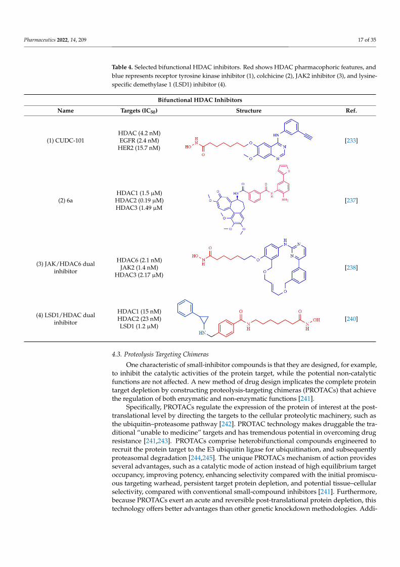
Pharmaceutics 2022, 14, 209 17 of 35
Table 4. Selected bifunctional HDAC inhibitors. Red shows HDAC pharmacophoric features, andblue represents receptor tyrosine kinase inhibitor (1), colchicine (2), JAK2 inhibitor (3), and lysine-specific demethylase 1 (LSD1) inhibitor (4).
Bifunctional HDAC Inhibitors
Name Targets (IC50) Structure Ref.
(1) CUDC-101HDAC (4.2 nM)EGFR (2.4 nM)
HER2 (15.7 nM)
Pharmaceutics 2022, 14, 209 17 of 36
Table 4. Selected bifunctional HDAC inhibitors. Red shows HDAC pharmacophoric features, and blue represents receptor tyrosine kinase inhibitor (1), colchicine (2), JAK2 inhibitor (3), and lysine-specific demethylase 1 (LSD1) inhibitor (4).
Bifunctional HDAC Inhibitors Name Targets (IC50) Structure Ref.
(1) CUDC-101 HDAC (4.2 nM) EGFR (2.4 nM) HER2 (15,7 nM)
[233]
(2) 6a HDAC1 (1.5 μM) HDAC2 (0.19 μM) HDAC3 (1.49 μM
[237]
(3) JAK/HDAC6 dual inhibitor
HDAC6 (2.1 nM) JAK2 (1.4 nM) HDAC3 (2.17 μM)
[238]
(4) LSD1/HDAC dual inhibitor
HDAC1 (15 nM) HDAC2 (23 nM) LSD1 (1.2 μM)
[240]
4.3. Proteolysis Targeting Chimeras One characteristic of small-inhibitor compounds is that they are designed, for exam-
ple, to inhibit the catalytic activities of the protein target, while the potential non-catalytic functions are not affected. A new method of drug design implicates the complete protein target depletion by constructing proteolysis-targeting chimeras (PROTACs) that achieve the regulation of both enzymatic and non-enzymatic functions [241].
Specifically, PROTACs regulate the expression of the protein of interest at the post-translational level by directing the targets to the cellular proteolytic machinery, such as the ubiquitin–proteasome pathway [242]. PROTAC technology makes druggable the tra-ditional “unable to medicine” targets and has tremendous potential in overcoming drug resistance [241,243]. PROTACs comprise heterobifunctional compounds engineered to re-cruit the protein target to the E3 ubiquitin ligase for ubiquitination, and subsequently pro-teasomal degradation [244,245]. The unique PROTACs mechanism of action provides sev-eral advantages, such as a catalytic mode of action instead of high equilibrium target oc-cupancy, improving potency, enhancing selectivity compared with the initial promiscu-ous targeting warhead, persistent target protein depletion, and potential tissue–cellular selectivity, compared with conventional small-compound inhibitors [241]. Furthermore, because PROTACs exert an acute and reversible post-translational protein depletion, this
[233]
(2) 6aHDAC1 (1.5 µM)
HDAC2 (0.19 µM)HDAC3 (1.49 µM
Pharmaceutics 2022, 14, 209 17 of 36
Table 4. Selected bifunctional HDAC inhibitors. Red shows HDAC pharmacophoric features, and blue represents receptor tyrosine kinase inhibitor (1), colchicine (2), JAK2 inhibitor (3), and lysine-specific demethylase 1 (LSD1) inhibitor (4).
Bifunctional HDAC Inhibitors Name Targets (IC50) Structure Ref.
(1) CUDC-101 HDAC (4.2 nM) EGFR (2.4 nM) HER2 (15,7 nM)
[233]
(2) 6a HDAC1 (1.5 μM) HDAC2 (0.19 μM) HDAC3 (1.49 μM
[237]
(3) JAK/HDAC6 dual inhibitor
HDAC6 (2.1 nM) JAK2 (1.4 nM) HDAC3 (2.17 μM)
[238]
(4) LSD1/HDAC dual inhibitor
HDAC1 (15 nM) HDAC2 (23 nM) LSD1 (1.2 μM)
[240]
4.3. Proteolysis Targeting Chimeras One characteristic of small-inhibitor compounds is that they are designed, for exam-
ple, to inhibit the catalytic activities of the protein target, while the potential non-catalytic functions are not affected. A new method of drug design implicates the complete protein target depletion by constructing proteolysis-targeting chimeras (PROTACs) that achieve the regulation of both enzymatic and non-enzymatic functions [241].
Specifically, PROTACs regulate the expression of the protein of interest at the post-translational level by directing the targets to the cellular proteolytic machinery, such as the ubiquitin–proteasome pathway [242]. PROTAC technology makes druggable the tra-ditional “unable to medicine” targets and has tremendous potential in overcoming drug resistance [241,243]. PROTACs comprise heterobifunctional compounds engineered to re-cruit the protein target to the E3 ubiquitin ligase for ubiquitination, and subsequently pro-teasomal degradation [244,245]. The unique PROTACs mechanism of action provides sev-eral advantages, such as a catalytic mode of action instead of high equilibrium target oc-cupancy, improving potency, enhancing selectivity compared with the initial promiscu-ous targeting warhead, persistent target protein depletion, and potential tissue–cellular selectivity, compared with conventional small-compound inhibitors [241]. Furthermore, because PROTACs exert an acute and reversible post-translational protein depletion, this
[237]
(3) JAK/HDAC6 dualinhibitor
HDAC6 (2.1 nM)JAK2 (1.4 nM)
HDAC3 (2.17 µM)
Pharmaceutics 2022, 14, 209 17 of 36
Table 4. Selected bifunctional HDAC inhibitors. Red shows HDAC pharmacophoric features, and blue represents receptor tyrosine kinase inhibitor (1), colchicine (2), JAK2 inhibitor (3), and lysine-specific demethylase 1 (LSD1) inhibitor (4).
Bifunctional HDAC Inhibitors Name Targets (IC50) Structure Ref.
(1) CUDC-101 HDAC (4.2 nM) EGFR (2.4 nM) HER2 (15,7 nM)
[233]
(2) 6a HDAC1 (1.5 μM) HDAC2 (0.19 μM) HDAC3 (1.49 μM
[237]
(3) JAK/HDAC6 dual inhibitor
HDAC6 (2.1 nM) JAK2 (1.4 nM) HDAC3 (2.17 μM)
[238]
(4) LSD1/HDAC dual inhibitor
HDAC1 (15 nM) HDAC2 (23 nM) LSD1 (1.2 μM)
[240]
4.3. Proteolysis Targeting Chimeras One characteristic of small-inhibitor compounds is that they are designed, for exam-
ple, to inhibit the catalytic activities of the protein target, while the potential non-catalytic functions are not affected. A new method of drug design implicates the complete protein target depletion by constructing proteolysis-targeting chimeras (PROTACs) that achieve the regulation of both enzymatic and non-enzymatic functions [241].
Specifically, PROTACs regulate the expression of the protein of interest at the post-translational level by directing the targets to the cellular proteolytic machinery, such as the ubiquitin–proteasome pathway [242]. PROTAC technology makes druggable the tra-ditional “unable to medicine” targets and has tremendous potential in overcoming drug resistance [241,243]. PROTACs comprise heterobifunctional compounds engineered to re-cruit the protein target to the E3 ubiquitin ligase for ubiquitination, and subsequently pro-teasomal degradation [244,245]. The unique PROTACs mechanism of action provides sev-eral advantages, such as a catalytic mode of action instead of high equilibrium target oc-cupancy, improving potency, enhancing selectivity compared with the initial promiscu-ous targeting warhead, persistent target protein depletion, and potential tissue–cellular selectivity, compared with conventional small-compound inhibitors [241]. Furthermore, because PROTACs exert an acute and reversible post-translational protein depletion, this
[238]
(4) LSD1/HDAC dualinhibitor
HDAC1 (15 nM)HDAC2 (23 nM)LSD1 (1.2 µM)
Pharmaceutics 2022, 14, 209 17 of 36
Table 4. Selected bifunctional HDAC inhibitors. Red shows HDAC pharmacophoric features, and blue represents receptor tyrosine kinase inhibitor (1), colchicine (2), JAK2 inhibitor (3), and lysine-specific demethylase 1 (LSD1) inhibitor (4).
Bifunctional HDAC Inhibitors Name Targets (IC50) Structure Ref.
(1) CUDC-101 HDAC (4.2 nM) EGFR (2.4 nM) HER2 (15,7 nM)
[233]
(2) 6a HDAC1 (1.5 μM) HDAC2 (0.19 μM) HDAC3 (1.49 μM
[237]
(3) JAK/HDAC6 dual inhibitor
HDAC6 (2.1 nM) JAK2 (1.4 nM) HDAC3 (2.17 μM)
[238]
(4) LSD1/HDAC dual inhibitor
HDAC1 (15 nM) HDAC2 (23 nM) LSD1 (1.2 μM)
[240]
4.3. Proteolysis Targeting Chimeras One characteristic of small-inhibitor compounds is that they are designed, for exam-
ple, to inhibit the catalytic activities of the protein target, while the potential non-catalytic functions are not affected. A new method of drug design implicates the complete protein target depletion by constructing proteolysis-targeting chimeras (PROTACs) that achieve the regulation of both enzymatic and non-enzymatic functions [241].
Specifically, PROTACs regulate the expression of the protein of interest at the post-translational level by directing the targets to the cellular proteolytic machinery, such as the ubiquitin–proteasome pathway [242]. PROTAC technology makes druggable the tra-ditional “unable to medicine” targets and has tremendous potential in overcoming drug resistance [241,243]. PROTACs comprise heterobifunctional compounds engineered to re-cruit the protein target to the E3 ubiquitin ligase for ubiquitination, and subsequently pro-teasomal degradation [244,245]. The unique PROTACs mechanism of action provides sev-eral advantages, such as a catalytic mode of action instead of high equilibrium target oc-cupancy, improving potency, enhancing selectivity compared with the initial promiscu-ous targeting warhead, persistent target protein depletion, and potential tissue–cellular selectivity, compared with conventional small-compound inhibitors [241]. Furthermore, because PROTACs exert an acute and reversible post-translational protein depletion, this
[240]
4.3. Proteolysis Targeting Chimeras
One characteristic of small-inhibitor compounds is that they are designed, for example,to inhibit the catalytic activities of the protein target, while the potential non-catalyticfunctions are not affected. A new method of drug design implicates the complete proteintarget depletion by constructing proteolysis-targeting chimeras (PROTACs) that achievethe regulation of both enzymatic and non-enzymatic functions [241].
Specifically, PROTACs regulate the expression of the protein of interest at the post-translational level by directing the targets to the cellular proteolytic machinery, such asthe ubiquitin–proteasome pathway [242]. PROTAC technology makes druggable the tra-ditional “unable to medicine” targets and has tremendous potential in overcoming drugresistance [241,243]. PROTACs comprise heterobifunctional compounds engineered torecruit the protein target to the E3 ubiquitin ligase for ubiquitination, and subsequentlyproteasomal degradation [244,245]. The unique PROTACs mechanism of action providesseveral advantages, such as a catalytic mode of action instead of high equilibrium targetoccupancy, improving potency, enhancing selectivity compared with the initial promiscu-ous targeting warhead, persistent target protein depletion, and potential tissue–cellularselectivity, compared with conventional small-compound inhibitors [241]. Furthermore,because PROTACs exert an acute and reversible post-translational protein depletion, thistechnology offers better advantages than other genetic knockdown methodologies. Addi-
Pharmaceutics 2022, 14, 209 18 of 35
tionally, PROTACs may function as molecular probes to elucidate the epigenetic regulationin health and diseases as a new modality in precision medicine [246].
In 2018, one of the first HDAC-targeting PROTACs was described as the first HDAC6degrader, generated based on nonselective HDAC inhibitors with E3 ubiquitin ligaseligand pomalidomide as cereblon (CRBN) (Table 5). CRBN ligands rely on the structureof the anticancer drug thalidomide and its derivatives [247]. The PROTAC compoundexhibits a degrader concentration (DC)50 of 34 nM and a maximum percentage of HDAC6degradation (Dmax) of 70.5% without significant effects on other HDACs family members.Moreover, the degradation of HDAC6 results in an upregulation of tubulin acetylation intreated MCF-7 cells [248]. Similarly, An et al. [249] synthesized an HDAC6 degrader (NP8)by combining Next-A, at the end of the aliphatic chain, with the CRBN pomalidomide.NP8 provokes a significant inhibition of the proliferation of the multiple myeloma cell lineMM.1S, comparable with parental agent Nex-A, with the DC50 value for NP8 of 3.8 nM.Moreover, the HDAC6-pomalidomide degrader showed a high grade of specificity, as otherrepresentative HDAC family members were not affected by NP8 treatment. Additionally,Yang et al. [250] described the synthesis of a degrader (NH2) combining Next-A, at the NH2residue, with pomalidomide as CRBN. NH2 effectively depletes HDAC6 in MM.1S witha DC50 of 3,2 nM after 24 h of treatment; moreover, the HDAC6–NH2–CRBN PROTACprovokes HDAC6 degradation at 1 h of treatment, reaching maximum degradation ataround 6–8 h. In addition, the NH2 washout in HeLa cells recovered the HDAC6 proteinlevel within 3 h, confirming the suitability of use of NH2 as a reversible knockdowncompound for HDAC6 protein in cancer cells.
In addition, a cell-permeable HDAC-targeting PROTAC based on Von Hippel-Lindau(VHL) as an E3 ligase-binding motif and Nexturastat A (Next-A), a well-known HDAC6-selective inhibitor, has been synthesized. VHL has demonstrated efficacy and the robustdegradation of a wide range of protein targets and shown the ability to bypass the pos-sible off-target effects of CRBN recruitment [247]. The resulting PROTAC agent inducesHDAC6 degradation with a DC50 of 7.1 nM and a Dmax of 90% in the MM1S cell line [251].Additionally, SR-3558, a potent and selective inhibitor of class I HDACs, with VHL as anE3 ligase ligand, generates a selective HDAC3 degrader (XZ9002) with a DC50 value of42 nM [252]. Similarly, Smalley et al. [253] generated a PROTAC molecule targeting thenucleus-localized class I HDACs. The class I o-aminoanilide HDAC inhibitor CI-994 wasconjugated to the VHL ligand via a twelve-carbon alkyl linker to synthesize a PROTACwith a DC50 of 1 µM for HDACs 1, 2 and 3 in HCT116 human colon cancer cells.
Finally, Cao et al. used bestatin, a cellular inhibitor of apoptosis (cIAP) ligand, asE3 ligase-binding motifs to develop cIAP–HDAC–PROTACs [254]. Namely, cIAP ligandsfunction as E3 ubiquitin ligases that target RIP1 for polyubiquitination and have shownsuccess in the design of PROTACs [247,255]. Several HDAC–cIAP hybrid molecules weresynthesized and one in particular, named P1, significantly reduced the HDAC1, HDAC6,and HDAC8 expression levels on the B lymphocytes cell line RPMI-8226 after 24 h treatment,at concentrations ranging from 1 µM to 4 µM, although it did not induce HDAC degradationat 6 h of treatment [255].

Pharmaceutics 2022, 14, 209 19 of 35
Table 5. Selected proteolysis targeting chimeras for HDACs degradation. Red shows HDAC pharma-cophoric features, black shows linker regions, and blue shows E3 ligases. VHL: Von Hippel–Lindau;DC50, concentration to reduce the target protein expression in a 50%; DCmax: maximum percentageof degradation; ND: non-determined.
Proteolysis Targeting Chimeras
Name Targets (DC50,Dmax) Structure Ref.
9c—HDAC6degrader
HDAC6(DC50 = 34 nM,Dmax = 70.5%)
Pharmaceutics 2022, 14, 209 19 of 36
Table 5. Selected proteolysis targeting chimeras for HDACs degradation. Red shows HDAC phar-macophoric features, black shows linker regions, and blue shows E3 ligases. VHL: Von Hippel–Lindau; DC50, concentration to reduce the target protein expression in a 50%; DCmax: maximum per-centage of degradation; ND: non-determined.
Proteolysis Targeting Chimeras
Name Targets (DC50,
Dmax) Structure Ref.
9c—HDAC6 degrader
HDAC6 (DC50 = 34 nM, Dmax = 70.5%)
[248]
NP8 HDAC6 (DC50 = 3,8nM, Dmax = ND)
[249]
NH2 HDAC6 (DC50 = 3,2 nM, Dmax = ND)
[250]
VHL-Next-A degrader
HDAC6 (DC50 = 7.1 nM, Dmax = 90%)
[251]
XZ9002 de-grader
HDAC3 (DC50 = 42 nM, Dmax = 70%)
[252]
P1 HDAC1, HDAC6, and HDAC8
[254]
[248]
NP8HDAC6
(DC50 = 3.8nM,Dmax = ND)
Pharmaceutics 2022, 14, 209 19 of 36
Table 5. Selected proteolysis targeting chimeras for HDACs degradation. Red shows HDAC phar-macophoric features, black shows linker regions, and blue shows E3 ligases. VHL: Von Hippel–Lindau; DC50, concentration to reduce the target protein expression in a 50%; DCmax: maximum per-centage of degradation; ND: non-determined.
Proteolysis Targeting Chimeras
Name Targets (DC50,
Dmax) Structure Ref.
9c—HDAC6 degrader
HDAC6 (DC50 = 34 nM, Dmax = 70.5%)
[248]
NP8 HDAC6 (DC50 = 3,8nM, Dmax = ND)
[249]
NH2 HDAC6 (DC50 = 3,2 nM, Dmax = ND)
[250]
VHL-Next-A degrader
HDAC6 (DC50 = 7.1 nM, Dmax = 90%)
[251]
XZ9002 de-grader
HDAC3 (DC50 = 42 nM, Dmax = 70%)
[252]
P1 HDAC1, HDAC6, and HDAC8
[254]
[249]
NH2HDAC6
(DC50 = 3.2 nM,Dmax = ND)
[250]
VHL-Next-Adegrader
HDAC6(DC50 = 7.1 nM,
Dmax = 90%)
Pharmaceutics 2022, 14, 209 19 of 36
Table 5. Selected proteolysis targeting chimeras for HDACs degradation. Red shows HDAC phar-macophoric features, black shows linker regions, and blue shows E3 ligases. VHL: Von Hippel–Lindau; DC50, concentration to reduce the target protein expression in a 50%; DCmax: maximum per-centage of degradation; ND: non-determined.
Proteolysis Targeting Chimeras
Name Targets (DC50,
Dmax) Structure Ref.
9c—HDAC6 degrader
HDAC6 (DC50 = 34 nM, Dmax = 70.5%)
[248]
NP8 HDAC6 (DC50 = 3,8nM, Dmax = ND)
[249]
NH2 HDAC6 (DC50 = 3,2 nM, Dmax = ND)
[250]
VHL-Next-A degrader
HDAC6 (DC50 = 7.1 nM, Dmax = 90%)
[251]
XZ9002 de-grader
HDAC3 (DC50 = 42 nM, Dmax = 70%)
[252]
P1 HDAC1, HDAC6, and HDAC8
[254]
[251]
XZ9002degrader
HDAC3(DC50 = 42 nM,
Dmax = 70%)
Pharmaceutics 2022, 14, 209 19 of 36
Table 5. Selected proteolysis targeting chimeras for HDACs degradation. Red shows HDAC phar-macophoric features, black shows linker regions, and blue shows E3 ligases. VHL: Von Hippel–Lindau; DC50, concentration to reduce the target protein expression in a 50%; DCmax: maximum per-centage of degradation; ND: non-determined.
Proteolysis Targeting Chimeras
Name Targets (DC50,
Dmax) Structure Ref.
9c—HDAC6 degrader
HDAC6 (DC50 = 34 nM, Dmax = 70.5%)
[248]
NP8 HDAC6 (DC50 = 3,8nM, Dmax = ND)
[249]
NH2 HDAC6 (DC50 = 3,2 nM, Dmax = ND)
[250]
VHL-Next-A degrader
HDAC6 (DC50 = 7.1 nM, Dmax = 90%)
[251]
XZ9002 de-grader
HDAC3 (DC50 = 42 nM, Dmax = 70%)
[252]
P1 HDAC1, HDAC6, and HDAC8
[254]
[252]
P1HDAC1,
HDAC6, andHDAC8
Pharmaceutics 2022, 14, 209 19 of 36
Table 5. Selected proteolysis targeting chimeras for HDACs degradation. Red shows HDAC phar-macophoric features, black shows linker regions, and blue shows E3 ligases. VHL: Von Hippel–Lindau; DC50, concentration to reduce the target protein expression in a 50%; DCmax: maximum per-centage of degradation; ND: non-determined.
Proteolysis Targeting Chimeras
Name Targets (DC50,
Dmax) Structure Ref.
9c—HDAC6 degrader
HDAC6 (DC50 = 34 nM, Dmax = 70.5%)
[248]
NP8 HDAC6 (DC50 = 3,8nM, Dmax = ND)
[249]
NH2 HDAC6 (DC50 = 3,2 nM, Dmax = ND)
[250]
VHL-Next-A degrader
HDAC6 (DC50 = 7.1 nM, Dmax = 90%)
[251]
XZ9002 de-grader
HDAC3 (DC50 = 42 nM, Dmax = 70%)
[252]
P1 HDAC1, HDAC6, and HDAC8
[254] [254]
5. Combined Clinical Strategies with Histone Deacetylases Inhibitors
The clinical approval of vorinostat, romidepsin, belinostat, panobinostat, and tucidi-nostat/chidamide allows the inclusion of HDACi as part of the therapeutic armament foran increasing number of cancers. Nevertheless, HDACi, when used as a mono-therapeutic

Pharmaceutics 2022, 14, 209 20 of 35
chemical agent, showed a limited cancer application, mainly in treating hematological ma-lignancies. Moreover, tumor resistance to HDACis treatment has been observed alongsidea limited therapy achievement of solid tumors [256,257], whereas combining HDACis withchemotherapy agents may improve and maximize cancer treatment and reduce treatment-associated toxicity side effects due to reduced agent doses. Furthermore, combined thera-pies can synergistically avoid or reduce undesirable cancer treatment resistance [257]. Inthis sense, HDACis may function as chemosensitizers and synergistically work with anincreasing number of diverse therapeutic chemical drugs, biological-derived active proteinsand polypeptides, and immunotherapies [258]. Thus, combined therapeutic strategies ofHDACis with other approved cancer therapies may increase the efficacy of clinical ap-proaches and have shown great promise in clinical trials. A considerable number of clinicaltrials have been implemented toward the combined application of HDACis, and in thissection we will analyze selected clinical studies focusing mainly on solid tumors (Table 6).
Receptor tyrosine kinases (RTKs), due to regulating a vast range of biological andmolecular functions and their dysregulated intracellular signals in several cancers, havebeen shown to be promising onco-targets. In particular, RTKs promote cancer cell prolif-eration, increasing c-Myc and cyclin D1 oncogenes expression. Cyclin D1 interacts withclass I/II HDACs to regulate c-Myc expression, thus making the combinations of RTKtargeted therapies and HDACi important strategies for cancer therapy [258–261]. A phaseI/II clinical trial (NCT01027676), addressing the capacity of vorinostat to synergize andovercome resistance to the EGFR-TKI gefitinib in treated patients with advanced non-small-cell lung cancer (NSCLC), has demonstrated this combination to be feasible andwell tolerated at the clinical regime of biweekly 400 mg/day vorinostat and daily doses of250 mg gefitinib. EGFR-mutant NSCLC-treated patients showed a median progression-freesurvival (PFS) of 9.1 months and median overall survival (OS) of 24.1 months. This combi-nation showed potential for improving the efficacy of gefitinib in EGFR-mutant NSCLCand suggested the potential benefit of vorinostat for improving the efficacy of EGFR-TKIsin these patients [262].
Angiogenesis is one of the hallmark events contributing to tumor growth and metas-tasis [36]. For instance, HDACs stabilize and inhibit the degradation of HIF-1α [37,38].Meanwhile, the vascular endothelial growth factor (VEGF) secreted by cancer cells stim-ulates endothelial cells to form new blood vessels. Moreover, anti-VEGF treatment inpreclinical studies has demonstrated the additive and synergistical benefits of combinedtreatments with other onco-therapies [263]. In this sense, a clinical study has tested thesafety and efficacy of vorinostat and the VEGF blocker bevacizumab in metastatic clear-cellrenal cell carcinoma (ccRCC) patients that were previously undertreated with differentagents, such as sunitinib, sorafenib, axitinib, and temsirolimus, among others. A total of200 mg vorinostat twice per day, for 14 days, and bevacizumab 15 mg kg−1, intravenously,every 21 days were administered as phase II doses. The toxicity of combined therapy seemsto be acceptable and safe, and the median PFS and OS were 5.7 months and 12.9 months,respectively. Nevertheless, the study is limited due to the sample size and the single-armdesign [264].
Additionally, hormone signaling is dysregulated in several cancers, such as breast andprostate cancer, and contributes to promoting tumorigenesis [258,265]. Thus, combinedhormonal therapies with HDACis are promising strategies for the treatment of hormone-dependent cancers. Recently, a phase III trial of tucidinostat/chidamide (30 mg twice aweek for four weeks) combined with the steroidal aromatase inhibitor exemestane (25 mgorally daily) improved the PFS compared with placebo plus exemestane, with a medianPFS of 7.4 months and 3.8 months, respectively. Moreover, a substantially increased overallresponse and clinical benefit was observed in combined therapy of patients with advanced,hormone receptor-positive (HR+) and HER2-negative breast cancer that progressed afterprior endocrine therapy [266]. Another phase I clinical study explored the safety andpreliminary efficacy of combined entinostat therapy (3 and 5 mg orally per week) withthe hormonal therapy drug enzalutamide in castration-resistant PCa (CRPC) patients. The

Pharmaceutics 2022, 14, 209 21 of 35
median duration of treatment with entinostat was 18 weeks. Entinostat did not affect thesteady plasma concentration of enzalutamide, and no defining dose-limiting toxicity (DLT)related to entinostat in these patients was observed. Despite the small number of enrolledpatients (6), 5 mg of entinostat weekly combined with enzalutamide seems to have anacceptable safety profile [267].
In addition, a multicenter, randomized, double-blind, placebo-controlled phase IIIstudy (E2112) enrolled patients with advanced HR-positive, HER2-negative breast cancerwhose disease progressed after nonsteroidal aromatase inhibitors had been performed. Thisphase III clinical trial was based on a previous ENCORE301 phase II study that reportedimprovement in PFS and OS with combined entinostat and exemestane therapy [268]. Inthis study, the patients were randomly treated with oral 25 mg exemestane once dailyand 5 mg entinostat or placebo 5 mg once weekly. The results indicate that the medianPFS of exemestane plus entinostat showed no significant difference with 3.3 months and3.1 months, respectively.
Similarly, no median OS differences between exemestane plus entinostat and exemes-tane plus placebo arms were obtained, being 23.4 months and 21.7 months, respectively.Although the pharmacodynamic endpoint analysis indicated that in the exemestane plusentinostat arm a significant increase in lysine acetylation in PBMCs by C1D15 analysis wasobserved, confirming HDACi function, the combination of entinostat and exemestane didnot improve advanced endocrine-resistant breast cancer patients’ outcomes [269].
In addition, HDACs regulate autophagy signals, including mTor pathways, thathelp tumor cells survive under metabolic stress conditions [22]. Moreover, preclinicalapproaches identified the mTOR/HDAC inhibitor combination as a promising strategy forcancer treatment [270]. In this regard, a phase I dose-finding trial (NCT01582009) for themTOR inhibitor everolimus combined with the HDAC inhibitor panobinostat in advancedclear-cell renal cell carcinoma (ccRCC) patients has been completed. Based on the maximumtolerated dose, the therapeutic strategy was everolimus 5 mg PO daily and panobinostat10 mg PO 3 times weekly (weeks 1 and 2) given in 21-day cycles. The 6-month PFS was 31%,with a median of 4.1 months. Although the combined everolimus/panobinostat therapyseems to be safe and tolerable in the indicated dosing regimen, with myelosuppression asthe major DLT, this pairing did not appear to improve the clinical outcomes in the studiedgroup of advanced ccRCC patients [271].
To date, chemotherapy continues to be among the most widely used cancer treat-ments, causing cell death. Despite the initial therapeutic success, cancer cells often developchemoresistance, particularly in non-germ-cell solid tumors, which severely reduce the clin-ical benefits for cancer patients [272]. Additionally, HDACi therapies, due to the broad roleof HDAC in diverse cellular functions, have been demonstrated to act as sensitizer agentsfor chemotherapy [273]. For instance, combining cisplatin with HDACi shows a promisingstrategy for increasing the efficacy of this platinum-based agent. A phase II, single-armclinical trial was conducted to investigate the efficacy and safety of a combination treatmentof tucidinostat/chidamide with cisplatin in metastatic advanced triple-negative breastcancer (TNBC) patients. Women with TNBC were treated with tucidinostat/chidamide andcisplatin, 20 mg twice weekly for 2 weeks, and 75 mg/m2 on a 21-day cycle, respectively.Despite the fact that this trial had several limitations, including a small sample size, non-randomized character, and the un-selection of the enrolled patients, the combination ofchidamide and cisplatin did not seem to exhibit a superior efficacy compared with classicalcisplatin-based chemotherapy in treating advanced TNBC [274]. Additionally, romidepsinhas been included in a phase I dose-escalation study (NCT01902225) in combination withliposomal doxorubicin, an anthracycline that intercalates within DNA base pairs and in-hibits topoisomerase II to treat cutaneous T-cell lymphoma (CTCL) and peripheral T-celllymphoma (PTCL). Liposomal doxorubicin dose was fixed at 20 mg/m2 i.v. over 1 h onday 1 of each 28-day cycle, and the maximum tolerated dose (MTD) of romidepsin wasdetermined to be 12 mg/m2. The results indicate that the overall response rate (ORR) inthe CTCL patients cohort was 70%, while the PTCL cohort was 27%. Combined romidepsin

Pharmaceutics 2022, 14, 209 22 of 35
and liposomal doxorubicin therapy regimes indicated high and rapid response rates inCTCL patients. The authors concluded that drug combination is highly active, safe, andwell tolerated [275].
Furthermore, preclinical studies demonstrated the feasibility and efficacy of the combi-nation of DNA synthesis inhibitor agents and HDACi in improving cancer sensitivity [276].An open-label, non-randomized phase I/II clinical trial study (NCT00372437) addressedthe safety and efficacy of the class I/IV HDACi mocetinostat combined with DNA synthesisinhibitor gemcitabine in patients with solid tumors. The therapeutic regime included gemc-itabine at 1000 mg/m2, day 1 of three consecutive weeks, in 4-week cycles, and 50–110 mg,three times per week of oral mocetinostat. MTD and the recommended phase II dose formocetinostat was 90 mg, while the ORR was 11% in the phase I study, and non-responseswere observed in phase II. Due to the lack of significant positive results and significanttoxicities in patients with advanced pancreatic cancer following the combined therapy, thestudy was terminated without merit for further testing [277].
One characteristic of cancer cells is their capacity to regulate immune functions and es-cape from immune surveillance [278]. In turn, HDACi demonstrated the ability to promotecancer cells’ immunogenicity [279], making the combination of HDACi with immunother-apies promising for cancer treatment. For example, the combination of vorinostat andthe anti-PD-1 pembrolizumab, an immune checkpoint inhibitor, was evaluated in a phaseII study (NCT02538510) in recurrent/metastatic squamous cell carcinomas of the headand neck (HN) and salivary gland cancer (SGC) patients. The therapeutic regime wasconducted with oral 400 mg vorinostat 5 days on and 2 days off during each 21-day cycle,while intravenous pembrolizumab 200 mg was administrated every 21 days. The combinedHDACi and immunotherapy demonstrated activity in HN patients with fewer responsesin SGC. Although the combination demonstrated activity in HN, with fewer responsesin SGC, high-grade toxicities compared with single-agent pembrolizumab were observed.Nevertheless, the obtained data provide new clues for the activity and tolerability of theproposed combined HDACis and immunotherapies in HN and SGC patients [280].
Similarly, a PEMDAC phase II clinical trial (NCT02697630) combining entinostat andpembrolizumab in patients with metastatic uveal melanoma (UM) showed manageabletoxicities and no treatment-related patient deaths. Patients were treated intravenouslywith 200 mg pembrolizumab every third week in combination with entinostat 5 mg orallyonce a week. Median OS was 13.4 months, and OS at one year was 59%, while medianPFS was 2.1 months, and one-year PFS was 17%. Additionally, combined therapy cancause tumor regression in a small subset of patients with metastatic UM, which providesdata for a small subset of patients who can benefit from combined epigenetic therapyand immunotherapy [281]. Furthermore, a phase Ib clinical trial combining the second-generation HDAC6-selective inhibitor citarinostat (ACY-241) with the immune checkpointinhibitor nivolumab has been conducted in patients with advanced NSCLC. Patientsreceived orally administered citarinostat once daily for 28 days of two 28-day treatmentcycles with a modified Fibonacci sequence of 180 mg QD, 360 mg QD, and 480 mg QD.For cycle 1, nivolumab was administered on day 15 at 3 mg/kg by intravenous injection,while in cycle 2, nivolumab was administered every 2 weeks on days 1 and 15. The resultsof this single-arm and dose-escalation study indicated an MTD for citarinostat of 360 mg.Additionally, a transient increase in histone (class I target) and tubulin (HDAC6 target)acetylation levels following treatment were observed in patients’ samples. Together, theresults indicate that a combined HDACi and immunotherapy strategy is safe and tolerableand may be feasible in patients with advanced NSCLC [282].
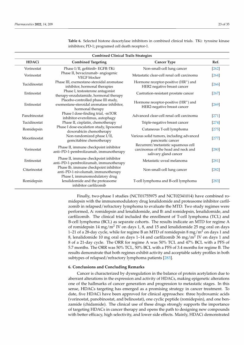
Pharmaceutics 2022, 14, 209 23 of 35
Table 6. Selected histone deacetylase inhibitors in combined clinical trials. TKi: tyrosine kinaseinhibitors; PD-1; programed cell death receptor-1.
Combined Clinical Trails Strategies
HDACi Combined Targeting Cancer Type Ref.
Vorinostat Phase I/II, gefitinib- EGFR-TKi Non-small-cell lung cancer [262]
Vorinostat Phase II, bevacizumab- angiogenicVEGF blocker Metastatic clear-cell renal cell carcinoma [264]
Tucidinostat Phase III, exemestane-steroidal aromataseinhibitor, hormonal therapies
Hormone receptor-positive (HR+) andHER2 negative breast cancer [266]
Entinostat Phase I, testosterone antagonisttherapy-enzalutamide, hormonal therapy Castration-resistant prostate cancer [267]
EntinostatPlacebo-controlled phase III study,
exemestane-steroidal aromatase inhibitor,hormonal therapy
Hormone receptor-positive (HR+) andHER2-negative breast cancer [269]
Panobinostat Phase I dose-finding trial, -mTORinhibitor-everolimus, autophagy Advanced clear-cell renal cell carcinoma [271]
Tucidinostat Phase II, cisplatin, chemotherapy Triple-negative breast cancer [274]
Romidepsin Phase I dose-escalation study, liposomaldoxorubicin chemotherapy Cutaneous T-cell lymphoma [275]
Mocetinostat Non-randomized phase I/II,gemcitabine chemotherapy
Various solid tumors, including advancedpancreatic cancer [277]
Vorinostat Phase II, immune checkpoint inhibitoranti–PD-1-pembrolizumab, immunotherapy
Recurrent/metastatic squamous cellcarcinomas of the head and neck and
salivary gland cancer[280]
Entinostat Phase II, immune checkpoint inhibitoranti–PD-1-pembrolizumab, immunotherapy Metastatic uveal melanoma [281]
Citarinostat Phase Ib, immune checkpoint inhibitoranti–PD-1-nivolumab, immunotherapy Non-small-cell lung cancer [282]
RomidepsinPhase I, immunomodulatory druglenalidomide and the proteasome
inhibitor carfilzomibT-cell lymphoma and B-cell lymphoma [283]
Finally, two-phase I studies (NCT01755975 and NCT02341014) have combined ro-midepsin with the immunomodulatory drug lenalidomide and proteasome inhibitor carfil-zomib in relapsed/refractory lymphoma to evaluate the MTD. Two study regimes wereperformed, A: romidepsin and lenalidomide, and B: and romidepsin, lenalidomide, andcarfilzomib. The clinical trial included the enrollment of T-cell lymphoma (TCL) andB-cell lymphoma (BCL) as separate cohorts. The results indicate an MTD for regime Aof romidepsin 14 mg/m2 IV on days 1, 8, and 15 and lenalidomide 25 mg oral on days1–21 of a 28-day cycle, while for regime B an MTD of romidepsin 8 mg/m2 on days 1 and8, lenalidomide 10 mg oral on days 1–14 and carfilzomib 36 mg/m2 IV on days 1 and8 of a 21-day cycle. The ORR for regime A was 50% TCL and 47% BCL with a PFS of5.7 months. The ORR was 50% TCL, 50% BCL with a PFS of 3.4 months for regime B. Theresults demonstrate that both regimes exhibit activity and acceptable safety profiles in bothsubtypes of relapsed/refractory lymphoma patients [283].
6. Conclusions and Concluding Remarks
Cancer is characterized by dysregulation in the balance of protein acetylation due toaberrant alterations in the expression and activity of HDACs, making epigenetic alterationsone of the hallmarks of cancer generation and progression to metastatic stages. In thissense, HDACs targeting has emerged as a promising strategy in cancer treatment. Todate, five HDACi have been approved for clinical approaches: three hydroxamic acids(vorinostat, panobinostat, and belinostat), one cyclic peptide (romidepsin), and one ben-zamide (chidamide). The clinical use of these drugs strongly supports the importanceof targeting HDACs in cancer therapy and opens the path to designing new compoundswith better efficacy, high selectivity, and lower side effects. Mainly, HDACi demonstrated

Pharmaceutics 2022, 14, 209 24 of 35
efficacy for treating hematological malignancies and lymphomas. At the same time, anincreasing number of clinical trials are ongoing to evaluate the safety and therapeuticbenefits for refractory, advanced, and recurrent solid tumors. Unfortunately, most clinicaltrials revealed adverse issues, such as different grades of thrombocytopenia, neutropenia,anemia, arrhythmia, and gastrointestinal toxicity. At molecular levels, the adverse effectsare alongside several events, such as abnormal gene transcription, genomic instability, andaberrant free radical production, which reduce the therapeutic success of HDACi [284].Therefore, the design of new HDACi to overcome undesirable side effects and increasedefficacy to penetrate solid tumors represents a critical pharmacology challenge.
In addition, a deeper understanding of the molecular mechanisms of the anticancerfunctions of HDACi, analyses of HDAC structural conformations, and chemical-specificmodifications are necessary to improve the therapeutic efficacy of HDACi. This reviewdescribes HDACs’ features and their involvement in several cancer-associated cellularand molecular events, which picture the HDAC involvement in cancer physiopathology.Two main groups of HDACi inhibitors are analyzed according to their use for cancertreatment and dietary-derived compounds for chemoprevention. However, they can behighly intertwined since natural agents provide almost unlimited chemical structures forHDACi design.
In addition, the currently approved drugs such as HDACi for cancer treatment dis-played relatively low selectivity. Due to the diversity of HDACs and cellular-specificfunctions, the design of more potent isoform-selective inhibitors is highly needed. Accord-ing to the pharmacophore model, HDACis are susceptible to be modified in all three of themain groups (ZBG, linker, and cap), which may help design selective HDAC inhibitorsvia the optimization of the interaction with particular HDACs. Additionally, due to theoncogenic nature of cancer and the development of more potent and selective oncogeneinhibitors, new investigations which addressed the potential of bifunctional inhibitors,which involve oncoprotein inhibitors and specific HDACi, have shown the potential ofimproving conventional therapies, in part by enhancing therapeutic efficacy and reducingHDACi side effects. Furthermore, PROTAC technology helps in the generation of a newclass of HDAC targeting agents. PROTAC leads to the proteasomal degradation of HDACswith several advantages, such as target protein depletion and potential tissue-cellular selec-tivity that, with the development of more HDACi specificity, may highly improve cancertreatment and drug resistance. Finally, the HDACi can be combined with different basicanticancer experimental strategies, for inhibiting tumor growth, angiogenesis, metastasis,and apoptosis induction, which has allowed the execution of an increasing number ofcombined clinical trials. Together, these therapeutic strategies might benefit the survivaland improve the quality of life of cancer patients when traditional treatments fail.
Author Contributions: Conceptualization, K.N. and J.F.S.; writing—original draft preparation, D.R.,N.D., C.E. and J.F.S.; writing—review and editing, D.R., K.N., T.S.-R., C.E. and J.F.S.; supervision andfunding acquisition, K.N. and J.F.S. All authors have read and agreed to the published version ofthe manuscript.
Funding: This research was funded by the Ministry of Education Science and Technological Develop-ment of the Republic of Serbia No 451-03-9/2021-14/200015 and Faculty of Pharmacy UB ContractNo. 451-03-9/2021-14/200161.
Data Availability Statement: The material supporting the conclusion of this review has been in-cluded in the article.
Acknowledgments: We thank the support of the visiting professor program of UBO to J.F.S.
Conflicts of Interest: The authors declare no conflict of interest.

Pharmaceutics 2022, 14, 209 25 of 35
References1. Feinberg, A.P.; Ohlsson, R.; Henikoff, S. The epigenetic progenitor origin of human cancer. Nat. Rev. Genet. 2006, 7, 21–33.
[CrossRef]2. Feinberg, A.P.; Vogelstein, B. Hypomethylation distinguishes genes of some human cancers from their normal counterparts.
Nature 1983, 301, 89–92. [CrossRef]3. Herman, J.G.; Baylin, S.B. Gene silencing in cancer in association with promoter hypermethylation. N. Engl. J. Med. 2003, 349,
2042–2054. [CrossRef] [PubMed]4. Li, Z.; Lin, G.-L.; Yan, P.; Huang, J.; Wang, Y.-L. The expression of H3K9Ac, H3K14Ac, and H4K20TriMe in epithelial ovarian
tumors and the clinical significance. Int. J. Gynecol. Cancer 2010, 20, 82–86. [CrossRef]5. Fraga, M.F.; Ballestar, E.; Villar-Garea, A.; Boix-Chornet, M.; Espada, J.; Schotta, G.; Bonaldi, T.; Haydon, C.; Ropero, S.;
Petrie, K.; et al. Loss of acetylation at Lys16 and trimethylation at Lys20 of histone H4 is a common hallmark of human cancer.Nat. Genet. 2005, 37, 391–400. [CrossRef]
6. Talbert, P.B.; Henikoff, S. Histone variants at a glance. J. Cell Sci. 2021, 134, jcs244749. [CrossRef] [PubMed]7. Li, B.; Carey, M.; Workman, J.L. The role of chromatin during transcription. Cell 2007, 128, 707–719. [CrossRef]8. Willhoft, O.; Costa, A. A structural framework for DNA replication and transcription through chromatin. Curr. Opin. Struct. Biol.
2021, 71, 51–58. [CrossRef]9. Rothbart, S.B.; Strahl, B.D. Interpreting the language of histone and DNA modifications. Biochim. Biophys. Acta 2014, 1839,
627–643. [CrossRef]10. Nitsch, S.; Zorro Shahidian, L.; Schneider, R. Histone acylations and chromatin dynamics: Concepts, challenges, and links to
metabolism. EMBO Rep. 2021, 22, e52774. [CrossRef]11. Seto, E.; Yoshida, M. Erasers of histone acetylation: The histone deacetylase enzymes. Cold Spring Harb. Perspect. Biol. 2014,
6, a018713. [CrossRef] [PubMed]12. Losson, H.; Schnekenburger, M.; Dicato, M.; Diederich, M. Natural Compound Histone Deacetylase Inhibitors (HDACi): Synergy
with Inflammatory Signaling Pathway Modulators and Clinical Applications in Cancer. Molecules 2016, 21, 1608. [CrossRef][PubMed]
13. Archer, S.Y.; Hodin, R.A. Histone acetylation and cancer. Curr. Opin. Genet. Dev. 1999, 9, 171–174. [CrossRef]14. Hanahan, D.; Weinberg, R.A. Hallmarks of cancer: The next generation. Cell 2011, 144, 646–674. [CrossRef]15. Liu, Q.L.; Luo, M.; Huang, C.; Chen, H.N.; Zhou, Z.G. Epigenetic Regulation of Epithelial to Mesenchymal Transition in the
Cancer Metastatic Cascade: Implications for Cancer Therapy. Front. Oncol. 2021, 11, 657546. [CrossRef]16. Mirzadeh Azad, F.; Atlasi, Y. Deregulation of Transcriptional Enhancers in Cancer. Cancers 2021, 13, 3532. [CrossRef] [PubMed]17. You, J.S.; Jones, P.A. Cancer genetics and epigenetics: Two sides of the same coin? Cancer Cell 2012, 22, 9–20. [CrossRef]18. Kanwal, R.; Gupta, S. Epigenetic modifications in cancer. Clin. Genet. 2012, 81, 303–311. [CrossRef]19. Peng, L.; Seto, E. Deacetylation of nonhistone proteins by HDACs and the implications in cancer. Handb. Exp. Pharmacol. 2011,
206, 39–56. [CrossRef]20. Darwiche, N. Epigenetic mechanisms and the hallmarks of cancer: An intimate affair. Am. J. Cancer Res. 2020, 10, 1954–1978.21. Pant, K.; Peixoto, E.; Richard, S.; Gradilone, S.A. Role of Histone Deacetylases in Carcinogenesis: Potential Role in Cholangiocarci-
noma. Cells 2020, 9, 780. [CrossRef]22. Li, Y.; Seto, E. HDACs and HDAC Inhibitors in Cancer Development and Therapy. Cold Spring Harb. Perspect. Med. 2016,
6, a026831. [CrossRef] [PubMed]23. Kim, Y.B.; Ki, S.W.; Yoshida, M.; Horinouchi, S. Mechanism of cell cycle arrest caused by histone deacetylase inhibitors in human
carcinoma cells. J. Antibiot. 2000, 53, 1191–1200. [CrossRef] [PubMed]24. Chun, P. Histone deacetylase inhibitors in hematological malignancies and solid tumors. Arch. Pharmacal Res. 2015, 38, 933–949.
[CrossRef] [PubMed]25. Gong, P.; Wang, Y.; Jing, Y. Apoptosis Induction byHistone Deacetylase Inhibitors in Cancer Cells: Role of Ku70. Int. J. Mol. Sci.
2019, 20, 1601. [CrossRef] [PubMed]26. Zhang, J.; Zhong, Q. Histone deacetylase inhibitors and cell death. Cell. Mol. Life Sci. 2014, 71, 3885–3901. [CrossRef]27. Roos, W.P.; Krumm, A. The multifaceted influence of histone deacetylases on DNA damage signalling and DNA repair. Nucleic
Acids Res. 2016, 44, 10017–10030. [CrossRef]28. Koeneke, E.; Witt, O.; Oehme, I. HDAC Family Members Intertwined in the Regulation of Autophagy: A Druggable Vulnerability
in Aggressive Tumor Entities. Cells 2015, 4, 135–168. [CrossRef]29. Rikiishi, H. Autophagic and apoptotic effects of HDAC inhibitors on cancer cells. J. Biomed. Biotechnol. 2011, 2011, 830260.
[CrossRef]30. Mrakovcic, M.; Kleinheinz, J.; Fröhlich, L.F. Histone Deacetylase Inhibitor-Induced Autophagy in Tumor Cells: Implications for
p53. Int. J. Mol. Sci. 2017, 18, 1883. [CrossRef]31. Sykes, S.M.; Mellert, H.S.; Holbert, M.A.; Li, K.; Marmorstein, R.; Lane, W.S.; McMahon, S.B. Acetylation of the p53 DNA-binding
domain regulates apoptosis induction. Mol. Cell 2006, 24, 841–851. [CrossRef] [PubMed]32. Liu, Y.L.; Yang, P.M.; Shun, C.T.; Wu, M.S.; Weng, J.R.; Chen, C.C. Autophagy potentiates the anti-cancer effects of the histone
deacetylase inhibitors in hepatocellular carcinoma. Autophagy 2010, 6, 1057–1065. [CrossRef]

Pharmaceutics 2022, 14, 209 26 of 35
33. Watanabe, M.; Adachi, S.; Matsubara, H.; Imai, T.; Yui, Y.; Mizushima, Y.; Hiraumi, Y.; Watanabe, K.; Kamitsuji, Y.;Toyokuni, S.Y.; et al. Induction of autophagy in malignant rhabdoid tumor cells by the histone deacetylase inhibitor FK228through AIF translocation. Int. J. Cancer 2009, 124, 55–67. [CrossRef]
34. von Burstin, J.; Eser, S.; Paul, M.C.; Seidler, B.; Brandl, M.; Messer, M.; von Werder, A.; Schmidt, A.; Mages, J.; Pagel, P.; et al.E-cadherin regulates metastasis of pancreatic cancer in vivo and is suppressed by a SNAIL/HDAC1/HDAC2 repressor complex.Gastroenterology 2009, 137, 361–371.e3715. [CrossRef] [PubMed]
35. Aghdassi, A.; Sendler, M.; Guenther, A.; Mayerle, J.; Behn, C.O.; Heidecke, C.D.; Friess, H.; Büchler, M.; Evert, M.;Lerch, M.M.; et al. Recruitment of histone deacetylases HDAC1 and HDAC2 by the transcriptional repressor ZEB1 downregulatesE-cadherin expression in pancreatic cancer. Gut 2012, 61, 439–448. [CrossRef]
36. Rajabi, M.; Mousa, S.A. The Role of Angiogenesis in Cancer Treatment. Biomedicines 2017, 5, 34. [CrossRef]37. Geng, H.; Harvey, C.T.; Pittsenbarger, J.; Liu, Q.; Beer, T.M.; Xue, C.; Qian, D.Z. HDAC4 protein regulates HIF1α protein lysine
acetylation and cancer cell response to hypoxia. J. Biol. Chem. 2011, 286, 38095–38102. [CrossRef]38. Chen, S.; Yin, C.; Lao, T.; Liang, D.; He, D.; Wang, C.; Sang, N. AMPK-HDAC5 pathway facilitates nuclear accumulation of
HIF-1α and functional activation of HIF-1 by deacetylating Hsp70 in the cytosol. Cell Cycle 2015, 14, 2520–2536. [CrossRef]39. Allfrey, V.G.; Faulkner, R.; Mirsky, A.E. Acetylation and Methylation of histones and their possible role in the regulation of RNA
synthesis. Proc. Natl. Acad. Sci. USA 1964, 51, 786–794. [CrossRef]40. Inoue, A.; Fujimoto, D. Enzymatic deacetylation of histone. Biochem. Biophys. Res. Commun. 1969, 36, 146–150. [CrossRef]41. Taunton, J.; Hassig, C.A.; Schreiber, S.L. A mammalian histone deacetylase related to the yeast transcriptional regulator Rpd3p.
Science 1996, 272, 408–411. [CrossRef]42. Ho, T.; Chan, A.; Ganesan, A. Thirty Years of HDAC Inhibitors: 2020 Insight and Hindsight. J. Med. Chem. 2020, 63, 12460–12484.
[CrossRef]43. Leipe, D.D.; Landsman, D. Histone deacetylases, acetoin utilization proteins and acetylpolyamine amidohydrolases are members
of an ancient protein superfamily. Nucleic Acids Res. 1997, 25, 3693–3697. [CrossRef]44. Gao, L.; Cueto, M.A.; Asselbergs, F.; Atadja, P. Cloning and functional characterization of HDAC11, a novel member of the human
histone deacetylase family. J. Biol. Chem. 2002, 277, 25748–25755. [CrossRef] [PubMed]45. de Ruijter, A.J.; van Gennip, A.H.; Caron, H.N.; Kemp, S.; van Kuilenburg, A.B. Histone deacetylases (HDACs): Characterization
of the classical HDAC family. Biochem. J. 2003, 370 Pt 3, 737–749. [CrossRef] [PubMed]46. Grozinger, C.M.; Hassig, C.A.; Schreiber, S.L. Three proteins define a class of human histone deacetylases related to yeast Hda1p.
Proc. Natl. Acad. Sci. USA 1999, 96, 4868–4873. [CrossRef] [PubMed]47. Yang, X.J.; Seto, E. The Rpd3/Hda1 family of lysine deacetylases: From bacteria and yeast to mice and men. Nat. Rev. Mol. Cell
Biol. 2008, 9, 206–218. [CrossRef] [PubMed]48. Kim, B.; Hong, J. An overview of naturally occurring histone deacetylase inhibitors. Curr. Top. Med. Chem. 2015, 14, 2759–2782.
[CrossRef] [PubMed]49. Adimoolam, S.; Sirisawad, M.; Chen, J.; Thiemann, P.; Ford, J.M.; Buggy, J.J. HDAC inhibitor PCI-24781 decreases RAD51
expression and inhibits homologous recombination. Proc. Natl. Acad. Sci. USA 2007, 104, 19482–19487. [CrossRef] [PubMed]50. Patra, S.; Panigrahi, D.P.; Praharaj, P.P.; Bhol, C.S.; Mahapatra, K.K.; Mishra, S.R.; Behera, B.P.; Jena, M.; Bhutia, S.K. Dysregulation
of histone deacetylases in carcinogenesis and tumor progression: A possible link to apoptosis and autophagy. Cell. Mol. Life Sci.2019, 76, 3263–3282. [CrossRef] [PubMed]
51. Wawruszak, A.; Kalafut, J.; Okon, E.; Czapinski, J.; Halasa, M.; Przybyszewska, A.; Miziak, P.; Okla, K.; Rivero-Muller, A.;Stepulak, A. Histone Deacetylase Inhibitors and Phenotypical Transformation of Cancer Cells. Cancers 2019, 11, 148. [CrossRef][PubMed]
52. Bassett, S.A.; Barnett, M.P. The role of dietary histone deacetylases (HDACs) inhibitors in health and disease. Nutrients 2014, 6,4273–4301. [CrossRef] [PubMed]
53. Chen, H.P.; Zhao, Y.T.; Zhao, T.C. Histone deacetylases and mechanisms of regulation of gene expression. Crit. Rev. Oncog. 2015,20, 35–47. [CrossRef]
54. Galluzzi, L.; Buqué, A.; Kepp, O.; Zitvogel, L.; Kroemer, G. Immunological Effects of Conventional Chemotherapy and TargetedAnticancer Agents. Cancer Cell 2015, 28, 690–714. [CrossRef]
55. Eckschlager, T.; Plch, J.; Stiborova, M.; Hrabeta, J. Histone Deacetylase Inhibitors as Anticancer Drugs. Int. J. Mol. Sci. 2017,18, 1414. [CrossRef]
56. Nussinov, R.; Zhang, M.; Maloney, R.; Tsai, C.J.; Yavuz, B.R.; Tuncbag, N.; Jang, H. Mechanism of activation and the rewirednetwork: New drug design concepts. Med. Res. Rev. 2021. [CrossRef]
57. de Melo, F.; Oliveira, J.S.; Sartorelli, V.; Montor, W.R. Cancer Chemoprevention: Classic and Epigenetic Mechanisms InhibitingTumorigenesis. What Have We Learned So Far? Front. Oncol. 2018, 8, 644. [CrossRef] [PubMed]
58. Perri, F.; Longo, F.; Giuliano, M.; Sabbatino, F.; Favia, G.; Ionna, F.; Addeo, R.; Della Vittoria Scarpati, G.; Di Lorenzo, G.; Pisconti, S.Epigenetic control of gene expression: Potential implications for cancer treatment. Crit. Rev. Oncol. Hematol. 2017, 111, 166–172.[CrossRef]
59. Dawson, M.A.; Kouzarides, T. Cancer epigenetics: From mechanism to therapy. Cell 2012, 150, 12–27. [CrossRef] [PubMed]60. Mair, B.; Kubicek, S.; Nijman, S.M. Exploiting epigenetic vulnerabilities for cancer therapeutics. Trends Pharmacol. Sci. 2014, 35,
136–145. [CrossRef]

Pharmaceutics 2022, 14, 209 27 of 35
61. Koschmann, C.; Nunez, F.J.; Mendez, F.; Brosnan-Cashman, J.A.; Meeker, A.K.; Lowenstein, P.R.; Castro, M.G. Mutated ChromatinRegulatory Factors as Tumor Drivers in Cancer. Cancer Res. 2017, 77, 227–233. [CrossRef]
62. Park, S.Y.; Kim, J.S. A short guide to histone deacetylases including recent progress on class II enzymes. Exp. Mol. Med. 2020, 52,204–212. [CrossRef]
63. Lombardi, P.M.; Cole, K.E.; Dowling, D.P.; Christianson, D.W. Structure, mechanism, and inhibition of histone deacetylases andrelated metalloenzymes. Curr. Opin. Struct. Biol. 2011, 21, 735–743. [CrossRef]
64. Roche, J.; Bertrand, P. Inside HDACs with more selective HDAC inhibitors. Eur. J. Med. Chem. 2016, 121, 451–483. [CrossRef][PubMed]
65. Smalley, J.P.; Cowley, S.M.; Hodgkinson, J.T. Bifunctional HDAC Therapeutics: One Drug to Rule Them All? Molecules 2020,25, 4394. [CrossRef] [PubMed]
66. Jung, M.; Brosch, G.; Kölle, D.; Scherf, H.; Gerhäuser, C.; Loidl, P. Amide analogues of trichostatin A as inhibitors of histonedeacetylase and inducers of terminal cell differentiation. J. Med. Chem. 1999, 42, 4669–4679. [CrossRef]
67. Melesina, J.; Praetorius, L.; Simoben, C.V.; Robaa, D.; Sippl, W. Design of selective histone deacetylase inhibitors: Rethinkingclassical pharmacophore. Future Med. Chem. 2018, 10, 1537–1540. [CrossRef]
68. Beshore, D.C.; Adam, G.C.; Barnard, R.; Burlein, C.; Gallicchio, S.N.; Holloway, M.K.; Krosky, D.; Lemaire, W.; Myers, R.W.;Patel, S.; et al. Redefining the Histone Deacetylase Inhibitor Pharmacophore: High Potency with No Zinc Cofactor Interaction.ACS Med. Chem. Lett. 2021, 12, 540–547. [CrossRef]
69. Dokmanovic, M.; Marks, P.A. Prospects: Histone deacetylase inhibitors. J. Cell. Biochem. 2005, 96, 293–304. [CrossRef]70. Fass, D.M.; Shah, R.; Ghosh, B.; Hennig, K.; Norton, S.; Zhao, W.N.; Reis, S.A.; Klein, P.S.; Mazitschek, R.; Maglathlin, R.L.; et al.
Effect of Inhibiting Histone Deacetylase with Short-Chain Carboxylic Acids and Their Hydroxamic Acid Analogs on VertebrateDevelopment and Neuronal Chromatin. ACS Med. Chem. Lett. 2010, 2, 39–42. [CrossRef] [PubMed]
71. Kruh, J. Effects of sodium butyrate, a new pharmacological agent, on cells in culture. Mol. Cell. Biochem. 1982, 42, 65–82.[CrossRef]
72. Sekhavat, A.; Sun, J.M.; Davie, J.R. Competitive inhibition of histone deacetylase activity by trichostatin A and butyrate. Biochem.Cell Biol. 2007, 85, 751–758. [CrossRef] [PubMed]
73. Sowa, Y.; Sakai, T. Butyrate as a model for “gene-regulating chemoprevention and chemotherapy”. BioFactors 2000, 12, 283–287.[CrossRef] [PubMed]
74. Scharlau, D.; Borowicki, A.; Habermann, N.; Hofmann, T.; Klenow, S.; Miene, C.; Munjal, U.; Stein, K.; Glei, M. Mechanisms ofprimary cancer prevention by butyrate and other products formed during gut flora-mediated fermentation of dietary fibre. Mutat.Res. 2009, 682, 39–53. [CrossRef] [PubMed]
75. Spira, A.I.; Carducci, M.A. Differentiation therapy. Curr. Opin. Pharmacol. 2003, 3, 338–343. [CrossRef]76. Home-ClinicalTrials.gov. Available online: https://clinicaltrials.gov/ (accessed on 15 July 2021).77. Blaheta, R.A.; Cinatl, J., Jr. Anti-tumor mechanisms of valproate: A novel role for an old drug. Med. Res. Rev. 2002, 22, 492–511.
[CrossRef] [PubMed]78. Göttlicher, M.; Minucci, S.; Zhu, P.; Krämer, O.H.; Schimpf, A.; Giavara, S.; Sleeman, J.P.; Lo Coco, F.; Nervi, C.; Pelicci, P.G.; et al.
Valproic acid defines a novel class of HDAC inhibitors inducing differentiation of transformed cells. EMBO J. 2001, 20, 6969–6978.[CrossRef]
79. Yoshida, M.; Kijima, M.; Akita, M.; Beppu, T. Potent and specific inhibition of mammalian histone deacetylase both in vivo andin vitro by trichostatin A. J. Biol. Chem. 1990, 265, 17174–17179. [CrossRef]
80. Charrier, C.; Bertrand, P.; Gesson, J.P.; Roche, J. Synthesis of rigid trichostatin A analogs as HDAC inhibitors. Bioorg. Med. Chem.Lett. 2006, 16, 5339–5344. [CrossRef] [PubMed]
81. Kudo, K.; Ozaki, T.; Shin-Ya, K.; Nishiyama, M.; Kuzuyama, T. Biosynthetic Origin of the Hydroxamic Acid Moiety of TrichostatinA: Identification of Unprecedented Enzymatic Machinery Involved in Hydroxylamine Transfer. J. Am. Chem. Soc. 2017, 139,6799–6802. [CrossRef] [PubMed]
82. Wetzel, M.; Premkumar, D.R.; Arnold, B.; Pollack, I.F. Effect of trichostatin A, a histone deacetylase inhibitor, on gliomaproliferation in vitro by inducing cell cycle arrest and apoptosis. J. Neurosurg. 2005, 103 (Suppl. 6), 549–556. [CrossRef] [PubMed]
83. Banerjee, A.; Trivedi, C.M.; Damera, G.; Jiang, M.; Jester, W.; Hoshi, T.; Epstein, J.A.; Panettieri, R.A., Jr. Trichostatin A abrogatesairway constriction, but not inflammation, in murine and human asthma models. Am. J. Respir. Cell Mol. Biol. 2012, 46, 132–138.[CrossRef] [PubMed]
84. Chuang, D.M.; Leng, Y.; Marinova, Z.; Kim, H.J.; Chiu, C.T. Multiple roles of HDAC inhibition in neurodegenerative conditions.Trends Neurosci. 2009, 32, 591–601. [CrossRef] [PubMed]
85. Finnin, M.S.; Donigian, J.R.; Cohen, A.; Richon, V.M.; Rifkind, R.A.; Marks, P.A.; Breslow, R.; Pavletich, N.P. Structures of a histonedeacetylase homologue bound to the TSA and SAHA inhibitors. Nature 1999, 401, 188–193. [CrossRef]
86. Yoon, S.; Eom, G.H. HDAC and HDAC Inhibitor: From Cancer to Cardiovascular Diseases. Chonnam Med. J. 2016, 52, 1–11.[CrossRef] [PubMed]
87. Tran, K.; Risingsong, R.; Royce, D.B.; Williams, C.R.; Sporn, M.B.; Pioli, P.A.; Gediya, L.K.; Njar, V.C.; Liby, K.T. The combinationof the histone deacetylase inhibitor vorinostat and synthetic triterpenoids reduces tumorigenesis in mouse models of cancer.Carcinogenesis 2013, 34, 199–210. [CrossRef] [PubMed]

Pharmaceutics 2022, 14, 209 28 of 35
88. Gowda, R.; Madhunapantula, S.V.; Desai, D.; Amin, S.; Robertson, G.P. Selenium-containing histone deacetylase inhibitors formelanoma management. Cancer Biol. Ther. 2012, 13, 756–765. [CrossRef] [PubMed]
89. Nowotarski, S.L.; Woster, P.M.; Casero, R.A., Jr. Polyamines and cancer: Implications for chemotherapy and chemoprevention.Expert Rev. Mol. Med. 2013, 15, e3. [CrossRef]
90. Varghese, S.; Senanayake, T.; Murray-Stewart, T.; Doering, K.; Fraser, A.; Casero, R.A., Jr.; Woster, P.M. Polyaminohydroxamicacids and polyaminobenzamides as isoform selective histone deacetylase inhibitors. J. Med. Chem. 2008, 51, 2447–2456. [CrossRef]
91. Plumb, J.A.; Finn, P.W.; Williams, R.J.; Bandara, M.J.; Romero, M.R.; Watkins, C.J.; La Thangue, N.B.; Brown, R. Pharmacodynamicresponse and inhibition of growth of human tumor xenografts by the novel histone deacetylase inhibitor PXD101. Mol. CancerTher. 2003, 2, 721–728.
92. Foss, F.; Advani, R.; Duvic, M.; Hymes, K.B.; Intragumtornchai, T.; Lekhakula, A.; Shpilberg, O.; Lerner, A.; Belt, R.J.;Jacobsen, E.D.; et al. A Phase II trial of Belinostat (PXD101) in patients with relapsed or refractory peripheral or cutaneous T-celllymphoma. Br. J. Haematol. 2015, 168, 811–819. [CrossRef]
93. Sawas, A.; Radeski, D.; O’Connor, O.A. Belinostat in patients with refractory or relapsed peripheral T-cell lymphoma: Aperspective review. Ther. Adv. Hematol. 2015, 6, 202–208. [CrossRef] [PubMed]
94. Prince, H.M.; Bishton, M.J.; Johnstone, R.W. Panobinostat (LBH589): A potent pan-deacetylase inhibitor with promising activityagainst hematologic and solid tumors. Future Oncol. 2009, 5, 601–612. [CrossRef]
95. Atadja, P. Development of the pan-DAC inhibitor panobinostat (LBH589): Successes and challenges. Cancer Lett. 2009, 280,233–241. [CrossRef]
96. Cohen, L.A.; Amin, S.; Marks, P.A.; Rifkind, R.A.; Desai, D.; Richon, V.M. Chemoprevention of carcinogen-induced mammarytumorigenesis by the hybrid polar cytodifferentiation agent, suberanilohydroxamic acid (SAHA). Anticancer Res. 1999, 19,4999–5005.
97. Desai, D.; Das, A.; Cohen, L.; el-Bayoumy, K.; Amin, S. Chemopreventive efficacy of suberoylanilide hydroxamic acid (SAHA)against 4-(methylnitrosamino)-1-(3-pyridyl)-1-butanone (NNK)-induced lung tumorigenesis in female A/J mice. Anticancer Res.2003, 23, 499–503.
98. Singh, A.; Patel, V.K.; Jain, D.K.; Patel, P.; Rajak, H. Panobinostat as Pan-deacetylase Inhibitor for the Treatment of PancreaticCancer: Recent Progress and Future Prospects. Oncol. Ther. 2016, 4, 73–89. [CrossRef] [PubMed]
99. Helland, Ø.; Popa, M.; Bischof, K.; Gjertsen, B.T.; McCormack, E.; Bjørge, L. The HDACi Panobinostat Shows Growth InhibitionBoth In Vitro and in a Bioluminescent Orthotopic Surgical Xenograft Model of Ovarian Cancer. PLoS ONE 2016, 11, e0158208.[CrossRef] [PubMed]
100. Bots, M.; Verbrugge, I.; Martin, B.P.; Salmon, J.M.; Ghisi, M.; Baker, A.; Stanley, K.; Shortt, J.; Ossenkoppele, G.J.; Zuber, J.; et al.Differentiation therapy for the treatment of t(8;21) acute myeloid leukemia using histone deacetylase inhibitors. Blood 2014, 123,1341–1352. [CrossRef] [PubMed]
101. Scuto, A.; Kirschbaum, M.; Buettner, R.; Kujawski, M.; Cermak, J.M.; Atadja, P.; Jove, R. SIRT1 activation enhances HDACinhibition-mediated upregulation of GADD45G by repressing the binding of NF-κB/STAT3 complex to its promoter in malignantlymphoid cells. Cell Death Dis. 2013, 4, e635. [CrossRef] [PubMed]
102. Merarchi, M.; Sethi, G.; Shanmugam, M.K.; Fan, L.; Arfuso, F.; Ahn, K.S. Role of Natural Products in Modulating HistoneDeacetylases in Cancer. Molecules 2019, 24, 1047. [CrossRef] [PubMed]
103. Porter, N.J.; Mahendran, A.; Breslow, R.; Christianson, D.W. Unusual zinc-binding mode of HDAC6-selective hydroxamateinhibitors. Proc. Natl. Acad. Sci. USA 2017, 114, 13459–13464. [CrossRef]
104. Santo, L.; Hideshima, T.; Kung, A.L.; Tseng, J.C.; Tamang, D.; Yang, M.; Jarpe, M.; van Duzer, J.H.; Mazitschek, R.; Ogier, W.C.; et al.Preclinical activity, pharmacodynamic, and pharmacokinetic properties of a selective HDAC6 inhibitor, ACY-1215, in combinationwith bortezomib in multiple myeloma. Blood 2012, 119, 2579–2589. [CrossRef] [PubMed]
105. Yee, A.J.; Bensinger, W.I.; Supko, J.G.; Voorhees, P.M.; Berdeja, J.G.; Richardson, P.G.; Libby, E.N.; Wallace, E.E.; Birrer, N.E.;Burke, J.N.; et al. Ricolinostat plus lenalidomide, and dexamethasone in relapsed or refractory multiple myeloma: A multicentrephase 1b trial. Lancet Oncol. 2016, 17, 1569–1578. [CrossRef]
106. Amengual, J.E.; Lue, J.K.; Ma, H.; Lichtenstein, R.; Shah, B.; Cremers, S.; Jones, S.; Sawas, A. First-in-Class Selective HDAC6Inhibitor (ACY-1215) Has a Highly Favorable Safety Profile in Patients with Relapsed and Refractory Lymphoma. Oncologist 2021,26, e184–e366. [CrossRef]
107. Wang, F.; Zhong, B.W.; Zhao, Z.R. ACY 1215, a histone deacetylase 6 inhibitor, inhibits cancer cell growth in melanoma. J. Biol.Regul. Homeost. Agents 2018, 32, 851–858.
108. Lee, D.H.; Won, H.R.; Ryu, H.W.; Han, J.M.; Kwon, S.H. The HDAC6 inhibitor ACY-1215 enhances the anticancer activity ofoxaliplatin in colorectal cancer cells. Int. J. Oncol. 2018, 53, 844–854. [CrossRef]
109. Asthana, J.; Kapoor, S.; Mohan, R.; Panda, D. Inhibition of HDAC6 deacetylase activity increases its binding with microtubulesand suppresses microtubule dynamic instability in MCF-7 cells. J. Biol. Chem. 2013, 288, 22516–22526. [CrossRef]
110. Huang, P.; Almeciga-Pinto, I.; Jarpe, M.; van Duzer, J.H.; Mazitschek, R.; Yang, M.; Jones, S.S.; Quayle, S.N. Selective HDACinhibition by ACY-241 enhances the activity of paclitaxel in solid tumor models. Oncotarget 2017, 8, 2694–2707. [CrossRef]
111. Connolly, R.M.; Rudek, M.A.; Piekarz, R. Entinostat: A promising treatment option for patients with advanced breast cancer.Future Oncol. 2017, 13, 1137–1148. [CrossRef] [PubMed]

Pharmaceutics 2022, 14, 209 29 of 35
112. Lim, B.; Murthy, R.K.; Lee, J.; Jackson, S.A.; Iwase, T.; Davis, D.W.; Willey, J.S.; Wu, J.; Shen, Y.; Tripathy, D.; et al. A phase Ib studyof entinostat plus lapatinib with or without trastuzumab in patients with HER2-positive metastatic breast cancer that progressedduring trastuzumab treatment. Br. J. Cancer 2019, 120, 1105–1112. [CrossRef]
113. Yeruva, S.; Zhao, F.; Miller, K.D.; Tevaarwerk, A.J.; Wagner, L.I.; Gray, R.J.; Sparano, J.A.; Connolly, R.M. E2112: Randomizedphase iii trial of endocrine therapy plus entinostat/placebo in patients with hormone receptor-positive advanced breast cancer.NPJ Breast Cancer 2018, 4, 1. [CrossRef]
114. Trapani, D.; Esposito, A.; Criscitiello, C.; Mazzarella, L.; Locatelli, M.; Minchella, I.; Minucci, S.; Curigliano, G. Entinostat for thetreatment of breast cancer. Expert Opin. Investig. Drugs 2017, 26, 965–971. [CrossRef] [PubMed]
115. Ning, Z.Q.; Li, Z.B.; Newman, M.J.; Shan, S.; Wang, X.H.; Pan, D.S.; Zhang, J.; Dong, M.; Du, X.; Lu, X.P. Chidamide (CS055/HBI-8000): A new histone deacetylase inhibitor of the benzamide class with antitumor activity and the ability to enhance immunecell-mediated tumor cell cytotoxicity. Cancer Chemother. Pharmacol. 2012, 69, 901–909. [CrossRef]
116. Shi, Y.; Jia, B.; Xu, W.; Li, W.; Liu, T.; Liu, P.; Zhao, W.; Zhang, H.; Sun, X.; Yang, H.; et al. Chidamide in relapsed or refractoryperipheral T cell lymphoma: A multicenter real-world study in China. J. Hematol. Oncol. 2017, 10, 69. [CrossRef]
117. Zhao, S.; Guo, J.; Zhao, Y.; Fei, C.; Zheng, Q.; Li, X.; Chang, C. Chidamide, a novel histone deacetylase inhibitor, inhibits theviability of MDS and AML cells by suppressing JAK2/STAT3 signaling. Am. J. Transl. Res. 2016, 8, 3169–3178.
118. Chang, T.Y.; Nepali, K.; Chen, Y.Y.; Yang, Y.; Hsu, K.C.; Yen, Y.; Pan, S.L.; Liou, J.P.; Lee, S.B. A novel histone deacetylaseinhibitor MPT0L184 dysregulates cell-cycle checkpoints and initiates unscheduled mitotic signaling. Biomed. Pharmacother. 2021,138, 111485. [CrossRef] [PubMed]
119. Boumber, Y.; Younes, A.; Garcia-Manero, G. Mocetinostat (MGCD0103): A review of an isotype-specific histone deacetylaseinhibitor. Expert Opin. Investig. Drugs 2011, 20, 823–829. [CrossRef] [PubMed]
120. Le Tourneau, C.; Siu, L.L. Promising antitumor activity with MGCD0103, a novel isotype-selective histone deacetylase inhibitor.Expert Opin. Investig. Drugs 2008, 17, 1247–1254. [CrossRef]
121. Choy, E.; Ballman, K.; Chen, J.; Dickson, M.A.; Chugh, R.; George, S.; Okuno, S.; Pollock, R.; Patel, R.M.; Hoering, A.; et al.SARC018_SPORE02: Phase II Study of Mocetinostat Administered with Gemcitabine for Patients with Metastatic Leiomyosarcomawith Progression or Relapse following Prior Treatment with Gemcitabine-Containing Therapy. Sarcoma 2018, 2018, 2068517.[CrossRef]
122. Batlevi, C.L.; Crump, M.; Andreadis, C.; Rizzieri, D.; Assouline, S.E.; Fox, S.; van der Jagt, R.; Copeland, A.; Potvin, D.;Chao, R.; et al. A phase 2 study of mocetinostat, a histone deacetylase inhibitor, in relapsed or refractory lymphoma. Br. J.Haematol. 2017, 178, 434–441. [CrossRef]
123. VanderMolen, K.M.; McCulloch, W.; Pearce, C.J.; Oberlies, N.H. Romidepsin (Istodax, NSC 630176, FR901228, FK228, depsipep-tide): A natural product recently approved for cutaneous T-cell lymphoma. J. Antibiot. 2011, 64, 525–531. [CrossRef]
124. Barbarotta, L.; Hurley, K. Romidepsin for the Treatment of Peripheral T-Cell Lymphoma. J. Adv. Pract. Oncol. 2015, 6, 22–36.125. Ueda, H.; Nakajima, H.; Hori, Y.; Fujita, T.; Nishimura, M.; Goto, T.; Okuhara, M. FR901228, a novel antitumor bicyclic
depsipeptide produced by Chromobacterium violaceum No. 968. I. Taxonomy, fermentation, isolation, physico-chemical andbiological properties, and antitumor activity. J. Antibiot. 1994, 47, 301–310. [CrossRef]
126. Shigematsu, N.; Ueda, H.; Takase, S.; Tanaka, H.; Yamamoto, K.; Tada, T. FR901228, a novel antitumor bicyclic depsipeptideproduced by Chromobacterium violaceum No. 968. II. Structure determination. J. Antibiot. 1994, 47, 311–314. [CrossRef][PubMed]
127. Furumai, R.; Matsuyama, A.; Kobashi, N.; Lee, K.H.; Nishiyama, M.; Nakajima, H.; Tanaka, A.; Komatsu, Y.; Nishino, N.;Yoshida, M.; et al. FK228 (depsipeptide) as a natural prodrug that inhibits class I histone deacetylases. Cancer Res. 2002, 62,4916–4921.
128. Rosato, R.R.; Almenara, J.A.; Grant, S. The histone deacetylase inhibitor MS-275 promotes differentiation or apoptosis in humanleukemia cells through a process regulated by generation of reactive oxygen species and induction of p21CIP1/WAF1 1. CancerRes. 2003, 63, 3637–3645. [PubMed]
129. Seidel, C.; Schnekenburger, M.; Dicato, M.; Diederich, M. Histone deacetylase modulators provided by Mother Nature. GenesNutr. 2012, 7, 357–367. [CrossRef]
130. Nakajima, H.; Kim, Y.B.; Terano, H.; Yoshida, M.; Horinouchi, S. FR901228, a potent antitumor antibiotic, is a novel histonedeacetylase inhibitor. Exp. Cell Res. 1998, 241, 126–133. [CrossRef] [PubMed]
131. Wagner, J.M.; Hackanson, B.; Lübbert, M.; Jung, M. Histone deacetylase (HDAC) inhibitors in recent clinical trials for cancertherapy. Clin. Epigenet. 2010, 1, 117–136. [CrossRef] [PubMed]
132. Issa, J.P. Cancer prevention: Epigenetics steps up to the plate. Cancer Prev. Res. 2008, 1, 219–222. [CrossRef]133. Jenke, R.; Reßing, N.; Hansen, F.K.; Aigner, A.; Büch, T. Anticancer Therapy with HDAC Inhibitors: Mechanism-Based Combina-
tion Strategies and Future Perspectives. Cancers 2021, 13, 634. [CrossRef] [PubMed]134. DiMarco-Crook, C.; Xiao, H. Diet-based strategies for cancer chemoprevention: The role of combination regimens using dietary
bioactive components. Annu. Rev. Food Sci. Technol. 2015, 6, 505–526. [CrossRef]135. Landis-Piwowar, K.R.; Iyer, N.R. Cancer chemoprevention: Current state of the art. Cancer Growth Metastasis 2014, 7, 19–25.
[CrossRef]

Pharmaceutics 2022, 14, 209 30 of 35
136. Koklesova, L.; Liskova, A.; Samec, M.; Qaradakhi, T.; Zulli, A.; Smejkal, K.; Kajo, K.; Jakubikova, J.; Behzadi, P.; Pec, M.; et al.Genoprotective activities of plant natural substances in cancer and chemopreventive strategies in the context of 3P medicine.EPMA J. 2020, 11, 261–287. [CrossRef]
137. Steward, W.P.; Brown, K. Cancer chemoprevention: A rapidly evolving field. Br. J. Cancer 2013, 109, 1–7. [CrossRef]138. Gan, F.F.; Ling, H.; Ang, X.; Reddy, S.A.; Lee, S.S.; Yang, H.; Tan, S.H.; Hayes, J.D.; Chui, W.K.; Chew, E.H. A novel shogaol
analog suppresses cancer cell invasion and inflammation, and displays cytoprotective effects through modulation of NF-κB andNrf2-Keap1 signaling pathways. Toxicol. Appl. Pharmacol. 2013, 272, 852–862. [CrossRef] [PubMed]
139. Din, F.V.; Valanciute, A.; Houde, V.P.; Zibrova, D.; Green, K.A.; Sakamoto, K.; Alessi, D.R.; Dunlop, M.G. Aspirin inhibits mTORsignaling, activates AMP-activated protein kinase, and induces autophagy in colorectal cancer cells. Gastroenterology 2012, 142,1504–1515.e3. [CrossRef] [PubMed]
140. Newman, D.J.; Cragg, G.M. Natural products as sources of new drugs over the last 25 years. J. Nat. Prod. 2007, 70, 461–477.[CrossRef]
141. Newman, D.J.; Cragg, G.M. Natural Products as Sources of New Drugs over the Nearly Four Decades from 01/1981 to 09/2019. J.Nat. Prod. 2020, 83, 770–803. [CrossRef]
142. Carlson, E.E. Natural products as chemical probes. ACS Chem. Biol. 2010, 5, 639–653. [CrossRef] [PubMed]143. Atanasov, A.G.; Zotchev, S.B.; Dirsch, V.M.; International Natural Product Sciences Taskforce; Supuran, C.T. Natural products in
drug discovery: Advances and opportunities. Nat. Rev. Drug Discov. 2021, 20, 200–216. [CrossRef] [PubMed]144. Schnekenburger, M.; Dicato, M.; Diederich, M. Plant-derived epigenetic modulators for cancer treatment and prevention.
Biotechnol. Adv. 2014, 32, 1123–1132. [CrossRef]145. Mijatovic, S.; Bramanti, A.; Nicoletti, F.; Fagone, P.; Kaluderovic, G.N.; Maksimovic-Ivanic, D. Naturally occurring compounds in
differentiation based therapy of cancer. Biotechnol. Adv. 2018, 36, 1622–1632. [CrossRef] [PubMed]146. Tabolacci, C.; Forni, C.; Jadeja, R.N.; Facchiano, F. Natural Compounds against Cancer, Inflammation, and Oxidative Stress.
BioMed Res. Int. 2019, 2019, 9495628. [CrossRef]147. Damiani, E.; Duran, M.N.; Mohan, N.; Rajendran, P.; Dashwood, R.H. Targeting Epigenetic ‘Readers’ with Natural Compounds
for Cancer Interception. J. Cancer Prev. 2020, 25, 189–203. [CrossRef]148. Kim, E.; Bisson, W.H.; Löhr, C.V.; Williams, D.E.; Ho, E.; Dashwood, R.H.; Rajendran, P. Histone and Non-Histone Targets of
Dietary Deacetylase Inhibitors. Curr. Top. Med. Chem. 2016, 16, 714–731. [CrossRef]149. Mazzone, R.; Zwergel, C.; Artico, M.; Taurone, S.; Ralli, M.; Greco, A.; Mai, A. The emerging role of epigenetics in human
autoimmune disorders. Clin. Epigenet. 2019, 11, 34. [CrossRef] [PubMed]150. Kraan, C.M.; Godler, D.E.; Amor, D.J. Epigenetics of fragile X syndrome and fragile X-related disorders. Dev. Med. Child Neurol.
2019, 61, 121–127. [CrossRef]151. Riggs, M.G.; Whittaker, R.G.; Neumann, J.R.; Ingram, V.M. n-Butyrate causes histone modification in HeLa and Friend ery-
throleukaemia cells. Nature 1977, 268, 462–464. [CrossRef] [PubMed]152. Hassig, C.A.; Tong, J.K.; Schreiber, S.L. Fiber-derived butyrate and the prevention of colon cancer. Chem. Biol. 1997, 4, 783–789.
[CrossRef]153. Lea, M.A.; Randolph, V.M.; Lee, J.E.; desBordes, C. Induction of histone acetylation in mouse erythroleukemia cells by some
organosulfur compounds including allyl isothiocyanate. Int. J. Cancer 2001, 92, 784–789. [CrossRef] [PubMed]154. Nian, H.; Delage, B.; Pinto, J.T.; Dashwood, R.H. Allyl mercaptan, a garlic-derived organosulfur compound, inhibits histone
deacetylase and enhances Sp3 binding on the P21WAF1 promoter. Carcinogenesis 2008, 29, 1816–1824. [CrossRef] [PubMed]155. Wisnieski, F.; Calcagno, D.Q.; Leal, M.F.; Santos, L.C.; Gigek, C.O.; Chen, E.S.; Demachki, S.; Artigiani, R.; Assumpção, P.P.;
Lourenço, L.G.; et al. CDKN1A histone acetylation and gene expression relationship in gastric adenocarcinomas. Clin. Exp. Med.2017, 17, 121–129. [CrossRef]
156. Ocker, M.; Bitar, S.A.; Monteiro, A.C.; Gali-Muhtasib, H.; Schneider-Stock, R. Epigenetic Regulation of p21cip1/waf1 in HumanCancer. Cancers 2019, 11, 1343. [CrossRef]
157. Druesne, N.; Pagniez, A.; Mayeur, C.; Thomas, M.; Cherbuy, C.; Duée, P.H.; Martel, P.; Chaumontet, C. Diallyl disulfide (DADS)increases histone acetylation and p21(waf1/cip1) expression in human colon tumor cell lines. Carcinogenesis 2004, 25, 1227–1236.[CrossRef] [PubMed]
158. Stan, S.D.; Singh, S.V.; Whitcomb, D.C.; Brand, R.E. Phenethyl isothiocyanate inhibits proliferation and induces apoptosis inpancreatic cancer cells in vitro and in a MIAPaca2 xenograft animal model. Nutr. Cancer 2014, 66, 747–755. [CrossRef]
159. Mitsiogianni, M.; Mantso, T.; Trafalis, D.T.; Vasantha Rupasinghe, H.P.; Zoumpourlis, V.; Franco, R.; Botaitis, S.; Pappa, A.;Panayiotidis, M.I. Allyl isothiocyanate regulates lysine acetylation and methylation marks in an experimental model of malignantmelanoma. Eur. J. Nutr. 2020, 59, 557–569. [CrossRef] [PubMed]
160. Batra, S.; Sahu, R.P.; Kandala, P.K.; Srivastava, S.K. Benzyl isothiocyanate-mediated inhibition of histone deacetylase leads toNF-kappaB turnoff in human pancreatic carcinoma cells. Mol. Cancer Ther. 2010, 9, 1596–1608. [CrossRef]
161. Myzak, M.C.; Karplus, P.A.; Chung, F.L.; Dashwood, R.H. A novel mechanism of chemoprotection by sulforaphane: Inhibition ofhistone deacetylase. Cancer Res. 2004, 64, 5767–5774. [CrossRef] [PubMed]
162. Myzak, M.C.; Dashwood, W.M.; Orner, G.A.; Ho, E.; Dashwood, R.H. Sulforaphane inhibits histone deacetylase in vivo andsuppresses tumorigenesis in Apc-minus mice. FASEB J. 2006, 20, 506–508. [CrossRef] [PubMed]

Pharmaceutics 2022, 14, 209 31 of 35
163. Zhang, C.; Su, Z.Y.; Khor, T.O.; Shu, L.; Kong, A.N. Sulforaphane enhances Nrf2 expression in prostate cancer TRAMP C1 cellsthrough epigenetic regulation. Biochem. Pharmacol. 2013, 85, 1398–1404. [CrossRef] [PubMed]
164. Wang, L.G.; Liu, X.M.; Fang, Y.; Dai, W.; Chiao, F.B.; Puccio, G.M.; Feng, J.; Liu, D.; Chiao, J.W. De-repression of the p21 promoterin prostate cancer cells by an isothiocyanate via inhibition of HDACs and c-Myc. Int. J. Oncol. 2008, 33, 375–380. [CrossRef][PubMed]
165. Cang, S.; Ma, Y.; Chiao, J.W.; Liu, D. Phenethyl isothiocyanate and paclitaxel synergistically enhanced apoptosis and alpha-tubulinhyperacetylation in breast cancer cells. Exp. Hematol. Oncol. 2014, 3, 5. [CrossRef]
166. Chou, Y.C.; Chang, M.Y.; Lee, H.T.; Shen, C.C.; Harnod, T.; Liang, Y.J.; Wu, R.S.; Lai, K.C.; Hsu, F.T.; Chung, J.G. PhenethylIsothiocyanate Inhibits In Vivo Growth of Xenograft Tumors of Human Glioblastoma Cells. Molecules 2018, 23, 2305. [CrossRef]
167. Park, J.E.; Sun, Y.; Lim, S.K.; Tam, J.P.; Dekker, M.; Chen, H.; Sze, S.K. Dietary phytochemical PEITC restricts tumor developmentvia modulation of epigenetic writers and erasers. Sci. Rep. 2017, 7, 40569. [CrossRef]
168. Ahmad, A.; Sakr, W.A.; Rahman, K.M. Anticancer properties of indole compounds: Mechanism of apoptosis induction and rolein chemotherapy. Curr. Drug Targets 2010, 11, 652–666. [CrossRef]
169. Bhatnagar, N.; Li, X.; Chen, Y.; Zhou, X.; Garrett, S.H.; Guo, B. 3,3’-diindolylmethane enhances the efficacy of butyrate in coloncancer prevention through down-regulation of survivin. Cancer Prev. Res. 2009, 2, 581–589. [CrossRef] [PubMed]
170. Li, Y.; Li, X.; Guo, B. Chemopreventive agent 3,3’-diindolylmethane selectively induces proteasomal degradation of class I histonedeacetylases. Cancer Res. 2010, 70, 646–654. [CrossRef]
171. Liskova, A.; Samec, M.; Koklesova, L.; Brockmueller, A.; Zhai, K.; Abdellatif, B.; Siddiqui, M.; Biringer, K.; Kudela, E.; Pec, M.; et al.Flavonoids as an effective sensitizer for anti-cancer therapy: Insights into multi-faceted mechanisms and applicability towardsindividualized patient profiles. EPMA J. 2021, 12, 155–176. [CrossRef]
172. Abotaleb, M.; Samuel, S.M.; Varghese, E.; Varghese, S.; Kubatka, P.; Liskova, A.; Büsselberg, D. Flavonoids in Cancer andApoptosis. Cancers 2018, 11, 28. [CrossRef] [PubMed]
173. Ijaz, S.; Akhtar, N.; Khan, M.S.; Hameed, A.; Irfan, M.; Arshad, M.A.; Ali, S.; Asrar, M. Plant derived anticancer agents: A greenapproach towards skin cancers. Biomed. Pharmacother. 2018, 103, 1643–1651. [CrossRef]
174. Biswas, S.; Reddy, N.D.; Jayashree, B.S.; Rao, C.M. Evaluation of Novel 3-Hydroxyflavone Analogues as HDAC Inhibitors againstColorectal Cancer. Adv. Pharmacol. Sci. 2018, 2018, 4751806. [CrossRef] [PubMed]
175. Priyadarsini, R.V.; Vinothini, G.; Murugan, R.S.; Manikandan, P.; Nagini, S. The flavonoid quercetin modulates the hallmarkcapabilities of hamster buccal pouch tumors. Nutr. Cancer 2011, 63, 218–226. [CrossRef] [PubMed]
176. Salehi, B.; Venditti, A.; Sharifi-Rad, M.; Kregiel, D.; Sharifi-Rad, J.; Durazzo, A.; Lucarini, M.; Santini, A.; Souto, E.B.; Novellino, E.;et al. The Therapeutic Potential of Apigenin. Int. J. Mol. Sci. 2019, 20, 1305. [CrossRef]
177. Shukla, S.; Gupta, S. Apigenin-induced cell cycle arrest is mediated by modulation of MAPK, PI3K-Akt, and loss of cyclin D1associated retinoblastoma dephosphorylation in human prostate cancer cells. Cell Cycle 2007, 6, 1102–1114. [CrossRef] [PubMed]
178. Pandey, M.; Kaur, P.; Shukla, S.; Abbas, A.; Fu, P.; Gupta, S. Plant flavone apigenin inhibits HDAC and remodels chromatin toinduce growth arrest and apoptosis in human prostate cancer cells: In Vitro and In Vivo study. Mol. Carcinog. 2012, 51, 952–962.[CrossRef]
179. Ganai, S.A.; Sheikh, F.A.; Baba, Z.A. Plant flavone Chrysin as an emerging histone deacetylase inhibitor for prosperous epigenetic-based anticancer therapy. Phytother. Res. 2021, 35, 823–834. [CrossRef]
180. Middleton, E., Jr.; Kandaswami, C.; Theoharides, T.C. The effects of plant flavonoids on mammalian cells: Implications forinflammation, heart disease, and cancer. Pharmacol. Rev. 2000, 52, 673–751.
181. Pal-Bhadra, M.; Ramaiah, M.J.; Reddy, T.L.; Krishnan, A.; Pushpavalli, S.N.; Babu, K.S.; Tiwari, A.K.; Rao, J.M.; Yadav, J.S.;Bhadra, U. Plant HDAC inhibitor chrysin arrest cell growth and induce p21WAF1 by altering chromatin of STAT response elementin A375 cells. BMC Cancer 2012, 12, 180. [CrossRef]
182. Jin, T.R. Curcumin and dietary polyphenol research: Beyond drug discovery. Acta Pharmacol. Sin. 2018, 39, 779–786. [CrossRef][PubMed]
183. Schnekenburger, M.; Dicato, M.; Diederich, M.F. Anticancer potential of naturally occurring immunoepigenetic modulators: Apromising avenue? Cancer 2019, 125, 1612–1628. [CrossRef] [PubMed]
184. Teiten, M.H.; Dicato, M.; Diederich, M. Curcumin as a regulator of epigenetic events. Mol. Nutr. Food Res. 2013, 57, 1619–1629.[CrossRef] [PubMed]
185. Lee, S.J.; Krauthauser, C.; Maduskuie, V.; Fawcett, P.T.; Olson, J.M.; Rajasekaran, S.A. Curcumin-induced HDAC inhibition andattenuation of medulloblastoma growth in vitro and in vivo. BMC Cancer 2011, 11, 144. [CrossRef]
186. Sarkar, R.; Mukherjee, A.; Mukherjee, S.; Biswas, R.; Biswas, J.; Roy, M. Curcumin augments the efficacy of antitumor drugs usedin leukemia by modulation of heat shock proteins via HDAC6. J. Environ. Pathol. Toxicol. Oncol. 2014, 33, 247–263. [CrossRef]
187. Chen, J.; Wang, G.; Wang, L.; Kang, J.; Wang, J. Curcumin p38-dependently enhances the anticancer activity of valproic acid inhuman leukemia cells. Eur. J. Pharm. Sci. 2010, 41, 210–218. [CrossRef]
188. Lecumberri, E.; Dupertuis, Y.M.; Miralbell, R.; Pichard, C. Green tea polyphenol epigallocatechin-3-gallate (EGCG) as adjuvant incancer therapy. Clin. Nutr. 2013, 32, 894–903. [CrossRef]
189. Singh, B.N.; Shankar, S.; Srivastava, R.K. Green tea catechin, epigallocatechin-3-gallate (EGCG): Mechanisms, perspectives andclinical applications. Biochem. Pharmacol. 2011, 82, 1807–1821. [CrossRef]

Pharmaceutics 2022, 14, 209 32 of 35
190. Pandey, M.; Shukla, S.; Gupta, S. Promoter demethylation and chromatin remodeling by green tea polyphenols leads tore-expression of GSTP1 in human prostate cancer cells. Int. J. Cancer 2010, 126, 2520–2533. [CrossRef]
191. Nandakumar, V.; Vaid, M.; Katiyar, S.K. (-)-Epigallocatechin-3-gallate reactivates silenced tumor suppressor genes, Cip1/p21 andp16INK4a, by reducing DNA methylation and increasing histones acetylation in human skin cancer cells. Carcinogenesis 2011, 32,537–544. [CrossRef]
192. Li, Y.; Yuan, Y.Y.; Meeran, S.M.; Tollefsbol, T.O. Synergistic epigenetic reactivation of estrogen receptor-α (ERα) by combinedgreen tea polyphenol and histone deacetylase inhibitor in ERα-negative breast cancer cells. Mol. Cancer 2010, 9, 274. [CrossRef]
193. Vanden Berghe, W. Epigenetic impact of dietary polyphenols in cancer chemoprevention: Lifelong remodeling of our epigenomes.Pharmacol. Res. 2012, 65, 565–576. [CrossRef] [PubMed]
194. Varoni, E.M.; Lo Faro, A.F.; Sharifi-Rad, J.; Iriti, M. Anticancer Molecular Mechanisms of Resveratrol. Front. Nutr. 2016, 3, 8.[CrossRef]
195. Venturelli, S.; Berger, A.; Böcker, A.; Busch, C.; Weiland, T.; Noor, S.; Leischner, C.; Schleicher, S.; Mayer, M.; Weiss, T.S.; et al.Resveratrol as a pan-HDAC inhibitor alters the acetylation status of histone proteins in human-derived hepatoblastoma cells.PLoS ONE 2013, 8, e73097. [CrossRef]
196. Farhan, M.; Ullah, M.F.; Faisal, M.; Farooqi, A.A.; Sabitaliyevich, U.Y.; Biersack, B.; Ahmad, A. Differential Methylation andAcetylation as the Epigenetic Basis of Resveratrol’s Anticancer Activity. Medicines 2019, 6, 24. [CrossRef]
197. Spagnuolo, C.; Russo, G.L.; Orhan, I.E.; Habtemariam, S.; Daglia, M.; Sureda, A.; Nabavi, S.F.; Devi, K.P.; Loizzo, M.R.;Tundis, R.; et al. Genistein and cancer: Current status, challenges, and future directions. Adv. Nutr. 2015, 6, 408–419. [CrossRef][PubMed]
198. Tuli, H.S.; Tuorkey, M.J.; Thakral, F.; Sak, K.; Kumar, M.; Sharma, A.K.; Sharma, U.; Jain, A.; Aggarwal, V.; Bishayee, A. MolecularMechanisms of Action of Genistein in Cancer: Recent Advances. Front. Pharmacol. 2019, 10, 1336. [CrossRef] [PubMed]
199. Li, Y.; Chen, H.; Hardy, T.M.; Tollefsbol, T.O. Epigenetic regulation of multiple tumor-related genes leads to suppression of breasttumorigenesis by dietary genistein. PLoS ONE 2013, 8, e54369. [CrossRef] [PubMed]
200. Li, Y.; Meeran, S.M.; Patel, S.N.; Chen, H.; Hardy, T.M.; Tollefsbol, T.O. Epigenetic reactivation of estrogen receptor-α (ERα) bygenistein enhances hormonal therapy sensitivity in ERα-negative breast cancer. Mol. Cancer 2013, 12, 9. [CrossRef]
201. Groh, I.A.; Chen, C.; Lüske, C.; Cartus, A.T.; Esselen, M. Plant polyphenols and oxidative metabolites of the herbal alkenylbenzenemethyleugenol suppress histone deacetylase activity in human colon carcinoma cells. J. Nutr. Metab. 2013, 2013, 821082. [CrossRef]
202. Phillip, C.J.; Giardina, C.K.; Bilir, B.; Cutler, D.J.; Lai, Y.H.; Kucuk, O.; Moreno, C.S. Genistein cooperates with the histonedeacetylase inhibitor vorinostat to induce cell death in prostate cancer cells. BMC Cancer 2012, 12, 145. [CrossRef] [PubMed]
203. Sundaram, M.K.; Ansari, M.Z.; Al Mutery, A.; Ashraf, M.; Nasab, R.; Rai, S.; Rais, N.; Hussain, A. Genistein Induces Alterations ofEpigenetic Modulatory Signatures in Human Cervical Cancer Cells. Anti-Cancer Agents Med. Chem. 2018, 18, 412–421. [CrossRef][PubMed]
204. Sundaram, M.K.; Unni, S.; Somvanshi, P.; Bhardwaj, T.; Mandal, R.K.; Hussain, A.; Haque, S. Genistein Modulates SignalingPathways and Targets Several Epigenetic Markers in HeLa Cells. Genes 2019, 10, 955. [CrossRef] [PubMed]
205. Mottamal, M.; Zheng, S.; Huang, T.L.; Wang, G. Histone deacetylase inhibitors in clinical studies as templates for new anticanceragents. Molecules 2015, 20, 3898–3941. [CrossRef] [PubMed]
206. Wang, P.; Wang, Z.; Liu, J. Role of HDACs in normal and malignant hematopoiesis. Mol. Cancer 2020, 19, 5. [CrossRef] [PubMed]207. Rana, Z.; Diermeier, S.; Hanif, M.; Rosengren, R.J. Understanding Failure and Improving Treatment Using HDAC Inhibitors for
Prostate Cancer. Biomedicines 2020, 8, 22. [CrossRef]208. Shah, R.R. Safety and Tolerability of Histone Deacetylase (HDAC) Inhibitors in Oncology. Drug Saf. 2019, 42, 235–245. [CrossRef]209. Melesina, J.; Simoben, C.V.; Praetorius, L.; Bülbül, E.F.; Robaa, D.; Sippl, W. Strategies To Design Selective Histone Deacetylase
Inhibitors. ChemMedChem 2021, 16, 1336–1359. [CrossRef]210. Micelli, C.; Rastelli, G. Histone deacetylases: Structural determinants of inhibitor selectivity. Drug Discov. Today 2015, 20, 718–735.
[CrossRef]211. Simoben, C.V.; Robaa, D.; Chakrabarti, A.; Schmidtkunz, K.; Marek, M.; Lancelot, J.; Kannan, S.; Melesina, J.; Shaik, T.B.;
Pierce, R.J.; et al. A Novel Class of Schistosoma mansoni Histone Deacetylase 8 (HDAC8) Inhibitors Identified by Structure-BasedVirtual Screening and In Vitro Testing. Molecules 2018, 23, 566. [CrossRef]
212. Stoddard, V.S.; Dodson, K.; Adams, K.; Watkins, D.L. In silico Design of Novel Histone Deacetylase 4 Inhibitors: DesignGuidelines for Improved Binding Affinity. Int. J. Mol. Sci. 2019, 21, 219. [CrossRef]
213. Mwakwari, S.C.; Patil, V.; Guerrant, W.; Oyelere, A.K. Macrocyclic histone deacetylase inhibitors. Curr. Top. Med. Chem. 2010, 10,1423–1440. [CrossRef] [PubMed]
214. Zhang, L.; Zhang, J.; Jiang, Q.; Zhang, L.; Song, W. Zinc binding groups for histone deacetylase inhibitors. J. Enzym. Inhib. Med.Chem. 2018, 33, 714–721. [CrossRef]
215. Wagner, F.F.; Weїwer, M.; Lewis, M.C.; Holson, E.B. Small molecule inhibitors of zinc-dependent histone deacetylases.Neurotherapeutics 2013, 10, 589–604. [CrossRef] [PubMed]
216. Lauffer, B.E.; Mintzer, R.; Fong, R.; Mukund, S.; Tam, C.; Zilberleyb, I.; Flicke, B.; Ritscher, A.; Fedorowicz, G.; Vallero, R.; et al.Histone deacetylase (HDAC) inhibitor kinetic rate constants correlate with cellular histone acetylation but not transcription andcell viability. J. Biol. Chem. 2013, 288, 26926–26943. [CrossRef]
Pharmaceutics 2022, 14, 209 33 of 35
217. Madsen, A.S.; Kristensen, H.M.; Lanz, G.; Olsen, C.A. The effect of various zinc binding groups on inhibition of histonedeacetylases 1-11. ChemMedChem 2014, 9, 614–626. [CrossRef] [PubMed]
218. Lobera, M.; Madauss, K.P.; Pohlhaus, D.T.; Wright, Q.G.; Trocha, M.; Schmidt, D.R.; Baloglu, E.; Trump, R.P.; Head, M.S.;Hofmann, G.A.; et al. Selective class IIa histone deacetylase inhibition via a nonchelating zinc-binding group. Nat. Chem. Biol.2013, 9, 319–325. [CrossRef]
219. Wagner, F.F.; Olson, D.E.; Gale, J.P.; Kaya, T.; Weïwer, M.; Aidoud, N.; Thomas, M.; Davoine, E.L.; Lemercier, B.C.; Zhang, Y.L.; et al.Potent and selective inhibition of histone deacetylase 6 (HDAC6) does not require a surface-binding motif. J. Med. Chem. 2013, 56,1772–1776. [CrossRef]
220. Haggarty, S.J.; Koeller, K.M.; Wong, J.C.; Grozinger, C.M.; Schreiber, S.L. Domain-selective small-molecule inhibitor of histonedeacetylase 6 (HDAC6)-mediated tubulin deacetylation. Proc. Natl. Acad. Sci. USA 2003, 100, 4389–4394. [CrossRef]
221. Yu, W.; Liu, J.; Yu, Y.; Zhang, V.; Clausen, D.; Kelly, J.; Wolkenberg, S.; Beshore, D.; Duffy, J.L.; Chung, C.C.; et al. Discovery ofethyl ketone-based HDACs 1, 2, and 3 selective inhibitors for HIV latency reactivation. Bioorg. Med. Chem. Lett. 2020, 30, 127197.[CrossRef]
222. Liu, J.; Kelly, J.; Yu, W.; Clausen, D.; Yu, Y.; Kim, H.; Duffy, J.L.; Chung, C.C.; Myers, R.W.; Carroll, S.; et al. Selective Class I HDACInhibitors Based on Aryl Ketone Zinc Binding Induce HIV-1 Protein for Clearance. ACS Med. Chem. Lett. 2020, 11, 1476–1483.[CrossRef] [PubMed]
223. Maolanon, A.R.; Kristensen, H.M.; Leman, L.J.; Ghadiri, M.R.; Olsen, C.A. Natural and Synthetic Macrocyclic Inhibitors of theHistone Deacetylase Enzymes. ChemBioChem 2017, 18, 5–49. [CrossRef] [PubMed]
224. Krennhrubec, K.; Marshall, B.L.; Hedglin, M.; Verdin, E.; Ulrich, S.M. Design and evaluation of ‘Linkerless’ hydroxamic acids asselective HDAC8 inhibitors. Bioorg. Med. Chem. Lett. 2007, 17, 2874–2878. [CrossRef]
225. Balasubramanian, S.; Ramos, J.; Luo, W.; Sirisawad, M.; Verner, E.; Buggy, J.J. A novel histone deacetylase 8 (HDAC8)-specificinhibitor PCI-34051 induces apoptosis in T-cell lymphomas. Leukemia 2008, 22, 1026–1034. [CrossRef] [PubMed]
226. Hontecillas-Prieto, L.; Flores-Campos, R.; Silver, A.; de Álava, E.; Hajji, N.; García-Domínguez, D.J. Synergistic Enhancement ofCancer Therapy Using HDAC Inhibitors: Opportunity for Clinical Trials. Front. Genet. 2020, 11, 578011. [CrossRef] [PubMed]
227. Thurn, K.T.; Thomas, S.; Moore, A.; Munster, P.N. Rational therapeutic combinations with histone deacetylase inhibitors for thetreatment of cancer. Future Oncol. 2011, 7, 263–283. [CrossRef] [PubMed]
228. Attwood, M.M.; Fabbro, D.; Sokolov, A.V.; Knapp, S.; Schiöth, H.B. Trends in kinase drug discovery: Targets, indications andinhibitor design. Nat. Rev. Drug Discov. 2021, 20, 839–861. [CrossRef]
229. Musso, L.; Dallavalle, S.; Zunino, F. Perspectives in the development of hybrid bifunctional antitumour agents. Biochem. Pharmacol.2015, 96, 297–305. [CrossRef] [PubMed]
230. Pisa, R.; Kapoor, T.M. Chemical strategies to overcome resistance against targeted anticancer therapeutics. Nat. Chem. Biol. 2020,16, 817–825. [CrossRef]
231. Anighoro, A.; Bajorath, J.; Rastelli, G. Polypharmacology: Challenges and opportunities in drug discovery. J. Med. Chem. 2014, 57,7874–7887. [CrossRef]
232. Proschak, E.; Stark, H.; Merk, D. Polypharmacology by Design: A Medicinal Chemist’s Perspective on Multitargeting Compounds.J. Med. Chem. 2019, 62, 420–444. [CrossRef] [PubMed]
233. Lai, C.J.; Bao, R.; Tao, X.; Wang, J.; Atoyan, R.; Qu, H.; Wang, D.G.; Yin, L.; Samson, M.; Forrester, J.; et al. CUDC-101, amultitargeted inhibitor of histone deacetylase, epidermal growth factor receptor, and human epidermal growth factor receptor 2,exerts potent anticancer activity. Cancer Res. 2010, 70, 3647–3656. [CrossRef] [PubMed]
234. Zhang, L.; Zhang, Y.; Mehta, A.; Boufraqech, M.; Davis, S.; Wang, J.; Tian, Z.; Yu, Z.; Boxer, M.B.; Kiefer, J.A.; et al. Dual inhibitionof HDAC and EGFR signaling with CUDC-101 induces potent suppression of tumor growth and metastasis in anaplastic thyroidcancer. Oncotarget 2015, 6, 9073–9085. [CrossRef] [PubMed]
235. Moertl, S.; Payer, S.; Kell, R.; Winkler, K.; Anastasov, N.; Atkinson, M.J. Comparison of Radiosensitization by HDAC InhibitorsCUDC-101 and SAHA in Pancreatic Cancer Cells. Int. J. Mol. Sci. 2019, 20, 3259. [CrossRef]
236. Liffers, K.; Kolbe, K.; Westphal, M.; Lamszus, K.; Schulte, A. Histone Deacetylase Inhibitors Resensitize EGFR/EGFRvIII-Overexpressing, Erlotinib-Resistant Glioblastoma Cells to Tyrosine Kinase Inhibition. Target. Oncol. 2016, 11, 29–40. [CrossRef][PubMed]
237. Zhang, X.; Kong, Y.; Zhang, J.; Su, M.; Zhou, Y.; Zang, Y.; Li, J.; Chen, Y.; Fang, Y.; Zhang, X.; et al. Design, synthesis and biologicalevaluation of colchicine derivatives as novel tubulin and histone deacetylase dual inhibitors. Eur. J. Med. Chem. 2015, 95, 127–135.[CrossRef] [PubMed]
238. Yang, E.G.; Mustafa, N.; Tan, E.C.; Poulsen, A.; Ramanujulu, P.M.; Chng, W.J.; Yen, J.J.; Dymock, B.W. Design and Synthesis ofJanus Kinase 2 (JAK2) and Histone Deacetlyase (HDAC) Bispecific Inhibitors Based on Pacritinib and Evidence of Dual PathwayInhibition in Hematological Cell Lines. J. Med. Chem. 2016, 59, 8233–8262. [CrossRef] [PubMed]
239. Lee, M.G.; Wynder, C.; Bochar, D.A.; Hakimi, M.A.; Cooch, N.; Shiekhattar, R. Functional interplay between histone demethylaseand deacetylase enzymes. Mol. Cell. Biol. 2006, 26, 6395–6402. [CrossRef]
240. Duan, Y.C.; Ma, Y.C.; Qin, W.P.; Ding, L.N.; Zheng, Y.C.; Zhu, Y.L.; Zhai, X.Y.; Yang, J.; Ma, C.Y.; Guan, Y.Y. Design and synthesisof tranylcypromine derivatives as novel LSD1/HDACs dual inhibitors for cancer treatment. Eur. J. Med. Chem. 2017, 140, 392–402.[CrossRef]

Pharmaceutics 2022, 14, 209 34 of 35
241. He, M.; Lv, W.; Rao, Y. Opportunities and Challenges of Small Molecule Induced Targeted Protein Degradation. Front. Cell Dev.Biol. 2021, 9, 685106. [CrossRef]
242. Utsugi, Y.; Miyamae, Y. Strategies for Post-Translational Control of Protein Expression and Their Applications. Appl. Sci. 2021,11, 8300. [CrossRef]
243. Burslem, G.M.; Crews, C.M. Proteolysis-Targeting Chimeras as Therapeutics and Tools for Biological Discovery. Cell 2020, 181,102–114. [CrossRef] [PubMed]
244. Amm, I.; Sommer, T.; Wolf, D.H. Protein quality control and elimination of protein waste: The role of the ubiquitin-proteasomesystem. Biochim. Biophys. Acta 2014, 1843, 182–196. [CrossRef]
245. Burslem, G.M.; Smith, B.E.; Lai, A.C.; Jaime-Figueroa, S.; McQuaid, D.C.; Bondeson, D.P.; Toure, M.; Dong, H.; Qian, Y.;Wang, J.; et al. The Advantages of Targeted Protein Degradation Over Inhibition: An RTK Case Study. Cell Chem. Biol. 2018, 25,67–77.e3. [CrossRef] [PubMed]
246. Nalawansha, D.A.; Crews, C.M. PROTACs: An Emerging Therapeutic Modality in Precision Medicine. Cell Chem. Biol. 2020, 27,998–1014. [CrossRef]
247. Fischer, F.; Alves Avelar, L.A.; Murray, L.; Kurz, T. Designing HDAC-PROTACs: Lessons learned so far. Future Med. Chem. 2021.[CrossRef]
248. Yang, K.; Song, Y.; Xie, H.; Wu, H.; Wu, Y.T.; Leisten, E.D.; Tang, W. Development of the first small molecule histone deacetylase 6(HDAC6) degraders. Bioorg. Med. Chem. Lett. 2018, 28, 2493–2497. [CrossRef]
249. An, Z.; Lv, W.; Su, S.; Wu, W.; Rao, Y. Developing potent PROTACs tools for selective degradation of HDAC6 protein. Protein Cell2019, 10, 606–609. [CrossRef] [PubMed]
250. Yang, H.; Lv, W.; He, M.; Deng, H.; Li, H.; Wu, W.; Rao, Y. Plasticity in designing PROTACs for selective and potent degradationof HDAC6. Chem. Commun. 2019, 55, 14848–14851. [CrossRef] [PubMed]
251. Yang, K.; Wu, H.; Zhang, Z.; Leisten, E.D.; Nie, X.; Liu, B.; Wen, Z.; Zhang, J.; Cunningham, M.D.; Tang, W. Development ofSelective Histone Deacetylase 6 (HDAC6) Degraders Recruiting Von Hippel-Lindau (VHL) E3 Ubiquitin Ligase. ACS Med. Chem.Lett. 2020, 11, 575–581. [CrossRef]
252. Xiao, Y.; Wang, J.; Zhao, L.Y.; Chen, X.; Zheng, G.; Zhang, X.; Liao, D. Discovery of histone deacetylase 3 (HDAC3)-specificPROTACs. Chem. Commun. 2020, 56, 9866–9869. [CrossRef]
253. Smalley, J.P.; Adams, G.E.; Millard, C.J.; Song, Y.; Norris, J.; Schwabe, J.; Cowley, S.M.; Hodgkinson, J.T. PROTAC-mediateddegradation of class I histone deacetylase enzymes in corepressor complexes. Chem. Commun. 2020, 56, 4476–4479. [CrossRef][PubMed]
254. Cao, J.; Zhao, W.; Zhao, C.; Liu, Q.; Li, S.; Zhang, G.; Chou, C.J.; Zhang, Y. Development of a Bestatin-SAHA Hybrid with DualInhibitory Activity against APN and HDAC. Molecules 2020, 25, 4991. [CrossRef]
255. Bertrand, M.J.; Milutinovic, S.; Dickson, K.M.; Ho, W.C.; Boudreault, A.; Durkin, J.; Gillard, J.W.; Jaquith, J.B.; Morris, S.J.;Barker, P.A. cIAP1 and cIAP2 facilitate cancer cell survival by functioning as E3 ligases that promote RIP1 ubiquitination. Mol.Cell 2008, 30, 689–700. [CrossRef]
256. Lee, J.H.; Choy, M.L.; Marks, P.A. Mechanisms of resistance to histone deacetylase inhibitors. Adv. Cancer Res. 2012, 116, 39–86.[CrossRef] [PubMed]
257. Halsall, J.A.; Turner, B.M. Histone deacetylase inhibitors for cancer therapy: An evolutionarily ancient resistance response mayexplain their limited success. BioEssays 2016, 38, 1102–1110. [CrossRef] [PubMed]
258. Suraweera, A.; O’Byrne, K.J.; Richard, D.J. Combination Therapy With Histone Deacetylase Inhibitors (HDACi) for the Treatmentof Cancer: Achieving the Full Therapeutic Potential of HDACi. Front. Oncol. 2018, 8, 92. [CrossRef]
259. Huang, L.; Jiang, S.; Shi, Y. Tyrosine kinase inhibitors for solid tumors in the past 20 years (2001–2020). J. Hematol. Oncol. 2020,13, 143. [CrossRef]
260. Kavarthapu, R.; Anbazhagan, R.; Dufau, M.L. Crosstalk between PRLR and EGFR/HER2 Signaling Pathways in Breast Cancer.Cancers 2021, 13, 4685. [CrossRef]
261. Jin, J.S.; Tsao, T.Y.; Sun, P.C.; Yu, C.P.; Tzao, C. SAHA inhibits the growth of colon tumors by decreasing histone deacetylase andthe expression of cyclin D1 and survivin. Pathol. Oncol. Res. 2021, 18, 713–720. [CrossRef]
262. Han, J.Y.; Lee, S.H.; Lee, G.K.; Yun, T.; Lee, Y.J.; Hwang, K.H.; Kim, J.Y.; Kim, H.T. Phase I/II study of gefitinib (Iressa(®)) andvorinostat (IVORI) in previously treated patients with advanced non-small cell lung cancer. Cancer Chemother. Pharmacol. 2015, 75,475–483. [CrossRef] [PubMed]
263. Apte, R.S.; Chen, D.S.; Ferrara, N. VEGF in Signaling and Disease: Beyond Discovery and Development. Cell 2019, 176, 1248–1264.[CrossRef] [PubMed]
264. Pili, R.; Liu, G.; Chintala, S.; Verheul, H.; Rehman, S.; Attwood, K.; Lodge, M.A.; Wahl, R.; Martin, J.I.; Miles, K.M.; et al.Combination of the histone deacetylase inhibitor vorinostat with bevacizumab in patients with clear-cell renal cell carcinoma: Amulticentre, single-arm phase I/II clinical trial. Br. J. Cancer 2017, 116, 874–883. [CrossRef] [PubMed]
265. Liu, W.J.; Zhao, G.; Zhang, C.Y.; Yang, C.Q.; Zeng, X.B.; Li, J.; Zhu, K.; Zhao, S.Q.; Lu, H.M.; Yin, D.C.; et al. Comparison of theroles of estrogens and androgens in breast cancer and prostate cancer. J. Cell. Biochem. 2020, 121, 2756–2769. [CrossRef]
266. Jiang, Z.; Li, W.; Hu, X.; Zhang, Q.; Sun, T.; Cui, S.; Wang, S.; Ouyang, Q.; Yin, Y.; Geng, C.; et al. Tucidinostat plus exemestanefor postmenopausal patients with advanced, hormone receptor-positive breast cancer (ACE): A randomised, double-blind,placebo-controlled, phase 3 trial. Lancet Oncol. 2019, 20, 806–815. [CrossRef]

Pharmaceutics 2022, 14, 209 35 of 35
267. Lin, J.; Elkon, J.; Ricart, B.; Palmer, E.; Zevallos-Delgado, C.; Noonepalle, S.; Burgess, B.; Siegel, R.; Ma, Y.; Villagra, A. Phase IStudy of Entinostat in Combination with Enzalutamide for Treatment of Patients with Metastatic Castration-Resistant ProstateCancer. Oncologist 2021, 26, e2136–e2142. [CrossRef]
268. Yardley, D.A.; Ismail-Khan, R.R.; Melichar, B.; Lichinitser, M.; Munster, P.N.; Klein, P.M.; Cruickshank, S.; Miller, K.D.; Lee, M.J.;Trepel, J.B. Randomized phase II, double-blind, placebo-controlled study of exemestane with or without entinostat in post-menopausal women with locally recurrent or metastatic estrogen receptor-positive breast cancer progressing on treatment with anonsteroidal aromatase inhibitor. J. Clin. Oncol. 2013, 31, 2128–2135. [CrossRef]
269. Connolly, R.M.; Zhao, F.; Miller, K.D.; Lee, M.J.; Piekarz, R.L.; Smith, K.L.; Brown-Glaberman, U.A.; Winn, J.S.; Faller, B.A.;Onitilo, A.A.; et al. E2112: Randomized Phase III Trial of Endocrine Therapy Plus Entinostat or Placebo in Hormone Receptor-Positive Advanced Breast Cancer. A Trial of the ECOG-ACRIN Cancer Research Group. J. Clin. Oncol. 2021, 39, 3171–3181.[CrossRef]
270. Malone, C.F.; Emerson, C.; Ingraham, R.; Barbosa, W.; Guerra, S.; Yoon, H.; Liu, L.L.; Michor, F.; Haigis, M.; Macleod, K.F.; et al.mTOR and HDAC Inhibitors Converge on the TXNIP/Thioredoxin Pathway to Cause Catastrophic Oxidative Stress andRegression of RAS-Driven Tumors. Cancer Discov. 2017, 7, 1450–1463. [CrossRef]
271. Wood, A.; George, S.; Adra, N.; Chintala, S.; Damayanti, N.; Pili, R. Phase I study of the mTOR inhibitor everolimus in combinationwith the histone deacetylase inhibitor panobinostat in patients with advanced clear cell renal cell carcinoma. Investig. New Drugs2020, 38, 1108–1116. [CrossRef]
272. Pan, S.T.; Li, Z.L.; He, Z.X.; Qiu, J.X.; Zhou, S.F. Molecular mechanisms for tumour resistance to chemotherapy. Clin. Exp.Pharmacol. Physiol. 2016, 43, 723–737. [CrossRef] [PubMed]
273. Karagiannis, D.; Rampias, T. HDAC Inhibitors: Dissecting Mechanisms of Action to Counter Tumor Heterogeneity. Cancers 2021,13, 3575. [CrossRef]
274. Meng, Y.; Jin, J.; Gong, C.; Miao, H.; Tao, Z.; Li, T.; Cao, J.; Wang, L.; Wang, B.; Zhang, J.; et al. Phase II study of chidamide incombination with cisplatin in patients with metastatic triple-negative breast cancer. Ann. Palliat. Med. 2021, 10, 11255–11264.[CrossRef]
275. Vu, K.; Wu, C.H.; Yang, C.Y.; Zhan, A.; Cavallone, E.; Berry, W.; Heeter, P.; Pincus, L.; Wieduwilt, M.J.; William, B.M.; et al.Romidepsin Plus Liposomal Doxorubicin Is Safe and Effective in Patients with Relapsed or Refractory T-Cell Lymphoma: Resultsof a Phase I Dose-Escalation Study. Clin. Cancer Res. 2020, 26, 1000–1008. [CrossRef] [PubMed]
276. Lee, H.S.; Park, S.B.; Kim, S.A.; Kwon, S.K.; Cha, H.; Lee, D.Y.; Ro, S.; Cho, J.M.; Song, S.Y. A novel HDAC inhibitor, CG200745,inhibits pancreatic cancer cell growth and overcomes gemcitabine resistance. Sci. Rep. 2017, 7, 41615. [CrossRef]
277. Chan, E.; Chiorean, E.G.; O’Dwyer, P.J.; Gabrail, N.Y.; Alcindor, T.; Potvin, D.; Chao, R.; Hurwitz, H. Phase I/II study ofmocetinostat in combination with gemcitabine for patients with advanced pancreatic cancer and other advanced solid tumors.Cancer Chemother. Pharmacol. 2018, 81, 355–364. [CrossRef]
278. Blomberg, O.S.; Spagnuolo, L.; de Visser, K.E. Immune regulation of metastasis: Mechanistic insights and therapeutic opportuni-ties. Dis. Models Mech. 2018, 11, dmm036236. [CrossRef] [PubMed]
279. Li, X.; Su, X.; Liu, R.; Pan, Y.; Fang, J.; Cao, L.; Feng, C.; Shang, Q.; Chen, Y.; Shao, C.; et al. HDAC inhibition potentiates anti-tumoractivity of macrophages and enhances anti-PD-L1-mediated tumor suppression. Oncogene 2021, 40, 1836–1850. [CrossRef][PubMed]
280. Rodriguez, C.P.; Wu, Q.V.; Voutsinas, J.; Fromm, J.R.; Jiang, X.; Pillarisetty, V.G.; Lee, S.M.; Santana-Davila, R.; Goulart, B.;Baik, C.S.; et al. A Phase II Trial of Pembrolizumab and Vorinostat in Recurrent Metastatic Head and Neck Squamous CellCarcinomas and Salivary Gland Cancer. Clin. Cancer Res. 2020, 26, 837–845. [CrossRef] [PubMed]
281. Ny, L.; Jespersen, H.; Karlsson, J.; Alsén, S.; Filges, S.; All-Eriksson, C.; Andersson, B.; Carneiro, A.; Helgadottir, H.; Levin, M.; et al.The PEMDAC phase 2 study of pembrolizumab and entinostat in patients with metastatic uveal melanoma. Nat. Commun. 2021,12, 5155. [CrossRef]
282. Awad, M.M.; Le Bruchec, Y.; Lu, B.; Ye, J.; Miller, J.; Lizotte, P.H.; Cavanaugh, M.E.; Rode, A.J.; Dumitru, C.D.; Spira, A. SelectiveHistone Deacetylase Inhibitor ACY-241 (Citarinostat) Plus Nivolumab in Advanced Non-Small Cell Lung Cancer: Results From aPhase Ib Study. Front. Oncol. 2021, 11, 696512. [CrossRef] [PubMed]
283. Mehta-Shah, N.; Lunning, M.A.; Moskowitz, A.J.; Boruchov, A.M.; Ruan, J.; Lynch, P.; Hamlin, P.A.; Leonard, J.; Matasar, M.J.;Myskowski, P.L.; et al. Romidepsin and lenalidomide-based regimens have efficacy in relapsed/refractory lymphoma: Combinedanalysis of two phase I studies with expansion cohorts. Am. J. Hematol. 2021, 96, 1211–1222. [CrossRef] [PubMed]
284. Li, G.; Tian, Y.; Zhu, W.G. The Roles of Histone Deacetylases and Their Inhibitors in Cancer Therapy. Front. Cell Dev. Biol. 2020,8, 576946. [CrossRef] [PubMed]