A New Amidohydrolase from Bordetella or Alcaligenes Strain FB188 with Similarities to Histone...
12
JOURNAL OF BACTERIOLOGY, Apr. 2004, p. 2328–2339 Vol. 186, No. 8 0021-9193/04/$08.000 DOI: 10.1128/JB.186.8.2328–2339.2004 Copyright © 2004, American Society for Microbiology. All Rights Reserved. A New Amidohydrolase from Bordetella or Alcaligenes Strain FB188 with Similarities to Histone Deacetylases Christian Hildmann, 1 † Milena Ninkovic, 1 † Ru ¨diger Dietrich, 1 † Dennis Wegener, 1 Daniel Riester, 1 Thomas Zimmermann, 2 Olwen M. Birch, 2 Christine Bernegger, 2 Peter Loidl, 3 and Andreas Schwienhorst 1 * Abteilung fuer Molekulare Genetik und Praeparative Molekularbiologie, Institut fuer Mikrobiologie und Genetik, D-37077 Goettingen, Germany 1 ; Lonza AG, Walliser Werke, CH-3930 Visp, Switzerland 2 ; and Department of Molecular Biology, University of Innsbruck, A-6020 Innsbruck, Austria 3 Received 28 April 2003/Accepted 11 December 2003 The full-length gene encoding the histone deacetylase (HDAC)-like amidohydrolase (HDAH) from Bordetella or Alcaligenes (Bordetella/Alcaligenes) strain FB188 (DSM 11172) was cloned using degenerate primer PCR combined with inverse-PCR techniques and ultimately expressed in Escherichia coli. The expressed enzyme was biochemically characterized and found to be similar to the native enzyme for all properties examined. Nucleotide sequence analysis revealed an open reading frame of 1,110 bp which encodes a polypeptide with a theoretical molecular mass of 39 kDa. Interestingly, peptide sequencing disclosed that the N-terminal methi- onine is lacking in the mature wild-type enzyme, presumably due to the action of methionyl aminopeptidase. Sequence database searches suggest that the new amidohydrolase belongs to the HDAC superfamily, with the closest homologs being found in the subfamily assigned acetylpolyamine amidohydrolases (APAH). The APAH subfamily comprises enzymes or putative enzymes from such diverse microorganisms as Pseudomonas aerugi- nosa, Archaeoglobus fulgidus, and the actinomycete Mycoplana ramosa (formerly M. bullata). The FB188 HDAH, however, is only moderately active in catalyzing the deacetylation of acetylpolyamines. In fact, FB188 HDAH exhibits significant activity in standard HDAC assays and is inhibited by known HDAC inhibitors such as trichostatin A and suberoylanilide hydroxamic acid (SAHA). Several lines of evidence indicate that the FB188 HDAH is very similar to class 1 and 2 HDACs and contains a Zn 2 ion in the active site which contributes significantly to catalytic activity. Initial biotechnological applications demonstrated the extensive substrate spectrum and broad optimum pH range to be excellent criteria for using the new HDAH from Bordetella/ Alcaligenes strain FB188 as a biocatalyst in technical biotransformations, e.g., within the scope of human immunodeficiency virus reverse transcriptase inhibitor synthesis. Class 1 and 2 histone deacetylases (HDACs) (19), acetoin utilization proteins, and acetylpolyamine amidohydrolases (APAH) are members of the same superfamily (17) and may very well descend from a common ancestor (14). Sequence comparison reveals that all three classes of proteins share a number of common motifs. Additionally, they show significant functional similarities such as recognition of acetylated amino- alkyl groups and the removal of the acetyl moiety by cleaving an amide bond. Interestingly, the biological role of APAH is still a matter of speculation, although these enzymes are thought to be involved in degradative pathways of polyamines (20), particularly in the deacetylation of acetylpolyamines (23). However, experimental evidence to support this idea is still rather scarce. To date, only the APAH from Mycoplana ramosa has been described in detail. While some APAH were reported to cleave only acetylputrescine in vitro, M. ramosa APAH was described as less specific in the same experiments (23). Besides having a role in the degradative pathways of polyamines, APAH could, in principle, also be involved in the regulation of other cellular processes (7), e.g., through the modification of proteins. Indeed, our data along with two recent findings by other groups indicate that not only eukaryotes but also eubac- teria (22) and archaea (3) contain proteins with biological activities that are eventually regulated through ε-acetylation of lysine residues. In Escherichia coli, deacetylation of the che- motaxis response regulator protein CheY affects signal gener- ation (22). In the archaeon Sulfolobus solfataricus P2 (3), deacetylation of the acetylated form of the DNA-binding pro- tein Alba is realized by Sir2, a protein belonging to the Sir class of HDACs unrelated to members of the HDAC superfamily such as the one described herein. Here, we describe the cloning, nucleotide sequence, and overexpression as well as the characterization of a novel HDAC-like amidohydrolase (HDAH) from Bordetella or Al- caligenes (Bordetella/Alcaligenes) strain FB188 (DSM 11172). This FB188 HDAH was isolated in an industrial screening program for new biocatalysts that could be used to synthesize cis-(1S,4R)-(4-aminocyclopent-2-enyl) methanol (AA), a pre- cursor substance in the synthesis of human immunodeficiency virus-reverse transcriptase inhibitor abacavir (13). The new FB188 HDAH gene is organized very similarly to the APAH gene (aphA) from M. ramosa. In contrast to the homologous APAH from M. ramosa, however, the novel FB188 HDAH is a monomer. Furthermore, this FB188 HDAH is a Zn 2 -depen- dent protein deacetylase rather than an APAH. Preliminary * Corresponding author. Mailing address: Abteilung fuer Moleku- lare Genetik und Praeparative Molekularbiologie, Institut fuer Mik- robiologie und Genetik, Grisebachstr. 8, D-37077 Goettingen, Ger- many. Phone: 49-551-393822. Fax: 49-551-393805. E-mail: aschwie1 @gwdg.de. † C.H., M.N., and R.D. contributed equally to the data presented. 2328
Transcript of A New Amidohydrolase from Bordetella or Alcaligenes Strain FB188 with Similarities to Histone...

JOURNAL OF BACTERIOLOGY, Apr. 2004, p. 2328–2339 Vol. 186, No. 80021-9193/04/$08.00�0 DOI: 10.1128/JB.186.8.2328–2339.2004Copyright © 2004, American Society for Microbiology. All Rights Reserved.
A New Amidohydrolase from Bordetella or Alcaligenes Strain FB188with Similarities to Histone Deacetylases
Christian Hildmann,1† Milena Ninkovic,1† Rudiger Dietrich,1† Dennis Wegener,1 Daniel Riester,1Thomas Zimmermann,2 Olwen M. Birch,2 Christine Bernegger,2 Peter Loidl,3 and
Andreas Schwienhorst1*Abteilung fuer Molekulare Genetik und Praeparative Molekularbiologie, Institut fuer Mikrobiologie und Genetik, D-37077
Goettingen, Germany1; Lonza AG, Walliser Werke, CH-3930 Visp, Switzerland2; and Department of MolecularBiology, University of Innsbruck, A-6020 Innsbruck, Austria3
Received 28 April 2003/Accepted 11 December 2003
The full-length gene encoding the histone deacetylase (HDAC)-like amidohydrolase (HDAH) from Bordetellaor Alcaligenes (Bordetella/Alcaligenes) strain FB188 (DSM 11172) was cloned using degenerate primer PCRcombined with inverse-PCR techniques and ultimately expressed in Escherichia coli. The expressed enzyme wasbiochemically characterized and found to be similar to the native enzyme for all properties examined.Nucleotide sequence analysis revealed an open reading frame of 1,110 bp which encodes a polypeptide with atheoretical molecular mass of 39 kDa. Interestingly, peptide sequencing disclosed that the N-terminal methi-onine is lacking in the mature wild-type enzyme, presumably due to the action of methionyl aminopeptidase.Sequence database searches suggest that the new amidohydrolase belongs to the HDAC superfamily, with theclosest homologs being found in the subfamily assigned acetylpolyamine amidohydrolases (APAH). The APAHsubfamily comprises enzymes or putative enzymes from such diverse microorganisms as Pseudomonas aerugi-nosa, Archaeoglobus fulgidus, and the actinomycete Mycoplana ramosa (formerly M. bullata). The FB188 HDAH,however, is only moderately active in catalyzing the deacetylation of acetylpolyamines. In fact, FB188 HDAHexhibits significant activity in standard HDAC assays and is inhibited by known HDAC inhibitors such astrichostatin A and suberoylanilide hydroxamic acid (SAHA). Several lines of evidence indicate that the FB188HDAH is very similar to class 1 and 2 HDACs and contains a Zn2� ion in the active site which contributessignificantly to catalytic activity. Initial biotechnological applications demonstrated the extensive substratespectrum and broad optimum pH range to be excellent criteria for using the new HDAH from Bordetella/Alcaligenes strain FB188 as a biocatalyst in technical biotransformations, e.g., within the scope of humanimmunodeficiency virus reverse transcriptase inhibitor synthesis.
Class 1 and 2 histone deacetylases (HDACs) (19), acetoinutilization proteins, and acetylpolyamine amidohydrolases(APAH) are members of the same superfamily (17) and mayvery well descend from a common ancestor (14). Sequencecomparison reveals that all three classes of proteins share anumber of common motifs. Additionally, they show significantfunctional similarities such as recognition of acetylated amino-alkyl groups and the removal of the acetyl moiety by cleavingan amide bond. Interestingly, the biological role of APAH isstill a matter of speculation, although these enzymes arethought to be involved in degradative pathways of polyamines(20), particularly in the deacetylation of acetylpolyamines (23).However, experimental evidence to support this idea is stillrather scarce. To date, only the APAH from Mycoplana ramosahas been described in detail. While some APAH were reportedto cleave only acetylputrescine in vitro, M. ramosa APAH wasdescribed as less specific in the same experiments (23). Besideshaving a role in the degradative pathways of polyamines,APAH could, in principle, also be involved in the regulation of
other cellular processes (7), e.g., through the modification ofproteins. Indeed, our data along with two recent findings byother groups indicate that not only eukaryotes but also eubac-teria (22) and archaea (3) contain proteins with biologicalactivities that are eventually regulated through ε-acetylation oflysine residues. In Escherichia coli, deacetylation of the che-motaxis response regulator protein CheY affects signal gener-ation (22). In the archaeon Sulfolobus solfataricus P2 (3),deacetylation of the acetylated form of the DNA-binding pro-tein Alba is realized by Sir2, a protein belonging to the Sir classof HDACs unrelated to members of the HDAC superfamilysuch as the one described herein.
Here, we describe the cloning, nucleotide sequence, andoverexpression as well as the characterization of a novelHDAC-like amidohydrolase (HDAH) from Bordetella or Al-caligenes (Bordetella/Alcaligenes) strain FB188 (DSM 11172).This FB188 HDAH was isolated in an industrial screeningprogram for new biocatalysts that could be used to synthesizecis-(1S,4R)-(4-aminocyclopent-2-enyl) methanol (AA), a pre-cursor substance in the synthesis of human immunodeficiencyvirus-reverse transcriptase inhibitor abacavir (13). The newFB188 HDAH gene is organized very similarly to the APAHgene (aphA) from M. ramosa. In contrast to the homologousAPAH from M. ramosa, however, the novel FB188 HDAH is amonomer. Furthermore, this FB188 HDAH is a Zn2�-depen-dent protein deacetylase rather than an APAH. Preliminary
* Corresponding author. Mailing address: Abteilung fuer Moleku-lare Genetik und Praeparative Molekularbiologie, Institut fuer Mik-robiologie und Genetik, Grisebachstr. 8, D-37077 Goettingen, Ger-many. Phone: 49-551-393822. Fax: 49-551-393805. E-mail: [email protected].
† C.H., M.N., and R.D. contributed equally to the data presented.
2328
results indicate that the FB188 HDAH recognizes an acety-lated 65-kDa protein from Bordetella/Alcaligenes strain FB188(DSM 11172) as a target of deacetylation.
MATERIALS AND METHODS
Bacterial strains, plasmids, and oligonucleotides. E. coli strains XL1-Blue andBL21 were purchased from Stratagene (La Jolla, Calif.). Alcaligenes/Bordetellaspecies FB188 (DSM 11172) was obtained from Lonza AG (Visp, Switzerland).Methanosarcina mazei was a kind gift from R. Schmitz-Streit (University ofGoettingen, Goettingen, Germany). Plasmids pUC19 and pBluescript were pur-chased from Stratagene. Plasmid pQEB-MCS was described previously (21).Plasmid preparations were performed using QIAprep (Qiagen, Hilden, Ger-many). Oligonucleotide primers were obtained from Metabion GmbH (Martins-ried, Germany). Oligonucleotide sequences are listed in Table 1.
Materials. Restriction enzymes and T4 DNA ligase were purchased from MBIFermentas (St. Leon-Rot, Germany), and AmpliTaq was from Perkin-Elmer(Boston, Mass.). Rat liver HDAC was obtained from Calbiochem (Bad Soden,Germany). AMC (7-amino-4-methyl-coumarin) was from Avocado (ABCR,Karlsruhe, Germany). Acetylchloride and propionylchloride were purchasedfrom Sigma-Aldrich (Deisenhofen, Germany). Boc-Lys(ε-acetyl)-4-methyl-cou-maryl-7-amide (MCA) was prepared as described previously (10). Z-His-Arg-Lys(ε-acetyl)-MCA, Ac-Arg-Gly-Lys(ε-acetyl)-MCA, Ac-Gly-Ala-Lys(ε-acetyl)-MCA, Tos-Gly-Pro-Lys(ε-acetyl)-MCA, and Ac-Gly-Gly-Lys(ε-acetyl)-MCAwere synthesized as published previously (30). Rabbit antiserum against thekeyhole limpet hemocyanin-coupled N-terminal 15-mer peptide of the FB188amidohydrolase was obtained from Eurogentec (Cologne, Germany). Prior touse it was incubated with total protein from E. coli BL21 for 5 min at roomtemperature (RT) followed by centrifugation for 10 min at 15,000 rpm (Biofuge;Hereaus-Kendro, Osterode, Germany) at RT. The supernatant was taken forfurther experiments. Suberoylanilide hydroxamic acid (SAHA) and its carboxylicacid analogue were prepared as described previously (27).
Screening for NAA-degrading microorganisms. A sample of wastewater wasenriched on liquid medium and on agar plates containing cis-(�)-N-[4-(hy-droxymethyl)cyclopent-2-enyl]acetamide (NAA) as a single carbon source. Mi-crobial growth or colonies appeared after 48 h of incubation at 37°C. Concom-itant strains were characterized by the Deutsche Sammlung vonMikroorganismen und Zellkulturen GmbH. One strain (FB188; DSM 11172)was classified (mainly by partial 16S ribosomal DNA sequencing and fatty acidanalysis) as belonging to the Alcaligenes/Bordetella group.
Purification of the amidohydrolase from FB188. FB188 was aerobically grownovernight in 10 liters of medium A [78.7 mM MgCl2, 3.95 mM CaCl2, 0.118 mMFeCl3, 15.4 mM (NH4)2SO4, 14.1 mM Na2HPO4, 7.3 mM KH2PO4, 34.2 mMNaCl, 1 ml of vitamin stock solution (25), and a final concentration of 10 mMNAA]. Cells were harvested by centrifugation, resuspended in 50 mM Tris-HClbuffer (pH 8.0), and disrupted using a French press (16,000 to 20,000 lb/in2). Thehomogenate was centrifuged at 15,000 � g for 30 min at RT. A total of 3,000 mlof cell extract containing 23.25 g of protein was loaded onto a Poros 50 HQcolumn (Perseptive Biosystems, Clearwater, Fla.) (column volume, 500 ml; ca-pacity, 25 g of protein) equilibrated with 50 mM Tris buffer containing 1 mMdithiothreitol (DTT), pH 8.0. The column was washed with the same buffer, andthe bound proteins were eluted with a linear gradient (0 to 100%) of 50 mM Trisbuffer containing 1 mM DTT and 1 M NaCl (pH 7.0) in 3,000 ml at a flow rateof 10 ml/min. Active fractions from this column were pooled, diluted with 50 mMTris buffer containing 1 mM DTT (pH 8.5) until the NaCl concentration was lessthan 50 mM, and loaded onto a Q-Sepharose HiLoad 26/10 column (AmershamPharmacia Biotech AG, Freiburg, Germany) equilibrated with the same buffer.After being washed with this buffer followed by a linear gradient ranging from 0
to 20% of 50 mM Tris buffer containing 1 mM DTT and 1 M NaCl (pH 8.5) in150 ml, the bound proteins were eluted with a linear gradient of 20 to 50% of thesame buffer in 600 ml at a flow rate of 5 ml/min. A 2-ml aliquot of the pooled,concentrated fractions (protein concentration, 27.2 mg/ml) was loaded onto aSephacryl S-100 HR column (Amersham Pharmacia Biotech AG) (column vol-ume, 100 ml; void volume, 25 ml; capacity, 11 ml) equilibrated with 50 mM Trisbuffer containing 100 mM NaCl and 1 mM DTT, pH 8.5. The flow rate was 0.5ml/min. The active fractions were pooled, and the buffer was exchanged to 10mM potassium phosphate, pH 6.8. From this, 3 ml was loaded onto a Bio-ScaleCHT5-1 hydroxyapatite column (Bio-Rad Laboratories AG, Reinach, Switzer-land) equilibrated with 10 mM potassium phosphate buffer containing 1 mMDTT (pH 6.8). The amidohydrolase was eluted by washing with 10 ml of the samebuffer at a flow rate of 0.3 ml/min and loaded onto a Uno Q column (Bio-RadLaboratories AG) (column volume, 1.3 ml; capacity, 20 mg of protein) equili-brated with the same buffer. After washing with the same buffer, bound proteinswere eluted with a linear gradient (0 to 30%) of Tris buffer containing 1 mMDTT and 0.5 M NaCl (pH 6.5) in 30 ml. The flow rate was 1 ml/min. Activefractions that were still slightly impure after this step were run a second time onthis column, in which case they were first diluted with 50 mM Tris buffercontaining 1 mM DTT (pH 7.5) to reduce the NaCl concentration and to achievethe appropriate pH value. The final enzyme preparation proved to be pureaccording to sodium dodecyl sulfate-polyacrylamide gel electrophoresis (SDS-PAGE) analysis (see Fig. 5).
Amino acid sequence analysis. Purified amidohydrolase was subjected to pro-tein sequence analysis (Edman sequencing and mass spectrometry) using stan-dard methods (18). Internal peptides were obtained by trypsin digestion andhigh-pressure liquid chromatography purification prior to direct sequencing us-ing an automatic gas-phase sequencer (catalog no. 470A; Applied Biosystems,Weiterstadt, Germany).
Preparation of genomic DNA and Southern blot analysis. Genomic DNA wasprepared according to standard methods (24). A total of 10 to 50 �g of genomicDNA was used for endonuclease digestion with PstI, KspI, DraII, EcoRI, orcombinations of the enzymes. Digested DNA was ethanol precipitated, and DNAfragments were separated by agarose gel electrophoresis. Southern blot analysiswas carried out according to standard methods (24) using an EcoRI fragmentfrom pBOCA3.5 radioactively labeled with [�-32P]ATP as a probe.
PCR amplification and cloning of the amidohydrolase gene. Genomic DNA(50 �g) was digested with 20 U of PstI in the appropriate buffer (0�) (MBIFermentas). DNA fragments were separated by electrophoresis on an agarosegel and visualized by ethidium bromide staining. DNA bands of approximately2.4 to 2.6 kb in length were cut out with a sharp razor blade. The DNA wasrecovered using a Qiagen extraction kit. About 0.2 �g of the DNA was circular-ized in a 10-�l ligation reaction mixture as described previously (5). Differentaliquots of the ligation reaction mixture were used directly as a template insubsequent standard PCRs using AmpliTaq along with Lon1B and Lon2A asprimers. PCR products were analyzed by agarose gel electrophoresis. Two prod-ucts (800 and 2,300 bp) could be traced. The 2,300-bp product was extractedfrom the gel with a Qiagen extraction kit. The PCR product was digested withEcoRI and ligated into an EcoRI-digested plasmid (pUC19). The insert was thensubjected to automated sequence analysis using an ABI 373A apparatus.
Sequence analysis of the amidohydrolase gene. BlastP (P � 2e-09) (1) wasused to search the nonredundant peptide sequence database at the NationalCenter for Biotechnology Information for proteins similar to FB188 amidohy-drolase. A multiple alignment has been produced as published by Leipe andLandsman (17) with DNasis software. RNA secondary structure prediction wasbased on algorithms provided by Vienna RNA software (9).
Expression and purification of recombinant amidohydrolase. The DNA se-quence of the full-length gene was amplified using either primers PQEB-RV and
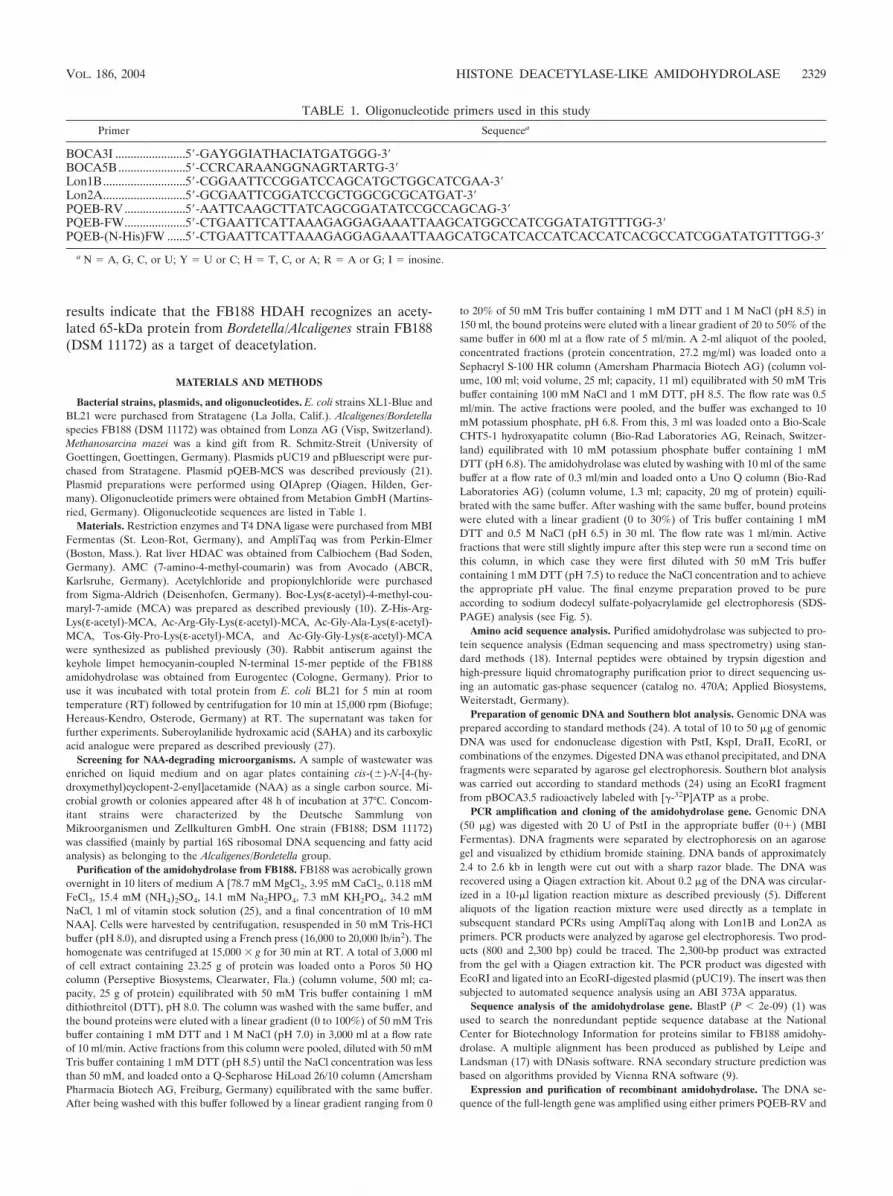
TABLE 1. Oligonucleotide primers used in this study
Primer Sequencea
BOCA3I .......................5�-GAYGGIATHACIATGATGGG-3�BOCA5B ......................5�-CCRCARAANGGNAGRTARTG-3�Lon1B...........................5�-CGGAATTCCGGATCCAGCATGCTGGCATCGAA-3�Lon2A...........................5�-GCGAATTCGGATCCGCTGGCGCGCATGAT-3�PQEB-RV ....................5�-AATTCAAGCTTATCAGCGGATATCCGCCAGCAG-3�PQEB-FW....................5�-CTGAATTCATTAAAGAGGAGAAATTAAGCATGGCCATCGGATATGTTTGG-3�PQEB-(N-His)FW ......5�-CTGAATTCATTAAAGAGGAGAAATTAAGCATGCATCACCATCACCATCACGCCATCGGATATGTTTGG-3�
a N � A, G, C, or U; Y � U or C; H � T, C, or A; R � A or G; I � inosine.
VOL. 186, 2004 HISTONE DEACETYLASE-LIKE AMIDOHYDROLASE 2329

PQEB-FW or primers PQEB-RV and PQEB-(N-His)FW (see Table 1), withFB188 genomic DNA as a template. The PCR products were double digestedwith EcoRI/HindIII and ligated into an EcoRI/HindIII double-digested plasmid(pQEB-MCS) (21). The constructs were used to transform E. coli strains XL1-Blue and BL21. The identities of all constructs were confirmed by automatedsequence analysis using an ABI 373A apparatus.
For gene expression studies, BL21 clones were grown in medium A with orwithout 1 mM NAA as the single C source. At an optical density of 0.8 at 600 nm,the cells were collected by centrifugation. The pellet was washed twice with 100mM sodium phosphate (pH 7.0). After the cells were resuspended in the samebuffer, the suspension was sonified 3 times for 30 s at 0°C. Cell debris wascollected by centrifugation, and the supernatant was subjected to immobilized-metal-ion affinity chromatography (IMAC) with Ni2� (as described by the sup-plier) (Qiagen) and an imidazole gradient. Alternatively, an IMAC procedurewith Zn2� was used. In each case, fractions containing active enzyme (as judgedby the hydrolysis of NAA) were pooled and dialyzed with three changes of 50mM sodium phosphate (pH 7.5). Aliquots were analyzed by SDS-–10% PAGE toreveal a purity of 95%. Protein concentration was estimated according to themethod of Bradford (4), with bovine serum albumin as a standard. The finalconcentration was 0.800 mg/ml, and the total yield was 1 mg of protein. Westernblotting analysis was performed according to standard protocols (24).
Assays for amidohydrolase activity. Enzyme activity was measured in fourdifferent assays. First, acetate enzymatically released from acetylputrescine, ace-tylcadaverine, or acetylspermidine by APAH-like activity was quantified by mon-itoring the absorption of NADH� at 340 nm with an acetic acid standard test(catalog no. 148261; Roche Diagnostics, Mannheim, Germany) as suggestedpreviously (2). Briefly, acetate is converted in the presence of acetyl-coenzyme A(CoA) synthetase with ATP and CoA to acetyl-CoA. The latter reacts withoxaloacetate to citrate in the presence of citrate synthetase. The oxaloacetaterequired for this reaction is formed from L-malate and NAD in the presence ofL-malate dehydrogenase upon reduction of NAD to NADH�. As a buffer, 100mM sodium phosphate (pH 7.5) was used.
Second, a two-step fluorogenic HDAC assay was carried out with peptidicsubstrate Boc-Lys(ε-acetyl)-MCA (see Fig. 6) as described previously (29, 30). Inthe first reaction catalyzed by the FB188 enzyme, acetate is released fromε-acetylated lysine moieties. In the second reaction, the deacetylated peptide isrecognized as a substrate by trypsin, which cleaves only after deacetylated lysineresidues. The release of AMC was monitored by measuring the fluorescence at460 nm (ex � 390 nm) with the help of a robotic workstation (CyBi-Screen-Machine; CyBio AG, Jena, Germany) including a Polarstar fluorescence reader(BMG, Offenburg, Germany). Fluorescence intensity was calibrated using freeAMC. Alternative pH values or additions of different salts were used in the firststep of the assay as indicated. To analyze substrate specificities of the enzyme,the assay was also carried out with Z-His-Arg-Lys(ε-acetyl)-MCA, Ac-Arg-Gly-Lys(ε-acetyl)-MCA, Ac-Gly-Ala-Lys(ε-acetyl)-MCA, Tos-Gly-Pro-Lys(ε-acetyl)-MCA, and Ac-Gly-Gly-Lys(ε-Ala)-MCA as substrates. With these tripeptidesubstrates, a novel protocol was used (29).
Third, HDAC activity was determined using [3H]acetate-prelabeled chickenhistones as described previously (16). Released radioactive acetate was extractedwith ethyl acetate, and the organic phase was counted for radioactivity in a liquidscintillation cocktail.
Finally, deacetylation of NAA (see Fig. 6) was monitored in an assay based onthe reaction of fluorescamine with primary amino groups (28). In a final volumeof 100 �l, 2 �l of enzyme in 100 mM phosphate buffer (pH 7.5) was incubated at37°C for 30 min, with a final concentration of 1 to 2,500 mM NAA. Subsequently,a 90-�l aliquot was transferred into the well of a microplate and mixed with 10�l of 0.3% fluorescamine in acetone. After 3 min at RT, fluorescence wasmeasured at a wavelength of 460 nm (ex � 390 nm). This fourth type of assaywas used throughout the purification of the enzyme to monitor enzymatic activityin fractions of eluates.
Western blot analysis of total soluble protein from FB188. Lysates from E. coliXL1-Blue (Stratagene), FB188, and M. mazei were obtained using a combinationof a French press (SLM Instruments, Urbana, Ill.) and a sonifier (Branson,Heusenstamm, Germany). Lysates were centrifuged for 10 min at 4°C, and thesupernatants were stored at 4°C until analysis. Aliquots of the supernatants weretreated for 1 h at 37°C with 0.2 �g of purified recombinant FB188 amidohydro-lase/�l. Equal amounts of amidohydrolase-treated or untreated total cellularprotein were loaded and electrophoresed on SDS-polyacrylamide gels, and theseparated proteins were transferred onto an Immobilon-P membrane (Millipore,Eschborn, Germany). An acetylated proteins antibody (ab193; Abcam, Cam-bridge, United Kingdom) was used that recognizes the ε-acetyl-lysine residue asan epitope and cross-reacts with a variety of acetylated proteins (acetylatedproteins antibody [ab193] data sheet; Abcam, 1998). Immune complexes were
detected with phosphatase-conjugated anti-rabbit immunoglobulin G (catalogno. A-8025; Sigma, Taufkirchen, Germany) and 5-bromo-4-chloro-3-indolylphosphate as a substrate.
Nucleotide sequence accession number. The nucleotide sequence of theamidohydrolase gene from FB188 and the encoded amino acid sequence havebeen deposited in the EMBL nucleotide database under accession numberAJ580773.
RESULTS
Amino acid sequence analysis of the FB188 HDAH. PurifiedHDAH was subjected to protein sequence analysis. Directanalysis of the N terminus yielded the peptide N terminus(AIGYVWNTLYGWVDTGTGSLAAANL). Thus, the N-terminal peptide did not start with a methionine. For fur-ther analysis, two internal sequences were obtained after gen-eration of peptides by trypsin digestion and high-pressure liq-uid chromatography purification, yielding peptide sequencesT-7 (VSNLPTGGDTGDGITMMGNGGLEIAR) and T-16(IVFVQEGGYSPHYLPFCGLAVIEELTGVR).
Cloning of the HDAH gene from FB188. Two degeneratedprimers (see Table 1), BOCA3I and BOCA5B, were designedaccording to the amino acid sequences of peptides T-7 andT-16. PCR using these primers and FB188 genomic DNA as atemplate revealed a 675-bp product. The PCR product wasfused to EcoRI linkers, digested with EcoRI, and ligated intoan EcoRI-digested plasmid, pBluescript, generating plasmidpBOCA3.5. Sequencing of the pBOCA3.5-insert revealed genesequences extending the primer sequences into T-7 and T-16encoding sequences, as expected. The EcoRI fragment radio-actively labeled with [�-32P]ATP was used as a probe in South-ern blot analysis to identify complementary genomic DNArestriction fragments. Separate digestions of genomic DNA byEcoRI, DraII, KspI, and PstI each revealed a single band of9.2, 7.3, 5.4, and 2.5 kb, respectively. Additionally, double di-gests with the aforementioned restriction endonucleases werecarried out to reveal a map of the coding region (Fig. 1). PstIfragments (2.4 to 2.6 kb) of genomic DNA were then isolatedfrom an agarose gel. The DNA was circularized and used as atemplate for inverse PCR (5) using Lon1B and Lon2A (seeTable 1) as primers. A 2,300-bp PCR product was digestedwith EcoRI and ligated into an EcoRI-digested plasmidpUC19. The insert was then subjected to automated sequenceanalysis using an ABI 373A apparatus. Two primers
FIG. 1. Restriction map of the chromosomal DNA fragment fromFB188 which carries the HDAH gene. EcoRI, DraII, KspI, and PstIeach revealed a single band hybridizing with the pBOCA3.5 probe.The region of DNA sequence analysis tallies with the PstI fragment.The HDAH open reading frame is depicted as a gray-shaded box withan arrow pointing from the start to the stop codon.
2330 HILDMANN ET AL. J. BACTERIOL.
(PQEB-RV and PQEB-FW; see Table 1) were designed on thebasis of the obtained sequence and used to amplify the full-length gene by PCR with genomic FB188 DNA as a template.The PCR product was ligated into plasmid pQEB-MCS (21),and the construct was used to transform E. coli strain XL1-Blue. Out of 10 clones analyzed, two clones (pQEB-AH6.6 andpQEB-AH6.8) carried the correct insertion, as confirmed byautomated sequence analysis. Alternatively, primersPQEB-RV and PQEB-(N-His)FW were used to amplify thegene containing an additional N-terminal His tag. Cloning ofthe extended gene into plasmid pQEB-MCS and subsequent
transformation of E. coli strain BL21 revealed clone pQEB-AH-NHis. The correct insertion was confirmed by automatedsequence analysis.
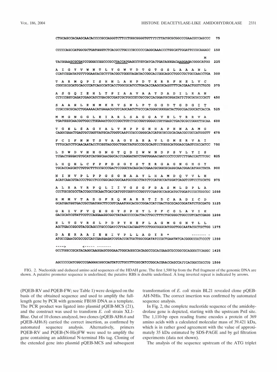
In Fig. 2, the complete nucleotide sequence of the amidohy-drolase gene is depicted, starting with the upstream PstI site.The 1,110-bp open reading frame encodes a protein of 369amino acids with a calculated molecular mass of 39.421 kDa,which is in rather good agreement with the value of approxi-mately 35 kDa estimated by SDS-PAGE and by gel filtrationexperiments (data not shown).
The analysis of the sequence upstream of the ATG triplet
FIG. 2. Nucleotide and deduced amino acid sequences of the HDAH gene. The first 1,500 bp from the PstI fragment of the genomic DNA areshown. A putative promoter sequence is underlined; the putative RBS is double underlined. A long inverted repeat is indicated by arrows.
VOL. 186, 2004 HISTONE DEACETYLASE-LIKE AMIDOHYDROLASE 2331
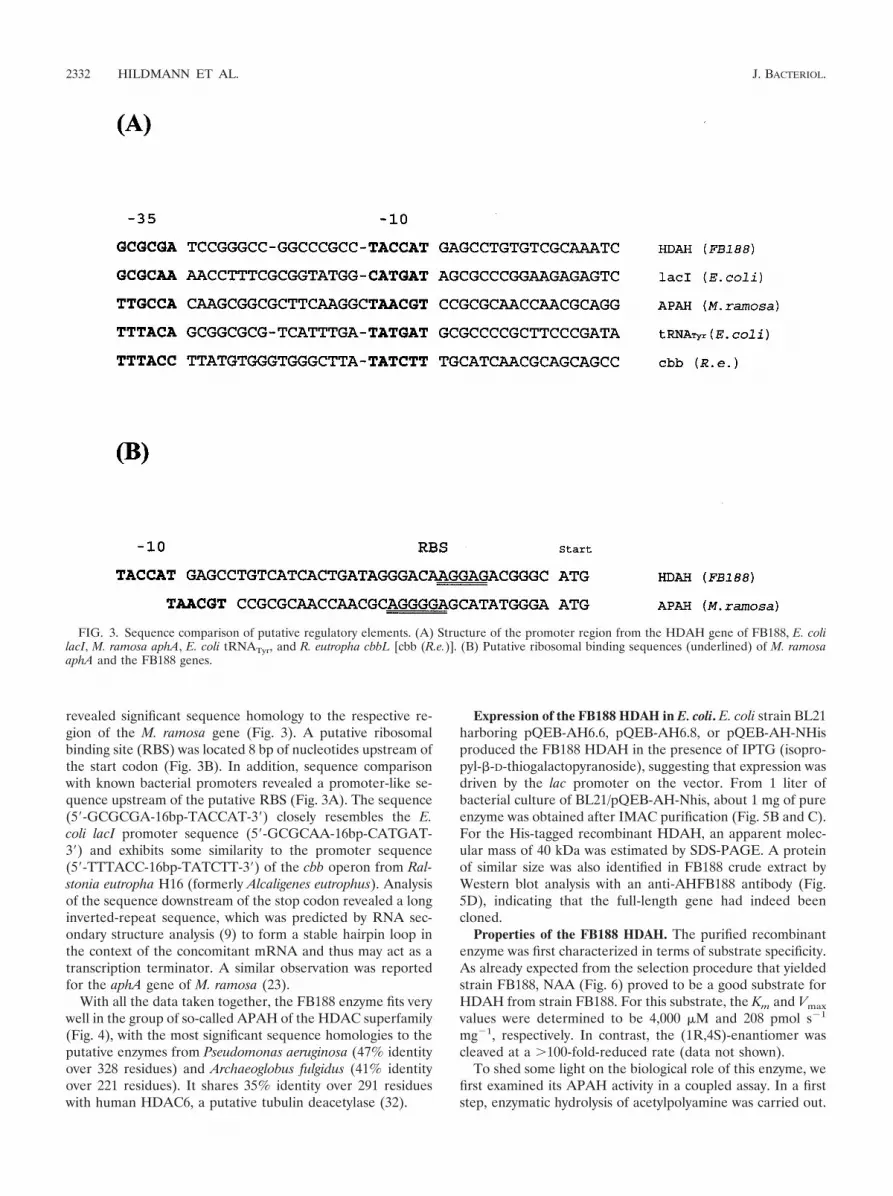
revealed significant sequence homology to the respective re-gion of the M. ramosa gene (Fig. 3). A putative ribosomalbinding site (RBS) was located 8 bp of nucleotides upstream ofthe start codon (Fig. 3B). In addition, sequence comparisonwith known bacterial promoters revealed a promoter-like se-quence upstream of the putative RBS (Fig. 3A). The sequence(5�-GCGCGA-16bp-TACCAT-3�) closely resembles the E.coli lacI promoter sequence (5�-GCGCAA-16bp-CATGAT-3�) and exhibits some similarity to the promoter sequence(5�-TTTACC-16bp-TATCTT-3�) of the cbb operon from Ral-stonia eutropha H16 (formerly Alcaligenes eutrophus). Analysisof the sequence downstream of the stop codon revealed a longinverted-repeat sequence, which was predicted by RNA sec-ondary structure analysis (9) to form a stable hairpin loop inthe context of the concomitant mRNA and thus may act as atranscription terminator. A similar observation was reportedfor the aphA gene of M. ramosa (23).
With all the data taken together, the FB188 enzyme fits verywell in the group of so-called APAH of the HDAC superfamily(Fig. 4), with the most significant sequence homologies to theputative enzymes from Pseudomonas aeruginosa (47% identityover 328 residues) and Archaeoglobus fulgidus (41% identityover 221 residues). It shares 35% identity over 291 residueswith human HDAC6, a putative tubulin deacetylase (32).
Expression of the FB188 HDAH in E. coli. E. coli strain BL21harboring pQEB-AH6.6, pQEB-AH6.8, or pQEB-AH-NHisproduced the FB188 HDAH in the presence of IPTG (isopro-pyl-�-D-thiogalactopyranoside), suggesting that expression wasdriven by the lac promoter on the vector. From 1 liter ofbacterial culture of BL21/pQEB-AH-Nhis, about 1 mg of pureenzyme was obtained after IMAC purification (Fig. 5B and C).For the His-tagged recombinant HDAH, an apparent molec-ular mass of 40 kDa was estimated by SDS-PAGE. A proteinof similar size was also identified in FB188 crude extract byWestern blot analysis with an anti-AHFB188 antibody (Fig.5D), indicating that the full-length gene had indeed beencloned.
Properties of the FB188 HDAH. The purified recombinantenzyme was first characterized in terms of substrate specificity.As already expected from the selection procedure that yieldedstrain FB188, NAA (Fig. 6) proved to be a good substrate forHDAH from strain FB188. For this substrate, the Km and Vmax
values were determined to be 4,000 �M and 208 pmol s�1
mg�1, respectively. In contrast, the (1R,4S)-enantiomer wascleaved at a 100-fold-reduced rate (data not shown).
To shed some light on the biological role of this enzyme, wefirst examined its APAH activity in a coupled assay. In a firststep, enzymatic hydrolysis of acetylpolyamine was carried out.
FIG. 3. Sequence comparison of putative regulatory elements. (A) Structure of the promoter region from the HDAH gene of FB188, E. colilacI, M. ramosa aphA, E. coli tRNATyr, and R. eutropha cbbL [cbb (R.e.)]. (B) Putative ribosomal binding sequences (underlined) of M. ramosaaphA and the FB188 genes.
2332 HILDMANN ET AL. J. BACTERIOL.

FIG. 4. Multiple sequence alignment of the regions of significant similarity in APAH, FB188 HDAH, and human HDAC6. Consensus residuesare indicated in shaded boxes. Numbers between dashes indicate the numbers of residues that have been omitted to improve the alignment asproposed by Leipe and Landsman (17). The following sequences are included in the alignment: HDAH (FB188), HDAH from Bordetella/Alcaligenes FB188; APAH (P. aeruginosa), putative APAH from proteobacterium P. aeruginosa; APAH (M. ramosa), APAH from proteobacteriumM. ramosa; APAH (M. jannaschii), putative APAH from archaeon M. jannaschii; APAH (A. fulgidus), putative APAH from A. fulgidus; APAH(“Pyrococcus abyssi”), putative APAH from “P. abyssi”; APAH (Aeropyrum pernix), putative APAH from A. pernix; APAH (M. thermautotr.),putative APAH from M. thermautotrophicus; APAH (Mesorhizobium loti), putative APAH from M. loti; APAH (Sy_cy. PCC6803), putative APAHfrom cyanobacterium Synechocystis sp. strain PCC6803; APAH (A. aeolicus) APAH from A. aeolicus; HDAC6 from H. sapiens (AF132609_1). Thepositions of putative zinc-binding amino acids in the M. ramosa enzyme (�) according to Sakurada et al. (23) and zinc-coordinating amino acids(*) in the structure of the A. aeolicus enzyme (6) are marked.
VOL. 186, 2004 HISTONE DEACETYLASE-LIKE AMIDOHYDROLASE 2333

In a second step, the released acetate was determined in astandard acetate assay. With acetylputrescine, acetylcadaver-ine, and acetylspermidine as substrates, 1 mg of the FB188HDAH was able to cleave 0.66, 0.48, and 0.24 �mol, respec-tively, in 1 h; these values compare to Vmax values of 29.1, 28,and 17.4 �mol min�1 mg�1 (Km of 0.19, 0.08, and 0.25 mM),respectively, for the enzyme from M. ramosa (23). This sug-gests a biological role of the FB188 HDAH different from thatof the APAH from M. ramosa.
Since the FB188 enzyme exhibited significant sequence sim-ilarities with HDACs, we next used standard HDAC assays toinvestigate any acetylprotein deacetylase activity. Interestingly,peptidic HDAC substrates proved to be substrates for theFB188 HDAH. For Boc-Lys(ε-acetyl)-MCA (Fig. 6), the Km
and Vmax values were determined to be 127 � 24 �M and 175� 20 pmol s�1 mg�1, respectively, compared to 3.7 � 1.7 �Mand 4.41 � 0.10 pmol s�1 mg�1 (30) for rat liver HDAC. Tofurther characterize the sequence specificity of the enzyme,time-dependent cleavage reactions were carried out for differ-ent peptidic substrates, including Z-His-Arg-Lys(ε-acetyl)-MCA, Ac-Arg-Gly-Lys(ε-acetyl)-MCA, Ac-Gly-Ala-Lys(ε-acetyl)-MCA, Tos-Gly-Pro-Lys(ε-acetyl)-MCA, and Ac-Gly-Gly-Lys(ε-acetyl)-MCA (Fig. 7). Here, Z-His-Arg-Lys(ε-acetyl)-MCA and Ac-Arg-Gly-Lys(ε-acetyl)-MCA also turnedout to be good substrates for the enzyme. However, the turn-over was diminished compared to that of Boc-Lys(ε-acetyl)-MCA. All other tripeptidic substrates proved to be poor sub-strates for the enzyme. In contrast, rat liver HDAC, whichcontains mainly HDAC-3 (D. Wegener and A. Schwienhorst,unpublished results), demonstrates a good turnover for alltripeptidic substrates, with a slight preference for Ac-Gly-Ala-Lys(ε-acetyl)-MCA (30). The HDAC-like activity of the FB188amidohydrolase could also be detected by incubation of theenzyme with acetate-radiolabeled histones within the scope ofthe most widely distributed type of HDAC assay (15). In thiscase, the FB188 HDAH was able to deacetylate [3H]acetate-
prelabeled chicken histones with a Km of 40 �M whereas trueHDACs of eukaryotic origin revealed Km values mostly be-tween 30 and 82 �M (16). Interestingly, acetylated tubulin wasable to act as a competitive inhibitor in the same assay (P.Loidl, unpublished results). Similarities with HDACs could beextended to inhibition of the latter by certain specific inhibi-tors. For trichostatin A (TSA), a known HDAC inhibitor (31),the FB188 HDAH showed a 50% inhibitory concentration(IC50) of 1.20 � 0.12 �M compared to 1.4 nM for rat liverHDAC (30). For the Aquifex aeolicus enzyme, however, anIC50 of 0.4 �M (similar to that of the FB188 HDAH) has beendetermined previously (6).
Since class 1 and 2 HDACs contain Zn2� in the active site,we subjected the FB188 enzyme to a thorough analysis con-cerning possible metal cofactors. First, the concentration ofzinc cations in active recombinant enzyme preparations fromstandard Ni-IMAC purification was determined by inductivelycoupled plasma (ICP) spectrometry using a Spectro ModulaICP apparatus (Spectro, Kleve, Germany). Three independentmeasurements revealed a concentration of 0.19 mol of Zn2�
per mol of enzyme (and an additional 1.17 mol Ni2�). As acontrol, recombinant HDAC8 (cloned and purified as de-scribed previously) (11) revealed a concentration of 5.9 mol ofZn2� per mol of enzyme. To further examine the possible roleof bivalent cations, the relative enzymatic activity levels in thepresence of 0 to 100 mM of EDTA, ZnSO4, CoCl2, NiCl2,CaCl2, MgCl2, and MnCl2 were determined using a two-stepHDAC assay with Boc-Lys(ε-acetyl)-MCA as a substrate (Fig.8). As a control, the hydrolysis of Boc-Lys-MCA was moni-tored under the same conditions to exclude a significant inhi-bition of trypsin in the second step of the assay. Concerning theinfluence on FB188 HDAH activity, metal cofactors fell intotwo classes. Only ZnSO4, CoCl2, and NiCl2 were able to in-crease the catalytic activity (2.2-, 1.3-, and 1.1-fold, respec-tively) at the relatively low concentration of 1 mM, whereashigher concentrations showed an inhibitory effect. In contrast,
FIG. 5. SDS gel electrophoretic analysis of cell lysates and purified enzyme fractions. (A) Native FB188 HDAH purified from Bordetella/Alcaligenes strain FB188 (DSM 11172). Lane 1, protein marker (purified FB188 HDAH prior to amino acid sequence analysis). (B) FB188 HDAHpurified by IMAC from E. coli BL21 harboring pQEB-AH-NHis. A benchmark prestained protein ladder (Gibco-Life Science, Karlsruhe,Germany) (lane 1), crude cell extract (lane 2), and purified protein (lane 3) were fractionated on an SDS–10% polyacrylamide gel. Protein bandswere visualized by Coomassie staining. (C) Western blotting using the anti-AHFB188 antibody (lanes are as described for panel B). (D) Westernblot analysis of FB188 crude cell extract with anti-AHFB188 antibody. Lane 1, benchmark prestained protein ladder; lane 2, purified recombinantFB188 HDAH; lane 3, FB188 cell extract; lane 4, E. coli XL1-Blue cell extract.
2334 HILDMANN ET AL. J. BACTERIOL.

MgCl2, MnCl2, and CaCl2 had no significant effect on activityof the FB188 HDAH at concentrations between 0 and 10 mM.At 100 mM concentrations, these compounds exhibited in-creases in catalytic activity of between 1.2- and 2.1-fold. WithEDTA, a similar effect was observed, with an increase in ac-tivity of 4.1-fold at 100 mM.
The stimulatory effect of additions of (in particular) Zn2�
ions as well as the results of the ICP measurements could (inprinciple) indicate that, e.g., as a result of the Ni-IMAC puri-fication only part of the enzyme preparation contains Zn2� inthe active site and thus is catalytically active. We thereforerepeated the IMAC purification, replacing Ni2� ions withZn2� ions. The resulting enzyme preparation showed in-creased specific activity. For Boc-Lys-(ε-acetyl)-MCA, the Km
and Vmax values were determined to be 14 � 3 �M and 136 �22 pmol s�1 mg�1, respectively. ICP analysis of the Zn-IMAC-purified enzyme revealed a concentration of 19.7 mol of Zn2�
per mol of enzyme.In the complexes of TSA or SAHA with HDAC-like enzyme
from A. aeolicus, hydroxamic acid coordinates a zinc in the
active site through its carbonyl and hydroxyl groups, contrib-uting significantly to the strong binding of the inhibitor (6). Itis known from comparative studies of hydroxamic acids and“parent” carboxylic acids (e.g., butyryl-hydroxamate and bu-tyrate) that carboxylic acid derivatives reveal a 30-fold in-crease in IC50 values compared to those of the correspondinghydroxamates (26). Thus, using the two-step assay to revealwhether Zn2� ions are also part of the active site of FB188HDAH, we studied the inhibition of the enzyme by HDACinhibitor SAHA and its carboxylic acid analogue SA-OH (Fig.9). For SAHA, an IC50 of 0.95 � 0.06 �M was determined withZn-IMAC-purified enzyme. The carboxylic acid analogueSA-OH was only a very weak inhibitor (less than 10% inhibi-tion at 100 �M) of FB188 HDAH.
The two-step HDAC assay was also used to analyze the pHdependence of the enzyme (Fig. 10). The pH profile is bellshaped, with a maximum value at pH 8.0, as in the case of theM. ramosa enzyme (23).
To directly assess the natural target of the FB188 HDAH,total protein from Bordetella/Alcaligenes species FB188 (DSM
FIG. 6. Substrates of the FB188 HDAH. Km and Vmax values were measured using different assays and the Ni-IMAC-purified enzyme.(A) Deacetylation of NAA was characterized using a standard fluorescamine assay (28). (B) For Boc-Lys(ε-acetyl)-MCA, the two-step HDACassay was used (30). (C) The deacetylation of [3H]acetate-prelabeled chicken histones was analyzed as described previously (16).
VOL. 186, 2004 HISTONE DEACETYLASE-LIKE AMIDOHYDROLASE 2335

11172) and a number of other bacteria was analyzed by West-ern blotting with acetylated proteins antibody ab193, recogniz-ing ε-acetyl-lysine residues (ab193 detects acetylated proteinsthat possibly become deacetylated upon incubation of total cellprotein with recombinant FB188 HDAH). Indeed, a numberof presumably acetylated proteins could be detected in totalcell protein from FB188, E. coli, Bacillus subtilis, and M. mazei.Preliminary Western blotting results revealed that only oneband corresponding to an acetylated protein of approximately65 kDa in the total protein of FB188 disappeared upon treat-ment with FB188 HDAH (data not shown). The same bandalso vanished upon treatment with trypsin or pepsin but wasnot affected by treatment with FB188 genomic DNA, tRNA,RNase A, DNase I, or ethidium bromide (data not shown).Studies using, e.g., specific high-affinity inhibitors of FB188HDAH are under way to verify these results and to furthercharacterize the presumable protein target of FB188 HDAH.
The enzyme obtained from FB188 exhibited essentially thesame biochemical properties as the recombinant enzyme puri-fied from E. coli, e.g., catalytic activity in the presence of metalcofactors, optimum pH, substrate specificity towards peptidicsubstrates, and a Km for Boc-Lys(ε-acetyl)-MCA of 92 � 31�M compared to 127 � 24 �M for the recombinant Ni-IMAC-purified enzyme.
DISCUSSION
Here, we present the nucleotide sequence of a new amidohy-drolase gene from Bordetella/Alcaligenes species FB188 (DSM11172). Putative regulatory sequences have been identified on
the basis of sequence comparison and certainly need verifica-tion by a thorough experimental analysis. A comparison of thededuced amino acid sequence and the N-terminal protein se-quence revealed that the N-terminal methionine is lacking inthe wild-type enzyme. As known from studies of E. coli methi-onine aminopeptidase, N-terminal methionines are efficientlyprocessed when the penultimate amino acid is small, as it is thecase with alanine in the FB188 HDAH (8). Comparison of thededuced amino acid sequence with those in the National Cen-ter for Biotechnology Information databases revealed a num-
FIG. 7. Sequence specificity of the FB188 HDAH. The deacetylationreactions of Boc-Lys(ε-acetyl)-MCA, Z-His-Arg-Lys(ε-acetyl)-MCA, Ac-Arg-Gly-Lys(ε-acetyl)-MCA, Ac-Gly-Ala-Lys(ε-acetyl)-MCA, Tos-Gly-Pro-Lys(ε-acetyl)-MCA, and Ac-Gly-Gly-Lys(ε-acetyl)-MCA were ana-lyzed using the two-step HDAC assay (30). Standard reaction mixturescontained 125 �M of the respective substrates in HDP buffer (15 mMTris-HCl [pH 8.1], 250 �M EDTA, 250 mM NaCl, 0.1% PEG 8000) and4 �g of amidohydrolase. After deacetylation, deacetylated substrate mol-ecules were cleaved by trypsin, which at the same time releases fluorescent7-amino-4-methylcoumarin. Fluorescence measurement is conducted at aex of 390 nm and a em of 460 nm. AFU, arbitrary fluorescence units.
FIG. 8. Effect of bivalent cations on amidohydrolase activity. Thetwo-step HDAC assay (30) was used to analyze the effect of ZnCl2,CoCl2, and NiCl2 (A) and CaCl2, MgCl2, MnCl2, and EDTA (B) on theactivity of the recombinant FB188 HDAH. AFU, arbitrary fluores-cence units.
2336 HILDMANN ET AL. J. BACTERIOL.

ber of homologs, all belonging to the subgroup of APAH of theHDAC superfamily (Fig. 2). The closest homolog among hu-man HDACs is HDAC6, a presumptive tubulin deacetylase(12).
To facilitate a thorough biochemical characterization, theFB188 HDAH has been cloned and overexpressed in E. coli.Purification has been expedited by introducing an N-terminalHis tag into the mature protein. Interestingly, a protein variantwith a C-terminal His tag revealed a 5- to 10-fold-lower activitylevel (data not shown). The purified recombinant enzymeshows biochemical properties essentially the same as those ofthe natural enzyme from Bordetella/Alcaligenes species FB188(DSM 11172), as determined for pH optimum, substrate spec-ificity, and cation requirements.
In agreement with findings concerning the enzymes from M.ramosa and A. aeolicus, several lines of evidence indicate that
the new enzyme is only catalytically active in the presence ofbivalent cations such as Zn2�. First, a comparative sequenceanalysis has been carried out to look for amino acids thatpossibly could form a metal-binding site in the FB188 HDAH.On the basis of sequence similarities with carboxypeptidases A,A1, and A2, the metal-binding site in the M. ramosa enzymehas been proposed to consist of four putative zinc bindingamino acids (8), two of which are lacking in the FB188 HDAH(Fig. 4). With the first crystal structure of an enzyme from theHDAC superfamily (6), however, the metal-binding site nowcan be identified far more reliably. Assuming that the FB188enzyme folds into a structure very similar to that of the ho-mologous enzyme from A. aeolicus, all amino acids responsiblefor zinc binding appear to be present in the FB188 HDAH. Inconclusion, a putative metal-binding site may be present in theFB188 HDAH.
Second, Zn2� (and, to a smaller extent, Co2� and Ni2� also)was able to increase the catalytic activity at relatively low con-centrations of 1 mM whereas higher concentrations revealedan inhibitory effect. An inhibitory effect of higher Zn2� con-centrations has also been observed for the APAH from M.ramosa, which was suggested to be an indication for a site-specific inhibitory binding of Zn2� to the M. ramosa enzyme(8). It is noteworthy at this point that (in comparison to theNi-IMAC-purified enzyme) the FB188 HDAH purified by Zn-IMAC presumably kept more Zn2� ions in the active site, sinceit showed higher specific activity and a significant decrease inthe Km value for Boc-Lys(ε-acetyl)-MCA. Third, in contrast tothe results seen with Co2�, Ni2�, and Zn2�, EDTA (as well asMgCl2, MnCl2, and CaCl2) had a stimulatory effect on catalyticactivity at high concentrations (100 mM). We are inclined tointerpret these results as an unspecific effect of the presence ofa high electrolyte concentration. At this point, it is interestingthat in addition to EDTA, imidazole (which had also been usedat concentrations of up to 200 mM in the IMAC purificationprocedure) was obviously unable to inactivate the FB188HDAH, e.g., through extraction of bivalent metals. The samebehavior is known from HDACs and the M. ramosa enzyme(23), which release Zn2� from the active site only under rela-tively harsh conditions. In contrast, the enzyme from A. aeoli-cus was reported to lose its metal cofactor completely duringthe purification procedure and has to be incubated with zincchloride to gain catalytic activity (6).
Finally, hydroxamates like TSA and SAHA (known to in-hibit HDAC-like enzymes by interacting with zinc in the activesite) have been shown to inhibit the FB188 HDAH. AlthoughFB188 HDAH inhibition was observed at IC50 values in-creased by a factor of 1,000 compared to those measured foreukaryotic HDACs, this fact does not necessarily support theview that zinc is absent from the active site of the enzyme.Alternatively, the increase in IC50 could be explained by adifferent shape of the active site that results in the loss ofoptimal inhibitor binding. This is presumably also the case forthe A. aeolicus enzyme (6), which contains zinc in the activesite and shows IC50 values very similar to those measured forthe FB188 HDAH. Further evidence for the presence of zinc inthe active site of the FB188 enzyme comes from inhibitionstudies with SAHA and its carboxylic acid analogue SA-OH;with regard to IC50 values, the latter was a less active inhibitorby a factor of 100 compared to SAHA. In the absence of zinc,
FIG. 9. IC50 measurements (using FB188 HDAH) for SAHA andits carboxylic acid analogue SA-OH. Inhibitory activity was determinedin the two-step assay (30) with inhibitor concentrations between 0.0001and 100 �M. The Zn-IMAC-purified enzyme was used. Similar resultswere obtained with the Ni-IMAC-purified enzyme (data not shown).AFU, arbitrary fluorescence units.
FIG. 10. pH dependency of amidohydrolase activity. The two-stepHDAC assay (30) was used to analyze the effect of pH on the activityof the recombinant FB188 HDAH. AFU, arbitrary fluorescence units.
VOL. 186, 2004 HISTONE DEACETYLASE-LIKE AMIDOHYDROLASE 2337
very similar inhibition constants would have to be expected. Inconclusion, the catalytically active form of the enzyme almostcertainly contains a zinc ion in the active site, as is known forclass 1 HDACs. Ongoing crystallographic studies should revealwhether similarities with other HDAC-like enzymes can beextended to the geometry of the active site and the bindingmode of HDAC inhibitors.
The independence of cofactors, the broad optimum pHrange, and the large substrate spectrum lead us to suggestusing the new HDAH from Bordetella/Alcaligenes strain FB188as a biocatalyst in technical biotransformations. Indeed, feasi-bility of the new HDAH as an enantioselective, technical bio-catalyst was demonstrated using NAA, a precursor of certainnew anti-human immunodeficiency virus drugs (13), as a sub-strate.
To narrow down the possible characteristics of the biologicalrole of this novel enzyme, a number of different substrateswere analyzed. Surprisingly, acetylpolyamines were rather poorsubstrates, indicating that the enzyme might not play a signif-icant role in the degradative pathway of acetylpolyamines inFB188 but might rather be involved in the regulation of othercellular processes. Since the sequence of the FB188 HDAHshowed significant homologies to those of HDACs, we testedthe enzyme for a possible HDAC-like activity in a variety ofassays. Indeed, the enzyme exhibited significant levels of pro-tein deacetylase activity comparable to those of eukaryoticHDACs in assays with both fluorogenic peptidic substrates andacetate-radiolabeled histones. The acetate-radiolabeled his-tone assays revealed that the FB188 HDAH is able to acceptproteins with ε-acetylated lysine residues as substrates. Fur-ther, [3H]acetate-prelabeled chicken histones are deacetylatedwith Km values comparable to those of eukaryotic HDACs.Interestingly, tubulin was able to act as a competitive inhibitorin these assays (P. Loidl, unpublished), adding further evi-dence for a similarity between the FB188 HDAH and HDAC6(32). With tripeptidic substrates, only Z-His-Arg-Lys(ε-acetyl)-MCA and Ac-Arg-Gly-Lys(ε-acetyl)-MCA proved to be supe-rior substrates, suggesting that the FB188 HDAH (in contrastto rat liver HDAC) might have a certain sequence specificitywith a preference for positive charges in the vicinity of thecleavage site. Similarities with eukaryotic HDACs, however,could be further extended by showing that classical HDACinhibitors such as TSA and SAHA were able to specificallyinhibit the FB188 HDAH.
To shed some light on a possible biological role of the FB188HDAH, we thought of further testing the enzyme for its in-herent protein deacetylase activity by identifying a possiblenatural target of the FB188 HDAH. Therefore, total protein ofBordetella/Alcaligenes species FB188 (DSM 11172), E. coliXL1-Blue, and M. mazei was analyzed by Western blotting withan antibody (ab193) recognizing ε-acetyl-lysine residues, e.g.,in a number of different proteins (acetylated proteins antibody[ab193] datasheet, Abcam, 1998). In each case, we assumedthat acetylated proteins were detected. Preliminary experi-ments concerning the treatment of total protein with FB188HDAH revealed that (specifically) one protein band at approx-imately 65 kDa in the total protein of FB188 disappeared upontreatment with FB188 HDAH. The same band also vanishedafter incubation of total cell protein with trypsin or pepsin butnot by treatment with tRNA, genomic double-stranded DNA,
RNase A, DNase I, or ethidium bromide, thus suggesting thatthe target is a protein that is not strongly bound to nucleic acid.
In conclusion, we would like to suggest that there is a pos-sibility that the FB188 HDAH from Bordetella/Alcaligenes spe-cies FB188 (DSM 11172) is the first example of a well-charac-terized bacterial protein deacetylase that (in vivo) specificallydeacetylates a protein that presumably is not related to thefunctional context of DNA binding, as is the case for Alba, anacetylatable 10-kDa protein from the archaeon S. solfataricusP2 (3). More far-reaching studies have now been started tofurther characterize the presumable substrate protein of theFB188 HDAH to illuminate what now seems to be a novelbiological role of HDAC-like enzymes.
ACKNOWLEDGMENTS
This work was supported in part by BioFuture grant 0311852 andgrant 0311816 from the Bundesministerium fur Forschung und Tech-nologie.
We are grateful to A. Kriegel from the Institut fuer Bodenkundeund Waldernaehrung, Georg-August University of Goettingen, forassistance concerning ICP spectrometry. We also thank H. Brass andR. Willmott for many fruitful discussions and for reading the manu-script. Technical support from Cybio AG and BMG GmbH is grate-fully acknowledged.
REFERENCES
1. Altschul, S. F., and D. J. Lipman. 1990. Protein database searches formultiple alignments. Proc. Natl. Acad. Sci. USA 87:5509–5513.
2. Baumann, M., R. Sturmer, and U. T. Bornscheuer. 2001. A high-throughput-screening method for the identification of enantioselective hydrolases. An-gew. Chem. Int. Ed. Engl. 40:4201–4204.
3. Bell, S. D., C. H. Botting, B. N. Wardleworth, S. P. Jackson, and M. F. White.2002. The interaction of Alba, a conserved archaeal chromatin protein, withSir2 and its regulation by acetylation. Science 296:148–151.
4. Bradford, M. M. 1976. A rapid and sensitive method for the quantitative ofmicrogram quantities of protein utilizing the principle of protein dye bind-ing. Anal. Biochem. 72:248–254.
5. Collins, F. S., and S. M. Weissman. 1984. Directional cloning of DNAfragments at a large distance from an initial probe: a circularization method.Proc. Natl. Acad. Sci. USA 81:6812–6816.
6. Finnin, M. S., J. R. Donigian, A. Cohen, V. M. Richon, R. A. Rifkind, P. A.Marks, R. Breslow, and N. P. Pavletich. 1999. Structures of a histonedeacetylase homologue bound to TSA and SAHA inhibitors. Nature 401:188–193.
7. Grozinger, C. M., and S. L. Schreiber. 2002. Deacetylase enzymes: biologicalfunctions and the use of small-molecule inhibitors. Chem. Biol. 9:3–16.
8. Hirel, P.-H., J.-M. Schmitter, P. Dessen, G. Fayat, and S. Blanquet. 1989.Extent of N-terminal methionine excision from Escherichia coli proteins isgoverned by the side-chain length of the penultimate amino acid. Proc. Natl.Acad. Sci. USA 86:8247–8251.
9. Hofacker, I. L., W. Fontana, and P. F. Stadler. 1994. Fast folding andcomparison of (RNA) secondary structures. Monatsh. Chem. 125:167–188.
10. Hoffmann, K., G. Brosch, P. Loidl, and M. Jung. 2000. First non-radioactiveassay for in vitro screening of histone deacetylase. Pharmazie 55:601–606.
11. Hu, E., Z. Chen, T. Fredrickson, Q. Zhu, R. Kirkpatrick, G.-F. Zhang, K.Johanson, C.-M. Sung, R. Liu, and J. Winkler. 2000. Cloning and charac-terization of a novel human class I histone deacetylase that functions as atranscriptional repressor. J. Biol. Chem. 275:15254–15264.
12. Hubbert, C., A. Guardiola, R. Shao, Y. Kawaguchi, A. Ito, A. Nixon, M.Yoshida, X.-F. Wang, and T.-P. Yao. 2002. HDAC6 is a microtubule-associ-ated deacetylase. Nature 417:455–458.
13. Huff, J. R. 1999. Abacavir sulfate. Bioorg. Med. Chem. 7:2667–2669.14. Khochbin, S., and A. P. Wolffe. 1997. The origin and utility of histone
deacetylase. FEBS Lett. 419:157–160.15. Kolle, D., G. Brosch, T. Lechner, A. Lusser, and P. Loidl. 1998. Biochemical
methods for analysis of histone deacetylases. Methods 15:323–331.16. Lechner, T., A. Lusser, G. Brosch, A. Eberharter, M. Goralik-Schramel, and
P. Loidl. 1996. A comparative study of histone deacetylases of plant, fungaland vertebrate cells. Biochim. Biophys. Acta 1296:181–188.
17. Leipe, D. D., and D. Landsman. 1997. Histone deacetylases, acetoin utiliza-tion proteins and acetylpolyamine amidohydrolases are members of an an-cient protein superfamily. Nucleic Acids Res. 25:3693–3697.
18. Link, A. J. 1999. 2-D Proteome analysis protocols. Humana Press, Totowa,N.J.
2338 HILDMANN ET AL. J. BACTERIOL.
19. Marmorstein, R. 2001. Structure of histone deacetylases: insights into sub-strate recognition and catalysis. Structure 9:1127–1133.
20. Marton, L. J., and A. E. Pegg. 1995. Polyamines as targets for therapeuticintervention. Annu. Rev. Pharmacol. Toxicol. 35:55–91.
21. Ninkovic, M., R. Dietrich, G. Aral, and A. Schwienhorst. 2001. High fidelityin vitro recombination using a proofreading polymerase. BioTechniques30:530–536.
22. Ramakrishnan, R., M. Schuster, and R. B. Bourret. 1998. Acetylation atLys-92 enhances signaling by the chemotaxis response regulator proteinCheY. Proc. Natl. Acad. Sci. USA 95:4918–4923.
23. Sakurada, K., T. Ohta, K. Fujishiro, M. Hasegawa, and K. Aisaka. 1996.Acetylpolyamine amidohydrolase from Mycoplana ramosa: gene cloning andcharacterization of the metal-substituted enzyme. J. Bacteriol. 178:5781–5786.
24. Sambrook, J., E. F. Fritsch, and T. Maniatis. 1989. Molecular cloning, 2nded. Cold Spring Harbor Laboratory Press, Cold Spring Harbor, N.Y.
25. Schlegel, H. G. 1985. Allgemeine Mikrobiologie. Thieme Verlag, Stuttgart,Germany.
26. Skarpidi, E., H. Cao, B. Heltweg, B. F. White, R. L. Marhenke, M. Jung, andG. Stamatoyannopoulos. 2003. Hydroxamide derivatives of short-chain fatty
acids are potent inducers of human fetal globin gene expression. Exp. He-matol. 31:197–203.
27. Stowell, J. C., R. I. Huot, and L. Van Voast. 1995. The synthesis of N-hydroxy-N�-phenyloctanediamide and its inhibitory effect on proliferation ofAXC rat prostate cells. J. Med. Chem. 38:1411–1413.
28. Udenfriend, S., S. Stein, P. Bohlen, W. Dairman, W. Leimgruber, and M.Weigele. 1972. Fluorescamine: a reagent for assay of amino acids, peptides,proteins, and primary amines in the picomole range. Science 178:871–872.
29. Wegener, D., C. Hildmann, D. Riester, and A. Schwienhorst. 2003. Improvedfluorogenic histone deacetylase assay for high-throughput-screening appli-cations. Anal. Biochem. 321:202–208.
30. Wegener, D., F. Wirsching, D. Riester, and A. Schwienhorst. 2003. A flu-orogenic histone deacetylase assay well suited for high-throughput activityscreening. Chem. Biol. 10:61–68.
31. Yoshida, M., M. Kijima, M. Akita, and T. Beppu. 1990. Potent and specificinhibition of mammalian histone deacetylase both in vivo and in vitro bytrichostatin A. J. Biol. Chem. 265:17174–17179.
32. Zhang, Y., N. Li, C. Caron, G. Matthias, D. Hess, S. Khochbin, and P.Matthias. 2003. HDAC-6 interacts with and deacetylates tubulin and micro-tubules in vivo. EMBO J. 22:1168–1179.
VOL. 186, 2004 HISTONE DEACETYLASE-LIKE AMIDOHYDROLASE 2339