Deacetylases and NF κ B in Redox Regulation of Cigarette Smoke-Induced Lung Inflammation:...
22
Deacetylases and NF-κB in Redox Regulation of Cigarette Smoke induced Lung Inflammation: Implications in Pathogenesis of COPD Saravanan Rajendrasozhan, Se-Ran Yang, Indika Edirisinghe, Hongwei Yao, David Adenuga, and Irfan Rahman Department of Environmental Medicine, Lung Biology and Disease Program, University of Rochester Medical Center, Rochester, NY, USA Abstract Oxidative stress has been implicated in the pathogenesis of several inflammatory lung disorders including chronic obstructive pulmonary disease (COPD) due to its effect on pro-inflammatory gene transcription. Cigarette smoke-mediated oxidative stress activates NF-κB-dependent transcription of pro-inflammatory mediators either through activation of inhibitor κB-α kinase (IKK) and/or the enhanced recruitment and activation of transcriptional co-activators. Enhanced NF-κB-co-activator complex formation results in targeted increase in chromatin modifications, such as histone acetylation leading to inflammatory gene transcription. NF-κB-dependent gene expression, at least in part, is regulated by changes in deacetylases such as histone deacetylases (HDACs) and sirtuins. Cigarette smoke and oxidants also alter the levels/activity of HDAC by post-translational modifications and in doing so further induces gene expression of pro-inflammatory mediators. In addition, cigarette smoke/oxidants can reduce glucocorticoid sensitivity by attenuating HDAC2 activity and expression, which may account for the glucocorticoid insensitivity in patients with COPD. Understanding the mechanisms of NF-κB regulation, and the balance between histone acetylation and deacetylation may lead to the development of novel therapies based on the pharmacological manipulation of IKK and deacetylases in lung inflammation and injury. Keywords COPD; reactive oxygen species; histone acetylation; HDAC; sirtuins INTRODUCTION Biological systems are continuously exposed to oxidants either generated endogenously by metabolic reactions (mitochondrial electron transport during respiration, during activating phagocytes) or exogenously (air pollutants or cigarette smoke). The great external surface area (1–2 m 2 ) of the human airway epithelium (300 million alveoli), alveolar ducts (14 million), capillary segments (280 billion) plus its direct contact with the high-oxygen environment for gas exchange between air and blood (alveolar and alveolar-capillary surface area=40 to 80 square meters) makes the lung a major target for oxidative injury from reactive oxygen species (ROS) and free radicals (5,71,73). Inflammation is an important protective response to cellular/ tissue injury. The purpose of this process is to destroy and remove the injurious agent and Address for correspondence: Dr. Irfan Rahman, Department of Environmental Medicine, Lung Biology and Disease Program, University of Rochester Medical Center, Box 850, 601 Elmwood Avenue, Rochester 14642, New York, USA, Tel: 1 585 275 6911, Fax: 1 585 506 0239, [email protected]. NIH Public Access Author Manuscript Antioxid Redox Signal. Author manuscript; available in PMC 2009 October 7. Published in final edited form as: Antioxid Redox Signal. 2008 April ; 10(4): 799–811. doi:10.1089/ars.2007.1938. NIH-PA Author Manuscript NIH-PA Author Manuscript NIH-PA Author Manuscript
Transcript of Deacetylases and NF κ B in Redox Regulation of Cigarette Smoke-Induced Lung Inflammation:...

Deacetylases and NF-κB in Redox Regulation of Cigarette Smokeinduced Lung Inflammation: Implications in Pathogenesis ofCOPD
Saravanan Rajendrasozhan, Se-Ran Yang, Indika Edirisinghe, Hongwei Yao, DavidAdenuga, and Irfan RahmanDepartment of Environmental Medicine, Lung Biology and Disease Program, University ofRochester Medical Center, Rochester, NY, USA
AbstractOxidative stress has been implicated in the pathogenesis of several inflammatory lung disordersincluding chronic obstructive pulmonary disease (COPD) due to its effect on pro-inflammatory genetranscription. Cigarette smoke-mediated oxidative stress activates NF-κB-dependent transcription ofpro-inflammatory mediators either through activation of inhibitor κB-α kinase (IKK) and/or theenhanced recruitment and activation of transcriptional co-activators. Enhanced NF-κB-co-activatorcomplex formation results in targeted increase in chromatin modifications, such as histone acetylationleading to inflammatory gene transcription. NF-κB-dependent gene expression, at least in part, isregulated by changes in deacetylases such as histone deacetylases (HDACs) and sirtuins. Cigarettesmoke and oxidants also alter the levels/activity of HDAC by post-translational modifications andin doing so further induces gene expression of pro-inflammatory mediators. In addition, cigarettesmoke/oxidants can reduce glucocorticoid sensitivity by attenuating HDAC2 activity and expression,which may account for the glucocorticoid insensitivity in patients with COPD. Understanding themechanisms of NF-κB regulation, and the balance between histone acetylation and deacetylationmay lead to the development of novel therapies based on the pharmacological manipulation of IKKand deacetylases in lung inflammation and injury.
KeywordsCOPD; reactive oxygen species; histone acetylation; HDAC; sirtuins
INTRODUCTIONBiological systems are continuously exposed to oxidants either generated endogenously bymetabolic reactions (mitochondrial electron transport during respiration, during activatingphagocytes) or exogenously (air pollutants or cigarette smoke). The great external surface area(1–2 m2) of the human airway epithelium (300 million alveoli), alveolar ducts (14 million),capillary segments (280 billion) plus its direct contact with the high-oxygen environment forgas exchange between air and blood (alveolar and alveolar-capillary surface area=40 to 80square meters) makes the lung a major target for oxidative injury from reactive oxygen species(ROS) and free radicals (5,71,73). Inflammation is an important protective response to cellular/tissue injury. The purpose of this process is to destroy and remove the injurious agent and
Address for correspondence: Dr. Irfan Rahman, Department of Environmental Medicine, Lung Biology and Disease Program,University of Rochester Medical Center, Box 850, 601 Elmwood Avenue, Rochester 14642, New York, USA, Tel: 1 585 275 6911, Fax:1 585 506 0239, [email protected].
NIH Public AccessAuthor ManuscriptAntioxid Redox Signal. Author manuscript; available in PMC 2009 October 7.
Published in final edited form as:Antioxid Redox Signal. 2008 April ; 10(4): 799–811. doi:10.1089/ars.2007.1938.
NIH
-PA Author Manuscript
NIH
-PA Author Manuscript
NIH
-PA Author Manuscript
injured tissues, thereby promoting tissue repair. When this crucial and normally beneficialresponse occurs in an uncontrolled manner, the result is excessive cellular/tissue damage thatresults in chronic inflammation and destruction of normal tissue. ROS, such as superoxideanion (O2
•-) liberated by phagocytes recruited to sites of inflammation, are proposed to be amajor cause of the cell and tissue damage associated with many chronic inflammatory diseasessuch as asthma and chronic obstructive pulmonary disease (COPD) (58,72). Increased levelsof ROS have been implicated in initiating the lung inflammatory responses through theactivation of redox sensitive transcription factor, nuclear factor-kappa B (NF-κB), andchromatin remodeling (histone acetylation/deacetylation), which regulate the gene expressionof pro-inflammatory mediators (70) (Figure 1). In this review, we present an overview of redoxregulation of NF-κB and describe the current evidence on involvement of histone deacetylasein oxidant-mediated lung inflammation.
OXIDATIVE STRESSAerobic metabolism is the major source of ROS and reactive nitrogen (RNS) species, whichare essentially the byproducts of normal oxygen metabolism. Biological systems are capableof forming highly reactive moieties, both free radicals and non-radicals. These biologicallyactive species participate in normal cell functions by serve in host defense and also in cellsignaling as messenger molecules of the autocrine or paracrine system (77), but their excessproduction may result in tissue injury and inflammation (38). To combat these unrelentinginsults, the lungs have well coordinated and efficient endogenous antioxidant defense systems,which protect against the injurious effects of oxidants by electron transfer, enzymatic removal,scavenging and by keeping transition metal ions tightly sequestered. There are three majorclassifications in the endogenous antioxidant defenses. i) Primary defense: It can preventradical formation. Transferrin and lactoferrin have this role in extracellular fluids. ii) Secondarydefense: It removes or inactivates formed ROS. This may be enzymes such as superoxidedismutase, and glutathione peroxidase as well as non-enzymatic molecules such as reducedglutathione, α-tocopherol, ascorbic acid, β-carotene, albumin, mucin, glucoase, bilirubin anduric acid. iii) Tertiary defense: This system operates to remove and or repair oxidativelydamaged molecules, and is particularly important for proteins and deoxyribonucleic acids(5). The lung has precise balance between the production/exposure of oxidants and endogenousantioxidants. Oxidative stress resulting from an imbalanced ratio between ROS production anddetoxification plays an important role in cellular processes such as signal transduction and geneexpression (25). During acute and chronic inflammations, ROS is produced at rates thatoverwhelm the capacity of the endogenous antioxidant defense system to remove it, resultingin oxidative stress (25). Increased ROS production and reduced endogenous antioxidantdefense has been reported in several lung diseases including chronic obstructive pulmonarydisease (52,68,73). This may results in oxidative stress condition, which appears involved invarious chronic inflammatory lung disease pathogenesis.
ENDOGENOUS ROS PRODUCTION IN THE LUNGReactive oxygen and nitrogen species are generated by several inflammatory and structuralcells of the airways. A common feature of all inflammatory lung diseases is involvement of aninflammatory-immune response, characterized by activation of epithelial cells, and residentmacrophages, and the recruitment and activation of neutrophils, eosinophils, monocytes, andlymphocytes. Inflammatory cells once recruited in the airspace become activated and generateROS in response to a sufficient level of a secretagogue stimulus (threshold condition). Theactivation of macrophages, neutrophils and eosinophils generates O2
•-, which is rapidlyconverted to hydrogen peroxide (H2O2) by superoxide dismutase (SOD), and hydroxyl radical(•OH) formed non-enzymatically in the presence of Fe2+ as a secondary reaction. ROS canalso be generated intracellularly from several sources such as mitochondrial respiration, the
Rajendrasozhan et al. Page 2
Antioxid Redox Signal. Author manuscript; available in PMC 2009 October 7.
NIH
-PA Author Manuscript
NIH
-PA Author Manuscript
NIH
-PA Author Manuscript
NADPH oxidase system and xanthine/xanthine oxidase (65). At sites of inflammation,increased free radical activity is associated with the activation of the neutrophil NADPHoxidase and/or the uncoupling of a variety of redox systems, including endothelial cell xanthinedehydrogenase (23,53).
ENVIRONMENTAL OXIDANTSThe air is polluted with cigarette smoke, automobile exhaust, diesel soot, ozone, sulfur dioxide,nitrogen dioxide and varying degree of other pollutants produced by anthropogenic activities.Due to the unique structure and function of the lung, it is vulnerable to numerous pathogens,pollutants, oxidants, gases and toxicants that are inhaled continuously with ambient air.Inhalation of such toxic pollutants and microorganisms results in lung injury and generationof reactive oxygen/nitrogen species leading to cascades of signaling events triggeringinflammatory influx, production of proinflammatory cytokines, chemokines, and other factors(21). Cigarette smoke is the major etiological factor in the pathogenesis of COPD (66). Anincreased oxidant burden in smokers derives from the fact that cigarette smoke contains anestimated 1017 oxidants/free radicals and 4,700 chemical compounds, including aldehydes andquinines per puff, and many of those are relatively long-lived such as tar-semiquinone that cangenerate •OH and H2O2 by Fenton reaction (14).
OXIDATIVE STRESS AND INFLAMMATIONOxidative stress has been implicated in cell and tissue damage associated with many chronicinflammatory lung diseases such as asthma, COPD, idiopathic pulmonary fibrosis (IPF) andadult respiratory distress syndrome (ARDS) (1). Increased ROS production has been directlylinked to oxidation of protein, DNA, and lipids which may cause direct lung injury or inducea variety of cellular responses through the generation of secondary metabolic reactive species.The pathogenesis of many forms of lung injury has implicated peroxidative breakdown ofpolyunsaturated fatty acids due to the effects on membrane function, inactivation of membrane-bound receptors and enzymes, and increased tissue permeability (67). There is increasingevidence that aldehydes, generated endogenously during the process of lipid peroxidation, areinvolved in many of the pathophysiological events associated with oxidative stress in cells andtissues (36). In addition to their cytotoxic properties, lipid peroxides are increasinglyrecognized as being important in signal transduction for a number of important events in theinflammatory response in the lungs (67).
ROS may alter the remodeling of extracellular matrix, apoptosis and mitochondrial respiration,cell proliferation, maintenance of surfactant and the antiprotease screen, effective alveolarrepair responses and immune modulation in the lung (65,66). ROS produced by phagocytesthat have been recruited to sites of inflammation, are a major cause of the cell and tissue damageassociated with many chronic inflammatory lung diseases such as asthma, COPD, IPF andARDS. All these diseases involve the recruitment of immune and inflammatory cells to thelungs. These cells are activated and produce mediators of inflammation including ROS andcytokines, such as the pro-inflammatory cytokine tumor necrosis factor-α (TNF-α) (65).
OXIDATIVE STRESS AND CELL SIGNALINGThere has been a paradigm shift in the belief that ROS may not always be detrimental and theconcept that ROS can be produced deliberately and act on specific targets to modulate signalingpathways is gaining credence. In fact, the role of ROS as second messengers, in particular thatof H2O2, is gaining acceptance, as the four characteristics of classical second messengers,i.e., regulated enzymatic production, degradation by specific enzymes, presence at lowconcentrations that can be transiently elevated to measurable amounts upon stimulation, and
Rajendrasozhan et al. Page 3
Antioxid Redox Signal. Author manuscript; available in PMC 2009 October 7.
NIH
-PA Author Manuscript
NIH
-PA Author Manuscript
NIH
-PA Author Manuscript
(at least for H2O2) ability to react at specific sites, such as metals and thiolates, are now gettingsupport from studies with protein tyrosine phosphatases and other thiol-containing proteins.
The response of a cell or organism to an increase in ROS often involves the activation ofnumerous intracellular signaling pathways. These cytosolic pathways can, in turn, regulate ahost of transcriptional changes that allow the cell to respond appropriately to the perceivedoxidative stress. In addition to the regulation achieved by classical cytosolic signalingpathways, such as the family of mitogen-activated protein kinases, evidence suggests thatcertain transcription factors can directly or indirectly alter their activity, depending on cellularredox conditions (49). ROS such as hydrogen peroxide (H2O2) and oxygen free radicals areimplicated in the regulation and pathogenesis of inflammation. There is growing body ofevidence that ROS can be purposely made within cells to serve as signaling molecules (49,67). Importantly, oxidants (ROS and RNS) produced by neutrophils participate as signalingmolecules that regulate diverse physiological signaling pathways in neutrophils. They alsoserve as modulators of protein and lipid kinases, phosphatases, membrane receptors, ionchannels, and transcription factors, including NF-κB. The latter regulates expression of keycytokines and chemokines that further modulate the inflammatory response (1).
Redox sensitive molecular targets usually contain highly conserved cysteine residues, and theiroxidation, nitration, and formation of disulfide links are crucial events in oxidant/redoxsignaling. It is hypothesized that oxidation of those sulfide groups in signaling proteins causesstructural modifications, resulting in the exposure of active sites and protein activation. Suchmolecular targets include transcription factors (NF-κB and AP-1), signaling molecules suchas ras/rac or c-Jun N-terminal kinase (JNK), protein tyrosine phosphatases and p21ras. Thiolmolecules such as intracellular reduced glutathione (GSH) and thioredoxin are of centraltherapeutic importance in the regulated control of such redox signaling pathways, by reducingdisulfide bridges or oxidized cysteine residues (68). Recent studies have shown that in responseto TNF-α and lipopolysaccharide (LPS), which are relevant stimuli for the inflammatoryresponse in chronic inflammatory lung diseases, airway epithelial cells can concurrentlyproduce increased amounts of intracellular ROS. This intracellular production of oxidants andthe subsequent changes in intracellular redox status is important in the molecular eventscontrolling the expression of genes for inflammatory mediators. The signaling pathways andactivation of transcription factors in response to ROS is subject of rigorous investigation.
REDOX SIGNALING AND OXIDATIVE STRESSAt this juncture it would be prudent to differentiate redox signaling from oxidative stress.Redox signaling involves at least one reaction of reversible oxidation of a signaling moleculethat is intended to modulate a signaling pathway, e.g. alteration of sulfhydryl group on proteinphosphatases as discussed above. On the other hand oxidative stress is often characterized asan imbalance in the oxidant/antioxidant ratio, tilted toward a higher proportion of oxidants.The ultimate outcome of oxidative stress is cell type specific, which can not only elicitresponses ranging from severe oxidative damage, loss of cell function and viability, toapoptosis and ultimately necrosis, but may also induce responses such as cell differentiationto cell cycle progression (2,60). Since both redox signaling and oxidative stress may occurduring different phases of inflammation, it is vital to understand the exact phenomenon thatunderlies a pathologic state before designing a therapeutic strategy to counter the aberrations.Also since cigarette smoke can elicit both redox and oxidative alterations (mainlycarbonylation) in a given type of a cell, it would be very vital to systematically delineate as towhich mechanistic and signaling pathway cigarette smoke alters. This knowledge would leadto better understanding of the pathology due to cigarette smoke and would also lead todevelopment of a specific therapeutic agent.
Rajendrasozhan et al. Page 4
Antioxid Redox Signal. Author manuscript; available in PMC 2009 October 7.
NIH
-PA Author Manuscript
NIH
-PA Author Manuscript
NIH
-PA Author Manuscript
REDOX REGULATION OF NF-κBNF-κB is a family of seven structurally related transcription factors (p50, p105, p52, p100,RelA/p65, C-Rel and RelB) that play a central role in inflammation and stress response bycontrolling gene network expression (30). The members of the NF-κB/Rel family oftranscription factors are known to activate expression of many pro-inflammatory genesinvolved in lung inflammation (71). The expression of inflammatory mediators can beregulated by the activation of redox-sensitive transcription factor, NF-κB, stimulated inresponse to ROS (66) (Figure 2). In addition to ROS, cellular redox status, particularly thiolstatus can be directly involved in activation of NF-κB, signal transduction and gene expressioninvolved in cellular pathophysiologic activities (66). Based on earlier studies with antioxidantsit was proposed that activation of NF-κB is redox-sensitive and is regulated by the changes inthe oxidant/antioxidant balance, further suggesting that agonists activating NF-κB, in particularTNF-α, could stimulate the production of ROS (8,62).
Many proteins in the NF-κB pathway have reactive cysteines that impact on NF-κB activation.Oxidation, nitration, and formation of disulfide links in cysteine residues of NF-κB are crucialevents in oxidant/redox signaling. Oxidation of these sulfhydryl groups may cause structuralmodifications, resulting in the exposure of active site of the signaling protein and proteinactivation (69). In addition to cysteine, other amino acids particularly those containing aromaticrings, the thioester of methionine, and the protein backbone itself can also be oxidized, whichmay impact signaling modules, such as NF-κB activation (17). While NF-κB subunitsthemselves can be a target of oxidative regulation, upstream kinases of the NF-κB pathway arealso subject to redox regulation, and may in fact determine whether NF-κB will be activatedor inhibited in the presence of ROS. Many kinases (IKK, AKT/PI3K, MEKK-1 and variousnuclear kinases) involved in direct or indirect activation of NF-κB are affected by oxidants andtherefore, have the potential to alter NF-κB activity (62). Antioxidants, for instance, have beenreported to block NF-κB activation in certain cell types, leading to the hypothesis the activationof this transcription factor is mediated by ROS in response to specific stimuli. A wide array ofantioxidants that can detoxify ROS/RNS have been reported to suppress the activation of NF-κB signaling (37).
ROLE OF NF-κB in HISTONE MODIFICATIONSThe complex structure of chromatin consists of DNA wrapped around an octamer of corehistones, which is composed of two molecules each of the histones H2A, H2B, H3, and H4.Nucleosomal DNA is dynamically packaged to varying degrees, resulting in different levelsof chromatin compaction ranging from the 10-nm fiber to higher order structures such as thecondensed mitotic chromosomes. Condensation of eukaryotic DNA in chromatin suppressesgene activity through the coiling of DNA on the surface of the nucleosome core and foldingof nucleosome assemblies, thus decreasing the accessibility of transcriptional factors, such asNF-κB (71,87). Alterations in the structure of chromatin are critical to the regulation of geneexpression (83). Gene expression is regulated by modifications such as acetylation of corehistones through the concerted action of coactivators such as CBP (cAMP-response elementbinding protein-binding protein)/adenoviral protein E1A (p300), CBP/p300 associated factor(P/CAF) and activating transcription factor-2 (ATF-2), which have intrinsic histoneacetyltransferase (HAT) activity and are able to recruit other HAT enzymes. Acetylation ofthe ε-group on lysine (K) residues results in neutralization of positive charge on histone tailsthat weakens the electrostatic interaction between the histone and the DNA backbone andfacilitates access to transcription factors (34). Increase in histone acetylation is thereforeassociated with increased gene expression. Recent studies postulates that histone tailsthemselves might participate directly in the binding of transcription factors, e.g., by virtue ofmodified amino acids recognized by these factors (39). In addition, phosphorylation of histone
Rajendrasozhan et al. Page 5
Antioxid Redox Signal. Author manuscript; available in PMC 2009 October 7.
NIH
-PA Author Manuscript
NIH
-PA Author Manuscript
NIH
-PA Author Manuscript

is known as early step in chromatin remodeling and has been shown to be important duringchromosomal replication. Recently, this modification also has been found to be associated withrapidly inducible pro-inflammatory gene promoters (32).
CBP/p300, transcriptional coactivators with intrinsic HAT activity, is regulated by p38 MAPkinase pathway which is activated by oxidative stress condition (67). Kinase-mediatedphosphorylation of CBP/p300 has local effects on the conformation and activity. The additionalcharge from the phosphate at Ser133 has been shown to increase the recruitment of CBP/p300by forming hydrogen bonds with the Tyr658 and Lys662 residues in KIX domain (64,96).Activation of CBP leads to acetylation of specific core histone lysine residues by an intrinsicHAT activity. Acetylation of the histone tails presumably destabilizes the nucleosome, therebyfacilitating access by transcription factors and regulatory factors. Transcription coactivator,p300 and CBP are reported to involve in the regulation of various DNA-binding transcriptionalfactors (61). Like other transcription factors, NF-κB (RelA/p65) relies on the lysine acetylationof histones by HAT to initiate DNA uncoiling and allow the accessibility for binding (12).Thus histone acetylation via CBP/p300 has a significant role in the activation of NF-κBmediated pro-inflammatory gene expression.
Repression of genes is associated with histone deacetylation, a process controlled by histonedeacetylases (HDACs). HDACs, conversely, act by catalyzing the removal of acetyl groupson amino-terminal lysine (K) residues of histones, producing DNA rewinding and a condensedchromatin structure, which results in transcriptional repression and gene silencing. Thus,HDACs play an important role in maintaining the balance of histone acetylation/deacetylationand/or transcriptional activity of pro-inflammatory genes in cells (67). Apart from theinhibition of gene transcription, HDACs also directly affect the nuclear activity of transcriptionfactors such as NF-κB (12). HDACs not only deacetylate histones but also have the ability todeacetylate non-histone proteins such as NF-κB and thereby have the ability to regulate NF-κB dependent pro-inflammatory gene transcription (76). HDACs are known to regulate criticalcellular processes such as cell cycle progression and differentiation while HDAC inhibitorsare being studied as potential anti-cancer therapies since they induce apoptosis and cell cyclearrest in certain transformed cell lines (56).
There are 18 potential deacetylase enzymes, HDAC1 to HDAC11 and Silent InformationRegulator (SIRT) SIRT1 to SIRT7, have been identified in humans (16,47). They are classifiedinto four classes: Class I HDACs include HDACs 1, 2, 3, 8 and are almost exclusively localizedto the nucleus depending on the presence or absence of a nuclear localization signal (NLS) anda nuclear export signal (NES) (18). Members of this class are related to the yeast RPD3 protein.Class II HDACs include HDACs 4–7, 9 and 10. These share similarities to the yeast HDAC1and HDAC2 are thought to be able to shuttle between the nucleus and cytoplasm. However itis thought that class II HDACs unlike members of the class I family are expressed in a cellspecific pattern suggesting they may play a role in specific cell functions. Class III HDACsinclude SIRT 1–7 NAD+ family. It is suggested they may play a role in aging, inflammationand cell senescence and possess non-histone protein substrates. Class IV HDACs includeHDAC 11 (18).
ROLE OF HDAC2 IN THE REGULATION OF NF-κB DEPENDENT GENESSeveral reports have shown that HDACs 1–3 can also be associated with inactive RelA/p65and play a role in the regulation of NF-κB-mediated gene transcription (3,12,95) (Table 1).Thus, changes in HDAC activity associated with RelA/p65 can enhance or repress NF-κB-mediated gene expression (95). Among several types of HDACs, HDAC2 is well characterizedand reported to play a role in the regulation of inflammation and has been implicated in thedysregulation in smokers and patients with COPD (4,44,78). HDAC2 expression and activity
Rajendrasozhan et al. Page 6
Antioxid Redox Signal. Author manuscript; available in PMC 2009 October 7.
NIH
-PA Author Manuscript
NIH
-PA Author Manuscript
NIH
-PA Author Manuscript

is decreased in smokers, COPD subjects, and mild asthma patients and that there is a goodcorrelation between cytokine production and HDAC activity in alveolar macrophages fromsmokers and non-smokers (1,4,44). Decreased levels/activities of HDAC2, i.e. disruption ofthe acetylation:deacetylation balance, may lead to sustained transcription of pro-inflammatorygene controlled by NF-κB, resulting in a chronic inflammatory response. Cigarette smokeextract-mediated reduction in HDAC2 was associated with increased RelA/p65, and indicatedRelA/p65 interacts with HDAC2 and RelA/p65 becomes available or retained in the nucleusfor pro-inflammatory gene transcription when HDAC2 is decreased (88). In addition, we alsodemonstrated HDAC3, but not HDAC1, interacted with RelA/p65, suggesting an importantrole of specific HDACs such as HDAC2 and HDAC3 in regulation of NF-κB signaling pathwayparticularly in response to cigarette smoke (88). The binding of NF-κB to HDAC2 presumablyreflects the recruitment of HDAC2 to specific NF-κB target promoters. The domains ofHDAC2 and NF-κB that interact remain to be identified. It is unlikely that this interactioninvolves the DNA binding domain of NF-κB, because trichostatin A (TSA) treatment did notsignificantly interfere with DNA binding (93).
HDAC2 has also been implicated in the anti-inflammatory effects of corticosteroids as ligandbound steroid receptors recruit HDAC2 to promoters of pro-inflammatory genes leading todeacetylation and transcriptional repression. However, a small proportion of severe asthmaticswho smoke and severe COPD patients exhibit unresponsiveness towards high doses of oralcorticosteroids. The current hypothesis indicates that cigarette smoke-induced oxidative stressalters the basal HDAC2 response via post-translational modifications and an induced net lossof HDAC2. The absence of HDAC2 or the presence of a defective HDAC2 protein may thusexplain the abnormal inflammatory response and corticosteroid inefficiency in patients withCOPD. Decreased HDAC2 protein expression and deacetylase activity observed in themacrophage of COPD patients (15,42) is implicated in an impaired ability of dexamethasoneto inhibit basal cytokine release in alveolar macrophages from COPD patients compared tocasual smokers (16) (Figure 3). However while many studies have focused on establishing alink between decreased HDAC2 expression and increased susceptibility to persistentinflammation, little is known on how HDAC2 expression is regulated in response to cigarettesmoke-induced oxidative stress.
POST-TRANSLATIONAL MODIFICATIONS OF HDAC2 AND ITS IMPACT ONTRANCRIPTIONAL REGULATION
Post-translational modifications of HDAC2 have recently been thought to be the primarymechanism involved in decreased activity of HDAC2 particularly as observed in alveolarmacrophages and peripheral lung tissue from healthy smokers and patients with COPD ascompared to healthy non-smokers (42,54,88). The decrease in HDAC2 activity correlated withincreased production of inflammatory cytokines in the lung (42,88). Decreased HDAC2activity was also observed in vitro experiments in monocytic cell lines treated with cigarettesmoke extract showing modifications by aldehydes and by protein nitration, and restoration ofHDAC2 activity by pre-treating the cells with GSH monoethyl ester (88). Synthetic nitric oxide(NO)/peroxynitrite donors also increases nitrosylation of tyrosine residues on HDAC2 leadingto a decrease in deacetylase activity (41,54). Moreover, alveolar macrophages from COPDpatients show a marked increase in nitrosylation of HDAC2 (15). The results suggested thatoxidative stress may play a role in reduced activity of HDAC2 by post-translationalmodification. Thus, several post-translational modifications to HDAC2 such as nitrosylationand α-β unsaturated aldehyde-adduct formation (protein carbonyl-adducts) have beenimplicated in the loss of deacetylase activity and as possible mechanisms for pro-inflammatoryresponse in patients with COPD (Figure 4).
Rajendrasozhan et al. Page 7
Antioxid Redox Signal. Author manuscript; available in PMC 2009 October 7.
NIH
-PA Author Manuscript
NIH
-PA Author Manuscript
NIH
-PA Author Manuscript

With the exception of mammalian HDAC8, recombinant HDACs are not enzymatically active(40) and require a co-repressor complex that involves complex protein-protein interactions thatenhance their deacetylase activities or mediate their association with histones or DNA sinceHDACs have no ability to bind DNA. HDAC2 is known to associate with three different co-repressor complexes; Sin3, NuRD/Mi2 and the CoRest complexes (Figure 5). Both the Sin3and Mi2 complexes contain a core group of four proteins HDAC1, HDAC2, RbAp46 andRbAp48 including the distinct mSin3A protein in the Sin3 complex and Mi2 in the NuRD/Mi2complex (76). RbAp48 is known to selectively bind to histone H4 in vivo (85), thus serving torecruit HDAC2 to DNA. Since histone deacetylases where first discovered to bephosphoproteins (82), an increasing body of evidence is elucidating mechanisms by whichHDAC2 is regulated by hyperphosphorylation. Galasinski et al first showed that HDAC2 existsin three forms; the unphosphorylated, basally phosphorylated and hyperphosphorylated forms(28). Hyperphosphorylation of HDAC2 induced by inhibiting cellular serine phosphataseactivity increased disruption of the HDAC2-mSin3A interaction (28); oxidative and nitrativestress was also shown to reduce the Mi2/mSin3A interaction with HDAC2 without a reductionin protein expression (29). In vitro phosphorylated flag-tagged HDAC2 showed a reduction indeacetylase activity (82) indicating that hyperphosphorylation of co-repressor complex maybe one plausible explanation for decreased HDAC2 deacetylase activity in response tooxidative stress. However the observation that hypophosphorylated HDAC2 disruptsinteraction with mSin3A/Mi2 (82) is further evidence of the complex mechanisms by whichphosphorylation regulates HDAC2. Site directed mutations of Ser421 and Ser423
phosphorylation sites in HDAC1 completely abolishes co-repressor complex formation (76).Basal phosphorylation is required for its deacetylase activity, however, hyperphosphorylationof HDAC2 has been shown to cause a dissociation of the co-repressor complex required fordeacetylase activity (28) (Figure 5).
Current theories on HDAC2 degradation has focused mainly on a possible role of the ubiquitin-proteasomal pathway. Proteolytic degradation regulates the basal turnover HDAC2 and it iscurrently hypothesized that hyperphosphorylation of HDAC2 may increase ubiquitination andeventual net loss of HDAC2. Valproic acid, a potent inhibitor of HDACs, increases thepolyubiquitination of HDAC2, while the over-expression of the E2 ubiquitin ligase Ubc8causes a decrease in HDAC2 protein expression (48). It remains to be seen whether oxidativestress-induced hyperphosphorylation may regulate HDAC2 in a similar fashion. While proteinkinase A (PKA) is known to phosphorylate HDAC1, only casein kinase 2 (CK2) is known tophosphorylate HDAC2 in vivo and in vitro (82). Oxidative stress-induced phosphoinositide-3kinase (PI3K) activation has been shown to be involved in reducing HDAC2 activity (22);other studies have shown a decreased inflammatory response to cigarette smoke exposure inthe PI3Kγ knock out mouse model (33) while PI3Kδ knock-in restored steroid function in amouse model of cigarette smoke exposure (53). However no direct link between PI3K andhyperphosphorylation of HDAC2 has been established. Cigarette smoke is known to causeincreased HDAC2 phosphorylation. Since valproic acid, an HDAC2 inhibitor induces HDAC2ubiquitination and proteasomal degradation (48), it is thought that cigarette smoke mightinduce degradation of HDAC2 via the ubiquitin-proteasome pathway and thathyperphosphorylation might be the trigger for this event. This contention of cigarette smoke-mediated HDAC2 regulation by ubiquitin-proteasome pathway requires further investigation.
The loss of HDAC2 levels in the lung of COPD patients may also be a result of observedreduction in HDAC2 mRNA expression (42). A recent study using a luciferase reporter systemlinked to the HDAC2 promoter showed diminished promoter activation in response to hypoxiaand oxidative stress which was rescued by corticosteroids (10). Possible mechanisms mayinvolve deacetylation of critical histones in the promoter region or increased methylation ofDNA which has been linked to gene silencing. However, very little is known about HDAC2
Rajendrasozhan et al. Page 8
Antioxid Redox Signal. Author manuscript; available in PMC 2009 October 7.
NIH
-PA Author Manuscript
NIH
-PA Author Manuscript
NIH
-PA Author Manuscript
promoter regulation that may prove to be an important link to how cells respond to oxidativestress.
ROLE OF HDAC1 and HDAC3 IN THE REGULATION OF NF-κBHDAC1 (class I HDAC) is crucial in suppressing NF-κB dependent gene expression, includingand silencing of pro-inflammatory cytokine (IL-8) expression (3,95). Moreover, HDAC1directly associates with the Rel homology domain of NF-κB p65 to alter its NF-κB activity incultured cells. HDAC2 may also regulate NF-κB activity through its association with HDAC1(3). Recruitment of HDAC1 results in local histone H3 deacetylation, which facilitates theclosed chromatin complex formation and prevents the association of RNA polymerase II withthe promoter, leading to its repression (86). On stimulation, NF-κB with RelA/p65phosphorylated possibly by PKA recruits CBP with intrinsic HAT activity and translocates tothe nucleus where it displaces the HDAC1/p50 complex to increase gene transcription (95).Oxidative stress-mediated phosphorylation of RelA/p65 may lead to dissociation from HDAC1and can then associate with CBP/p300 (88,95) which leads to expression of NF-κB mediatedpro-inflammatory gene expression. Oxidative stress may also cause post-translationalmodifications of HDAC1 and prevent it from efficiently binding unstimulated RelA/p65, thismay predispose the cell to a higher NF-κB activation than would normally be possible.
HDAC3 functions as a co-repressor for many sequence-specific transcription factors, includingNF-κB. Through a physical interaction with transcription factors, HDAC3 is recruited tospecific promoters, where it brings about transcriptional repression through histonedeacetylation. In addition to this local function, HDAC3 is also important for global genome-wide histone deacetylation and specific inactivation of HDAC3 leads to an increase in globalhistone acetylation (31,94). HDAC3 associated with the acetyltransferases p300 and p300/CBP-associated factor (PCAF) to reverse autoacetylation. Reversible deacetylation of theinducible transcription factor NF-κB RelA by HDAC3 promotes effective binding to IκBα andnuclear export of the complex to regulate target gene transcription (12).
Post-translational modification of histone proteins in chromatin architecture plays a centralrole in the epigenetic regulation of transcription (1). Cigarette smoke is reported to decreasethe activity of HDAC and the levels of HDAC 1, 2 and 3, which are associated with thesignificant increase in 4-hydroxy-2-nonenal (4-HNE) and nitrotyrosine post-translationalmodification of HDAC proteins (41,88). This decline in HDAC1–3 protein was howeverrestored by pre-treating with GSH monoethyl ester suggesting a role for oxidative stress inreduced HDAC activity (88). Since HDACs involved in the repression of pro-inflammatorygenes, activation of HDAC may have therapeutic potential, and theophylline has been shownto have this property, resulting in marked potentiation of the anti-inflammatory therapy (45).However, it is not known whether theophylline bypasses oxidative stress modifications orinhibits phosphorylation of HDAC2 to restore steroid efficacy. Similarly, we have recentlyshown that curcumin, a dietary polyphenolic antioxidant, reversed the steroid efficacy byattenuating the CSE-induced reduction of HDAC2 in monocytes. This effect was not due tocurcumin’s antioxidant effect.
ROLE OF SIRTUIN (CLASS III HDAC) IN THE REGULATION OF NF-κBSirtuins (SIRT, Silent information regulator 2, Sir2 family of enzymes) belong to class IIIHDACs that regulate gene expression in a variety of organisms by deacetylation of modifiedlysine residues on histones, transcription factors and other proteins. SIRTs are widelydistributed in all phyla and are implicated in aging, cell cycle regulation, apoptosis, metabolismand inflammation. SIRT include five homologues in yeast (ySir2 and Hst1–4) and seven inhumans (SIRT1–7, classes I – IV and U) (26). Unlike class I and II HDACs, which requirewater molecule for deacetylation, sirtuins require NAD+ as a co-substrate. The stoichiometry
Rajendrasozhan et al. Page 9
Antioxid Redox Signal. Author manuscript; available in PMC 2009 October 7.
NIH
-PA Author Manuscript
NIH
-PA Author Manuscript
NIH
-PA Author Manuscript

between NAD+ and the substrate (acetylated protein) is 1:1 and the products of the reactionare: 2-O-acetyl-ADP-ribose (ADPR), nicotinamide and deacetylated protein. Thedeacetylating activity of sirtuins depends on NAD+ and is inhibited by the reaction product,nicotinamide (IC50 <50 µM) (6). The other reaction product, O-acetyl-ADP-ribose is nowhypothesized to have signaling function (7).
Sirtuin1 (SIRT1) is an important mammalian sirtuin, predominantly present in the nucleus andis homologous to yeast Sir2α(55). SIRT1 histone deacetylases bind directly to one or moreconstituents in the chromatin complex resulting in structural re-organization, and therefore hasthe ability to establish silent chromatin domains (24,79). The involvement of NAD+ in thedeacetylation reaction is also thought to link sirtuin deacetylase activity to metabolism (19).SIRT1 control the energy metabolism and mediate the longevity effect of dietary caloricrestriction by activating gluconeogenesis and repressing glycolysis in the liver viadeacetylation of PPARγ coactivator-1 (PGC1). In addition, SIRT1 inhibits fat storage andincreases fat release in white adipose tissue via repression of PPARγ (35,63,74). In humans,SIRT1 deacetylates transcription factors such as drosophila forkhead transcription factor(FOXO), p53 and NF-κB, which mediates stress resistance, apoptosis, and inflammatoryresponses that participate in physiological responses to toxicity (57,90). Since thetranscriptional activity of FOXO3 is regulated by phosphorylation and acetylation, SIRT1mediated deacetylation of FOXO3 leads to its activation and induction of cell cycle arrest.Thus high SIRT1 activity would promote cell survival (59,92). SIRT1 also upregulates stress-positive pathways via deacetylation of FOXO3 transcription factor leading to increasedtranscription of GADD45 (DNA repair) and MnSOD (reactive oxygen detoxification) (9,47).Interestingly, SIRT1 interacts with p53 and deacetylates the C-terminal regulatory domain(84), whereas reduction of SIRT1 leads to increased of acetylation of p53 thereby increasingits pro-apoptotic and cell senescence function (50,84). Oxidative stress accelerates cellularsenescence by accumulation of acetylated p53 via decrease in the function of SIRT1 byNAD+ depletion (27). Nuclear SIRT1 levels were decreased in vivo and in vitro in response tocigarette smoke exposure (89), but it is not known if SIRT1-mediated regulation of FOXO3(acetylation) plays a role in cigarette smoke-mediated apoptosis and senescence.
Recently, it has been shown that endogenous and exogenous SIRT1 were both able to partiallylocalize in cytoplasm in certain cell lines. Cytoplasm-localized SIRT1 enhances apoptosis(46), whereas the nuclear form suppressed the oxidative stress-mediated apoptosis. Thus,nucleocytoplasmic shuttling is important in the regulatory mechanism of SIRT1, which mayinvolve in differentiation and in inhibition of cell death (80). Evidence indicates that sirtuinshave evolved to mediate signaling initiated by stress condition such as nutrient deprivation toproduce adaptation to improve organism survival (35). SIRT1 regulates NF-κB-dependenttranscription and cell survival in response to TNF-α (11,84,89,91). SIRT1 protein associateddirectly with RelA/p65 subunit of NF-κB and deacetylates Lys310 of RelA/p65, a site thatcritical for NF-κB transcriptional activity, and thereby inhibit the activity of NF-κB (13,91).Genetic ablation of SIRT1 leads to increased NF-κB activation and pro-inflammatory cytokinerelease in mouse lung suggesting that SIRT1 knockdown leads to lung inflammation andpulmonary edema (55). Similar to HDAC2, we have recently shown that decreased level ofSIRT1 in response to cigarette smoke extract was associated with increased activation of RelA/p65 subunit of NF-κB and pro-inflammatory mediator release in human monocytic cell lines(89). This may be partially explained on the basis that cigarette smoke-derived oxidants causedpost-transcriptional modification by reactive aldehydes/reactive oxygen species/reactivenitrogen species, thereby disrupting SIRT1-RelA/p65 complex and increased the release ofpro-inflammatory mediators (89). Such oxidative/aldehyde/nitrosative modifications may inturn render SIRT1 ineffective for interaction with other signaling components. Recently, Yanget al have shown that resveratrol, a polypenolic antioxidant which induces SIRT1 activity,attenuated CSE-induced pro-inflammatory cytokine release in monocytes (89). Overall, sirtuin
Rajendrasozhan et al. Page 10
Antioxid Redox Signal. Author manuscript; available in PMC 2009 October 7.
NIH
-PA Author Manuscript
NIH
-PA Author Manuscript
NIH
-PA Author Manuscript
is a redox-sensitive protein and prone to alterations by oxidants and/or free radicals, which inturn linked with the abnormal regulation of various pro-inflammatory transcription regulatorypathways.
CONCLUSIONSROS are critical in the pro-inflammatory response to cigarette smoke or other environmentaloxidants through the activation of NF-κB dependent redox-sensitive transcription factors andalteration in histone acetylation/deacetylation balance (chromatin remodeling). ROS alters/affects deacetylases such as HDAC 1,2,3 and SIRT1 (increased HAT/HDAC ratio) which leadsto acetylation of RelA/p65, disruption of HDAC-RelA/p65 complex and opening of chromatinculminating in NF-κB dependent gene transcription. Oxidative stress-mediated post-translational modifications (nitration, carbonylation or adduct formation) and phosphorylationof HDACs and SIRT1 resulting in ubiquitination and proteosomal degradation. These post-translational modifications will also lead to disruption of co-repressor complex, which isessential for enzyme activity of HDAC2 and SIRT1. Since HDAC2 and its co-repressorcomplex are involved in steroid efficacy, decreased HDAC2 or disruption of co-repressorcomplex will cause steroid resistance which is seen in severe asthmatics and in patients withCOPD. Understanding the redox regulation of HDACs and their signaling mechanisms throughNF-κB will lead to better understanding of therapeutic targets and development of therapiesin chronic inflammatory lung diseases where oxidative stress occurs. Furthermore, thesemechanisms might be a key to overcome the limitations of glucocorticoid treatment in COPDpatients.
ACKNOWLEDGEMENTSThis work was supported by the NIEHS Environmental Health Sciences Center (ES01247), NIEHS ToxicologyTraining Program Grant (ES07026), and National Institute of Health grant (R01-HL085613).
ABBREVIATIONSAP-1, activator protein-1AKT, protein kinase B/serine-threonine kinaseATF-2, activating transcription factor-2CBP, cAMP-response element binding protein-binding proteinCOPD, chronic obstructive pulmonary diseaseCoRest, co-repressor for element-1-silencing transcription factorFOXO, fork-head box, class O proteinGADD45, growth arrest and DNA-damage-inducible protein 45GSH, reduced glutathioneH2O2, hydrogen peroxideHAT, histone acetyl transferaseHDAC, histone deacetylaseHNE, 4-hydroxyl-2-nonenolIKK, inhibitor κB-α kinaseJNK, c-Jun N-terminal kinaseLPS, lipopolysaccharideMAP, mitogen-activated proteinMEKK-1, extracellular signal-regulated kinase kinaseMnSOD, manganese superoxide dismutaseNAD, nicotinamide adenine dinucleotideNADPH, reduced nicotinamide adenine dinucleotide phosphateNF-κB, nuclear factor kappa B
Rajendrasozhan et al. Page 11
Antioxid Redox Signal. Author manuscript; available in PMC 2009 October 7.
NIH
-PA Author Manuscript
NIH
-PA Author Manuscript
NIH
-PA Author Manuscript
NuRD, nucleosome remodeling/histone deacetylaseO2
•-, superoxide radical•OH, hydroxyl radicalPI3K, phosphoinositide-3 kinasePKA, protein kinase APPARγ, peroxisome proliferator-activated receptor-γRbAp, retinoblastoma-associated proteinROS, reactive oxygen speciesRNS, reactive nitrogen speciesSIRT, silent information regulatorTNF-α, tissue necrosis factor-α
REFERENCES1. Adcock IM, Ford P, Barnes PJ, Ito K. Epigenetics and airways disease. Respir Res 2006;7:21. [PubMed:
16460559]2. Adler V, Yin Z, Tew KD, Ronai Z. Role of redox potential and reactive oxygen species in stress
signaling. Oncogene 1999;18:6104–6111. [PubMed: 10557101]3. Ashburner BP, Westerheide SD, Baldwin AS Jr. The p65 (RelA) subunit of NF-kappaB interacts with
the histone deacetylase (HDAC) corepressors HDAC1 and HDAC2 to negatively regulate geneexpression. Mol Cell Biol 2001;21:7065–7077. [PubMed: 11564889]
4. Barnes PJ, Ito K, Adcock IM. Corticosteroid resistance in chronic obstructive pulmonary disease:inactivation of histone deacetylase. Lancet 2004;363:731–733. [PubMed: 15001333]
5. Bast A, Haenen GR, Doelman CJ. Oxidants and antioxidants: state of the art. Am J Med 1991;91:2S–13S. [PubMed: 1928207]
6. Bitterman KJ, Anderson RM, Cohen HY, Latorre-Esteves M, Sinclair DA. Inhibition of silencing andaccelerated aging by nicotinamide, a putative negative regulator of yeast sir2 and human SIRT1. J BiolChem 2002;277:45099–45107. [PubMed: 12297502]
7. Borra MT, O'Neill FJ, Jackson MD, Marshall B, Verdin E, Foltz KR, Denu JM. Conserved enzymaticproduction and biological effect of O-acetyl-ADP-ribose by silent information regulator 2-like NAD+-dependent deacetylases. J Biol Chem 2002;277:12632–12641. [PubMed: 11812793]
8. Bowie A, O'Neill LA. Oxidative stress and nuclear factor-kappaB activation: a reassessment of theevidence in the light of recent discoveries. Biochem Pharmacol 2000;59:13–23. [PubMed: 10605930]
9. Brunet A, Sweeney LB, Sturgill JF, Chua KF, Greer PL, Lin Y, Tran H, Ross SE, Mostoslavsky R,Cohen HY, Hu LS, Cheng HL, Jedrychowski MP, Gygi SP, Sinclair DA, Alt FW, Greenberg ME.Stress-dependent regulation of FOXO transcription factors by the SIRT1 deacetylase. Science2004;303:2011–2015. [PubMed: 14976264]
10. Charron C, Adcock I, Barnes P, Ito K. HDAC2 promoter activation is diminished under hypoxia andoxidative stress but enhanced by corticosteroid. American Journal of Respiratory and Critical CareMedicine 2007;175:A112.
11. Chen J, Zhou Y, Mueller-Steiner S, Chen LF, Kwon H, Yi S, Mucke L, Gan L. SIRT1 protects againstmicroglia-dependent amyloid-beta toxicity through inhibiting NF-kappaB signaling. J Biol Chem2005;280:40364–40374. [PubMed: 16183991]
12. Chen L, Fischle W, Verdin E, Greene WC. Duration of nuclear NF-kappaB action regulated byreversible acetylation. Science 2001;293:1653–1657. [PubMed: 11533489]
13. Chen LF, Mu Y, Greene WC. Acetylation of RelA at discrete sites regulates distinct nuclear functionsof NF-kappaB. Embo J 2002;21:6539–6548. [PubMed: 12456660]
14. Church DF, Pryor WA. Free-radical chemistry of cigarette smoke and its toxicological implications.Environ Health Perspect 1985;64:111–126. [PubMed: 3007083]
15. Cosio BG, Tsaprouni L, Ito K, Jazrawi E, Adcock IM, Barnes PJ. Theophylline restores histonedeacetylase activity and steroid responses in COPD macrophages. J Exp Med 2004;200:689–695.[PubMed: 15337792]
Rajendrasozhan et al. Page 12
Antioxid Redox Signal. Author manuscript; available in PMC 2009 October 7.
NIH
-PA Author Manuscript
NIH
-PA Author Manuscript
NIH
-PA Author Manuscript

16. Culpitt SV, Rogers DF, Shah P, De Matos C, Russell RE, Donnelly LE, Barnes PJ. Impaired inhibitionby dexamethasone of cytokine release by alveolar macrophages from patients with chronicobstructive pulmonary disease. Am J Respir Crit Care Med 2003;167:24–31. [PubMed: 12406856]
17. Davies MJ. The oxidative environment and protein damage. Biochim Biophys Acta 2005;1703:93–109. [PubMed: 15680218]
18. de Ruijter AJ, van Gennip AH, Caron HN, Kemp S, van Kuilenburg AB. Histone deacetylases(HDACs): characterization of the classical HDAC family. Biochem J 2003;370:737–749. [PubMed:12429021]
19. Denu JM. Linking chromatin function with metabolic networks: Sir2 family of NAD(+)-dependentdeacetylases. Trends Biochem Sci 2003;28:41–48. [PubMed: 12517451]
20. Dryden SC, Nahhas FA, Nowak JE, Goustin AS, Tainsky MA. Role for human SIRT2 NAD-dependent deacetylase activity in control of mitotic exit in the cell cycle. Mol Cell Biol 2003;23:3173–3185. [PubMed: 12697818]
21. Emmendoerffer A, Hecht M, Boeker T, Mueller M, Heinrich U. Role of inflammation in chemical-induced lung cancer. Toxicol Lett 2000;112–113:185–191.
22. Failla M, To Y, Ito M, Adcock I, Barnes P, Ito K. Oxidative stress-induced PI3-kinase activationreduces HDAC activity and is inhibited by theophylline. Am J Respir Crit Care Med 2007;175:A45.
23. Fialkow L, Wang Y, Downey GP. Reactive oxygen and nitrogen species as signaling moleculesregulating neutrophil function. Free Radic Biol Med 2007;42:153–164. [PubMed: 17189821]
24. Fraga MF, Esteller M. Epigenetics and aging: the targets and the marks. Trends Genet. 200725. Fridovich I. Fundamental aspects of reactive oxygen species, or what's the matter with oxygen? Ann
N Y Acad Sci 1999;893:13–18. [PubMed: 10672226]26. Frye RA. Characterization of five human cDNAs with homology to the yeast SIR2 gene: Sir2-like
proteins (sirtuins) metabolize NAD and may have protein ADP-ribosyltransferase activity. BiochemBiophys Res Commun 1999;260:273–279. [PubMed: 10381378]
27. Furukawa A, Tada-Oikawa S, Kawanishi S, Oikawa S. H2O2 accelerates cellular senescence byaccumulation of acetylated p53 via decrease in the function of SIRT1 by NAD+ depletion. CellPhysiol Biochem 2007;20:45–54. [PubMed: 17595514]
28. Galasinski SC, Resing KA, Goodrich JA, Ahn NG. Phosphatase inhibition leads to histonedeacetylases 1 and 2 phosphorylation and disruption of corepressor interactions. J Biol Chem2002;277:19618–19626. [PubMed: 11919195]
29. Getting S, Adcock I, Barnes P, Ito K. Reduced association of co-repressors under oxidative stressreduces HDAC2 activity in airway epithelial cells. Am J Respir Crit Care Med 2007;175:A175.
30. Ghosh S, Karin M. Missing pieces in the NF-kappaB puzzle. Cell 2002;109:S81–S96. [PubMed:11983155]
31. Glaser KB, Li J, Staver MJ, Wei RQ, Albert DH, Davidsen SK. Role of class I and class II histonedeacetylases in carcinoma cells using siRNA. Biochem Biophys Res Commun 2003;310:529–536.[PubMed: 14521942]
32. Gloire G, Horion J, El Mjiyad N, Bex F, Chariot A, Dejardin E, Piette J. Promoter-dependent effectof IKKalpha on NF-kappa B/p65 DNA binding. J Biol Chem. 2007
33. Grumelli S, Lu B, Shapiro S, Gerard C. Decreased inflammation in the smoking model of PI3K knock-out mice. Am J Respir Crit Care Med 2007;175:A648.
34. Grunstein M. Histone acetylation in chromatin structure and transcription. Nature 1997;389:349–352.[PubMed: 9311776]
35. Guarente L, Picard F. Calorie restriction--the SIR2 connection. Cell 2005;120:473–482. [PubMed:15734680]
36. Gutteridge JM. Lipid peroxidation and antioxidants as biomarkers of tissue damage. Clin Chem1995;41:1819–1828. [PubMed: 7497639]
37. Haddad JJ. Antioxidant and prooxidant mechanisms in the regulation of redox(y)-sensitivetranscription factors. Cell Signal 2002;14:879–897. [PubMed: 12220615]
38. Halliwell B, Gutteridge JM, Cross CE. Free radicals, antioxidants, and human disease: where are wenow? J Lab Clin Med 1992;119:598–620. [PubMed: 1593209]
Rajendrasozhan et al. Page 13
Antioxid Redox Signal. Author manuscript; available in PMC 2009 October 7.
NIH
-PA Author Manuscript
NIH
-PA Author Manuscript
NIH
-PA Author Manuscript
39. Hildmann C, Riester D, Schwienhorst A. Histone deacetylases--an important class of cellularregulators with a variety of functions. Appl Microbiol Biotechnol 2007;75:487–497. [PubMed:17377789]
40. Hu E, Chen Z, Fredrickson T, Zhu Y, Kirkpatrick R, Zhang GF, Johanson K, Sung CM, Liu R, WinklerJ. Cloning and characterization of a novel human class I histone deacetylase that functions as atranscription repressor. J Biol Chem 2000;275:15254–15264. [PubMed: 10748112]
41. Ito K, Hanazawa T, Tomita K, Barnes PJ, Adcock IM. Oxidative stress reduces histone deacetylase2 activity and enhances IL-8 gene expression: role of tyrosine nitration. Biochem Biophys ResCommun 2004;315:240–245. [PubMed: 15013452]
42. Ito K, Ito M, Elliott WM, Cosio B, Caramori G, Kon OM, Barczyk A, Hayashi S, Adcock IM, HoggJC, Barnes PJ. Decreased histone deacetylase activity in chronic obstructive pulmonary disease. NEngl J Med 2005;352:1967–1976. [PubMed: 15888697]
43. Ito K, Jazrawi E, Cosio B, Barnes PJ, Adcock IM. p65-activated histone acetyltransferase activity isrepressed by glucocorticoids: mifepristone fails to recruit HDAC2 to the p65-HAT complex. J BiolChem 2001;276:30208–30215. [PubMed: 11395507]
44. Ito K, Lim S, Caramori G, Chung KF, Barnes PJ, Adcock IM. Cigarette smoking reduces histonedeacetylase 2 expression, enhances cytokine expression, and inhibits glucocorticoid actions inalveolar macrophages. Faseb J 2001;15:1110–1112. [PubMed: 11292684]
45. Ito K, Lim S, Caramori G, Cosio B, Chung KF, Adcock IM, Barnes PJ. A molecular mechanism ofaction of theophylline: Induction of histone deacetylase activity to decrease inflammatory geneexpression. Proc Natl Acad Sci U S A 2002;99:8921–8926. [PubMed: 12070353]
46. Jin Q, Yan T, Ge X, Sun C, Shi X, Zhai Q. Cytoplasm-localized SIRT1 enhances apoptosis. J CellPhysiol. 2007
47. Kobayashi Y, Furukawa-Hibi Y, Chen C, Horio Y, Isobe K, Ikeda K, Motoyama N. SIRT1 is criticalregulator of FOXO-mediated transcription in response to oxidative stress. Int J Mol Med2005;16:237–243. [PubMed: 16012755]
48. Kramer OH, Zhu P, Ostendorff HP, Golebiewski M, Tiefenbach J, Peters MA, Brill B, Groner B,Bach I, Heinzel T, Gottlicher M. The histone deacetylase inhibitor valproic acid selectively inducesproteasomal degradation of HDAC2. Embo J 2003;22:3411–3420. [PubMed: 12840003]
49. Liu H, Colavitti R, Rovira II, Finkel T. Redox-dependent transcriptional regulation. Circ Res2005;97:967–974. [PubMed: 16284189]
50. Luo J, Li M, Tang Y, Laszkowska M, Roeder RG, Gu W. Acetylation of p53 augments its site-specificDNA binding both in vitro and in vivo. Proc Natl Acad Sci U S A 2004;101:2259–2264. [PubMed:14982997]
51. Luo J, Nikolaev AY, Imai S, Chen D, Su F, Shiloh A, Guarente L, Gu W. Negative control of p53by Sir2alpha promotes cell survival under stress. Cell 2001;107:137–148. [PubMed: 11672522]
52. MacNee W, Rahman I. Oxidants and antioxidants as therapeutic targets in chronic obstructivepulmonary disease. Am J Respir Crit Care Med 1999;160:S58–S65. [PubMed: 10556172]
53. Marwick J, Stevenson C, Koremu M, Barnes P, Ito K, Adcock I, Kirkham P. PI-3K-delta dead knock-in restores steroid function in smoking mouse model. Am J Respir Crit Care Med 2007;175:A696.
54. Marwick JA, Kirkham PA, Stevenson CS, Danahay H, Giddings J, Butler K, Donaldson K, MacneeW, Rahman I. Cigarette smoke alters chromatin remodeling and induces proinflammatory genes inrat lungs. Am J Respir Cell Mol Biol 2004;31:633–642. [PubMed: 15333327]
55. McBurney MW, Yang X, Jardine K, Hixon M, Boekelheide K, Webb JR, Lansdorp PM, LemieuxM. The mammalian SIR2alpha protein has a role in embryogenesis and gametogenesis. Mol CellBiol 2003;23:38–54. [PubMed: 12482959]
56. McLaughlin F, Finn P, La Thangue NB. The cell cycle, chromatin and cancer: mechanism-basedtherapeutics come of age. Drug Discov Today 2003;8:793–802. [PubMed: 12946642]
57. Michan S, Sinclair D. Sirtuins in mammals: insights into their biological function. Biochem J2007;404:1–13. [PubMed: 17447894]
58. Morcillo EJ, Estrela J, Cortijo J. Oxidative stress and pulmonary inflammation: pharmacologicalintervention with antioxidants. Pharmacol Res 1999;40:393–404. [PubMed: 10527653]
Rajendrasozhan et al. Page 14
Antioxid Redox Signal. Author manuscript; available in PMC 2009 October 7.
NIH
-PA Author Manuscript
NIH
-PA Author Manuscript
NIH
-PA Author Manuscript

59. Motta MC, Divecha N, Lemieux M, Kamel C, Chen D, Gu W, Bultsma Y, McBurney M, GuarenteL. Mammalian SIRT1 represses forkhead transcription factors. Cell 2004;116:551–563. [PubMed:14980222]
60. Nakajima Y, Aoshiba K, Yasui S, Nagai A. H2O2 induces apoptosis in bovine tracheal epithelial cellsin vitro. Life Sci 1999;64:2489–2496. [PubMed: 10403508]
61. Ogryzko VV, Schiltz RL, Russanova V, Howard BH, Nakatani Y. The transcriptional coactivatorsp300 and CBP are histone acetyltransferases. Cell 1996;87:953–959. [PubMed: 8945521]
62. Pantano C, Reynaert NL, van der Vliet A, Janssen-Heininger YM. Redox-sensitive kinases of thenuclear factor-kappaB signaling pathway. Antioxid Redox Signal 2006;8:1791–1806. [PubMed:16987032]
63. Picard F, Kurtev M, Chung N, Topark-Ngarm A, Senawong T, Machado De Oliveira R, Leid M,McBurney MW, Guarente L. Sirt1 promotes fat mobilization in white adipocytes by repressingPPAR-gamma. Nature 2004;429:771–776. [PubMed: 15175761]
64. Radhakrishnan I, Perez-Alvarado GC, Parker D, Dyson HJ, Montminy MR, Wright PE. Solutionstructure of the KIX domain of CBP bound to the transactivation domain of CREB: a model foractivator:coactivator interactions. Cell 1997;91:741–752. [PubMed: 9413984]
65. Rahman I. Oxidative stress in pathogenesis of chronic obstructive pulmonary disease: cellular andmolecular mechanisms. Cell Biochem Biophys 2005;43:167–188. [PubMed: 16043892]
66. Rahman I. Oxidative stress, chromatin remodeling and gene transcription in inflammation and chroniclung diseases. J Biochem Mol Biol 2003;36:95–109. [PubMed: 12542980]
67. Rahman I, Adcock IM. Oxidative stress and redox regulation of lung inflammation in COPD. EurRespir J 2006;28:219–242. [PubMed: 16816350]
68. Rahman I, MacNee W. Oxidative stress and regulation of glutathione in lung inflammation. Eur RespirJ 2000;16:534–554. [PubMed: 11028671]
69. Rahman I, MacNee W. Regulation of redox glutathione levels and gene transcription in lunginflammation: therapeutic approaches. Free Radic Biol Med 2000;28:1405–1420. [PubMed:10924859]
70. Rahman I, MacNee W. Role of transcription factors in inflammatory lung diseases. Thorax1998;53:601–612. [PubMed: 9797762]
71. Rahman I, Marwick J, Kirkham P. Redox modulation of chromatin remodeling: impact on histoneacetylation and deacetylation, NF-kappaB and pro-inflammatory gene expression. BiochemPharmacol 2004;68:1255–1267. [PubMed: 15313424]
72. Rahman I, Morrison D, Donaldson K, MacNee W. Systemic oxidative stress in asthma, COPD, andsmokers. Am J Respir Crit Care Med 1996;154:1055–1060. [PubMed: 8887607]
73. Repine JE, Bast A, Lankhorst I. Oxidative stress in chronic obstructive pulmonary disease. OxidativeStress Study Group. Am J Respir Crit Care Med 1997;156:341–357. [PubMed: 9279209]
74. Rodgers JT, Lerin C, Haas W, Gygi SP, Spiegelman BM, Puigserver P. Nutrient control of glucosehomeostasis through a complex of PGC-1alpha and SIRT1. Nature 2005;434:113–118. [PubMed:15744310]
75. Senese S, Zaragoza K, Minardi S, Muradore I, Ronzoni S, Passafaro A, Bernard L, Draetta GF, AlcalayM, Seiser C, Chiocca S. Role for histone deacetylase 1 in human tumor cell proliferation. Mol CellBiol 2007;27:4784–4795. [PubMed: 17470557]
76. Sengupta N, Seto E. Regulation of histone deacetylase activities. J Cell Biochem 2004;93:57–67.[PubMed: 15352162]
77. Suzuki YJ, Forman HJ, Sevanian A. Oxidants as stimulators of signal transduction. Free Radic BiolMed 1997;22:269–285. [PubMed: 8958153]
78. Szulakowski P, Crowther AJ, Jimenez LA, Donaldson K, Mayer R, Leonard TB, Macnee W, DrostEM. The effect of smoking on the transcriptional regulation of lung inflammation in patients withchronic obstructive pulmonary disease. Am J Respir Crit Care Med 2006;174:41–50. [PubMed:16574938]
79. Takata T, Ishikawa F. Human Sir2-related protein SIRT1 associates with the bHLH repressors HES1and HEY2 and is involved in HES1- and HEY2-mediated transcriptional repression. BiochemBiophys Res Commun 2003;301:250–257. [PubMed: 12535671]
Rajendrasozhan et al. Page 15
Antioxid Redox Signal. Author manuscript; available in PMC 2009 October 7.
NIH
-PA Author Manuscript
NIH
-PA Author Manuscript
NIH
-PA Author Manuscript

80. Tanno M, Sakamoto J, Miura T, Shimamoto K, Horio Y. Nucleocytoplasmic shuttling of the NAD+-dependent histone deacetylase SIRT1. J Biol Chem 2007;282:6823–6832. [PubMed: 17197703]
81. Trapp J, Jung M. The role of NAD+ dependent histone deacetylases (sirtuins) in ageing. Curr DrugTargets 2006;7:1553–1560. [PubMed: 17100594]
82. Tsai SC, Seto E. Regulation of histone deacetylase 2 by protein kinase CK2. J Biol Chem2002;277:31826–31833. [PubMed: 12082111]
83. Urnov FD, Wolffe AP. Chromatin remodeling and transcriptional activation: the cast (in order ofappearance). Oncogene 2001;20:2991–3006. [PubMed: 11420714]
84. Vaziri H, Dessain SK, Ng Eaton E, Imai SI, Frye RA, Pandita TK, Guarente L, Weinberg RA. hSIR2(SIRT1) functions as an NAD-dependent p53 deacetylase. Cell 2001;107:149–159. [PubMed:11672523]
85. Vermaak D, Wade PA, Jones PL, Shi YB, Wolffe AP. Functional analysis of the SIN3-histonedeacetylase RPD3-RbAp48-histone H4 connection in the Xenopus oocyte. Mol Cell Biol1999;19:5847–5860. [PubMed: 10454532]
86. Wooten-Blanks LG, Song P, Senkal CE, Ogretmen B. Mechanisms of ceramide-mediated repressionof the human telomerase reverse transcriptase promoter via deacetylation of Sp3 by histonedeacetylase 1. Faseb J. 2007
87. Wu C. Chromatin remodeling and the control of gene expression. J Biol Chem 1997;272:28171–28174. [PubMed: 9353261]
88. Yang SR, Chida AS, Bauter MR, Shafiq N, Seweryniak K, Maggirwar SB, Kilty I, Rahman I. Cigarettesmoke induces proinflammatory cytokine release by activation of NF-kappaB and posttranslationalmodifications of histone deacetylase in macrophages. Am J Physiol Lung Cell Mol Physiol2006;291:L46–L57. [PubMed: 16473865]
89. Yang SR, Wright J, Bauter M, Seweryniak K, Kode A, Rahman I. Sirtuin regulates cigarette smoke-induced proinflammatory mediator release via RelA/p65 NF-kappaB in macrophages in vitro and inrat lungs in vivo: implications for chronic inflammation and aging. Am J Physiol Lung Cell MolPhysiol 2007;292:L567–L576. [PubMed: 17041012]
90. Yang T, Sauve AA. NAD metabolism and sirtuins: metabolic regulation of protein deacetylation instress and toxicity. Aaps J 2006;8:E632–E643. [PubMed: 17233528]
91. Yeung F, Hoberg JE, Ramsey CS, Keller MD, Jones DR, Frye RA, Mayo MW. Modulation of NF-kappaB-dependent transcription and cell survival by the SIRT1 deacetylase. Embo J 2004;23:2369–2380. [PubMed: 15152190]
92. You H, Mak TW. Crosstalk between p53 and FOXO transcription factors. Cell Cycle 2005;4:37–38.[PubMed: 15611669]
93. Yu Z, Zhang W, Kone BC. Histone deacetylases augment cytokine induction of the iNOS gene. JAm Soc Nephrol 2002;13:2009–2017. [PubMed: 12138131]
94. Zhang X, Wharton W, Yuan Z, Tsai SC, Olashaw N, Seto E. Activation of the growth-differentiationfactor 11 gene by the histone deacetylase (HDAC) inhibitor trichostatin A and repression by HDAC3.Mol Cell Biol 2004;24:5106–5118. [PubMed: 15169878]
95. Zhong H, May MJ, Jimi E, Ghosh S. The phosphorylation status of nuclear NF-kappa B determinesits association with CBP/p300 or HDAC-1. Mol Cell 2002;9:625–636. [PubMed: 11931769]
96. Zor T, Mayr BM, Dyson HJ, Montminy MR, Wright PE. Roles of phosphorylation and helixpropensity in the binding of the KIX domain of CREB-binding protein by constitutive (c-Myb) andinducible (CREB) activators. J Biol Chem 2002;277:42241–42248. [PubMed: 12196545]
Rajendrasozhan et al. Page 16
Antioxid Redox Signal. Author manuscript; available in PMC 2009 October 7.
NIH
-PA Author Manuscript
NIH
-PA Author Manuscript
NIH
-PA Author Manuscript

Figure 1. ROS-mediated inflammation in the lungOxidative stress condition is produced in the lung by inhaled oxidants (external sources) and/or release of ROS by the cellular sources. Inflammatory cells such as activated neutrophils(PMNs), alveolar macrophages (AMs), eosinophils (Eos) are the most important cells involvedin endogenous generation of ROS. Intra and extra cellular generation of ROS lead to oxidant/antioxidant imbalance and oxidation of biomolecules. ROS-mediated activation of NF-κB andchromatin remodeling leads to up-regulated transcription of pro-inflammatory chemokine andcytokine genes, and increased release of pro-inflammatory mediators which are involved inthe inflammatory responses in patients with COPD. ROS: Reactive Oxygen Species; RNS:Reactive Nitrogen Species.
Rajendrasozhan et al. Page 17
Antioxid Redox Signal. Author manuscript; available in PMC 2009 October 7.
NIH
-PA Author Manuscript
NIH
-PA Author Manuscript
NIH
-PA Author Manuscript

Figure 2. Mechanism of ROS-mediated activation of NF-κB and pro-inflammatory genetranscriptionNF-κB exists as a heterodimeric complex of p50 and p65/RelA subunits. In the absence ofstimulation, NF-κB is found in the cytoplasm as an inactive, non-DNA binding form inassociation with inhibitory κB (IκB). ROS are involved in the activation of NF-κB by redoxmodulation of critical cysteine residue present on IκB kinase (IKK) subunit. Activation of NF-κB involves the phosphorylation, ubiquitination, and subsequent proteolytic degradation of theinhibitory protein IκB. Free/active NF-κB then translocate into the nucleus and binds with itsconsenses sites which leads to the expression of pro-inflammatory genes. p50 and p65: Subunitsof NF-κB; ROS: Reactive Oxygen Species; P: Phosphate, Ub: Ubiquitin.
Rajendrasozhan et al. Page 18
Antioxid Redox Signal. Author manuscript; available in PMC 2009 October 7.
NIH
-PA Author Manuscript
NIH
-PA Author Manuscript
NIH
-PA Author Manuscript
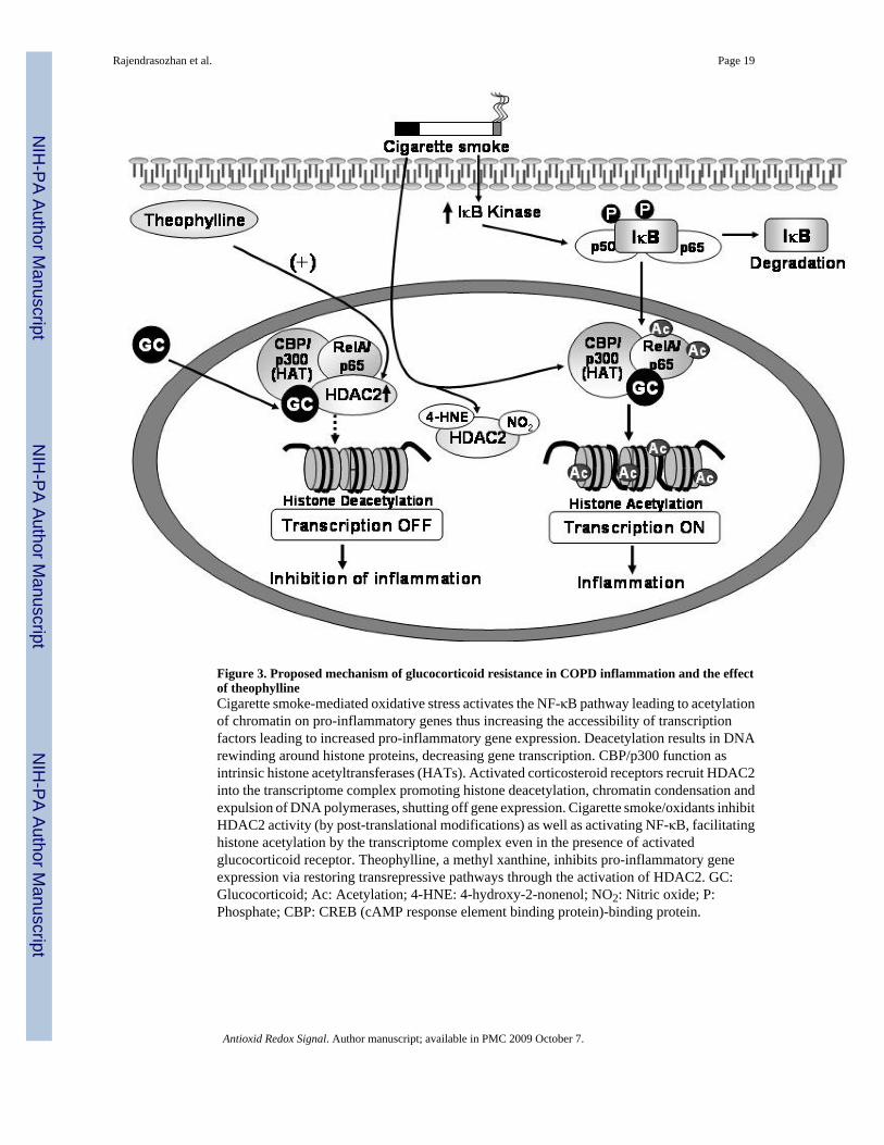
Figure 3. Proposed mechanism of glucocorticoid resistance in COPD inflammation and the effectof theophyllineCigarette smoke-mediated oxidative stress activates the NF-κB pathway leading to acetylationof chromatin on pro-inflammatory genes thus increasing the accessibility of transcriptionfactors leading to increased pro-inflammatory gene expression. Deacetylation results in DNArewinding around histone proteins, decreasing gene transcription. CBP/p300 function asintrinsic histone acetyltransferases (HATs). Activated corticosteroid receptors recruit HDAC2into the transcriptome complex promoting histone deacetylation, chromatin condensation andexpulsion of DNA polymerases, shutting off gene expression. Cigarette smoke/oxidants inhibitHDAC2 activity (by post-translational modifications) as well as activating NF-κB, facilitatinghistone acetylation by the transcriptome complex even in the presence of activatedglucocorticoid receptor. Theophylline, a methyl xanthine, inhibits pro-inflammatory geneexpression via restoring transrepressive pathways through the activation of HDAC2. GC:Glucocorticoid; Ac: Acetylation; 4-HNE: 4-hydroxy-2-nonenol; NO2: Nitric oxide; P:Phosphate; CBP: CREB (cAMP response element binding protein)-binding protein.
Rajendrasozhan et al. Page 19
Antioxid Redox Signal. Author manuscript; available in PMC 2009 October 7.
NIH
-PA Author Manuscript
NIH
-PA Author Manuscript
NIH
-PA Author Manuscript

Figure 4. Oxidant-mediated degradation of deacetylases (HDAC2/SIRT1) leading to increasedexpression of pro-inflammatory cytokinesDNA is coiled around the histone proteins and form chromatin structure. Acetylation ofchromatin (on pro-inflammatory genes) increasing the accessibility of transcription factors andthere by increasing gene transcription. Deacetylation results in DNA rewinding around histoneproteins, and decreasing gene transcription. Thus, HDACs involved in the maintenance ofhistone acetylation and deacetylation balance and thereby control the gene expression. HDACand/or SIRT1 may be post-translationally modified by phosphorylation, carbonylation,aldehyde adducts formation and nitration leading to increased ubiquitination and subsequentdegradation of HDACs. Decreased HDACs leads to increased histone acetylation/deacetylation ratio which cause increased acetylation of chromatin, chromatin remodulationand increased transcription of pro-inflammatory mediators. HAT: Histone acetyltransferase,HDAC: Histone deacetylase; P: phosphate; Ac: Acetylation; 4-HNE: 4-hydroxynonenol;NO2: Nitric oxide.
Rajendrasozhan et al. Page 20
Antioxid Redox Signal. Author manuscript; available in PMC 2009 October 7.
NIH
-PA Author Manuscript
NIH
-PA Author Manuscript
NIH
-PA Author Manuscript
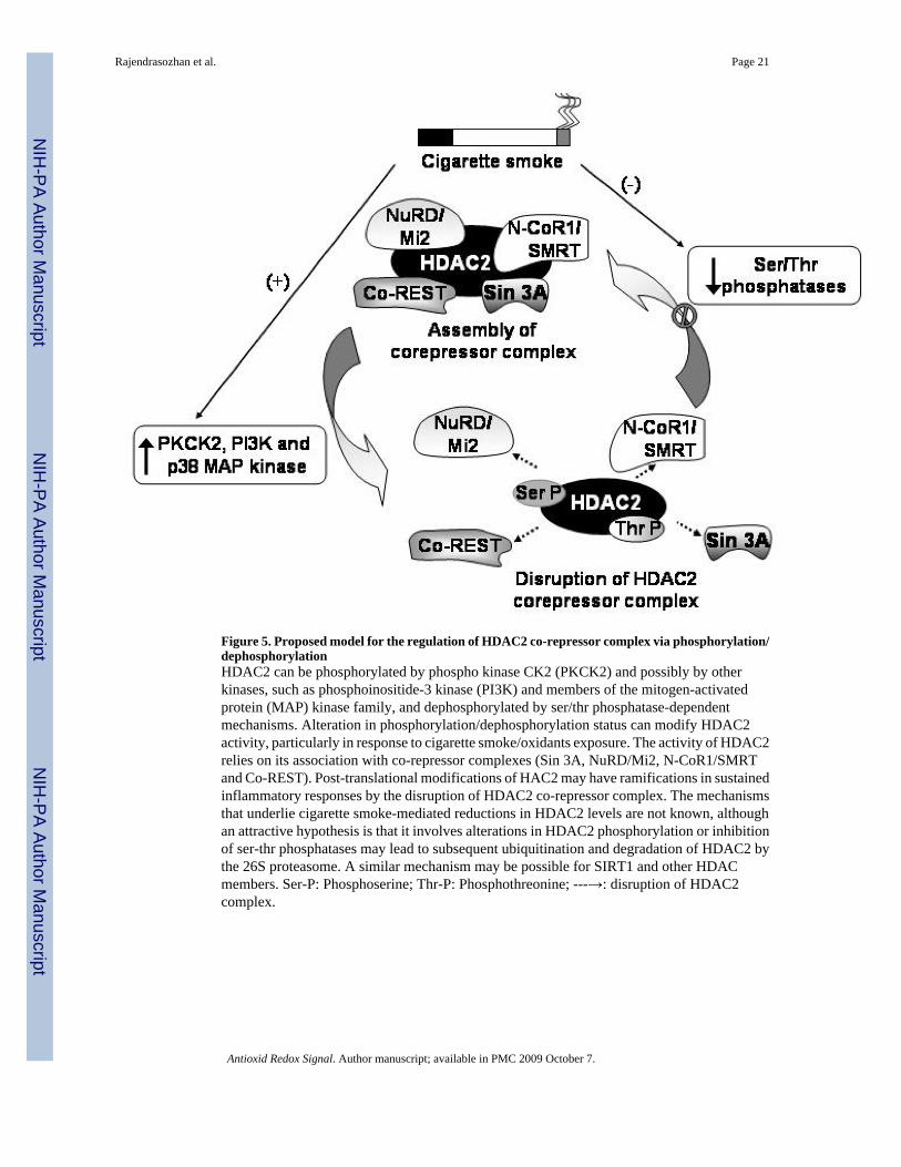
Figure 5. Proposed model for the regulation of HDAC2 co-repressor complex via phosphorylation/dephosphorylationHDAC2 can be phosphorylated by phospho kinase CK2 (PKCK2) and possibly by otherkinases, such as phosphoinositide-3 kinase (PI3K) and members of the mitogen-activatedprotein (MAP) kinase family, and dephosphorylated by ser/thr phosphatase-dependentmechanisms. Alteration in phosphorylation/dephosphorylation status can modify HDAC2activity, particularly in response to cigarette smoke/oxidants exposure. The activity of HDAC2relies on its association with co-repressor complexes (Sin 3A, NuRD/Mi2, N-CoR1/SMRTand Co-REST). Post-translational modifications of HAC2 may have ramifications in sustainedinflammatory responses by the disruption of HDAC2 co-repressor complex. The mechanismsthat underlie cigarette smoke-mediated reductions in HDAC2 levels are not known, althoughan attractive hypothesis is that it involves alterations in HDAC2 phosphorylation or inhibitionof ser-thr phosphatases may lead to subsequent ubiquitination and degradation of HDAC2 bythe 26S proteasome. A similar mechanism may be possible for SIRT1 and other HDACmembers. Ser-P: Phosphoserine; Thr-P: Phosphothreonine; ---→: disruption of HDAC2complex.
Rajendrasozhan et al. Page 21
Antioxid Redox Signal. Author manuscript; available in PMC 2009 October 7.
NIH
-PA Author Manuscript
NIH
-PA Author Manuscript
NIH
-PA Author Manuscript

NIH
-PA Author Manuscript
NIH
-PA Author Manuscript
NIH
-PA Author Manuscript
Rajendrasozhan et al. Page 22
Table 1Intracellular localization and functions of some of the deacetylases discussed in this review
Deacetylase Intracellular Localization Target Function Ref.
HDAC1 Nucleus Histone H1, andp50 and RelA/p65 subunits ofNF-κB
•Involved in cell proliferation (75)
•Interacts with p50 and RelA/p65 (12,95)
HDAC2 Cytoplasm and nucleus Histones,glucocorticoidreceptor, RelA/p65 and CBP/p300
•Recruited by glucocorticoid on pro-inflammatory genes
(4,43)
•Post-translationally modified byoxidants/nitric oxide/carbonyls
(41,88)
•Interacts with RelA/p65 to inhibitNF-κB transactivation
(88)
•Activated by theophylline (15,45)
•Phosphorylated and degraded (28,48,82)
HDAC3 Nucleus Histone H4 andRelA/p65
•Interacts with RelA/p65 and inhibitsits transactivation
(12)
SIRT1 Cytoplasm and nucleus RelA/p65, p53and FOXO3
•Interacts with RelA/p65 (89,91)
•Involved in regulation of pro-inflammatory mediators (cytokines)and MMPs
(57,89)
•Calorie restriction (6,35)
•Regulation of p53, FOXO3, DNArepair, cell proliferation, cell cycle,senescence and aging
(9,20,47,51,59,81,84)
Activity of HDAC1, HDAC2 and HDAC3 is inhibited by trichostain A, sodium butyrate, valproic acid and suberoylanilide hydroxamic acid, whereas
SIRT1 activity is inhibited by sirtinol and nicotinamide adenine dinucleotide (NAD+), and activated by resveratrol.
Antioxid Redox Signal. Author manuscript; available in PMC 2009 October 7.


![A new polymorph of bis[2,6-bis(1 H -benzimidazol-2-yl-κ N 3 )pyridinido-κ N ]zinc(II)](https://static.fdokumen.com/doc/165x107/63254f317fd2bfd0cb036571/a-new-polymorph-of-bis26-bis1-h-benzimidazol-2-yl-k-n-3-pyridinido-k-n-zincii.jpg)


![Tetra-kis(μ(2)-cyanido-κ(2)C:N)dicyanido-tetra-kis-[tris-(2-amino-eth-yl)amine-κ(3)N,N',N'',N''']tetra-copper(II)iron(II) bis[pentacyanidonitrosoferrate(II)] hexahydrate](https://static.fdokumen.com/doc/165x107/634199118718ae62200b4f38/tetra-kism2-cyanido-k2cndicyanido-tetra-kis-tris-2-amino-eth-ylamine-k3nnnntetra-copperiiironii.jpg)