
Elucidation of the ion binding mechanism in heterogeneous carbon-composite adsorbents
Upload
independentCategory
view
2download
0
J O U R N A L O F P R O T E O M I C S X X ( 2 0 1 2 ) X X X – X X X
Ava i l ab l e on l i ne a t www.sc i enced i r ec t . com
www.e l sev i e r . com/ loca te / j p ro t
JPROT-00790; No of Pages 15
Review
Proteomics in chromatin biology and epigenetics: Elucidationof post-translational modifications of histone proteins bymass spectrometry☆
Simone Sidoli, Lei Cheng, Ole N. Jensen⁎
Department of Biochemistry and Molecular Biology, University of Southern Denmark, Campusvej 55, DK-5230 Odense M, Denmark
A R T I C L E I N F O
Abbreviations: ChIP, chromatin immunopretransfer dissociation; HDACs, histone deacacetylglucosamine; PRC2, Polycomb repressiv☆ This article is part of a Special Issue entit⁎ Corresponding author. Tel.: +45 6550 2368.
E-mail address: [email protected] (O.N
1874-3919/$ – see front matter © 2011 Elseviedoi:10.1016/j.jprot.2011.12.029
Please cite this article as: Sidoli S, et al,modifications of histone proteins by ma
A B S T R A C T
Keywords:
Histone proteins contribute to the maintenance and regulation of the dynamic chromatinstructure, to gene activation, DNA repair and many other processes in the cell nucleus.Site-specific reversible and irreversible post-translational modifications of histone proteinsmediate biological functions, including recruitment of transcription factors to specific DNAregions, assembly of epigenetic reader/writer/eraser complexes onto DNA, and modulationof DNA-protein interactions. Histones thereby regulate chromatin structure and function,propagate inheritance and provide memory functions in the cell. Dysfunctional chromatinstructures and misregulation may lead to pathogenic states, including diabetes and cancer,and the mapping and quantification of multivalent post-translational modifications hastherefore attracted significant interest. Mass spectrometry has quickly been accepted as aversatile tool to achieve insights into chromatin biology and epigenetics. High sensitivityand high mass accuracy and the ability to sequence post-translationally modified peptidesand perform large-scale analyses make this technique very well suited for histone proteincharacterization. In this review we discuss a range of analytical methods and variousmass spectrometry-based approaches for histone analysis, from sample preparation todata interpretation. Mass spectrometry-based proteomics is already an integrated and in-dispensable tool in modern chromatin biology, providing insights into the mechanismsand dynamics of nuclear and epigenetic processes. This article is part of a Special Issue en-titled: SI: Genome regulations and Genetic diversity.© 2011 Elsevier B.V. All rights reserved.
Functional proteomicsPost-translational modificationsEpigeneticsTandem mass spectrometryChromatographyBioinformatics
Contents
1. Introduction . . . . . . . . . . . . . . . . . . . . . . . . . . . . . . . . . . . . . . . . . . . . . . . . . . . . . . . . . . 02. Histones are heavily decorated by PTMs . . . . . . . . . . . . . . . . . . . . . . . . . . . . . . . . . . . . . . . . . . 0
cipitation; DNMTs, DNAmethyltransferases; ECD, electron capture dissociation; ETD, electronetylases; HILIC, hydrophilic interaction liquid chromatography; O-GlcNAc, O-linked N-e complex 2; TCA, trichloroacetic acid; WCX, weak cation exchange.led: SI: Genome regulations and Genetic diversity.
. Jensen).
r B.V. All rights reserved.
Proteomics in chromatin biology and epigenetics: Elucidation of post-translationalss spectrometry, J Prot (2012), doi:10.1016/j.jprot.2011.12.029

2 J O U R N A L O F P R O T E O M I C S X X ( 2 0 1 2 ) X X X – X X X
2.1. Methylation . . . . . . . . . . . . . . . . . . . . . . . . . . . . . . . . . . . . . . . . . . . . . . . . . . . . . . . 02.2. Acetylation . . . . . . . . . . . . . . . . . . . . . . . . . . . . . . . . . . . . . . . . . . . . . . . . . . . . . . . 02.3. Phosphorylation . . . . . . . . . . . . . . . . . . . . . . . . . . . . . . . . . . . . . . . . . . . . . . . . . . . . 02.4. Biotinylation . . . . . . . . . . . . . . . . . . . . . . . . . . . . . . . . . . . . . . . . . . . . . . . . . . . . . . 02.5. Citrullination . . . . . . . . . . . . . . . . . . . . . . . . . . . . . . . . . . . . . . . . . . . . . . . . . . . . . . 02.6. ADP-ribosylation . . . . . . . . . . . . . . . . . . . . . . . . . . . . . . . . . . . . . . . . . . . . . . . . . . . . 02.7. Ubiquitination and other modifications . . . . . . . . . . . . . . . . . . . . . . . . . . . . . . . . . . . . . . . 0
3. Histone isolation, separation of intact isotypes and enrichment of PTMs . . . . . . . . . . . . . . . . . . . . . . . . . . . 03.1. Separation of histone isotypes . . . . . . . . . . . . . . . . . . . . . . . . . . . . . . . . . . . . . . . . . . . . 03.2. Enrichment of modified peptides . . . . . . . . . . . . . . . . . . . . . . . . . . . . . . . . . . . . . . . . . . . 0
4. Strategies for histone structure analysis by mass spectrometry . . . . . . . . . . . . . . . . . . . . . . . . . . . . . . 04.1. Bottom–up analysis of histones . . . . . . . . . . . . . . . . . . . . . . . . . . . . . . . . . . . . . . . . . . . . 04.2. Top–down analysis of histones . . . . . . . . . . . . . . . . . . . . . . . . . . . . . . . . . . . . . . . . . . . . 04.3. Middle–down strategy for histone analysis . . . . . . . . . . . . . . . . . . . . . . . . . . . . . . . . . . . . . . 0
5. Quantitative approaches in proteomics and epigenetics . . . . . . . . . . . . . . . . . . . . . . . . . . . . . . . . . . 06. Chromatin immunoprecipitation and mass spectrometry . . . . . . . . . . . . . . . . . . . . . . . . . . . . . . . . . 07. Data analysis . . . . . . . . . . . . . . . . . . . . . . . . . . . . . . . . . . . . . . . . . . . . . . . . . . . . . . . . . . 08. Conclusion . . . . . . . . . . . . . . . . . . . . . . . . . . . . . . . . . . . . . . . . . . . . . . . . . . . . . . . . . . . 0Acknowledgments . . . . . . . . . . . . . . . . . . . . . . . . . . . . . . . . . . . . . . . . . . . . . . . . . . . . . . . . . . 0References . . . . . . . . . . . . . . . . . . . . . . . . . . . . . . . . . . . . . . . . . . . . . . . . . . . . . . . . . . . . . . 0
1. Introduction
Since the discovery of the structural complementarity of the twonucleic acid strands of theDNAdouble helix, themechanisms ofinheritance of genetic informationhave been thoroughly studiedand are well understood. In recent years, however, the emergingfield of epigenetics has provided novel insights and elucidatedadditionalmechanisms for propagationof information frompre-cursor cells to offspring cells, without changes in the DNA se-quence [1]. Epigenetic (“above genetic”) mechanisms includeDNA methylation, post-translational modifications of his-tone proteins and non-coding silencing RNA, which serve toactivate or silence certain genes, e.g. as part of the ‘cell fate’programming of stem cells. Epigenetic mechanisms mayserve as amemory for cell fate decisions, as specific informa-tion is ‘imprinted’ in the chromatin structure, including fac-tors that modify DNA and/or its associated protein factors.This additional level of regulation of the genetic material incells, originally described byWaddington in 1942 [2], is currentlyreshaping theway inwhich researchers think about propagationand inheritance, cell development and differentiation, and dis-ease etiology [3–5].
Chromatin consists of DNA and many proteins that main-tain and regulate this highly organized and dynamic struc-ture. Histones are hydrophilic and basic nuclear proteins.They participate as subunits of the nucleosomes that consti-tute one of the structural core units of chromatin. Nucleo-somes are histone octamers that consists of four canonicalhistone isotypes (H2A, H2B, H3 and H4) present in two copieseach. DNA is wrapped around these octamers, which are lo-cated along the DNA with a spacing of 177–207 base pairs, ina pearls-on-a-string manner. One additional histone subunit,histone H1, serves to ‘lock’ the nucleosome into place ontothe DNA. In addition to the canonical isoforms, several otherhistone isotypes can be present in the nucleosome, replacingthe canonical isoforms in specific genomic areas under
Please cite this article as: Sidoli S, et al, Proteomics in chromatmodifications of histone proteins by mass spectrometry, J Prot (
certain conditions. For instance, mammalian cells containfive different isotypes of histone H3, namely H3.1, H3.2, H3.3,CENH3/CENP-A and H3t; CENP-A replaces conventional H3 innucleosomes placed in centromeric chromatin at the internalplate of the kinetochore. Obtaining a clear distinction is cru-cial, since post-translational modifications (PTMs) betweenthese variants may be different, providing distinct biologicalfunctions [6].
Histone proteins can be heavily decorated with PTMs, suchas acetyl-, methyl-, and phosphoryl-groups at distinct aminoacid residues. These modifications are mainly located in theN-terminal tails of the histones, which protrude from thecore nucleosome structure. PTMs contribute to gene regula-tion in two distinct manners: via cis or via trans actions [7].Cis mechanisms alter intra- and inter-nucleosomal interac-tions, thereby leading to changes in chromatin structure andcompactness. A classical example is acetylation of lysine resi-dues: when the positively charged (protonated) ε-amino groupof lysine 16 of histone H4 becomes acetylated (H4K16ac) it alsobecomes electrostatically neutral. Thus, the interaction betweenthe lysine side chain and the negatively charged DNA is weak-ened and, consequently, the chromatin structure becomes‘loose’, or is decompressed [8]. In contrast, trans regulatorymechanisms serve as signals for recruitment of specific reader/writer/eraser factors, which are involved in chromatin remodel-ing, gene activation, or other dynamic biological processes [9,10].Cis and trans regulatory mechanisms work in concert andcontribute to modulation and regulation of transcription, DNAreplication, chromosome condensation, imprinting, and X-inactivation. All of these processes have consequences for cellu-lar fate and the equilibrium between an organism and the envi-ronment. For instance, histone acetylation and deacetylation inplants are reported to be responsible for a myriad of functions,suchas the circadian clock [11], the response to cold temperature[12] and pathogens [13], and the development of leaves [14],flowers [15] and seeds [16]. Inherited histone methylation plays
in biology and epigenetics: Elucidation of post-translational2012), doi:10.1016/j.jprot.2011.12.029

3J O U R N A L O F P R O T E O M I C S X X ( 2 0 1 2 ) X X X – X X X
a crucial role in Arabidopsis early embryogenesis [17], andepigenetic transmission via sperm and oocytes was also demon-strated inmammals [18]. Histonemodificationswere also foundto be related to important diseases such as cancer [3,4] or psy-chiatric andneurologicalmalfunctions [19]. Improvedknowledgeon theprogressionof cancer cell lines allows for thedevelopmentof more specific drugs. Currently, there are two groups of drugswhich have been adopted for this purpose: inhibitors of DNAmethyltransferase (DNMTi) and inhibitors of histone deacety-lases (HDACi) [20]. Due to their specificity and the ability to in-duce apoptosis and autophagy, HDAC inhibitors provedeffective in some cancer lines [21] and malaria therapies [22].
The discovery of the features and functions of PTMs of his-tones lead to the hypothesis of the “histone code” [23,24], i.e.that certain PTMs or combinations of few specific PTMs havedistinct biological functions. However, recent findings suggestthat individual site-specific modifications may have multipleroles depending on the cellular environment, context andcues. Furthermore, coexisting and multivalent histonemarks appear to be highly important for modulatingprotein-protein interactions and enzyme activities in dy-namic DNA binding protein complexes during cell develop-ment, growth, and differentiation. It is evident that theinterplay between multiple PTMs in histones is much moresignificant than the contribution of any single PTM [25],and it therefore becomes important to elucidate the mecha-nisms of molecular cross-talk within and between histonesin nucleosomes and within chromatin [26].
The necessity of obtaining more detailed structural infor-mation on histone PTMs calls for novel analytical strategiesand technologies. For instance, a specific type of modificationcan play very different roles when located at different aminoacid positions in a histone subunit: Trimethylation of histoneH3K4 is usually related to transcriptional activation, while tri-methylation of H3K9 leads to transcriptional repression. Fur-thermore, adjacent modified amino acid residues makehistone PTM characterization a significant technical challenge(Fig. 1). Mass spectrometry is increasingly used to study chro-matin biology and epigenetic mechanisms because very de-tailed insights into PTMs are obtained. In the present review,we describe state-of-the-art mass spectrometry-based tech-niques for elucidation of the highly diverse ensemble of his-tones and their PTMs.
Fig. 1 – Fromphenotype to histone codes. Cellular behavior and phemodifications. In the diagram: schematic magnification down to thestructure or modifications can lead to important alterations of the ctechniques for accurate characterization of these changes.
Please cite this article as: Sidoli S, et al, Proteomics in chromatmodifications of histone proteins by mass spectrometry, J Prot (
2. Histones are heavily decorated by PTMs
Histone modifications are conventionally described by a stan-dardized nomenclature, also known as Brno nomenclature[27]. It lists the histone isotype followed by the amino acid sym-bol and position and then by the abbreviated modification. Forexample, H3K9me2 denotes histone H3modified at the 9th res-idue, a lysine, with a dimethyl group. In this section, we brieflydescribe the most commonly observed PTMs on histones, in-cluding their biological roles and other features.
2.1. Methylation
The first report on N-methylation of lysine residues in his-tones goes back to 1964, when Murray identified an ε-N-meth-yl lysine on calf histones, by using amino acid analysis, massspectrometry, and paper chromatography [28]. Until very re-cently, histone methylation was thought to be an irreversiblemodification [29]. Even though histone methyl transferases(HMTs) were known since the late 1970s to methylate histoneproteins [30], no evidence for the presence of histonedemethylase enzymes was provided [31]. However, in 2006,Jumonji domain-containing (JmjC) histone demethylaseswere discovered, and methylation was immediately definedas yet another reversible modification of histone proteins[32]. Lysine residues can be mono-, di-, or tri-methylated atthe ε-amino group (ε-N-methyl-Lys), while arginines can bemono- or di-methylated in different configurations, includingω-NG monomethyl arginine, ω-NG,NG-asymmetric dimethylarginine and ω-NG,N´G-symmetric dimethyl arginine. The de-gree of methylation often defines different biological func-tions. For instance, monomethylation is usually associatedwith transcription activation, while lysine trimethylation isoften related to repression. Arginine [33] and lysine [34] meth-ylations are involved in transcriptional regulation, chromatinremodeling, DNA repair, RNA processing, and signal transduc-tion. Thus, characterization of histonemethylation processes,including accurate localization and quantification of distinctmethylation sites, is highly relevant. H3R2, H3K4 and H3K9are examples of closely located candidate methylation sites,which are occupied to varying degrees. They are sometimesmodified in a coordinated fashion, so-called methylation
notype can be changed by epigenetic regulation such as histoneN-terminal tails of core histones. A small difference in histone
ellular machinery. Thus, it is fundamental to adopt appropriate
in biology and epigenetics: Elucidation of post-translational2012), doi:10.1016/j.jprot.2011.12.029

4 J O U R N A L O F P R O T E O M I C S X X ( 2 0 1 2 ) X X X – X X X
interplay [35]. For instance, it was demonstrated how methyl-ation at H3R2 antagonizes tri-methylation of H3K4 in yeast[36].
A methyl group leads to a mass increment of 14.016 Da ofthe modified amino acid residue. In addition, peptide ionscontaining monomethyl arginine and symmetric dimethyl ar-ginine generate a neutral loss signal of 31.04 Da, while theneutral loss suggestive of asymmetric dimethyl arginine is45.05 Da. Amino acid sequencing by tandemmass spectrome-try provides highly-informative spectra for methylated pep-tide species, e.g. to localize closely-spaced methyl groupswithin histone tails.
2.2. Acetylation
ε-N-acetylation of lysine residues in histones was alsoreported in 1964 [37]. At that time histones were expected tobe involved in regulating DNA activity and the discovery ofhistone acetylation reinforced the theory of their fundamen-tal role in transcriptional regulation. ε-N-acetylation is mainlyinvolved in gene activation, due to the property of neutraliz-ing a positive charge of lysine residues thereby weakeninghistone interactions with DNA. In addition, acetylation is im-plicated in DNA repair, replication and condensation. ε-N-acetylation of lysine is catalyzed by histone acetyl transfer-ases (HATs) and the removal is mediated by histone deacety-lases (HDACs). A number of HDAC inhibitors induceapoptosis and autophagy, potentially offering new ap-proaches in malaria [22] and cancer therapy [38]. In addition,acetylation plays a crucial role in modification cross-talk. Forinstance, mutation of H3K14, a known acetylation site, resultsin the specific loss of H3K4 trimethylation, but not mono- ordi-methylation [39].
Acetylation is detected by mass spectrometry due to amass increment of 42.011 Da and often also the observationof diagnostic fragment ions at m/z 126.091 and sometimesalsom/z 143.118 in the tandemmass spectra of ε-N-acetylatedpeptides [40,41].
2.3. Phosphorylation
Reversible phosphorylation of distinct amino acid residues inproteins plays an important role in cell signaling, includingsignaling networks that feed into chromatin. Phosphorylationis conveniently detected by mass spectrometry due to amass increase of 79.966 Da of peptides upon addition of onephosphoryl group, and often also a neutral loss of H3PO4
(−97.982 Da, pSer/pThr) or the presence of an immonium ion(m/z 216.043, pTyr) in MS/MS spectra of phosphopeptideions. Phosphorylation of histone proteins is reported to be in-volved in regulation of DNA repair and chromatin structure[42–45]. Detailed analysis of histone phosphorylation eventswas greatly improved by combining IMAC enrichment andMS/MS, as described in a study of histone H1 and its isoforms[46]. The discovery of a phosphorylation “switch” (H3S10ph/H3K9ac), that histone phosphorylation can regulate interac-tions and modifications, was first demonstrated usingan MS-basedmethod followed by western blotting and immu-nofluorescence analyses [47–49]. Lately, additional evidenceof the cross-talk between histone phosphorylation and
Please cite this article as: Sidoli S, et al, Proteomics in chromatmodifications of histone proteins by mass spectrometry, J Prot (
acetylation at distinct amino acids was shown; H3S28phK27acand H3S10phK14ac were both found to be responsible for im-portant epigenetic regulations including transcriptional acti-vation and cell cycle regulation [50,51]. H4 histidine kinasewas suggested to be an onco-developmental marker [52].Other than the usual STY phosphorylation, the biologicalfunctions of H4 histidine phosphorylation have not yet beenwidely explored. This is largely due to the lack of suitabledetection methods, since histidine phosphorylation is consid-ered acid labile. Attwood et al. reported anMS-based detectionmethod for H4 histidine phosphorylation [53]. Chromato-graphic and mass spectrometric analysis of histidine phos-phorylation, combined with protein histidine kinase activityassay, has been successfully applied to the study of histoneH4 phosphorylation and H4 histidine kinases [54].
2.4. Biotinylation
Biotinylationofhistoneswasdetected in several histonevariants,including H2A, H3 and H4. This PTM is supposedly involved ingene silencing [55,56], by altering the structure of nucleosomesand reducing DNA accessibility towards the transcriptional ma-chinery [56]. Biotinylation is catalyzed by holocarboxylase syn-thetase, while biotinidase is involved in debiotinylation. Severaldistinct aminoacid residues in thehistoneH4 tailwere character-ized as targets of these enzymes [57]. Healy et al. in 2009 reporteda proof of the presence of biotin in vivo by using radiolabeling,western blotting, andmass spectrometry. However, by examina-tionofprevious reports onhistonebiotinylation, they reached theconclusion that the in vivo presence of this modification still hasto be demonstrated [58]. In fact, methodologies used in previousarticles were shown to produce artifacts in biotin detection. Forinstance, avidin was found to exhibit a high level of non-specific interaction with unmodified histones, while antibodiessuch as anti-H4K5bio and anti-H4K8bio provided positive signalseven though histones were filtered through neutravidin-agaroseto remove biotinylated proteins. This was most likely due tocross-reactions with other modifications.
2.5. Citrullination
Citrullination (or deimination) is a PTM of rather low abun-dance, which occurs at arginine residues. This modificationis catalyzed by peptidylarginine deiminases (PADs) [59], andis often related to gene silencing, since it antagonizes argininemethylation [60]. From an analytical perspective chemicaltags such as 18O, 2,3-butanedione, or antipyrine are useful toconvert protein citrullination sites into derivatives for effi-cient and robust MS detection [61,62], since the mass incre-ment of deimination (+0.984 Da) is rather small and may beobscured by isotope splitting of the peptide signals in MSand MS/MS.
2.6. ADP-ribosylation
ADP-ribosylation occurs on lysine residues (ε-N-amino group,mass increment of 541.061 Da) and it is presumably oflow abundance, i.e. less than 1% of all histone proteins areADP-ribosylated [63,64]. The role of this modification hasnot been completely characterized, but involvement in DNA
in biology and epigenetics: Elucidation of post-translational2012), doi:10.1016/j.jprot.2011.12.029

5J O U R N A L O F P R O T E O M I C S X X ( 2 0 1 2 ) X X X – X X X
replication, transcription, and chromatin remodeling is sug-gested [65]. Histone H1 was found to be one of the major sub-strates for poly(ADP-ribose) polymerase [66]. ADP-ribosylationis mostly identified by using radioactive phosphorus or bywestern blotting with antibodies directed against poly ADP-ribose polymers. Optimization of mass spectrometry-basedstrategies will be necessary to reveal the extent and role ofthis PTM in chromatin biology and epigenetics.
2.7. Ubiquitination and other modifications
Ubiquitination of Lys residues is predominately found in his-tones H2A and H2B. Poly-ubiquitination is known to contributein the lability of H2A-H2B dimers, facilitating the initial steps ofnucleosome disassembly during transcriptional activation [67].Monoubiquitination was only recently investigated in detail asbeing involved in transcriptional regulation and DNA damageresponse [68,69]. Ubiquitination participates also in the com-plex mechanism of cross-talk between, and not only within,histones; an MS-based protein differential expression analy-sis reported the down regulation of H3/H4 acetylation andH2B ubiquitination with impaired chromatin remodelingalong with other insights on epigenetic interaction network.Several proteins involved in H2B ubiquitination and de-ubiquitination were identified and quantified. In this specificstudy, down-regulation of H2B ubiquitination has been vali-dated using targeted LC-MS/MS by multiple reaction moni-toring (MRM) [70].
Recently, SUMOylation [71], propionylation/butyrylation[72], and O-linked N-acetylglucosamine (O-GlcNAc) [73] werealso discovered as variable modifications on histone proteins.O-GlcNAc was characterized by using multiple immunologicaland enzymatic approaches, while MS technique contributedin determining specific PTM positions in histones H2A, H2B,and H4.
3. Histone isolation, separation of intact isotypesand enrichment of PTMs
The peculiar chemical properties of histone proteins maketheir extraction a relatively easy task. It is possible to obtainlarge amounts of histone proteins from cells and tissues,which is important for detection of sub-stoichiometric modifi-cations. An effective method for histone purification is accu-rately described by Shechter et al. [74]. Briefly, this protocolis based on solubility of histones in strong acids (HCl orH2SO4) and subsequent precipitation using trichloroaceticacid (TCA). The protein pellet is then rinsed with cold acetoneand resuspended. Due to their hydrophilic properties histonesare soluble even at high protein concentrations (more than10 mg/ml) in e.g. 0.1% trifluoroacetic acid. However, TCA maygenerate insoluble precipitates and the presence of strongacids may hydrolyse some labile modifications such as phos-phorylation. Because of this, an alternative high salt-basedextraction protocol was suggested [75]. Several commercialkits for histone isolation are also available, including those pro-duced by Active Motif (Histone Purification Kit) and EpiQuik(Total Histone Extraction Kit).
Please cite this article as: Sidoli S, et al, Proteomics in chromatmodifications of histone proteins by mass spectrometry, J Prot (
3.1. Separation of histone isotypes
Further separation of extracts of intact histones is commonlyperformed by traditional reversed-phase high performanceliquid chromatography (RP-HPLC) using C18 columns. Thisfractionation method is preferred for practical reasons overother techniques such as electrophoretic gels, even thoughthe latter may also achieve good separation of histones [76].Reversed-phase HPLC presents several advantages: (i) his-tones are small hydrophilic proteins that are very solubleand histone separation by RP-HPLC can be baseline resolved;(ii) the eluted histone proteins are dissolved in aqueous, salt-free solvents, which are suitable for on-line or off-line analy-sis by mass spectrometry; (iii) The LC can be easily coupledon-line to MS for detection and mass measurement of intactproteins.
An alternative method for LC separation of histone iso-types is hydrophilic interaction liquid chromatography(HILIC) [77]. This resin is particularly effective for detecting avariety of histone acetylation states. HILIC and RPLC have or-thogonal separation properties, and this suggests a possibleuse of both for two-dimensional separations (2D-LC). On theother hand, histone solubility becomes a limiting factor inhigh concentrations of organic solvents, which are used inHILIC. Histone separation is also feasible by using monolithiccolumns made of polystyrene-divinylbenzene (PS-DVB), butthis technique is not so widespread [78,79].
3.2. Enrichment of modified peptides
Enrichment of PTM-bearing peptides is mostly performed atthe peptide level, i.e. after enzymatic digestion or chemicaldegradation of protein to obtain peptides suitable for massspectrometry-based detection and sequencing (Zhao and Jen-sen, 2009). For instance, reversible protein phosphorylation isusually of low abundance and sub-stoichiometric in nature.The development of enrichment methods for phosphopep-tides, such as IMAC and TiO2 enrichment [80], have allowedfor dramatic improvements in sensitivity and specificity inphosphoproteomics experiments, including determination ofphosphorylation sites in histones. Garcia et al. successfullyadapted IMAC enrichment for histone H1 phosphorylationanalysis [46]. A different approach is used for detection of ubi-quitination or SUMOylation sites in proteins. Using molecularbiology approaches, an epitope tag of six histidines was fusedwith ubiquitin and SUMO proteins, to allow for affinity purifi-cation of conjugated proteins prior to protein identification byMS. Using this technique Rosas-Acosta et al. identifiedSUMOylation of histone H2A isotypes [81]. A histidine tagwas combined with an S-peptide tag for protein purification[82]. Alternative strategies for enrichment is the use ofubiquitin-binding protein or use of techniques adapted fromphosphopeptide enrichment, which can be applied to bindthe –GG ubiquitin tag after tryptic digestion. These methodsare described in more detail in a recent review by Kirkpatricket al. [83].
In summary, the combination of chromatography oraffinity-based separations with mass spectrometry enables de-tailed and sensitive analysis of histones. In some cases capillaryelectrophoresis was used to separate histones [84,85], with the
in biology and epigenetics: Elucidation of post-translational2012), doi:10.1016/j.jprot.2011.12.029

6 J O U R N A L O F P R O T E O M I C S X X ( 2 0 1 2 ) X X X – X X X
possibility to achieve online detection by MS [86]. In the remain-der of review we will mainly focus on liquid chromatographymethods, which are directly interfaced to MS and MS/MS analy-sis, thereby providing a reproducible and robust platform for his-tone analysis.
4. Strategies for histone structure analysis bymass spectrometry
Themain challenge in histone characterization is the high de-gree of complexity of the histone proteins due to the allelicvariants and the diversity and multivalency of the PTMs inhistones. More than 40 variants of human histones are cur-rently known. Some of these differ by very few amino acid res-idues in the protein sequence, necessitating very specific andsensitive analysis tools to distinguish them. The three mainMS based strategies for histone characterization are shownin Fig. 2. The most commonly used approach is the peptide-centric analysis by mass spectrometry, known as the ‘bottom–up’ approach [87]. In this workflow proteins are cleaved intosmaller peptides of about 7–25 amino acids in length, whichare suitable for sequencing by MS/MS using for example colli-sion induced dissociation (CID) for gas-phase fragmentation.However, by using this approach the knowledge of the globalstatus of the PTMs along the protein backbone is lost and it isdifficult to map multivalent, co-existing PTM patterns.
A different strategy aims to characterize the intact histoneproteins bymass spectrometry, i.e.without cleaving the proteininto peptides. This approach is known as the ‘top–down’ strate-gy [88]. This approach allows for mapping of co-existing PTMs,even those that are located far apart within the histone
Fig. 2 – The threemajor proteomic strategies. Selecting the appropriand the aimof theproject: (i) `bottom–up´ approach cleaves proteinsreversed-phase chromatography. Due to the reduced size of themois not always necessary. (ii) By adopting the ‘middle–down’ strategachieved. Resins asHILIC ormixed bed ion exchange-HILIC provedmass, high resolutionMS/MS is required to determine isotope pattehistone analysis, or ‘top–down’ approach, requires accurate purificaoffline. Once the sample is pure, proteins canbedirectly sprayed inhETD fragmentation is suggested. As general remark, more complexmoment middle–down and top–down proteomics have lower amou
Please cite this article as: Sidoli S, et al, Proteomics in chromatmodifications of histone proteins by mass spectrometry, J Prot (
sequence. However, from a practical point of view the top–down strategy presents some challenges, particularly due tothe higher charge states of intact protein ions generated in ESIMS. This often results in complex MS and MS/MS spectra, aswell as some difficulties in charge state determination unlesshigh-resolution tandemmass analyzers are used. Furthermore,CID is not a very efficient fragmentation method for generationof sequence specific ions from intact proteins. Electron capturedissociation (ECD) [89] is better suited to fragment intact pro-teins. ECD is a soft fragmentation method, which preservesmost PTMs even though the polypeptide backbone is cleavedvery efficiently. The disadvantages of ECD MS/MS are related tothe maintenance costs, since it has been mainly implementedon FT-ICR instruments, and to sensitivity, which is generallylower than traditional CID. In 2004, ElectronTransferDissociation(ETD) was introduced [90]. This fragmentation method is relatedto ECD, but it is easily implemented in linear ion trap analyzerssince the electrons used for polypeptide ion fragmentation areprovided by gas-phase anions (fluoranthene or nitrosobenzene)rather than by a heated filament as in ECD.
A third strategy for histone analysis is the ‘middle–down’ ap-proach (Fig. 2), which takes advantage of the fact that the N-terminal tail of the histone can be cleaved off by specific prote-ases, generating a polypeptide of 40–50 residues that contain amajority of the PTMs of the histone. This 5–6 kDa polypeptidecan be further purified and/or analyzed by MS and MS/MSusing CID and ETD. This approach is a suitable compromise be-tween the bottom–up and top–down methods, reducing techni-cal challenges of intact histone analysis while preserving asemi-global overview of coexisting modifications [91].
In the following section we discuss details of these threemain strategies for histone analysis by MS.
ateworkflow ismainly dependent onavailable instrumentationinto small peptides,which canbe separated byusing traditionallecules, inMS/MS identification high resolution for product ionsy a comprehensive overview of the entire histone tail can bemost effective for separation of histone tails. Because of the highrns and charge states. ETD is usually preferred to CID. (iii) Intacttionof thedifferent histone isotypes,which ismostlyperformedigh resolutionmass spectrometers. High resolutionMS/MSandspectra require more advanced bioinformatic tools. At thents of available software (described in Section 7).
in biology and epigenetics: Elucidation of post-translational2012), doi:10.1016/j.jprot.2011.12.029

7J O U R N A L O F P R O T E O M I C S X X ( 2 0 1 2 ) X X X – X X X
4.1. Bottom–up analysis of histones
As previously mentioned, the bottom–up approach is themost widespread strategy for protein characterization at thistime. Protein digestion is commonly performed by using pro-teolytic enzymes rather than chemical methods. Proteasesguarantee higher cleavage site specificity and efficiency witha majority of proteins, with few side reactions and artifacts.Advantages of mass spectrometry analysis of smaller pep-tides of 1–2 kDa are mainly related to higher mass accuracyand sequencing efficiency and in straightforward sequencedatabase searching for peptide identification and PTM assign-ments. However, for histones we note that the traditional en-zymatic digestion protocols, which apply trypsin or LysCproteases are not so appropriate, unless derivatization of ly-sine side chains is performed. Lysine is a frequently occuringamino acid residue in histones, in particular in histone N-terminal tails, and trypsin will generate many very small pep-tides, which are unsuitable for MS and MS/MS analysis. Thisproblem might be overcome by reducing trypsin incubationtime and the enzyme/substrate ratio [92]. Derivatization ofunmodified and monomethyl lysines with propionic anhy-dride before trypsin digestion [6,49,93] or using alternativeproteases such as Arg-C [94–96] are viable methods. Both ap-proaches lead to protein cleavage exclusively at arginine resi-dues, resulting in generation of peptides of a length thatfacilitates MS detection.
Histones are hydrophilic and basic proteins, and so aremost of the peptides generated by protease treatment. Sepa-ration of histone peptides is performed by chromatographyusing reversed-phase materials, e.g. C18 hydrophobic resins.Histone peptides are rather hydrophilic and they tend toelute early from C18 columns.
The peptide-centric bottom–up approach has advantagesin terms of chromatographic resolution and MS/MS spectraquality, and a number of LC-MS/MS data analysis tools are al-ready available. We recommend this method for the initialcharacterization of histones, including mapping of PTMs andsimple comparative analysis of histones preparations.
4.2. Top–down analysis of histones
Histones are rather small proteins (11–15 kDa) and the top–down approach is therefore very attractive for characteriza-tion of intact histones using mass spectrometry. However,ionization of intact proteins is less efficient than for smallerpeptides. Nevertheless, intact proteins in the range of 50 to200 kDa have been analyzed and sequenced by MS/MS[88,97]. The top–down approach is usually beneficial in termsof faster histone sample preparation since no digestion stepsare required, and it achieves a more comprehensive overviewof the protein sequence and the PTM pattern. On the otherhand, the top–down method obviously requires more ad-vanced instrumentation and software, especially in terms ofmass resolution and mass accuracy to deconvolute the verycomplex MS/MS and MSn spectra. For example, obtaininghigh sequence coverage via MS/MS fragmentation is muchmore complicated than the more traditional bottom–up ap-proach, and it might be difficult to localize PTMs based onvery complex MS/MS spectra with multiple charge states of a
Please cite this article as: Sidoli S, et al, Proteomics in chromatmodifications of histone proteins by mass spectrometry, J Prot (
plethora of fragment ions. Despite these challenges, a numberof encouraging top–down studies have been published. Thefirst accurate analysis of histone modifications by top–downanalysis, by Banks et al., characterized the differential de-phosphorylation of histone H1 isotypes under the effect ofthe hormone dexamethasone [98]. Histones were separatedusing a columnwith C4 resin, and then fractions were directlyinfused in a Q-TOF hybrid instrument. Top–down histone pro-teomics is further developed by Kelleher and colleagues, in-troducing tandem mass spectrometry directly on the intactproteins. By using direct infusion of fractionated histones(via RPLC) and ECD fragmentation, they accomplished detailedinformation about all themajor histone variants, including H3[99], H2A [100], H2B [101] and H4 [102].
The top–down strategy relies on efficient separation of in-tact histones prior to MS and MS/MS analysis. An online LC-MS/MS setup is usually preferred, since it is beneficial forthroughput, automation, and reproducibility. RP-HPLC andHILIC are suited for separation of hydrophilic proteins priorto MS analysis. Whereas interesting results are achievedusing HILIC materials [103], the stationary phase of choicefor intact histone analysis is still based on C18. This is mostlydue to practical reasons: (i) histones have higher solubility inaqueous conditions than in organic solvents; (ii) sample solu-tion evaporation is dramatically decreased and (iii) a loweramount of organic solvents is required in RP-HPLC than inHILIC. Recently, ultra performance liquid chromatography(UPLC), which permits the combination of high-resolutionseparation and fast analysis, has gained popularity. A com-plex but efficient 2D-LC system was proposed by Tian et al.for analyzing histone PTMs [104]. The setup achieved interest-ing results in terms of detected modifications of histone H4,but the hardware seems prohibitively complex to be widelyadopted. In the same period, two more practical examples oftop–down proteomics using UPLC were presented. Eliuk et al.performed histone analysis by LC-MS/MS of the entire histoneextract (without pre-fractionation) by using ETD for sequenc-ing [105]. Contrepois et al. presented a similar method usingCID fragmentation [106]. In this case, more information onH3 variants was presented, but a further step of protein diges-tion and peptide analysis was needed. This additional stepwas required in order to achieve more reliable annotation ofthe histone PTMs.
4.3. Middle–down strategy for histone analysis
As the name implies, middle–down analysis falls in betweentop–down and bottom–up strategies. The middle–down ap-proach is still a peptide-centric strategy, but it differs frombottom–up in terms of the size of peptides generated. In fact,by using proteolytic enzymes such as AspN or GluC, whichcleave less frequently-occurring amino acid residues in his-tones, very long peptides of about 5–6 kDa are obtained. Theadvantage in this strategy is the possibility to cleave and ana-lyze entire histone tails (aa1-50), where the majority of modi-fications reside, while still reducing the size and complexity ofthe amino acid sequence as compared to the intact histones.For instance, histone H3 isotypes in mammals and manymodel organisms contain the first glutamic acid in position50. At pH 8 endoproteinase GluC hydrolyses the amide bond
in biology and epigenetics: Elucidation of post-translational2012), doi:10.1016/j.jprot.2011.12.029

8 J O U R N A L O F P R O T E O M I C S X X ( 2 0 1 2 ) X X X – X X X
at the C-terminal of glutamic acid, generating a polypeptide ofapproximately 6 kDa that is amenable to mass spectrometryanalysis and sequencing by CID and ETD MS/MS.
This approach was reported for the first time in 2006 in astudyof theprotozoaTetrahymena thermophila [107], and it rapidlybecame popular for studies of other species. EndoproteinaseAsp-N is also suitable for middle–down analysis of histones,since it cleaves at the N-terminal side of aspartic acid residues.Histone H3 isotypes display the first aspartic acid in position 77of the histone tail inmost organisms. A recent alternativemeth-od to obtain similar long peptides has been illustrated by Swat-koski et al. [108]: non-enzymatic cleavage at aspartic acid byusing microwave irradiation. However, labile modificationssuch as phosphorylation may be lost during irradiation. Byusing amiddle–down strategy, Young et al. detected the highestnumber of combinations ofmodifications in a single experimentfor human H3.2 and H4 tails reported thus far. To demonstratethe efficiency of themethod, cells were treated with sodium bu-tyrate to induce additional non-natural histone acetylations[109].
In general, the middle–down strategy can be seen as com-promise between a comprehensive overview of the intact pro-tein and relatively less complex analysis involving smallerportions of the protein. In fact, separation of 5–6 kDa peptidesis generally an easier task than separating intact proteins.HILIC is currently the best-suited resin for this purpose. Dueto the orthogonal properties with RP, intact histones can beseparated with C18 columns, and then HILIC can be coupledonline with MS for separation and MS analysis of the5–6 kDa polypeptide tails derived from the histones. The de-gree of methylation and acetylation is inversely proportionalto the retention time in hydrophilic interaction chromatogra-phy, i.e. the most extensively modified histone tails elute ear-lier than less modified polypeptides. Retention time is thenused as an additional parameter to discriminate between(near-)isobaric species. Acetylated and trimethylated peptidescan be distinguished by their relative elution time, since tri-methylated species are generally retained more efficientlythan acetylated species [110]. As accurately described byGarciain 2010 in a critical insight, mixed resins such as ion exchange/HILIC provide the highest resolving power for analysis of
Table 1 – Quantitative approaches in MS-based histone analysi
Quantitative strategy Suited for top–down Is
Metabolic labeling2H, 13C and 18O +SILAC +
Label-freeMS-based +MS/MS-based +
Addition of standardsSynthetic labeled peptides −
Chemical tagsiTRAQ −Acetic anhydride −Propionic anhydride −
⁎ The listed chemical tags react with peptide N-terminals, unmodified lysinesdimethyl lysine peptide and peptide with twomethyl lysines, because they remore linked tag compared to a dimethyl lysine one.
Please cite this article as: Sidoli S, et al, Proteomics in chromatmodifications of histone proteins by mass spectrometry, J Prot (
histone tails [111]. This material enhances resolution power bycombining HILIC properties and interaction between the nega-tive charge of the resin with the positive charges of basic his-tone tails. Elution is achieved by using gradients of decreasingorganic solvent and increasing salt concentration [112]. On theother hand, the use of salts is problematic in case of onlineLC-MS, because of interferencewith ionization by ESI. The chro-matographically most effective salts, for instance NaClO4, areusually incompatible with online MS analysis. Therefore,Young et al. proposed a high-throughput middle–down ap-proach using a gradient of decreasing organic solvent and de-creasing pH on a commercial weak cation exchange-HILIC(WCX-HILIC) resin (Polycat A, PolyLC, Columbia, MD) [109].Lower pH contributes to neutralizing the negative charges ofaspartic acid, which reside on the stationary phase, reducingthe interaction with the positive charges of protonated lysinesand arginines of histone tails. Although it has numerous advan-tages as outlined above, themiddle–down strategy still providesonly a limited view of the global distribution ofmodifications inhistones. H3K56ac [113], H3K79me [114,115] and H4K59ac [116]are examples ofmodifications which reside on the nucleosomecore. Thus, these specific PTMswill not be detected by applyingthis strategy to isolated N-terminal tails of histones.
5. Quantitative approaches in proteomics andepigenetics
Quantitative mass spectrometry methods are needed to de-termine the relative abundance of histone variants and thedynamics of their post-translational modifications. Compara-tive analysis of histones extracted from wild-type cells orhealthy tissues may be compared to histones isolated fromperturbed cells or from diseased tissues. Perturbations mayinclude inhibitor treatment, e.g. by HDAC inhibitors, the addi-tion of growth factors to induce developmental or differentia-tion programs in cells, or gene inactivation by RNAi.Comparative histone analysis will often reveal the molecularchanges in histones that are induced by such perturbations.A number of different approaches have been developed forquantitative histone analysis by mass spectrometry, as
s.
omers discrimination Examples for histone analysis
− [123]− [125–127]
− [133]+ [102,131,132]
− [134]
+ ⁎ [128–130]+ ⁎ [120]+ ⁎ [121,122]
, or monomethyl lysines; i.e. they can discriminate betweenact with the latter, but not with the former. In the latter there will be one
in biology and epigenetics: Elucidation of post-translational2012), doi:10.1016/j.jprot.2011.12.029

9J O U R N A L O F P R O T E O M I C S X X ( 2 0 1 2 ) X X X – X X X
summarized in Table 1. They vary from metabolic labeling ofproteins in cell cultures by stable isotopes, to chemicalmethods using stable-isotope labeled reagents, and to ionintensity-based (label-free) quantitation techniques [117].These methods achieve relative quantitation, which is suit-able for comparative analysis of histones across various ex-perimental conditions. Quantitative MS-based proteomicshas been applied in epigenetic studies, and a series of exam-ples were described in recent reviews [118,119]. In 2003,Smith et al. used quantitative MS to study histonemodifications[120]. They investigated the endogenous levels of acetylation atindividual lysine residues in histone H4 by using stable isotopelabeling (deuterated acetic anhydride). Lateron, Plazas-Mayorcaet al. proposed amethod that combined fast sample preparationwith relative quantitation by MS, by labeling histones with pro-pionic anhydride [121,122]. This compound is commonly usedto derivatize free lysine side chains and allows for histone di-gestion using trypsin instead of endoproteinase Arg-C. Byusing d0- and d10-propionic anhydride it was possible to dif-ferentially label several histone samples and perform rela-tive quantitation.
In 1986 Yamasu and Senshu investigated reassembly ofoctamers during chromatin replication by using stable iso-topes such as 2H, 13C and 18O [123]. More recently, stable-isotope labeling by amino acids in cell culture (SILAC) [124]has gained popularity in proteomics for elucidation of chro-matin dynamics and epigenetic studies. The possibility ofstable-isotope labeling of proteins at the metabolic level isan important advantage, since it permits to combine the dif-ferent conditions in a very early stage of the workflow, avoid-ing effects of experimental variations in sample preparation.Bonenfant et al. used SILAC and mass spectrometry to moni-tor the dynamic changes of histone PTMs during the humancell cycle [125]. They noticed a significant change in the extentof histone modifications during mitosis. We recently investi-gated the activity of the core component Suz12 of the Poly-comb Repressive Complex 2 (PRC2) in mouse embryonicstem cells. We used SILAC, histone fractionation, and LC-MS/MS to compare the histone PTM profiles of the wild type andthe Suz12 knock-out cells. [126]. Through SILAC quantitationwe detected a reduction of the extent of H3K27me2 andH3K27me3, with a concomitant increase of H3K27ac, revealinga methyl/acetyl switch at H3K27. We also noted the limita-tions of the western blot technique, which is not generallysuited for the monitoring of multiple, co-existing histonemodifications. In another study, Zee et al. showed howSILAC can replace more laborious methods to investigate his-tone turnover, including the possibility of monitoring differ-ent histone isotypes and their PTMs [127].
Quantitative protein analysis by iTRAQ and LC-MS/MS inchromatin biology has been reported [128–130]. However,these examples report protein-level quantitation, but no in-formation on the abundance of PTMs was provided.
Ion intensity-based (label-free) MS/MS quantitation hasalso been applied to studies of histone isomers [146]. His-tone isomers present a problem, particularly in middle–down or top–down strategies, since isomers have thesame molecular weight (isobars) but different configura-tions of PTMs. Such species do not separate well in LC runsand cannot be separated by MS due to their identical molecular
Please cite this article as: Sidoli S, et al, Proteomics in chromatmodifications of histone proteins by mass spectrometry, J Prot (
weights. Thus, label-free MS/MS quantitation is useful to dis-tinguish co-eluting, isobaric species because their MS/MSpatterns differ due to the positional selectivity of ion frag-mentation in MS/MS. For example, Pesavento et al. appliedthis method for quantitation of intact histone H4, by using therelative ratios of their fragment ions [102] with a deviation of≤5% from the expected ratios of the histone species. A similarmethodwas applied byGarcia and coworkers to quantify 150 iso-mers of intact histone H3.2 [131] and by Phanstiel et al. for 74 H4isomers [132].
6. Chromatin immunoprecipitation and massspectrometry
Gene-specific analysis of histone PTMs is a requirement forelucidating the fundamental mechanisms of chromatin dy-namics. The feasibility of mapping histone modificationswithin the genome and in specific gene and promoter regionsprovides important insights into gene-specific patterns ofprotein-DNA interactions. Whereas the mass spectrometryanalysis of histone extracts allows for a global view of themost abundant histonemodifications, then chromatin immu-noprecipitation enables a much more detailed and ‘local’analysis of protein composition and PTM patterns within de-fined domains of the genome, approaching the nucleosomelevel. [135]. The combination of chromatin immunoprecipita-tion (ChIP) with state-of-the-art high-throughput next-generation DNA sequencing technologies has evolved intoChIP-seq [136]. As an example, ChIP-seq was applied by Mik-kelsen et al. to study trimethylation markers, obtaining ahighly-resolved map of their genomic localization [137].H3K4me3 and H3K27me3 were found to be the major determi-nants of the cellular state by their association with gene acti-vation and repression states. In terms of gene location,H3K4me3 and H3K36me3 were mainly detected on promoterregions. ChIP-seq is likely to out-compete the traditionalmethod, which is based on DNA microarray technology(ChIP-chip) [138]. The latter technique is restricted by thenumber of loci that can be present on the array (Fig. 3).
Recently, MS-based quantitative proteomics and ChIPwere combined in studies of protein interactions. For in-stance, Vermeulen et al. investigated trimethylations on his-tone H3 and H4 by using SILAC labeling and ChIP-seq [139].Protein quantitation was exploited to identify the major interac-tors of trimethylation marks, because protein interactors of tri-methyl lysine redidues were co-immunoprecipitated with themodified histones containing these marks.
The combination of chromatin immunoprecipitation,next-generation sequencing and mass spectrometry is a verypromising toolset for gaining deeper insights into chromatinbiology. However, some limitations are still to be overcome,which are mainly related to the specificity of the methodand to the difficulty to obtain good-quality antibodies for his-tone modifications. By definition, antibody-based techniquesaddress information about one or a few modifications at atime, lowering the possibility of PTM-specific high-throughput analysis and studies on coexisting modificationsand their cross-talk. The high variability of histone epitopes
in biology and epigenetics: Elucidation of post-translational2012), doi:10.1016/j.jprot.2011.12.029
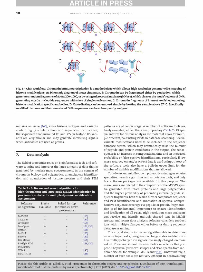
Fig. 3 – ChIP workflow. Chromatin immunoprecipitation is amethodology which allows high resolution genome-widemapping ofhistone modifications. A: Schematic diagram of intact chromatin. B: Chromatin can be fragmented either by sonication, whichgenerates randomfragments of about 200–1000, or byusingmicrococcal nuclease (MNase),which cleaves the ‘nude’ regions ofDNA,generating mostly nucleotide sequences with sizes of single nuclesomes. C: Chromatin fragments of interest are fished out usinghistone modification specific antibodies. D: Cross-linking can be removed simply by heating the sample above 67 °C. Specificallymodified histones and their associated DNA sequences can be subsequently analyzed.
10 J O U R N A L O F P R O T E O M I C S X X ( 2 0 1 2 ) X X X – X X X
remains an issue [140], since histone isotypes and variantscontain highly similar amino acid sequences; for instance,the sequences that surround K9 and K27 in histone H3 vari-ants are very similar and may generate interfering signalswhen antibodies are used as probes.
7. Data analysis
The field of proteomics relies on bioinformatics tools and soft-ware to mine and interpret the large amount of data that isgenerated by modern mass spectrometers. In the context ofchromatin biology and epigenetics, unambiguous identifica-tion and quantitation of histone proteins and their PTM
Table 2 – Software and search algorithms forhigh-throughput and large-scale MS/MS identification inproteomics suitable for multiple dynamic modificationassignments.
Softwareor algorithm
Freelyavailable
Suited for top(or middle)–down
proteomics
Reference
MASCOT – – [153]SEQUEST – – [154]X!Tandem + – [155]MaxQuant + – [156,157]OMSSA + + [158]InsPecT + – [159]Phenyx + – [160]BIG Mascot – + [151]ProSight PTM + + [149,150]ProSightPC – +VEMS + – [145]PILOT_PTM + + [148]
Please cite this article as: Sidoli S, et al, Proteomics in chromatmodifications of histone proteins by mass spectrometry, J Prot (
patterns are at center stage. A number of software tools arefreely available, while others are proprietary (Table 2). Of spe-cial interest for histone analysis are tools that allow for multi-ple different, co-existing PTMs in database searching. Severalvariable modifications need to be included in the sequencedatabase search, which may dramatically raise the numberof peptide and protein candidates in the output. The conse-quence is an increase in computational time and an increasedprobability or false-positive identifications, particularly if lowmass accuracyMS and/or MS/MS data is used as input. Most ofthe software tools also have a built-in upper limit for thenumber of variable modifications that are allowed.
Top–down andmiddle–down proteomics strategies requirespecialized search algorithms and annotation tools, and onlyfew software packages are available for this purpose. Themain issues are related to the complexity of the MS/MS spec-tra generated from intact proteins and large polypeptides,and the higher probability of generating internal peptide se-quence fragments, both of which further complicate sequenceand PTM identification and annotation of spectra. Compre-hensive sequence coverage via peptide or protein fragmenta-tion is of fundamental importance to ensure identificationand localization of all PTMs. High-resolution mass analyzerscan resolve and identify multiply-charged ions in MS/MSspectra and recent data analysis software considers productions with multiple charges either before or during sequencedatabase searching.
The crucial step is to use an algorithm able to determinemonoisotopic peaks, recognize ion charge states and deconvo-lute multiply-charged ion signals into singly-charged ion massvalues. There are several freeware tools available for this pur-pose. They usually remove isotopes and clean spectra from ran-dom noise as, for example, MS-Cleaner [141]. Unfortunately, anumber of such tools are not very efficient in deconvoluting
in biology and epigenetics: Elucidation of post-translational2012), doi:10.1016/j.jprot.2011.12.029

11J O U R N A L O F P R O T E O M I C S X X ( 2 0 1 2 ) X X X – X X X
MS/MS spectra, where coinciding and overlapping peaks, iso-tope patterns and fragment ion charge states present a chal-lenge. Algorithms such as THRASH [142] and Xtract weredeveloped for this purpose and they are integrated into Pro-SightPC. A modified version of THRASH was used in top–downstudies of histones byKelleher and coworkers [99–102]. THRASHis also integrated into ICR-2LS (unpublished, but freely availablefor download) and Decon2LS [143]. Liu et al. proposed a newJava-based algorithm for intact proteins MS/MS spectra, calledMS-Deconv [144]. They demonstrate that it can achieve moreextensive results than THRASH and Xtract in terms of numberof correctly-recovered monoisotopic masses, and in less time.Mascot Distiller (Matrix Science) also provides tools for MS andMS/MS peak deconvolution, accepts most types of data filesfromMS vendors and allows for multiple output file formats.
Several algorithms were developed with the aim to inter-pret MS/MS spectra from highly-modified proteins, such ashistones. In our group we developed VEMS (Virtual ExpertMass Spectrometrist) [145], which has been successfully ap-plied to histone data analysis, PTM annotation and quantita-tion [76,146]. VEMS can discriminate between acetyl lysineand trimethyl lysine by using additional information embed-ded in diagnostic ions and neutral loss signals in MS/MS spec-tra. DiMaggio et al. proposed an algorithm based on a mixedinteger linear optimization using ETD LC-MS/MS data [147],which is designed for large-scale data analysis in middle–down proteomics experiments [109]. In the following year, ithas been further developed, and currently is public availableunder the name of PILOT_PTM [148]. Commercial software ded-icated to middle–down and top–down proteomics include Pro-SightPC provided by ThermoFisher, based on the freely-available ProSightPTM [149,150]. An optimized version of Mascot,called BIGMascot, was developed for top–down proteomics [151].However, there are other data analysis strategies that are notbased on searching protein sequence databases. The mass spec-tral alignment method was implemented in a manner that ac-cepts top–down MS and MS/MS data, including up to 10–20modifications and successfully applied to analyze histone H4spectra [152].
In summary, a range of software and bioinformatics toolsare available, including special programs for middle–downand top–down strategies. The recent developments in high-resolution tandem mass spectrometry and new fragmenta-tion techniques provide a foundation for the generation ofhigh mass-accuracy and high mass-resolution MS and MS/MS data, which is amenable to automated computationaldata analysis. The availability of high quality MS and MS/MSdata, and further improvements in data analysis algorithmsand annotation tools points towards the development of ro-bust platforms for histone analysis in the near future, with afocus on middle–down and top–down analysis.
8. Conclusion
The realization over the last decade that chromatin is a highlyfunctional entity has propelled advances in biochemical andanalytical tools to study protein–DNA interactions. Histonesare at center stage since they constitute the nucleosome scaf-fold around which DNA is wrapped and, at the same time, the
Please cite this article as: Sidoli S, et al, Proteomics in chromatmodifications of histone proteins by mass spectrometry, J Prot (
highly modified N-terminal tails of histones protrude from thenucleosome and serve to recruit extrinsic factors and com-plexes to chromatin. In turn, elucidation of the PTMs of histonetails has become a very active field inmass spectrometry-basedproteomics in recent years. While the bottom–up strategy forhistone analysis achieves very efficient amino acid sequencingand improved throughput for complex samples, we expect thatmiddle–down strategies and eventually also top–down strate-gies will provide a comprehensive overview of multivalent, co-existing histone marks with distinct biological functions. Fur-thermore, the combination of ChIP-seq and proteomics willallow for gene- and promoter-specific analysis of coexistinghis-tone marks. Before we get to that scenario, several challengeshave to be overcome in order to achieve the required sensitivityand specificity for accurate annotation and quantitation of his-tone PTMs. We believe that ongoing developments in chroma-tography, mass spectrometry, and computational methodswill provide indispensible tool for new discoveries in epige-netics and chromatin biology.
Acknowledgments
Research in the O.N.J. laboratory is supported by a grant fromthe Danish National Research Foundation to the Center forEpigenetics. We thank Dr. Veit Schwämmle for reading andcommenting on this manuscript.
R E F E R E N C E S
[1] Russo VEA, Martienssen RA, Riggs AD. Epigeneticmechanisms of gene regulation. Plainview, N.Y.: Cold SpringHarbor Laboratory Press; 1996
[2] Waddington CH. Analization of development and theinheritance of acquired characters. Nature1942;150:563–5.
[3] Portela A, Esteller M. Epigenetic modifications and humandisease. Nat Biotechnol 2010;28:1057–68.
[4] Chi P, Allis CD, Wang GG. Covalent histone modifications –miswritten, misinterpreted and mis-erased in humancancers. Nat Rev Cancer 2010;10:457–69.
[5] Harshaw C. Alimentary epigenetics: a developmentalpsychobiological systems view of the perception of hunger,thirst and satiety. Dev Rev 2008;28:541–69.
[6] Hake SB, Garcia BA, Duncan EM, Kauer M, Dellaire G,Shabanowitz J, et al. Expression patterns and post-translational modifications associated with mammalianhistone H3 variants. J Biol Chem2006;281:559–68.
[7] Bonasio R, Tu S, Reinberg D. Molecular signals of epigeneticstates. Science 2010;330:612–6.
[8] Shogren-Knaak M, Ishii H, Sun JM, Pazin MJ, Davie JR,Peterson CL. Histone H4-K16 acetylation controls chromatinstructure and protein interactions. Science 2006;311:844–7.
[9] Taverna SD, Li H, Ruthenburg AJ, Allis CD, Patel DJ. Howchromatin-binding modules interpret histonemodifications: lessons from professional pocketpickers. Nat Struct Mol Biol 2007;14:1025–40.
[10] Kouzarides T. Chromatin modifications and their function.Cell 2007;128:693–705.
in biology and epigenetics: Elucidation of post-translational2012), doi:10.1016/j.jprot.2011.12.029

12 J O U R N A L O F P R O T E O M I C S X X ( 2 0 1 2 ) X X X – X X X
[11] Stratmann T, Mas P. Chromatin, photoperiod and theArabidopsis circadian clock: a question of time. Semin CellDev Biol 2008;19:554–9.
[12] Mao Y, Pavangadkar KA, Thomashow MF, Triezenberg SJ.Physical and functional interactions of Arabidopsis ADA2transcriptional coactivator proteins with theacetyltransferase GCN5 and with the cold-induced tran-scription factor CBF1. Biochim Biophys Acta 2006;1759:69–79.
[13] Zhou C, Zhang L, Duan J, Miki B, Wu K. HISTONEDEACETYLASE19 is involved in jasmonic acid and ethylenesignaling of pathogen response in Arabidopsis. Plant Cell2005;17:1196–204.
[14] Gray JC, Chua YL, Channeliere S, Mott E. The bromodomainprotein GTE6 controls leaf development in Arabidopsis byhistone acetylation at ASYMMETRIC LEAVES1. Genes Dev2005;19:2245–54.
[15] He Y, Michaels SD, Amasino RM. Regulation of flowering timeby histone acetylation in Arabidopsis. Science2003;302:1751–4.
[16] Wu K, Tian L, Malik K, Brown D, Miki B. Functional analysisof HD2 histone deacetylase homologues in Arabidopsisthaliana. Plant J 2000;22:19–27.
[17] Autran D, Baroux C, Raissig MT, Lenormand T, Wittig M,Grob S, et al. Maternal epigenetic pathways control parentalcontributions to Arabidopsis early embryogenesis. Cell2011;145:707–19.
[18] Puri D, Dhawan J, Mishra RK. The paternal hidden agenda:epigenetic inheritance through sperm chromatin.Epigenetics 2010;5:386–91.
[19] Riccio A. Dynamic epigenetic regulation in neurons:enzymes, stimuli and signaling pathways. Nat Neurosci2010;13:1330–7.
[20] Peedicayil J. Epigenetic therapy – a new development inpharmacology. Indian J Med Res 2006;123:17–24.
[21] Marks PA. The clinical development of histone deacetylaseinhibitors as targeted anticancer drugs. Expert Opin InvestigDrugs 2010;19:1049–66.
[22] Salcedo-Amaya AM, Hoeijmakers WAM, Bartfai R,Stunnenberg HG. Malaria: could its unusual epigenome bethe weak spot? Int J Biochem Cell Biol 2010;42:781–4.
[23] Strahl BD, Allis CD. The language of covalent histonemodifications. Nature 2000;403:41–5.
[24] Jenuwein T, Allis CD. Translating the histone code. Science2001;293:1074–80.
[25] Berger SL. The complex language of chromatin regulationduring transcription. Nature 2007;447:407–12.
[26] Lee JS, Smith E, Shilatifard A. The language of histonecrosstalk. Cell 2010;142:682–5.
[27] Turner BM. Reading signals on the nucleosome with a newnomenclature for modified histones. Nat Struct Mol Biol2005;12:110–2.
[28] Murray K. Occurrence of Epsilon-N-Methyl Lysine in Histones.Biochemistry 1964;3:10.
[29] Bannister AJ, Schneider R, Kouzarides T. Histonemethylation: dynamic or static? Cell 2002;109:801–6.
[30] Wallwork JC, Lee CT, Duerre JA. A sensitive assay for histonemethyltransferase. Anal Biochem 1978;84:103–10.
[31] Rice JC, Allis CD. Histone methylation versus histoneacetylation: new insights into epigenetic regulation. CurrOpin Cell Biol 2001;13:263–73.
[32] Yamane K, Toumazou C, Tsukada Y, Erdjument-Bromage H,Tempst P, Wong J, et al. JHDM2A, a JmjC-containing H3K9demethylase, facilitates transcription activation byandrogen receptor. Cell 2006;125:483–95.
[33] Litt M, Qiu Y, Huang SM. Histone arginine methylations:their roles in chromatin dynamics and transcriptionalregulation. Biosci Rep 2009;29:131–41.
[34] Martin C, Zhang Y. The diverse functions of histone lysinemethylation. Nat Rev Mol Cell Biol 2005;6:838–49.
Please cite this article as: Sidoli S, et al, Proteomics in chromatmodifications of histone proteins by mass spectrometry, J Prot (
[35] Oliver SS, Denu JM. Dynamic interplay between histone H3modifications and protein interpreters: emerging evidencefor a “histone language”. Chembiochem2011;12:299–307.
[36] Kirmizis A, Santos-Rosa H, Penkett CJ, Singer MA,Vermeulen M, Mann M, et al. Arginine methylation athistone H3R2 controls deposition of H3K4 trimethylation.Nature 2007;449 928-U17.
[37] Allfrey VG, Faulkner R, Mirsky AE. Acetylation + Methylationof Histones + Their Possible Role in Regulation of RnaSynthesis. Proc Natl Acad Sci U S A 1964;51:786.
[38] Martinet N, Bertrand P. Interpreting clinical assays forhistone deacetylase inhibitors. Cancer Manag Res 2011;3:117–41.
[39] Nakanishi S, Sanderson BW, Delventhal KM, Bradford WD,Staehling-Hampton K, Shilatifard A. A comprehensivelibrary of histone mutants identifies nucleosomal residuesrequired for H3K4 methylation. Nat Struct Mol Biol 2008;15:881–8.
[40] Trelle MB, Jensen ON. Utility of immonium ions forassignment of epsilon-N-acetyllysine-containing peptidesby tandem mass spectrometry. Anal Chem2008;80:3422–30.
[41] Trelle MB, Jensen ON. Functional proteomics in histoneresearch and epigenetics. Expert Rev Proteomics 2007;4:491–503.
[42] Rogakou EP, Pilch DR, Orr AH, Ivanova VS, Bonner WM. DNAdouble-stranded breaks induce histone H2AX phosphorylationon serine 139. J Biol Chem 1998;273:5858–68.
[43] Xu Y, Price BD. Chromatin dynamics and the repair of DNAdouble strand breaks. Cell Cycle 2011;10:261–7.
[44] Deng H, Bao X, Cai W, Blacketer MJ, Belmont AS, Girton J,et al. Ectopic histone H3S10 phosphorylation causeschromatin structure remodeling in Drosophila.Development 2008;135:699–705.
[45] Johansen KM, Johansen J. Regulation of chromatin structureby histone H3S10 phosphorylation. Chromosome Res2006;14:393–404.
[46] Garcia BA, Joshi S, Thomas CE, Chitta RK, Diaz RL, Busby SA,et al. Comprehensive phosphoprotein analysis of linkerhistone H1 from Tetrahymena thermophila. Mol CellProteomics 2006;5:1593–609.
[47] Fischle W, Tseng BS, Dormann HL, Ueberheide BM, Garcia BA,Shabanowitz J, et al. Regulation of HP1-chromatin binding byhistone H3 methylation and phosphorylation. Nature2005;438:1116–22.
[48] Fischle W, Wang Y, Allis CD. Binary switches andmodification cassettes in histone biology and beyond.Nature 2003;425:475–9.
[49] Garcia BA, Barber CM, Hake SB, Ptak C, Turner FB, Busby SA,et al. Modifications of human histone H3 variants duringmitosis. Biochemistry 2005;44:13202–13.
[50] Lau PN, Cheung P. Histone code pathway involving H3 S28phosphorylation and K27 acetylation activates transcriptionand antagonizes polycomb silencing. Proc Natl Acad SciU S A 2011;108:2801–6.
[51] Simboeck E, Sawicka A, Zupkovitz G, Senese S, Winter S,Dequiedt F, et al. A phosphorylation switch regulates thetranscriptional activation of cell cycle regulator p21 byhistonedeacetylase inhibitors. J Biol Chem 2010;285:41062–73.
[52] Yeoh GC, Tan EL, Besant PG, Zu XL, Turck CW, BogoyevitchMA, et al. Histone H4 histidine kinase displays theexpression pattern of a liver oncodevelopmental marker.Carcinogenesis 2004;25:2083–8.
[53] Attwood PV, Zu XL, Besant PG, Imhof A. Mass spectrometricanalysis of protein histidine phosphorylation. Amino Acids2007;32:347–57.
[54] Besant PG, Attwood PV. Histidine Phosphorylation inHistones and in Other Mammalian Proteins. Methods in
in biology and epigenetics: Elucidation of post-translational2012), doi:10.1016/j.jprot.2011.12.029

13J O U R N A L O F P R O T E O M I C S X X ( 2 0 1 2 ) X X X – X X X
Enzymology, Vol 471. Two-Component Signaling Systems,Part C; 2010. p. 403–26.
[55] Kothapalli N, Camporeale G, Kueh A, Chew YC, OommenAM, Griffin JB, et al. Biological functions of biotinylatedhistones. J Nutr Biochem 2005;16:446–8.
[56] Filenko NA, Kolar C, West JT, Smith SA, Hassan YI, BorgstahlGE, et al. The role of histone H4 biotinylation in the structureof nucleosomes. PLoS One 2011;6:e16299.
[57] Camporeale G, Shubert EE, Sarath G, Cerny R, Zempleni J. K8and K12 are biotinylated in human histone H4. Eur JBiochem 2004;271:2257–63.
[58] Healy S, Perez-Cadahia B, Jia DX, McDonald MK, Davie JR,Gravel RA. Biotin is not a natural histone modification.Bba-Gene Regul Mech 2009;1789:719–33.
[59] Anzilotti C, Pratesi F, Tommasi C, Migliorini P.Peptidylarginine deiminase 4 and citrullination in healthand disease. Autoimmun Rev 2010;9:158–60.
[60] Cuthbert GL, Daujat S, Snowden AW, Erdjument-Bromage H,Hagiwara T, Yamada M, et al. Histone deiminationantagonizes arginine methylation. Cell 2004;118:545–53.
[61] Kubota K, Yoneyama-Takazawa T, Ichikawa K.Determination of sites citrullinated by peptidylargininedeiminase using O-18 stable isotope labeling and massspectrometry. Rapid CommunMass Spectrom 2005;19:683–8.
[62] Stensland M, Holm A, Kiehne A, Fleckenstein B. Targetedanalysis of protein citrullination using chemicalmodification and tandem mass spectrometry. RapidCommun Mass Spectrom 2009;23:2754–62.
[63] Boulikas T. DNA strand breaks alter histone ADP-ribosylation.Proc Natl Acad Sci U S A 1989;86:3499–503.
[64] Stone PR, Lorimer WS, Kidwell WR. Properties of complexbetween histone H-1 and poly(Adp-Ribose) synthesized inhela-cell nuclei. Eur J Biochem 1977;81:9–18.
[65] Hottiger MO. ADP-ribosylation of histones by ARTD1: anadditional module of the histone code? FEBS Lett 2011;585:1595–9.
[66] Zlatanova J, Caiafa P, Van Holde K. Linker histone bindingand displacement: versatile mechanism for transcriptionalregulation. FASEB J 2000;14:1697–704.
[67] Li W, Nagaraja S, Delcuve GP, Hendzel MJ, Davie JR. Effects ofhistone acetylation, ubiquitination and variants onnucleosome stability. Biochem J 1993;296(Pt 3):737–44.
[68] Weake VM, Workman JL. Histone ubiquitination: triggeringgene activity. Mol Cell 2008;29:653–63.
[69] Bergink S, Salomons FA, Hoogstraten D, Groothuis TA, deWaard H, Wu J, et al. DNA damage triggers nucleotideexcision repair-dependent monoubiquitylation of histoneH2A. Genes Dev 2006;20:1343–52.
[70] Xiong L, Darwanto A, Sharma S, Herring J, Hu S, Filippova M,et al. Mass spectrometric studies on epigenetic interactionnetworks in cell differentiation. J Biol Chem2011;286:13657–68.
[71] Shilo Y, Eisenman RN. Histone sumoylation is associatedwith transcriptional repression. Proc Natl Acad Sci U S A2003;100:13225–30.
[72] Chen Y, Sprung R, Tang Y, Ball H, Sangras B, Kim SC, et al.Lysine propionylation and butyrylation are novelpost-translational modifications in histones. Mol CellProteomics 2007;6:812–9.
[73] Sakabe K, Wang ZH, Hart GW. beta-N-acetylglucosamine(O-GlcNAc) is part of the histone code. Proc Natl Acad Sci U SA 2010;107:19915–20.
[74] Shechter D, Dormann HL, Allis CD, Hake SB. Extraction,purification and analysis of histones. Nat Protoc 2007;2:1445–57.
[75] Vonholt C, Brandt WF, Greyling HJ, Lindsey GG, Retief JD,Rodrigues JD, et al. Isolation and characterization ofhistones. Methods Enzymol 1989;170:431–523.
[76] Trelle MB, Salcedo-Amaya AM, Cohen AM, Stunnenberg HG,Jensen ON. Global histone analysis by mass spectrometry
Please cite this article as: Sidoli S, et al, Proteomics in chromatmodifications of histone proteins by mass spectrometry, J Prot (
reveals a high content of acetylated lysine residues in themalaria parasite Plasmodium falciparum. J Proteome Res2009;8:3439–50.
[77] Boersema PJ, Mohammed S, Heck AJR. Hydrophilicinteraction liquid chromatography (HILIC) in proteomics.Anal Bioanal Chem 2008;391:151–9.
[78] MackayCL, Ramsahoye B, Burgess K, CookK,Weidt S, Creanor J,et al. Sensitive, specific, and quantitative FTICR massspectrometry of combinatorial post-translationalmodificationsin intact histone H4. Anal Chem 2008;80:4147–53.
[79] Sneekes EJ, Han J, Elliot M, Ausio J, Swart R, Heck AJR, et al.Accurate molecular weight analysis of histones using FFEand RP-HPLC on monolithic capillary columns. J Sep Sci2009;32:2691–8.
[80] Rosenqvist H, Ye J, Jensen ON. Analytical strategies in massspectrometry-based phosphoproteomics. Methods Mol Biol2011;753:183–213.
[81] Rosas-Acosta G, Russell WK, Deyrieux A, Russell DH, WilsonVG. A universal strategy for proteomic studies of SUMO andother ubiquitin-like modifiers. Mol Cell Proteomics 2005;4:56–72.
[82] Hackbarth JS, Lee SH, Meng XW, Vroman BT, Kaufmann SH,Karnitz LM. S-peptide epitope tagging for proteinpurification, expression monitoring, and localization inmammalian cells. Biotechniques 2004;37:835–9.
[83] Kirkpatrick DS, Denison C, Gygi SP. Weighing in onubiquitin: the expanding role of mass-spectrometry-basedproteomics. Nat Cell Biol 2005;7:750–7.
[84] Gurley LR, London JE, Valdez JG. High-performance capillaryelectrophoresis of histones. J Chromatogr 1991;559:431–43.
[85] Sarg B, Helliger W, Hoertnagl B, Puschendorf B, Lindner H.The N-terminally acetylated form of mammalian histone H1(o), but not that of avian histone H5, increases with age. ArchBiochem Biophys 1999;372:333–9.
[86] Aguilar C, Hofte AJ, Tjaden UR, van der Greef J. Analysis ofhistones by on-line capillary zone electrophoresis-electrospray ionisation mass spectrometry. J Chromatogr A2001;926:57–67.
[87] Aebersold R, Mann M. Mass spectrometry-based proteomics.Nature 2003;422:198–207.
[88] McLafferty FW, Breuker K, Jin M, Han X, Infusini G, Jiang H,et al. Top–down MS, a powerful complement to the highcapabilities of proteolysis proteomics. FEBS J 2007;274:6256–68.
[89] Zubarev RA, Kelleher NL, McLafferty FW. Electron capturedissociation of multiply charged protein cations. Anonergodic process. J Am Chem Soc 1998;120:3265–6.
[90] Syka JE, Coon JJ, Schroeder MJ, Shabanowitz J, Hunt DF.Peptide and protein sequence analysis by electron transferdissociation mass spectrometry. Proc Natl Acad Sci U S A2004;101:9528–33.
[91] Mikesh LM, Ueberheide B, Chi A, Coon JJ, Syka JE,Shabanowitz J, et al. The utility of ETDmass spectrometry inproteomic analysis. Biochim Biophys Acta 2006;1764:1811–22.
[92] Zhang K, Tang H, Huang L, Blankenship JW, Jones PR, XiangF, et al. Identification of acetylation and methylation sites ofhistone H3 from chicken erythrocytes by high-accuracymatrix-assisted laser desorption ionization-time-of-flight,matrix-assisted laser desorption ionization-postsourcedecay, and nanoelectrospray ionization tandem massspectrometry. Anal Biochem 2002;306:259–69.
[93] Garcia BA, Mollah S, Ueberheide BM, Busby SA, Muratore TL,Shabanowitz J, et al. Chemical derivatization of histones forfacilitated analysis by mass spectrometry. Nat Protoc 2007;2:933–8.
[94] Jufvas A, Stralfors P, Vener AV. Histone Variants and TheirPost-Translational Modifications in Primary Human FatCells. PLoS One 2011;6(1):e15960.
in biology and epigenetics: Elucidation of post-translational2012), doi:10.1016/j.jprot.2011.12.029

14 J O U R N A L O F P R O T E O M I C S X X ( 2 0 1 2 ) X X X – X X X
[95] Bonaldi T, Imhof A, Regula JT. A combination of differentmass spectroscopic techniques for the analysis of dynamicchanges of histone modifications. Proteomics 2004;4:1382–96.
[96] McKittrick E, Gaften PR, Ahmad K, Henikoff S. Histone H3.3 isenriched in covalent modifications associated with activechromatin. Proc Natl Acad Sci U S A 2004;101:1525–30.
[97] Han X, Jin M, Breuker K, McLafferty FW. Extending top–downmass spectrometry to proteins with masses greater than 200kilodaltons. Science 2006;314:109–12.
[98] Banks GC, Deterding LJ, Tomer KB, Archer TK.Hormone-mediated dephosphorylation of specific histoneH1 isoforms. J Biol Chem 2001;276:36467–73.
[99] Thomas CE, Kelleher NL, Mizzen CA. Mass spectrometriccharacterization of human histone H3: a bird's eye view.J Proteome Res 2006;5:240–7.
[100] Boyne II MT, Pesavento JJ, Mizzen CA, Kelleher NL. Precisecharacterization of humanhistones in theH2A gene family bytop downmass spectrometry. J Proteome Res 2006;5:248–53.
[101] Siuti N, Roth MJ, Mizzen CA, Kelleher NL, Pesavento JJ.Gene-specific characterization of human histone H2B byelectron capture dissociation. J Proteome Res2006;5:233–9.
[102] Pesavento JJ, Mizzen CA, Kelleher NL. Quantitative analysis ofmodified proteins and their positional isomers by tandemmassspectrometry: human histone H4. Anal Chem2006;78:4271–80.
[103] Lindner H, Sarg B, Meraner C, Helliger W. Separation ofacetylated core histones by hydrophilic-interaction liquidchromatography. J Chromatogr A 1996;743:137–44.
[104] Tian Z, Zhao R, Tolic N, Moore RJ, Stenoien DL, Robinson EW,et al. Two-dimensional liquid chromatography system foronline top–down mass spectrometry. Proteomics 2010;10:3610–20.
[105] Eliuk SM, Maltby D, Panning B, Burlingame AL. Highresolution electron transfer dissociation studies ofunfractionated intact histones from murine embryonicstem cells using on-line capillary LC separation:determination of abundant histone isoforms andpost-translational modifications. Mol Cell Proteomics2010;9:824–37.
[106] Contrepois K, Ezan E, Mann C, Fenaille F. Ultra-highperformance liquid chromatography-mass spectrometry forthe fast profiling of histone post-translationalmodifications. J Proteome Res 2010;9:5501–9.
[107] Taverna SD, Ueberheide BM, Liu YF, Tackett AJ, Diaz RL,Shabanowitz J, et al. Long-distance combinatorial linkagebetween methylation and acetylation on histone H3Ntermini. Proc Natl Acad Sci U S A 2007;104:2086–91.
[108] Swatkoski S, Gutierrez P, Wynne C, Petrov A, Dinman JD,Edwards N, et al. Evaluation of microwave-acceleratedresidue-specific acid cleavage for proteomic applications.J Proteome Res 2008;7:579–86.
[109] Young NL, DiMaggio PA, Plazas-Mayorca MD, Baliban RC,Floudas CA, Garcia BA. High throughput characterization ofcombinatorial histone codes. Mol Cell Proteomics 2009;8:2266–84.
[110] Yang L, Tu S, Ren C, Bulloch EM, Liao CL, Tsai MD, et al.Unambiguous determination of isobaric histonemodifications by reversed-phase retention time andhigh-mass accuracy. Anal Biochem 2010;396:13–22.
[111] Garcia BA. What does the future hold for top down massspectrometry? J Am Soc Mass Spectrom 2010;21:193–202.
[112] Mant CT, Hodges RS. Mixed-mode hydrophilicinteraction/cation-exchange chromatography (HILIC/CEX)of peptides and proteins. J Sep Sci 2008;31:2754–73.
[113] Berndsen CE, Denu JM. Catalysis and substrate selection byhistone/protein lysine acetyltransferases. Curr Opin StructBiol 2008;18:682–9.
Please cite this article as: Sidoli S, et al, Proteomics in chromatmodifications of histone proteins by mass spectrometry, J Prot (
[114] Frederiks F, Stulemeijer IJ, Ovaa H, van Leeuwen F. Amodified epigenetics toolbox to study histone modificationson the nucleosome core. Chembiochem 2011;12:308–13.
[115] Nguyen AT, Zhang Y. The diverse functions of Dot1 andH3K79 methylation. Genes Dev2011;25:1345–58.
[116] Choudhary C, Kumar C, Gnad F, Nielsen ML, Rehman M,Walther TC, et al. Lysine acetylation targets proteincomplexes and co-regulates major cellular functions.Science 2009;325:834–40.
[117] Bantscheff M, Schirle M, Sweetman G, Rick J, Kuster B.Quantitative mass spectrometry in proteomics: a criticalreview. Anal Bioanal Chem 2007;389:1017–31.
[118] Eberl HC, Mann M, Vermeulen M. Quantitative proteomicsfor epigenetics. Chembiochem 2011;12:224–34.
[119] Arnaudo AM, Molden RC, Garcia BA. Revealing histonevariant induced changes via quantitative proteomics. CritRev Biochem Mol Biol 2011;46:284–94.
[120] Smith CM, Gafken PR, Zhang ZL, Gottschling DE, Smith JB,Smith DL. Mass spectrometric quantification of acetylationat specific lysines within the amino-terminal tail of histoneH4. Anal Biochem 2003;316:23–33.
[121] Plazas-Mayorca MD, Zee BM, Young NL, Fingerman IM,LeRoy G, Briggs SD, et al. One-pot shotgun quantitative massspectrometry characterization of histones. J Proteome Res2009;8:5367–74.
[122] Plazas-Mayorca MD, Bloom JS, Zeissler U, Leroy G, Young NL,DiMaggio PA, et al. Quantitative proteomics reveals directand indirect alterations in the histone code followingmethyltransferase knockdown. Mol Biosyst 2010;6:1719–29.
[123] Yamasu K, Senshu T. Reassembly of nucleosomal histoneoctamers during replication of chromatin. J Biochem1987;101:1041–9.
[124] Ong SE, Blagoev B, Kratchmarova I, Kristensen DB, Steen H,Pandey A, et al. Stable isotope labeling by amino acids in cellculture, SILAC, as a simple and accurate approach toexpression proteomics. Mol Cell Proteomics 2002;1:376–86.
[125] Bonenfant D, Towbin H, Coulot M, Schindler P, Mueller DR,van Oostrum J. Analysis of dynamic changes in post-translational modifications of human histones during cellcycle by mass spectrometry. Mol Cell Proteomics 2007;6:1917–32.
[126] Jung HR, Pasini D, Helin K, Jensen ON. Quantitative massspectrometry of histones H3.2 and H3.3 in Suz12-deficientmouse embryonic stem cells reveals distinct, dynamicpost-translational modifications at Lys-27 and Lys-36. MolCell Proteomics 2010;9:838–50.
[127] Zee BM, Levin RS, Dimaggio PA, Garcia BA. Global turnoverof histone post-translational modifications and variants inhuman cells. Epigenetics Chromatin 2010;3:22.
[128] Salim K, Kehoe L, Minkoff MS, Bilsland JG, Munoz-Sanjuan I,Guest PC. Identification of differentiating neural progenitorcell markers using shotgun isobaric tagging massspectrometry. Stem Cells Dev 2006;15:461–70.
[129] Montes de Oca R, Shoemaker CJ, Gucek M, Cole RN, WilsonKL. Barrier-to-autointegration factor proteome revealschromatin-regulatory partners. PLoS One2009;4:e7050.
[130] Yuan X, Gu X, Crabb JS, Yue X, Shadrach K, Hollyfield JG,et al. Quantitative proteomics: comparison of the macularBruch membrane/choroid complex from age-relatedmacular degeneration and normal eyes. Mol Cell Proteomics2010;9:1031–46.
[131] Garcia BA, Pesavento JJ, Mizzen CA, Kelleher NL. Pervasivecombinatorial modification of histone H3 in human cells.Nat Methods 2007;4:487–9.
[132] Phanstiel D, Brumbaugh J, Berggren WT, Conard K, Feng X,Levenstein ME, et al. Mass spectrometry identifies andquantifies 74 unique histone H4 isoforms in differentiating
in biology and epigenetics: Elucidation of post-translational2012), doi:10.1016/j.jprot.2011.12.029

15J O U R N A L O F P R O T E O M I C S X X ( 2 0 1 2 ) X X X – X X X
human embryonic stem cells. Proc Natl Acad Sci U S A2008;105:4093–8.
[133] Jensen ON, Beck HC, Nielsen EC, Matthiesen R, Jensen LH,Sehested M, et al. Quantitative proteomic analysis ofposttranslational modifications of human histones. Mol CellProteomics 2006;5:1314–25.
[134] Zhang KL, Darwanto A, Curtis MP, Schrag M, Kirsch W, Liu P,et al. A modified “cross-talk” between histone H2B Lys-120ubiquitination and H3 Lys-79 methylation. J Biol Chem2010;285:21868–76.
[135] Liu CL, Kaplan T, Kim M, Buratowski S, Schreiber SL,Friedman N, et al. Single-nucleosome mapping of histonemodifications in S. cerevisiae. PLoS Biol 2005;3:e328.
[136] Park PJ. ChIP-seq: advantages and challenges of a maturingtechnology. Nat Rev Genet 2009;10:669–80.
[137] Mikkelsen TS, Ku M, Jaffe DB, Issac B, Lieberman E,Giannoukos G, et al. Genome-wide maps of chromatin statein pluripotent and lineage-committed cells. Nature2007;448:553–60.
[138] Bernstein BE, Kamal M, Lindblad-Toh K, Bekiranov S, BaileyDK, Huebert DJ, et al. Genomic maps and comparativeanalysis of histone modifications in human and mouse. Cell2005;120:169–81.
[139] Vermeulen M, Eberl HC, Matarese F, Marks H, Denissov S,Butter F, et al. Quantitative interaction proteomics andgenome-wide profiling of epigenetic histone marks andtheir readers. Cell 2010;142:967–80.
[140] EgelhoferTA,MinodaA, KlugmanS, LeeK, Kolasinska-Zwierz P,Alekseyenko AA, et al. An assessment of histone-modificationantibody quality. Nat Struct Mol Biol 2011;18:91–3.
[141] Mujezinovic N, Raidl G, Hutchins JR, Peters JM, Mechtler K,Eisenhaber F. Cleaning of raw peptide MS/MS spectra:improved protein identification following deconvolution ofmultiply charged peaks, isotope clusters, and removal ofbackground noise. Proteomics 2006;6:5117–31.
[142] Horn DM, Zubarev RA, McLafferty FW. Automated reductionand interpretation of high resolution electrospraymass spectraof large molecules. J Am Soc Mass Spectrom 2000;11:320–32.
[143] Jaitly N, Mayampurath A, Littlefield K, Adkins JN, AndersonGA, Smith RD. Decon2LS: an open-source software packagefor automated processing and visualization of highresolution mass spectrometry data. BMC Bioinformatics2009;10:87.
[144] Liu X, Inbar Y, Dorrestein PC, Wynne C, Edwards N, Souda P,et al. Deconvolution and database search of complextandem mass spectra of intact proteins: a combinatorialapproach. Mol Cell Proteomics 2010;9:2772–82.
[145] Matthiesen R, Trelle MB, Hojrup P, Bunkenborg J, Jensen ON.VEMS 3.0: algorithms and computational tools for tandemmass spectrometry based identification of post-translational modifications in proteins. J Proteome Res2005;4:2338–47.
[146] Beck HC, Nielsen EC, Matthiesen R, Jensen LH, Sehested M,Finn P, et al. Quantitative proteomic analysis ofpost-translational modifications of human histones. MolCell Proteomics 2006;5:1314–25.
Please cite this article as: Sidoli S, et al, Proteomics in chromatmodifications of histone proteins by mass spectrometry, J Prot (
[147] DiMaggio Jr PA, Young NL, Baliban RC, Garcia BA, FloudasCA. A mixed integer linear optimization framework for theidentification and quantification of targeted post-translational modifications of highly modified proteinsusing multiplexed electron transfer dissociation tandemmass spectrometry. Mol Cell Proteomics 2009;8:2527–43.
[148] Baliban RC, DiMaggio PA, Plazas-Mayorca MD, Young NL,Garcia BA, Floudas CA. A novel approach for untargetedpost-translational modification identification using integerlinear optimization and tandem mass spectrometry. MolCell Proteomics 2010;9:764–79.
[149] LeDuc RD, Taylor GK, Kim YB, Januszyk TE, Bynum LH, SolaJV, et al. ProSight PTM: an integrated environment forprotein identification and characterization by top–downmass spectrometry. Nucleic Acids Res 2004;32:W340–5.
[150] Zamdborg L, LeDuc RD, Glowacz KJ, Kim YB, Viswanathan V,Spaulding IT, et al. ProSight PTM 2.0: improved proteinidentification and characterization for top down massspectrometry. Nucleic Acids Res 2007;35:W701–6.
[151] Karabacak NM, Li L, Tiwari A, Hayward LJ, Hong P, EasterlingML, et al. Sensitive and specific identification of wild typeand variant proteins from 8 to 669 kDa using top–downmass spectrometry. Mol Cell Proteomics 2009;8:846–56.
[152] Frank AM, Pesavento JJ, Mizzen CA, Kelleher NL, Pevzner PA.Interpreting top–down mass spectra using spectralalignment. Anal Chem 2008;80:2499–505.
[153] PerkinsDN, PappinDJ, CreasyDM, Cottrell JS. Probability-basedprotein identification by searching sequence databases usingmass spectrometry data. Electrophoresis 1999;20:3551–67.
[154] Eng JK, Mccormack AL, Yates JR. An approach to correlatetandem mass-spectral data of peptides withamino-acid-sequences in a protein database.J Am Soc Mass Spectrom 1994;5:976–89.
[155] Craig R, Beavis RC. TANDEM: matching proteins withtandem mass spectra. Bioinformatics 2004;20:1466–7.
[156] Cox J, Mann M. MaxQuant enables high peptideidentification rates, individualized p.p.b.-range massaccuracies and proteome-wide protein quantification. NatBiotechnol 2008;26:1367–72.
[157] Cox J, Neuhauser N, Michalski A, Scheltema RA, Olsen JV,Mann M. Andromeda: a peptide search engine integratedinto the MaxQuant environment. J Proteome Res 2011;10:1794–805.
[158] Geer LY, Markey SP, Kowalak JA, Wagner L, Xu M, MaynardDM, et al. Open mass spectrometry search algorithm. JProteome Res 2004;3:958–64.
[159] Tanner S, Shu HJ, Frank A, Wang LC, Zandi E, Mumby M,et al. InsPecT: Identification of posttransiationally modifiedpeptides from tandem mass spectra. Anal Chem 2005;77:4626–39.
[160] Colinge J,Masselot A, Cusin I,Mahe E,NiknejadA, Argoud-PuyG, et al. High-performance peptide identification by tandemmass spectrometry allows reliable automatic data processingin proteomics. Proteomics 2004;4:1977–84.
in biology and epigenetics: Elucidation of post-translational2012), doi:10.1016/j.jprot.2011.12.029
Copyright © 2022 FDOKUMEN


![Analysis of [FeFe]hydrogenase genes for the elucidation of a ...](https://static.fdokumen.com/doc/165x107/6324e1b9051fac18490cfd07/analysis-of-fefehydrogenase-genes-for-the-elucidation-of-a-.jpg)


