Integrating Clinical and Translational Research Networks - MDPI
252
Integrating Clinical and Translational Research Networks Building Team Medicine Printed Edition of the Special Issue Published in Journal of Clinical Medicine www.mdpi.com/journal/jcm Ravi Salgia and Prakash Kulkarni Edited by
-
Upload
khangminh22 -
Category
Documents
-
view
0 -
download
0
Transcript of Integrating Clinical and Translational Research Networks - MDPI
Integrating Clinical and Translational Research Netw
orks • Ravi Salgia and Prakash Kulkarni
Integrating Clinical and Translational Research NetworksBuilding Team Medicine
Printed Edition of the Special Issue Published in Journal of Clinical Medicine
www.mdpi.com/journal/jcm
Ravi Salgia and Prakash KulkarniEdited by

Integrating Clinical and Translational Research Networks—Building Team Medicine


Integrating Clinical and Translational Research Networks—Building Team Medicine
Editors
Ravi Salgia
Prakash Kulkarni
MDPI • Basel • Beijing • Wuhan • Barcelona • Belgrade • Manchester • Tokyo • Cluj • Tianjin
Prakash Kulkarni
City of Hope National Medical Center
US
EditorsRavi Salgia
City of Hope National Medical Center
USA
Editorial Office
MDPISt. Alban-Anlage 66
4052 Basel, Switzerland
This is a reprint of articles from the Special Issue published online in the open access journal
Journal of Clinical Medicine (ISSN 2077-0383) (available at: https://www.mdpi.com/journal/jcm/
special issues/Team Medicine).
For citation purposes, cite each article independently as indicated on the article page online and as
indicated below:
LastName, A.A.; LastName, B.B.; LastName, C.C. Article Title. Journal Name Year, Volume Number,
Page Range.
ISBN 978-3-0365-0396-7 (Hbk)
ISBN 978-3-0365-0397-4 (PDF)
© 2021 by the authors. Articles in this book are Open Access and distributed under the Creative
Commons Attribution (CC BY) license, which allows users to download, copy and build upon
published articles, as long as the author and publisher are properly credited, which ensures maximum
dissemination and a wider impact of our publications.
The book as a whole is distributed by MDPI under the terms and conditions of the Creative Commons
license CC BY-NC-ND.
Contents
About the Editors . . . . . . . . . . . . . . . . . . . . . . . . . . . . . . . . . . . . . . . . . . . . . . vii
Ravi Salgia and Prakash Kulkarni
Integrating Clinical and Translational Research Networks—Building Team MedicineReprinted from: J. Clin. Med. 2020, 9, 2975, doi:10.3390/jcm9092975 . . . . . . . . . . . . . . . . . 1
Rebecca Pharaon, Samuel Chung, Arya Amini, Ellie Maghami, Arnab Chowdhury, Nayana Vora, Sue Chang, Robert Kang, Thomas Gernon, Kelly Hansen, Christina Kelly, Denise Ackerman, Lalit Vora, Sagus Sampath and Erminia Massarelli
An Analysis and Comparison of Survival and Functional Outcomes in Oropharyngeal Squamous Cell Carcinoma Patients Treated with Concurrent Chemoradiation Therapy within City of Hope Cancer Center SitesReprinted from: J. Clin. Med. 2020, 9, 3083, doi:10.3390/jcm9103083 . . . . . . . . . . . . . . . . . 3
Joanne E. Mortimer, Laura Kruper, Mary Cianfrocca, Sayeh Lavasani, Sariah Liu, Niki Tank-Patel, Mina Sedrak, Wade Smith, Daphne Stewart, James Waisman, Christina Yeon, Tina Wang and Yuan Yuan
Use of HER2-Directed Therapy in Metastatic Breast Cancer and How Community Physicians Collaborate to Improve CareReprinted from: J. Clin. Med. 2020, 9, 1984, doi:10.3390/jcm9061984 . . . . . . . . . . . . . . . . . 15
Daniel J. Kim, Dan Otap, Nora Ruel, Naveen Gupta, Naveed Khan and Tanya Dorff
NCI–Clinical Trial Accrual in a Community Network Affiliated with a DesignatedCancer CenterReprinted from: J. Clin. Med. 2020, 9, 1970, doi:10.3390/jcm9061970 . . . . . . . . . . . . . . . . . 25
Ravi Salgia, Isa Mambetsariev, Tingting Tan, Amanda Schwer, Daryl P. Pearlstein, Hazem Chehabi, Angel Baroz, Jeremy Fricke, Rebecca Pharaon, Hannah Romo, Thomas Waddington, Razmig Babikian, Linda Buck, Prakash Kulkarni, Mary Cianfrocca, Benjamin Djulbegovic and Sumanta K. Pal
Complex Oncological Decision-Making Utilizing Fast-and-Frugal Trees in a Community Setting—Role of Academic and Hybrid ModelingReprinted from: J. Clin. Med. 2020, 9, 1884, doi:10.3390/jcm9061884 . . . . . . . . . . . . . . . . . 33
Cary A. Presant, Ravi Salgia, Prakash Kulkarni, Brian L. Tiep, Shamel Sanani, Benjamin Leach, Kimlin Ashing, Jossie Sandoval, Mina S. Sedrak, Shana Landau, Sophia Yeung, Dan Raz and Shanmugga Subbiah
Implementing Lung Cancer Screening and Prevention in Academic Centers, Affiliated Network Offices and Collaborating Care SitesReprinted from: J. Clin. Med. 2020, 9, 1820, doi:10.3390/jcm9061820 . . . . . . . . . . . . . . . . . 57
Nicholas J. Salgia, Errol J. Philip, Mohammadbagher Ziari, Kelly Yap and Sumanta Kumar Pal
Advancing the Science and Management of Renal Cell Carcinoma: Bridging the Divide between Academic and Community PracticesReprinted from: J. Clin. Med. 2020, 9, 1508, doi:10.3390/jcm9051508 . . . . . . . . . . . . . . . . . 69
v
Linda D. Bosserman, Mary Cianfrocca, Bertram Yuh, Christina Yeon, Helen Chen, Stephen Sentovich, Amy Polverini, Finly Zachariah, Debbie Deaville, Ashley B. Lee, Mina S. Sedrak, Elisabeth King, Stacy Gray, Denise Morse, Scott Glaser, Geetika Bhatt, Camille Adeimy, TingTing Tan, Joseph Chao, Arin Nam, Isaac B. Paz, Laura Kruper, Poornima Rao, Karen Sokolov, Prakash Kulkarni, Ravi Salgia, Jonathan Yamzon and Deron Johnson
Integrating Academic and Community Cancer Care and Research through Multidisciplinary Oncology Pathways for Value-Based Care: A Review and the City of Hope ExperienceReprinted from: J. Clin. Med. 2021, 10, 188, doi:10.3390/jcm10020188 . . . . . . . . . . . . . . . . 87
Edward Wenge Wang, Christina Hsiao Wei, Sariah Liu, Stephen Jae-Jin Lee, Susan Shehayeb,
Scott Glaser, Richard Li, Siamak Saadat, James Shen, Thanh Dellinger, Ernest Soyoung Han,
Daphne Stewart, Sharon Wilczynski, Mihaela Cristea and Lorna Rodriguez-Rodriguez
Frontline Management of Epithelial Ovarian Cancer—Combining Clinical Expertise withCommunity Practice Collaboration and Cutting-Edge ResearchReprinted from: J. Clin. Med. 2020, 9, 2830, doi:10.3390/jcm9092830 . . . . . . . . . . . . . . . . . 127
Shanmuga Subbiah, Arin Nam, Natasha Garg, Amita Behal, Prakash Kulkarni and Ravi Salgia
Small Cell Lung Cancer from Traditional to Innovative Therapeutics: Building a Comprehensive Network to Optimize Clinical and Translational ResearchReprinted from: J. Clin. Med. 2020, 9, 2433, doi:10.3390/jcm9082433 . . . . . . . . . . . . . . . . . 143
Marilena Melas, Shanmuga Subbiah, Siamak Saadat, Swapnil Rajurkar and Kevin J. McDonnell
The Community Oncology and Academic Medical Center Alliance in the Age of Precision Medicine: Cancer Genetics and Genomics ConsiderationsReprinted from: J. Clin. Med. 2020, 9, 2125, doi:10.3390/jcm9072125 . . . . . . . . . . . . . . . . . 163
Swapnil Rajurkar, Isa Mambetsariev, Rebecca Pharaon, Benjamin Leach, TingTing Tan,
Prakash Kulkarni and Ravi Salgia
Non-Small Cell Lung Cancer from Genomics to Therapeutics: A Framework for CommunityPractice Integration to Arrive at Personalized Therapy StrategiesReprinted from: J. Clin. Med. 2020, 9, 1870, doi:10.3390/jcm9061870 . . . . . . . . . . . . . . . . . 189
Misagh Karimi, Chongkai Wang, Bahareh Bahadini, George Hajjar and Marwan Fakih
Integrating Academic and Community Practices in the Management of Colorectal Cancer: TheCity of Hope ModelReprinted from: J. Clin. Med. 2020, 9, 1687, doi:10.3390/jcm9061687 . . . . . . . . . . . . . . . . . 215
Tina Wang, Sariah Liu, Thomas Joseph and Yung Lyou
Managing Bladder Cancer Care during the COVID-19 Pandemic Using a Team-Based ApproachReprinted from: J. Clin. Med. 2020, 9, 1574, doi:10.3390/jcm9051574 . . . . . . . . . . . . . . . . . 223
Jennifer Liu, Eutiquio Gutierrez, Abhay Tiwari, Simran Padam, Daneng Li, William Dale,
Sumanta K. Pal, Daphne Stewart, Shanmugga Subbiah, Linda D. Bosserman, Cary Presant,
Tanyanika Phillips, Kelly Yap, Addie Hill, Geetika Bhatt, Christina Yeon, Mary Cianfrocca,
Yuan Yuan, Joanne Mortimer and Mina S. Sedrak
Strategies to Improve Participation of Older Adults in Cancer ResearchReprinted from: J. Clin. Med. 2020, 9, 1571, doi:10.3390/jcm9051571 . . . . . . . . . . . . . . . . . 229
vi

About the Editors
Ravi Salgia, MD, Ph.D., is Chair of Medical Oncology & Therapeutics Research at City of Hope National Medical Center, in Duarte, California. Dr. Salgia also holds the Arthur and Rosalie Kaplan Chair in Medical Oncology, and is the Associate Director for Clinical Sciences Research in the City of Hope’s Comprehensive Cancer Center. Previously, Dr. Salgia was Professor of Medicine, Pathology and Dermatology, and the Director of the Thoracic Oncology Program, and Aerodigestive Tract Program Translational Research at the University of Chicago. His research interests focus on novel therapeutics against lung cancer. Dr. Salgia has been honored with numerous awards, including being named one of the Top Doctors in America. He is a member of the editorial board for several top Journals and has authored or coauthored ~300 peer-reviewed publications and book chapters and is the sole Editor of two books that are currently under publication. Prior to his tenure at the University of Chicago School of Medicine, Dr. Salgia was faculty at the Dana-Farber Cancer Institute and Harvard Medical School. Dr. Salgia earned his undergraduate summa cum laude in mathematics, biology, and chemistry, and then his MD and Ph.D. degrees from Loyola University in Chicago, IL, where he also completed fellowships in neurochemistry and physiology. He continued his postgraduate training with an internship and residency in internal medicine at The Johns Hopkins University School of Medicine in Baltimore, MD, followed by a fellowship in medical oncology at Dana-Farber Cancer Institute in Boston, MA, during which time he also served as a clinical fellow at Harvard Medical School in Boston.
Prakash Kulkarni, Ph.D., is currently a Research Professor at the Department of Medical Oncology & Therapeutics Research at the City of Hope National Medical Center, Duarte, California. Dr. Kulkarni obtained a Ph.D. in biochemistry from India and did his postdoctoral training in cell biology at New York University. Subsequently, he held Staff Scientist positions in the Division of Chemistry & Chemical Engineering as well as in the Division of Biology at the California Institute of Technology, and later, in the Department of Genetics at Yale University. Dr. Kulkarni began his independent academic career as an Assistant Professor of Urology and Oncology in the Brady Urological Institute at Johns Hopkins University where he was named the Irene and Bernard L. Schwartz Scholar of the Patrick C Walsh Prostate Research Fund. He then moved as Research Associate Professor to the Institute of Bioscience & Biotechnology Research at the University of Maryland before he moved to City of Hope. Dr. Kulkarni is an Editorial Board Member of several Journals and also is the Associate Editor-in-Chief of Biomolecules. In addition, he is a member of the Organizing Committee of International Meetings, and serves as a Scientific Expert to various government and philanthropic organizations in the US, Europe, and Australia. In addition to cancer biology, he maintains a strong interest in prostate cancer as well as other solid tumors.
vii

Journal of
Clinical Medicine
Editorial
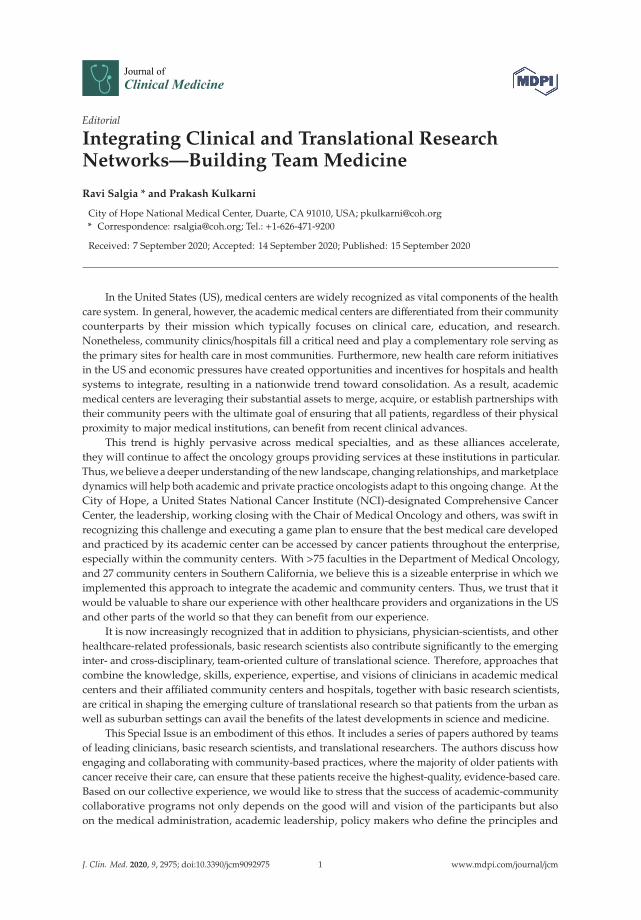
Integrating Clinical and Translational ResearchNetworks—Building Team Medicine
Ravi Salgia * and Prakash Kulkarni
City of Hope National Medical Center, Duarte, CA 91010, USA; [email protected]* Correspondence: [email protected]; Tel.: +1-626-471-9200
Received: 7 September 2020; Accepted: 14 September 2020; Published: 15 September 2020
In the United States (US), medical centers are widely recognized as vital components of the healthcare system. In general, however, the academic medical centers are differentiated from their communitycounterparts by their mission which typically focuses on clinical care, education, and research.Nonetheless, community clinics/hospitals fill a critical need and play a complementary role serving asthe primary sites for health care in most communities. Furthermore, new health care reform initiativesin the US and economic pressures have created opportunities and incentives for hospitals and healthsystems to integrate, resulting in a nationwide trend toward consolidation. As a result, academicmedical centers are leveraging their substantial assets to merge, acquire, or establish partnerships withtheir community peers with the ultimate goal of ensuring that all patients, regardless of their physicalproximity to major medical institutions, can benefit from recent clinical advances.
This trend is highly pervasive across medical specialties, and as these alliances accelerate,they will continue to affect the oncology groups providing services at these institutions in particular.Thus, we believe a deeper understanding of the new landscape, changing relationships, and marketplacedynamics will help both academic and private practice oncologists adapt to this ongoing change. At theCity of Hope, a United States National Cancer Institute (NCI)-designated Comprehensive CancerCenter, the leadership, working closing with the Chair of Medical Oncology and others, was swift inrecognizing this challenge and executing a game plan to ensure that the best medical care developedand practiced by its academic center can be accessed by cancer patients throughout the enterprise,especially within the community centers. With >75 faculties in the Department of Medical Oncology,and 27 community centers in Southern California, we believe this is a sizeable enterprise in which weimplemented this approach to integrate the academic and community centers. Thus, we trust that itwould be valuable to share our experience with other healthcare providers and organizations in the USand other parts of the world so that they can benefit from our experience.
It is now increasingly recognized that in addition to physicians, physician-scientists, and otherhealthcare-related professionals, basic research scientists also contribute significantly to the emerginginter- and cross-disciplinary, team-oriented culture of translational science. Therefore, approaches thatcombine the knowledge, skills, experience, expertise, and visions of clinicians in academic medicalcenters and their affiliated community centers and hospitals, together with basic research scientists,are critical in shaping the emerging culture of translational research so that patients from the urban aswell as suburban settings can avail the benefits of the latest developments in science and medicine.
This Special Issue is an embodiment of this ethos. It includes a series of papers authored by teamsof leading clinicians, basic research scientists, and translational researchers. The authors discuss howengaging and collaborating with community-based practices, where the majority of older patients withcancer receive their care, can ensure that these patients receive the highest-quality, evidence-based care.Based on our collective experience, we would like to stress that the success of academic-communitycollaborative programs not only depends on the good will and vision of the participants but alsoon the medical administration, academic leadership, policy makers who define the principles and
J. Clin. Med. 2020, 9, 2975; doi:10.3390/jcm9092975 www.mdpi.com/journal/jcm1
J. Clin. Med. 2020, 9, 2975
rules by which cooperation within the health care industry occurs. We refer to this cooperation andcollaboration as ‘Team Medicine’.
We take this opportunity to thank the City of Hope leadership for the support and encouragement;the authors for taking time from their hectic schedules, especially during this unprecedented pandemicto share their unique experience, vision, and ideas; and the patients and their families for theirparticipation and enduring spirit. We trust that our experience embodied in this singular compendiumwill serve as a ‘Rosetta Stone’ for other institutions and practitioners.
Funding: This research received no external funding.
Conflicts of Interest: The authors declare no conflict of interest.
© 2020 by the authors. Licensee MDPI, Basel, Switzerland. This article is an open accessarticle distributed under the terms and conditions of the Creative Commons Attribution(CC BY) license (http://creativecommons.org/licenses/by/4.0/).
2
Journal of
Clinical Medicine
Article
An Analysis and Comparison of Survival andFunctional Outcomes in Oropharyngeal SquamousCell Carcinoma Patients Treated with ConcurrentChemoradiation Therapy within City of Hope CancerCenter Sites
Rebecca Pharaon 1,†, Samuel Chung 1,†, Arya Amini 2, Ellie Maghami 3, Arnab Chowdhury 4,
Nayana Vora 2, Sue Chang 5, Robert Kang 3, Thomas Gernon 3, Kelly Hansen 6, Christina Kelly 6,
Denise Ackerman 7, Lalit Vora 8, Sagus Sampath 2 and Erminia Massarelli 1,*1 Department of Medical Oncology and Therapeutics Research, City of Hope National Medical Center,
Duarte, CA 91010, USA; [email protected] (R.P.); [email protected] (S.C.)2 Department of Radiation Oncology, City of Hope National Medical Center, Duarte, CA 91010, USA;
[email protected] (A.A.); [email protected] (N.V.); [email protected] (S.S.)3 Department of Head and Neck Surgery, City of Hope National Medical Center, Duarte, CA 91010, USA;
[email protected] (E.M.); [email protected] (R.K.); [email protected] (T.G.)4 Department of Computational and Quantitative Medicine, City of Hope National Medical Center,
Duarte, CA 91010, USA; [email protected] Department of Pathology, City of Hope National Medical Center, Duarte, CA 91010, USA; [email protected] Department of Speech and Language Pathology, City of Hope National Medical Center, Duarte, CA 91010,
USA; [email protected] (K.H.); [email protected] (C.K.)7 Department of Clinical Nutrition, City of Hope National Medical Center, Duarte, CA 91010, USA;
[email protected] Department of Diagnostic Radiology, City of Hope National Medical Center, Duarte, CA 91010, USA;
[email protected]* Correspondence: [email protected]; Tel.: +1-626-256-4673† These authors contributed equally to this work and should be considered co-first authors.
Received: 8 July 2020; Accepted: 22 September 2020; Published: 24 September 2020
Abstract: Oropharyngeal squamous cell carcinoma (OPSCC) is a subset of head and neck cancersthat can arise due to human papillomavirus (HPV) infection. We designed a retrospective analysisto determine differences in outcomes of OPSCC patients treated at City of Hope (COH) CancerCenter’s main campus versus selected satellite sites with COH-associated faculty and facilities.Patients diagnosed with OPSCC and treated with concurrent chemoradiation therapy (n = 94) wereidentified and included in the study. Patients underwent treatment at the COH main campus site(n = 50) or satellite sites (n = 44). The majority of patients were Caucasian, male, and diagnosedwith p16 positive stage IV locally advanced OPSCC by AJCC 7th edition. Most patients completedtheir prescribed cumulative radiation therapy dose and had a complete response to treatment.No significant difference in overall survival and progression-free survival was observed betweenthe main campus and the satellite sites. Our study demonstrates successful treatment completionrates as well as comparable recurrence rates between the main campus and COH-associated satellitesites. A trend toward significant difference in feeding tube dependency at 6-months was observed.Differences in feeding tube placement and dependency rates could be addressed by the establishmentof on-site supportive services in satellite sites.
Keywords: oropharyngeal cancer; concurrent chemoradiation therapy; human papillomavirus;feeding tube dependency
J. Clin. Med. 2020, 9, 3083; doi:10.3390/jcm9103083 www.mdpi.com/journal/jcm3
J. Clin. Med. 2020, 9, 3083
1. Introduction
Head and neck cancers, a heterogenous group of cancers originating in the head and neck region,are strongly linked to risk factors such as tobacco use, alcohol consumption, and human papillomavirus(HPV) infection. Oropharyngeal squamous cell carcinomas (OPSCC) are one of the most common headand neck cancers; however, HPV-associated OPSCCs denote a distinct subtype of oropharyngeal cancerwith rising incidence compared to the smoking-associated counterpart [1,2]. It has been shown thatHPV-associated OPSCC has better prognosis [3–7] and is less likely to have a second primary site [8],but the occurrence of distant metastases is not significantly different than HPV-negative cancer [7,9].
Due to the association between OPSCC and HPV-mediated oncogenesis, the 8th edition of theAmerican Joint Committee on Cancer (AJCC) denoted specific staging criteria and standard treatmentfor HPV-associated OPSCC, as well as emphasized the importance of extranodal extension (ENE)when determining staging and treatment for patients [10]. Standard treatment generally includes amultimodality approach consisting of transoral robotic surgery (TORS) for early stages or resectabletumors, and radiation therapy with or without concurrent chemotherapy as adjuvant or definitivetreatment depending on tumor (T) and nodal (N) disease or high-risk pathologic features such asENE or carcinoma-involved margins. In the incurable recurrent or metastatic (R/M) OPSCC setting,systemic treatment options include immune checkpoint inhibitors plus or minus chemotherapy withplatinum/5-fluorouracil depending on PD-L1 expression, and novel approaches in immunotherapycombinations and HPV vaccines are currently undergoing investigation [11,12].
Cancer patient outcomes after treatment at academic versus satellite sites have been previouslyexplored in esophageal cancer, soft tissue sarcoma, and laryngeal cancer, reporting a trend of improvedsurvival for patients treated at high-volume teaching facilities [13–15]. In choosing their treatmentcenter, patients have to account for the geographical location, financial cost, and insurance-approvedproviders, which can greatly limit their options. Previous studies have examined head and neckcancer outcomes in patients treated at academic centers versus community sites with controversialresults [16–18]. Currently, academic center and community clinic affiliations have increased to allowpatients to have access to high quality and standardized care. However, outcomes analysis has notbeen studied in partnered academic and satellite sites operating under the same umbrella.
At our institution, City of Hope (COH) Comprehensive Cancer Center, we have establishedvarious satellite sites distributed throughout the Southern California region in order to accommodategeographic restrictions and traffic limitations for our patients. Due to the complex nature of treatinghead and neck cancers, the coordination of various treatments and providers requires precise andeffective medical practice. Functional and survival outcomes are dependent on the timing of adjuvantor definitive treatment as well as dedicated follow-up of patients with adequate supportive servicesuch as speech pathology, nutrition specialists, and physical and occupational therapy. Several qualityassurance measures are in place at COH, including reviews of satellite site radiation treatment plansperformed at the main campus prior to treatment start.
We designed a retrospective study to analyze and compare survival and functional outcomes inpatients treated at COH’s main campus versus selected satellite sites with COH-associated faculty andradiation therapy facilities.
2. Materials and Methods
2.1. Patient and Study Criteria
A retrospective analysis of patients diagnosed with OPSCC and treated with concurrentchemoradiation therapy (CRT) was conducted at COH main campus and selected satellite sites.Main eligibility criteria included a diagnosis of OPSCC, age 18 years or older, definitive/adjuvanttreatment including concurrent CRT at a COH site between February 2009 to February 2017, availabledemographic, treatment, and survival data, functional outcomes, and recent follow-up at a COHsite within a year. We utilized our institutional electronic medical record (EMR) to analyze 400
4
J. Clin. Med. 2020, 9, 3083
consecutive head and neck cancer patients from various COH sites to enroll in our study. Of the initial400 patients reviewed, only 94 met the inclusion criteria including OPSCC diagnosis, recent follow-up,and history of CRT treatment at a COH site. Confounding factors considered were age, gender, and Tstage. The study was conducted in accordance with the Declaration of Helsinki, and the protocol wasapproved by the COH institutional review board (#19010).
The primary endpoint was overall survival (OS) of patients treated at the main COH campusversus patients treated at selected COH satellite sites, including South Pasadena, Rancho Cucamonga,Antelope Valley, and Santa Clarita. Secondary endpoints included treatment responses, feeding tubedependency, weight loss, and narcotic use.
2.2. Statistical Considerations
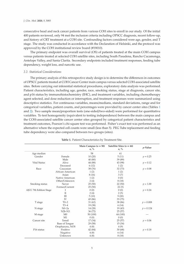
The primary analysis of this retrospective study design is to determine the differences in outcomesof OPSCC patients treated at COH Cancer Center main campus versus selected COH-associated satellitesites. Before carrying out inferential statistical procedures, exploratory data analysis was performed.Patient characteristics, including age, gender, race, smoking status, stage at diagnosis, cancer site,and p16 status by immunohistochemistry (IHC), and treatment variables, including chemotherapyagent selected, and dose reduction or interruption, and treatment responses were summarized usingdescriptive statistics. For continuous variables, means/medians, standard deviations, range and forcategorical variables, patient counts, and percentages were provided by cancer center sites (Tables 1and 2). Two sample mean/proportion tests (one-sided/two-sided) were performed for quantitativevariables. To test homogeneity (equivalent to testing independence) between the main campus andthe COH-associated satellite cancer center sites grouped by categorical patient characteristics andtreatment outcomes, Pearson’s chi-square test was performed. Fisher’s exact test was performed as analternative where the expected cell counts were small (less than 5). PEG Tube replacement and feedingtube dependency were also compared between two groups (sites).
Table 1. Patient Characteristics by Treatment Site.
Main Campus (n = 50)n, %
Satellite Sites (n = 44)n, %
p-Value
Age median 58 61Gender Female 10 (20) 5 (11) p = 0.25
Male 40 (80) 39 (89)Vital Status Alive 44 (88) 43 (98) p = 0.12
Deceased 6 (12) 1 (2)Race Caucasian 38 (76) 32 (73) p = 0.08
African American 1 (2) 1 (2)Asian 8 (16) 3 (7)
Native American 1 (2) 0 (0)Other/Unknown 2 (4) 8 (18)
Smoking status Never 25 (50) 22 (50) p = 1.00Former/Current 25 (50) 22 (5)
AJCC 7th Edition Stage I 0 (0) 0 (0) p = 0.24II 2 (4) 2 (5)III 5 (10) 9 (20)IV 43 (86) 33 (75)
T stage T0–2 31 (62) 38 (86) p = 0.008T3–4 19 (38) 6 (14)
N stage N0–2a 14 (28) 19 (43) p = 0.124N2b-N3 36 (72) 25 (57)
M stage M0 50 (100) 44 (100) —M1 0 (0) 0 (0)
Cancer site Tonsil 17 (34) 25 (57) p = 0.06Base of Tongue 29 (58) 15 (34)
Oropharynx, NOS 4 (8) 4 (9)P16 status Positive 42 (84) 30 (68) p = 0.18
Negative 4 (8) 6 (14)Unknown 4 (8) 8 (18)
5
J. Clin. Med. 2020, 9, 3083
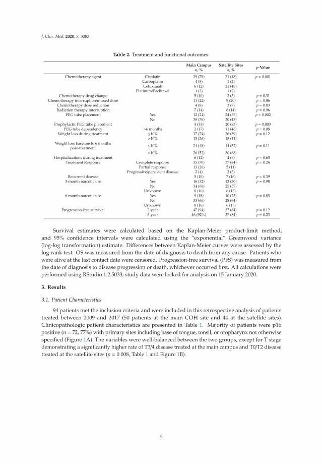
Table 2. Treatment and functional outcomes.
Main Campusn, %
Satellite Sitesn, %
p-Value
Chemotherapy agent Cisplatin 39 (78) 21 (48) p = 0.001Carboplatin 4 (8) 1 (2)Cetuximab 6 (12) 21 (48)
Platinum/Paclitaxel 1 (2) 1 (2)Chemotherapy drug change 5 (10) 2 (5) p = 0.31
Chemotherapy interruption/missed dose 11 (22) 9 (20) p = 0.86Chemotherapy dose reduction 4 (8) 3 (7) p = 0.83Radiation therapy interruption 7 (14) 6 (14) p = 0.96
PEG tube placement Yes 12 (24) 24 (55) p = 0.002No 38 (76) 20 (45)
Prophylactic PEG tube placement 4 (33) 20 (83) p = 0.003PEG tube dependency >6 months 2 (17) 11 (46) p = 0.08
Weight loss during treatment ≤10% 37 (74) 26 (59) p = 0.12>10% 13 (26) 18 (41)
Weight loss baseline to 6 monthspost-treatment ≤10% 24 (48) 14 (32) p = 0.11
>10% 26 (52) 30 (68)Hospitalizations during treatment 6 (12) 4 (9) p = 0.65
Treatment Response Complete response 35 (70) 37 (84) p = 0.24Partial response 13 (26) 5 (11)
Progressive/persistent disease 2 (4) 2 (5)Recurrent disease 5 (10) 7 (16) p = 0.39
3-month narcotic use Yes 16 (32) 13 (30) p = 0.98No 34 (68) 25 (57)
Unknown 8 (16) 6 (13)6-month narcotic use Yes 9 (18) 10 (23) p = 0.83
No 33 (66) 28 (64)Unknown 8 (16) 6 (13)
Progression-free survival 2-year 47 (94) 37 (84) p = 0.125-year 46 (92%) 37 (84) p = 0.23
Survival estimates were calculated based on the Kaplan-Meier product-limit method,and 95% confidence intervals were calculated using the “exponential” Greenwood variance(log-log transformation) estimate. Differences between Kaplan-Meier curves were assessed by thelog-rank test. OS was measured from the date of diagnosis to death from any cause. Patients whowere alive at the last contact date were censored. Progression-free survival (PFS) was measured fromthe date of diagnosis to disease progression or death, whichever occurred first. All calculations wereperformed using RStudio 1.2.5033; study data were locked for analysis on 15 January 2020.
3. Results
3.1. Patient Characteristics
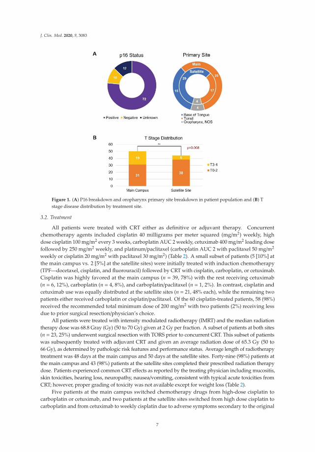
94 patients met the inclusion criteria and were included in this retrospective analysis of patientstreated between 2009 and 2017 (50 patients at the main COH site and 44 at the satellite sites).Clinicopathologic patient characteristics are presented in Table 1. Majority of patients were p16positive (n = 72, 77%) with primary sites including base of tongue, tonsil, or oropharynx not otherwisespecified (Figure 1A). The variables were well-balanced between the two groups, except for T stagedemonstrating a significantly higher rate of T3/4 disease treated at the main campus and T0/T2 diseasetreated at the satellite sites (p = 0.008, Table 1 and Figure 1B).
6
J. Clin. Med. 2020, 9, 3083
Figure 1. (A) P16 breakdown and oropharynx primary site breakdown in patient population and (B) Tstage disease distribution by treatment site.
3.2. Treatment
All patients were treated with CRT either as definitive or adjuvant therapy. Concurrentchemotherapy agents included cisplatin 40 milligrams per meter squared (mg/m2) weekly, highdose cisplatin 100 mg/m2 every 3 weeks, carboplatin AUC 2 weekly, cetuximab 400 mg/m2 loading dosefollowed by 250 mg/m2 weekly, and platinum/paclitaxel (carboplatin AUC 2 with paclitaxel 50 mg/m2
weekly or cisplatin 20 mg/m2 with paclitaxel 30 mg/m2) (Table 2). A small subset of patients (5 [10%] atthe main campus vs. 2 [5%] at the satellite sites) were initially treated with induction chemotherapy(TPF—docetaxel, cisplatin, and fluorouracil) followed by CRT with cisplatin, carboplatin, or cetuximab.Cisplatin was highly favored at the main campus (n = 39, 78%) with the rest receiving cetuximab(n = 6, 12%), carboplatin (n = 4, 8%), and carboplatin/paclitaxel (n = 1, 2%). In contrast, cisplatin andcetuximab use was equally distributed at the satellite sites (n = 21, 48% each), while the remaining twopatients either received carboplatin or cisplatin/paclitaxel. Of the 60 cisplatin-treated patients, 58 (98%)received the recommended total minimum dose of 200 mg/m2 with two patients (2%) receiving lessdue to prior surgical resection/physician’s choice.
All patients were treated with intensity modulated radiotherapy (IMRT) and the median radiationtherapy dose was 68.8 Gray (Gy) (50 to 70 Gy) given at 2 Gy per fraction. A subset of patients at both sites(n = 23, 25%) underwent surgical resection with TORS prior to concurrent CRT. This subset of patientswas subsequently treated with adjuvant CRT and given an average radiation dose of 65.3 Gy (50 to66 Gy), as determined by pathologic risk features and performance status. Average length of radiotherapytreatment was 48 days at the main campus and 50 days at the satellite sites. Forty-nine (98%) patients atthe main campus and 43 (98%) patients at the satellite sites completed their prescribed radiation therapydose. Patients experienced common CRT effects as reported by the treating physician including mucositis,skin toxicities, hearing loss, neuropathy, nausea/vomiting, consistent with typical acute toxicities fromCRT; however, proper grading of toxicity was not available except for weight loss (Table 2).
Five patients at the main campus switched chemotherapy drugs from high-dose cisplatin tocarboplatin or cetuximab, and two patients at the satellite sites switched from high dose cisplatin tocarboplatin and from cetuximab to weekly cisplatin due to adverse symptoms secondary to the original
7
J. Clin. Med. 2020, 9, 3083
chemotherapy. The reasons for chemotherapy drug switch included tinnitus, elevated creatinine,and azotemia. Overall, the chemotherapy dose was reduced by at least 20% in seven patients(four at the main campus vs. three at the satellite sites, p = 0.83) treated with high-dose cisplatin(100 mg/m2) due to adverse symptoms. The number of patients who missed a therapy dose(s) orhad chemotherapy treatment interruptions because of significant treatment-related adverse events(i.e., azotemia, leukopenia, neutropenia, thrombocytopenia), unplanned hospitalizations, or patientdecisions were similar among the COH main campus and satellite groups (11 vs. nine patients, p = 0.86).During the course of radiation therapy treatment, seven patients in the main campus and six patientsin the satellite sites had treatment interruptions/breaks of at least two days. The reasons for treatmentinterruptions were related to severe weight loss (n = 2), thus requiring another radiotherapy treatmentplanning computed tomography scan, toxicities secondary to treatment (n = 7), insurance issues (n = 1),patient’s choice (n = 1), and unplanned hospitalizations (n = 2). Reasons for unexpected hospitalization(six at the main campus vs. four at the satellite sites) were related to significant treatment-relatedadverse events such as severe dehydration, severe adverse infusion reaction to cetuximab, aspirationpneumonia, G-tube cellulitis, or pulmonary embolism.
The percentage of patients who completed the prescribed full treatment dose of both chemotherapyand radiation therapy without interruptions of radiation therapy was 86% (n = 43) at the main campusand 82% (n = 36) at the satellite locations.
3.3. Survival Outcomes
Patients underwent restaging imaging 12 weeks after completion of treatment to measure theirresponse to treatment. Thirty-five (70%) patients achieved a complete response (CR) to treatment,13 (26%) patients had a partial response (PR), and two (4%) patients had progression of disease(POD)/persistent disease at the main center site. Thirty-seven (84%) patients achieved a CR, five (11%)patients had a PR, and two (5%) patients had POD/persistent disease at the satellite sites. The patientswith POD/persistent disease were subsequently treated with pembrolizumab at the main cancer site,or further lines of chemotherapy (carboplatin/paclitaxel, carboplatin/gemcitabine, cetuximab) followedby nivolumab or pembrolizumab at the satellite sites.
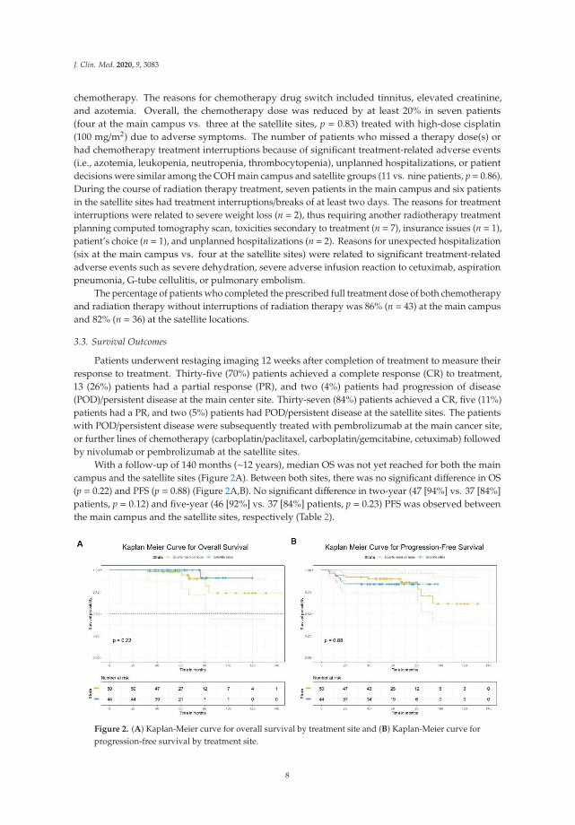
With a follow-up of 140 months (~12 years), median OS was not yet reached for both the maincampus and the satellite sites (Figure 2A). Between both sites, there was no significant difference in OS(p = 0.22) and PFS (p = 0.88) (Figure 2A,B). No significant difference in two-year (47 [94%] vs. 37 [84%]patients, p = 0.12) and five-year (46 [92%] vs. 37 [84%] patients, p = 0.23) PFS was observed betweenthe main campus and the satellite sites, respectively (Table 2).
Figure 2. (A) Kaplan-Meier curve for overall survival by treatment site and (B) Kaplan-Meier curve forprogression-free survival by treatment site.
8

J. Clin. Med. 2020, 9, 3083
3.4. Functional Outcomes
Modified Barium Swallow Studies (MBSS) were performed more frequently at the main cancercenter than the satellite sites due to the presence of a comprehensive team of speech pathologists.12 patients (24%) underwent feeding tube placement at the main campus, prophylactically (n = 4) orduring treatment (n = 8). Two of the 12 patients continued to be feeding tube dependent after 6 months.Twenty-four patients (55%) underwent feeding tube placement at the satellite sites, prophylactically(n = 20) or during treatment (n = 4). Eleven of the 24 patients continued to be feeding tube-dependentat 6 months. Prophylactic feeding tube placement was associated with higher rates of 6-month feedingtube dependency (n = 2, 100% at main site, n = 9, 92% at satellite sites). Weight loss was recordedthroughout CRT treatment and during the 6-month post-treatment follow-up. Thirty-seven patients(74%) lost≤10% baseline weight during treatment while the remaining 13 (26%) lost >10% of their initialbody weight at the main site. Twenty-six (59%) patients lost ≤10% baseline weight, while the remaining18 (41%) lost >10% of their baseline weight during treatment at the satellite sites. Twenty-four patients(48%) lost ≤10% baseline weight at the 6-month post-treatment follow-up, while 26 (53%) lost >10% oftheir initial body weight at the main site. Fourteen patients (32%) and 30 patients (68%), respectively,lost ≤ and >10% of their initial body weight at the 6-month post-treatment follow-up at the satellitesites. Overall, at 6 months after completion of treatment, both sites noted an increased number ofpatients experiencing >10% baseline weight loss (13 to 26 patients at the main campus and 18 to30 patients at the satellite sites). Of the 12 patients who underwent feeding tube placement at themain site, 4 (33%) and 7 (58%) patients lost >10% baseline weight during treatment and at 6 monthspost-treatment, respectively. Of the 24 patients who underwent feeding tube placement at the satellitesites, eight (33%) and 16 (66%) patients lost >10% baseline weight during treatment and at 6 monthspost-treatment, respectively.
Narcotic use was recorded at 3- and 6-month timepoints after completion of radiation therapy.At the main center, 16 (32%) patients had recorded persistent use of narcotics at a 3 months post-treatmentinterval and 9 (18%) patients required persistent narcotic use at the 6-month mark. At the satellite sites,a similar number of patients (n = 13, 30%) reported narcotic use 3 months post-treatment, and 10 (23%)patients registered persistent narcotic use at the 6-month mark.
4. Discussion
We conducted a retrospective analysis of HPV-positive OPSCC patients treated with definitiveor postoperative CRT at a large NCI-designated comprehensive cancer center (COH) main campusand compared it to selected institutional satellite sites with availability of dedicated radiation therapyfacilities staffed with COH faculty with primary endpoint of OS and secondary endpoints includingtreatment response, feeding tube dependency, and weight loss throughout treatment and 6 monthspost-treatment. Our analysis is the first to compare patient outcomes in a comprehensive NCI designatedcancer center and its affiliated satellite sites. COH main site and satellite sites demonstrated OS ratesconsistent with already published favorable survival rates in OPSCC treated with multimodalitytherapy approach [7,19], while median OS at ~12 years was not reached (Figure 2A). No significantdifference in two-year (94% vs. 84%, p = 0.12) and five-year (92% vs. 84%, p = 0.23) PFS was observedbetween the main campus and the satellite sites (Table 2). Our data did not align with previouslyreported analyses of head and neck cancer outcomes in academic centers versus community sites thatdemonstrated superior survival rates in patients treated at academic institutions [16,18]. One majordifference between our study and the previously mentioned analyses [16,18] relates to the fact thatour patients were treated under an umbrella institution, COH, regardless of treatment center location.Overall, our institution takes strides to standardize patient care across all sites by the longstandingestablishment of weekly radiation oncology web rounds dedicated to reviewing all radiation treatmentplans at any COH site. Our outcomes are possible because of these efforts of treatment compliancebetween main and satellite centers where there is some uniformity in dose delivery, nodal coveragebased on primary site and nodal disease distribution, overall treatment volumes, and established
9
J. Clin. Med. 2020, 9, 3083
guidelines for treatment planning. Furthermore, the medical oncologists at the COH satellites arepart of the medical oncology main department and are required to attend quarterly retreats with theoncologists at main campus.
Regarding differences in the choice of concurrent systemic therapy, the oncologists at the maincampus site strongly favored cisplatin as the chemotherapy agent of choice (78%), whereas satellitesite medical oncologists equally favored cisplatin (48%) and cetuximab (48%) when treating theirpatients (p = 0.001). Considering the different distribution of tumor size between the two groups ofpatients with main campus seeing a higher number of T3–4 disease (19 [38%] vs. six [14%], p = 0.008)(Table 1 and Figure 1B), the choice of cisplatin-based therapy could have been influenced by themore advanced stage of disease at presentation. In addition, these patients were treated before thecompletion and publication of The Radiation Therapy Oncology Group (RTOG) 1016 clinical trialthat compared systemic therapy agent cisplatin versus cetuximab, a monoclonal antibody againstEGFR, in HPV-positive OPSCC, and reported superior PFS with cisplatin over cetuximab (78% vs.67%, p < 0.001) [20]. A small subset of patients underwent treatment with induction chemotherapyfollowed by CRT in our analysis (five at the main campus vs. two patients at the satellite sites, p = 0.31).While the use of induction chemotherapy in head and neck cancers is controversial, a recent phaseII trial demonstrated feasibility of different induction regimens stratified by disease risk in locallyadvanced head and neck squamous cell carcinoma [21].
In our study, chemotherapy dose reductions occurred among patients who were treated withhigh-dose cisplatin, underscoring the heavy burden of side effects that this treatment carries and theneed for continuous monitoring by the medical oncologist to avoid permanent renal and neurologicalimpairment effect on quality of life and survival outcomes. The majority of patients completed theirprescribed cumulative radiation therapy dose (n = 94, 98%). The rate of treatment interruptions andhospitalizations were comparable between the two groups of patients when adjusted by systemictreatment choice (Table 2). Chronic narcotic use at 3 months did not differ between the two groups(32% at the main campus vs. 30% at the satellite sites, p = 0.98) (Table 2), demonstrating a significantnumber of patients persistently using narcotics after completing treatment. Chronic narcotic usewas trending downwards at 6 months, and is in line with previously reported data in patients withOPSCC [22].
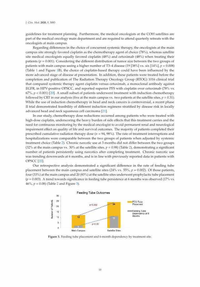
Our retrospective analysis demonstrated a significant difference in the rate of feeding tubeplacement between the main campus and satellite sites (24% vs. 55%, p = 0.002). Of those patients,four (33%) at the main campus and 20 (83%) at the satellite sites underwent prophylactic tube placement(p = 0.003). A trend towards significance in feeding tube persistence at 6 months was observed (17% vs.46%, p = 0.08) (Table 2 and Figure 3).
Figure 3. Feeding tube placement and 6-month dependency by treatment site.
10
J. Clin. Med. 2020, 9, 3083
MBSS were more likely to be performed at the main campus versus satellite sites given theavailability of on-site speech pathology at the main campus. Multiple studies have been conductedto determine the advantage of prophylactic versus reactive PEG tube placement and the results arecontroversial [23–29]. However, studies have reported the significance of prophylactic PEG tubeplacement on enteral feed dependency rates [23,24,29], consistent with our feeding tube outcomes.This difference highlights the need for supportive service availability at satellite sites that could bepotentially reached with remote consultation with institutional dedicated nutritionists and speechpathologists at our COH satellite centers. There was no significant difference in the degree of weightloss between the two groups of patients with the majority maintaining a ≤10% baseline weight loss(75% vs. 59%, p = 0.12); however, this could be the result of a higher prophylactic feeding tube rate inthe satellite centers than main campus (Table 2).
Overall, patients who are at a higher risk of recurrence due to poor pathologic features,comorbidities, and nutritional status should be identified earlier during treatment so that theycan have access to supportive services. At our institution, we are working on directing those patientsearlier on to speech pathologists and nutritionists to eventually identify ones who might benefit fromtreatment modifications or early implementation of dietary needs. Furthermore, we are exploring thefeasibility of implementing a program that allows for an initial comprehensive speech and nutritionevaluation at the main campus with continued telehealth follow-ups in the satellite sites and returnto the main campus for repeat instrumental testing when necessary. This would reduce the need ofin-person visits with speech pathologists and nutritionists and we plan to understand if this initiativecould be effective in improving the 6 months feeding tube persistence rate. Due to the COVID-19pandemic, numerous institutions have implemented telehealth services in order to accommodatepatients and decrease needless exposure. Our institution offers outpatient telemedicine consults andfollow-up visits via telephone or video call, and this service has been extended to our satellite sites.In the future, this implementation will greatly benefit patients undergoing treatment at satellite sites thatneed speech pathology and nutritionist oversight but do not have access to the main academic center,in order to minimize unnecessary feeding tube placements and ensure good functional outcomes.
Precision medicine in oropharyngeal cancers is difficult to accomplish as we do not select treatmentbased on molecular targets other than HPV status. Currently, HPV status is the only biological featureon which we establish disease risk and prognosis. We clearly understand that there is a need formolecular-driven treatment selection in the future. Due to EMR integration between the main campusand satellite sites, a wide range of patient information is readily accessible, thus allowing for academicand community physicians to efficiently collaborate on research studies. Based on this joint retrospectiveanalysis, we are conducting and examining the molecular status of these academic and communitypatients to try to understand if we can translate meaningful results.
We recognize that our study contains limitations including intrinsic bias of patient selection ina retrospective analysis and limitations of our EMR search which identified our cohort of patients.We designated the HPV-associated OPSCC according to p16 IHC status, based on the evidence ofconcordance between p16 IHC staining and HPV in situ hybridization in HPV-associated oropharyngealcarcinomas that makes p16 IHC a surrogate marker for HPV infection [30]. The discrepancies betweenour study in OPSCC and those who observed improved survival in head and neck cancer patientstreated at an academic institution could be attributed to the excellent prognosis of HPV-positiveOPSCC. Disparities in disease stage, socioeconomic factors, patient location, and insurance play a rolein the different characteristics of patients treated at the main campus compared to the satellite sites.Worse prognosis primary sites such as oral cavity and larynx could potentially demonstrate differencesin survival.
In conclusion, our study highlights the importance of the necessity to treat OPSCC in tertiarycancer centers and their associated satellite network with availability of dedicated and experiencedradiation and medical oncologists and up-to-date facilities that allow continuous communicationbetween physicians and supportive services including speech pathology and nutrition. We are currently
11
J. Clin. Med. 2020, 9, 3083
designing a prospective study to assess the implementation of telemedicine for supportive services inthe satellite sites. This is of great importance as poor functional outcomes represent a heavy burdenfor our current HPV-positive OPSCC population that is young, very functional, and deserves highstandards of post-treatment quality of life.
Author Contributions: Conceptualization, R.P., S.C. (Samuel Chung), and E.M. (Erminia Massarelli); Methodology,R.P., S.C. (Samuel Chung), and E.M. (Erminia Massarelli); Validation, R.P., S.C. (Samuel Chung), E.M.(Ellie Maghami), A.A., A.C., S.S., and E.M. (Erminia Massarelli); Formal Analysis, R.P., S.C. (Samuel Chung),A.A., A.C., and E.M. (Erminia Massarelli); Data Curation, R.P., S.C. (Samuel Chung), A.A., S.C. (Samuel Chung),S.S., and E.M. (Erminia Massarelli); Writing—Original Draft Preparation, R.P., S.C. (Samuel Chung), and E.M.(Erminia Massarelli); Writing—Review and Editing, R.P., S.C. (Samuel Chung), E.M. (Ellie Maghami), A.A., A.C.,N.V., S.C. (Sue Chang), T.G., R.K., K.H., C.K., D.A., L.V., S.S., and E.M. (Erminia Massarelli). All authors have readand agreed to the manuscript.
Funding: This research received no external funding.
Acknowledgments: We would like to thank the City of Hope head and neck cancer medical, surgical, and radiationoncologists, nurses, and supportive teams for their dedication to their patients. The authors have no fundingto disclose.
Conflicts of Interest: The authors declare no conflict of interest.
References
1. Gillison, M.L.; D’Souza, G.; Westra, W.; Sugar, E.; Xiao, W.; Begum, S.; Viscidi, R. Distinct Risk Factor Profilesfor Human Papillomavirus Type 16–Positive and Human Papillomavirus Type 16–Negative Head andNeck Cancers. JNCI J. Natl. Cancer Inst. 2008, 100, 407–420. [CrossRef]
2. Applebaum, K.M.; Furniss, C.S.; Zeka, A.; Posner, M.R.; Smith, J.F.; Bryan, J.; Eisen, E.A.; Peters, E.S.;McClean, M.D.; Kelsey, K.T. Lack of Association of Alcohol and Tobacco with HPV16-Associated Head andNeck Cancer. JNCI J. Natl. Cancer Inst. 2007, 99, 1801–1810. [CrossRef]
3. Benson, E.; Li, R.; Eisele, D.; Fakhry, C. The clinical impact of HPV tumor status upon head and necksquamous cell carcinomas. Oral Oncol. 2014, 50, 565–574. [CrossRef] [PubMed]
4. Chaturvedi, A.K. Epidemiology and clinical aspects of HPV in head and neck cancers. Head Neck Pathol.2012, 6 (Suppl. 1), S16–S24. [CrossRef] [PubMed]
5. Chung, C.H.; Gillison, M.L. Human papillomavirus in head and neck cancer: Its role in pathogenesis andclinical implications. Clin. Cancer Res. 2009, 15, 6758–6762. [CrossRef] [PubMed]
6. Fakhry, C.; Westra, W.H.; Li, S.; Cmelak, A.; Ridge, J.A.; Pinto, H.; Forastiere, A.; Gillison, M.L. Improvedsurvival of patients with human papillomavirus-positive head and neck squamous cell carcinoma in aprospective clinical trial. J. Natl. Cancer Inst. 2008, 100, 261–269. [CrossRef] [PubMed]
7. Ang, K.K.; Harris, J.; Wheeler, R.; Weber, R.; Rosenthal, D.I.; Nguyen-Tan, P.F.; Westra, W.H.; Chung, C.H.;Jordan, R.C.; Lu, C.; et al. Human papillomavirus and survival of patients with oropharyngeal cancer.N. Engl. J. Med. 2010, 363, 24–35. [CrossRef]
8. Morris, L.G.; Sikora, A.G.; Patel, S.G.; Hayes, R.B.; Ganly, I. Second primary cancers after an index head andneck cancer: Subsite-specific trends in the era of human papillomavirus-associated oropharyngeal cancer.J. Clin. Oncol. 2011, 29, 739–746. [CrossRef]
9. O’Sullivan, B.; Huang, S.H.; Siu, L.L.; Waldron, J.; Zhao, H.; Perez-Ordonez, B.; Weinreb, I.; Kim, J.; Ringash, J.;Bayley, A.; et al. Deintensification candidate subgroups in human papillomavirus-related oropharyngealcancer according to minimal risk of distant metastasis. J. Clin. Oncol. 2013, 31, 543–550. [CrossRef]
10. Lydiatt, W.M.; Patel, S.G.; O’Sullivan, B.; Brandwein, M.S.; Ridge, J.A.; Migliacci, J.C.; Loomis, A.M.; Shah, J.P.Head and Neck cancers-major changes in the American Joint Committee on cancer eighth edition cancerstaging manual. CA Cancer J. Clin. 2017, 67, 122–137. [CrossRef]
11. Burtness, B.; Harrington, K.J.; Greil, R.; Soulières, D.; Tahara, M.; De Castro, G., Jr.; Psyrri, A.;Rotlan, N.B.; Neupane, P.C.; Bratland, A. KEYNOTE-048: Phase 3 study of first-line pembrolizumab(P) for recurrent/metastatic head and neck squamous cell carcinoma (R/M HNSCC). In Proceedings of theESMO 2018 Congress, Munich, Germany, 19–23 October 2018.
12

J. Clin. Med. 2020, 9, 3083
12. Massarelli, E.; William, W.; Johnson, F.; Kies, M.; Ferrarotto, R.; Guo, M.; Feng, L.; Lee, J.J.; Tran, H.;Kim, Y.U.; et al. Combining Immune Checkpoint Blockade and Tumor-Specific Vaccine for Patients WithIncurable Human Papillomavirus 16-Related Cancer: A Phase 2 Clinical Trial. JAMA Oncol. 2018, 5, 67–73.[CrossRef] [PubMed]
13. Verhoef, C.; van de Weyer, R.; Schaapveld, M.; Bastiaannet, E.; Plukker, J.T. Better survival in patientswith esophageal cancer after surgical treatment in university hospitals: A plea for performance by surgicaloncologists. Ann. Surg. Oncol. 2007, 14, 1678–1687. [CrossRef] [PubMed]
14. Gutierrez, J.C.; Perez, E.A.; Moffat, F.L.; Livingstone, A.S.; Franceschi, D.; Koniaris, L.G. Should soft tissuesarcomas be treated at high-volume centers? An analysis of 4205 patients. Ann. Surg. 2007, 245, 952–958.[CrossRef] [PubMed]
15. Chen, A.Y.; Fedewa, S.; Pavluck, A.; Ward, E.M. Improved survival is associated with treatment at high-volumeteaching facilities for patients with advanced stage laryngeal cancer. Cancer 2010, 116, 4744–4752. [CrossRef][PubMed]
16. Lassig, A.A.D.; Joseph, A.M.; Lindgren, B.R.; Fernandes, P.; Cooper, S.; Schotzko, C.; Khariwala, S.;Reynolds, M.; Yueh, B. The effect of treating institution on outcomes in head and neck cancer. Otolaryngol. HeadNeck Surg. 2012, 147, 1083–1092. [CrossRef]
17. Kubicek, G.J.; Wang, F.; Reddy, E.; Shnayder, Y.; Cabrera, C.E.; Girod, D.A. Importance of treatment institutionin head and neck cancer radiotherapy. Otolaryngol. Head Neck Surg. 2009, 141, 172–176. [CrossRef]
18. Benasso, M.; Lionetto, R.; Corvò, R.; Ponzanelli, A.; Vitale, V.; Rosso, R. Impact of the treating institution onthe survival of patients with head and neck cancer treated with concomitant alternating chemotherapy andradiation. Eur. J. Cancer 2003, 39, 1895–1898. [CrossRef]
19. Patel, M.A.; Blackford, A.L.; Rettig, E.M.; Richmon, J.D.; Eisele, D.W.; Fakhry, C. Rising population ofsurvivors of oral squamous cell cancer in the United States. Cancer 2016, 122, 1380–1387. [CrossRef]
20. Trotti, A.; Harris, J.; Gillison, M.; Eisbruch, A.; Harari, P.M.; Adelstein, D.J.; Sturgis, E.M.; Galvin, J.M.;Koyfman, S.; Blakaj, D.; et al. NRG-RTOG 1016: Phase III Trial Comparing Radiation/Cetuximab toRadiation/Cisplatin in HPV-related Cancer of the Oropharynx. Int. J. Radiat. Oncol. Biol. Phys. 2018, 102,1604–1605. [CrossRef]
21. Haddad, R.I.; Massarelli, E.; Lee, J.J.; Lin, H.Y.; Hutcheson, K.; Lewis, J.; Garden, A.S.; Blumenschein, G.R., Jr.;William, W.N., Jr.; Pharaon, R.R.; et al. Weekly paclitaxel, carboplatin, cetuximab, and cetuximab, docetaxel,cisplatin, and fluorouracil, followed by local therapy in previously untreated, locally advanced head andneck squamous cell carcinoma. Ann. Oncol. 2019, 30, 471–477. [CrossRef]
22. McDermott, J.D.; Eguchi, M.; Stokes, W.A.; Amini, A.; Hararah, M.; Ding, D.; Valentine, A.; Bradley, C.J.;Karam, S.D. Short- and Long-term Opioid Use in Patients with Oral and Oropharynx Cancer. Otolaryngol. HeadNeck Surg. 2019, 160, 409–419. [CrossRef] [PubMed]
23. Koyfman, S.A.; Adelstein, D.J. Enteral Feeding Tubes in Patients Undergoing Definitive ChemoradiationTherapy for Head-and-Neck Cancer: A Critical Review. Int. J. Radiat. Oncol. Biol. Phys. 2012, 84, 581–589.[CrossRef] [PubMed]
24. Chen, A.M.; Li, B.-Q.; Lau, D.H.; Farwell, D.G.; Luu, Q.; Stuart, K.; Newman, K.; Purdy, J.A.; Vijayakumar, S.Evaluating the Role of Prophylactic Gastrostomy Tube Placement Prior to Definitive Chemoradiotherapy forHead and Neck Cancer. Int. J. Radiat. Oncol. Biol. Phys. 2010, 78, 1026–1032. [CrossRef] [PubMed]
25. Lee, J.H.; Machtay, M.; Unger, L.D.; Weinstein, G.S.; Weber, R.S.; Chalian, A.A.; Rosenthal, D.I.Prophylactic gastrostomy tubes in patients undergoing intensive irradiation for cancer of the head and neck.Arch. Otolaryngol. Head Neck Surg. 1998, 124, 871–875. [CrossRef]
26. Kramer, S.; Newcomb, M.; Hessler, J.; Siddiqui, F. Prophylactic versus reactive PEG tube placement in headand neck cancer. Otolaryngol. Head Neck Surg. 2014, 150, 407–412. [CrossRef]
27. Silander, E.; Nyman, J.; Bove, M.; Johansson, L.; Larsson, S.; Hammerlid, E. Impact of prophylacticpercutaneous endoscopic gastrostomy on malnutrition and quality of life in patients with head and neckcancer: A randomized study. Head Neck 2012, 34, 1–9. [CrossRef]
28. Olson, R.; Karam, I.; Wilson, G.; Bowman, A.; Lee, C.; Wong, F. Population-based comparison of two feedingtube approaches for head and neck cancer patients receiving concurrent systemic-radiation therapy: Is aprophylactic feeding tube approach harmful or helpful? Support. Care Cancer 2013, 21, 3433–3439. [CrossRef]
13

J. Clin. Med. 2020, 9, 3083
29. Williams, G.F.; Teo, M.T.; Sen, M.; Dyker, K.E.; Coyle, C.; Prestwich, R.J. Enteral feeding outcomes afterchemoradiotherapy for oropharynx cancer: A role for a prophylactic gastrostomy? Oral Oncol. 2012, 48,434–440. [CrossRef]
30. Lewis, J.S., Jr.; Thorstad, W.L.; Chernock, R.D.; Haughey, B.H.; Yip, J.H.; Zhang, Q.; El-Mofty, S.K. p16positive oropharyngeal squamous cell carcinoma:an entity with a favorable prognosis regardless of tumorHPV status. Am. J. Surg. Pathol. 2010, 34, 1088–1096. [CrossRef]
© 2020 by the authors. Licensee MDPI, Basel, Switzerland. This article is an open accessarticle distributed under the terms and conditions of the Creative Commons Attribution(CC BY) license (http://creativecommons.org/licenses/by/4.0/).
14
Journal of
Clinical Medicine
Article
Use of HER2-Directed Therapy in Metastatic BreastCancer and How Community Physicians Collaborateto Improve Care
Joanne E. Mortimer 1,*, Laura Kruper 2, Mary Cianfrocca 1, Sayeh Lavasani 1, Sariah Liu 1,
Niki Tank-Patel 1, Mina Sedrak 1, Wade Smith 1, Daphne Stewart 1, James Waisman 1,
Christina Yeon 1, Tina Wang 1 and Yuan Yuan 1
1 Department of Medical Oncology and Therapeutics Research, City of Hope Comprehensive Cancer Center,Duarte, CA 91010, USA; [email protected] (M.C.); [email protected] (S.L.); [email protected] (S.L.);[email protected] (N.T.-P.); [email protected] (M.S.); [email protected] (W.S.); [email protected] (D.S.);[email protected] (J.W.); [email protected] (C.Y.); [email protected] (T.W.); [email protected] (Y.Y.)
2 Department of Surgery, Division of Breast Surgery, City of Hope Comprehensive Cancer Center,Duarte, CA 91010, USA; [email protected]
* Correspondence: [email protected]; Tel.: +626-471-9200
Received: 6 May 2020; Accepted: 18 June 2020; Published: 24 June 2020
Abstract: The development of new HER2-directed therapies has resulted in a significant prolongationof survival for women with metastatic HER2-positive breast cancer. Discoveries in the laboratoryinform clinical trials which are the basis for improving the standard of care and are also the backbonefor quality improvement. Clinical trials can be completed more rapidly by expanding trial enrollmentto community sites. In this article we review some of the challenges in treating metastatic breastcancer with HER2-directed therapies and our strategies for incorporating our community partnersinto the research network.
Keywords: breast cancer; community; research; HER2-directed therapy
1. Introduction
Although breast cancers arise from a single organ, the biology and natural history of the diseasecan be extremely variable. Gene expression profiling allows us to subcategorize breast cancer into four“intrinsic subtypes”, each with a unique natural history and response to therapy—Luminal A, Luminal B,Basal-like, and HER2 (human epidermal growth factor receptor 2) “enriched” [1,2]. However, untiluniform genomic profiling becomes feasible, clinical decision making is based on tumor histology,stage, hormone receptor status, and HER2 status. This paper focuses on the use of HER2-targetedtherapies in metastatic breast cancer and the importance of clinical trials and community practicecollaboration in understanding the biology of this disease and advancing therapy.
2. HER2 and Breast Cancer
Four membrane tyrosine kinase receptors constitute the HER family. HER1 (also known as EGFR;epidermal growth factor receptor), HER3, and HER4 bind to almost a dozen different ligands,while HER2 has no known ligands. Rather, HER2 is activated through homo- and heterodimerizationwith other HER family members, and these complexes activate intracellular signaling pathways suchas MAPK (mitogen-activated protein kinase) and PI3K (phosphoinositide 3-kinase) that results intumor growth, invasion, migration, and survival [3]. The different receptors, ligands, and intracellularsignaling pathways provide opportunities for drug development with agents used alone, in combinationwith each other, and with chemotherapy. The gene that encodes HER2 is amplified in 15–20% of
J. Clin. Med. 2020, 9, 1984; doi:10.3390/jcm9061984 www.mdpi.com/journal/jcm15
J. Clin. Med. 2020, 9, 1984
all breast cancers and defines a uniquely aggressive natural history with high grade histologies,greater likelihood to metastasize to visceral sites, and shortened survivals [4,5]. Gene amplification orprotein over expression identifies patients who are candidates for HER2-directed therapies.
The humanized IgG monoclonal antibody, trastuzumab, targets the extracellular domain ofHER2 effectively preventing intracellular signaling. While trastuzumab has minimal activity as asingle agent, when combined with chemotherapy there is a prolongation in survival for women withmetastatic breast cancer [6–8]. Trastuzumab has been shown to improve disease outcomes in all stagesof HER2-amplified breast cancer [9]. The development of HER2-directed therapies, and trastuzumabin particular, is one of the greatest success stories in the treatment of breast cancer and is as importantas hormone receptor status in its clinical impact.
3. Assessment of HER2 Status
Candidacy for HER2-directed therapies is based on a pathologic determination from the primarytumor or metastatic focus of disease. It is generally agreed that HER2 positivity is defined as 3+ stainingby immunohistochemical staining (IHC) of the protein or gene amplification of HER2 by fluorescence insitu hybridization (FISH). Recently, companion studies described using in situ hybridization to reportgene copy number per cell. This resulted in a HER2 “equivocal” status and required modification ofthe ASCO/CAP (American Society of Clinical Oncology/College of American Pathologists) guidelinesfor HER2 assessment [10].
The standard of care dictates that a tumor biopsy be performed on all patients suspected ofhaving recurrent or metastatic breast cancer in order to document the disease as well as to determinethe hormone receptor and HER2 status, which guide the choice of systemic therapy. The estrogenreceptor (ER), progesterone receptor (PR), or HER2 status on a recurrence may differ from theoriginal primary in 20–30% of instances. Furthermore, although it is not feasible to biopsy all sitesof metastatic disease, discordance of receptors within an individual patient occurs in 15–20% oflesions [11–13]. Ultimately, the choice of systemic therapy is determined on a small sample of tumorobtained on a larger site of disease and thus may not reflect the tumor status at all sites.
HER2-directed therapies are effective only in patients whose cancers are HER2-positive.The NSABP B47 randomized over 3000 women with early stage breast cancer whose HER2 status wasnegative (1+, and 2+ by IHC of FISH< 2.0) to receive conventional adjuvant chemotherapy with orwithout 12 months of trastuzumab. No benefit was seen with the addition of trastuzumab and cardiactoxicity was observed in 2.3% receiving trastuzumab [14]. It is probably not realistic to assume thatall HER2-positive patients benefit from HER2-directed therapies. Given a predictable incidence ofcardiac toxicity, it would be ideal to treat only women who are likely to benefit from these treatments.The goal of precision medicine is to identify which patients benefit from HER2-directed therapies.
4. Functional Imaging to Predict Response to HER2 Therapies
We have developed 64Cu-labeled trastuzumab as a PET imaging agent to study women withrecurrent/metastatic breast cancer. In our experience uptake on 64Cu-trastuzumab PET/CT correlateswith the qualitative assessment of HER2 by IHC. Higher uptake of 64Cu-trastuzumab is seen inwomen with IHC levels of 3+ compared to 1+ [15,16]. Functional imaging with 64Cu-trastuzumabPET/CT does not assess HER2 status, but demonstrates that trastuzumab is effectively delivered tothe cancer. This pharmacodynamics data is clinically more relevant than knowing the intensity ofuptake by IHC or degree of gene amplification. The current standard of care for patients with newlydiagnosed metastatic HER2-positive cancer utilizes a combination of chemotherapy, trastuzumab,and pertuzumab. It is, therefore, not possible for functional imaging with radiolabeled trastuzumab topredict impact of trastuzumab on treatment efficacy when response to therapy is confounded by theconcurrent administration of chemotherapy.
Ado-trastuzumab emtansine (TDM1) binds to the extracellular domain of HER2 and undergoesreceptor-mediated internalization inhibiting intracellular signaling pathways [17]. Because its activity is
16
J. Clin. Med. 2020, 9, 1984
dependent upon trastuzumab binding to the HER2 protein, it is the ideal drug to test whether functionalimaging with 64Cu-trastuzumab PET/CT is able to predict for response to therapy. In our experience,pretreatment 64Cu-trastuzumab PET/CT in women with recurrent/metastatic disease is able to identifywhich individuals are unlikely to benefit from TDM1. Additionally, 64Cu-trastuzumab PET/CT hasidentified disease within the CNS and has also demonstrated that uptake of 64Cu-trastuzumab canbe heterogeneous within a single patient. If that uptake in individual sites falls below a certain level,TDM1 is not effective in those areas and the response is mixed [18].
5. Current Treatment for Metastatic HER2-Positive Breast Cancer
Like trastuzumab, pertuzumab is also a monoclonal antibody that binds to the extracellulardomain of HER2, but at a different epitope from that bound by trastuzumab. Pertuzumab preventsdimerization with other HER family members (HER1, HER3, and HER4) [19]. Neither trastuzumabor pertuzumab are very active as single agents, but the combination is synergistic. In women withmetastatic HER2-positive breast cancer that has progressed on a trastuzumab-containing regimen,the combination of pertuzumab with trastuzumab produces clinical benefit in more than half ofpatients [20]. The CLEOPATRA study demonstrated that the addition of pertuzumab to trastuzumaband docetaxel improved overall survival and established this three-drug combination as first-linetherapy in metastatic disease. With more than 8 years of follow up, 37% of women who startedtreatment with pertuzumab, trastuzumab, and docetaxel are still alive. The study was designed toadminister at least 6 cycles of docetaxel or until toxicity, and the duration of docetaxel was left tothe discretion of the treating oncologist. Pertuzumab and trastuzumab were continued until diseaseprogression. The median number of treatment cycles containing docetaxel was 8 (range 6–10) and themedian total cycles was 24 with a maximum of 167 [21,22]. The prolonged survival in this populationis also related to the efficacy of subsequent treatment.
Two antibody drug conjugates (ADC) have utilized trastuzumab to deliver a cytotoxic payload.Ado-trastuzumab emtansine (TDM1) utilizes maytansine as the cytotoxic component and has beenfound to be superior to other regimens containing chemotherapy and HER2-directed agents. It iscurrently considered second or third line therapy [17,23]. Fam-trastuzumab-deruxtecan uses thetopoisomerase I inhibitor, deruxtecan as its payload. In the DESTINY 01 trial, 184 women withHER2-positive metastatic breast cancer who had received a median of six prior therapies weretreated with fam-trastuzumab-deruxtecan. Objective responses were observed in 112 (60.9%) womenwith a median duration of response of 14.8 months [24]. Fam-trastuzumab-deruxtecan is used asthird-line therapy.
The two ADCs differ in other ways. Fam-trastuzumab-deruxtecan has a higher drug-to-antibodyratio than TDM1 (DAR = 8 vs. DAR = 3.4, respectively) and fam-trastuzumab-deruxtecan is selectivelycleaved by cathepsins that are up regulated in tumor cells. Deruxtecan is also highly cell-membranepermeable and is readily released across cell membranes. A potential benefit for this mechanism isthat release of the cytotoxic component may have cytotoxic effects on adjacent tumor cells regardlessof the tumor’s HER2 dependency [25]. Since the majority of breast cancers (~60%) express HER2to some degree, fam-trastuzumab-deruxtecan has the potential to benefit many patients [26,27].Currently the drug is considered for 3rd line and beyond [28].
The oral tyrosine kinase inhibitor, tucatinib, is a selective inhibitor of HER2. In combinationwith capecitabine and trastuzumab, objective tumor responses have been demonstrated in heavilypretreated patients with metastatic HER2-positive breast cancer including women with brain metastases.The HER2CLIMB study randomized women with metastatic HER2-positive breast cancer who hadreceived prior trastuzumab, pertuzumab, and TDM1 to receive trastuzumab and capecitabine withor without tucatinib. Progression-free survival (PFS) favored the addition of tucatinib (7.8 monthscompared with 5.4 months). The overall survival also favored the tucatinib arm with a survivalprolongation of 4.5 months; 21.9 months vs. 17.4 months. For the 291 women with brain metastases,the median PFS was 7.6 months in the tucatinib arm and 5.4 months in the control arm (p < 0.001) [29].
17
J. Clin. Med. 2020, 9, 1984
Tucatinib was recently approved by the FDA. Tucatinib + trastuzumab + capecitabine is currentlyconsidered as a treatment for advanced unresectable or metastatic HER2-positive breast cancer,including patients with brain metastases, who have received one or more lines of prior HER2-targetedtherapy in the metastatic setting [30]. In the HER2CLIMB study, 291 patients were enrolled whohad brain metastases—48% in the tucatinib arm and 46% in the control arm. The CNS (centralnervous system) progression-free survival and median overall survival favored the tucatinib armmaking this an important therapy in patients with HER2+ breast cancer metastatic to brain [31].
There are additional active agents that also deserve mention. Lapatinib was the first dual inhibitorof HER2 and HER1. Until the development of TDM1, lapatinib and capecitabine were considered tobe second-line therapy [32]. Currently, lapatinib is combined with trastuzumab as a “next-line” oftherapy [33]. The NALA trial randomized patients who had received at least two prior treatmentsfor metastatic disease to receive either neratinib or lapatinib in combination with capecitabine. At 6and 12 months, the progression-free survival was significantly longer with neratinib as well as asignificant increase in the time to intervention for symptomatic CNS. Neratinib provides anothertreatment option for treating metastatic disease [34]. Even after disease progression on trastuzumab,additional chemotherapy with trastuzumab has been shown to be more effective than chemotherapyalone [35].
The explosion in new and highly effective therapies has transformed metastatic HER2-positivebreast cancer into a chronic disease. Conventional chemotherapy still has a role as salvage treatmentand is more effective when combined with trastuzumab [35]. New drug development and combiningthe currently available HER2-directed agents with drugs that modulate intracellular signaling arecurrently on-going in the clinic.
6. HER2 Activating Mutations—A Unique Clinical Entity
Bose used DNA sequencing on samples obtained from ACOSOG (American College of SurgeonsOncology Groupz) 1031 and data from other sequencing studies to identify somatic mutations in tumorsthat were pathologically determined to be HER2-negative. Seven of the 13 mutations were classifiedas activating mutations. Cell line studies showed a unique dependence on EGFR phosphorylationand led to an assessment of the therapeutic efficacy of the dual kinase inhibitors of HER2 and EGFR,lapatinib and neratinib. Neratinib demonstrated a stronger inhibition of cell growth than lapatinib andwas effective in all activating mutations [36]. It is estimated that HER2-activating mutations occur in1.6–2.5% of invasive ductal carcinomas and 7.5% of invasive lobular cancers [36,37]. Clinical trials ofneratinib in this population have demonstrated efficacy and have identified another way of targetingHER2 [38,39]. This unique genotype demonstrates the potential use of next generation sequencing inbreast cancer.
7. Engaging the Community in Clinical Research
City of Hope recently acquired 30 community practice sites and in doing so dramatically expandedthe number of colleagues in surgical, medical, and radiation oncology and almost tripled the numberof new breast patients. With such a rapid expansion, our goal was to develop a common cultureof research and quality clinical care that is at the heart of City of Hope’s values. This initiative wasled by a steering committee of interdisciplinary subspecialty physicians and business leaders whomet regularly. This team identified the individual community physicians who had an interest intreating breast cancer and in conducting clinical research and worked with all the stakeholders todefine quality of care. We convened a meeting of all clinical faculty with an interest in breast cancer,compiled a brief resource inventory (systematic identification and access to any potential contributionsto this program), and identified barriers to achieving our predefined quality of care. The sharing of ourunique differences in practice and the challenges that each individual experienced, resulted in a respectfor what each partner brought to the program and how we could work together to solve these problems.Through a culture of respect and trust we were able to define quality metrics for the clinical care of
18
J. Clin. Med. 2020, 9, 1984
patients with breast cancer and created treatment guidelines which included eligibility for clinicaltrial participation (see Table 1). Clinical research requires physicians who are interested in conductingclinical trials and a practice site that has a patient population interested in study participation. It is alsocritical to have competent clinical research coordinators, a research pharmacy, and radiologists whosupport serial radiologic interpretation of x-rays using RECIST and PERCIST.
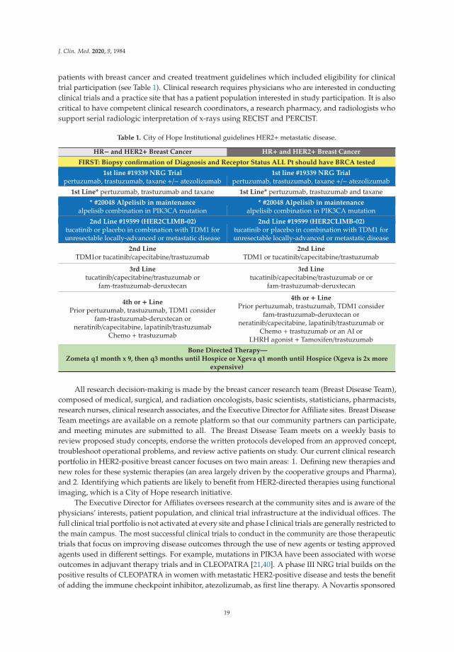
Table 1. City of Hope Institutional guidelines HER2+metastatic disease.
HR− and HER2+ Breast Cancer HR+ and HER2+ Breast Cancer
FIRST: Biopsy confirmation of Diagnosis and Receptor Status ALL Pt should have BRCA tested
1st line #19339 NRG Trialpertuzumab, trastuzumab, taxane +/− atezolizumab
1st line #19339 NRG Trialpertuzumab, trastuzumab, taxane +/− atezolizumab
1st Line* pertuzumab, trastuzumab and taxane 1st Line* pertuzumab, trastuzumab and taxane
* #20048 Alpelisib in maintenancealpelisib combination in PIK3CA mutation
* #20048 Alpelisib in maintenancealpelisib combination in PIK3CA mutation
2nd Line #19599 (HER2CLIMB-02)tucatinib or placebo in combination with TDM1 forunresectable locally-advanced or metastatic disease
2nd Line #19599 (HER2CLIMB-02)tucatinib or placebo in combination with TDM1 forunresectable locally-advanced or metastatic disease
2nd LineTDM1or tucatinib/capecitabine/trastuzumab
2nd LineTDM1 or tucatinib/capecitabine/trastuzumab
3rd Linetucatinib/capecitabine/trastuzumab or
fam-trastuzumab-deruxtecan
3rd Linetucatinib/capecitabine/trastuzumab or or
fam-trastuzumab-deruxtecan
4th or + LinePrior pertuzumab, trastuzumab, TDM1 consider
fam-trastuzumab-deruxtecan orneratinib/capecitabine, lapatinib/trastuzumab
Chemo + trastuzumab
4th or + LinePrior pertuzumab, trastuzumab, TDM1 consider
fam-trastuzumab-deruxtecan orneratinib/capecitabine, lapatinib/trastuzumab or
Chemo + trastuzumab or an AI orLHRH agonist + Tamoxifen/trastuzumab
Bone Directed Therapy—Zometa q1 month x 9, then q3 months until Hospice or Xgeva q1 month until Hospice (Xgeva is 2x more
expensive)
All research decision-making is made by the breast cancer research team (Breast Disease Team),composed of medical, surgical, and radiation oncologists, basic scientists, statisticians, pharmacists,research nurses, clinical research associates, and the Executive Director for Affiliate sites. Breast DiseaseTeam meetings are available on a remote platform so that our community partners can participate,and meeting minutes are submitted to all. The Breast Disease Team meets on a weekly basis toreview proposed study concepts, endorse the written protocols developed from an approved concept,troubleshoot operational problems, and review active patients on study. Our current clinical researchportfolio in HER2-positive breast cancer focuses on two main areas: 1. Defining new therapies andnew roles for these systemic therapies (an area largely driven by the cooperative groups and Pharma),and 2. Identifying which patients are likely to benefit from HER2-directed therapies using functionalimaging, which is a City of Hope research initiative.
The Executive Director for Affiliates oversees research at the community sites and is aware of thephysicians’ interests, patient population, and clinical trial infrastructure at the individual offices. Thefull clinical trial portfolio is not activated at every site and phase I clinical trials are generally restricted tothe main campus. The most successful clinical trials to conduct in the community are those therapeutictrials that focus on improving disease outcomes through the use of new agents or testing approvedagents used in different settings. For example, mutations in PIK3A have been associated with worseoutcomes in adjuvant therapy trials and in CLEOPATRA [21,40]. A phase III NRG trial builds on thepositive results of CLEOPATRA in women with metastatic HER2-positive disease and tests the benefitof adding the immune checkpoint inhibitor, atezolizumab, as first line therapy. A Novartis sponsored
19
J. Clin. Med. 2020, 9, 1984
trial randomizes women with metastatic disease and a PIK3A mutation to a placebo controlled doubleblind trial of alpelisib added during the patients’ maintenance of trastuzumab and pertuzumab. We arecurrently enrolling patients with documented HER2-activating mutations to a phase II trial sponsoredby PUMA of neratinib, trastuzumab, (and fulvestrant if also ER-positive).
Our radiolabeled trastuzumab imaging studies require a radiopharmacy and expertise by acommitted team of radiologists and physicist who interpret the images. Our community oncologistsplay an important role in these studies by referring patients, thereby allowing us to complete accrualexpeditiously. They are authors on our publications and are recognized by their local communities astrue experts who participate in innovative research.
8. Optimizing Partnerships
The Breast Disease Team has a number of initiatives to ensure inclusion of our community partnersin research, and the weekly Breast Disease Team meeting is at the heart of these collaborations.Discussions at weekly Tumor Boards and a bi-weekly treatment planning meeting frequentlyhighlight on-going studies and identify study candidates. City of Hope is a founding memberof the NCCN (national comprehensive cancer network) and has a long history of participating inguideline panels. While some of the guidelines for treating breast cancer are definitive, others offeracceptable treatment options. The breast cancer team on the main campus meets on an annual basis tocompile our guideline preferences for treatment, including, not only standard of care but the openstudies for clinical trial participation as well (see Table 1).
9. Future Directions
We continue to activate investigator-initiated, cooperative group, and Pharma trials in thecommunity that focus on improving the outcomes for women with metastatic HER2-positive disease.Our imaging research will utilize radiolabeled trastuzumab to predict for response to new agentssuch as fam-trastuzumab-deruxtecan and tucatinib [24,29]. These studies will rely on our communitypartners to refer interested patients for study participation.
National Cancer Institute-designated Comprehensive Cancer Centers (NCI-CCC) such asCity of Hope strive to provide expert interdisciplinary clinical care and cutting-edge treatmentsand technologies. Between 1998 and 2008, 6.4% of adults between 22 and 65 years of age who werediagnosed with cancer in Los Angeles County received treatment at an NCI-CCC. For all primary tumorsites, the survival of patients treated at an NCI-CCC was superior to patients treated at a non-NCI-CCC.In this study, 6.5% of the 31,764 breast cancer patients included in the analysis were treated at anNCI-CCC and their survival was superior to those treated at non NCI-CCC sites [41]. Realistically, themajority of cancer patients will receive their care in community sites, and it is an important part of themission of a Comprehensive Cancer Center to ensure that the standard of care in the community iscomparable to the care that is delivered in these large research centers. Clinical research is at the heartof improving quality care.
Author Contributions: Conceptualization and writing, J.E.M., L.K. and Y.Y.; writing, review and editing, M.C.,S.L. (Sayeh Lavasani), S.L. (Sariah Liu), N.T.-P., M.S., W.S., D.S., J.W., C.Y. and T.W. All authors have read andagreed to the published version of the manuscript.
Funding: This research received no external funding.
Acknowledgments: The authors thank Nicola Welch, CMPP for editorial assistance and critical review ofthe manuscript.
Conflicts of Interest: The authors declare no conflict of interest.
20
J. Clin. Med. 2020, 9, 1984
References
1. Prat, A.; Pineda, E.; Adamo, B.; Galván, P.; Fernández, A.; Gaba, L.; Díez, M.; Viladot, M.; Arance, A.;Muñoz, M. Clinical implications of the intrinsic molecular subtypes of breast cancer. Breast 2015, 24, S26–S35.[CrossRef]
2. Prat, A.; Pascual, T.; Adamo, B. Intrinsic molecular subtypes of HER2+ breast cancer. Oncotarget 2017, 8,73362–73363. [CrossRef]
3. Hayes, D.F. HER2 and Breast Cancer—A Phenomenal Success Story. N. Engl. J. Med. 2019, 381, 1284–1286.[CrossRef]
4. Slamon, D.; Clark, G.; Wong, S.; Levin, W.; Ullrich, A.; McGuire, W. Human breast cancer: Correlation ofrelapse and survival with amplification of the HER-2/neu oncogene. Science 1987, 235, 177–182. [CrossRef]
5. Press, M.F.; Pike, M.C.; Chazin, V.R.; Hung, G.; Udove, J.A.; Markowicz, M.; Danyluk, J.; Godolphin, W.;Sliwkowski, M.; Akita, R.; et al. Her-2/neu Expression in Node-negative Breast Cancer: Direct TissueQuantitation by Computerized Image Analysis and Association of Overexpression with Increased Risk ofRecurrent Disease. Cancer Res. 1993, 53, 4960–4970.
6. Cobleigh, M.A.; Vogel, C.L.; Tripathy, D.; Robert, N.J.; Scholl, S.; Fehrenbacher, L.; Wolter, J.M.; Paton, V.;Shak, S.; Lieberman, G.; et al. Multinational Study of the Efficacy and Safety of Humanized Anti-HER2Monoclonal Antibody in Women Who Have HER2-Overexpressing Metastatic Breast Cancer That HasProgressed After Chemotherapy for Metastatic Disease. J. Clin. Oncol. 1999, 17, 2639. [CrossRef]
7. Vogel, C.; Cobleigh, M.; Tripathy, D.; Harris, L.; Fehrenbacher, L.; Slamon, D.; Ash, M.; Novotny, W.;Stewart, S.; Shak, S. First-line, non-hormonal, treatment of women with HER2 overexpressing metastaticbreast cancer with herceptin (trastuzumab, humanised anti-HER2 antibody) [abstract 275]. Proc. Am. Soc.Clin. Oncol. 2001, 20, 71a.
8. Slamon, D.J.; Leyland-Jones, B.; Shak, S.; Fuchs, H.; Paton, V.; Bajamonde, A.; Fleming, T.; Eiermann, W.;Wolter, J.; Pegram, M.; et al. Use of Chemotherapy plus a Monoclonal Antibody against HER2 for MetastaticBreast Cancer That Overexpresses HER2. N. Engl. J. Med. 2001, 344, 783–792. [CrossRef]
9. Giordano, S.H.; Temin, S.; Chandarlapaty, S.; Crews, J.R.; Esteva, F.J.; Kirshner, J.J.; Krop, I.E.; Levinson, J.;Lin, N.U.; Modi, S.; et al. Systemic Therapy for Patients With Advanced Human Epidermal Growth FactorReceptor 2–Positive Breast Cancer: ASCO Clinical Practice Guideline Update. J. Clin. Oncol. 2018, 36,2736–2740. [CrossRef]
10. Wolff, A.C.; Hammond, M.E.H.; Allison, K.H.; Harvey, B.E.; Mangu, P.B.; Bartlett, J.M.S.; Bilous, M.; Ellis, I.O.;Fitzgibbons, P.; Hanna, W.; et al. Human Epidermal Growth Factor Receptor 2 Testing in Breast Cancer:American Society of Clinical Oncology/College of American Pathologists Clinical Practice Guideline FocusedUpdate. J. Clin. Oncol. 2018, 36, 2105–2122. [CrossRef]
11. Niehans, G.A.; Singleton, T.P.; Dykoski, D.; Kiang, D.T. Stability of HER-2/neu Expression Over Time and atMultiple Metastatic Sites. J. Natl. Cancer Inst. 1993, 85, 1230–1235. [CrossRef] [PubMed]
12. Bergh, J. Quo Vadis With Targeted Drugs in the 21st Century? J. Clin. Oncol. 2009, 27, 2–5. [CrossRef] [PubMed]13. Blancas, I.; Muñoz-Serrano, A.J.; Legerén, M.; Ruiz-Ávila, I.; Jurado, J.M.; Delgado, M.T.; Garrido, J.M.;
González Bayo, B.; Rodríguez-Serrano, F. Immunophenotypic Conversion between Primary and RelapseBreast Cancer and its Effects on Survival. Gynecol. Obstet. Investig. 2020, in press. [CrossRef] [PubMed]
14. Fehrenbacher, L.; Cecchini, R.S.; Jr, C.E.G.; Rastogi, P.; Costantino, J.P.; Atkins, J.N.; Crown, J.P.; Polikoff, J.;Boileau, J.-F.; Provencher, L.; et al. NSABP B-47/NRG Oncology Phase III Randomized Trial ComparingAdjuvant Chemotherapy With or Without Trastuzumab in High-Risk Invasive Breast Cancer Negative forHER2 by FISH and With IHC 1+ or 2+. J. Clin. Oncol. 2020, 38, 444–453. [CrossRef] [PubMed]
15. Mortimer, J.; Conti, P.; Shan, T.; Carroll, M.; Kofi, P.; Colcher, D.; Raubitschek, A.; Bading, J.64Cu-DOTA-trastuzumab positron emission tomography imaging of HER2 in women with advancedbreast cancer. Cancer Res. 2012, 72, 229s.
16. Mortimer, J.E.; Bading, J.R.; Colcher, D.M.; Conti, P.S.; Frankel, P.H.; Carroll, M.I.; Tong, S.; Poku, E.; Miles, J.K.;Shively, J.E.; et al. Functional Imaging of Human Epidermal Growth Factor Receptor 2–Positive MetastaticBreast Cancer Using 64Cu-DOTA-Trastuzumab PET. J. Nucl. Med. 2014, 55, 23–29. [CrossRef]
21

J. Clin. Med. 2020, 9, 1984
17. Burris, H.A.; Rugo, H.S.; Vukelja, S.J.; Vogel, C.L.; Borson, R.A.; Limentani, S.; Tan-Chiu, E.; Krop, I.E.;Michaelson, R.A. Girish, S.; et al. Phase II Study of the Antibody Drug Conjugate Trastuzumab-DM1 forthe Treatment of Human Epidermal Growth Factor Receptor 2 (HER2)–Positive Breast Cancer After PriorHER2-Directed Therapy. J. Clin. Oncol. 2011, 29, 398–405. [CrossRef]
18. Mortimer, J.E.; Shively, J.E. Functional Imaging of Human Epidermal Growth Factor Receptor 2–PositiveBreast Cancers and a Note about NOTA. J. Nucl. Med. 2019, 60, 23–25. [CrossRef]
19. Barthélémy, P.; Leblanc, J.; Goldbarg, V.; Wendling, F.; Kurtz, J.-E. Pertuzumab: Development Beyond BreastCancer. Anticancer Res. 2014, 34, 1483–1491.
20. Baselga, J.; Gelmon, K.A.; Verma, S.; Wardley, A.; Conte, P.; Miles, D.; Bianchi, G.; Cortes, J.; McNally, V.A.;Ross, G.A.; et al. Phase II Trial of Pertuzumab and Trastuzumab in Patients With Human Epidermal GrowthFactor Receptor 2–Positive Metastatic Breast Cancer That Progressed During Prior Trastuzumab Therapy.J. Clin. Oncol. 2010, 28, 1138–1144. [CrossRef]
21. Swain, S.M.; Miles, D.; Kim, S.-B.; Im, Y.-H.; Im, S.-A.; Semiglazov, V.; Ciruelos, E.; Schneeweiss, A.; Loi, S.;Monturus, E.; et al. Pertuzumab, trastuzumab, and docetaxel for HER2-positive metastatic breast cancer(CLEOPATRA): End-of-study results from a double-blind, randomised, placebo-controlled, phase 3 study.Lancet Oncol. 2020, 21, 519–530. [CrossRef]
22. Swain, S.M.; Baselga, J.; Kim, S.-B.; Ro, J.; Semiglazov, V.; Campone, M.; Ciruelos, E.; Ferrero, J.-M.;Schneeweiss, A.; Heeson, S.; et al. Pertuzumab, Trastuzumab, and Docetaxel in HER2-Positive MetastaticBreast Cancer. N. Engl. J. Med. 2015, 372, 724–734. [CrossRef]
23. Yan, H.; Yu, K.; Zhang, K.; Liu, L.; Li, Y. Efficacy and safety of trastuzumab emtansine (T-DM1) in thetreatment of HER2-positive metastatic breast cancer (MBC): A meta-analysis of randomized controlled trial.Oncotarget. 2017, 8, 102458. [CrossRef] [PubMed]
24. Modi, S.; Saura, C.; Yamashita, T.; Park, Y.H.; Kim, S.-B.; Tamura, K.; Andre, F.; Iwata, H.; Ito, Y.; Tsurutani, J.;et al. Trastuzumab Deruxtecan in Previously Treated HER2-Positive Breast Cancer. N. Engl. J. Med. 2020,382, 610–621. [CrossRef] [PubMed]
25. Ogitani, Y.; Aida, T.; Hagihara, K.; Yamaguchi, J.; Ishii, C.; Harada, N.; Soma, M.; Okamoto, H.; Oitate, M.;Arakawa, S.; et al. DS-8201a, A Novel HER2-Targeting ADC with a Novel DNA Topoisomerase I Inhibitor,Demonstrates a Promising Antitumor Efficacy with Differentiation from T-DM1. Clin. Cancer Res. 2016, 22,5097–5108. [CrossRef] [PubMed]
26. Cuadros, M.; Villegas, R. Systematic Review of HER2 Breast Cancer Testing. Appl. Immunohistochem. Mol.Morphol. 2009, 17, 1–7. [CrossRef] [PubMed]
27. Yau, T.K.; Sze, H.; Soong, I.S.; Hioe, F.; Khoo, U.S.; Lee, A.W.M. HER2 overexpression of breast cancers inHong Kong: Prevalence and concordance between immunohistochemistry and in-situ hybridisation assays.Hong Kong Med. J. 2008, 14, 130–135. [PubMed]
28. NCCN. Clinical Practice Guidelines in Oncology: Prostate Cancer (v.3.2011). Available online: http://www.nccn.org (accessed on 9 June 2011).
29. Murthy, R.K.; Loi, S.; Okines, A.; Paplomata, E.; Hamilton, E.; Hurvitz, S.A.; Lin, N.U.; Borges, V.; Abramson, V.;Anders, C.; et al. Tucatinib, Trastuzumab, and Capecitabine for HER2-Positive Metastatic Breast Cancer.N. Engl. J. Med. 2019, 382, 597–609. [CrossRef]
30. Network, N.C.C. Breast Version 4. 2020. Available online: https://www.nccn.org/professionals/physician_gls/pdf/breast.pdf (accessed on 28 April 2020).
31. Lin, N.U.; Borges, V.; Anders, C.; Murthy, R.K.; Paplomata, E.; Hamilton, E.; Hurvitz, S.; Loi, S.; Okines, A.;Abramson, V.; et al. Intracranial Efficacy and Survival With Tucatinib Plus Trastuzumab and Capecitabinefor Previously Treated HER2-Positive Breast Cancer With Brain Metastases in the HER2CLIMB Trial.J. Clin. Oncol. 2020, in press. [CrossRef]
32. Geyer, C.E.; Forster, J.; Lindquist, D.; Chan, S.; Romieu, C.G.; Pienkowski, T.; Jagiello-Gruszfeld, A.; Crown, J.;Chan, A.; Kaufman, B.; et al. Lapatinib plus Capecitabine for HER2-Positive Advanced Breast Cancer. N.Engl. J. Med. 2006, 355, 2733–2743. [CrossRef]
33. Blackwell, K.L.; Burstein, H.J.; Storniolo, A.M.; Rugo, H.S.; Sledge, G.; Aktan, G.; Ellis, C.; Florance, A.;Vukelja, S.; Bischoff, J.; et al. Overall Survival Benefit With Lapatinib in Combination With Trastuzumab forPatients With Human Epidermal Growth Factor Receptor 2–Positive Metastatic Breast Cancer: Final ResultsFrom the EGF104900 Study. J. Clin. Oncol. 2012, 30, 2585–2592. [CrossRef] [PubMed]
22
J. Clin. Med. 2020, 9, 1984
34. Nasrazadani, A.; Brufsky, A. Neratinib: The emergence of a new player in the management of HER2+ breastcancer brain metastasis. Future Oncol. 2020, 16, 247–254. [CrossRef] [PubMed]
35. Tripathy, D.; Slamon, D.J.; Cobleigh, M.; Arnold, A.; Saleh, M.; Mortimer, J.E. Safety of treatment of metastaticbreast cancer with trastuzumab beyond disease progression. J. Clin. Oncol. 2004, 22, 1063–1070. [CrossRef]
36. Bose, R.; Kavuri, S.M.; Searleman, A.C.; Shen, W.; Shen, D.; Koboldt, D.C.; Monsey, J.; Goel, N.; Aronson, A.B.;Li, S.; et al. Activating HER2 Mutations in HER2 Gene Amplification Negative Breast Cancer. Cancer Discov.2013, 3, 224–237. [CrossRef]
37. Ma, C.X.; Bose, R.; Gao, F.; Freedman, R.A.; Telli, M.L.; Kimmick, G.; Winer, E.; Naughton, M.; Goetz, M.P.;Russell, C.; et al. Neratinib Efficacy and Circulating Tumor DNA Detection of HER2 Mutations in HER2Nonamplified Metastatic Breast Cancer. Clin. Cancer Res. 2017, 23, 5687–5695. [CrossRef]
38. Ben-Baruch, N.E.; Bose, R.; Kavuri, S.M.; Ma, C.X.; Ellis, M.J. HER2-Mutated Breast Cancer Responds toTreatment With Single-Agent Neratinib, a Second-Generation HER2/EGFR Tyrosine Kinase Inhibitor. J. Natl.Compr. Cancer Netw. 2015, 13, 1061–1064. [CrossRef]
39. Hyman, D.M.; Piha-Paul, S.A.; Won, H.; Rodon, J.; Saura, C.; Shapiro, G.I.; Juric, D.; Quinn, D.I.; Moreno, V.;Doger, B.; et al. HER kinase inhibition in patients with HER2- and HER3-mutant cancers. Nature 2018, 554,189–194. [CrossRef] [PubMed]
40. Pogue-Geile, K.L.; Kim, C.; Jeong, J.-H.; Tanaka, N.; Bandos, H.; Gavin, P.G.; Fumagalli, D.; Goldstein, L.C.;Sneige, N.; Burandt, E.; et al. Predicting Degree of Benefit From Adjuvant Trastuzumab in NSABP Trial B-31.J. Natl. Cancer Inst. 2013, 105, 1782–1788. [CrossRef]
41. Wolfson, J.A.; Sun, C.-L.; Wyatt, L.P.; Hurria, A.; Bhatia, S. Impact of care at comprehensive cancer centers onoutcome: Results from a population-based study. Cancer 2015, 121, 3885–3893. [CrossRef] [PubMed]
© 2020 by the authors. Licensee MDPI, Basel, Switzerland. This article is an open accessarticle distributed under the terms and conditions of the Creative Commons Attribution(CC BY) license (http://creativecommons.org/licenses/by/4.0/).
23

Journal of
Clinical Medicine
Article
NCI–Clinical Trial Accrual in a Community NetworkAffiliated with a Designated Cancer Center
Daniel J. Kim, Dan Otap, Nora Ruel, Naveen Gupta, Naveed Khan and Tanya Dorff *
City of Hope Comprehensive Cancer Center, Department of Medical Oncology and DevelopmentalTherapeutics, Duarte, CA 91010, USA; [email protected] (D.J.K.); [email protected] (D.O.); [email protected] (N.R.);[email protected] (N.G.); [email protected] (N.K.)* Correspondence: [email protected]
Received: 29 April 2020; Accepted: 17 June 2020; Published: 24 June 2020
Abstract: Most cancer care is delivered in the community, while most clinical trials exist in academiccenters. We analyzed clinical trial accrual of a tertiary care cancer center and its affiliated communitysites to better understand what types of trials accrued at the community sites and whether communityaccrual increased ethnic diversity. The institutional clinical trial database was searched for solid tumoraccruals during 2018–2019. Patient’s race was abstracted, and trial’s funding source, phase, and diseasetype/stage were tabulated. Of 3689 accruals, 133 were at community sites, representing 26 uniquetrials while the main campus accrued to 93 unique trials. Community site accruals were highest forbreast and colorectal cancer, but patients with less common cancers such as renal, nasopharyngeal,and gastric cancer were also accrued at community sites. Accruals occurred to randomized trials,as well as phase Ib and translational biomarker studies. Minority patients constituted 20.0% and32.5% of community site accruals for therapeutic and non-therapeutic trials respectively, compared to20.6% and 29.8% of main campus accruals for therapeutic and non-therapeutic trials, respectively.We conclude that community sites affiliated with an academic cancer center can accrue to a broadspectrum of clinical trials while enhancing racial diversity in participation of clinical trials. Furtherexpansion of access to clinical trials in community sites is necessary to broaden patient access tostate-of-the-art and next-generation treatment options.
Keywords: clinical trials; community practice; minorities; ethnicity; race
1. Introduction
Community oncology practices provide approximately 55% of all care for cancer patients in theUnited States [1]. Providing access to clinical trials in community oncology practices has been a majorinitiative of the National Cancer Institute (NCI, Rockville, MD, USA), as evidenced by the creation of theNational Community Oncology Research Program (NCORP, Bethesda, MD, USA) in 2014. The NCORPwas created by consolidating the previous NCI supported networks of the CCOP–community clinicaloncology program and the NCI Community Cancer Centers Program (NCCCP, Bethesda, MD, USA).Up to a third of clinical trial accrual in NCI studies is contributed by these community oncologypractices [2], yet the overall clinical trial accrual remains lower than desirable: fewer than 5% of adultcancer patients enroll in clinical trials [3], despite 70% of cancer patients expressing interest in enrollingin a clinical trial [4].
Aside from increasing treatment options and providing access to the latest innovations in cancercare, participation in clinical trials has been shown to improve overall treatment quality vis-à-visutilization of optimal standards of care [5]. One barrier for community practice physicians hasbeen cost; clinical trial enrollment takes time, which takes resources away from overall clinic flow,and research staff are required regardless of how robust accrual is. Understanding the types of clinicaltrials that readily accrue in the community setting could help improve trial selection to foster financial
J. Clin. Med. 2020, 9, 1970; doi:10.3390/jcm9061970 www.mdpi.com/journal/jcm25
J. Clin. Med. 2020, 9, 1970
sustainability, since fewer trials, each of which accrue larger numbers of patients, is more efficient inthe setting of having limited clinical research staff at community sites.
In addition, inadequate representation of racially diverse cancer patients has been noted to be amajor problem in the applicability of most clinical trials to everyday practice. For example, clinical trialsin metastatic castration-resistant prostate cancer (CRPC) were found to have 80% under-representationof African American patients [6]. In the case of geriatric patients, a study of NCI-sponsored trials foundthat patients over 65 made up only 32% of participants, whereas they made up 61% of patients affectedby the cancers [7]. Given that community oncology practices may be located closer to patients’ homes,the availability of trials in a local setting could reduce potential barriers for patients to access clinicaltrials. Furthermore, if community practices are located near specific ethnic enclaves, their participationin clinical trials could help alleviate the lack of racial diversity, as indicated by the NCCCP initiative [8].
City of Hope is an NCI-designated comprehensive cancer center in Duarte, California, with anextensive network of 21 community practice sites located within its vicinity in Southern California,thus allowing for delivery of specialized care near where patients reside. As part of the overall missionof City of Hope, select clinical trials have been opened to accrual at six of the community sites whichservice a high enough patient volume to support research recruitment. We sought to characterizeand evaluate clinical trials accrual with a focus on City of Hope’s community sites, and whetherracial diversity was enhanced in patients accrued at community sites compared to the main cancercenter campus.
2. Material and Methods
After obtaining IRB exemption for the study, we identified the individual accrual events in ourclinical trials database during the calendar years 2018 and 2019 at all City of Hope sites. This datasetwas extracted from our institution’s secure online trial database, devoid of any personal identifiers.Inclusion criteria was prospective adult clinical trials. Study title, sponsor, location of accrual, and dateof study were extracted from the dataset. We decided ahead of time to exclude trials which were felt toreflect highly tertiary patient populations, such as hematologic malignancy trials which, at City ofHope, were likely to be heavily transplant and chimeric antigen receptor T cell therapy-based, or anyother trials felt to introduce bias into the analysis.
Results were tabulated for comparison between main campus and community sites and stratified bythe type of trials. The Chi-square and Fisher’s exact tests were used to test the differences in distributionof race, minority status, and age groups between community sites and main campus participants.
3. Results
Of the 33,558 individual accrual events identified in our analysis, 23,809 accrual events wereidentified to be retrospective in nature and thus excluded, along with an additional 5625 accrualevents which were identified to be trials involving hematologic malignancies. Sixteen pediatric trialaccrual events were also excluded, as well as 419 healthy volunteer accruals. Also excluded were twohigh-accruing non-therapeutic trials specifically recruiting only Hispanic women, as ethnicity was nota variable and thus biasing the minority accrual question.
Our final analysis was thus based on 3689 accrual events. Community sites in the City of Hopenetwork accrued to 26 distinct, solid tumor clinical trials (n = 22 therapeutic, n = 4 non-therapeutic)during the years 2018–2019, compared with accrual to 93 solid tumor trials at the main campus duringthe same time period. A description of all therapeutic trials which accrued at least one subject at acommunity site is delineated in Table 1. During the same time period, an additional 57 trials wereopen at community sites but did not accrue patients there.
26
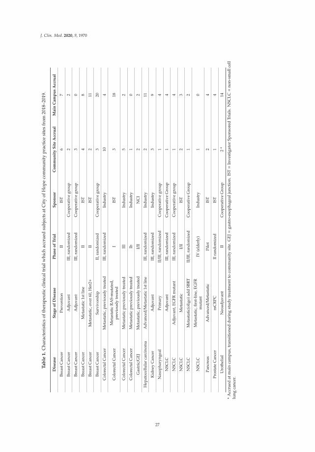
J. Clin. Med. 2020, 9, 1970
Ta
ble
1.
Cha
ract
eris
tics
ofth
erap
euti
ccl
inic
altr
ialw
hich
accr
ued
subj
ects
atC
ity
ofH
ope
com
mun
ity
prac
tice
site
sfr
om20
18–2
019.
Dis
ease
Sta
ge
of
Dis
ease
Ph
ase
of
Tri
al
Sp
on
sor
Co
mm
un
ity
Sit
eA
ccru
al
Main
Cam
pu
sA
ccru
al
Brea
stC
ance
rPr
even
tion
IIIS
T6
7
Brea
stC
ance
rA
djuv
ant
III,
rand
omiz
edC
oope
rati
vegr
oup
22
Brea
stC
ance
rA
djuv
ant
III,
rand
omiz
edC
oope
rati
vegr
oup
30
Brea
stC
ance
rM
etas
tati
c1s
tlin
eII
IST
48
Brea
stC
ance
rM
etas
tati
c,ov
er60
,Her
2+II
IST
211
Brea
stC
ance
rSu
rviv
orsh
ipII
,ran
dom
ized
Coo
pera
tive
grou
p3
20
Col
orec
talC
ance
rM
etas
tati
c,pr
evio
usly
trea
ted
III,
rand
omiz
edIn
dust
ry10
4
Col
orec
talC
ance
rM
etas
tati
cR
AS-
mut
ated
,pr
evio
usly
trea
ted
IIS
T3
18
Col
orec
talC
ance
rM
etas
tati
cpr
evio
usly
trea
ted
III
Indu
stry
52
Col
orec
talC
ance
rM
etas
tati
cpr
evio
usly
trea
ted
IbIn
dust
ry1
0
Gas
tric/G
EJM
etas
tati
c,pr
evio
usly
trea
ted
I/II
NC
I2
2
Hep
atoc
ellu
lar
carc
inom
aA
dvan
ced/
Met
asta
tic
1stl
ine
III,
rand
omiz
edIn
dust
ry2
11
Kid
ney
Can
cer
Adj
uvan
tII
I,ra
ndom
ized
Indu
stry
39
Nas
opha
ryng
eal
Prim
ary
II/I
II,r
ando
miz
edC
oope
rati
vegr
oup
14
NSC
LCA
djuv
ant
III,
rand
omiz
edC
oope
rati
veG
roup
14
NSC
LCA
djuv
ant,
EGFR
mut
ant
III,
rand
omiz
edC
oope
rati
vegr
oup
14
NSC
LCM
etas
tati
cI/
IIIS
T2
3
NSC
LCM
etas
tati
c(ol
igo)
add
SBR
TII/I
II,r
ando
miz
edC
oope
rati
veG
roup
12
NSC
LCM
etas
tati
c,fir
st-l
ine,
EGFR
mut
ant
IV(e
lder
ly)
Indu
stry
10
Panc
reas
Adv
ance
d/M
etas
tati
cPi
lot
IST
24
Pros
tate
Can
cer
mC
RPC
IIra
ndom
ized
IST
14
Uro
thel
ial
Neo
adju
vant
IIC
oope
rati
veG
roup
2*
14
*A
ccru
edat
mai
nca
mpu
s,tr
ansi
tion
edd
urin
gst
udy
trea
tmen
tto
com
mun
ity
site
.GE
J=ga
stro
-eso
phag
ealj
unct
ion.
IST=
Inve
stig
ator
Spon
sore
dTr
ials
.NSC
LC=
non-
smal
lcel
llu
ngca
ncer
.
27
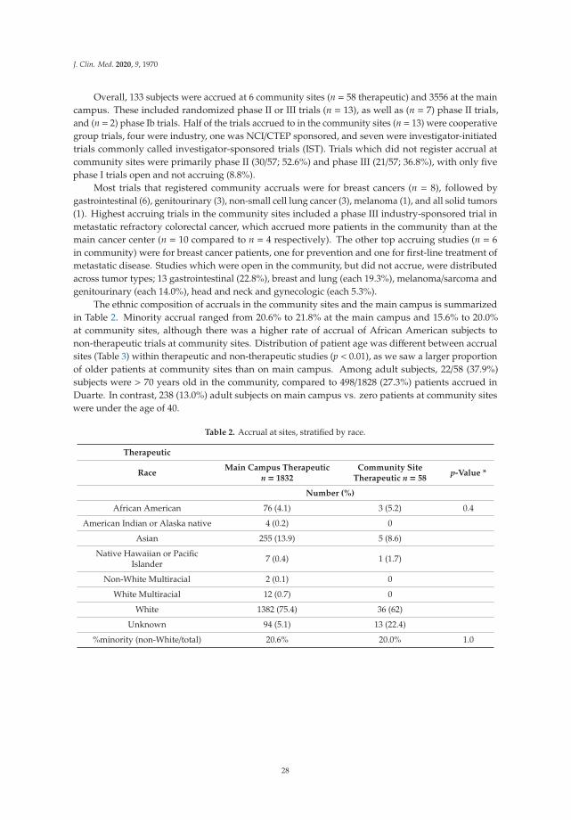
J. Clin. Med. 2020, 9, 1970
Overall, 133 subjects were accrued at 6 community sites (n = 58 therapeutic) and 3556 at the maincampus. These included randomized phase II or III trials (n = 13), as well as (n = 7) phase II trials,and (n = 2) phase Ib trials. Half of the trials accrued to in the community sites (n = 13) were cooperativegroup trials, four were industry, one was NCI/CTEP sponsored, and seven were investigator-initiatedtrials commonly called investigator-sponsored trials (IST). Trials which did not register accrual atcommunity sites were primarily phase II (30/57; 52.6%) and phase III (21/57; 36.8%), with only fivephase I trials open and not accruing (8.8%).
Most trials that registered community accruals were for breast cancers (n = 8), followed bygastrointestinal (6), genitourinary (3), non-small cell lung cancer (3), melanoma (1), and all solid tumors(1). Highest accruing trials in the community sites included a phase III industry-sponsored trial inmetastatic refractory colorectal cancer, which accrued more patients in the community than at themain cancer center (n = 10 compared to n = 4 respectively). The other top accruing studies (n = 6in community) were for breast cancer patients, one for prevention and one for first-line treatment ofmetastatic disease. Studies which were open in the community, but did not accrue, were distributedacross tumor types; 13 gastrointestinal (22.8%), breast and lung (each 19.3%), melanoma/sarcoma andgenitourinary (each 14.0%), head and neck and gynecologic (each 5.3%).
The ethnic composition of accruals in the community sites and the main campus is summarizedin Table 2. Minority accrual ranged from 20.6% to 21.8% at the main campus and 15.6% to 20.0%at community sites, although there was a higher rate of accrual of African American subjects tonon-therapeutic trials at community sites. Distribution of patient age was different between accrualsites (Table 3) within therapeutic and non-therapeutic studies (p < 0.01), as we saw a larger proportionof older patients at community sites than on main campus. Among adult subjects, 22/58 (37.9%)subjects were > 70 years old in the community, compared to 498/1828 (27.3%) patients accrued inDuarte. In contrast, 238 (13.0%) adult subjects on main campus vs. zero patients at community siteswere under the age of 40.
Table 2. Accrual at sites, stratified by race.
Therapeutic
RaceMain Campus Therapeutic
n = 1832Community Site
Therapeutic n = 58p-Value *
Number (%)
African American 76 (4.1) 3 (5.2) 0.4
American Indian or Alaska native 4 (0.2) 0
Asian 255 (13.9) 5 (8.6)
Native Hawaiian or PacificIslander 7 (0.4) 1 (1.7)
Non-White Multiracial 2 (0.1) 0
White Multiracial 12 (0.7) 0
White 1382 (75.4) 36 (62)
Unknown 94 (5.1) 13 (22.4)
%minority (non-White/total) 20.6% 20.0% 1.0
28
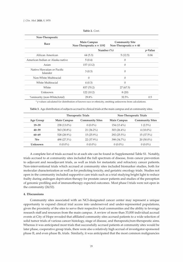
J. Clin. Med. 2020, 9, 1970
Table 2. Cont.
Non-Therapeutic
RaceMain Campus
Non-Therapeutic n = 1192Community Site
Non-Therapeutic n = 40
Number (%) p-Value
African American 64 (5.3) 5 (12.5) 0.04
American Indian or Alaska native 5 (0.4) 0
Asian 157 (13.2) 0
Native Hawaiian or PacificIslander 3 (0.3) 0
Non-White Multiracial 0 0
White Multiracial 4 (0.3) 0
White 837 (70.2) 27 (67.5)
Unknown 122 (10.2) 8 (20)
%minority (non-White/total) 29.8% 32.5% 0.5
* p values calculated for distribution of known race or ethnicity, omitting unknowns from calculations.
Table 3. Age distribution of subjects accrued to clinical trials at the main campus and at community sites.
Therapeutic Trials Non-Therapeutic Trials
Age Group Main Campus Community Sites Main Campus Community Sites
19–39 238 (13.0%) 0 (0.0%) 154 (13.4%) 1 (2.5%)
40–59 563 (30.8%) 21 (36.2%) 303 (26.4%) 4 (10.0%)
60–69 528 (28.9%) 15 (25.9%) 292 (25.5%) 15 (37.5%)
70+ 498 (27.3%) 22 (37.9%) 398 (34.7%) 20 (50.0%)
Unknown 0 (0.0%) 0 (0.0%) 0 (0.0%) 0 (0.0%)
A complete list of trials accrued to at each site can be found in Supplemental Table S1. Notably,trials accrued to at community sites included the full spectrum of disease, from cancer preventionto adjuvant and neoadjuvant trials, as well as trials for metastatic and refractory cancer patients.Non-interventional trials which accrued at community sites included biomarker studies, both formolecular characterization as well as for predicting toxicity, and geriatric oncology trials. Studies notopen in the community included supportive care trials such as a trial studying bright light to reducefrailty during androgen deprivation therapy for prostate cancer patients and studies of the perceptionof genomic profiling and of immunotherapy expected outcomes. Most phase I trials were not open inthe community (26/32).
4. Discussions
Community sites associated with an NCI-designated cancer center may represent a uniqueopportunity to expand clinical trial access into underserved and under-represented populations,given the proximity of the sites to serve their respective local communities and the ability to leverageresearch staff and resources from the main campus. A review of more than 33,000 individual accrualevents at City of Hope revealed that affiliated community sites accrued patients to a wide selection ofsolid tumor trials of various cancer histology, stage of disease, and therapeutic/non-therapeutic aims.Whereas it was anticipated most trials that successfully accrued patients at community sites would belater phase, cooperative group trials, there were also a relatively high accrual of investigator-sponsoredphase II, and even phase Ib, trials. Similarly, it was anticipated that the most common malignancies
29

J. Clin. Med. 2020, 9, 1970
would make up the largest percentage of accruals in the community sites, but uncommon tumor typessuch as hepatocellular, kidney, and pancreatic cancers also accrued at community sites.
City of Hope community practice sites are strategically located throughout five counties acrossSouthern California (Ventura, Los Angeles, San Bernardino, Riverside, and Orange), aiming toprovide cancer care to all communities, including socioeconomically disadvantaged populations andcommunities with cancer disparities. Our analysis did reveal a higher accrual of African Americanpatients to non-therapeutic trials at the community sites compared to main campus but did notsignificantly improve diversity of trial accrual as had been hoped. Loree et al. [9] had reported AfricanAmericans and Hispanics representing 3.1% and 6.1% of trial participants respectively, and City ofHope’s rate of African American representation in trial accrual was similarly low. A limitation of ouranalysis is that our percentages are derived from accrued subjects rather than all subjects; that is to say,the denominator are trial participants, rather than all screened patients or all eligible patients or allpatients seen at the clinic, as such data was not available for analysis. Our assessment was also limitedby a relatively high rate of “unknown” ethnicity among community accruals (22.4%); this can be dueto incomplete datasets or patients declining to answer the question. In order to minimize missing data,standard operating procedures for research staff and centralization of clinical data management couldimprove consistency and data quality.
Community sites did enroll a higher rate of geriatric patients, which may reflect the difficultyelderly patients face in traveling greater distances to a comprehensive cancer center in order to obtainoncology care. Admittedly, the City of Hope experience may be unique, as geriatric oncology is aflagship program with dedicated resources. However, this finding is an encouraging signal, andwarrants further study as increasing accrual of geriatric patients to cancer protocols is an importantgoal for the oncology community given that many cancers have substantially higher prevalence inpeople over the age of 65.
Additional limitation of our study involves the inherent selection process involved in extendingclinical trial offerings to the community sites. Based on limited resources and anticipated accrualrates, only a select few clinical trials were extended from the main campus to the community sites,and of the 21 community sites only six higher-volume sites were selected for clinical trial participation.Trial accrual rate could have been higher for the community sites if trial offerings were identical acrossmain campus and community sites, but such an offering would require far greater investment intotrial resources at all of the community sites and may not be practical or feasible. Our analysis alsocould not capture the number of patients who were referred from the community sites to the maincampus for enrollment into trials not offered at the community sites, thus shifting trial accrual numbersfrom community sites to main campus. We did notice, however, that a bladder cancer trial enrolledsubjects at the main campus and then transitioned ongoing cycles of treatment to community sitesdue to patient travel burden (Table 1). This flexibility is an example of how academic-communitycollaboration can optimize both accrual and patient-centered care and could represent an efficient“just in time” model of opening the right trials in the right areas. We concur with the value of maincampus regularly meeting with community sites to review currently available clinical trials [10], andwe additionally propose that the community sites must be actively engaged in and collaborate withthe main campus in selecting the trials offered at the community sites based on needs assessment oftheir unique patient population and community site clinician interests.
In contrast to published experiences, however, we found that phase I and even non-therapeutictrials did not present an insurmountable barrier to accrual in community oncology practices. In aprevious survey of 51 community practices, which included both federally sponsored and privatenon-academic affiliated practices, half of the practices self-reported that they did not accrue to phase Itrials, 53% did not accrue to investigator-initiated trials, and 33% did not enroll on correlative sciencetrials [11]. It is likely that many investigator-initiated trials and translational “non-therapeutic” trialscan meet the needs of community oncology patients, and that academic-affiliated community sites arebest poised to engage patients in accrual to these types of studies.
30

J. Clin. Med. 2020, 9, 1970
Supplementary Materials: The following are available online at http://www.mdpi.com/2077-0383/9/6/1970/s1,Table S1: summary of phases and disease states of trials accrued to at community sites and City of Hopemain campus.
Author Contributions: Conceptualization and methodology, T.D.; data curation, D.O.; validation, D.J.K., D.O.,N.R., and T.D.; formal analysis, N.R. and T.D.; writing—original draft preparation, D.J.K., N.R., N.G., and N.K.;writing—review and editing, D.K., T.D.; project administration and funding acquisition, T.D. All authors haveread and agreed to the published version of the manuscript.
Funding: This research was funded by an internal core grant #P30CA033572 from City of Hope, and received noexternal funding.
Conflicts of Interest: The authors declare no conflicts of interest.
References
1. Community Oncology Alliance. Available online: https://communityoncology.org (accessed on 28 March 2020).2. Carpenter, W.R.; Fortune-Greeley, A.; Zullig, L.L.; Lee, S.-Y.; Weiner, B.J. Sustainability and performance of
the National Cancer Institute’s Community Clinical Oncology Program. Contemp. Clin. Trials 2011, 33, 46–54.[CrossRef] [PubMed]
3. Unger, J.M.; Cook, E.; Tai, E.; Bleyer, A. Role of clinical trial participation in cancer research: barriers, evidenceand strategies. Am. Soc. Clin. Oncol. Educ. Book 2016, 35, 185–198. [CrossRef] [PubMed]
4. Comis, R.L.; Miller, J.D.; Aldigé, C.R.; Krebs, L.; Stoval, E. Public Attitudes Toward Participation in CancerClinical Trials. J. Clin. Oncol. 2003, 21, 830–835. [CrossRef] [PubMed]
5. Laliberte, L.; Fennell, M.L.; Papandonatos, G.D. The Relationship of Membership in Research Networksto Compliance with Treatment Guidelines for Early-Stage Breast Cancer. Med. Care 2005, 43, 471–479.[CrossRef] [PubMed]
6. Spratt, D.E.; Osborne, J. Disparities in Castration-Resistant Prostate Cancer Trials. J. Clin. Oncol. 2015, 33,1101–1103. [CrossRef] [PubMed]
7. Lewis, J.H.; Kilgore, M.L.; Goldman, D.P.; Trimble, E.L.; Kaplan, R.; Montello, M.J.; Housman, M.G.;Escarce, J.J. Participation of Patients 65 Years of Age or Older in Cancer Clinical Trials. J. Clin. Oncol. 2003,21, 1383–1389. [CrossRef] [PubMed]
8. Hirsch, B.R.; Locke, S.; Abernethy, A.P. Experience of the National Cancer Institute Community CancerCenters Program on Community-Based Cancer Clinical Trials Activity. J. Oncol. Pract. 2016, 12, e350–e358.[CrossRef] [PubMed]
9. Loree, J.M.; Anand, S.; Dasari, A.; Unger, J.M.; Gothwal, A.; Ellis, L.M.; Varadhachary, G.; Kopetz, S.;Overman, M.J.; Raghav, K. Disparity of Race Reporting and Representation in Clinical Trials Leading toCancer Drug Approvals from 2008 to 2018. JAMA Oncol. 2019, 5, e191870. [CrossRef] [PubMed]
10. Ludmir, E.B.; Adlakha, E.K.; Chun, S.G.; Reed, V.K.; Arzu, I.Y.; Ahmad, N.; Bloom, E.; Chronowski, G.M.;Delclos, M.E.; Mayo, L.L.; et al. Enhancing clinical trial enrollment at MD Anderson Cancer Center satellitecommunity campuses. Acta Oncol. 2019, 58, 1135–1137. [CrossRef] [PubMed]
11. Good, M.J.; Lubejko, B.; Humphries, K.; Medders, A. Measuring Clinical Trial–Associated Workload in aCommunity Clinical Oncology Program. J. Oncol. Pract. 2013, 9, 211–215. [CrossRef] [PubMed]
© 2020 by the authors. Licensee MDPI, Basel, Switzerland. This article is an open accessarticle distributed under the terms and conditions of the Creative Commons Attribution(CC BY) license (http://creativecommons.org/licenses/by/4.0/).
31

Journal of
Clinical Medicine
Article
Complex Oncological Decision-Making UtilizingFast-and-Frugal Trees in a Community Setting—Roleof Academic and Hybrid Modeling
Ravi Salgia 1,*, Isa Mambetsariev 1, Tingting Tan 1, Amanda Schwer 2, Daryl P. Pearlstein 3,
Hazem Chehabi 2, Angel Baroz 1, Jeremy Fricke 1, Rebecca Pharaon 1, Hannah Romo 1,
Thomas Waddington 4, Razmig Babikian 1, Linda Buck 1, Prakash Kulkarni 1, Mary Cianfrocca 1,
Benjamin Djulbegovic 5 and Sumanta K. Pal 1
1 Department of Medical Oncology and Therapeutics Research, 1500 E Duarte Road, City of Hope NationalMedical Center, Duarte, CA 91010, USA; [email protected] (I.M.); [email protected] (T.T.);[email protected] (A.B.); [email protected] (J.F.); [email protected] (R.P.); [email protected] (H.R.);[email protected] (R.B.); [email protected] (L.B.); [email protected] (P.K.); [email protected] (M.C.);[email protected] (S.K.P.)
2 Newport Diagnostic Center, Newport Beach, CA 92660, USA; [email protected] (A.S.);[email protected] (H.C.)
3 Department of Thoracic Surgery, Hoag Hospital, CA 92660, USA; [email protected] Department of Medicine, City of Hope National Medical Center, Duarte, CA 91010, USA;
[email protected] Department of Hematology & Hematopoietic Cell Transplantation, City of Hope National Medical Center,
Duarte, CA 91010, USA; [email protected]* Correspondence: [email protected]
Received: 15 May 2020; Accepted: 12 June 2020; Published: 16 June 2020
Abstract: Non-small cell lung cancer is a devastating disease and with the advent of targetedtherapies and molecular testing, the decision-making process has become complex. While establishedguidelines and pathways offer some guidance, they are difficult to utilize in a busy communitypractice and are not always implemented in the community. The rationale of the study was toidentify a cohort of patients with lung adenocarcinoma at a City of Hope community site (n = 11)and utilize their case studies to develop a decision-making framework utilizing fast-and-frugal tree(FFT) heuristics. Most patients had stage IV (N = 9, 81.8%) disease at the time of the first consultation.The most common symptoms at initial presentation were cough (N = 5, 45.5%), shortness of breath(N = 3, 27.2%), and weight loss (N = 3, 27.2%). The Eastern Cooperative Oncology Group (ECOG)performance status ranged from 0-1 in all patients in this study. Distribution of molecular driversamong the patients were as follows: EGFR (N = 5, 45.5%), KRAS (N = 2, 18.2%), ALK (N = 2, 18.2%),MET (N = 2, 18.2%), and RET (N = 1, 9.1%). Seven initial FFTs were developed for the variouscase scenarios, but ultimately the decisions were condensed into one FFT, a molecular stage IV FFT,that arrived at accurate decisions without sacrificing initial information. While these FFT decisiontrees may seem arbitrary to an experienced oncologist at an academic site, the simplicity of theirutility is essential for community practice where patients often do not get molecular testing and arenot assigned proper therapy.
Keywords: non-small cell lung cancer; actionable mutations; next-generation sequencing;fast-and-frugal trees; community practice; personalized medicine
J. Clin. Med. 2020, 9, 1884; doi:10.3390/jcm9061884 www.mdpi.com/journal/jcm33
J. Clin. Med. 2020, 9, 1884
1. Introduction
Lung cancer is a devastating disease with an overall survival rate of 16% at five years in non-smallcell lung cancer (NSCLC) and 6% in small cell lung cancer (SCLC) [1]. Lung cancer incidence in2020 is estimated at 228,820 new cases and will remain the leading cause of cancer death in the USwith an estimated 135,720 deaths [2]. NSCLC accounts for approximately 85% of lung cancer casesand is a heterogeneous group of tumors comprised of adenocarcinomas, squamous cell carcinomas,and large cell carcinomas as the major subtypes [3]. These individual subtypes have different molecularcharacteristics, clinical features, and complex therapeutic options with unique outcomes for individualpatients. More so, the advent of targeted therapy and immunotherapy have drastically shifted thetherapeutic landscape of NSCLC towards precision medicine, or personalized medicine, where theindividual patient is treated selectively based on their molecular characteristics [4]. Due to the rise ofpersonalized medicine, lung cancer decision-making has grown complex, and chemotherapy, once thedefault treatment of choice for all patients, is no longer universally used.
This advance towards personalized medicine is primarily driven by advances in targetedtherapeutic options, genomic testing, and immunotherapy that have shown improvement in patientoutcomes [5–10]. EGFR therapy, once limited to first-generation tyrosine-kinase inhibitors (TKIs) such aserlotinib [11], has grown to second and third generation TKIs with osimertinib showing improvementsnot only in response rates and progression-free survival (PFS), but also in durable overall survival (OS)benefits as compared to chemotherapy and other TKIs [12–14]. These advances in therapeutic optionsfor EGFR has further spurred greater understanding of NSCLC biology to reveal multiple distinctmolecular subtypes—a widely supported theory that oncogenic “driver mutations” are responsiblefor NSCLC’s malignant phenotype [15]. Along with EGFR, gene alterations in ALK, ROS1, NTRK,and BRAF have therapies that are FDA-approved and routinely tested in practice [16]. Recent long-termfollow-up results from 110 ALK-positive patients who received the ALK inhibitor crizotinib, showed amedian OS of 6.8 years in patients with metastatic disease [17]. Moreover, several inhibitors for METand RET are slowly being implemented in standard clinical practice and await FDA approval [18–22].Initial results from a phase II study of MET exon 14 inhibitor tepotinib, showed a sustained durationof response (12.4 months) and a 45% objective response rate (ORR), earning a breakthrough FDAdesignation [23,24]. Beyond these driver alterations, approximately 25% of lung adenocarcinomaspresent with KRAS alterations that occur in codons 12 or 13 [25]. No approved direct targetedtherapy is available for KRAS-mutant NSCLC, but two promising inhibitors—MRTX849 and AMG510—are currently under clinical consideration with early results reporting antitumor activity in theinitial patients [26,27]. Furthermore, several studies have shown that immunotherapy is a viabletreatment option for KRAS-mutant patients, with some patients showing remarkable clinical benefitfrom anti-PD-1/PD-L1 immunotherapy [28–30].
The availability of several targeted therapies and a variety of immunotherapies that have beenapproved for lung cancer has complicated oncology decision-making in clinical practice. Furthermore,advances in genomics and therapies have not been accompanied with advances in the science ofmedical decision-making. Clinical practice guidelines, such as the National Cancer Center Network(NCCN) and the American Society for Clinical Oncology (ASCO), are commonly used to aid indecision-making, but the recommendations are often difficult to interpret and are not easily manageableduring a busy oncology clinic [31,32]. We have previously shown that a novel method of utilizingfast-and-frugal trees (FFTs) to arrive at decision-making trees can allow for physicians to make a quickbut accurate decision [33]. Our previous work shows how guidelines can be converted into FFTs toallow development of individualized patient care, and the quantitative analysis of the performancecharacteristics of such a strategy [34,35]. The advantage of FFTs is that they are highly effective,simple decision trees composed of sequentially ordered cues (tests) and binary (yes/no) decisionsformulated via a series of “if-then” statements [34]. Therefore, they can be easily applied to lungcancer decision-making where, if a genetic alteration is revealed to be actionable (e.g., EGFR exon19 deletion), then an appropriate TKI (e.g., osimertinib) would be given [33]. While these decisions
34
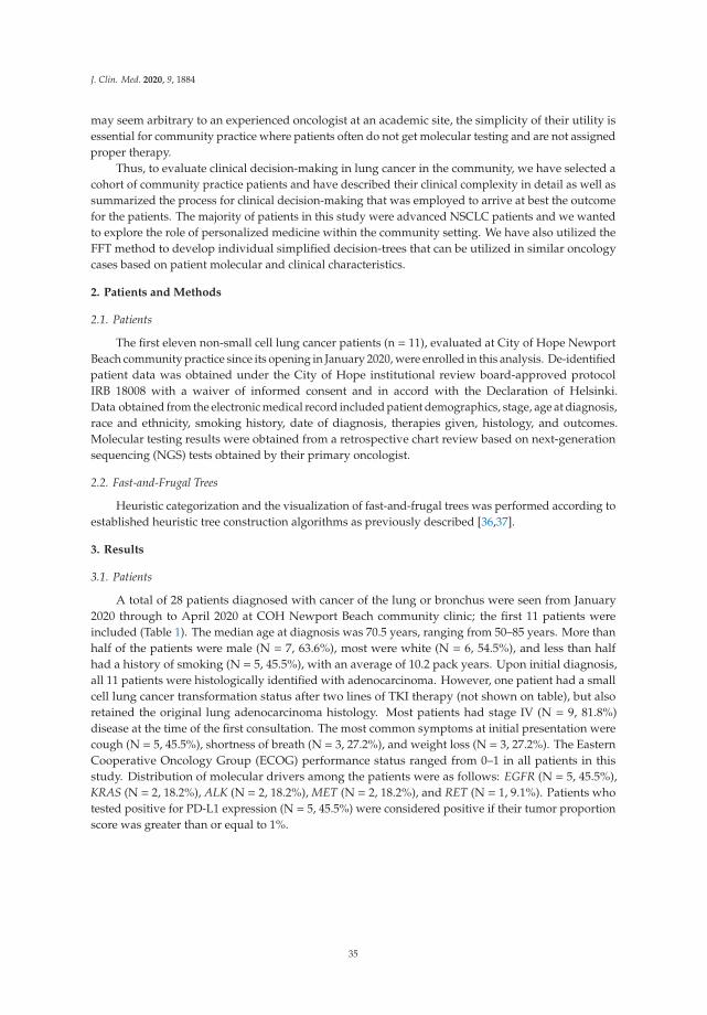
J. Clin. Med. 2020, 9, 1884
may seem arbitrary to an experienced oncologist at an academic site, the simplicity of their utility isessential for community practice where patients often do not get molecular testing and are not assignedproper therapy.
Thus, to evaluate clinical decision-making in lung cancer in the community, we have selected acohort of community practice patients and have described their clinical complexity in detail as well assummarized the process for clinical decision-making that was employed to arrive at best the outcomefor the patients. The majority of patients in this study were advanced NSCLC patients and we wantedto explore the role of personalized medicine within the community setting. We have also utilized theFFT method to develop individual simplified decision-trees that can be utilized in similar oncologycases based on patient molecular and clinical characteristics.
2. Patients and Methods
2.1. Patients
The first eleven non-small cell lung cancer patients (n = 11), evaluated at City of Hope NewportBeach community practice since its opening in January 2020, were enrolled in this analysis. De-identifiedpatient data was obtained under the City of Hope institutional review board-approved protocolIRB 18008 with a waiver of informed consent and in accord with the Declaration of Helsinki.Data obtained from the electronic medical record included patient demographics, stage, age at diagnosis,race and ethnicity, smoking history, date of diagnosis, therapies given, histology, and outcomes.Molecular testing results were obtained from a retrospective chart review based on next-generationsequencing (NGS) tests obtained by their primary oncologist.
2.2. Fast-and-Frugal Trees
Heuristic categorization and the visualization of fast-and-frugal trees was performed according toestablished heuristic tree construction algorithms as previously described [36,37].
3. Results
3.1. Patients
A total of 28 patients diagnosed with cancer of the lung or bronchus were seen from January2020 through to April 2020 at COH Newport Beach community clinic; the first 11 patients wereincluded (Table 1). The median age at diagnosis was 70.5 years, ranging from 50–85 years. More thanhalf of the patients were male (N = 7, 63.6%), most were white (N = 6, 54.5%), and less than halfhad a history of smoking (N = 5, 45.5%), with an average of 10.2 pack years. Upon initial diagnosis,all 11 patients were histologically identified with adenocarcinoma. However, one patient had a smallcell lung cancer transformation status after two lines of TKI therapy (not shown on table), but alsoretained the original lung adenocarcinoma histology. Most patients had stage IV (N = 9, 81.8%)disease at the time of the first consultation. The most common symptoms at initial presentation werecough (N = 5, 45.5%), shortness of breath (N = 3, 27.2%), and weight loss (N = 3, 27.2%). The EasternCooperative Oncology Group (ECOG) performance status ranged from 0–1 in all patients in thisstudy. Distribution of molecular drivers among the patients were as follows: EGFR (N = 5, 45.5%),KRAS (N = 2, 18.2%), ALK (N = 2, 18.2%), MET (N = 2, 18.2%), and RET (N = 1, 9.1%). Patients whotested positive for PD-L1 expression (N = 5, 45.5%) were considered positive if their tumor proportionscore was greater than or equal to 1%.
35
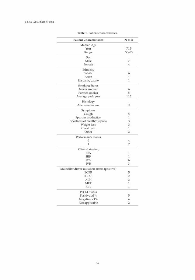
J. Clin. Med. 2020, 9, 1884
Table 1. Patient characteristics.
Patient Characteristics N = 11
Median AgeYear 70.5
Range 50–85
SexMale 7
Female 4
EthnicityWhite 6Asian 4
Hispanic/Latino 1
Smoking StatusNever smoker 6
Former smoker 5Average pack year 10.2
HistologyAdenocarcinoma 11
SymptomsCough 5
Sputum production 1Shortness of breath/dyspnea 3
Weight loss 3Chest pain 1
Other 2
Performance status0 41 7
Clinical stagingIIIA 1IIIB 1IVA 6IVB 3
Molecular driver mutation status (positive)EGFR 5KRAS 2ALK 2MET 1RET 1
PD-L1 StatusPositive ≥1% 5
Negative <1% 4Not applicable 2
36
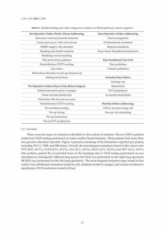
J. Clin. Med. 2020, 9, 1884
Table 2. Detailed timing and order categories to address in ERAS pathways cancer surgeries.
Pre-Operative Orders Weeks Ahead Addressing: Intra-Operative Orders Addressing:
Education visit and potential handouts Fluid management
Assess post-op n/v risks and educate IV/inhalational anesthetics
NSQIP surgery risk calculator Regional anesthesia
Smoking and alcohol cessation Deep Venus Thrombosis prophylaxis
Breathing exercise teaching
Diet and activity guidance Post-Anesthesia Care Unit:
Prehabilitation OT/PT teaching Pain guidelines
Lab orders Catheter guidelines
Medication education for pre-op and post-op
Bathing instructions Extended Stay Orders:
Nursing care
Pre-Operative Orders Days or Day Before Surgery: Medications
Patient instructions prior to surgery DVT prophylaxis
Nasal and skin disinfection As needed medications
Medication fills for post-op needs
Prehabilitation OT/PT teaching Post-Op Orders Addressing:
Pre-anesthesia testing Follow-up nurse triage call
Pre-op fasting Post-op visit scheduling
Pre-op medications
Pre-op DVT prophylaxis
3.2. Genomics
There were ten types of mutations identified in this cohort of patients. Eleven (100%) patientsunderwent NGS testing performed on tumor and/or liquid biopsies. Most patients had more thanone genomic alteration reported. Figure 1 presents a heatmap of the biomarkers reported per patient,including PD-L1, TMB, and MSI status. Overall, the most frequent mutations found in this cohort wereTP53 (8/11, 66.7%), EGFR (5/11, 45.5%), ALK (2/11, 18.2%), KRAS (2/11, 18.2%), and MET (2/11, 18.2%).One patient, patient #4, is included twice in the heatmap due to NGS testing performed on twosimultaneous, biologically different lung tumors (4A NGS was performed on the right lung specimen;4B NGS was performed on the left lung specimen). The most frequent mutation types found in thiscohort were substitution mutations (noted in red), deletions (noted in orange), and variant of unknownsignificance (VUS) mutations (noted in blue).
37
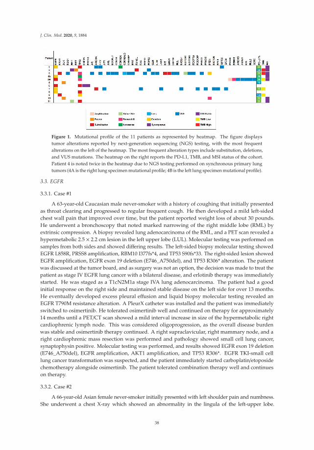
J. Clin. Med. 2020, 9, 1884
Figure 1. Mutational profile of the 11 patients as represented by heatmap. The figure displaystumor alterations reported by next-generation sequencing (NGS) testing, with the most frequentalterations on the left of the heatmap. The most frequent alteration types include substitution, deletions,and VUS mutations. The heatmap on the right reports the PD-L1, TMB, and MSI status of the cohort.Patient 4 is noted twice in the heatmap due to NGS testing performed on synchronous primary lungtumors (4A is the right lung specimen mutational profile; 4B is the left lung specimen mutational profile).
3.3. EGFR
3.3.1. Case #1
A 63-year-old Caucasian male never-smoker with a history of coughing that initially presentedas throat clearing and progressed to regular frequent cough. He then developed a mild left-sidedchest wall pain that improved over time, but the patient reported weight loss of about 30 pounds.He underwent a bronchoscopy that noted marked narrowing of the right middle lobe (RML) byextrinsic compression. A biopsy revealed lung adenocarcinoma of the RML, and a PET scan revealed ahypermetabolic 2.5 × 2.2 cm lesion in the left upper lobe (LUL). Molecular testing was performed onsamples from both sides and showed differing results. The left-sided biopsy molecular testing showedEGFR L858R, PRSS8 amplification, RBM10 I377fs*4, and TP53 S90fs*33. The right-sided lesion showedEGFR amplification, EGFR exon 19 deletion (E746_A750del), and TP53 R306* alteration. The patientwas discussed at the tumor board, and as surgery was not an option, the decision was made to treat thepatient as stage IV EGFR lung cancer with a bilateral disease, and erlotinib therapy was immediatelystarted. He was staged as a T1cN2M1a stage IVA lung adenocarcinoma. The patient had a goodinitial response on the right side and maintained stable disease on the left side for over 13 months.He eventually developed excess pleural effusion and liquid biopsy molecular testing revealed anEGFR T790M resistance alteration. A PleurX catheter was installed and the patient was immediatelyswitched to osimertinib. He tolerated osimertinib well and continued on therapy for approximately14 months until a PET/CT scan showed a mild interval increase in size of the hypermetabolic rightcardiophrenic lymph node. This was considered oligoprogression, as the overall disease burdenwas stable and osimertinib therapy continued. A right supraclavicular, right mammary node, and aright cardiophrenic mass resection was performed and pathology showed small cell lung cancer,synaptophysin positive. Molecular testing was performed, and results showed EGFR exon 19 deletion(E746_A750del), EGFR amplification, AKT1 amplification, and TP53 R306*. EGFR TKI-small celllung cancer transformation was suspected, and the patient immediately started carboplatin/etoposidechemotherapy alongside osimertinib. The patient tolerated combination therapy well and continueson therapy.
3.3.2. Case #2
A 66-year-old Asian female never-smoker initially presented with left shoulder pain and numbness.She underwent a chest X-ray which showed an abnormality in the lingula of the left-upper lobe.
38
J. Clin. Med. 2020, 9, 1884
A CT-guided biopsy was performed and revealed TTF-1 positivity, moderately differentiated pulmonaryadenocarcinoma with a predominant acinar pattern. Staging CT of the chest, abdomen, and pelvis displayeda 1.5 × 2.0 × 2.2 cm left upper lobe mass. A subsequent PET/CT demonstrated F-18 fluorodeoxyglucose(FDG) activity in the left upper lobe, but also in multiple mediastinal and left hilar lymph nodes includingstations 4R, 4L, 7, and 11R. These would be later confirmed for metastatic adenocarcinoma after sheunderwent an endobronchial ultrasound with fine needle biopsy. NGS sequencing of the station 7 lymphnode revealed EGFR exon 19 deletion (E746_A750del), CDKN2A G120*, and TP53 Y163C. PD-L1 22C3stained negative with 0% expression. The PET/CT also exhibited a 1.1 cm calcified isthmus thyroid glandnodule with an SUV of 4.9. This was also biopsied and revealed papillary carcinoma. She was referred tothe endocrine surgery department but has not had surgery considering her lung cancer diagnosis. An MRIof the brain showed no metastatic disease. She was staged as a T1cN3M0 stage IIIB lung adenocarcinoma.She initiated therapy on weekly chemoradiation therapy to the left lung, hilum, and mediastinumwith carboplatin and paclitaxel. She completed radiation with a few acute toxicities including chestdiscomfort, fatigue, and esophagitis, for which she received a 10-day course of dexamethasone. Due toher acute toxicities, she only received three weekly infusions of carboplatin and paclitaxel. She is plannedto continue systemic treatment by initiating adjuvant osimertinib, and while this is not yet approved,the decision was made based on preliminary trial data and the factor that the patient could not completechemotherapy due to toxicities.
3.3.3. Case #3
An 86-year-old male with no history of tobacco use initially presented to the emergencydepartment with a complaint of dry cough, shortness of breath, and concern of gradualweight loss. He developed a dry cough about two months prior, without any specific orpotential triggering cause. The persistent shortness of breath occurred over a few days, however,without limitations to his usual routines. Chest X-ray showed extensive consolidation and effusionon the right. Subsequent CT chest with IV contrast revealed severe right-sided pleural effusion,multiple nodular and patchy opacities in the right lung, with possible combination of atelectasis andedema. There were multiple sub-centimeter lesions throughout the lungs bilaterally, suspiciousfor pulmonary metastasis of more than 20 lesions. During his admission, he underwentthoracenteses that identified rare atypical cells suggestive of adenocarcinoma with lung primary(TTF-1 positive, Napsin A positive, mucicarmine cytochemistry negative). His disease progressedone month after admission. PET/CT confirmed diffuse FDG avid right pulmonary consolidationand bilateral pulmonary nodules with a max SUV of 11.6, FDG avid cervical and thoraciclymphadenopathy, and FDG avid osseous lesions. Brain MRI showed a motion-limited exam withoutfindings to suggest intracranial metastatic disease. Single gene NGS panels showed EGFR exon21 L858R mutation. Approximately 2.5 months after diagnosis, the patient had an initial consultationat COH community site. He was staged as a T2N3M1c stage IVB lung adenocarcinoma. He wastreatment-naïve prior to consultation. Osimertinib was immediately administered after liquid biopsyconfirmed EGFR exon 21 L858R and TP53 R273L. However, he experienced severe diarrhea 19 days aftertreatment start date. Osimertinib was held for 20 days and dosing was reduced to 40 mg daily. A fewweeks following treatment re-initiation, the patient was admitted to the hospital for worsening shortnessof breath associated with decreased output from his pleural catheter. Oxygen saturation ranged from88–92% on room air. He transitioned from supplemental oxygen to high flow oxygen-enriched FiO2.Empiric antibiotics were administered for hospital-acquired pneumonia. His respiratory distresscontinued to deteriorate with drops in oxygen saturation, requiring continuous BIPAP treatmentand nebulizer therapy. When he required BIPAP support, his family decided on comfort care.In combination with his comorbidities of atrial fibrillation, chronic respiratory failure with hypoxia,and bilateral DVT of lower extremities, the patient unfortunately passed away 23 weeks after diagnosisdue to post-obstructive pneumonia and respiratory complications.
39
J. Clin. Med. 2020, 9, 1884
3.3.4. Case #4
A 63-year-old male never-smoker with a history of well-controlled type II diabetes withoutretinopathy initially presented to his ophthalmologist with blurred vision over the left eye withintermittent, mild irritation ongoing for one month. He denied any focal neurological deficits.On examination, an amelanotic choroidal lesion with subretinal fluid on the left eye was prominentand appeared malignant. CT of chest revealed a large spiculated centrally necrotic right upper lobemass that extended to the suprahilar region measuring approximately 5.0 × 4.3 cm. There wereenlarged centrally necrotic conglomerated right lower paratracheal lymph nodes, measuring up to20 mm in maximum short axis. Subsequent CT of chest, abdomen, and pelvis demonstrated anindeterminate 1.6 cm left adrenal nodule, and an indeterminate 8 mm lucent lesion in the L4 spinousprocess, metastasis not excluded. The patient followed up with pulmonologist and his symptoms ofmild fatigue and recent weight loss were noted. Further workup involved endobronchial ultrasoundbronchoscopy of right upper lobe mass and lymph node stations. Pathology showed adenocarcinomaimmunohistochemically consistent with lung primary (TTF-1 positive, Napsin A positive, CK7 positive,CK20 negative, synaptophysin negative, chromogranin negative). PET/CT confirmed the large FDGavid right upper lobe pulmonary mass extending to the right suprahilar region measuring 7.7 SUV,ground glass and consolidative opacities involving right upper lobe, FDG right level 4 and mediastinallymph nodes, FDG avid osseous lesions involving the T1 vertebral body, right posterior iliac bone andleft acetabulum, and a 14 mm FDG avid left adrenal nodule. Brain MRI showed 4 mm enhancementin the left superior frontal gyrus, concerning for metastasis. He had his first initial consultationat COH community site approximately two months after his ophthalmology visit. He was stagedas a T3N2M1c stage IVB lung adenocarcinoma. Complete NGS testing demonstrated EGFR exon19 deletion (L747_A750 delinsP). No other driver mutations were found. Osimertinib was administeredsix weeks after pathologic diagnosis. Patient continues on medication and osimertinib is well tolerated.A recommendation to treat brain metastasis with radiation was withdrawn due to osimertinib’sCNS penetration.
3.3.5. Case #5
A 50-year-old Asian male never-smoker presented with a productive cough and clear sputumwith specks of blood approximately two years ago. His primary care doctor directed him to hisgastroenterologist with the belief that the cough was secondary to gastroesophageal reflux disease.A chest X-ray ordered by the gastroenterologist revealed right lower lobe consolidation. The patientunderwent a CT scan of the chest, abdomen, and pelvis a month later which showed a moderatemass-like consolidation and multifocal nodular opacities in the right lower lobe, possibly obscuring anunderlying mass. Additionally, there were bilateral small 0.4 cm lung nodules, a 0.7 cm AP windowlymph node, a 1.3 cm subcarinal lymph node, and a 2.2 cm right infra-hilar lymph node. The patientunderwent further workup in hospital. A sputum test ruled out tuberculosis and a CT-guided lungbiopsy was negative for malignancy. The patient then underwent a transbronchial lung bronchoscopywhich also did not reveal malignancy. However, the bronchial brushing and washing demonstratedmalignant cells consistent with adenocarcinoma with weak TTF-1 positive staining. Two monthslater, the patient endorsed chest wall pain and shortness of breath after long periods of coughing.An MRI scan of the brain demonstrated no evidence of brain metastasis. A PET/CT scan performedrevealed strong FDG uptake in mediastinal lymph nodes, small right supraclavicular lymph nodes,and hypermetabolic pulmonary nodules bilaterally. He was staged as a TXN3M1a stage IVA lungadenocarcinoma. Molecular testing was performed and showed EGFR exon 19 deletion (E746_A750del)and TP53 286K mutation. Osimertinib was administered with good tolerance. Figure 2A–C shows therepresentative FFTs developed from the EGFR cases above.
40
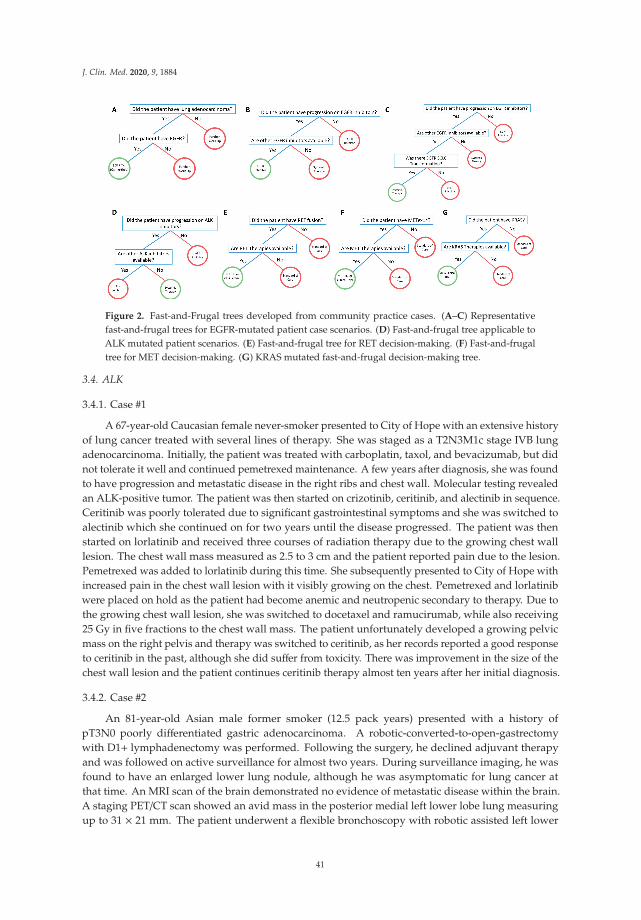
J. Clin. Med. 2020, 9, 1884
Figure 2. Fast-and-Frugal trees developed from community practice cases. (A–C) Representativefast-and-frugal trees for EGFR-mutated patient case scenarios. (D) Fast-and-frugal tree applicable toALK mutated patient scenarios. (E) Fast-and-frugal tree for RET decision-making. (F) Fast-and-frugaltree for MET decision-making. (G) KRAS mutated fast-and-frugal decision-making tree.
3.4. ALK
3.4.1. Case #1
A 67-year-old Caucasian female never-smoker presented to City of Hope with an extensive historyof lung cancer treated with several lines of therapy. She was staged as a T2N3M1c stage IVB lungadenocarcinoma. Initially, the patient was treated with carboplatin, taxol, and bevacizumab, but didnot tolerate it well and continued pemetrexed maintenance. A few years after diagnosis, she was foundto have progression and metastatic disease in the right ribs and chest wall. Molecular testing revealedan ALK-positive tumor. The patient was then started on crizotinib, ceritinib, and alectinib in sequence.Ceritinib was poorly tolerated due to significant gastrointestinal symptoms and she was switched toalectinib which she continued on for two years until the disease progressed. The patient was thenstarted on lorlatinib and received three courses of radiation therapy due to the growing chest walllesion. The chest wall mass measured as 2.5 to 3 cm and the patient reported pain due to the lesion.Pemetrexed was added to lorlatinib during this time. She subsequently presented to City of Hope withincreased pain in the chest wall lesion with it visibly growing on the chest. Pemetrexed and lorlatinibwere placed on hold as the patient had become anemic and neutropenic secondary to therapy. Due tothe growing chest wall lesion, she was switched to docetaxel and ramucirumab, while also receiving25 Gy in five fractions to the chest wall mass. The patient unfortunately developed a growing pelvicmass on the right pelvis and therapy was switched to ceritinib, as her records reported a good responseto ceritinib in the past, although she did suffer from toxicity. There was improvement in the size of thechest wall lesion and the patient continues ceritinib therapy almost ten years after her initial diagnosis.
3.4.2. Case #2
An 81-year-old Asian male former smoker (12.5 pack years) presented with a history ofpT3N0 poorly differentiated gastric adenocarcinoma. A robotic-converted-to-open-gastrectomywith D1+ lymphadenectomy was performed. Following the surgery, he declined adjuvant therapyand was followed on active surveillance for almost two years. During surveillance imaging, he wasfound to have an enlarged lower lung nodule, although he was asymptomatic for lung cancer atthat time. An MRI scan of the brain demonstrated no evidence of metastatic disease within the brain.A staging PET/CT scan showed an avid mass in the posterior medial left lower lobe lung measuringup to 31 × 21 mm. The patient underwent a flexible bronchoscopy with robotic assisted left lower
41

J. Clin. Med. 2020, 9, 1884
lobectomy and mediastinal lymph node dissection. The results revealed adenocarcinoma of lowerlobe of the left lung measuring approximately 3.5 × 3 × 2.5 cm in size, with one lymph nodeinvolved. The initial immunohistochemistry performed on the specimen was positive for CK7, TTF-1,and Napsin A. Following surgical resection, the patient endorsed symptoms of productive cough withclear sputum, fatigue, poor appetite, and weight loss. Molecular testing was performed and revealedEML4-ALK fusion, PTEN R74Tfs, and SMARCA4 D696Vfs*3. He was staged as a T2aN2M0 stageIIIA lung adenocarcinoma. The patient was planned to initiate alectinib, but has not yet begun due tofear of side effects (Figure 2D). While adjuvant alectinib is not yet officially approved, the patient wasdeemed a poor candidate for chemotherapy due to his frailty and did not wish to have chemotherapy.Thus, based on the ALK mutation, alectinib was recommended due to the early trial results.
3.5. RET
Case #1
An 80-year-old Caucasian male with a light smoking history (<1 pack years) developed a cough,and a chest X-ray was performed that showed a persistent right upper lobe (RUL) airspace opacityconsistent with pneumonia. A week later, a chest CT showed a 2.5 × 3.2 × 5 cm right upper lobeconsolidation extending from the right suprahilar region to the right lung apex, and a narrowing of theright mainstem bronchus. There were also multiple right and left sub-centimeter nodules, and lesionswithin the superior aspect of the T4 and T9 spine. He underwent a CT-guided biopsy of the RULwhich was consistent with moderate to poorly differentiated lung adenocarcinoma staining positive forTTF-1 and Napsin A and negative for p63. Partial molecular testing was negative for EGFR mutationsor ALK rearrangements but stained positive for PD-L1 at 50%. Further work-up with a diagnosticPET/CT also showed an FDG-avid hepatic lesion. A brain MRI was unremarkable, with no evidence ofmetastatic disease. A CT-guided biopsy of the T9 lesion confirmed metastatic lung adenocarcinomaand was used for complete molecular profiling. This test revealed a RET-KIF5B fusion, TMB-highwith 11 Muts/Mb, and FANCL exon 12 c.1020 + 2T > C detected. He was staged as a T3N2M1cstage IVB lung adenocarcinoma. The patient started first line systemic therapy with a combination ofcarboplatin, pemetrexed, and pembrolizumab and received four cycles before starting maintenancepemetrexed/pembrolizumab. He received nine maintenance cycles before developing diarrhea. A CTof the chest, abdomen, and pelvis showed rectosigmoid and proximal transverse colitis for which hewas treated with prednisone, and maintenance therapy was discontinued. He remained off therapy forsix months before a CT of the chest, abdomen, and pelvis showed new low-density hepatic lesions andpathologic compression fractures at T4 and T9, consistent with progressive disease. He was screenedand consented for a clinical trial for the expanded access of selpercatinib (LOXO-292) for RET fusions;however, he needed to control his hypertension prior to trial initiation. After multiple adjustmentsof his hypertensive medications by his cardiologist, he was able to start treatment with selpercatinib(Figure 2E).
3.6. MET
Case #1
A 70-year-old female never-smoker with a past medical history of arthritis presented to an outsideinstitution with shortness of breath and a cough. CT abdomen was performed and showed a small rightpleural effusion and no evidence of an acute intra-abdominopelvic process. The patient underwent aPET/CT scan which demonstrated a medial right apical hypermetabolic pulmonary mass measuringapproximately 3.1 x 2.7 cm (SUV max 12.5), with extension into the adjacent pleura, multiple additionalhypermetabolic pleural-based soft tissue masses within the right hemithorax, interval mild to moderateincrease in right-sided pleural effusion, and no evidence of distant metastatic disease. A CT-guidedbiopsy was performed on the right upper lobe mass and revealed lung adenocarcinoma. Both NGS
42
J. Clin. Med. 2020, 9, 1884
tests demonstrated MET exon 14 splice site mutation (2888-42_2892del47) (MAF: 1.1%), TP53 G334Vmissense mutation (MAF: 1.5%), MSI Stable, TMB-low (9 Muts/Mb), and PD-L1 0%. She underwentan MRI of her lumbar and thoracic spine that showed a loculated pleural effusion with enhancingpleural lesions compatible with metastatic disease, lytic changes to the medial right 12th rib compatiblewith osseous metastatic disease, disk bulging was incidentally noted at T8, and a nonspecific 0.6 cmlesion at L1. Brain MRI at the time was negative for metastatic disease. She was staged as a T3N2M1cstage IVB lung adenocarcinoma. The patient was subsequently enrolled in a clinical trial investigatingMET inhibitor capmatinib 400 BID and continued with symptomatic improvement with the firstrestaging scan showing response. The second interval scan showed progression with a slight increasein the right apical pleural-based mass, multiple right basilar pleural-based lesions, and a loculatedright pleural effusion. The patient underwent liquid NGS testing that revealed MET exon 14 splicesite (2888-42_2892del47) (MAF: 0.63% deceased from 1.1%) and TP53 G334V (MAF: 0.78% decreasedfrom 1.5%). Progression of disease was confirmed on the third interval scan and the patient was takenoff trial. She was subsequently started on carboplatin and nab-paclitaxel. Unfortunately, a restagingscan after two cycles showed interval progression of disease in the pleural-based mass, lymph nodes,and pleura. The patient presented to City of Hope for treatment options and was started on a clinicaltrial examining a bispecific monoclonal antibody targeting MET. She is status/post three cycles of thedrug and is exhibiting stable disease thus far (Figure 2F).
3.7. KRAS
3.7.1. Case #1
A 68-year-old Caucasian female former smoker (20 pack years) presented with a cough. An initialchest CT showed a 5.4 × 5.7 cm left upper lobe lesion with numerous bilateral sub-centimeter nodules.CT-guided core needle biopsy of the left upper lobe mass revealed moderately differentiatedlung adenocarcinoma. Molecular profiling of the biopsy was negative for EGFR mutations,ALK rearrangements, ROS1 rearrangements, and PD-L1. Subsequent NGS of the biopsy revealedKRAS G12A, CDKN2A/B loss, LMO1 R34H, NOTCH1 splice site 2467+ 1G>TA, and TMB-intermediatewith 13 Muts/Mb. A brain MRI showed a 2 mm lesion in the right prefrontal gyrus adjacent to the centralsulcus of unclear etiology. In repeat brain MRIs, this lesion had resolved and was no longer present.To finish staging, a diagnostic PET/CT showed a 6 cm mass FDG avid with an SUV of 9.6 and multiplebilateral FDG avid pulmonary nodules. She was staged as a T3NXM1a stage IVA lung adenocarcinoma.She initiated first-line systemic therapy with cisplatin and pemetrexed for three cycles with verylittle response. Concurrent radiation to the left upper lobe primary and mediastinum was addedto regimen with no significant toxicities. With no other signs of metastatic disease, she underwenta wedge resection and one of the pulmonary nodules showed metastatic disease. Thus, the rest ofthe procedure was aborted. She received second-line atezolizumab without disease progression for17 months. A new CT-guided biopsy of the right lower lobe lung was performed and was confirmedas metastatic lung adenocarcinoma. NGS molecular profiling revealed KRAS G12A, KRAS L19F,KIT A829S, PDGFRA T365N, PTEN deletion, and TMB-high with 11.4 Muts/Mb. She started third-linesystemic therapy with carboplatin, paclitaxel, bevacizumab, and pembrolizumab for a total of fivecycles. She is currently status/post seven cycles of maintenance bevacizumab and pembrolizumabwithout signs of disease progression.
3.7.2. Case #2
An 80-year-old Caucasian male never-smoker had a history of hairy cell leukemia that wastreated approximately 20 years ago with chemotherapy (cladribine) for eight days and continuesto follow up regularly. The patient was diagnosed with early stage lung cancer two years ago,following a biopsy of a lung nodule in the right lower lobe. He had a platinum coil as fiducialmarker placed at that time. A year later, the patient underwent radiation therapy for a malignant
43
J. Clin. Med. 2020, 9, 1884
neoplasm of the lower lobe right bronchus. Unfortunately, his disease progressed, and the patientwas deemed non-operable due to medical comorbidities including diabetes mellitus, a history ofcoronary artery disease, and a history of hairy cell leukemia. A PET/CT scan performed ten monthsafter radiation revealed a hypermetabolic right lower lobe pulmonary mass with central metallicfiducial marker unchanged in size and metabolic activity compared to a prior study with increasedright pleural effusion. A thoracentesis performed shortly after demonstrated malignant cells seen assingle cells, comprising 1–2% cellularity. Following the thoracentesis procedure, the patient reportedimprovement to his shortness of breath. Additionally, he reported neuropathy secondary to diabetesmellitus onset eight years ago, which is controlled, and he continues to follow up with endocrinology.He was staged as a T2NXM1a stage IVA lung adenocarcinoma. Molecular testing was performed, andresults revealed BRAF V600E, NRAS G12D, KRAS G12S, PDL1 100%, meanwhile the BRAF V600E IHCwas negative. The patient was subsequently initiated on pembrolizumab. One week after his first cycle,he was admitted to hospital for pneumonitis, chest pain, onset fever, transaminitis, and elevated bloodpressure without previous diagnosis of HTN, thrombocytopenia, and nephrolithiasis. He was treatedwith IV Rocephin, oral oxycline, pain control, and given a pulmonary consultation. The patient wasdischarged with antibiotics. The patient continues on pembrolizumab with continued symptoms offatigue and decreased appetite (Figure 2G).
4. Discussion
Between 2013–2017, there were 68,369 total cases of invasive cancers and 6,541 cases of lung cancerin Orange County, according to the California Cancer Registry. Age-adjusted invasive cancer incidencedemonstrates that Orange County accounted for higher cancer rates compared to Los Angeles County(394.93 vs. 371.36 per 100,000 cases, respectively). Similarly, age-adjusted lung cancer incidencerate in Orange County is higher than in Los Angeles county (38.29 vs. 35.88 per 100,000 cases,respectively) [38]. Located in Orange County, Newport Beach is the newest community site in the Cityof Hope network. Demographic information was assessed for new patients in the first four monthsof operation, beginning in January 2020. Only initial new patient visits were accounted for, therebyexcluding subsequent follow-up visits from the count. We identified 182 new patients, 118 (64.8%) ofwhom were female and 64 (35.2%) of whom were male. The median age of the patients was 63 years,ranging from 22 to 90 years. The most prevalent ethnicities were Caucasian (n = 96, 52.7%) and Asian(n = 16, 8.8%). Among the 182 patients, 28 were diagnosed with cancer in the lung or bronchus, 11 ofwhom were presented in this report.
Fast-and-frugal trees (FFTs) are an effective way to make binary classification decisions under timepressure conditions with only limited information [36,39]. One example to illustrate this is the decisionof whether to send a patient to coronary care or a regular hospital bed if a patient reports chest pain [40].FFTs can be used to evaluate standard guidelines and establish new decision-making heuristic strategies,allowing physicians to make fast and accurate decisions without relying on statistical training orcalculation devices. An FFT is a decision tree composed of sequentially ordered cues such asdiagnostic test results or clinical information and the accompanying binary decision (yes/no) of thosecues [36,41–43]. The relationship between the cues can be framed as “if-then” statements; for instance,if a patient has severe chest pain, then the physician should perform tests for myocardial infraction.If the condition of the cues is met, then the decision can be made, and the FFT exited. If the condition isnot met, the FFT sequentially considers other cues, until a cue’s exit condition is met. The last cue of anFFT has two exits to ensure that a decision is ultimately made [36,41–44]. Formally, an FFT is definedas a decision tree that has m + 1 exits, with one exit for each of the first m-1 cues, and two exits forthe last cue [36,41–44]. The FFT tool is robust, and it can be used to both evaluate established clinicalpathways and guidelines, but also to develop new FFT-based decision-making trees that incorporateadditional clinical data like molecular testing results. The identification of driver mutations for NSCLChas transformed patient care, such that patients who are properly screened with molecular testing
44
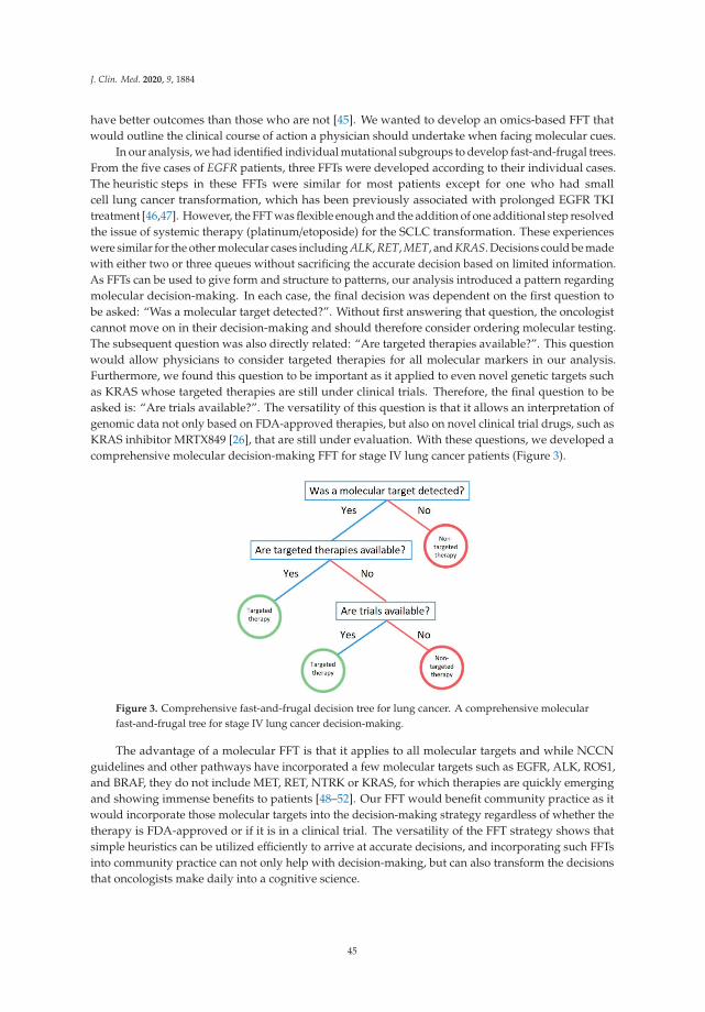
J. Clin. Med. 2020, 9, 1884
have better outcomes than those who are not [45]. We wanted to develop an omics-based FFT thatwould outline the clinical course of action a physician should undertake when facing molecular cues.
In our analysis, we had identified individual mutational subgroups to develop fast-and-frugal trees.From the five cases of EGFR patients, three FFTs were developed according to their individual cases.The heuristic steps in these FFTs were similar for most patients except for one who had smallcell lung cancer transformation, which has been previously associated with prolonged EGFR TKItreatment [46,47]. However, the FFT was flexible enough and the addition of one additional step resolvedthe issue of systemic therapy (platinum/etoposide) for the SCLC transformation. These experienceswere similar for the other molecular cases including ALK, RET, MET, and KRAS. Decisions could be madewith either two or three queues without sacrificing the accurate decision based on limited information.As FFTs can be used to give form and structure to patterns, our analysis introduced a pattern regardingmolecular decision-making. In each case, the final decision was dependent on the first question tobe asked: “Was a molecular target detected?”. Without first answering that question, the oncologistcannot move on in their decision-making and should therefore consider ordering molecular testing.The subsequent question was also directly related: “Are targeted therapies available?”. This questionwould allow physicians to consider targeted therapies for all molecular markers in our analysis.Furthermore, we found this question to be important as it applied to even novel genetic targets suchas KRAS whose targeted therapies are still under clinical trials. Therefore, the final question to beasked is: “Are trials available?”. The versatility of this question is that it allows an interpretation ofgenomic data not only based on FDA-approved therapies, but also on novel clinical trial drugs, such asKRAS inhibitor MRTX849 [26], that are still under evaluation. With these questions, we developed acomprehensive molecular decision-making FFT for stage IV lung cancer patients (Figure 3).
Figure 3. Comprehensive fast-and-frugal decision tree for lung cancer. A comprehensive molecularfast-and-frugal tree for stage IV lung cancer decision-making.
The advantage of a molecular FFT is that it applies to all molecular targets and while NCCNguidelines and other pathways have incorporated a few molecular targets such as EGFR, ALK, ROS1,and BRAF, they do not include MET, RET, NTRK or KRAS, for which therapies are quickly emergingand showing immense benefits to patients [48–52]. Our FFT would benefit community practice as itwould incorporate those molecular targets into the decision-making strategy regardless of whether thetherapy is FDA-approved or if it is in a clinical trial. The versatility of the FFT strategy shows thatsimple heuristics can be utilized efficiently to arrive at accurate decisions, and incorporating such FFTsinto community practice can not only help with decision-making, but can also transform the decisionsthat oncologists make daily into a cognitive science.
45
J. Clin. Med. 2020, 9, 1884
4.1. EGFR
EGFR is a transmembrane protein that exists as a monomer on the surface of a cell. EGFR isa receptor tyrosine kinase, therefore it dimerizes with its specific ligand to activate the tyrosinekinase (TK) [53]. Once activated, the EGFR TK activates pathways which synthesize DNA and causecell proliferation [54]. Thus, mutations or amplification of EGFR result in tumor growth/cancer.Predominant mutation locations include exon 19 deletions and exon 21 L858R point mutations [55].EGFR mutations also occur more commonly in nonsmokers, women, and in people of Asian descent.A study demonstrated that EGFR mutations occurred in 42% of females compared to 14% of malesand in 51% of nonsmokers compared to 10% of smokers [56]. EGFR TKIs have been heavily exploredand single agent TKIs such as erlotinib, gefitinib (first generation), and afatinib (second generation)have become standard of care for EGFR mutant NSCLC patients. Osimertinib (a third generation TKI)emerged as a first-line treatment when it demonstrated improved outcomes when compared tooutcomes with erlotinib and gefitinib in the FLAURA trial [57]. The FLAURA trial was conducted with556 NSCLC patients with EGFR exon deletion or exon 21 L858R point mutation. Osimertinib showedimproved PFS (18.9 vs. 10.2 months) and OS (83% vs. 71% at 18 months) when compared to standardof care treatment with gefitinib or erlotinib [58]. Dacomitinib, a second-generation single agentEGFR TKI, was approved by the FDA in 2018 as a first-line treatment for metastatic NSCLC based onthe ARCHER 1050 trial [59]. The ARCHER 1050 trial compared dacomitinib to gefitinib in 452 patientswith metastatic NSCLC and demonstrated a survival benefit in PFS (14.7 vs. 9.2 months) and OS(34.1 vs. 26.8 months) [60]. Several studies have demonstrated that patients who initially respond wellto an EGFR TKI develop resistance, and subsequently their disease progresses [61–63]. Secondary EGFRmutations and MET amplifications are the most well-known causes of resistance [64,65]. Osimertinibselectively inhibits both EGFR-TKI-sensitizing and EGFR T790M resistance mutations, making ita valuable treatment against TKI-acquired resistance [12]. Alternative treatment options currentlyexplored are combination therapies such as EGFR inhibitors along with bevacizumab or ramucirumab,vascular endothelial growth factor receptor inhibitors, EGFR inhibitors with chemotherapy, and EGFRinhibitors with other targeted therapy [66–68].
4.2. ALK
Initially identified in 2007, the formation of echinoderm microtubule-associated protein like-4(EML4)-ALK fusion protein in NSCLC leads to constitutive oncogenic signaling. Patients withNSCLC harboring ALK translocations were estimated to be in approximately 6% of allNSCLC cases. Four years later, crizotinib became the first oral, ALK TKI approved by the FDA [69].As a first-line treatment, crizotinib was shown to be safe and effective in a single arm, open-label, phaseIII clinical trial [70,71]. Among the patients who received prior systemic therapy, crizotinib showedan ORR of 50% and an improved quality of life compared to those on chemotherapy. PFS wasimproved for patients on crizotinib compared to standard of care chemotherapy by 3.9 months(10.9 vs. 7.0 months, respectively). However, the study showed that there was no difference in OS(approximately 12 months). Subsequently, other targeted therapies were developed as resistanceeventually developed in advanced ALK-positive NSCLC patients. Acquired resistance mechanismssuch as the amino acid substitution L1196M in the ALK gene against crizotinib have been explored [72].Acting as a gatekeeper mutation, ALK L1196M impedes crizotinib’s efficacy in ALK-positive NSCLCpatients. A hypothesis of resistance involves decreased binding energy within an unstable secondarystructure of the mutant ALK complex [73]. The equivalence of this mutation is the EGFR mutationT790M that confers resistance to erlotinib or gefitinib [74]. Development of second-generation ALKinhibitors including ceritinib, alectinib, and brigatinib, were sequentially approved after crizotinibin 2011. Barrows et al. outlined observational studies of sequential treatment with several ALKinhibitors [75]. Effects from consecutive treatment with ALK inhibitors may be additive in termsof survival outcomes in patients. In order to overcome ALK resistance, the discovery of new ALKinhibitors is pivotal and essential in the clinical setting.
46
J. Clin. Med. 2020, 9, 1884
4.3. RET
Activation of the RET gene leads to cell proliferation, migration, and differentiation, and canbe found on chromosome 10q11.2 [76,77]. The proto-oncogene RET interacts with multipledownstream pathways including JAK/STAT, MAPK/ERK, and PI3K/AKT [78]. In 1985, the RETrearrangement was seen for the first time when lymphoma DNA was transfected in NIH-3T3cells [79]. Mutations and rearrangements in RET are most commonly observed in papillary thyroidcarcinomas and in lung cancer [80]. RET fusions lead to tumorigenesis by continuous activation vialigand-independent dimerization and autophosphorylation of the RET kinase and its downstreampartners [81]. RET most frequently fuses with KIF5B in NSCLC but other fusion gene partners havebeen observed in CCDC6, NCOA4, and TRIM33 [82]. In NSCLC, RET rearrangement is detectedin 1–2% of cases, typically adenocarcinoma histology, and without a history of smoking [83,84].Several multi-kinase TKIs have been utilized in targeting RET rearrangements in NSCLC. In a phaseII study, cabozantinib, targeting VEGFR, MET, and RET, was shown to have an ORR of 28%, a medianPFS of 5.5 months, and a median OS of 9.9 months in 26 RET fusion-positive NSCLC patients [20].A phase II study of 17 RET fusion-positive NSCLC patients previously treated with chemotherapyreceived vandetanib, targeting VEGFR 2 and 3, EGFR, and RET, and the trial demonstrated a 53% ORR,a median PFS of 4.7 months, and a median OS of 11.1 months [85]. A new generation of novel RETinhibitors are currently being investigated such as selpercatinib (LOXO-292) and pralsetinib (BLU-667).The phase I/II LIBRETTO-001 trial investigating selpercatinib RET-rearranged NSCLC patients withprevious therapy found an ORR of 68% [86,87]. Selpercatinib was recently awarded FDA approval foradult patients with RET-positive NSCLC based on the previous trial with an ORR of 85% in treatmentof naïve patients [88]. In a phase I study of pralsetinib, an ORR of 58% was demonstrated for allpatients regardless of RET fusion type and was effective against intracranial metastases [89].
4.4. MET
MET proto-oncogene encodes for c-Met, a member of the receptor tyrosine kinase (RTK) family.Once activated, c-MET is involved in various signaling cascades linked to cell growth, apoptosis,motility, and invasion. Mutations in MET represent 6% of all NSCLC cases with frequent alterationsincluding MET exon 14 skipping mutation and MET amplification [90]. Since its discovery in lungcancer and other solid tumor malignancies, research has been investigating potential targeting ofMET through small molecule inhibitors or monoclonal antibodies. Initially, a phase II clinical trialenrolled recurrent NSCLC, which were randomized to receive onartuzumab with erlotinib or placebowith erlotinib [91]. In NSCLC patients with MET-mutated disease, treatment with onartuzumab pluserlotinib demonstrated improved PFS and OS, although the drug combination failed in a subsequentphase III trial [92]. A landmark study of 4,622 NSCLC patients with ALK rearrangement or METamplification, of which 16 exhibited both alterations. The ORR was 86.7% in that subset of patients,suggesting a role of MET TKI treatment in MET-expressing tumors [93]. METROS, a phase II clinical trial,investigated the use of crizotinib—a multikinase inhibitor against ALK, ROS1, and MET—in MET orROS1-positive pretreated, advanced NSCLC [94]. The trial confirmed anti-tumor activity with an ORRof 27%, a median PFS of 4.4 months, and an OS of 5.4 months. Another recent trial of 69 patients withMET exon 14 skipping mutated-NSCLC treated with crizotinib demonstrated encouraging data withan ORR of 32%, a median PFS of 7.3 months, and a median OS of 20.5 months [95]. Currently, there areseveral promising trials examining the therapeutic efficacy of other MET TKIs, such as cabozantinib(NCT03911193), cabozantinib (NCT02750215), REGN5093 (NCT04077099), tepotinib monotherapy(NCT02864992) plus osimertinib (NCT03940703), and telisotuzumab vedotin (NCT03539536). Of note,capmantinib was recently approved by the FDA for treatment of MET exon 14 skipping mutated-NSCLCdue to results from the GEOMETRY mono-1 clinical trial [96].
47

J. Clin. Med. 2020, 9, 1884
4.5. KRAS
With an incidence rate of up to 22% in solid tumor malignancies, KRAS is one of the most prevalentoncogenic driver mutations in cancer [97]. In 1973, a murine sarcoma virus transformed mammaliangenes into an oncogene in KRAS, and nearly a decade later, a human lung carcinoma cell line firstshowed human sequences analogous to the KRAS oncogenes in mice [98,99]. KRAS mutations inpatients with advanced NSCLC are shown to have a poorer prognosis and often lack a response tostandard therapies [100]. The KRAS oncoprotein triggers tumor cell growth and survival because itacts as a crucial mediator of intrasellar signaling pathways [101,102]. KRAS activating point mutationsgenerally cause a loss of GTPase activity, and consequentially allow for the continuous activationof downstream signaling pathways such as MAPK and PI3K [103]. KRAS mutations have mostrecently been seen in exon 12 and 13 with the most common being G12C at 39%, G12V between18–21%, and G12D between 17–18% [104]. In patients with lung adenocarcinoma, approximately 13%harbor a mutation of KRAS G12C [105]. KRAS mutant lung adenocarcinoma patients with concurrentinactivating mutations in the tumor suppressor genes of KEAP1 and STK11 have demonstrated lowerclinical response rates treated with standard chemotherapy or immunotherapies [30,106]. KRAS G12Csmall molecule inhibitor, AMG-510, is the first in-human, phase I investigation in patients withadvanced KRAS G12C solid tumors (NCT03600883). Taken orally, once daily, AMG-510 is a specificand irreversible inhibitor of KRAS G12C by fixing the guanosine diphosphate-bound state of theprotein at an His95 groove in the P2 pocket of KRAS. Overall, the treatment was well tolerated.Twenty-two patients with results were presented and six of these patients had NSCLC. The medianduration of treatment was 9.7 weeks. Two patients experienced a partial response and two had stabledisease after six weeks [27,107]. Several other inhibitors including MRTX849 (NCT03785249) that targetKRAS G12C and other KRAS mutation variants are currently being investigated in clinical trials.
4.6. Future Directions
The advantage of FFTs is that they are quick and accurate decisions based on limited information.This is useful in the world of oncology as unlike guidelines and pathways, FFTs do not restrictphysicians to “on-pathway” and “off-pathway” decisions. FFTs incorporate physician’s “instinct”and “personal expertise” to arrive at a quick decision most beneficial to the patient. While this studywas limited by sample size and a relatively focused cohort to one community oncology practice,the lesson of a standardized FFT frameworks have been shown to be applicable to not only stage IVlung cancer, but also to the early stage setting in the future. This study was limited to patients from onecommunity practice, with a majority of patients demonstrating actionable mutations which does notcompletely represent the comprehensive population in community practice. Evaluating a larger cohortwould be needed to further understand other treatment paradigms in NSCLC. However, in the future,it will be important to standardize the utilization of these FFTs to a usable application that physicianscan refer to when reviewing their cases. This application would be most beneficial to community sites,as it would be a direct translation of the academic model to the community and would guide physicianstowards the accurate decision without sacrificing their expertise. At the same time, we understand thatthe complexity of oncology practice varies from the cancer and the stage of the disease. Therefore, it willbe important in certain situations to apply not only FFT heuristics, but also to incorporate multi-layereddecisions of convolutional neural networks [108] and the quick capabilities of fast-and-frugal decisiontrees [33,35] into a hybrid model, conditional neural network. This would allow for the model to utilizethe efficiency of fast-and-frugal decision trees and the accuracy of convolutional neural networks in aunified model.
48

J. Clin. Med. 2020, 9, 1884
Author Contributions: Conceptualization, R.S., I.M., T.T., D.P.P., H.C., A.B., J.F., R.P., H.R., T.W., R.B., L.B., P.K.,M.C., B.D., S.K.P.; Methodology, I.M., T.T., A.S., A.B., J.F., R.P., H.R., P.K., M.C., B.D., S.K.P.; Validation, R.S., I.M.,T.T., J.F., R.P., H.R., T.W., L.B., M.C., S.K.P.; Investigation, R.S., I.M., T.T., A.S., D.P.P., H.C., A.B., J.F., R.P., R.B., P.K.,B.D., S.K.P.; Formal Analysis, R.S., I.M., A.S., D.P.P., A.B., J.F., R.P., H.R., B.D.; Data Curation, R.S., I.M., T.T., A.S.,A.B., J.F., R.P., H.R., B.D., S.K.P.; Funding acquisition, R.S.; Project administration, R.S.; Resources, R.S., T.T., H.C.;Software, I.M.; Supervision, R.S., D.P.P.; Visualization, I.M., T.T., H.C., R.P.; Writing—Original Draft Preparation;R.S., I.M.; Writing—Review and Editing, R.S., I.M., T.T., A.S., D.P.P., H.C., A.B., J.F., R.P., H.R., T.W., R.B., L.B., P.K.,M.C., B.D., S.K.P. All authors have read and agreed to the published version of the manuscript.
Funding: This research received no external funding.
Acknowledgments: We would like to thank Newport Beach City of Hope staff for their skill and dedication inhelping the patients. We would also like to express our deepest gratitude for the philanthropic funding by theTenenblatt Family. The work was supported by the National Cancer Institute of the National Institutes of Healthunder award numbers P30CA033572, U54CA209978, R01CA247471, and R01CA218545.
Conflicts of Interest: The authors declare no conflict of interest.
References
1. Jemal, A.; Siegel, R.; Ward, E.; Murray, T.; Xu, J.; Smigal, C.; Thun, M.J. Cancer statistics, 2006. CA CancerJ. Clin. 2006, 56, 106–130. [CrossRef] [PubMed]
2. Siegel, R.L.; Miller, K.D.; Jemal, A. Cancer statistics, 2020. CA Cancer J. Clin. 2020, 70, 7–30. [CrossRef][PubMed]
3. Salgia, R. Mutation testing for directing upfront targeted therapy and post-progression combination therapystrategies in lung adenocarcinoma. Expert Rev. Mol. Diagn. 2016, 16, 737–749. [CrossRef] [PubMed]
4. Hensing, T.; Chawla, A.; Batra, R.; Salgia, R. A Personalized Treatment for Lung Cancer: Molecular Pathways,Targeted Therapies, and Genomic Characterization. Adv. Exp. Med. Biol. 2014, 799, 85–117. [CrossRef]
5. Mok, T.S.; Wu, Y.-L.; Thongprasert, S.; Yang, J.C.-H.; Chu, D.-T.; Saijo, N.; Sunpaweravong, P.; Han, B.;Margono, B.; Ichinose, Y.; et al. Gefitinib or Carboplatin–Paclitaxel in Pulmonary Adenocarcinoma. N. Engl.J. Med. 2009, 361, 947–957. [CrossRef]
6. Planchard, D.; Besse, B.; Groen, H.J.M.; Souquet, P.-J.; Quoix, E.; Baik, C.S.; Barlesi, F.; Kim, T.M.; Mazieres, J.;Novello, S.; et al. Dabrafenib plus trametinib in patients with previously treated BRAF(V600E)-mutantmetastatic non-small cell lung cancer: An open-label, phase 2 trial. Lancet Oncol. 2017, 18, 1307–1316.[CrossRef]
7. Soria, J.-C.; Wu, Y.-L.; Nakagawa, K.; Kim, S.-W.; Yang, J.-J.; Ahn, M.-J.; Wang, J.; Yang, J.C.-H.; Lu, Y.;Atagi, S.; et al. Gefitinib plus chemotherapy versus placebo plus chemotherapy in EGFR-mutation-positivenon-small-cell lung cancer after progression on first-line gefitinib (IMPRESS): A phase 3 randomised trial.Lancet Oncol. 2015, 16, 990–998. [CrossRef]
8. Rosell, R.; Carcereny, E.; Gervais, R.; Vergnenègre, A.; Massuti, B.; Felip, E.; Palmero, R.; Garcia-Gomez, R.;Pallares, C.; Sanchez, J.M.; et al. Erlotinib versus standard chemotherapy as first-line treatment for Europeanpatients with advanced EGFR mutation-positive non-small-cell lung cancer (EURTAC): A multicentre,open-label, randomised phase 3 trial. Lancet Oncol. 2012, 13, 239–246. [CrossRef]
9. Sequist, L.V.; Yang, J.C.-H.; Yamamoto, N.; O’Byrne, K.; Hirsh, V.; Mok, T.S.; Geater, S.L.; Orlov, S.V.;Tsai, C.-M.; Boyer, M.; et al. Phase III Study of Afatinib or Cisplatin Plus Pemetrexed in Patients withMetastatic Lung Adenocarcinoma with EGFR Mutations. J. Clin. Oncol. 2013, 31, 3327–3334. [CrossRef]
10. Le Tourneau, C.; Delord, J.-P.; Gonçalves, A.; Gavoille, C.; Dubot, C.; Isambert, N.; Campone, M.; Tredan, O.;Massiani, M.-A.; Mauborgne, C.; et al. Molecularly targeted therapy based on tumour molecular profilingversus conventional therapy for advanced cancer (SHIVA): A multicentre, open-label, proof-of-concept,randomised, controlled phase 2 trial. Lancet Oncol. 2015, 16, 1324–1334. [CrossRef]
11. Shepherd, F.A.; Pereira, J.R.; Ciuleanu, T.; Tan, E.H.; Hirsh, V.; Thongprasert, S.; Campos, D.;Maoleekoonpiroj, S.; Smylie, M.; Martins, R.; et al. Erlotinib in Previously Treated Non–Small-CellLung Cancer. N. Engl. J. Med. 2005, 353, 123–132. [CrossRef]
12. Ramalingam, S.S.; Vansteenkiste, J.; Planchard, D.; Cho, B.C.; Gray, J.E.; Ohe, Y.; Zhou, C.;Reungwetwattana, T.; Cheng, Y.; Chewaskulyong, B.; et al. Overall Survival with Osimertinib in Untreated,EGFR-Mutated Advanced NSCLC. N. Engl. J. Med. 2020, 382, 41–50. [CrossRef]
49
J. Clin. Med. 2020, 9, 1884
13. Zhou, C.; Wu, Y.L.; Chen, G.; Feng, J.; Liu, X.-Q.; Wang, C.; Zhang, S.; Wang, J.; Zhou, S.; Ren, S.; et al.Final overall survival results from a randomised, phase III study of erlotinib versus chemotherapy as first-linetreatment of EGFR mutation-positive advanced non-small-cell lung cancer (OPTIMAL, CTONG-0802).Ann. Oncol. 2015, 26, 1877–1883. [CrossRef]
14. Leighl, N.B.; Karaseva, N.; Nakagawa, K.; Cho, B.C.; Gray, J.E.; Hovey, T.; Walding, A.; Rydén, A.;Novello, S. Patient-reported outcomes from FLAURA: Osimertinib versus erlotinib or gefitinib in patientswith EGFR-mutated advanced non-small-cell lung cancer. Eur. J. Cancer 2020, 125, 49–57. [CrossRef]
15. Kris, M.G.; Johnson, B.E.; Kwiatkowski, D.J.; Iafrate, A.J.; Wistuba, I.I.; Aronson, S.L.; Engelman, J.A.;Shyr, Y.; Khuri, F.R.; Rudin, C.M.; et al. Identification of driver mutations in tumor specimens from1,000 patients with lung adenocarcinoma: The NCI’s Lung Cancer Mutation Consortium (LCMC). J. Clin. Oncol.2011, 29 (Suppl. 18). [CrossRef]
16. Lindeman, N.I.; Cagle, P.T.; Aisner, D.L.; Arcila, M.E.; Beasley, M.B.; Bernicker, E.H.; Colasacco, C.; Dacic, S.;Hirsch, F.R.; Kerr, K.; et al. Updated Molecular Testing Guideline for the Selection of Lung Cancer Patients forTreatment With Targeted Tyrosine Kinase Inhibitors: Guideline From the College of American Pathologists,the International Association for the Study of Lung Cancer, and the Association for Molecular Pathology.J. Thorac. Oncol. 2018, 13, 323–358.
17. Pacheco, J.M.; Gao, D.; Smith, D.; Purcell, T.; Hancock, M.; Bunn, P.; Robin, T.; Liu, A.; Karam, S.; Gaspar, L.;et al. Natural History and Factors Associated with Overall Survival in Stage IV ALK-Rearranged Non–SmallCell Lung Cancer. J. Thorac. Oncol. 2019, 14, 691–700. [CrossRef] [PubMed]
18. Ramalingam, S.S.; Yang, J.C.-H.; Lee, C.K.; Kurata, T.; Kim, D.-W.; John, T.; Nogami, N.; Ohe, Y.;Mann, H.; Rukazenkov, Y.; et al. Osimertinib As First-Line Treatment of EGFR Mutation–Positive AdvancedNon–Small-Cell Lung Cancer. J. Clin. Oncol. 2018, 36, 841–849. [CrossRef] [PubMed]
19. Shaw, A.T.; Felip, E.; Bauer, T.M.; Besse, B.; Navarro, A.; Postel-Vinay, S.; Gainor, J.; Johnson, M.; Dietrich, J.;James, L.P.; et al. Lorlatinib in non-small-cell lung cancer with ALK or ROS1 rearrangement: An international,multicentre, open-label, single-arm first-in-man phase 1 trial. Lancet Oncol. 2017, 18, 1590–1599. [CrossRef]
20. Drilon, A.; Rekhtman, N.; Arcila, M.; Wang, L.; Ni, A.; Albano, M.; Van Voorthuysen, M.; Somwar, R.;Smith, R.S.; Montecalvo, J.; et al. Cabozantinib in patients with advanced RET-rearranged non-small-cell lungcancer: An open-label, single-centre, phase 2, single-arm trial. Lancet Oncol. 2016, 17, 1653–1660. [CrossRef]
21. Hyman, D.M.; Puzanov, I.; Subbiah, V.; Faris, J.E.; Chau, I.; Blay, J.-Y.; Wolf, J.; Raje, N.S.; Diamond, E.L.;Hollebecque, A.; et al. Vemurafenib in Multiple Nonmelanoma Cancers with BRAF V600 Mutations. N. Engl.J. Med. 2015, 373, 726–736. [CrossRef]
22. Chuang, J.C.; Stehr, H.; Liang, Y.; Das, M.; Huang, J.; Diehn, M.; Wakelee, H.A.; Neal, J.W. ERBB2-MutatedMetastatic Non-Small Cell Lung Cancer: Response and Resistance to Targeted Therapies. J. Thorac. Oncol.2017, 12, 833–842. [CrossRef]
23. Paik, P.K.; Veillon, R.; Cortot, A.B.; Felip, E.; Sakai, H.; Mazieres, J.; Griesinger, F.; Horn, L.; Senellart, H.; VanMeerbeeck, J.P.; et al. Phase II study of tepotinib in NSCLC patients with METex14 mutations. J. Clin. Oncol.2019, 37, 9005. [CrossRef]
24. Adjei, A.A.; Mauer, A.; Bruzek, L.; Marks, R.S.; Hillman, S.; Geyer, S.; Hanson, L.J.; Wright, J.J.; Erlichman, C.;Kaufmann, S.H.; et al. Phase II Study of the Farnesyl Transferase Inhibitor R115777 in Patients With AdvancedNon–Small-Cell Lung Cancer. J. Clin. Oncol. 2003, 21, 1760–1766. [CrossRef]
25. Kris, M.G.; Johnson, B.E.; Berry, L.D.; Kwiatkowski, D.J.; Iafrate, A.J.; Wistuba, I.I.; Varella-Garcia, M.;Franklin, W.A.; Aronson, S.L.; Su, P.-F.; et al. Using multiplexed assays of oncogenic drivers in lung cancersto select targeted drugs. JAMA 2014, 311, 1998–2006. [CrossRef]
26. Hallin, J.; Engstrom, L.D.; Hargis, L.; Calinisan, A.; Aranda, R.; Briere, D.M.; Sudhakar, N.; Bowcut, V.;Baer, B.R.; Ballard, J.A.; et al. The KRASG12C Inhibitor MRTX849 Provides Insight toward TherapeuticSusceptibility of KRAS-Mutant Cancers in Mouse Models and Patients. Cancer Discov. 2020, 10, 54–71.[CrossRef]
27. Canon, J.; Rex, K.; Saiki, A.Y.; Mohr, C.; Cooke, K.; Bagal, D.; Gaida, K.; Holt, T.; Knutson, C.G.; Koppada, N.;et al. The clinical KRAS(G12C) inhibitor AMG 510 drives anti-tumour immunity. Nature 2019, 575, 217–223.[CrossRef]
28. Liu, C.; Zheng, S.; Jin, R.; Wang, X.; Wang, F.; Zang, R.; Xu, H.; Lu, Z.; Huang, J.; Lei, Y.; et al. The superiorefficacy of anti-PD-1/PD-L1 immunotherapy in KRAS-mutant non-small cell lung cancer that correlates withan inflammatory phenotype and increased immunogenicity. Cancer Lett. 2020, 470, 95–105. [CrossRef]
50
J. Clin. Med. 2020, 9, 1884
29. Amanam, I.; Mambetsariev, I.; Gupta, R.; Achuthan, S.; Wang, Y.; Pharaon, R.; Massarelli, E.; Koczywas, M.;Reckamp, K.; Salgia, R. Role of immunotherapy and co-mutations on KRAS-mutant non-small cell lungcancer survival. J Thorac Dis. 2020. In Press.
30. Jeanson, A.; Tomasini, P.; Souquet-Bressand, M.; Brandone, N.; Boucekine, M.; Grangeon, M.; Chaleat, S.;Khobta, N.; Milia, J.; Mhanna, L.; et al. Efficacy of Immune Checkpoint Inhibitors in KRAS-Mutant Non-SmallCell Lung Cancer (NSCLC). J. Thorac. Oncol. 2019, 14, 1095–1101. [CrossRef]
31. Hanna, N.H.; Temin, S.; Masters, G. Therapy for Stage IV Non-Small-Cell Lung Cancer without DriverAlterations: ASCO and OH (CCO) Joint Guideline Update. J. Oncol. Pract. 2020, 38, 1608–1632. [CrossRef]
32. National Comprehensive Cancer Network. NCCN Clinical Practice Guidelines in Oncology. 2019.Available online: https://www.nccn.org/professionals/physician_gls/default.aspx (accessed on 8 May 2020).
33. Mambetsariev, I.; Pharaon, R.; Nam, A.; Knopf, K.; Djulbegovic, B.; Villaflor, V.M.; Vokes, E.E.; Salgia, R.Heuristic value-based framework for lung cancer decision-making. Oncotarget 2018, 9, 29877–29891.[CrossRef] [PubMed]
34. Hozo, I.; Djulbegovic, B.; Luan, S.; Tsalatsanis, A.; Gigerenzer, G. Towards theory integration: Thresholdmodel as a link between signal detection theory, fast-and-frugal trees and evidence accumulation theory.J. Eval. Clin. Pract. 2017, 23, 49–65. [CrossRef] [PubMed]
35. Djulbegovic, B.; Hozo, I.; Dale, W. Transforming clinical practice guidelines and clinical pathways intofast-and-frugal decision trees to improve clinical care strategies. J. Eval. Clin. Pract. 2018, 24, 1247–1254.[CrossRef]
36. Martignon, L.; Katsikopoulos, K.V.; Woike, J.K. Categorization with limited resources: A family of simpleheuristics. J. Math. Psychol. 2008, 52, 352–361. [CrossRef]
37. Woike, J.K.; Hoffrage, U.; Hertwig, R. Estimating Quantities: Comparing Simple Heuristics and MachineLearning Algorithms. In Lecture Notes in Computer Science, Proceedings of the Artificial Neural Networks andMachine Learning—ICANN 2012, Lausanne, Switzerland, 11–14 September 2012; Villa, A.E.P., Duch, W., Érdi, P.,Masulli, F., Palm, G., Eds.; Springer: Berlin/Heidelberg, Germany; p. 7553.
38. Registry, C.C. Age-Adjusted Cancer Mortality Rates in California. Cancer Incidence and Mortality Databy Geographic Region. Cancer-Rates.Info Is a Contractual Web Service Hosted by the Kentucky CancerRegistry at the Markey Cancer Center at the University of Kentucky (http://www.kcr.uky.edu). DataAre Provided by Participating Population-Based Central Cancer Registries. Inquiries or QuestionsRelated to Data Content Should Be Directed to California Cancer Registry. Available online:http://cancer-rates.info/ca (accessed on 9 May 2020).
39. Martignon, L.; Vitouch, O.; Takezawa, M.; Forster, R. Naive and Yet Enlightened: From Natural Frequenciesto Fast and Frugal Decision Trees. In Thinking: Psychological Perspectives on Reasoning, Judgment andDecision Making; Hardman, D., Macchi, L., Eds.; Wiley Online Library: Hoboken, NJ, USA, 2003.
40. Green, L.; Mehr, D.R. What alters physicians’ decisions to admit to the coronary care unit? J. Fam. Pract.1997, 45, 219–226.
41. Woike, J.K.; Hoffrage, U.; Martignon, L. Integrating and testing natural frequencies, naïve Bayes,and fast-and-frugal trees. Decision 2017, 4, 234–260. [CrossRef]
42. Phillips, N.; Neth, H.; Woike, J.K.; Gaissmaier, W. FFTrees: A toolbox to create, visualize, and evaluatefast-and-frugal decision trees. Judgm. Decis. Mak. 2017, 12, 344–368.
43. Luan, S.; Schooler, L.J.; Gigerenzer, G. A signal-detection analysis of fast-and-frugal trees. Psychol. Rev.2011, 118, 316–338. [CrossRef]
44. Gigerenzer, G.E.; Hertwig, R.E.; Pachur, T.E. Heuristics: The Foundations of Adaptive Behavior;Oxford University Press: Oxford, UK; New York, NY, USA, 2011; Volume xxv, p. 844.
45. Moscow, J.A.; Fojo, T.; Schilsky, R.L. The evidence framework for precision cancer medicine. Nat. Rev.Clin. Oncol. 2018, 15, 183–192. [CrossRef]
46. Oser, M.G.; Niederst, M.J.; Sequist, L.V.; Engelman, J.A. Transformation from non-small-cell lung cancer tosmall-cell lung cancer: Molecular drivers and cells of origin. Lancet Oncol. 2015, 16, e165–e172. [CrossRef]
47. Marcoux, N.; Gettinger, S.N.; O’Kane, G.; Arbour, K.C.; Neal, J.W.; Husain, H.; Evans, T.L.; Brahmer, J.R.;Muzikansky, A.; Bonomi, P.D.; et al. EGFR-Mutant Adenocarcinomas That Transform to Small-Cell LungCancer and Other Neuroendocrine Carcinomas: Clinical Outcomes. J. Clin. Oncol. 2019, 37, 278–285.[CrossRef] [PubMed]
51
J. Clin. Med. 2020, 9, 1884
48. National Comprehensive Cancer Network. NCCN Clinical Practice Guidelines in Oncology (NCCNGuidelines) for Non-Small Cell Lung Caner. 2019. Available online: https://www.nccn.org/professionals/physician_gls/default.aspx (accessed on 8 May 2020).
49. Salgia, R. MET in Lung Cancer: Biomarker Selection Based on Scientific Rationale. Mol. Cancer Ther.2017, 16, 555–565. [CrossRef] [PubMed]
50. Ackermann, C.J.; Stock, G.; Tay, R.; Dawod, M.; Gomes, F.; Califano, R. Targeted Therapy ForRET-Rearranged Non-Small Cell Lung Cancer: Clinical Development And Future Directions. OncoTargets Ther.2019, 12, 7857–7864. [CrossRef]
51. Cocco, E.; Scaltriti, M.; Drilon, A. NTRK fusion-positive cancers and TRK inhibitor therapy. Nat. Rev.Clin. Oncol. 2018, 15, 731–747. [CrossRef]
52. Bar-Sagi, D.; Knelson, E.H.; Sequist, L.V. A bright future for KRAS inhibitors. Nat. Cancer 2020, 1, 25–27.[CrossRef]
53. Zhang, H.; Berezov, A.; Wang, Q.; Zhang, G.; Drebin, J.; Murali, R.; Greene, M.I. ErbB receptors: Fromoncogenes to targeted cancer therapies. J. Clin. Investig. 2007, 117, 2051–2058. [CrossRef]
54. Shtivelman, E.; Hensing, T.; Simon, G.R.; Dennis, P.A.; Otterson, G.A.; Bueno, R.; Salgia, R.Molecular pathways and therapeutic targets in lung cancer. Oncotarget 2014, 5, 1392–1433. [CrossRef][PubMed]
55. Da Cunha Santos, G.; Shepherd, F.A.; Tsao, M.S. EGFR mutations and lung cancer. Annu. Rev. Pathol.2011, 6, 49–69. [CrossRef]
56. Sakurada, A.; Shepherd, F.A.; Tsao, M.-S. Epidermal growth factor receptor tyrosine kinase inhibitors inlung cancer: Impact of primary or secondary mutations. Clin. Lung Cancer 2006, 7 (Suppl. 4), S138–S144.[CrossRef]
57. FDA. FDA Approves Osimertinib for First-Line Treatment of Metastatic NSCLC with Most CommonEGFR Mutations. 2018. Available online: https://www.fda.gov/drugs/resources-information-approved-drugs/fda-approves-osimertinib-first-line-treatment-metastatic-nsclc-most-common-egfr-mutations(accessed on 8 May 2020).
58. Soria, J.-C.; Ohe, Y.; Vansteenkiste, J.; Reungwetwattana, T.; Chewaskulyong, B.; Lee, K.H.; Dechaphunkul, A.;Imamura, F.; Nogami, N.; Kurata, T.; et al. Osimertinib in UntreatedEGFR-Mutated Advanced Non–Small-CellLung Cancer. N. Engl. J. Med. 2018, 378, 113–125. [CrossRef] [PubMed]
59. FDA. FDA Approves Dacomitinib for Metastatic Non-Small Cell Lung Cancer. 2018.Available online: https://www.fda.gov/drugs/drug-approvals-and-databases/fda-approves-dacomitinib-metastatic-non-small-cell-lung-cancer-0 (accessed on 8 May 2020).
60. Wu, Y.-L.; Cheng, Y.; Zhou, X.; Lee, K.H.; Nakagawa, K.; Niho, S.; Tsuji, F.; Linke, R.; Rosell, R.; Corral, J.; et al.Dacomitinib versus gefitinib as first-line treatment for patients with EGFR-mutation-positive non-small-celllung cancer (ARCHER 1050): A randomised, open-label, phase 3 trial. Lancet Oncol. 2017, 18, 1454–1466.[CrossRef]
61. Kosaka, T.; Yatabe, Y.; Endoh, H.; Yoshida, K.; Hida, T.; Tsuboi, M.; Tada, H.; Kuwano, H.; Mitsudomi, T.Analysis of Epidermal Growth Factor Receptor Gene Mutation in Patients with Non-Small Cell Lung Cancerand Acquired Resistance to Gefitinib. Clin. Cancer Res. 2006, 12, 5764–5769. [CrossRef]
62. Balak, M.N.; Gong, Y.; Riely, G.J.; Somwar, R.; Li, A.R.; Zakowski, M.F.; Chiang, A.; Yang, G.; Ouerfelli, O.;Kris, M.G.; et al. Novel D761Y and Common Secondary T790M Mutations in Epidermal Growth FactorReceptor-Mutant Lung Adenocarcinomas with Acquired Resistance to Kinase Inhibitors. Clin. Cancer Res.2006, 12, 6494–6501. [CrossRef] [PubMed]
63. Kobayashi, S.; Boggon, T.J.; Dayaram, T.; Jänne, P.A.; Kocher, O.; Meyerson, M.; Johnson, B.E.; Eck, M.J.;Tenen, D.G.; Halmos, B. EGFR mutation and resistance of non-small-cell lung cancer to gefitinib. N. Engl.J. Med. 2005, 352, 786–792. [CrossRef] [PubMed]
64. Su, K.-Y.; Chen, H.-Y.; Li, K.-C.; Kuo, M.-L.; Yang, J.C.-H.; Chan, W.-K.; Ho, B.-C.; Chang, G.-C.; Shih, J.-Y.;Yu, S.-L.; et al. Pretreatment Epidermal Growth Factor Receptor (EGFR) T790M Mutation PredictsShorter EGFR Tyrosine Kinase Inhibitor Response Duration in Patients With Non–Small-Cell Lung Cancer.J. Clin. Oncol. 2012, 30, 433–440. [CrossRef]
65. Yu, H.; Arcila, M.E.; Hellmann, M.D.; Kris, M.G.; Ladanyi, M.; Riely, G.J. Poor response to erlotinib inpatients with tumors containing baseline EGFR T790M mutations found by routine clinical molecular testing.Ann. Oncol. 2014, 25, 423–428. [CrossRef]
52
J. Clin. Med. 2020, 9, 1884
66. Saito, H.; Fukuhara, T.; Furuya, N.; Watanabe, K.; Sugawara, S.; Iwasawa, S.; Tsunezuka, Y.; Yamaguchi, O.;Okada, M.; Yoshimori, K.; et al. Erlotinib plus bevacizumab versus erlotinib alone in patients withEGFR-positive advanced non-squamous non-small-cell lung cancer (NEJ026): Interim analysis of anopen-label, randomised, multicentre, phase 3 trial. Lancet Oncol. 2019, 20, 625–635. [CrossRef]
67. Zhao, Y.; Liu, J.; Cai, X.; Pan, Z.; Liu, J.; Yin, W.; Chen, H.; Xie, Z.; Liang, H.; Wang, W.; et al. Efficacy andsafety of first line treatments for patients with advanced epidermal growth factor receptor mutated, non-smallcell lung cancer: Systematic review and network meta-analysis. BMJ 2019, 367, l5460. [CrossRef]
68. Nakagawa, K.; Garon, E.B.; Seto, T.; Nishio, M.; Aix, S.P.; Paz-Ares, L.; Chiu, C.-H.; Park, K.; Novello, S.;Nadal, E.; et al. Ramucirumab plus erlotinib in patients with untreated, EGFR-mutated, advancednon-small-cell lung cancer (RELAY): A randomised, double-blind, placebo-controlled, phase 3 trial.Lancet Oncol. 2019, 20, 1655–1669. [CrossRef]
69. Soda, M.; Choi, Y.L.; Enomoto, M.; Takada, S.; Yamashita, Y.; Ishikawa, S.; Fujiwara, S.-I.; Watanabe, H.;Kurashina, K.; Hatanaka, H.; et al. Identification of the transforming EML4–ALK fusion gene in non-small-celllung cancer. Nature 2007, 448, 561–566. [CrossRef] [PubMed]
70. Solomon, B.J.; Kim, N.-W.; Mekhail, T.; Paolini, J.; Usari, T.; Reisman, A.; Wilner, K.D.; Tursi, J.; Mok, T.S.;Wu, Y.-L.; et al. First-Line Crizotinib versus Chemotherapy in ALK-Positive Lung Cancer. N. Engl. J. Med.2014, 371, 2167–2177. [CrossRef] [PubMed]
71. Lee, J.-K.; Park, H.S.; Kim, D.-W.; Kulig, K.; Kim, T.M.; Lee, S.-H.; Jeon, Y.; Chung, D.H.; Heo, D.S.; Kim, W.-H.;et al. Comparative analyses of overall survival in patients with anaplastic lymphoma kinase-positive andmatched wild-type advanced nonsmall cell lung cancer. Cancer 2012, 118, 3579–3586. [CrossRef]
72. Katayama, R.; Khan, T.M.; Benes, C.; Lifshits, E.; Ebi, H.; Rivera, V.M.; Shakespeare, W.C.; Iafrate, A.J.;Engelman, J.A.; Shaw, A.T. Therapeutic strategies to overcome crizotinib resistance in non-small cell lungcancers harboring the fusion oncogene EML4-ALK. Proc. Natl. Acad. Sci. USA 2011, 108, 7535–7540.[CrossRef] [PubMed]
73. Kay, M.; Dehghanian, F. Exploring the crizotinib resistance mechanism of NSCLC with the L1196M mutationusing molecular dynamics simulation. J. Mol. Model. 2017, 23, 323. [CrossRef] [PubMed]
74. Pao, W.; Miller, V.A.; Politi, K.A.; Riely, G.J.; Somwar, R.; Zakowski, M.F.; Kris, M.G.; Varmus, H.Acquired Resistance of Lung Adenocarcinomas to Gefitinib or Erlotinib Is Associated with a SecondMutation in the EGFR Kinase Domain. PLoS Med. 2005, 2, e73. [CrossRef]
75. Barrows, S.; Wright, K.; Copley-Merriman, C.; Kaye, J.A.; Chioda, M.; Wiltshire, R.; Torgersen, K.M.;Masters, E.T. Systematic review of sequencing of ALK inhibitors in ALK-positive non-small-cell lung cancer.Lung Cancer (Auckl.) 2019, 10, 11–20. [CrossRef]
76. Wang, R.; Hu, H.; Pan, Y.; Li, Y.; Ye, T.; Li, C.; Luo, X.; Wang, L.; Li, H.; Zhang, Y.; et al. RET FusionsDefine a Unique Molecular and Clinicopathologic Subtype of Non–Small-Cell Lung Cancer. J. Clin. Oncol.2012, 30, 4352–4359. [CrossRef]
77. Ishizaka, Y.; Itoh, F.; Tahira, T.; Ikeda, I.; Sugimura, T.; Tucker, J.; Fertitta, A.; Carrano, A.V.; Nagao, M.Human ret proto-oncogene mapped to chromosome 10q11.2. Oncogene 1989, 4, 1519–1521.
78. Phay, J.E.; Shah, M.H. Targeting RET Receptor Tyrosine Kinase Activation in Cancer. Clin. Cancer Res.2010, 16, 5936–5941. [CrossRef]
79. Takahashi, M.; Ritz, J.; Cooper, G.M. Activation of a novel human transforming gene, ret, by DNArearrangement. Cell 1985, 42, 581–588. [CrossRef]
80. Drilon, A.; Wang, L.; Hasanovic, A.; Suehara, Y.; Lipson, R.; Stephens, P.; Ross, J.; Miller, V.; Ginsberg, M.;Zakowski, M.F.; et al. Response to Cabozantinib in patients with RET fusion-positive lung adenocarcinomas.Cancer Discov. 2013, 3, 630–635. [CrossRef] [PubMed]
81. Drilon, A.; Hu, Z.I.; Lai, G.G.Y.; Tan, D.S.W. Targeting RET-driven cancers: Lessons from evolving preclinicaland clinical landscapes. Nat. Rev. Clin. Oncol. 2018, 15, 151–167. [CrossRef] [PubMed]
82. Gautschi, O.; Milia, J.; Filleron, T.; Wolf, J.; Carbone, D.P.; Owen, D.H.; Camidge, R.; Narayanan, V.;Doebele, R.C.; Besse, B.; et al. Targeting RET in Patients With RET-Rearranged Lung Cancers: Results Fromthe Global, Multicenter RET Registry. J. Clin. Oncol. 2017, 35, 1403–1410. [CrossRef] [PubMed]
83. Takeuchi, K.; Soda, M.; Togashi, Y.; Suzuki, R.; Sakata, S.; Hatano, S.; Asaka, R.; Hamanaka, W.; Ninomiya, H.;Uehara, H.; et al. RET, ROS1 and ALK fusions in lung cancer. Nat. Med. 2012, 18, 378–381. [CrossRef]
84. Gainor, J.; Shaw, A.T. Novel Targets in Non-Small Cell Lung Cancer: ROS1 and RET Fusions. Oncologist2013, 18, 865–875. [CrossRef]
53
J. Clin. Med. 2020, 9, 1884
85. Yoh, K.; Seto, T.; Satouchi, M.; Nishio, M.; Yamamoto, N.; Murakami, H.; Nogami, N.; Matsumoto, S.; Kohno, T.;Tsuta, K.; et al. Vandetanib in patients with previously treated RET-rearranged advanced non-small-cell lungcancer (LURET): An open-label, multicentre phase 2 trial. Lancet Respir. Med. 2017, 5, 42–50. [CrossRef]
86. Drilon, A.; Subbiah, V.; Oxnard, G.R.; Bauer, T.M.; Velcheti, V.; Lakhani, N.J.; Besse, B.; Park, K.; Patel, J.D.;Cabanillas, M.E.; et al. A phase 1 study of LOXO-292, a potent and highly selective RET inhibitor, in patientswith RET-altered cancers. J. Clin. Oncol. 2018, 36, 102. [CrossRef]
87. Oxnard, G.; Subbiah, V.; Park, K.; Bauer, T.; Wirth, L.; Velcheti, V.; Shah, M.; Besse, B.; Boni, V.; Reckamp, K.;et al. OA12.07 Clinical Activity of LOXO-292, a Highly Selective RET Inhibitor, in Patients with RET Fusion+Non-Small Cell Lung Cancer. J. Thorac. Oncol. 2018, 13, S349–S350. [CrossRef]
88. FDA. FDA Approves First Therapy for Patients with Lung and Thyroid Cancers with a Certain GeneticMutation or Fusion. 2020. Available online: https://www.fda.gov/news-events/press-announcements/fda-approves-first-therapy-patients-lung-and-thyroid-cancers-certain-genetic-mutation-or-fusion(accessed on 8 May 2020).
89. Gainor, J.F.; Lee, D.H.; Curigliano, G.; Doebele, R.C.; Kim, D.-W.; Baik, C.S.; Tan, D.S.-W.; Lopes, G.;Gadgeel, S.M.; Cassier, P.A.; et al. Clinical activity and tolerability of BLU-667, a highly potent and selectiveRET inhibitor, in patients (pts) with advanced RET-fusion+ non-small cell lung cancer (NSCLC). J. Clin. Oncol.2019, 37, 9008. [CrossRef]
90. Cancer Genome Atlas Research Network. Comprehensive molecular profiling of lung adenocarcinoma.Nature 2014, 511, 543–550. [CrossRef] [PubMed]
91. Spigel, D.R.; Ervin, T.J.; Ramlau, R.; Daniel, D.B.; Goldschmidt, J.H.; Blumenschein, G.R.; Krzakowski, M.;Robinet, G.; Godbert, B.; Barlesi, F.; et al. Randomized Phase II Trial of Onartuzumab in Combinationwith Erlotinib in Patients with Advanced Non–Small-Cell Lung Cancer. J. Clin. Oncol. 2013, 31, 4105–4114.[CrossRef]
92. Spigel, D.R.; Edelman, M.J.; O’Byrne, K.; Paz-Ares, L.; Mocci, S.; Phan, S.; Shames, D.S.; Smith, D.; Yu, W.;Paton, V.E.; et al. Results From the Phase III Randomized Trial of Onartuzumab Plus Erlotinib VersusErlotinib in Previously Treated Stage IIIB or IV Non–Small-Cell Lung Cancer: METLung. J. Clin. Oncol.2017, 35, 412–420. [CrossRef] [PubMed]
93. Chen, R.-L.; Zhao, J.; Zhang, X.-C.; Lou, N.-N.; Chen, H.-J.; Yang, X.; Su, J.; Xie, Z.; Zhou, Q.; Tu, H.-Y.;et al. Crizotinib in advanced non-small-cell lung cancer with concomitant ALK rearrangement andc-Met overexpression. BMC Cancer 2018, 18, 1171. [CrossRef]
94. Landi, L.; Chiari, R.; Tiseo, M.; D’Incà, F.; Dazzi, C.; Chella, A.; Delmonte, A.; Bonanno, L.; Giannarelli, D.;Cortinovis, D.L.; et al. Crizotinib in MET-Deregulated or ROS1-Rearranged Pretreated Non–Small Cell LungCancer (METROS): A Phase II, Prospective, Multicenter, Two-Arms Trial. Clin. Cancer Res. 2019, 25, 7312–7319.[CrossRef] [PubMed]
95. Drilon, A.; Clark, J.W.; Weiss, J.; Ou, S.-H.I.; Camidge, D.R.; Solomon, B.J.; Otterson, G.A.; Villaruz, L.C.;Riely, G.J.; Heist, R.S.; et al. Antitumor activity of crizotinib in lung cancers harboring a MET exon 14 alteration.Nat. Med. 2020, 26, 47–51. [CrossRef] [PubMed]
96. Wolf, J.; Seto, T.; Han, J.-Y.; Reguart, N.; Garon, E.B.; Groen, H.J.; Tan, D.S.-W.; Hida, T.; De Jonge, M.J.;Orlov, S.V.; et al. Capmatinib (INC280) in METΔex14-mutated advanced non-small cell lung cancer (NSCLC):Efficacy data from the phase II GEOMETRY mono-1 study. J. Clin. Oncol. 2019, 37, 9004. [CrossRef]
97. Prior, I.; Lewis, K.; Mattos, C. A comprehensive survey of Ras mutations in cancer. Cancer Res.2012, 72, 2457–2467. [CrossRef]
98. Scolnick, E.M.; Rands, E.; Williams, D.; Parks, W.P. Studies on the Nucleic Acid Sequences of Kirsten SarcomaVirus: A Model for Formation of a Mammalian RNA-Containing Sarcoma Virus. J. Virol. 1973, 12, 458–463.[CrossRef] [PubMed]
99. Der, C.J.; Krontiris, T.G.; Cooper, G.M. Transforming genes of human bladder and lung carcinoma celllines are homologous to the ras genes of Harvey and Kirsten sarcoma viruses. Proc. Natl. Acad. Sci. USA1982, 79, 3637–3640. [CrossRef]
100. Johnson, M.L.; Sima, C.S.; Chaft, J.E.; Paik, P.K.; Pao, W.; Kris, M.G.; Ladanyi, M.; Riely, G.J. Associationof KRAS and EGFR mutations with survival in patients with advanced lung adenocarcinomas. Cancer2013, 119, 356–362. [CrossRef]
101. Barbacid, M. Ras genes. Annu. Rev. Biochem. 1987, 56, 779–827. [CrossRef] [PubMed]
54
J. Clin. Med. 2020, 9, 1884
102. Simanshu, D.K.; Nissley, D.V.; McCormick, F. RAS Proteins and Their Regulators in Human Disease. Cell2017, 170, 17–33. [CrossRef] [PubMed]
103. Campbell, S.L.; Khosravi-Far, R.; Rossman, K.L.; Clark, G.J.; Der, C.J. Increasing complexity of Ras signaling.Oncogene 1998, 17, 1395–1413. [CrossRef] [PubMed]
104. Dogan, S.; Shen, R.; Ang, D.C.; Johnson, M.L.; D’Angelo, S.P.; Paik, P.K.; Brzostowski, E.B.; Riely, G.J.;Kris, M.G.; Zakowski, M.F.; et al. Molecular epidemiology of EGFR and KRAS mutations in 3,026lung adenocarcinomas: Higher susceptibility of women to smoking-related KRAS-mutant cancers.Clin. Cancer Res. 2012, 18, 6169–6177. [CrossRef] [PubMed]
105. AACR Project GENIE Consortium. AACR Project GENIE: Powering Precision Medicine through anInternational Consortium. Cancer Discov. 2017, 7, 818–831. [CrossRef]
106. Skoulidis, F.; Goldberg, M.E.; Greenawalt, D.M.; Hellmann, M.D.; Awad, M.M.; Gainor, J.F.; Schrock, A.B.;Hartmaier, R.J.; Trabucco, S.E.; Gay, L.; et al. STK11/LKB1 Mutations and PD-1 Inhibitor Resistance inKRAS-Mutant Lung Adenocarcinoma. Cancer Discov. 2018, 8, 822–835. [CrossRef]
107. Fakih, M.; O’Neil, B.; Price, T.J.; Falchook, G.S.; Desai, J.; Kuo, J.; Govindan, R.; Rasmussen, E.; Morrow, P.K.H.;Ngang, J.; et al. Phase 1 study evaluating the safety, tolerability, pharmacokinetics (PK), and efficacy of AMG510, a novel small molecule KRASG12C inhibitor, in advanced solid tumors. J. Clin. Oncol. 2019, 37, 3003.[CrossRef]
108. Wang, J.X.; Sullivan, D.K.; Wells, A.J.; Wells, A.C.; Chen, J.H. Neural Networks for Clinical OrderDecision Support. AMIA Jt. Summits Transl. Sci. Proc. 2019, 2019, 315–324.
© 2020 by the authors. Licensee MDPI, Basel, Switzerland. This article is an open accessarticle distributed under the terms and conditions of the Creative Commons Attribution(CC BY) license (http://creativecommons.org/licenses/by/4.0/).
55


Journal of
Clinical Medicine
Article
Implementing Lung Cancer Screening and Preventionin Academic Centers, Affiliated Network Offices andCollaborating Care Sites
Cary A. Presant 1,2,*, Ravi Salgia 1,2, Prakash Kulkarni 1,2, Brian L. Tiep 1,2, Shamel Sanani 1,2,
Benjamin Leach 1,2, Kimlin Ashing 1,2, Jossie Sandoval 1,2, Mina S. Sedrak 1,2, Shana Landau 1,2,
Sophia Yeung 1,2, Dan Raz 1,2 and Shanmugga Subbiah 1,2
1 City of Hope Medical Center, Duarte, CA 91010, USA; [email protected] (R.S.); [email protected] (P.K.);[email protected] (B.L.T.); [email protected] (S.S.); [email protected] (B.L.); [email protected] (K.A.);[email protected] (J.S.); [email protected] (M.S.S.); [email protected] (S.L.); [email protected] (S.Y.);[email protected] (D.R.); [email protected] (S.S.)
2 City of Hope Medical Center, West Covina, CA 91790, USA* Correspondence: [email protected]
Received: 18 April 2020; Accepted: 9 June 2020; Published: 11 June 2020
Abstract: Lung cancer is one of the deadliest and yet largely preventable neoplasms. Smoking cessationand lung cancer screening are effective yet underutilized lung cancer interventions. City of HopeMedical Center, a National Cancer Institute (NCI)- designated comprehensive cancer center, has 27community cancer centers and has prioritized tobacco control and lung cancer screening throughoutits network. Despite challenges, we are implementing and monitoring the City of Hope TobaccoControl Initiative including (1) a Planning and Implementation Committee; (2) integration of IT, e.g.,medical records and clinician notification/prompts to facilitate screening, cessation referral, and digitalhealth, e.g., telehealth and social media; (3) clinician training and endorsing national guidelines;(4) providing clinical champions at all sites for site leadership; (5) Coverage and Payment reform andaids to facilitate patient access and reduce cost barriers; (6) increasing tobacco exposure screening for allpatients; (7) smoking cessation intervention and evaluation—patient-centered recommendations forsmoking cessation for all current and recent quitters along with including QuitLine referral for currentsmokers and smoking care-givers; and (8) establishing a Tobacco Registry for advancing science anddiscoveries including team science for basic, translation and clinical studies. These strategies areintended to inform screening, prevention and treatment research and patient-centered care.
Keywords: cancer center; lung cancer; lung cancer screening; low-dose CT scans; cancer prevention;smoking cessation; tobacco control; national guidelines for screening and prevention; pharmaceuticalaids to smoking cessation
1. Introduction
Lung cancer is the leading cause of cancer-related deaths. However, lung cancer is one of the mostpreventable human malignancies. A screening program is able to detect lung cancer at an earlier andmore successfully treatable stage, and thus improves survival. In order to control lung cancer incidence,morbidity and mortality, it is important for a health care delivery system to provide and monitorscreening and prevention programs including tobacco cessation. In this report, we will review theexperience of the City of Hope Medical Center at its academic campus in Duarte, CA, as well as its 27community practice sites throughout much of Southern California. This is an observational descriptivestudy, and broader results of our experience are under study and will be published separately.
J. Clin. Med. 2020, 9, 1820; doi:10.3390/jcm9061820 www.mdpi.com/journal/jcm57
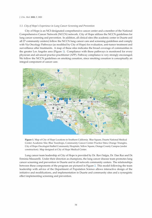
J. Clin. Med. 2020, 9, 1820
1.1. City of Hope’s Experience in Lung Cancer Screening and Prevention
City of Hope is an NCI-designated comprehensive cancer center and a member of the NationalComprehensive Cancer Network (NCCN) network. City of Hope utilizes the NCCN guidelines forlung cancer screening and prevention. In addition, all clinical sites (the academic center in Duarte andall 27 community centers) follow the NCCN lung cancer care and screening guidelines and complywith Via Oncology Pathways (as modified by City of Hope) for evaluation, anti-tumor treatment andsurveillance after treatments. A map of these sites indicates the broad coverage of communities inthe greater Los Angeles area (Figure 1). Compliance with these pathways is monitored for everyphysician and advanced practice practitioner (APP). Pathway compliance is very strongly encouraged.We follow the NCCN guidelines on smoking cessation, since smoking cessation is conceptually anintegral component of cancer care.
Figure 1. Map of City of Hope Locations in Southern California. Blue Square, Duarte National MedicalCenter Academic Site; Blue Teardrops, Community Cancer Center Practice Sites; Orange Triangles,City of Hope Oncologist-Staffed Community Hospitals; Yellow Square, Orange County Campus (underconstruction). Map designed at City of Hope Medical Center.
Lung cancer team leadership at City of Hope is provided by Dr. Ravi Salgia, Dr. Dan Raz and Dr.Erminia Massarelli. Under their direction as champions, the lung cancer disease team promotes lungcancer screening and prevention in Duarte and in all network community centers. The relationshipsbetween these components of the program are pictured in Figure 2. This model following the teamleadership with advice of the Department of Population Science allows interactive design of theinitiative and modifications, and implementation in Duarte and community sites and a synergisticeffect implementing screening and prevention.
58
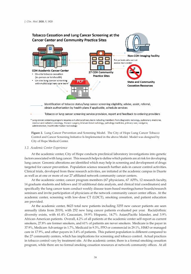
J. Clin. Med. 2020, 9, 1820
Figure 2. Lung Cancer Prevention and Screening Model. The City of Hope Lung Cancer TobaccoControl and Cancer Screening Initiative Is Implemented in the above Model. Model was designed byCity of Hope Medical Center.
1.2. Academic Center Experience
At the academic center, City of Hope conducts preclinical laboratory investigations into geneticfactors associated with lung cancer. This research helps to define which patients are at risk for developinglung cancer. Genomic alterations are identified which may help in screening and development of drugstargeted for cancer prevention. Population science research further aids in cancer control activities.Clinical trials, developed from these research activities, are initiated at the academic campus in Duarteas well as at one or more of our 27 affiliated network community cancer centers.
At the academic center, cancer program members (67 physicians, 67 APPs, 12 research faculty,14 graduate students and fellows and 10 additional data analysts, and clinical trial coordinators) andspecifically the lung cancer team conduct weekly disease team-based meetings/tumor boards/researchseminars and invite participation of physicians at the network community cancer center offices. At theacademic center, screening with low-dose CT (LDCT), smoking cessation, and patient educationare provided.
At the academic center, 8623 total new patients including 3255 new cancer patients are seenannually (data from 2019), with 252 new lung cancer patients evaluated per year. Racial/ethnicdiversity exists, with 61.4% Caucasian, 19.9% Hispanic, 14.7% Asian/Pacific Islander, and 3.9%African-American patients. Overall, 4.2% of all patients at the academic center self-report as currentsmokers, 27.8% are former smokers, and 61% of patients are never smokers. Medicare is the payer in37.9%, Medicare Advantage in 1.7%, Medicaid in 9.3%, PPO or commercial in 29.1%, HMO or managedcare in 17.9%, and other payers in 3.4% of patients. This patient population is different compared tothe 27 community centers, which has implications for screening and tobacco control. Actual practicesin tobacco control vary by treatment site. At the academic center, there is a formal smoking cessationprogram, while there are no formal smoking cessation resources at network community offices. At all
59
J. Clin. Med. 2020, 9, 1820
sites, patients are referred to the California Smokers Helpline 800 No Butts program (800-No-Butts) orto the California QuitLine operated out of the University of California San Diego (800 Quit Now).
At the academic center, LDCT screenings were ordered in 434 patients over a 4 year period, and 424(98%) completed the procedure. We universally use the Lung-RADS system of evaluation of LDCTscans [1]. Of those patients undergoing LDCT, Lung-RADS results showed suspicious lung cancercategory 4A results in 3.3% and highly suspicious category 4B results in 1.7%. Of the six patients whohad biopsies performed at the academic center, four (67%) had stage 1 lung cancer diagnosed, and two(33%) had stage 4 lung cancer. Other patients were referred back to their primary care physiciansfor diagnostic biopsies. Long-term follow up of patients diagnosed through our LDCT screeningprogram will determine reductions in risk of death and will be reported in the future. Any cost savingsassociated with earlier diagnosis depend on the health system, and different health payers are involvedwith our patients (see above).
One of the issues in lung cancer screening is the excessive use of scans. At City of Hope,Shared Decision-Making Consults and Lung-RADS are some of our tools to address the issue ofreducing excessive imaging, false positives, excessive harm as a result of the additional testing,patient anxiety, and cost. During the Shared Decision-Making Consults, ordering clinicians usechecklist and visual tools to discuss the risk and benefits with the patients, address patients’ concerns,and assist patients to determine whether the lung cancer screening is suitable. The clinician alsoeducates patients on what is low-dose CT, and advises postponing the screening if they have had achest/PET-CT in the past 12 months. In the Duarte cancer center LCS program, we also coordinate withthe attending clinicians to prevent excessive imaging. Lung-RADS is a widely accepted and logicalnodule reporting and management tool used by the radiologists to minimize false-positive findings,excessive imaging, and unnecessary procedures. As part of our lung cancer screening initiative,we are developing educational tools and informational aids to help patients understand these issuesbefore screening.
1.3. Community Centers Experience
Our community cancer centers are staffed with 43 medical oncologists, 40 radiation oncologists,7 APPs and a clinical trials coordinator. In order to improve lung cancer control, physicians and APPsat the 27 network community cancer centers advise screening and prevention at local tumor boardsand medical lectures, and depend on collaborating/referring community primary care physicians,pulmonologists, radiologists and hospitals for lung cancer control. Primary care physicians who are thereferring doctors of patients to City of Hope, as well as City of Hope providers themselves (to varyingdegrees), make assessments of lung cancer risk, order LDCT screening exams, advise or providesmoking cessation to current smokers or recently quit patients, refer patients to other local smokingcessation programs if available, prescribe smoking cessation medications, and provide behaviorintervention and counseling with follow-up visits to assess compliance.
In only one-half of the network community practices do the oncologists prescribe medicationsto assist in smoking cessation, while the other half simply refer the patients back to their primarycare physicians with recommendations. In a current survey of community practice sites, almost allof the network community center physicians discuss screening and prevention at tumor boards orhospital meetings where primary physicians and pulmonologists are in attendance, but a few still donot, even today.
At the 27 community centers, 22,000 new patients are seen annually (data from 2019). In total,19,201 patients are seen by medical oncology/hematology specialists. The incidence of current smokersis 7.2%. Of the patients, 49.4% are Caucasian, 20.3% are Hispanic, 4.5% are African-American, and 6%are Asian/Pacific Islander. Among our patients, 43.7% are insured by government programs, 22.9% arePPO/commercially insured, and 22.7% are insured by HMOs or IPA medical groups.
60
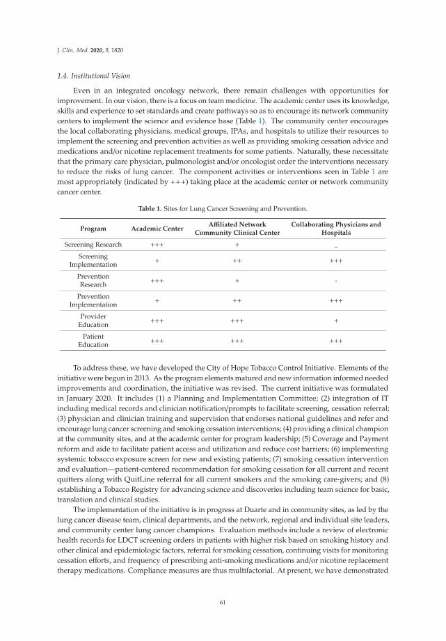
J. Clin. Med. 2020, 9, 1820
1.4. Institutional Vision
Even in an integrated oncology network, there remain challenges with opportunities forimprovement. In our vision, there is a focus on team medicine. The academic center uses its knowledge,skills and experience to set standards and create pathways so as to encourage its network communitycenters to implement the science and evidence base (Table 1). The community center encouragesthe local collaborating physicians, medical groups, IPAs, and hospitals to utilize their resources toimplement the screening and prevention activities as well as providing smoking cessation advice andmedications and/or nicotine replacement treatments for some patients. Naturally, these necessitatethat the primary care physician, pulmonologist and/or oncologist order the interventions necessaryto reduce the risks of lung cancer. The component activities or interventions seen in Table 1 aremost appropriately (indicated by +++) taking place at the academic center or network communitycancer center.
Table 1. Sites for Lung Cancer Screening and Prevention.
Program Academic CenterAffiliated Network
Community Clinical CenterCollaborating Physicians and
Hospitals
Screening Research +++ + _
ScreeningImplementation + ++ +++
PreventionResearch +++ + -
PreventionImplementation + ++ +++
ProviderEducation +++ +++ +
PatientEducation +++ +++ +++
To address these, we have developed the City of Hope Tobacco Control Initiative. Elements of theinitiative were begun in 2013. As the program elements matured and new information informed neededimprovements and coordination, the initiative was revised. The current initiative was formulatedin January 2020. It includes (1) a Planning and Implementation Committee; (2) integration of ITincluding medical records and clinician notification/prompts to facilitate screening, cessation referral;(3) physician and clinician training and supervision that endorses national guidelines and refer andencourage lung cancer screening and smoking cessation interventions; (4) providing a clinical championat the community sites, and at the academic center for program leadership; (5) Coverage and Paymentreform and aide to facilitate patient access and utilization and reduce cost barriers; (6) implementingsystemic tobacco exposure screen for new and existing patients; (7) smoking cessation interventionand evaluation—patient-centered recommendation for smoking cessation for all current and recentquitters along with QuitLine referral for all current smokers and the smoking care-givers; and (8)establishing a Tobacco Registry for advancing science and discoveries including team science for basic,translation and clinical studies.
The implementation of the initiative is in progress at Duarte and in community sites, as led by thelung cancer disease team, clinical departments, and the network, regional and individual site leaders,and community center lung cancer champions. Evaluation methods include a review of electronichealth records for LDCT screening orders in patients with higher risk based on smoking history andother clinical and epidemiologic factors, referral for smoking cessation, continuing visits for monitoringcessation efforts, and frequency of prescribing anti-smoking medications and/or nicotine replacementtherapy medications. Compliance measures are thus multifactorial. At present, we have demonstrated
61
J. Clin. Med. 2020, 9, 1820
that all 27 community cancer centers can operate together, in a coordinated well-integrated effort withthe academic site.
2. Focus on Lung Cancer Screening
The Science
Unfortunately, LDCT is a lung cancer screening method which is underused [2]. The frequency ofperforming lung cancer screening in patients who are eligible for screening by nationally acceptedcriteria is 3.9% as of 2015 [2]. However, this may be improving, since a recent study suggests that in asurvey of 10 states in 2017, 12.7% of people 55–80 years old were eligible for screening by nationalcriteria, and 12.5% actually had received screening [3]. There was a variation by state ranging from9.7% to 16%.
As summarized in NCCN guidelines [4], patients who are generally eligible for LDCT screeningare those at high risk, defined as 55 to 77 years old, have a 30 or more pack year history of smoking,and are active smokers or have stopped smoking within the last 15 years. Although not defined in thescreening trials as high risk, other patients who are also at higher risk are those who are age 50 years orover, with a 20 or more pack year smoking history, and have other risk factors. These risk factors caninclude prior lung or head and neck cancer, a family history of lung cancer, prior chest radiotherapy,asbestos or radon exposure, presence of HIV or COPD or pulmonary fibrosis, or significant second-handsmoke exposure.
Criteria for evaluation are also nationally standardized as outlined in the NCCN guidelines [4].They cover not only which findings require immediate biopsy or excision, but also which require closefollow up versus those who require a repeat LDCT in 1 year.
LDCT has been shown to detect earlier state lung cancer. In fact, 63% of lung cancers which werefound by LDCT were stage IA or IB [5]. Further, this earlier detection has been associated with reducedlung cancer deaths by 20% worldwide [6].
It is important to identify patient symptoms which might be associated with lung cancer.Persistent cough, hemoptysis, shortness of breath, chest pain, weight loss, and/or wheezing may besymptoms of lung cancer. Someone who has any of these symptoms should not get a low-dose CTscan, but they should get a diagnostic CT scan with and without contrast. Payment for diagnosticCT in the presence of symptoms is covered by insurance even in managed care plans. Requests forprior authorization should include diagnostic codes for the specific symptoms. For patients with priorlung cancer, annual diagnostic CT chest scans are indicated, with details included in NCCN guidelines.Clinicians both at Duarte and in community sites are committed to compliance with those guidelines.
If the LDCT is negative, patients should be counseled regarding prevention. This is addressedin the following section. Since the prevalence of LDCT screening is low among patients who meetrequirements to receive it, there remains considerable need for medical leadership in local communitiesto encourage more widespread use.
3. Smoking Prevention
The Science
The most prevalent causative factor for lung cancer is smoking—an estimated 85–90% of patients [7].Thus, smoking cessation is crucial. Smoking cessation programs include a recommendation from aphysician to a patient to stop smoking, followed by referral to a smoking cessation program. In orderto prevent withdrawal symptoms and facilitate cessation, the physician can prescribe pharmaceuticalssuch as varenicline, nicotine replacement therapy (NRT), or buproprion with or without NRT [8].The success of cessation in achieving complete abstinence is poor with a 1 year abstinence rateof 20% using buproprion with NRT, and a 24 week abstinence rate of 26% with varenicline [8].Behavioral interventions have been shown to improve odds of success over medications alone.
62
J. Clin. Med. 2020, 9, 1820
Therefore, follow up is most necessary to continue to promote cessation on the individual patient level.Referral to smoking cessation is best performed by the primary care physician or APP, since theirrelationships with the patient are generally closer than relationships with the oncologists. However,cessation is also often prescribed and facilitated by an oncologist as part of standard cancer care. Further,the oncologist has a role in promoting smoking control at the collaborating hospital or medical staffmeetings. The immediate and long-term benefits of smoking cessation include not only better survival,but also critical positive impact on cancer care, improved success of treatment, reduced recurrence of theprimary cancer, less frequent development of new cancers, and less progression of comorbid conditions.
If smoking cessation is achieved, lung cancer incidence rate is reduced by 83% in totally abstinentnon-smokers, and by 55% in partially abstinent smokers [9]. Recidivism rate, with patients againstarting to smoke, are unfortunately high. Continued motivational support for the patient is necessary.Many hospitals including the City of Hope academic center in Duarte have smoking cessationsupport programs. Alternative programs are provided by such organizations as the American CancerSociety California Division and state of California via phone or internet-based support programcalled 1-800-No-Butts.
There is high and growing use of E-cigarettes for either nicotine replacement or combustiblesmoking cessation. The exact impact of E-cigarettes in transitioning from combustible smoking tonon-smoking remains controversial. Recent studies cast doubt on the true efficacy of substitutingvaping for combustible cigarette smoking. For many individuals who turn to vaping, dual use iscommon. This leads to an additive effect of toxicities from smoking and vaping, which is counter tothe overall goal of tobacco cessation.
City of Hope has helped to develop and adheres to NCCN guidelines on smoking cessation. We donot recommend E-cigarettes or vaping as a smoking cessation or smoking replacement tool either forsingle use or in combination with FDA-approved smoking cessation medication. If a patient has quitsmoking using E-cigarettes, we support continuation of non-smoking status, encourage transition toapproved smoking cessation medications, encourage behavior support, and provide support to reducethe risk of lapses or relapses. Of special concerns are the dual use of tobacco and E-cigarettes andE-cigarettes in children where lifetime addiction to nicotine products are of particular concern.
In addition to smoking cessation, other interventions are known to be effective in reducinglung cancer rates and are advised by the National Institutes of Health [10,11]. Patients should avoidsecond-hand smoke, avoid excessive radiologic scans, reduce exposure to radon (present in 1 of every15 homes), not be exposed to asbestos (and also chromium, nickel, beryllium, cadmium, tar, uranium,coal products and diesel fuel), and avoid beta carotene use in smokers.
Other interventions to reduce lung cancer risk which have evidence to support them but not yetaccepted in national guidelines include eating a Mediterranean-style diet with increased fruits andvegetables, possibly leading to a 33–50% reduction in lung cancer [12]; increasing exercise, with possiblya 25% reduction [13]; or using green tea, with possibly a 22% reduction [14].
A number of nutritional interventions have been recommended without clear evidence of possibleimprovement. These interventions include red wine, flax seed, garlic, ginger, selenium, turmeric andvitamin D.
4. Translating Screening and Prevention from the Academic Center
It is difficult to successfully promote lung cancer screening and prevention at the local communitysince insurance often does not adequately reimburse for these programs or for pharmaceuticals.The evidence base is clear that screening and prevention should be strongly encouraged. Our survey ofprimary care physicians in our catchment area of greater Los Angeles indicated that these physicianswere aware of LDCT screening guidelines—only 12% of primary care physicians in the community (notat academic centers) had referred patients for lung cancer screening in the past year [15]. This indicatesa need for community leadership by City of Hope oncologists at local hospital meetings and tumorboards to promote appropriate LDCT screening.
63
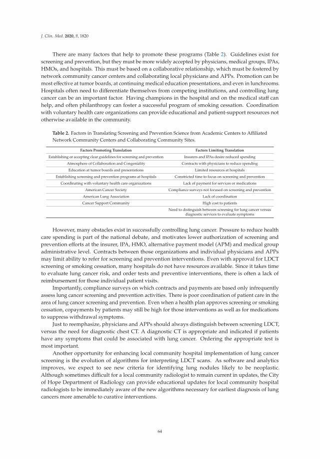
J. Clin. Med. 2020, 9, 1820
There are many factors that help to promote these programs (Table 2). Guidelines exist forscreening and prevention, but they must be more widely accepted by physicians, medical groups, IPAs,HMOs, and hospitals. This must be based on a collaborative relationship, which must be fostered bynetwork community cancer centers and collaborating local physicians and APPs. Promotion can bemost effective at tumor boards, at continuing medical education presentations, and even in lunchrooms.Hospitals often need to differentiate themselves from competing institutions, and controlling lungcancer can be an important factor. Having champions in the hospital and on the medical staff canhelp, and often philanthropy can foster a successful program of smoking cessation. Coordinationwith voluntary health care organizations can provide educational and patient-support resources nototherwise available in the community.
Table 2. Factors in Translating Screening and Prevention Science from Academic Centers to AffiliatedNetwork Community Centers and Collaborating Community Sites.
Factors Promoting Translation Factors Limiting Translation
Establishing or accepting clear guidelines for screening and prevention Insurers and IPAs desire reduced spending
Atmosphere of Collaboration and Congeniality Contracts with physicians to reduce spending
Education at tumor boards and presentations Limited resources at hospitals
Establishing screening and prevention programs at hospitals Constricted time to focus on screening and prevention
Coordinating with voluntary health care organizations Lack of payment for services or medications
American Cancer Society Compliance surveys not focused on screening and prevention
American Lung Association Lack of coordination
Cancer Support Community High cost to patients
Need to distinguish between screening for lung cancer versusdiagnostic services to evaluate symptoms
However, many obstacles exist in successfully controlling lung cancer. Pressure to reduce healthcare spending is part of the national debate, and motivates lower authorization of screening andprevention efforts at the insurer, IPA, HMO, alternative payment model (APM) and medical groupadministrative level. Contracts between those organizations and individual physicians and APPsmay limit ability to refer for screening and prevention interventions. Even with approval for LDCTscreening or smoking cessation, many hospitals do not have resources available. Since it takes timeto evaluate lung cancer risk, and order tests and preventive interventions, there is often a lack ofreimbursement for those individual patient visits.
Importantly, compliance surveys on which contracts and payments are based only infrequentlyassess lung cancer screening and prevention activities. There is poor coordination of patient care in thearea of lung cancer screening and prevention. Even when a health plan approves screening or smokingcessation, copayments by patients may still be high for those interventions as well as for medicationsto suppress withdrawal symptoms.
Just to reemphasize, physicians and APPs should always distinguish between screening LDCT,versus the need for diagnostic chest CT. A diagnostic CT is appropriate and indicated if patientshave any symptoms that could be associated with lung cancer. Ordering the appropriate test ismost important.
Another opportunity for enhancing local community hospital implementation of lung cancerscreening is the evolution of algorithms for interpreting LDCT scans. As software and analyticsimproves, we expect to see new criteria for identifying lung nodules likely to be neoplastic.Although sometimes difficult for a local community radiologist to remain current in updates, the Cityof Hope Department of Radiology can provide educational updates for local community hospitalradiologists to be immediately aware of the new algorithms necessary for earliest diagnosis of lungcancers more amenable to curative interventions.
64
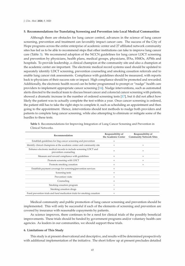
J. Clin. Med. 2020, 9, 1820
5. Recommendations for Translating Screening and Prevention into Local Medical Communities
Although there are obstacles for lung cancer control, advances in the science of lung cancerscreening, prevention and treatment can favorably impact cancer care. The success of the City ofHope programs across the entire enterprise of academic center and 27 affiliated network communitysites has led us to be able to recommend steps that other institutions can take to improve lung cancercare (Table 3). We recommend adoption of the NCCN guidelines for lung cancer LDCT screeningand prevention by physicians, health plans, medical groups, physicians, IPAs, HMOs, APMs andhospitals. To provide leadership, a clinical champion at the community site and also a champion atthe academic center are important. The electronic medical record systems used should be updated toseparately identify LDCT screening, prevention counseling and smoking cessation referrals and toenable lung cancer risk assessments. Compliance with guidelines should be measured, with reportsback to physicians of their success rate or impact. High compliance should be promoted and rewarded.Additionally, the electronic health record can be better programmed to prompt or “nudge” health careproviders to implement appropriate cancer screening [16]. Nudge interventions, such as automatedalerts directed to the medical team to discuss breast cancer and colorectal cancer screening with patients,showed a dramatic increase in the number of ordered screening tests [17], but it did not affect howlikely the patient was to actually complete the test within a year. Once cancer screening is ordered,the patient still has to take the right steps to complete it, such as scheduling an appointment and thengoing to the appointment. Hence, interventions should test methods to nudge both providers andpatients to complete lung cancer screening, while also attempting to eliminate or mitigate some of thehurdles to these tests.
Table 3. Recommendations for Improving Integration of Lung Cancer Screening and Prevention inClinical Networks.
RecommendationResponsibility of
the Academic CenterResponsibility of
Community Network Sites
Establish guidelines for ling cancer screening and prevention +
Identify clinical champions at the academic center and community site + +
Enhance electronic medical records to include screening LDCT andprevention counseling + +
Measure and reward compliance with guidelines + +
Promote screening with LDCT + +
Promote smoking cessation + +
Establish payment coverage for screening/prevention services +
Screening tests +
Prevention visits +
Counseling +
Smoking cessation program +
Smoking cessation drugs +
Fund prevention trials and fund medication trials for smoking cessation +
Medical community and public promotion of lung cancer screening and prevention should beimplemented. This will only be successful if each of the elements of screening and prevention arecovered by insurance with reasonable copayments by patients.
As science improves, there continues to be a need for clinical trials of the possibly beneficialimprovements. These trials should be funded by government programs and/or voluntary health careagencies. As leaders in our communities, we should support these trials.
6. Limitations of This Study
This study is at present observational and descriptive, and results will be determined prospectivelywith additional implementation of the initiative. The short follow up at present precludes detailed
65
J. Clin. Med. 2020, 9, 1820
reporting of results on cessation success, reductions in lung cancer mortality, and cost savings.Results will be analyzed and published in future communications. Comparisons of lung cancer stagingresults with LDCT compared to staging resulting from diagnosis when symptoms were present will beevaluated when more patients have been diagnosed at Duarte and in the community network.
7. Conclusions
Lung cancer continues to be a challenge to patients, providers, and health care systems despitedramatic improvements in treatment. We can use successful screening and prevention programs toreduce these life-threatening diagnoses and give patients more hope for the future. Smoking cessationin particular must become an integral component of cancer care and prevention. Reporting on theexperience at City of Hope, the recommendations of the translational medical system of City of Hope,and the City of Hope Tobacco Control Initiative is designed to promote broader lung cancer screeningand prevention efforts at other institutions and in other communities. We hope our experience willhelp contribute to improved patient outcomes and increase tobacco control.
Author Contributions: Conceptualization, C.A.P., S.S. (Shamel Sanani), B.L., K.A., B.L.T., R.S., P.K., J.S., M.S.S.,S.L., and S.S. (Shanmugga Subbiah); Methodology, C.A.P., S.S. (Shamel Sanani), B.L., K.A., B.L.T., R.S., P.K., J.S.,M.S.S., S.L., S.Y., D.R., and S.S. (Shanmugga Subbiah); Investigation, C.A.P., S.S. (Shamel Sanani), B.L., K.A., B.L.T.,R.S., P.K., J.S., M.S.S., S.L., D.R., and S.S. (Shanmugga Subbiah); Data Evaluation, C.A.P., S.S. (Shamel Sanani), B.L.,K.A., B.L.T., R.S., P.K., and S.S. (Shanmugga Subbiah); Writing—Initial Draft, C.A.P.; Writing—Review and Editing,C.A.P., S.S. (Shamel Sanani), B.L., K.A., B.L.T., R.S., P.K., J.S., M.S.S., S.L., S.Y., D.R., and S.S. (Shanmugga Subbiah);Supervision, C.A.P. and R.S.; Project Administration, C.A.P., B.T., R.S., P.K., and S.S. (Shanmugga Subbiah). Allauthors have read and agreed to the published version of the manuscript.
Funding: This work was funded in part by NIH Grant P30 CA033572
Conflicts of Interest: The authors declare no conflicts of interest.
List of Abbreviations
APM—alternative payment model organization; APP—advanced practice provider; COH—City of Hope; COPD—chronic obstructive pulmonary disease; HMO—health maintenance organization; IPA—independent practiceassociation; IT—information technology; LDCT—low-dose computed chest tomography; L-RADS—lung imagingreporting and data system; NCI—National Cancer Institute; NCCN—National Comprehensive Cancer Network;NRT—nicotine replacement therapy; PPO—preferred provider organization; Via—Via Oncology Clinical Pathways,now also known as ClinicalPath (Elsevier)
References
1. Pinsky, P.F.; Gierada, D.S.; Black, W.; Munden, R.; Nath, H.; Aberle, D.; Kazerooni, E. Performance ofLung-RADS in the national screening trial—A retrospective assessment. Ann. Int. Med. 2015, 162, 485–491.[CrossRef] [PubMed]
2. Jemal, A.; Fedewa, S.A. Lung cancer screening with low dose computed tomography in the UnitedStates—2010 to 2015. JAMA Oncol. 2017, 3, 1278–1281. [CrossRef] [PubMed]
3. Richards, T.B.; Ashwini, S.; Thomas, C.C.; VanFrank, B.; Henley, J.; Gallaway, M.S. Screening for lungcancer—10 states 2017. Morb. Mortal. Wkly. 2020, 69, 201–206. [CrossRef]
4. NCCN. Available online: org/professionals/physician_gls/pdf/lung_screening.pdf (accessed on 27 February 2020).5. Aberle, D.R.; Adams, A.M.; Berg, C.D. Reduced lung cancer mortality with low dose computed tomographic
screening. N. Engl. J. Med. 2011, 365, 395–409.6. Pinsky, P.F. Lung cancer screening with lose dose CT: A world wide view. Transl. Lung Cancer Res. 2018, 7,
234–242. [CrossRef] [PubMed]7. Samet, J.M.; Avila-Tang, E.; Boffetta, P. Lung cancer in never smokers: Clinical epidemiology and
environmental risk factors. Clin. Cancer Res. 2009, 15, 5625–5645. [CrossRef]8. Shields, P.G.; Herbst, R.S.; Arenberg, D.; Benowitz, N.L.; Bierut, L.; Luckart, J.B.; Hitsman, B.; Lang, M.;
Park, E.R.; Selzle, J.; et al. Smoking Cessation, Version 1.2016, NCCN Clinical Practice Guidelines in Oncology.J. Natl. Compr. Cancer Netw. 2016, 4, 1430–1468. [CrossRef] [PubMed]
9. Godtfredsen, N.S.; Prescott, E.; Oder, M. Effect of smoking reduction on lung cancer risk. JAMA 2005, 294,1505–1510. [CrossRef]
66
J. Clin. Med. 2020, 9, 1820
10. PDQ Advisory Committee. Lung Cancer Prevention (PDQ). Available online: https://www.ncbi.nlm.nih.gov/books/NBK66063/ (accessed on 22 May 2020).
11. Beveridge, R.; Pintos, J.; Parent, M.-E.; Asselin, J.; Siemiatycki, J. Lung cancer risk associated with occupationalexposure to nickel, chromium VI, and cadmium in two population based case control studies in Montreal.Ann. Int. Med. 2010, 53, 476–485. [CrossRef]
12. Gnagnarella, P.; Maisonnueve, P.; Belloni, M.; Rampinelli, C.; Bertolotti, R.; Spaggiari, L.; Veronesi, G.Red meat, Mediterranean diet and lung cancer risk among heavy smokers in the COSMOS screening study.Ann. Oncol. 2013, 24, 2606–2611. [CrossRef] [PubMed]
13. Thune, I.; Lund, E. The influence of physical activity on lung cancer risk: A prospective study of 81,516 menand women. Int. J. Cancer 1997, 70, 57–62. [CrossRef]
14. Tang, N.; Wu, Y.; Zhou, B.; Wang, B.; Yu, R. Green tea, black tea consumption and risk of lung cancer:A meta-analysis. Lung Cancer 2009, 65, 274–283. [CrossRef]
15. Raz, D.J.; Wu, G.X.; Consunji, M.; Nelson, R.; Sun, C.; Erhunmwunsee, L.; Ferrell, B.; Sun, V.; Kim, J.Y.Perceptions and utilization of lung cancer screening among primary care physicians. J. Thorac. Oncol. 2016,11, 1856–1862. [CrossRef] [PubMed]
16. Patel, M.S.; Volpo, K.G.; Asch, D.A. Nudge units to improve the delivery of health care. N. Engl. J. Med. 2018,378, 214–216. [CrossRef] [PubMed]
17. Hsiang, E.Y.; Mehta, S.J.; Small, D.S.; Rareshide, C.A.; Snider, C.K.; Day, S.C.; Patel, M.S. Association ofan Active Choice Intervention in the electronic health record directed to medical assistants with clinicianordering and patient completion of breast and colorectal cancer screening tests. JAMA Netw. Open 2019, 2,e1915619. [CrossRef] [PubMed]
© 2020 by the authors. Licensee MDPI, Basel, Switzerland. This article is an open accessarticle distributed under the terms and conditions of the Creative Commons Attribution(CC BY) license (http://creativecommons.org/licenses/by/4.0/).
67

Journal of
Clinical Medicine
Article
Advancing the Science and Management of RenalCell Carcinoma: Bridging the Divide betweenAcademic and Community Practices
Nicholas J. Salgia 1, Errol J. Philip 2, Mohammadbagher Ziari 3, Kelly Yap 4 and
Sumanta Kumar Pal 1,*
1 Department of Medical Oncology & Experimental Therapeutics, City of Hope ComprehensiveCancer Center, Duarte, CA 91107, USA; [email protected]
2 University of California San Francisco School of Medicine, San Francisco, CA 94143, USA;[email protected]
3 Department of Medical Oncology & Experimental Therapeutics, City of Hope ComprehensiveCancer Center, Corona, CA 92879, USA; [email protected]
4 Department of Medical Oncology & Experimental Therapeutics, City of Hope ComprehensiveCancer Center, South Pasadena, CA 91030, USA; [email protected]
* Correspondence: [email protected]; Tel.: +1-(626)-256-4673; Fax: +1-(626)-301-8233
Received: 29 April 2020; Accepted: 14 May 2020; Published: 17 May 2020
Abstract: The treatment of metastatic renal cell carcinoma (mRCC) has rapidly evolved; however,the progress made in the field is heavily contingent upon timely and efficient accrual to clinicaltrials. While a substantial proportion of accrual occurs at tertiary care centers, community sites areplaying an increasing role in patient recruitment. In this article, we discuss strategies to optimizecollaborations between academic and community sites to facilitate clinical research. Further, as therole of biomarker discovery has become increasingly important in tailoring therapy, we will discussopportunities to bridge diverse accrual sites for the purpose of translational research.
Keywords: renal cell carcinoma; team medicine; translational research; community practice;clinical trials
1. Introduction
Two decades ago, the outlook for patients with metastatic renal cell carcinoma (mRCC) wasbleak. Treatments such as interferon-α (IFN-α) and interleukin-2 (IL-2) yielded a median survival ofapproximately one year [1]. The landscape changed tremendously with the introduction of targetedtherapies from 2005 onwards, and, once again, with the introduction of checkpoint blockade a decadelater [2–5]. In 2020, the current standard for front-line treatment of mRCC remains either dualcheckpoint inhibition or a combination of targeted therapy with checkpoint blockade [6–8]. Survivalestimates have essentially tripled what was anticipated in the cytokine era.
The rapid developments in mRCC therapy have been made possible by the timely and efficientcompletion of both early and late phase clinical trials. In addition, the discovery of novel therapieshas been fostered by translational research efforts—the intersect of the bench and bedside. This isespecially true in RCC, which has directly benefitted from research discoveries that have led to both the2018 and 2019 Nobel Prizes in Physiology or Medicine. While tertiary centers (e.g., cancer centers andacademic hospitals) serve as a common ground for early phase clinical trials and translational work,there is no doubt that their efforts can be bolstered through the participation of community oncologypractices. Further, late-phase clinical trials, which often require less stringent data collection and lessintensive evaluation schedules, can perhaps be done equally well at community and academic sites.
J. Clin. Med. 2020, 9, 1508; doi:10.3390/jcm9051508 www.mdpi.com/journal/jcm69
J. Clin. Med. 2020, 9, 1508
Whereas the prognosis of mRCC has improved, it is important to note that most patients with thisdisease are not cured, and thus, further research is imperative. In the current manuscript, we reviewthe clinical landscape for renal cell carcinoma and delve into opportunities to maximize collaborationsbetween community-based practices and academic sites in the hope of promoting more efficientand effective research endeavors. We approach the latter objective in the specific context of City ofHope—a tertiary cancer center that serves the greater Los Angeles area, a catchment that includesapproximately 20,000,000 people. City of Hope has within its network 30 community-based satelliteclinics, stretching over a radius of 100 miles [9]. With this vast network of community sites andthe robust patient population that comes with it, multiple opportunities for collaborative researchexist. However, selecting the right research for community versus academic practices is no small feat.Whereas our academic practice at City of Hope is well-staffed, with 5 research nurses and 7 clinicalresearch associates devoted to genitourinary cancers, community sites often have a limited number ofresearch staffwho are responsible for managing studies across multiple disease types. These limitationsnotwithstanding, the integration of academic and community centers has grown into a successfulmodel for conducting clinical and translational research.
2. Renal Cell Carcinoma Incidence and Histology
Renal cell carcinomas are a broad classification referring to tumors originating from the renalpelvis and medulla. The incidence of RCC is increasing, with an estimated 73,750 newly diagnosedcases in 2020 in the United States [10]. The disease now accounts for 5% of cancer diagnoses inmen and 3% in women. The rising incidence of RCC can be partially attributed to an increase inthe incidental detection of small renal masses during the performance of abdominal imaging fornon-specific complaints [11]. Although the majority of incidental findings are small, early-stage masses,these diagnoses have not led to a substantial change in the mortality rate attributed to RCC; in 2020,it is estimated that RCC will account for approximately 15,000 deaths in the United States [10].
The histological variations in RCC have been well-documented [12]. Clear cell RCC (ccRCC)represents the most common subtype, accounting for 80% of diagnoses. ccRCC is pathologicallycharacterized by its clear cell histology and acinar growth patterns. The hallmark molecular alterationsof ccRCC include chromosome 3p loss and inactivation of the Von-Hippel Lindau (VHL) gene, which liesat the 3p25 locus [13]. VHL inactivation can occur either somatically or in the germline. Inactivation ofVHL has been reported in up to 90% of sporadic ccRCC cases [14,15]. Germline inactivations of VHLare associated with Von-Hippel Lindau syndrome, which predisposes individuals to bilateral renalmasses, as well as hemangioblastomas and retinal angiomas [16]. Other mutations associated withccRCC are PBRM1, SETD2, and BAP1, all of which also lie on chromosome 3p [15,17].
Non-clear cell RCC (nccRCC) is a heterogenous classification that encompasses the other 20%of RCC diagnoses. Papillary RCC (pRCC) composes the majority of nccRCCs (~15% of total RCCdiagnoses). pRCC has historically been subdivided into two classifications based on histology: type1 involves low-grade nuclei arranged in a single layer, and type 2 encompasses a wide range ofmorphological presentations that can be further divided into at least three additional subtypes. Type 1pRCC has been associated with sporadic mutations in the MET protooncogene (10–13% of patients) [18].Other nccRCCs include chromophobe RCC, MiT translocation RCCs, collecting duct carcinoma,and renal medullary carcinoma. Each of the above-listed subtypes accounts for less than 10% of allRCC diagnoses [19].
As ccRCC represents the overwhelming majority of RCC diagnoses, the remainder of this reviewwill focus primarily on the management of clear cell disease and use RCC interchangeably for ccRCC.
3. Managing Localized Renal Cell Carcinoma
Localized RCC comprises the majority of RCC cases, and in turn, is typically cured with definitivetreatment. The standard of care for most localized tumors has been nephrectomy, whether it bepartial or radical. Cancer-specific survival rates for localized RCC patients have been estimated to be
70
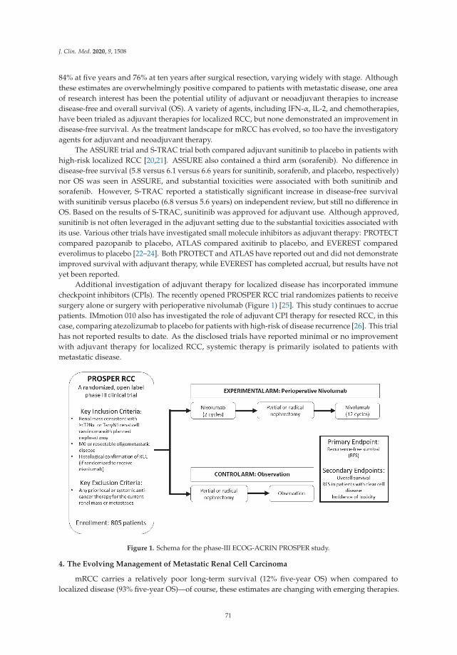
J. Clin. Med. 2020, 9, 1508
84% at five years and 76% at ten years after surgical resection, varying widely with stage. Althoughthese estimates are overwhelmingly positive compared to patients with metastatic disease, one areaof research interest has been the potential utility of adjuvant or neoadjuvant therapies to increasedisease-free and overall survival (OS). A variety of agents, including IFN-α, IL-2, and chemotherapies,have been trialed as adjuvant therapies for localized RCC, but none demonstrated an improvement indisease-free survival. As the treatment landscape for mRCC has evolved, so too have the investigatoryagents for adjuvant and neoadjuvant therapy.
The ASSURE trial and S-TRAC trial both compared adjuvant sunitinib to placebo in patients withhigh-risk localized RCC [20,21]. ASSURE also contained a third arm (sorafenib). No difference indisease-free survival (5.8 versus 6.1 versus 6.6 years for sunitinib, sorafenib, and placebo, respectively)nor OS was seen in ASSURE, and substantial toxicities were associated with both sunitinib andsorafenib. However, S-TRAC reported a statistically significant increase in disease-free survivalwith sunitinib versus placebo (6.8 versus 5.6 years) on independent review, but still no difference inOS. Based on the results of S-TRAC, sunitinib was approved for adjuvant use. Although approved,sunitinib is not often leveraged in the adjuvant setting due to the substantial toxicities associated withits use. Various other trials have investigated small molecule inhibitors as adjuvant therapy: PROTECTcompared pazopanib to placebo, ATLAS compared axitinib to placebo, and EVEREST comparedeverolimus to placebo [22–24]. Both PROTECT and ATLAS have reported out and did not demonstrateimproved survival with adjuvant therapy, while EVEREST has completed accrual, but results have notyet been reported.
Additional investigation of adjuvant therapy for localized disease has incorporated immunecheckpoint inhibitors (CPIs). The recently opened PROSPER RCC trial randomizes patients to receivesurgery alone or surgery with perioperative nivolumab (Figure 1) [25]. This study continues to accruepatients. IMmotion 010 also has investigated the role of adjuvant CPI therapy for resected RCC, in thiscase, comparing atezolizumab to placebo for patients with high-risk of disease recurrence [26]. This trialhas not reported results to date. As the disclosed trials have reported minimal or no improvementwith adjuvant therapy for localized RCC, systemic therapy is primarily isolated to patients withmetastatic disease.
Figure 1. Schema for the phase-III ECOG-ACRIN PROSPER study.
4. The Evolving Management of Metastatic Renal Cell Carcinoma
mRCC carries a relatively poor long-term survival (12% five-year OS) when compared tolocalized disease (93% five-year OS)—of course, these estimates are changing with emerging therapies.
71
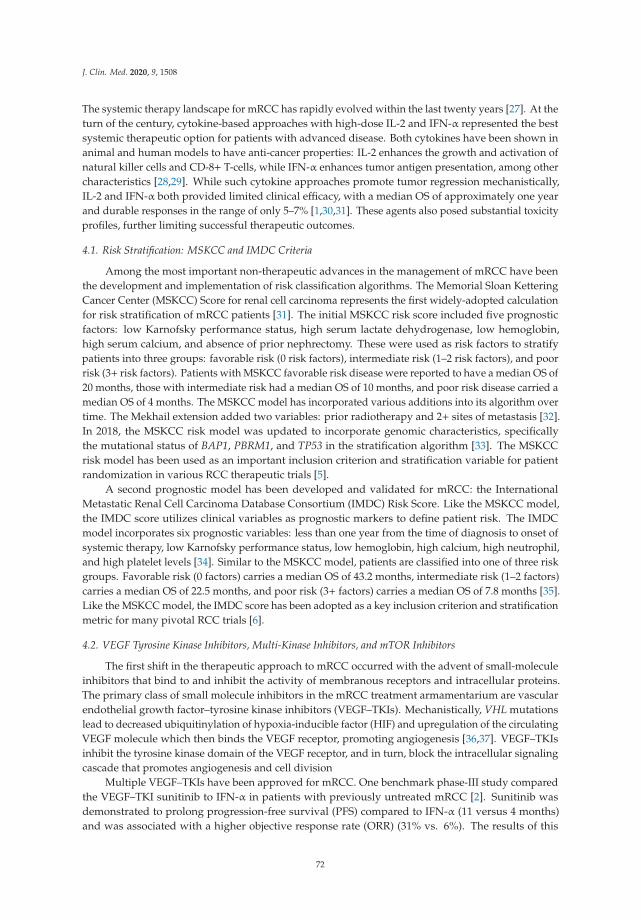
J. Clin. Med. 2020, 9, 1508
The systemic therapy landscape for mRCC has rapidly evolved within the last twenty years [27]. At theturn of the century, cytokine-based approaches with high-dose IL-2 and IFN-α represented the bestsystemic therapeutic option for patients with advanced disease. Both cytokines have been shown inanimal and human models to have anti-cancer properties: IL-2 enhances the growth and activation ofnatural killer cells and CD-8+ T-cells, while IFN-α enhances tumor antigen presentation, among othercharacteristics [28,29]. While such cytokine approaches promote tumor regression mechanistically,IL-2 and IFN-α both provided limited clinical efficacy, with a median OS of approximately one yearand durable responses in the range of only 5–7% [1,30,31]. These agents also posed substantial toxicityprofiles, further limiting successful therapeutic outcomes.
4.1. Risk Stratification: MSKCC and IMDC Criteria
Among the most important non-therapeutic advances in the management of mRCC have beenthe development and implementation of risk classification algorithms. The Memorial Sloan KetteringCancer Center (MSKCC) Score for renal cell carcinoma represents the first widely-adopted calculationfor risk stratification of mRCC patients [31]. The initial MSKCC risk score included five prognosticfactors: low Karnofsky performance status, high serum lactate dehydrogenase, low hemoglobin,high serum calcium, and absence of prior nephrectomy. These were used as risk factors to stratifypatients into three groups: favorable risk (0 risk factors), intermediate risk (1–2 risk factors), and poorrisk (3+ risk factors). Patients with MSKCC favorable risk disease were reported to have a median OS of20 months, those with intermediate risk had a median OS of 10 months, and poor risk disease carried amedian OS of 4 months. The MSKCC model has incorporated various additions into its algorithm overtime. The Mekhail extension added two variables: prior radiotherapy and 2+ sites of metastasis [32].In 2018, the MSKCC risk model was updated to incorporate genomic characteristics, specificallythe mutational status of BAP1, PBRM1, and TP53 in the stratification algorithm [33]. The MSKCCrisk model has been used as an important inclusion criterion and stratification variable for patientrandomization in various RCC therapeutic trials [5].
A second prognostic model has been developed and validated for mRCC: the InternationalMetastatic Renal Cell Carcinoma Database Consortium (IMDC) Risk Score. Like the MSKCC model,the IMDC score utilizes clinical variables as prognostic markers to define patient risk. The IMDCmodel incorporates six prognostic variables: less than one year from the time of diagnosis to onset ofsystemic therapy, low Karnofsky performance status, low hemoglobin, high calcium, high neutrophil,and high platelet levels [34]. Similar to the MSKCC model, patients are classified into one of three riskgroups. Favorable risk (0 factors) carries a median OS of 43.2 months, intermediate risk (1–2 factors)carries a median OS of 22.5 months, and poor risk (3+ factors) carries a median OS of 7.8 months [35].Like the MSKCC model, the IMDC score has been adopted as a key inclusion criterion and stratificationmetric for many pivotal RCC trials [6].
4.2. VEGF Tyrosine Kinase Inhibitors, Multi-Kinase Inhibitors, and mTOR Inhibitors
The first shift in the therapeutic approach to mRCC occurred with the advent of small-moleculeinhibitors that bind to and inhibit the activity of membranous receptors and intracellular proteins.The primary class of small molecule inhibitors in the mRCC treatment armamentarium are vascularendothelial growth factor–tyrosine kinase inhibitors (VEGF–TKIs). Mechanistically, VHL mutationslead to decreased ubiquitinylation of hypoxia-inducible factor (HIF) and upregulation of the circulatingVEGF molecule which then binds the VEGF receptor, promoting angiogenesis [36,37]. VEGF–TKIsinhibit the tyrosine kinase domain of the VEGF receptor, and in turn, block the intracellular signalingcascade that promotes angiogenesis and cell division
Multiple VEGF–TKIs have been approved for mRCC. One benchmark phase-III study comparedthe VEGF–TKI sunitinib to IFN-α in patients with previously untreated mRCC [2]. Sunitinib wasdemonstrated to prolong progression-free survival (PFS) compared to IFN-α (11 versus 4 months)and was associated with a higher objective response rate (ORR) (31% vs. 6%). The results of this
72
J. Clin. Med. 2020, 9, 1508
study led to the approval of sunitinib for first-line mRCC patients. Other VEGF–TKI agents approvedin mRCC include sorafenib, pazopanib, and axitinib. While these agents have prolonged PFS andproduced improved response rates compared to the previous standard-of-care cytokine therapies,VEGF–TKIs are not curative, and patients are susceptible to disease progression upon the developmentof drug resistance.
An additional class of targeted therapies, so-called multi-kinase inhibitors have also been approvedin mRCC. These agents not only act as VEGF–TKIs but also inhibit the tyrosine kinase domains ofadditional protooncogenes [38]. Cabozantinib is a multi-kinase inhibitor with activity as a VEGF–TKIand also as an inhibitor of MET and AXL, both of which are associated with resistance to VEGF–TKIs.Cabozantinib was first approved for mRCC patients with treatment-refractory disease but was soontrialed as first-line therapy. The phase-II CABOSUN trial compared cabozantinib to sunitinib in thefront-line setting [39]. This study met its primary endpoint of improvement in PFS with cabozantinib(8.6 versus 5.3 months) and demonstrated a higher ORR with cabozantinib based on an independentreview (20% versus 9%). These results led to the approval of cabozantinib across all lines of therapyfor patients with mRCC. However, like sunitinib and other VEGF–TKIs, cabozantinib has limitedcurative potential. As such, the approach of managing mRCC in the front-line with VEGF–TKIs andmulti-kinase inhibitors has been replaced by the recent introduction of immune checkpoint inhibitors.
The mammalian target of rapamycin (mTOR) represents a highly-important intracellular targetof mRCC therapy. mTOR is an enzymatic intermediate in the PI3K/AKT/mTOR signaling pathwaythat regulates the cell cycle [40]. Dysregulation of this pathway is a metabolic feature of manyRCC tumors, making the components of its signaling cascade viable targets for pharmacologicinhibition. Two therapies approved for the management of mRCC act in this manner on mTOR:everolimus and temsirolimus. Everolimus is an oral agent that has been approved in combinationfor mRCC with the VEGF–TKI lenvatinib, following the results of a phase II study comparing thecombination to each respective monotherapy [41]. Everolimus with lenvatinib resulted in PFS benefitcompared to everolimus alone (14.6 versus 5.5 months) for patients who had received at least one priorVEGF–directed therapy. Temsirolimus, an inhibitory ester analog of mTOR that is applied intravenously,is also approved for mRCC [42]. A phase III trial demonstrated an OS benefit for temsirolimus overIFN-α (10.9 versus 7.3 months) with fewer incidences of serious adverse events [43]. Although nolonger a mainstay of early-line therapy, the mTOR inhibitors everolimus and temsirolimus remainimportant interventions for the management of mRCC. Like VEGF–TKIs and multikinase inhibitors,however, mTOR inhibitors have given way to immune checkpoint inhibitors for the early-line treatmentof mRCC.
4.3. Immune Checkpoint Inhibitors
Immune checkpoint inhibition is a relatively recent development in solid tumors, first explored inthe context of melanoma [44]. CPIs are synthesized monoclonal antibodies capable of overcomingT-cell inactivation to elicit an anti-tumor response [45]. Approved CPIs exhibit activity on one oftwo immunoregulatory axes: programmed-death-1/programmed-death-ligand-1 (PD-1/PD-L1) andthe cytotoxic-T-lymphocyte-associated protein 4 (CTLA-4). The treatment paradigm for mRCC hasadvanced greatly since the advent of PD-(L)1 and CTLA-4 inhibitors.
CheckMate-025 was the first study to investigate nivolumab, an anti-PD-1 CPI, in mRCC [5].This study enrolled patients who had progressed on one or two previous VEGF–TKIs and randomizedthem to receive nivolumab or everolimus. Nivolumab outperformed everolimus in OS (25 versus19.6 months) and ORR (25% versus 5%), leading to the approval of nivolumab monotherapy for mRCCpatients who had progressed on previous therapy.
The ensuing CheckMate-214 trial compared the combined regimen of nivolumab with ipilimumab,an anti-CTLA-4 CPI, to sunitinib in previously-untreated mRCC [6]. Patients were stratified based onthe International Metastatic Renal Cell Carcinoma Database Consortium (IMDC) risk score. In patientswith intermediate or poor risk disease, nivolumab + ipilimumab greatly outperformed sunitinib,
73
J. Clin. Med. 2020, 9, 1508
demonstrating greater ORR (42% versus 27%) and a rate of complete response totaling 9%. However,among patients with favorable risk disease by IMDC criteria, sunitinib was shown to result in prolongedPFS (25.1 versus 15.3 months) and a greater ORR (52% versus 29%). The results from CheckMate-214established a new treatment algorithm for front-line mRCC, in which dual checkpoint inhibitionwith nivolumab + ipilimumab form the backbone of therapy for intermediate/poor risk patients butVEGF–TKIs remain a viable first-line option for patients with favorable risk.
4.4. Combination Therapies
Further endeavors have clinically investigated combinations of CPIs with VEGF–TKIs. A myriadof large phase III clinical trials have been undertaken, with three having reported out in early 2019.The IMmotion 151 trial compared the investigatory combination of atezolizumab (an anti PD-L1 CPI)with bevacizumab (an anti-VEGF monoclonal antibody) against sunitinib [46]. This trial met its primaryendpoint of improvement in PFS with bevacizumab + atezolizumab (11.2 versus 7.7 months) in thePD-L1+ population. In the intention-to-treat cohort that included all patients regardless of PD-L1 status,PFS favored the combination therapy over sunitinib (11.2 versus 8.4 months). JAVELIN-101 followeda similar design model, comparing the combination of avelumab (an anti-PD-L1 CPI) and axitinibagainst sunitinib in patients with mRCC who had received no prior therapy [8]. The primary endpointof improved PFS with axitinib + avelumab in patients who had PD-L1+ disease was met (13.8 versus7.2 months). Likewise, irrespective of PD-L1 status, axitinib + avelumab was again associated withimproved PFS (13.8 versus 8.4 months).
The KEYNOTE-426 trial utilized an experimental arm of pembrolizumab (an anti-PD-1 CPI)with axitinib, which was compared against sunitinib [7]. The axitinib + pembrolizumab combinationresulted in a prolonged PFS compared to sunitinib (15.1 versus 11.1 months), and, additionally, ORR wasgreater in the axitinib + pembrolizumab arm (59% versus 36%). These three trials all reported withinthe span of two months in 2019. Table 1 summarizes results from trials testing CPIs (in combinationwith an additional CPI or with VEGF–TKIs) for the first-line treatment of mRCC. A recent pressrelease in April 2020 has also indicated that the CheckMate-9ER trial (NCT03141177) of nivolumab +cabozantinib versus sunitinib for front-line mRCC has met its primary endpoint of improved PFS andsecondary endpoints of ORR and OS. The combination of axitinib + pembrolizumab, in particular,has already established a role in the mRCC treatment algorithm for front-line intervention in patientswith IMDC good-risk disease [47]. As data from these trials continue to mature and additional trials areundertaken, it is reasonable to believe the approval and usage of combination therapies will expand.
74
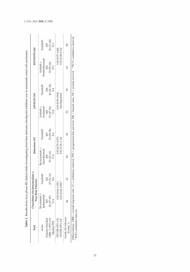
J. Clin. Med. 2020, 9, 1508
Ta
ble
1.
Res
ults
from
four
phas
e-II
Iclin
ical
tria
lsin
vest
igat
ing
fron
t-lin
eim
mun
ech
eckp
oint
inhi
bito
rus
ein
met
asta
tic
rena
lcel
lcar
cino
ma.
Tri
al
Ch
eck
Mate
-214
(In
term
ed
iate+
Po
or
Ris
kP
ati
en
ts)
IMm
oti
on
-151
JAV
EL
IN-1
01
KE
YN
OT
E-4
26
Arm
sN
ivol
umab+
Ipili
mum
abSu
niti
nib
Beva
cizu
mab+
Ate
zoliz
umab
Suni
tini
bA
xiti
nib+
Ave
lum
abSu
niti
nib
Axi
tini
b+
Pem
brol
izum
abSu
niti
nib
Pati
ents
Enro
lled
425
422
454
461
442
444
432
429
OR
R,%
(95%
CI)
42(3
7–47
)27
(22–
31)
37(3
2–41
)33
(29–
38)
51(4
7–56
)26
(22–
30)
59(5
5–64
)36
(31–
40)
Med
ian
PFS
11.6
8.4
11.2
8.4
13.8
8.4
15.1
11.1
PFS
HR
(95%
CI)
0.82
(0.6
4–1.
05)*
0.83
(0.7
0–0.
97)
0.69
(0.5
6–0.
84)
0.69
(0.5
7–0.
84)
OS
HR
(95%
CI)
0.63
(0.4
4–0.
89)†
0.93
(0.7
6–1.
14)
Not
Rep
orte
d0.
53(0
.38–
0.74
)
Gra
de3/
4A
dver
seEv
ents
,%46
6340
5455
5563
58
Abb
revi
atio
ns:
OR
R=
over
all
resp
onse
rate
,C
I=
confi
den
cein
terv
al,
PFS=
pro
gres
sion
-fre
esu
rviv
al,
HR=
haza
rdra
tio,
OS=
over
all
surv
ival
.*
99.1
%co
nfid
ence
inte
rval
;† 9
9.8%
confi
denc
ein
terv
al.
75
J. Clin. Med. 2020, 9, 1508
4.5. Cytoreductive Nephrectomy for Metastatic Disease: A Continuing Discussion
The utility of cytoreductive nephrectomy as a component of care for mRCC remains ahighly-investigated topic. Cytoreductive nephrectomy for removal of the primary tumor in patientswith metastatic disease was a standard-of-care for twenty years. However, two recent trials, SURTIMEand CARMENA, have changed the paradigm for nephrectomy in mRCC. SURTIME comparedimmediate nephrectomy (followed by sunitinib therapy) versus deferred nephrectomy (preceded bysunitinib therapy) [48]. The results of this study indicated that deferred nephrectomy did not improvethe progression-free rate at 28 weeks compared to immediate nephrectomy (43% versus 42%), but OSwas higher in the deferred arm (32.4 versus 15.0 months). These results suggested that initial systemictherapy followed by a potential debulking nephrectomy may be more efficacious for mRCC patientsthan the former standard of upfront nephrectomy.
CARMENA randomized patients with mRCC to undergo nephrectomy followed by sunitinib or toreceive sunitinib alone [49]. Results from this study indicated that sunitinib alone offered a non-inferioralternative to nephrectomy followed by sunitinib, with OS favoring the former (18.4 versus 13.9 months).The results of CARMENA and SURTIME have led to a shift away from cytoreductive nephrectomy as astandard-of-care for mRCC, except in carefully planned cases. However, ongoing trials continue to studythe relevance of cytoreductive nephrectomy. The NORDIC-SUN trial (NCT03977571) investigatesnephrectomy following therapy with nivolumab + ipilimumab. Patients with IMDC favorable-or intermediate-risk disease are randomized to receive nephrectomy or maintenance nivolumab.Patients who present with poor-risk disease can be randomized within this study as well, so long astheir risk classification is adjusted to intermediate-risk following initial therapy with nivolumab +ipilimumab or while on maintenance nivolumab. An additional trial, CYTOSHRINK (NCT04090710),uses stereotactic body radiation therapy (SBRT) as an investigational definitive treatment of the primarytumor site for patients with mRCC. In this trial, patients are randomized to receive the standard-of-carenivolumab + ipilimumab for four cycles, followed by maintenance nivolumab or the experimentalarm. The experimental arm consists of one cycle of nivolumab + ipilimumab followed by five fractionsof SBRT to the kidney lesion before resuming nivolumab + ipilimumab on the standard schedule.Both these trials are currently open to accrual.
5. Integrating Community and Academic Practices for Renal Cell Carcinoma Research
As RCC incidence continues to grow, the role of the community oncologist has grown increasinglyimportant in the network of care for RCC patients. While clinical and translational research hasprimarily been led by academic oncologists, a new approach incorporating community practitionersinto the research paradigm is now viable. It is important that medical oncologists at academic centersintegrate community practitioners, particularly urologists and oncologists, into a collaborative modelthat promotes exceptional, multi-disciplinary care alongside cutting-edge clinical and translationalresearch. Through the City of Hope network, which encompasses a central academic center anda diffuse network of community sites, we have developed a collaborative model for conductingtranslational research and clinical trial accrual that integrates academic and community oncologists.Below, we have highlighted our experiences utilizing this integrated structure to advance clinical andtranslational research and discovery in RCC.
6. Collaborations in Translational Research
As detailed above, the therapies currently in use for mRCC reflect two biological principles,namely, that (1) RCC is driven by angiogenesis and that (2) defects in the immune system can propagatethe disease. One of the genomic hallmarks of RCC are defects in the VHL gene [15,50]. Mutation ofVHL (largely sporadic, but possibly hereditary) leads to decreased ubiquitinylation of HIF [51–54].This, in turn, leads to the upregulation of vascular endothelial growth factor (VEGF), a potent mediatorof angiogenesis [55,56]. The interplay of RCC with the immune system is much more complex. Multiple
76
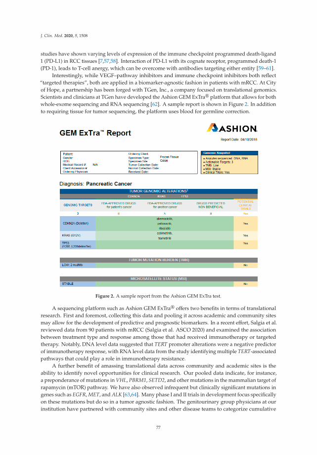
J. Clin. Med. 2020, 9, 1508
studies have shown varying levels of expression of the immune checkpoint programmed death-ligand1 (PD-L1) in RCC tissues [7,57,58]. Interaction of PD-L1 with its cognate receptor, programmed death-1(PD-1), leads to T-cell anergy, which can be overcome with antibodies targeting either entity [59–61].
Interestingly, while VEGF–pathway inhibitors and immune checkpoint inhibitors both reflect“targeted therapies”, both are applied in a biomarker-agnostic fashion in patients with mRCC. At Cityof Hope, a partnership has been forged with TGen, Inc., a company focused on translational genomics.Scientists and clinicians at TGen have developed the Ashion GEM ExTra® platform that allows for bothwhole-exome sequencing and RNA sequencing [62]. A sample report is shown in Figure 2. In additionto requiring tissue for tumor sequencing, the platform uses blood for germline correction.
Figure 2. A sample report from the Ashion GEM ExTra test.
A sequencing platform such as Ashion GEM ExTra® offers two benefits in terms of translationalresearch. First and foremost, collecting this data and pooling it across academic and community sitesmay allow for the development of predictive and prognostic biomarkers. In a recent effort, Salgia et al.reviewed data from 90 patients with mRCC (Salgia et al. ASCO 2020) and examined the associationbetween treatment type and response among those that had received immunotherapy or targetedtherapy. Notably, DNA level data suggested that TERT promoter alterations were a negative predictorof immunotherapy response, with RNA level data from the study identifying multiple TERT-associatedpathways that could play a role in immunotherapy resistance.
A further benefit of amassing translational data across community and academic sites is theability to identify novel opportunities for clinical research. Our pooled data indicate, for instance,a preponderance of mutations in VHL, PBRM1, SETD2, and other mutations in the mammalian target ofrapamycin (mTOR) pathway. We have also observed infrequent but clinically significant mutations ingenes such as EGFR, MET, and ALK [63,64]. Many phase I and II trials in development focus specificallyon these mutations but do so in a tumor agnostic fashion. The genitourinary group physicians at ourinstitution have partnered with community sites and other disease teams to categorize cumulative
77
J. Clin. Med. 2020, 9, 1508
mutational frequency. By pooling this data across our sites, we can make logical decisions regardingthe most appropriate clinical trials to bring into our portfolio.
Biomarker discovery and validation are not isolated to genomics, however. Recent work haselucidated associations between microbiome composition and clinical response to CPIs in mRCC [65].Additional work from our group and collaborators at TGen, Inc. has investigated the enteric microbiomeas a potential biomarker for responses to the above-mentioned therapy regimens in RCC [66]. We haveutilized whole genome shotgun metagenomic sequencing to correlate the stool microbiome profile andmicrobiome diversity of mRCC patients with clinical benefit, both for patients receiving VEGF–TKIsand for patients receiving CPIs (Dizman et al. ASCO 2020). In these studies, we have identified a varietyof microbial species (such as B. Intestihominis and B. adolescentis) that are correlated with clinical benefitin patients receiving systemic therapy for mRCC. The diversification of biomarker pursuits providesanother avenue for research collaboration between academic and community partners. Protocols for thecollection, storage, and shipment of stool, for example, can be implemented at any site, even those withlimited research infrastructure. The clinical volume of community practice sites poses an untappedresource for furthering biomarker discovery and other translational studies in RCC.
7. Collaborations in Clinical Research
7.1. Phase I and II Clinical Trials
Phase I clinical trials have morphed in recent years, moving away from the classical 3+ 3 design [67].More recent iterations of phase I clinical trials have started with a dose-escalation phase, expandingquickly into phase Ib trial designs, all while seeking validation in disease- or mutation-specific settings.We propose that phase I studies in dose escalation are perhaps most appropriate to remain at academiccenters. The dose-escalation phase is one where rigorous oversight is necessary, with multiple visitsand correlative studies. Furthermore, this is often the phase in which novel toxicities associated withan agent or combination are recognized and documented. We propose that highly selected phase Ibstudies could be conducted in the community (Figure 3). Criteria for site selection in these studies mustaccount for the frequency of study visits and the rigor of correlatives. For instance, if a proposed studyrequires pharmacokinetic and pharmacodynamic blood assessments on hourly intervals (not unusualin a phase-I protocol), the trial may be challenging in a busy community practice. Similarly, if a studyrequires repeat biopsies or other invasive assessments, academic centers may be better suited.
Indeed, there are real-world examples of phase I studies that may be too challenging to envisionin a community practice setting at this time. For instance, our genitourinary cancers group is heavilyinvested in studies of chimeric antigen receptor (CAR)–T-cell therapies (Dorff et al. ASCO GU 2020).In addition to the complexities associated with the manufacturing and storage of CAR–T-cells, patientsmust be kept in an inpatient setting for prompt recognition and treatment of unique side effects of thistreatment, such as cytokine release syndrome [68]. At present, commercially available CAR–T-celltherapies are used in a limited subset of patients with hematologic malignancies [69]. Until thesetherapies are adopted in a much more widespread fashion in the community (with appropriateinfrastructure and monitoring), we recommend these studies remain within academic centers.
The phase II study is a vanishing entity, as companies often now move quickly from a large phaseIb effort to phase III. This is true in mRCC space as well—as one example, COSMIC-021 (NCT03170960)is an international study chaired by investigators at our site. This phase Ib study of cabozantinib withatezolizumab enrolled cohorts in RCC, lung cancer, and prostate cancer, as well as more than a dozenother histologies. Based on significant activity in the three noted cohorts, phase III clinical trials havebeen rapidly launched, including CONTACT-03 (NCT04338269) for mRCC [70]. In general, if phase IIstudies are to be considered in the community setting, the same factors (visit frequency, correlativestudies, and so on) should aid in deciding whether studies are appropriate and should proceed.
78
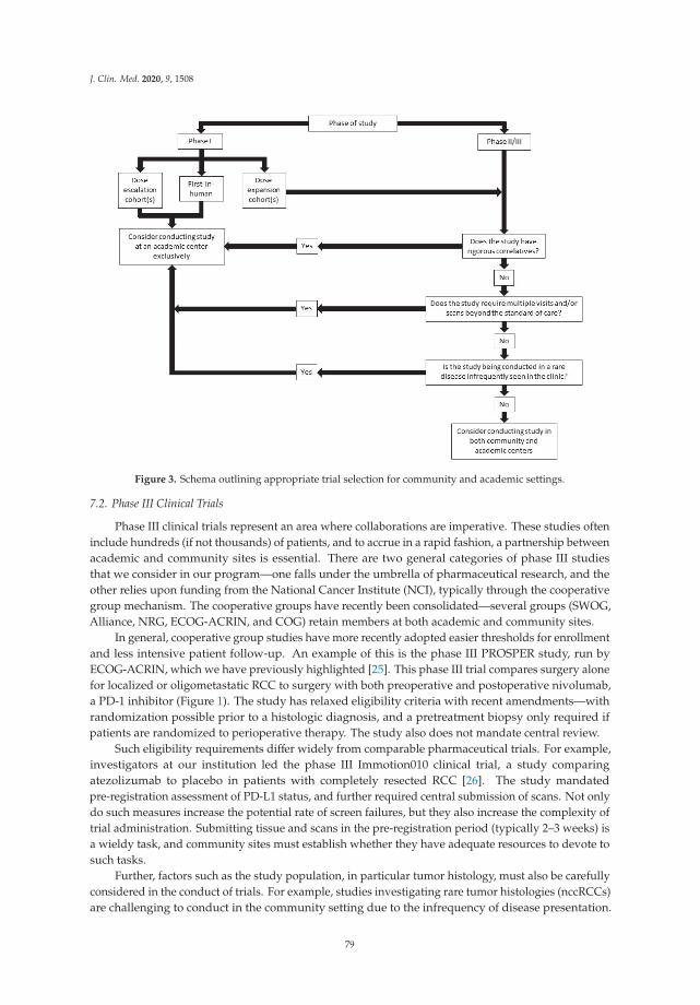
J. Clin. Med. 2020, 9, 1508
Figure 3. Schema outlining appropriate trial selection for community and academic settings.
7.2. Phase III Clinical Trials
Phase III clinical trials represent an area where collaborations are imperative. These studies ofteninclude hundreds (if not thousands) of patients, and to accrue in a rapid fashion, a partnership betweenacademic and community sites is essential. There are two general categories of phase III studiesthat we consider in our program—one falls under the umbrella of pharmaceutical research, and theother relies upon funding from the National Cancer Institute (NCI), typically through the cooperativegroup mechanism. The cooperative groups have recently been consolidated—several groups (SWOG,Alliance, NRG, ECOG-ACRIN, and COG) retain members at both academic and community sites.
In general, cooperative group studies have more recently adopted easier thresholds for enrollmentand less intensive patient follow-up. An example of this is the phase III PROSPER study, run byECOG-ACRIN, which we have previously highlighted [25]. This phase III trial compares surgery alonefor localized or oligometastatic RCC to surgery with both preoperative and postoperative nivolumab,a PD-1 inhibitor (Figure 1). The study has relaxed eligibility criteria with recent amendments—withrandomization possible prior to a histologic diagnosis, and a pretreatment biopsy only required ifpatients are randomized to perioperative therapy. The study also does not mandate central review.
Such eligibility requirements differ widely from comparable pharmaceutical trials. For example,investigators at our institution led the phase III Immotion010 clinical trial, a study comparingatezolizumab to placebo in patients with completely resected RCC [26]. The study mandatedpre-registration assessment of PD-L1 status, and further required central submission of scans. Not onlydo such measures increase the potential rate of screen failures, but they also increase the complexity oftrial administration. Submitting tissue and scans in the pre-registration period (typically 2–3 weeks) isa wieldy task, and community sites must establish whether they have adequate resources to devote tosuch tasks.
Further, factors such as the study population, in particular tumor histology, must also be carefullyconsidered in the conduct of trials. For example, studies investigating rare tumor histologies (nccRCCs)are challenging to conduct in the community setting due to the infrequency of disease presentation.
79
J. Clin. Med. 2020, 9, 1508
One such example is SWOG 1500 (NCT02761057), a study of selective MET-kinase inhibitors forpatients with papillary renal cell carcinoma that was chaired by physicians in our genitourinaryoncology disease team. Although the study was sponsored by an NCI cooperative group, the relativerarity of papillary histology (~10–15% of metastatic renal cell carcinomas) dictated that the study onlybe available at the academic center [71]. As patients with rare histologies are often referred fromthe community to experts at academic centers, consolidation of trial sites in these instances shouldbe considered.
8. Optimizing Partnerships between Community and Academic Sites
As noted previously, the academic cancer center is typically well staffed with multiple researchnurses and clinical research associates who work within a single disease space. Each has ownershipover a selected number of protocols (typically 2–3), and there are often regular meetings with theprincipal investigator. These face to face meetings with principal investigators offer an opportunityfor education regarding the protocol and, furthermore, allow for a closer degree of oversight ofprotocol-related activities. At City of Hope, investigators in the genitourinary disease team frequentlyhost didactic sessions for research staff, offering them insight into the current standard management ofRCC, bladder, and prostate cancer. As research staff are often charged with screening for enrollmentor suggesting toxicity attributions, understanding the underlying disease and associated treatmentlandscape is critically important.
Models of research in community practices vary but commonly require research staff to managea much broader swath of clinical protocols across multiple cancer types. Furthermore, the principalinvestigator for a study may not reside at the community practice site, and thus is unable to engagewith clinical and administrative staff on a routine basis. At City of Hope, the vast majority of principalinvestigators reside at the main campus; thus, elements of study oversight (filing significant adverseevents, reporting study deviations) may be somewhat delayed at community sites. In addition, servicesreadily available at the main campus may be less accessible in the community practices; for example,labs may be obtained off-site through third-party vendors. This can create delays in lab reporting,and complicate study protocols that require same-day lab results to issue therapy. Many protocols alsoembed correlates that require additional processing methods (e.g., centrifugation for separation ofplasma aliquots).
Similarly, access to appropriate and timely scans can vary by recruitment location, with anacademic center generally able to accommodate access to both standard and novel imaging tests(e.g., fluciclovine or PSMA PET scans for prostate cancer) and not have to rely on third parties totransfer images so that relevant tumor measurements can be obtained. Whereas these may be trivialissues on a main academic campus, access to a centrifuge, processing equipment, and appropriateimaging modalities may be limited at community sites.
Despite these noted limitations, community practices can serve as valuable contributors to clinicalresearch endeavors housed at a main campus. To optimize this relationship and promote successfulcollaborations, we have implemented several strategies. Firstly, self-nominated community diseaseteam leads are identified to serve as liaisons between the research team on the main campus andoncologists practicing in the community. These individuals also play a role in determining the feasibilityof specific clinical trials in the community, taking into account many of the considerations that we haveoutlined throughout. In addition, these individuals are invited to bimonthly genitourinary diseaseteam meetings, where the group conducts a thorough review of all existing protocols and evaluates newclinical trial proposals. Finally, in recognition of the limited time permitted to community oncologists tolearn about existing research endeavors, our genitourinary cancers group has established a newsletterthat highlights ongoing and planned clinical trials with only the most salient information—basiceligibility requirements and study schema, for instance. These newsletters have triggered a number ofdirect referrals for clinical trial enrollment.
80
J. Clin. Med. 2020, 9, 1508
One relatively recent development among our community-based satellite clinics is theimplementation of a standardized electronic health record (EHR) mirroring that at the main campus,optimizing the process by which physicians can evaluate and triage patients who may be suitableclinical trial candidates.
9. Conclusions
The clinical approach to RCC has drastically changed over the past two decades. The noveltherapeutic options introduced to the medical oncologist’s armamentarium have leveragedground-breaking work on angiogenesis and induced hypoxia in addition to the principle of immunecheckpoint blockade to overcome T-cell anergy. To maintain the momentum of RCC drug development,rigorous translational research must continue advancing our understanding of RCC biology andclinical trials must continue to accrue patients at a rapid pace. As we have highlighted herein, this canhappen effectively and efficiently if community practices are integrated with academic centers inthe conduct of translational research and clinical trials. We have proposed a number of strategies tooptimize this relationship, with the common theme being enhanced communication. When oncologistsat community and academic practices frequently dialogue, gaining a deeper understanding of eachother’s practice patterns, a portfolio of trials can be developed that optimizes accrual and advancesscientific findings more rapidly.
The positive effects of an academic and community collaboration on trial accrual have beenevidenced in our network—with our genitourinary cancers group recognized as the top accrual sitefor the adjuvant EVEREST study, a trial comparing everolimus and placebo in patients with resectedlocalized RCC [24]. Our enrollment was driven in large part by patients seen at our community-basedsatellite offices. The rapid success of this collaborative effort was used as a foundation for the enrollmentof patients into further adjuvant trials (e.g., E2810 and Immotion010), with similar positive outcomes.In sum, a strong collaborative approach between our academic center and affiliated communitypractices has resulted in a model conducive to high accrual rates across a broad range of clinical trials.This feat, along with the opportunities for collaboration in the translational sciences, underscores thebenefit of these partnerships.
Author Contributions: Conceptualization, N.J.S., S.K.P.; Methodology, N.J.S., S.K.P.; Resources, S.K.P.; DataCuration, N.J.S., S.K.P.; Writing—Original Draft Preparation, N.J.S., S.K.P.; Writing—Review & Editing, N.J.S.,E.J.P., K.Y., M.Z., S.K.P.; Visualization, N.J.S., S.K.P.; Supervision, S.K.P.; Project Administration, N.J.S., E.J.P., K.Y.,M.Z., S.K.P. All authors have read and agreed to the published version of the manuscript.
Funding: This research received no external funding.
Conflicts of Interest: Nicholas Salgia, Errol Philip, Mohammadbagher Ziari, and Kelly Yap have no conflicts ofinterest that might be relevant to the contents of this manuscript. Sumanta K. Pal, MD—honoraria: Novartis,Medivation, Astellas Pharma; consulting or advisory role: Pfizer, Novartis, Aveo, Myriad; pharmaceuticals:Genentech, Exelixis, Bristol-Myers Squibb, Astellas Pharma; research funding: Medivation.
References
1. Atzpodien, J.; Kirchner, H.; Jonas, U.; Bergmann, L.; Schott, H.; Heynemann, H.; Fornara, P.; Loening, S.A.;Roigas, J.; Muller, S.C.; et al. Interleukin-2- and interferon alfa-2a-based immunochemotherapy in advancedrenal cell carcinoma: A Prospectively Randomized Trial of the German Cooperative Renal CarcinomaChemoimmunotherapy Group (DGCIN). J. Clin. Oncol. Off. J. Am. Soc. Clin. Oncol. 2004, 22, 1188–1194.[CrossRef] [PubMed]
2. Motzer, R.J.; Hutson, T.E.; Tomczak, P.; Michaelson, M.D.; Bukowski, R.M.; Rixe, O.; Oudard, S.; Negrier, S.;Szczylik, C.; Kim, S.T.; et al. Sunitinib versus interferon alfa in metastatic renal-cell carcinoma. N. Engl. J.Med. 2007, 356, 115–124. [CrossRef] [PubMed]
3. Sternberg, C.N.; Davis, I.D.; Mardiak, J.; Szczylik, C.; Lee, E.; Wagstaff, J.; Barrios, C.H.; Salman, P.;Gladkov, O.A.; Kavina, A.; et al. Pazopanib in locally advanced or metastatic renal cell carcinoma: Results ofa randomized phase III trial. J. Clin. Oncol. Off. J. Am. Soc. Clin. Oncol. 2010, 28, 1061–1068. [CrossRef][PubMed]
81
J. Clin. Med. 2020, 9, 1508
4. Hutson, T.E.; Al-Shukri, S.; Stus, V.P.; Lipatov, O.N.; Shparyk, Y.; Bair, A.H.; Rosbrook, B.; Andrews, G.I.;Vogelzang, N.J. Axitinib Versus Sorafenib in First-Line Metastatic Renal Cell Carcinoma: Overall SurvivalFrom a Randomized Phase III Trial. Clin. Genitourin. Cancer 2017, 15, 72–76. [CrossRef] [PubMed]
5. Motzer, R.J.; Escudier, B.; McDermott, D.F.; George, S.; Hammers, H.J.; Srinivas, S.; Tykodi, S.S.; Sosman, J.A.;Procopio, G.; Plimack, E.R.; et al. Nivolumab versus Everolimus in Advanced Renal-Cell Carcinoma. N. Engl.J. Med. 2015, 373, 1803–1813. [CrossRef] [PubMed]
6. Motzer, R.J.; Tannir, N.M.; McDermott, D.F.; Aren Frontera, O.; Melichar, B.; Choueiri, T.K.; Plimack, E.R.;Barthelemy, P.; Porta, C.; George, S.; et al. Nivolumab plus Ipilimumab versus Sunitinib in AdvancedRenal-Cell Carcinoma. N. Engl. J. Med. 2018, 378, 1277–1290. [CrossRef]
7. Rini, B.I.; Plimack, E.R.; Stus, V.; Gafanov, R.; Hawkins, R.; Nosov, D.; Pouliot, F.; Alekseev, B.; Soulieres, D.;Melichar, B.; et al. Pembrolizumab plus Axitinib versus Sunitinib for Advanced Renal-Cell Carcinoma.N. Engl. J. Med. 2019, 380, 1116–1127. [CrossRef]
8. Motzer, R.J.; Penkov, K.; Haanen, J.; Rini, B.; Albiges, L.; Campbell, M.T.; Venugopal, B.; Kollmannsberger, C.;Negrier, S.; Uemura, M.; et al. Avelumab plus Axitinib versus Sunitinib for Advanced Renal-Cell Carcinoma.N. Engl. J. Med. 2019, 380, 1103–1115. [CrossRef]
9. Locations, Maps, and Directions. Available online: https://www.cityofhope.org/about-city-of-hope/locations(accessed on 10 April 2020).
10. Siegel, R.L.; Miller, K.D.; Jemal, A. Cancer statistics, 2020. CA Cancer J. Clin. 2020, 70, 7–30. [CrossRef]11. Capitanio, U.; Montorsi, F. Renal cancer. Lancet 2016, 387, 894–906. [CrossRef]12. Manini, C.; López, J.I. The Labyrinth of Renal Cell Carcinoma. Cancers 2020, 12, 521. [CrossRef] [PubMed]13. Signoretti, S.; Flaifel, A.; Chen, Y.B.; Reuter, V.E. Renal Cell Carcinoma in the Era of Precision Medicine:
From Molecular Pathology to Tissue-Based Biomarkers. J. Clin. Oncol. Off. J. Am. Soc. Clin. Oncol. 2018, 36,Jco2018792259. [CrossRef] [PubMed]
14. Sato, Y.; Yoshizato, T.; Shiraishi, Y.; Maekawa, S.; Okuno, Y.; Kamura, T.; Shimamura, T.; Sato-Otsubo, A.;Nagae, G.; Suzuki, H.; et al. Integrated molecular analysis of clear-cell renal cell carcinoma. Nat. Genet. 2013,45, 860–867. [CrossRef] [PubMed]
15. Comprehensive molecular characterization of clear cell renal cell carcinoma. Nature 2013, 499, 43–49.[CrossRef] [PubMed]
16. Mikhail, M.I.; Singh, A.K. Von Hippel Lindau Syndrome. In StatPearls; StatPearls Publishing LLC: TreasureIsland, FL, USA, 2020.
17. Hakimi, A.A.; Ostrovnaya, I.; Reva, B.; Schultz, N.; Chen, Y.-B.; Gonen, M.; Liu, H.; Takeda, S.; Voss, M.H.;Tickoo, S.K.; et al. Adverse Outcomes in Clear Cell Renal Cell Carcinoma with Mutations of 3p21 EpigeneticRegulators BAP1 and SETD2: A Report by MSKCC and the KIRC TCGA Research Network. Clin. CancerRes. 2013, 19, 3259–3267. [CrossRef] [PubMed]
18. Comprehensive Molecular Characterization of Papillary Renal-Cell Carcinoma. N. Engl. J. Med. 2015, 374,135–145. [CrossRef]
19. Linehan, W.M.; Srinivasan, R.; Garcia, J.A. Non-clear cell renal cancer: Disease-based management andopportunities for targeted therapeutic approaches. Semin. Oncol. 2013, 40, 511–520. [CrossRef]
20. Haas, N.B.; Manola, J.; Uzzo, R.G.; Flaherty, K.T.; Wood, C.G.; Kane, C.; Jewett, M.; Dutcher, J.P.;Atkins, M.B.; Pins, M.; et al. Adjuvant sunitinib or sorafenib for high-risk, non-metastatic renal-cellcarcinoma (ECOG-ACRIN E2805): A double-blind, placebo-controlled, randomised, phase 3 trial. Lancet2016, 387, 2008–2016. [CrossRef]
21. Ravaud, A.; Motzer, R.J.; Pandha, H.S.; George, D.J.; Pantuck, A.J.; Patel, A.; Chang, Y.-H.; Escudier, B.;Donskov, F.; Magheli, A.; et al. Adjuvant Sunitinib in High-Risk Renal-Cell Carcinoma after Nephrectomy.N. Engl. J. Med. 2016, 375, 2246–2254. [CrossRef]
22. Motzer, R.J.; Haas, N.B.; Donskov, F.; Gross-Goupil, M.; Varlamov, S.; Kopyltsov, E.; Lee, J.L.; Melichar, B.;Rini, B.I.; Choueiri, T.K.; et al. Randomized Phase III Trial of Adjuvant Pazopanib Versus Placebo AfterNephrectomy in Patients With Localized or Locally Advanced Renal Cell Carcinoma. J. Clin. Oncol. 2017, 35,3916–3923. [CrossRef]
23. Gross-Goupil, M.; Kwon, T.G.; Eto, M.; Ye, D.; Miyake, H.; Seo, S.I.; Byun, S.S.; Lee, J.L.; Master, V.; Jin, J.;et al. Axitinib versus placebo as an adjuvant treatment of renal cell carcinoma: Results from the phase III,randomized ATLAS trial. Ann. Oncol. 2018, 29, 2371–2378. [CrossRef]
82
J. Clin. Med. 2020, 9, 1508
24. Synold, T.W.; Plets, M.; Tangen, C.M.; Heath, E.I.; Palapattu, G.S.; Mack, P.C.; Stein, M.N.; Meng, M.V.;Lara, P.; Vogelzang, N.J.; et al. Everolimus Exposure as a Predictor of Toxicity in Renal Cell Cancer Patientsin the Adjuvant Setting: Results of a Pharmacokinetic Analysis for SWOG S0931 (EVEREST), a Phase IIIStudy (NCT01120249). Kidney Cancer 2019, 3, 111–118. [CrossRef] [PubMed]
25. Patel, H.D.; Puligandla, M.; Shuch, B.M.; Leibovich, B.C.; Kapoor, A.; Master, V.A.; Drake, C.G.; Heng, D.Y.;Lara, P.N.; Choueiri, T.K.; et al. The future of perioperative therapy in advanced renal cell carcinoma: Howcan we PROSPER? Future Oncol. (Lond. Engl.) 2019, 15, 1683–1695. [CrossRef] [PubMed]
26. Uzzo, R.; Bex, A.; Rini, B.I.; Albiges, L.; Suarez, C.; Donaldson, F.; Asakawa, T.; Schiff, C.; Pal, S.K. A phase IIIstudy of atezolizumab (atezo) vs placebo as adjuvant therapy in renal cell carcinoma (RCC) patients (pts) athigh risk of recurrence following resection (IMmotion010). J. Clin. Oncol. 2017, 35, TPS4598. [CrossRef]
27. Angulo, J.C.; Lawrie, C.H.; López, J.I. Sequential treatment of metastatic renal cancer in a complex evolvinglandscape. Ann. Transl. Med. 2019, 7, S272. [CrossRef]
28. Amin, A.; White, R.L. Interleukin-2 in Renal Cell Carcinoma: A Has-Been or a Still-Viable Option? J. KidneyCancer Vhl. 2014, 1, 74–83. [CrossRef]
29. Dranoff, G. Cytokines in cancer pathogenesis and cancer therapy. Nat. Rev. Cancer 2004, 4, 11–22. [CrossRef]30. Interleukin 2 Gene Therapy for Prostate Cancer: Phase I Clinical Trial and Basic Biology. Hum. Gene Ther.
2001, 12, 883–892. [CrossRef]31. Motzer, R.J.; Mazumdar, M.; Bacik, J.; Berg, W.; Amsterdam, A.; Ferrara, J. Survival and Prognostic
Stratification of 670 Patients With Advanced Renal Cell Carcinoma. J. Clin. Oncol. 1999, 17, 2530. [CrossRef]32. Mekhail, T.M.; Abou-Jawde, R.M.; Boumerhi, G.; Malhi, S.; Wood, L.; Elson, P.; Bukowski, R. Validation and
extension of the Memorial Sloan-Kettering prognostic factors model for survival in patients with previouslyuntreated metastatic renal cell carcinoma. J. Clin. Oncol. Off. J. Am. Soc. Clin. Oncol. 2005, 23, 832–841.[CrossRef]
33. Voss, M.H.; Reising, A.; Cheng, Y.; Patel, P.; Marker, M.; Kuo, F.; Chan, T.A.; Choueiri, T.K.; Hsieh, J.J.;Hakimi, A.A.; et al. Genomically annotated risk model for advanced renal-cell carcinoma: A retrospectivecohort study. Lancet Oncol. 2018, 19, 1688–1698. [CrossRef]
34. Heng, D.Y.; Xie, W.; Regan, M.M.; Warren, M.A.; Golshayan, A.R.; Sahi, C.; Eigl, B.J.; Ruether, J.D.; Cheng, T.;North, S.; et al. Prognostic factors for overall survival in patients with metastatic renal cell carcinoma treatedwith vascular endothelial growth factor-targeted agents: Results from a large, multicenter study. J. Clin.Oncol. Off. J. Am. Soc. Clin. Oncol. 2009, 27, 5794–5799. [CrossRef] [PubMed]
35. Heng, D.Y.C.; Xie, W.; Regan, M.M.; Harshman, L.C.; Bjarnason, G.A.; Vaishampayan, U.N.; Mackenzie, M.;Wood, L.; Donskov, F.; Tan, M.-H.; et al. External validation and comparison with other models of theInternational Metastatic Renal-Cell Carcinoma Database Consortium prognostic model: A population-basedstudy. Lancet Oncol. 2013, 14, 141–148. [CrossRef]
36. Kaelin, W.G., Jr. The von Hippel-Lindau tumour suppressor protein: O2 sensing and cancer. Nat. Rev. Cancer2008, 8, 865–873. [CrossRef] [PubMed]
37. Kaelin, W.G., Jr. Treatment of kidney cancer: Insights provided by the VHL tumor-suppressor protein.Cancer 2009, 115, 2262–2272. [CrossRef] [PubMed]
38. Makhov, P.; Joshi, S.; Ghatalia, P.; Kutikov, A.; Uzzo, R.G.; Kolenko, V.M. Resistance to Systemic Therapiesin Clear Cell Renal Cell Carcinoma: Mechanisms and Management Strategies. Mol. Cancer Ther. 2018, 17,1355–1364. [CrossRef]
39. Choueiri, T.K.; Halabi, S.; Sanford, B.L.; Hahn, O.; Michaelson, M.D.; Walsh, M.K.; Feldman, D.R.; Olencki, T.;Picus, J.; Small, E.J.; et al. Cabozantinib Versus Sunitinib As Initial Targeted Therapy for Patients WithMetastatic Renal Cell Carcinoma of Poor or Intermediate Risk: The Alliance A031203 CABOSUN Trial. J. Clin.Oncol. 2017, 35, 591–597. [CrossRef]
40. Guo, H.; German, P.; Bai, S.; Barnes, S.; Guo, W.; Qi, X.; Lou, H.; Liang, J.; Jonasch, E.; Mills, G.B.; et al. ThePI3K/AKT Pathway and Renal Cell Carcinoma. J. Genet. Genom. 2015, 42, 343–353. [CrossRef]
41. Motzer, R.J.; Hutson, T.E.; Glen, H.; Michaelson, M.D.; Molina, A.; Eisen, T.; Jassem, J.; Zolnierek, J.;Maroto, J.P.; Mellado, B.; et al. Lenvatinib, everolimus, and the combination in patients with metastaticrenal cell carcinoma: A randomised, phase 2, open-label, multicentre trial. Lancet Oncol. 2015, 16, 1473–1482.[CrossRef]
42. Boni, J.P.; Hug, B.; Leister, C.; Sonnichsen, D. Intravenous temsirolimus in cancer patients: Clinicalpharmacology and dosing considerations. Semin. Oncol. 2009, 36 (Suppl. 3), S18–S25. [CrossRef]
83
J. Clin. Med. 2020, 9, 1508
43. Hudes, G.; Carducci, M.; Tomczak, P.; Dutcher, J.; Figlin, R.; Kapoor, A.; Staroslawska, E.; Sosman, J.;McDermott, D.; Bodrogi, I.; et al. Temsirolimus, interferon alfa, or both for advanced renal-cell carcinoma.N. Engl. J. Med. 2007, 356, 2271–2281. [CrossRef] [PubMed]
44. Hodi, F.S.; O’Day, S.J.; McDermott, D.F.; Weber, R.W.; Sosman, J.A.; Haanen, J.B.; Gonzalez, R.; Robert, C.;Schadendorf, D.; Hassel, J.C.; et al. Improved Survival with Ipilimumab in Patients with Metastatic Melanoma.N. Engl. J. Med. 2010, 363, 711–723. [CrossRef] [PubMed]
45. Wei, S.C.; Duffy, C.R.; Allison, J.P. Fundamental Mechanisms of Immune Checkpoint Blockade Therapy.Cancer Discov. 2018, 8, 1069–1086. [CrossRef] [PubMed]
46. Rini, B.I.; Powles, T.; Atkins, M.B.; Escudier, B.; McDermott, D.F.; Suarez, C.; Bracarda, S.; Stadler, W.M.;Donskov, F.; Lee, J.L.; et al. Atezolizumab plus bevacizumab versus sunitinib in patients with previouslyuntreated metastatic renal cell carcinoma (IMmotion151): A multicentre, open-label, phase 3, randomisedcontrolled trial. Lancet (Lond. Engl.) 2019, 393, 2404–2415. [CrossRef]
47. Motzer, R.J.; Jonasch, E.; Michaelson, M.D.; Nandagopal, L.; Gore, J.L.; George, S.; Alva, A.; Haas, N.;Harrison, M.R.; Plimack, E.R.; et al. NCCN Guidelines Insights: Kidney Cancer, Version 2.2020. J. Natl.Compr. Cancer Netw. JNCCN 2019, 17, 1278–1285. [CrossRef]
48. Bex, A.; Mulders, P.; Jewett, M.; Wagstaff, J.; van Thienen, J.V.; Blank, C.U.; van Velthoven, R.; del PilarLaguna, M.; Wood, L.; van Melick, H.H.E.; et al. Comparison of Immediate vs Deferred CytoreductiveNephrectomy in Patients With Synchronous Metastatic Renal Cell Carcinoma Receiving Sunitinib:The SURTIME Randomized Clinical Trial. JAMA Oncol. 2019, 5, 164–170. [CrossRef]
49. Mejean, A.; Ravaud, A.; Thezenas, S.; Colas, S.; Beauval, J.B.; Bensalah, K.; Geoffrois, L.; Thiery-Vuillemin, A.;Cormier, L.; Lang, H.; et al. Sunitinib Alone or after Nephrectomy in Metastatic Renal-Cell Carcinoma.N. Engl. J. Med. 2018, 379, 417–427. [CrossRef]
50. Linehan, W.M.; Lerman, M.I.; Zbar, B. Identification of the von Hippel-Lindau (VHL) gene. Its role in renalcancer. JAMA 1995, 273, 564–570. [CrossRef]
51. Ohh, M.; Park, C.W.; Ivan, M.; Hoffman, M.A.; Kim, T.Y.; Huang, L.E.; Pavletich, N.; Chau, V.;Kaelin, W.G. Ubiquitination of hypoxia-inducible factor requires direct binding to the beta-domain ofthe von Hippel-Lindau protein. Nat. Cell Biol. 2000, 2, 423–427. [CrossRef]
52. Cockman, M.E.; Masson, N.; Mole, D.R.; Jaakkola, P.; Chang, G.W.; Clifford, S.C.; Maher, E.R.; Pugh, C.W.;Ratcliffe, P.J.; Maxwell, P.H. Hypoxia inducible factor-alpha binding and ubiquitylation by the vonHippel-Lindau tumor suppressor protein. J. Biol. Chem. 2000, 275, 25733–25741. [CrossRef]
53. Tanimoto, K.; Makino, Y.; Pereira, T.; Poellinger, L. Mechanism of regulation of the hypoxia-inducible factor-1alpha by the von Hippel-Lindau tumor suppressor protein. Embo J. 2000, 19, 4298–4309. [CrossRef] [PubMed]
54. Krieg, M.; Haas, R.; Brauch, H.; Acker, T.; Flamme, I.; Plate, K.H. Up-regulation of hypoxia-inducible factorsHIF-1alpha and HIF-2alpha under normoxic conditions in renal carcinoma cells by von Hippel-Lindautumor suppressor gene loss of function. Oncogene 2000, 19, 5435–5443. [CrossRef] [PubMed]
55. Na, X.; Wu, G.; Ryan, C.K.; Schoen, S.R.; di’Santagnese, P.A.; Messing, E.M. Overproduction ofvascular endothelial growth factor related to von Hippel-Lindau tumor suppressor gene mutations andhypoxia-inducible factor-1 alpha expression in renal cell carcinomas. J. Urol. 2003, 170, 588–592. [CrossRef][PubMed]
56. Conway, E.M.; Collen, D.; Carmeliet, P. Molecular mechanisms of blood vessel growth. Cardiovasc. Res. 2001,49, 507–521. [CrossRef]
57. Choueiri, T.K.; Albiges, L.; Haanen, J.B.A.G.; Larkin, J.M.G.; Uemura, M.; Pal, S.K.; Gravis, G.; Campbell, M.T.;Penkov, K.; Lee, J.-L.; et al. Biomarker analyses from JAVELIN Renal 101: Avelumab + axitinib (A+Ax)versus sunitinib (S) in advanced renal cell carcinoma (aRCC). J. Clin. Oncol. 2019, 37, 101. [CrossRef]
58. Taube, J.M.; Klein, A.; Brahmer, J.R.; Xu, H.; Pan, X.; Kim, J.H.; Chen, L.; Pardoll, D.M.; Topalian, S.L.;Anders, R.A. Association of PD-1, PD-1 ligands, and other features of the tumor immune microenvironmentwith response to anti-PD-1 therapy. Clin. Cancer Res. Off. J. Am. Assoc. Cancer Res. 2014, 20, 5064–5074.[CrossRef]
59. Barber, D.L.; Wherry, E.J.; Masopust, D.; Zhu, B.; Allison, J.P.; Sharpe, A.H.; Freeman, G.J.; Ahmed, R.Restoring function in exhausted CD8 T cells during chronic viral infection. Nature 2006, 439, 682–687.[CrossRef]
84
J. Clin. Med. 2020, 9, 1508
60. Sakuishi, K.; Apetoh, L.; Sullivan, J.M.; Blazar, B.R.; Kuchroo, V.K.; Anderson, A.C. Targeting Tim-3 and PD-1pathways to reverse T cell exhaustion and restore anti-tumor immunity. J. Exp. Med. 2010, 207, 2187–2194.[CrossRef]
61. LaFleur, M.W.; Muroyama, Y.; Drake, C.G.; Sharpe, A.H. Inhibitors of the PD-1 Pathway in Tumor Therapy.J. Immunol. 2018, 200, 375–383. [CrossRef]
62. Nasser, S.; Kurdolgu, A.A.; Izatt, T.; Aldrich, J.; Russell, M.L.; Christoforides, A.; Tembe, W.; Keifer, J.A.;Corneveaux, J.J.; Byron, S.A.; et al. An integrated framework for reporting clinically relevant biomarkersfrom paired tumor/normal genomic and transcriptomic sequencing data in support of clinical trials inpersonalized medicine. Pac. Symp. Biocomput. 2015, 56–67. [CrossRef]
63. Pal, S.K.; Sonpavde, G.; Agarwal, N.; Vogelzang, N.J.; Srinivas, S.; Haas, N.B.; Signoretti, S.; McGregor, B.A.;Jones, J.; Lanman, R.B.; et al. Evolution of Circulating Tumor DNA Profile from First-line to SubsequentTherapy in Metastatic Renal Cell Carcinoma. Eur. Urol. 2017, 72, 557–564. [CrossRef] [PubMed]
64. Pal, S.K.; Bergerot, P.; Dizman, N.; Bergerot, C.; Adashek, J.; Madison, R.; Chung, J.H.; Ali, S.M.; Jones, J.O.;Salgia, R. Responses to Alectinib in ALK-rearranged Papillary Renal Cell Carcinoma. Eur. Urol. 2018, 74,124–128. [CrossRef]
65. Routy, B.; Le Chatelier, E.; Derosa, L.; Duong, C.P.M.; Alou, M.T.; Daillère, R.; Fluckiger, A.; Messaoudene, M.;Rauber, C.; Roberti, M.P.; et al. Gut microbiome influences efficacy of PD-1–based immunotherapy againstepithelial tumors. Science (N. Y.) 2018, 359, 91–97. [CrossRef] [PubMed]
66. Pal, S.K.; Li, S.M.; Wu, X.; Qin, H.; Kortylewski, M.; Hsu, J.; Carmichael, C.; Frankel, P. Stool BacteriomicProfiling in Patients with Metastatic Renal Cell Carcinoma Receiving Vascular Endothelial GrowthFactor-Tyrosine Kinase Inhibitors. Clin. Cancer Res. Off. J. Am. Assoc. Cancer Res. 2015, 21, 5286–5293.[CrossRef]
67. Paoletti, X.; Drubay, D.; Collette, L. Dose-Finding Methods: Moving Away from the 3 + 3 to Include RicherOutcomes. Clin. Cancer Res. Off. J. Am. Assoc. Cancer Res. 2017, 23, 3977–3979. [CrossRef] [PubMed]
68. Bonifant, C.L.; Jackson, H.J.; Brentjens, R.J.; Curran, K.J. Toxicity and management in CAR T-cell therapy.Mol. Ther. Oncolytics 2016, 3, 16011. [CrossRef] [PubMed]
69. Gill, S.; Maus, M.V.; Porter, D.L. Chimeric antigen receptor T cell therapy: 25years in the making. Blood Rev.2016, 30, 157–167. [CrossRef] [PubMed]
70. Exelixis Partners with Roche to Trial Cancer Combination Therapy. Available online: https://www.clinicaltrialsarena.com/news/exelixis-roche-cancer-trial/ (accessed on 10 April 2020).
71. Cheville, J.C.; Lohse, C.M.; Zincke, H.; Weaver, A.L.; Blute, M.L. Comparisons of outcome and prognosticfeatures among histologic subtypes of renal cell carcinoma. Am. J. Surg. Pathol. 2003, 27, 612–624. [CrossRef][PubMed]
© 2020 by the authors. Licensee MDPI, Basel, Switzerland. This article is an open accessarticle distributed under the terms and conditions of the Creative Commons Attribution(CC BY) license (http://creativecommons.org/licenses/by/4.0/).
85


Journal of
Clinical Medicine
Review
Integrating Academic and Community Cancer Care andResearch through Multidisciplinary OncologyPathways for Value-Based Care: A Review and the Cityof Hope Experience
Linda D. Bosserman 1,*, Mary Cianfrocca 1, Bertram Yuh 2, Christina Yeon 3, Helen Chen 4,
Stephen Sentovich 2, Amy Polverini 5, Finly Zachariah 6, Debbie Deaville 7, Ashley B. Lee 8, Mina
S. Sedrak 1, Elisabeth King 9, Stacy Gray 9, Denise Morse 10, Scott Glaser 11, Geetika Bhatt 12,
Camille Adeimy 13, TingTing Tan 14, Joseph Chao 1, Arin Nam 1, Isaac B. Paz 5, Laura Kruper 2,
Poornima Rao 5, Karen Sokolov 15, Prakash Kulkarni 1, Ravi Salgia 1, Jonathan Yamzon 2 and
Deron Johnson 16
1 Department of Medical Oncology and Therapeutics Research, City of Hope, Duarte, CA 91010, USA;[email protected] (M.C.); [email protected] (M.S.S.); [email protected] (J.C.); [email protected] (A.N.);[email protected] (P.K.); [email protected] (R.S.)
2 Department of Surgery, City of Hope, Duarte, CA 91010, USA; [email protected] (B.Y.); [email protected] (S.S.);[email protected] (L.K.); [email protected] (J.Y.)
3 Department of Medical Oncology and Therapeutics Research, City of Hope, South Pasadena, CA 91030, USA;[email protected]
4 Department of Radiation Oncology, City of Hope, South Pasadena, CA 91030, USA; [email protected] Department of Surgery, City of Hope, South Pasadena, CA 91030, USA; [email protected] (A.P.);
[email protected] (I.B.P.); [email protected] (P.R.)6 Department Supportive Care Medicine, City of Hope, Duarte, CA 91010, USA; [email protected] Department of Enterprise Business Intelligence, City of Hope, Duarte, CA 91010, USA; [email protected] Department of Clinical Research Operations, City of Hope, Duarte, CA 91010, USA; [email protected] Department of Population Sciences in the Division of Clinical Cancer Genomics, City of Hope,
Duarte, CA 91010, USA; [email protected] (E.K.); [email protected] (S.G.)10 Department of Quality, Risk and Regulatory Management, City of Hope, Duarte, CA 91010, USA;
[email protected] Department of Radiation Oncology, City of Hope, Duarte, CA 91010, USA; [email protected] Department of Medical Oncology and Therapeutics Research, City of Hope, Lancaster, CA 93534, USA;
[email protected] Department of Medical Oncology and Therapeutics Research, City of Hope, Upland, CA 91784, USA;
[email protected] Department of Medical Oncology and Therapeutics Research, City of Hope, Newport Beach, CA 92660, USA;
[email protected] Department of Radiation Oncology, City of Hope, Torrance, CA 90503, USA; [email protected] Department of Clinical Informatics, City of Hope, Duarte, CA 91010, USA; [email protected]* Correspondence: [email protected]
Received: 10 November 2020; Accepted: 29 December 2020; Published: 7 January 2021
Abstract: As the US transitions from volume- to value-based cancer care, many cancer centers andcommunity groups have joined to share resources to deliver measurable, high-quality cancer care andclinical research with the associated high patient satisfaction, provider satisfaction, and practice healthat optimal costs that are the hallmarks of value-based care. Multidisciplinary oncology care pathwaysare essential components of value-based care and their payment metrics. Oncology pathways are
J. Clin. Med. 2021, 10, 188; doi:10.3390/jcm10020188 www.mdpi.com/journal/jcm
87
J. Clin. Med. 2021, 10, 188
evidence-based, standardized but personalizable care plans to guide cancer care. Pathways have beendeveloped and studied for the major medical, surgical, radiation, and supportive oncology disciplinesto support decision-making, streamline care, and optimize outcomes. Implementing multidisciplinaryoncology pathways can facilitate comprehensive care plans for each cancer patient throughout theircancer journey and across large multisite delivery systems. Outcomes from the delivered pathway-basedcare can then be evaluated against individual and population benchmarks. The complexity of adoption,implementation, and assessment of multidisciplinary oncology pathways, however, presents manychallenges. We review the development and components of value-based cancer care and detail City ofHope’s (COH) academic and community-team-based approaches for implementing multidisciplinarypathways. We also describe supportive components with available results towards enterprise-widevalue-based care delivery.
Keywords: value-based care; value-based cancer care; oncology pathways; Early Recovery After Surgery(ERAS); team-based care; oncology medical home; integrated cancer care; supportive care pathways;surgical pathways; cancer care plans
1. Introduction: Multidisciplinary Oncology Pathways Are a Foundation of Value-Based Cancer Care
While the majority of cancer care in the US is provided in the community (non-academic) setting,over the past 12 years, the organizational structure of oncology care delivery has shifted to networks ofcommunity oncologists partnered with academic centers, hospital systems, or other community practices.Aligned enterprises have evolved from the well-documented closing, merging, or acquisition of communityoncology practices since 2008 [1,2]. This change has been driven by two major factors: the move fromvolume- to value-based payments, requiring more infrastructure management, resources, and marketshare as well as the rapidly increasing complexity of information needed to provide each cancer patientwith the highest quality care while maintaining high patient and staff satisfaction, institutional health,and delivering these at optimal costs [3–5]. Meeting these goals has required innovations and teamworkthat was pioneered first in several medical oncology home pilots from practices on the east coast by Dr.Sprandio and on the west coast by Dr. Bosserman as well as other practices across the US [6–11]. Inthe southwest, Dr. McAneny expanded on her New Mexico medical home model to engage six otherUS oncology practices in the successful Center for Medicare and Medicaid Innovation (CMMI)-fundedCOME HOME project [8]. Oncology pathways are a major component of these programs, with studiesof implemented pathway programs showing equal or improved outcomes with lower costs in medicaloncology [12–17], as well as in other disciplines including surgery, radiation oncology, supportive care, andend of life [18–23]. The evaluation and adoption of oncology pathways were further stimulated by Centerfor Medicare and Medicaid Services (CMS)’s oncology care MIPPs and Oncology Care Model (OCM)alternate payment programs and private payer pilots with academic and community-based networks andvarious private payors [24–33].
While multidisciplinary oncology pathway programs have evolved as essential components forevidence-based cancer care delivery within value-based cancer care (VBCC) programs, challenges remain tofully integrate and measure outcomes from multidisciplinary oncology pathways, including when they arecombined into care plans across a patient’s cancer journey [34–40]. As a practical point, multidisciplinarypathways first must be adopted, implemented, and understood for each specialty before their combinedimpacts can be evaluated. The processes, teams, and tools to develop, implement, analyze, and iteratemultidisciplinary pathway projects throughout a large enterprise remain a work in progress. We detail here
88
J. Clin. Med. 2021, 10, 188
the work to date by the City of Hope (COH) academic community network enterprise as an example of themethodology and tools, some well-established and some being pioneered, to implement multidisciplinaryoncology pathways and outline many supporting projects that facilitate and optimize their impacts. Weshare available analytics which continue to evolve and expand in the hope that our work can help othersin their transformation to VBCC.
2. Prioritizing Care Model Redesign for Value-Based Cancer Care (VBCC) at City of Hope withMultidisciplinary Oncology Pathways
In response to the City of Hope Enterprise leadership prioritizing the delivery of reportablevalue-based oncology care, a Value Realization Project (VRP), led by a multidisciplinary group of clinicians,pharmacists, nurses, administrators, informaticists, program managers, quality, data analysts, risk andoutcome experts, and our chief medical and administrative officers, was formed. This VRP groupdeveloped a Value Framework (VF) with specific projects identified under each of the three pillarsof evidence-based care, care management, and care after cancer, as shown in Figure 1. These pillars arederived from the key components identified in the medical oncology home and OCM models as well asseveral value-based payer initiatives which have shown value [6–8,11,23–33]. A full discussion of ourvalue-based care initiative is beyond the scope of this paper; however, we present our value frameworkand highlights of several other key projects within the framework to show the interdependence of thoseprojects in advancing the implementation and evolving outcome analytics of multidisciplinary oncologypathways. Interdependent projects include the capture of complete staging data in the electronic healthrecord (EHR), systemic therapy regimen orders built into the EHR, integration of clinical trials and pathwaydecision support into the EHR, and incorporation of precision medicine, among others. These companionprojects support measurable evidenced-based cancer care delivery. We discuss some of the pathways andprojects in the other two pillars of VBCC: management of care while on active treatments, and care afteractive cancer, whether in survivorship or at end of life, as part of our COH supportive care programs. Notshown in Figure 1 are the enterprise-wide initiatives to engage and measure patient and staff satisfaction,institutional health as well as the administrative support needed in informatics, precision medicine,EHR enhancements, care models, and payer contracting to fully meet VBCC goals. As we build each ofthe identified projects supporting multidisciplinary oncology pathways, we are working on measuringindividual pathway use and impacts while working toward measurement of the larger clinical, financial,clinical trial, and quality outcomes. We also continue to iterate methodologies for team-based care andpatient engagement trough academic and community staff teams in the evolution toward a mature VBCCmodel.
89
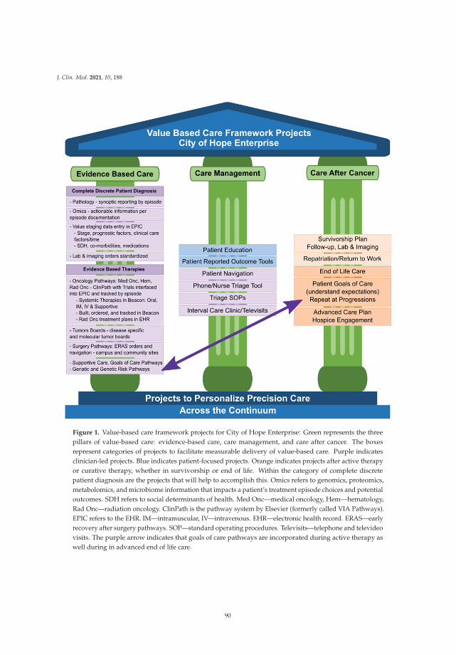
J. Clin. Med. 2021, 10, 188
Figure 1. Value-based care framework projects for City of Hope Enterprise: Green represents the threepillars of value-based care: evidence-based care, care management, and care after cancer. The boxesrepresent categories of projects to facilitate measurable delivery of value-based care. Purple indicatesclinician-led projects. Blue indicates patient-focused projects. Orange indicates projects after active therapyor curative therapy, whether in survivorship or end of life. Within the category of complete discretepatient diagnosis are the projects that will help to accomplish this. Omics refers to genomics, proteomics,metabolomics, and microbiome information that impacts a patient’s treatment episode choices and potentialoutcomes. SDH refers to social determinants of health. Med Onc—medical oncology, Hem—hematology,Rad Onc—radiation oncology. ClinPath is the pathway system by Elsevier (formerly called VIA Pathways).EPIC refers to the EHR. IM—intramuscular, IV—intravenous. EHR—electronic health record. ERAS—earlyrecovery after surgery pathways. SOP—standard operating procedures. Televisits—telephone and televideovisits. The purple arrow indicates that goals of care pathways are incorporated during active therapy aswell during in advanced end of life care.
90
J. Clin. Med. 2021, 10, 188
3. NCCN Guidelines and Growing Complexity of Evidence-Based Cancer Care Also StimulatedMultidisciplinary Oncology Pathway Development at COH
City of Hope (COH) is a National Cancer Institute (NCI)-designated comprehensive care centerand one of the original 13 member institutions that formed the National Comprehensive Cancer CenterNetwork (NCCN) in January of 1995. Within the NCCN, our faculty have participated in and led thedevelopment of guidelines for the treatment of cancers for the past 25 years. The COH enterprise nowserves patients with cancer and metabolic diagnoses in a region of 25 million residents living in fivecounties in Southern California as well as more distantly based national and international patients.The COH enterprise includes a central academic, clinical research, hospital and clinical care campus inDuarte, California and a clinically integrated, community practice network of 31 community practices.Approximately 30% of COH physicians practice in the community (either solely or in a mixed hybrid ofcommunity/academic practice). Clinicians in the community are mostly general oncology practitioners,while the academic clinicians are disease and disease subtype specialists. Over the past year, we haveidentified community doctors who have a special interest in a specific cancer and will see patients withthat disease preferentially (referred to as disease-leaning), such as breast, thoracic, gastrointestinal, andgenitourinary cancers. A single foundation faculty model employs both academic center and communityclinicians. The mission of the organization is to develop and bring cutting-edge precision cancer care andclinical research to every patient that is also personalized and delivered with compassion, high quality,and cost consciousness within the value-based cancer care initiative.
Although COH faculty participated in the development of the original eight disease guidelinespublished by the NCCN in November of 1996 and continue to participate and lead disease guidelinecommittees at NCCN, until the more recent partnership between NCCN and US Oncology and theirdevelopment of Level 1 Pathways powered by NCCN, the NCCN guidelines were broad and withoutthe detailed pathway approach to better guide individual patient care [32]. COH disease teams regularlydiscussed narrowing the NCCN guidelines into pathways for specific patient issues and cancer subtypesand some, such as the breast, lung, and renal teams, develop and review pathways that are shared withCOH faculty, as described in companion articles in this series [41–43]. With the growth of the COHenterprise, however, the need to more formally establish not only pathways for each disease but theability to prompt for those choices, keep the pathways up to date, incorporate clinical trials as well astrack the use and variations of treatments for each patient (by disease, disease subtype, clinicians, site,and payer), a formal pathway program became a priority. As part of the VBCC initiative, availablepathway programs and options were evaluated by a multi-stakeholder group of faculty, staff, nurses, andadministrators with campus and community network representatives over a 2-year period. This wasled early on by an oncology surgeon who practiced at both the academic center and in the communityand understood the practical needs for a tool to guide standardized care for similar patients. Workinitially focused on the development of multimodality clinical care pathways, defined as standardized,evidence-based interdisciplinary care management plans, which would identify an appropriate sequenceof clinical interventions, timeframes, milestones, and expected outcomes for a comparable patient group,i.e., by diagnosis or surgical procedure. As the work evolved, however, it became clear that the startingpoint for these larger multimodality care plans should be pathway programs starting with the specialtiesof medical, radiation, and surgical oncology.
A formal pathway tool became a priority not only to support the measurable delivery of care acrossthe growing COH enterprise but to bring expert input to the point of clinical care for the growing numberand complexity of cancer therapies. The continued growth in new Food and Drug Administration (FDA)hematology–oncology drug approvals since 2000 is a telling example of the need to provide real-timeexpert decision support across an enterprise to aid the delivery of high-quality, high-value cancer care.
91

J. Clin. Med. 2021, 10, 188
The FDA website for oncology–hematology drugs reports that from 2016 through June of 2020, 230 newdrug approvals were issued, up from 22 in 2016 to 58 in 2017, 63 in 2018, 46 in 2019, and 41 in the firsthalf of 2020 [44]. These approvals are also increasingly complex and include not only new drugs butnew indications for existing drugs. The approval can be under the expedited review process so that theapproval could also change with further data. Approvals can include whether the use is approved in earlydisease, as neoadjuvant, adjuvant therapy, or extended adjuvant or for advanced disease. The approvalscan include use only with specific biomarker results from the patient or their tumor, and sometimes onlyafter biomarker testing by specific laboratories. The approvals often specify use only in specific lines oftherapy, after previous general or specific therapies, in combination with specific other drugs or drugcategories. Biosimilar drugs are also listed in the FDA approvals with their specific uses referenced to thereference drug [44]. Thus, keeping up with the details of the almost weekly new FDA approvals, whichindividually can impact one or many kinds of cancers, requires increasingly sophisticated and constantlyupgraded decision support pathways not only for clinicians but also for patient education to support theirparticipation in shared decision-making and to ensure appropriate use and documentation requirementsfor authorizations and payer coverage. Financial toxicity has been identified as another growing barrier topatients receiving appropriate cancer therapies, especially targeted, molecular, and immunotherapy-basedtreatments [45]. Thus, building the workflow infrastructure to support the use of pathway tools whichinclude increasingly higher cost therapies can also support more timely authorization of treatments forpatients and can help to engage early patient assistance, when needed, so patients can receive timelycare which improves outcomes [46,47]. Several examples of the growing complexity of high-quality careare described in companion articles on colorectal and non-small cell lung cancer by colleagues in thisseries [48,49].
The development of surgical pathways was driven by the benefits of standardizing surgical type andprocesses but also by the benefits of reducing morbidity, mortality, and costs by coordinating education andcare processes with the many providers involved in a patient’s care before, during, and after surgery [50–53]. These comprehensive care plans have come to be known as Early Recovery after Surgery (ERAS)pathways [54–57]. At City of Hope, ERAS pathway work began in the early 2000s by the urology facultywith the development and implementation of a successful ERAS pathway for cystectomies showinglower length of stay, complications, and readmission rates that were confirmed by outside groups aswell [19,58,59]. This stimulated the development of further urology ERAS pathways as well as ongoingwork on ERAS pathways for other surgical subspecialty departments for the COH academic campus andat regional hospitals where COH faculty practice.
Given these many drivers toward standardizing care in all specialties that could provide bettercare, better outcomes, and potentially lower costs, City of Hope’s Value Realization Project identified aValue-Based Core (VBC) team of experts to meet regularly and work with a multidisciplinary physicianVBC group team, using the Value Framework projects noted in Figure 1 above, to engage disease leaders inmedical oncology, surgery, and radiation oncology along with administrators, information technology (IT)and informatics and EHR experts to evaluate, implement, and iterate oncology pathways for the enterprise.Insights and outcomes from this work follow and add to the work of other academic community networkenterprises working together to improve the quality of care for cancer patients [60,61].
4. Medical Oncology, Radiation Oncology, and Hematology Subspecialty Pathways
4.1. Evaluation and Adoption Process for Pathway Processes and Tool for the Enterprise
Initial meetings identified medical oncology pathways as the starting point for a formal oncologypathway tool. Available oncology pathway programs, their implementation, and integration abilities
92
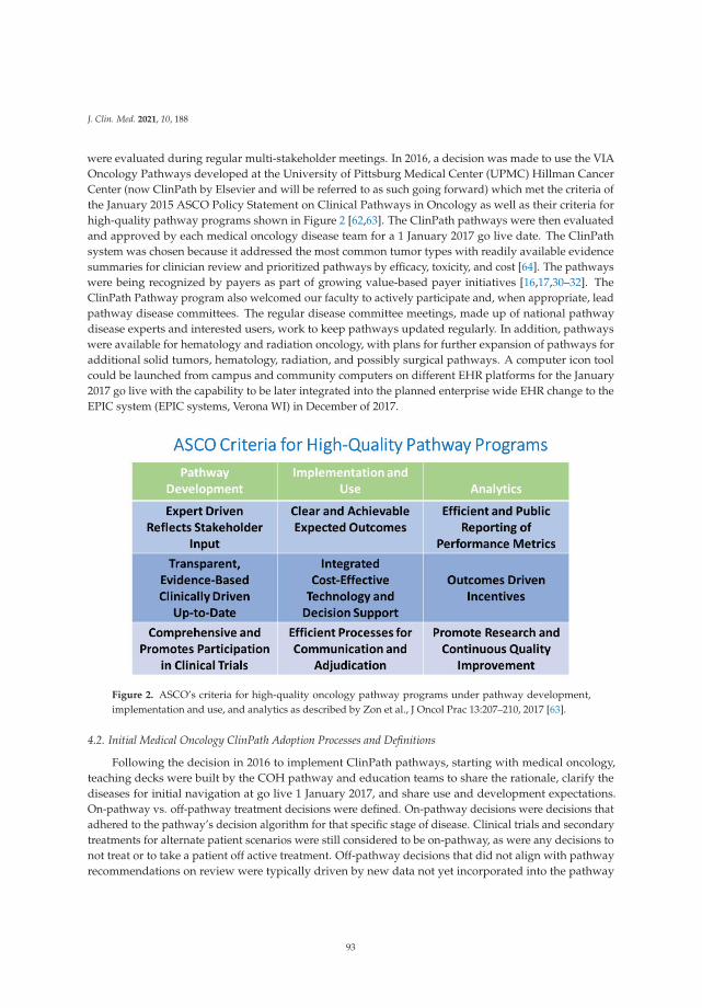
J. Clin. Med. 2021, 10, 188
were evaluated during regular multi-stakeholder meetings. In 2016, a decision was made to use the VIAOncology Pathways developed at the University of Pittsburg Medical Center (UPMC) Hillman CancerCenter (now ClinPath by Elsevier and will be referred to as such going forward) which met the criteria ofthe January 2015 ASCO Policy Statement on Clinical Pathways in Oncology as well as their criteria forhigh-quality pathway programs shown in Figure 2 [62,63]. The ClinPath pathways were then evaluatedand approved by each medical oncology disease team for a 1 January 2017 go live date. The ClinPathsystem was chosen because it addressed the most common tumor types with readily available evidencesummaries for clinician review and prioritized pathways by efficacy, toxicity, and cost [64]. The pathwayswere being recognized by payers as part of growing value-based payer initiatives [16,17,30–32]. TheClinPath Pathway program also welcomed our faculty to actively participate and, when appropriate, leadpathway disease committees. The regular disease committee meetings, made up of national pathwaydisease experts and interested users, work to keep pathways updated regularly. In addition, pathwayswere available for hematology and radiation oncology, with plans for further expansion of pathways foradditional solid tumors, hematology, radiation, and possibly surgical pathways. A computer icon toolcould be launched from campus and community computers on different EHR platforms for the January2017 go live with the capability to be later integrated into the planned enterprise wide EHR change to theEPIC system (EPIC systems, Verona WI) in December of 2017.
Figure 2. ASCO’s criteria for high-quality oncology pathway programs under pathway development,implementation and use, and analytics as described by Zon et al., J Oncol Prac 13:207–210, 2017 [63].
4.2. Initial Medical Oncology ClinPath Adoption Processes and Definitions
Following the decision in 2016 to implement ClinPath pathways, starting with medical oncology,teaching decks were built by the COH pathway and education teams to share the rationale, clarify thediseases for initial navigation at go live 1 January 2017, and share use and development expectations.On-pathway vs. off-pathway treatment decisions were defined. On-pathway decisions were decisions thatadhered to the pathway’s decision algorithm for that specific stage of disease. Clinical trials and secondarytreatments for alternate patient scenarios were still considered to be on-pathway, as were any decisions tonot treat or to take a patient off active treatment. Off-pathway decisions that did not align with pathwayrecommendations on review were typically driven by new data not yet incorporated into the pathway
93
J. Clin. Med. 2021, 10, 188
(indicated as physician disagrees with pathway choice or a free texted comment to that effect, reports notshown), unique patient presentations, specific patient preferences, and on occasion by physician discretion.It was emphasized that while it was expected that for most cancers, on-pathway choices would be in therange of 80% or more, there would never be an expectation of a specific pathway compliance percentageby doctor or by disease type. It was also recognized that in these times of rapid new drug discovery andmolecular targeting, the best therapy for an individual patient may not yet be in the pathway tool. Allclinicians were encouraged to join one or more pathway disease committees to participate in the ongoingdevelopment process, review the latest evidence, and share best practices.
4.3. Key Milestones and Practical Time Implementations for ClinPath Pathways across the COH Enterprise
January 2017: Medical Oncology go live for six disease types: Medical oncologists at the academiccampus and community sites were trained and instructed to use the pathway tool over 6–8 weeks prior togo live for all new medical oncology therapies including both initial therapy and subsequent therapiesin six disease categories: breast, lung, genitourinary (GU), gastrointestinal (GI), gynecologic (GYN), andhead and neck. Educational tip sheets and training team personnel were available on an ongoing basisfor individual support as well. Other solid tumor types and some hematology diseases were available inthe tool but optional for use and not assessed as part of our initial metrics. COH had two EHR systemswhen the VIA pathways went live: Allscripts on campus and Touch Works in the community. Both wereinterfaced to pull patient demographic and physician schedule information into the pathway tool. Afterthat, however, clinicians would navigate pathways by entering discrete staging and prognostic or clinicalcare feature elements to reach the recommended pathway choice or choices. Physicians were required toenter a reason when an off-pathway decision was chosen. After making a therapy choice, clinicians thenwent back to their EHR to order the chosen therapy. Clinicians were made aware of plans for future EPICintegration, addition of clinical trials into the pathways, and expansion of tracking and reporting for otherdisease types and specialties. Although formal surveys were not done, clinicians expressed frustrationsfrom the double entry of data from their EHR notes into the pathway system, having to use the systemwhen they were confident and familiar with the appropriate pathway choice as well as skepticism onfuture upgrades and benefits to their disease programs (personal communications).
2 December 2017: Enterprise-wide transition to Epic: Treatment protocols were built in Beacon by thedisease teams based on the regimens used in the previous systems and a review of those in the pathwaysystem before go live. Additional COH-specific regimen preferences and many clinical trials were alsobuilt. Oral chemotherapy regimens were not prioritized. Standardized nausea regimens based on NCCNemetogenic levels were added for each regimen as well as hypersensitivity premedication regimens andthey were integrated when needed to avoid overuse of steroid medications. All documentation, data entry,and ordering were then done in the EPIC EHR across the enterprise.
January 2018: OnCore clinical trial management system went live. Its benefit to the pathway programis discussed below.
August 2018 and ongoing: Clinical trials were formally added into the ClinPath pathway tool bydisease teams starting with the most commonly seen cancers in the community. Additional solid tumorclinical trials have been added and closed trials removed as noted below.
March 2019: Radiation oncology clinicians on campus and community sites went live on ClinPathradiation pathways with integration of COH radiation oncology clinical trials as described below.
20 July 2019: The one-way integration between the Epic EHR and the pathway tool was started and isdescribed below.
August 2020: Hematology pathways had been available in the ClinPath system but their formalevaluation by disease leads began as well as identification of clinical trials to be added to the ClinPath
94
J. Clin. Med. 2021, 10, 188
pathways. As reviews are being completed, trials are being added and formal launch of enterprise-wideuse and measurement of ClinPath for the common hematology diagnoses is scheduled to start December2020 as described below.
6 October 2020: The two-way integration between the EPIC EHR and the ClinPath tool went live inthe EHR for 11 disease subtypes (breast, bladder, colorectal, gastroesophageal, melanoma, mesothelioma,ovary, prostate, testicular, small cell lung, and thyroid). With two-way integration, we expect that mostif not all discrete staging elements from the EPIC staging forms will be interfaced to auto-populate intothe ClinPath tool over time and as elements are added to the EPIC system. Any additional or missingdiscrete data elements in the EHR system would still have to be entered into the pathway tool to trigger apathway prompt. At go live, tumor types fell into three categories from this pioneering work: fully mappedtumors (breast, colorectal, gastroesophageal, melanoma, mesothelioma, bladder, ovary, sarcoma, small-celllung cancer, testicular, thyroid, and uterus), partially mapped tumors (head and neck, prostate), andpending mapped tumors (anal, non-small cell lung, neuroendocrine, pancreatic, renal). When cliniciansnavigate within the EHR to the ClinPath system, the data elements entered into the EPIC staging formsare then shown on a field next to the ClinPath data entry choices. For fully mapped tumors, one clickof an APPLY button automates the entry of the EPIC data into the ClinPath system. The doctor thencontinues navigating to the pathway choices. For a partially mapped tumor, those elements that aremapped will auto-populate in ClinPath and the others will have to be manually entered. For non-mappedtumors, the EPIC data are there to see but each element has to be manually clicked before continuing thenavigation. Whether or not a tumor is fully, partially, or not yet mapped, however, after a pathway choiceis made, whether for a clinical trial or a standard regimen, the user is then taken back to the EPIC Beaconregimen for completing the order or ordering a clinical trial team evaluation. This two-way integrationwill continue to expand by matching additional data elements captured in EPIC to the pathway decisiontrees to more efficiently guide pathway choices. In addition, the EHR system is working to include dataelements that more fully describe a patient’s disease status as required by decision support tools. It isexpected that ongoing work will continuously reduce duplicate data entry requirements, including forthe growing number of actionable genomic results, while ensuring that the primary clinical informationremains in the enterprise EHR [65]. A survey of provider experience, time savings (if any), and satisfactionwas built and, pending team approval, will be sent to all faculty who use the ClinPath system in Novemberof 2020 to further inform this work.
4.4. Clinical Trials Incorporated into ClinPath System and Integrated with Clinical Trials Management System CanPrompt for Available Trials and Track Adoption
Studies have shown improved clinical trial assessment and accruals for clinical trials incorporatedinto pathway tools that are routinely used, including by multisite organizations [66,67]. The ability to addclinical trials available at and through our COH enterprise was another key reason the ClinPath pathwaysystem was chosen in 2016 [68]. Prior to incorporating clinical trials into our pathway tool, though, COHimplemented the OnCore clinical trials management system (CTMS) in January 2018. This system providesfaculty and clinical research teams with a comprehensive integrated system that supports virtually allaspects of clinical trial offerings including features for managing studies, electronically capturing protocoland patient data, creating custom reports, and supporting financial activities. Having it fully linkedinto the decision support pathway tool enhances clinician notification of available clinical trial options,especially timely notices of pending, open, on hold, or closed trials to maximize clinical efficiencies.
The OnCore tool integrated with the EHR will hopefully enhance the value of the pathways toolthrough bi-directional data flows indicating protocol status and patient demographics. Phase I of theproject included the decommissioning of the legacy clinical trials management system (MIDAS) andmigration of protocol status and data. Phase II of the project linked detailed subject management activities
95
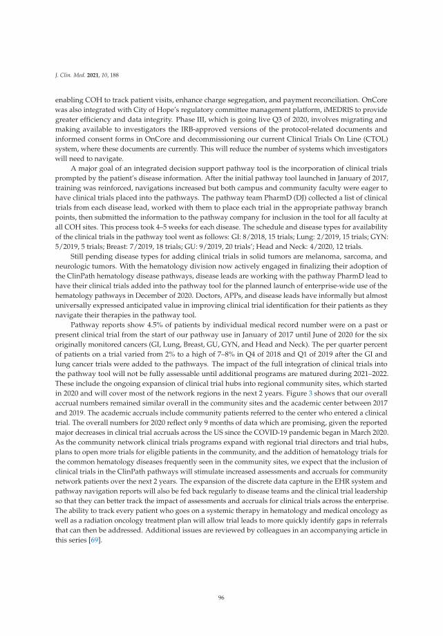
J. Clin. Med. 2021, 10, 188
enabling COH to track patient visits, enhance charge segregation, and payment reconciliation. OnCorewas also integrated with City of Hope’s regulatory committee management platform, iMEDRIS to providegreater efficiency and data integrity. Phase III, which is going live Q3 of 2020, involves migrating andmaking available to investigators the IRB-approved versions of the protocol-related documents andinformed consent forms in OnCore and decommissioning our current Clinical Trials On Line (CTOL)system, where these documents are currently. This will reduce the number of systems which investigatorswill need to navigate.
A major goal of an integrated decision support pathway tool is the incorporation of clinical trialsprompted by the patient’s disease information. After the initial pathway tool launched in January of 2017,training was reinforced, navigations increased but both campus and community faculty were eager tohave clinical trials placed into the pathways. The pathway team PharmD (DJ) collected a list of clinicaltrials from each disease lead, worked with them to place each trial in the appropriate pathway branchpoints, then submitted the information to the pathway company for inclusion in the tool for all faculty atall COH sites. This process took 4–5 weeks for each disease. The schedule and disease types for availabilityof the clinical trials in the pathway tool went as follows: GI: 8/2018, 15 trials; Lung: 2/2019, 15 trials; GYN:5/2019, 5 trials; Breast: 7/2019, 18 trials; GU: 9/2019, 20 trials’; Head and Neck: 4/2020, 12 trials.
Still pending disease types for adding clinical trials in solid tumors are melanoma, sarcoma, andneurologic tumors. With the hematology division now actively engaged in finalizing their adoption ofthe ClinPath hematology disease pathways, disease leads are working with the pathway PharmD lead tohave their clinical trials added into the pathway tool for the planned launch of enterprise-wide use of thehematology pathways in December of 2020. Doctors, APPs, and disease leads have informally but almostuniversally expressed anticipated value in improving clinical trial identification for their patients as theynavigate their therapies in the pathway tool.
Pathway reports show 4.5% of patients by individual medical record number were on a past orpresent clinical trial from the start of our pathway use in January of 2017 until June of 2020 for the sixoriginally monitored cancers (GI, Lung, Breast, GU, GYN, and Head and Neck). The per quarter percentof patients on a trial varied from 2% to a high of 7–8% in Q4 of 2018 and Q1 of 2019 after the GI andlung cancer trials were added to the pathways. The impact of the full integration of clinical trials intothe pathway tool will not be fully assessable until additional programs are matured during 2021–2022.These include the ongoing expansion of clinical trial hubs into regional community sites, which startedin 2020 and will cover most of the network regions in the next 2 years. Figure 3 shows that our overallaccrual numbers remained similar overall in the community sites and the academic center between 2017and 2019. The academic accruals include community patients referred to the center who entered a clinicaltrial. The overall numbers for 2020 reflect only 9 months of data which are promising, given the reportedmajor decreases in clinical trial accruals across the US since the COVID-19 pandemic began in March 2020.As the community network clinical trials programs expand with regional trial directors and trial hubs,plans to open more trials for eligible patients in the community, and the addition of hematology trials forthe common hematology diseases frequently seen in the community sites, we expect that the inclusion ofclinical trials in the ClinPath pathways will stimulate increased assessments and accruals for communitynetwork patients over the next 2 years. The expansion of the discrete data capture in the EHR system andpathway navigation reports will also be fed back regularly to disease teams and the clinical trial leadershipso that they can better track the impact of assessments and accruals for clinical trials across the enterprise.The ability to track every patient who goes on a systemic therapy in hematology and medical oncology aswell as a radiation oncology treatment plan will allow trial leads to more quickly identify gaps in referralsthat can then be addressed. Additional issues are reviewed by colleagues in an accompanying article inthis series [69].
96
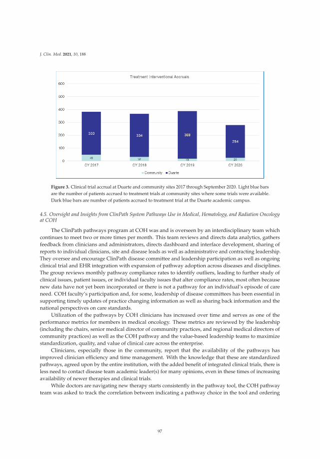
J. Clin. Med. 2021, 10, 188
Figure 3. Clinical trial accrual at Duarte and community sites 2017 through September 2020. Light blue barsare the number of patients accrued to treatment trials at community sites where some trials were available.Dark blue bars are number of patients accrued to treatment trial at the Duarte academic campus.
4.5. Oversight and Insights from ClinPath System Pathways Use in Medical, Hematology, and Radiation Oncologyat COH
The ClinPath pathways program at COH was and is overseen by an interdisciplinary team whichcontinues to meet two or more times per month. This team reviews and directs data analytics, gathersfeedback from clinicians and administrators, directs dashboard and interface development, sharing ofreports to individual clinicians, site and disease leads as well as administrative and contracting leadership.They oversee and encourage ClinPath disease committee and leadership participation as well as ongoingclinical trial and EHR integration with expansion of pathway adoption across diseases and disciplines.The group reviews monthly pathway compliance rates to identify outliers, leading to further study ofclinical issues, patient issues, or individual faculty issues that alter compliance rates, most often becausenew data have not yet been incorporated or there is not a pathway for an individual’s episode of careneed. COH faculty’s participation and, for some, leadership of disease committees has been essential insupporting timely updates of practice changing information as well as sharing back information and thenational perspectives on care standards.
Utilization of the pathways by COH clinicians has increased over time and serves as one of theperformance metrics for members in medical oncology. These metrics are reviewed by the leadership(including the chairs, senior medical director of community practices, and regional medical directors ofcommunity practices) as well as the COH pathway and the value-based leadership teams to maximizestandardization, quality, and value of clinical care across the enterprise.
Clinicians, especially those in the community, report that the availability of the pathways hasimproved clinician efficiency and time management. With the knowledge that these are standardizedpathways, agreed upon by the entire institution, with the added benefit of integrated clinical trials, there isless need to contact disease team academic leader(s) for many opinions, even in these times of increasingavailability of newer therapies and clinical trials.
While doctors are navigating new therapy starts consistently in the pathway tool, the COH pathwayteam was asked to track the correlation between indicating a pathway choice in the tool and ordering
97

J. Clin. Med. 2021, 10, 188
the regimen in the EHR as a quality control measure. A Variation of Care report was designed in 2019and reports the monthly rates of any difference between what was navigated in the ClinPath system andwhat was ordered in the EPIC EHR through the Beacon module. The data from May to December of2019 showed that the difference varied from a low of 0 variations from 871 navigations in 11/2019 to amaximum of 18 variations from 688 (2.6%) navigations in October of 2019. Overall, discrepancies wererarely over 3%. A random chart evaluation noted that these discrepancies occurred when patients orproviders changed their treatment plan based on additional work-up, most often when additional diseasewas found. They then ordered the therapy in the EPIC EHR Beacon treatment orders but did not go backinto the ClinPath tool to change the navigation information in the pathway tool. This also happened forrare patient preference for an alternate therapy after the initial ClinPath navigation was done.
Since our ClinPath go live in January of 2017 until early Q3 2020 when data were reported for thismanuscript, clinicians had done 28,271 total navigations, including 16,034 navigations from the academicclinicians and 12,190 navigations from the community clinicians, including 47 at our newest NewportBeach site, opened in January of 2020. The percentage of therapies ordered in Beacon that were navigatedin the pathway tool by enterprise, community, and campus doctors since implementation is shown inFigure 4. Pathway navigation was higher on the academic campus except for two quarters. The reasonsare under further evaluation.
Figure 4. Percentage of new EPIC Beacon starts that were navigated in ClinPath by medical oncologists forsolid tumors since go live for enterprise overall then by Duarte campus site and all community sites. NoteQ3 data were incomplete at time of manuscript data collection in May 2020, so the final% of patients in Q3who were navigated in ClinPath is likely higher, as is seen in other recent quarters.
Simply having a pathway system does not ensure the use, usefulness, or production of informativeanalytics. A multidisciplinary pathway team of clinicians, analytic, quality, her, and informatics expertsmeets every 2 weeks to evaluate pathway use, compliance by disease type and clinician, track lowcompliance issues to understand if a new therapy not yet incorporated into the pathway or other reason isincreasing off-pathway choices, use of clinical trials, and whether the indicated therapy in the pathwaysystem is, in fact, the ordered therapy in the EHR. An enterprise-wide incentive program to capturepathway use and choices for medical oncology was launched in 2020 and final percent navigations inthe pathway for all medical oncology therapies in the EHR trends have risen significantly. Disease leadshave also requested regular reporting of every patient who meets specific disease, stage, and other criteriafor an available trial as to whether or not the trial was considered and if a trial evaluation and ultimate
98
J. Clin. Med. 2021, 10, 188
accrual occurred. Several leads are asking for real-time reports so that they can more proactively reach outto network clinicians to resolve any barriers for patient accrual to an available clinical trial. Ultimately,by tracking specific clinical trial eligibility and accrual by site, disease, and trial type across the enterprise,the trial team can optimize which types of trials are opened at which sites to maximize clinical trialresources.
Having a pathway tool enables the tracking of the percentage of patients who are treated on-pathway(including clinical trials) and off-pathway across the enterprise and by academic vs. community networksites as shown in Figure 5. Academic clinicians consistently see more patients treated off-pathway, which,on reviews, has been explained by the higher number of patients seen where a pathway does not exist forthe patient presentation or where a different treatment is recommended due to new molecular findings oremerging data are not yet incorporated into the pathway. After reviewing data, the pathway committeehas identified and recommended to the pathway vendor that they add that additional reason as an optionin the dropdown list when entering a reason for choosing an off-pathway therapy. Such a new choicefor going off-pathway, “new information not yet incorporated into the pathway” could then be trackedfor each tumor type and stage during six month intervals over twelve to twenty four month periods tosee if and when such a therapy choice is added to the pathway. This could also better inform cliniciansabout how rapidly new, practice-changing therapies that experts agree should be used become availablefor prompting in the ClinPath decision support pathway tool.
Figure 5. Pathway compliance percentage over time for medical oncology patients navigated in pathwaytool (ClinPath) for enterprise, Duarte academic campus, and community sites for new therapy regimensordered in the EPIC-BEACON EHR from January 2019 through June of 2020. Green bars are on-pathwaychoices (including clinical trials) and yellow bars are off-pathway choices. Note that the scale of the x-axisrepresenting the numbers of patients is different for each group to show the comparison percentages inone graph. The combined number of patients navigated in Duarte and community sites is reflected in thenumber totals described on the enterprise graph.
99
J. Clin. Med. 2021, 10, 188
4.6. Pathways for Radiation Oncology Using ClinPath Program
As with medical oncology pathways, radiation therapy pathways were piloted by the University ofPittsburgh Medical Center (UPMC) group, who led the development of the ClinPath (then called VIA)pathways for radiation oncology based on the same principles used for the development of medicaloncology pathways to standardize care for the most common radiation treatments, prioritizing efficacythen toxicity then costs. Studies by their group showed that using the pathways with a peer review processappeared to encourage compliance with clinical pathway recommendations [70]. Specific studies includedone showing increased community practice physicians’ adoption of hypofractionated regimens for wholebreast radiation [71].
This is significant as several studies have shown slow and or poor adoption of evidence-based useof shorter course radiation treatments which have lower costs and shorter treatment times for patientswith equivalent outcomes. Hypofractionation for breast cancer is a good example. While the majorityof women undergoing radiation therapy for early breast cancer have equal disease control with equalor better cosmetics and a shorter course for patients has been adopted by UK and Canadian cliniciansfor the majority of patients, it has been much slower to be adopted in the US despite positive long-termoutcomes in studies and expanded guidelines for use by the American Society for Radiation Therapy(ASTRO) [72–75]. As in medical oncology and hematology, the growing options for the use of differentradiation techniques such as 3D conformal, intensity-modulated radiation therapy (IMRT), proton beam,and other methods of localized radiation therapies along with shorter treatment courses have led toincreasing use of radiation therapy pathways to support busy clinicians through their prioritizing efficacy,toxicity, and safety as another component of value-based care [76,77].
As the COH enterprise expanded with many satellite sites delivering convenient local radiationtherapy treatments, the radiation oncology department chose to implement and use ClinPath radiationpathways at all sites in March 2019. COH has the largest single institution radiation oncology network inCalifornia, comprised of 46 physicians practicing at 20 satellite sites. ClinPath pathways were integratedinto the day-to-day clinical practice for 35 radiation oncologists at 16 sites as four outside hospital sitedoctors are not yet on the system. Radiation oncologists with specific disease interests contributed to thedevelopment of some of the pathways and some faculty now co-lead some radiation oncology diseasepathway committees. Clinical trial options were also integrated into the pathway options customized toour institution.
The radiation oncologists use the EPIC EHR for all care, except the specific radiation therapy planning,which is done in the radiation-specific modules of MOSAIQ or ARIA EHRs at different sites. The pathwayshave had a launch button integrated in the EPIC EHR and the radiation oncologists are looking forward tothe two-way integration in progress so that the staging in EPIC will be pulled into the pathway systemwhich is on the planning agenda for 2021–2022.
The pathways, besides giving case-by-case feedback regarding treatment options to clinicians,also give detailed recommendations regarding the technical aspects of radiation (i.e., dose constraints,fractionation options), which is useful at the time of actual radiation treatment planning. The pathwayscover the most common disease sites and scenarios. There are still complex or rare cases that do not fit intoany of the existing pathways, where the radiation oncologist notes “no pathway”, and there is an option toadd a narrative comment.
In general, the pathways program has been well received. There has been >90% participation rateby COH radiation oncologists in using the pathway tool to capture their treatment decisions, with mostphysicians using it to indicate 100% of their decisions. Physicians are also sent auto-generated weeklyreminders listing any patients where pathways data about their treatment choice were not entered. COHdoes not require strict adherence to the pathway’s preferred choice, but the analytic team gives physicians
100
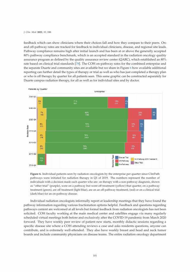
J. Clin. Med. 2021, 10, 188
feedback which can show clinicians where their choices fall and how they compare to their peers. On-and off-pathway rates are tracked for feedback to individual clinicians, disease, and regional site leads.Pathway compliance remains high after initial launch and has been at or above the generally accepted80% pathway compliance benchmark, which is an accepted standard in the radiation oncology qualityassurance program as defined by the quality assurance review center (QARC), which established an 80%rate based on clinical trial standards [78]. The COH on-pathway rates for the combined enterprise andthe separate Duarte and community sites are available but we share in Figure 6 how available additionalreporting can further detail the types of therapy or trial as well as who has just completed a therapy planor who is off therapy by quarter for all patients seen. This same graphic can be constructed separately forDuarte campus radiation therapy, for all as well as for individual sites and by doctor.
Figure 6. Individual patients seen by radiation oncologists by the enterprise per quarter since ClinPathpathways were initiated for radiation therapy in Q1 of 2019. The numbers represent the number ofindividuals with a decision made each quarter who are: on therapy with a non-pathway diagnosis, shownas “other trial” (purple), were on a pathway but went off treatment (yellow) that quarter, on a pathwaytreatment (green), are off treatment (light blue), are on an off-pathway treatment, (red) or on a clinical trial(dark blue) for an on-pathway disease.
Individual radiation oncologists informally report at leadership meetings that they have found thepathway information regarding various fractionation options helpful. Feedback and questions regardingpathways content are welcomed at all levels but formal feedback from radiation oncologists has not beensolicited. COH faculty working at the main medical center and satellites engage via many regularlyscheduled virtual meetings both before and exclusively after the COVID-19 pandemic from March 2020forward. They have weekly peer review of patient new starts, monthly didactic sessions regarding aspecific disease site where a COH attending reviews a case and asks residents questions, anyone cancontribute, and is extremely well-attended. They also have weekly breast and head and neck tumorboards and include community physicians on disease teams. The entire radiation oncology department
101
J. Clin. Med. 2021, 10, 188
has previously gotten together for a yearly in-person one-day science and clinical care retreat, which willbe held virtually during the pandemic. Although no single physician has time to participate in everydiscussion, the regular meetings are available with technology support. Clinical leaders note that it remainschallenging to engage and include new and distant satellites as they continue to be added to the COHnetwork, but this is an identified priority.
The COH radiation oncology department has also created an internal Microsoft Teams messagingsystem where anyone can share case questions and message the entire department. It has yet to be regularlyutilized but doctors are in the process of being trained on the Microsoft Teams functionalities. E-mail,therefore, is still the most utilized form of communication department-wide. Overall, pathway complianceis high for the enterprise, as shown in Figure 6 above. Reasons for off-pathway choices are captured asfor other specialties, reported and reviewed by the disease teams. Any recommendations for pathwayupdates are communicated to the appropriate ClinPath disease committees.
4.7. Additional Multidisciplinary Pathways for VBCC: Geriatric Oncology and Genetic Counseling/RiskAssessment
Geriatric oncology is an established field pioneered by the late COH faculty member, Dr. Arti Hurria.Dr. Hurria’s goal was to bring the principles of geriatric oncology into practice through standards andpathways to improve the care of older adults with cancer. Given that one in three Americans aged 60years or older will be diagnosed with cancer in their lifetime and the population of older adults is growingrapidly with the aging of the U.S. and worldwide populations, cancer incidence is expected to rise by 67%for individuals aged 65 or older between 2010 and 2030. Although older adults represent the majority ofindividuals with cancer, they are severely under-represented in clinical trials and research. Hence, themajority of evidence that sets the standards of care for oncology treatment pathways is derived fromyounger individuals.
To address these unmet needs, COH formed the Cancer and Aging program in 2006, under theleadership of Dr. Hurria, which ultimately led to the formation of the Center in Cancer and Aging (CCA)in 2017. The mission of the CCA is to join investigators from all cancer disciplines to study the biology,treatment, and survivorship issues of older adults with cancer. Over the last decade, COH staff have ledover 30 studies in cancer and aging, enrolled more than 5000 patients on studies, disseminated researchfindings through more than 250 publications, and received competitive grants from the NIH as well asphilanthropic funding. CCA is one of the elite centers in cancer and aging in the nation.
With this background work, COH’s CCA is now leading the way in expanding pilot pathway projectsinto community sites, starting with standardizing geriatric assessments (GA), which can help to guidepersonalized care for our increasingly aging population while achieving their best health outcome. Forexample, an ongoing implementation study of an APP-led multidisciplinary team telehealth interventionis being conducted in one of COH’s 31 community cancer sites. This Antelope Valley regional site is in aremote area of high need and limited resources. Learnings from this pilot will be used to expand geriatricpathways for assessments and care planning across our enterprise.
Colleagues at COH have also examined the multifaceted barriers hindering participation of olderadults in cancer trials. They offer strategies to improve the participation of older adults in clinical trials inan accompanying article in this series [79]. It is expected that many of the projects outlined in COH’s ValueFramework, such as the complete discrete staging and collection of social determinants of health alongwith geriatric assessments with clinical trial integrations into the pathway navigation tool for systemictherapies in medical oncology, hematology, and radiation oncology, will help to support the piloting ofthese strategies.
Clinical cancer genomics has been an expertise at COH since the department was established in 1996which has grown to provide training, research, and consultative services overseen now by the division of
102
J. Clin. Med. 2021, 10, 188
clinical cancer genomics (CCG). A counseling, research, and professional teaching program, internationallyrecognized for its onsite and telehealth training of healthcare providers worldwide, continues withongoing professional conferences open to trainees and the COH faculty at all sites. At the academic center,the department provides consultations for individual cancer risk assessments, genetic testing and resultinterpretation, risk reduction counseling, and clinical trial enrollment. The CCG division is also engagedin the support of COH’s enterprise precision medicine initiative to expand somatic and germline testingwith associated counseling on results, integration of results into care pathways and into research teamsto power new discoveries. Work to standardize these processes through discrete data entry in EPIC onthe upgraded staging forms is in progress, along with ways to present clinically actionable results ofexpanding somatic and germ line genomic testing to clinicians within the data for each patient’s episodeof cancer throughout their journey. Having relevant clinically actionable data for integration into decisionsupport pathway tools and molecular tumor boards while having the larger dataset easily accessible foranalytics and discovery by the research teams is a current priority of the precision medicine, genomics,informatics, value-based care, and disease specialty teams informed by input from faculty at both theacademic and community sites.
Genomics pathway work is in the earlier stages of development and will be informed by the expansionof CCG services to the enterprise through academic community faculty and staff teamwork. Pathwaywork has started with one day per week onsite genetic counseling consultations at one local networksite. This has now expanded to one day per week of genetic counseling services being available nowby telehealth to patients at all community sites. MD geneticist counseling is offered 2 days per monthfor the community sites after an initial pilot at one community site. Another pilot pathway for geneticassessments is underway with two breast surgeons, trained and supported by the academic CCG team toprovide genetic cancer risk assessment (GCRA) to their breast cancer patients with referral to campus foradditional genetic counseling or genetics MD for complicated cases as needed. A breast surgeon at anothercommunity site is being trained and will provide GCRA services to her patients as well as to other affectedand unaffected patients seen by faculty at that site as well as any external referrals. That surgeon alsoplans to develop a high-risk screening clinic for all cancer types at that community site. Two additionalmedical geneticists and two additional genetic counselors are being recruited for our second academiccampus site in Orange County to provide precision medicine and clinical cancer genomics initiatives.
Standardizing the collection and validated data entry of discrete multigenerational family historiesand incorporating these data into individual patients’ cancer or cancer risk data is another project in theplanning stages in the value-based framework. It is being worked on in sequential pilots and projectstoward enabling our EHR database to capture relevant family and patient histories that can prompt thedevelopment of genomics pathways to trigger precision medicine and clinical cancer genomic services aswell as personalized therapies within the ClinPath pathway tool. The opportunity to leverage the expertiseof the genomics division, working as a team with the community faculty and COH informaticists, holdsthe promise of developing more pathways for automating risk assessments, genomic counseling, testing,and interpretation as well as clinical trials as part of each patient’s comprehensive care plan. Pathways atCOH in other disciplines as described in this manuscript are serving as models to build these to betterserve patients across our enterprise.
4.8. Expansion of ClinPath Pathways Use for Hematology Diseases
While our acute leukemia teams are working to develop comprehensive care plans and pathwaysto include diagnosis, therapy, trials, and follow-up for acute myelogenous leukemia (AML) and acutelymphoblastic leukemia (ALL), which is primarily treated on the campus, they have developed referraltrees to ease access to campus leukemia experts and their pathways by community faculty and outside
103
J. Clin. Med. 2021, 10, 188
providers. For the other major hematology diseases, lymphomas, myeloma, myelodysplastic diseases, andbenign idiopathic thrombocytopenic purpura (ITP), which are commonly treated in the community andcan involve years of sequenced therapies with growing costs, the ClinPath system has expert hematologydisease committees and pathways available in the ClinPath tool alongside the medical and radiationoncology pathways.
The growth of value-based contracting, where payers track metrics on compliance with diseasepathways for commonly treated diseases, made adding hematology disease team engagement andenterprise-wide clinical use of the ClinPath hematology pathways a priority for Q4 2020. Since manyof these diseases are seen and treated by community faculty, the pathway oversight team reviewed andfound that 983 navigations had been done for five types of hematology diseases using the ClinPath toolwithout any requirement or specific training for their use. While a formal survey was not done, members ofthe pathway team talked to community faculty and several reported that when they have a question, theyuse the pathway for hematologic diseases to rapidly inform them of the latest therapies and regimen detailsfor patients with these diseases, especially for the most common diagnoses. They report saving morecomplex cases for individual campus faculty expert discussions, tumor boards, or second opinion referrals.On the campus side, informal discussion with disease experts raised concerns that clinical trials be morewidely disseminated as well as nuanced strategies adopted by campus disease experts who meet weeklyto standardize their approaches. While academic faculty welcome calls, referrals, and interactions with thegrowing community network faculty, they felt that hematology care could be enhanced by hematologypathway adoption.
With the March 2020 California-wide COVID-19 pandemic slowdown in onsite clinical care, thevalue, staging, and pathway leadership engaged the department chair of hematology in demonstrationsof the current EHR, ClinPath pathway tool, and pending interfacing capabilities for the hematologicdiseases with clinical trials available in the ClinPath system. Five initial academic and community-baseddisease leads for diffuse large B-cell lymphoma (DLBCL), chronic lymphocytic leukemia (CLL), Hodgkin’sdisease, multiple myeloma, and immune thrombocytopenia (ITP) were identified and those for chronicmyelogenous leukemia (CML), myelodysplastic syndromes (MDS), and the other lymphoma subtypesfollowed. For each disease or subtype of the lymphomas, a video meeting was scheduled, ClinPathpathways and EHR staging data element capture on the current EHR staging forms were shared, andfeedback solicited. Disease leads identified additional discrete data elements that track to pathway promptsand a list of those recommended for addition as part of an EHR staging upgrade program was capturedand shared with the staging form upgrade build team. Hematology faculty were signed up on the pathwaysystem, made aware of the disease committee meeting timing, and encouraged to participate in addingCOH expertise to the pathway regimen discussions. A major drawing point is the ability to add the clinicaltrials to the flow sheet nodes in the pathway tool as we did for medical and radiation oncology. The diseaseleads anticipate that this will expand awareness, screening, and ultimately accrual to clinical trials inhematology. The leads have also requested real-time in-basket messaging for patients with specific diseasefeatures who would be eligible for a trial and reporting on whether or not a trial was chosen so they couldreach out to their colleagues in real time to gain a deeper understanding and to address any barriers toaccruals. Having the expanded datasets of disease data and therapy choices will also better inform diseaseleads on populations served at various sites to better inform opening of targeted clinical trials. After 9months of meetings, trainings, and engagement, the hematology pathways in ClinPath will be tracked foruse and on-pathway compliance starting in December of 2020 with planned ongoing engagement withdisease leads and faculty who see hematology patients and build out of more comprehensive analyticreports.
104
J. Clin. Med. 2021, 10, 188
5. Oncology Surgery Pathways with ERAS and COH Experience
5.1. ERAS Pathways for Value-Based Surgical Care
While pathway work in medical oncology expanded during the early 2000s, surgical pathways,referred to as “early recovery after surgery”, or ERAS pathways were developed out of early work byProfessor Henrik Kehlet in Denmark on multidisciplinary care approaches to standardize colorectalcancer surgical care that improved outcomes [51,56]. An international ERAS study group was formedin 2001 to “develop perioperative care and to improve recovery through research, education, audit andimplementation of evidence-based practice.” The group later formed an ERAS society in 2010 and a USbranch, ERAS@USA in 2016, as data accumulated that orders alone were not enough to improve outcomesbut comprehensive approaches to pre, day of, and postoperative (post op) care could improve outcomes ingrowing numbers of cancer surgeries [80]. The goals of ERAS pathways are to decrease practice variability,lessen morbidity and mortality, and lower costs through education, care coordination, and specific ordersbefore, during, and after cancer surgeries. These are built into care plans on paper or in EHRs to promptfor all agreed upon steps across the surgical journey. Clinical, quality, clinical trial, and financial outcomescan then be analyzed and benchmarked as in other oncology disciplines.
Depending on the surgery, the preoperative (pre-op) processes might include performing an AmericanCollege of Surgeons developed National Surgical Quality Improvement Program (NSQIP) risk score. Thistool predicts the risk of post-op complications “using data from a large number of patients who had asurgical procedure similar to the one the patient may have” [80,81]. This tool is available online and isallowed to be opened from EHRs. Geriatric risk indicators based on data from patients >age 65 wereadded in 2019 [82]. Using such calculations, for example, post-op nausea and vomiting risks can informtailored nausea and vomiting prevention education and mediation orders to improve outcomes. Thepre-op orders can include breathing training, drain training, smoking cessation, and education on limitingalcohol. They include a review of home medications and any modifications recommended during thepre and postoperative periods. Other goals include improving pain control with limited opioid usethrough education and standardized orders as well as increasing early mobility to minimize length of stayand enhance the patient experience. The clinical, quality, clinical trial, and financial outcomes of thesestandardized approaches for specific cancer patients undergoing specific cancer surgeries can then bebenchmarked. Teams can use these data for continuous improvement for cancer surgery patients and tobetter prepare and care for patients at multiple sites. Another key opportunity is standardizing directionof specific high-risk surgeries to high-volume centers where extensive subspecialty surgical expertisehas been shown to lower morbidity and mortality and often improve survival across multiple cancertypes, systems, and countries despite ongoing controversies on methodologies and the retrospective datareviews [83–90]. Having teams of academic center and community network clinicians working closelytogether can ease the transitions and the teamwork between academic- and community-based surgicalspecialists, help to ensure that patients can be easily referred to the academic center for appropriatesurgeries per the group pathways while providing continuity and standardization of care using the generaland disease-specific ERAS order sets.
City of Hope’s urologic oncologic surgeons started adopting ERAS pathways for cystectomyback in 2008 and garnered a large database on outcome benefits published in 2016 [19]. City ofHope’s academic-based surgeons have the most substantial experience worldwide in performing roboticcystectomy. These patients were an ideal population to design further care improvements around dueto the high-risk patient demographic (elderly, many comorbid conditions), complexity of surgery (5–8-hsurgeries mixing together multiple organ systems), and challenges of baseline post-surgery recovery (>80%complication rates, >30% major complication rates, and >30% readmission rates). The evolution of a
105

J. Clin. Med. 2021, 10, 188
pathway that became the cystectomy ERAS pathway started when the campus urologists began usingalmivopan to assist with bowel recovery as bowel resection is a key recovery factor in cystectomies. Theacademic urologists oversaw their high volume of surgeries while integrating teams to deliver standardizedcare at COH’s academic specialty cancer hospital. They adopted standard orders for pre-op preparation,operative intervention, and post-op management to facilitate recovery for patients. In collaboration withthe supportive medicine department, they developed a multidisciplinary rounding team specifically forcystectomy patients that provided an organized plan of care for each patient daily and kept patientsactively involved in understanding and participating in their care. Their pathway details the ordercomponents for medical management, symptom management, patient education, supportive care, andcase management from the operative day through the day of discharge and specifies the follow-up visitorder. Adopting a coordinated care program led to several improvements: the hospital length of stayafter surgery decreased from 8 days to 6 days, the 30-day complication rate decreased from 68% to 50%,multiple late-stage patients were primarily referred by urology to hospice (in contrast to prior practicewhere all poor prognosis patients were referred to medical oncology), and some patients and caregiversreported in Press Ganey surveys that they felt much more informed and empowered in their care [19].
5.2. Expanding Cystectomy ERAS Pathways to Regional Hospitals and for Prostate Cancer Surgeries
After establishing a successful ERAS program at COH, urology teams rolled out the cystectomypathway with similar principles to surrounding community hospitals where faculty practiced. Althoughoutcome data show that surgeries of this complexity ideally should be conducted at specialized carecenters [59,91], the realities of where our patients reside, their insurance company restrictions, anddistributed care delivery necessitate being able to deliver the same standard of care outside COH. Thus, theacademic community team faculty partnership built a general ERAS pathway in the order system of theCerner EHR for a regional hospital where faculty practiced. Over the course of 6 months, they conductednumerous meetings with hospital leadership, nursing leadership, pharmacy, social work, case management,operating room, and nutrition to replicate the model created for the COH campus. At the central academiccampus and community sites, teams continue to make iterative implementation improvements using thededicated pathway functionalities within the EPIC EHR system that incorporates standardized care toreduce variation and cost, organize day-by-day care coordination and documentation, and prompt fordiscrete capture of outcomes to build robust reporting of outcome and quality measurements.
The urology group next adopted ERAS pathways to drive same day discharges after roboticprostatectomy. Historically, there was a disincentive in fact to discharge these patients the same dayas they were considered inpatient only surgeries. However, CMS rule changes have now considered thisan outpatient surgery. The group adopted prehabilitation, usage of regional transverse abdominis plane(TAP) anesthesia blocks, early ambulation and feeding, and use of non-narcotic pain medication. Thissignificantly reduced the need for patients to use narcotics, which is beneficial to them individually and incombatting the opioid crisis. Internal data review showed that patients returned home sooner to recoverin the comfort of their family and returned to baseline functional and dietary levels much sooner as well.
5.3. Expanding ERAS Pathways for Breast Cancer, Colorectal, GYN, Thoracic, and Other Cancer Surgeries
Other surgical oncology teams at COH have worked to develop ERAS pathways for on-campussurgeries as well as for many regional hospitals where our faculty work to achieve similar goals ofstandardizing surgical care, improving outcomes, and lowering healthcare costs while more activelyengaging patients [91–94]. In one regional hospital, a generalized ERAS pathway was developed andbuilt into their hospital’s EHR to fast-track postoperative mastectomy recovery and minimize narcotic use.At COH, the EPIC EHR has an ERAS breast surgery pathway that incorporates pre-op education, a visit
106

J. Clin. Med. 2021, 10, 188
with occupational therapy, and a tailored medication order set. The educational component involves aone-on-one session with a breast team nurse that is performed during the patient’s pre-anesthesia testingvisit. Patients receive information on nutrition, exercise, and alcohol/smoking cessation. The informationis aimed at preparation for surgery, as well as post-op and long-term recovery strategies. The patientsthen have an appointment with an occupational therapist to learn about recommended post-op exercisesand lymphedema precautions. This “prehabilitation” visit has been found to expedite return to baselinefunction for patients following breast cancer surgery. Finally, the dedicated breast surgery order setincludes medications shown to improve postoperative pain control and minimize intractable postoperativenausea and vomiting [95,96]. The importance of non-narcotic pain control is twofold, as opioids lead toa host of troublesome side effects and COH is focused on combatting the current opioid epidemic [97].Additionally, the medications given to minimize post-op nausea and vomiting are especially important inbreast surgery, as these patients are more likely to develop significant post-op symptoms that prohibitearly hospital discharge [98].
The COH ERAS breast surgery pathway can also be tailored to fit an individual surgeon’s preferencesor patient needs. The COH breast team has an internal (available on request) detailed mastectomy ERASpathway order set which details the orders on pre-op education, patient preparation, prehabilitationwith physical therapy (PT), or occupational therapy (OT) if needed, educate and order specific medicineprescriptions for post-op care before surgery, and ensures that patients understand the post-op caregivingneeds before surgery. On the day of surgery, the anesthesia team is actively involved both at campusand at any of the regional hospitals in their part of optimizing enhanced recovery strategies includingas appropriate: intraoperative administration of anti-emetics, intravenous (as opposed to inhalational)anesthetic agents, maintenance of euvolemia, and minimization of opioids. These orders have beenaligned with studies showing lessened nausea and vomiting and minimizing post-op opioid requirements,which facilitates faster patient recovery and lessens length of stay [99]. Following surgery, post-op visits,nursing care, and care coordination have helped to lower readmission rates while reducing sufferingand promoting faster recoveries. We have surgical specialty teams developing, piloting, or using ERASpathways in the breast, colorectal, thoracic, and gynecologic surgery programs. As they are developed andimplemented with structured orders in the EHR from paper orders, the informatics, analytic, and financeteams are getting engaged to develop outcome reports.
Although expanding comprehensive ERAS pathways to every site our patients are seen in remains inprocess, other departments at COH and various regional hospitals have reported having benefited frominvolvement in the ERAS pathway work and order set development. Faculty and staff in the anesthesiadepartment were engaged to align perioperative and intraoperative patient management which, onceadopted, were embraced for other surgeries. Surgical faculty have reported informally that some outsideanesthesia groups have also eagerly participated in ERAS approaches in our community hospital networkto improve the care for patients on and off ERAS pathways.
5.4. Challenges with ERAS Pathway Use
While COH surgical specialists uniformly report that they are very successful in getting togetherand developing clinical agreement and alignment with each other and with other community oncologysurgeons at outside hospitals when needed, full implementation of the pathways for all cancer surgeries atCOH campus and in all community hospital sites remains a work in progress. A review of the example ofthe process steps for our mastectomy ERAS pathway is shown in Table 1 and illustrates the complexityof ERAS pathways. To achieve optimal surgical outcomes for cancer surgery patients, ERAS pathwayshave shown that many different participants in different locations with specific timing, educational,procedural, medication, or care-related tasks need to be facilitated and remain an active focus of our care
107
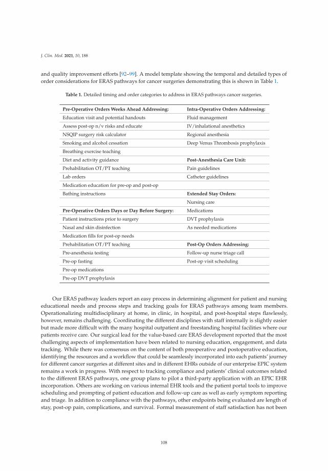
J. Clin. Med. 2021, 10, 188
and quality improvement efforts [92–99]. A model template showing the temporal and detailed types oforder considerations for ERAS pathways for cancer surgeries demonstrating this is shown in Table 1.
Table 1. Detailed timing and order categories to address in ERAS pathways cancer surgeries.
Pre-Operative Orders Weeks Ahead Addressing: Intra-Operative Orders Addressing:
Education visit and potential handouts Fluid management
Assess post-op n/v risks and educate IV/inhalational anesthetics
NSQIP surgery risk calculator Regional anesthesia
Smoking and alcohol cessation Deep Venus Thrombosis prophylaxis
Breathing exercise teaching
Diet and activity guidance Post-Anesthesia Care Unit:
Prehabilitation OT/PT teaching Pain guidelines
Lab orders Catheter guidelines
Medication education for pre-op and post-op
Bathing instructions Extended Stay Orders:
Nursing care
Pre-Operative Orders Days or Day Before Surgery: Medications
Patient instructions prior to surgery DVT prophylaxis
Nasal and skin disinfection As needed medications
Medication fills for post-op needs
Prehabilitation OT/PT teaching Post-Op Orders Addressing:
Pre-anesthesia testing Follow-up nurse triage call
Pre-op fasting Post-op visit scheduling
Pre-op medications
Pre-op DVT prophylaxis
Our ERAS pathway leaders report an easy process in determining alignment for patient and nursingeducational needs and process steps and tracking goals for ERAS pathways among team members.Operationalizing multidisciplinary at home, in clinic, in hospital, and post-hospital steps flawlessly,however, remains challenging. Coordinating the different disciplines with staff internally is slightly easierbut made more difficult with the many hospital outpatient and freestanding hospital facilities where ourpatients receive care. Our surgical lead for the value-based care ERAS development reported that the mostchallenging aspects of implementation have been related to nursing education, engagement, and datatracking. While there was consensus on the content of both preoperative and postoperative education,identifying the resources and a workflow that could be seamlessly incorporated into each patients’ journeyfor different cancer surgeries at different sites and in different EHRs outside of our enterprise EPIC systemremains a work in progress. With respect to tracking compliance and patients’ clinical outcomes relatedto the different ERAS pathways, one group plans to pilot a third-party application with an EPIC EHRincorporation. Others are working on various internal EHR tools and the patient portal tools to improvescheduling and prompting of patient education and follow-up care as well as early symptom reportingand triage. In addition to compliance with the pathways, other endpoints being evaluated are length ofstay, post-op pain, complications, and survival. Formal measurement of staff satisfaction has not been
108
J. Clin. Med. 2021, 10, 188
done but is discussed at surgical department meetings. Patient satisfaction is measured for their overallcare by standardized Press Ganey surveys, which consistently report high satisfaction scores but do notevaluate the impact of ERAS pathways alone.
6. Supportive Care Pathways
As with other oncology care pathways, those in supportive care have also been shown to be effectivein improving care quality and lowering costs based on many trials evaluating care navigation, emotionalsupport for patients and their families with cancer, electronic patient-reported outcomes integratedinto care models that address identified symptoms regularly as well as those formalizing goals of carediscussions and advanced care planning [20,21,100–105].
COH, like many large academic centers, has a palliative care department focused on clinical care andresearch. Projects have been done in pain management, regular biopsychosocial assessments, and carecoordination as well as in end of life care. The larger enterprise focus on value-based care has worked toengage the many expert providers and research teams to more formally identify the projects in supportivecare that are essential in the value-based framework to provide interval care, care navigation, and care aftercancer with the goals of improving care quality as well as clinical outcomes while optimizing the patientand staff experience. The expansion of several ongoing or supportive care pilots was underway beforethe COVID-19 pandemic expanded our use of telehealth for patient visits across the enterprise. Supportservices at COH had discussed pilots to expand social work, nutrition counseling, and other projects orpilots were in discussion to expand supportive care pathways to all enterprise sites. With the launch oftelehealth technology enterprise-wide after March 2020 that included an iterative EPIC EHR integrationand expansion of telehealth coverage by payers, some of these projects are being expedited and optimizedthrough hybrid in-person and telehealth service pathways. Highlights of some of the pathways in oursupportive care programs are:
Expanding Goals of Care Discussions as part of shared decision-making can help to avoid ineffectivecare, especially at the end of life, and help patients to achieve their broad life goals. While there arenumerous other services provided by the Department of Supportive Care Medicine at COH, we willhighlight three distinct efforts: the integrated care service, Hospice in the Village, and a goals of care pilotin poor prognosis patients.
The Integrated Care Service is a dedicated supportive care team comprised of a palliative carehospitalist, advance practice provider, social worker, chaplain, and pharmacist who provides intensiveinterdisciplinary inpatient and outpatient care to patients with multiple complex care needs, includinghigh symptom burden, advanced disease, and challenging psychosocial concerns. In the last year, theteam has cared for approximately 300 medical oncology and hematology inpatients, with over 50 beingcared for in a primary capacity. As processes and results are studied, they will serve as potential modelsto deploy in partnering with community hospitals where COH faculty practice to build unique facultyoutside hospital and clinician partnerships to achieve pathways for similar care improvements for patientshospitalized outside of the academic campus hospital.
City of Hope’s Hospice in the Village on the Duarte campus has furnished apartment units with fullamenities, specially designed for hospice care. Hospice in the Village allows patients to receive hospicecare, from their preferred provider, in a private and comfortable home-like environment, when home is notan option. For the last year and a half, overall, 100 patients were cared for in the village, representing over550 avoided inpatient days. A pilot in a community site for a similar home-based hospice and higher-levelcare (below acute level) is being explored.
A collaborative hematology, oncology, and supportive care pilot is underway utilizing various meansto identify high-risk patients, including supervised machine learning, which triggers a supportive care
109
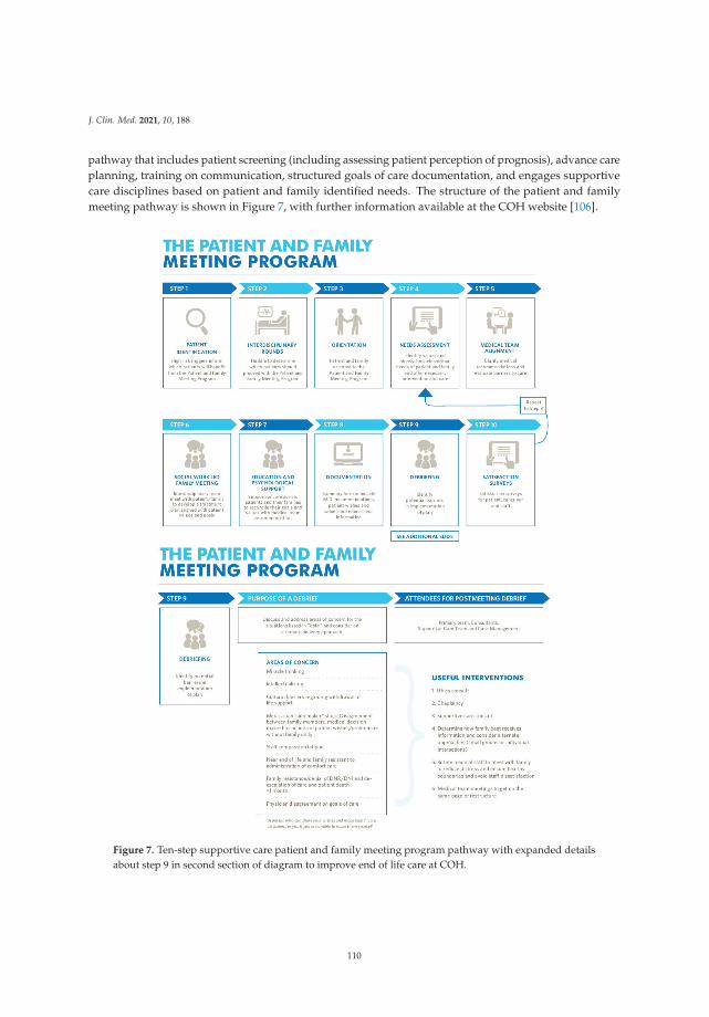
J. Clin. Med. 2021, 10, 188
pathway that includes patient screening (including assessing patient perception of prognosis), advance careplanning, training on communication, structured goals of care documentation, and engages supportivecare disciplines based on patient and family identified needs. The structure of the patient and familymeeting pathway is shown in Figure 7, with further information available at the COH website [106].
Figure 7. Ten-step supportive care patient and family meeting program pathway with expanded detailsabout step 9 in second section of diagram to improve end of life care at COH.
110
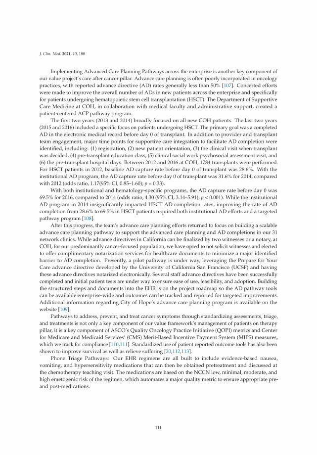
J. Clin. Med. 2021, 10, 188
Implementing Advanced Care Planning Pathways across the enterprise is another key component ofour value project’s care after cancer pillar. Advance care planning is often poorly incorporated in oncologypractices, with reported advance directive (AD) rates generally less than 50% [107]. Concerted effortswere made to improve the overall number of ADs in new patients across the enterprise and specificallyfor patients undergoing hematopoietic stem cell transplantation (HSCT). The Department of SupportiveCare Medicine at COH, in collaboration with medical faculty and administrative support, created apatient-centered ACP pathway program.
The first two years (2013 and 2014) broadly focused on all new COH patients. The last two years(2015 and 2016) included a specific focus on patients undergoing HSCT. The primary goal was a completedAD in the electronic medical record before day 0 of transplant. In addition to provider and transplantteam engagement, major time points for supportive care integration to facilitate AD completion wereidentified, including: (1) registration, (2) new patient orientation, (3) the clinical visit when transplantwas decided, (4) pre-transplant education class, (5) clinical social work psychosocial assessment visit, and(6) the pre-transplant hospital days. Between 2012 and 2016 at COH, 1784 transplants were performed.For HSCT patients in 2012, baseline AD capture rate before day 0 of transplant was 28.6%. With theinstitutional AD program, the AD capture rate before day 0 of transplant was 31.6% for 2014, comparedwith 2012 (odds ratio, 1.17(95% CI, 0.85–1.60); p = 0.33).
With both institutional and hematology-specific programs, the AD capture rate before day 0 was69.5% for 2016, compared to 2014 (odds ratio, 4.30 (95% CI, 3.14–5.91); p < 0.001). While the institutionalAD program in 2014 insignificantly impacted HSCT AD completion rates, improving the rate of ADcompletion from 28.6% to 69.5% in HSCT patients required both institutional AD efforts and a targetedpathway program [108].
After this progress, the team’s advance care planning efforts returned to focus on building a scalableadvance care planning pathway to support the advanced care planning and AD completions in our 31network clinics. While advance directives in California can be finalized by two witnesses or a notary, atCOH, for our predominantly cancer-focused population, we have opted to not solicit witnesses and electedto offer complimentary notarization services for healthcare documents to minimize a major identifiedbarrier to AD completion. Presently, a pilot pathway is under way, leveraging the Prepare for YourCare advance directive developed by the University of California San Francisco (UCSF) and havingthese advance directives notarized electronically. Several staff advance directives have been successfullycompleted and initial patient tests are under way to ensure ease of use, feasibility, and adoption. Buildingthe structured steps and documents into the EHR is on the project roadmap so the AD pathway toolscan be available enterprise-wide and outcomes can be tracked and reported for targeted improvements.Additional information regarding City of Hope’s advance care planning program is available on thewebsite [109].
Pathways to address, prevent, and treat cancer symptoms through standardizing assessments, triage,and treatments is not only a key component of our value framework’s management of patients on therapypillar, it is a key component of ASCO’s Quality Oncology Practice Initiative (QOPI) metrics and Centerfor Medicare and Medicaid Services’ (CMS) Merit-Based Incentive Payment System (MIPS) measures,which we track for compliance [110,111]. Standardized use of patient reported outcome tools has also beenshown to improve survival as well as relieve suffering [20,112,113].
Phone Triage Pathways: Our EHR regimens are all built to include evidence-based nausea,vomiting, and hypersensitivity medications that can then be obtained pretreatment and discussed atthe chemotherapy teaching visit. The medications are based on the NCCN low, minimal, moderate, andhigh emetogenic risk of the regimen, which automates a major quality metric to ensure appropriate pre-and post-medications.
111
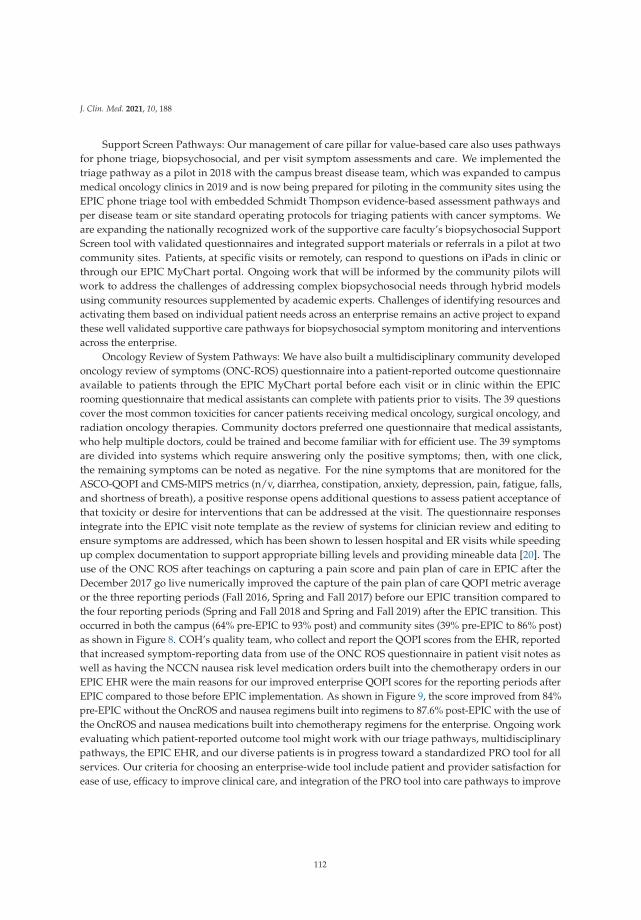
J. Clin. Med. 2021, 10, 188
Support Screen Pathways: Our management of care pillar for value-based care also uses pathwaysfor phone triage, biopsychosocial, and per visit symptom assessments and care. We implemented thetriage pathway as a pilot in 2018 with the campus breast disease team, which was expanded to campusmedical oncology clinics in 2019 and is now being prepared for piloting in the community sites using theEPIC phone triage tool with embedded Schmidt Thompson evidence-based assessment pathways andper disease team or site standard operating protocols for triaging patients with cancer symptoms. Weare expanding the nationally recognized work of the supportive care faculty’s biopsychosocial SupportScreen tool with validated questionnaires and integrated support materials or referrals in a pilot at twocommunity sites. Patients, at specific visits or remotely, can respond to questions on iPads in clinic orthrough our EPIC MyChart portal. Ongoing work that will be informed by the community pilots willwork to address the challenges of addressing complex biopsychosocial needs through hybrid modelsusing community resources supplemented by academic experts. Challenges of identifying resources andactivating them based on individual patient needs across an enterprise remains an active project to expandthese well validated supportive care pathways for biopsychosocial symptom monitoring and interventionsacross the enterprise.
Oncology Review of System Pathways: We have also built a multidisciplinary community developedoncology review of symptoms (ONC-ROS) questionnaire into a patient-reported outcome questionnaireavailable to patients through the EPIC MyChart portal before each visit or in clinic within the EPICrooming questionnaire that medical assistants can complete with patients prior to visits. The 39 questionscover the most common toxicities for cancer patients receiving medical oncology, surgical oncology, andradiation oncology therapies. Community doctors preferred one questionnaire that medical assistants,who help multiple doctors, could be trained and become familiar with for efficient use. The 39 symptomsare divided into systems which require answering only the positive symptoms; then, with one click,the remaining symptoms can be noted as negative. For the nine symptoms that are monitored for theASCO-QOPI and CMS-MIPS metrics (n/v, diarrhea, constipation, anxiety, depression, pain, fatigue, falls,and shortness of breath), a positive response opens additional questions to assess patient acceptance ofthat toxicity or desire for interventions that can be addressed at the visit. The questionnaire responsesintegrate into the EPIC visit note template as the review of systems for clinician review and editing toensure symptoms are addressed, which has been shown to lessen hospital and ER visits while speedingup complex documentation to support appropriate billing levels and providing mineable data [20]. Theuse of the ONC ROS after teachings on capturing a pain score and pain plan of care in EPIC after theDecember 2017 go live numerically improved the capture of the pain plan of care QOPI metric averageor the three reporting periods (Fall 2016, Spring and Fall 2017) before our EPIC transition compared tothe four reporting periods (Spring and Fall 2018 and Spring and Fall 2019) after the EPIC transition. Thisoccurred in both the campus (64% pre-EPIC to 93% post) and community sites (39% pre-EPIC to 86% post)as shown in Figure 8. COH’s quality team, who collect and report the QOPI scores from the EHR, reportedthat increased symptom-reporting data from use of the ONC ROS questionnaire in patient visit notes aswell as having the NCCN nausea risk level medication orders built into the chemotherapy orders in ourEPIC EHR were the main reasons for our improved enterprise QOPI scores for the reporting periods afterEPIC compared to those before EPIC implementation. As shown in Figure 9, the score improved from 84%pre-EPIC without the OncROS and nausea regimens built into regimens to 87.6% post-EPIC with the use ofthe OncROS and nausea medications built into chemotherapy regimens for the enterprise. Ongoing workevaluating which patient-reported outcome tool might work with our triage pathways, multidisciplinarypathways, the EPIC EHR, and our diverse patients is in progress toward a standardized PRO tool for allservices. Our criteria for choosing an enterprise-wide tool include patient and provider satisfaction forease of use, efficacy to improve clinical care, and integration of the PRO tool into care pathways to improve
112
J. Clin. Med. 2021, 10, 188
value-based outcomes and ease documentation to support team-based triage communication and providemineable data to support quality assessments.
Figure 8. Pain plan of care compliance for those reporting moderate to severe pain. Blue represents resultsfrom the 3 QOPI reporting quarters (Fall 2016, Spring and Fall 2017) from the previous EHRs (Allscripts oncampus and TouchWorks in community sites) prior to the EPIC implementation. Orange represents resultsfrom the 4 QOPI reporting quarters (Spring and Fall 2018 and Spring and Fall 2019) after the December2017 EPIC transition for both the campus and community sites.
Figure 9. COH enterprise QOPI final certification scores. Blue represents the 3 reporting quarters (Fall2016, Spring 2017 and Fall 2017) before the -EPIC implementation. Orange represents the 4 QOPI reportingquarters (Spring and Fall 2018 and Spring and Fall 2019) after the EPIC go live December 2017.
7. Other Enterprise Academic Community Teamwork Supporting Personalized Pathways forValue-Based Care Delivery
Disease team meetings include academic and community doctors and APPs who get to know anddevelop respect for each other. They discuss the newest science, clinical trials, and care opportunities thatare not yet in pathways, pending disease committee reviews and the 6–12-month processes. Disease teams
113
J. Clin. Med. 2021, 10, 188
maintain internal pathway flow diagrams with some modifications embraced before they are formallyavailable in national pathway tools.
New pre-ClinPath pathway update tool for practice changing therapies in development: COH hasidentified the need for a new tool to capture and share in a standardized monthly update format newpractice changing updates agreed to by disease teams. Tracking analytics are being developed concurrentlyto track the sharing of new practice changing knowledge with the agreement to add to pathways, theavailability in the pathway tool, and the uptake by faculty across the enterprise for long-term qualityimprovement work. Baseline studies in three tumor types are underway to inform this new process.
Tumor boards: These are all now done for tumor types by teleconferencing so the multidisciplinaryteams of campus and community faculty actively participate. Some community doctors report that theyare submitting at least one case to each tumor board and regularly submit to the tumor board of 1–2cancers which they may be particularly focused on. They report that the move to tele-tumor boards forall faculty has increased participation and feel that they are sharing and learning more from the entiremultidisciplinary team in very open and collegial discussions. This is true for the weekly molecular tumorboard as well.
Joint clinics: Many academic faculty have agreed to cover or see patients from time to time in differentcommunity clinics and some community doctors spend regular time in academic campus clinics. Theacademic faculty appreciates the tight knit and efficient teamwork at community sites who evolvedto be very efficient in a highly managed care world. Academic experts get to share one-on-one thelatest molecular or other discoveries during real-time patient encounters with their community facultycolleagues and observe the use of ClinPath and other pathways at community sites. Friendships andincreased collegiality have blossomed, which encourages ongoing sharing that offers every patient accessto cutting-edge knowledge. A system to facilitate second opinions between the campus and communityfaculty has also helped to increase patient peace of mind while allowing them to receive care at the siteclosest to their home.
Regional, disease team and campus symposia and lectures are shared with all faculty. The medicaloncology department holds quarterly half-day symposia where campus and community faculty presentthe latest science and care pathways as well as sharing clinical trial, value-based care, and programmaticupdates. Disease teams also hold retreats to share the latest disease-specific knowledge and clinical trials,which is another helpful format for networking and building of collegial relationships. The pandemicrestrictions on in person meetings led the medical oncology department to hold their sixth symposia inOctober of 2020, where 118 members participated in a 5-h update on science and clinical programs. Thefeedback was enthusiastic for the format, where there was active engagement and extensive group andinterpersonal sharing of ideas in the chat function while members avoided having to travel from family ona weekend.
8. Other Value-Based Care Framework Projects Integrated with the Pathways Programs
Development of enterprise disease trees is a newer initiative being done to aid further in thestandardization process of developing personalized multidisciplinary care plans throughout the enterprise.Community disease and regional leaders are closely involved with academic-based colleagues in buildingthe initial decision tree for breast cancer. Next up in the queue are GI and GU multispecialty decisiontree builds. The campus disease trees include locations for complex surgeries or other cancers as wellas timing and genomic testing for both somatic and germline mutations. Within these disease trees isan enterprise-wide initiative to extend germline and somatic genomic testing to all patients as well asthe integration of the results into the disease pathways. This will be piloted in one site (South Pasadena)close to the academic center and then expand to all sites by 2021. Pulling in the disease-specific pathways
114
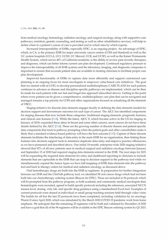
J. Clin. Med. 2021, 10, 188
from medical oncology, hematology, radiation oncology, and surgical oncology, along with supportive carepathways, nutrition, genetic counseling, and testing as well as other rehabilitative services, will help todefine when in a patient’s course of care is provided and in which sites by which experts.
Increased interoperability of EHRs, especially EPIC, is an ongoing project. An advantage of EPIC,which, in CA, is the primary EHR for major university cancer centers (COH and Stanford) as well as theUC system hospitals (UCLA, UCI, UCSD, UC Merced, UCR, and UCSF), as well as the Kaiser PermanenteHealth System, which serves 40% of California residents, is the ability to review past records, therapies,and diagnoses, which can better inform current care plan development. Continued regulatory pressure toimprove the interoperability of all EHR systems and the laboratory, imaging, and diagnostic companies isimportant to ensure that accurate patient data are available to treating clinicians to facilitate proper careplan development.
Improved functionality of EHRs to capture data more efficiently and support customized careplanning is an ongoing focus for most oncologists to empower value-based care initiatives. The goalthat we started with at COH, to develop personalized multidisciplinary CARE PLANS for each patient,continues to advance as disease and discipline-specific pathways are implemented, which can be thenbe made for each patient with our fast and frugal tree approach (described above). Getting to the pointwhere every patient can be given a comprehensive, multidisciplinary care plan that can be navigated andmanaged remains a top priority for COH and other organizations focused on actualizing all the elementsof VBCC.
Staging initiative for discrete data elements engages faculty in defining the data elements needed fordecision support and downstream outcomes for each type of cancer. The AJCC has identified componentsfor staging diseases that now include three categories: traditional staging elements, prognostic features,and clinical care features [114]. While the latest, AJCC 8, which became active in the US for staging inJanuary of 2018, expanded these ideas in breast and some other cancers, most cancers do not have thesedetails defined by the AJCC [114]. These are the growing number of discrete disease and patient-specificdata components that track to pathway prompting when the patient goals and other comorbidities make itlikely that a standard evidence-based pathway will have the best outcome [115]. Capture of these discreteelements facilitates the interfacing of data entry in the main EHR for an organization, then linking thosefeatures into decision support tools to minimize duplicate data entry and improve practice efficiencies,as we have pioneered and described above. Our initial 10-month, enterprise-wide 2020 staging initiativeshowed that 92% of all new patients seen in medical surgical and radiation oncology between Januaryand September 15 of 2020 had required staging data elements entered in the EHR. The next steps for 2021will be expanding the required data elements for entry and dashboard reporting to clinicians to includeelements that are capturable in the EHR that can map to decision support in the pathway tool while wesimultaneously expand the tumor types we have full mapping of EHR data elements into the pathwaytool and back to therapy orders for medical and radiation oncology, as discussed above.
Oral hemotherapy drugs are built into the EHR as regimens: In preparation for further integrationbetween our EHR and the ClinPath pathway tool, we identified 58 oral cancer drugs which had not beenbuilt into our chemotherapy ordering system (Beacon for EPIC). These are included in 96 protocols whichwill be prompted from the pathway program, so both academic and community medical oncologists thenhematologists were recruited, agreed to build specific protocols including the references, associated NCCNnausea level, dosing, visit, lab, and specific drug guidance using a standardized Excel tool. Examples ofcurrent protocols were shared at individual or small group teaching sessions held through video visits.The builds for all but the last 25 protocols have been completed, including validation by an oncologyPharm D since April 2020, which was stimulated by the March 2020 COVID-19 pandemic work from homeemphasis. We anticipate that the remaining 25 regimens will be built and validated by December of 2020and have a goal that all of the 96 regimens will be available in the EPIC Beacon ordering system by Q1 2021.
115
J. Clin. Med. 2021, 10, 188
Once completed, we will launch a best practice alert if a clinician orders an oral chemotherapy agent inthe stand-alone prescription system of the HER rather than ordering it in the fully integrated chemotherapyorder section in the EPIC Beacon system. The advantage of the build in the specific chemotherapy ordersystem is the tie into the pathway, prompting the ability to standardize chemotherapy teaching visits ineach order, support for the authorization processes, and integration into our recently launched specialtypharmacy program. With these integrations, any additional coverage needs for high-cost specialty andother cancer therapies can be identified and addressed early on while also facilitating analytic reportingon specific patient tumor types and disease features treated by specific regimens over measurable timeperiods for clinical and financial outcomes.
Outreach to community hospitals and ambulatory surgery centers is ongoing to share ERAS andother supportive care pathway work and engage diverse teams in meeting the care steps. Working acrosssystems remains a challenge as noted. There is a clear focus, however, on developing these so that,wherever the patient chooses to get their care, the same high-quality care standards will be incorporatedinto the care processes and embraced by all staff, whether they work for our enterprise, a partnered entity,or a standalone facility. An advantage of our academic community faculty team model is the easy sharingand customization abilities of our clinicians and administrators so that the additional tools and processesdeveloped at our NCI designated comprehensive cancer center can be shared with community-basedhospitals and clinicians outside our enterprise.
The COH Enterprise data warehouse collects data elements from all digital platforms used by theenterprise and supports VBCC as well as research and other initiatives. It is overseen by a governancecommittee with clinical informatics, precision medicine, and information technology experts, who makedata available to empower care and insights. From the data warehouse, Tableau dashboards can be createdfor feedback to clinicians and administrators along with many other informatic formats to process largedatasets for study, some of which are shown as figures in this manuscript. The digital framework to turninformation into real-world data is described in Figure 10.
116
J. Clin. Med. 2021, 10, 188
Figure 10. City of Hope’s overriding digital framework to empower value-based care and real-worldinsights.
9. Challenges of Keeping Pathways Updated and Minimizing Burnout
Among the many workstreams we have discussed here, two other challenges should be highlighted:the challenge to keep decision support tools up to date in real time and the importance of engagingphysicians in the design and efficient implementation of value-based projects as team members to minimizeburnout. Our enterprise has recognized and is addressing methods to keep pathway tools up to dateand engage clinicians in the many value-based project processes. These challenges are both minimizedby the integrated academic community faculty foundation teamwork, which not only gains from thediverse experiences and wisdom of the faculty but can help to reduce the growing burnout rates andearlier retirement events for cancer clinicians. Three components of burnout described by the AMA StepsForward highlight a culture of wellness, personal resilience, and practice efficiencies as the major areascontributing to burnout [116]. Internal COH faculty surveys identified practice efficiencies as the mostimportant factor contributing to burnout in our faculty as collected and shared at regular faculty meetings.These surveys have prioritized work to address efficiencies and teamwork as we implement VBCC projectswith academic and community team inputs.
Implementing multidisciplinary pathways across an enterprise and expanding to outsideorganizations faces the ongoing challenge of keeping pathway and decision support tools up to datein real time so that clinicians can deliver value-based care across the enterprise. Oncology is fortunateto have so many continuing new discoveries that can improve outcomes, but they require real-timetools and programming to bring practicing changing diagnostic and therapeutic knowledge to clinicians.Doing this with academic disease team leads working with integrated academic community diseaseteams helps to share the knowledge and build real-time enterprise tool updates until the larger pathwaydecision tools are updated. This can also improve workflows that can enhance patient and practitioner
117
J. Clin. Med. 2021, 10, 188
satisfaction [116]. It can also help to reduce burnout from extended work hours from individual clinicianshaving to individually evaluate the copious amounts of detailed new cancer information for the manykinds of cancer patients that they see.
A second challenge is to engage clinicians and staff teams in pathways and value-based initiativework so that the data entry, programming of digital tools, and workflow processes align to minimize theircontribution to burnout while empowering evidence-based care delivery. Faculty who are encouraged andrewarded by leadership to participate in their passion for cutting-edge care and clinical research, whilecontributing to enterprise and professional initiatives, can lessen the sense of loss of meaning in workand a feeling they are a replaceable robot in an assembly line of patient care. Dashboard feedback thatengages doctors and their teams with the results of discrete data entry and clinical trial outcomes can fuelpride in their and their care team’s work with patients as well as in their role in the success of the largerenterprise. While many clinicians expressed initial push back on the increased workloads to enter discretestaging and pathway navigation data, as familiarity with the systems increased and the integration ofthe tools as well as the clinical trials expanded, there was a growing sense, which has not been formallymeasured, that the time spent entering defined, discrete data once is likely offset by time savings fromauthorization clerks, searching for patient information to inform clinical trial prioritization, expandedefficiencies in database-related clinical research, improved inter-specialty communication of the patient’sdisease and course, and, as sequences of disease descriptions build across a patient journey, markedlyspeeding up the ability of clinicians to gain a rapid understanding of what a patient has had, what theywere treated with, and where the next steps in diagnostics, follow-up, or subsequent therapies should be.All of these tradeoffs would benefit from formal study that might also help to inform new pilots aboutreturn on investments regarding who and when on the team with what training are optimal for specificdata entry if not the doctors or APPs [116].
This work can also be informed by many experts outside of medicine as we continue to iterate andlearn from pioneering efforts. Dr. Etienne Wenger, PhD is one such globally recognized thought leader inthe field of learning theory and its application to business who is a pioneer of “community of practice”research. Especially relevant to our work is his statement that “Communities of practice are important tothe functioning of any organization, but they become crucial to those that recognize knowledge as a keyasset” [117].
COH as an enterprise is addressing these two challenges along with the many others discussed.The pathways and care plan development programs benefit with leadership supporting active clinicianengagement across our academic and community cancer center sites, which addresses several majorcomponents to minimize burnout: improving work place efficiencies, reducing social isolation at work,and the sense of loss of meaning in work that care standardization without engagement can bring. Thishas helped clinicians and staff to embrace the shift to value-based care through personalized precisionmedicine with integrated clinical research. The addition of teamwork across the enterprise by clinicians,managers, nurses, pharmacists, administrators, quality and informatics experts with our IT teams alsoiterates improvements in EHR functionalities, allowing more patient-reported input and team-based dataentry, which can free up clinician time to analyze data and engage patients in developing a personalized,multidisciplinary care plan. The many projects outlined with multidisciplinary oncology pathways asa key component towards enterprise delivery of high-quality cancer care can be done with thoughtfulattention to methodologies and team engagement so that value-based care, defined as the combination ofhigh-quality care, patient satisfaction, provider satisfaction, and enterprise health at the appropriate costs,can be achieved.
118
J. Clin. Med. 2021, 10, 188
10. Conclusions
Patients, clinicians, patients’ families, employers, and payers want to have the best care plan available,discussed and implemented seamlessly with ongoing patient support to achieve the best cancer careoutcome, whether this is a cure or improved quality of life. Diagnostic testing, genomics, imaging, andclinical assessments have increasingly complex discrete data elements that need to be transformed intoactionable information and decision support. This knowledge also needs to be communicated effectivelyin the shared decision-making process with patients and payers. Pathway programs with decision supporttools integrated into the clinical workflows can inform high-quality clinical trial, treatment, and supportivecare decisions efficiently when overseen and supplemented by disease teams informed by integratedacademic community faculty partnerships. This knowledge, integrated into EHRs and digital tools, canempower true VBCC throughout a fully integrated, multisite enterprise.
Thus, while many see oncology pathways as focusing only on medical oncology and hematologyregimens, they are just as important for specific patient populations such as geriatrics, clinical cancergenetics, AYA, and pediatric populations as well as for surgical oncology, radiation oncology, andsupportive care. Combining pathways personalized to each patient can then build the evidence-basedcomprehensive care plans to optimize each patient’s cancer journey for their best health outcome.Foundational work on multidisciplinary oncology pathways, which started with COH’s early engagementand leadership with the NCCN, has progressed to the development and implementation of disease-specific,supportive care, subpopulation-specific (i.e., geriatrics), and multidisciplinary pathways toward our goalof comprehensive, individual patient care plans. This work, coordinated under our Value Realizationenterprise project, is overseen by faculty and administrative teams with diverse scientific and practicaloperational knowledge as well as academic and community expertise. The partnered teams havefacilitated this pioneering work as an enterprise to develop, implement, and analyze the methodology andinfrastructure to care for each cancer patient with a personalized care plan based on their cancer diagnosis,detailed and evolving molecular diagnostics, clinical trials, and bio-psychosocial needs with tools tosupport engagement in shared decision-making at initial diagnosis and any subsequent recurrences.
As more community oncology practitioners with practical implementation and local culturalexpertise join local, regional, and national networks with experts who maintain the intense, detailedfocus in subspecialty areas of oncology care, this combined knowledge and perspective can be sharedthrough standardized decision support oncology pathway tools supplemented with real-time, team-ledmodifications as new knowledge helps us to achieve better outcomes with less toxicity for patients,regardless of where they live. Our COH enterprise commitment to one standard of high-quality carefor every patient led to the pilots and multidisciplinary oncology pathway programs using tools, teams,and processes that can serve to inform others as network affiliations grow. While this work is empoweredby the team-based, one faculty, one standard of care philosophy across our enterprise, there are manyoperational and implementation steps to deliver and measure this care standard. The work has benefittedfrom the combined strength of the academic faculty’s deep scientific knowledge and focus with thecommunity faculty’s deep clinical and operational knowledge in partnership with administrators, projectleads, informaticists, IT experts, and committees.
Implementing and integrating multidisciplinary oncology pathway programs also benefits from otherpartnered projects with subspecialty groups, IT interfaces, EHR functionalities, care teams, and analyticreporting as reviewed. While multidisciplinary oncology pathways are key components in the delivery ofhigh-quality cancer care, they are only one of the many important component projects of VBCC. Achievingmeasurable delivery of all component projects within the three VBCC pillars of evidenced-based care,care management, and care after cancer with high levels of patient satisfaction, clinician satisfaction, and
119
J. Clin. Med. 2021, 10, 188
institutional health with optimized costs remains a work in progress. Early reports of COH’s successes andthe successes reported by others can serve to inform and inspire ongoing work in VBCC [8,11,26,27,30,34].
Author Contributions: Conceptualization: L.D.B., B.Y., S.S., H.C., S.G. (Stacy Gray), F.Z., E.K., S.G. (Scott Glaser), R.S.,D.J. Formal Analysis: L.D.B., F.Z., D.D., A.B.L., D.M., S.G.(Scott Glaser), J.C., A.N., D.J. Data Curation: L.D.B., B.Y., S.S.,A.P., F.Z., D.D., A.B.L., M.S.S., E.K., S.G.(Scott Glaser), D.M., A.N., D.J. Writing—original draft preparation: L.D.B.,B.Y., C.Y., H.C., S.S., A.P., F.Z., A.B.L., M.S.S., E.K., S.G. (Stacy Gray), S.G. (Scott Glaser), T.T., A.N., I.B.P., P.K., D.J.Writing—review and editing: L.D.B., M.C., B.Y., C.Y., H.C., S.S., A.P., F.Z., D.D., A.B.L., M.S.S., E.K., S.G. (Stacy Gray),D.M., S.G. (Scott Glaser), G.B., C.A., T.T., J.C., A.N., I.B.P., L.K., P.R., K.S., A.N., P.K., R.S., J.Y., D.J. All authors haveread and agreed to the published version of the manuscript.
Funding: This research received no external funding. The work was conducted as quality improvement projects underinstitutional programs.
Conflicts of Interest: Linda Bosserman serves as an unpaid member and co-chair of the breast cancer pathwaycommittee for Elsevier’s Clinical Pathways and serves as an unpaid member of the adult oncology steering boardfor the EPIC electronic health record. Chao disclosed he has financial relationships in the last 36 months with Merck,Amgen, Macrogenetics, Ono Pharmaceutical, Foundation Medicine and Daiichi-Sankyo. Elisabeth King is a memberof a Pfizer advisory board and on AstraZeneca’s speaker’s bureau. No conflicts were reported by other coauthorsrelated to this manuscript.
References
1. Kirkwood, M.K.; Hanley, A.; Bruinooge, S.S.; Garrett-Mayer, E.; Levit, L.A.; Schenkel, C.; Seid, J.E.; Polite, B.N.;Schilsky, R.L. The State of Oncology Practice in America, 2018: Results of the ASCO Practice Census Survey. J.Oncol. Pract. 2018, 14, e412–e420. [CrossRef] [PubMed]
2. Community Oncology Alliance, 2018 Community Oncology Practice Impact Report. 2 April 2018. Availableonline: https://communityoncology.org/2018-community-oncology-practice-impact-report/ (accessed on 23June 2020).
3. Levit, L.; Balogh, E.; Nass, S. Delivering High-Quality Cancer Care: Charting a New Course for A System in Crisis;Ganz, P., Ed.; Committee on Improving the Quality of Cancer Care: Addressing the Challenges of an AgingPopulation; Board on Health Care Services; Institute of Medicine; National Academies Press (US): Washington,DC, USA, 2013.
4. Bosserman, L.D. Benefits and Challenges of Growing Oncology Networks in the United States. J. Oncol. Pract.2018, 14, 761–762. [CrossRef] [PubMed]
5. Albanese, C.; Aaby, D.; Platchek, T. Advanced Lean in Healthcare; CreateSpace Independent Publishing Platform:Scotts Valley, CA, USA, 2014; ISBN 149614189X.
6. Sprandio, J.D. Oncology patient-centered medical home and accountable cancer care. Commun. Oncol. 2010, 7,565–572. [CrossRef]
7. Bosserman, L.D.; Verrilli, D.; McNatt, W. Partnering with a payer to develop a value-based medical home pilot:A West Coast practice’s experience. Am. J. Manag. Care 2012, 18, SP189–SP190. [CrossRef]
8. Waters, T.M.; Kaplan, C.M.; Graetz, I.; Price, M.M.; Stevens, L.A.; McAneny, B.L. Patient-Centered MedicalHomes in Community Oncology Practices: Changes in Spending and Care Quality Associated with the COMEHOME Experience. J. Oncol. Pract. 2019, 15, e56–e64. [CrossRef]
9. Piana, R. Integration across the Spectrum: Community Perspective on the Medical Oncology Home Model; The ASCOPost: Huntington, NY, USA, 2015.
10. Thompson, G. The Oncology Medical Home: Embodiment of the American Pioneering Spirit. Oncol. Pract.Manag 2012, 2, 6.
11. Page, R.D.; Newcomer, L.N.; Sprandio, J.D.; McAneny, B.L. The Patient-Centered Medical Home in Oncology:From Concept to Reality. Am. Soc. Clin. Oncol. Educ. Book 2015, e82–e89. [CrossRef]
12. Neubauer, M.A.; Hoverman, J.R.; Kolodziej, M.; Reisman, L.; Gruschkus, S.K.; Hoang, S.; Beveridge, R.A. Costeffectiveness of evidence-based treatment guidelines for the treatment of non-small-cell lung cancer in thecommunity setting. J. Oncol. Pract. 2010, 6, 12–18. [CrossRef]
120
J. Clin. Med. 2021, 10, 188
13. Hoverman, J.R.; Cartwright, T.H.; Patt, D.A.; Espirito, J.L.; Clayton, M.P.; Garey, J.S.; Pyenson, B. Pathways,outcomes, and costs in colon cancer: Retrospective evaluations in two distinct databases. J. Oncol. Pract. 2011, 7(Suppl. 3), 52s–59s. [CrossRef]
14. Weese, J.L.; Shamah, C.J.; Sanchez, F.A.; Perez Moreno, A.C.; Mitchell, D.; Sessa, T.; Amy, B. Use of treatmentpathways reduce cost and decrease ED utilization and un-planned hospital admissions in patients (pts) withstage II breast cancer. J. Clin. Oncol. 2019, 37 (Suppl. 15), e12012. [CrossRef]
15. Jackman, D.M.; Zhang, Y.; Dalby, C.; Nguyen, T.; Nagle, J.; Lydon, C.A.; Jacobson, J.O. Cost and survival analysisbefore and after implementation of Dana-Farber clin-ical pathways for patients with stage IV non-small-cell lungcancer. J. Oncol. Pract. 2017, 13, e346–e352. [CrossRef] [PubMed]
16. Newcomer, L.N.; Gould, B.; Page, R.D.; Donelan, S.A.; Perkins, M. Changing Physician Incentives for Affordable,Quality Cancer Care: Results of an Episode Payment Model. J. Oncol. Pract. 2014, 10, 322–326. [CrossRef]
17. Doyle, C. Anthem’s Clinical Pathways Demonstrate Value: The Payer Perspective. Am. Health Drug Benefits 2015,8, 28. [PubMed]
18. Scott, J.A.; Milligan, S.; Wong, W.; Winn, D.; Cooper, J.; Schneider, N.; Parkes, S.; Feinberg, B.A. Validation ofobserved savings from an oncology clinical pathways program. J. Clin. Oncol. 2013, 31, 6553. [CrossRef]
19. Kardos, S.V.; Chan, K.G.; Yuh, B.E.; Yamzon, J.; Ruel, N.; Zachariah, F.; Lau, C.S.; Crocitto, L.E. Recovery aftersurgery and care coordination pathway at City of Hope: Length of stay, readmissions, and complications. J. Clin.Oncol. 2016, 34, 410. [CrossRef]
20. Basch, E.; Deal, A.M.; Dueck, A.C.; Scher, H.I.; Kris, M.G.; Hudis, C.; Schrag, D. Overall Survival Results of aTrial Assessing Patient-Reported Outcomes for Symp-tom Monitoring During Routine Cancer Treatment. JAMA2017, 318, 197–198. [CrossRef] [PubMed]
21. Koprowski, C.; Petrilli, N.; Johnson, E.J.; Sites, K. The Supportive Care of Oncology Patients (SCOOP) Pathway:A novel Ap-proach to Improving the Patient Experience in the Curative Treatment of Cancer. J. Clin. Pathw. 2019,5, 39–43.
22. Grant, M.; Ferrell, B.R.; Rivera, L.M.; Lee, J. Unscheduled readmissions for uncontrolled symptoms. A healthcare challenge for nurses. Nurs. Clin. N. Am. 1995, 30, 673–682.
23. Quality Payment Program Executive Summary, MACRA. 16 October 2016. Available online: https://qpp.cms.gov/docs/QPP_Executive_Summary_of_Final_Rule.pdf (accessed on 12 October 2020).
24. Findlay, M.; Rankin, N.M.; Shaw, T.; White, K.; Boyer, M.; Milross, C.G.; Lourenço, R.D.A.; Brown, C.; Collett, G.;Beale, P.; et al. Best Evidence to Best Practice: Implementing an Innovative Model of Nutrition Care for Patientswith Head and Neck Cancer Improves Outcomes. Nutrients 2020, 12, 1465. [CrossRef]
25. Smith, T.J.; Hillner, B.E. Ensuring Quality Cancer Care by the Use of Clinical Practice Guidelines and CriticalPathways. J. Clin. Oncol. 2001, 19, 2886–2897. [CrossRef]
26. Lederman, L. Are Medical Homes and ACOs the Future of Cancer Care? Oncology Business Review. September2012 Edition, Volume 11, Issue 9. Available online: https://obroncology.com/article/are-medical-homes-and-acos-the-future-of-cancer-care-2/ (accessed on 7 October 2020).
27. Kuznar, W. Patient-Centered Oncology Medical Home Improves Health Outcomes at LowerCosts to Patients and Payers. Value Based Cancer Care 2014, 5, 1579. Available online: http://valuebasedcancer.com/issues/2014/october-2014-vol-5-no-8/1579-patient-centered-oncology-medical-home-improves-health-outcomes-at-lower-costs-to-patients-and-payers (accessed on 7 October 2020).
28. Bunkhead, C. Achieving Higher Quality, Lower Cost through Oncology Medical Homes. Oncol. Pract. Manag.2015, 5, 6. Available online: http://oncpracticemanagement.com/issues/2015/september-2015-vol-5-no-6/550-achieving-higher-quality-lower-cost-through-oncology-medical-homes (accessed on 7 October 2020).
29. Patel, M. The Oncology Care Model: Aligning Financial Incentives to Improve Outcomes. Oncology PracticeManag. 2016, 6, 12. [CrossRef]
30. Colligan, E.M.; Ewald, E.; Ruiz, S.; Spafford, M.; Cross-Barnet, C.; Parashuram, S. Innovative Oncology CareModels Improve End-Of-Life Quality, Reduce Utilization And Spending. Health Aff. 2017, 36, 433–440. [CrossRef]
121

J. Clin. Med. 2021, 10, 188
31. Hoverman, J.R.; Neubauer, M.A.; Jameson, M.; Hayes, J.E.; Eagye, K.J.; Abdullahpour, M.; Verrilli, D.K.Three-Year Results of a Medicare Advantage Cancer Management Program. J. Oncol. Pract. 2018, 14, e229–e237.[CrossRef]
32. Neubauer, M. Clinical pathways: Reducing costs and improving quality across a network. Am J Manag. Care2020, 26, SP60–SP61.
33. Walker, B.; Frytak, J.; Hayes, J.; Neubauer, M.; Robert, N.; Wilfong, L. Evaluation of Practice Patterns AmongOncologists Par-ticipating in the Oncology Care Model. JAMA Netw. Open 2020, 3, e205165. [CrossRef]
34. Ellis, P.G. Development and Implementation of Oncology Care Pathways in an Integrated Care Network: TheVia Oncology Pathways Experience. J. Oncol. Pract. 2013, 9, 171–173. [CrossRef]
35. Miller, B. How the First Oncology ACO Achieves Savings Every Year, Advisory Board: Oncology Rounds. 1March 2016. Available online: https://www.advisory.com/research/oncology-roundtable/oncology-rounds/2016/03/miami-cancer-institute (accessed on 12 October 2020).
36. Frois, C.; Howe, A.; Jarvis, J.; Grice, K.; Wong, K.; Zacker, C.; Sasane, R. Drug Treatment Value in a ChangingOncology Landscape: A Literature and Provider Perspective. J. Manag. Care Spec. Pharm. 2019, 25, 246–259.[CrossRef]
37. Cox, J.V.; Ward, J.C.; Hornberger, J.C.; Temel, J.S.; McAneny, B.L. Community Oncology in an Era of PaymentReform. Am. Soc. Clin. Oncol. Educ. Book 2014, e447–e452. [CrossRef]
38. Avalere Health. Clinical Pathways: Overview of Current Practices and Potential Implications for Patients, Payers,and Providers. July 2015. Available online: https://www.phrma.org/report/clinical-pathways-overview-of-current-practices-and-potential-impliationsor-patients-payers-and-providers (accessed on 12 October 2020).
39. Rocque, G.B.; Williams, C.; Kenzik, K.; Jackson, B.E.; Halilova, K.I.; Sullivan, M.M.; Rocconi, R.P.; Azuero, A.;Kvale, E.; Huh, W.K.; et al. Where Are the Opportunities for Reducing Health Care Spending Within AlternativePayment Models? J. Oncol. Pract. 2018, 14, e375–e383. [CrossRef] [PubMed]
40. Neubauer, M. Clinical Pathways Can Lead to Cost Savings, Better Care. Onc. Bus Rev. Green 2014, 11. Availableonline: https://obroncology.com/article/clinical-pathways-can-lead-to-cost-savings-better-care-2/ (accessedon 12 October 2020).
41. Mortimer, J.E.; Kruper, L.; Cianfrocca, M.; Lavasani, S.; Liu, S.; Tank-Patel, N.; Yeon, C. Use of HER2-DirectedTherapy in Metastatic Breast Cancer and How Com-munity Physicians Collaborate to Improve Care. J. Clin. Med.2020, 9, 1984. [CrossRef] [PubMed]
42. Salgia, R.; Mambetsariev, I.; Tan, T.; Schwer, A.; Pearlstein, D.P.; Chehabi, H.; Baroz, A.; Fricke, J.; Pharaon,R.; Romo, H.; et al. Complex Oncological Decision-Making Utilizing Fast-and-Frugal Trees in a CommunitySetting—Role of Academic and Hybrid Modeling. J. Clin. Med. 2020, 9, 1884. [CrossRef]
43. Salgia, N.J.; Philip, E.J.; Ziari, M.; Yap, K.; Pal, S.K. Advancing the Science and Management of Renal CellCarcinoma: Bridging the Divide between Academic and Community Practices. J. Clin. Med. 2020, 9, 1508.[CrossRef]
44. FDA. Hematology/Oncology (Cancer) Approvals and Safety Notifications. 2016–June 2020. Availableonline: https://www.fda.gov/drugs/resources-information-approved-drugs/hematologyoncology-cancer-approvals-safety-notifications (accessed on 23 June 2020).
45. Tran, G.; Zafar, S.Y. Financial toxicity and implications for cancer care in the era of molecular and immunetherapies. Ann. Transl. Med. 2018, 6, 166. [CrossRef] [PubMed]
46. Zafar, S.Y.; Peppercorn, J.M.; Schrag, D.; Taylor, D.H.; Goetzinger, A.M.; Zhong, X.; Abernethy, A.P. The financialtoxicity of cancer treatment: A pilot study assessing out-of-pocket expenses and the insured cancer patient’sexperience. Oncologist 2013, 18, 381–390. [CrossRef] [PubMed]
47. Zafar, S.Y. Financial Toxicity of Cancer Care: It’s Time to Intervene. J. Natl. Cancer Inst. 2016, 108, djv370.[CrossRef]
48. Karimi, M.; Wang, C.; Bahadini, B.; Hajjar, G.; Fakih, M. Integrating Academic and Community Practices in theManagement of Colorectal Cancer: The City of Hope Model. J. Clin. Med. 2020, 9, 1687. [CrossRef] [PubMed]
122
J. Clin. Med. 2021, 10, 188
49. Rajurkar, S.; Mambetsariev, I.; Pharaon, R.; Leach, B.; Tan, T.; Kulkarni, P.; Salgia, R. Non-Small Cell Lung Cancerfrom Genomics to Therapeutics: A Frame-work for Community Practice Integration to Arrive at PersonalizedTherapy Strategies. J. Clin. Med. 2020, 9, 1870. [CrossRef]
50. Basse, L.; Hjort Jakobsen, D.; Billesbølle, P.; Werner, M.; Kehlet, H. A clinical pathway to accelerate recovery aftercolonic re-section. Ann. Surg. 2000, 232, 51–57. [CrossRef]
51. Kehlet, H.; Mogensen, T. Hospital stay of 2 days after open sigmoidectomy with a multimodal rehabilitationprogramme. BJS 1999, 86, 227–230. [CrossRef] [PubMed]
52. Fearon, K.C.; Ljungqvist, O.; Von Meyenfeldt, M.; Revhaug, A.; DeJong, C.H.C.; Lassen, K.; Nygren, J.; Hausel, J.;Soop, M.; Andersen, J.P.; et al. Enhanced recovery after surgery: A consensus review of clinical care for patientsundergoing colonic resection. Clin. Nutr. 2005, 24, 466–477. [CrossRef] [PubMed]
53. Ljungqvist, O. ERAS–enhanced recovery after surgery: Moving evidence-based perioperative care to practice.JPEN J. Parenter. Enteral Nutr. 2014, 38, 559–566. [CrossRef] [PubMed]
54. Carmichael, J.C.; Keller, D.S.; Baldini, G.; Bordeianou, L.; Weiss, E.; Lee, L.; Steele, S.R. Clinical Practice Guidelinesfor Enhanced Recovery After Colon and Rectal Surgery From the American Society of Colon and Rectal Surgeonsand Society of American Gastrointestinal and Endo-scopic Surgeons. Dis. Colon Rectum 2017, 60, 761–784.[CrossRef] [PubMed]
55. Ljungqvist, O.; Scott, M.; Fearon, K.C. Enhanced Recovery after Surgery: A Review. JAMA Surg. 2017, 152,292–298. [CrossRef]
56. Kehlet, H. Enhanced Recovery Protocols (ERP): Need for Action. Ann Surg. 2018, 268, e85. [CrossRef]57. Brindle, M.E.; Nelson, G.; Lobo, D.; Ljungqvist, O.; Gustafsson, U.O. Recommendations from the ERAS®Society
for standards for the development of enhanced recovery after surgery guidelines. BJS Open 2019, 4, 157–163.[CrossRef]
58. Chenam, A.; Chan, K.G. Enhanced Recovery after Surgery for Radical Cystectomy. Cancer Treat. Res. 2018, 175,215–239. [CrossRef]
59. Scarberry, K.; Berger, N.G.; Scarberry, K.B.; Agrawal, S.; Francis, J.J.; Yih, J.M.; Abouassaly, R. Improved surgicaloutcomes following radical cystectomy at high-volume centers influence overall survival. Urol. Oncol. 2018, 36,308.e11–308.e17. [CrossRef]
60. Ellis, P.G.; O’Neil, B.H.; Earle, M.F.; McCutcheon, S.; Benson, H. The usefulness of clinical pathways (CP) inmanaging quality and cost in oncology networks. J. Clin. Oncol. 2016, 34 (Suppl. 4), 715. [CrossRef]
61. Chiang, A.C.; Lake, J.; Sinanis, N.; Brandt, D.; Kanowitz, J.; Kidwai, W.; Tara, H. Measuring the Impact ofAcademic Cancer Network Development on Clinical Inte-gration, Quality of Care, and Patient Satisfaction. J.Oncol. Pract. 2018, 14, e823–e833. [CrossRef] [PubMed]
62. Zon, R.T.; Frame, J.N.; Neuss, M.N.; Page, R.D.; Wollins, D.S.; Stranne, S.; Bosserman, L.D. American Societyof Clinical Oncology Policy Statement on Clinical Pathways in Oncology. J. Oncol. Pract. 2016, 12, 261–266.[CrossRef] [PubMed]
63. Zon, R.T.; Edge, S.B.; Page, R.D.; Frame, J.N.; Lyman, G.H.; Omel, J.L.; Wollins, D.S.; Green, S.R.; Bosserman, L.D.American Society of Clinical Oncology Criteria for High-Quality Clinical Pathways in Oncology. J. Oncol. Pract.2017, 13, 207–210. [CrossRef]
64. McCutcheon, S.; Ellis, P.G.; Hess, R.; Krebs, M.; Lokay, K. Frequency of efficacy, toxicity and cost as the decidingfactor when determining clinical pathways. J. Clin. Oncol. 2016, 34, e18169. [CrossRef]
65. Ersek, J.L.; Black, L.J.; Thompson, M.A.; Kim, E.S. Implementing Precision Medicine Programs and Clinical Trialsin the Community-Based Oncology Practice: Barriers and Best Practices. Am. Soc. Clin. Oncol. Educ. Book 2018,38, 188–196. [CrossRef]
66. Shamah, C.J.; Saphner, T.J. Effect on clinical trial participation by integration of a clinical pathway program intoan electronic health record (EHR). J. Clin. Oncol. 2016, 34, 167. [CrossRef]
67. Saphner, T.; Thompson, M.A.; Planton, S.; Singh, M.; Glandt, N.; Robinson, L.; DeBartolo, J. Insights fromBuilding a New National Cancer Institute Community On-cology Research Program Site. WMJ 2016, 115,191–195.
123
J. Clin. Med. 2021, 10, 188
68. Ellis, P.G.; Weese, J.L. Clinical pathways as a platform to support clinical research. In Proceedings of the 107thAnnual Meet-ing of the American Association for Cancer Research, New Orleans, LA, USA, 16–20 April 2016.
69. Kim, D.J.; Otap, D.; Ruel, N.; Gupta, N.; Khan, N.; Dorff, T.B. NCI–Clinical Trial Accrual in a CommunityNetwork Affiliated with a Designated Cancer Center. J. Clin. Med. 2020, 9, 1970. [CrossRef]
70. Gebhardt, B.J.; Heron, D.E.; Beriwal, S. A peer review process as part of the implementation of clinical pathwaysin radiation oncology: Does it improve compliance? Pract. Radiat. Oncol. 2017, 7, 332–338. [CrossRef]
71. Rajagopalan, M.S.; Flickinger, J.C.; Heron, D.E.; Beriwal, S. Changing practice patterns for breast cancer radiationtherapy with clinical pathways: An analysis of hypofractionation in a large, integrated cancer center network.Pract. Radiat. Oncol. 2015, 5, 63–69. [CrossRef] [PubMed]
72. Haviland, J.S.; Owen, J.R.; Dewar, J.A.; Agrawal, R.K.; Barrett, J.; Barrett-Lee, P.J.; Mills, J. The UK Standardisationof Breast Radiotherapy (START) trials of radiotherapy hypofractionation for treatment of early breast cancer:10-year follow-up results of two randomised controlled trials. Lancet Oncol. 2013, 14, 1086–1094. [CrossRef]
73. Koulis, T.A.; Phan, T.; Olivotto, I.A. Hypofractionated whole breast radiotherapy: Current perspectives. BreastCancer Targets Ther. 2015, 7, 363–370. [CrossRef] [PubMed]
74. Whelan, T.J.; Pignol, J.-P.; Levine, M.N.; Julian, J.A.; MacKenzie, R.; Parpia, S.; Shelley, W.; Grimard, L.; Bowen, J.;Lukka, H.; et al. Long-Term Results of Hypofractionated Radiation Therapy for Breast Cancer. N. Engl. J. Med.2010, 362, 513–520. [CrossRef] [PubMed]
75. Smith, B.D.; Bellon, J.R.; Blitzblau, R.; Freedman, G.; Haffty, B.; Hahn, C.; Patton, C. Radiation therapy for thewhole breast: Executive summary of an American Soci-ety for Radiation Oncology (ASTRO) evidence-basedguideline. Pract. Radiat. Oncol. 2018, 8, 145–152. [CrossRef]
76. Rodríguez-Lopéz, J.L.; Ling, D.C.; Heron, D.E.; Beriwal, S. Lag Time Between Evidence and Guidelines: CanClinical Pathways Bridge the Gap? J. Oncol. Pract. 2019, 15, e195–e201. [CrossRef]
77. Heron, D.E.; Beriwal, S.; Benson, H.; Lorinc, Z.; Barry, A.; Lokay, K. The benefits of clinical pathways (CP) forradiation oncology in a large cancer care network. J. Clin. Oncol. 2016, 34, 148. [CrossRef]
78. Fitzgerald, T.; Urie, M.; Ulin, K.; Laurie, F.; Yorty, J.; Hanusik, R.; Kessel, S.; Jodoin, M.B.; Osagie, G.; Cicchetti,M.G.; et al. Processes for Quality Improvements in Radiation Oncology Clinical Trials. Int. J. Radiat. Oncol. 2008,71, S76–S79. [CrossRef]
79. Liu, J.; Gutierrez, E.; Tiwari, A.; Padam, S.; Li, D.; Dale, W.; Pal, S.K.; Stewart, D.; Subbiah, S.; Bosserman, L.; et al.Strategies to Improve Participation of Older Adults in Cancer Research. J. Clin. Med. 2020, 9, 1571. [CrossRef]
80. ERAS Society Mission and History. Available online: https://erassociety.org/about/history/ (accessed on 29October 2020).
81. Bilimoria, K.Y.; Liu, Y.; Paruch, J.L.; Zhou, L.; Kmiecik, T.E.; Ko, C.Y.; Cohen, M.E. Development and Evaluationof the Universal ACS NSQIP Surgical Risk Calculator: A Decision Aid and Informed Consent Tool for Patientsand Surgeons. J. Am. Coll. Surg. 2013, 217, 833–842.e3. [CrossRef]
82. Cohen, M.E.; Liu, Y.; Ko, C.Y.; Hall, B.L. An Examination of American College of Surgeons NSQIP Surgical RiskCalculator Accuracy. J. Am. Coll. Surg. 2017, 224, 787–795. [CrossRef] [PubMed]
83. NSQIP Surgical Risk Score Calculator. Available online: https://riskcalculator.facs.org/RiskCalculator/PatientInfo.jsp (accessed on 19 October 2020).
84. Begg, C.B.; Cramer, L.D.; Hoskins, W.J.; Brennan, M.F. Impact of Hospital Volume on Operative Mortality forMajor Cancer Surgery. JAMA 1998, 280, 1747–1751. [CrossRef] [PubMed]
85. Halm, E.A.; Lee, C.; Chassin, M.R. Is volume related to quality in health care. A systematic review andmethodologic cri-tique of the research literature. Ann. Intern. Med. 2002, 137, 511–520. [CrossRef] [PubMed]
86. Birkmeyer, J.D.; Siewers, A.E.; Finlayson, E.V.; Stukel, T.A.; Lucas, F.L.; Batista, I.; Welch, H.G.; Wennberg, D.E.Hospital Volume and Surgical Mortality in the United States. N. Engl. J. Med. 2002, 346, 1128–1137. [CrossRef][PubMed]
87. Birkmeyer, J.D.; Skinner, J.S.; Wennberg, D.E. Will Volume-Based Referral Strategies Reduce Costs Or Just SaveLives? Health Aff. 2002, 21, 234–241. [CrossRef]
88. Ihse, I. The volume-outcome relationship in cancer surgery: A hard sell. Ann. Surg. 2003, 238, 777–781. [CrossRef]
124
J. Clin. Med. 2021, 10, 188
89. Birkmeyer, J.D.; Sun, Y.; Wong, S.L.; Stukel, T.A. Hospital Volume and Late Survival after Cancer Surgery. Ann.Surg. 2007, 245, 777–783. [CrossRef]
90. Finlayson, E.V.A.; Goodney, P.P.; Birkmeyer, J.D. Hospital Volume and Operative Mortality in Cancer Surgery.Arch. Surg. 2003, 138, 721–725. [CrossRef]
91. Hillner, B.E.; Smith, T.; Desch, C.E. Hospital and physician volume or specialization and outcomes in cancertreatment: Im-portance in quality of cancer care. J. Clin. Oncol. 2000, 18, 2327–2340. [CrossRef]
92. Carli, F.; Gillis, C.; Scheede-Bergdahl, C. Promoting a culture of prehabilitation for the surgical cancer patient.Acta Oncol. 2017, 56, 128–133. [CrossRef]
93. Forsmo, H.M.; Erichsen, C.; Rasdal, A.; Tvinnereim, J.M.; Kørner, H.; Pfeffer, F. Randomized Controlled Trial ofExtended Perioperative Counseling in Enhanced Recovery After Colorectal Surgery. Dis. Colon Rectum 2018, 61,724–732. [CrossRef] [PubMed]
94. Hoag Hospital ERAS Video for Patient Education. Available online: https://www.hoag.org/about-hoag/videos/enhanced-recovery-after-surgery-eras-/ (accessed on 23 June 2020).
95. Wesmiller, S.W.; Sereika, S.M.; Bender, C.M.; Bovbjerg, D.; Ahrendt, G.; Bonaventura, M.; Conley, Y.P. Exploringthe multifactorial nature of postoperative nausea and vomiting in women following surgery for breast cancer.Auton. Neurosci. 2017, 202, 102–107. [CrossRef] [PubMed]
96. Conway, B. Prevention and Management of Postoperative Nausea and Vomiting in Adults. AORN J. 2009, 90,391–413. [CrossRef] [PubMed]
97. Benyamin, R.; Trescot, A.; Datta, S.; Buenaventura, R.; Adlaka, R.; Sehgal, N.; Glaser, S.E.; Vallejo, R. Opioidcomplications and side effects. Pain Physician 2008, 11, S105–S120. [PubMed]
98. Jacobs, A.; Lemoine, A.; Joshi, G.P.; Van De Velde, M.; Bonnet, F.; Pogatzki-Zahn, E.; Schug, S.; Kehlet, H.;Rawal, N.; Delbos, A.; et al. PROSPECT guideline for oncological breast surgery: A systematic review andprocedure-specific postoperative pain management recommendations. Anaesthesia 2020, 75, 664–673. [CrossRef]
99. Practice Guidelines for Preoperative Fasting and the Use of Pharmacologic Agents to Reduce the Risk ofPulmonary As-piration: Application to Healthy Patients Undergoing Elective Procedures: An Updated Reportby the American Society of Anesthesiologists Task Force on Preoperative Fasting and the Use of PharmacologicAgents to Reduce the Risk of Pulmonary Aspiration. Anesthesiology 2017, 126, 376–393.
100. Wagner, E.H.; Ludman, E.J.; Bowles, E.J.A.; Penfold, R.; Reid, R.J.; Rutter, C.M.; Chubak, J.; McCorkle, R. NurseNavigators in Early Cancer Care: A Randomized, Controlled Trial. J. Clin. Oncol. 2014, 32, 12–18. [CrossRef]
101. Temel, J.S.; Greer, J.A.; Muzikansky, A.; Gallagher, E.R.; Admane, S.; Jackson, V.A.; Dahlin, C.M.; Blinderman,C.D.; Jacobsen, J.; Pirl, W.F.; et al. Early Palliative Care for Patients with Metastatic Non–Small-Cell Lung Cancer.N. Engl. J. Med. 2010, 363, 733–742. [CrossRef]
102. Zimmermann, C.; Swami, N.; Krzyzanowska, M.; Hannon, B.; Leighl, N.; Oza, A.; Donner, A. Early palliativecare for patients with advanced cancer: A clus-ter-randomised trial. Lancet 2014, 383, 1721–1730. [CrossRef]
103. Rummans, T.A.; Clark, M.M.; Sloan, J.A.; Frost, M.H.; Bostwick, J.M.; Ms, P.J.A.; Johnson, M.E.; Gamble, G.;Richardson, J.; Brown, P.; et al. Impacting Quality of Life for Patients With Advanced Cancer With a StructuredMultidisciplinary Intervention: A Randomized Controlled Trial. J. Clin. Oncol. 2006, 24, 635–642. [CrossRef]
104. Rabow, M.W.; Dibble, S.L.; Pantilat, S.Z.; McPhee, S.J. The comprehensive care team: A controlled trial ofoutpatient palliative medicine consultation. Arch. Intern. Med. 2004, 164, 83–91. [CrossRef] [PubMed]
105. Bakitas, M.; Lyons, K.D.; Hegel, M.T.; Balan, S.; Brokaw, F.C.; Seville, J.; Ahles, T.A. Effects of a palliativecare intervention on clinical outcomes in patients with ad-vanced cancer: The Project ENABLE II randomizedcontrolled trial. JAMA 2009, 302, 741–749. [CrossRef] [PubMed]
106. Patient and Family Meeting Program Pathway. Available online: https://www.cityofhope.org/education/health-professional-education/supportive-care-medicine-professional-education/supportive-care-clinical-programs/patient-and-family-meeting (accessed on 23 June 2020).
107. Yadav, K.N.; Gabler, N.B.; Cooney, E.; Kent, S.; Kim, J.; Herbst, N.; Courtright, K.R. Approximately One In ThreeUS Adults Completes Any Type Of Advance Di-rective For End-Of-Life Care. Health Aff. 2017, 36, 1244–1251.[CrossRef] [PubMed]
125
J. Clin. Med. 2021, 10, 188
108. Zachariah, F.; Popplewell, L.; Forman, S.J.; Gorospe, G.; Wong-Toh, J.; Morse, D.; Emanuel, L.; Ito-Hamerling, G.;Garcia, N.; Horak, D.; et al. The advance directive completion rates in the hematopoietic stem cell transplantpopulation in a major transplant cancer center. J. Clin. Oncol. 2017, 35, 16. [CrossRef]
109. Information on City of Hope’s Advanced Care Planning Program. Available online: https://www.cityofhope.org/education/health-professional-education/supportive-care-medicine-professional-education/supportive-care-clinical-programs/advance-care-planning (accessed on 23 June 2020).
110. ASCO QOPI-Related Measures and Certification Standards Manual. Available online: http://practice.asco.org/quality-improvement/quality-programs/quality-oncology-practice-initiative/qopi-related-measures (accessedon 30 October 2020).
111. The Quality Payment Program-CMS. Available online: https://qpp.cms.gov/ (accessed on 30 October 2020).112. Handley, N.R.; Schuchter, L.M.; Bekelman, J.E. Best Practices for Reducing Unplanned Acute Care for Patients
with Cancer. J. Oncol. Pract. 2018, 14, 306–313. [CrossRef]113. Barkley, R.; Soobader, M.-J.; Wang, J.; Blau, S.; Page, R.D. Reducing Cancer Costs Through Symptom Management
and Triage Pathways. J. Oncol. Pract. 2019, 15, e91–e97. [CrossRef]114. Edge, S.B.; Hortobagyi, G.N.; Giuliano, A.E. New and important changes in breast cancer TNM: Incorporation of
biologic factors into staging. Expert Rev. Anticancer. Ther. 2019, 19, 309–318. [CrossRef]115. Amin, M.B.; Greene, F.L.; Edge, S.B.; Compton, C.C.; Gershenwald, J.E.; Brookland, R.K.; Meyer, L.; Gress, D.M.;
Byrd, D.R.; Winchester, D.P. The Eighth Edition AJCC Cancer Staging Manual: Continuing to build a bridgefrom a population-based to a more “personalized” approach to cancer staging. CA Cancer J. Clin. 2017, 67, 93–99.[CrossRef]
116. AMA Web Site: Redesign Your Practice. Reignite Your Purpose. AMA’s Practice Improvement Strategies.Available online: https://www.ama-assn.org/amaone/practice-support (accessed on 6 November 2020).
117. Wenger, E. Communities of Practice: Learning as a Social System. Systems Thinker. 9:5 June/July 1998. Availableonline: https://thesystemsthinker.com/communities-of-practice-learning-as-a-social-system/ (accessed on 23June 2020).
© 2021 by the authors. Licensee MDPI, Basel, Switzerland. This article is an open access articledistributed under the terms and conditions of the Creative Commons Attribution (CC BY)license (http://creativecommons.org/licenses/by/4.0/).
126
Journal of
Clinical Medicine
Review
Frontline Management of Epithelial OvarianCancer—Combining Clinical Expertise withCommunity Practice Collaboration andCutting-Edge Research
Edward Wenge Wang 1,*, Christina Hsiao Wei 2, Sariah Liu 1, Stephen Jae-Jin Lee 3,
Susan Shehayeb 4, Scott Glaser 5, Richard Li 5, Siamak Saadat 1, James Shen 1, Thanh Dellinger 3,
Ernest Soyoung Han 3, Daphne Stewart 1, Sharon Wilczynski 2, Mihaela Cristea 1 and
Lorna Rodriguez-Rodriguez 3
1 Department of Medical Oncology and Therapeutics Research, City of Hope National Medical Center,Duarte, CA 91010, USA; [email protected] (S.L.); [email protected] (S.S.); [email protected] (J.S.);[email protected] (D.S.); [email protected] (M.C.)
2 Department of Pathology, City of Hope National Medical Center, Duarte, CA 91010, USA;[email protected] (C.H.W.); [email protected] (S.W.)
3 Department of Surgical Oncology, City of Hope National Medical Center, Duarte, CA 91010, USA;[email protected] (S.J.-J.L.); [email protected] (T.D.); [email protected] (E.S.H.); [email protected] (L.R.-R.)
4 Department of Population Sciences, City of Hope National Medical Center, Duarte, CA 91010, USA;[email protected]
5 Department of Radiation Oncology, City of Hope National Medical Center, Duarte, CA 91010, USA;[email protected] (S.G.); [email protected] (R.L.)
* Correspondence: [email protected]; Fax: +1-626-301-8233
Received: 21 July 2020; Accepted: 28 August 2020; Published: 1 September 2020
Abstract: Epithelial ovarian cancer (EOC) is the most common histology of ovarian cancer definedas epithelial cancer derived from the ovaries, fallopian tubes, or primary peritoneum. It is the fifthmost common cause of cancer-related death in women in the United States. Because of a lack ofeffective screening and non-specific symptoms, EOC is typically diagnosed at an advanced stage(FIGO stage III or IV) and approximately one third of patients have malignant ascites at initialpresentation. The treatment of ovarian cancer consists of a combination of cytoreductive surgeryand systemic chemotherapy. Despite the advances with new cytotoxic and targeted therapies,the five-year survival rate for all-stage EOC in the United States is 48.6%. Delivery of up-to-dateguideline care and multidisciplinary team efforts are important drivers of overall survival. In thispaper, we review our frontline management of EOC that relies on a multi-disciplinary approachdrawing on clinical expertise and collaboration combined with community practice and cuttingedge clinical and translational research. By optimizing partnerships through team medicine andclinical research, we combine our cancer center clinical expertise, community practice partnership,and clinical and translational research to understand the biology of this deadly disease, advancetherapy and connect our patients with the optimal treatment that offers the best possible outcomes.
Keywords: epithelial ovarian cancer; frontline treatment; surgical debulking; adjuvant chemotherapy;maintenance therapy; PARP inhibitor; genetics counseling; clinical research; team medicine
1. Introduction
Epithelial ovarian cancer (EOC) is the most common histology of ovarian cancer, defined asepithelial cancer derived from the ovaries, fallopian tubes, or primary peritoneum [1]. It is the fifth most
J. Clin. Med. 2020, 9, 2830; doi:10.3390/jcm9092830 www.mdpi.com/journal/jcm127
J. Clin. Med. 2020, 9, 2830
common cause of cancer-related death in women in the United States, with an estimated 21,750 newcases and 13,940 deaths in 2020 [2]. Because of a lack of effective screening [3] and non-specificsymptoms, EOC is typically diagnosed at an advanced stage (FIGO stage III or IV) and approximatelyone third of patients have malignant ascites at initial presentation. The treatment of ovarian cancer isprimarily limited to cytoreductive surgery and systemic chemotherapy. Despite the advances withnew cytotoxic and targeted therapies, the five-year survival rate for all-stage EOC in the United Statesis 48.6% [4]. The delivery of up-to-date guideline care and multidisciplinary team efforts are importantdrivers of overall survival [5].
The City of Hope National Medical Center (COH) is an NCI-designated Comprehensive CancerCenter based in Duarte, California. Its service area includes Los Angeles, San Bernadino, Riverside,and Orange Counties. Together, these four counties are home to 46% of California’s total population.COH delivers high quality cancer care to this sizable demographic through its large network ofcommunity oncology practice clinics in the area. In this paper, we review the frontline management ofEOC and how we combine our cancer center clinical expertise, community practice partnership, andclinical and translational research to understand the biology of this deadly disease and advance therapy.
2. Surgical Management
Cytoreductive surgery (debulking) plays a fundamental role in managing EOC. Studies show thatsurvival is inversely correlated with the volume of residual disease after cytoreductive surgery [6–13].Thus, the goal of surgery is to remove all visible disease [6,9,12,14–18]. In a 2011 meta-analysis of11 retrospective studies of primary cytoreduction for advanced EOC, there was improved survivalwith optimal (residual disease ≤ 1 cm in maximum tumor diameter) versus suboptimal (residualdisease > 1 cm in maximum tumor diameter) cytoreduction (hazard ratio (HR) 1.36, 95% CI 1.10–1.68),and further improved survival with no gross residual disease (HR 2.20, 95% CI 1.90–2.54) [19]. In a2013 meta-analysis of 18 studies (retrospective and prospective) of women with stage IIB or higherEOC who underwent cytoreduction and platinum/taxane chemotherapy, each 10% increase in theproportion of patients undergoing complete cytoreduction was associated with a 2.3 month increase inmedian survival compared with a 1.8 month increase for optimal cytoreduction [14].
Furthermore, improved outcomes in advanced EOC have been shown in high volume hospitals(≥20 cases/year) and high-volume surgeons (≥10 cases/year) [20]. Given the importance of theextent of cytoreduction and volume of cases on outcome and the potential morbidity with anextensive major abdominal surgery, predicting which patients will be able to have at least an optimalcytoreduction is valuable. This is primarily performed through physical examination and computedtomography (CT) of the chest, abdomen, and pelvis. Diagnostic laparoscopy can also be utilizedto help triage patients with primary debulking or neoadjuvant chemotherapy [21–23]. It is ofutmost importance that a gynecologic oncologist experienced in extensive cytoreductive surgeriesevaluates the patient to determine resectability, as achieving no gross residual disease or optimalcytoreduction largely depends on the judgment, experience, skill, and aggressiveness of the surgeon.Additionally, patient factors, such as age, performance status, medical comorbidities, and preoperativenutritional status, are important considerations, as some patients may not be able to tolerate anextensive cytoreduction. The commonly accepted criteria for unresectability include mesenteric rootinvolvement, diffuse involvement of the stomach and/or large parts of the small or large bowel,extra-abdominal disease, infiltration of the duodenum and/or parts of the pancreas (not limited tothe pancreatic tail), or involvement of the large vessels of the hepatoduodenal ligament, celiac trunk,or behind the porta hepatis [24].
Our strong partnership with community practices provides a large number of patients inLos Angeles and the Greater Los Angeles area with access to a high volume, high complexity cancercenter. In addition to hysterectomy, bilateral salpingo-oophorectomy, and omentectomy, additionalprocedures can include small bowel resection, large bowel resection, stoma formation, diaphragmperitonectomy plus/minus segmental full-thickness diaphragm resection, splenectomy plus/minus
128
J. Clin. Med. 2020, 9, 2830
distal pancreatectomy, segmental liver resection, cholecystectomy, partial stomach resection, and partialbladder/ureteral resection. We advocate against routine lymphadenectomy (pelvic, para-aortic) inpatients undergoing cytoreduction for stage III or IV disease as it has not been shown to improve overallsurvival and results in increased postoperative morbidity [25]. However, we do resect suspicious orenlarged lymph nodes to achieve a complete or optimal cytoreduction. An intraperitoneal (IP) catheterfor IP delivery of adjuvant chemotherapy may be placed in select patients who have obtained optimalprimary cytoreduction, as combination treatment with intravenous (IV) and IP chemotherapy has beenshown to prolong overall survival [26–28]; although newer trials have advocated for IV delivery ofchemotherapeutics that may have similar outcomes but less morbidity than IP chemotherapy [29].
Patients referred to COH from our community clinics for the surgical management of EOC areassessed by our gynecologic surgical oncologist team and we perform primary cytoreduction forEOC in selected patients (those medically fit to undergo an extensive surgery and in whom it isdeemed a resection to no gross residual disease or at least in whom an optimal debulking can beachieved) followed by adjuvant chemotherapy. Other patients deemed unresectable may undergoneoadjuvant chemotherapy and then re-evaluation for possible interval cytoreduction. We performheated intraperitoneal chemotherapy (HIPEC) in a clinical trial setting for translational purpose towardpersonalized medicine. We collect biospecimens including peritoneal samples with and without tumorcells, blood samples before and after HIPEC. Paired tumor/normal whole exome sequencing (WES)and whole transcriptome sequencing (RNAseq) is performed for analyses of germline and somaticgenomic landscapes, as well as gene expression phenotypes before and after treatment, including theassessment of driver mutations, mutation signatures, tumor mutation burden, and immune signatures.Hyperthermia increases the penetration of chemotherapy and increases the chemosensitivity ofthe cancer by impairing DNA repair. Additionally, hyperthermia induces apoptosis and activatesheat-shock proteins that serve as receptors for natural killer cells, inhibits angiogenesis, and hasa direct cytotoxic effect by promoting the denaturation of proteins. In a 2018 randomized trial,van Driel et al. reported a nearly 12-month survival benefit in those receiving HIPEC versus no HIPECafter undergoing at least an optimal interval cytoreduction with a similar rate of grade 3 or 4 adverseevents between the two groups [30]. It is unclear if the IP administration, the heat, or the additionaldose of chemotherapy is responsible for the benefit as all three interventions were utilized. Theseresults are encouraging; however, further studies are needed before there is widespread adoption ofthis technique, which requires additional technical expertise [31,32].
Pressurized intraperitoneal aerosol chemotherapy (PIPAC) is another approach we are evaluatingin the clinical trial setting. PIPAC is a novel minimally-invasive drug delivery system in whichnormothermic chemotherapy is administered into the abdominal cavity as an aerosol underpressure [33,34]. This approach uses the advantage of the physical properties of gas and pressure bygenerating an artificial pressure gradient and enhancing tissue uptake of the aerosolized chemotherapy.Due to high local bioavailability during PIPAC, lower concentrations of chemotherapy can be utilized,thus minimizing side effects and toxicity.
3. Gynecologic Pathology: Diagnostic Evaluation
Accurate pathologic diagnosis is the cornerstone of our treatment approach. When patients cometo COH with a diagnosis of EOC made in the community, their surgical pathology is reviewed by ourgynecologic pathology team. There are four major histologic types of ovarian epithelial tumors—serous,mucinous, endometrioid, and clear cell. High grade serous carcinoma (HGSC) is the most common,and lethal histologic subtypes of all ovarian epithelial malignancies are diagnosed, often presentingat an advanced stage. A subset of these patients carry germline mutations in double-strand DNArepair genes, such as BRCA1, BRCA2, RAD51c, and PALB2. Therefore, diagnosis of HGSC carriesspecific prognostic, therapeutic, and genetic implications. The ovarian cancer TCGA study showedthat HGSC is characterized by a near universal p53 mutation [35]. Most of the p53 mutations lead tothe overexpression or deletion of the protein, and these can be detected using immunohistochemistry.
129
J. Clin. Med. 2020, 9, 2830
In morphologically ambiguous cases, performing a p53 mutation analysis may be helpful, and p53mutation status can be used to temporally track patients’ tumors over time. Knowledge about theclinical and functional consequences of various p53 mutations is emerging. We perform whole-exomeand RNA sequencing using the next generation sequencing platform for HGSC tumors. This allowsus to define the p53 mutation profile in tumors and helps us to better understand clinical andtreatment significance.
HGSC also displays genomic instability with high copy-number variations across the genome [36].This unstable genomic landscape is a collective reflection of high tumor replication rate and the tumorcells’ underlying defective DNA repair mechanisms, specifically homologous recombination repair(HRR) [37]. In HGSC, which displays homologous recombination deficiency (HRD), tumors relyon alternative but error-prone pathways, including non-homologous end-joining and single-strandannealing repair pathways [38]. Women with germline BRCA1/2 mutations are enriched for the HRDphenotype [39]. The underlying HRD phenotype explains why some HGSC patients are sensitive toplatinum-based chemotherapy (carboplatin, paclitaxel, or docetaxel) or poly-(ADP-ribose)-polymerase1 (PARP1) inhibitors (such as olaparib and niraparib). Platinum-based chemotherapy induces syntheticlethality by covalent binding with DNA, forming DNA-platinum adducts that eventually triggerdouble strand break. PARP1 inhibitors impede the PARP1-mediated repair of DNA single strandbreaks, a component of the HRR pathway. In HGSC with underlying HRD, double strand breakscannot be repaired efficiently and their accumulation in the genome result in cell death [38].
HGSC is diagnosed using the MD Anderson histologic 2-tier system [40,41]. Corroborating withthe molecular event of p53 mutation, the diagnosis of HGSC can be further supported by performingimmunohistochemical staining for p53. HGSC is staged using the current American Joint Committeeon Cancer/College of American Pathologists Cancer Staging Form and the FIGO Staging System.The molecular diagnosis of ovarian cancer subtypes that correlate with prognosis may also be adoptedas standard procedure in the future. Verhaak et al. analyzed the TCGA database and revealed fourovarian tumor subtypes, each associated with a different prognosis [42].
4. Molecular Studies Available for Diagnostic or Therapeutic Decision Support and EmergingTranslational Research
We perform extensive molecular testing, including whole exome sequencing, transcriptomicsequencing, copy number information, mismatch repair (MMR) deficiency, microsatellite instability(MSI) status, tumor mutation burden (TMB), HRD, and PD-L1 protein expression levels,using paired formalin-fixed paraffin-embedded tumor tissue and patient saliva or peripheral blood.This comprehensive approach allows us to detect somatic and germline mutations, clinically actionablemutations, potential therapeutic targets, and markers to help guide checkpoint inhibitor therapy. Thegenomic analysis makes tailored therapy possible and informs clinical trial options that best matchwith patient tumor genotype.
Germline and somatic BRCA1 and BRCA2 mutations are assessed in specific clinical contexts toinform genetic counseling and therapy selection. Younger age at presentation and family history oftubo-ovarian and breast cancer malignancies are risk factors suggestive of the presence of germlinecancer predisposition syndrome. Referral to a genetic counselor and establishing germline mutationinformation is crucial for informing patients about BRCA-related cancer risks for themselves and theirfamily members. Most importantly, this allows patients the opportunity to access BRCA-related cancerrisk reduction surgeries (e.g., risk-reducing salpingo-ophorectomy, mastectomy), where the timing ofsurgery can be crucial to successful risk reduction.
Germline and somatic BRCA1/2 mutation information is also important for informing PARPinhibitor eligibility in Stage II, III, and IV HGSC patients post primary treatment. The NCCN guidelinesrecommend screening for BRCA mutations early in the treatment course to avoid the possibility ofdelay in instituting PARP inhibitor therapy [43].
130

J. Clin. Med. 2020, 9, 2830
HRD positivity is determined by BRCA mutation status (deleterious or suspected deleterious)or HRD/genomic instability score (mathematically derived from genomic assessment of loss ofheterozygosity, telomeric allelic imbalance, and large-scale state transitions). Due to the inherentbiocomputational complexity with HRD score derivation and inter-laboratory analytic variability,most large medical centers perform HRD testing on a research basis and not for routine clinicaldiagnostic use.
Circulating miRNAs in blood and urine are being explored as potential early detection markers.However, the evidence on this approach is currently limited, and no consistent miRNA signatures haveemerged [44–46]. The lack of reproducibility may be attributable, in part, to technical issues, such asdifferent statistical modeling and approaches, the utilization of different miRNA detection platforms,and patient and tumor heterogeneity [46]. Besides early detection, liquid biopsy-based circulatingtumor cells have been leveraged in a recent small pilot preclinical study to provide chemosensitivityinformation and therapy response prediction in patients presenting with recurrent ovarian cancer [47].The quest for providing precision oncology to patients using minimally invasive liquid biopsies isexpanding, and hopefully it will become a reality in the not so distant future.
With numerous genomic alterations present in HGSC, an integrative analytical approachis necessary to characterize the dominant biologic drivers of carcinogenesis, cancer progression,and prognosis. The TCGA (Cancer Genome Atlas Research Network) and CPTAC (Clinical ProteomicTumor Analysis Consortium) investigators have paved the way for combining multiple omics inovarian HGSC—including genomics, proteomics, and phosphoproteomics. Using transcriptomic data,TCGA has built a HGSC molecular taxonomy comprised of four subtypes: differentiated, mesenchymal,immunoreactive, and proliferative [35]. This framework was recapitulated using the proteomicdata [36]. However, this molecular taxonomy does not correlate with patient survival [36]. Instead,proteomic signatures (cytoskeleton involved in invasion and migration, apoptosis, and epithelialjunction/adhesion) showed more robust correlation with survival [36]. However, this proteomicsignature is currently research-based only, awaiting further validation in larger independent cohorts,and is not currently used in clinical setting.
5. Adjuvant Chemotherapy
With the exception of patients with early-stage disease and low-grade cancers with a high curerate, such as stage 1A and 1B grade 1 endometrioid ovarian cancer, mucinous carcinoma, and lowgrade serous carcinoma [48–50], patients with EOC who have undergone surgical debulking usuallyrequire adjuvant platinum- and taxane-based chemotherapy to reduce the risk of recurrence or prolongdisease-free survival. Optimal time from surgery to initiate adjuvant chemotherapy has been shownto be 4–6 weeks [49,51]. Table 1 summarizes the main clinical studies of frontline treatment andmaintenance of EOC. The standard adjuvant chemotherapy regimen includes: IV paclitaxel 175 mg/m2
and carboplatin AUC 5–6 every 3 weeks. Alternatively, dose dense weekly paclitaxel 80 mg/m2
and carboplatin AUC 5–6 every 3 weeks may be applied [52–55]—although this regimen has showndiffering outcomes in different studies—the JGOG3016 study [52,56] showed a favorable outcome overevery 3-week standard regimen, while the ICON-8 [55], and GOG-262 studies [53] failed to showeda significant improvement. The MITO-7 study used weekly paclitaxel 60 mg/m2 and carboplatinAUC 2 for up to 18 weeks—this regimen has a high tolerance and is effective for elderly patients orthose with poor performance status [54]. Single agent carboplatin is also acceptable if patients cannottolerate the combination treatment. Docetaxel is an acceptable taxane alternative to paclitaxel withequivalent efficacy [57]. Carboplatin plus liposomal doxorubicin is also an acceptable combinationfor adjuvant chemotherapy when patients cannot tolerate taxanes [58,59]. Recently, bevacizumabwas incorporated into the adjuvant chemotherapeutic regimen, showing improved progression-freesurvival and also overall survival in the high risk of progression subgroup, including those with stageIV disease and inoperable or sub-optimally debulked stage III disease (ICON-7, GOG-218) [60,61],especially in patients with ascites [60,62,63].
131
J. Clin. Med. 2020, 9, 2830
In patients with EOC, the peritoneal cavity is usually the primary site of recurrence. Thus, theadministration of adjuvant IV/IP chemotherapy to treat residual cancer cells with highly concentratedchemotherapeutics is an attractive approach. The GOG-172 study showed that IV paclitaxel 135 mg/m2
on day 1 plus IP cisplatin 75–100 mg/m2 on day 2 and IP paclitaxel 60 mg/m2 on day 8, every 3 weeksfor up to six cycles, improved survival by 16 months in patients with optimally debulked stage III EOCcompared with IV delivery of paclitaxel and cisplatin [27]; IP carboplatin is a suitable substitute forIP cisplatin in the GOG-252 study, as the median progression-free survival and overall survival weresimilar in the IP carboplatin and IP cisplatin arms [28]. However, the IV/IP chemotherapy regimenresulted in more side effects [64], including abdominal pain, catheter-related infection and blockage,and myelosuppression, all of which may delay treatment and compromise efficacy. We routinely useIV/IP adjuvant chemotherapy based on the favorable survival outcomes [27,65]. A recent publicationshowed that, when bevacizumab was added to IV/IV carboplatin and paclitaxel, IV/IP carboplatinand paclitaxel, or IV/IP cisplatin and paclitaxel, there was no significant difference in progression-freesurvival in all of these groups of patients [28]. Therefore, there is debate as to whether or notIP chemotherapy is still an acceptable option in primary adjuvant chemotherapy for patients withadvanced EOC, given its higher toxicity, inconvenience, catheter complications, and uncertain long-termbenefits [29]. At City of Hope, we have been treating patients with the IV/IP protocol. Due to recentadvances in maintenance therapy, we are reconsidering if it is still necessary to perform the IP deliveryof chemotherapeutics.
6. Maintenance Therapy
EOC patients who undergo surgical debulking and adjuvant chemotherapy still experience ahigh rate of disease recurrence. Thus, there is a need for effective maintenance therapy after adjuvantchemotherapy for patients with EOC to help prevent recurrence or prolong disease-free survival. In thepast, patients who completed adjuvant chemotherapy usually underwent active surveillance withregular follow-up, labs, and imaging as needed. However, this practice was changed after the ICON-7and GOG-218 studies showed clinical benefit by adding bevacizumab to the adjuvant chemotherapyregimen [59–62]. The ICON-7 study added bevacizumab (7.5 mg/kg) to IV paclitaxel and carboplatinon day 1, repeated every 3 weeks for 5–6 cycles, continuing bevacizumab for up to 12 additional cyclesand showed a modest prolongation of progression-free survival by 2.4 months. Overall, survival wasalso increased in patients with a poor prognosis [61,66]. The GOG-218 study added bevacizumabto IV paclitaxel and carboplatin on day 1 of cycle 2 (15 mg/kg), every 3 weeks for up to 22 cycles.This regimen showed a significant benefit to progression-free survival (14.1 months vs. 10.3 months,p < 0.001). Patients with ascites who received bevacizumab in addition to paclitaxel and carboplatinhad significantly improved progression-free survival and overall survival compared to those whoreceived paclitaxel and carboplatin alone [63]. However, maintenance with PARP inhibitors may befavored over bevacizumab due to improved survival.
Following success in treating recurrent EOC, PARP inhibitors have also recently become anattractive choice for maintenance after adjuvant chemotherapy in newly diagnosed EOC patients.Olaparib was FDA-approved (2018) for the maintenance treatment of adult patients with deleteriousor suspected deleterious germline or somatic BRCA-mutated advanced EOC who are experiencing acomplete or partial response to first-line platinum-based chemotherapy. This is based on the SOLO-1study [67], a randomized, double-blind, placebo-controlled, multi-center trial that compared the efficacyof olaparib with placebo in patients with BRCA-mutated advanced ovarian, fallopian tube, or primaryperitoneal cancer following first-line platinum-based chemotherapy. After a median follow-up of41 months, the risk of disease progression or death was 70% lower with olaparib than with placebo.In May 2020, the FDA expanded the indication of olaparib to include its combination with bevacizumabfor first-line maintenance treatment of adult patients with advanced EOC who have complete or partialresponse to first-line platinum-based chemotherapy and whose cancers are HRD-positive, defined byeither a deleterious or suspected deleterious BRCA mutation and/or genomic instability score. This
132

J. Clin. Med. 2020, 9, 2830
recommendation was based on the study by Ray-Coquard et al. [68], which showed that, in patientswith advanced EOC receiving first-line standard therapy bevacizumab, the addition of maintenanceolaparib provided a significant progression-free survival benefit, which was substantial in patientswith HRD-positive tumors (37.2 vs. 17.7 months). Patients with HRD-positivity but without a BRCAmutation also had significantly improved progression-free survival (28.1 vs. 16.6 months).
Niraparib, another PARP inhibitor, was granted approval by the FDA in April 2020 as afirst-line maintenance treatment of adult patients with advanced EOC who experienced a completeor partial response to first-line platinum-based chemotherapy, regardless of biomarker status.This recommendation is based on the PRIMA study [69] (Table 1) which showed that patientswith newly diagnosed advanced EOC who had a response to platinum-based chemotherapy andreceived niraparib had significantly longer progression-free survival than those who received placebo(13.8 vs. 8.2 months), regardless of the presence or absence of HRD. We use niraparib for patientswithout BRCA mutation or HRD, or patients with unknown BCRA/HRD status.
Additional maintenance options are being studied in clinical trials, including new PARP inhibitors,anti-angiogenesis agents, immune checkpoint inhibitors, agents targeting other signal transductionpathways, and new rational combinations. We expect to have improved maintenance options in thefuture to further reduce recurrence and prolong disease-free survival. Choosing the right maintenancetherapy for each patient is highly complex and benefits from multi-disciplinary discussion. At COH,the community oncologists have access to the COH Gynecologic Cancer Tumor Board (discussed furtherbelow) to present their challenging cases for in-depth discussion.
133
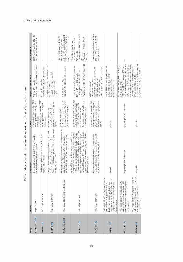
J. Clin. Med. 2020, 9, 2830
Ta
ble
1.
Maj
orcl
inic
altr
ials
onfr
ontl
ine
trea
tmen
tofe
pith
elia
lova
rian
canc
er.
Stu
dy
Pa
tie
nts
Ex
pe
rim
en
tal
Co
ntr
ol
Pro
gre
ssio
nF
ree
Su
rviv
al
Ov
era
llS
urv
iva
l
JGO
G3
01
6[5
2,5
6]
Stag
eII
-IV
EOC
thre
e-w
eekl
yca
rbop
lati
n(A
UC
6)an
dw
eekl
ypa
clit
axel
(80
mg/
m2 )f
orsi
xcy
cles
thre
e-w
eekl
yca
rbop
lati
n(A
UC
6)an
dpa
clit
axel
(180
mg/
m2 )
for
six
cycl
es
28.0
vs.1
7.2
mon
ths;
HR
0.71
,95%
CI0
.58–
0.88
;p=
0.00
1510
0.5
vs.6
2.2
mon
ths
(HR
0.79
,95
%C
I0.6
3–0.
99;p=
0.03
9
MIT
O-7
[54]
FIG
Ost
age
IC-I
VEO
CW
eekl
yca
rbop
lati
n(A
UC
2)an
dpa
clit
axel
(60
mg/
m2 )f
or18
wee
ks
thre
e-w
eekl
yca
rbop
lati
n(A
UC
6)an
dpa
clit
axel
(175
mg/
m2 )
for
six
cycl
es
18.3
vs.1
7.3
mon
ths;
HR
0·96,
95%
CI0·80
–1.1
6;p=
0·66
-
ICO
N-8
[55]
FIG
Ost
age
IC-I
VEO
C
Gro
up2:
thre
e-w
eekl
yca
rbop
lati
n(A
UC
5/6)
and
wee
kly
pacl
itax
el(8
0m
g/m
2 )for
six
cycl
esG
roup
3:W
eekl
yca
rbop
latin
(AU
C6)
and
pacl
itaxe
l(6
0m
g/m
2 )for
18w
eeks
Gro
up1:
thre
e-w
eekl
yca
rbop
lati
n(A
UC
5/6)
and
pacl
itax
el(1
75m
g/m
2 )for
six
cycl
es
Gro
up1
vs.G
roup
2vs
.Gro
up3:
17.7
vs.2
0.8
vs.2
1.0
Gro
up2
vs.G
roup
1:p=
0.35
Gro
up3
vs.G
roup
1:p=
0.51
-
GO
G-1
72
[27,6
5]
FIG
Ost
age
IIIw
ith
opti
mal
debu
lkin
gpa
clit
axel
135
mg/
m2
cont
inuo
usiv
infu
sion
over
24h
onda
y1,
cisp
lati
n10
0m
g/m
2IP
onda
y2,
pacl
itax
el60
mg/
m2
IPon
day
8fo
rsi
xcy
cles
pacl
itax
el13
5m
g/m
2
cont
inuo
usIV
infu
sion
over
24h
onda
y1,
cisp
lati
n75
mg/
m2
IVon
day
2fo
rsi
xcy
cles
23.8
vs.1
8.3
mon
ths;
HR
0.80
,95%
CI0
.64–
1.00
;p=
0.05
65.6
vs.4
9.7
mon
ths;
HR
0.75
,95
%C
I,0.
58–0
.97;
p=
0.03
61.8
vs.5
1.4
mon
ths;
Adj
uste
dH
R0.
77;9
5%C
I,0.
65–0
.90;
p=
0.00
2
GO
G-2
52
[28]
FIG
Ost
age
II-I
VEO
C
pacl
itax
el80
mg/
m2
IVon
days
1,8,
and
15pl
usca
rbop
lati
nA
UC
6IP
onda
y1
ever
y21
days
for
cycl
es1–
6pl
usbe
vaci
zum
ab15
mg/
kgIV
ever
y21
days
for
cycl
es2–
22pa
clit
axel
135
mg/
m2
IVon
day
1pl
usci
spla
tin
75m
g/m
2IP
onda
y2
plus
pacl
itax
el60
mg/
m2
IVon
day
8ev
ery
21da
ysfo
rcy
cles
1–6
plus
beva
cizu
mab
15m
g/kg
IVev
ery
21da
ysfo
rcy
cles
2–22
pacl
itax
el80
mg/
m2
IVon
days
1,8,
and
15pl
usca
rbop
lati
nA
UC
6IV
onda
y1
ever
y21
days
for
cycl
es1–
6pl
usbe
vaci
zum
ab15
mg/
kgIV
ever
y21
days
for
cycl
es2–
22
IVvs
.IP-
carb
opla
tin
vs.I
P-ci
spla
tin:
24.9
vs.2
7.4
vs.2
6.2
mon
ths
IP-c
arbo
plat
in:H
R0.
93,9
5%C
I0.
80–1
.07
IP-c
ispl
atin
:HR
0.98
,95%
CI0
.84–
1.13
IVvs
.IP-
carb
opla
tin
vs.
IP-c
ispl
atin
:75.
5vs
.78.
9vs
.72
.9m
onth
sIP
-car
bopl
atin
:HR
0.95
,95%
CI
0.80
–1.1
3IP
-cis
plat
in:H
R1.
05,9
5%C
I;0.
88–1
.24;
GO
G-2
62
[53]
FIG
Ost
age
III-
IVEO
Cth
ree-
wee
kly
carb
opla
tin
(AU
C6)
and
wee
kly
pacl
itax
el(8
0m
g/m
2 ),pl
us/m
inus
thre
e-w
eekl
ybe
vaci
zum
ab15
mg/
kgfo
rsi
xcy
cles
thre
e-w
eekl
yca
rbop
lati
n(A
UC
6)an
dpa
clit
axel
(175
mg/
m2 ),
plus/m
inus
thre
e-w
eekl
ybe
vaci
zum
ab15
mg/
kgfo
rsi
xcy
cles
Wit
hbe
vaci
zum
ab:
14.9
vs.1
4.7
mon
ths;
HR
0.99
,95%
CI0
.83–
1.20
;p=
0.60
Wit
hout
beva
cizu
mab
:14
.2vs
.10.
3m
onth
s;H
R0.
62,9
5%C
I0.4
0–0.
95;p=
0.03
Wit
han
dw
itho
utbe
vaci
zum
ab:
40.2
vs.3
9.0
mon
ths;
HR
0.94
;95
%C
I,0.
72–1
.2
SO
LO
-1[6
7]
FIG
Ost
age
IIIo
rIV
high
-gra
dese
rous
oren
dom
etri
oid
EOC
pati
ents
wit
ha
dele
teri
ous
orsu
spec
ted
dele
teri
ous
germ
line
orso
mat
icBR
CA
1/2
mut
atio
n,co
mpl
eted
fron
tlin
epl
atin
um-b
ased
chem
othe
rapy
olap
arib
plac
ebo
Not
reac
hed
vs.1
3.8
mon
ths;
HR
0.30
,95
%C
I0.2
3–0.
41);
p<
0.00
013-
year
:60%
vs.2
7%;
4-ye
ar:5
3%vs
.11%
-
PA
OL
A-1
[68]
FIG
Ost
age
IIIo
rIV
high
-gra
deEO
Cpa
tien
tsaf
ter
first
-lin
etr
eatm
entw
ith
plat
inum
–tax
ane
chem
othe
rapy
plus
beva
cizu
mab
olap
arib
plus
beva
cizu
mab
plac
ebo
plus
beva
cizu
mab
22.1
vs.1
6.6
mon
ths;
HR
0.59
;95%
CI
0.49
–0.7
2;p<
0.00
1H
RD
plus
BRC
Am
utat
ion:
37.2
vs.
17.7
mon
ths;
HR
0.33
,95%
CI0
.25–
0.45
HR
Dm
inus
BRC
Am
utat
ion:
28.1
vs.
16.6
mon
ths;
HR
0.43
,95%
CI0
.28–
0.66
-
PR
IMA
[69]
FIG
Ost
age
IIIo
rIV
high
-gra
dese
rous
oren
dom
etri
oid
EOC
pati
ents
afte
rfir
st-l
ine
trea
tmen
twit
hpl
atin
um-b
ased
chem
othe
rapy
nira
pari
bpl
aceb
o
Ove
rall:
13.8
vs.8
.2m
onth
s;H
R.6
2,95
%C
I0.5
0–0.
76;p<
0.00
1H
RD
-pos
itive
:21.
9vs
.10.
4m
onth
s;H
R0.
43,9
5%C
I0.3
1–0.
59;p<
0.00
1
-
134
J. Clin. Med. 2020, 9, 2830
7. Genetic Counseling
HGSC is a single case indicator for germline genetic testing [70]. Germline genetic testing shouldbe considered both due to the relatively high percentage of hereditary ovarian cancer with some studiesestimating that more than 20% is hereditary in etiology [71–73], and due to the potential for treatmentimplications [74]. Generally, it is preferable for an individual to undergo germline testing as soon asdiagnosis occurs [75,76]. This allows ample time to obtain and disclose results, especially in the settingof a patient who may have a guarded prognosis. Urgent testing of BRCA1/2 and other breast cancergenes with high or moderate penetrance by multi-gene panel can currently be performed. While thisstrategy is often used for women with breast cancer undergoing surgical decision-making, it can alsobe employed in the gynecologic oncology setting to provide results that may affect eligibility for PARPinhibitors or other therapies in a timely manner.
Germline testing in an affected individual is the most informative strategy and can help clarify riskfor relatives. Close female relatives may have increased empiric risk to develop EOC, although olderstudies may include some families with risk alleles that would be identified by current technology [77,78].The ascertainment of a multi-generational pedigree allows both for appropriate test selection as well asfor proper assessment of family structure and identification of at-risk relatives [79]. Pedigree assessmentin the setting of genetic counseling can also facilitate understanding of social relationships betweenrelatives to help develop appropriate strategies to encourage familial communication about risk.
Germline testing for women with EOC at our center typically includes evaluation via a multi-genepanel to include EOC risk genes beyond BRCA1/2, such as the mismatch repair (Lynch syndrome)genes, BRIP1, RAD51C, and RAD51D [71,80]. Beyond informing therapeutic strategy, germline testingin the setting of appropriate counseling can have significant implications for patients and familymembers. Germline testing can help stratify the risk of developing other cancers and guide thedevelopment of appropriate management strategies, especially as the prognosis for EOC improveswith better treatment options. For example, patients with Lynch syndrome are at significantly elevatedrisk to develop colorectal cancer [81] and patients with pathogenic alterations in the BRCA genes areat significantly elevated risk to develop breast cancer [82]. Understanding a patient’s risk may helpprevent a second primary cancer, especially in the setting of well-controlled ovarian disease or in thesetting where the development of a new cancer may interfere with the patient’s current treatment.
Germline testing may be even more impactful in terms of implications for relatives. Identifyingan ovarian cancer risk allele can allow relatives with the same allele to undergo preventative measures,such as risk-reducing salpingo-oopherectomy, which is especially relevant when screening is noteffective. Moreover, in some cases, over-treatment may be avoided in relatives who do not carrythe risk allele but who may have otherwise chosen to move forward with preventative measuresor screening due to concerns over risk, based on family history. Many genes implicated in EOC inthe setting of a monoallelic pathogenic variant also have implications for typically childhood-onsetsyndromes in the setting of biallelic pathogenic variants. For example, biallelic variants in BRCA1/2,BRIP1, and RAD51C [83–86] are associated with Fanconi anemia and biallelic variants in the mismatchrepair genes are associated with Constitutional Mismatch Repair Deficiency syndrome [81]. Thus,individuals contemplating childbearing may also wish to learn their germline status to informreproductive decisions.
Importantly, negative somatic testing does not obviate the need for germline testing. Reasons forthis can include the loss of a germline mutation in the tumor, limited analysis of the tumor genome,and differences in variant calling between somatic and germline laboratories. Conversely, somatictesting may identify variants that are germline in origin [87,88]. Therefore, patients should be counseledabout this possibility, and if somatic results are available, they should be reviewed to help informgermline test selection. Other genes may also be included based on clinical suspicion and the evaluationof additional personal and family history. Reevaluation should be considered over time as changes tothe family history, as well as advances in the field of cancer genetics, occur [79].
135
J. Clin. Med. 2020, 9, 2830
8. Team Medicine: Optimizing Partnerships and Clinical Research
We have a number of initiatives to ensure the inclusion of our community partners in research,education, and the integration of research-based advances into novel therapeutics by clinical trials.We aim to personalize therapy for patients so our community physicians can recommend improvedtherapy considerations, including clinical trials beyond the standard of care. One way we achievethis is via comprehensive molecular testing. All EOC patients at COH undergo GEM ExTra® testing(facilitated by TGen, a COH affiliate). This test reports clinically actionable mutations, copy numberalterations, transcript variants, and fusions, detected in any gene in patient DNA or RNA. The goal isto uncover true tumor-specific (somatic) alterations by comparing the sequence of the tumor againstthe paired normal DNA from each patient. The test also includes whole-transcriptome RNA profiling,interrogating the patient’s tumor transcriptome for fusions and transcriptional variants known to berelevant to cancer (e.g., EGFR vIII). Each tumor’s cancer-specific alterations are then queried against aproprietary knowledge base algorithm to identify potential therapeutic associations. The final reportprovides the physician with a list of FDA-approved agents that are associated with tumor-specificDNA alterations, as well as biomarker summaries on the variants found and tumor-specific evidencefor drug matches, including matches with investigational agents, as available on clinicaltrials.gov.The results are reviewed by our multidisciplinary gynecologic cancer research team to aid in treatmentdecision-making, highlight on-going studies and identify study candidates.
Our current clinical research portfolio in the frontline management of EOC focuses on developingsuperior treatment options for patients that reduce recurrence and prolong disease-free survival. We areexploring the use of HIPEC and PIPAC in the clinical trial setting as well as novel drug combinationsthat help to tailor and personalize treatment for superior results. Our HIPEC trial includes studyingthe molecular changes triggered by HIPEC to identify molecular signatures of response. Our PIPACtrial is the first in the United States to study aerosolized, pressurized chemotherapy for patients withperitoneal carcinomatosis, including ovarian cancer. Our community oncologists play an importantrole in these studies by referring patients, thereby allowing us to complete accrual expeditiously.
9. Summary
Management of EOC requires a multi-disciplinary approach, drawing on clinical expertise andcollaboration combined with community practice and cutting edge clinical and translational research.Our goal is to understand the biology of this disease, advance therapy and connect our patients withthe optimal treatment that offers the best possible outcomes.
Author Contributions: Conceptualization, E.W.W., C.H.W., S.L., S.J.-J.L., S.S. (Susan Shehayeb), S.G., R.L., S.S.(Siamak Saadat), J.S.; validation, E.W.W., M.C. and L.R.-R.; writing—original draft preparation, E.W.W., C.H.W.,S.L., S.J.-J.L., S.S. (Susan Shehayeb), S.G., R.L., S.S. (Siamak Saadat), J.S.; writing—review and editing, E.W.W.,C.H.W., S.L., S.J.-J.L., S.S. (Susan Shehayeb), T.D., E.S.H., D.S., S.W., M.C. and L.R.-R.; supervision, E.W.W. andL.R.-R.; project administration, E.W.W.; funding acquisition, E.W.W. All authors have read and agreed to thepublished version of the manuscript.
Funding: This research received no external funding.
Acknowledgments: The authors thank Nicola Welch, CMPP for assistance with writing and editing the manuscript.This work was supported by the National Cancer Institute of the National Institutes of Health under awardnumber K12CA001727. The content is solely the responsibility of the authors and does not necessarily representthe official views of the National Institutes of Health.
Conflicts of Interest: The authors declare no conflict of interest.
References
1. Board PDQATE. Ovarian Epithelial, Fallopian Tube, and Primary Peritoneal Cancer Treatment (PDQ(R)): HealthProfessional Version; PDQ Cancer Information Summaries; National Cancer Institute: Bethesda, MD, USA, 2002.
2. Siegel, R.L.; Miller, K.D.; Jemal, A. Cancer statistics, 2020. CA A Cancer J. Clin. 2020, 70, 145–164. [CrossRef]
136
J. Clin. Med. 2020, 9, 2830
3. Nebgen, D.R.; Lu, K.H.; Bast, R.C., Jr. Novel Approaches to Ovarian Cancer Screening. Curr. Oncol. Rep.2019, 21, 75. [CrossRef]
4. Howlader, N.N.A.; Krapcho, M.; Miller, D.; Brest, A.; Yu, M.; Ruhl, J.; Tatalovich, Z.; Mariotto, A.; Lewis, D.R.;Chen, H.S.; et al. SEER Cancer Statistics Review, 1975–2017; National Cancer Institute: Bethesda, MD, USA;Available online: https://seer.cancer.gov/csr/1975_2017/ (accessed on 25 August 2020).
5. Cliby, W.A.; Powell, M.A.; Al-Hammadi, N.; Chen, L.; Miller, J.P.; Roland, P.Y.; Mutch, D.G.; Bristow, R.E.Ovarian cancer in the United States: Contemporary patterns of care associated with improved survival.Gynecol. Oncol. 2015, 136, 11–17. [CrossRef] [PubMed]
6. Hoskins, W.J.; McGuire, W.P.; Brady, M.; Homesley, H.D.; Creasman, W.T.; Berman, M.; Ball, H.; Berek, J.S.The effect of diameter of largest residual disease on survival after primary cytoreductive surgery in patientswith suboptimal residual epithelial ovarian carcinoma. Am. J. Obs. Gynecol. 1994, 170, 974–979. [CrossRef]
7. Eisenkop, S.M.; Friedman, R.L.; Wang, H.J. Complete cytoreductive surgery is feasible and maximizessurvival in patients with advanced epithelial ovarian cancer: A prospective study. Gynecol. Oncol. 1998, 69,103–108. [CrossRef] [PubMed]
8. Allen, D.G.; Heintz, A.P.; Touw, F.W. A meta-analysis of residual disease and survival in stage III and IVcarcinoma of the ovary. Eur. J. Gynaecol. Oncol. 1995, 16, 349–356. [PubMed]
9. Chi, D.S.; Eisenhauer, E.L.; Lang, J.; Huh, J.; Haddad, L.; Abu-Rustum, N.R.; Sonoda, Y.; Levine, D.;Hensley, M.; Barakat, R. What is the optimal goal of primary cytoreductive surgery for bulky stage IIICepithelial ovarian carcinoma (EOC)? Gynecol. Oncol. 2006, 103, 559–564. [CrossRef]
10. Winter, W.E., 3rd; Maxwell, G.L.; Tian, C.; Carlson, J.W.; Ozols, R.F.; Rose, P.G.; Markman, M.; Armstrong, D.K.;Muggia, F.; McGuire, W.P. Prognostic factors for stage III epithelial ovarian cancer: A Gynecologic OncologyGroup Study. J. Clin. Oncol. Off. J. Am. Soc. Clin. Oncol. 2007, 25, 3621–3627. [CrossRef]
11. Wimberger, P.; Lehmann, N.; Kimmig, R.; Burges, A.; Meier, W.; Du Bois, A. Prognostic factors for completedebulking in advanced ovarian cancer and its impact on survival. An exploratory analysis of a prospectivelyrandomized phase III study of the Arbeitsgemeinschaft Gynaekologische Onkologie Ovarian Cancer StudyGroup (AGO-OVAR). Gynecol. Oncol. 2007, 106, 69–74.
12. Bristow, R.E.; Tomacruz, R.S.; Armstrong, D.K.; Trimble, E.L.; Montz, F.J. Survival effect of maximalcytoreductive surgery for advanced ovarian carcinoma during the platinum era: A meta-analysis. J. Clin.Oncol. Off. J. Am. Soc. Clin. Oncol. 2002, 20, 1248–1259. [CrossRef]
13. Teramukai, S.; Ochiai, K.; Tada, H.; Fukushima, M. Japan Multinational Trial Organization OC. PIEPOC:A new prognostic index for advanced epithelial ovarian cancer–Japan Multinational Trial OrganizationOC01–01. J. Clin. Oncol. Off. J. Am. Soc. Clin. Oncol. 2007, 25, 3302–3306. [CrossRef] [PubMed]
14. Chang, S.J.; Hodeib, M.; Chang, J.; Bristow, R.E. Survival impact of complete cytoreduction to no grossresidual disease for advanced-stage ovarian cancer: A meta-analysis. Gynecol. Oncol. 2013, 130, 493–498.[CrossRef] [PubMed]
15. Eisenkop, S.M.; Spirtos, N.M.; Lin, W.C. “Optimal” cytoreduction for advanced epithelial ovarian cancer:A commentary. Gynecol. Oncol. 2006, 103, 329–335. [CrossRef] [PubMed]
16. Winter, W.E., 3rd; Maxwell, G.L.; Tian, C.; Sundborg, M.J.; Rose, G.S.; Rose, P.G.; Rubin, S.C.; Muggia, F.;McGuire, W.P. Tumor residual after surgical cytoreduction in prediction of clinical outcome in stage IVepithelial ovarian cancer: A Gynecologic Oncology Group Study. J. Clin. Oncol. Off. J. Am. Soc. Clin. Oncol.2008, 26, 83–89. [CrossRef] [PubMed]
17. Eisenhauer, E.L.; Abu-Rustum, N.R.; Sonoda, Y.; Aghajanian, C.; Barakat, R.R.; Chi, D.S. The effect of maximalsurgical cytoreduction on sensitivity to platinum-taxane chemotherapy and subsequent survival in patientswith advanced ovarian cancer. Gynecol. Oncol. 2008, 108, 276–281. [CrossRef] [PubMed]
18. Hoskins, W.J. Epithelial ovarian carcinoma: Principles of primary surgery. Gynecologic Oncol. 1994, 55(3 Pt 2), S91–S96. [CrossRef]
19. Elattar, A.; Bryant, A.; Winter-Roach, B.A.; Hatem, M.; Naik, R. Optimal primary surgical treatment foradvanced epithelial ovarian cancer. Cochrane Database Syst. Rev. 2011, 2011, Cd007565. [CrossRef]
20. Wright, J.D.; Chen, L.; Hou, J.Y.; Burke, W.M.; Tergas, A.I.; Ananth, C.V.; Neugut, A.I.; Hershman, D.L.Association of Hospital Volume and Quality of Care With Survival for Ovarian Cancer. Obstet. Gynecol. 2017,130, 545–553. [CrossRef]
137
J. Clin. Med. 2020, 9, 2830
21. Rutten, M.J.; Gaarenstroom, K.N.; Van Gorp, T.; van Meurs, H.S.; Arts, H.J.; Bossuyt, P.M.; Ter Brugge, H.G.;Hermans, R.H.; Opmeer, B.C.; Pijnenborg, J.M.; et al. Laparoscopy to predict the result of primarycytoreductive surgery in advanced ovarian cancer patients (LapOvCa-trial): A multicentre randomizedcontrolled study. BMC Cancer 2012, 12, 31. [CrossRef]
22. Rutten, M.J.; van Meurs, H.S.; van de Vrie, R.; Gaarenstroom, K.N.; Naaktgeboren, C.A.; van Gorp, T.;Ter Brugge, H.G.; Hofhuis, W.; Schreuder, H.W.; Arts, H.J.; et al. Laparoscopy to Predict the Result of PrimaryCytoreductive Surgery in Patients With Advanced Ovarian Cancer: A Randomized Controlled Trial. J. Clin.Oncol. Off. J. Am. Soc. Clin. Oncol. 2017, 35, 613–621. [CrossRef]
23. Fagotti, A.; Ferrandina, G.; Fanfani, F.; Ercoli, A.; Lorusso, D.; Rossi, M.; Scambia, G. A laparoscopy-basedscore to predict surgical outcome in patients with advanced ovarian carcinoma: A pilot study. Ann. Surg.Oncol. 2006, 13, 1156–1161. [CrossRef] [PubMed]
24. Wright, A.A.; Bohlke, K.; Armstrong, D.K.; Bookman, M.A.; Cliby, W.A.; Coleman, R.L.; Dizon, D.S.; Kash, J.J.;Meyer, L.A.; Moore, K.N.; et al. Neoadjuvant Chemotherapy for Newly Diagnosed, Advanced OvarianCancer: Society of Gynecologic Oncology and American Society of Clinical Oncology Clinical PracticeGuideline. J. Clin. Oncol. Off. J. Am. Soc. Clin. Oncol. 2016, 34, 3460–3473. [CrossRef] [PubMed]
25. Harter, P.; Sehouli, J.; Lorusso, D.; Reuss, A.; Vergote, I.; Marth, C.; Kim, J.-W.; Raspagliesi, F.; Lampe, B.;Aletti, G.; et al. A Randomized Trial of Lymphadenectomy in Patients with Advanced Ovarian Neoplasms.N. Engl. J. Med. 2019, 380, 822–832. [CrossRef] [PubMed]
26. Walker, J.L.; Armstrong, D.K.; Huang, H.Q.; Fowler, J.; Webster, K.; Burger, R.A.; Clarke-Pearson, D.Intraperitoneal catheter outcomes in a phase III trial of intravenous versus intraperitoneal chemotherapy inoptimal stage III ovarian and primary peritoneal cancer: A Gynecologic Oncology Group Study. Gynecol. Oncol.2006, 100, 27–32. [CrossRef] [PubMed]
27. Armstrong, D.K.; Bundy, B.; Wenzel, L.; Huang, H.Q.; Baergen, R.; Lele, S.; Copeland, L.J.; Walker, J.;Burger, R.A. Intraperitoneal cisplatin and paclitaxel in ovarian cancer. N. Engl. J. Med. 2006, 354, 34–43.[CrossRef] [PubMed]
28. Walker, J.L.; Brady, M.F.; Wenzel, L.; Fleming, G.F.; Huang, H.Q.; DiSilvestro, P.A.; Fujiwara, K.; Alberts, D.S.;Zheng, W.; Tewari, K.S.; et al. Randomized Trial of Intravenous Versus Intraperitoneal Chemotherapy PlusBevacizumab in Advanced Ovarian Carcinoma: An NRG Oncology/Gynecologic Oncology Group Study.J. Clin. Oncol. Off. J. Am. Soc. Clin. Oncol. 2019, 37, 1380–1390. [CrossRef] [PubMed]
29. Monk, B.J.; Chan, J.K. Is intraperitoneal chemotherapy still an acceptable option in primary adjuvantchemotherapy for advanced ovarian cancer? Ann. Oncol. Off. J. Eur. Soc. Med. Oncol. 2017, 28 (Suppl. 8),viii40–viii45. [CrossRef]
30. van Driel, W.J.; Koole, S.N.; Sikorska, K.; Schagen van Leeuwen, J.H.; Schreuder, H.W.R.; Hermans, R.H.M.; DeHingh, I.H.; Van Der Velden, J.; Arts, H.J.; Massuger, L.F.; et al. Hyperthermic Intraperitoneal Chemotherapyin Ovarian Cancer. N. Engl. J. Med. 2018, 378, 230–240. [CrossRef]
31. Bouchard-Fortier, G.; Cusimano, M.C.; Fazelzad, R.; Sajewycz, K.; Lu, L.; Espin-Garcia, O.; May, T.;Bouchard-Fortier, A.; Ferguson, S.E. Oncologic outcomes and morbidity following heated intraperitonealchemotherapy at cytoreductive surgery for primary epithelial ovarian cancer: A systematic review andmeta-analysis. Gynecol. Oncol. 2020, 158, 218–228. [CrossRef]
32. Pletcher, E.; Gleeson, E.; Labow, D. Peritoneal Cancers and Hyperthermic Intraperitoneal Chemotherapy.Surg. Clin. N. Am. 2020, 100, 589–613. [CrossRef]
33. Nadiradze, G.; Horvath, P.; Sautkin, Y.; Archid, R.; Weinreich, F.J.; Königsrainer, A.; Reymond, M.A.Overcoming Drug Resistance by Taking Advantage of Physical Principles: Pressurized IntraperitonealAerosol Chemotherapy (PIPAC). Cancers 2019, 12, 34. [CrossRef]
34. Tate, S.J.; Torkington, J. Pressurized intraperitoneal aerosol chemotherapy: A review of the introduction of anew surgical technology using the IDEAL framework. BJS Open 2020, 4, 206–215. [CrossRef] [PubMed]
35. Cancer Genome Atlas Research Network. Integrated genomic analyses of ovarian carcinoma. Nature 2011,474, 609–615. [CrossRef] [PubMed]
36. Zhang, H.; Liu, T.; Zhang, Z.; Payne, S.H.; Zhang, B.; McDermott, J.E.; Zhou, J.-Y.; Petyuk, V.A.; Chen, L.;Ray, D.; et al. Integrated Proteogenomic Characterization of Human High-Grade Serous Ovarian Cancer.Cell 2016, 166, 755–765. [CrossRef] [PubMed]
37. Gee, M.E.; Faraahi, Z.; McCormick, A.; Edmondson, R.J. DNA damage repair in ovarian cancer: Unlockingthe heterogeneity. J. Ovarian Res. 2018, 11, 50. [CrossRef] [PubMed]
138
J. Clin. Med. 2020, 9, 2830
38. Milanesio, M.C.; Giordano, S.; Valabrega, G. Clinical Implications of DNA Repair Defects in High-GradeSerous Ovarian Carcinomas. Cancers 2020, 12, 1315. [CrossRef]
39. Takaya, H.; Nakai, H.; Takamatsu, S.; Mandai, M.; Matsumura, N. Homologous recombination deficiencystatus-based classification of high-grade serous ovarian carcinoma. Sci. Rep. 2020, 10, 2757. [CrossRef]
40. Bodurka, D.C.; Deavers, M.T.; Tian, C.; Sun, C.C.; Malpica, A.; Coleman, R.L.; Lu, K.H.; Sood, A.K.; Birrer, M.J.;Ozols, R.; et al. Reclassification of serous ovarian carcinoma by a 2-tier system: A Gynecologic OncologyGroup Study. Cancer 2012, 118, 3087–3094. [CrossRef]
41. Malpica, A.; Deavers, M.T.; Lu, K.; Bodurka, D.C.; Atkinson, E.N.; Gershenson, D.M.; Silva, E.G. Gradingovarian serous carcinoma using a two-tier system. Am. J. Surg. Pathol. 2004, 28, 496–504. [CrossRef]
42. Verhaak, R.G.; Tamayo, P.; Yang, J.Y.; Hubbard, D.; Zhang, H.; Creighton, C.J.; Fereday, S.; Lawrence, M.;Carter, S.L.; Mermel, C.; et al. Prognostically relevant gene signatures of high-grade serous ovarian carcinoma.J. Clin. Investig. 2013, 123, 517–525. [CrossRef]
43. Armstrong, D.K.; Alvarez, R.D.; Bakkum-Gamez, J.N.; Barroilhet, L.; Behbakht, K.; Berchuck, A.; Berek, J.S.;Chen, L.-M.; Cristea, M.; DeRosa, M.; et al. NCCN Guidelines Insights: Ovarian Cancer, Version 1.2019.J. Natl. Compr. Cancer Netw. 2019, 17, 896–909. [CrossRef] [PubMed]
44. Yokoi, A.; Yoshioka, Y.; Hirakawa, A.; Yamamoto, Y.; Ishikawa, M.; Ikeda, S.I.; Kato, T.; Niimi, K.; Kajiyama, H.;Kikkawa, F.; et al. A combination of circulating miRNAs for the early detection of ovarian cancer. Oncotarget2017, 8, 89811–89823. [CrossRef] [PubMed]
45. Otsuka, I.; Matsuura, T. Screening and Prevention for High-Grade Serous Carcinoma of the Ovary Based onCarcinogenesis-Fallopian Tube- and Ovarian-Derived Tumors and Incessant Retrograde Bleeding. Diagnostics2020, 10, 120. [CrossRef] [PubMed]
46. Elias, K.M.; Fendler, W.; Stawiski, K.; Fiascone, S.J.; Vitonis, A.F.; Berkowitz, R.S.; Frendl, G.;Konstantinopoulos, P.A.; Crum, C.P.; Kedzierska, M.; et al. Diagnostic potential for a serum miRNAneural network for detection of ovarian cancer. eLife 2017, 6, e28932. [CrossRef] [PubMed]
47. Guadagni, S.; Clementi, M.; Masedu, F.; Fiorentini, G.; Sarti, D.; Deraco, M.; Kusamura, S.; Papasotiriou, I.;Apostolou, P.; Aigner, K.R.; et al. A Pilot Study of the Predictive Potential of Chemosensitivity and GeneExpression Assays Using Circulating Tumour Cells from Patients with Recurrent Ovarian Cancer. Int. J. Mol.Sci. 2020, 21, 4813. [CrossRef]
48. Young, R.C.; Walton, L.A.; Ellenberg, S.S.; Homesley, H.D.; Wilbanks, G.D.; Decker, D.G.; Miller, A.; Park, R.;Major, F. Adjuvant therapy in stage I and stage II epithelial ovarian cancer. Results of two prospectiverandomized trials. N. Eng. J. Med. 1990, 322, 1021–1027. [CrossRef]
49. Winter-Roach, B.A.; Kitchener, H.C.; Lawrie, T.A. Adjuvant (post-surgery) chemotherapy for early stageepithelial ovarian cancer. Cochrane Database Syst. Rev. 2012, 3, Cd004706.
50. Hogberg, T.; Glimelius, B.; Nygren, P. A systematic overview of chemotherapy effects in ovarian cancer.Acta Oncolog. 2001, 40, 340–360. [CrossRef]
51. Chan, J.K.; Java, J.J.; Fuh, K.; Monk, B.J.; Kapp, D.S.; Herzog, T.; Bell, J.; Young, R. The association betweentiming of initiation of adjuvant therapy and the survival of early stage ovarian cancer patients—An analysisof NRG Oncology/Gynecologic Oncology Group trials. Gynecol. Oncol. 2016, 143, 490–495. [CrossRef]
52. Harano, K.; Terauchi, F.; Katsumata, N.; Takahashi, F.; Yasuda, M.; Takakura, S.; Takano, M.; Yamamoto, Y.;Sugiyama, T. Quality-of-life outcomes from a randomized phase III trial of dose-dense weekly paclitaxeland carboplatin compared with conventional paclitaxel and carboplatin as a first-line treatment for stageII-IV ovarian cancer: Japanese Gynecologic Oncology Group Trial (JGOG3016). Ann. Oncol. Off. J. Eur. Soc.Med.Oncol. 2014, 25, 251–257.
53. Chan, J.K.; Brady, M.F.; Penson, R.T.; Huang, H.; Birrer, M.J.; Walker, J.L.; DiSilvestro, P.A.; Rubin, S.C.;Martin, L.P.; Davidson, S.A.; et al. Weekly vs. Every-3-Week Paclitaxel and Carboplatin for Ovarian Cancer.N. Eng. J. Med. 2016, 374, 738–748. [CrossRef] [PubMed]
54. Pignata, S.; Scambia, G.; Katsaros, D.; Gallo, C.; Pujade-Lauraine, E.; De Placido, S.; Bologna, A.; Weber, B.;Raspagliesi, F.; Panici, P.B.; et al. Carboplatin plus paclitaxel once a week versus every 3 weeks in patientswith advanced ovarian cancer (MITO-7): A randomised, multicentre, open-label, phase 3 trial. Lancet Oncol.2014, 15, 396–405. [CrossRef]
139
J. Clin. Med. 2020, 9, 2830
55. Clamp, A.R.; James, E.C.; McNeish, I.A.; Dean, A.; Kim, J.W.; O’Donnell, D.M.; Hook, J.; Coyle, C.; Blagden, S.;Brenton, J.D.; et al. Weekly dose-dense chemotherapy in first-line epithelial ovarian, fallopian tube, or primaryperitoneal carcinoma treatment (ICON8): Primary progression free survival analysis results from a GCIGphase 3 randomised controlled trial. Lancet 2019, 394, 2084–2095. [CrossRef]
56. Katsumata, N.; Yasuda, M.; Isonishi, S.; Takahashi, F.; Michimae, H.; Kimura, E.; Aoki, D.; Jobo, T.; Kodama, S.;Terauchi, F.; et al. Long-term results of dose-dense paclitaxel and carboplatin versus conventional paclitaxeland carboplatin for treatment of advanced epithelial ovarian, fallopian tube, or primary peritoneal cancer(JGOG 3016): A randomised, controlled, open-label trial. Lancet Oncol. 2013, 14, 1020–1026. [CrossRef]
57. Hsu, Y.; Sood, A.K.; Sorosky, J.I. Docetaxel versus paclitaxel for adjuvant treatment of ovarian cancer:Case-control analysis of toxicity. Am. J. Clin. Oncol. 2004, 27, 14–18. [CrossRef]
58. Nguyen, J.; Solimando, D.A., Jr.; Waddell, J.A. Carboplatin and Liposomal Doxorubicin for Ovarian Cancer.Hosp. Pharm. 2016, 51, 442–449. [CrossRef]
59. Pignata, S.; Scambia, G.; Ferrandina, G.; Savarese, A.; Sorio, R.; Breda, E.; Gebbia, V.; Musso, P.; Frigerio, L.;Del Medico, P.; et al. Carboplatin plus paclitaxel versus carboplatin plus pegylated liposomal doxorubicin asfirst-line treatment for patients with ovarian cancer: The MITO-2 randomized phase III trial. J. Clin. Oncol.Off. J. Am. Soc. Clin. Oncol. 2011, 29, 3628–3635. [CrossRef]
60. Burger, R.A.; Brady, M.F.; Rhee, J.; Sovak, M.A.; Kong, G.; Nguyen, H.P.; Bookman, M.A. Independentradiologic review of the Gynecologic Oncology Group Study 0218, a phase III trial of bevacizumab in theprimary treatment of advanced epithelial ovarian, primary peritoneal, or fallopian tube cancer. Gynecol.Oncol. 2013, 131, 21–26. [CrossRef]
61. Perren, T.J.; Swart, A.M.; Pfisterer, J.; Ledermann, J.A.; Pujade-Lauraine, E.; Kristensen, G.; Carey, M.S.;Beale, P.; Cervantes, A.; Kurzeder, C.; et al. A phase 3 trial of bevacizumab in ovarian cancer. N. Eng. J. Med.2011, 365, 2484–2496. [CrossRef]
62. Burger, R.A.; Brady, M.; Bookman, M.A.; Fleming, G.F.; Monk, B.J.; Huang, H.; Mannel, R.S.; Homesley, H.D.;Fowler, J.; Greer, B.E.; et al. Incorporation of bevacizumab in the primary treatment of ovarian cancer. N. Eng.J. Med. 2011, 365, 2473–2483. [CrossRef]
63. Ferriss, J.S.; Java, J.J.; Bookman, M.A.; Fleming, G.F.; Monk, B.J.; Walker, J.L.; Homesley, H.D.; Fowler, J.;Greer, B.E.; Boente, M.P.; et al. Ascites predicts treatment benefit of bevacizumab in front-line therapy ofadvanced epithelial ovarian, fallopian tube and peritoneal cancers: An NRG Oncology/GOG study. Gynecol.Oncol. 2015, 139, 17–22. [CrossRef]
64. Wenzel, L.B.; Huang, H.Q.; Armstrong, D.K.; Walker, J.L.; Cella, D. Health-related quality of life during andafter intraperitoneal versus intravenous chemotherapy for optimally debulked ovarian cancer: A GynecologicOncology Group Study. J. Clin. Oncol. Off. J. Am. Soc. Clin. Oncol. 2007, 25, 437–443. [CrossRef] [PubMed]
65. Tewari, D.; Java, J.J.; Salani, R.; Armstrong, D.K.; Markman, M.; Herzog, T.; Monk, B.J.; Chan, J.K. Long-termsurvival advantage and prognostic factors associated with intraperitoneal chemotherapy treatment inadvanced ovarian cancer: A gynecologic oncology group study. J. Clin. Oncol. Off. J. Am. Soc. Clin. Oncol.2015, 33, 1460–1466. [CrossRef]
66. Oza, A.M.; Cook, A.D.; Pfisterer, J.; Embleton, A.; Ledermann, J.A.; Pujade-Lauraine, E.; Kristensen, G.;Carey, M.S.; Beale, P.; Cervantes, A.; et al. Standard chemotherapy with or without bevacizumab for womenwith newly diagnosed ovarian cancer (ICON7): Overall survival results of a phase 3 randomised trial. LancetOncol. 2015, 16, 928–936. [CrossRef]
67. Moore, K.N.; Colombo, N.; Scambia, G.; Kim, B.-G.; Oaknin, A.; Friedlander, M.; Lisyanskaya, A.; Floquet, A.;Leary, A.; Sonke, G.S.; et al. Maintenance Olaparib in Patients with Newly Diagnosed Advanced OvarianCancer. N. Eng. J. Med. 2018, 379, 2495–2505. [CrossRef] [PubMed]
68. Ray-Coquard, I.; Pautier, P.; Pignata, S.; Pérol, D.; González-Martín, A.; Berger, R.; Fujiwara, K.; Vergote, I.;Colombo, N.; Mäenpää, J.; et al. Olaparib plus Bevacizumab as First-Line Maintenance in Ovarian Cancer.N. Eng. J. Med. 2019, 381, 2416–2428. [CrossRef] [PubMed]
69. González-Martín, A.; Pothuri, B.; Vergote, I.; Christensen, R.D.; Graybill, W.; Mirza, M.R.; McCormick, C.;Lorusso, D.; Hoskins, P.; Freyer, G.; et al. Niraparib in Patients with Newly Diagnosed Advanced OvarianCancer. N. Engl. J. Med. 2019, 381, 2391–2402. [CrossRef]
70. Daly, M.B.; Pilarski, R.; Yurgelun, M.B.; Berry, M.P.; Buys, S.S.; Dickson, P.; Domchek, S.M.; Elkhanany, A.;Friedman, S.; Garber, J.E.; et al. NCCN Guidelines Insights: Genetic/Familial High-Risk Assessment: Breast,Ovarian, and Pancreatic, Version 1.2020. J. Natl. Compr. Cancer Netw. 2020, 18, 380–391. [CrossRef]
140

J. Clin. Med. 2020, 9, 2830
71. Walsh, T.; Casadei, S.; Lee, M.K.; Pennil, C.C.; Nord, A.S.; Thornton, A.M.; Roeb, W.; Agnew, K.J.; Stray, S.M.;Wickramanayake, A.; et al. Mutations in 12 genes for inherited ovarian, fallopian tube, and peritonealcarcinoma identified by massively parallel sequencing. Proc. Natl. Acad. Sci. USA 2011, 108, 18032–18037.[CrossRef]
72. Zhang, S.; Royer, R.; Li, S.; McLaughlin, J.R.; Rosen, B.; Risch, H.A.; Fan, I.; Bradley, L.; Shaw, P.A.; Narod, S.A.Frequencies of BRCA1 and BRCA2 mutations among 1,342 unselected patients with invasive ovarian cancer.Gynecol. Oncol. 2011, 121, 353–357. [CrossRef]
73. Pal, T.; Permuth-Wey, J.; Betts, J.A.; Krischer, J.P.; Fiorica, J.; Arango, H.; Lapolla, J.; Hoffman, M.; Martino, M.A.;Wakeley, K.; et al. BRCA1 and BRCA2 mutations account for a large proportion of ovarian carcinoma cases.Cancer 2005, 104, 2807–2816. [CrossRef] [PubMed]
74. Fong, P.C.; Boss, D.S.; Yap, T.A.; Tutt, A.; Wu, P.; Mergui-Roelvink, M.; Mortimer, P.; Swaisland, H.; Lau, A.;O’Connor, M.J.; et al. Inhibition of poly(ADP-ribose) polymerase in tumors from BRCA mutation carriers.N. Engl. J. Med. 2009, 361, 123–134. [CrossRef] [PubMed]
75. Novetsky, A.; Smith, K.; Babb, S.A.; Jeffe, N.B.; Hagemann, A.R.; Thaker, P.H.; Powell, M.A.; Mutch, D.G.;Massad, L.S.; Zighelboim, I. Timing of referral for genetic counseling and genetic testing in patients withovarian, fallopian tube, or primary peritoneal carcinoma. Int. J. Gynecol. Cancer 2013, 23, 1016–1021.[CrossRef] [PubMed]
76. Neviere, Z.; De La Motte Rouge, T.; Floquet, A.; Johnson, A.; Berthet, P.; Joly, F. How and when to refer patientsfor oncogenetic counseling in the era of PARP inhibitors. Adv. Med. Oncol. 2020, 12, 1758835919897530.[CrossRef]
77. Stratton, J.F.; Pharoah, P.; Smith, S.K.; Easton, D.; Ponder, B.A. A systematic review and meta-analysis offamily history and risk of ovarian cancer. Br. J. Obstet. Gynaecol. 1998, 105, 493–499. [CrossRef] [PubMed]
78. Jervis, S.; Song, H.; Lee, A.; Dicks, E.; Harrington, P.; Baynes, C.; Manchanda, R.; Easton, U.F.; Jacobs, I.;Pharoah, P.P.D.; et al. A risk prediction algorithm for ovarian cancer incorporating BRCA1, BRCA2, commonalleles and other familial effects. J. Med. Genet. 2015, 52, 465–475. [CrossRef]
79. Lu, K.H.; Wood, M.E.; Daniels, M.S.; Burke, C.; Ford, J.; Kauff, N.D.; Kohlmann, W.; Lindor, N.M.; Mulvey, T.M.;Robinson, L.; et al. American Society of Clinical Oncology Expert Statement: Collection and use of a cancerfamily history for oncology providers. J. Clin. Oncol. Off. J. Am. Soc. Clin. Oncol. 2014, 32, 833–840.[CrossRef]
80. Desmond, A.; Kurian, A.W.; Gabree, M.; Mills, M.A.; Anderson, M.J.; Kobayashi, Y.; Horick, N.; Yang, S.;Shannon, K.M.; Tung, N.; et al. Clinical Actionability of Multigene Panel Testing for Hereditary Breast andOvarian Cancer Risk Assessment. JAMA Oncol. 2015, 1, 943–951. [CrossRef]
81. Wimmer, K.; Kratz, C.P. Constitutional mismatch repair-deficiency syndrome. Haematologica 2010, 95, 699–701.[CrossRef]
82. Ford, D.; Easton, D.; Stratton, M.; Narod, S.; Goldgar, D.; Devilee, P.; Bishop, D.T.; Weber, B.; Lenoir, G.;Chang-Claude, J.; et al. Genetic heterogeneity and penetrance analysis of the BRCA1 and BRCA2 genesin breast cancer families. The Breast Cancer Linkage Consortium. Am. J. Hum. Genet. 1998, 62, 676–689.[CrossRef]
83. Rafnar, T.; Gudbjartsson, D.F.; Sulem, P.; Jonasdottir, A.; Sigurdsson, A.; Jonasdottir, A.; Besenbacher, S.;Lundin, P.; Stacey, S.N.; Gudmundsson, J.; et al. Mutations in BRIP1 confer high risk of ovarian cancer.Nat. Genet. 2011, 43, 1104–1107. [CrossRef] [PubMed]
84. Vaz, F.; Hanenberg, H.; Schuster, B.; Barker, K.; Wiek, C.; Erven, V.; Neveling, K.; Endt, D.; Kesterton, I.;Autore, F.; et al. Mutation of the RAD51C gene in a Fanconi anemia-like disorder. Nat. Genet. 2010, 42,406–409. [CrossRef] [PubMed]
85. Sawyer, S.L.; Tian, L.; Kähkönen, M.; Schwartzentruber, J.; Kircher, M.; Majewski, J.; Dyment, D.A.;Innes, A.M.; Boycott, K.M.; Moreau, L.A.; et al. Biallelic mutations in BRCA1 cause a new Fanconi anemiasubtype. Cancer Discov. 2015, 5, 135–142. [CrossRef] [PubMed]
86. Wagner, J.E.; Tolar, J.; Levran, O.; Scholl, T.; Deffenbaugh, A.; Satagopan, J.; Ben-Porat, L.; Mah, K.; Batish, S.D.;Kutler, D.I.; et al. Germline mutations in BRCA2: Shared genetic susceptibility to breast cancer, early onsetleukemia, and Fanconi anemia. Blood 2004, 103, 3226–3229. [CrossRef] [PubMed]
141

J. Clin. Med. 2020, 9, 2830
87. Ngeow, J.; Eng, C. Precision medicine in heritable cancer: When somatic tumour testing and germlinemutations meet. NPJ Genomic Med. 2016, 1, 15006. [CrossRef]
88. Slavin, T.P.; Banks, K.C.; Chudova, D.; Oxnard, G.R.; Odegaard, J.I.; Nagy, R.J.; Tsang, K.W.; Neuhausen, S.L.;Gray, S.W.; Cristofanilli, M.; et al. Identification of Incidental Germline Mutations in Patients With AdvancedSolid Tumors Who Underwent Cell-Free Circulating Tumor DNA Sequencing. J. Clin. Oncol. Off. J. Am. Soc.Clin. Oncol. 2018, 36, JCO1800328.
© 2020 by the authors. Licensee MDPI, Basel, Switzerland. This article is an open accessarticle distributed under the terms and conditions of the Creative Commons Attribution(CC BY) license (http://creativecommons.org/licenses/by/4.0/).
142
Journal of
Clinical Medicine
Review
Small Cell Lung Cancer from Traditional toInnovative Therapeutics: Building a ComprehensiveNetwork to Optimize Clinical andTranslational Research
Shanmuga Subbiah, Arin Nam, Natasha Garg, Amita Behal, Prakash Kulkarni and Ravi Salgia *
Department of Medical Oncology and Therapeutics Research, City of Hope National Medical Center,Duarte, CA 91010, USA; [email protected] (S.S.); [email protected] (A.N.); [email protected] (N.G.);[email protected] (A.B.); [email protected] (P.K.)* Correspondence: [email protected]
Received: 21 June 2020; Accepted: 28 July 2020; Published: 30 July 2020
Abstract: Small cell lung cancer (SCLC) is an aggressive, complex disease with a distinct biology thatcontributes to its poor prognosis. Management of SCLC is still widely limited to chemotherapy andradiation therapy, and research recruitment still poses a considerable challenge. Here, we review thecurrent standard of care for SCLC and advances made in utilizing immunotherapy. We also highlightresearch in the development of targeted therapies and emphasize the importance of a team-basedapproach to make clinical advances. Building an integrative network between an academic site andcommunity practice sites optimizes biomarker and drug target discovery for managing and treating adifficult disease like SCLC.
Keywords: small cell lung cancer; translational research; immunotherapy; clinical trials; teammedicine; community practice
1. Introduction
Lung cancer is the leading cause of cancer deaths in both men and women in the United States [1].Small cell lung cancer (SCLC) is a subtype of lung cancer that has an incidence of 13% and is stronglyassociated with smoking [2,3]. A distinct biology, aggressive clinical course with distant metastasis,and poor survival outcomes characterize SCLC. The disease is classified into extensive stage andlimited stage. While limited stage SCLC (LS-SCLC) is disease confined to one hemithorax that canbe enclosed within a radiation field, extensive stage SCLC (ES-SCLC) is more prevalent (66%) andincludes malignant pleural or pericardial effusions along with distant metastasis [4]. Despite the bleakprognosis, standard chemotherapies for patients with SCLC have not changed significantly in the last30 years. However, recently, immunotherapy with checkpoint inhibitors have shown promising efficacyin advanced disease [5,6]. The lack of advances in SCLC therapies are partly due to disease complexity,research recruitment, and resource utilization. Thus, collaborative efforts between academic andcommunity practices that combine knowledge, skills, experiences, and expertise of academicians,clinicians, and researchers, can accelerate advances in treatment and patient care. Academic centersare critical in the advancement of cancer treatment, but community hospital care plays an equallyimportant and complementary role. Indeed, academic–community collaboration, or ‘team medicine’,has become an emerging culture to advance and shape clinical care.
In this article, we first highlight the current standard treatments in SCLC as well as recent advancesin immunotherapies. We also review potential targeted therapies and underscore the importance ofa team-based approach toward SCLC based on our experience at the City of Hope (COH).
J. Clin. Med. 2020, 9, 2433; doi:10.3390/jcm9082433 www.mdpi.com/journal/jcm143
J. Clin. Med. 2020, 9, 2433
2. Current Standard Therapies
2.1. Radiation Therapy
For LS-SCLC the standard of care is chemotherapy with concurrent radiation therapy [7].Two meta-analyses established that concurrent cisplatin and etoposide treatment combined withradiation therapy improves survival compared to chemotherapy alone [8,9], although the dosage(once daily vs. twice daily) of radiation with chemotherapy remains equivocal. One study showeda significant survival advantage of patients who received 1.5 Gy in 30 fractions twice daily comparedto 1.8 Gy in 25 fractions after a median follow-up of 8 years [10]. However, a more recent trial thatrandomized patients to receiving 1.5 Gy twice daily fractions (45 Gy dose) or 2 Gy once daily fractions(66 Gy dose) concurrently with platinum based chemotherapy showed that survival outcomes did notdiffer between the regimens, although the trial was not powered for equivalence [11]. The ongoingtrial of CALGB 30,610 comparing 45 Gy twice daily to 70 Gy once daily (NCT00632853) is likely toshed more light on this issue.
2.2. Chemotherapy
Regardless of the stage, platinum with etoposide (EP) is the standard of care for patients withSCLC in the United States. Outside the United States, some patients are given platinum with irinotecanas an alternative treatment [12–14]. The overall response rates (ORR) range from 40–70% with up to10% of the patients achieving complete radiographic response, and the median overall survival (OS)spans 7–12 months, with a two-year survival rate of less than 5% [15]. Eventually, however, most SCLCtumors become resistant to chemotherapy resulting in disease progression. For patients with diseaserelapse, topotecan as a single agent is the only approved second-line drug that has demonstratedincreased survival compared to supportive care [16,17]. Nonetheless, the ORR of patients treated withtopotecan is only about 5% [18] and even worse, in SCLC patients who develop disease recurrencewithin 3 months of the first-line platinum doublet chemotherapy, in which case topotecan is ineffective.Other chemotherapeutic agents such as gemcitabine, docetaxel, paclitaxel, temozolomide, irinotecan,and vinorelbine, may be used in certain cases based on limited clinical evidence [19–24]. Amrubicinis an anthracycline agent that has been developed more recently and approved only in Japan forsecond-line therapy [25]. Unfortunately, beyond second-line therapy, currently there are no standardguidelines of care although, newer immunotherapies appear promising in some patients.
2.3. Surgery
Compared to non-small cell lung cancer (NSCLC), SCLC is rarely treated surgically. However,over the years the fraction of SCLC patients treated surgically has increased considerably from 14.9%in 2004 to 28.5% in 2013. This is at least in part due to availability of better diagnostic tools in the formof positron emission tomography (PET) scans and increasing usage of low-dose computed tomography(CT) screening. Randomized trials reported in the late 1960s and early 1970s showed no survivaladvantage for surgery alone or in combination with radiation therapy compared with radiation therapyalone [26,27]. Subsequently, it was reported that chemotherapy given sequentially with radiation andthen randomized to surgery vs. non-surgical group did not show any benefit to surgery [28]. However,recently there have been increasing numbers of retrospective studies showing survival benefit of surgerycompared to non-surgical therapy [29–32]. For example, a meta-analysis published by Liu et al. [33],which included two randomized control trials described above and thirteen retrospective studies fora total of 41,483 patients, concluded that surgical resection significantly improved overall survival whencompared to non-surgical treatment (hazard ratio (HR)= 0.56, p < 0.001) for retrospective studies, and inthe two randomized trials there was no survival advantage to the surgical arm. This meta-analysisalso showed that lobectomy was associated with superior OS compared with sub lobar resection(HR = 0.64, p < 0.001). Based on these data, the National Comprehensive Cancer Network (NCCN) alsorecommends surgery for T1-2N0M0 SCLC provided preoperative evaluation of mediastinal lymph
144
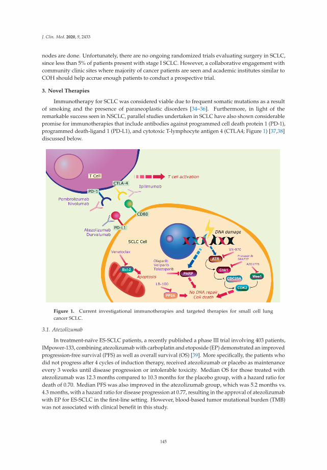
J. Clin. Med. 2020, 9, 2433
nodes are done. Unfortunately, there are no ongoing randomized trials evaluating surgery in SCLC,since less than 5% of patients present with stage I SCLC. However, a collaborative engagement withcommunity clinic sites where majority of cancer patients are seen and academic institutes similar toCOH should help accrue enough patients to conduct a prospective trial.
3. Novel Therapies
Immunotherapy for SCLC was considered viable due to frequent somatic mutations as a resultof smoking and the presence of paraneoplastic disorders [34–36]. Furthermore, in light of theremarkable success seen in NSCLC, parallel studies undertaken in SCLC have also shown considerablepromise for immunotherapies that include antibodies against programmed cell death protein 1 (PD-1),programmed death-ligand 1 (PD-L1), and cytotoxic T-lymphocyte antigen 4 (CTLA4; Figure 1) [37,38]discussed below.
Figure 1. Current investigational immunotherapies and targeted therapies for small cell lungcancer SCLC.
3.1. Atezolizumab
In treatment-naïve ES-SCLC patients, a recently published a phase III trial involving 403 patients,IMpower-133, combining atezolizumab with carboplatin and etoposide (EP) demonstrated an improvedprogression-free survival (PFS) as well as overall survival (OS) [39]. More specifically, the patients whodid not progress after 4 cycles of induction therapy, received atezolizumab or placebo as maintenanceevery 3 weeks until disease progression or intolerable toxicity. Median OS for those treated withatezolizumab was 12.3 months compared to 10.3 months for the placebo group, with a hazard ratio fordeath of 0.70. Median PFS was also improved in the atezolizumab group, which was 5.2 months vs.4.3 months, with a hazard ratio for disease progression at 0.77, resulting in the approval of atezolizumabwith EP for ES-SCLC in the first-line setting. However, blood-based tumor mutational burden (TMB)was not associated with clinical benefit in this study.
145

J. Clin. Med. 2020, 9, 2433
3.2. Durvalumab
Another phase III trial, the CASPIAN trial, which used durvalumab as the immunotherapy incombination with platinum with etoposide to treat treatment-naïve ES-SCLC patients, also showedimprovement in OS compared to platinum-etoposide alone (13 months vs. 10.3 months, with a hazardratio of 0.73) [40]. Based on these results, the Food and Drug Administration (FDA) also approveddurvalumab for ES-SCLC.
3.3. Ipilimumab and Nivolumab
In contrast to atezolizumab or durvalumab, ipilimumab (an anti-cytotoxic T-lymphocyte-associatedprotein 4 (CTLA4) antibody) in combination with chemotherapy prolongs PFS, but does not improve OSin treatment-naïve ES-SCLC [41]. However, maintenance therapy in such patients with nivolumab/ipilimumab combination or nivolumab alone did not show improvement in OS, according to resultsfrom the phase III CheckMate 451 study presented at the recent European Lung Cancer Congress2019 [42]. Another trial CheckMate 032 assessed nivolumab as a single agent or in combination withipilimumab in previously treated SCLC and found that ORR with single agent nivolumab was 11%compared to 22% in the cohort with combination of nivolumab with ipilimumab. The median OSfor nivolumab alone was 4.1 months, and for nivolumab with ipilimumab, it was 6 months to 7.8months based on the doses received [43]. Because the long-term survival benefits with nivolumab alonedemonstrated better outcomes compared to previous agents used in the third-line setting, nivolumabreceived FDA approval for third-line treatment of SCLC.
3.4. Pembrolizumab
Pembrolizumab was studied in relapsed SCLC patients in the KEYNOTE-028 and KEYNOTE-158trials. In KEYNOTE-028, the study included only patients with PD-L1 combined positive score(CPS) ≥1%. Among 24 patients with relapsed SCLC, 12.5% were treated with pembrolizumab in thesecond-line setting and 50% in the third-line. ORR was 33%, median PFS was 1.9 months, one-yearPFS was 23.8%, median OS was 9.7 months, and the one-year OS was 37.7% [44]. In the KEYNOTE-158trial, 79% of 107 patients with relapsed SCLC were treated with pembrolizumab in the second-line orthird-line setting. A total of 47% of patients were PD-L1-negative, with an ORR of 18.7% (35.7% inthe PD-L1-positive subgroup and 6.0% PD-L1-negative subgroup). The median PFS was 2 months,and median OS was 9.1 months [45]. This led to the approval of pembrolizumab in metastatic SCLCpatients whose disease progresses on or after platinum-based chemotherapy and at least one other lineof treatment. Considered together, although immunotherapy appears promising for SCLC patients,its benefits are modest, and there is significant room for further improvement.
4. Targeted Therapy
Unlike in the case of NSCLC, there are currently no targeted therapies available for SCLC. The lackof knowledge of the key genetic mutations and molecular targets that drive SCLC initiation andprogress to a more aggressive disease has been a major impediment in developing targeted therapies.However, recent genome-wide studies have identified the universal loss of tumor suppressor genessuch as tumor protein 53 (TP53) in 75–90% of patients and retinoblastoma 1 (RB1) and by frequent3P deletion [46–51]. Consistent with these observations, studies using genetically engineered mousemodels have confirmed that the introduction of these two events in pulmonary cells can give rise tohigh frequency of SCLC development [52]. Nonetheless, more than 120 clinical trials are ongoing thatare evaluating new drugs in SCLC targeting various/multiple pathways. Below, we review a few keystudies and are depicted in Figure 1.
Aberrant signaling driven by epidermal growth factor receptor (EGFR), stem cell factor receptortyrosine kinase (c-KIT), PI3K/AKT/mTOR, insulin-like growth factor receptor (IGFR1), and hedgehogsignaling pathways have been identified in SCLC. However, inhibitors targeting these pathways
146
J. Clin. Med. 2020, 9, 2433
have shown minimal efficacy in first-line, maintenance and relapsed SCLC [34,53–70]. Additionally,overexpression and amplification of MET and fibroblast growth factor receptor (FGFR) that areassociated with regulating cell proliferation, survival, motility, ability of invasion, and chemoresistancewere also observed in SCLC. However, therapies targeting these pathways have not fared welland next generation inhibitors need to be evaluated in combination with either chemotherapy orimmunotherapy [71–76].
The apoptotic pathway and the cell cycle checkpoint are some of the other pathways that havebeen targeted in SCLC. In the former case, B-cell lymphoma 2 (BCL-2) is the favorite therapeutic target.However, BCL-2 antisense oligonucleotide oblimersen and other agents, including obatoclax andnavitoclax, have not shown significant activity against SCLC in both phase I and phase II trials [77,78].Another BCL-2-specific inhibitor venetoclax, demonstrated efficacy in SCLC cell lines expressing highlevels of BCL-2 [79] and a phase I trial of venetoclax together with ABBV-075, a bromodomain andextra-terminal domain (BET) inhibitor, is currently under way (NCT02391480). As far as the cell cyclecheckpoint is concerned, ataxia telangiectasia and Rad3-related protein (ATR), checkpoint kinase-1(CHK1), WEE1, and aurora kinase (AURKA), have been the preferred targets among others [80].
In addition to the pathways discussed above, transcription and DNA repair pathways havealso been investigated in SCLC. Of these, the MYC pathway stands out since MYC is amplified ina significant (~20%) fraction of SCLC patients and appears to have higher sensitivity to certain newertargeted therapies, such as AURKA and BET inhibitors [81–84]. Other agents that are currently beingevaluated are: chiauranib, an aurora B kinase inhibitor for relapsed SCLC (NCT03216343), GSK525762,a BET inhibitor as monotherapy for patients harboring MYC amplification (NCT01587703), and incombination with trametinib for patients carrying RAS mutations (NCT03266159).
Wee-like protein kinase 1 (WEE1) is a key tyrosine kinase involved in halting the G2-to-Mphase transition of the cell cycle upon DNA damage [85] that is overexpressed in SCLC [86]. In apreclinical study, the combination of Poly(ADP-Ribose) Polymerase (PARP) inhibitors and WEE1inhibitors demonstrated a synergistic effect [87]. A phase II, multi-arm trial (BALTIC) is currentlyevaluating the efficacy of novel therapies in patients with ES-SCLC refractory to platinum-based agents.These novel therapies include PD-L1 inhibitor durvalumab, PARP inhibitor olaparib, and WEE1 kinaseinhibitor AZD1775.
Poly (ADP-ribose) polymerase 1 (PARP1) another key player in DNA repair is overexpressed inSCLC [88]. PARP inhibitors prevent cancer cells from repairing DNA damage caused by cytotoxicdrugs. Several PARP inhibitors have demonstrated antitumor efficacy in preclinical SCLC modelsand are currently being studied in several clinical trials. A phase II study investigating veliparib incombination with cisplatin and etoposide in untreated ES-SCLC patients showed improvement in theprimary endpoint PFS (6.1 months vs. 5.5 months) although no significant differences in OS wereobserved [89]. However, Schlafen-11 (SLFN11), which is involved in regulating response to DNAdamage and is overexpressed in about 48% of SCLC, has been identified as a potential biomarker forveliparib benefit [90]. Recently, another PARP inhibitor talazoparib also caused higher sensitizationto radiotherapy in SCLC cell lines and patient-derived xenografts. Thus, PARP inhibitors have greatpotential to emerge as a promising therapy for SCLC [91].
Activation of the Notch pathway is oncogenic in some cancer types, but in SCLC, the inhibitionof Notch pathway is involved in tumorigenic signaling, progression, and chemoresistance [92].Consistently, the inhibitory Notch ligand Delta-like protein 3 (DLL3) is upregulated in 85% ofSCLCs compared to normal lung [93]. Rovalpituzumab tesirine (Rova-T), a first-in-class DLL3antibody-drug conjugate, initially exhibited promising results of 18% ORR in heavily pretreatedSCLC [94]. Unfortunately, high toxicity rates in the phase II TRINITY trial (NCT02674568) precludedthe study from meeting its primary endpoint. In addition, the phase III MERU trial (NCT03033511)evaluating Rova-T in the maintenance setting following first-line chemotherapy, was also terminatedearly as a result of lack of survival benefit at an interim analysis. Likewise, another phase III (TAHOE)study that assessed Rova-T as a second-line therapy for advanced SCLC compared to topotecan,
147
J. Clin. Med. 2020, 9, 2433
stopped enrollment due to shorter OS in the Rova-T group compared with the topotecan controlgroup [95,96]. A phase I/II study evaluating the safety of Rova-T administered in combination withnivolumab or nivolumab and ipilimumab for adults with ES-SCLC has been recently completed andcould decide the future of Rova-T (NCT03026166).
5. Protein Phosphatase 2A (PP2A)
Protein phosphatase 2A (PP2A), a serine/threonine phosphatase that functions as a tumorsuppressor in many cancers [97], is also involved in various cellular processes, such as protein synthesis,cellular signaling, cell cycle, apoptosis, metabolism, and stress responses [98]. Several small-moleculeactivators of PP2A (SMAPs) have emerged as first-in-class agents for this target [99–102]. Further,a recent study has shown that PP2A suppression leads to resistance to kinase inhibitors in KRAS-drivenlung cancer cell lines. In KRAS-driven xenograft mouse models, combination treatment of SMAP andselumetinib (a MEK inhibitor used in clinical trials) led to significant tumor regression compared toeither agent alone [103,104]. Although PP2A is generally held to have tumor suppressor function,several lines of evidence suggest that it could also function as an oncogene. Thus, small moleculeinhibitors of PP2A such as LB-100 [105], are emerging as novel strategies for SCLC. Furthermore,since PP2A is also associated with immune response by downregulating cytotoxic T-lymphocytefunction [106,107], PP2A inhibition combined with immunotherapy appears to be effective in mediatingan antitumor response. Indeed, in colon cancer and melanoma cells, a combination of LB-100 and animmune checkpoint inhibitor led to greater T-cell-dependent anti-tumor response, with more effectorT-cell and reduced regulatory T-cell infiltration [106]. Carbonic anhydrase IX (CAIX), an enzymeinvolved in hypoxia inducible factor 1 alpha (HIF-1α) hypoxic signaling, is a promising target forchimeric antigen receptor-T (CAR-T) cells in an intracranial mouse model for glioblastoma [108]. In thismouse model, LB-100 was shown to augment the cytotoxic response of anti-carbonic anhydrase (CAIX)CAR-T cells, underscoring the therapeutic potential of synergistic LB-100 and CAR-T cell therapy inSCLC and other solid tumors [109].
6. Mitochondria
Mitochondria play an essential role in cell survival, apoptosis, adenosine triphosphate (ATP)production, as well as tumorigenesis [110]. Multiple therapeutic strategies have been developed totarget mitochondrial functions, such as oxidative phosphorylation, glycolysis, tricarboxylic acid (TCA)cycle, apoptosis, reactive oxygen species (ROS) regulation, permeability transition pore complex,mitochondrial DNA, and dihydroorotate de-hydrogenase (DHODH)-linked pyrimidine synthesis [111].Drugs that target mitochondrial metabolism through inhibition of pyruvate dehydrogenase (CPI-613,dichloroacetate), isocitrate dehydrogenase (AG-22, AG-120, AG-881) and targeting apoptotic pathways(birinapant, Minnelide, ME-334, Debio 1143, ONC201, LCL161) have been studied in early phase clinicaltrials [112–117]. These drugs have modest clinical activity as single agents in various tumors includingSCLC and are currently being explored as combination therapies with chemotherapy, immunotherapy,and other targeted therapies [118].
7. Stem Cell Therapy
Cancer stem cells (CSCs) are defined as a small population of cells within a heterogeneous tumorthat exhibit similar traits of normal stem cells. CSCs can originate from either somatic stem cellsor differentiated progenitor cells. They prompt tumorigenic activity by undergoing self-renewaland differentiation, leading to tumor relapse, resistance to therapy, and metastasis [119–121]. Hence,targeting CSCs has become a novel therapeutic strategy for cancer treatment. CSCs often haveupregulated signaling involved in development and tissue homeostasis, such as Notch, Hedgehogand wingless type 1 (WNT) pathways, all of which can be found in SCLC [122,123]. Unfortunately,Notch signaling inhibition with Rova-T and tarextumab have failed, but combination therapies studiesare ongoing that could potentially yield positive results. Lysine demethylase 1 (LSD1) is implicated
148
J. Clin. Med. 2020, 9, 2433
in maintaining stemness properties and hence, has emerged as a potential target for inhibiting lungCSCs [124]. A phase II trial (CLEPSIDRA) investigating the LSD1 inhibitor iadademstat in combinationwith standard-of-care in relapsing SCLC patients showed remarkable response rates (up to 75%).Preclinical studies with another LSD1 inhibitor GSK2879552 using 150 cancer cell lines, showed thatSCLC and acute myeloid leukemia (AML) cell lines were sensitive to growth inhibition by the LSD1inhibitor [125–127]. Furthermore, dual inhibition of LSD1 and PD-1 appear to be more effective thaneither therapy alone [128]. Another route to target stem cells is through CD47 inhibition by RRx-001,which targets tumor-associated macrophages and CSCs via downregulation of the antiphagocyticCD47/SIRPα checkpoint axis. A phase II trial (QUADRUPLE THREAT) involving 26 previouslyplatinum-treated third-line SCLC patients showed that the OS and PFS for patients treated withRRx-001 and a reintroduced platinum doublet were 8.6 months and 7.5 months, respectively, which ismuch higher for a third-line treatment reported in literature [129]. Interestingly, biopsies taken frompatients have correlated response to CD47 inhibition with a high density of infiltrated tumor-associatedmacrophages that are abundant in SCLC. Based on these observations, a phase III (REPLATINUM)randomized study of RRx-001 with platinum doublet vs. only platinum doublet in third-line SCLC iscurrently ongoing (NCT03699956).
8. Improving Outcomes
8.1. Biomarker-Based Therapy
Although immunotherapy has improved median survival for treatment-naïve ES-SCLC patients,the benefits have been limited with improvement in both PFS and OS approximately 1 and 2 months,respectively, with only 12.6% of patients remaining progression-free after one-year [39]. To improve theoutcomes, better selection of patients based on predictive biomarkers, given the high mutational loadand rapid resistance in SCLC, and therapies targeting multiple pathways with combination strategiesneed to be developed.
PD-L1 remains the most common immune-based biomarker for several malignancies. PD-L1staining in SCLC is less intense and infrequent compared to NSCLC [130]. In KEYNOTE-158,a combined score >1 for PD-L1 expression by the Dako 22C3 assay appeared to predict increasedresponse to pembrolizumab and improved survival when compared with patients negative forPD-L1 [44]; however, the PD-L1 positivity based on Dako 28-8 assay as in the CheckMate 032 didnot replicate those results [131]. The assays differ in that KEYNOTE-158 used a PD-L1 score basedon staining of tumor cells, lymphocytes and macrophages, whereas CheckMate 032 used stainingof only tumor cells to determine positivity. The ongoing phase II REACTION (NCT02580994) andthe phase III KEYNOTE-604 (NCT03066778) trials will require measurement of PD-L1 at baseline toprovide insight into the predictive role of PD-L1 expression. Higher tumor mutation burden (TMB) hasbeen recognized as a likely predictor of response to immunotherapy across disease types [132]. In anexome-sequencing analysis of CheckMate 032, patients with high TMB appeared to have a greaterimprovement in OS when treated with nivolumab. Patients with high-, medium-, and low-TMB hada median OS of 5.4 months, 3.9 months, and 3.1 months, respectively; the one-year OS rates were35.2%, 26.0%, and 22.1%, respectively [133]. However, in the IMpower-133, a blood-based TMB failedto predict benefit for atezolizumab, thus requiring further prospective randomized validation TMBstudies. Circulating tumor cells (CTCs) can be detected in 85% of SCLC patients and can potentiallyserve as a biomarker [134]. CTCs have been explored in multiple studies as a biomarker to predictresponse and resistance to therapy; however, additional studies looking into the genomic, epigenetic,and transcriptomic heterogeneity of CTCs at diagnosis and during relapse need to be done beforeit can be applied in clinics [135,136]. Cell-free DNA (cfDNA) widely used in NSCLC has also beenexamined in SCLC. A study with 27 patients showed cfDNA was able to mirror treatment responseand even identified disease recurrence before radiological progression [137]. Future work based ontumor or blood-based biomarkers will help a long way in understanding treatment resistance in SCLC.
149

J. Clin. Med. 2020, 9, 2433
8.2. Combination Therapy
To overcome treatment resistance, novel combination approaches targeting multiple pathwaysare being explored in combination with chemotherapy and immunotherapies. Lurbinectedin, a novelcytotoxic drug, is a transcriptional inhibitor that inhibits RNA polymerase II. In a phase II trialfor both sensitive and resistant disease, lurbinectedin was active as a single agent in second-lineSCLC with an ORR of 35.2% [138]. Based on this study, the FDA has approved lurbinectedin forthe treatment of ES-SCLC patients with disease progression after platinum-based chemotherapy.A phase III study (ATLANTIS trial, NCT02566993) of lurbinectedin in combination with doxorubicinvs. chemotherapy has completed recruitment and results are pending. Current trials investigatingtargeted therapies with or without chemotherapy include WEE1 inhibitor AZD1775 in combinationwith carboplatin (NCT02937818) and olaparib (NCT02511795), checkpoint kinase 1 (CHK1) inhibitorSRA737 in combination with cisplatin/gemcitabine (NCT027979770), ataxia–telangiectasia and Rad3related (ATR) inhibitor AZD6738 in combination with olaparib (NCT02937818), another ATR inhibitorVX-970 in combination with topotecan (NCT02487095), Bcl-2 inhibitor navitoclax and mTOR inhibitorvistusertib (NCT03366103), Bcl-2 inhibitor venetoclax and ABBV-075 (NCT02391480), and AuroraB kinase inhibitor (NCT02579226) are all ongoing, which should shed some light on the future oftargeted therapy in SCLC. There are numerous early phase trials investigating the combination ofimmunotherapy and targeted drugs as well. Durvalumab with olaparib (NCT02734004, MENDIOLA),avelumab with utomilumab, which is a humanized monoclonal antibody (mAb) that stimulatessignaling through CD137 (NCT02554812), nivolumab plus ipilimumab with dendritic cell-based p53vaccine 9 (NCT03406715) in relapsed SCLC, atezolizumab with chemotherapy and a CDK 4/6 inhibitortrilaciclib (NCT03041311) in first-line ES-SCLC are some of the novel combinations of immunotherapywith targeted agents.
8.3. Other Modalities
Other modalities of therapies targeting cell surface antigens expressed on tumor cells by monoclonalantibodies or surface-targeting immunotherapies, such as CAR-T cells and bispecific T-cell engagers(BiTEs), are in early stages of development. CD56 is expressed in almost all SCLC tumors, and thus, presents tobe an attractive target for treating SCLC [139]. In a preclinical study, promiximab-duocarmycin (DUBA),a CD56 antibody conjugated to a potent DNA alkylating agent with a novel linker, showed promisingresults. This antibody drug conjugate (ADC) demonstrated high efficacy in SCLC xenograft models [140],suggesting that further clinical evaluation of this compound may be beneficial. Another ADCsacituzumab govitecan is comprised of a humanized mAb targeting Trop-2 (trophoblastic antigen-2),which is highly expressed in several epithelial cancers [141], fused to SN-38 (the active metaboliteof irinotecan), which induces double- and single-strand DNA breaks by inhibiting topoisomeraseI [142]. Sacituzumab govitecan is currently being investigated in several trials, including a phaseI/II trial where it is being evaluated as a single agent in patients with previously treated, advancedSCLC (NCT01631552). CAR-T cells targeting DLL3 have entered a phase I clinical trial (NCT03392064).AMG 757, a BiTE, is also being evaluated in a phase I trial that includes ES-SCLC patients requiringfirst-line maintenance therapy and those with SCLC recurrence (NCT03319940). In patients withmetastatic solid tumors including relapsed SCLC, targeting other immune checkpoints, such as PD-1and CTLA-4, with immunotherapies, including TIM3 and LAG3, are being evaluated in clinical trials incombination with anti-PD-1 or anti-PD-L1 antibodies (NCT03708328, NCT03365791). Finally, radiationtherapy is assumed to modulate immune response, as it can increase tumor antigen production andpresentation and also enhance cytotoxic T-lymphocyte activity [143]. Potential synergy of radiotherapyin combination with immunotherapy in patients with ES-SCLC is being evaluated in innovativeongoing trials, and results are expected in the near future. Altogether, advancement in biomarkers,targeting multiple critical pathways, and enhancing immunotherapy efficacy in SCLC will hopefullyimprove the survival outcomes for SCLC patients, which has been elusive for many years.
150
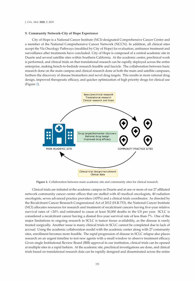
J. Clin. Med. 2020, 9, 2433
9. Community Network-City of Hope Experience
City of Hope is a National Cancer Institute (NCI)-designated Comprehensive Cancer Center anda member of the National Comprehensive Cancer Network (NCCN). In addition, all clinical sitesaccept the Via Oncology Pathways (modified by City of Hope) for evaluation, antitumor treatment andsurveillance after treatments have concluded. City of Hope is composed of a central academic site inDuarte and several satellite sites within Southern California. At the academic center, preclinical workis performed, and clinical trials on that translational research can be rapidly deployed across the entireenterprise, making bench-to-bedside research feasible and fascicle. The collaboration between basicresearch done on the main campus and clinical research done at both the main and satellite campuses,furthers the discovery of disease biomarkers and novel drug targets. This results in more rational drugdesign, improved therapeutic efficacy, and quicker optimization of high priority drugs for clinical use(Figure 2).
Figure 2. Collaboration between main academic site and community sites for clinical research.
Clinical trials are initiated at the academic campus in Duarte and at one or more of our 27 affiliatednetwork community cancer center offices that are staffed with 43 medical oncologists, 40 radiationoncologists, seven advanced practice providers (APPs) and a clinical trials coordinator. As directed bythe Recalcitrant Cancer Research Congressional Act of 2012 (H.R.733), the National Cancer Institute(NCI) allocates resources for research and treatment of recalcitrant cancers having five-year relativesurvival rates of <20% and estimated to cause at least 30,000 deaths in the US per year. SCLC isconsidered a recalcitrant cancer having a dismal five-year survival rate of less than 7%. One of themajor limitations to ongoing research in SCLC is tumor tissue availability, as the disease is rarelytreated surgically. Another issue is many clinical trials in SCLC cannot be completed due to lack ofaccrual. Using the academic collaboration model with the academic center along with 27 communitysites, enrollment becomes more feasible. The rapid progression of disease in SCLC relapse also placesresearch on an urgent timeline to test new agents with a small window to observe treatment efficacy.Given single Institutional Review Board (IRB) approval in our institution, clinical trials can be openedat multiple sites in a rapid fashion. At the academic site, preclinical investigations are done, and clinicaltrials based on translational research data can be rapidly designed and disseminated across the entire
151
J. Clin. Med. 2020, 9, 2433
enterprise, facilitating bench-to-bedside SCLC research in a more feasible manner. The collectiveknowledge gained from the interaction between the academic and community sites will provide insightinto how to overcome challenges that continuously hinder therapeutic advancements in SCLC.
10. Future Directions
Traditionally, SCLC has been regarded as a homogenous disease, which led to most SCLC patientsbeing treated with essentially one standard regimen. Recent studies from molecular analysis of patienttissues and genetically defined models indicate that there is notable heterogeneity among SCLCs interms of histology, growth characteristics, expression of neuroendocrine cell differentiation markers,MYC activation, Notch pathway inactivation, and role of neuronal lineage-specific transcription factorsin this disease (ASCL1, achaete-scute homologue 1; NeuroD1, neurogenic differentiation factor 1;POU2F3, POU class 2 homeobox 3; YAP1, yes-associated protein 1) [144–148]. Currently, SCLC isclassified into four subtypes based on increased expression of different markers: ASCL1 high (SCLC-A),NEUROD1 high (SCLC-N), POU2F3 high (SCLC-P), and YAP1 high (SCLC-Y) [79]. SCLC-A andSCLC-N are neuroendocrine subtypes, whereas SCLC-P and SCLC-Y are non-neuroendocrine subtypes.These subtypes can be associated with specific biomarkers that are either drug-specific targets orpredictors of drug response (e.g., DLL3 in SCLC-A, AURKA in SCLC-N, CDK4/6 in SCLC-Y andIGF1R in SCLC-P). These distinct gene expression profiles will guide us in designing new clinicaltrials. Recent advances in using patient-derived xenograft (PDX) models based on biopsy/resectedtumors, CTCs, genetically engineered mouse models (GEMM), as well as omics profiling will drasticallyenhance our capacity to identify and test novel drugs and discover biomarkers for treatment andprognostication [93,149].
In conclusion, we recommend: (i) setting up a centralized biobank and repository leading tocreation of a database incorporating full genomic, proteomic, and microRNA information; (ii) enrollinga higher proportion of SCLC patients into clinical trials with obligatory biomarker analysis; (iii) creatinga master protocol which will help reduce duplicative effort and thus ease the eligibility requirementsfor clinical trials; (iv) create and incentivize academic and community research partnership centers ofexcellence, since most SCLC patients are treated in community sites; (v) collaborate with bioengineers,cancer biologists, and biophysicists to gather the genetic aberrations discovered and harness the powerof computational modeling of genetic information, which will be a powerful tool in understandingSCLC and developing future therapies. Given the academic and community partnership we haveestablished at City of Hope, this should be achievable and pave way for success in treating thischallenging disease.
Author Contributions: Conceptualization, S.S., A.N., and R.S.; methodology, S.S., A.N., and N.G.; writing—originaldraft preparation, S.S. and A.N.; writing—review and editing, S.S., A.N., N.G., A.B., P.K., and R.S.; supervision,R.S.; project administration, P.K.; funding acquisition, R.S. All authors have read and agreed to the publishedversion of the manuscript.
Funding: This research received no external funding.
Conflicts of Interest: The authors declare no conflict of interest.
References
1. Centers for Disease Control and Prevention. National Center for Health Statistics. CDC On-Line Database,Compiled from Compressed Mortality File 1999–2016 Series 20 No. 2V, 2017. Available online: https://www.cdc.gov/nchs/data_access/cmf.htm (accessed on 28 July 2020).
2. Gazdar, A.F.; Bunn, P.A.; Minna, J.D. Small-cell lung cancer: What we know, what we need to know and thepath forward. Nat. Rev. Cancer 2017, 17, 765. [CrossRef] [PubMed]
3. Herbst, R.S.; Morgensztern, D.; Boshoff, C. The biology and management of non-small cell lung cancer.Nature 2018, 553, 446–454. [CrossRef] [PubMed]
4. Kalemkerian, G.P. Staging and imaging of small cell Lung cancer. Cancer Imaging 2012, 11, 253–258. [CrossRef][PubMed]
152
J. Clin. Med. 2020, 9, 2433
5. Navarro, A.; Felip, E. Pembrolizumab in advanced pretreated small cell lung cancer patients with PD-L1expression: Data from the KEYNOTE-028 trial: A reason for hope? Transl. Lung Cancer Res. 2017, 6(Suppl. S1), S78–S83. [CrossRef]
6. U.S. Food & Drug Administration. FDA Grants Nivolumab Accelerated Approval for Third-Line Treatmentof Metastatic Small Cell Lung Cancer. Available online: https://www.fda.gov/drugs/resources-information-approved-drugs/fda-grants-nivolumab-accelerated-approval-third-line-treatment-metastatic-small-cell-lung-cancer (accessed on 28 July 2020).
7. NCCN. NCCN Clinical Practice Guidelines in Oncology: Small Cell Lung Cancer; The National ComprehensiveCancer Network (NCCN): Whitemarsh, PA, USA, 2019.
8. Warde, P.; Payne, D. Does thoracic irradiation improve survival and local control in limited-stage small-cellcarcinoma of the lung? A meta-analysis. J. Clin. Oncol. 1992, 10, 890–895. [CrossRef]
9. Pignon, J.P.; Arriagada, R.; Ihde, D.C.; Johnsosn, D.H.; Perry, M.C.; Souhami, R.L.; Brodin, O.; Joss, R.A.;Kies, M.S.; Lebeau, B.; et al. A meta-analysis of thoracic radiotherapy for small-cell lung cancer. N. Engl.J. Med. 1992, 327, 1618–1624. [CrossRef]
10. Turrisi, A.T., III; Kim, K.; Blum, R.; Sause, W.T.; Livingston, R.B.; Komaki, R.; Wagner, H.; Aisner, S.;Johnson, D.H. Twice-daily compared with once-daily thoracic radiotherapy in limited small-cell lung cancertreated concurrently with cisplatin and etoposide. N. Engl. J. Med. 1999, 340, 265–271. [CrossRef]
11. Faivre-Finn, C.; Snee, M.; Ashcroft, L.; Appel, W.; Barlesi, F.; Bhatnagar, A.; Bezjak, A.; Cardenal, F.; Fournel, P.;Harden, S.; et al. Concurrent once-daily versus twice-daily chemoradiotherapy in patients with limited-stagesmall-cell lung cancer (CONVERT): An open-label, phase 3, randomised, superiority trial. Lancet Oncol.2017, 18, 1116–1125. [CrossRef]
12. Fukuoka, M.; Furuse, K.; Saijo, N.; Nishiwaki, Y.; Ikegami, H.; Tamura, T.; Shimoyama, M.; Suemasu, K.Randomized trial of cyclophosphamide, doxorubicin, and vincristine versus cisplatin and etoposide versusalternation of these regimens in small-cell lung cancer. J. Natl. Cancer Inst. 1991, 83, 855–861. [CrossRef]
13. Roth, B.J.; Johnson, D.H.; Einhorn, L.H.; Schacter, L.P.; Cherng, N.C.; Cohen, H.J.; Crawford, J.; Randolph, J.A.;Goodlow, J.L.; Broun, G.O.; et al. Randomized study of cyclophosphamide, doxorubicin, and vincristineversus etoposide and cisplatin versus alternation of these two regimens in extensive small-cell lung cancer:A phase III trial of the Southeastern cancer study group. J. Clin. Oncol. 1992, 10, 282–291. [CrossRef]
14. Noda, K.; Nishiwaki, Y.; Kawahara, M.; Negoro, C.; Sugiura, T.; Yokoyama, A.; Fukuoka, M.; Mori, K.;Watanabe, K.; Tamura, T.; et al. Irinotecan plus cisplatin compared with etoposide plus cisplatin for extensivesmall-cell lung cancer. N. Engl. J. Med. 2002, 346, 85–91. [CrossRef] [PubMed]
15. Jackman, D.M.; Johnson, B.E. Small-cell lung cancer. Lancet 2005, 366, 1385–1396. [CrossRef]16. O’Brien, M.E.R.; Ciuleanu, T.E.; Tsekov, H.; Shparyk, Y.; Cuceviá, B.; Juhasz, G.; Thatcher, N.; Ross, G.A.;
Dane, G.C.; Crofts, T. Phase III trial comparing supportive care alone with supportive care with oral topotecanin patients with relapsed small-cell lung cancer. J. Clin. Oncol. 2006, 24, 5441–5447. [CrossRef] [PubMed]
17. Von Pawel, J.; Schiller, J.H.; Shepherd, F.A.; Fields, S.Z.; Kleisbauer, J.P.; Chrysson, N.G.; Stewart, D.J.;Clark, P.I.; Palmer, M.C.; Depierre, A.; et al. Topotecan versus cyclophosphamide, doxorubicin, andvincristine for the treatment of recurrent small-cell lung cancer. J. Clin. Oncol. 1999, 17, 658–667. [CrossRef]
18. Horita, N.; Yamamoto, M.; Sato, T.; Tsukahara, T.; Nagakura, H.; Tashiro, K.; Shinata, Y.; Watanabe, H.;Nagai, K.; Inoue, M.; et al. Topotecan for relapsed small-cell lung cancer: Systematic review and meta-analysisof 1347 patients. Sci. Rep. 2015, 5, 15437. [CrossRef]
19. Masters, G.A.; Declerck, L.; Blanke, C.; Sandler, A.; DeVore, R.; Miller, K.; Johnson, D.; Eastern CooperativeOncology Group. Phase II trial of gemcitabine in refractory or relapsed small-cell lung cancer: EasternCooperative Oncology Group Trial 1597. J. Clin. Oncol. 2003, 21, 1550–1555. [CrossRef]
20. Smyth, J.F.; Smith, I.E.; Sessa, C.; Schoffski, P.; Wanders, J.; Franklin, H.; Kaye, S.B. Activity of docetaxel(Taxotere) in small cell lung cancer: The Early Clinical Trials Group of the EORTC. Eur. J. Cancer 1994, 30A,1058–1060. [CrossRef]
21. Yamamoto, N.; Tsurutani, J.; Yoshimura, N.; Asai, G.; Moriyama, A.; Nakagawa, K.; Kudoh, S.; Takada, M.;Minato, Y.; Fukuoka, M. Phase II study of weekly paclitaxel for relapsed and refractory small cell lung cancer.Anticancer Res. 2006, 26, 777–781.
153
J. Clin. Med. 2020, 9, 2433
22. Pietanza, M.C.; Kadota, K.; Huberman, K.; Sima, C.S.; Fiore, J.J.; Sumner, D.K.; Travis, W.D.; Heguy, A.;Ginsberg, M.S.; Holodny, A.I.; et al. Phase II trial of temozolomide in patients with relapsed sensitive orrefractory small cell lung cancer, with assessment of methylguanine-DNA methyltransferase as a potentialbiomarker. Clin. Cancer Res. 2012, 18, 1138–1145. [CrossRef]
23. Masuda, N.; Fukuoka, M.; Kusunoki, Y.; Matsui, K.; Takifuji, N.; Kudoh, S.; Negoro, S.; Nishioka, M.;Nakagawa, K.; Takada, M. CPT-11: A new derivative of camptothecin for the treatment of refractory orrelapsed small-cell lung cancer. J. Clin. Oncol. 1992, 10, 1225–1229. [CrossRef]
24. Jassem, J.; Karnicka-Mlodkowska, H.; van Pottelsberghe, C.; van Glabbeke, M.; Noseda, M.A.; Ardizzoni, A.;Gozzelino, F.; Planting, A.; van Zandwijk, N. Phase II study of vinorelbine (Navelbine) in previously treatedsmall cell lung cancer patients: EORTC Lung Cancer Cooperative Group. Eur. J. Cancer 1993, 29A, 1720–1722.[CrossRef]
25. Ding, Q.; Zhan, J. Amrubicin: Potential in combination with cisplatin or carboplatin to treat small-cell lungcancer. Drug Des. Dev. Ther. 2013, 7, 681–689.
26. Miller, A.B.; Fox, W.; Tall, R. Five-year follow-up of the Medical Research Council comparative trial of surgeryand radiotherapy for the primary treatment of small-celled or oat-celled carcinoma of the bronchus. Lancet1969, 2, 501–505. [CrossRef]
27. Fox, W.; Scadding, J.G. Treatment of oat-celled carcinoma of the bronchus. Lancet 1973, 2, 616–617. [CrossRef]28. Lad, T.; Piantadosi, S.; Thomas, P.; Payne, D.; Ruckdeschel, J.; Giaccone, G. A prospective randomized trial to
determine the benefit of surgical resection of residual disease following response of small cell lung cancer tocombination chemotherapy. Chest 1994, 106 (Suppl. S6), 320S–323S. [CrossRef]
29. Rostad, H.; Naalsund, A.; Jacobsen, R.; Strand, T.E.; Scott, H.; Strøm, E.H.; Norstein, J. Small cell lung cancerin Norway. Should more patients have been offered surgical therapy? Eur. J. Cardiothorac. Surg. 2004, 26,782–786. [CrossRef]
30. Brock, M.V.; Hooker, C.M.; Syphard, J.E.; Westra, W.; Xu, L.; Alberg, A.J.; Mason, D.; Baylin, S.B.; Herman, J.G.;Yung, R.C.; et al. Surgical resection of limited disease small cell lung cancer in the new era of platinumchemotherapy: Its time has come. J. Thorac. Cardiovasc. Surg. 2005, 129, 64–72. [CrossRef]
31. Lim, E.; Belcher, E.; Yap, Y.K.; Nicholson, A.G.; Goldstraw, P. The role of surgery in the treatment of limiteddisease small cell lung cancer: Time to reevaluate. J. Thorac. Oncol. 2008, 3, 1267–1271. [CrossRef]
32. Schreiber, D.; Rineer, J.; Weedon, J.; Vongtama, D.; Wortham, A.; Kim, A.; Han, P.; Choi, K.; Rotman, M.Survival outcomes with the use of surgery in limited-stage small cell lung cancer: Should its role bere-evaluated? Cancer 2010, 116, 1350–1357. [CrossRef]
33. Liu, T.; Chen, Z.; Dang, J.; Li, G. The role of surgery in stage I to III small cell lung cancer: A systematicreview and meta-analysis. PLoS ONE 2018, 13, e0210001. [CrossRef]
34. Peifer, M.; Fernández-Cuesta, L.; Sos, M.L.; George, J.; Seidel, D.; Kasper, L.H.; Plenker, D.; Leenders, F.;Sun, R.; Zander, T.; et al. Integrative genome analyses identify key somatic driver mutations of small-celllung cancer. Nat. Genet. 2012, 44, 1104–1110. [CrossRef] [PubMed]
35. Alexandrov, L.B.; Nik-Zainal, S.; Wedge, D.C.; Aparicio, S.A.; Behjati, S.; Biankin, A.V.; Bignell, G.R.; Bolli, N.;Borg, A.; Børrensen-Dale, A.L.; et al. Signatures of mutational processes in human cancer. Nature 2013, 500,415–421. [CrossRef] [PubMed]
36. Horn, L.; Reck, M.; Spigel, D.R. The future of immunotherapy in the treatment of small cell lung cancer.Oncologist 2016, 21, 910–921. [CrossRef] [PubMed]
37. Reck, M.; Heigener, D.; Reinmuth, N. Immunotherapy for small-cell lung cancer: Emerging evidence.Future Oncol. 2016, 12, 931–943. [CrossRef] [PubMed]
38. Horn, L.; Mansfield, A.S.; Szczesna, A.; Havel, L.; Krzakowski, M.; Hochmair, M.J.; Huemer, F.; Losonczy, G.;Johnson, M.L.; IMpower133 Study Group; et al. First-line atezolizumab plus chemotherapy in extensive-stagesmall-cell lung cancer. N. Engl. J. Med. 2018, 379, 2220–2229. [CrossRef]
39. Paz-Ares, L.; Dvorkin, M.; Chen, Y.; Reinmuth, N.; Hotta, K.; Trukhin, D.; Statsenko, G.; Hochmair, M.J.;Özgüroglu, M.; Ji, J.H.; et al. Durvalumab plus platinum–etoposide versus platinum–etoposide in first-linetreatment of extensive-stage small-cell lung cancer (CASPIAN): A randomised, controlled, open-label,phase 3 trial. Lancet 2019, 394, 1929–1939. [CrossRef]
154
J. Clin. Med. 2020, 9, 2433
40. Reck, M.; Luft, A.; Szczesna, A.; Havel, L.; Kim, S.-W.; Akerley, W.; Pietanza, M.C.; Wu, Y.-L.; Zielinski, C.;Thomas, M.; et al. Phase III randomized trial of ipilimumab plus etoposide and platinum versus placeboplus etoposide and platinum in extensive-stage small-cell lung cancer. J. Clin. Oncol. 2016, 34, 3740–3748.[CrossRef]
41. Owonikoko, T.K.; Kim, H.R.; Govindan, R.; Ready, N.; Reck, M.; Peters, S.; Dakhil, S.R.; Navarro, A.;Rodriguez-Cid, J.; Schenker, M.; et al. Nivolumab (nivo) plus ipilimumab (ipi), nivo, or placebo (pbo)as maintenance therapy in patients (pts) with extensive disease small cell lung cancer (ED-SCLC) afterfirst-line (1L) platinum-based chemotherapy (chemo): Results from the double-blind, randomized phase IIICheckMate 451 study. Ann. Oncol. 2019, 30 (Suppl. S2), ii77–ii80.
42. Ready, N.E.; Ott, P.A.; Hellmann, M.D.; Zugazagoitia, J.; Hann, C.L.; de Braud, F.; Antonia, S.J.; Ascierto, P.A.;Moreno, V.; Atmaca, A.; et al. Nivolumab Monotherapy and Nivolumab Plus Ipilimumab in Recurrent SmallCell Lung Cancer: Results from the CheckMate 032 Randomized Cohort. J. Thorac. Oncol. 2020, 15, 426–435.[CrossRef]
43. Ott, P.A.; Elez, E.; Hiret, S.; Kim, D.-W.; Morosky, A.; Saraf, S.; Piperdi, B.; Mehnert, J.M. Pembrolizumabin patients with extensive-stage small-cell lung cancer: Results from the phase Ib KEYNOTE-028 study.J. Clin. Oncol. 2017, 35, 3823–3829. [CrossRef]
44. Chung, H.C.; Lopez-Martin, J.A.; Kao, S.C.-H.; Miller, W.H.; Ros, W.; Gao, B.; Marabelle, A.; Gottfried, M.;Zer, A.; Delord, J.-P.; et al. Phase 2 study of pembrolizumab in advanced small-cell lung cancer (SCLC):KEYNOTE-158. J. Clin. Oncol. 2018, 15, 8506. [CrossRef]
45. Rudin, C.M.; Poirier, J.T. Small-cell lung cancer in 2016: Shining light on novel targets and therapies. Nat. Rev.Clin. Oncol. 2017, 14, 75–76. [CrossRef] [PubMed]
46. Miller, C.W.; Simon, K.; Aslo, A.; Kok, Y.; Yokota, J.; Buys, C.H.; Terada, M.; Koeffler, H.P. p53 mutations inhuman lung tumors. Cancer Res. 1992, 52, 1695–1698. [PubMed]
47. Takahashi, T.; Takahashi, T.; Suzuki, H.; Hida, T.; Sekido, Y.; Ariyoshi, Y.; Ueda, R. The p53 gene is veryfrequently mutated in small-cell lung cancer with a distinct nucleotide substitution pattern. Oncogene 1991, 6,1775–1778. [PubMed]
48. Helin, K.; Holm, K.; Niebuhr, A.; Eiberg, H.; Tommerup, N.; Hougaard, S.; Poulsen, H.S.; Spang-Thomsen, M.;Norgaard, P. Loss of the retinoblastoma protein-related p130 protein in small cell lung carcinoma. Proc. Natl.Acad. Sci. USA 1997, 94, 6933–6938. [CrossRef] [PubMed]
49. Kaye, F.J. RB and cyclin dependent kinase pathways: Defining a distinction between RB and p16 loss in lungcancer. Oncogene 2002, 21, 6908–6914. [CrossRef]
50. Wistuba, I.I.; Behrens, C.; Virmani, A.K.; Mele, G.; Milchgrub, S.; Girard, L.; Fondon, J.W., 3rd; Garner, H.R.;McKay, B.; Latif, F.; et al. High resolution chromosome 3p allelotyping of human lung cancer andpreneoplastic/preinvasive bronchial epithelium reveals multiple, discontinuous sites of 3p allele loss and3 regions of frequent breakpoints. Cancer Res. 2000, 60, 1949–1960.
51. Wistuba, I.I.; Berry, J.; Behrens, C.; Maitra, A.; Shivapurkar, N.; Milchgrub, S.; Mackay, B.; Minna, J.D.;Gazdar, A.F. Molecular changes in the bronchial epithelium of patients with small cell lung cancer.Clin. Cancer Res. 2000, 6, 2604–2610.
52. Bordi, P.; Tiseo, M.; Barbieri, F.; Bavieri, M.; Sartori, G.; Marchetti, A.; Buttitta, F.; Bortesi, B.;Ambrosini-Spaltro, A.; Gnetti, L.; et al. Gene mutations in small-cell lung cancer (SCLC): Results ofa panel of 6 genes in a cohort of Italian patients. Lung Cancer 2014, 86, 324–328. [CrossRef]
53. Ross, J.S.; Wang, K.; Elkadi, O.R.; Tarasen, A.; Foulke, L.; Sheehan, C.E.; Otto, G.A.; Palmer, G.; Yelensky, R.;Lipson, D.; et al. Next-generation sequencing reveals frequent consistent genomic alterations in small cellundifferentiated lung cancer. J. Clin. Pathol. 2014, 67, 772–776. [CrossRef]
54. Wakuda, K.; Kenmotsu, H.; Serizawa, M.; Koh, Y.; Isaka, M.; Takahashi, S.; Ono, A.; Taira, T.; Naito, T.;Murakami, H.; et al. Molecular profiling of small cell lung cancer in a Japanese cohort. Lung Cancer 2014, 84,139–144. [CrossRef] [PubMed]
55. Metro, G.; Duranti, S.; Fischer, M.J.; Cappuzzo, F.; Crinò, L. Emerging drugs for small cell lung cancer—Anupdate. Expert Opin. Emerg. Drugs 2012, 17, 31–36. [CrossRef] [PubMed]
56. Rudin, C.M.; Durinck, S.; Stawiski, E.W.; Poirier, J.T.; Modrusan, Z.; Shames, D.S.; Bergbower, E.A.; Guan, Y.;Shin, J.; Guillory, J.; et al. Comprehensive genomic analysis identifies SOX2 as a frequently amplified gene insmall-cell lung cancer. Nat. Genet. 2012, 44, 1111–1116. [CrossRef]
155

J. Clin. Med. 2020, 9, 2433
57. Umemura, S.; Mimaki, S.; Makinoshima, H.; Tada, S.; Ishii, G.; Ohmatsu, H.; Niho, S.; Yoh, K.; Matsumoto, S.;Takahashi, A.; et al. Therapeutic priority of the PI3K/AKT/mTOR pathway in small cell lung cancers asrevealed by a comprehensive genomic analysis. J. Thorac. Oncol. 2014, 9, 1324–1331. [CrossRef] [PubMed]
58. Umemura, S.; Tsuchihara, K.; Goto, K. Genomic profiling of small-cell lung cancer: The era of targetedtherapies. Jpn. J. Clin. Oncol. 2015, 45, 513–519. [CrossRef] [PubMed]
59. Badzio, A.; Wynes, M.W.; Dziadziuszko, R.; Merrick, D.T.; Pardo, M.; Rzyman, W.; Kowalczyk, A.; Singh, S.;Ranger-Moore, J.; Manriquez, G.; et al. Increased insulin-like growth factor 1 receptor protein expressionand gene copy number in small cell lung cancer. J. Thorac. Oncol. 2010, 5, 1905–1911. [CrossRef] [PubMed]
60. Potti, A.; Moazzam, N.; Ramar, K.; Hanekom, D.S.; Kargas, S.; Koch, M. CD117 (c-KIT) overexpression inpatients with extensive-stage small-cell lung carcinoma. Ann. Oncol. 2003, 14, 894–897. [CrossRef]
61. Park, K.S.; Martelotto, L.G.; Peifer, M.; Sos, M.L.; Karnezis, A.N.; Mahjoub, M.R.; Bernard, K.; Conklin, J.F.;Szczepny, A.; Yuan, J.; et al. A crucial requirement for Hedgehog signaling in small cell lung cancer. Nat. Med.2011, 17, 1504–1508. [CrossRef]
62. Belani, C.P.; Dahlberg, S.E.; Rudin, C.M.; Fleisher, M.; Chen, H.X.; Takebe, N.; Ramalingam, S.S.; Schiller, J.H.Three-arm randomized phase II study of cisplatin and etoposide (CE) versus CE with either vismodegib(V) or cixutumumab (Cx) for patients with extensive stage-small cell lung cancer (ES-SCLC) (ECOG 1508).J. Clin. Oncol. 2013, 31, 7508. [CrossRef]
63. Johnson, B.E.; Fischer, T.; Fischer, B.; Dunlop, D.; Rischin, D.; Silberman, S.; Kowalski, M.O.; Sayles, D.;Dimitrijevic, S.; Fletcher, C.; et al. Phase II study of imatinib in patients with small cell lung cancer.Clin. Cancer Res. 2003, 9, 5880–5887.
64. Pandya, K.J.; Dahlberg, S.; Hidalgo, M.; Cohen, R.B.; Lee, M.W.; Schiller, J.H.; Johnson, D.H.; EasternCooperative Oncology Group. A randomized, phase II trial of two dose levels of temsirolimus (CCI-779) inpatients with extensive-stage small cell lung cancer who have responding or stable disease after inductionchemotherapy: A trial of the Eastern Cooperative Oncology Group (E1500). J. Thorac. Oncol. 2007, 2,1036–1041. [CrossRef] [PubMed]
65. Schneider, B.J.; Kalemkerian, G.P.; Ramnath, N.; Kraut, M.J.; Wozniak, A.J.; Worden, F.P.; Ruckdeschel, J.C.;Zhang, X.; Chen, W.; Gadgeel, S.M. Phase II trial of imatinib maintenance therapy after irinotecan andcisplatin in patients with c-Kit-positive, extensive-stage small-cell lung cancer. Clin. Lung Cancer 2010, 11,223–227. [CrossRef] [PubMed]
66. Spigel, D.R.; Hainsworth, J.D.; Simons, L.; Meng, C.; Burris, H.A., 3rd; Yardley, D.A.; Grapski, R.; Schreeder, M.;Mallidi, P.V.; Greco, F.A.; et al. Irinotecan, carboplatin, and imatinib in untreated extensive-stage small-celllung cancer: A phase II trial of the Minnie Pearl Cancer Research Network. J. Thorac. Oncol. 2007, 2, 854–861.[CrossRef]
67. Krug, L.M.; Crapanzano, J.P.; Azzoli, C.G.; Miller, V.A.; Rizvi, N.; Gomez, J.; Kris, M.G.; Pizzo, B.; Tyson, L.;Dunne, M.; et al. Imatinib mesylate lacks activity in small cell lung carcinoma expressing c-kit protein: Aphase II clinical trial. Cancer 2005, 103, 2128–2131. [CrossRef]
68. Moore, A.M.; Einhorn, L.H.; Estes, D.; Govindan, R.; Axelson, J.; Vinson, J.; Breen, T.E.; Yu, M.; Hanna, N.H.Gefitinib in patients with chemo-sensitive and chemo-refractory relapsed small cell cancers: A HoosierOncology Group phase II trial. Lung Cancer 2006, 52, 93–97. [CrossRef]
69. Tarhini, A.; Kotsakis, A.; Gooding, W.; Shuai, Y.; Petro, D.; Friedland, D.; Belani, C.P.; Dacic, S.; Argiris, A.Phase II study of everolimus (RAD001) in previously treated small cell lung cancer. Clin. Cancer Res. 2010, 16,5900–5907. [CrossRef]
70. Sattler, M.; Salgia, R. Molecular and cellular biology of small cell lung cancer. Semin. Oncol. 2003, 30, 57–71.[CrossRef]
71. Bikfalvi, A.; Klein, S.; Pintucci, G.; Rifkin, D.B. Biological roles of fibroblast growth factor-2. Endocr. Rev.1997, 18, 26–45.
72. Ruotsalainen, T.; Joensuu, H.; Mattson, K.; Salven, P. High Pretreatment Serum Concentration of BasicFibroblast Growth Factor Is a Predictor of Poor Prognosis in Small Cell Lung Cancer. Cancer Epidemiol.Biomark. Prev. 2002, 11, 1492–1495.
73. Dai, S.; Zhou, Z.; Chen, Z.; Xu, G.; Chen, Y. Fibroblast Growth Factor Receptors (FGFRs): Structures andSmall Molecule Inhibitors. Cells 2019, 8, 614. [CrossRef] [PubMed]
74. Koch, J.P.; Aebersold, D.M.; Zimmer, Y.; Medová, M. MET targeting: Time for a rematch. Oncogene 2020, 39,2845–2862. [CrossRef] [PubMed]
156

J. Clin. Med. 2020, 9, 2433
75. Desai, A.; Adjei, A.A. FGFR signaling as a target for lung cancer therapy. J. Thorac. Oncol. 2016, 11, 9–20.[CrossRef] [PubMed]
76. Rudin, C.M.; Salgia, R.; Wang, X.; Hodgson, L.D.; Masters, G.A.; Green, M.; Vokes, E.E. Randomized phaseII study of carboplatin and etoposide with or without the bcl-2 antisense oligonucleotide oblimersen forextensive-stage small-cell lung cancer: CALGB 30103. J. Clin. Oncol. 2008, 26, 870–876. [CrossRef] [PubMed]
77. Rudin, C.M.; Hann, C.L.; Garon, E.B.; de Oliveira, M.R.; Bonomi, P.D.; Camidge, D.R.; Chu, Q.; Giaccone, G.;Khaira, D.; Ramalingam, S.S.; et al. Phase II study of single-agent navitoclax (ABT-263) and biomarkercorrelates in patients with relapsed small cell lung cancer. Clin. Cancer Res. 2012, 18, 3163–3169. [CrossRef][PubMed]
78. Lochmann, T.L.; Floros, K.V.; Naseri, M.; Powell, K.M.; Cook, W.; March, R.J.; Stein, G.T.; Greninger, P.;Maves, Y.K.; Saunders, L.R.; et al. Venetoclax is effective in small-cell lung cancers with high BCL-2 expression.Clin. Cancer Res. 2018, 24, 360–369. [CrossRef]
79. Rudin, C.M.; Poirier, J.T.; Byers, L.A.; Dive, C.; Dowlati, A.; George, J.; Heymach, J.V.; Johnson, J.E.;Lehman, J.M.; MacPherson, D.; et al. Molecular subtypes of small cell lung cancer: A synthesis of humanand mouse model data. Nat. Rev. Cancer 2019, 19, 289–297. [CrossRef]
80. Simos, D.; Sajjady, G.; Sergi, M.; Liew, M.S.; Califano, R.; Ho, C.; Leighl, N.; White, S.; Summers, Y.; Petrcich, W.;et al. Third-line chemotherapy in small-cell lung cancer: An international analysis. Clin. Lung Cancer 2014, 15,110–118. [CrossRef]
81. Sos, M.L.; Dietlein, F.; Peifer, M.; Schöttle, J.; Balke-Want, H.; Müller, C.; Koker, M.; Richters, A.; Heynck, S.;Malchers, F.; et al. A framework for identification of actionable cancer genome dependencies in small celllung cancer. Proc. Natl. Acad. Sci. USA 2012, 109, 17034–17039. [CrossRef]
82. Mertz, J.A.; Conery, A.R.; Bryant, B.M.; Sandy, P.; Balasubramanian, S.; Mele, D.A.; Bergeron, L.; Sims, R.J., 3rd.Targeting MYC dependence in cancer by inhibiting BET bromodomains. Proc. Natl. Acad. Sci. USA 2011, 108,16669–16674. [CrossRef]
83. Owonikoko, T.; Nackaerts, K.; Csoszi, T.; Ostoros, G.; Baik, C.; Ullmann, C.D.; Zagadailov, E.;Sheldon-Waniga, E.; Huebner, D. OA05.05 randomized phase 2 study: Alisertib (MLN8237) or placebo +paclitaxel as second-line therapy for small-cell lung cancer (SCLC). J. Thorac. Oncol. 2017, 12, S261–S262.[CrossRef]
84. Mir, S.E.; De Witt Hamer, P.C.; Krawczyk, P.M.; Balaj, L.; Claes, A.; Niers, J.M.; Van Tilborg, A.A.G.;Zwinderman, A.H.; Geerts, D.; Kaspers, G.J.L.; et al. In silico analysis of kinase expression identifies WEE1 asa gatekeeper against mitotic catastrophe in glioblastoma. Cancer Cell 2010, 18, 244–257. [CrossRef] [PubMed]
85. Sen, T.; Tong, P.; Diao, L.; Li, L.; Fan, Y.; Hoff, J.; Heymach, J.V.; Wang, J.; Byers, L.A. Targeting AXL andmTOR pathway overcomes primary and acquired resistance to WEE1 inhibition in small-cell lung cancer.Clin. Cancer Res. 2017, 23, 6239–6253. [CrossRef] [PubMed]
86. Fang, Y.; McGrail, D.J.; Sun, C.; Labrie, M.; Chen, X.; Zhang, D.; Ju, Z.; Vellano, C.P.; Lu, Y.; Li, Y.; et al.Sequential therapy with PARP and WEE1 inhibitors minimizes toxicity while maintaining efficacy. Cancer Cell2019, 35, 851.e7–867.e7. [CrossRef] [PubMed]
87. Cardnell, R.J.; Feng, Y.; Mukherjee, S.; Diao, L.; Tong, P.; Stewart, C.A.; Masrorpour, F.; Fan, Y.; Nilsson, M.;Shen, Y.; et al. Activation of the PI3K/mTOR pathway following PARP inhibition in small cell Lung cancer.PLoS ONE 2016, 11, e0152584. [CrossRef]
88. Owonikoko, T.K.; Dahlberg, S.E.; Sica, G.L.; Wagner, L.I.; Wade, J.L.; Srkalovic, G.; Lash, B.W.; Leach, J.W.;Leal, T.B.; Aggarwal, C.; et al. Randomized phase II trial of cisplatin and etoposide in combination withveliparib or placebo for extensive-stage small-cell lung cancer: ECOG-ACRIN 2511 study. J. Clin. Oncol.2018, 3, 222–229. [CrossRef]
89. Inno, A.; Stagno, A.; Gori, S. Schlafen-11 (SLFN11): A step forward towards personalized medicine insmall-cell lung cancer? Transl. Lung Cancer Res. 2018, 7 (Suppl. S4), S341–S345. [CrossRef]
90. Laird, J.H.; Lok, B.H.; Ma, J.; Bell, A.; de Stanchina, E.; Poirier, J.T.; Rudin, C.M. Talazoparib is a potentradiosensitizer in small cell lung cancer cell lines and xenografts. Clin. Cancer Res. 2018, 24, 5143–5152.[CrossRef]
91. Kunnimalaiyaan, M.; Chen, H. Tumor suppressor role of Notch-1 signaling in neuroendocrine tumors.Oncologist 2007, 12, 535–542. [CrossRef]
157
J. Clin. Med. 2020, 9, 2433
92. Chapman, G.; Sparrow, D.B.; Kremmer, E.; Dunwoodie, S.L. Notch inhibition by the ligand DELTA-LIKE 3defines the mechanism of abnormal vertebral segmentation in spondylocostal dysostosis. Hum. Mol. Genet.2011, 20, 905–916. [CrossRef]
93. Saunders, L.R.; Bankovich, A.J.; Anderson, W.C.; Aujay, M.A.; Bheddah, S.; Black, K.; Desai, R.; Escarpe, P.A.;Hampl, J.; Laysang, A.; et al. A DLL3-targeted antibody-drug conjugate eradicates high-grade pulmonaryneuroendocrine tumor initiating cells in vivo. Sci. Transl. Med. 2015, 7, 302ra136. [CrossRef]
94. Morgensztern, D.; Besse, B.; Greillier, L.; Santana-Davila, R.; Ready, N.; Hann, C.L.; Glisson, B.S.; Farago, A.F.;Dowlati, A.; Rudin, C.M.; et al. Efficacy and safety of rovalpituzumab tesirine in third-line and beyondpatients with DLL3-Expressing, relapsed/refractory small-cell lung cancer: Results from the phase II trinitystudy. Clin. Cancer Res. 2019, 25, 6958–6966. [CrossRef] [PubMed]
95. Mullard, A. Cancer stem cell candidate Rova-T discontinued. Nat. Rev. Drug Discov. 2019, 18, 814. [CrossRef][PubMed]
96. Mumby, M. PP2A: Unveiling a reluctant tumor suppressor. Cell 2007, 130, 21–24. [CrossRef] [PubMed]97. Perrotti, D.; Neviani, P. Protein phosphatase 2A: A target for anticancer therapy. Lancet Oncol. 2013, 14,
e229–e238. [CrossRef]98. Neviani, P.; Perrotti, D. SETting OP449 into the PP2A-activating drug family. Clin. Cancer Res. 2014, 20,
2026–2028. [CrossRef] [PubMed]99. Matsuoka, Y.; Nagahara, Y.; Ikekita, M.; Shinomiya, T. A novel immunosuppressive agent FTY720 induced
Akt dephosphorylation in leukemia cells. Br. J. Pharmacol. 2003, 138, 1303–1312. [CrossRef]100. Cristóbal, I.; Madoz-Gúrpide, J.; Manso, R.; González-Alonso, P.; Rojo, F.; García-Foncillas, J. Potential
anti-tumor effects of FTY720 associated with PP2A activation: A brief review. Curr. Med. Res. Opin. 2016, 32,1137–1141. [CrossRef]
101. Gutierrez, A.; Pan, L.; Groen, R.W.J.; Baleydier, F.; Kentsis, A.; Marineau, J.; Grebliunaite, R.; Kozakewich, E.;Reed, C.; Pflumio, F.; et al. Phenothiazines induce PP2A-mediated apoptosis in T cell acute lymphoblasticleukemia. J. Clin. Investig. 2014, 124, 644–655. [CrossRef]
102. Kauko, O.; O’Connor, C.M.; Kulesskiy, E.; Sangodkar, J.; Aakula, A.; Izadmehr, S.; Yetukuri, L.; Yadav, B.;Padzik, A.; Laajala, T.D.; et al. PP2A inhibition is a druggable MEK inhibitor resistance mechanism inKRAS-mutant lung cancer cells. Sci. Transl. Med. 2018, 10, eaaq1093. [CrossRef]
103. Shimizu, T.; Tolcher, A.W.; Papadopoulos, K.P.; Beeram, M.; Rasco, D.W.; Smith, L.S.; Gunn, S.; Smetzer, L.;Mays, T.A.; Kaiser, B.; et al. The clinical effect of the dual-targeting strategy involving PI3K/AKT/mTOR andRAS/MEK/ERK pathways in patients with advanced cancer. Clin. Cancer Res. 2012, 18, 2316–2325. [CrossRef]
104. Chung, V.; Mansfield, A.S.; Braiteh, F.; Richards, D.; Durivage, H.; Ungerleider, R.S.; Johnson, F.; Kovach, J.S.Safety, Tolerability, and Preliminary Activity of LB-100, an Inhibitor of Protein Phosphatase 2A, in Patientswith Relapsed Solid Tumors: An Open-Label, Dose Escalation, First-in-Human, Phase I Trial. Clin. Cancer Res.2017, 23, 3277–3284. [CrossRef] [PubMed]
105. Ho, W.S.; Wang, H.; Maggio, D.; Kovach, J.S.; Zhang, Q.; Song, Q.; Marincola, F.M.; Heiss, J.D.; Gilbert, M.R.;Lu, R.; et al. Pharmacologic inhibition of protein phosphatase-2A achieves durable immune-mediatedantitumor activity when combined with PD-1 blockade. Nat. Commun. 2018, 9, 2126. [CrossRef] [PubMed]
106. Taffs, R.E.; Redegeld, F.A.; Sitkovsky, M.V. Modulation of cytolytic T lymphocyte functions by an inhibitor ofserine/threonine phosphatase, okadaic acid. Enhancement of cytolytic T lymphocyte-mediated cytotoxicity.J. Immunol. 1991, 147, 722–728. [PubMed]
107. Cui, J.; Wang, H.; Medina, R.; Zhang, Q.; Xu, C.; Indig, I.H.; Zhou, J.; Song, Q.; Dmitriev, P.; Sun, M.Y.; et al.Inhibition of PP2A with LB-100 Enhances Efficacy of CAR-T Cell Therapy Against Glioblastoma. Cancers2020, 12, 139. [CrossRef] [PubMed]
108. Hong, C.S.; Ho, W.; Zhang, C.; Yang, C.; Elder, J.B.; Zhuang, Z. LB100, a small molecule inhibitor of PP2Awith potent chemo- and radio-sensitizing potential. Cancer Biol. Ther. 2015, 16, 821–833. [CrossRef]
109. Cui, Q.; Wen, S.; Huang, P. Targeting cancer cell mitochondria as a therapeutic approach: Recent updates.Future Med. Chem. 2017, 9, 929–949. [CrossRef]
110. Kalyanaraman, B.; Cheng, G.; Hardy, M.; Quari, M.; Lopez, M.; Joseph, J.; Zielonka, J.; Dwinell, M.B. Areview of the basics of mitochondrial bioenergetics, metabolism, and related signaling pathways in cancercells: Therapeutic targeting of tumor mitochondria with lipophilic cationic compounds. Redox Biol. 2018, 14,316–327. [CrossRef]
158
J. Clin. Med. 2020, 9, 2433
111. Hamilton, E.P.; Birrer, M.J.; DiCarlo, B.A.; Gaillard, S.; Martin, L.P.; Nemunaitis, J.J.; Perez, R.P.; Schilder, R.J.;Annunziata, C.M.; Begley, C.G.; et al. A phase 1b, open-label, non-randomized multicenter study ofbirinapant in combination with conatumumab in subjects with relapsed epithelial ovarian cancer, primaryperitoneal cancer, or fallopian tube cancer. J. Clin. Oncol. 2015, 33, 5571. [CrossRef]
112. Hurwitz, H.I.; Smith, D.C.; Pitot, H.C.; Brill, J.M.; Chugh, R.; Rouits, E.; Rubin, J.; Strickler, J.; Vuagniaux, G.;Sorensen, J.M.; et al. Safety, pharmacokinetics and pharmacodynamics properties of oral DEBIO1143 (AT-406)in patients with advanced cancer: Results of a first-in-man study. Cancer Chemother. Pharmacol. 2015, 75,851–859. [CrossRef]
113. Bendell, J.C.; Patel, M.R.; Infante, J.R.; Kurkjian, C.D.; Jones, S.F.; Pant, S.; Burris, H.A., 3rd; Moreno, O.;Esquibel, V.; Levin, W.; et al. Phase 1, open-label, dose escalation, safety, and pharmacokinetics study ofME-344 as a single agent in patients with refractory solid tumors. Cancer 2015, 121, 1056–1063. [CrossRef]
114. Diamond, J.R.; Goff, B.; Forster, M.D.; Bendell, J.C.; Britten, C.D.; Gordon, M.S.; Gabra, H.; Waterhouse, D.M.;Poole, M.; Camidge, D.R.; et al. Phase Ib study of the mitochondrial inhibitor ME-344 plus topotecan inpatients with previously treated, locally advanced or metastatic small cell lung, ovarian and cervical cancers.Investig. New Drugs 2017, 35, 627–633. [CrossRef] [PubMed]
115. Greeno, E.; Borazanci, E.H.; Gockerman, J.P.; Korn, R.L.; Saluja, A.; VonHoff, D.D. Phase I dose escalation andpharmokinetic study of a modified schedule of 14-ophosphonooxymethyltriptolide. J. Clin. Oncol. 2016, 34,TPS472. [CrossRef]
116. Stein, M.N.; Bertino, J.R.; Kaufman, H.L.; Mayer, T.; Moss, R.; Silk, A.; Chan, N.; Malhotra, J.; Rodriguez, L.;Aisner, J.; et al. First-in-human clinical trial of oral ONC201 in patients with refractory solid tumors.Clin. Cancer Res. 2017, 23, 4163–4169. [CrossRef] [PubMed]
117. Roth, K.G.; Mambetsariev, I.; Kulkarni, P.; Salgia, R. The mitochondrion as an emerging therapeutic target incancer. Trends Mol. Med. 2020, 26, 119–134. [CrossRef] [PubMed]
118. Lobo, N.A.; Shimono, Y.; Qian, D.; Clarke, M.F. The biology of cancer stem cells. Annu. Rev. Cell Dev. Biol.2007, 23, 675–699. [CrossRef]
119. Yang, Y.M.; Chang, J.W. Current status and issues in cancer stem cell study. Cancer Investig. 2008, 26, 741–755.[CrossRef]
120. Wicha, M.S.; Liu, S.; Dontu, G. Cancer stem cells: An old idea—A paradigm shift. Cancer Res. 2006, 66,1883–1896. [CrossRef]
121. Takebe, N.; Miele, L.; Harris, P.J.; Jeong, W.; Bando, H.; Kahn, M.; Yang, S.X.; Ivy, S.P. Targeting Notch,Hedgehog, and Wnt pathways in cancer stem cells: Clinical update. Nat. Rev. Clin. Oncol. 2015, 12, 445–464.[CrossRef]
122. Codony-Servat, J.; Verlicchi, A.; Rosell, R. Cancer stem cells in small cell lung cancer. Transl. Lung Cancer Res.2016, 5, 16–25.
123. Stewart, C.A.; Byers, L.A. Altering the course of small cell lung cancer: Targeting cancer stem cells via LSD1inhibition. Cancer Cell 2015, 28, 4–6. [CrossRef]
124. Bauer, T.M.; Besse, B.; Martinez-Marti, A.; Trigo, J.M.; Moreno, V.; Garrido, P.; Ferron-Brady, G.; Wu, Y.;Park, J.; Collingwood, T.; et al. Phase I, Open-Label, Dose-Escalation Study of the Safety, Pharmacokinetics,Pharmacodynamics, and Efficacy of GSK2879552 in Relapsed/Refractory SCLC. J. Thorac. Oncol. 2019, 14,1828–1838. [CrossRef] [PubMed]
125. Mohammad, H.P.; Kruger, R.G. Antitumor activity of LSD1 inhibitors in Lung cancer. Mol. Cell Oncol.2016, 3, e1117700. [CrossRef] [PubMed]
126. Ropacki, M.; Navarro, A.; Maes, T.; Gutierrez, S.; Bullock, R.; Buesa, C. P2.12-04 CLEPSIDRA: A PhaseII Trial Combining Iadademstat with Platinum-Etoposide in Platinum-Sensitive Relapsed SCLC Patients.J. Thorac. Oncol. 2019, 14, S813. [CrossRef]
127. Sheng, W.; LaFleur, M.W.; Nguyen, T.H.; Chen, S.; Chakravarthy, A.; Conway, J.R.; Li, Y.; Chen, H.; Yang, H.;Hsu, P.-H.; et al. LSD1 ablation stimulates anti-tumor immunity and enables checkpoint blockade. Cell2018, 174, 549–563. [CrossRef]
128. Morgensztern, D.; Rose, M.; Waqar, S.N.; Morris, J.; Ma, P.C.; Reid, T.; Brzezniak, C.E.; Zeman, K.G.;Padmanabhan, A.; Hirth, J.; et al. RRx-001 followed by platinum plus etoposide in patients with previouslytreated small-cell lung cancer. Br. J. Cancer 2019, 121, 211–217. [CrossRef] [PubMed]
159
J. Clin. Med. 2020, 9, 2433
129. Weiskopf, K.; Jahchan, N.S.; Schnorr, P.J.; Cristea, S.; Ring, A.M.; Maute, R.L.; Volkmer, A.K.; Volkmer, J.-P.;Liu, J.; Lim, J.S.; et al. CD47-blocking immunotherapies stimulate macrophage-mediated destruction ofsmall-cell lung cancer. J. Clin. Investig. 2016, 126, 2610–2620. [CrossRef]
130. Bonanno, L.; Pavan, A.; Dieci, M.V.; Di Liso, E.; Schiavon, M.; Comacchio, G.; Attili, I.; Pasello, G.;Calabrese, F.; Rea, F.; et al. The role of immune microenvironment in small-cell lung cancer: Distribution ofPD-L1 expression and prognostic role of FOXP3-positive tumour infiltrating lymphocytes. Eur. J. Cancer2018, 101, 191–200. [CrossRef]
131. Antonia, S.J.; López-Martin, J.A.; Bendell, J.; Ott, P.A.; Taylor, M.; Eder, J.P.; Jäger, D.; Pietanza, M.C.; Le, D.T.;de Braud, F.; et al. Nivolumab alone and nivolumab plus ipilimumab in recurrent small-cell lung cancer(CheckMate 032): A multicentre, open-label, phase 1/2 trial. Lancet Oncol. 2016, 17, 883–895. [CrossRef]
132. Goodman, A.M.; Kato, S.; Bazhenova, L.; Patel, S.P.; Frampton, G.M.; Miller, V.; Stephens, P.J.; Daniels, G.A.;Kurzrock, R. Tumor mutational burden as an independent predictor of response to immunotherapy indiverse cancers. Mol. Cancer Ther. 2017, 16, 2598–2608. [CrossRef]
133. Hellmann, M.D.; Callahan, M.K.; Awad, M.M.; Calvo, E.; Ascierto, P.A.; Atmaca, A.; Rizvi, N.A.; Hirsch, F.R.;Selvaggi, G.; Szustakowski, J.; et al. Tumor mutational burden and efficacy of Nivolumab monotherapy andin combination with ipilimumab in small-cell lung cancer. Cancer Cell 2018, 33, 853.e4–861.e4. [CrossRef]
134. Hou, J.M.; Krebs, M.G.; Lancashire, L.; Sloane, R.; Backen, A.; Swain, R.K.; Priest, L.J.; Greystoke, A.; Zhou, C.;Morris, K.; et al. Clinical significance and molecular characteristics of circulating tumor cells and circulatingtumor microemboli in patients with small-cell lung cancer. J. Clin. Oncol. 2012, 30, 525–532. [CrossRef][PubMed]
135. Belani, C.P.; Dahlberg, S.E.; Rudin, C.M.; Fleisher, M.; Chen, H.X.; Takebe, N.; Velasco, M.R., Jr.; Tester, W.J.;Sturtz, K.; Hann, C.L.; et al. Vismodegib or cixutumumab in combination with standard chemotherapy forpatients with extensive-stage small cell lung cancer: A trial of the ECOG-ACRIN Cancer Research Group(E1508). Cancer 2016, 122, 2371–2378. [CrossRef] [PubMed]
136. Pietanza, M.C.; Litvak, A.M.; Varghese, A.M.; Krug, L.M.; Fleisher, M.; Teitcher, J.B.; Holodny, A.I.; Sima, C.S.;Woo, K.M.; Ng, K.K.; et al. A phase I trial of the Hedgehog inhibitor, sonidegib (LDE225), in combinationwith etoposide and cisplatin for the initial treatment of extensive stage small cell lung cancer. Lung Cancer2016, 99, 23–30. [CrossRef] [PubMed]
137. Almodovar, K.; Iams, W.T.; Meador, C.B.; Zhao, Z.; York, S.; Horn, L.; Yan, Y.; Hernandez, J.; Chen, H.;Shyr, Y.; et al. Longitudinal cell-free DNA analysis in patients with small cell lung cancer reveals dynamicinsights into treatment efficacy and disease relapse. J. Thorac. Oncol. 2018, 13, 112–123. [CrossRef]
138. Paz-Ares, L.G.; Trigo Perez, J.M.; Besse, B.; Moreno, V.; Lopez, R.; Sala, M.A.; Aix, S.P.; Fernandez, C.M.;Siguero, M.; Kahatt, C.M.; et al. Efficacy and safety profile of lurbinectedin in second-line SCLC patients:Results from a phase II single-agent trial. J. Clin. Oncol. 2019, 37, 8506. [CrossRef]
139. Kontogianni, K.; Nicholson, A.G.; Butcher, D.; Sheppard, M.N. CD56: A useful tool for the diagnosis of smallcell lung carcinomas on biopsies with extensive crush artefact. J. Clin. Pathol. 2005, 58, 978–980. [CrossRef]
140. Yu, L.; Lu, Y.; Yao, Y.; Liu, Y.; Wang, Y.; Lai, Q.; Zhang, R.; Li, W.; Wang, R.; Fu, Y.; et al.Promiximab-duocarmycin, a new CD56 antibody-drug conjugates, is highly efficacious in small celllung cancer xenograft models. Oncotarget 2017, 9, 5197–5207. [CrossRef]
141. Zaman, S.; Jadid, H.; Denson, A.C.; Gray, J.E. Targeting Trop-2 in solid tumors: Future prospects.Onco Targets Ther. 2019, 12, 1781–1790. [CrossRef]
142. Ocean, A.J.; Starodub, A.N.; Bardia, A.; Vahdat, L.T.; Isakoff, S.J.; Guarino, M.; Messersmith, W.A.; Picozzi, V.J.;Mayer, I.A.; Wegener, W.A.; et al. Sacituzumab govitecan (IMMU-132), an Anti-Trop-2-SN-38 antibody-drugconjugate for the treatment of diverse epithelial cancers: Safety and pharmacokinetics. Cancer 2017, 123,3843–3854. [CrossRef]
143. Simone, C.B., 2nd; Berman, A.T.; Jabbour, S.K. Harnessing the potential synergy of combining radiationtherapy and immunotherapy for thoracic malignancies. Transl. Lung Cancer Res. 2017, 6, 109–112. [CrossRef]
144. McColl, K.; Wildey, G.; Sakre, N.; Lipka, M.B.; Behtaj, M.; Kresak, A.; Chen, Y.; Yang, M.; Velcheti, V.; Fu, P.;et al. Reciprocal expression of INSM1 and YAP1 defines subgroups in small cell lung cancer. Oncotarget2017, 8, 73745–73756. [CrossRef] [PubMed]
145. Huang, Y.H.; Klingbeil, O.; He, X.Y.; Wu, X.S.; Arun, G.; Lu, B.; Somerville, T.; Milazzo, J.P.; Wilkinson, J.E.;Demerdash, O.E.; et al. POU2F3 is a master regulator of a tuft cell-like variant of small cell lung cancer.Genes Dev. 2018, 32, 915–928. [CrossRef] [PubMed]
160
J. Clin. Med. 2020, 9, 2433
146. Polley, E.; Kunkel, M.; Evans, D.; Silvers, T.; Delosh, R.; Laudeman, J.; Ogle, C.; Reinhart, R.; Selby, M.;Connelly, J.; et al. Small cell lung cancer screen of oncology drugs, investigational agents, and gene andmicroRNA expression. J. Natl. Cancer Inst. 2016, 108, djw122. [CrossRef] [PubMed]
147. Barretina, J.; Caponigro, G.; Stransky, N.; Venkatesan, K.; Margolin, A.A.; Kim, S.; Wilson, C.J.; Lehár, J.;Kryukov, G.V.; Sonkin, D.; et al. The Cancer Cell Line Encyclopedia enables predictive modelling of anticancerdrug sensitivity. Nature 2012, 483, 603–607. [CrossRef]
148. Mollaoglu, G.; Guthrie, M.R.; Böhm, S.; Brägelmann, J.; Can, I.; Ballieu, P.M.; Marx, A.; George, J.; Heinen, C.;Chalishazar, M.D.; et al. MYC drives progression of small cell lung cancer to a variant neuroendocrinesubtype with vulnerability to aurora kinase inhibition. Cancer Cell 2017, 31, 270–285. [CrossRef]
149. Sonkin, D.; Vural, S.; Thomas, A.; Teicher, B.A. Neuroendocrine negative SCLC is mostly RB1 WT and maybe sensitive to CDK4/6 inhibition: Supplemental Tables. bioRxiv 2019. [CrossRef]
© 2020 by the authors. Licensee MDPI, Basel, Switzerland. This article is an open accessarticle distributed under the terms and conditions of the Creative Commons Attribution(CC BY) license (http://creativecommons.org/licenses/by/4.0/).
161

Journal of
Clinical Medicine
Review
The Community Oncology and Academic MedicalCenter Alliance in the Age of Precision Medicine:Cancer Genetics and Genomics Considerations
Marilena Melas 1, Shanmuga Subbiah 2, Siamak Saadat 3, Swapnil Rajurkar 4 and
Kevin J. McDonnell 5,6,*
1 The Steve and Cindy Rasmussen Institute for Genomic Medicine, Nationwide Children’s Hospital,Columbus, OH 43205, USA; [email protected]
2 Department of Medical Oncology and Therapeutics Research, City of Hope Comprehensive Cancer Center,Glendora, CA 91741, USA; [email protected]
3 Department of Medical Oncology and Therapeutics Research, City of Hope Comprehensive Cancer Center,Colton, CA 92324, USA; [email protected]
4 Department of Medical Oncology and Therapeutics Research, City of Hope Comprehensive Cancer Center,Upland, CA 91786, USA; [email protected]
5 Department of Medical Oncology and Therapeutics Research, City of Hope Comprehensive Cancer Centerand Beckman Research Institute, Duarte, CA 91010, USA
6 Center for Precision Medicine, City of Hope Comprehensive Cancer Center, Duarte, CA 91010, USA* Correspondence: [email protected]
Received: 13 June 2020; Accepted: 2 July 2020; Published: 6 July 2020
Abstract: Recent public policy, governmental regulatory and economic trends have motivatedthe establishment and deepening of community health and academic medical center alliances.Accordingly, community oncology practices now deliver a significant portion of their oncologycare in association with academic cancer centers. In the age of precision medicine, this alliancehas acquired critical importance; novel advances in nucleic acid sequencing, the generation andanalysis of immense data sets, the changing clinical landscape of hereditary cancer predispositionand ongoing discovery of novel, targeted therapies challenge community-based oncologists to delivermolecularly-informed health care. The active engagement of community oncology practices withacademic partners helps with meeting these challenges; community/academic alliances result inimproved cancer patient care and provider efficacy. Here, we review the community oncology andacademic medical center alliance. We examine how practitioners may leverage academic centerprecision medicine-based cancer genetics and genomics programs to advance their patients’ needs.We highlight a number of project initiatives at the City of Hope Comprehensive Cancer Center thatseek to optimize community oncology and academic cancer center precision medicine interactions.
Keywords: community oncology; academic cancer center; precision medicine; cancer genetics;cancer genomics
1. Introduction
Historically, the practice and delivery of healthcare in the community contrasted significantlywith medical care provided at the academic medical center [1,2]. These differences manifested acrossspecialty practices, including oncology [3,4]. Rapid advances in molecular diagnostics, the advent oftargeted therapies and the introduction of precision medicine amplified differences between communityand academic oncology practices [5,6]. Reversing this historical divide, however, new financial realities,public policy initiatives and legislative mandateshave forced community oncologists and academiccancer centers to more closely align their healthcare efforts [7]. This forced alliance has lessened the
J. Clin. Med. 2020, 9, 2125; doi:10.3390/jcm9072125 www.mdpi.com/journal/jcm163
J. Clin. Med. 2020, 9, 2125
separation between community and academic oncology practices and permitted broader access andutilization of precision medicine-based cancer genetics services and tumor genomic analyses. Thealliance between community and academic oncology expands the capabilities and effectiveness of thecommunity practitioner, reinforces the mission of the academic cancer center and, ultimately, securesbetter oncologic care for the cancer patient.
2. The Emergence and Evolution of the Community Health Care and Academic MedicalCenter Alliance
A number of key distinctions differentiate the medical care provided at community health centers(CHCs) versus academic health centers (AHCs); these differences result in complementary advantages.The overwhelming majority of patients receive their healthcare through CHCs; the CHC patientpopulation typically exhibits great diversity across economic, racial, ethnic and social spectra [8].CHCs offer their patients increased accessibility and enhanced client engagement [9]. In contrast,AHCs, characteristically, have focused on specialty medical care, biomedical research, the educationand training of health care professionals and the stopgap provision of health care to uninsured anddestitute populations [10]. These activities underlie the strengths of AHCs. These strengths includethe presence of medical expertise, scientific innovation and clinical trial availability; additionally,AHCs possess unique physical resources such as libraries, computerized database management andinformatics infrastructure, research laboratories and emergency room facilities [11,12]. Leveragingthese strengths, AHCs have established their reputations and acquired leadership roles in shapingmedical care and policy [10].
Until two decades ago, CHCs and AHCs functioned largely in parallel, without administrative oroperational intersection. A variety of recent economic, social and regulatory circumstances, however,diminished the independence of AHCs. With the rise of community-based health care markets,particularly managed care plans, many of the operations traditionally carried out at AHCs shiftedto CHCs; this shift often undercut the previously reliable revenue streams of AHCs. This situationforced reconsideration of the AHC financial model and provided impetus for the implementation ofmore efficient, cost-effective health care delivery strategies [13–18]. At the same time, governmentalfunding agencies, to ensure faithful representation of population diseases, placed a premium on theinclusion of community patients into research protocols. These agencies also issued directives to AHCsto provide comprehensive population care and mandated the formal reporting of AHC involvementwith community patient populations [19–25]. Overall, these influences forced AHCs to redefine theircore mission with a new emphasis on the integration of the CHC and their patient populations [26–28].Given their previous work in shaping medical policy, their stewardship of medical education, andtheir diverse and extensive resources, AHCs readily assumed a leadership role in the restructuring ofthe CHC/AHC relationship and the creation of integrated partnerships [2,29–35].
The alliance between CHCs and AHCs provides advantages to both partners. CHCs and AHCsenjoy better positioning within the healthcare marketplace. The improved marketplace positioningresults primarily from economy of scale pricing that accompanies the integration and expansion ofpatient services, procedures and therapeutics; the alliance secures for both partners more stable financialfootings [36]. The alliance makes possible specific benefits for the CHC. This alliance permits the CHCmore direct access to AHC-generated experimental therapeutics, clinical trials, translational research,medical devices and protocols [37]. Further, evidence suggests that affiliation with an AHC oftenenhances the prestige and attractiveness of the CHC, increases patient and clinical staff retention, fostersmore opportunity for continuing professional development, frequently results in greater professionalsatisfaction and has the potential to enhance the quality and efficacy of the CHC [38–40]. For theAHC, partnerships with a CHC allow for enhanced opportunities to interact more tangibly with thecommunity patient population and expand and diversify patient pools for translational research andclinical trial enrollment; partnerships also increase the ability of AHCs to mitigate outcomes and
164
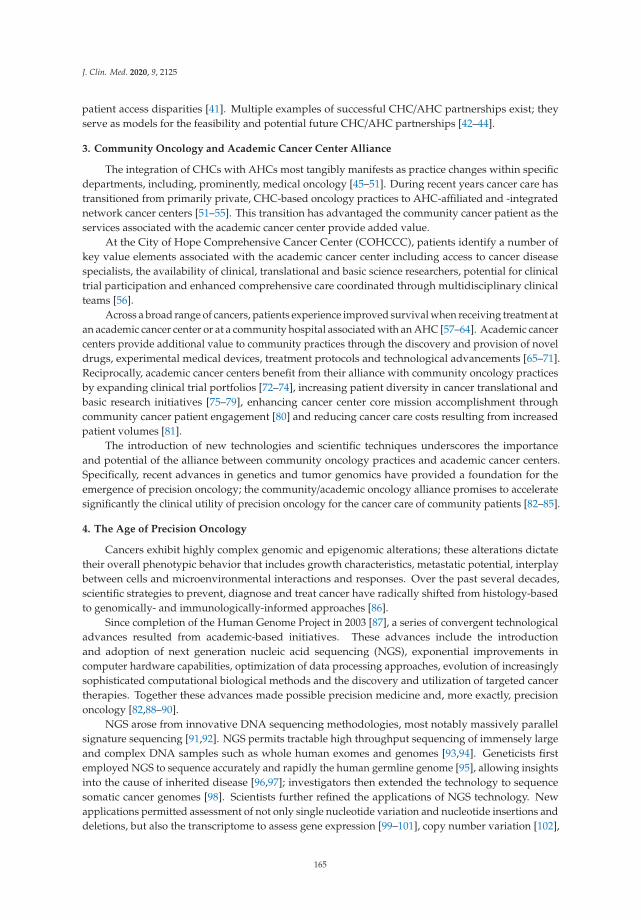
J. Clin. Med. 2020, 9, 2125
patient access disparities [41]. Multiple examples of successful CHC/AHC partnerships exist; theyserve as models for the feasibility and potential future CHC/AHC partnerships [42–44].
3. Community Oncology and Academic Cancer Center Alliance
The integration of CHCs with AHCs most tangibly manifests as practice changes within specificdepartments, including, prominently, medical oncology [45–51]. During recent years cancer care hastransitioned from primarily private, CHC-based oncology practices to AHC-affiliated and -integratednetwork cancer centers [51–55]. This transition has advantaged the community cancer patient as theservices associated with the academic cancer center provide added value.
At the City of Hope Comprehensive Cancer Center (COHCCC), patients identify a number ofkey value elements associated with the academic cancer center including access to cancer diseasespecialists, the availability of clinical, translational and basic science researchers, potential for clinicaltrial participation and enhanced comprehensive care coordinated through multidisciplinary clinicalteams [56].
Across a broad range of cancers, patients experience improved survival when receiving treatment atan academic cancer center or at a community hospital associated with an AHC [57–64]. Academic cancercenters provide additional value to community practices through the discovery and provision of noveldrugs, experimental medical devices, treatment protocols and technological advancements [65–71].Reciprocally, academic cancer centers benefit from their alliance with community oncology practicesby expanding clinical trial portfolios [72–74], increasing patient diversity in cancer translational andbasic research initiatives [75–79], enhancing cancer center core mission accomplishment throughcommunity cancer patient engagement [80] and reducing cancer care costs resulting from increasedpatient volumes [81].
The introduction of new technologies and scientific techniques underscores the importanceand potential of the alliance between community oncology practices and academic cancer centers.Specifically, recent advances in genetics and tumor genomics have provided a foundation for theemergence of precision oncology; the community/academic oncology alliance promises to acceleratesignificantly the clinical utility of precision oncology for the cancer care of community patients [82–85].
4. The Age of Precision Oncology
Cancers exhibit highly complex genomic and epigenomic alterations; these alterations dictatetheir overall phenotypic behavior that includes growth characteristics, metastatic potential, interplaybetween cells and microenvironmental interactions and responses. Over the past several decades,scientific strategies to prevent, diagnose and treat cancer have radically shifted from histology-basedto genomically- and immunologically-informed approaches [86].
Since completion of the Human Genome Project in 2003 [87], a series of convergent technologicaladvances resulted from academic-based initiatives. These advances include the introductionand adoption of next generation nucleic acid sequencing (NGS), exponential improvements incomputer hardware capabilities, optimization of data processing approaches, evolution of increasinglysophisticated computational biological methods and the discovery and utilization of targeted cancertherapies. Together these advances made possible precision medicine and, more exactly, precisiononcology [82,88–90].
NGS arose from innovative DNA sequencing methodologies, most notably massively parallelsignature sequencing [91,92]. NGS permits tractable high throughput sequencing of immensely largeand complex DNA samples such as whole human exomes and genomes [93,94]. Geneticists firstemployed NGS to sequence accurately and rapidly the human germline genome [95], allowing insightsinto the cause of inherited disease [96,97]; investigators then extended the technology to sequencesomatic cancer genomes [98]. Scientists further refined the applications of NGS technology. Newapplications permitted assessment of not only single nucleotide variation and nucleotide insertions anddeletions, but also the transcriptome to assess gene expression [99–101], copy number variation [102],
165

J. Clin. Med. 2020, 9, 2125
complex genomic structural variation [103], protein-DNA interactions [104], targetable epigeneticalterations [105] and epigenetic mechanisms regulating 3D genome structure [106].
In addition to examining tumor genomics, there arose an interest in understanding theimmune profiles of the tumor and its microenvironment using NGS; in part, this interest developedfrom the recognition that tumor genomic changes frequently result in the production of unique,highly immunogenic neoantigens that render the tumor vulnerable to immune surveillance anddestruction [107,108]. With the appreciation that the immune system plays an important role in cancerinitiation and progression, there has also occurred new interest in targeted therapies aimed at activationof the immune axis [109,110].
NGS generates enormous caches of data; use of these immense data sets for precisiononcology requires ever increasing levels of computer hardware performance. Employment ofDennard scaling [111] and multicore architectures [112] have sustained exponential increases incomputer chip performance [113–115]. Data processing innovations have included parallel algorithmimplementation [116] and parallel data computing [117]; such innovations have force multiplied theefficiency and speed of computation. These approaches allow data analysts to keep pace with the everincreasing information workloads of precision oncology [118].
The realization of precision oncology required adoption of computational biological approaches.The creation of computational biology as an independent academic discipline resulted from thecomplexity and size of biological data sets. In the case of NGS, the sheer number of nucleotides reads,the task of aligning these reads to reference sequences, predicting functional consequences of genomicvariation and the translation of these findings into clinically actionable information necessitatedcomputational biological expertise [119–122]. Computational biological analysis now constitutes anintegral element of the data workflow in precision oncology [123–126]; effective clinical translationdepends inextricably upon the availability of these computational resources [127–130].
In the early 1970′s, Drs. Janet Rowley, Peter Nowell and Alfred Knudson, studying leukemiacell chromosomes under the microscope, suggested that a specific chromosomal translocation thatresulted in the formation of the BCR-ABL fusion oncogene caused chronic myelogenous leukemia(CML); this observation established a foundation for clinical cancer genomics [131]. Oncogenic proteinsconsequently became a focus of therapeutic drug design; targeted therapies aimed to suppress theaberrant functions of these proteins in order to inhibit tumor progression [132,133].
The successful harnessing of precision therapeutics in oncology ultimately relies upon theavailability and efficacy of targeted agents. The discovery that imatinib effectively treats CML harboringthe BCR-ABL fusion protein [134] led to the drug’s FDA approval in 2001 [135], demonstrated the utilityof targeted cancer therapy [136,137], kindled enthusiasm for the identification of other geneticallyvulnerable cancers and their treatments [90,138] and underscored the clinical value and potential ofprecision oncology [98,139,140]. Since the success of imatinib, the FDA has approved a multitude ofadditional therapies to target molecularly-altered cancers [141,142].
The clinical provision of precision oncology requires multidisciplinary support [143]; thecomplexity of this support will become more intense as precision oncology continues to undergoaccelerating change [144–146]. AHCs possess the resources and organization to create this supportstructure; their alliance with CHC oncology practitioners will make precision oncology available to thelarger CHC cancer population.
5. The Community Oncology/Academic Cancer Center Alliance in Germline Cancer Genetics
NGS and precision oncology have had a profound effect upon the practice of cancer genetics,including the evaluation and care of community patients with hereditary predisposition tocancer [147–151]. Until recently, genetic testing involved clinical assessment followed by sequential,single gene Sanger sequencing of suspect genes [152–156]. The advent of NGS brought high throughputgermline multigene panel [157–161], whole exome [162–167] and whole genome assessment [168–172]to clinical cancer genetics. These platforms provide tremendous benefit to cancer genetics patients both
166

J. Clin. Med. 2020, 9, 2125
in community oncology practices and at academic cancer centers; these advantages include increaseddiagnostic yield, increased speed of testing, optimized testing workflows, decreased expense and thediscovery of new cancer-causing genes [173–177]. However, together with advantages, challenges andlimitations arise; AHCs have the specialized resources to address these issues.
In accordance with the American College of Medical Genetics and the Association for MolecularPathology guidelines, variants from clinical genetic testing fall along a spectrum ranging frompathogenic/likely pathogenic to benign/likely benign [178]; variants of uncertain significance (VUS)occur when there exists insufficient information for variant assignment to either the pathogenicor benign categories [179,180]. For pathogenic/likely pathogenic and benign/likely benign variants,genetic providers typically have the ability to communicate clear interpretation of results and to provideconsensus health recommendations. As their pathogenicity remains uncertain, VUS challenge healthcare specialists to formulate and relay unambiguous health care instructions [181–185]; furthermore,VUS frequently cause confusion and anxiety for the patient [186–190]. VUS impose a significantclinical burden. More than one third of NGS-based cancer gene panel tests result in identificationof a VUS [191]; whole exome and genome testing generate even greater numbers of VUS [192–195].Moreover, if a patient belongs to a minority group, for whom genome annotations remain less wellconfirmed, VUS additionally increase [196].
Geneticists classify genes according to their penetrance, that is, how likely will a pathogenic variantof a gene cause disease [197]. For pathogenic variants of high penetrance genes, clinicians more oftenhave firmly established guidelines that inform recommendations for patient screening and surveillance.However, for pathological variants of low penetrance genes, less definitive clinical guidelines exist.NGS-based testing results in increasing detection of pathogenic variants of low penetrance genes; thisincreased detection adds complexity and uncertainty to patient management [198,199].
Clinicians face another challenge when selecting NGS gene panels for genetic evaluation: theymust select the composition of the gene panel that they will employ. This selection requiresspecialized education and training [200,201]. The cancer genetics expertise required to addressthis challenge remains scarce [202–205]; the wider use of NGS platforms in clinical oncology andcontinued technological advances has made this expertise even more scarce [206–208].
AHCs possess the clinical expertise, facilities, support personnel, and administrative structuresto meet the burgeoning demands of cancer genetics and to overcome the obstacles associated withthe use of NGS in the clinic. Allied community oncology practices and their patients have accessto these resources and services through their partnerships with AHCs. Four access models enablecommunity oncology patient engagement with the AHC: (1) patient consultation visits to the academiccancer center, (2) cancer genetics specialist visits to community oncology sites, (3) telemedicine- andweb-based remote visits and (4) AHC-sponsored genetic education initiatives that train communityoncology practitioners to assess and manage cancer genetic risk and disease (Figure 1).
Conventionally, community oncology patients have received their cancer genetics care byconsulting, in person, with a specialist at an AHC [155,209–211]. This model disadvantages communitypatients who live substantial distances from an AHC as it involves significant travel time and costcommitments [210,212,213]. Alternative cancer genetics delivery models have the potential to mitigatethese problems.
In the community satellite clinic model, AHC cancer genetic specialists travel to the CHC clinicon an interval basis to meet the cancer genetic needs of community patients. This approach has provensuccessful in a variety of circumstances where logistical or economic challenges create barriers toeffective cancer genetics care [214–217].
167
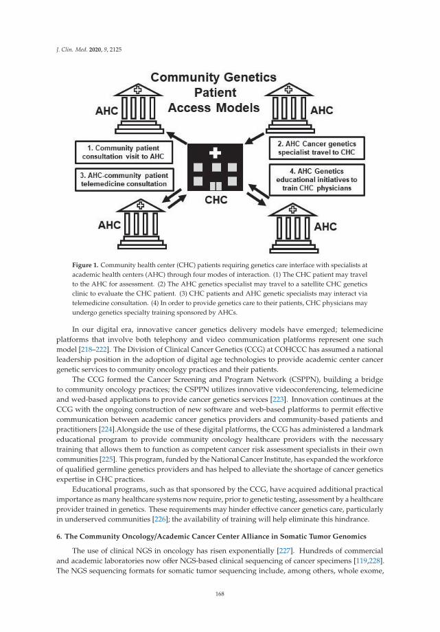
J. Clin. Med. 2020, 9, 2125
Figure 1. Community health center (CHC) patients requiring genetics care interface with specialists atacademic health centers (AHC) through four modes of interaction. (1) The CHC patient may travelto the AHC for assessment. (2) The AHC genetics specialist may travel to a satellite CHC geneticsclinic to evaluate the CHC patient. (3) CHC patients and AHC genetic specialists may interact viatelemedicine consultation. (4) In order to provide genetics care to their patients, CHC physicians mayundergo genetics specialty training sponsored by AHCs.
In our digital era, innovative cancer genetics delivery models have emerged; telemedicineplatforms that involve both telephony and video communication platforms represent one suchmodel [218–222]. The Division of Clinical Cancer Genetics (CCG) at COHCCC has assumed a nationalleadership position in the adoption of digital age technologies to provide academic center cancergenetic services to community oncology practices and their patients.
The CCG formed the Cancer Screening and Program Network (CSPPN), building a bridgeto community oncology practices; the CSPPN utilizes innovative videoconferencing, telemedicineand wed-based applications to provide cancer genetics services [223]. Innovation continues at theCCG with the ongoing construction of new software and web-based platforms to permit effectivecommunication between academic cancer genetics providers and community-based patients andpractitioners [224].Alongside the use of these digital platforms, the CCG has administered a landmarkeducational program to provide community oncology healthcare providers with the necessarytraining that allows them to function as competent cancer risk assessment specialists in their owncommunities [225]. This program, funded by the National Cancer Institute, has expanded the workforceof qualified germline genetics providers and has helped to alleviate the shortage of cancer geneticsexpertise in CHC practices.
Educational programs, such as that sponsored by the CCG, have acquired additional practicalimportance as many healthcare systems now require, prior to genetic testing, assessment by a healthcareprovider trained in genetics. These requirements may hinder effective cancer genetics care, particularlyin underserved communities [226]; the availability of training will help eliminate this hindrance.
6. The Community Oncology/Academic Cancer Center Alliance in Somatic Tumor Genomics
The use of clinical NGS in oncology has risen exponentially [227]. Hundreds of commercialand academic laboratories now offer NGS-based clinical sequencing of cancer specimens [119,228].The NGS sequencing formats for somatic tumor sequencing include, among others, whole exome,
168
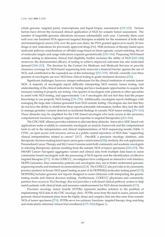
J. Clin. Med. 2020, 9, 2125
whole genome, targeted panel, transcriptome and liquid biopsy assessments [229–235]. Variousfactors have driven the increased clinical application of NGS for somatic tumor assessment. Thenumber of targetable genomic alterations increases substantially each year. Currently, there existwell over one hundred FDA-approved targeted therapies available for the treatment of both solidand hematological cancers [98]; over the past year alone, the FDA granted approval to nearly 20 newdrugs or new indications for previously approved drugs [96]. With inclusion of therapy based uponmolecular pathway considerations or off-label usage based on tissue-agnostic variant matching, the setof molecular targets and usage indications expands geometrically [236–244]. Purposing NGS-basedsomatic testing to determine clinical trial eligibility further increases the utility of NGS [245–248];moreover, the demonstrated efficacy of testing to achieve improved outcomes has also motivateddemand [248–252]. The decision by the Centers for Medicare and Medicaid Services to provideinsurance coverage for NGS-based sequencing tests removed a financial barrier against the use ofNGS, and contributed to the expanded use of this technology [253–255]. All told, currently over threequarters of oncologists use now NGS-base clinical testing to guide treatment decisions [256].
Significant challenges, however, temper enthusiasm for the clinical institution of somatic tumorNGS. A majority of oncologists report difficulty interpreting NGS somatic tumor testing, lackunderstanding of the clinical indications for testing and have inadequate opportunities to acquire thenecessary training to properly use testing. One quarter of oncologists refer patients to other specialiststo assist with NGS testing, and approximately 1 in 5 oncologists did not feel they had the properknowledge to use properly NGS testing [256–258]. Additionally, oncologists report challenges withmanaging the large data volumes generated from NGS somatic testing. Oncologists also feel that theydo not have the ability to distill from these reports actionable information; further, they lack the skillto manage germline variants detected as incidental findings in somatic NGS tumor testing [259–262].These obstacles may be amplified for the CHC-based oncologist who lacks access to the necessarycomputational resources, logistical support and expertise in targeted therapeutics [263–266].
The CHC/AHC alliance provides solutions to alleviate these obstacles. Innovative AHC-based webapplications make available to community oncologists an analytic framework and the computationaltools to aid in the interpretation and clinical implementation of NGS sequencing results (Table 1).CIViC, an open access web resource, serves as a public central repository of NGS data “supportingclinical interpretations related to cancer” [267]. OncoKB, a precision oncology database, aidstherapeutic decision-making based upon cancer gene variant status [268]; similarly, the web applicationsPersonalized Cancer Therapy and My Cancer Genome assist both community and academic oncologistsin selecting therapeutic options resulting from the somatic NGS of tumor specimens [269,270]. TheSMART Cancer Navigator aggregates variant and clinical data from multiple data bases to assistcommunity-based oncologists with the processing of NGS reports and the identification of effectivetargeted therapies [271]. At the COHCCC, investigators have configured an interactive web interface,HOPE-Genomics, that community patients and oncologists may use to better understand genomicsequencing results and treatment recommendations [224]. The COHCCC also provides to its communitypractice partners in-house NGS panel testing as part of its HOPESEQ molecular testing panel [272];HOPESEQ includes genomic test reports designed to assist clinicians with interpreting the genetictesting results and clinical decision making. Furthermore, COHCCC physicians and communitypartners have access to Via Oncology; this tool provides a web-based clinical pathway system to helpmatch patients with clinical trials and insurance reimbursement for NGS driven treatments [273].
Precision oncology tumor boards (POTBs) represent another solution to the problem ofimplementing NGS data in the CHC oncology clinic. POTBs arose from the need to assess, process andgenerate clinical treatment plans from the highly dense and complex data sets that arise from somaticNGS of tumor specimens [274]. POTBs serve two primary functions: targeted therapy drug matchingand molecularly-informed clinical trial enrollment [275–280] (Figure 2).
169
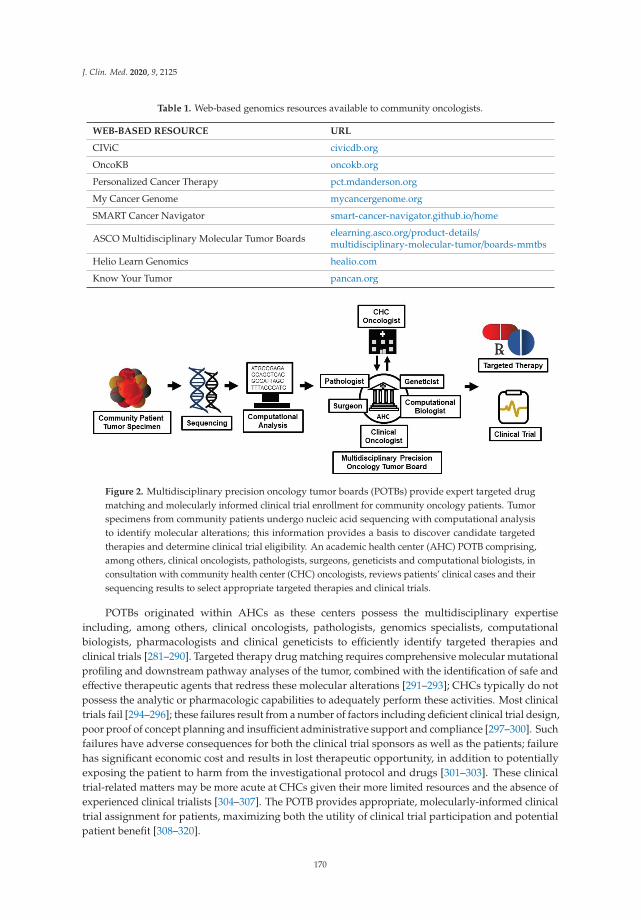
J. Clin. Med. 2020, 9, 2125
Table 1. Web-based genomics resources available to community oncologists.
WEB-BASED RESOURCE URL
CIViC civicdb.org
OncoKB oncokb.org
Personalized Cancer Therapy pct.mdanderson.org
My Cancer Genome mycancergenome.org
SMART Cancer Navigator smart-cancer-navigator.github.io/home
ASCO Multidisciplinary Molecular Tumor Boards elearning.asco.org/product-details/multidisciplinary-molecular-tumor/boards-mmtbs
Helio Learn Genomics healio.com
Know Your Tumor pancan.org
Figure 2. Multidisciplinary precision oncology tumor boards (POTBs) provide expert targeted drugmatching and molecularly informed clinical trial enrollment for community oncology patients. Tumorspecimens from community patients undergo nucleic acid sequencing with computational analysisto identify molecular alterations; this information provides a basis to discover candidate targetedtherapies and determine clinical trial eligibility. An academic health center (AHC) POTB comprising,among others, clinical oncologists, pathologists, surgeons, geneticists and computational biologists, inconsultation with community health center (CHC) oncologists, reviews patients’ clinical cases and theirsequencing results to select appropriate targeted therapies and clinical trials.
POTBs originated within AHCs as these centers possess the multidisciplinary expertiseincluding, among others, clinical oncologists, pathologists, genomics specialists, computationalbiologists, pharmacologists and clinical geneticists to efficiently identify targeted therapies andclinical trials [281–290]. Targeted therapy drug matching requires comprehensive molecular mutationalprofiling and downstream pathway analyses of the tumor, combined with the identification of safe andeffective therapeutic agents that redress these molecular alterations [291–293]; CHCs typically do notpossess the analytic or pharmacologic capabilities to adequately perform these activities. Most clinicaltrials fail [294–296]; these failures result from a number of factors including deficient clinical trial design,poor proof of concept planning and insufficient administrative support and compliance [297–300]. Suchfailures have adverse consequences for both the clinical trial sponsors as well as the patients; failurehas significant economic cost and results in lost therapeutic opportunity, in addition to potentiallyexposing the patient to harm from the investigational protocol and drugs [301–303]. These clinicaltrial-related matters may be more acute at CHCs given their more limited resources and the absence ofexperienced clinical trialists [304–307]. The POTB provides appropriate, molecularly-informed clinicaltrial assignment for patients, maximizing both the utility of clinical trial participation and potentialpatient benefit [308–320].
170
J. Clin. Med. 2020, 9, 2125
Given the resource limitations of the CHC oncology clinic, community POTB operationrequires innovation and dedicated planning [83,321,322]. One innovation available to communityoncologists, the web-based ASCO Multidisciplinary Molecular Tumor Boards, assists oncologists withunderstanding precision medicine-based tumor testing and the therapy recommendations resultingfrom these tests [323] (Table 1). Helio Learn Genomics, another web platform, offers a number ofeducational modules, including POTB cases, to help providers understand the molecular bases ofcarcinogenesis and precision therapeutics [324]. The Pancreatic Cancer Action Network administers aKnow Your Tumor program, a turn-key precision medicine initiative, that allows community oncologypractitioners to submit their patients’ pancreatic cancer specimens for NGS molecular testing and toreceive back a precision medicine-based treatment plan [325].
Another version of the POTB, the virtual POTB, permits the distance participation of communityoncologists in an academic POTB. In this model an AHC hosts the POTB and reviews the clinicalhistory and precision oncology testing results of the community oncology patient; subsequently,the POTB discusses with the community oncologist, using a live interactive video teleconferencinglink, targeted treatment and clinical trial recommendations [263,311,326–330]. The TranslationalGenomics Research Institute (TGEN), an academic affiliate of the COHCCC, has successfully builta comprehensive, integrated, high-throughput sequencing and reporting framework that, whencombined with remote teleconferencing, has proven tremendously successful in establishing efficientcollaborative POTBs [331–335]. Together, these various models of providing clinical somatic NGSdemonstrate the feasibility of leveraging precision oncology for the community-based cancer oncologistand their patients.
7. Conclusions
We have entered the age of precision oncology. Precision oncology offers the potential ofmolecularly informed medicine for the assessment of inherited cancer predisposition, as well as for thediagnosis and treatment of cancers. Realization of this potential depends upon access to specializedexpertise and significant analytic and technological resources. While frequently available at AHCs,these resources have previously been limited for CHC oncology practices and their patients. In thispaper, we have examined the CHC/AHC alliance and discussed examples illustrating how this allianceprovides a structure that allows community cancer patients to benefit from germline and somaticprecision oncology advances. Looking forward, multidisciplinary efforts, improved technology andcontinuing innovation promise to strengthen and facilitate the CHC/AHC alliance in oncology; thisalliance offers community oncologists and their patients the prospect of unambiguous interpretation ofgenetic and genomic test results and optimized precision oncology care.
Author Contributions: Writing-Original Draft Preparation, M.M., S.S. (Shanmuga Subbiah), K.J.M.;Writing-Review and Editing, S.S. (Swapnil Rajurkar). All authors have read and agreed to the publishedversion of the manuscript.
Funding: This research received no external funding.
Conflicts of Interest: The authors declare no conflict of interest.
References
1. Wartman, S.A. The Transformation of Academic Health Centers: Meeting The Challenges Of Healthcare’s ChangingLandscape; Academic Press: Fribourg, Switzerland, 2015.
2. Dzau, V.J.; Ackerly, D.C.; Sutton-Wallace, P.; Merson, M.H.; Williams, R.S.; Krishnan, K.R.; Taber, R.C.;Califf, R.M. The role of academic health science systems in the transformation of medicine. Lancet 2010, 375,949–953. [CrossRef]
3. Desch, C.E.; Blayney, D.W. Making the choice between academic oncology and community practice: the bigpicture and details about each career. J. Oncol. Pract. 2006, 2, 132–136. [CrossRef]
4. Todd, R.F., III. A guide to planning careers in hematology and oncology. Hematol. Am. Soc. Hemat. 2001,2001, 499–506. [CrossRef] [PubMed]
171
J. Clin. Med. 2020, 9, 2125
5. Levit, L.A.; Kim, E.S.; McAneny, B.L.; Nadauld, L.D.; Levit, K.; Schenkel, C.; Schilsky, R.L. Implementingprecision medicine in community-based oncology programs: Three models. J. Oncol. Pract. 2019, 15, 325–329.[CrossRef] [PubMed]
6. Thompson, M.A.; Godden, J.J.; Weissman, S.M.; Wham, D.; Wilson, A.; Ruggeri, A.; Mullane, M.P.; Weese, J.L.Implementing an oncology precision medicine clinic in a large community health system. Am. J. Manag.Care 2017, 23, SP425–SP427.
7. Levine, D.M.; Becker, D.M.; Bone, L.R.; Hill, M.N.; Tuggle, M.B., II; Zeger, S.L. Community-academic healthcenter partnerships for underserved minority populations. One solution to a national crisis. J. Am. Med.Assoc. 1994, 272, 309–311. [CrossRef]
8. Shin, P.; Sharac, J.; Rosenbaum, S. Community health centers and medicaid at 50: An. Enduring relationshipessential for health system transformation. Health Aff. (Millwood) 2015, 34, 1096–1104. [CrossRef]
9. Sharma, A.E.; Huang, B.; Knox, M.; Willard-Grace, R.; Potter, M.B. Patient engagement in community healthcenter leadership: How does it happen? J. Community Health 2018, 43, 1069–1074. [CrossRef] [PubMed]
10. Blumenthal, D.; Meyer, G.S. Academic health centers in a changing environment. Health Aff. (Millwood) 1996,15, 200–215. [CrossRef]
11. Nash, D.B.; Veloski, J.J. Emerging opportunities for educational partnerships between managed careorganizations and academic health centers. West. J. Med. 1998, 168, 319–327. [PubMed]
12. Roper, W.L.; Newton, W.P. The role of academic health centers in improving health. Ann. Fam. Med. 2006, 4,S55–S57. [CrossRef] [PubMed]
13. Blumenthal, D.; Meyer, G.S. The future of the academic medical center under health care reform. N. Engl. J.Med. 1993, 329, 1812–1814. [CrossRef]
14. Fox, P.D.; Wasserman, J. Academic medical centers and managed care: Uneasy partners. Health Aff. (Millwood)1993, 12, 85–93. [CrossRef]
15. Epstein, A.M. US teaching hospitals in the evolving health care system. J. Am. Med. Assoc. 1995, 273,1203–1207. [CrossRef]
16. Iglehart, J.K. Academic medical centers enter the market: The case of Philadelphia. N. Engl. J. Med. 1995,333, 1019–1023. [CrossRef] [PubMed]
17. Lofgren, R.; Karpf, M.; Perman, J.; Higdon, C.M. The U.S. health care system is in crisis: Implications foracademic medical centers and their missions. Acad. Med. 2006, 81, 713–720. [CrossRef]
18. Park, B.; Frank, B.; Likumahuwa-Ackman, S.; Brodt, E.; Gibbs, B.K.; Hofkamp, H.; DeVoe, J. Health equityand the tripartite mission: Moving from academic health centers to academic-community health systems.Acad. Med. 2019, 94, 1276–1282. [CrossRef]
19. Bartlett, S.J.; Barnes, T.; McIvor, R.A. Integrating patients into meaningful real-world research. Ann. Am.Thorac. Soc. 2014, 11, S112–S117. [CrossRef]
20. Gourevitch, M.N. Population health and the academic medical center: The time is right. Acad. Med. 2014, 89,544–549. [CrossRef] [PubMed]
21. Vitale, K.; Newton, G.L.; Abraido-Lanza, A.F.; Aguirre, A.N.; Ahmed, S.; Esmond, S.L.; Evans, J.; Gelmon, S.B.;Hart, C.; Hendricks, D.; et al. Community engagement in academic health centers: A model for capturingand advancing our successes. J. Commun. Engagem. Scholarsh. 2018, 10, 81–90.
22. Zerhouni, E. Medicine: The NIH roadmap. Science 2003, 302, 63–72. [CrossRef] [PubMed]23. Schwenk, T.L.; Green, L.A. The Michigan Clinical Research Collaboratory: Following the NIH Roadmap to
the community. Ann. Fam. Med. 2006, 4, S49–S54. [CrossRef] [PubMed]24. Zerhouni, E.A. Translational and clinical science—Time for a new vision. N. Engl. J. Med. 2005, 353, 1621–1623.
[CrossRef] [PubMed]25. Zerhouni, E.A. US biomedical research: Basic, translational, and clinical sciences. J. Am. Med. Assoc. 2005,
294, 1352–1358. [CrossRef]26. Kassirer, J.P. Academic medical centers under siege. N. Engl. J. Med. 1994, 331, 1370–1371. [CrossRef]27. Iglehart, J.K. Rapid changes for academic medical centers. 2. N. Engl. J. Med. 1995, 332, 407–411. [CrossRef]28. Iglehart, J.K. Rapid changes for academic medical centers. 1. N. Engl. J. Med. 1994, 331, 1391–1395. [CrossRef]29. Moses, H., III; Matheson, D.H.M.; Poste, G. Serving individuals and populations within integrated health
systems: A bridge too far? J. Am. Med. Assoc. 2019, 321, 1975–1976. [CrossRef]30. Cutler, D.M.; Morton, F.S. Hospitals, market share, and consolidation. J. Am. Med. Assoc. 2013, 310,
1964–1970. [CrossRef]
172
J. Clin. Med. 2020, 9, 2125
31. Moses, H., III; Matheson, D.H.; Dorsey, E.R.; George, B.P.; Sadoff, D.; Yoshimura, S. The anatomy of healthcare in the United States. J. Am. Med. Assoc. 2013, 310, 1947–1963. [CrossRef]
32. Cohen, M.D.; Jennings, G. Mergers involving academic medical institutions: Impact on academic radiologydepartments. J. Am. Coll. Radiol. 2005, 2, 174–182. [CrossRef] [PubMed]
33. Denham, A.C.; Hay, S.S.; Steiner, B.D.; Newton, W.P. Academic health centers and community health centerspartnering to build a system of care for vulnerable patients: Lessons from Carolina Health Net. Acad. Med.2013, 88, 638–643. [CrossRef] [PubMed]
34. Rieselbach, R.E.; Rieselbach, R.E.; Epperly, T.; Friedman, A.; Keahey, D.; McConnell, E.; Nichols, K.; Nycz, G.;Roberts, J.; Schmader, K.; et al. A new community health center/academic medicine partnership for medicaidcost control, powered by the Mega Teaching Health Center. Acad. Med. 2018, 93, 406–413. [CrossRef]
35. Blumenthal, D.; Campbell, E.G.; Weissman, J.S. The social missions of academic health centers. N. Engl. J.Med. 1997, 337, 1550–1553. [CrossRef]
36. Fleishon, H.B.; Itri, J.N.; Boland, G.W.; Duszak, R., Jr. Academic medical centers and community hospitalsintegration: Trends and strategies. J. Am. Coll. Radiol. 2017, 14, 45–51. [CrossRef]
37. Ellner, A.L.; Stout, S.; Sullivan, E.E.; Griffiths, E.P.; Mountjoy, A.; Phillips, R.S. Health systems innovationat academic health centers: Leading in a new era of health care delivery. Acad. Med. 2015, 90, 872–880.[CrossRef] [PubMed]
38. Moore, G.T.; Inui, T.S.; Ludden, J.M.; Schoenbaum, S.C. The “teaching HMO”: A new academic partner.Acad. Med. 1994, 69, 595–600. [CrossRef] [PubMed]
39. Poncelet, A.N.; Mazotti, L.A.; Blumberg, B.; Wamsley, M.A.; Grennan, T.; Shore, W.B. Creating a longitudinalintegrated clerkship with mutual benefits for an academic medical center and a community health system.Perm. J. 2014, 18, 50–56. [CrossRef]
40. Berkowitz, S.A.; Brown, P.; Brotman, D.J.; Deutschendorf, A.; Dunbar, L.; Everett, A.; Hickman, D.; Howell, E.;Purnell, L.; Sylvester, C.; et al. Case Study: Johns Hopkins Community Health Partnership: A model fortransformation. Healthc (Amst.) 2016, 4, 264–270. [CrossRef]
41. Institute of Medicine (US) Committee on Understanding and Eliminating Racial and Ethnic Disparitiesin Health Care. Unequal Treatment: Confronting Racial and Ethnic Disparities in Health Care; Smedley, B.D.,Stith, A.Y., Nelson, A.R., Eds.; National Academies Press: Washington, DC, USA, 2003.
42. Ahmed, S.M.; Maurana, C.; Nelson, D.; Meister, T.; Young, S.N.; Lucey, P. Opening the black box:Conceptualizing community engagement from 109 community-academic partnership programs. Prog.Community Health Partnersh. 2016, 10, 51–61. [CrossRef]
43. Croft, C.R.; Dial, R.; Doyle, G.; Schaadt, J.; Merchant, L. Integrating a community hospital-based radiologydepartment with an academic medical center. J. Am. Coll. Radiol. 2016, 13, 300–302. [CrossRef]
44. Sussman, A.J.; Otten, J.R.; Goldszer, R.C.; Hanson, M.; Trull, D.J.; Paulus, K.; Brown, M.; Dzau, V.; Brennan, T.A.Integration of an academic medical center and a community hospital: The Brigham and Women’s/Faulknerhospital experience. Acad. Med. 2005, 80, 253–260. [CrossRef] [PubMed]
45. Spalluto, L.B.; Thomas, D.; Beard, K.R.; Campbell, T.; Audet, C.M.; McBride Murry, V.; Shrubsole, M.J.;Barajas, C.P.; Joosten, Y.A.; Dittus, R.S.; et al. A community-academic partnership to reduce health caredisparities in diagnostic imaging. J. Am. Coll. Radiol. 2019, 16, 649–656. [CrossRef] [PubMed]
46. Kelley-Quon, L.I.; Thomas, D.; Beard, K.R.; Campbell, T.; Audet, C.M.; McBride Murry, V.; Shrubsole, M.J.;Barajas, C.P.; Joosten, Y.A.; Dittus, R.S.; et al. Academic-community partnerships improve outcomes inpediatric trauma care. J. Pediatr. Surg. 2015, 50, 1032–1036. [CrossRef] [PubMed]
47. Phillip, C.R.; Mancera-Cuevas, K.; Leatherwood, C.; Chmiel, J.S.; Erickson, D.L.; Freeman, E.; Granville, G.;Dollear, M.; Walker, K.; McNeil, R.; et al. Implementation and dissemination of an African Americanpopular opinion model to improve lupus awareness: An academic-community partnership. Lupus 2019, 28,1441–1451. [CrossRef] [PubMed]
48. Rees, T. Academic medical center, community hospital partner to market center of excellence. Profiles Healthc.Mark. 1999, 15, 40–43.
49. Yaggy, S.D.; Michener, J.L.; Yaggy, D.; Champagne, M.T.; Silberberg, M.; Lyn, M.; Johnson, F.; Yarnall, K.S.Just for Us: An academic medical center-community partnership to maintain the health of a frail low-incomesenior population. Gerontologist 2006, 46, 271–276. [CrossRef]
50. Natesan, R.; Yang, W.T.; Tannir, H.; Parikh, J. Strategic expansion models in academic radiology. J. Am. Coll.Radiol. 2016, 13, 329–334. [CrossRef]
173
J. Clin. Med. 2020, 9, 2125
51. Kirkwood, M.K.; Hanley, A.; Bruinooge, S.S.; Garrett-Mayer, E.; Levit, L.A.; Schenkel, C.; Seid, J.E.; Polite, B.N.;Schilsky, R.L. The State of oncology practice in America, 2018: results of the ASCO practice census survey. J.Oncol. Pract. 2018, 14, e412–e420. [CrossRef]
52. OBR New Perspective Catalyst. Most Cancer Patients Will Be Treated in Integrated Delivery Networks (IDN) andCancer Institutions by 2016, Predicts New Report; OBR New Perspective Catalyst: Sausalito, CA, USA, 2012;Volume 11.
53. Genetech. The 2018 Genentech Oncology Trend Report; Genetech: San Francisco, CA, USA, 2018.54. Genetech. The 2019 Genentech Oncology Trend Report; Genetech: San Francisco, CA, USA, 2019.55. Academic Cancer Centers (NCCC). Trends Impacting Key Account. Management; Academic Cancer Centers:
Pipersville, PA, USA, 2016.56. Nardi, E.A.; Wolfson, J.A.; Rosen, S.T.; Diasio, R.B.; Gerson, S.L.; Parker, B.A.; Alvarnas, J.C.; Levine, H.A.;
Fong, Y.; Weisenburger, D.D.; et al. Value, access, and cost of cancer care delivery at academic cancer centers.J. Natl. Compr. Cancer Netw. 2016, 14, 837–847. [CrossRef]
57. Speicher, P.J.; Englum, B.R.; Ganapathi, A.M.; Wang, X.; Hartwig, M.G.; D’Amico, T.A.; Berry, M.F. Travelingto a high-volume center is associated with improved survival for patients with esophageal cancer. Ann. Surg.2017, 265, 743–749. [CrossRef] [PubMed]
58. Lidsky, M.E.; Sun, Z.; Nussbaum, D.P.; Adam, M.A.; Speicher, P.J.; Blazer, D.G., III. Going the extra mile:Improved survival for pancreatic cancer patients traveling to high-volume centers. Ann. Surg. 2017, 266,333–338. [CrossRef] [PubMed]
59. David, J.M.; Ho, A.S.; Luu, M.; Yoshida, E.J.; Kim, S.; Mita, A.C.; Scher, K.S.; Shiao, S.L.; Tighiouart, M.;Zumsteg, Z.S. Treatment at high-volume facilities and academic centers is independently associated withimproved survival in patients with locally advanced head and neck cancer. Cancer 2017, 123, 3933–3942.[CrossRef]
60. Chen, A.Y.; Fedewa, S.; Pavluck, A.; Ward, E.M. Improved survival is associated with treatment at high-volumeteaching facilities for patients with advanced stage laryngeal cancer. Cancer 2010, 116, 4744–4752. [CrossRef][PubMed]
61. Pfister, D.G.; Rubin, D.M.; Elkin, E.B.; Neill, U.S.; Duck, E.; Radzyner, M.; Bach, P.B. Risk adjusting survivaloutcomes in hospitals that treat patients with cancer without information on cancer stage. JAMA Oncol. 2015,1, 1303–1310. [CrossRef] [PubMed]
62. Schmitz, R.; Adam, M.A.; Blazer, D.G., III. Overcoming a travel burden to high-volume centers for treatmentof retroperitoneal sarcomas is associated with improved survival. World J. Surg. Oncol. 2019, 17, 180.[CrossRef] [PubMed]
63. Dillman, R.O.; Chico, S.D. Cancer patient survival improvement is correlated with the opening of a communitycancer center: Comparisons with intramural and extramural benchmarks. J. Oncol. Pract. 2005, 1, 84–92.[CrossRef] [PubMed]
64. Ramalingam, S.; Dinan, M.A.; Crawford, J. Survival comparison in patients with stage iv lung cancer inacademic versus community centers in the United States. J. Thorac. Oncol. 2018, 13, 1842–1850. [CrossRef][PubMed]
65. Carugo, A.; Draetta, G.F. Academic discovery of anticancer drugs: Historic and future perspectives. Ann.Rev. Cancer Biol. 2019, 3, 385–408. [CrossRef]
66. Everett, J.R. Academic drug discovery: Current status and prospects. Expert Opin. Drug Discov. 2015, 10,937–944. [CrossRef] [PubMed]
67. Matter, A. Bridging academic science and clinical research in the search for novel targeted anti-cancer agents.Cancer Biol. Med. 2015, 12, 316–327. [PubMed]
68. Dorfman, G.S.; Lawrence, T.S.; Matrisian, L.M.; Translational Research Working Group. The TranslationalResearch Working Group developmental pathway for interventive devices. Clin. Cancer Res. 2008, 14,5700–5706. [CrossRef] [PubMed]
69. Barrios, C.H.; Reinert, T.; Werutsky, G. Global breast cancer research: Moving forward. Am. Soc. Clin. Oncol.Educ. Book. 2018, 38, 441–450. [CrossRef] [PubMed]
70. Huber, M.A.; Kraut, N. Key drivers of biomedical innovation in cancer drug discovery. EMBO Mol. Med.2015, 7, 12–16. [CrossRef] [PubMed]
174

J. Clin. Med. 2020, 9, 2125
71. Grodzinski, P.; Liu, C.H.; Hartshorn, C.M.; Morris, S.A.; Russell, L.M. NCI Alliance for Nanotechnologyin Cancer—From academic research to clinical interventions. Biomed. Microdev. 2019, 21, 32. [CrossRef][PubMed]
72. Clauser, S.B.; Johnson, M.R.; O’Brien, D.M.; Beveridge, J.M.; Fennell, M.L.; Kaluzny, A.D. Improving clinicalresearch and cancer care delivery in community settings: Evaluating the NCI community cancer centersprogram. Implement. Sci. 2009, 4, 63. [CrossRef]
73. Hirsch, B.R.; Locke, S.C.; Abernethy, A.P. Experience of the national cancer institute community cancer centersprogram on community-based cancer clinical trials activity. J. Oncol. Pract. 2016, 12, e350-8. [CrossRef]
74. Copur, M.S.; Ramaekers, R.; Gonen, M.; Gulzow, M.; Hadenfeldt, R.; Fuller, C.; Scott, J.; Einspahr, S.;Benzel, H.; Mickey, M.; et al. Impact of the national cancer institute community cancer centers program onclinical trial and related activities at a community cancer center in rural Nebraska. J. Oncol. Pract. 2016, 12,67–68. [CrossRef]
75. DH, F. Clinical trials have the best medicine but do not enroll the patients who need it. Sci. Am. 2019, 320,61–65.
76. Copur, M.S. Inadequate awareness of and participation in cancer clinical trials in the community oncologysetting. Oncology 2019, 33, 54–57.
77. Green, M.A.; Michaels, M.; Blakeney, N.; Odulana, A.A.; Isler, M.R.; Richmond, A.; Long, D.G.; Robinson, W.S.;Taylor, Y.J.; Corbie-Smith, G. Evaluating a community-partnered cancer clinical trials pilot intervention withAfrican American communities. J. Cancer Educ. 2015, 30, 158–166. [CrossRef] [PubMed]
78. Best, A.; Hiatt, R.A.; Cameron, R.; Rimer, B.K.; Abrams, D.B. The evolution of cancer control research: Aninternational perspective from Canada and the United States. Cancer Epidemiol Biomarkers Prev. 2003, 12,705–712. [PubMed]
79. Greenwald, P.; Cullen, J.W. The new emphasis in cancer control. J. Natl. Cancer. Inst. 1985, 74, 543–551.[CrossRef]
80. Noel, L.; Phillips, F.; Tossas-Milligan, K.; Spear, K.; Vanderford, N.L.; Winn, R.A.; Vanderpool, R.C.;Eckhardt, S.G. Community-academic partnerships: Approaches to engagement. Am. Soc. Clin. Oncol. Educ.Book. 2019, 39, 88–95. [CrossRef]
81. Harris, A.; Kumar, P.; Sutaria, S. Unlocking the Potential of Acdemic and Community Health System Partnerships;McKinsey and Company: New York, NY, USA, 2015.
82. Johnson, T.M. Perspective on precision medicine in oncology. Pharmacotherapy 2017, 37, 988–989. [CrossRef][PubMed]
83. Ersek, J.L.; Black, L.J.; Thompson, M.A.; Kim, E.S. Implementing precision medicine programs and clinicaltrials in the community-based oncology practice: Barriers and best practices. Am. Soc. Clin. Oncol. Educ.Book. 2018, 38, 188–196. [CrossRef]
84. Nadauld, L.D.; Ford, J.M.; Pritchard, D.; Brown, T. Strategies for clinical implementation: Precision oncologyat three distinct institutions. Health Aff. (Millwood) 2018, 37, 751–756. [CrossRef]
85. Thompson, M.A.; Godden, J.J.; Wham, D.; Ruggeri, A.; Mullane, M.P.; Wilson, A.; Virani, S.; Weissman, S.M.;Ramczyk, B.; Vanderwall, P.; et al. Coordinating an oncology precision medicine clinic within an integratedhealth system: lessons learned in year one. J. Patient Cent. Res. Rev. 2019, 6, 36–45. [CrossRef]
86. Carpten, J.C.; Mardis, E.R. The era of precision oncogenomics. Cold Spring Harb. Mol. Case Stud. 2018, 4.[CrossRef]
87. International Human Genome Sequencing Consortium. Finishing the euchromatic sequence of the humangenome. Nature 2004, 431, 931–945. [CrossRef]
88. Ashley, E.A. Towards precision medicine. Nat. Rev. Genet. 2016, 17, 507–522. [CrossRef] [PubMed]89. Sanchez, N.S.; Mills, G.B.; Mills Shaw, K.R. Precision oncology: Neither a silver bullet nor a dream.
Pharmacogenomics 2017, 18, 1525–1539. [CrossRef] [PubMed]90. Berger, M.F.; Mardis, E.R. The emerging clinical relevance of genomics in cancer medicine. Nat. Rev. Clin.
Oncol. 2018, 15, 353–365. [CrossRef] [PubMed]91. Mitra, R.D.; Church, G.M. In situ localized amplification and contact replication of many individual DNA
molecules. Nucleic Acids Res. 1999, 27, e34. [CrossRef]92. Brenner, S.; Johnson, M.; Bridgham, J.; Golda, G.; Lloyd, D.H.; Johnson, D.; Luo, S.; McCurdy, S.; Foy, M.;
Ewan, M.; et al. Gene expression analysis by massively parallel signature sequencing (MPSS) on microbeadarrays. Nat. Biotechnol. 2000, 18, 630–634. [CrossRef]
175
J. Clin. Med. 2020, 9, 2125
93. Mardis, E.R. Next-generation DNA sequencing methods. Annu. Rev. Genomics Hum. Genet. 2008, 9, 387–402.[CrossRef]
94. Shendure, J.; Ji, H. Next-generation DNA sequencing. Nat. Biotechnol. 2008, 26, 1135–1145. [CrossRef][PubMed]
95. Bentley, D.R.; Balasubramanian, S.; Swerdlow, H.P.; Smith, G.P.; Milton, J.; Brown, C.G.; Hall, K.P.; Evers, D.J.;Barnes, C.L.; Bignell, H.R.; et al. Accurate whole human genome sequencing using reversible terminatorchemistry. Nature 2008, 456, 53–59. [CrossRef] [PubMed]
96. Mardis, E.R. The impact of next-generation sequencing technology on genetics. Trends Genet. 2008, 24,133–141. [CrossRef]
97. Tucker, T.; Marra, M.; Friedman, J.M. Massively parallel sequencing: The next big thing in genetic medicine.Am. J. Hum. Genet. 2009, 85, 142–154. [CrossRef]
98. Ley, T.J.; Mardis, E.R.; Ding, L.; Fulton, B.; McLellan, M.D.; Chen, K.; Dooling, D.; Dunford-Shore, B.H.;McGrath, S.; Hickenbotham, M.; et al. DNA sequencing of a cytogenetically normal acute myeloid leukaemiagenome. Nature 2008, 456, 66–72. [CrossRef] [PubMed]
99. Reinartz, J.; Bruyns, E.; Lin, J.Z.; Burcham, T.; Brenner, S.; Bowen, B.; Kramer, M.; Woychik, R. Massivelyparallel signature sequencing (MPSS) as a tool for in-depth quantitative gene expression profiling in allorganisms. Brief. Funct. Genomic. Proteomic. 2002, 1, 95–104. [CrossRef] [PubMed]
100. Torres, T.T.; Metta, M.; Ottenwalder, B.; Schlotterer, C. Gene expression profiling by massively parallelsequencing. Genome Res. 2008, 18, 172–177. [CrossRef] [PubMed]
101. Cancer Genome Atlas Research, N.; Weinstein, J.N.; Collisson, E.A.; Mills, G.B.; Shaw, K.R.; Ozenberger, B.A.;Ellrott, K.; Shmulevich, I.; Sander, C.; Stuart, J.M. The cancer genome atlas pan-cancer analysis project. Nat.Genet. 2013, 45, 1113–1120.
102. Wang, H.; Nettleton, D.; Ying, K. Copy number variation detection using next generation sequencing readcounts. BMC Bioinform. 2014, 15, 109. [CrossRef] [PubMed]
103. Medvedev, P.; Stanciu, M.; Brudno, M. Computational methods for discovering structural variation withnext-generation sequencing. Nat. Methods 2009, 6, S13–S20. [CrossRef]
104. Johnson, D.S.; Mortazavi, A.; Myers, R.M.; Wold, B. Genome-wide mapping of in vivo protein-DNAinteractions. Science 2007, 316, 1497–1502. [CrossRef]
105. Morel, D.; Jeffery, D.; Aspeslagh, S.; Almouzni, G.; Postel-Vinay, S. Combining epigenetic drugs with othertherapies for solid tumours—Past lessons and future promise. Nat. Rev. Clin. Oncol. 2020, 17, 91–107.[CrossRef]
106. Lazaris, C.; Aifantis, I.; Tsirigos, A. On epigenetic plasticity and genome topology. Trends Cancer 2020, 6,177–180. [CrossRef]
107. Segal, N.H.; Parsons, D.W.; Peggs, K.S.; Velculescu, V.; Kinzler, K.W.; Vogelstein, B.; Allison, J.P. Epitopelandscape in breast and colorectal cancer. Cancer Res. 2008, 68, 889–892. [CrossRef]
108. Alexandrov, L.B.; Nik-Zainal, S.; Wedge, D.C.; Aparicio, S.A.; Behjati, S.; Biankin, A.V.; Bignell, G.R.; Bolli, N.;Borg, A.; Borresen-Dale, A.L.; et al. Signatures of mutational processes in human cancer. Nature 2013, 500,415–421. [CrossRef] [PubMed]
109. Sanmamed, M.F.; LChe. A paradigm shift in cancer immunotherapy: From enhancement to normalization.Cell 2018, 175, 313–326. [CrossRef] [PubMed]
110. Zewde, M.; Kiyotani, K.; Park, J.H.; Fang, H.; Yap, K.L.; Yew, P.Y.; Alachkar, H.; Kato, T.; Mai, T.H.; Ikeda, Y.;et al. The era of immunogenomics/immunopharmacogenomics. J. Hum. Genet. 2018, 63, 865–875. [CrossRef]
111. Dennard, R.H.; Gaensslen, F.H.; Yu, H.N.; Rideout, V.L.; Bassous, E.; Leblanc, A.R. Design of ion-implantedMOSFET’s with very small physical dimensions. IEEE J. Solid State Circuits 1974, 9, 256–268. [CrossRef]
112. Gorder, P.F. Multicore processors for science and engineering. Comput. Sci. Eng. 2007, 9, 3–7. [CrossRef]113. Denning, P.J.; Lewis, T.G. Exponential laws of computing growth. Commun. Acm 2017, 60, 54–65. [CrossRef]114. Thackray, A.; Brock, D.C. Moore’s Law: The Life of Gordon Moore, Silicon Valley’s Quiet Revolutionary; Basic
Books: New York, NY, USA, 2015.115. Koomey, J.G.; Berard, S.; Sanchez, M.; Wong, H. Implications of historical trends in the electrical efficiency of
computing. IEEE Comput. Soc. 2011, 33, 46–54. [CrossRef]116. Hill, M.D.; Marty, M.R. Amdahl’s law in the multicore era. Computer 2008, 41, 33. [CrossRef]117. Denning, P.J.; Tichy, W.F. Highly parallel computation. Science 1990, 250, 1217–1222. [CrossRef]
176
J. Clin. Med. 2020, 9, 2125
118. Hinkson, I.V.; Davidsen, T.M.; Klemm, J.D.; Kerlavage, A.R.; Kibbe, W.A.; Chandramouliswaran, I. Acomprehensive infrastructure for big data in cancer research: Accelerating cancer research and precisionmedicine. Front. Cell. Dev. Biol. 2017, 5, 83. [CrossRef]
119. Wing, J.M. Computational thinking. Commun. Acm 2006, 49, 33–35. [CrossRef]120. Regev, A.; Shapiro, E. Cellular abstractions: Cells as computation. Nature 2002, 419, 343-343. [CrossRef]
[PubMed]121. Searls, D.B. The roots of bioinformatics. PLoS Comput. Biol. 2010, 6. [CrossRef]122. Moorthie, S.; Hall, A.; Wright, C.F. Informatics and clinical genome sequencing: Opening the black box.
Genet. Med. 2013, 15, 165–171. [CrossRef] [PubMed]123. Funari, V.; Canosa, S. The importance of bioinformatics in NGS: Breaking the bottleneck in data interpretation.
Science 2014, 344, 653-653. [CrossRef]124. Oliver, G.R.; Hart, S.N.; Klee, E.W. Bioinformatics for clinical next generation sequencing. Clin. Chem. 2015,
61, 124–135. [CrossRef] [PubMed]125. Gullapalli, R.R.; Lyons-Weiler, M.; Petrosko, P.; Dhir, R.; Becich, M.J.; LaFramboise, W.A. Clinical integration
of next-generation sequencing technology. Clin. Lab. Med. 2012, 32, 585–599. [CrossRef]126. Hundal, J.; Miller, C.A.; Griffith, M.; Griffith, O.L.; Walker, J.; Kiwala, S.; Graubert, A.; McMichael, J.;
Coffman, A.; Mardis, E.R. Cancer immunogenomics: computational neoantigen identification and vaccinedesign. Cold Spring Harb. Symp. Quant. Biol. 2016, 81, 105–111. [CrossRef]
127. Tenenbaum, J.D. Translational bioinformatics: Past, present, and future. Genom. Proteomics Bioinform. 2016,14, 31–41. [CrossRef]
128. Weissenbach, J. The rise of genomics. C R Biol. 2016, 339, 231–239. [CrossRef]129. Morganti, S.; Tarantino, P.; Ferraro, E.; D’Amico, P.; Viale, G.; Trapani, D.; Duso, B.A.; Curigliano, G.
Complexity of genome sequencing and reporting: Next generation sequencing (NGS) technologies andimplementation of precision medicine in real life. Crit. Rev. Oncol. Hematol. 2019, 133, 171–182. [CrossRef][PubMed]
130. Koboldt, D.C.; Steinberg, K.M.; Larson, D.E.; Wilson, R.K.; Mardis, E.R. The next-generation sequencingrevolution and its impact on genomics. Cell 2013, 155, 27–38. [CrossRef] [PubMed]
131. Nowell, P.C.; Rowley, J.D.; Knudson, A.G., Jr. Cancer genetics, cytogenetics—Defining the enemy within.Nat. Med. 1998, 4, 1107–1111. [CrossRef]
132. Druker, B.J.; Lydon, N.B. Lessons learned from the development of an abl tyrosine kinase inhibitor for chronicmyelogenous leukemia. J. Clin. Invest. 2000, 105, 3–7. [CrossRef] [PubMed]
133. Rossari, F.; Minutolo, F.; Orciuolo, E. Past, present, and future of Bcr-Abl inhibitors: From chemicaldevelopment to clinical efficacy. J. Hematol. Oncol. 2018, 11, 84. [CrossRef]
134. Druker, B.J.; Talpaz, M.; Resta, D.J.; Peng, B.; Buchdunger, E.; Ford, J.M.; Lydon, N.B.; Kantarjian, H.;Capdeville, R.; Ohno-Jones, S.; et al. Efficacy and safety of a specific inhibitor of the BCR-ABL tyrosine kinasein chronic myeloid leukemia. N. Engl. J. Med. 2001, 344, 1031–1037. [CrossRef]
135. Savage, D.G.; Antman, K.H. Imatinib mesylate—A new oral targeted therapy. N. Engl. J. Med. 2002, 346,683–693. [CrossRef]
136. Lee, Y.T.; Tan, Y.J.; Oon, C.E. Molecular targeted therapy: Treating cancer with specificity. Eur. J. Pharmacol.2018, 834, 188–196. [CrossRef]
137. Scholl, C.; Frohling, S. Exploiting rare driver mutations for precision cancer medicine. Curr. Opin. Genet.Dev. 2019, 54, 1–6. [CrossRef]
138. Jackson, S.E.; Chester, J.D. Personalised cancer medicine. Int. J. Cancer 2015, 137, 262–266. [CrossRef]139. Neal, J.W.; Sledge, G.W. Decade in review-targeted therapy: Successes, toxicities and challenges in solid
tumours. Nat. Rev. Clin. Oncol. 2014, 11, 627–628. [CrossRef] [PubMed]140. Prasad, V.; Fojo, T.; MBrad. Precision oncology: Origins, optimism, and potential. Lancet Oncol. 2016, 17,
e81–e86. [CrossRef]141. El-Deiry, W.S.; Goldberg, R.M.; Lenz, H.J.; Shields, A.F.; Gibney, G.T.; Tan, A.R.; Brown, J.; Eisenberg, B.;
Heath, E.I.; Phuphanich, S.; et al. The current state of molecular testing in the treatment of patients withsolid tumors, 2019. Cancer J. Clin. 2019, 69, 305–343. [CrossRef] [PubMed]
142. National Cancer Institute. Targeted Cancer Therapies. Available online: https://www.cancer.gov/about-cancer/treatment/types/targeted-therapies/targeted-therapies-fact-sheet (accessed on 2 July 2020).
177
J. Clin. Med. 2020, 9, 2125
143. Khotskaya, Y.B.; Mills, G.B.; Mills Shaw, K.R. Next-Generation Sequencing and Result Interpretation inClinical Oncology: Challenges of Personalized Cancer Therapy. Annu. Rev. Med. 2017, 68, 113–125.[CrossRef]
144. Buchanan, M. The law of accelerating returns. Nat. Phys. 2008, 4, 507-507. [CrossRef]145. Kurzweil, R. The Law of Accelerating Returns. 2001. Available online: https://www.kurzweilai.net/the-law-
of-accelerating-returns (accessed on 12 April 2020).146. Blazer, K.R.; Nehoray, B.; Solomon, I.; Niell-Swiller, M.; Culver, J.O.; Uman, G.C.; Weitzel, J.N. Next-generation
testing for cancer risk: Perceptions, experiences, and needs among early adopters in community healthcaresettings. Genet. Test. Mol. Biomarkers 2015, 19, 657–665. [CrossRef]
147. Mauer, C.B.; Pirzadeh-Miller, S.M.; Robinson, L.D.; Euhus, D.M. The integration of next-generation sequencingpanels in the clinical cancer genetics practice: An. institutional experience. Genet. Med. 2014, 16, 407–412.[CrossRef]
148. Sabour, L.; Sabour, M.; Ghorbian, S. Clinical applications of next-generation sequencing in cancer diagnosis.Pathol. Oncol. Res. 2017, 23, 225–234. [CrossRef]
149. Sylvester, D.E.; Chen, Y.; Jamieson, R.V.; Dalla-Pozza, L.; Byrne, J.A. Investigation of clinically relevantgermline variants detected by next-generation sequencing in patients with childhood cancer: A review ofthe literature. J. Med. Genet. 2018, 55, 785–793. [CrossRef]
150. Obrochta, E.; Godley, L.A. Identifying patients with genetic predisposition to acute myeloid leukemia. BestPract. Res. Clin. Haematol. 2018, 31, 373–378. [CrossRef]
151. Gomy, I.; Mdel, P.D. Hereditary cancer risk assessment: Insights and perspectives for the next-generationsequencing era. Genet. Mol. Biol. 2016, 39, 184–188. [CrossRef] [PubMed]
152. Pensabene, M.; Spagnoletti, I.; Capuano, I.; Condello, C.; Pepe, S.; Contegiacomo, A.; Lombardi, G.;Bevilacqua, G.; Caligo, M.A. Two mutations of BRCA2 gene at exon and splicing site in a woman whounderwent oncogenetic counseling. Ann. Oncol. 2009, 20, 874–878. [CrossRef] [PubMed]
153. Kamps, R.; Brandao, R.D.; Bosch, B.J.; Paulussen, A.D.; Xanthoulea, S.; Blok, M.J.; Romano, A. Next-generationsequencing in oncology: Genetic diagnosis, risk prediction and cancer classification. Int. J. Mol. Sci. 2017, 18,308. [CrossRef] [PubMed]
154. Domchek, S.M.; Bradbury, A.; Garber, J.E.; Offit, K.; Robson, M.E. Multiplex genetic testing for cancersusceptibility: Out on the high wire without a net? J. Clin. Oncol. 2013, 31, 1267–1270. [CrossRef]
155. Weitzel, J.N.; Blazer, K.R.; MacDonald, D.J.; Culver, J.O.; Offit, K. Genetics, genomics, and cancer riskassessment: State of the Art and future directions in the era of personalized medicine. Cancer J. Clin. 2011,61, 327–359. [CrossRef] [PubMed]
156. Lynce, F.; Isaacs, C. How far do we go with genetic evaluation. Am. Soc. Clin. Oncol. Educ. Book. 2016, 35,e72–e78. [CrossRef]
157. Lui, S.T.; Shuch, B. Genetic testing in kidney cancer patients: Who, when, and how? Eur. Urol. Focus 2019, 5,973–976. [CrossRef]
158. Piccinin, C.; Panchal, S.; Watkins, N.; Kim, R.H. An update on genetic risk assessment and prevention: Therole of genetic testing panels in breast cancer. Expert Rev. Anticancer Ther. 2019, 19, 787–801. [CrossRef]
159. Plichta, J.K.; Sebastian, M.L.; Smith, L.A.; Menendez, C.S.; Johnson, A.T.; Bays, S.M.; Euhus, D.M.; Clifford, E.J.;Jalali, M.; Kurtzman, S.H.; et al. Germline genetic testing: What the breast surgeon needs to know. Ann.Surg. Oncol. 2019, 26, 2184–2190. [CrossRef]
160. Valle, L.; Vilar, E.; Tavtigian, S.V.; Stoffel, E.M. Genetic predisposition to colorectal cancer: Syndromes,genes, classification of genetic variants and implications for precision medicine. J. Pathol. 2019, 247, 574–588.[CrossRef]
161. Suszynska, M.; Klonowska, K.; Jasinska, A.J.; Kozlowski, P. Large-scale meta-analysis of mutations identifiedin panels of breast/ovarian cancer-related genes—Providing evidence of cancer predisposition genes. Gynecol.Oncol. 2019, 153, 452–462. [CrossRef] [PubMed]
162. Muskens, I.S.; de Smith, A.J.; Zhang, C.; Hansen, H.M.; Morimoto, L.; Metayer, C.; Ma, X.; Walsh, K.M.;Wiemels, J.L. Germline cancer predisposition variants and pediatric glioma: A population-based study inCalifornia. Neuro. Oncol. 2020, 22, 864–874. [CrossRef] [PubMed]
163. Abdel-Rahman, M.H.; Sample, K.M.; Pilarski, R.; Walsh, T.; Grosel, T.; Kinnamon, D.; Boru, G.; Massengill, J.B.;Schoenfield, L.; Kelly, B.; et al. Whole exome sequencing identifies candidate genes associated with hereditarypredisposition to uveal melanoma. Ophthalmology 2020, 127, 668–678. [CrossRef] [PubMed]
178
J. Clin. Med. 2020, 9, 2125
164. Johansson, P.A.; Nathan, V.; Bourke, L.M.; Palmer, J.M.; Zhang, T.; Symmons, J.; Howlie, M.; Patch, A.M.;Read, J.; Holland, E.A.; et al. Evaluation of the contribution of germline variants in BRCA1 and BRCA2 touveal and cutaneous melanoma. Melanoma Res. 2019, 29, 483–490. [CrossRef] [PubMed]
165. Shivakumar, M.; Miller, J.E.; Dasari, V.R.; Gogoi, R.; Kim, D. Exome-wide rare variant analysis from thediscovEHR study identifies novel candidate predisposition genes for endometrial cancer. Front. Oncol. 2019,9, 574. [CrossRef]
166. Bertelsen, B.; Tuxen, I.V.; Yde, C.W.; Gabrielaite, M.; Torp, M.H.; Kinalis, S.; Oestrup, O.; Rohrberg, K.;Spangaard, I.; Santoni-Rugiu, E.; et al. High. frequency of pathogenic germline variants within homologousrecombination repair in patients with advanced cancer. NPJ Genom. Med. 2019, 4, 13. [CrossRef]
167. Akhavanfard, S.; Padmanabhan, R.; Yehia, L.; Cheng, F.; Eng, C. Comprehensive germline genomic profilesof children, adolescents and young adults with solid tumors. Nat. Commun. 2020, 11, 2206. [CrossRef]
168. Jin, Z.B.; Li, Z.; Liu, Z.; Jiang, Y.; Cai, X.B.; Wu, J. Identification of de novo germline mutations and causalgenes for sporadic diseases using trio-based whole-exome/genome sequencing. Biol. Rev. Camb. Philos. Soc.2018, 93, 1014–1031. [CrossRef]
169. Johnson, L.M.; Hamilton, K.V.; Valdez, J.M.; Knapp, E.; Baker, J.N.; Nichols, K.E. Ethical considerationssurrounding germline next-generation sequencing of children with cancer. Expert Rev. Mol. Diagn. 2017, 17,523–534. [CrossRef]
170. Stadler, Z.K.; Schrader, K.A.; Vijai, J.; Robson, M.E.; Offit, K. Cancer genomics and inherited risk. J. Clin.Oncol. 2014, 32, 687–698. [CrossRef]
171. Backman, S.; Bajic, D.; Crona, J.; Hellman, P.; Skogseid, B.; Stalberg, P. Whole genome sequencing ofapparently mutation-negative MEN1 patients. Eur. J. Endocrinol. 2020, 182, 35–45. [CrossRef] [PubMed]
172. Nissim, S.; Leshchiner, I.; Mancias, J.D.; Greenblatt, M.B.; Maertens, O.; Cassa, C.A.; Rosenfeld, J.A.; Cox, A.G.;Hedgepeth, J.; Wucherpfennig, J.I.; et al. Mutations in RABL3 alter KRAS prenylation and are associatedwith hereditary pancreatic cancer. Nat. Genet. 2019, 51, 1308–1314. [CrossRef] [PubMed]
173. Okur, V.; Chung, W.K. The impact of hereditary cancer gene panels on clinical care and lessons learned. ColdSpring Harb. Mol. Case Stud. 2017, 3. [CrossRef] [PubMed]
174. Lumish, H.S.; Steinfeld, H.; Koval, C.; Russo, D.; Levinson, E.; Wynn, J.; Duong, J.; Chung, W.K. Impact ofpanel gene testing for hereditary breast and ovarian cancer on patients. J. Genet. Couns. 2017, 26, 1116–1129.[CrossRef] [PubMed]
175. Rosenthal, E.T.; Bernhisel, R.; Brown, K.; Kidd, J.; Manley, S. Clinical testing with a panel of 25 genesassociated with increased cancer risk results in a significant increase in clinically significant findings across abroad range of cancer histories. Cancer Genet. 2017, 218–219, 58–68. [CrossRef]
176. Foley, S.B.; Rios, J.J.; Mgbemena, V.E.; Robinson, L.S.; Hampel, H.L.; Toland, A.E.; Durham, L.; Ross, T.S. Useof whole genome sequencing for diagnosis and discovery in the cancer genetics clinic. Ebiomedicine 2015, 2,74–81. [CrossRef]
177. Fewings, E.; Larionov, A.; Redman, J.; Goldgraben, M.A.; Scarth, J.; Richardson, S.; Brewer, C.; Davidson, R.;Ellis, I.; Evans, D.G.; et al. Germline pathogenic variants in PALB2 and other cancer-predisposing genes infamilies with hereditary diffuse gastric cancer without CDH1 mutation: A whole-exome sequencing study.Lancet Gastroenterol. Hepatol. 2018, 3, 489–498. [CrossRef]
178. Richards, S.; Aziz, N.; Bale, S.; Bick, D.; Das, S.; Gastier-Foster, J.; Grody, W.W.; Hegde, M.; Lyon, E.; Spector, E.;et al. Standards and guidelines for the interpretation of sequence variants: A joint consensus recommendationof the American College of Medical Genetics and Genomics and the Association for Molecular Pathology.Genet. Med. 2015, 17, 405–424. [CrossRef]
179. Eccles, D.M.; Mitchell, G.; Monteiro, A.N.; Schmutzler, R.; Couch, F.J.; Spurdle, A.B.; Gomez-Garcia, E.B.;ENIGMA Clinical Working Group. BRCA1 and BRCA2 genetic testing-pitfalls and recommendations formanaging variants of uncertain clinical significance. Ann. Oncol. 2015, 26, 2057–2065. [CrossRef]
180. Li, M.M.; Datto, M.; Duncavage, E.J.; Kulkarni, S.; Lindeman, N.I.; Roy, S.; Tsimberidou, A.M.;Vnencak-Jones, C.L.; Wolff, D.J.; Younes, A.; et al. Standards and guidelines for the interpretation andreporting of sequence variants in cancer: A joint consensus recommendation of the Association for MolecularPathology, American Society of Clinical Oncology, and College of American Pathologists. J. Mol. Diagn.2017, 19, 4–23. [CrossRef]
181. Hoffman-Andrews, L. The known unknown: The challenges of genetic variants of uncertain significance inclinical practice. J. Law Biosci. 2017, 4, 648–657. [CrossRef] [PubMed]
179
J. Clin. Med. 2020, 9, 2125
182. Cheon, J.Y.; Mozersky, J.; Cook-Deegan, R. Variants of uncertain significance in BRCA: A harbinger of ethicaland policy issues to come? Genome. Med. 2014, 6, 121. [CrossRef] [PubMed]
183. Welsh, J.L.; Hoskin, T.L.; Day, C.N.; Thomas, A.S.; Cogswell, J.A.; Couch, F.J.; Boughey, J.C. Clinicaldecision-making in patients with variant of uncertain significance in BRCA1 or BRCA2 Genes. Ann. Surg.Oncol. 2017, 24, 3067–3072. [CrossRef] [PubMed]
184. Voelker, R. Quick uptakes: Taking the uncertainty out of interpreting BRCA variants. J. Am. Med. Assoc.2019, 321, 1340–1341. [CrossRef]
185. Domchek, S.; Weber, B.L. Genetic variants of uncertain significance: Flies in the ointment. J. Clin. Oncol.2008, 26, 16–17. [PubMed]
186. Medendorp, N.M.; Hillen, M.A.; Murugesu, L.; Aalfs, C.M.; Stiggelbout, A.M.; Smets, E.M.A. Uncertaintyrelated to multigene panel testing for cancer: A qualitative study on counsellors’ and counselees’ views. J.Community Genet. 2019, 10, 303–312. [CrossRef]
187. Medendorp, N.M.; Hillen, M.A.; van Maarschalkerweerd, P.E.A.; Aalfs, C.M.; Ausems, M.; Verhoef, S.; vander Kolk, L.E.; Berger, L.P.V.; Wevers, M.R.; Wagner, A.; et al. “We don’t know for sure”: Discussion ofuncertainty concerning multigene panel testing during initial cancer genetic consultations. Fam. Cancer 2020,19, 65–76. [CrossRef]
188. Hamilton, J.G.; Robson, M.E. Psychosocial effects of multigene panel testing in the context of cancer genomics.Hastings Cent. Rep. 2019, 49, S44–S52. [CrossRef]
189. Afghahi, A.; AWKuria. The changing landscape of genetic testing for inherited breast cancer predisposition.Curr. Treat Option Oncol. 2017, 18, 27. [CrossRef]
190. Richter, S.; Haroun, I.; Graham, T.C.; Eisen, A.; Kiss, A.; Warner, E. Variants of unknown significance in BRCAtesting: Impact on risk perception, worry, prevention and counseling. Ann. Oncol. 2013, 24, viii69–viii74.[CrossRef]
191. Idos, G.E.; Kurian, A.W.; Ricker, C.; Sturgeon, D.; Culver, J.O.; Kingham, K.E.; Koff, R.; Chun, N.M.;Rowe-Teeter, C.; Lebensohn, A.P.; et al. Multicenter prospective cohort study of the diagnostic yield andpatient experience of multiplex gene panel testing for hereditary cancer risk. JCO Precis. Oncol. 2019.[CrossRef]
192. Federici, G.; Soddu, S. Variants of uncertain significance in the era of high-throughput genome sequencing:A lesson from breast and ovary cancers. J. Exp. Clin. Cancer Res. 2020, 39, 46. [CrossRef] [PubMed]
193. Valencia, C.A.; Husami, A.; Holle, J.; Johnson, J.A.; Qian, Y.; Mathur, A.; Wei, C.; Indugula, S.R.; Zou, F.;Meng, H.; et al. Clinical impact and cost-effectiveness of whole exome sequencing as a diagnostic tool: Apediatric center’s experience. Front. Pediatr. 2015, 3, 67. [CrossRef] [PubMed]
194. Gieldon, L.; Mackenroth, L.; Kahlert, A.K.; Lemke, J.R.; Porrmann, J.; Schallner, J.; von der Hagen, M.;Markus, S.; Weidensee, S.; Novotna, B.; et al. Diagnostic value of partial exome sequencing in developmentaldisorders. PLoS ONE 2018, 13, e0201041. [CrossRef] [PubMed]
195. Maxwell, K.N.; Hart, S.N.; Vijai, J.; Schrader, K.A.; Slavin, T.P.; Thomas, T.; Wubbenhorst, B.; Ravichandran, V.;Moore, R.M.; Hu, C.; et al. Evaluation of ACMG-guideline-based variant classification of cancer susceptibilityand non-cancer-associated genes in families affected by breast cancer. Am. J. Hum. Genet. 2016, 98, 801–817.[CrossRef]
196. Eggington, J.M.; Bowles, K.R.; Moyes, K.; Manley, S.; Esterling, L.; Sizemore, S.; Rosenthal, E.; Theisen, A.;Saam, J.; Arnell, C.; et al. A comprehensive laboratory-based program for classification of variants ofuncertain significance in hereditary cancer genes. Clin. Genet. 2014, 86, 229–237. [CrossRef]
197. Sud, A.; Kinnersley, B.; Houlston, R.S. Genome-wide association studies of cancer: Current insights andfuture perspectives. Nat. Rev. Cancer 2017, 17, 692–704. [CrossRef]
198. Turnbull, C.; Sud, A.; Houlston, R.S. Cancer genetics, precision prevention and a call to action. Nat. Genet.2018, 50, 1212–1218. [CrossRef]
199. Kurian, A.W.; Hare, E.E.; Mills, M.A.; Kingham, K.E.; McPherson, L.; Whittemore, A.S.; McGuire, V.;Ladabaum, U.; Kobayashi, Y.; Lincoln, S.E.; et al. Clinical evaluation of a multiple-gene sequencing panel forhereditary cancer risk assessment. J. Clin. Oncol. 2014, 32, 2001–2009. [CrossRef]
200. Grissom, A.A.; Friend, P.J. Multigene panel testing for hereditary cancer risk. J. Adv. Pract. Oncol. 2016, 7,394–407.
201. Kurian, A.W.; Ford, J.M. Multigene panel testing in oncology practice: How should we respond? JAMAOncol. 2015, 1, 277–278. [CrossRef] [PubMed]
180
J. Clin. Med. 2020, 9, 2125
202. Secretary’s Advisory Committee on Genetics, Health and Society. Genetics Education and Training; TheHonorable Kathleen Sebelius Secretary of Health and Human Services: Washington, DC, USA, 2011.
203. Campion, M.; Goldgar, C.; Hopkin, R.J.; Prows, C.A.; Dasgupta, S. Genomic education for the next generationof health-care providers. Genet. Med. 2019, 21, 2422–2430. [CrossRef] [PubMed]
204. Guttmacher, A.E.; Porteous, M.E.; McInerney, J.D. Educating health-care professionals about genetics andgenomics. Nat. Rev. Genet. 2007, 8, 151–157. [CrossRef] [PubMed]
205. Douma, K.F.; Smets, E.M.; Allain, D.C. Non-genetic health professionals’ attitude towards, knowledge ofand skills in discussing and ordering genetic testing for hereditary cancer. Fam. Cancer 2016, 15, 341–350.[CrossRef] [PubMed]
206. Maiese, D.R.; Keehn, A.; Lyon, M.; Flannery, D.; Watson, M.; Working Groups of the National CoordinatingCenter for Seven Regional Genetics Service Collaboratives. Current conditions in medical genetics practice.Genet. Med. 2019, 21, 1874–1877. [CrossRef] [PubMed]
207. Salari, K. The dawning era of personalized medicine exposes a gap in medical education. PLoS Med. 2009, 6,e1000138. [CrossRef]
208. Stoll, K.; Kubendran, S.; Cohen, S.A. The past, present and future of service delivery in genetic counseling:Keeping up in the era of precision medicine. Am. J. Med. Genet. C Semin. Med. Genet. 2018, 178, 24–37.[CrossRef]
209. Daly, M.B.; Stearman, B.; Masny, A.; Sein, E.; Mazzoni, S. How to establish a high-risk cancer genetics clinic:Limitations and successes. Curr. Oncol. Rep. 2005, 7, 469–474. [CrossRef]
210. Cohen, S.A.; Bradbury, A.; Henderson, V.; Hoskins, K.; Bednar, E.; Arun, B.K. Genetic counseling and testingin a community setting: Quality, access, and efficiency. Am. Soc. Clin. Oncol. Educ. Book. 2019, 39, e34–e44.[CrossRef]
211. Stopfer, J.E. Genetic counseling and clinical cancer genetics services. Semin. Surg. Oncol. 2000, 18, 347–357.[CrossRef]
212. Anderson, B.; McLosky, J.; Wasilevich, E.; Lyon-Callo, S.; Duquette, D.; Copeland, G. Barriers and facilitatorsfor utilization of genetic counseling and risk assessment services in young female breast cancer survivors. J.Cancer Epidemiol. 2012, 2012, 298745. [CrossRef] [PubMed]
213. Cohen, S.A.; Marvin, M.L.; Riley, B.D.; Vig, H.S.; Rousseau, J.A.; Gustafson, S.L. Identification of geneticcounseling service delivery models in practice: A report from the NSGC service delivery model task force. J.Genet. Couns. 2013, 22, 411–421. [CrossRef] [PubMed]
214. Ricker, C.; Lagos, V.; Feldman, N.; Hiyama, S.; Fuentes, S.; Kumar, V.; Farwell Hagman, K.; Palomares, M.;Blazer, K.; Lowstuter, K.; et al. If we build it . . . will they come?—Establishing a cancer genetics servicesclinic for an underserved predominantly Latina cohort. J. Genet. Couns. 2007, 15, 505–514. [CrossRef]
215. Epstein, C.J.; Erickson, R.P.; Hall, B.D.; Golbus, M.S. The center-satellite system for the wide-scale distributionof genetic counseling services. Am. J. Hum. Genet. 1975, 27, 322–332. [PubMed]
216. Reid, K.J.; Sakati, N.; Prichard, L.L.; Schneiderman, L.J.; Jones, O.W.; Dixson, B.K. Genetic counseling: Anevaluation of public health genetic clinics. West. J. Med. 1976, 124, 6–12. [PubMed]
217. Riccardi, V.M. Health care and disease prevention through genetic counseling: A regional approach. Am. J.Public Health 1976, 66, 268–272. [CrossRef]
218. Weissman, S.M.; Zellmer, K.; Gill, N.; Wham, D. Implementing a virtual health telemedicine program. in acommunity setting. J. Genet. Couns. 2018, 27, 323–325. [CrossRef]
219. Brown, J.; Athens, A.; Tait, D.L.; Crane, E.K.; Higgins, R.V.; Naumann, R.W.; Gusic, L.H.; Amacker-North, L.A comprehensive program: Enabling effective delivery of regional genetic counseling. Int. J. Gynecol. Cancer2018, 28, 996–1002. [CrossRef]
220. Fournier, D.M.; Bazzell, A.F.; Dains, J.E. Comparing outcomes of genetic counseling options in breast andovarian cancer: An integrative review. Oncol. Nurs. Forum 2018, 45, 96–105. [CrossRef]
221. Buchanan, A.H.; Rahm, A.K.; Williams, J.L. Alternate service delivery models in cancer genetic counseling:A mini-review. Front. Oncol. 2016, 6, 120. [CrossRef]
222. McDonald, E.; Lamb, A.; Grillo, B.; Lucas, L.; Miesfeldt, S. Acceptability of telemedicine and other cancergenetic counseling models of service delivery in geographically remote settings. J. Genet. Couns. 2014, 23,221–228. [CrossRef] [PubMed]
181
J. Clin. Med. 2020, 9, 2125
223. MacDonald, D.J.; KRBlazer; JNWeitze. Extending comprehensive cancer center expertise in clinical cancergenetics and genomics to diverse communities: The power of partnership. J. Natl. Compr. Canc. Netw. 2010,8, 615–624. [CrossRef] [PubMed]
224. Solomon, I.B.; McGraw, S.; Shen, J.; Albayrak, A.; Alterovitz, G.; Davies, M.; Fitz, C.D.V.; Freedman, R.A.;Lopez, L.N.; Sholl, L.M. Engaging patients in precision oncology: development and usability of a web-basedpatient-facing genomic sequencing report. JCO Precis. Oncol. 2020, 4, 307–318. [CrossRef]
225. Blazer, K.R.; Macdonald, D.J.; Culver, J.O.; Huizenga, C.R.; Morgan, R.J.; Uman, G.C.; Weitzel, J.N.Personalized cancer genetics training for personalized medicine: Improving community-based healthcarethrough a genetically literate workforce. Genet. Med. 2011, 13, 832–840. [CrossRef] [PubMed]
226. Whitworth, P.; Beitsch, P.; Arnell, C.; Cox, H.C.; Brown, K.; Kidd, J.; Lancaster, J.M. Impact of payer constraintson access to genetic testing. J. Oncol. Pract. 2017, 13, e47–e56. [CrossRef] [PubMed]
227. Karlovich, C.A.; Williams, P.M. Clinical applications of next-generation sequencing in precision oncology.Cancer J. 2019, 25, 264–271. [CrossRef]
228. Wadapurkar, R.; Vyas, R. Computational analysis of next generation sequencing data and its applications inclinical oncology. Inform. Med. Unlocked 2018, 11, 75–82. [CrossRef]
229. Hyman, D.M.; Taylor, B.S.; Baselga, J. Implementing genome-driven oncology. Cell 2017, 168, 584–599.[CrossRef]
230. Nangalia, J.; Campbell, P.J. Genome sequencing during a patient’s journey through cancer. N. Engl. J. Med.2019, 381, 2145–2156. [CrossRef]
231. Pleasance, E.D.; Cheetham, R.K.; Stephens, P.J.; McBride, D.J.; Humphray, S.J.; Greenman, C.D.; Varela, I.;Lin, M.L.; Ordonez, G.R.; Bignell, G.R.; et al. A comprehensive catalogue of somatic mutations from a humancancer genome. Nature 2010, 463, 191–196. [CrossRef]
232. Campbell, P.J.; Getz, G.; Korbel, J.O.; Stuart, J.M.; Jennings, J.L.; Stein, L.D.; Perry, M.D.; Nahal-Bose, H.K.;Ouellette, B.F.F.; Li, C.H.; et al. Pan-cancer analysis of whole genomes. Nature 2020, 578, 82–93.
233. Demircioglu, D.; Cukuroglu, E.; Kindermans, M.; Nandi, T.; Calabrese, C.; Fonseca, N.A.; Kahles, A.;Lehmann, K.V.; Stegle, O.; Brazma, A.; et al. A pan-cancer transcriptome analysis reveals pervasiveregulation through alternative promoters. Cell 2019, 178, 1465–1477. [CrossRef] [PubMed]
234. Nagahashi, M.; Shimada, Y.; Ichikawa, H.; Kameyama, H.; Takabe, K.; Okuda, S.; Wakai, T. Next generationsequencing-based gene panel tests for the management of solid tumors. Cancer Sci. 2019, 110, 6–15. [CrossRef][PubMed]
235. El Achi, H.; Khoury, J.D.; Loghavi, S. Liquid biopsy by next-generation sequencing: A multimodality test formanagement of cancer. Curr. Hematol. Malig. Rep. 2019, 14, 358–367. [CrossRef] [PubMed]
236. Kancherla, J.; Rao, S.; Bhuvaneshwar, K.; Riggins, R.B.; Beckman, R.A.; Madhavan, S.; Bravo, H.C.; Boca, S.M.Evidence-based network approach to recommending targeted cancer therapies. JCO Clin. Cancer Inform.2020, 4, 71–88. [CrossRef]
237. Zhang, W.; Chien, J.; Yong, J.; Kuang, R. Network-based machine learning and graph theory algorithms forprecision oncology. NPJ Precis. Oncol. 2017, 1, 25. [CrossRef]
238. Jiang, X.L.; Martinez-Ledesma, E.; Morcos, F. Revealing protein networks and gene-drug connectivity incancer from direct information. Sci. Rep. 2017, 7, 3739. [CrossRef]
239. Zhang, Y.; Tao, C. Network analysis of cancer-focused association network reveals distinct network associationpatterns. Cancer Inform. 2014, 13, 45–51.
240. Cheng, F.; Liu, C.; Jiang, J.; Lu, W.; Li, W.; Liu, G.; Zhou, W.; Huang, J.; Tang, Y. Prediction of drug-targetinteractions and drug repositioning via network-based inference. PLoS Comput. Biol. 2012, 8, e1002503.[CrossRef]
241. Garber, K. In a major shift, cancer drugs go “tissue-agnostic”. Science 2017, 356, 1111–1112. [CrossRef]242. Luoh, S.W.; Flaherty, K.T. When tissue is no longer the issue: tissue-agnostic cancer therapy comes of age.
Ann. Intern. Med. 2018, 169, 233–239. [CrossRef] [PubMed]243. Mullard, A. FDA approves landmark tissue-agnostic cancer drug. Nat. Rev. Drug Discov. 2018, 1, 87.
[CrossRef] [PubMed]244. Torres-Ayuso, P.; Sahoo, S.; Ashton, G.; An, E.; Simms, N.; Galvin, M.; Leong, H.S.; Frese, K.K.; Simpson, K.;
Cook, N.; et al. Signaling pathway screening platforms are an efficient approach to identify therapeutictargets in cancers that lack known driver mutations: A case report for a cancer of unknown primary origin.NPJ Genom. Med. 2018, 3, 15. [CrossRef] [PubMed]
182
J. Clin. Med. 2020, 9, 2125
245. Siu, L.L.; Conley, B.A.; Boerner, S.; LoRusso, P.M. Next-generation sequencing to guide clinical trials. Clin.Cancer Res. 2015, 21, 4536–4544. [CrossRef] [PubMed]
246. Beaubier, N.; Bontrager, M.; Huether, R.; Igartua, C.; Lau, D.; Tell, R.; Bobe, A.M.; Bush, S.; Chang, A.L.;Hoskinson, D.C.; et al. Integrated genomic profiling expands clinical options for patients with cancer. Nat.Biotechnol. 2019, 37, 1351–1360. [CrossRef]
247. Lih, C.J.; Harrington, R.D.; Sims, D.J.; Harper, K.N.; Bouk, C.H.; Datta, V.; Yau, J.; Singh, R.R.; Routbort, M.J.;Luthra, R.; et al. Analytical validation of the next-generation sequencing assay for a nationwidesignal—Finding clinical trial: Molecular analysis for therapy choice clinical trial. J. Mol. Diagn. 2017,19, 313–327. [CrossRef]
248. Bitzer, M.; Ostermann, L.; Horger, M.; Biskup, S.; Schulze, M.; Ruhm, K.; Hilke, F.; Öner, Ö.; Nikolaou, K.;Schroeder, C.; et al. Next-generation sequencing of advanced gi tumors reveals individual treatment options.JCO Precis. Oncol. 2020, 4, 258–271. [CrossRef]
249. Sicklick, J.K.; Kato, S.; Okamura, R.; Schwaederle, M.; Hahn, M.E.; Williams, C.B.; De, P.; Krie, A.; Piccioni, D.E.;Miller, V.A.; et al. Molecular profiling of cancer patients enables personalized combination therapy: TheI-PREDICT study. Nat. Med. 2019, 25, 744–750. [CrossRef]
250. Rodon, J.; Soria, J.C.; Berger, R.; Miller, W.H.; Rubin, E.; Kugel, A.; Tsimberidou, A.; Saintigny, P.; Ackerstein, A.;Brana, I.; et al. Genomic and transcriptomic profiling expands precision cancer medicine: The WINTHERtrial. Nat. Med. 2019, 25, 751–758. [CrossRef]
251. Rothwell, D.G.; Ayub, M.; Cook, N.; Thistlethwaite, F.; Carter, L.; Dean, E.; Smith, N.; Villa, S.; Dransfield, J.;Clipson, A.; et al. Utility of ctDNA to support patient selection for early phase clinical trials: The TARGETstudy. Nat. Med. 2019, 25, 738–743. [CrossRef]
252. Reitsma, M.; Fox, J.; Vanden Borre, P.; Cavanaugh, M.; Chudnovsky, Y.; Erlich, R.L.; Gribbin, T.E.; Anhorn, R.Effect of a collaboration between a health plan, oncology practice, and comprehensive genomic profilingcompany from the payer perspective. J. Manag. Care Spec. Pharm. 2019, 25, 601–611. [CrossRef] [PubMed]
253. Luh, F.; Yen, Y. Benefits and harms of the centers for medicare & medicaid services ruling on next-generationsequencing. JAMA Oncol. 2018, 4, 1171–1172. [PubMed]
254. Phillips, K.A.; Trosman, J.R.; Deverka, P.A.; Quinn, B.; Tunis, S.; Neumann, P.J.; Chambers, J.D.;Garrison, L.P., Jr.; Douglas, M.P.; Weldon, C.B. Insurance coverage for genomic tests. Science 2018, 360,278–279.
255. Trosman, J.R.; Weldon, C.B.; Gradishar, W.J.; Benson, A.; Cristofanilli, M.; Kurian, A.W.; Ford, J.M.; Balch, A.;Watkins, J.; Phillips, K.A. From the past to the present: Insurer coverage frameworks for next-generationtumor sequencing. Value Health 2018, 21, 1062–1068. [CrossRef] [PubMed]
256. Freedman, A.N.; Klabunde, C.N.; Wiant, K.; Enewold, L.; Gray, S.W.; Filipski, K.K.; Keating, N.L.;Leonard, D.G.B.; Lively, T.; McNeel, T.S.; et al. Use of next-generation sequencing tests to guide cancertreatment: Results from a nationally representative survey of oncologists in the United States. JCO PrecisionOncol. 2018, 13, 1–13. [CrossRef]
257. Miller, F.A.; Krueger, P.; Christensen, R.J.; Ahern, C.; Carter, R.F.; Kamel-Reid, S. Postal survey of physiciansand laboratories: Practices and perceptions of molecular oncology testing. BMC Health Serv. Res. 2009, 9, 131.[CrossRef]
258. Gray, S.W.; Hicks-Courant, K.; Cronin, A.; Rollins, B.J.; Weeks, J.C. Physicians’ attitudes about multiplextumor genomic testing. J. Clin. Oncol. 2014, 32, 1317–1323. [CrossRef]
259. Gray, S.W.; Park, E.R.; Najita, J.; Martins, Y.; Traeger, L.; Bair, E.; Gagne, J.; Garber, J.; Janne, P.A.; Lindeman, N.;et al. Oncologists’ and cancer patients’ views on whole-exome sequencing and incidental findings: Resultsfrom the CanSeq study. Genet. Med. 2016, 18, 1011–1019. [CrossRef]
260. Malone, E.R.; Oliva, M.; Sabatini, P.J.B.; Stockley, T.L.; Siu, L.L. Molecular profiling for precision cancertherapies. Genome Med. 2020, 12, 8. [CrossRef]
261. Bieg-Bourne, C.C.; Millis, S.Z.; Piccioni, D.E.; Fanta, P.T.; Goldberg, M.E.; Chmielecki, J.; Parker, B.A.;Kurzrock, R. Next-generation sequencing in the clinical setting clarifies patient characteristics and potentialactionability. Cancer Res. 2017, 77, 6313–6320. [CrossRef]
262. Clark, D.F.; Maxwell, K.N.; Powers, J.; Lieberman, D.B.; Ebrahimzadeh, J.; Long, J.M.; McKenna, D.; Shah, P.;Bradbury, A.; Morrissette, J.J.D.; et al. Identification and confirmation of potentially actionable germlinemutations in tumor-only genomic sequencing. JCO Precis. Oncol. 2019, 3. [CrossRef]
183
J. Clin. Med. 2020, 9, 2125
263. Madhavan, S.; Subramaniam, S.; Brown, T.D.; Chen, J.L. Art and challenges of precision medicine: Interpretingand integrating genomic data into clinical practice. Am. Soc. Clin. Oncol. Educ. Book. Educ. Book. 2018, 38,546–553. [CrossRef] [PubMed]
264. Gutierrez, M.E.; Choi, K.; Lanman, R.B.; Licitra, E.J.; Skrzypczak, S.M.; Pe Benito, R.; Wu, T.; Arunajadai, S.;Kaur, S.; Harper, H.; et al. Genomic profiling of advanced non-small cell lung cancer in community settings:Gaps and opportunities. Clin. Lung. Cancer 2017, 18, 651–659. [CrossRef]
265. Trosman, J.R.; Weldon, C.B.; Kelley, R.K.; Phillips, K.A. Challenges of coverage policy development fornext-generation tumor sequencing panels: Experts and payers weigh in. J. Natl. Compr. Canc. Netw. 2015, 13,311–318. [CrossRef] [PubMed]
266. Hughes, K.S.; Ambinder, E.P.; Hess, G.P.; Yu, P.P.; Bernstam, E.V.; Routbort, M.J.; Clemenceau, J.R.; Hamm, J.T.;Febbo, P.G.; Domchek, S.M.; et al. Identifying health information technology needs of oncologists to facilitatethe adoption of genomic medicine: Recommendations from the 2016 American Society of Clinical OncologyOmics and Precision Oncology Workshop. J. Clin. Oncol. 2017, 35, 3153–3159. [CrossRef]
267. Griffith, M.; Spies, N.C.; Krysiak, K.; McMichael, J.F.; Coffman, A.C.; Danos, A.M.; Ainscough, B.J.;Ramirez, C.A.; Rieke, D.T.; Kujan, L.; et al. CIViC is a community knowledgebase for expert crowdsourcingthe clinical interpretation of variants in cancer. Nat. Genet. 2017, 49, 170–174. [CrossRef] [PubMed]
268. Chakravarty, D.; Phillips, S.; Kundra, R.; Zhang, H.; Wang, J.; Rudolph, J.E.; Yaeger, R.; Soumerai, T.;Nissan, M.H.; Chang, M.T.; et al. OncoKB: A precision oncology knowledge base. JCO Precis. Oncol. 2017.[CrossRef] [PubMed]
269. Kurnit, K.C.; Bailey, A.M.; Zeng, J.; Johnson, A.M.; Shufean, M.A.; Brusco, L.; Litzenburger, B.C.; Sanchez, N.S.;Khotskaya, Y.B.; Holla, V.; et al. “Personalized cancer therapy”: A publicly available precision oncologyresource. Cancer Res. 2017, 77, e123–e126. [CrossRef]
270. My Cancer Genome: Genetically Informed Cancer Medicine. Available online: https://www.mycancergenome.org/ (accessed on 25 April 2020).
271. Warner, J.L.; Prasad, I.; Bennett, M.; Arniella, M.; Beeghly-Fadiel, A.; Mandl, K.D.; Alterovitz, G. SMARTcancer navigator: A framework for implementing ASCO workshop recommendations to enable precisioncancer medicine. JCO Precis. Oncol. 2018. [CrossRef]
272. City of Hope. Molecular Oncology. Available online: https://www.cityofhope.org/clinical-molecular-diagnostic-laboratory/list-of-cmdl-tests/molecular-oncology (accessed on 3 June 2020).
273. Ellis, P.G. Development and implementation of oncology care pathways in an integrated care network: Thevia oncology pathways experience. J. Oncol. Pract. 2013, 9, 171–173. [CrossRef]
274. Luchini, C.; Lawlor, R.T.; Milella, M.; Scarpa, A. Molecular tumor boards in clinical practice. Trends Cancer2020. [CrossRef]
275. Rolfo, C.; Manca, P.; Salgado, R.; Van Dam, P.; Dendooven, A.; Machado Coelho, A.; Ferri Gandia, J.;Rutten, A.; Lybaert, W.; Vermeij, J.; et al. Multidisciplinary molecular tumour board: A tool to improveclinical practice and selection accrual for clinical trials in patients with cancer. ESMO Open. 2018, 3, e000398.[CrossRef] [PubMed]
276. Remon, J.; Dienstmann, R. Precision oncology: Separating the wheat from the chaff. ESMO Open 2018, 3,e000446. [CrossRef] [PubMed]
277. Romero, D. Optimizing access to matched therapies. Nat. Rev. Clin. Oncol. 2019, 16, 401. [CrossRef][PubMed]
278. Andre, F.; Mardis, E.; Salm, M.; Soria, J.C.; Siu, L.L.; Swanton, C. Prioritizing targets for precision cancermedicine. Ann. Oncol. 2014, 25, 2295–2303. [CrossRef] [PubMed]
279. Dalton, W.S.; Sullivan, D.; Ecsedy, J.; Caligiuri, M.A. Patient enrichment for precision-based cancer clinicaltrials: Using prospective cohort surveillance as an approach to improve clinical trials. Clin. Pharmacol. Ther.2018, 104, 23–26. [CrossRef] [PubMed]
280. Chakradhar, S. Matching up. Nat. Med. 2018, 24, 882–884. [CrossRef]281. Roychowdhury, S.; Iyer, M.K.; Robinson, D.R.; Lonigro, R.J.; Wu, Y.M.; Cao, X.; Kalyana-Sundaram, S.; Sam, L.;
Balbin, O.A.; Quist, M.J.; et al. Personalized oncology through integrative high-throughput sequencing: Apilot study. Sci. Transl. Med. 2011, 3, 111ra121. [CrossRef]
282. Kurnit, K.C.; Dumbrava, E.E.I.; Litzenburger, B.; Khotskaya, Y.B.; Johnson, A.M.; Yap, T.A.; Rodon, J.; Zeng, J.;Shufean, M.A.; Bailey, A.M.; et al. Precision oncology decision support: Current approaches and strategiesfor the future. Clin. Cancer Res. 2018, 24, 2719–2731. [CrossRef]
184
J. Clin. Med. 2020, 9, 2125
283. Dalton, W.B.; Forde, P.M.; Kang, H.; Connolly, R.M.; Stearns, V.; Gocke, C.D.; Eshleman, J.R.; Axilbund, J.;Petry, D.; Geoghegan, C.; et al. Personalized medicine in the oncology clinic: Implementation and outcomesof the Johns Hopkins Molecular Tumor Board. JCO Precis. Oncol. 2017. [CrossRef]
284. Meric-Bernstam, F.; Johnson, A.; Holla, V.; Bailey, A.M.; Brusco, L.; Chen, K.; Routbort, M.; Patel, K.P.; Zeng, J.;Kopetz, S.; et al. A decision support framework for genomically informed investigational cancer therapy. J.Natl. Cancer Inst. 2015, 107. [CrossRef]
285. Hyman, D.M.; Solit, D.B.; Arcila, M.E.; Cheng, D.T.; Sabbatini, P.; Baselga, J.; Berger, M.F.; Ladanyi, M.Precision medicine at Memorial Sloan Kettering Cancer Center: Clinical next-generation sequencing enablingnext-generation targeted therapy trials. Drug Discov. Today 2015, 20, 1422–1428. [CrossRef] [PubMed]
286. Huang, L.D.; Fernandes, H.; Zia, H.; Tavassoli, P.; Rennert, H.; Pisapia, D.; Imielinski, M.; Sboner, A.;Rubin, M.A.; Kluk, M.; et al. The cancer precision medicine knowledge base for structured clinical-grademutations and interpretations. J. Am. Med. Inform. Assoc. 2017, 24, 513–519. [CrossRef] [PubMed]
287. Beltran, H.; Eng, K.; Mosquera, J.M.; Sigaras, A.; Romanel, A.; Rennert, H.; Kossai, M.; Pauli, C.; Faltas, B.;Fontugne, J.; et al. Whole-exome sequencing of metastatic cancer and biomarkers of treatment response.JAMA Oncol. 2015, 1, 466–474. [CrossRef] [PubMed]
288. Schwaederle, M.; Parker, B.A.; Schwab, R.B.; Daniels, G.A.; Piccioni, D.E.; Kesari, S.; Helsten, T.L.;Bazhenova, L.A.; Romero, J.; Fanta, P.T.; et al. Precision oncology: The UC San Diego Moores Cancer CenterPREDICT Experience. Mol. Cancer Ther. 2016, 15, 743–752. [CrossRef] [PubMed]
289. Said, R.; Tsimberidou, A.M. Basket trials and the MD Anderson Precision Medicine Clinical trials platform.Cancer J. 2019, 25, 282–286. [CrossRef] [PubMed]
290. Trivedi, H.; Acharya, D.; Chamarthy, U.; Meunier, J.; Ali-Ahmad, H.; Hamdan, M.; Herman, J.; Srkalovic, G.Implementation and outcomes of a molecular tumor board at Herbert-Herman Cancer Center, SparrowHospital. Acta Med. Acad. 2019, 48, 105–115.
291. Grandori, C.; Kemp, C.J. Personalized cancer models for target. discovery and precision medicine. TrendsCancer 2018, 4, 634–642. [CrossRef]
292. Pauli, C.; Hopkins, B.D.; Prandi, D.; Shaw, R.; Fedrizzi, T.; Sboner, A.; Sailer, V.; Augello, M.; Puca, L.;Rosati, R.; et al. Personalized in vitro and in vivo cancer models to guide precision medicine. Cancer Discov.2017, 7, 462–477. [CrossRef]
293. Chen, H.Z.; Bonneville, R.; Roychowdhury, S. Implementing precision cancer medicine in the genomic era.Semin. Cancer Biol. 2019, 55, 16–27. [CrossRef]
294. Wong, C.H.; Siah, K.W.; Lo, A.W. Estimation of clinical trial success rates and related parameters. Biostatistics2019, 20, 273–286. [CrossRef] [PubMed]
295. Sacks, L.V.; Shamsuddin, H.H.; Yasinskaya, Y.I.; Bouri, K.; Lanthier, M.L.; Sherman, R.E. Scientific andregulatory reasons for delay and denial of FDA approval of initial applications for new drugs, 2000–2012. J.Am. Med. Assoc. 2014, 311, 378–384. [CrossRef] [PubMed]
296. Hay, M.; Thomas, D.W.; Craighead, J.L.; Economides, C.; Rosenthal, J. Clinical development success rates forinvestigational drugs. Nat. Biotechnol. 2014, 32, 40–51. [CrossRef] [PubMed]
297. Fogel, D.B. Factors associated with clinical trials that fail and opportunities for improving the likelihood ofsuccess: A review. Contemp. Clin. Trials Commun. 2018, 11, 156–164. [CrossRef] [PubMed]
298. Khozin, S.; Liu, K.; Jarow, J.P.; Pazdur, R. Regulatory watch: Why do oncology drugs fail to gain US regulatoryapproval? Nat. Rev. Drug. Discov. 2015, 14, 450–451. [CrossRef]
299. Heneghan, C.; Goldacre, B.; Mahtani, K.R. Why clinical trial outcomes fail to translate into benefits forpatients. Trials 2017, 18, 122. [CrossRef]
300. Seruga, B.; Ocana, A.; Amir, E.; Tannock, I.F. Failures in phase III: Causes and consequences. Clin. Cancer Res.2015, 21, 4552–4560. [CrossRef]
301. Schmidt, C. The struggle to do no harm. Nature 2017, 552, S74–S75. [CrossRef]302. Mullard, A. How much do phase III trials cost? Nat. Rev. Drug Discov. 2018, 1, 7777-7777. [CrossRef]303. Vincent Rajkumar, S. The high cost of prescription drugs: Causes and solutions. Blood Cancer J. 2020, 10, 71.
[CrossRef]304. Sanders, A.B.; Fulginiti, J.V.; Witzke, D.B.; Bangs, K.A. Characteristics influencing career decisions of academic
and nonacademic emergency physicians. Ann. Emerg. Med. 1994, 23, 81–87. [CrossRef]305. Tsang, J.L.Y.; Ross, K. It’s time to increase community hospital-based health research. Acad. Med. 2017, 92,
727. [CrossRef]
185
J. Clin. Med. 2020, 9, 2125
306. Dimond, E.P.; St Germain, D.; Nacpil, L.M.; Zaren, H.A.; Swanson, S.M.; Minnick, C.; Carrigan, A.;Denicoff, A.M.; Igo, K.E.; Acoba, J.D.; et al. Creating a “culture of research” in a community hospital:Strategies and tools from the National Cancer Institute Community Cancer Centers Program. Clin. Trials2015, 12, 246–256. [CrossRef]
307. Boaz, A.; Hanney, S.; Jones, T.; Soper, B. Does the engagement of clinicians and organisations in researchimprove healthcare performance: A three-stage review. BMJ Open 2015, 5, e009415. [CrossRef]
308. Knepper, T.C.; Bell, G.C.; Hicks, J.K.; Padron, E.; Teer, J.K.; Vo, T.T.; Gillis, N.K.; Mason, N.T.; McLeod, H.L.;Walko, C.M. Key lessons learned from moffitt’s molecular tumor board: The clinical genomics actioncommittee experience. Oncologist 2017, 22, 144–151. [CrossRef] [PubMed]
309. Overman, M.J.; Morris, V.; Kee, B.; Fogelman, D.; Xiao, L.; Eng, C.; Dasari, A.; Shroff, R.; Mazard, T.;Shaw, K.; et al. Utility of a molecular prescreening program in advanced colorectal cancer for enrollment onbiomarker-selected clinical trials. Ann. Oncol. 2016, 27, 1068–1074. [CrossRef] [PubMed]
310. Dienstmann, R.; Garralda, E.; Aguilar, S.; Sala, G.; Viaplana, C.; Ruiz-Pace, F.; González-Zorelle, J.;LoGiacco, D.G.; Ogbah, Z.; Masdeu, L.R.; et al. Evolving landscape of molecular prescreening strategies foroncology early clinical trials. JCO Precis. Oncol. 2020, 4, 505–513. [CrossRef]
311. Pishvaian, M.J.; Blais, E.M.; Bender, R.J.; Rao, S.; Boca, S.M.; Chung, V.; Hendifar, A.E.; Mikhail, S.; Sohal, D.P.S.;Pohlmann, P.R.; et al. A virtual molecular tumor board to improve efficiency and scalability of deliveringprecision oncology to physicians and their patients. JAMIA Open 2019, 2, 505–515. [CrossRef]
312. Schwaederle, M.; Zhao, M.; Lee, J.J.; Lazar, V.; Leyland-Jones, B.; Schilsky, R.L.; Mendelsohn, J.; Kurzrock, R.Association of biomarker-based treatment strategies with response rates and progression-free survival inrefractory malignant neoplasms: A meta-analysis. JAMA Oncol. 2016, 2, 1452–1459. [CrossRef]
313. Schwaederle, M.; Zhao, M.; Lee, J.J.; Eggermont, A.M.; Schilsky, R.L.; Mendelsohn, J.; Lazar, V.; Kurzrock, R.Impact of precision medicine in diverse cancers: A meta-analysis of phase II clinical trials. J. Clin. Oncol.2015, 33, 3817–3825. [CrossRef]
314. Stockley, T.L.; Oza, A.M.; Berman, H.K.; Leighl, N.B.; Knox, J.J.; Shepherd, F.A.; Chen, E.X.;Krzyzanowska, M.K.; Dhani, N.; Joshua, A.M.; et al. Molecular profiling of advanced solid tumorsand patient outcomes with genotype-matched clinical trials: The Princess Margaret IMPACT/COMPACTtrial. Genome Med. 2016, 8, 109. [CrossRef] [PubMed]
315. Haslem, D.S.; Van Norman, S.B.; Fulde, G.; Knighton, A.J.; Belnap, T.; Butler, A.M.; Rhagunath, S.; Newman, D.;Gilbert, H.; Tudor, B.P.; et al. A retrospective analysis of precision medicine outcomes in patients withadvanced cancer reveals improved progression-free survival without increased health care costs. J. Oncol.Pract. 2017, 13, e108–e119. [CrossRef] [PubMed]
316. Radovich, M.; Kiel, P.J.; Nance, S.M.; Niland, E.E.; Parsley, M.E.; Ferguson, M.E.; Jiang, G.;Ammakkanavar, N.R.; Einhorn, L.H.; Cheng, L.; et al. Clinical benefit of a precision medicine basedapproach for guiding treatment of refractory cancers. Oncotarget 2016, 7, 56491–56500. [CrossRef]
317. Aust, S.; Schwameis, R.; Gagic, T.; Mullauer, L.; Langthaler, E.; Prager, G.; Grech, C.; Reinthaller, A.;Krainer, M.; Pils, D.; et al. Precision medicine tumor boards: Clinical applicability of personalized treatmentconcepts in ovarian cancer. Cancers 2020, 12, 548. [CrossRef] [PubMed]
318. Tafe, L.J.; Gorlov, I.P.; de Abreu, F.B.; Lefferts, J.A.; Liu, X.; Pettus, J.R.; Marotti, J.D.; Bloch, K.J.; Memoli, V.A.;Suriawinata, A.A.; et al. Implementation of a molecular tumor board: The impact on treatment decisions for35 patients evaluated at Dartmouth-Hitchcock Medical Center. Oncologist 2015, 20, 1011–1018. [CrossRef]
319. Lamping, M.; Benary, M.; Leyvraz, S.; Messerschmidt, C.; Blanc, E.; Kessler, T.; Schutte, M.; Lenze, D.;Johrens, K.; Burock, S.; et al. Support of a molecular tumour board by an evidence-based decision managementsystem for precision oncology. Eur. J. Cancer 2020, 127, 41–51. [CrossRef]
320. Boddu, S.; Walko, C.M.; Bienasz, S.; Bui, M.M.; Henderson-Jackson, E.; Naghavi, A.O.; Mullinax, J.E.;Joyce, D.M.; Binitie, O.; Letson, G.D.; et al. Clinical utility of genomic profiling in the treatment of advancedsarcomas: A single-center experience. JCO Precis. Oncol. 2018, 1–8. [CrossRef]
321. Lane, B.R.; Bissonnette, J.; Waldherr, T.; Ritz-Holland, D.; Chesla, D.; Cottingham, S.L.; Alberta, S.; Liu, C.;Thompson, A.B.; Graveel, C.; et al. Development of a center for personalized cancer care at a regional cancercenter: Feasibility trial of an institutional tumor sequencing advisory board. J. Mol. Diagn. 2015, 17, 695–704.[CrossRef]
186
J. Clin. Med. 2020, 9, 2125
322. Overton, L.C.; Corless, C.L.; Agrawal, M.; Assikis, V.J.; Beegle, N.L.; Blau, S.; Chernoff, M.; Divers, S.G.;Henry, D.H.; Nikolinakos, P.; et al. Impact of next-generation sequencing (NGS) on treatment decisions inthe community oncology setting. J. Clin. Oncol. 2014, 32, 11028-11028. [CrossRef]
323. ASCO eLearning. Multidisciplinary Molecular Tumor Boards (MMTBs). Available online: https://elearning.asco.org/product-details/multidisciplinary-molecular-tumor-boards-mmtbs (accessed on 1 May 2020).
324. Healio Learn Genomics. Available online: https://www.healio.com/hematology-oncology/learn-genomics(accessed on 1 May 2020).
325. Pishvaian, M.J.; Bender, R.J.; Halverson, D.; Rahib, L.; Hendifar, A.E.; Mikhail, S.; Chung, V.; Picozzi, V.J.;Sohal, D.; Blais, E.M.; et al. Molecular profiling of patients with pancreatic cancer: Initial results from theknow your tumor initiative. Clin. Cancer Res. 2018, 24, 5018–5027. [CrossRef]
326. Burkard, M.E.; Deming, D.A.; Parsons, B.M.; Kenny, P.A.; Schuh, M.R.; Leal, T.; Uboha, N.; Lang, J.M.;Thompson, M.A.; Warren, R.; et al. Implementation and clinical utility of an integrated academic-communityregional molecular tumor board. JCO Precis. Oncol. 2017, 1–10. [CrossRef]
327. Heifetz, L.J.; Christensen, S.D.; Devere-White, R.W.; Meyers, F.J. A model for rural oncology. J. Oncol. Pract.2011, 7, 168–171. [CrossRef] [PubMed]
328. Shea, C.M.; Teal, R.; Haynes-Maslow, L.; McIntyre, M.; Weiner, B.J.; Wheeler, S.B.; Jacobs, S.R.; Mayer, D.K.;Young, M.; Shea, T.C. Assessing the feasibility of a virtual tumor board program: A case study. J. Healthc.Manag. 2014, 59, 177–193. [CrossRef] [PubMed]
329. Farhangfar, C. Utilization of consultative molecular tumor board in community setting. J. Clin. Oncol. 2017,35, 6508. [CrossRef]
330. Marshall, C.L.; Petersen, N.J.; Naik, A.D.; Vander Velde, N.; Artinyan, A.; Albo, D.; Berger, D.H.; Anaya, D.A.Implementation of a regional virtual tumor board: A prospective study evaluating feasibility and provideracceptance. Telemed. J. E Health 2014, 20, 705–711. [CrossRef]
331. Nasser, S.; Kurdolgu, A.A.; Izatt, T.; Aldrich, J.; Russell, M.L.; Christoforides, A.; Tembe, W.; Keifer, J.A.;Corneveaux, J.J.; Byron, S.A.; et al. An Integrated framework for reporting clinically relevant biomarkersfrom paired tumor/normal genomic and transcriptomic sequencing data in support of clinical trials inpersonalized medicine. Pac. Symp. Biocomput. 2015, 2015, 56–67.
332. Von Hoff, D.; Han, H. Precision Medicine in Cancer Therapy; Springer International Publishing: New York, NY,USA, 2019.
333. Weiss, G.J.; Byron, S.A.; Aldrich, J.; Sangal, A.; Barilla, H.; Kiefer, J.A.; Carpten, J.D.; Craig, D.W.; Whitsett, T.G.A prospective pilot study of genome-wide exome and transcriptome profiling in patients with small celllung cancer progressing after first-line therapy. PLoS ONE 2017, 12, e0179170. [CrossRef]
334. Byron, S.A.; Tran, N.L.; Halperin, R.F.; Phillips, J.J.; Kuhn, J.G.; de Groot, J.F.; Colman, H.; Ligon, K.L.;Wen, P.Y.; Cloughesy, T.F.; et al. Prospective feasibility trial for genomics-informed treatment in recurrentand progressive glioblastoma. Clin. Cancer Res. 2018, 24, 295–305. [CrossRef]
335. Mueller, S.; Jain, P.; Liang, W.S.; Kilburn, L.; Kline, C.; Gupta, N.; Panditharatna, E.; Magge, S.N.; Zhang, B.;Zhu, Y.; et al. A pilot precision medicine trial for children with diffuse intrinsic pontine glioma-PNOC003: Areport from the Pacific Pediatric Neuro-Oncology Consortium. Int. J. Cancer 2019, 145, 1889–1901. [CrossRef]
© 2020 by the authors. Licensee MDPI, Basel, Switzerland. This article is an open accessarticle distributed under the terms and conditions of the Creative Commons Attribution(CC BY) license (http://creativecommons.org/licenses/by/4.0/).
187

Journal of
Clinical Medicine
Review
Non-Small Cell Lung Cancer from Genomicsto Therapeutics: A Framework for CommunityPractice Integration to Arrive at PersonalizedTherapy Strategies
Swapnil Rajurkar †, Isa Mambetsariev †, Rebecca Pharaon, Benjamin Leach, TingTing Tan,
Prakash Kulkarni and Ravi Salgia *
Department of Medical Oncology and Therapeutics Research, City of Hope, Duarte, CA 91010, USA;[email protected] (S.R.); [email protected] (I.M.); [email protected] (R.P.); [email protected] (B.L.);[email protected] (T.T.); [email protected] (P.K.)* Correspondence: [email protected]† These authors contributed equally to this work and should be considered co-first authors.
Received: 26 May 2020; Accepted: 12 June 2020; Published: 15 June 2020
Abstract: Non-small cell lung cancer (NSCLC) is a heterogeneous disease, and therapeuticmanagement has advanced with the identification of various key oncogenic mutations that promotelung cancer tumorigenesis. Subsequent studies have developed targeted therapies against theseoncogenes in the hope of personalizing therapy based on the molecular genomics of the tumor.This review presents approved treatments against actionable mutations in NSCLC as well as promisingtargets and therapies. We also discuss the current status of molecular testing practices in communityoncology sites that would help to direct oncologists in lung cancer decision-making. We proposea collaborative framework between community practice and academic sites that can help improvethe utilization of personalized strategies in the community, through incorporation of increased testingrates, virtual molecular tumor boards, vendor-based oncology clinical pathways, and an academic-typesingular electronic health record system.
Keywords: non-small cell lung cancer; driver mutations; testing rates; receptor tyrosine kinases;team medicine
1. Introduction
Lung cancer remains the leading cause of cancer deaths in the United States and, in 2020, it willbe responsible for an estimated 230,000 cases and 135,000 deaths in the US alone [1]. Non-small celllung cancer (NSCLC) is the major histological subtype that accounts for approximately 85% of all lungcancer cases and encompasses several subtypes, including adenocarcinoma, squamous cell carcinoma,and large cell carcinoma [2]. Despite advances in screening and diagnosis, most patients still presentwith metastatic disease, at which point surgical intervention is no longer an option [3]. The adventof targeted therapy and immunotherapy has altered the course of treatment for the majority ofpatients—with molecular testing now a standard recommendation for late-stage lung adenocarcinomapatients. Tyrosine kinase inhibitors (TKIs) that target abnormalities in several genes, such as ALKand EGFR, have shown better progression-free survival (PFS) as compared with standard chemotherapyin a number of NSCLC trials [4–6]. More recently, other molecular markers, including ROS1, RET,NTRK, BRAF, and MET, have delivered similar clinical benefits to patients with late-stage NSCLC [7–12].Furthermore, mature outcome data from second-generation TKIs is showing durable overall survivalbenefit for patients [13,14], a factor that was previously disputed with earlier TKIs [15].
J. Clin. Med. 2020, 9, 1870; doi:10.3390/jcm9061870 www.mdpi.com/journal/jcm189

J. Clin. Med. 2020, 9, 1870
Several molecular targets that were previously considered “unactionable”, such as KRAS, nowhave several targeted therapies under consideration with promising early results [16,17]. Nevertheless,for patients without an actionable target or progression of disease, immune checkpoint inhibitors (ICIs)have resulted in durable outcomes and clinical benefit across several NSCLC trials in various lines oftherapy [18–24]. Protein expression testing of programmed death-ligand 1 (PD-L1) has been identifiedas a potential, though not definitive, biomarker of predicting response to immunotherapy [21,25–27].Beyond tumor response, recent results from KEYNOTE-001 showed that pembrolizumab monotherapywas associated with a 23.2% 5-year overall survival as compared to 15.5% for previously treatedpatients [28]. However, therapeutic advancements and outcome improvements have not been uniformlyapplied in practice, with the majority of trials and novel therapies being more prevalent in academicsites as compared to community practice. We previously showed in a retrospective study that in a cohortof 253 patients from nine community practice centers, the molecular testing rate for first-line treatmentdecisions was 81.75%, with testing for PD-L1 at only 56% [29]. This suggests that while communitysites are on pace to improving their testing rates, the current results are inadequate and require moreeducation and understanding of novel upcoming personalized therapies. The purpose of the currentreview is to shed light on the available and upcoming therapies in lung cancer, to report the gaps incommunity practice testing rates, and to identify the available tools that can assist in complex lungcancer management and decision-making.
2. Advances in Genomic Testing and Personalized Therapy
In the last 20 years, therapeutic management of lung cancer has progressed from cytotoxicchemotherapies to personalized targeted therapies that act upon specific genomic alterations. Priorto this, while cytotoxic therapies showed a benefit for early-stage disease [30,31], there was noreported outcome benefit in patients with late-stage lung cancer [32]. Following the completion ofthe multi-billion dollar endeavor of the Human Genome Project in 2003 [33], the development ofnext-generation sequencing with high-throughput has enabled large-scale parallel sequencing ofthe lung cancer genome revealing a plethora of genomic targets including EGFR (10–50%), KRAS(25%), ALK (2–7%), ROS1 (1–2%), RET (1%), BRAF (4%), and others [34,35]. Initially, EGFR tyrosinekinase inhibitors were evaluated in unselected populations with mixed responses due to inadequateselection of patients with EGFR alterations [36,37]. However, the results from randomized Phase IIItrials for EGFR and ALK tyrosine kinase inhibitors [5] led to the acceptance of genomic testing forALK and EGFR alterations in routine clinical practice, and in turn, led to the development of fasterand more efficient next-generation sequencing platforms that were Clinical Laboratory ImprovementAmendments (CLIA)-certified and became widely accepted commercially and at academic sites [38].While first-generation EGFR TKIs, including gefitinib and erlotinib, showed improved progression-freesurvival, retrospective studies and outcomes data failed to show improvements in overall survivaloutcomes [13,39–42]. In contrast to these results, the FLAURA trial for second-generation TKI,osimertinib, showed significant progression-free survival benefit (median PFS 18.9 vs. 10.2 months)and a considerable overall survival benefit of 35.8 months as compared to 27.0 months in the control [43].The durable survival benefit of targeted therapies had previously been disputed, but recent resultsfrom the long-term survival of advanced ALK-rearranged patients treated with crizotinib showedan undisputable benefit of median OS of 6.8 years and a 5-year OS rate of 36% as compared tothe historical 2% [44]. Moreover, advances in immunotherapy have yielded similar improvementsand KEYNOTE-189 showed that patients who received immunotherapy resulted in a 20% improvementin the overall survival [45].
The promise of precision medicine and the arrival of personalized therapy has transformedlung cancer care with a number of genetic alterations that have come to fruition or are quicklyrising with promising trial results, including EGFR, ALK, ROS1, MET, RET, NTRK, BRAF, KRAS,and immunotherapies (Table 1). However, the rapid and dynamic nature of emerging trial resultshas made lung cancer management difficult and while academic sites are familiar with trial results
190

J. Clin. Med. 2020, 9, 1870
and the latest available therapies, a community oncologist, who may see a variety of solid tumors, mayhave difficulty grasping the complexity of these genomic alterations. In our experience at the academicsite, actionable alterations were identified in 53.5% of patients with lung cancer, and the use ofgenomic-informed therapy was associated with improved survival benefit as compared to patientswith no actionable alterations [46]. The use of genomic-informed therapy and selective immunotherapymust be standardized within community practice to ensure improved outcomes.
Table 1. Actionable targets in lung cancer and available therapeutics.
Biomarker StrategyApproved and
Investigational TherapiesToxicities
Preferred FrontlineTherapy
Incidence Ratesin NSCLC
EGFROsimertinib, Erlotinib,
Gefitinib, Afatinib,Dacomitinib
Cutaneous(acneiform rash),gastrointestinal
(diarrhea)
Osimertinib 10–50%
ALKCrizotinib, Ceritinib,Alectinib, Brigatinib,
Lorlatinib
Gastrointestinal(nausea, diarrhea),
transaminitis,visual changes,
pneumonitis
Alectinib 1–7%
ROS1 Crizotinib, Ceritinib,Entrectinib, Lorlatinib
Gastrointestinal(nausea, diarrhea),
transaminitis,visual changes,
pneumonitis
Crizotinib or Entrectinib 1–2%
METCrizotinib, Capmatinib,
Tepotinib, Telisotuzumabvedotin
Gastrointestinal,transaminitis Crizotinib or Capmatinib 3–6%
RET
Cabozantinib, Vandetanib,Sunitinib, Selpercatinib,
Pralsetnib(BLU-667)Selpercatinib (LOXO-292)
Fatigue,transaminitis,hypertension,
diarrhea
Selpercatinib 1–2%
NTRK Larotrectinib, Entrectinib,Loxo-195
Fatigue, edema,dizziness,
constipation,diarrhea, liverabnormalities
Larotrectinib orEntrectinib 3–4%
BRAF Dabrafenib, Trametinib,Vemurafenib
Rash, fever,headache, diarrhea Dabrafenib+Trametinib 7%
PD-L1 expressionPembrolizumab, Nivolumab,Ipilimumab, Atezolizumab,
Durvalumab
Immune-mediatedtoxicities, including
pulmonaryand gastrointestinal
Various combinationoptions of chemotherapyand immunotherapy or
single-agentimmunotherapy
~22–47% [47]
2.1. EGFR
The epidermal growth factor receptor is a transmembrane cell-surface receptor that is activatedin 10–50% of patients with NSCLC, which varies based on populations and is more common inAsians and nonsmokers [34,48]. The receptors in the EGFR family exist as inactive monomers,but the binding of extracellular growth factors, such as epidermal growth factor (EGF), has been shownto cause receptor dimerization and induced autophosphorylation of the tyrosine kinase domain, withdownstream and intercellular signaling cascades that in turn affect cell motility, invasion, proliferation,and angiogenesis [49]. Initial mutations in EGFR were first described in 2004 and activating mutationsin EGFR occurring in exons 18–21 of the kinase domain were associated with sensitivity and responseto gefitinib and erlotinib [50–52]. This led to the selection of patients with adenocarcinoma histologyand EGFR alterations and, in 2009, a landmark Phase III Iressa Pan-Asia Study (IPASS) identified clinicalresponsiveness and increased progression-free survival in EGFR mutant patients who received gefitinibas compared to standard chemotherapy [50]. The landmark Phase III trial, EURTAC, evaluatingerlotinib, an EGFR TKI, as a first-line therapy for patients with EGFR mutations, showed an increased
191
J. Clin. Med. 2020, 9, 1870
median PFS of 9.7 months as compared to 5.2 months with standard chemotherapy [53]. Two otherPhase III trials, the OPTIMAL and ENSURE trials, showed a similar improvement with erlotiniband the US Food and Drug Administration (FDA) approved erlotinib as a first-line cancer therapyfor EGFR mutation-positive patients [4,53,54]. Similarly, afatinib, a second-generation TKI, receivedFDA approval in 2013 following two Phase III trials, Lux-Lung 3 and Lux-Lung 6, that both showedimproved PFS of 11.1 months and 11 months respectively, as compared to standard chemotherapy inthe first-line setting [55,56].
In 2015, efficacy results for patients with exon 19 deletions or exon 21 (L858R) mutations treatedwith gefitinib showed a 50% objective response rate (ORR) and led to the FDA approval of gefitinibas a first-line therapy for EGFR mutation-positive patients [57]. However, at that time erlotinibbecame the standard choice of therapy for many EGFR mutated patients, and mechanisms of primaryand secondary resistance to TKI therapy began to emerge. The most commonly identified acquiredresistance to early-generation TKIs was the T790M substitution, a secondary EGFR mutation in exon20, that accounted for approximately 60% of cases [53,55,58,59]. The development of mutant selectivepyrimidine-based third-generation TKIs that could block the T790M substitution led to the AURA3trial evaluating osimertinib, a third-generation TKI, as second-line therapy following T790M EGFRTKI resistance [6]. In 2017, the results of the AURA3 trial showed a significantly improved PFSof 10.1 months and a response rate of 71% as compared to standard chemotherapy [6], and thisled to the issuance of FDA approval for osimertinib in the second-line setting for EGFR T790Mmutation-positive patients treated with first-line EGFR TKI. Compounding results also exhibitedhigher CNS response rates with osimertinib (40% vs. 17%) and a longer CNS PFS of 11.7 months vs. 5.6months [60]. Brain metastases occur in approximately 20–40% of EGFR patients at presentation [61,62]and CNS activity of osimertinib hinted at its potential as a first-line therapy. Unsurprisingly, in2018, the results of the FLAURA trial showed osimertinib as superior in the first-line setting ascompared to first-generation TKIs, with a median PFS of 18.9 months (vs. 10.2 months), ORR of 77%(vs. 69%), and a median duration of response (DOR) at 17.6 months (vs. 9.6 months) [13]. This led tothe issuance of FDA approval for osimertinib as the first-line therapy option for EGFR mutant lungcancer. Furthermore, mature data from the FLAURA trial also showed a medial overall survival benefitof 38.6 months over 31.8 months in the control and there was a significant improvement in quality oflife, a clinical factor that was never previously achieved in first-generation TKIs [43].
However, despite advances in therapy, acquired resistance inevitably occurs, includingEGFR-dependent resistance (6–10%), MET and HER2 amplifications (8–17%), small cell lung cancer(SCLC), and squamous cell carcinoma (SCC) transformation (15%), and others [63]. EGFR-dependentresistance includes S768I, L861Q, G719X, and other alterations that are resistant to most first-generationTKIs except for afatinib that was approved for first-line therapy for patients with rare EGFRalterations [64]. Additional TKIs such as poziotinib are currently under consideration for suchalterations and Phase II preliminary data showed a response rate of 43% and a median PFS of 5.5months in previously treated EGFR-mutant patients [65]. Additionally, other TKIs including TAK-788(NCT03807778), TAS6417 (NCT04036682), and tarloxotinib (TH-4000) (NCT03805841) are currentlyunder investigation in this setting. There are other trials available for less-frequent mutations of EGFR,such as exon 18 or exon 20 EGFR insertions. The availability of numerous EGFR TKIs in the firstand refractory setting is strictly contingent upon appropriate assignment to therapy following reflexmolecular testing. The improvements in survival are dependent on early identification of molecularmarkers and appropriate sequence of TKI therapy. In one retrospective study of rates of moleculartesting in a community-based academic center, EGFR testing following the approval of reflex testingwas only 62% [66]. In another larger cohort of 814 community practice patients, testing rates weresimilarly low, with only 69% of patients who were tested for EGFR mutations, and approximately70% of patients who tested positive received appropriate targeted therapy [67]. In a retrospectiveevaluation of 1,203 advanced NSCLC patients from five community oncology practices, the testingrates of EGFR were at 54% [68]. A comprehensive retrospective cohort of 191 community oncology
192
J. Clin. Med. 2020, 9, 1870
practices with 5688 patients performed by Flatiron Health, selected patients who were tested for EGFRalterations with either broad genomic sequencing or routine-testing and identified 154 EGFR-mutatedpatients in the broad-based sequencing group, but reported that only 25% of these patients receivedappropriate EGFR-targeted therapy [69]. The findings of the study concluded that there was nosurvival difference between broad-based and routine genomic sequencing, but this misrepresentedthe utility of broad-based genomic sequencing in the community, as better outcomes cannot be achievedwithout appropriate assignment to targeted therapy. Meanwhile, in our own community practiceexperience of 253 patients, we reported testing rates of 94% for EGFR and 96.2% of patients withan EGFR sensitizing mutation received a TKI therapy [29]. The translation of outcomes reported inclinical trials to real-world outcomes requires cooperation and acceptance of molecular testing withincommunity practice and the integration of targeted therapies in community decision-making.
2.2. ALK
ALK, a receptor tyrosine kinase, was originally identified in lung cancer in 2007 with the detectionof an echinoderm microtubule-associated protein-like 4 (EML4) gene and anaplastic lymphomakinase (ALK) gene fusion from a surgically resected lung adenocarcinoma patient [70]. This generearrangement is largely independent of EGFR alterations and has been described as an actionableoncogene with incidence in 1–7% of lung cancer patients [71]. ALK-rearranged patients tend to beyounger and—similar to EGFR—have a limited history of smoking. Crizotinib, while originallydeveloped as a MET therapeutic, showed a preclinical efficacy for ALK [72]. The Phase I trial lead tothe FDA approval of crizotinib in ALK-positive NSCLC [5]. In 2013, the results of the Phase III trialevaluating crizotinib compared to standard chemotherapy showed PFS of 7.7 months (vs. 3.0 months)and ORR of 65% (vs. 20%) [5], resulting in FDA approval of crizotinib for first-line therapy as a standardof care. As with other TKIs, while patients initially respond to ALK inhibitors, resistance invariablydevelops and one of the most common resistance mechanisms is an acquired ALK mutation (1151Tins,L1152R, C1156Y, F1174V/L, G1269A, and others) [73]. Other resistance mechanisms include EGFRactivation, KIT activation, KRAS mutation, and IGF1R activation [74–79]. It was estimated that 25% ofALK-mutated patients do not respond to crizotinib in the first-line setting and, in response to theseresistance mechanisms [77], other ALK TKIs have been developed. In 2014, the results from the PhaseI trial evaluating ceritinib as a potential therapy in ALK-rearranged NSCLC patients with diseaseprogression on crizotinib showed a median progression-free survival of 7.0 months and a response rateof 56% [80]. Based on only the Phase I trial results, the FDA approved ceritinib in patients who haveprogressed on crizotinib, and in 2017, it expanded its approval for first-line use. Alectinib receivedsimilar approval in 2015 in the refractory setting that was later expanded to first-line in 2017 [81–83].In the first-line, alectinib showed a median PFS of 34.8 months with an OS rate of 62.5% as comparedto crizotinib with 11 months and 52% [81–83]. Brigatinib, a second-generation ALK TKI, was initiallyidentified to have preclinical efficacy and grater potency against all 17 ALK mutants as compared withcrizotinib [84,85]. Initial results for brigatinib from a Phase II trial in the refractory setting showedpromising responses and yielded FDA approval in 2017 [86]. While alectinib has been shown to beeffective against L1196M, C1156Y, and F1174L ALK gatekeeper mutations [87], brigatinib has shownefficacy against ROS1, FLT3, and IGF-1 secondary mutations [88]. The results of the Phase III trial forbrigatinib vs. crizotinib in the first-line showed an estimated PFS of 12 months as compared to 11months with crizotinib, and two-year follow-up data showed brigatinib reduced the risk of progressionor death by 76% [14,89]. Several other new generation ALK TKIs including lorlatinib and ensartinibdemonstrated 73% and 72% ORR, respectively, following crizotinib and we are awaiting first-lineresults [90,91].
The availability of a number of ALK inhibitors has complicated management of ALK patients,but in a long-term assessment of 110 patients with an ALK inhibitor, a remarkable OS for advancedALK NSCLC patients of 6.8 years was reported with 78.4% of patients receiving another ALK inhibitorafter first-line progression [44]. Therefore, many studies are reporting that the success of ALK inhibition
193
J. Clin. Med. 2020, 9, 1870
therapy may lie in the sequence of administrating ALK inhibitors based on metastatic progressionand resistance profiles [92,93]. In a retrospective analysis of 31,483 patients with advanced NSCLCat community practices, ALK overall testing rates were 53.1% and rose to 62.1% in 2016, with 21.5% ofpatients who were initiated into non-targeted therapy before receiving test results [94]. Gierman et al.in 2019 evaluated 1,203 advanced NSCLC patients from five community practices and results showedthat only 51% of patients were tested for ALK rearrangement, with approximately 45% of actionablepatients receiving targeted therapy [68]. A concurrent study of 814 community practice patientsshowed that only 65% were tested for ALK alterations [67]. A retrospective study of advanced NSCLCacross over 70 community sites in the US showed that only ~50% of patients were tested for ALKalterations during their cancer care [95], suggesting that advancements in liquid biopsies and testingare not translating to real-world practice. The use of liquid biopsies in a large cfDNA study showed thatgenomic results were concordant with tissue and utilizing cfDNA liquid biopsies increased detectionand rates of testing by 48% [96]. The integration of liquid biopsy testing and further controls on tissuebiopsy testing may improve the rates of ALK testing and translate the 6.8-year median survival benefitfrom academic site-wide studies into real-world efficacy.
2.3. ROS1
ROS1 has been identified as an oncogene in lung cancer and rearrangements have been reported in 1to 2% of patients with NSCLC [34]. The fusion mutations lead to the dysregulation of the tyrosine-kinasedependent multi-use intracellular signaling pathway, which in turn accelerates growth, proliferation,and progression [97]. Similar to EGFR and ALK alterations, ROS1 fusions and rearrangements aremutually exclusive and independent of other oncogenes such as KRAS or MET [98]. Followingthe discovery of ROS1 fusions in 2007 and in part due to the high degree of homology betweenALK and ROS1, the tyrosine kinase inhibitor crizotinib was explored as a therapeutic option [99,100].Crizotinib was approved by the FDA in 2016 contingent upon clinical benefit from a PROFILE 1001Phase I study, where patients had a median PFS of 19.2 months and an ORR of 72% [101]. A Phase IIstudy of ceritinib with 32 patients showed an ORR response rate of 62% and a PFS of 19.3 months forcrizotinib-naïve patients, but FDA approval is pending and ceritinib was ineffective against resistancemutations but had activity against CNS disease, as intracranial ORR was 25% and intracranial DCR was63% [102]. Unlike ceritinib, entrectinib has been shown to be effective against some resistance mutationsand had similar CNS activity with a median PFS of 13.6 months and ORR of 55% for patients withCNS disease [103]. This led to the FDA’s approval of entrectinib in the management of ROS1-positiveNSCLC. However, lorlatinib is currently the only inhibitor under consideration for ROS1 that iseffective against most resistance mutations and in a Phase II trial it induced an ORR of 26.5% with a PFSof 8.5, with considerable CNS activity inducing an ORR of 52.6% [104]. Other agents such as DS6051b(NCT02279433) and repotrectinib (NCT03093116) are also currently under investigation with resultsawaiting. A 2018 study by Friends of Cancer Research and Deerfield Institute announced the responseof a survey of 157 oncologists and showed that ROS1 testing in the community centers was 32% [105].However, a comprehensive study of 14,461 patients treated in the community showed testing rates forROS1 were incrementally lower at 5.7% with 35.5% and 32.9% for EGFR and ALK respectively [106].Of the three major approved alterations, ROS1 has the lowest testing rates in several studies [67,105,106].While tissue biopsies remain the gold standard in detecting ROS1 fusions and rearrangements, advancesin liquid biopsy have shown that it is a viable option for ROS1 and implementation of this practicemay increase the testing rates within the community practice [29,107].
2.4. MET
MET oncogenic mutations and amplification has been noted in various solid tumor malignancies,including NSCLC, breast cancer, and head and neck cancer [108–112]. MET alterations or itsligand activation (hepatocyte growth factor) causes the activation of the tyrosine kinase whichsubsequently activates downstream signaling pathways related to cell growth, apoptosis, motility,
194

J. Clin. Med. 2020, 9, 1870
and invasiveness [113]. Initially discovered in familial and sporadic papillary renal carcinomas [114],subsequent studies revealed the incidence of MET alterations in SCLC and NSCLC, especially METexon 14 skipping as identified initially by our laboratory [115,116]. MET alterations have an incidencerate of 6% in lung adenocarcinoma and 3% of lung squamous cell carcinoma [117,118]. The mostfrequent alteration is the MET exon 14 skipping mutation, which has been identified in 4% of lungcancers. A 2015 study was the first to demonstrate clinical efficacy of crizotinib or cabozantinib inNSCLC patients with MET exon 14 skipping mutations [119]. A recent study enrolled 69 NSCLCpatients harboring MET exon 14 alterations that were treated with crizotinib and reported an ORR of32% and a median PFS of 7.3 months, suggesting antitumor activity with crizotinib treatment [120].Several clinical trials, such as the GEOMETRY mono-1 trial and the VISION trial, are evaluating otherTKIs like capmatinib and tepotinib in MET exon 14-mutated NSCLC and have shown promisingresults [12,121]. Interim results of the Phase II GEOMETRY mono-1 trial with 97 enrolled patientsreported good ORR and a median PFS of 9.13 months in the treatment-naïve cohort [12]. Recently,capmantinib was granted accelerated FDA approval in metastatic NSCLC patients with MET exon14 skipping mutation, the first TKI approved for MET NSCLC patients. MET amplification, whichaccounts for 1–4% of NSCLC patients who have not been treated with EGFR TKIs, is associated witha poor prognosis [122,123]. A Phase I trial investigated telisotuzumab vedotin, an antibody-drugconjugate, in NSCLC patients with MET overexpression and demonstrated safety and tolerability ofthe drug with promising antitumor efficacy [124]. In a study of NGS testing rates of genomic biomarkersin NSCLC patients treated at community sites, only 15% of the 814 patients underwent NGS testing forMET, a sharp decline compared to EGFR (69%) or ALK (65%) testing rates [67]. This testing rate wasrecapitulated in another community analysis [69], however, MET testing rates were reported as low as6% in an analysis of NGS screening rates between private clinics, academic centers, and communitysites [105].
2.5. RET
Activation of RET results in downstream pathway signaling including MAPK, JAK/STAT,and PI3K/AKT, leading to cell proliferation and migration. Alterations in RET are most frequentlyfound in medullary thyroid carcinoma and NSCLC. In NSCLC, RET rearrangements are found inapproximately 1–2% of cases [117]. These patients tend to be non- or former light smokers withadenocarcinoma histology and present with advanced disease [125]. Since its discovery, severaltargeted therapies have been investigated including multikinase inhibitors and selective RET inhibitors.A Phase II trial of RET fusion-positive NSCLC patients were treated with cabozantinib, a TKItargeting RET, VEGFR, and MET. The results demonstrated good clinical efficacy with an ORR of28% and a median PFS of 5.5 months [126]. The most promising selective RET inhibitors currentlyunder investigation are BLU-677 and selpercatinib (LOXO-292). Interim results from a Phase I clinicaltrial of 79 RET fusion-positive NSCLC patients treated with BLU-677 demonstrated an ORR of 56%among the 57 evaluable patients and encouraging central nervous system (CNS) activity against brainmetastases [127]. The Phase I/II LIBRETTO-001 trial evaluating selpercatinib in a cohort of previouslytreated NSCLC patients with RET rearrangements (N = 105) also demonstrated marked antitumorefficacy with an ORR of 68%, a remarkable CNS response of 91%, and a median PFS of 18.4 months [8].In the treatment-naïve cohort (N = 34) of the trial, the ORR was 85%, resulting in the FDA approvalof selpercatinib for patients with RET-positive NSCLC. Like MET testing rates, RET demonstrateda 14–15% testing rate in community NSCLC patients [67,69]. Also similar to MET, RET testing rateswere reported as low as 8% [105]. This is a staggeringly low rate considering the recent FDA approvaland great antitumor activity of selective RET inhibitors.
2.6. NTRK
NTRK genes (NTRK1, NTRK2, and NTRK3) encode three TRK proteins (TRKA, TRKB, and TRKC),which play an important role in the cell growth, differentiation, and apoptosis of peripheral and CNS
195
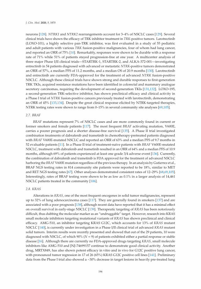
J. Clin. Med. 2020, 9, 1870
neurons [128]. NTRK1 and NTRK2 rearrangements account for 3–4% of NSCLC cases [129]. Severalclinical trials have shown the efficacy of TRK inhibitor treatment in TRK-positive tumors. Larotrectinib(LOXO-101), a highly selective pan-TRK inhibitor, was first evaluated in a study of 55 pediatricand adult patients with various TRK fusion-positive malignancies, four of whom had lung cancer,and reported an ORR of 75% [10]. Remarkably, responses were shown to be durable with a responserate of 71% while 51% of patients stayed progression-free at one year. A multicenter analysis ofthree major Phase I/II clinical trials—STARTRK-1, STARTRK-2, and ALKA-372-001—investigatingentrectinib in 54 patients diagnosed with advanced or metastatic NTRK-positive tumors demonstratedan ORR of 57%, a median PFS of 11.2 months, and a median OS of 20.9 months [130]. Larotrectiniband entrectinib are currently FDA-approved for the treatment of advanced NTRK fusion-positiveNSCLC. Although these clinical trials have shown strong and durable responses to first-generationTRK TKIs, acquired resistance mutations have been identified in colorectal and mammary analoguesecretory carcinomas, requiring the development of second-generation TKIs [131,132]. LOXO-195,a second-generation TRK-selective inhibitor, has shown preclinical efficacy and clinical activity ina Phase I trial of NTRK fusion-positive cancers previously treated with larotrectinib, demonstratingan ORR of 45% [133,134]. Despite the great clinical response elicited by NTRK-targeted therapies,NTRK testing rates were shown to range from 0–15% in several community site analyses [69,105].
2.7. BRAF
BRAF mutations represent 7% of NSCLC cases and are more commonly found in current orformer smokers and female patients [117]. The most frequent BRAF activating mutation, V600E,carries a poorer prognosis and a shorter disease-free survival [135]. A Phase II trial investigatedcombination treatments of dabrafenib and trametinib in chemotherapy-pretreated patients diagnosedwith BRAF V600E-mutated NSCLC and reported an ORR of 63% and a median PFS of 9.7 months in52 evaluable patients [11]. In a Phase II trial of treatment-naïve patients with BRAF V600E-mutatedNSCLC, treatment with dabrafenib and trametinib resulted in an ORR of 64% and a median PFS of 10.9months, although 69% of patients experienced at least one grade 3/4 adverse event [136]. Currently,the combination of dabrafenib and trametinib is FDA approved for the treatment of advanced NSCLCharboring the BRAF V600E mutation regardless of the previous therapy. In an analysis by Gutierrez et al.,BRAF NGS testing rates in 814 community site patients were reported to be 18%, similar to METand RET NGS testing rates [67]. Other analyses demonstrated consistent rates of 12–29% [68,69,105].Interestingly, rates of BRAF testing were shown to be as low as 0.1% in a larger analysis of 14,461NSCLC patients treated in the community [106].
2.8. KRAS
Alterations in KRAS, one of the most frequent oncogenes in solid tumor malignancies, representup to 32% of lung adenocarcinoma cases [117]. They are generally found in smokers [137] and areassociated with a poor prognosis [138], although recent data have reported that it has a minimal effecton overall survival in early-stage NSCLC [139]. Therapeutic targeting of KRAS has been notoriouslydifficult, thus dubbing the molecular marker as an “undruggable” target. However, research into KRASsmall molecule inhibitors targeting mutational variants of KRAS has shown preclinical and clinicalefficacy. AMG-510, an inhibitor targeting KRAS G12C, which accounts for 13% of KRAS mutantNSCLC [140], is currently under investigation in a Phase I/II clinical trial of advanced KRAS mutantsolid tumors. Interim results were recently presented and showed that out of the 29 patients, 10 werediagnosed with NSCLC, of which 90% (N = 9) of patients exhibited either a partial response or stabledisease [16]. Although there are currently no FDA-approved drugs targeting KRAS, small moleculeinhibitors like AMG-510 and JNJ-74699157 continue to demonstrate good clinical activity. Anotherdrug, MRTX849, has also shown potent efficacy in vitro and in vivo for G12C positive lung cancer,with pronounced tumor regression in 17 of 26 (65%) KRAS G12C positive cell lines [141]. Preliminarydata from the Phase I trial also showed a ~30% decrease in target lesions in heavily pre-treated lung
196

J. Clin. Med. 2020, 9, 1870
cancer patients [141]. NGS testing of KRAS, although still important now, will become necessaryonce targeted therapies become approved. In several studies of molecular testing rates in communitysites, KRAS testing has widely varied, ranging from 0–43% [66,67,69,105]. As more and more targetssuch as KRAS become clinically actionable, the landscape of lung cancer therapeutic managementwill continue to change. However, a number of actionable alterations are currently FDA approvedand have distinct therapeutic strategies currently available (Figure 1).
The testing rates reported in the community have been rising over the years, and the maindriver of this transformation has been education and dissemination of novel therapeutics available forthe different oncogenes. However, more effort is required as the primary challenge remains that manynewly approved targets face an astronomical hurdle in being implemented in daily community practice(Table 2). The most distinct example of this is the testing rates of BRAF reported in community practiceat 0.1% in 14,445 patients—the lack of testing also poses a threat towards clinical trial enrollmentand delivery of novel therapeutics to patients [106].
Table 2. Reported testing rates of clinically actionable and clinically relevant oncogenes incommunity practice.
Reported Study EGFR ALK ROS1 MET RET NTRK BRAF KRASPD-L1
Expression
Inal et al. [66] 62% 23% N/A N/A N/A N/A N/A 43% N/A
Gutierrez et al. [67] 69% 65% 25% 15% 14% N/A 18% 34% N/A
Gierman et al. [68] 54% 51% 43% N/A N/A N/A 29% N/A N/A
Presley et al. [69] 100% 95% ~15% ~15% ~15% ~15% ~15% ~15% ~15%
Illei et al. [94] N/A 53.1% N/A N/A N/A N/A N/A N/A N/A
Hussein et al. [95] ~60% ~50% N/A N/A N/A N/A N/A N/A N/A
Mason et al. [29] 94% 92% 85% N/A N/A N/A N/A N/A 56%
Audibert et al. [105] 68% 67% 32% 6% 8% 0% 12% 0% N/A
Khozin et al. [142] 64% 61% N/A N/A N/A N/A N/A N/A 8.3%
Nadler et al. 2018 [143] 37% 35% N/A N/A N/A N/A N/A N/A 1.2%
Nadler et al. 2019 [106] 35.5% 32.9% 5.7% N/A N/A N/A 0.1% N/A 5.7%
197
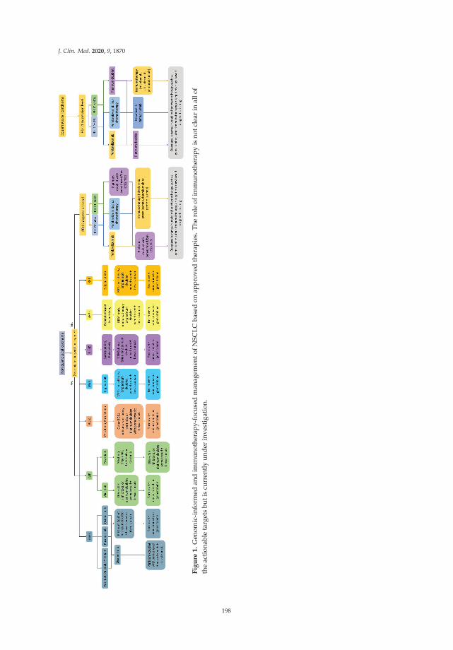
J. Clin. Med. 2020, 9, 1870
Fig
ure
1.
Gen
omic
-inf
orm
edan
dim
mun
othe
rapy
-foc
used
man
agem
ento
fNSC
LCba
sed
onap
prov
edth
erap
ies.
The
role
ofim
mun
othe
rapy
isno
tcle
arin
allo
fth
eac
tion
able
targ
ets
buti
scu
rren
tly
unde
rin
vest
igat
ion.
198

J. Clin. Med. 2020, 9, 1870
2.9. Immunotherapy
The availability and discovery of more and more targeted therapies makes it a priority thatall advanced NSCLC patients are tested at presentation. However, when an actionable alterationis not available, treatment decisions may depend on PD-L1 expression, histology, or the onset ofprogressive disease. In these situations, immune checkpoint inhibitors have induced response throughinteraction with cytotoxic T cells, helper T cells, NK cells, macrophages, and other immune mechanisms.In 2015, the first results of monoclonal antibodies against programmed death ligand-1 (PD-1) inthe refractory setting showed efficacy of nivolumab, PD-1 inhibitor, with OS (12.2 months) as comparedto second-line chemotherapy (9.5 months) [18–20]. This led to the FDA approval of nivolumab inadvanced NSCLC. Similar approval of pembrolizumab, a PD-1 inhibitor, was contingent upon resultsfrom KEYNOTE-001 that showed ORR of 19.4 in refractory NSCLC patients [21]. Soon after, two PD-L1inhibitors, atezolizumab for stage IV metastatic disease and durvalumab for stage III disease, werealso approved based on positive ORRs and OS [22,144]. However, the preliminary analysis reportedthat PD-L1 expression may be a potential biomarker of response and resistance with only 6.6% ofpatients whose tumors were negative to PD-L1 responding to durvalumab [22]. In the front-linesetting, pembrolizumab was the first immune checkpoint inhibitor (ICI) to demonstrate medianPFS of 10.3 months (vs. 6 months) and a response rate of 44.8% (vs. 27.8%) based on the resultsof KEYNOTE-024 as compared to chemotherapy [145], and it can be utilized as a monotherapy orin combination with chemotherapy depending on PD-L1 expression and the performance statusof the patient at presentation [146]. The addition of chemotherapy to pembrolizumab resulted inan increased OS at 12 months of 69.2% (vs. 49.4%) and a median PFS of 8.8 months (vs. 4.9 months), witha comparable adverse event rate of 67.2% vs. 65.8% [147]. These results were surprisingly not recreatedwhen nivolumab was evaluated as a monotherapy, showing a median PFS of 4.2 months with nivolumabvs. 5.9 months, and a similar OS benefit of 14.4 months vs. 13.2 months in the chemotherapy controlgroup [23]. However, it did have success in combination with ipilimumab, showing an improvementin overall survival of 17.1 months vs. 13.9 months with chemotherapy, and a nominal duration ofresponse of 23.3 months (vs. 6.2 months) for the front line setting [148].
Nivolumab plus ipilimumab remains a controversial choice due to grade 3 and 4 adverse eventsin 32.8% of patients [148]. Atezolizumab monotherapy achieved similar approval with incrementalimprovements in OS [24], but durvalumab in combination and alone failed to improve survival [149].While the availability of therapies is beneficial to patients, pembrolizumab is slowly becomingthe first-choice option for front-line immunotherapy, partially due to its favorable toxicity profileand versatility as a monotherapy and in combination therapy [150]. However, the availability oftherapies has not translated into practice and a retrospective observational study of 55,969 NSCLCpatients from the community showed that only 1,344 patients received nivolumab or pembrolizumabin the metastatic setting [142]. More surprisingly, only 8% of these patients were tested for PD-L1expression [142]. More so, an outcomes study of 423 patients with high PD-L1 who received first-linepembrolizumab monotherapy in the community showed that community clinical outcomes werecomparable to clinical trial results with a median PFS of 6.8 months vs. 6.1 months and a median OSof 19.1 months vs. 20 months [151]. A larger study of 10,689 patients in the community showed thatutilization of immunotherapy in the first-line is not yet implemented, with <1% of patients treated withimmunotherapy in the first-line, but rates were improved in the second and third-line setting [143].PD-L1 expression was equally underperformed and was tested in <1% of patients [143]. Furthermore,in a quality improvement study of 100 patients who received immunotherapy in the community, only61% fully completed immunotherapy as planned and 81% had immune-related adverse events [152].While it is concerning that the reported use of immunotherapy in the community practice is limited,based on experience from melanoma and immunotherapy, the rates are anticipated to slowly increaseover time with more education and acceptance of various immunotherapy options [153].
While PD-L1 remains an imperfect biomarker, several subgroup analyses in the trials mentionedabove show an increased benefit in patients with PD-L1 ≥1% or ≥50%. Therefore, PD-L1 testing should
199
J. Clin. Med. 2020, 9, 1870
be considered in everyday decision-making, and currently four PD-L1 testing types are available:22C3, 28-8, SP263, and SP142 [154]. The 22C3 IHC assays were developed alongside pembrolizumab inthe Phase I trial as a biomarker for patients who may benefit from treatment [155]. Meanwhile, IHC28-8 test was developed to be used in conjunction with nivolumab, and SP142 was developed for trialuse with atezolizumab [18,19,156,157]. SP263 is the most recent assay that was developed for use withdurvalumab, especially in the Stage III setting in NSCLC [156]. All four assays are FDA approvedin their individual setting and while testing is not required to initiate treatment, it may supportclinical decision-making [156]. Meta-analysis reports show that there is high concordance between22C3, 28-8, and SP263 assays, but SP142 detected significantly lower PD-L1 expression [154,156].At the same time, evidence shows that non-commercial laboratory-developed tests (LDTs) used byacademic centers detect similar overall percentages of PD-L1 (≥1%) at 63% (vs. 22C3 61%), but PD-L1≥50% were much lower at 23% (vs. 22C3 33%) suggesting LDTs are less sensitive than commercialtests [158]. LDTs are becoming more and more utilized in practice and offer a potential solution tothe complexity of commercial PD-L1 tests. However, the lack of PD-L1 testing and the difficultyof immune-related toxicities is a challenge that is more difficult to address, and we believe thatthe integration of community practice with the academic site model is one solution to this grave issue.
3. Integration of Personalized Therapy and Molecular Testing in the Communitythrough an Academic Site to Community Practice Network
Advances in targeted therapy and immunotherapy have lowered the costs of molecular testing,making it a viable practice in the academic sites and the community [159]. While academic sites havebenefited from a close knowledge of clinical trials and novel therapies, the drive of personalized medicinehas not been uniform, with the majority of patients in the community lacking appropriate testingand assignment to therapy [66–69,94,95,106,142,143,152]. This is especially concerning as the majority ofpatients or approximately 85% with cancer are treated in the community setting and 50% of collaborativegroup trial accruals occur in the community [160]. Several models have been proposed to integratecommunity oncologists into the academic paradigm of personalized medicine, with the most promisingbeing the establishment of interpersonal relationships between community oncologists and academicsite physicians through molecular tumor board (MTB) teams [161–165]. The establishment of an MTBteam would allow for the proper evaluation of imaging, histopathology, and genomic information thatis required to make the appropriate therapeutic decision [166]. One reported study involving 1725patients who were evaluated through a cloud-based virtual molecular tumor board (VMTB) showedthat oncologists chose the VMTB-derived therapies over others, resulting in an increase of matchedtherapies [165]. Such a model also allows for the dissemination of information regarding availableCLIA-certified vendors and platforms for both tissue and liquid biopsy testing that are imperative toimproving testing rates and outcomes [167]. The MTB model can be scaled into the community throughvirtual or physical collaboration, and would further improve collaboration between communitysites and academic sites through the interactions between pathologists, oncologists, primary carephysicians, radiologists, and pulmonologists in the decision-making process (Figure 2). This team-basedapproach can be utilized in all cancers, especially during crises such as the recent pandemic ofnovel coronavirus [168]. The improvement in the relationships with various experts and free-flowof information from the academic site to the community will invariably yield improvements inpatient outcomes.
Another available tool in building the community and academic network is the incorporation ofguidelines and pathways into everyday practice. As the majority of oncologists in the community seea number of patients with varying histologies, it is often difficult to keep track of various therapiesavailable, especially for lung cancer. While guidelines such as the National Comprehensive CancerNetwork (NCCN) and the American Society of Clinical Oncology provide guidelines regardingthe use of immunotherapy and targeted therapy, as well as genomic testing for FDA approvedalterations [169], the results in our review show that the gaps in testing rates still remain prevalent
200
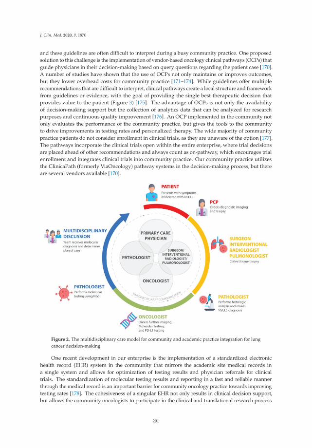
J. Clin. Med. 2020, 9, 1870
and these guidelines are often difficult to interpret during a busy community practice. One proposedsolution to this challenge is the implementation of vendor-based oncology clinical pathways (OCPs) thatguide physicians in their decision-making based on query questions regarding the patient case [170].A number of studies have shown that the use of OCPs not only maintains or improves outcomes,but they lower overhead costs for community practice [171–174]. While guidelines offer multiplerecommendations that are difficult to interpret, clinical pathways create a local structure and frameworkfrom guidelines or evidence, with the goal of providing the single best therapeutic decision thatprovides value to the patient (Figure 3) [175]. The advantage of OCPs is not only the availabilityof decision-making support but the collection of analytics data that can be analyzed for researchpurposes and continuous quality improvement [176]. An OCP implemented in the community notonly evaluates the performance of the community practice, but gives the tools to the communityto drive improvements in testing rates and personalized therapy. The wide majority of communitypractice patients do not consider enrollment in clinical trials, as they are unaware of the option [177].The pathways incorporate the clinical trials open within the entire enterprise, where trial decisionsare placed ahead of other recommendations and always count as on-pathway, which encourages trialenrollment and integrates clinical trials into community practice. Our community practice utilizesthe ClinicaPath (formerly ViaOncology) pathway systems in the decision-making process, but thereare several vendors available [170].
Figure 2. The multidisciplinary care model for community and academic practice integration for lungcancer decision-making.
One recent development in our enterprise is the implementation of a standardized electronichealth record (EHR) system in the community that mirrors the academic site medical records ina single system and allows for optimization of testing results and physician referrals for clinicaltrials. The standardization of molecular testing results and reporting in a fast and reliable mannerthrough the medical record is an important barrier for community oncology practice towards improvingtesting rates [178]. The cohesiveness of a singular EHR not only results in clinical decision support,but allows the community oncologists to participate in the clinical and translational research process
201
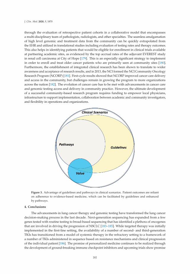
J. Clin. Med. 2020, 9, 1870
through the evaluation of retrospective patient cohorts in a collaborative model that encompassesa multi-disciplinary team of pathologists, radiologists, and other specialties. The seamless amalgamationof high level genomic and treatment data from the community can be quickly extrapolated fromthe EHR and utilized in translational studies including evaluation of testing rates and therapy outcomes.This also helps in identifying patients that would be eligible for enrollment in clinical trials availableat partnering academic sites, as evidenced by the top accrual rates of the adjuvant EVEREST studyin renal cell carcinoma at City of Hope [179]. This is an especially significant strategy to implementin order to enroll and treat older cancer patients who are primarily seen at community sites [180].Furthermore, the establishment of integrated clinical research has been shown to translate to widerawareness and acceptance of research results, and in 2013, the NCI formed the NCI Community OncologyResearch Program (NCORP) [181]. First-cycle results showed that NCORP improved cancer care deliveryand access in the community, but challenges remain in growing the program to more organizationsacross the nation [182]. The evolution of cancer care has to be met with advancements in cancer careand genomic testing access and delivery in community practice. However, the ultimate developmentof a successful community-based research program requires funding to empower local physicians,infrastructure to support implementation, collaboration between academic and community investigators,and flexibility in operations and organizations.
Figure 3. Advantage of guidelines and pathways in clinical scenarios. Patient outcomes are relianton adherence to evidence-based medicine, which can be facilitated by guidelines and enhancedby pathways.
4. Conclusions
The advancements in lung cancer therapy and genomic testing have transformed the lung cancerdecision-making process in the last decade. Next-generation sequencing has expanded from a fewgenes tested with routine testing to broad-based sequencing that has identified a plethora of oncogenesthat are involved in driving the progression of NSCLC [183–185]. While targeted therapy was initiallyimplemented in the first-line setting, the availability of a number of second- and third-generationTKIs has transitioned from a model of systemic therapy in the refractory setting to a framework ofa number of TKIs administered in sequence based on resistance mechanisms and clinical progressionof the individual patient [186]. The promise of personalized medicine continues to be realized throughthe development of ground-breaking immune checkpoint inhibitors and upcoming trials show promise
202
J. Clin. Med. 2020, 9, 1870
for chimeric antigen receptor (CAR) T-cell therapy [187]. To further realize this mission of precisionmedicine and to deliver improved outcomes, rigorous clinical data science, and translational researchof the care delivery model and access have to be expanded beyond academic sites and into communitypractice. As we have brought to attention in this review, the community practice, while currentlylagging behind academic sites in delivery oncology care, can be systematically and procedurallyintegrated with academic centers in a unified model for lung cancer decision-making and clinicalcollaboration. Our identified tools and collaborative concepts, including pathways and MTBs, can berealized in any community setting to enhance communication and trial enrollment.
Author Contributions: Conceptualization, R.S., S.R., P.K, I.M.; Writing—original draft preparation, S.R., I.M., R.P.,R.S., P.K.; Writing—review and editing, S.R., I.M., R.P., R.S., P.K., B.L. and T.T. All authors have read and agreed tothe published version of the manuscript.
Funding: The work was supported by the National Cancer Institute of Health under award numbers P30CA033572,U54CA209978, R01CA247471 and R01CA218545
Acknowledgments: We would like to express our deepest gratitude for philanthropic funding bythe Tenenblatt Family.
Conflicts of Interest: S.R: Speaker for Boehringer Ingelheim Pharmaceuticals Inc., Puma Biotechnology Inc.; I.M.,R.P., B.L., T.T., P.K. and R.S. declare no conflict of interest.
References
1. Siegel, R.L.; Miller, K.D.; Jemal, A. Cancer statistics, 2020. CA A Cancer J. Clin. 2020, 70, 7–30. [CrossRef]2. Molina, J.R.; Yang, P.; Cassivi, S.D.; Schild, S.E.; Adjei, A.A. Non-small cell lung cancer: Epidemiology, risk
factors, treatment, and survivorship. Mayo Clin. Proc. 2008, 83, 584–594. [CrossRef]3. Cancer Stat Facts: Lung and Bronchus Cancer, Statistics at a Glance. Available online: https://seer.cancer.gov/
statfacts/html/lungb.html (accessed on 20 May 2020).4. Zhou, C.; Wu, Y.L.; Chen, G.; Feng, J.; Liu, X.Q.; Wang, C.; Zhang, S.; Wang, J.; Zhou, S.; Ren, S.; et al.
Erlotinib versus chemotherapy as first-line treatment for patients with advanced EGFR mutation-positivenon-small-cell lung cancer (OPTIMAL, CTONG-0802): A multicentre, open-label, randomised, phase 3 study.Lancet Oncol. 2011, 12, 735–742. [CrossRef]
5. Shaw, A.T.; Kim, D.W.; Nakagawa, K.; Seto, T.; Crino, L.; Ahn, M.J.; De Pas, T.; Besse, B.; Solomon, B.J.;Blackhall, F.; et al. Crizotinib versus chemotherapy in advanced ALK-positive lung cancer. N. Engl. J. Med.2013, 368, 2385–2394. [CrossRef] [PubMed]
6. Mok, T.S.; Wu, Y.-L.; Ahn, M.-J.; Garassino, M.C.; Kim, H.R.; Ramalingam, S.S.; Shepherd, F.A.; He, Y.;Akamatsu, H.; Theelen, W.S.M.E.; et al. Osimertinib or Platinum–Pemetrexed in EGFR T790M–Positive LungCancer. N. Engl. J. Med. 2016, 376, 629–640. [CrossRef]
7. Shaw, A.T.; Riely, G.J.; Bang, Y.J.; Kim, D.W.; Camidge, D.R.; Solomon, B.J.; Varella-Garcia, M.; Iafrate, A.J.;Shapiro, G.I.; Usari, T.; et al. Crizotinib in ROS1-rearranged advanced non-small-cell lung cancer (NSCLC):Updated results, including overall survival, from PROFILE 1001. Ann. Oncol. 2019, 30, 1121–1126. [CrossRef]
8. Drilon, A.; Oxnard, G.; Wirth, L.; Besse, B.; Gautschi, O.; Tan, S.W.D.; Loong, H.; Bauer, T.; Kim, Y.J.;Horiike, A.; et al. PL02.08 Registrational Results of LIBRETTO-001: A Phase 1/2 Trial of LOXO-292 in Patientswith RET Fusion-Positive Lung Cancers. J. Thoracic Oncol. 2019, 14, S6–S7. [CrossRef]
9. Drilon, A.; Siena, S.; Dziadziuszko, R.; Barlesi, F.; Krebs, M.G.; Shaw, A.T.; de Braud, F.; Rolfo, C.; Ahn, M.-J.;Wolf, J.; et al. Entrectinib in ROS1 fusion-positive non-small-cell lung cancer: Integrated analysis of threephase 1–2 trials. Lancet Oncol. 2020, 21, 261–270. [CrossRef]
10. Drilon, A.; Laetsch, T.W.; Kummar, S.; DuBois, S.G.; Lassen, U.N.; Demetri, G.D.; Nathenson, M.; Doebele, R.C.;Farago, A.F.; Pappo, A.S.; et al. Efficacy of Larotrectinib in TRK Fusion–Positive Cancers in Adultsand Children. N. Engl. J. Med. 2018, 378, 731–739. [CrossRef]
11. Planchard, D.; Besse, B.; Groen, H.J.M.; Souquet, P.J.; Quoix, E.; Baik, C.S.; Barlesi, F.; Kim, T.M.; Mazieres, J.;Novello, S.; et al. Dabrafenib plus trametinib in patients with previously treated BRAF(V600E)-mutantmetastatic non-small cell lung cancer: An open-label, multicentre phase 2 trial. Lancet Oncol. 2016, 17,984–993. [CrossRef]
203
J. Clin. Med. 2020, 9, 1870
12. Wolf, J.; Seto, T.; Han, J.-Y.; Reguart, N.; Garon, E.B.; Groen, H.J.M.; Tan, D.S.-W.; Hida, T.; Jonge, M.J.D.;Orlov, S.V.; et al. Capmatinib (INC280) in METΔex14-mutated advanced non-small cell lung cancer (NSCLC):Efficacy data from the phase II GEOMETRY mono-1 study. J. Clin. Oncol. 2019, 37, 9004. [CrossRef]
13. Soria, J.-C.; Ohe, Y.; Vansteenkiste, J.; Reungwetwattana, T.; Chewaskulyong, B.; Lee, K.H.; Dechaphunkul, A.;Imamura, F.; Nogami, N.; Kurata, T.; et al. Osimertinib in Untreated EGFR-Mutated AdvancedNon–Small-Cell Lung Cancer. N. Engl. J. Med. 2017, 378, 113–125. [CrossRef] [PubMed]
14. Camidge, D.R.; Kim, H.R.; Ahn, M.-J.; Yang, J.C.-H.; Han, J.-Y.; Lee, J.-S.; Hochmair, M.J.; Li, J.Y.-C.;Chang, G.-C.; Lee, K.H.; et al. Brigatinib versus Crizotinib in ALK-Positive Non–Small-Cell Lung Cancer.N. Engl. J. Med. 2018, 379, 2027–2039. [CrossRef]
15. Kuan, F.-C.; Kuo, L.-T.; Chen, M.-C.; Yang, C.-T.; Shi, C.-S.; Teng, D.; Lee, K.-D. Overall survival benefits offirst-line EGFR tyrosine kinase inhibitors in EGFR-mutated non-small-cell lung cancers: A systematic reviewand meta-analysis. Br. J. Cancer 2015, 113, 1519–1528. [CrossRef] [PubMed]
16. Govindan, R.; Fakih, M.; Price, T.; Falchook, G.; Desai, J.; Kuo, J.; Strickler, J.; Krauss, J.; Li, B.; Denlinger, C.; et al.Phase 1 Study of AMG 510, a Novel Molecule Targeting KRAS G12C Mutant Solid Tumors. ESMO 2019Congress 2019, 30 (Suppl. 5), 159–193. [CrossRef]
17. Jänne, P. A phase 1 clinical trial evaluating the pharmacokinetics (PK), safety, and clinical activity of MRTX849,a mutant-selective small molecule KRAS G12C inhibitor, in advanced solid tumors. In Proceedings ofthe AACR-NCI-EORTC International Conference on Molecular Targets and Cancer Therapeutics, Boston,MA, USA, 26–30 October 2019.
18. Brahmer, J.; Reckamp, K.L.; Baas, P.; Crinò, L.; Eberhardt, W.E.E.; Poddubskaya, E.; Antonia, S.; Pluzanski, A.;Vokes, E.E.; Holgado, E.; et al. Nivolumab versus Docetaxel in Advanced Squamous-Cell Non–Small-CellLung Cancer. N. Engl. J. Med. 2015, 373, 123–135. [CrossRef]
19. Borghaei, H.; Paz-Ares, L.; Horn, L.; Spigel, D.R.; Steins, M.; Ready, N.E.; Chow, L.Q.; Vokes, E.E.; Felip, E.;Holgado, E.; et al. Nivolumab versus Docetaxel in Advanced Nonsquamous Non-Small-Cell Lung Cancer.N. Engl. J. Med. 2015, 373, 1627–1639. [CrossRef] [PubMed]
20. Horn, L.; Spigel, D.R.; Vokes, E.E.; Holgado, E.; Ready, N.; Steins, M.; Poddubskaya, E.; Borghaei, H.;Felip, E.; Paz-Ares, L.; et al. Nivolumab Versus Docetaxel in Previously Treated Patients With AdvancedNon-Small-Cell Lung Cancer: Two-Year Outcomes From Two Randomized, Open-Label, Phase III Trials(CheckMate 017 and CheckMate 057). J. Clin. Oncol. 2017, 35, 3924–3933. [CrossRef] [PubMed]
21. Herbst, R.S.; Baas, P.; Kim, D.W.; Felip, E.; Pérez-Gracia, J.L.; Han, J.Y.; Molina, J.; Kim, J.H.; Arvis, C.D.;Ahn, M.J.; et al. Pembrolizumab versus docetaxel for previously treated, PD-L1-positive, advancednon-small-cell lung cancer (KEYNOTE-010): A randomised controlled trial. Lancet 2016, 387, 1540–1550.[CrossRef]
22. Garassino, M.C.; Cho, B.C.; Kim, J.H.; Mazières, J.; Vansteenkiste, J.; Lena, H.; Corral Jaime, J.; Gray, J.E.;Powderly, J.; Chouaid, C.; et al. Durvalumab as third-line or later treatment for advanced non-small-cell lungcancer (ATLANTIC): An open-label, single-arm, phase 2 study. Lancet Oncol. 2018, 19, 521–536. [CrossRef]
23. Carbone, D.P.; Reck, M.; Paz-Ares, L.; Creelan, B.; Horn, L.; Steins, M.; Felip, E.; van den Heuvel, M.M.;Ciuleanu, T.-E.; Badin, F.; et al. First-Line Nivolumab in Stage IV or Recurrent Non–Small-Cell Lung Cancer.N. Engl. J. Med. 2017, 376, 2415–2426. [CrossRef] [PubMed]
24. Spigel, D.; de Marinis, F.; Giaccone, G.; Reinmuth, N.; Vergnenegre, A.; Barrios, C.H.; Morise, M.; Felip, E.;Andric, Z.G.; Geater, S.; et al. LBA78-IMpower110: Interim overall survival (OS) analysis of a phase IIIstudy of atezolizumab (atezo) vs platinum-based chemotherapy (chemo) as first-line (1L) treatment (tx) inPD-L1–selected NSCLC. Ann. Oncol. 2019, 30, v915. [CrossRef]
25. Cottrell, T.R.; Taube, J.M. PD-L1 and Emerging Biomarkers in Immune Checkpoint Blockade Therapy.Cancer J. 2018, 24, 41–46. [CrossRef] [PubMed]
26. Davis, A.A.; Patel, V.G. The role of PD-L1 expression as a predictive biomarker: An analysis of all US Foodand Drug Administration (FDA) approvals of immune checkpoint inhibitors. J. Immunother. Cancer 2019, 7,278. [CrossRef] [PubMed]
27. Lantuejoul, S.; Sound-Tsao, M.; Cooper, W.A.; Girard, N.; Hirsch, F.R.; Roden, A.C.; Lopez-Rios, F.; Jain, D.;Chou, T.-Y.; Motoi, N.; et al. PD-L1 Testing for Lung Cancer in 2019: Perspective From the IASLC PathologyCommittee. J. Thorac. Oncol. 2020, 15, 499–519. [CrossRef]
204
J. Clin. Med. 2020, 9, 1870
28. Garon, E.B.; Hellmann, M.D.; Rizvi, N.A.; Carcereny, E.; Leighl, N.B.; Ahn, M.J.; Eder, J.P.; Balmanoukian, A.S.;Aggarwal, C.; Horn, L.; et al. Five-Year Overall Survival for Patients With Advanced Non-Small-Cell LungCancer Treated With Pembrolizumab: Results From the Phase I KEYNOTE-001 Study. J. Clin. Oncol. 2019,37, 2518–2527. [CrossRef] [PubMed]
29. Mason, C.; Ellis, P.G.; Lokay, K.; Barry, A.; Dickson, N.; Page, R.; Polite, B.; Salgia, R.; Savin, M.; Shamah, C.; et al.Patterns of Biomarker Testing Rates and Appropriate Use of Targeted Therapy in the First-Line, MetastaticNon-Small Cell Lung Cancer Treatment Setting. J. Clin. Pathw. 2018, 4, 49–54. [CrossRef]
30. Dillman, R.O.; Seagren, S.L.; Propert, K.J.; Guerra, J.; Eaton, W.L.; Perry, M.C.; Carey, R.W.; Frei, E.F., 3rd;Green, M.R. A randomized trial of induction chemotherapy plus high-dose radiation versus radiation alonein stage III non-small-cell lung cancer. N. Engl. J. Med. 1990, 323, 940–945. [CrossRef] [PubMed]
31. Curran, W.J., Jr.; Paulus, R.; Langer, C.J.; Komaki, R.; Lee, J.S.; Hauser, S.; Movsas, B.; Wasserman, T.;Rosenthal, S.A.; Gore, E.; et al. Sequential vs. concurrent chemoradiation for stage III non-small cell lungcancer: Randomized phase III trial RTOG 9410. J. Natl. Cancer Inst. 2011, 103, 1452–1460. [CrossRef][PubMed]
32. Schiller, J.H.; Harrington, D.; Belani, C.P.; Langer, C.; Sandler, A.; Krook, J.; Zhu, J.; Johnson, D.H. Comparisonof four chemotherapy regimens for advanced non-small-cell lung cancer. N. Engl. J. Med. 2002, 346, 92–98.[CrossRef]
33. Evans, J.P. The Human Genome Project at 10 years: A teachable moment. Genet. Med. 2010, 12, 477.[CrossRef] [PubMed]
34. Salgia, R. Mutation testing for directing upfront targeted therapy and post-progression combination therapystrategies in lung adenocarcinoma. Expert Rev. Mol. Diagn. 2016, 16, 737–749. [CrossRef] [PubMed]
35. Ashley, E.A. Towards precision medicine. Nat. Rev. Genet. 2016, 17, 507–522. [CrossRef] [PubMed]36. Kris, M.G.; Natale, R.B.; Herbst, R.S.; Lynch, T.J., Jr.; Prager, D.; Belani, C.P.; Schiller, J.H.; Kelly, K.;
Spiridonidis, H.; Sandler, A.; et al. Efficacy of gefitinib, an inhibitor of the epidermal growth factor receptortyrosine kinase, in symptomatic patients with non-small cell lung cancer: A randomized trial. Jama 2003,290, 2149–2158. [CrossRef]
37. Fukuoka, M.; Yano, S.; Giaccone, G.; Tamura, T.; Nakagawa, K.; Douillard, J.Y.; Nishiwaki, Y.; Vansteenkiste, J.;Kudoh, S.; Rischin, D.; et al. Multi-institutional randomized phase II trial of gefitinib for previously treatedpatients with advanced non-small-cell lung cancer (The IDEAL 1 Trial) [corrected]. J. Clin. Oncol. 2003, 21,2237–2246. [CrossRef] [PubMed]
38. Lindeman, N.I.; Cagle, P.T.; Beasley, M.B.; Chitale, D.A.; Dacic, S.; Giaccone, G.; Jenkins, R.B.;Kwiatkowski, D.J.; Saldivar, J.S.; Squire, J.; et al. Molecular testing guideline for selection of lung cancerpatients for EGFR and ALK tyrosine kinase inhibitors: Guideline from the College of American Pathologists,International Association for the Study of Lung Cancer, and Association for Molecular Pathology. J. Thorac.Oncol. 2013, 8, 823–859. [CrossRef]
39. Blumenthal, G.M.; Karuri, S.W.; Zhang, H.; Zhang, L.; Khozin, S.; Kazandjian, D.; Tang, S.; Sridhara, R.;Keegan, P.; Pazdur, R. Overall response rate, progression-free survival, and overall survival with targetedand standard therapies in advanced non-small-cell lung cancer: US Food and Drug Administration trial-leveland patient-level analyses. J. Clin. Oncol. 2015, 33, 1008–1014. [CrossRef]
40. Simeone, J.C.; Nordstrom, B.L.; Patel, K.; Klein, A.B. Treatment patterns and overall survival in metastaticnon-small-cell lung cancer in a real-world, US setting. Future Oncol. 2019, 15, 3491–3502. [CrossRef]
41. Arbour, K.C.; Riely, G.J. Systemic Therapy for Locally Advanced and Metastatic Non-Small Cell Lung Cancer:A Review. Jama 2019, 322, 764–774. [CrossRef]
42. Buyse, M.E.; Squifflet, P.; Laporte, S.; Fossella, F.V.; Georgoulias, V.; Pujol, J.; Kubota, K.; Monnier, A.;Kudoh, S.; Douillard, J. Prediction of survival benefits from progression-free survival in patients withadvanced non small cell lung cancer: Evidence from a pooled analysis of 2,838 patients randomized in 7trials. J. Clin. Oncol. 2008, 26, 8019. [CrossRef]
43. Ramalingam, S.S.; Vansteenkiste, J.; Planchard, D.; Cho, B.C.; Gray, J.E.; Ohe, Y.; Zhou, C.;Reungwetwattana, T.; Cheng, Y.; Chewaskulyong, B.; et al. Overall Survival with Osimertinib in Untreated,EGFR-Mutated Advanced NSCLC. N. Engl. J. Med. 2020, 382, 41–50. [CrossRef] [PubMed]
44. Pacheco, J.M.; Gao, D.; Smith, D.; Purcell, T.; Hancock, M.; Bunn, P.; Robin, T.; Liu, A.; Karam, S.;Gaspar, L.; et al. Natural History and Factors Associated with Overall Survival in Stage IV ALK-RearrangedNon-Small Cell Lung Cancer. J. Thorac Oncol. 2019, 14, 691–700. [CrossRef] [PubMed]
205

J. Clin. Med. 2020, 9, 1870
45. Gadgeel, S.; Rodríguez-Abreu, D.; Speranza, G.; Esteban, E.; Felip, E.; Dómine, M.; Hui, R.; Hochmair, M.J.;Clingan, P.; Powell, S.F.; et al. Updated Analysis From KEYNOTE-189: Pembrolizumab or Placebo PlusPemetrexed and Platinum for Previously Untreated Metastatic Nonsquamous Non-Small-Cell Lung Cancer.J. Clin. Oncol. 2020, 38, 1505–1517. [CrossRef] [PubMed]
46. Mambetsariev, I.; Wang, Y.; Chen, C.; Nadaf, S.; Pharaon, R.; Fricke, J.; Amanam, I.; Amini, A.; Bild, A.;Chu, P.; et al. Precision medicine and actionable alterations in lung cancer: A single institution experience.PLoS ONE 2020, 15, e0228188. [CrossRef] [PubMed]
47. Dietel, M.; Savelov, N.; Salanova, R.; Micke, P.; Bigras, G.; Hida, T.; Piperdi, B.; Burke, T.; Khambata-Ford, S.;Deitz, A. 130O Real-world prevalence of PD-L1 expression in locally advanced or metastatic non-small celllung cancer (NSCLC): The global, multicentre EXPRESS study. J. Thorac. Oncol. 2018, 13, S74–S75. [CrossRef]
48. Gómez, X.E.; Soto, A.; Gómez, M.A. Survival and prognostic factors in non-small cell lung cancer patientswith mutation of the EGFR gene treated with tyrosine kinase inhibitors in a peruvian hospital. Am. J. CancerRes. 2019, 9, 1009–1016.
49. Ciardiello, F.; Tortora, G. EGFR Antagonists in Cancer Treatment. N. Engl. J. Med. 2008, 358, 1160–1174.[CrossRef]
50. Lynch, T.J.; Bell, D.W.; Sordella, R.; Gurubhagavatula, S.; Okimoto, R.A.; Brannigan, B.W.; Harris, P.L.;Haserlat, S.M.; Supko, J.G.; Haluska, F.G.; et al. Activating mutations in the epidermal growth factor receptorunderlying responsiveness of non-small-cell lung cancer to gefitinib. N. Engl. J. Med. 2004, 350, 2129–2139.[CrossRef]
51. Paez, J.G.; Jänne, P.A.; Lee, J.C.; Tracy, S.; Greulich, H.; Gabriel, S.; Herman, P.; Kaye, F.J.; Lindeman, N.;Boggon, T.J.; et al. EGFR mutations in lung cancer: Correlation with clinical response to gefitinib therapy.Science 2004, 304, 1497–1500. [CrossRef]
52. Pao, W.; Miller, V.; Zakowski, M.; Doherty, J.; Politi, K.; Sarkaria, I.; Singh, B.; Heelan, R.; Rusch, V.;Fulton, L.; et al. EGF receptor gene mutations are common in lung cancers from "never smokers" and areassociated with sensitivity of tumors to gefitinib and erlotinib. Proc. Natl. Acad. Sci. USA 2004, 101,13306–13311. [CrossRef]
53. Rosell, R.; Carcereny, E.; Gervais, R.; Vergnenegre, A.; Massuti, B.; Felip, E.; Palmero, R.; Garcia-Gomez, R.;Pallares, C.; Sanchez, J.M.; et al. Erlotinib versus standard chemotherapy as first-line treatment for Europeanpatients with advanced EGFR mutation-positive non-small-cell lung cancer (EURTAC): A multicentre,open-label, randomised phase 3 trial. Lancet Oncol. 2012, 13, 239–246. [CrossRef]
54. Wu, Y.L.; Zhou, C.; Liam, C.K.; Wu, G.; Liu, X.; Zhong, Z.; Lu, S.; Cheng, Y.; Han, B.; Chen, L.; et al. First-lineerlotinib versus gemcitabine/cisplatin in patients with advanced EGFR mutation-positive non-small-celllung cancer: Analyses from the phase III, randomized, open-label, ENSURE study. Ann. Oncol. 2015, 26,1883–1889. [CrossRef] [PubMed]
55. Wu, Y.L.; Zhou, C.; Hu, C.P.; Feng, J.; Lu, S.; Huang, Y.; Li, W.; Hou, M.; Shi, J.H.; Lee, K.Y.; et al. Afatinibversus cisplatin plus gemcitabine for first-line treatment of Asian patients with advanced non-small-celllung cancer harbouring EGFR mutations (LUX-Lung 6): An open-label, randomised phase 3 trial. LancetOncol. 2014, 15, 213–222. [CrossRef]
56. Yang, J.C.; Wu, Y.L.; Schuler, M.; Sebastian, M.; Popat, S.; Yamamoto, N.; Zhou, C.; Hu, C.P.; O’Byrne, K.;Feng, J.; et al. Afatinib versus cisplatin-based chemotherapy for EGFR mutation-positive lung adenocarcinoma(LUX-Lung 3 and LUX-Lung 6): Analysis of overall survival data from two randomised, phase 3 trials.Lancet Oncol. 2015, 16, 141–151. [CrossRef]
57. Kazandjian, D.; Blumenthal, G.M.; Yuan, W.; He, K.; Keegan, P.; Pazdur, R. FDA Approval of Gefitinib forthe Treatment of Patients with Metastatic EGFR Mutation-Positive Non-Small Cell Lung Cancer. Clin. Cancer Res.2016, 22, 1307–1312. [CrossRef]
58. Han, J.Y.; Park, K.; Kim, S.W.; Lee, D.H.; Kim, H.Y.; Kim, H.T.; Ahn, M.J.; Yun, T.; Ahn, J.S.; Suh, C.; et al.First-SIGNAL: First-line single-agent iressa versus gemcitabine and cisplatin trial in never-smokers withadenocarcinoma of the lung. J. Clin. Oncol. 2012, 30, 1122–1128. [CrossRef]
59. Sequist, L.V.; Yang, J.C.; Yamamoto, N.; O’Byrne, K.; Hirsh, V.; Mok, T.; Geater, S.L.; Orlov, S.; Tsai, C.M.;Boyer, M.; et al. Phase III study of afatinib or cisplatin plus pemetrexed in patients with metastatic lungadenocarcinoma with EGFR mutations. J. Clin. Oncol. 2013, 31, 3327–3334. [CrossRef]
206
J. Clin. Med. 2020, 9, 1870
60. Mok, T.; Ahn, M.-J.; Han, J.-Y.; Kang, J.H.; Katakami, N.; Kim, H.; Hodge, R.; Ghiorghiu, D.C.; Cantarini, M.;Wu, Y.-L.; et al. CNS response to osimertinib in patients (pts) with T790M-positive advanced NSCLC:Data from a randomized phase III trial (AURA3). J. Clin. Oncol. 2017, 35, 9005. [CrossRef]
61. Sun, M.; Behrens, C.; Feng, L.; Ozburn, N.; Tang, X.; Yin, G.; Komaki, R.; Varella-Garcia, M.; Hong, W.K.;Aldape, K.D.; et al. HER family receptor abnormalities in lung cancer brain metastases and correspondingprimary tumors. Clin. Cancer Res. 2009, 15, 4829–4837. [CrossRef]
62. Daniele, L.; Cassoni, P.; Bacillo, E.; Cappia, S.; Righi, L.; Volante, M.; Tondat, F.; Inghirami, G.; Sapino, A.;Scagliotti, G.V.; et al. Epidermal growth factor receptor gene in primary tumor and metastatic sites fromnon-small cell lung cancer. J. Thorac Oncol. 2009, 4, 684–688. [CrossRef]
63. Leonetti, A.; Sharma, S.; Minari, R.; Perego, P.; Giovannetti, E.; Tiseo, M. Resistance mechanisms to osimertinibin EGFR-mutated non-small cell lung cancer. Br. J. Cancer 2019, 121, 725–737. [CrossRef] [PubMed]
64. Yang, J.C.; Schuler, M.; Popat, S.; Miura, S.; Heeke, S.; Park, K.; Märten, A.; Kim, E.S. Afatinib for the Treatmentof NSCLC Harboring Uncommon EGFR Mutations: A Database of 693 Cases. J. Thorac Oncol. 2020, 15,803–815. [CrossRef] [PubMed]
65. Heymach, J.; Negrao, M.; Robichaux, J.; Carter, B.; Patel, A.; Altan, M.; Gibbons, D.; Fossella, F.; Simon, G.;Lam, V.; et al. OA02.06 A Phase II Trial of Poziotinib in EGFR and HER2 exon 20 Mutant Non-Small CellLung Cancer (NSCLC). J. Thoracic Oncol. 2018, 13, S323–S324. [CrossRef]
66. Inal, C.; Yilmaz, E.; Cheng, H.; Zhu, C.; Pullman, J.; Gucalp, R.A.; Keller, S.M.; Perez-Soler, R.; Piperdi, B.Effect of reflex testing by pathologists on molecular testing rates in lung cancer patients: Experience froma community-based academic center. J. Clin. Oncol. 2014, 32, 8098. [CrossRef]
67. Gutierrez, M.E.; Choi, K.; Lanman, R.B.; Licitra, E.J.; Skrzypczak, S.M.; Pe Benito, R.; Wu, T.; Arunajadai, S.;Kaur, S.; Harper, H.; et al. Genomic Profiling of Advanced Non-Small Cell Lung Cancer in CommunitySettings: Gaps and Opportunities. Clin. Lung Cancer 2017, 18, 651–659. [CrossRef]
68. Gierman, H.J.; Goldfarb, S.; Labrador, M.; Weipert, C.M.; Getty, B.; Skrzypczak, S.M.; Catasus, C.; Carbral, S.;Singaraju, M.; Singleton, N.; et al. Genomic testing and treatment landscape in patients with advancednon-small cell lung cancer (aNSCLC) using real-world data from community oncology practices. J. Clin.Oncol. 2019, 37, 1585. [CrossRef]
69. Presley, C.J.; Tang, D.; Soulos, P.R.; Chiang, A.C.; Longtine, J.A.; Adelson, K.B.; Herbst, R.S.; Zhu, W.;Nussbaum, N.C.; Sorg, R.A.; et al. Association of Broad-Based Genomic Sequencing With Survival AmongPatients With Advanced Non-Small Cell Lung Cancer in the Community Oncology Setting. Jama 2018, 320,469–477. [CrossRef]
70. Soda, M.; Choi, Y.L.; Enomoto, M.; Takada, S.; Yamashita, Y.; Ishikawa, S.; Fujiwara, S.; Watanabe, H.;Kurashina, K.; Hatanaka, H.; et al. Identification of the transforming EML4-ALK fusion gene in non-small-celllung cancer. Nature 2007, 448, 561–566. [CrossRef]
71. Shaw, A.T.; Yeap, B.Y.; Mino-Kenudson, M.; Digumarthy, S.R.; Costa, D.B.; Heist, R.S.; Solomon, B.; Stubbs, H.;Admane, S.; McDermott, U.; et al. Clinical features and outcome of patients with non-small-cell lung cancerwho harbor EML4-ALK. J. Clin. Oncol. 2009, 27, 4247–4253. [CrossRef]
72. Koivunen, J.P.; Mermel, C.; Zejnullahu, K.; Murphy, C.; Lifshits, E.; Holmes, A.J.; Choi, H.G.; Kim, J.;Chiang, D.; Thomas, R.; et al. EML4-ALK fusion gene and efficacy of an ALK kinase inhibitor in lung cancer.Clin. Cancer Res. 2008, 14, 4275–4283. [CrossRef]
73. Liao, B.C.; Lin, C.C.; Shih, J.Y.; Yang, J.C. Treating patients with ALK-positive non-small cell lung cancer:Latest evidence and management strategy. Ther. Adv. Med. Oncol. 2015, 7, 274–290. [CrossRef] [PubMed]
74. Choi, Y.L.; Soda, M.; Yamashita, Y.; Ueno, T.; Takashima, J.; Nakajima, T.; Yatabe, Y.; Takeuchi, K.; Hamada, T.;Haruta, H.; et al. EML4-ALK mutations in lung cancer that confer resistance to ALK inhibitors. N. Engl.J. Med. 2010, 363, 1734–1739. [CrossRef] [PubMed]
75. Sasaki, T.; Koivunen, J.; Ogino, A.; Yanagita, M.; Nikiforow, S.; Zheng, W.; Lathan, C.; Marcoux, J.P.; Du, J.;Okuda, K.; et al. A novel ALK secondary mutation and EGFR signaling cause resistance to ALK kinaseinhibitors. Cancer Res. 2011, 71, 6051–6060. [CrossRef]
76. Lovly, C.M.; Pao, W. Escaping ALK inhibition: Mechanisms of and strategies to overcome resistance.Sci. Transl. Med. 2012, 4, 120ps122. [CrossRef] [PubMed]
77. Katayama, R.; Shaw, A.T.; Khan, T.M.; Mino-Kenudson, M.; Solomon, B.J.; Halmos, B.; Jessop, N.A.; Wain, J.C.;Yeo, A.T.; Benes, C.; et al. Mechanisms of acquired crizotinib resistance in ALK-rearranged lung Cancers. Sci.Transl. Med. 2012, 4, 120ra117. [CrossRef]
207
J. Clin. Med. 2020, 9, 1870
78. Katayama, R.; Khan, T.M.; Benes, C.; Lifshits, E.; Ebi, H.; Rivera, V.M.; Shakespeare, W.C.; Iafrate, A.J.;Engelman, J.A.; Shaw, A.T. Therapeutic strategies to overcome crizotinib resistance in non-small cell lungcancers harboring the fusion oncogene EML4-ALK. Proc. Natl. Acad. Sci. USA 2011, 108, 7535–7540.[CrossRef]
79. Doebele, R.C.; Pilling, A.B.; Aisner, D.L.; Kutateladze, T.G.; Le, A.T.; Weickhardt, A.J.; Kondo, K.L.;Linderman, D.J.; Heasley, L.E.; Franklin, W.A.; et al. Mechanisms of resistance to crizotinib in patients withALK gene rearranged non-small cell lung cancer. Clin. Cancer Res. 2012, 18, 1472–1482. [CrossRef]
80. Shaw, A.T.; Kim, D.-W.; Mehra, R.; Tan, D.S.W.; Felip, E.; Chow, L.Q.M.; Camidge, D.R.; Vansteenkiste, J.;Sharma, S.; De Pas, T.; et al. Ceritinib in ALK-Rearranged Non–Small-Cell Lung Cancer. N. Engl. J. Med.2014, 370, 1189–1197. [CrossRef]
81. Camidge, D.R.; Dziadziuszko, R.; Peters, S.; Mok, T.; Noe, J.; Nowicka, M.; Gadgeel, S.M.; Cheema, P.;Pavlakis, N.; de Marinis, F.; et al. Updated Efficacy and Safety Data and Impact of the EML4-ALK FusionVariant on the Efficacy of Alectinib in Untreated ALK-Positive Advanced Non-Small Cell Lung Cancer inthe Global Phase III ALEX Study. J. Thorac Oncol. 2019, 14, 1233–1243. [CrossRef]
82. Hida, T.; Nokihara, H.; Kondo, M.; Kim, Y.H.; Azuma, K.; Seto, T.; Takiguchi, Y.; Nishio, M.; Yoshioka, H.;Imamura, F.; et al. Alectinib versus crizotinib in patients with ALK-positive non-small-cell lung cancer(J-ALEX): An open-label, randomised phase 3 trial. Lancet 2017, 390, 29–39. [CrossRef]
83. Peters, S.; Camidge, D.R.; Shaw, A.T.; Gadgeel, S.; Ahn, J.S.; Kim, D.-W.; Ou, S.-H.I.; Pérol, M.; Dziadziuszko, R.;Rosell, R.; et al. Alectinib versus Crizotinib in Untreated ALK-Positive Non–Small-Cell Lung Cancer. N. Engl.J. Med. 2017, 377, 829–838. [CrossRef] [PubMed]
84. Zhang, S.; Anjum, R.; Squillace, R.; Nadworny, S.; Zhou, T.; Keats, J.; Ning, Y.; Wardwell, S.D.; Miller, D.;Song, Y.; et al. The Potent ALK Inhibitor Brigatinib (AP26113) Overcomes Mechanisms of Resistance toFirst- and Second-Generation ALK Inhibitors in Preclinical Models. Clin. Cancer Res. 2016, 22, 5527–5538.[CrossRef] [PubMed]
85. Amanam, I.; Gupta, R.; Mambetsariev, I.; Salgia, R. The brigatinib experience: A new generation of therapyfor ALK-positive non-small-cell lung cancer. Future Oncol. 2018, 14, 1897–1908. [CrossRef]
86. Kim, D.W.; Tiseo, M.; Ahn, M.J.; Reckamp, K.L.; Hansen, K.H.; Kim, S.W.; Huber, R.M.; West, H.L.;Groen, H.J.M.; Hochmair, M.J.; et al. Brigatinib in Patients With Crizotinib-Refractory Anaplastic LymphomaKinase-Positive Non-Small-Cell Lung Cancer: A Randomized, Multicenter Phase II Trial. J. Clin. Oncol. 2017,35, 2490–2498. [CrossRef] [PubMed]
87. Sakamoto, H.; Tsukaguchi, T.; Hiroshima, S.; Kodama, T.; Kobayashi, T.; Fukami, T.A.; Oikawa, N.; Tsukuda, T.;Ishii, N.; Aoki, Y. CH5424802, a selective ALK inhibitor capable of blocking the resistant gatekeeper mutant.Cancer Cell. 2011, 19, 679–690. [CrossRef]
88. Rivera, V.M.; Wang, F.; Anjum, R.; Zhang, S.; Squillace, R.; Keats, J.; Miller, D.; Ning, Y.; Wardwell, S.D.;Moran, L.; et al. Abstract 1794: AP26113 is a dual ALK/EGFR inhibitor: Characterization against EGFRT790M in cell and mouse models of NSCLC. Cancer Res. 2012, 72, 1794. [CrossRef]
89. Camidge, R.; Kim, H.R.; Ahn, M.J.; Yang, J.C.H.; Han, J.Y.; Hochmair, M.J.; Lee, K.H.; Delmonte, A.; GarciaCampelo, M.R.; Kim, D.W.; et al. Brigatinib vs crizotinib in patients with ALK inhibitor-naive advanced ALK+NSCLC: Updated results from the phase III ALTA-1L trial. Ann. Oncol. 2019, 30, ix195–ix196. [CrossRef]
90. Horn, L.; Infante, J.R.; Reckamp, K.L.; Blumenschein, G.R.; Leal, T.A.; Waqar, S.N.; Gitlitz, B.J.; Sanborn, R.E.;Whisenant, J.G.; Du, L.; et al. Ensartinib (X-396) in ALK-Positive Non–Small Cell Lung Cancer: Results froma First-in-Human Phase I/II, Multicenter Study. Clin. Cancer Res. 2018, 24, 2771–2779. [CrossRef]
91. Shaw, A.T.; Solomon, B.J.; Besse, B.; Bauer, T.M.; Lin, C.C.; Soo, R.A.; Riely, G.J.; Ou, S.I.; Clancy, J.S.; Li, S.; et al.ALK Resistance Mutations and Efficacy of Lorlatinib in Advanced Anaplastic Lymphoma Kinase-PositiveNon-Small-Cell Lung Cancer. J. Clin. Oncol. 2019, 37, 1370–1379. [CrossRef]
92. Barrows, S.M.; Wright, K.; Copley-Merriman, C.; Kaye, J.A.; Chioda, M.; Wiltshire, R.; Torgersen, K.M.;Masters, E.T. Systematic review of sequencing of ALK inhibitors in ALK-positive non-small-cell lung cancer.Lung Cancer (Auckl) 2019, 10, 11–20. [CrossRef]
93. Xu, H.; Ma, D.; Yang, G.; Li, J.; Hao, X.; Xing, P.; Yang, L.; Xu, F.; Wang, Y. Sequential therapy according todistinct disease progression patterns in advanced ALK-positive non-small-cell lung cancer after crizotinibtreatment. Chin. J. Cancer Res. 2019, 31, 349–356. [CrossRef]
94. Illei, P.B.; Wong, W.; Wu, N.; Chu, L.; Gupta, R.; Schulze, K.; Gubens, M.A. ALK Testing Trends and PatternsAmong Community Practices in the United States. JCO Precis. Oncol. 2018, 2, 1–11. [CrossRef]
208
J. Clin. Med. 2020, 9, 1870
95. Hussein, M.; Richards, D.A.; Ulrich, B.; Korytowsky, B.; Pandya, D.; Cogswell, J.; Batenchuk, C.; Burns, V.ORAL01.02: Biopsies in Initial Diagnosis of Non–Small Cell Lung Cancer in US Community OncologyPractices: Implications for First-Line Immunotherapy: Topic: Medical Oncology. J. Thorac. Oncol. 2016, 11,S249–S250. [CrossRef]
96. Leighl, N.B.; Page, R.D.; Raymond, V.M.; Daniel, D.B.; Divers, S.G.; Reckamp, K.L.; Villalona-Calero, M.A.;Dix, D.; Odegaard, J.I.; Lanman, R.B.; et al. Clinical Utility of Comprehensive Cell-Free DNA Analysis toIdentify Genomic Biomarkers in Patients with Newly Diagnosed Metastatic Non-Small Cell Lung Cancer.Clin. Cancer Res. 2019. [CrossRef] [PubMed]
97. Rikova, K.; Guo, A.; Zeng, Q.; Possemato, A.; Yu, J.; Haack, H.; Nardone, J.; Lee, K.; Reeves, C.; Li, Y.; et al.Global survey of phosphotyrosine signaling identifies oncogenic kinases in lung cancer. Cell 2007, 131,1190–1203. [CrossRef] [PubMed]
98. Korpanty, G.J.; Graham, D.M.; Vincent, M.D.; Leighl, N.B. Biomarkers That Currently Affect Clinical Practicein Lung Cancer: EGFR, ALK, MET, ROS-1, and KRAS. Front. Oncol. 2014, 4, 204. [CrossRef] [PubMed]
99. Gainor, J.F.; Tseng, D.; Yoda, S.; Dagogo-Jack, I.; Friboulet, L.; Lin, J.J.; Hubbeling, H.G.; Dardaei, L.;Farago, A.F.; Schultz, K.R.; et al. Patterns of Metastatic Spread and Mechanisms of Resistance to Crizotinib inROS1-Positive Non-Small-Cell Lung Cancer. JCO Precis Oncol. 2017, 2017. [CrossRef] [PubMed]
100. Dagogo-Jack, I.; Shaw, A.T. Expanding the Roster of ROS1 Inhibitors. J. Clin. Oncol. 2017, 35, 2595–2597.[CrossRef]
101. Shaw, A.T.; Ou, S.H.; Bang, Y.J.; Camidge, D.R.; Solomon, B.J.; Salgia, R.; Riely, G.J.; Varella-Garcia, M.;Shapiro, G.I.; Costa, D.B.; et al. Crizotinib in ROS1-rearranged non-small-cell lung cancer. N. Engl. J. Med.2014, 371, 1963–1971. [CrossRef]
102. Lim, S.M.; Kim, H.R.; Lee, J.S.; Lee, K.H.; Lee, Y.G.; Min, Y.J.; Cho, E.K.; Lee, S.S.; Kim, B.S.; Choi, M.Y.; et al.Open-Label, Multicenter, Phase II Study of Ceritinib in Patients With Non-Small-Cell Lung Cancer HarboringROS1 Rearrangement. J. Clin. Oncol. 2017, 35, 2613–2618. [CrossRef]
103. Doebele, R.; Ahn, M.; Siena, S.; Drilon, A.; Krebs, M.; Lin, C.; De Braud, F.; John, T.; Tan, D.; Seto, T.; et al.OA02.01 Efficacy and Safety of Entrectinib in Locally Advanced or Metastatic ROS1 Fusion-PositiveNon-Small Cell Lung Cancer (NSCLC). J. Thorac. Oncol. 2018, 13, S321–S322. [CrossRef]
104. Ou, S.; Shaw, A.; Riely, G.; Chiari, R.; Bauman, J.; Clancy, J.; Thurm, H.; Peltz, G.; Abbattista, A.; Solomon, B.OA02.03 Clinical Activity of Lorlatinib in Patients with ROS1+ Advanced Non-Small Cell Lung Cancer:Phase 2 Study Cohort EXP-6. J. Thorac. Oncol. 2018, 13, S322–S323. [CrossRef]
105. Research, F.o.C. Trends in the Molecular Diagnosis of Lung Cancer, Results from an Online MarketResearch Survey. Available online: https://www.focr.org/sites/default/files/pdf/FINAL%202017%20Friends%20NSCLC%20White%20Paper.pdf (accessed on 20 May 2020).
106. Nadler, E.; Pavilack, M.; Clark, J.; Espirito, J.; Fernandes, A. Biomarker Testing Rates in Patients withAdvanced Non-Small Cell Lung Cancer Treated in the Community. J. Cancer Ther. 2019, 10, 971–984.[CrossRef]
107. Rijavec, E.; Coco, S.; Genova, C.; Rossi, G.; Longo, L.; Grossi, F. Liquid Biopsy in Non-Small Cell Lung Cancer:Highlights and Challenges. Cancers (Basel) 2019, 12, 17. [CrossRef] [PubMed]
108. de Melo Gagliato, D.; Jardim, D.L.F.; Falchook, G.; Tang, C.; Zinner, R.; Wheler, J.J.; Janku, F.; Subbiah, V.;Piha-Paul, S.A.; Fu, S.; et al. Analysis of MET genetic aberrations in patients with breast cancer at MDAnderson Phase I unit. Clin. Breast Cancer 2014, 14, 468–474. [CrossRef] [PubMed]
109. Cappuzzo, F.; Marchetti, A.; Skokan, M.; Rossi, E.; Gajapathy, S.; Felicioni, L.; Del Grammastro, M.;Sciarrotta, M.G.; Buttitta, F.; Incarbone, M.; et al. Increased MET gene copy number negatively affectssurvival of surgically resected non-small-cell lung cancer patients. J. Clin. Oncol. Off. J. Am. Soc. Clin. Oncol.2009, 27, 1667–1674. [CrossRef] [PubMed]
110. Carracedo, A.; Egervari, K.; Salido, M.; Rojo, F.; Corominas, J.M.; Arumi, M.; Corzo, C.; Tusquets, I.;Espinet, B.; Rovira, A.; et al. FISH and immunohistochemical status of the hepatocyte growth factor receptor(c-Met) in 184 invasive breast tumors. Breast Cancer Res. 2009, 11, 402. [CrossRef]
111. Drilon, A.; Cappuzzo, F.; Ou, S.-H.I.; Camidge, D.R. Targeting MET in Lung Cancer: Will ExpectationsFinally Be MET? J. Thorac. Oncol. 2017, 12, 15–26. [CrossRef]
112. Ghadjar, P.; Blank-Liss, W.; Simcock, M.; Hegyi, I.; Beer, K.T.; Moch, H.; Aebersold, D.M.; Zimmer, Y. METY1253D-activating point mutation and development of distant metastasis in advanced head and neck cancers.Clin. Exp. Metastasis 2009, 26, 809–815. [CrossRef]
209
J. Clin. Med. 2020, 9, 1870
113. Cipriani, N.A.; Abidoye, O.O.; Vokes, E.; Salgia, R. MET as a target for treatment of chest tumors. Lung Cancer2009, 63, 169–179. [CrossRef]
114. Schmidt, L.; Duh, F.M.; Chen, F.; Kishida, T.; Glenn, G.; Choyke, P.; Scherer, S.W.; Zhuang, Z.; Lubensky, I.;Dean, M.; et al. Germline and somatic mutations in the tyrosine kinase domain of the MET proto-oncogenein papillary renal carcinomas. Nat. Genet. 1997, 16, 68–73. [CrossRef] [PubMed]
115. Ma, P.C.; Jagadeeswaran, R.; Jagadeesh, S.; Tretiakova, M.S.; Nallasura, V.; Fox, E.A.; Hansen, M.; Schaefer, E.;Naoki, K.; Lader, A.; et al. Functional expression and mutations of c-Met and its therapeutic inhibition withSU11274 and small interfering RNA in non-small cell lung cancer. Cancer Res. 2005, 65, 1479–1488. [CrossRef]
116. Ma, P.C.; Kijima, T.; Maulik, G.; Fox, E.A.; Sattler, M.; Griffin, J.D.; Johnson, B.E.; Salgia, R. c-MET mutationalanalysis in small cell lung cancer: Novel juxtamembrane domain mutations regulating cytoskeletal functions.Cancer Res. 2003, 63, 6272–6281. [PubMed]
117. Comprehensive molecular profiling of lung adenocarcinoma. Nature 2014, 511, 543–550. [CrossRef] [PubMed]118. Comprehensive genomic characterization of squamous cell lung cancers. Nature 2012, 489, 519–525.
[CrossRef]119. Paik, P.K.; Drilon, A.; Fan, P.-D.; Yu, H.; Rekhtman, N.; Ginsberg, M.S.; Borsu, L.; Schultz, N.; Berger, M.F.;
Rudin, C.M.; et al. Response to MET Inhibitors in Patients with Stage IV Lung Adenocarcinomas HarboringMET Mutations Causing Exon 14 Skipping. Cancer Discov. 2015, 5, 842–849. [CrossRef]
120. Drilon, A.; Clark, J.W.; Weiss, J.; Ou, S.I.; Camidge, D.R.; Solomon, B.J.; Otterson, G.A.; Villaruz, L.C.;Riely, G.J.; Heist, R.S.; et al. Antitumor activity of crizotinib in lung cancers harboring a MET exon 14alteration. Nat. Med. 2020, 26, 47–51. [CrossRef]
121. Felip, E.; Horn, L.; Patel, J.D.; Sakai, H.; Scheele, J.; Bruns, R.; Paik, P.K. Tepotinib in patients with advancednon-small cell lung cancer (NSCLC) harboring MET exon 14-skipping mutations: Phase II trial. J. Clin. Oncol.2018, 36, 9016. [CrossRef]
122. Nakamura, Y.; Niki, T.; Goto, A.; Morikawa, T.; Miyazawa, K.; Nakajima, J.; Fukayama, M. c-Met activation inlung adenocarcinoma tissues: An immunohistochemical analysis. Cancer Sci 2007, 98, 1006–1013. [CrossRef]
123. Cappuzzo, F.; Janne, P.A.; Skokan, M.; Finocchiaro, G.; Rossi, E.; Ligorio, C.; Zucali, P.A.; Terracciano, L.;Toschi, L.; Roncalli, M.; et al. MET increased gene copy number and primary resistance to gefitinib therapyin non-small-cell lung cancer patients. Ann. Oncol. 2009, 20, 298–304. [CrossRef]
124. Strickler, J.H.; Weekes, C.D.; Nemunaitis, J.; Ramanathan, R.K.; Heist, R.S.; Morgensztern, D.; Angevin, E.;Bauer, T.M.; Yue, H.; Motwani, M.; et al. First-in-Human Phase I, Dose-Escalation and -Expansion Study ofTelisotuzumab Vedotin, an Antibody-Drug Conjugate Targeting c-Met, in Patients With Advanced SolidTumors. J. Clin. Oncol. 2018, 36, 3298–3306. [CrossRef] [PubMed]
125. Wang, R.; Hu, H.; Pan, Y.; Li, Y.; Ye, T.; Li, C.; Luo, X.; Wang, L.; Li, H.; Zhang, Y.; et al. RET fusions definea unique molecular and clinicopathologic subtype of non-small-cell lung cancer. J. Clin. Oncol. 2012, 30,4352–4359. [CrossRef] [PubMed]
126. Drilon, A.; Rekhtman, N.; Arcila, M.; Wang, L.; Ni, A.; Albano, M.; Van Voorthuysen, M.; Somwar, R.;Smith, R.S.; Montecalvo, J.; et al. Cabozantinib in patients with advanced RET-rearranged non-small-cell lungcancer: An open-label, single-centre, phase 2, single-arm trial. Lancet Oncol. 2016, 17, 1653–1660. [CrossRef]
127. Gainor, J.F.; Lee, D.H.; Curigliano, G.; Doebele, R.C.; Kim, D.-W.; Baik, C.S.; Tan, D.S.-W.; Lopes, G.;Gadgeel, S.M.; Cassier, P.A.; et al. Clinical activity and tolerability of BLU-667, a highly potent and selectiveRET inhibitor, in patients (pts) with advanced RET-fusion+ non-small cell lung cancer (NSCLC). J. Clin.Oncol. 2019, 37, 9008. [CrossRef]
128. Nakagawara, A. Trk receptor tyrosine kinases: A bridge between cancer and neural development. Cancer Lett. 2001,169, 107–114. [CrossRef]
129. Vaishnavi, A.; Capelletti, M.; Le, A.T.; Kako, S.; Butaney, M.; Ercan, D.; Mahale, S.; Davies, K.D.; Aisner, D.L.;Pilling, A.B.; et al. Oncogenic and drug-sensitive NTRK1 rearrangements in lung cancer. Nat. Med. 2013, 19,1469–1472. [CrossRef]
130. Doebele, R.C.; Drilon, A.; Paz-Ares, L.; Siena, S.; Shaw, A.T.; Farago, A.F.; Blakely, C.M.; Seto, T.; Cho, B.C.;Tosi, D.; et al. Entrectinib in patients with advanced or metastatic NTRK fusion-positive solid tumours:Integrated analysis of three phase 1-2 trials. Lancet Oncol. 2020, 21, 271–282. [CrossRef]
210

J. Clin. Med. 2020, 9, 1870
131. Drilon, A.; Li, G.; Dogan, S.; Gounder, M.; Shen, R.; Arcila, M.; Wang, L.; Hyman, D.M.; Hechtman, J.;Wei, G.; et al. What hides behind the MASC: Clinical response and acquired resistance to entrectinib afterETV6-NTRK3 identification in a mammary analogue secretory carcinoma (MASC). Ann. Oncol. 2016, 27,920–926. [CrossRef]
132. Russo, M.; Misale, S.; Wei, G.; Siravegna, G.; Crisafulli, G.; Lazzari, L.; Corti, G.; Rospo, G.; Novara, L.;Mussolin, B.; et al. Acquired Resistance to the TRK Inhibitor Entrectinib in Colorectal Cancer. Cancer Discov.2016, 6, 36–44. [CrossRef]
133. Drilon, A.; Nagasubramanian, R.; Blake, J.F.; Ku, N.; Tuch, B.B.; Ebata, K.; Smith, S.; Lauriault, V.;Kolakowski, G.R.; Brandhuber, B.J.; et al. A Next-Generation TRK Kinase Inhibitor Overcomes AcquiredResistance to Prior TRK Kinase Inhibition in Patients with TRK Fusion–Positive Solid Tumors. Cancer Discov.2017, 7, 963–972. [CrossRef]
134. Hyman, D.; Kummar, S.; Farago, A.; Geoerger, B.; Mau-Sorensen, M.; Taylor, M.; Garralda, E.;Nagasubramanian, R.; Natheson, M.; Song, L.; et al. Abstract CT127: Phase I and expanded accessexperience of LOXO-195 (BAY 2731954), a selective next-generation TRK inhibitor (TRKi). Cancer Res. 2019,79, CT127. [CrossRef]
135. Marchetti, A.; Felicioni, L.; Malatesta, S.; Sciarrotta, M.G.; Guetti, L.; Chella, A.; Viola, P.; Pullara, C.; Mucilli, F.;Buttitta, F. Clinical Features and Outcome of Patients With Non–Small-Cell Lung Cancer Harboring BRAFMutations. J. Clin. Oncol. 2011, 29, 3574–3579. [CrossRef] [PubMed]
136. Planchard, D.; Smit, E.F.; Groen, H.J.M.; Mazieres, J.; Besse, B.; Helland, Å.; Giannone, V.; D’Amelio, A.M., Jr.;Zhang, P.; Mookerjee, B.; et al. Dabrafenib plus trametinib in patients with previously untreatedBRAF(V600E)-mutant metastatic non-small-cell lung cancer: An open-label, phase 2 trial. Lancet Oncol. 2017,18, 1307–1316. [CrossRef]
137. Mao, C.; Qiu, L.X.; Liao, R.Y.; Du, F.B.; Ding, H.; Yang, W.C.; Li, J.; Chen, Q. KRAS mutations and resistance toEGFR-TKIs treatment in patients with non-small cell lung cancer: A meta-analysis of 22 studies. Lung Cancer2010, 69, 272–278. [CrossRef] [PubMed]
138. Johnson, M.L.; Sima, C.S.; Chaft, J.; Paik, P.K.; Pao, W.; Kris, M.G.; Ladanyi, M.; Riely, G.J. Association ofKRAS and EGFR mutations with survival in patients with advanced lung adenocarcinomas. Cancer 2013,119, 356–362. [CrossRef]
139. Shepherd, F.A.; Lacas, B.; Le Teuff, G.; Hainaut, P.; Janne, P.A.; Pignon, J.P.; Le Chevalier, T.; Seymour, L.;Douillard, J.Y.; Graziano, S.; et al. Pooled Analysis of the Prognostic and Predictive Effects of TP53 ComutationStatus Combined With KRAS or EGFR Mutation in Early-Stage Resected Non-Small-Cell Lung Cancer inFour Trials of Adjuvant Chemotherapy. J. Clin. Oncol. 2017, 35, 2018–2027. [CrossRef]
140. AACR Project GENIE: Powering Precision Medicine through an International Consortium. Cancer Discov.2017, 7, 818–831. [CrossRef]
141. Christensen, J.G.; Hallin, J.; Engstrom, L.D.; Hargis, L.; Calinisan, A.; Aranda, R.; Briere, D.M.; Sudhakar, N.;Bowcut, V.; Baer, B.R.; et al. The KRASG12C Inhibitor, MRTX849, Provides Insight Toward TherapeuticSusceptibility of KRAS Mutant Cancers in Mouse Models and Patients. Cancer Discov. 2019, 10, 54–71.[CrossRef]
142. Khozin, S.; Abernethy, A.P.; Nussbaum, N.C.; Zhi, J.; Curtis, M.D.; Tucker, M.; Lee, S.E.; Light, D.E.; Gossai, A.;Sorg, R.A.; et al. Characteristics of Real-World Metastatic Non-Small Cell Lung Cancer Patients Treatedwith Nivolumab and Pembrolizumab During the Year Following Approval. Oncologist 2018, 23, 328–336.[CrossRef]
143. Nadler, E.; Espirito, J.L.; Pavilack, M.; Boyd, M.; Vergara-Silva, A.; Fernandes, A. Treatment Patternsand Clinical Outcomes Among Metastatic Non-Small-Cell Lung Cancer Patients Treated in the CommunityPractice Setting. Clin. Lung Cancer 2018, 19, 360–370. [CrossRef] [PubMed]
144. Fehrenbacher, L.; Spira, A.; Ballinger, M.; Kowanetz, M.; Vansteenkiste, J.; Mazieres, J.; Park, K.; Smith, D.;Artal-Cortes, A.; Lewanski, C.; et al. Atezolizumab versus docetaxel for patients with previously treatednon-small-cell lung cancer (POPLAR): A multicentre, open-label, phase 2 randomised controlled trial. Lancet2016, 387, 1837–1846. [CrossRef]
145. Reck, M.; Rodríguez-Abreu, D.; Robinson, A.G.; Hui, R.; Csoszi, T.; Fülöp, A.; Gottfried, M.; Peled, N.;Tafreshi, A.; Cuffe, S.; et al. Updated Analysis of KEYNOTE-024: Pembrolizumab Versus Platinum-BasedChemotherapy for Advanced Non-Small-Cell Lung Cancer With PD-L1 Tumor Proportion Score of 50% orGreater. J. Clin. Oncol. 2019, 37, 537–546. [CrossRef] [PubMed]
211
J. Clin. Med. 2020, 9, 1870
146. Zhou, Y.; Lin, Z.; Zhang, X.; Chen, C.; Zhao, H.; Hong, S.; Zhang, L. First-line treatment for patients withadvanced non-small cell lung carcinoma and high PD-L1 expression: Pembrolizumab or pembrolizumabplus chemotherapy. J. Immunother. Cancer 2019, 7, 120. [CrossRef] [PubMed]
147. Gandhi, L.; Rodríguez-Abreu, D.; Gadgeel, S.; Esteban, E.; Felip, E.; De Angelis, F.; Domine, M.; Clingan, P.;Hochmair, M.J.; Powell, S.F.; et al. Pembrolizumab plus Chemotherapy in Metastatic Non–Small-Cell LungCancer. N. Engl. J. Med. 2018, 378, 2078–2092. [CrossRef]
148. Hellmann, M.D.; Paz-Ares, L.; Bernabe Caro, R.; Zurawski, B.; Kim, S.-W.; Carcereny Costa, E.; Park, K.;Alexandru, A.; Lupinacci, L.; de la Mora Jimenez, E.; et al. Nivolumab plus Ipilimumab in AdvancedNon–Small-Cell Lung Cancer. N. Engl. J. Med. 2019, 381, 2020–2031. [CrossRef]
149. Rizvi, N.A.; Cho, B.C.; Reinmuth, N.; Lee, K.H.; Luft, A.; Ahn, M.J.; van den Heuvel, M.M.; Cobo, M.;Vicente, D.; Smolin, A.; et al. Durvalumab With or Without Tremelimumab vs Standard Chemotherapy inFirst-line Treatment of Metastatic Non-Small Cell Lung Cancer: The MYSTIC Phase 3 Randomized ClinicalTrial. JAMA Oncol. 2020, 6, 661–674. [CrossRef]
150. Theelen, W.S.M.E.; Baas, P. Pembrolizumab monotherapy for PD-L1 ≥50% non-small cell lung cancer,undisputed first choice? Ann. Transl. Med. 2019, 7, S140. [CrossRef]
151. Velcheti, V.; Chandwani, S.; Chen, X.; Pietanza, M.C.; Piperdi, B.; Burke, T. Outcomes of first-linepembrolizumab monotherapy for PD-L1-positive (TPS ≥50%) metastatic NSCLC at US oncology practices.Immunotherapy 2019, 11, 1541–1554. [CrossRef]
152. Shivakumar, L.; Weldon, C.B.; Lucas, L.; Perloff, T. Identifying obstacles to optimal integration of cancerimmunotherapies in the community setting. J. Clin. Oncol. 2019, 37, 87. [CrossRef]
153. Krimphove, M.J.; Tully, K.H.; Friedlander, D.F.; Marchese, M.; Ravi, P.; Lipsitz, S.R.; Kilbridge, K.L.; Kibel, A.S.;Kluth, L.A.; Ott, P.A.; et al. Adoption of immunotherapy in the community for patients diagnosed withmetastatic melanoma. J. Immunother. Cancer 2019, 7, 289. [CrossRef]
154. Ancevski Hunter, K.; Socinski, M.A.; Villaruz, L.C. PD-L1 Testing in Guiding Patient Selection for PD-1/PD-L1Inhibitor Therapy in Lung Cancer. Mol. Diagn. Ther. 2018, 22, 1–10. [CrossRef] [PubMed]
155. Roach, C.; Zhang, N.; Corigliano, E.; Jansson, M.; Toland, G.; Ponto, G.; Dolled-Filhart, M.; Emancipator, K.;Stanforth, D.; Kulangara, K. Development of a Companion Diagnostic PD-L1 Immunohistochemistry Assayfor Pembrolizumab Therapy in Non-Small-cell Lung Cancer. Appl. Immunohistochem. Mol. Morphol. AIMM2016, 24, 392–397. [CrossRef] [PubMed]
156. Büttner, R.; Gosney, J.R.; Skov, B.G.; Adam, J.; Motoi, N.; Bloom, K.J.; Dietel, M.; Longshore, J.W.; López-Ríos, F.;Penault-Llorca, F.; et al. Programmed Death-Ligand 1 Immunohistochemistry Testing: A Review of AnalyticalAssays and Clinical Implementation in Non-Small-Cell Lung Cancer. J. Clin. Oncol. 2017, 35, 3867–3876.[CrossRef]
157. Rittmeyer, A.; Barlesi, F.; Waterkamp, D.; Park, K.; Ciardiello, F.; von Pawel, J.; Gadgeel, S.M.; Hida, T.;Kowalski, D.M.; Dols, M.C.; et al. Atezolizumab versus docetaxel in patients with previously treatednon-small-cell lung cancer (OAK): A phase 3, open-label, multicentre randomised controlled trial. Lancet2017, 389, 255–265. [CrossRef]
158. Velcheti, V.; Patwardhan, P.D.; Liu, F.X.; Chen, X.; Cao, X.; Burke, T. Real-world PD-L1 testing and distributionof PD-L1 tumor expression by immunohistochemistry assay type among patients with metastatic non-smallcell lung cancer in the United States. PLoS ONE 2018, 13, e0206370. [CrossRef]
159. Gong, J.; Pan, K.; Fakih, M.; Pal, S.; Salgia, R. Value-based genomics. Oncotarget Adv. Publ. 2018, 9, 15792.[CrossRef] [PubMed]
160. Ellis, L.M.; Bernstein, D.S.; Voest, E.E.; Berlin, J.D.; Sargent, D.; Cortazar, P.; Garrett-Mayer, E.; Herbst, R.S.;Lilenbaum, R.C.; Sima, C.; et al. American Society of Clinical Oncology perspective: Raising the bar forclinical trials by defining clinically meaningful outcomes. J. Clin. Oncol. 2014, 32, 1277–1280. [CrossRef]
161. Chu, L.; Kelly, K.; Gandara, D.; Lara, P.; Borowsky, A.; Meyers, F.; McPherson, J.; Erlich, R.; Almog, N.;Schrock, A.; et al. P3.13-26 Outcomes of Patients with Metastatic Lung Cancer Presented in a MultidisciplinaryMolecular Tumor Board. J. Thoracic Oncol. 2018, 13, S986. [CrossRef]
162. Koopman, B.; Wekken, A.J.v.d.; Elst, A.t.; Hiltermann, T.J.N.; Vilacha, J.F.; Groves, M.R.; Berg, A.v.d.;Hiddinga, B.I.; Hijmering-Kappelle, L.B.M.; Stigt, J.A.; et al. Relevance and Effectiveness of Molecular TumorBoard Recommendations for Patients With Non–Small-Cell Lung Cancer With Rare or Complex MutationalProfiles. JCO Precis. Oncol. 2020, 4, 393–410. [CrossRef]
212
J. Clin. Med. 2020, 9, 1870
163. Planchard, D.; Faivre, L.; Sullivan, I.; Kahn-charpy, V.; Lacroix, L.; Auger, N.; Adam, J.; De Montpreville, V.T.;Dorfmuller, P.; Pechoux, C.L.; et al. 3081 Molecular Tumor Board (MTB) in non-small cell lung cancers(NSCLC) to optimize targeted therapies: 4 years’ experience at Gustave Roussy. Eur. J. Cancer 2015, 51, S624.[CrossRef]
164. Rolfo, C.; Manca, P.; Salgado, R.; Van Dam, P.; Dendooven, A.; Machado Coelho, A.; Ferri Gandia, J.;Rutten, A.; Lybaert, W.; Vermeij, J.; et al. Multidisciplinary molecular tumour board: A tool to improveclinical practice and selection accrual for clinical trials in patients with cancer. ESMO Open 2018, 3, e000398.[CrossRef]
165. Pishvaian, M.J.; Blais, E.M.; Bender, R.J.; Rao, S.; Boca, S.M.; Chung, V.; Hendifar, A.E.; Mikhail, S.; Sohal, D.P.S.;Pohlmann, P.R.; et al. A virtual molecular tumor board to improve efficiency and scalability of deliveringprecision oncology to physicians and their patients. JAMIA Open 2019, 2, 505–515. [CrossRef]
166. Lesslie, M.; Parikh, J.R. Implementing a Multidisciplinary Tumor Board in the Community Practice Setting.Diagnostics (Basel) 2017, 7, 55. [CrossRef] [PubMed]
167. El-Deiry, W.S.; Goldberg, R.M.; Lenz, H.-J.; Shields, A.F.; Gibney, G.T.; Tan, A.R.; Brown, J.; Eisenberg, B.;Heath, E.I.; Phuphanich, S.; et al. The current state of molecular testing in the treatment of patients withsolid tumors, 2019. CA A Cancer J. Clin. 2019, 69, 305–343. [CrossRef] [PubMed]
168. Wang, T.; Liu, S.; Joseph, T.; Lyou, Y. Managing Bladder Cancer Care during the COVID-19 Pandemic Usinga Team-Based Approach. J. Clin. Med. 2020, 9, 1574. [CrossRef] [PubMed]
169. Bironzo, P.; Di Maio, M. A review of guidelines for lung cancer. J. Thorac Dis. 2018, 10, S1556–S1563.[CrossRef]
170. Daly, B.; Zon, R.T.; Page, R.D.; Edge, S.B.; Lyman, G.H.; Green, S.R.; Wollins, D.S.; Bosserman, L.D. OncologyClinical Pathways: Charting the Landscape of Pathway Providers. J. Oncol. Pract. 2018, 14, e194–e200.[CrossRef]
171. Neubauer, M.A.; Hoverman, J.R.; Kolodziej, M.; Reisman, L.; Gruschkus, S.K.; Hoang, S.; Alva, A.A.;McArthur, M.; Forsyth, M.; Rothermel, T.; et al. Cost effectiveness of evidence-based treatment guidelinesfor the treatment of non-small-cell lung cancer in the community setting. J. Oncol. Pract. 2010, 6, 12–18.[CrossRef]
172. Hoverman, J.R.; Cartwright, T.H.; Patt, D.A.; Espirito, J.L.; Clayton, M.P.; Garey, J.S.; Kopp, T.J.; Kolodziej, M.;Neubauer, M.A.; Fitch, K.; et al. Pathways, outcomes, and costs in colon cancer: Retrospective evaluations intwo distinct databases. J. Oncol. Pract. 2011, 7, 52s–59s. [CrossRef]
173. Kreys, E.D.; Koeller, J.M. Documenting the benefits and cost savings of a large multistate cancer pathwayprogram from a payer’s perspective. J. Oncol. Pract. 2013, 9, e241–e247. [CrossRef] [PubMed]
174. Jackman, D.M.; Zhang, Y.; Dalby, C.; Nguyen, T.; Nagle, J.; Lydon, C.A.; Rabin, M.S.; McNiff, K.K.; Fraile, B.;Jacobson, J.O. Cost and Survival Analysis Before and After Implementation of Dana-Farber Clinical Pathwaysfor Patients With Stage IV Non-Small-Cell Lung Cancer. J. Oncol. Pract. 2017, 13, e346–e352. [CrossRef]
175. Lawal, A.K.; Rotter, T.; Kinsman, L.; Machotta, A.; Ronellenfitsch, U.; Scott, S.D.; Goodridge, D.; Plishka, C.;Groot, G. What is a clinical pathway? Refinement of an operational definition to identify clinical pathwaystudies for a Cochrane systematic review. BMC Med. 2016, 14, 35. [CrossRef] [PubMed]
176. Zon, R.T.; Edge, S.B.; Page, R.D.; Frame, J.N.; Lyman, G.H.; Omel, J.L.; Wollins, D.S.; Green, S.R.;Bosserman, L.D. American Society of Clinical Oncology Criteria for High-Quality Clinical Pathwaysin Oncology. J. Oncol. Pract. 2017, 13, 207–210. [CrossRef] [PubMed]
177. Comis, R.L.; Miller, J.D.; Aldigé, C.R.; Krebs, L.; Stoval, E. Public attitudes toward participation in cancerclinical trials. J. Clin. Oncol. 2003, 21, 830–835. [CrossRef] [PubMed]
178. Ohno-Machado, L.; Kim, J.; Gabriel, R.A.; Kuo, G.M.; Hogarth, M.A. Genomics and electronic health recordsystems. Hum. Mol. Genet. 2018, 27, R48–R55. [CrossRef] [PubMed]
179. Salgia, N.; Philip, E.; Ziari, M.; Yap, K.; Pal, S. Advancing the Science and Management of Renal CellCarcinoma: Bridging the Divide between Academic and Community Practices. J. Clin. Med. 2020, 9, 1508.[CrossRef]
180. Liu, J.; Gutierrez, E.; Tiwari, A.; Padam, S.; Li, D.; Dale, W.; Pal, S.; Stewart, D.; Subbiah, S.; Bosserman, L.; et al.Strategies to Improve Participation of Older Adults in Cancer Research. J. Clin. Med. 2020, 9, 1571. [CrossRef]
181. Zon, R.T.; Bruinooge, S.S.; Lyss, A.P. The Changing Face of Research in Community Practice. J. Oncol. Pract.2014, 10, 155–160. [CrossRef] [PubMed]
213
J. Clin. Med. 2020, 9, 1870
182. Geiger, A.M.; O’Mara, A.M.; McCaskill-Stevens, W.J.; Adjei, B.; Tuovenin, P.; Castro, K.M. Evolution ofCancer Care Delivery Research in the NCI Community Oncology Research Program. JNCI J. Natl. Cancer Inst.2019. [CrossRef]
183. Herbst, R.S.; Morgensztern, D.; Boshoff, C. The biology and management of non-small cell lung cancer.Nature 2018, 553, 446–454. [CrossRef]
184. Aggarwal, C.; Thompson, J.C.; Black, T.A.; Katz, S.I.; Fan, R.; Yee, S.S.; Chien, A.L.; Evans, T.L.; Bauml, J.M.;Alley, E.W.; et al. Clinical Implications of Plasma-Based Genotyping With the Delivery of PersonalizedTherapy in Metastatic Non-Small Cell Lung Cancer. JAMA Oncol. 2019, 5, 173–180. [CrossRef] [PubMed]
185. Jonna, S.; Subramaniam, D.S. Molecular diagnostics and targeted therapies in non-small cell lung cancer(NSCLC): An update. Discov. Med. 2019, 27, 167–170. [PubMed]
186. Doroshow, D.B.; Herbst, R.S. Treatment of Advanced Non-Small Cell Lung Cancer in 2018. JAMA Oncol.2018, 4, 569–570. [CrossRef] [PubMed]
187. Zeltsman, M.; Dozier, J.; McGee, E.; Ngai, D.; Adusumilli, P.S. CAR T-cell therapy for lung cancerand malignant pleural mesothelioma. Transl. Res. 2017, 187, 1–10. [CrossRef] [PubMed]
© 2020 by the authors. Licensee MDPI, Basel, Switzerland. This article is an open accessarticle distributed under the terms and conditions of the Creative Commons Attribution(CC BY) license (http://creativecommons.org/licenses/by/4.0/).
214
Journal of
Clinical Medicine
Review
Integrating Academic and Community Practices in theManagement of Colorectal Cancer: The City ofHope Model
Misagh Karimi 1, Chongkai Wang 1, Bahareh Bahadini 2, George Hajjar 2 and Marwan Fakih 1,*
1 Department of Medical Oncology and Therapeutic Research, City of Hope Comprehensive Cancer Center,Duarte, CA 91010, USA; [email protected] (M.K.); [email protected] (C.W.)
2 Department of Medical Oncology and Hematology, City of Hope National Medical Center, Mission Hills,CA 91345, USA; [email protected] (B.B.); [email protected] (G.H.)
* Correspondence: [email protected]
Received: 27 March 2020; Accepted: 26 May 2020; Published: 2 June 2020
Abstract: Colorectal cancer (CRC) management continues to evolve. In metastatic CRC, severalclinical and molecular biomarkers are now recommended to guide treatment decisions. Primarytumor location (right versus left) has been shown to predict benefit from anti-epidermal growthfactor receptors (EGFRs) in rat sarcoma viral oncogene homologue (RAS) and v-raf murine sarcomaviral oncogene homolog B1 (BRAF) wild-type patients. Anti-EGFR therapy has not resulted inany benefit in RAS-mutated tumors, irrespective of the primary tumor location. BRAF-V600Emutations have been associated with poor prognosis and treatment resistance but may benefit from acombination of anti-EGFR therapy and BRAF inhibitors. Human epidermal growth factor receptor 2(HER-2) amplification was recently shown to predict relative resistance to anti-EGFR therapy but aresponse to dual HER-2 targeting within the RAS wild-type population. Finally, the mismatch repair(MMR)-deficient subgroup benefits significantly from immunotherapeutic strategies. In additionto the increasingly complex biomarker landscape in CRC, metastatic CRC remains one of the fewmalignancies that benefits from metastasectomies, ablative therapies, and regional hepatic treatments.This treatment complexity requires a multi-disciplinary approach to treatment and close collaborationsbetween various stakeholders in large cancer center networks. Here, we describe the City of Hopeexperience and strategy to enhance colorectal cancer care across its network.
Keywords: colorectal cancer; precisian medicine; academic and community oncology
1. Introduction
Colorectal cancer is the second most common cause of cancer-related death in the United States,with an annual incidence of 145,000 cases in 2019 and 51,000 deaths according to the American CancerSociety [1]. The life-time risk of developing colorectal cancer in men and women is 4.6% and 4.2%,respectively, according to 2014–2016 data [2]. The five-year survival rate for metastatic disease andregional disease are 14.2% and 71.3%, respectively [3,4]. Therefore, significant progress is still needed,especially in metastatic colorectal cancer settings.
In this report, we will review the latest approaches in the treatment of metastatic disease. We willalso explore the City of Hope approach to delivering optimized care with partnership between academicresearchers and community clinicians in cancer care.
2. Our Path to Precision Medicine in the Treatment of Metastatic CRC
The management of metastatic colorectal cancer must take into consideration sidedness, as well asmolecular biomarkers including RAS, BRAF, HER-2, and MMR status. We have previously extensively
J. Clin. Med. 2020, 9, 1687; doi:10.3390/jcm9061687 www.mdpi.com/journal/jcm215
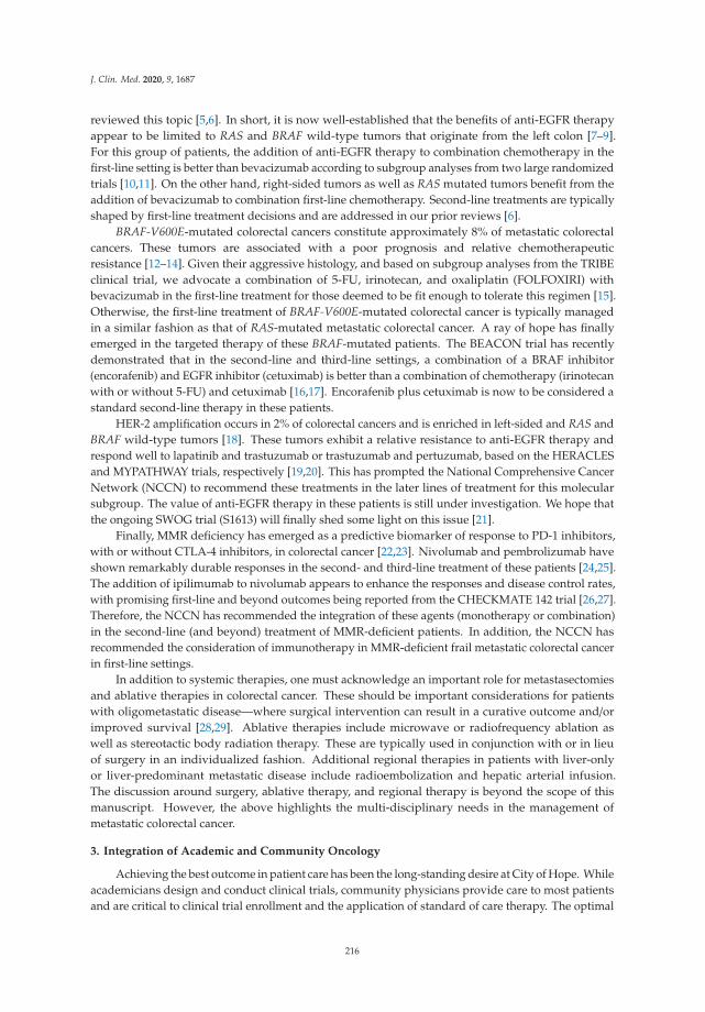
J. Clin. Med. 2020, 9, 1687
reviewed this topic [5,6]. In short, it is now well-established that the benefits of anti-EGFR therapyappear to be limited to RAS and BRAF wild-type tumors that originate from the left colon [7–9].For this group of patients, the addition of anti-EGFR therapy to combination chemotherapy in thefirst-line setting is better than bevacizumab according to subgroup analyses from two large randomizedtrials [10,11]. On the other hand, right-sided tumors as well as RAS mutated tumors benefit from theaddition of bevacizumab to combination first-line chemotherapy. Second-line treatments are typicallyshaped by first-line treatment decisions and are addressed in our prior reviews [6].
BRAF-V600E-mutated colorectal cancers constitute approximately 8% of metastatic colorectalcancers. These tumors are associated with a poor prognosis and relative chemotherapeuticresistance [12–14]. Given their aggressive histology, and based on subgroup analyses from the TRIBEclinical trial, we advocate a combination of 5-FU, irinotecan, and oxaliplatin (FOLFOXIRI) withbevacizumab in the first-line treatment for those deemed to be fit enough to tolerate this regimen [15].Otherwise, the first-line treatment of BRAF-V600E-mutated colorectal cancer is typically managedin a similar fashion as that of RAS-mutated metastatic colorectal cancer. A ray of hope has finallyemerged in the targeted therapy of these BRAF-mutated patients. The BEACON trial has recentlydemonstrated that in the second-line and third-line settings, a combination of a BRAF inhibitor(encorafenib) and EGFR inhibitor (cetuximab) is better than a combination of chemotherapy (irinotecanwith or without 5-FU) and cetuximab [16,17]. Encorafenib plus cetuximab is now to be considered astandard second-line therapy in these patients.
HER-2 amplification occurs in 2% of colorectal cancers and is enriched in left-sided and RAS andBRAF wild-type tumors [18]. These tumors exhibit a relative resistance to anti-EGFR therapy andrespond well to lapatinib and trastuzumab or trastuzumab and pertuzumab, based on the HERACLESand MYPATHWAY trials, respectively [19,20]. This has prompted the National Comprehensive CancerNetwork (NCCN) to recommend these treatments in the later lines of treatment for this molecularsubgroup. The value of anti-EGFR therapy in these patients is still under investigation. We hope thatthe ongoing SWOG trial (S1613) will finally shed some light on this issue [21].
Finally, MMR deficiency has emerged as a predictive biomarker of response to PD-1 inhibitors,with or without CTLA-4 inhibitors, in colorectal cancer [22,23]. Nivolumab and pembrolizumab haveshown remarkably durable responses in the second- and third-line treatment of these patients [24,25].The addition of ipilimumab to nivolumab appears to enhance the responses and disease control rates,with promising first-line and beyond outcomes being reported from the CHECKMATE 142 trial [26,27].Therefore, the NCCN has recommended the integration of these agents (monotherapy or combination)in the second-line (and beyond) treatment of MMR-deficient patients. In addition, the NCCN hasrecommended the consideration of immunotherapy in MMR-deficient frail metastatic colorectal cancerin first-line settings.
In addition to systemic therapies, one must acknowledge an important role for metastasectomiesand ablative therapies in colorectal cancer. These should be important considerations for patientswith oligometastatic disease—where surgical intervention can result in a curative outcome and/orimproved survival [28,29]. Ablative therapies include microwave or radiofrequency ablation aswell as stereotactic body radiation therapy. These are typically used in conjunction with or in lieuof surgery in an individualized fashion. Additional regional therapies in patients with liver-onlyor liver-predominant metastatic disease include radioembolization and hepatic arterial infusion.The discussion around surgery, ablative therapy, and regional therapy is beyond the scope of thismanuscript. However, the above highlights the multi-disciplinary needs in the management ofmetastatic colorectal cancer.
3. Integration of Academic and Community Oncology
Achieving the best outcome in patient care has been the long-standing desire at City of Hope. Whileacademicians design and conduct clinical trials, community physicians provide care to most patientsand are critical to clinical trial enrollment and the application of standard of care therapy. The optimal
216
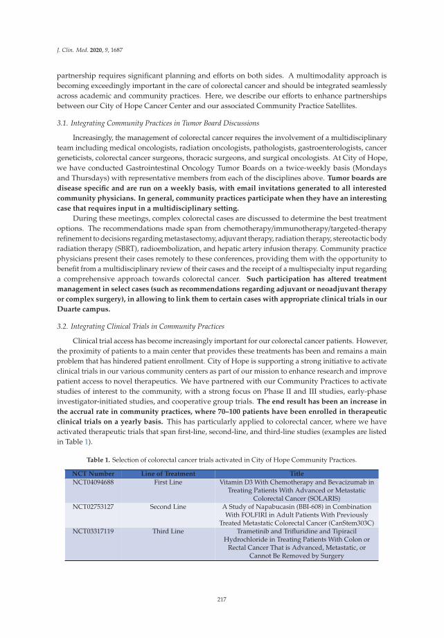
J. Clin. Med. 2020, 9, 1687
partnership requires significant planning and efforts on both sides. A multimodality approach isbecoming exceedingly important in the care of colorectal cancer and should be integrated seamlesslyacross academic and community practices. Here, we describe our efforts to enhance partnershipsbetween our City of Hope Cancer Center and our associated Community Practice Satellites.
3.1. Integrating Community Practices in Tumor Board Discussions
Increasingly, the management of colorectal cancer requires the involvement of a multidisciplinaryteam including medical oncologists, radiation oncologists, pathologists, gastroenterologists, cancergeneticists, colorectal cancer surgeons, thoracic surgeons, and surgical oncologists. At City of Hope,we have conducted Gastrointestinal Oncology Tumor Boards on a twice-weekly basis (Mondaysand Thursdays) with representative members from each of the disciplines above. Tumor boards are
disease specific and are run on a weekly basis, with email invitations generated to all interested
community physicians. In general, community practices participate when they have an interesting
case that requires input in a multidisciplinary setting.
During these meetings, complex colorectal cases are discussed to determine the best treatmentoptions. The recommendations made span from chemotherapy/immunotherapy/targeted-therapyrefinement to decisions regarding metastasectomy, adjuvant therapy, radiation therapy, stereotactic bodyradiation therapy (SBRT), radioembolization, and hepatic artery infusion therapy. Community practicephysicians present their cases remotely to these conferences, providing them with the opportunity tobenefit from a multidisciplinary review of their cases and the receipt of a multispecialty input regardinga comprehensive approach towards colorectal cancer. Such participation has altered treatment
management in select cases (such as recommendations regarding adjuvant or neoadjuvant therapy
or complex surgery), in allowing to link them to certain cases with appropriate clinical trials in our
Duarte campus.
3.2. Integrating Clinical Trials in Community Practices
Clinical trial access has become increasingly important for our colorectal cancer patients. However,the proximity of patients to a main center that provides these treatments has been and remains a mainproblem that has hindered patient enrollment. City of Hope is supporting a strong initiative to activateclinical trials in our various community centers as part of our mission to enhance research and improvepatient access to novel therapeutics. We have partnered with our Community Practices to activatestudies of interest to the community, with a strong focus on Phase II and III studies, early-phaseinvestigator-initiated studies, and cooperative group trials. The end result has been an increase in
the accrual rate in community practices, where 70–100 patients have been enrolled in therapeutic
clinical trials on a yearly basis. This has particularly applied to colorectal cancer, where we haveactivated therapeutic trials that span first-line, second-line, and third-line studies (examples are listedin Table 1).
Table 1. Selection of colorectal cancer trials activated in City of Hope Community Practices.
NCT Number Line of Treatment Title
NCT04094688 First Line Vitamin D3 With Chemotherapy and Bevacizumab inTreating Patients With Advanced or Metastatic
Colorectal Cancer (SOLARIS)NCT02753127 Second Line A Study of Napabucasin (BBI-608) in Combination
With FOLFIRI in Adult Patients With PreviouslyTreated Metastatic Colorectal Cancer (CanStem303C)
NCT03317119 Third Line Trametinib and Trifluridine and TipiracilHydrochloride in Treating Patients With Colon or
Rectal Cancer That is Advanced, Metastatic, orCannot Be Removed by Surgery
217
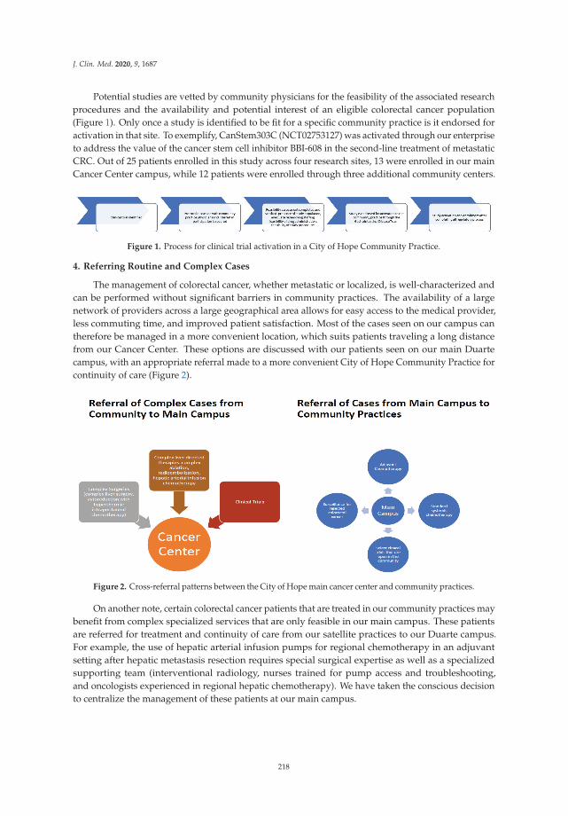
J. Clin. Med. 2020, 9, 1687
Potential studies are vetted by community physicians for the feasibility of the associated researchprocedures and the availability and potential interest of an eligible colorectal cancer population(Figure 1). Only once a study is identified to be fit for a specific community practice is it endorsed foractivation in that site. To exemplify, CanStem303C (NCT02753127) was activated through our enterpriseto address the value of the cancer stem cell inhibitor BBI-608 in the second-line treatment of metastaticCRC. Out of 25 patients enrolled in this study across four research sites, 13 were enrolled in our mainCancer Center campus, while 12 patients were enrolled through three additional community centers.
Figure 1. Process for clinical trial activation in a City of Hope Community Practice.
4. Referring Routine and Complex Cases
The management of colorectal cancer, whether metastatic or localized, is well-characterized andcan be performed without significant barriers in community practices. The availability of a largenetwork of providers across a large geographical area allows for easy access to the medical provider,less commuting time, and improved patient satisfaction. Most of the cases seen on our campus cantherefore be managed in a more convenient location, which suits patients traveling a long distancefrom our Cancer Center. These options are discussed with our patients seen on our main Duartecampus, with an appropriate referral made to a more convenient City of Hope Community Practice forcontinuity of care (Figure 2).
Figure 2. Cross-referral patterns between the City of Hope main cancer center and community practices.
On another note, certain colorectal cancer patients that are treated in our community practices maybenefit from complex specialized services that are only feasible in our main campus. These patientsare referred for treatment and continuity of care from our satellite practices to our Duarte campus.For example, the use of hepatic arterial infusion pumps for regional chemotherapy in an adjuvantsetting after hepatic metastasis resection requires special surgical expertise as well as a specializedsupporting team (interventional radiology, nurses trained for pump access and troubleshooting,and oncologists experienced in regional hepatic chemotherapy). We have taken the conscious decisionto centralize the management of these patients at our main campus.
218
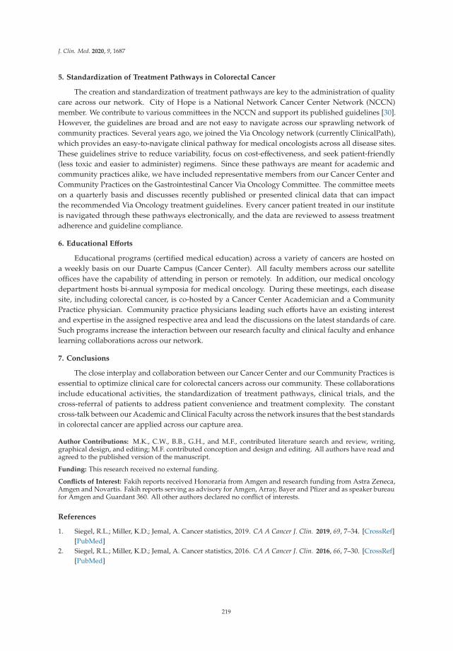
J. Clin. Med. 2020, 9, 1687
5. Standardization of Treatment Pathways in Colorectal Cancer
The creation and standardization of treatment pathways are key to the administration of qualitycare across our network. City of Hope is a National Network Cancer Center Network (NCCN)member. We contribute to various committees in the NCCN and support its published guidelines [30].However, the guidelines are broad and are not easy to navigate across our sprawling network ofcommunity practices. Several years ago, we joined the Via Oncology network (currently ClinicalPath),which provides an easy-to-navigate clinical pathway for medical oncologists across all disease sites.These guidelines strive to reduce variability, focus on cost-effectiveness, and seek patient-friendly(less toxic and easier to administer) regimens. Since these pathways are meant for academic andcommunity practices alike, we have included representative members from our Cancer Center andCommunity Practices on the Gastrointestinal Cancer Via Oncology Committee. The committee meetson a quarterly basis and discusses recently published or presented clinical data that can impactthe recommended Via Oncology treatment guidelines. Every cancer patient treated in our instituteis navigated through these pathways electronically, and the data are reviewed to assess treatmentadherence and guideline compliance.
6. Educational Efforts
Educational programs (certified medical education) across a variety of cancers are hosted ona weekly basis on our Duarte Campus (Cancer Center). All faculty members across our satelliteoffices have the capability of attending in person or remotely. In addition, our medical oncologydepartment hosts bi-annual symposia for medical oncology. During these meetings, each diseasesite, including colorectal cancer, is co-hosted by a Cancer Center Academician and a CommunityPractice physician. Community practice physicians leading such efforts have an existing interestand expertise in the assigned respective area and lead the discussions on the latest standards of care.Such programs increase the interaction between our research faculty and clinical faculty and enhancelearning collaborations across our network.
7. Conclusions
The close interplay and collaboration between our Cancer Center and our Community Practices isessential to optimize clinical care for colorectal cancers across our community. These collaborationsinclude educational activities, the standardization of treatment pathways, clinical trials, and thecross-referral of patients to address patient convenience and treatment complexity. The constantcross-talk between our Academic and Clinical Faculty across the network insures that the best standardsin colorectal cancer are applied across our capture area.
Author Contributions: M.K., C.W., B.B., G.H., and M.F., contributed literature search and review, writing,graphical design, and editing; M.F. contributed conception and design and editing. All authors have read andagreed to the published version of the manuscript.
Funding: This research received no external funding.
Conflicts of Interest: Fakih reports received Honoraria from Amgen and research funding from Astra Zeneca,Amgen and Novartis. Fakih reports serving as advisory for Amgen, Array, Bayer and Pfizer and as speaker bureaufor Amgen and Guardant 360. All other authors declared no conflict of interests.
References
1. Siegel, R.L.; Miller, K.D.; Jemal, A. Cancer statistics, 2019. CA A Cancer J. Clin. 2019, 69, 7–34. [CrossRef][PubMed]
2. Siegel, R.L.; Miller, K.D.; Jemal, A. Cancer statistics, 2016. CA A Cancer J. Clin. 2016, 66, 7–30. [CrossRef][PubMed]
219
J. Clin. Med. 2020, 9, 1687
3. Milano, A.F.; Singer, R.B. The Cancer Mortality Risk Project—Cancer Mortality Risks by Anatomic Site: Part1—Introductory Overview; Part II—Carcinoma of the Colon: 20-Year Mortality Follow-up Derived from1973-2013 (NCI) SEER*Stat Survival Database. J. Insur. Med. 2017, 47, 65–94. [CrossRef] [PubMed]
4. Siegel, R.L.; Miller, K.D.; Goding Sauer, A.; Fedewa, S.A.; Butterly, L.F.; Anderson, J.C.; Cercek, A.; Smith, R.A.;Jemal, A. Colorectal cancer statistics. CA A Cancer J. Clin. 2020. [CrossRef]
5. Fakih, M.G. Metastatic Colorectal Cancer: Current State and Future Directions. J. Clin. Oncol. 2015, 33,1809–1824. [CrossRef]
6. Sandhu, J.; Lavingia, V.; Fakih, M. Systemic treatment for metastatic colorectal cancer in the era of precisionmedicine. J. Surg. Oncol. 2019, 119, 564–582. [CrossRef]
7. Qin, S.; Li, J.; Wang, L.; Xu, J.; Cheng, Y.; Bai, Y.; Li, W.; Xu, N.; Lin, L.-Z.; Wu, Q.; et al. Efficacy andTolerability of First-Line Cetuximab Plus Leucovorin, Fluorouracil, and Oxaliplatin (FOLFOX-4) VersusFOLFOX-4 in Patients With RAS Wild-Type Metastatic Colorectal Cancer: The Open-Label, Randomized,Phase III TAILOR Trial. J. Clin. Oncol. 2018, 36, 3031–3039. [CrossRef]
8. Cutsem, E.V.; Lenz, H.-J.; Köhne, C.-H.; Heinemann, V.; Tejpar, S.; Melezínek, I.; Beier, F.; Stroh, C.; Rougier, P.;Krieken, J.H.V.; et al. Fluorouracil, Leucovorin, and Irinotecan Plus Cetuximab Treatment and RAS Mutationsin Colorectal Cancer. J. Clin. Oncol. 2015, 33, 692–700. [CrossRef]
9. Douillard, J.-Y.; Oliner, K.S.; Siena, S.; Tabernero, J.; Burkes, R.; Barugel, M.; Humblet, Y.; Bodoky, G.;Cunningham, D.; Jassem, J.; et al. Panitumumab-FOLFOX4 Treatment and RAS Mutations in ColorectalCancer. N. Engl. J. Med. 2013, 369, 1023–1034. [CrossRef]
10. Heinemann, V.; von Weikersthal, L.F.; Decker, T.; Kiani, A.; Vehling-Kaiser, U.; Al-Batran, S.E.; Heintges, T.;Lerchenmuller, C.; Kahl, C.; Seipelt, G.; et al. FOLFIRI plus cetuximab versus FOLFIRI plus bevacizumab asfirst-line treatment for patients with metastatic colorectal cancer (FIRE-3): A randomised, open-label, phase3 trial. Lancet Oncol. 2014, 15, 1065–1075. [CrossRef]
11. Venook, A.P.; Niedzwiecki, D.; Lenz, H.-J.; Innocenti, F.; Fruth, B.; Meyerhardt, J.A.; Schrag, D.; Greene, C.;O’Neil, B.H.; Atkins, J.N.; et al. Effect of First-Line Chemotherapy Combined With Cetuximab orBevacizumab on Overall Survival in Patients With KRAS Wild-Type Advanced or Metastatic ColorectalCancer: A Randomized Clinical Trial. JAMA 2017, 317, 2392–2401. [CrossRef] [PubMed]
12. Samowitz, W.S.; Sweeney, C.; Herrick, J.; Albertsen, H.; Levin, T.R.; Murtaugh, M.A.; Wolff, R.K.; Slattery, M.L.Poor Survival Associated with the BRAF V600E Mutation in Microsatellite-Stable Colon Cancers. Cancer Res.2005, 65, 6063–6069. [CrossRef] [PubMed]
13. Pai, R.K.; Jayachandran, P.; Koong, A.C.; Chang, D.T.; Kwok, S.; Ma, L.; Arber, D.A.; Balise, R.R.;Tubbs, R.R.; Shadrach, B.; et al. BRAF-mutated, Microsatellite-stable Adenocarcinoma of the Proximal Colon:An Aggressive Adenocarcinoma With Poor Survival, Mucinous Differentiation, and Adverse MorphologicFeatures. Am. J. Surg. Pathol. 2012, 36, 744–752. [CrossRef] [PubMed]
14. Gong, J.; Cho, M.; Fakih, M. RAS and BRAF in metastatic colorectal cancer management. J. Gastrointest.Oncol. 2016, 7, 687–704. [CrossRef] [PubMed]
15. Cremolini, C.; Loupakis, F.; Antoniotti, C.; Lupi, C.; Sensi, E.; Lonardi, S.; Mezi, S.; Tomasello, G.; Ronzoni, M.;Zaniboni, A.; et al. FOLFOXIRI plus bevacizumab versus FOLFIRI plus bevacizumab as first-line treatmentof patients with metastatic colorectal cancer: Updated overall survival and molecular subgroup analyses ofthe open-label, phase 3 TRIBE study. Lancet Oncol. 2015, 16, 1306–1315. [CrossRef]
16. Kopetz, S.; Grothey, A.; Yaeger, R.; Van Cutsem, E.; Desai, J.; Yoshino, T.; Wasan, H.; Ciardiello, F.; Loupakis, F.;Hong, Y.S.; et al. Encorafenib, Binimetinib, and Cetuximab in BRAF V600E-Mutated Colorectal Cancer.N. Engl. J. Med. 2019, 381, 1632–1643. [CrossRef]
17. Cutsem, E.V.; Cuyle, P.-J.; Huijberts, S.; Yaeger, R.; Schellens, J.H.M.; Elez, E.; Tabernero, J.; Fakih, M.;Montagut, C.; Peeters, M.; et al. BEACON CRC study safety lead-in (SLI) in patients with BRAFV600Emetastatic colorectal cancer (mCRC): Efficacy and tumor markers. J. Clin. Oncol. 2018, 36, 627. [CrossRef]
18. Richman, S.D.; Southward, K.; Chambers, P.; Cross, D.; Barrett, J.; Hemmings, G.; Taylor, M.; Wood, H.;Hutchins, G.; Foster, J.M.; et al. HER2 overexpression and amplification as a potential therapeutic targetin colorectal cancer: Analysis of 3256 patients enrolled in the QUASAR, FOCUS and PICCOLO colorectalcancer trials. J. Pathol. 2016, 238, 562–570. [CrossRef]
19. Sartore-Bianchi, A.; Trusolino, L.; Martino, C. Dual-targeted therapy with trastuzumab and lapatinib intreatment-refractory, KRAS codon 12/13 wild-type, HER2-positive metastatic colorectal cancer (HERACLES):A proof-of-concept, multicentre, open-label, phase 2 trial. Lancet Oncol. 2016, 17, 738–746. [CrossRef]
220
J. Clin. Med. 2020, 9, 1687
20. Meric-Bemstam, F.; Hurwitz, H.; Raghav, K.P.S.; McWilliams, R.R.; Fakih, M.; VanderWalde, A.; Swanton, C.;Kurzrock, R.; Burris, H.; Sweeney, C.; et al. Pertuzumab plus trastuzumab for HER2-amplified metastaticcolorectal cancer (MyPathway): An updated report from a multicentre, open-label, phase 2a, multiple basketstudy. Lancet Oncol. 2019, 20, 518–530. [CrossRef]
21. Raghav, K.P.S.; McDonough, S.L.; Tan, B.R.; Denlinger, C.S.; Magliocco, A.M.; Choong, N.W.; Sommer, N.;Scappaticci, F.A.; Campos, D.; Guthrie, K.A.; et al. A randomized phase II study of trastuzumab andpertuzumab (TP) compared to cetuximab and irinotecan (CETIRI) in advanced/metastatic colorectal cancer(mCRC) with HER2 amplification: S1613. J. Clin. Oncol. 2018, 36, TPS3620. [CrossRef]
22. Le, D.T.; Uram, J.N.; Wang, H.; Bartlett, B.R.; Kemberling, H.; Eyring, A.D.; Skora, A.D.; Luber, B.S.; Azad, N.S.;Laheru, D.; et al. PD-1 Blockade in Tumors with Mismatch-Repair Deficiency. N. Engl. J. Med. 2015, 372,2509–2520. [CrossRef] [PubMed]
23. Le, D.T.; Durham, J.N.; Smith, K.N.; Wang, H.; Bartlett, B.R.; Aulakh, L.K.; Lu, S.; Kemberling, H.; Wilt, C.;Luber, B.S.; et al. Mismatch repair deficiency predicts response of solid tumors to PD-1 blockade. Science2017, 357, 409–413. [CrossRef] [PubMed]
24. Overman, M.J.; McDermott, R.; Leach, J.L. Nivolumab in patients with metastatic DNA mismatchrepair-deficient or microsatellite instability-high colorectal cancer (CheckMate 142): An open-label,multicentre, phase 2 study. Lancet Oncol. 2017, 18, 1182–1191. [CrossRef]
25. Marabelle, A.; Le, D.T.; Ascierto, P.A.; Giacomo, A.M.D.; Jesus-Acosta, A.D.; Delord, J.-P.; Geva, R.;Gottfried, M.; Penel, N.; Hansen, A.R.; et al. Efficacy of Pembrolizumab in Patients With Noncolorectal HighMicrosatellite Instability/Mismatch Repair–Deficient Cancer: Results From the Phase II KEYNOTE-158 Study.J. Clin. Oncol. 2020, 38, 1–10. [CrossRef] [PubMed]
26. Overman, M.J.; Lonardi, S.; Wong, K.Y.M.; Lenz, H.-J.; Gelsomino, F.; Aglietta, M.; Morse, M.A.; Cutsem, E.V.;McDermott, R.; Hill, A.; et al. Durable Clinical Benefit With Nivolumab Plus Ipilimumab in DNA MismatchRepair-Deficient/Microsatellite Instability-High Metastatic Colorectal Cancer. J. Clin. Oncol. 2018, 36, 773–779.[CrossRef] [PubMed]
27. Andre, T.; Lonardi, S.; Wong, M.; Lenz, H.J.; Gelsomino, F.; Aglietta, M.; Morse, M.; Van Cutsem, E.;McDermott, R.S.; Hill, A.G.; et al. Nivolumab plus ipilimumab combination in patients with DNA mismatchrepair-deficient/microsatellite instability-high (dMMR/MSI-H) metastatic colorectal cancer (mCRC): Firstreport of the full cohort from CheckMate-142. J. Clin. Oncol. 2018, 36, 553. [CrossRef]
28. Minagawa, M.; Makuuchi, M.; Torzilli, G.; Takayama, T.; Kawasaki, S.; Kosuge, T.; Yamamoto, J.; Imamura, H.Extension of the frontiers of surgical indications in the treatment of liver metastases from colorectal cancer:Long-term results. Ann. Surg. 2000, 231, 487–499. [CrossRef]
29. Elias, D.; Liberale, G.; Vernerey, D.; Pignon, J.P.; Lasser, P.; Pocard, M.; Ducreux, M.; Voige, V.; Malka, D.Hepatic and extrahepatic colorectal metastases: When resectable, their localization does not matter, but theirtotal number has a prognostic impact. Ann. Surg. Oncol. 2005, 12, 900–909. [CrossRef]
30. National Comprehensive Cancer Network. Available online: https://www.nccn.org/professionals/physician_gls/default.aspx#site (accessed on 16 May 2020).
© 2020 by the authors. Licensee MDPI, Basel, Switzerland. This article is an open accessarticle distributed under the terms and conditions of the Creative Commons Attribution(CC BY) license (http://creativecommons.org/licenses/by/4.0/).
221

Journal of
Clinical Medicine
Review
Managing Bladder Cancer Care during the COVID-19Pandemic Using a Team-Based Approach
Tina Wang, Sariah Liu, Thomas Joseph and Yung Lyou *
Department of Medical Oncology & Experimental Therapeutics, City of Hope Comprehensive Cancer Center,Duarte, CA 91010, USA; [email protected] (T.W.); [email protected] (S.L.); [email protected] (T.J.)* Correspondence: [email protected]; Tel.: +1-626-218-9200; Fax: +1-626-218-8233
Received: 14 April 2020; Accepted: 19 May 2020; Published: 22 May 2020
Abstract: The recent novel coronavirus, named coronavirus disease 2019 (COVID-19), has developedinto an international pandemic affecting millions of individuals with hundreds of thousands of deathsworldwide. The highly infectious nature and widespread prevalence of this disease create a new set ofobstacles for the bladder cancer community in both delivering and receiving care. In this manuscript,we address the unique issues regarding treatment prioritization for the patient with bladder cancer andhow we at City of Hope have adjusted our clinical practices using a team-based approach that utilizesshared decision making with all stakeholders (physicians, patients, caregivers) to optimize outcomesduring this difficult time. In addition to taking standard precautions for minimizing COVID-19 riskof exposure for those entering a healthcare facility (screening all personnel upon entry and donningfacemasks at all times), we suggest the following three measures: (1) delay post-treatment surveillancevisits until there is a decrease in local COVID-19 cases, (2) continue curative intent treatments forlocalized bladder cancer with COVID-19 precautions (i.e., choosing gemcitabine/cisplatin (GC) overdose-dense methotrexate, vinblastine, doxorubicin, cisplatin (ddMVAC) neoadjuvant chemotherapy),and (3) increase the off-treatment period between cycles of palliative systemic therapy in metastaticurothelial carcinoma patients.
Keywords: bladder cancer; urothelial carcinoma; COVID-19; team-based medicine
1. Introduction
Recently, a novel coronavirus, named severe acute respiratory syndrome coronavirus 2 (SARS-CoV-2),has developed into an international pandemic affecting millions of individuals in more than 150 countrieswith hundreds of thousands of deaths worldwide [1,2]. This disease has been named coronavirus disease2019 (COVID-19) by the World Health Organization (WHO) [1]. Patients with this disease are at highrisk for developing septic shock and hypoxemia, which can frequently progress to acute respiratorydistress syndrome (ARDS) and death [3]. This disease creates a new set of obstacles for the bladdercancer community in both delivering and receiving care. In this manuscript, we address the uniqueissues regarding treatment prioritization for the patient with bladder cancer and how we at City of Hopehave adjusted our clinical practices using a team-based, shared decision approach with all stakeholders(patients, caregivers, and physicians) to optimize outcomes during this difficult time.
2. Balancing the Need for Bladder Cancer Treatments and Risk of Exposure to COVID-19
2.1. Patients with Bladder Cancer Undergoing Treatments Are at a Higher Risk for COVID-19 Infections andWorse Outcomes Compared to the General Population without Cancer
For the patient with bladder cancer undergoing treatment, there are several safety issues thatplace them at higher risk of infection for COVID-19 compared to the general population without cancer.First, patients must physically leave the safety of their residences to go to the clinic, infusion center,
J. Clin. Med. 2020, 9, 1574; doi:10.3390/jcm9051574 www.mdpi.com/journal/jcm223
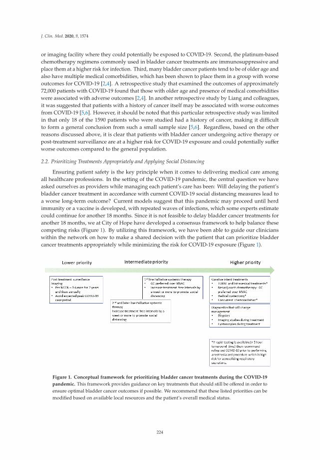
J. Clin. Med. 2020, 9, 1574
or imaging facility where they could potentially be exposed to COVID-19. Second, the platinum-basedchemotherapy regimens commonly used in bladder cancer treatments are immunosuppressive andplace them at a higher risk for infection. Third, many bladder cancer patients tend to be of older age andalso have multiple medical comorbidities, which has been shown to place them in a group with worseoutcomes for COVID-19 [2,4]. A retrospective study that examined the outcomes of approximately72,000 patients with COVID-19 found that those with older age and presence of medical comorbiditieswere associated with adverse outcomes [2,4]. In another retrospective study by Liang and colleagues,it was suggested that patients with a history of cancer itself may be associated with worse outcomesfrom COVID-19 [5,6]. However, it should be noted that this particular retrospective study was limitedin that only 18 of the 1590 patients who were studied had a history of cancer, making it difficultto form a general conclusion from such a small sample size [5,6]. Regardless, based on the otherreasons discussed above, it is clear that patients with bladder cancer undergoing active therapy orpost-treatment surveillance are at a higher risk for COVID-19 exposure and could potentially sufferworse outcomes compared to the general population.
2.2. Prioritizing Treatments Appropriately and Applying Social Distancing
Ensuring patient safety is the key principle when it comes to delivering medical care amongall healthcare professions. In the setting of the COVID-19 pandemic, the central question we haveasked ourselves as providers while managing each patient’s care has been: Will delaying the patient’sbladder cancer treatment in accordance with current COVID-19 social distancing measures lead toa worse long-term outcome? Current models suggest that this pandemic may proceed until herdimmunity or a vaccine is developed, with repeated waves of infections, which some experts estimatecould continue for another 18 months. Since it is not feasible to delay bladder cancer treatments foranother 18 months, we at City of Hope have developed a consensus framework to help balance thesecompeting risks (Figure 1). By utilizing this framework, we have been able to guide our clinicianswithin the network on how to make a shared decision with the patient that can prioritize bladdercancer treatments appropriately while minimizing the risk for COVID-19 exposure (Figure 1).
Figure 1. Conceptual framework for prioritizing bladder cancer treatments during the COVID-19
pandemic. This framework provides guidance on key treatments that should still be offered in order toensure optimal bladder cancer outcomes if possible. We recommend that these listed priorities can bemodified based on available local resources and the patient’s overall medical status.
224
J. Clin. Med. 2020, 9, 1574
2.3. Applying COVID-19 Risk Mitigation Measures for Bladder Cancer Treatment
In the state of California, there is a “shelter in place” order that was initiated on 19 March 2020along with other social distancing measures due to the concern that individuals may be at highrisk of becoming infected and could also infect others, further propagating this pandemic. Currentepidemiology modeling suggests that the peak incidence of COVID-19 will have occurred sometime inmid-to-late April in the state of California. This framework assumes that the number of new cases willstart to decrease in the months of May and June 2020. In the case that there is indeed a second wave ofinfections later during the fall and winter months of 2020, one could reapply this framework based onthe expected peaks. As a result, we suggest the following framework to assist the practicing oncologistin determining optimal treatment strategies for the patient with bladder cancer.
2.3.1. Delay Post-Treatment Surveillance Visits until There Is a Decrease in COVID-19 Cases
For patients undergoing surveillance imaging after completion of cystectomy or other definitivetherapies, the National Comprehensive Cancer Network (NCCN) guidelines currently recommendimaging every 6–12 months [7]. Keeping these guidelines in mind, we have rescheduled the patient’sclinic and imaging visit to avoid the expected COVID-19 peak period (April–May) so that it will takeplace during the next 2–3 months in June or July as a way to minimize risk of exposure.
2.3.2. Continue Curative Intent Treatments for Localized Bladder Cancer with COVID-19 Precautions
Even in these difficult times, urothelial bladder cancer is an aggressive disease with poor prognosiswhen it progresses to metastatic disease. Therefore, we have been vigilant in continuing to delivercurative intent treatments when patients have localized urothelial carcinoma, if possible in a timelymanner. A meta-analysis of 13 studies suggested that a delay of more than 12 weeks from time ofdiagnosis to execution of radical cystectomy, only in muscle invasive urothelial cancer, was associatedwith worse outcomes [8]. Another study showed that initiating neoadjuvant chemotherapy (NAC)with a delay of more than 8 weeks from time of diagnosis led to worse outcomes [9]. Therefore, we havecontinued to offer cisplatin-eligible patients NAC within 8 weeks and cisplatin-ineligible patientsradical cystectomy within 12 weeks from time of diagnosis, while the infusion center and operatingroom resources are available for those patients with localized disease since there is a limited windowof curative treatment opportunity.
The first set of measures we have instituted to minimize potential risk for COVID-19 exposure forall on-site people (visitors and healthcare workers) is to create a single, separate point of entry to theactive clinical areas and institute a strict policy limiting visitors to patients only. Prior to entering theclinical area, all personnel (including patients and healthcare workers) are screened for COVID-19symptoms (i.e., cough, dyspnea, and fever) and have their temperatures measured. People determinedto be asymptomatic and afebrile are then required to don a face mask and are issued an entrance bandindicating they have passed screening measures for that day. If someone is found to be symptomatic,we then refer this individual to an on-site “fever clinic” staffed by designated clinical personnel whohave been trained and equipped with the appropriate personal protective equipment (PPE) to performa nasopharyngeal swab for in-house COVID-19 testing. We have also repurposed one of our hospitalwards with negative pressure rooms to serve as the COVID-19 unit with its own set of designated staffto decrease exposure within the facility. In both the inpatient and outpatient areas, all people (patientsand healthcare workers) are required to don a face mask at all times, which has been suggested as away to prevent sustained exposure to COVID-19 and reduce risk for infection [10].
The second set of COVID-19 risk mitigation measures specifically pertain to treatments usedfor urothelial bladder cancer. Current NCCN guidelines recommend neoadjuvant chemotherapy inmuscle invasive bladder cancer [7]. In the choice of regimen, the two most commonly used regimensare dose-dense methotrexate, vinblastine, doxorubicin, cisplatin (ddMVAC) and gemcitabine/cisplatin(GC) [7,11,12]. During this time, we have advocated for using gemcitabine/cisplatin over ddMVAC
225

J. Clin. Med. 2020, 9, 1574
for the following reasons. Although there is some discussion suggesting that ddMVAC may have atrend towards higher efficacy, it has yet to be definitively supported in a head-to-head prospective trialand retrospective studies have shown similar amounts of efficacy between these two regimens [11,12].In addition, ddMVAC tends to be more myelosuppressive than GC, placing patients at higher risk forinfections due to the neutropenia and symptomatic anemia requiring blood transfusions, which duringthis time have been especially challenging due to a steep drop in blood donations [11,12]. Finally,ddMVAC is given as a 14-day cycle whereas GC is given as a 21-day cycle. The 14-day cycle ofddMVAC allows a patient to proceed sooner to radical cystectomy compared to GC, but duringthis time we would recommend GC because it allows the oncologist to space out the patient visitsand can help adhere better to the principle of social distancing [11,12]. Another measure we havetaken is to implement weekly telephone checks with patients undergoing active systemic therapy.This allows us to determine if a patient is having any significant chemotherapy-related adverse effectsor other acute medical issues, for which they could potentially be treated as an outpatient before theyprogress to needing emergency room or acute inpatient care. For example, if a patient is experiencingsignificant dysuria due to a potential urinary tract infection, one can prescribe antibiotics empiricallyat their local pharmacy and help them avoid the need to seek emergency room care, which is mostlikely to be overcrowded during this pandemic. For those patients that are undergoing concurrentradiation and chemotherapy with curative intent, we have continued their treatments while takingthe abovementioned general COVID-19 precautions (i.e., screening at entry, donning facemasks,and weekly telephone checks).
2.3.3. Increasing Off-Treatment Period between Cycles for Palliative Systemic Therapy in MetastaticUrothelial Carcinoma Patients
For those patients already undergoing palliative first-line systemic therapy, we have continuedtheir treatments as those regimens provide overall survival benefit. In this situation, if chemotherapyneeds to be started we would recommend, as discussed above, to prescribe anti-emetics and painmedications for the patient to have immediately available at home as an outpatient. Additionally,weekly telephone checks would be conducted to prevent any chemotherapy-related complications early.Another important factor to consider, as discussed above, is lengthening the period of time betweentreatments. Normally, gemcitabine/cisplatin or gemcitabine/carboplatin is administered as a two weekson, 1 week off schedule. In this case, it is reasonable to do 2 weeks on, 2 weeks off to help spreadout the treatment duration as much as possible to maximize social distancing. Second-line treatmentusually involves the use of immune checkpoint inhibitors such as pembrolizumab or atezolizumab.Pembrolizumab is dosed every 3 weeks, but in order to maximize social distancing for the patient, it isreasonable to stretch it to every 4 weeks during this period since it is unlikely the cancer will growsignificantly during the extra week off. In this setting, atezolizumab and nivolumab already has anFederal Drug Administration (FDA)-approved every-4-week dosing, which would also make it a viablealternative. The use of third-line treatment with enfortumab vedotin requires administration onceevery week for 3 weeks straight and then taking the fourth week off. Again, to provide more spacebetween visits, it would be reasonable to increase the off-treatment period from 1 week to 2 weeks toprovide the patient more social distancing.
Even during these difficult times, it is crucial to continue clinical trials to the best of our ability andhelp advance the field of oncology. In order to preserve needed resources for COVID-19 preventionand treatment within our institution, we have focused our efforts on continuing current open clinicaltrials and slowing down the pace of opening new trials.
226
J. Clin. Med. 2020, 9, 1574
3. Conclusions
COVID-19 has developed into an international pandemic affecting millions of individuals andhas created a new set of obstacles for the bladder cancer community in both delivering and receivingcare. Because patients with bladder cancer require treatment even in these difficult times, we havedeveloped a framework that utilizes a team-based approach with shared decision making among allstakeholders involved (physicians, patients, caregivers) to optimize outcomes during this difficulttime. It is our hope that the conceptual framework presented above and institutional experience can beadjusted to fit the available local resources for others that are looking to balance these two competingneeds when treating patients with bladder cancer during the COVID-19 pandemic.
Author Contributions: Y.L. conceived the article; T.W., S.L., and T.J. wrote the manuscript with input from Y.L.All authors have read and agreed to the published version of the manuscript.
Funding: This work was supported by the National Cancer Institute of the National Institutes of Health undergrant number P30CA033572 to City of Hope Comprehensive Cancer Center.
Conflicts of Interest: The authors declare no conflicts of interest.
Abbreviations
COVID-19 coronavirus disease 2019FDA Federal Drug administrationGC gemcitabine/cisplatinddMVAC dose-dense methotrexate, vinblastine, doxorubicin, cisplatinNCCN National Comprehensive Cancer Network
References
1. Novel Coronavirus (2019-nCoV) Situation Reports. Available online: https://www.who.int/emergencies/diseases/novel-coronavirus-2019/situation-reports (accessed on 4 April 2020).
2. Wu, Z.; McGoogan, J.M. Characteristics of and Important Lessons from the Coronavirus Disease 2019(COVID-19) Outbreak in China: Summary of a Report of 72 314 Cases from the Chinese Center for DiseaseControl and Prevention. JAMA 2020, 323, 1239–1242. [CrossRef] [PubMed]
3. Alhazzani, W.; Møller, M.H.; Arabi, Y.M.; Loeb, M.; Gong, M.N.; Fan, E.; Oczkowski, S.; Levy, M.M.; Derde, L.;Dzierba, A.; et al. Surviving Sepsis Campaign: Guidelines on the management of critically ill adults withCoronavirus Disease 2019 (COVID-19). Intensive Care Med. 2020, 46, 854–887. [CrossRef] [PubMed]
4. Zhou, F.; Yu, T.; Du, R.; Fan, G.; Liu, Y.; Liu, Z.; Xiang, J.; Wang, Y.; Song, B.; Gu, X.; et al. Clinical course andrisk factors for mortality of adult inpatients with COVID-19 in Wuhan, China: A retrospective cohort study.Lancet 2020, 395, 1054–1062. [CrossRef]
5. Wang, H.; Zhang, L. Risk of COVID-19 for patients with cancer. Lancet Oncol. 2020, 21, e181. [CrossRef]6. Xia, Y.; Jin, R.; Zhao, J.; Li, W.; Shen, H. Risk of COVID-19 for patients with cancer. Lancet Oncol. 2020, 21,
e180. [CrossRef]7. Flaig, T.W.; Spiess, P.E.; Agarwal, N.; Bangs, R.; Boorjian, S.A.; Buyyounouski, M.K.; Chang, S.; Downs, T.M.;
Efstathiou, J.A.; Friedlander, T.; et al. Bladder Cancer, Version 3.2020, NCCN Clinical Practice Guidelines inOncology. J. Natl. Compr. Cancer Netw. 2020, 18, 329–354. [CrossRef] [PubMed]
8. Fahmy, N.M.; Mahmud, S.; Aprikian, A.G. Delay in the surgical treatment of bladder cancer and survival:Systematic review of the literature. Eur. Urol. 2006, 50, 1176–1182. [CrossRef] [PubMed]
9. Audenet, F.; Sfakianos, J.P.; Waingankar, N.; Ruel, N.H.; Galsky, M.D.; Yuh, B.E.; Gin, G.E. A delay ≥8 weeksto neoadjuvant chemotherapy before radical cystectomy increases the risk of upstaging. Urol. Oncol. 2019,37, 116–122. [CrossRef] [PubMed]
10. Gawande, A. Keeping the Coronavirus from Infecting Health-Care Workers. Available online: https://www.newyorker.com/news/news-desk/keeping-the-coronavirus-from-infecting-health-care-workers (accessed on12 April 2020).
227

J. Clin. Med. 2020, 9, 1574
11. Dash, A.; Pettus, J.A.; Herr, H.W.; Bochner, B.H.; Dalbagni, G.; Donat, S.M.; Russo, P.; Boyle, M.G.;Milowsky, M.I.; Bajorin, D.F. A Role for Neoadjuvant Gemcitabine Plus Cisplatin in Muscle-InvasiveUrothelial Carcinoma of the Bladder: A Retrospective Experience. Cancer 2008, 113, 2471–2477. [CrossRef][PubMed]
12. Choueiri, T.K.; Jacobus, S.; Bellmunt, J.; Qu, A.; Appleman, L.J.; Tretter, C.; Bubley, G.J.; Stack, E.C.;Signoretti, S.; Walsh, M.; et al. Neoadjuvant dose-dense methotrexate, vinblastine, doxorubicin, and cisplatinwith pegfilgrastim support in muscle-invasive urothelial cancer: Pathologic, radiologic, and biomarkercorrelates. J. Clin. Oncol. 2014, 32, 1889–1894. [CrossRef] [PubMed]
© 2020 by the authors. Licensee MDPI, Basel, Switzerland. This article is an open accessarticle distributed under the terms and conditions of the Creative Commons Attribution(CC BY) license (http://creativecommons.org/licenses/by/4.0/).
228
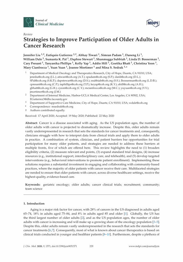
Journal of
Clinical Medicine
Review
Strategies to Improve Participation of Older Adults inCancer Research
Jennifer Liu 1,†, Eutiquio Gutierrez 2,†, Abhay Tiwari 1, Simran Padam 1, Daneng Li 1,
William Dale 3, Sumanta K. Pal 1, Daphne Stewart 1, Shanmugga Subbiah 1, Linda D. Bosserman 1,
Cary Presant 1, Tanyanika Phillips 1, Kelly Yap 1, Addie Hill 1, Geetika Bhatt 1, Christina Yeon 1,
Mary Cianfrocca 1, Yuan Yuan 1, Joanne Mortimer 1 and Mina S. Sedrak 1,*
1 Department of Medical Oncology and Therapeutics Research, City of Hope, Duarte, CA 91010, USA;[email protected] (J.L.); [email protected] (A.T.); [email protected] (S.P.); [email protected] (D.L.);[email protected] (S.K.P.); [email protected] (D.S.); [email protected] (S.S.); [email protected] (L.D.B.);[email protected] (C.P.); [email protected] (T.P.); [email protected] (K.Y.); [email protected] (A.H.);[email protected] (G.B.); [email protected] (C.Y.); [email protected] (M.C.); [email protected] (Y.Y.);[email protected] (J.M.)
2 Department of Internal Medicine, Harbor-UCLA Medical Center, Los Angeles, CA 90502, USA;[email protected]
3 Department of Supportive Care Medicine, City of Hope, Duarte, CA 91010, USA; [email protected]* Correspondence: [email protected]† Authors contributed equally.
Received: 17 April 2020; Accepted: 19 May 2020; Published: 22 May 2020
Abstract: Cancer is a disease associated with aging. As the US population ages, the number ofolder adults with cancer is projected to dramatically increase. Despite this, older adults remainvastly underrepresented in research that sets the standards for cancer treatments and, consequently,clinicians struggle with how to interpret data from clinical trials and apply them to older adultsin practice. A combination of system, clinician, and patient barriers bar opportunities for trialparticipation for many older patients, and strategies are needed to address these barriers atmultiple fronts, five of which are offered here. This review highlights the need to (1) broadeneligibility criteria, (2) measure relevant end points, (3) expand standard trial designs, (4) increaseresources (e.g., institutional support, interdisciplinary care, and telehealth), and (5) develop targetedinterventions (e.g., behavioral interventions to promote patient enrollment). Implementing thesesolutions requires a substantial investment in engaging and collaborating with community-basedpractices, where the majority of older patients with cancer receive their care. Multifaceted strategiesare needed to ensure that older patients with cancer, across diverse healthcare settings, receive thehighest-quality, evidence-based care.
Keywords: geriatric oncology; older adults; cancer clinical trials; recruitment; community;team science
1. Introduction
Aging is a major risk factor for cancer, with 28% of cancers in the US diagnosed in adults aged65–74, 18% in adults aged 75–84, and 8% in adults aged 85 and older [1]. Globally, the US hasthe third largest number of older adults [2], and as the US population ages, the number of olderadults with cancer is increasing and will make up a growing share of the oncology population [3–5].Despite this, older adults remain vastly underrepresented in the research that sets the standards forcancer treatments [6,7]. Consequently, most of what is known about cancer therapeutics is based onclinical trials conducted in younger and healthier patients [8–10]. Furthermore, despite a plethora of
J. Clin. Med. 2020, 9, 1571; doi:10.3390/jcm9051571 www.mdpi.com/journal/jcm229
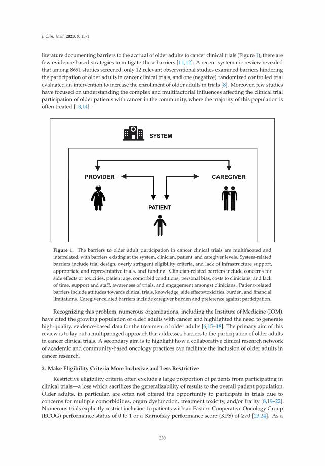
J. Clin. Med. 2020, 9, 1571
literature documenting barriers to the accrual of older adults to cancer clinical trials (Figure 1), there arefew evidence-based strategies to mitigate these barriers [11,12]. A recent systematic review revealedthat among 8691 studies screened, only 12 relevant observational studies examined barriers hinderingthe participation of older adults in cancer clinical trials, and one (negative) randomized controlled trialevaluated an intervention to increase the enrollment of older adults in trials [8]. Moreover, few studieshave focused on understanding the complex and multifactorial influences affecting the clinical trialparticipation of older patients with cancer in the community, where the majority of this population isoften treated [13,14].
Figure 1. The barriers to older adult participation in cancer clinical trials are multifaceted andinterrelated, with barriers existing at the system, clinician, patient, and caregiver levels. System-relatedbarriers include trial design, overly stringent eligibility criteria, and lack of infrastructure support,appropriate and representative trials, and funding. Clinician-related barriers include concerns forside effects or toxicities, patient age, comorbid conditions, personal bias, costs to clinicians, and lackof time, support and staff, awareness of trials, and engagement amongst clinicians. Patient-relatedbarriers include attitudes towards clinical trials, knowledge, side effects/toxicities, burden, and financiallimitations. Caregiver-related barriers include caregiver burden and preference against participation.
Recognizing this problem, numerous organizations, including the Institute of Medicine (IOM),have cited the growing population of older adults with cancer and highlighted the need to generatehigh-quality, evidence-based data for the treatment of older adults [6,15–18]. The primary aim of thisreview is to lay out a multipronged approach that addresses barriers to the participation of older adultsin cancer clinical trials. A secondary aim is to highlight how a collaborative clinical research networkof academic and community-based oncology practices can facilitate the inclusion of older adults incancer research.
2. Make Eligibility Criteria More Inclusive and Less Restrictive
Restrictive eligibility criteria often exclude a large proportion of patients from participating inclinical trials—a loss which sacrifices the generalizability of results to the overall patient population.Older adults, in particular, are often not offered the opportunity to participate in trials due toconcerns for multiple comorbidities, organ dysfunction, treatment toxicity, and/or frailty [8,19–22].Numerous trials explicitly restrict inclusion to patients with an Eastern Cooperative Oncology Group(ECOG) performance status of 0 to 1 or a Karnofsky performance score (KPS) of ≥70 [23,24]. As a
230
J. Clin. Med. 2020, 9, 1571
result, trial data and findings are often derived from younger patients who are more fit and withoutorgan dysfunction and comorbidities, as commonly seen in older adults [23].
A variety of efforts are underway to broaden the eligibility criteria to make trials more relevantto patients of all ages, including initiatives by the National Institutes of Health (“Inclusion Acrossthe Lifespan policy”), American Society of Clinical Oncology (ASCO), and the Food and DrugAdministration (FDA) [16,25,26]. Sponsors and investigators are encouraged to work together tofollow these policy recommendations aimed at making the eligibility criteria less restrictive and moreinclusive of demographically and clinically diverse patients, representative of the populations seen incommunity-based oncology settings. Investigators should review the eligibility criteria closely andforgo exclusion on the basis of lab values (e.g., creatinine), performance status, comorbid conditions,or second malignancy when designing clinical trials. Instead, a patient’s functional or biological ageshould be taken into consideration, which may be a better indicator of how a patient will tolerate acertain treatment regimen.
Additionally, investigators should work closely with community clinicians and other stakeholders(i.e., patient advocates) to understand the patient populations seen in community-based practices anddesign eligibility criteria that are more inclusive, thus allowing trial results to be applicable to a broaderpatient population. Inclusive clinical trial systems within a collaborative research network ought toconsider patients as they are, rather than as they should be. Trial eligibility should be evaluated andrevised to ensure that investigators are broadening study access to new, successful cancer treatmentregimens for older adults with cancer across diverse healthcare delivery settings.
3. Capture Relevant End Points that Matter to Older Patients
Sponsors and investigators should capture relevant end points that are important to the olderpatient, including measures of tolerability as well as clinical and biological aging consequences ofcancer and its treatment [17,27–29].
Studies have shown that most older adults do not want to compromise their quality of life orfunction for survival benefits [30]. Hence, trials designed to collect information on this populationmust include patient-reported measures that are relevant to the patient’s experience and move beyondclinician-reported toxicity. Therefore, data are needed to understand the immediate (short-term)and longitudinal (long-term) impact of cancer and cancer therapy on patient health and quality oflife [29,31]. Given that each individual is unique and can respond differently to therapy, incorporatingassessments that focus on patient-reported outcomes may allow investigators to better describe apatient’s tolerability beyond the traditional numerical grades (NCI CTCAE) [32–34], and the use ofmixed-methods approaches (e.g., surveys, interviews, focus groups) may allow investigators to betterunderstand a patient’s experience and priorities for treatment. Furthermore, measuring the frequencyof toxicities over time can help investigators to characterize the longitudinal impact of treatment andbetter determine the relevance to the older patient population. This information can inform new waystreatment approaches and optimal strategies to reduce toxicities, avoid early treatment discontinuation,and achieve an effective dose intensity.
In addition to capturing measures of quality of life and tolerability, clinical measures of agingshould also be considered as end points in cancer trials of older adults. This is important becauseaging is a heterogeneous process. While certain declines in organ function are universal as thehuman body ages, the consequences of this decline on everyday function proceed at a uniquepace in each individual [35,36]. Therefore, chronologic age tells us relatively little about a specificindividual [37]. A more detailed evaluation of an older patient is needed to capture factors that moreeffectively predict morbidity and mortality. A geriatric assessment (GA) may serve this purpose.The GA includes an evaluation of functional status, co-morbid medical conditions, cognitive function,nutritional status, social support, and psychological state, as well as a review of medications [38].Additionally, geriatric screening tools (i.e., G8, VES-13), instruments that assess life expectancy(i.e., ePrognosis), and predictors for risks of chemotherapy toxicity (i.e., CARG or CRASH Score) can
231
J. Clin. Med. 2020, 9, 1571
be utilized in trials to better describe or risk-stratify this population [39–41]. The ASCO guidelinesnow recommend the use of these assessments in the evaluation and management of older patientswith cancer, and several NCI-sponsored cooperative groups have demonstrated the feasibility of usingthese measures in clinical trials [40].
Biological measures of aging are also important to consider when designing studies for olderpatients with cancer. These measures may provide insights on mechanisms behind aging-relatedclinical consequences due to cancer treatment, such as the biological drivers of functional and cognitivedecline. Several studies have hypothesized that cancer and cancer treatment may accentuate oraccelerate the rate of aging, leading to a decrease in multisystem reserve and increased risk forcardiomyopathy, secondary malignant neoplasms, frailty, muscular weakness, and neurocognitiveissues [42]. Biological processes including stem cell exhaustion, cellular senescence, telomere attrition,increased free radical production, and epigenetic modifications have all been shown to play a rolein accelerated aging due to cancer therapies [42,43]. Understanding the hallmarks of aging and theimplications of cancer therapies associated with accelerated aging may provide insights into the short-and long-term effects of certain therapies, and further research in this area is needed. Knowledge ofthe underlying mechanisms to accelerated aging could ultimately help us to identify new strategiesfor targeting these processes in order to ameliorate accelerated aging and improve the health andwell-being of this growing population.
4. Optimize Trial Designs for a Special Population
Inclusion of older patients may, in some cases, impose the need for larger sample sizes, which isnot always feasible or fundable. Sponsors and investigators should consider high-yield approaches tocollect data on older patients to efficiently facilitate the conduct of larger-scale trials at a reasonablecost. For example, pragmatic or innovative trials (e.g., adaptive, extended, embedded, N-of-1 studies)designed specifically for older patients may be used to fill knowledge gaps and improve the evidencebase guiding the treatment of older adults [29,44].
There are several innovative trial designs that may be leveraged when considering a specialpopulation such as older adults with cancer (Table 1 adapted from prior literature [16,39]). For example,adaptive trials can be leveraged to allow for modifications to be made as the study proceeds [45,46].Based on interim data analysis, the underperforming treatment arm may be eliminated to allow for alarger proportion of participants to be assigned to the more effective treatment arm. This is ideal forolder adults because it reduces the number of participants in the treatment group that is performingpoorly [16,39,47]. While adapted trial designs can be applicable to both exploratory and confirmatorystudies, prospective cohort studies can be used to generate data on current standard-of-care treatmentsin older patients and provide insight into patterns of care and decision making [16,39].
Extended trial design can also be used in cases where the results of a trial have been reported,but there was an insufficient number of older adults enrolled to draw conclusions. A cohort of olderadults can be added to the superior treatment arm to fill knowledge gaps and obtain data on theolder population [16]. Embedded studies, also known as correlative or ancillary studies, can alsobe used to include additional measures of interest that are specific to older adults (i.e., toxicity, GAdomains) within the infrastructure of a parent study [39]. Lastly, an N-of-1 or single-subject trialpresents a feasible and innovative approach to better understanding how to care for older adults byfollowing one patient over time, to examine aging-related consequences that occur throughout thecancer continuum [48].
232

J. Clin. Med. 2020, 9, 1571
Ta
ble
1.
Clin
ical
tria
ldes
igns
for
geri
atri
con
colo
gyre
sear
ch.
De
sig
nD
esc
rip
tio
n/C
ha
ract
eri
stic
sP
ote
nti
al
Ob
ject
ive
sa
nd
Ou
tco
me
sA
dv
an
tag
es
Lim
ita
tio
ns
Prag
mat
icTr
ial
Inte
rven
tion
typi
cally
perf
orm
edin
the
cont
exto
fst
anda
rdca
rePa
tien
tsre
crui
ted
from
ava
riet
yof
prac
tice
sett
ings
,us
ing
broa
der,
mor
ein
clus
ive
elig
ibili
tycr
iter
ia
Det
erm
ine
the
effec
tive
ness
ofan
inte
rven
tion
inda
y-to
-day
prac
tice
-Mor
eac
cess
ible
-Les
sre
sour
ce-i
nten
sive
-Pla
ces
min
imal
addi
tion
albu
rden
onpa
rtic
ipan
ts-O
utco
mes
rele
vant
toth
ose
who
will
use
tria
lres
ults
-Non
-adh
eren
cean
dlo
ssto
follo
w-u
p-P
oor
inte
rnal
valid
ity
limit
sth
eab
ility
tode
term
ine
defin
itiv
eeff
ects
Ran
dom
ized
Con
trol
led
Tria
l
Subj
ects
are
rand
omly
assi
gned
totr
eatm
enta
rms
Toge
nera
teda
taon
the
olde
rpa
tien
tpop
ulat
ion:
-Acc
rue
only
olde
rad
ults
,-S
trat
ify
enro
llmen
tint
oag
egr
oups
Ada
ptiv
ede
sign
:mod
ifica
tion
sar
em
ade
asth
est
udy
proc
eeds
base
don
inte
rim
data
anal
ysis
Com
pare
effica
cyan
dto
lera
bilit
yof
diff
eren
ttr
eatm
ents
regi
men
s
-Dir
ectc
ompa
riso
nof
trea
tmen
tre
gim
ens
-Unb
iase
d-M
inim
izes
conf
ound
ing
-Gen
eral
izab
leto
the
over
all
popu
lati
onbe
ing
stud
ied
-Req
uire
sa
larg
esa
mpl
esi
ze-C
ostl
yan
dti
me-
cons
umin
g-L
ogis
tica
llyde
man
ding
-Slo
wac
crua
l
Pros
pect
ive
Coh
ortS
tudy
Ass
essm
ento
ftre
atm
ents
alre
ady
appr
oved
byth
eFD
AC
ohor
tdefi
ned
byho
st,t
umor
,or
trea
tmen
tfac
tors
Obs
erva
tion
alH
ypot
hesi
s-dr
iven
Und
erst
and
deci
sion
-mak
ing,
patt
erns
ofca
re-F
indi
ngs
are
gene
raliz
able
-Lac
kof
rand
omiz
atio
n-R
equi
res
ala
rge
sam
ple
size
-Log
isti
cally
dem
andi
ng
Exte
nded
Tria
lA
ddit
ion
ofa
coho
rtof
olde
rpa
tien
tsto
the
supe
rior
trea
tmen
tarm
Det
erm
ine
tole
rabi
lity
inol
der
adul
ts
Tria
linf
rast
ruct
ure
alre
ady
esta
blis
hed
Exis
ting
data
ontr
eatm
enteffi
cacy
will
mak
eac
crua
lofo
lder
pati
ents
easi
er
-Lac
kof
data
onth
ein
feri
ortr
eatm
enta
rmin
olde
rad
ults
Embe
dded
Stud
y(C
orre
lati
veor
Anc
illar
ySt
udy)
Incl
udes
addi
tion
alm
easu
res
ofin
tere
stin
the
infr
astr
uctu
reof
apa
rent
stud
y
Des
crib
ea
coho
rtor
unde
rsta
ndth
eim
pact
oftr
eatm
entu
sing
geri
atri
cas
sess
men
tmea
sure
s
-Bet
ter
unde
rsta
ndth
ech
arac
teri
stic
sof
the
olde
rpa
tien
tpop
ulat
ion
-Ide
ntifi
cati
onof
pred
icto
rsof
func
tion
alde
clin
e
-In
stud
ies
nots
peci
ficto
olde
rad
ults
,the
sam
ple
size
ofol
der
pati
ents
may
belim
ited
N-o
f-1
(Sin
gle-
Subj
ect
Tria
l)D
eter
min
esth
eop
tim
alth
erap
yfo
ra
sing
lein
divi
dual
Det
erm
ine
the
opti
mal
orbe
stin
terv
enti
onfo
ran
indi
vidu
alpa
tien
tusi
ngob
ject
ive
data
-dri
ven
crit
eria
-Ind
ivid
ualiz
edm
edic
ine
-Gai
nin
sigh
tsin
toco
mpa
rati
vetr
eatm
enteff
ecti
vene
ssam
ong
aw
ide
vari
ety
ofpa
tien
ts
-Ran
dom
izat
ion
oftr
eatm
ento
rder
-Car
ryov
ereff
ects
-Was
h-ou
tper
iods
-Blin
ding
233
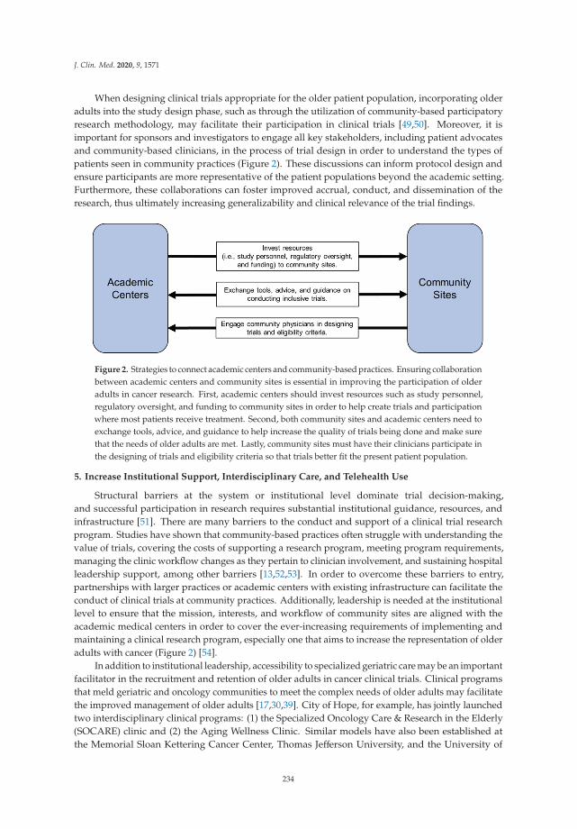
J. Clin. Med. 2020, 9, 1571
When designing clinical trials appropriate for the older patient population, incorporating olderadults into the study design phase, such as through the utilization of community-based participatoryresearch methodology, may facilitate their participation in clinical trials [49,50]. Moreover, it isimportant for sponsors and investigators to engage all key stakeholders, including patient advocatesand community-based clinicians, in the process of trial design in order to understand the types ofpatients seen in community practices (Figure 2). These discussions can inform protocol design andensure participants are more representative of the patient populations beyond the academic setting.Furthermore, these collaborations can foster improved accrual, conduct, and dissemination of theresearch, thus ultimately increasing generalizability and clinical relevance of the trial findings.
Figure 2. Strategies to connect academic centers and community-based practices. Ensuring collaborationbetween academic centers and community sites is essential in improving the participation of olderadults in cancer research. First, academic centers should invest resources such as study personnel,regulatory oversight, and funding to community sites in order to help create trials and participationwhere most patients receive treatment. Second, both community sites and academic centers need toexchange tools, advice, and guidance to help increase the quality of trials being done and make surethat the needs of older adults are met. Lastly, community sites must have their clinicians participate inthe designing of trials and eligibility criteria so that trials better fit the present patient population.
5. Increase Institutional Support, Interdisciplinary Care, and Telehealth Use
Structural barriers at the system or institutional level dominate trial decision-making,and successful participation in research requires substantial institutional guidance, resources, andinfrastructure [51]. There are many barriers to the conduct and support of a clinical trial researchprogram. Studies have shown that community-based practices often struggle with understanding thevalue of trials, covering the costs of supporting a research program, meeting program requirements,managing the clinic workflow changes as they pertain to clinician involvement, and sustaining hospitalleadership support, among other barriers [13,52,53]. In order to overcome these barriers to entry,partnerships with larger practices or academic centers with existing infrastructure can facilitate theconduct of clinical trials at community practices. Additionally, leadership is needed at the institutionallevel to ensure that the mission, interests, and workflow of community sites are aligned with theacademic medical centers in order to cover the ever-increasing requirements of implementing andmaintaining a clinical research program, especially one that aims to increase the representation of olderadults with cancer (Figure 2) [54].
In addition to institutional leadership, accessibility to specialized geriatric care may be an importantfacilitator in the recruitment and retention of older adults in cancer clinical trials. Clinical programsthat meld geriatric and oncology communities to meet the complex needs of older adults may facilitatethe improved management of older adults [17,30,39]. City of Hope, for example, has jointly launchedtwo interdisciplinary clinical programs: (1) the Specialized Oncology Care & Research in the Elderly(SOCARE) clinic and (2) the Aging Wellness Clinic. Similar models have also been established atthe Memorial Sloan Kettering Cancer Center, Thomas Jefferson University, and the University of
234
J. Clin. Med. 2020, 9, 1571
Rochester, among others [55–57]. These specialized programs offer interdisciplinary, individualized,and integrated treatment for older patients and survivors with cancer. However, these programsrequire resources and institutional support, and further research is warranted to understand how thesecare models lead to improved patient outcomes and participation in clinical trials.
Furthermore, the integration of technology in the form of virtual visits and telehealth encountersmay facilitate the enrollment of older adults in cancer research. There are two potential strategiesfor this. First, technology may be leveraged to reduce the burden of the patient and caregiver—aknown barrier to older participation in research [8,17,39]. Studies have shown that telehealth use isassociated with high patient satisfaction, improved access to care, and reduced health disparities amongolder adults [58–62]. Challenges to telehealth use among the older patient population must be takeninto consideration, including practical barriers to adoption, anxiety using technology, and financialburden, among other factors that promote the digital divide [53]. To address some of these barriers,simple and intuitive graphical interfaces, the availability of technical support, and the provision oftelehealth services at no cost may reduce the barriers to entry and improve adoption of telehealthin this population [63]. However, further efforts are needed to better understand the limits andbenefits of telehealth in geriatric oncology. Moreover, the COVID-19 pandemic has led to the rapidimplementation of telehealth services for both standard care and clinical research [64,65]. This swiftparadigm shift in how patients receive their care provides a unique opportunity to gain insight on howtelehealth can further be used to improve patient participation in cancer clinical trials—particularlyin older adults. However, the feasibility, adoption, and sustainability of telehealth in clinical trials islimited, and further research is needed [66].
Second, telehealth can be a key player in providing specialized geriatric care to sites that maynot have access to multidisciplinary programs with geriatric expertise. For example, City of Hopehas an ongoing pilot study to evaluate the feasibility of delivering a GA-driven intervention at acommunity-affiliated site using telehealth. In this way, telehealth may help fill the gap in care forolder patients who otherwise would not have access to multidisciplinary, specialized geriatric-basedcare [67]. Whether this improves clinical trial accrual warrants further investigation.
6. Leverage Principles of Behavioral Economics
There is a growing interest in the general medical literature around the use of behavioraleconomics to shape clinician and patient behavior, with some even suggesting the use of “nudges”to facilitate patient enrollment in research [68]. A nudge is defined as a change in the way choicesare presented or information is framed that alters a person’s behavior in a predictable way withoutrestricting choice [68,69]. If implemented properly, nudges are transparent, easy to opt out of if needed,and aligned with the best interest or welfare of the person being nudged. It is a principle of behavioraleconomics that leverages the fact that our decisions and behaviors are heavily influenced by theenvironment in which they occur, and interventions can be systematically developed to influence howindividuals behave (e.g., weight loss, exercise, and statin utilization) [70–74].
Nudges may also be a novel way to facilitate patient participation in cancer clinical trials,especially among highly underrepresented groups such as older adults [75]. For example, surveys thatassess patients’ interest in participating in clinical research can help identify eligible patients whoare likely to participate and provide insight into the types of trials that are desirable. Using theseinterest surveys, clinicians can identify patients who may be more inclined to participate in studieswhen presented the opportunity to do so due to their prior expressed interest (i.e., foot-in-the-doortechnique) [75]. Another nudging strategy that can be utilized to encourage the enrollment of olderadults in cancer trials is the personalization of trials [75]. Explaining why a patient was specificallychosen to participate, how they may personally benefit, and how their participation will contribute tothe scientific community and future patients may improve patient enrollment. Furthermore, nudges canbe directed towards providers. Clinicians, for example, can be nudged by incorporating the trialinformation in the clinical pathway of electronic health records [69]. Including the trial information in
235
J. Clin. Med. 2020, 9, 1571
electronic health records may improve clinician awareness of potentially beneficial trials regardingtheir patients and help facilitate enrollment of older adults.
Evidence on the use of nudges to influence patient and clinician behavior in the contextof clinical trials is still in its infancy. Further research is warranted to examine whether thesestrategies can be employed to influence enrollment and how they can be successfully implemented(e.g., feasible, acceptable, and sustainable) in both academic and community-based cancer practices.
7. Conclusions
The underrepresentation of older adults in cancer clinical trials is undeniably a multifacetedproblem that requires a multifaceted solution. There have been promising steps toward improvingtrial participation among older adults, but there are still significant gaps in knowledge that hinderolder patients from receiving high-quality, individualized, evidence-based care. The recommendationspresented in this review range from small changes that can be adopted by individuals and researchteams to large-scale, systemic changes that are needed at the institutional and/or policy level.Regardless, multiple steps are needed on multiple fronts across a collaborative network of academic andcommunity-based cancer practices in order to have a cumulative and palpable effect. An investment inacademic and community clinical trial partnerships could help to further cancer research, and moreimportantly, ensure that older adults with cancer have equal access to new treatments and advancesin care.
Author Contributions: Conceptualization, J.L., E.G., A.T., S.P., and M.S.; Writing—Original Draft Preparation, J.L.,E.G., A.T., S.P., and M.S.; Writing—Review and Editing, all authors; Visualization, J.L. and A.T.; Supervision, M.S.;Project Administration, M.S.; Funding Acquisition, M.S. All authors have read and agreed to the published versionof the manuscript.
Funding: This work was supported by the National Institute of Aging (NIA R03AG064377), the National CancerInstitute (NCI K12CA001727), the Waisman Innovation Fund, and Circle 1500. The funder had no role in thedesign and conduct of the study; in the collection, management, analysis, and interpretation of the data; in thepreparation, review, or approval of the manuscript; or in the decision to submit the manuscript for publication.
Acknowledgments: We dedicate this work to the late Arti Hurria, a leader in geriatric oncology and an advocatefor expanding inclusion of older adults in cancer clinical trials. We hope this manuscript will help carry Hurria’slegacy forward and foster new collaborations in a way that she would have advocated for.
Conflicts of Interest: The authors declare no conflict of interest.
References
1. Howlader, N.; Noone, A.M.; Krapcho, M.; Miller, D.; Brest, A.; Yu, M.; Ruhl, J.; Tatalovich, Z.; Mariotto, A.;Lewis, D.R.; et al. SEER Cancer Statistics Review, 1975–2017; National Cancer Institute: Bethesda, MD, USA,2020. Available online: https://seer.cancer.gov/csr/1975_2017/ (accessed on 1 April 2020).
2. PRB. Countries with the Oldest Populations in the World, March 23, 2020 ed.; Population Reference Bureau:Washington, DC, USA, 2020.
3. Smith, B.D.; Smith, G.L.; Hurria, A.; Hortobagyi, G.N.; Buchholz, T.A. Future of cancer incidence in theUnited States: Burdens upon an aging, changing nation. J. Clin. Oncol. Off. J. Am. Soc. Clin. Oncol. 2009, 27,2758–2765. [CrossRef] [PubMed]
4. Mather, M.; Jacobsen, L.A.; Pollard, K.M. Population Bulletin: Aging in the United States, 2015.Available online: http://www.prb.org/pdf16/aging-us-population-bulletin.pdf (accessed on 1 April 2020).
5. Bluethmann, S.M.; Mariotto, A.B.; Rowland, J.H. Anticipating the “Silver Tsunami”: Prevalence Trajectoriesand Comorbidity Burden among Older Cancer Survivors in the United States. Cancer Epidemiol. Biomark.Prev. 2016, 25, 1029–1036. [CrossRef]
6. Hurria, A.; Naylor, M.; Cohen, H.J. Improving the Quality of Cancer Care in an Aging Population:Recommendations from an IOM Report. JAMA 2013, 310, 1795–1796. [CrossRef] [PubMed]
7. Talarico, L.; Chen, G.; Pazdur, R. Enrollment of Elderly Patients in Clinical Trials for Cancer Drug Registration:A 7-Year Experience by the US Food and Drug Administration. J. Clin. Oncol. 2004, 22, 4626–4631. [CrossRef][PubMed]
236
J. Clin. Med. 2020, 9, 1571
8. Sedrak, M.S.; Hurria, A.; Li, D.; George, K.; Padam, S.; Liu, J.; Wong, A.R.; Vargas, N.; Eskandar, J.; Katheria, V.;et al. Barriers to clinical trial enrollment of older adults with cancer: A systematic review. J. Clin. Oncol.2019, 37, e18130. [CrossRef]
9. Singh, H.; Kanapuru, B.; Smith, C.; Fashoyin-Aje, L.A.; Myers, A.; Kim, G.; Pazdur, R. FDA analysis ofenrollment of older adults in clinical trials for cancer drug registration: A 10-year experience by the U.S.Food and Drug Administration. J. Clin. Oncol. 2017, 35, 10009. [CrossRef]
10. Singh, H.; Beaver, J.A.; Kim, G.; Pazdur, R. Enrollment of older adults on oncology trials: An FDA perspective.J. Geriatr. Oncol. 2017, 8, 149–150. [CrossRef]
11. Sedrak, M.S.; Mohile, S.G.; Sun, V.; Sun, C.L.; Chen, B.T.; Li, D.; Wong, A.R.; George, K.; Padam, S.; Liu, J.;et al. Barriers to clinical trial enrollment of older adults with cancer: A qualitative study of the perceptionsof community and academic oncologists. J. Geriatr. Oncol. 2020, 11, 327–334. [CrossRef]
12. Townsley, C.A.; Selby, R.; Siu, L.L. Systematic review of barriers to the recruitment of older patients withcancer onto clinical trials. J. Clin. Oncol. 2005, 23, 3112–3124. [CrossRef]
13. Wong, A.R.; Sun, V.; George, K.; Liu, J.; Padam, S.; Chen, B.A.; George, T.; Amini, A.; Li, D.; Sedrak, M.S.Barriers to Participation in Therapeutic Clinical Trials as Perceived by Community Oncologists. JCO Oncol.Pract. 2020. JOP.19.00662. [CrossRef]
14. Nabhan, C.; Jeune-Smith, Y.; Klinefelter, P.; Kelly, R.J.; Feinberg, B.A. Challenges, Perceptions, and Readinessof Oncology Clinicians for the MACRA Quality Payment Program. JAMA Oncol. 2018, 4, 252–253. [CrossRef][PubMed]
15. Committee on Improving the Quality of Cancer Care: Addressing the Challenges of an Aging Population,B.o.H.C.S.; Institute of Medicine. Delivering High-Quality Cancer Care: Charting a New Course for a System inCrisis; Levit, L., Balogh, E., Nass, S., Ganz, P.A., Eds.; National Academies Press: Washington, DC, USA, 2013.[CrossRef]
16. Hurria, A.; Levit, L.A.; Dale, W.; Mohile, S.G.; Muss, H.B.; Fehrenbacher, L.; Magnuson, A.; Lichtman, S.M.;Bruinooge, S.S.; Soto-Perez-de-Celis, E.; et al. Improving the Evidence Base for Treating Older Adults withCancer: American Society of Clinical Oncology Statement. J. Clin. Oncol. Off. J. Am. Soc. Clin. Oncol. 2015,33, 3826–3833. [CrossRef] [PubMed]
17. Levit, L.A.; Singh, H.; Klepin, H.D.; Hurria, A. Expanding the Evidence Base in Geriatric Oncology:Action Items From an FDA-ASCO Workshop. J. Natl. Cancer Inst. 2018, 110, 1163–1170. [CrossRef] [PubMed]
18. Oncology Center of Excellence, Center for Drug Evaluation and Research (CDER) and Center for BiologicsEvaluation and Research (CBER) at the Food and Drug Administration. Inclusion of Older Adults in CancerClinical Trials, Guidance for Industry; U.S. Department of Health and Human Services, Food and DrugAdministration: Silver Spring, MD, USA, 2020.
19. Kornblith, A.B.; Kemeny, M.; Peterson, B.L.; Wheeler, J.; Crawford, J.; Bartlett, N.; Fleming, G.; Graziano, S.;Muss, H.; Cohen, H.J. Survey of oncologists’ perceptions of barriers to accrual of older patients with breastcarcinoma to clinical trials. Cancer 2002, 95, 989–996. [CrossRef]
20. McCleary, N.J.; Hubbard, J.; Mahoney, M.R.; Meyerhardt, J.A.; Sargent, D.; Venook, A.; Grothey, A. Challengesof conducting a prospective clinical trial for older patients: Lessons learned from NCCTG N0949 (alliance).J. Geriatr. Oncol. 2018, 9, 24–31. [CrossRef]
21. Hamaker, M.E.; Seynaeve, C.; Nortier, J.W.; Wymenga, M.; Maartense, E.; Boven, E.; van Leeuwen-Stok, A.E.;de Rooij, S.E.; van Munster, B.C.; Smorenburg, C.H. Slow accrual of elderly patients with metastatic breastcancer in the Dutch multicentre OMEGA study. Breast 2013, 22, 556–559. [CrossRef]
22. Freedman, R.A.; Dockter, T.J.; Lafky, J.M.; Hurria, A.; Muss, H.J.; Cohen, H.J.; Jatoi, A.; Kemeny, M.M.;Ruddy, K.J. Promoting Accrual of Older Patients with Cancer to Clinical Trials: An Alliance for Clinical Trialsin Oncology Member Survey (A171602). Oncology 2018, 23, 1016–1023. [CrossRef]
23. Lichtman, S.M.; Harvey, R.D.; Smit, M.-A.D.; Rahman, A.; Thompson, M.A.; Roach, N.; Schenkel, C.;Bruinooge, S.S.; Cortazar, P.; Walker, D.; et al. Modernizing Clinical Trial Eligibility Criteria:Recommendations of the American Society of Clinical Oncology–Friends of Cancer Research OrganDysfunction, Prior or Concurrent Malignancy, and Comorbidities Working Group. J. Clin. Oncol. 2017, 35,3753–3759. [CrossRef]
24. Unger, J.M.; Cook, E.; Tai, E.; Bleyer, A. The Role of Clinical Trial Participation in Cancer Research:Barriers, Evidence, and Strategies. Am. Soc. Clin. Oncol. Educ. Book 2016, 35, 185–198. [CrossRef]
237
J. Clin. Med. 2020, 9, 1571
25. Kim, E.S.; Bruinooge, S.S.; Roberts, S.; Ison, G.; Lin, N.U.; Gore, L.; Uldrick, T.S.; Lichtman, S.M.; Roach, N.;Beaver, J.A.; et al. Broadening Eligibility Criteria to Make Clinical Trials More Representative: AmericanSociety of Clinical Oncology and Friends of Cancer Research Joint Research Statement. J. Clin. Oncol. 2017,35, 3737–3744. [CrossRef]
26. Vaughan, C.P.; Dale, W.; Allore, H.G.; Binder, E.F.; Boyd, C.M.; Bynum, J.P.W.; Gurwitz, J.H.; Lundebjerg, N.E.;Trucil, D.E.; Supiano, M.A.; et al. AGS Report on Engagement Related to the NIH Inclusion Across theLifespan Policy. J. Am. Geriatr. Soc. 2019, 67, 211–217. [CrossRef] [PubMed]
27. Wildiers, H.; Mauer, M.; Pallis, A.; Hurria, A.; Mohile, S.G.; Luciani, A.; Curigliano, G.; Extermann, M.;Lichtman, S.M.; Ballman, K.; et al. End points and trial design in geriatric oncology research: A joint Europeanorganisation for research and treatment of cancer—Alliance for Clinical Trials in Oncology—InternationalSociety Of Geriatric Oncology position article. J. Clin. Oncol. Off. J. Am. Soc. Clin. Oncol. 2013, 31, 3711–3718.[CrossRef] [PubMed]
28. McKenna, R.J., Sr. Clinical aspects of cancer in the elderly. Treatment decisions, treatment choices,and follow-up. Cancer 1994, 74, 2107–2117. [CrossRef]
29. BrintzenhofeSzoc, K.; Krok-Schoen, J.L.; Canin, B.; Parker, I.; MacKenzie, A.R.; Koll, T.; Vankina, R.; Hsu, C.D.;Jang, B.; Pan, K.; et al. The underreporting of phase III chemo-therapeutic clinical trial data of older patientswith cancer: A systematic review. J. Geriatr. Oncol. 2020, 11, 369–379. [CrossRef]
30. Mohile, S.G.; Hurria, A.; Cohen, H.J.; Rowland, J.H.; Leach, C.R.; Arora, N.K.; Canin, B.; Muss, H.B.;Magnuson, A.; Flannery, M.; et al. Improving the quality of survivorship for older adults with cancer. Cancer2016, 122, 2459–2568. [CrossRef]
31. Sacks, C.A.; Miller, P.W.; Longo, D.L. Talking about Toxicity—“What We’ve Got Here Is a Failure toCommunicate”. N. Engl. J. Med. 2019, 381, 1406–1408. [CrossRef]
32. Handforth, C.; Hall, P.; Marshall, H.; Seymour, M. Overall treatment utility: A novel outcome measure toconvey the balance of benefits and harms from cancer treatment. J. Geriatr. Oncol. 2013, 4, S49. [CrossRef]
33. Sloan, J.A.; Mahoney, M.R.; Sargent, D.J.; Hubbard, J.M.; Liu, H.; Basch, E.M.; Shields, A.F.; Chan, E.; Goldberg, R.M.;Gill, S.; et al. Was it worth it (WIWI)? Patient satisfaction with clinical trial participation: Results from NorthCentral Cancer Treatment Group (NCCTG) phase III trial N0147. J. Clin. Oncol. 2011, 29, 6122. [CrossRef]
34. Dueck, A.C.; Mendoza, T.R.; Mitchell, S.A.; Reeve, B.B.; Castro, K.M.; Rogak, L.J.; Atkinson, T.M.; Bennett, A.V.;Denicoff, A.M.; O’Mara, A.M.; et al. Validity and Reliability of the US National Cancer Institute’sPatient-Reported Outcomes Version of the Common Terminology Criteria for Adverse Events (PRO-CTCAE).JAMA Oncol. 2015, 1, 1051–1059. [CrossRef]
35. Lowsky, D.J.; Olshansky, S.J.; Bhattacharya, J.; Goldman, D.P. Heterogeneity in Healthy Aging. J. Gerontol.Ser. A 2013, 69, 640–649. [CrossRef]
36. Hurria, A.; Wildes, T.; Blair, S.L.; Browner, I.S.; Cohen, H.J.; Deshazo, M.; Dotan, E.; Edil, B.H.; Extermann, M.;Ganti, A.K.; et al. Senior adult oncology, version 2.2014: Clinical practice guidelines in oncology. J. Natl.Compr. Cancer Netw. JNCCN 2014, 12, 82–126. [CrossRef] [PubMed]
37. Soto-Perez-de-Celis, E.; Li, D.; Yuan, Y.; Lau, Y.M.; Hurria, A. Functional versus chronological age: Geriatricassessments to guide decision making in older patients with cancer. Lancet. Oncol. 2018, 19, e305–e316.[CrossRef]
38. Mohile, S.G.; Epstein, R.M.; Hurria, A.; Heckler, C.E.; Canin, B.; Culakova, E.; Duberstein, P.; Gilmore, N.;Xu, H.; Plumb, S.; et al. Communication With Older Patients With Cancer Using Geriatric Assessment:A Cluster-Randomized Clinical Trial From the National Cancer Institute Community Oncology ResearchProgram. JAMA Oncol. 2020, 6, 196–204. [CrossRef] [PubMed]
39. Hurria, A.; Dale, W.; Mooney, M.; Rowland, J.H.; Ballman, K.V.; Cohen, H.J.; Muss, H.B.; Schilsky, R.L.;Ferrell, B.; Extermann, M.; et al. Designing therapeutic clinical trials for older and frail adults with cancer:U13 conference recommendations. J. Clin. Oncol. Off. J. Am. Soc. Clin. Oncol. 2014, 32, 2587–2594. [CrossRef]
40. Association of Community ‘Cancer Centers. Multidisciplinary Approaches to Caring for Older Adults with Cancer;Association of Community Cancer Centers: Rockville, MD, USA, 2019.
41. Hurria, A.; Siccion, E.P. Assessing the ‘fit’ older patient for chemotherapy. Oncology 2014, 28, 598–599.42. Cupit-Link, M.C.; Kirkland, J.L.; Ness, K.K.; Armstrong, G.T.; Tchkonia, T.; LeBrasseur, N.K.; Armenian, S.H.;
Ruddy, K.J.; Hashmi, S.K. Biology of premature ageing in survivors of cancer. Esmo Open 2017, 2, e000250.[CrossRef]
238
J. Clin. Med. 2020, 9, 1571
43. Hill, A.; Sadda, J.; LaBarge, M.A.; Hurria, A. How cancer therapeutics cause accelerated aging: Insights fromthe hallmarks of aging. J. Geriatr. Oncol. 2020, 11, 191–193. [CrossRef]
44. Nipp, R.D.; Yao, N.A.; Lowenstein, L.M.; Buckner, J.C.; Parker, I.R.; Gajra, A.; Morrison, V.A.; Dale, W.;Ballman, K.V. Pragmatic study designs for older adults with cancer: Report from the U13 conference.J. Geriatr. Oncol. 2016, 7, 234–241. [CrossRef]
45. Papadimitrakopoulou, V.; Lee, J.J.; Wistuba, I.I.; Tsao, A.S.; Fossella, F.V.; Kalhor, N.; Gupta, S.; Byers, L.A.;Izzo, J.G.; Gettinger, S.N.; et al. The BATTLE-2 Study: A Biomarker-Integrated Targeted Therapy Study inPreviously Treated Patients With Advanced Non–Small-Cell Lung Cancer. J. Clin. Oncol. 2016, 34, 3638–3647.[CrossRef]
46. Park, J.W.; Liu, M.C.; Yee, D.; Yau, C.; van’t Veer, L.J.; Symmans, W.F.; Paoloni, M.; Perlmutter, J.; Hylton, N.M.;Hogarth, M.; et al. Adaptive Randomization of Neratinib in Early Breast Cancer. N. Engl. J. Med. 2016, 375,11–22. [CrossRef]
47. Bhatt, D.L.; Mehta, C. Adaptive Designs for Clinical Trials. N. Engl. J. Med. 2016, 375, 65–74. [CrossRef][PubMed]
48. Lillie, E.O.; Patay, B.; Diamant, J.; Issell, B.; Topol, E.J.; Schork, N.J. The n-of-1 clinical trial: The ultimatestrategy for individualizing medicine? Pers. Med. 2011, 8, 161–173. [CrossRef] [PubMed]
49. Blair, T.; Minkler, M. Participatory Action Research With Older Adults: Key Principles in Practice.The Gerontologist 2009, 49, 651–662. [CrossRef] [PubMed]
50. Higginbottom, G.; Liamputtong, P. Participatory Qualitative Research Methodologies in Health; SAGEPublications Ltd: London, UK, 2015. [CrossRef]
51. Dimond, E.P.; St. Germain, D.; Nacpil, L.M.; Zaren, H.A.; Swanson, S.M.; Minnick, C.; Carrigan, A.;Denicoff, A.M.; Igo, K.E.; Acoba, J.D.; et al. Creating a “culture of research” in a community hospital:Strategies and tools from the National Cancer Institute Community Cancer Centers Program. Clin. Trials2015, 12, 246–256. [CrossRef]
52. McAlearney, A.S.; Reiter, K.L.; Weiner, B.J.; Minasian, L.; Song, P.H. Challenges and facilitators of communityclinical oncology program participation: A qualitative study. J. Healthc. Manag. 2013, 58, 29–46. [CrossRef]
53. Likumahuwa, S.; Song, H.; Singal, R.; Weir, R.C.; Crane, H.; Muench, J.; Sim, S.-C.; DeVoe, J.E. BuildingResearch Infrastructure in Community Health Centers: A Community Health Applied Research Network(CHARN) Report. J. Am. Board Fam. Med. 2013, 26, 579–587. [CrossRef]
54. Minasian, L.M.; Unger, J.M. What Keeps Patients Out of Clinical Trials? JCO Oncol. Pract. 2020, 16, 125–127.[CrossRef]
55. Shahrokni, A.; Kim, S.J.; Bosl, G.J.; Korc-Grodzicki, B. How We Care for an Older Patient With Cancer.J. Oncol. Pr. 2017, 13, 95–102. [CrossRef]
56. Chapman, A.E.; Swartz, K.; Schoppe, J.; Arenson, C. Development of a comprehensive multidisciplinarygeriatric oncology center, the Thomas Jefferson University Experience. J. Geriatr. Oncol. 2014, 5, 164–170.[CrossRef]
57. Magnuson, A.; Dale, W.; Mohile, S. Models of Care in Geriatric Oncology. Curr. Geriatr. Rep. 2014, 3, 182–189.[CrossRef]
58. Greenwald, P.; Stern, M.E.; Clark, S.; Sharma, R. Older adults and technology: In telehealth, they may not bewho you think they are. Int. J. Emerg. Med. 2018, 11, 2. [CrossRef] [PubMed]
59. Gellis, Z.D.; Kenaley, B.; McGinty, J.; Bardelli, E.; Davitt, J.; Ten Have, T. Outcomes of a Telehealth Interventionfor Homebound Older Adults With Heart or Chronic Respiratory Failure: A Randomized Controlled Trial.The Gerontologist 2012, 52, 541–552. [CrossRef] [PubMed]
60. Weinstock, R.S.; Teresi, J.A.; Goland, R.; Izquierdo, R.; Palmas, W.; Eimicke, J.P.; Ebner, S.; Shea, S.;Consortium, I.D. Glycemic control and health disparities in older ethnically diverse underserved adults withdiabetes: Five-year results from the Informatics for Diabetes Education and Telemedicine (IDEATel) study.Diabetes Care 2011, 34, 274–279. [CrossRef] [PubMed]
61. Lum, H.D.; Nearing, K.; Pimentel, C.B.; Levy, C.R.; Hung, W.W. Anywhere to Anywhere: Use of Telehealthto Increase Health Care Access for Older, Rural Veterans. Public Policy Aging Rep. 2019, 30, 12–18. [CrossRef]
62. Merrell, R.C. Geriatric Telemedicine: Background and Evidence for Telemedicine as a Way to Address theChallenges of Geriatrics. Healthc Inf. Res 2015, 21, 223–229. [CrossRef]
63. Cimperman, M.; Brencic, M.M.; Trkman, P.; Stanonik, M.d.L. Older adults’ perceptions of home telehealthservices. Telemed. eHealth 2013, 19, 786–790. [CrossRef]
239
J. Clin. Med. 2020, 9, 1571
64. Liu, R.; Sundaresan, T.; Reed, M.E.; Trosman, J.R.; Weldon, C.B.; Kolevska, T. Telehealth in Oncology Duringthe COVID19 Outbreak: Bringing the house call back virtually. JCO Oncol. Pract. 2020. [CrossRef]
65. Waterhouse, D.; Harvey, R.D.; Hurley, P.; Levit, L.A.; Kim, E.S.; Klepin, H.D.; Mileham, K.F.; Nowakowski, G.;Schenkel, C.; Davis, C.; et al. Early Impact of COVID-19 on the Conduct of Oncology Clinical Trials andLong-term Opportunities for Transformation: Findings from an American Society of Clinical OncologySurvey. JCO Oncol. Pract. Press 2020. [CrossRef]
66. Hede, K. Teleoncology Gaining Acceptance with Physicians, Patients. JNCI J. Natl. Cancer Inst. 2010, 102,1531–1533. [CrossRef]
67. Chien, L.; Roberts, E.; Soto-Perez-de-Celis, E.; Katheria, V.; Hite, S.; Tran, R.; Bhatt, D.; Donner, A.; Burhenn, P.;Charles, K.; et al. Telehealth in geriatric oncology: A novel approach to deliver multidisciplinary care forolder adults with cancer. J. Geriatr. Oncol. 2020, 11, 197–199. [CrossRef]
68. Thaler, R.; Sunstein, C. Nudge: Improving Decisions about Health, Wellness, and Happiness; Penguin RandomHouse: New York, NY, USA, 2009.
69. Patel, M.S.; Volpp, K.G.; Asch, D.A. Nudge Units to Improve the Delivery of Health Care. N. Engl. J. 2018,378, 214–216. [CrossRef] [PubMed]
70. Arno, A.; Thomas, S. The efficacy of nudge theory strategies in influencing adult dietary behaviour:A systematic review and meta-analysis. BMC Public Health 2016, 16, 676. [CrossRef] [PubMed]
71. Kulendran, M.; King, D.; Schmidtke, K.A.; Curtis, C.; Gately, P.; Darzi, A.; Vlaev, I. The use of commitmenttechniques to support weight loss maintenance in obese adolescents. Psychol. Health 2016, 31, 1332–1341.[CrossRef] [PubMed]
72. Patel, M.S.; Kurtzman, G.W.; Kannan, S.; Small, D.S.; Morris, A.; Honeywell, S., Jr.; Leri, D.; Rareshide, C.A.L.;Day, S.C.; Mahoney, K.B.; et al. Effect of an Automated Patient Dashboard Using Active Choice and PeerComparison Performance Feedback to Physicians on Statin Prescribing: The PRESCRIBE Cluster RandomizedClinical Trial. JAMA Netw. Open 2018, 1, e180818. [CrossRef] [PubMed]
73. Forberger, S.; Reisch, L.; Kampfmann, T.; Zeeb, H. Nudging to move: A scoping review of the use of choicearchitecture interventions to promote physical activity in the general population. Int. J. Behav. Nutr. Phys.Act. 2019, 16, 77. [CrossRef]
74. Landais, L.L.; Damman, O.C.; Schoonmade, L.J.; Timmermans, D.R.M.; Verhagen, E.A.L.M.; Jelsma, J.G.M.Choice architecture interventions to change physical activity and sedentary behavior: A systematic reviewof effects on intention, behavior and health outcomes during and after intervention. Int. J. Behav. Nutr. Phys.Act. 2020, 17, 47. [CrossRef]
75. VanEpps, E.M.; Volpp, K.G.; Halpern, S.D. A nudge toward participation: Improving clinical trial enrollmentwith behavioral economics. Sci. Transl. Med. 2016, 8, 348fs313. [CrossRef]
© 2020 by the authors. Licensee MDPI, Basel, Switzerland. This article is an open accessarticle distributed under the terms and conditions of the Creative Commons Attribution(CC BY) license (http://creativecommons.org/licenses/by/4.0/).
240

MDPISt. Alban-Anlage 66
4052 BaselSwitzerland
Tel. +41 61 683 77 34Fax +41 61 302 89 18
www.mdpi.com
Journal of Clinical Medicine Editorial OfficeE-mail: [email protected]
www.mdpi.com/journal/jcm


MDPI St. Alban-Anlage 66 4052 Basel Switzerland
Tel: +41 61 683 77 34 Fax: +41 61 302 89 18
www.mdpi.com ISBN 978-3-0365-0397-4