Understanding protein lids: structural analysis of active hinge mutants in triosephosphate isomerase
Upload
independentCategory
view
2download
0
The Mechanism of the Reaction Catalyzed by Uronate IsomeraseIllustrates How an Isomerase May Have Evolved from a Hydrolasewithin the Amidohydrolase Superfamily†
Tinh T. Nguyen‡, Alexander A. Fedorov||, LaKenya Williams‡, Elena V. Fedorov||, YingchunLi‡, Chengfu Xu‡, Steven C. Almo||,*, and Frank M. Raushel‡,*‡ Department of Chemistry, P.O. Box 30012, Texas A&M University, College Station, Texas77842-3012|| Department of Biochemistry, Albert Einstein College of Medicine, 1300 Morris Park Avenue, Bronx,New York 10461
AbstractUronate isomerase (URI) catalyzes the reversible isomerization of D-glucuronate to D-fructuronateand of D-galacturonate to D-tagaturonate. URI is a member of the amidohydrolase superfamily(AHS), a highly divergent group of enzymes that catalyzes primarily hydrolytic reactions. Thechemical mechanism and active site structure of URI was investigated in an attempt to obtain a greaterunderstanding of how an active site template that apparently evolved to catalyze hydrolytic reactionshas been re-forged to catalyze an isomerization reaction. The pH-rate profiles for kcat and kcat/Kmfor URI from Escherichia coli are bell-shaped and indicate that one group must be unprotonated andanother residue must be protonated for catalytic activity. Primary isotope effects on the kineticconstants with [2-2H]-D-glucuronate and the effects of changes in solvent viscosity are consistentwith product release as the rate limiting step. The X-ray structure of Bh0493, a URI from Bacillushalodurans, was determined in the presence of the substrate D-glucuronate. The bound complexshowed that the mononuclear metal center in the active site is ligated to the C-6 carboxylate and theC-5 hydroxyl group of the substrate. This hydroxyl group is also hydrogen bonded to Asp-355 in thesame orientation as the hydroxide/water is bound in those members of the AHS that catalyzehydrolytic reactions. In addition, the C-2 and C-3 hydroxyl groups of the substrate are hydrogenbonded to Arg-357 and the carbonyl group at C-1 is hydrogen bonded to Tyr-50. A chemicalmechanism is proposed that utilizes a proton transfer from C-2 of D-glucuronate to C-1 that is initiatedby the combined actions of Asp-355 from the end of β-strand 8 and the C-5 hydroxyl of the substratethat is bound to the metal ion. The formation of the proposed cis-enediol intermediate is furtherfacilitated by the shuttling of the proton between the C-2 and C-1 oxygens by the conserved Tyr-50and/or Arg-355.
Uronate isomerase (URI1) catalyzes the first step in the pathway for the metabolism of D-glucuronate and D-galacturonate. In this transformation, D-glucuronate and D-galacturonateare initially isomerized into their corresponding keto products, D-fructuronate and D-
†This work was supported in part by the NIH (GM71790) and the Robert A. Welch Foundation (A-840). The X-ray coordinates andstructure factors for Bh0493 have been deposited in the Protein Data Bank (PDB accession codes: 3HK5, 3HK7, 3HK8, 3HK9, and3HKA)*To whom correspondence may be sent: (FMR) telephone: (979) 845-3373; fax: (979)-845-9452; [email protected]. (SCA) telephone:(718) 430-2746; fax: (718)-430-8565; [email protected]: URI, uronate isomerase; KDG, 2-keto-3-deoxy-D-gluconic acid; KDG-6-P, 2-keto-3-deoxy-6-phospho-D-gluconicacid; MDH, mannonate dehydrogenase, ICP-MS, inductively coupled plasma mass spectrometry.
NIH Public AccessAuthor ManuscriptBiochemistry. Author manuscript; available in PMC 2010 September 22.
Published in final edited form as:Biochemistry. 2009 September 22; 48(37): 8879–8890. doi:10.1021/bi901046x.
NIH
-PA Author Manuscript
NIH
-PA Author Manuscript
NIH
-PA Author Manuscript

tagaturonate, respectively (1). D-Fructuronate and D-tagaturonate are then reduced to D-mannonate and D-altronate, respectively, by mannonate and altronate dehydrogenase in thepresence of NADH (2). The pathways converge through a dehydration reaction wheremannonate dehydrase and altronate dehydrase convert mannonate and altronate to 2-keto-3-deoxy-D-gluconic acid (KDG). This product is then phosphorylated by the enzymeketodeoxygluconic acid kinase with ATP to form 2-keto-3-deoxy-6-phosphogluconic acid(KDG-6-P). In the final step of this pathway, 2-keto-3-deoxy-6-phosphogluconic acid iscleaved by an aldolase to yield pyruvate and D-glyceraldehyde-3-phosphate, which enter thecitric acid cycle and glycolysis. The entire pathway is summarized in Scheme 1 (1,2).
We have demonstrated that uronate isomerase is a member of the amidohydrolase superfamilyof enzymes based on sequence alignments and three dimensional structural comparisons (3).The majority of the functionally characterized members of the amidohydrolase superfamilycatalyze the hydrolysis of amide or ester bonds to carbon or phosphorus centers (4,5). Wellcharacterized examples include dihydroorotase (6), urease (7) and phosphotriesterase (8).Members of this superfamily also catalyze the deamination of many nucleotides includingadenosine (9), cytosine (10), and guanine (11). The active sites of these enzymes generallycontain a mononuclear or binuclear metal center that is perched at the C-terminal end of theβ-barrel within a (β/α)8 structural fold. The most highly conserved residues in the AHS includetwo histidines from β-strand 1, histidines after the ends of β-stands 5 and 6, and an asparticacid from β-strand 8. Since URI catalyzes an isomerization of an aldose sugar to thecorresponding ketose product, this enzyme is one of the most divergent members of theamidohydrolase superfamily. The mechanistic details of this transformation are therefore ofsignificant interest for a greater understanding of how an active site that originally evolved tocatalyze hydrolytic reactions has been re-forged to undergo an isomerization reaction.
We have previously demonstrated that the hydrogen originally at C-2 of D-glucuronate isultimately found at the pro-R position at C-1 of D-fructuronate and that this hydrogen slowlyexchanges with solvent (12). These results are consistent with a proton transfer mechanismwith a cis-enediol intermediate. The general mechanism, shown in Scheme 2, indicates arequirement for at least two residues that participate in the transformation of D-glucuronateinto D-fructuronate. A general base (:B1) abstracts the proton from C-2 of D-glucuronate anda general acid (H:B2) facilitates the transfer of a proton to the carbonyl oxygen at C-1 to producethe cis-enediol intermediate. In the subsequent step, the ketose product is generated by a protontransfer from the hydroxyl group at C-2 of the proposed intermediate and protonation of C-1by H:B1. For compounds such as D-glucuronate the enzymatic transformation is made morecomplicated by the fact that in solution the substrate exists almost entirely as a mixture of twoanomeric cyclic hemiacetals.
This paper focuses on a determination of the chemical mechanism for the isomerizationreaction catalyzed by URI from E. coli. The rate limiting steps have been interrogated bymeasuring the primary kinetic isotope effects with [2-2H]-D-glucuronate and solvent isotopeeffects with D2O for the wild type and mutant enzymes. The rate limitation imposed by productrelease has been examined using solvent viscosity effects. The identity of the residues involvedin the proton transfer events has been probed by pH-rate profiles and characterization of thekinetic constants for mutant enzymes. These approaches have been augmented by thedetermination of the X-ray structure of a uronate isomerase from Bacillus halodurans (Bh0493)in the presence of D-glucuronate, D-fructuronate, and two mimics of the cis-enediolintermediate.
Nguyen et al. Page 2
Biochemistry. Author manuscript; available in PMC 2010 September 22.
NIH
-PA Author Manuscript
NIH
-PA Author Manuscript
NIH
-PA Author Manuscript

Materials and MethodsMaterials
D-Glucuronic acid (I), NADH, buffers, and all other chemicals were purchased from Sigma-Aldrich or Acros, unless otherwise stated. D-arabinaric acid (III) and the monohydroxamatederivative of this compound (II) were synthesized as previously described (12). 2,6-Anhydro-L-gulonic acid (IV) was synthesized starting from L-xylose (13,14). The structures of thesecompounds are presented in Scheme 3. Oligonucleotide syntheses and DNA sequencing wereperformed by the Gene Technologies Lab of Texas A&M University. Metal analyses weredone using inductively coupled plasma mass spectrometry (ICP-MS) as previously described(12).
Site-Directed MutagenesisSite-directed mutagenesis of URI was performed using the QuikChange mutagenesis kit fromStratagene. The following mutants were obtained by this method: H33N, H33A, H35N, H35A,H59N, H59A, Y60F, Y60A, R186K, R186M, D238N, H297N, R302K, R302M, H297A,W381F, W381A, D412N, D412A, R414K, and R414M. The mutations were confirmed byDNA sequencing of the modified plasmids.
Protein Expression and PurificationThe uxaC gene encoding uronate isomerase in E. coli was cloned into the expression vectorpET28. The protein was expressed in the E. coli strain BL21(DE3) and purified as previouslydescribed (12). The enzymes contained up to 1 equivalent of zinc (depending on the mutant)as measured by ICP-MS.
Enzyme AssaysThe conversion of D-glucuronate to D-fructuronate by URI was coupled to the reduction ofD-fructuronate with NADH by mannonate dehydrogenase (MDH) as previously described(12). The assays were monitored spectrophotometrically by following the decrease inabsorbance at 340 nm. The standard assay conditions contained 50 mM HEPES (pH 8.0),varying concentrations of D-glucuronate, 0.2 mM NADH, excess MDH, and URI in a finalvolume of 250 μL. The pH-dependence of the kinetic parameters, kcat and kcat/Km, weremeasured over the pH range of 5.3–10.3 at 0.20 pH intervals. The buffers used for the pH-rateprofiles were MES, PIPES, HEPES, CHES, and CAPS. The pH values were recorded after thecompletion of the assays. The effects of solvent viscosity on the kinetic constants weredetermined at pH 8.0 using sucrose as the micro-viscogen at 25 °C. The concentrations ofsucrose were 0%, 10%, 14%, 20%, 24%, and 32% (w/w), and the corresponding relativeviscosities were 1, 1.3, 1.5, 1.9, 2.2, and 3.2 (15,16). The solvent isotope effects on the kineticparameters for URI and two mutant enzymes (D412N and R414M) were measured in 99%D2O at a pD of 8.4. The primary deuterium kinetic isotope effects were obtained by directcomparison of the kinetic constants at pH 8.0 for [2-1H]-D-glucuronate and [2-2H]-D-glucuronate.
Preparation of [2-2H]-Glucuronic Acid[2-2H]-D-glucuronate was prepared from [2-2H]-D-glucose in three steps. The [2-2H]-D-glucose was refluxed in methanol in the presence of dry Dowex-50(H+) for 12 h to form amixture of α- and β-methyl [2-2H]-D-glucopyranoside (17). The solvent was removed underreduced pressure to yield crystals of the pure α-anomer. Methyl [2-2H]-α-D-glucopyranosidewas quantitatively oxidized at C-6 using 2,2,6,6-tetramethyl-1-piperidinyloxy/sodiumbromide/sodium hypochlorite at pH 10, to form methyl [2-2H]-α-D-glucuronopyranoside(18,19). The product was washed with methanol and purified using a column of DEAE
Nguyen et al. Page 3
Biochemistry. Author manuscript; available in PMC 2010 September 22.
NIH
-PA Author Manuscript
NIH
-PA Author Manuscript
NIH
-PA Author Manuscript

Sephadex with a gradient of sodium bicarbonate. The fractions containing the desired productwere evaporated to dryness. The methyl [2-2H]-D-glucuronopyranoside was demethylatedwith concentrated HCl at 4 °C and the pH adjusted to ~10 with sodium hydroxide. Theconcentration of [2-2H]-D-glucuronate was determined enzymatically and was produced in anoverall yield of 46%. The products from each step in the synthesis were characterized by 1Hand 13C NMR and mass spectrometry.
Data AnalysisThe kinetic parameters, kcat and kcat/Km, for uronate isomerase with D-glucuronate as thesubstrate were determined by fitting the initial velocity data to equation 1 where ν is the initialvelocity, Et is the total enzyme concentration, kcat is the turnover number, [A] is the substrateconcentration, and Km is the Michaelis constant. The profiles for the variation of kcat or kcat/Km with pH were fit to equation 2, where c is the pH-independent value of y, Ka and Kb arethe dissociation constants of the ionizable groups and H is the proton concentration. Thecompetitive inhibition patterns were fit to equation 3, where Kis is the slope inhibition constantand I is the concentration of the inhibitor.
(1)
(2)
(3)
Crystallization and Data CollectionFive different crystalline complexes (Table 1) were grown by the hanging drop method at roomtemperature for Bh0493 from B. halodurans: (a) complex with D-arabinarate, crystal form 1;(b) complex with D-arabinarate, crystal form 2; (c) complex with arabinohydroxamate; (d)complex with D-glucuronate; and (e) complex with D-fructuronate. The initial protein solutionfor all 5 crystallizations contained Bh0493 (16 mg/mL) in 10 mM HEPES (pH 7.5), 150 mMNaCl, 10 mM methionine, 10% glycerol, 1.0 mM DTT, 0.2 mM ZnCl2, and 40 mM of thecorresponding substrate or inhibitor. The crystallization conditions utilized the followingconditions: For the Bh0493·D-arabinarate complex (form 1), the protein solution contained 40mM D-arabinarate and the precipitant contained 25% PEG 3350, 0.1 M Tris (pH 8.5), and 0.2M NaCl. Crystals appeared in 4–5 days and exhibited diffraction consistent with the spacegroup R32, with two copies of the complex per asymmetric unit. For the Bh0493·D-arabinaratecomplex (form 2), the protein solution contained 40 mM D-arabinarate and the precipitantcontained 20% PEG 3350, and 0.2 M sodium citrate (pH 6.0). Crystals appeared in 4 days andexhibited a diffraction pattern consistent with space group C2, with 12 copies of the complexper asymmetric unit. For the Bh0493·arabinohydroxamate complex, the protein solutioncontained 40 mM arabinohydroxamate and the precipitant contained 25% PEG 3350, 0.1 MTris (pH 8.5), and 0.2 M NaCl. Crystals appeared in 2 days and exhibited a diffraction patternconsistent with space group R32 with two copies of the complex per asymmetric unit. For theBh0493·D-glucuronate complex, the protein solution contained 40 mM D-glucuronate and theprecipitant contained 20% PEG 3350, and 0.2 M ammonium citrate (pH 6.0). For this complexthe protein solution was incubated on ice with D-glucuronate for two hours beforecrystallization. The crystals appeared in 9 days and exhibited diffraction consistent with thespace group C2 with 12 copies of the complex per asymmetric unit. For the Bh0493·D-
Nguyen et al. Page 4
Biochemistry. Author manuscript; available in PMC 2010 September 22.
NIH
-PA Author Manuscript
NIH
-PA Author Manuscript
NIH
-PA Author Manuscript

fructuronate complex, the protein solution was incubated on ice for ~ two months with 40 mMD-glucuronate before crystallization. The precipitant contained 25% PEG 3350, 0.1 M Tris(pH 8.5), and 0.2 M NaCl. Crystals appeared in two weeks and exhibited diffraction consistentwith the space group P4122 with three copies of the complex per asymmetric unit.
Prior to data collection, the crystals of all Bh0493 complexes (Table 1) were transferred tocryoprotectant solutions composed of their mother liquids and 20% glycerol and flash-cooledin a nitrogen stream. All data sets were collected at the NSLS X4A beamline (BrookhavenNational Laboratory) on an ADSC CCD detector (Table 1). Diffraction intensities wereintegrated and scaled with programs DENZO and SCALEPACK (20). The data collectionstatistics are given in Table 1.
Structure Determination and Model RefinementAll five URI structures (Table 1) were determined by molecular replacement with the fullyautomated molecular replacement pipeline BALBES (21), using only input diffraction andsequence data. The native uronate isomerase from B. halodurans (PDB ID: 2Q08) was usedby BALBES as a template in all five structure determinations. Partially refined structures ofall URI crystal forms (Table 1) were output from BALBES without manual intervention.Several iterative cycles of refinement were performed for each crystal form including: manualmodel rebuilding with TOM (22), refinement with CNS (23), automatic model rebuilding withARP (24), and solvent building with the CCP4 suite (25).
The rhombohedral crystal form of the complex Bh0493·D-arabinarate contains 2 copies of thecomplex in the asymmetric unit of the cell; the monoclinic crystal form of the same complexcontains 12 copies of the complex in the asymmetric unit packed as four trimers. The firstresidue and last 13 residues of every molecule are disordered in the first crystal form of thiscomplex. The four N-terminal residues and 14 last residues are disordered in every moleculeof the second crystal form of the Bh0493·D-arabinarate complex. The disordered residues arenot included in the final models. The asymmetric unit of the crystalline complexBh0493·arabinohydroxamate contains two molecules of the complex where the first residueand the last 14 residues of every molecule are disordered. The asymmetric unit of the crystallinecomplex Bh0493·D-glucuronate contains 12 copies of the complex packed as four trimers. Thefirst residue and the last 13 residues are disordered in every molecule of this complex. Theasymmetric unit of the crystalline complex Bh0493·D-fructuronate contains one trimer of thecomplex. The first two residues and last 14 residues are disordered in every molecule and arenot included in the final models. This complex was produced by a long incubation andsubsequent co-crystallization of Bh0493 with D-glucuronate but the electron density of thebound inhibitor can be interpreted only as the product, D-fructuronate. The Zn2+ ions boundin the active sites were clearly visible in every molecule of every URI complexes shown inTable 1. Additional ions (Na+, Zn2+, and Cl−) located on local 3-fold axis of every Bh0493trimer also exhibited good density in all five URI crystalline complexes. Final crystallographicrefinement statistics for all of the URI complexes are provided in the Table 1.
ResultsRequirement for Divalent Cation
The importance of a metal ion for the catalytic activity of uronate isomerase was reinvestigated.The apo-enzyme was prepared and subsequently tested for enzymatic activity using D-glucuronate as the substrate. The wild-type URI from E. coli was found to contain 0.9equivalents of zinc after purification. This protein (3 mL) at a concentration of 3.0 mg/mL wasdialyzed against 1 L of dialysis buffer containing 20 mM dipicolinate in 50 mM MES, pH 6.0.The buffer was changed three times over the course of 48 h, after which the catalytic activity
Nguyen et al. Page 5
Biochemistry. Author manuscript; available in PMC 2010 September 22.
NIH
-PA Author Manuscript
NIH
-PA Author Manuscript
NIH
-PA Author Manuscript

and metal content of the enzyme were determined. The chelator affectively removed more than98% of the bound zinc as indicated by ICP-MS. The activity of the enzyme was assayed withmannonate dehydrogenase and NADH to detect the formation of D-fructuronate in the presenceof 10 μM dipicolinate. The apo-enzyme exhibited less than 1% activity of the native enzymewith bound zinc. This result is at variance with our previous report which had concluded thatthe activity of uronate isomerase is independent of the presence or absence of divalent cationswithin the active site (12). The reason for this difference in results has not been determined butwas perhaps due to contaminating divalent cations in the assay solution.
Inhibition by 2,6-Anhydro-L-Gulonic Acid (IV)Compound IV was synthesized as a cyclic analogue mimic of the pyranose form of D-glucuronate. The inhibitory properties of IV were determined with the wild-type uronateisomerase from E. coli and B. halodurans (Bh0493). This compound was found to be acompetitive inhibitor for both enzymes. The data were fit to equation 3 and the values of Kiswere determined to be 45 ± 4 and 24 ± 2 μM for the URI from E. coli and Bh0493, respectively.
pH-Rate ProfilesThe kinetic constants for the conversion of D-glucuronate to D-fructuronate were obtained asa function of pH. The pH-rate profiles for the effects of pH on kcat and kcat/Km are presentedin Figures 1A and 1B, respectively. The pH profiles are bell-shaped and are consistent with asingle functional group that must be unprotonated for activity and another functional groupthat must be protonated for catalytic activity. From a fit of the data to equation 2 the kineticpKa values from the kcat/Km plot are 5.5 ± 0.1 and 9.5 ± 0.1, respectively. From the plot ofkcat vs. pH, kinetic pKa values of 5.8 ± 0.1 and 10.2 ± 0.1 were obtained.
Site-Directed MutantsSite-directed mutagenesis was utilized to identify the involvement of specific amino acids inmetal binding, substrate recognition, and the catalytic mechanism of uronate isomerase.Conserved residues were chosen based on the location within the active site of Bh0493, auronate isomerase found in B. halodurans. His-33 and His-35 were mutated to investigate theimportance of metal binding and the potential role of the divalent cation on catalytic activity.Mutations at either of these two resides resulted in the dramatic loss of affinity for the divalentcation and a significant reduction in the catalytic activity. The diminution of catalytic activityfor the mutants is more severe when these histidine residues are changed to alanine than toasparagine. The highly conserved histidine at the end of β-strand 5 (His-297) and the invariantaspartate at the end of β-strand 8 (Asp-412) were mutated to asparagine and alanine. For themutations at His-297 there were substantial increases in the Michaelis constant. In contrast,with the mutation of Asp-412 the reduction in catalytic activity was more pronounced onkcat. These four residues are broadly conserved in all members of the amidohydrolasesuperfamily.
Additional residues that are conserved among all of the known uronate isomerases weremutated as a probe of functional participation in binding and catalysis. These residues includeTrp-381 from the conserved WWF motif after β-strand 7, His-59 and Tyr-60 after β-strand 1,and three conserved arginines (Arg-186, Arg-302, and Arg-414). Mutation of residues Trp-381and and Arg-302 resulted in increases in the value of Km and small changes in kcat, indicatingthat these residues most likely take part in substrate recognition and binding. For the methioninemutation at Arg-186 there were significant changes in both kcat and kcat/Km. For His-59 andArg-414 there were relatively small changes in Km but drastic reductions in the value of kcat.The kinetic constants for the mutants constructed for this investigation are presented in Table2.
Nguyen et al. Page 6
Biochemistry. Author manuscript; available in PMC 2010 September 22.
NIH
-PA Author Manuscript
NIH
-PA Author Manuscript
NIH
-PA Author Manuscript
Kinetic Isotope EffectsPrimary deuterium kinetic isotope effects on the isomerization of D-glucuronate weremeasured as a probe of the rate limiting steps in the overall reaction mechanism. For the solventdeuterium isotope effects, the kinetic parameters were obtained for the wild type enzyme inH2O and D2O with D-glucuronate as the substrate. The double reciprocal plots are presentedin Figure 2. The solvent isotope effects for D2Okcat and D2O(kcat/Km) are 1.22 ± 0.02 and 1.10± 0.09, respectively. Solvent isotope effects were also determined for four of the site directedmutants, H59N, Y60F, D412N and R414M. The solvent isotope effects for the H59N mutantwere 1.7 ± 0.1 for D2Okcat and 1.4 ± 0.1 for D2O(kcat/Km), while the effects for the Y60F mutantwere 2.1 ± 0.1 and 1.2 ± 0.1 for D2Okcat and D2O(kcat/Km), respectively. For the D412N mutant,the solvent isotope effects were determined to be 1.3 ± 0.1 and 1.5 ± 0.2 for D2Okcat and D2O(kcat/Km), respectively. For the R414M mutant, the solvent isotope effects were 1.8 ± 0.1for D2Okcat and 2.0 ± 0.3 for D2O(kcat/Km).
The primary deuterium isotope effects for abstraction of the proton from C-2 of D-glucuronatewere determined for the wild type enzyme and two mutants, D412N and R414M. The double-reciprocal plots are shown in Figure 3. For the wild-type enzyme the primary deuterium isotopeeffects on kcat and kcat/Km were determined to be 1.4 ± 0.1 and 1.2 ± 0.1, respectively. For theD412N mutant, the primary deuterium isotope effects were determined to be 2.0 ± 0.2for Dkcat and 1.9 ± 0.3 for D(kcat/Km). For the R414M mutant, the isotope effect on kcat was3.2 ± 0.1 and the effect on kcat/Km was 3.5 ± 0.4.
Solvent Viscosity EffectsAlterations in solvent viscosity were utilized to probe the degree of rate limitation by thebinding and dissociation of products and substrates on the kinetic constants of uronateisomerase (15). The effects of changes in solvent viscosity on kcat and kcat/Km were made bythe addition of sucrose (26). A plot of okcat/
ηkcat versus the relative solvent viscosity for thewild-type enzyme exhibits a slope of 0.72 ± 0.08. The slope for the effect of solvent viscosityon kcat/Km is 0.64 ± 0.05 for the wild-type enzyme. For the two mutant enzymes, D412N andR414M, the slopes for the effect of solvent viscosity on kcat were found to be 0.07 ± 0.05 and0.05 ± 0.01, respectively. For the effect on kcat/Km the slopes were 0.03 ± 0.05 and −0.17 ±0.02, respectively, with the D412N and R414M mutant enzymes. The kinetic data are presentedin Figures 4A and 4B.
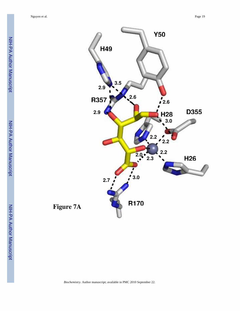
Structures of Inhibitor ComplexesThe crystal structure of Bh0493 from B. halodurans was previously determined in the absenceof bound substrates or inhibitors (PDB code: 2Q08, 3). In that structure zinc is bound in theactive site and coordinated to two histidines from β-strand 1 (H26 and H28) and the invariantaspartate found at the end of β-strand 8 (D355). The structure of Bh0493 was determined inthe presence of the substrates D-glucuronate (I), D-fructuronate, and two inhibitors that mimicthe proposed cis-enediol(ate) intermediate, D-arabinarate (III) and its hydroxamate derivative(II). Portions of the electron density maps that show the D-glucuronate and D-fructuronate inthe active site of Bh0493 are presented in Figures 5a and 5b. These compounds occupy theactive site in a similar manner and interact with the same set of amino acid residues.Stereoscopic images of the active site complexes for compounds I, II, and III are presented inFigures 6A–C and the distances between specific amino acid residues and the bound ligandsare provided in Figures 7A–C.
In the complex with D-glucuronate, the substrate is bound in the open chain configuration. Theterminal carboxylate is ion-paired with the guanidino group of Arg-170 and there is amonodentate coordination with the bound zinc. The zinc is also ligated with the hydroxyl groupattached to C-5 of the substrate. The hydroxyl group at C-4 does not make any specific
Nguyen et al. Page 7
Biochemistry. Author manuscript; available in PMC 2010 September 22.
NIH
-PA Author Manuscript
NIH
-PA Author Manuscript
NIH
-PA Author Manuscript

interactions with the protein, which is consistent with the observation that both D-glucuronateand D-galacturonate are substrates for this enzyme (1). The hydroxyl at C-3 interacts with bothArg-357 and His-49. The nearest residue to the hydroxyl at C-2 of D-glucuronate is Arg-357.The hydroxyl group from Tyr-50 hydrogen bonds with the carbonyl group at C-1 of thesubstrate. The closest residue to the hydrogen that is abstracted from C-2 of D-glucuronate isthe side chain carboxylate of Asp-355. Similar interactions are found in the complexes withD-fructuronate, D-arabinarate and the corresponding hydroxamate derivative. The D-fructuronate complex was identified in a crystal that was grown after a long incubation andsubsequent co-crystallization of Bh0493 with D-glucuronate.
DiscussionThe uronate isomerase from E. coli has previously been shown to catalyze the isomerizationof D-glucuronate and D-galacturonate to D-fructuronate and D-tagaturonate, respectively (1,2). With D-glucuronate the hydrogen at C-2 was shown to be transferred to the pro-R positionat C-1 of the product (12). This hydrogen exchanges with solvent at a rate that is 4 orders ofmagnitude slower than the net interconversion of substrate and product. These results wereinterpreted to be consistent with a reaction mechanism that was initiated by proton abstractionat C-2, formation of a cis-enediol(ate) intermediate and subsequent reprotonation at C-1 (12).This minimal reaction mechanism thus requires at least two different residues within the activefor these proton transfers. A general base (B1:) is required for the removal of the proton at C-2and delivery to C-1 and a general acid (B2:H) is needed to shuttle a proton between the oxygensattached to C-1 and C-2 of the substrate/product pair. This transformation has been summarizedin Scheme 2. A requirement for a minimum of two amino acids that must be in a specific stateof protonation is experimentally supported by the measurement of the effects of pH on themagnitude of kcat and kcat/Km for the conversion of D-glucuronate to D-fructuronate. The pH-rate profiles for both kcat and kcat/Km are bell-shaped and indicate that one residue must beunprotonated and another protonated for catalytic activity. In these profiles the general basehas a kinetic pKa between 5.5 and 5.8 whereas the general acid has a kinetic pKa between 9.5and 10.2. Candidates for these residues were identified through the elucidation of the three-dimensional crystal structure of a uronate isomerase from B. halodurans (Bh0493) in thepresence of D-glucuronate, D-fructuronate, and two mimics of the cis-enediol(ate)intermediate.
The crystal structure of Bh0493 determined with D-glucuronate has identified those residuesin the active site that interact directly with the substrate. In this structure the zinc is ligated bythree amino acids from the protein: the two conserved histidine residues from β-strand 1 andthe aspartate from β-strand 8. The C-6 carboxylate group of the substrate is ligated to the zincand also ion-paired with Arg-170. The hydroxyl group from C-5 is ligated to the zinc in theactive site and hydrogen bonded to the aspartate from β-strand 8. At the other end of thesubstrate, Arg-357 hydrogen bonds with the two hydroxyls from C-3 and C-2. The phenolicgroup of Tyr-50 forms a hydrogen bond with the carbonyl group at C1. The closest residuefrom the protein to the hydrogen at C-2 that must be abstracted during the chemicaltransformation is Asp-355 at 3.15 Å. In addition, His-49 is within hydrogen bonding distanceto the hydroxyl at C-2 in the hydroxamate inhibitor (II) but not in the complex with the boundD-glucuronate (I) Thus, the most likely residues that are required for the isomerization reaction(in Bh0493) are His-49, Tyr-50, Asp-355 and Arg-357. In the E. coli enzyme these residuesare equivalent to His-59, Tyr-60, Asp-412, and Arg-414, respectively. These residues, inaddition to His-33 and His-35 (ligands to the zinc) Arg-186 (equivalent to R170 in Bh0493),His-297 (a conserved histidine at the end of β-stand 5 in most members of the amidohydrolasesuperfamily), Arg-302 and Trp-381, were mutated as probes of functional significance.
Nguyen et al. Page 8
Biochemistry. Author manuscript; available in PMC 2010 September 22.
NIH
-PA Author Manuscript
NIH
-PA Author Manuscript
NIH
-PA Author Manuscript

The mutation of specific residues within the active site of URI results in significantperturbations to the magnitude of the kinetic constants for substrate turnover. Changes to eitherof the two histidine residues that originate from the end of β-strand 1 weakens the binding ofzinc to the active site and this results in a diminution of catalytic activity. This observation isconsistent with the proposed role of zinc in the direct ligation of the substrate through the C-6carboxylate and the hydroxyl from C-5. A drastic reduction in the affinity of the substrateoccurs with the mutation of Arg-186 to methionine. In this case the Km for the substrateincreases by nearly two orders of magnitude and the value of kcat/Km is reduced by more thanthree orders of magnitude. This result is consistent with an ion-pair interaction between theC-6 carboxylate and the guanidino group of Arg-186 (R170 in Bh0493) that is observed in theX-ray structure of Bh0493. There is also a substantial increase in the Km for D-glucuronatewhen His-297 is mutated to alanine or asparagine. This residue originates from the end of β-strand 5 and is highly conserved in nearly all members of the amidohydrolase superfamily butthis residue is not conserved in Bh0493 and thus it is not easy to discern the effect on thestructure of URI from E. coli.
The most dramatic reductions in kcat occur with the mutation of Asp-412 to alanine or Arg-414to methionine. Arg-414 is equivalent to Arg-357 from Bh0493, which is hydrogen bonded tothe C-2 and C-3 hydroxyls of the bound D-glucuronate. Therefore, this residue is a suitablecandidate for assisting in the movement of the proton from the hydroxyl at C-2 during thetransformation to D-fructuronate. The other candidate for this process is the phenolic side chainfrom Tyr-60. This group is hydrogen bonded to the carbonyl oxygen at C-1 of D-glucuronatein the X-ray crystal structure. Mutation of Tyr-60 to phenylalanine results in reductions inkcat and kcat/Km of about an order of magnitude. The only residue from the protein that issuitably positioned to function as the general base for the abstraction of the proton from C-2and delivery to C-1 is Asp-412. Many members of the amidohydrolase superfamily thatcatalyze hydrolytic reactions have been shown to use this residue from the end of β-strand 8to abstract a proton from the hydrolytic hydroxide or water (6,8,27,28).
Changes in solvent viscosity and isotopic substitution were used to address the degree of ratelimitation on the bond breaking steps and product release (15,16). A simplified kineticmechanism for the conversion of substrate to product for the uronate isomerization reaction ispresented in Scheme 4, where EA represents the enzyme-glucuronate complex and EPrepresents the enzyme-fructuronate complex. In this mechanism the expression for kcat is givenby (k3k5)/(k3 + k4 + k5). If one assumes that k5 is inversely proportional to the relative solventviscosity then the value of k5, relative to the sum of k3 and k4 can be determined from a plotof the ratio of o(kcat)/η(kcat) as a function of the relative solvent viscosity, η. The slope of thisplot is equal to (k3+ k4)/(k3 + k4 + k5). For the wild-type enzyme the slope was found to be~0.7 and thus the sum of k3 and k4 is greater than the product release step, k5. This result isconsistent with the release of the product as the rate limiting step for the wild type enzyme andthe relatively small primary isotpe effect for [2-2H]-D-glucuronate. With the D412N andR412M mutants the slope of this plot, for changes in the relative value of kcat as a function ofsolvent viscosity, is reduced substantially. This result is consistent with a significant reductionin the rate constants for the interconversion of the substrate/product pair to the point wherek5 is now greater than the sum of k3 and k4. This result is also consistent with the significantincrease in the value of Dkcat. With the D412N and R414M mutants, the primary isotope effectsare 2.0 and 3.2, respectively, and thus with these two mutants the interconversion of thesubstrate/product pair is substantially rate limiting.
Mechanism of ActionBased upon the X-ray crystal structure of Bh0493 in the presence of the bound substrate, thecatalytic properties of selected site-directed mutants and the stereochemical constraints for the
Nguyen et al. Page 9
Biochemistry. Author manuscript; available in PMC 2010 September 22.
NIH
-PA Author Manuscript
NIH
-PA Author Manuscript
NIH
-PA Author Manuscript

conversion of D-glucuronate to D-fructuronate, a minimal chemical mechanism can be writtenfor uronate isomerase. In the proposed mechanism for URI from E. coli, D-glucuronate is boundin the active site through electrostatic interactions to five highly conserved amino acid residuesand the divalent cation. The carboxylate group at C-6 is coordinated to the divalent cation andArg-186. The hydroxyl group at C-5 is also coordinated to the zinc. The hydroxyl groups atC-3 and C-2 interact with the side chain guanidino group of Arg-414 and the hydroxyl at C-2is also apparently able to hydrogen bond to His-59. The carbonyl group at C-1 is hydrogenbonded to the phenolic oxygen of Tyr-60. The pH-rate profiles for URI are consistent with twoamino acid residues that must be in a specific state of protonation for catalytic activity. Thegeneral base, with a kinetic pKa of approximately 5.8, is consistent with Asp-412. This residueis conserved in all members of the amidohydrolase superfamily and for those enzymes thatcatalyze hydrolytic reactions it has been shown to initiate proton transfers from water/hydroxide to the leaving group (6,8,27,28). The general acid, with a kinetic pKa ofapproximately 10.2 from the pH-rate profiles, may be due to Tyr-60 but it is difficult to excludea role for Arg-414.
In the simplest mechanism D-glucuronate binds in the open chain conformation in the activesite and then Asp-412 abstracts the proton from C-2 as the carbonyl group of C-1 is protonatedby Tyr-60 to form a cis-enediol intermediate. In the subsequent step, Tyr-60 abstracts the protonfrom the hydroxyl at C-2 as Asp-414 delivers a proton to C-1 with proR stereochemistry. Itshould be noted that the mutation of Tyr-60 to alanine or phenylalanine diminishes kcat by onlya factor of 10. This reduction in rate is perhaps smaller than what may be expected for this rolein catalysis. However, the lack of a primary deuterium isotope effect for the wild-type enzymeindicates that the chemical step is not rate-limiting. In addition, a water molecule may substitutefor the phenolic group in the mutant enzymes. Additional mechanisms can be written that utilizea combination of Tyr-60, Arg-414, and His-59 to facilitate the proton movements between theoxygens at C-2 and C-1 within the cis-enediol intermediate and in the opening of thehemiacetal. The proposed reaction mechanism for URI is different from that of metallo-enzymexylose isomerase since that enzyme catalyzes a hydride transfer mechanism rather than a protontransfer mechanism (29).
An alternative mechanism can be proposed in which Asp-412 abstracts the proton from thehydroxyl at C-5, which then abstracts a proton from C-2 to initiate the formation of the cis-enediol intermediate. The hydroxyl at C-5 is additionally activated through direct ligation tothe bound zinc. These two variations are presented graphically in Figure 8. This lattermechanism is particularly attractive since it retains elements that are common to nearly allmembers of the amidohydrolase superfamily that have been interrogated mechanistically (6,8,27,28). The structural similarities in the active site of uronate isomerase with those membersof the amidohydrolase superfamily nicely illustrates the evolutionary link between thosemembers of the AHS that catalyze hydrolytic reactions and those that catalyze 1,2-protontransfers. A structural alignment of the active sites of Bh0493/D-glucuronate anddihydroorotase (DHO)/dihydroorotate (PDB entry code 1j79) supports this proposition asillustrated in Figure 9. In this structural alignment one of the C-6 carboxylate oxygens fromD-glucuronate is positioned in nearly the same place as the carboxylate oxygen of the bridgingcarbamate functional group in DHO. These oxygen atoms interact directly with the alpha-metal(Mα) in their respective structures. Moreover, the C-5 oxygen of D-glucuronate is positionedin the same way as the nucleophilic hydroxide in DHO and is oriented to favor protonabstraction by Asp-412 at a distance of 2.9 Å. The C-5 hydroxyl is 2.0 Å away from thehydrogen at C-2 of the bound substrate.
Nguyen et al. Page 10
Biochemistry. Author manuscript; available in PMC 2010 September 22.
NIH
-PA Author Manuscript
NIH
-PA Author Manuscript
NIH
-PA Author Manuscript

References1. Ashwell G, Wahba AJ, Hickman J. Uronic acid metabolism in bacteria. I. Purification and properties
of uronic acid isomerases in Escherichia coli. J Biol Chem 1960;235:1559–1565. [PubMed: 13794771]2. Wahba AJ, Hickman J, Ashwell G. Uronic acid metabolism in bacteria. III. Purification and properties
of D-altronic acid and D-mannonic acid dehydrogenases in Escherichia coli. J Biol Chem1960;235:1566–1570. [PubMed: 14401695]
3. Nguyen TT, Brown S, Fedorov AA, Fedorov EV, Babbitt PC, Almo SC, Raushel FM. At the peripheryof the amidohydrolase superfamily: Bh0493 from Bacillus halodurans catalyzes the isomerization ofD-galacturonate to D-tagaturonate. Biochemistry 2008;47:1194–1206. [PubMed: 18171028]
4. Siebert CM, Raushel FM. Structural and catalytic diversity within the amidohydrolase superfamily.Biochemistry 2005;44:6383–6391. [PubMed: 15850372]
5. Holm L, Sander C. An evolutionary treasure: unification of a broad set of amidohydrolases related tourease. Proteins 1997;28:72–82. [PubMed: 9144792]
6. Porter TN, Li Y, Raushel FM. Mechanism for the dihydroorotase reaction. Biochemistry2004;43:16285–16292. [PubMed: 15610022]
7. Benini S, Rypniewski WR, Wilson KS, Miletti S, Ciurli S, Mangani S. A new proposal for ureasemechanism based on the crystal structures of the native and inhibited enzyme from Bacilluspasteurii: Why urea hydrolysis costs two nickels. Struct Folding Des 1999;7:205–216.
8. Aubert SA, Li Y, Raushel FM. Mechanism for the hydrolysis of organophosphates by the bacterialphosphotriesterase. Biochemistry 2004;43:5707–5715. [PubMed: 15134445]
9. Wilson DK, Rudolph FB, Quiocho FA. Atomic structure of adenosine deaminase complexed with atransition state analog: Understanding catalysis and immunodeficiency mutation. Science1991;252:1278–1284. [PubMed: 1925539]
10. Ireton GC, McDermott G, Black ME, Stoddard BL. The structure of Escherichia coli cytosinedeaminase. J Mol Biol 2002;315:687–697. [PubMed: 11812140]
11. Roy JE, Roy KL. The mechanism and specificity of guanine deaminase. Can J Biochem1967;45:1263–1269. [PubMed: 6067951]
12. Williams L, Nguyen T, Li Y, Porter TN, Raushel FM. Uronate isomerase: A nonhydrolytic memberof the amidohydrolase superfamily with an ambivalent requirement for a divalent metal ion.Biochemistry 2006;45:7453–7462. [PubMed: 16768441]
13. Sowden JC, Oftedahl ML. Anhydridization of 1-deoxy-1-nitrohexitols. J Org Chem 1961;26:1974–1977.
14. Dromowicz M, Koll P. Synthesis of 2,6-anhydroaldonic acids from the correspondinganhydrodeoxynitroalditols (glycopyranosylnitromethanes) and their conversion into methyl esters,amides, and alditols. Carbohydrate Research 1998;311:103–119.
15. Brouwer AC, Kirsch JF. Investigation of diffusion-limited rates of chymotrypsin reactions byviscosity variation. Biochemistry 1982;21:1302–1307. [PubMed: 7074086]
16. Maurice M, Bearne SL. Kinetics and thermodynamics of mandalate racemase catalysis. Biochemistry2002;41:4048–4058. [PubMed: 11900548]
17. Podlasek CA, Wu J, Atripe WA, Bondo PB, Serianni AS. [13C]-Enriched methyl aldopyranosides:Structural interpretations of 13C-1H spin-coupling constants and 1H chemical shifts. J Am Chem Soc1995;117:8635–8644.
18. de Nooy AEJ, Besemer AC, van Bekkum H. Highly selective nitroxyl radical-mediated oxidation ofprimary alcohol groups in water-soluble glucans. Carbohydrate Research 1995;269:89–98.
19. Bragd PL, van Bekkum H, Besemer AC. TEMPO-mediated oxidation of polysaccharides: survey ofmethods and applications. Topics in Catalysis 2004;27:49–66.
20. Otwinowski, Z.; Minor, W. Processing of X-ray diffraction data collected in oscillation mode. In:Carter, CWJ.; Sweet, RM.; Abelson, JN.; Simon, MI., editors. Methods in Enzymology. AcademicPres; New York: 1997. p. 307-326.
21. Long F, Vagin A, Young P, Murshudov GN. BALBES: a Molecular Replacement Pipeline. ActaCrystallogr D 2008;64:125–132. [PubMed: 18094476]
22. Jones AT. Interactive computer graphics: FRODO. Methods Enzymol 1985;115:157–171. [PubMed:3841179]
Nguyen et al. Page 11
Biochemistry. Author manuscript; available in PMC 2010 September 22.
NIH
-PA Author Manuscript
NIH
-PA Author Manuscript
NIH
-PA Author Manuscript

23. Brunger AT, Adams PD, Clore GM, DeLAno WL, Gros P, Grosse-Kunstleve RW, Jiang JS,Kuszewski J, Nilges M, Pannu NS, Read RJ, Rice LM, Simonson T, Warren GL. Crystallography &NMR system: A new software suite for macromolecular structure determination. Acta Crystallogr1998;D54
24. Lamzin VS, Wilson KS. Automated refinement of protein models. Acta Crystallogr 1993;D49:129–147.
25. Collaborative Computational Project No. 4. The CCP4 suite: Programs for Protein Crystallography.Acta Crystallogr 1994;D50:760–763.
26. Hardy LW, Kirsch JK. Diffusion-limited component of reactions catalyzed by Bacillus cereus β-lactamases I. Biochemistry 1984;23:1275–1282. [PubMed: 11491129]
27. Hall RS, Xiang DF, Xu C, Raushel FM. N-acetyl-D-glucosamine-6-phosphate deacetylase: Substrateactivate via a single divalent metal ion. Biochemistry 2007;46:7942–7952. [PubMed: 17567047]
28. Marti-Arbona R, Fresquet V, Thoden JB, Davis ML, Holden HM, Raushel FM. Mechanism of thereaction catalyzed by isoaspartyl dipeptidase from Escherichia coli. Biochemistry 2005;44:7115–7124. [PubMed: 15882050]
29. Allen KN, Lavie A, Farber GK, Glasfeld A, Petsko GA, Ringe D. Isotopic exchange plus substrateand inhibition kinetics of D-xylose isomerase do not support a proton-transfer mechanism.Biochemistry 1994;33:1481–1487. [PubMed: 8312268]
Nguyen et al. Page 12
Biochemistry. Author manuscript; available in PMC 2010 September 22.
NIH
-PA Author Manuscript
NIH
-PA Author Manuscript
NIH
-PA Author Manuscript
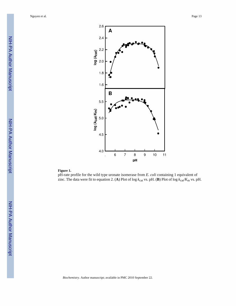
Figure 1.pH-rate profile for the wild type uronate isomerase from E. coli containing 1 equivalent ofzinc. The data were fit to equation 2. (A) Plot of log kcat vs. pH. (B) Plot of log kcat/Km vs. pH.
Nguyen et al. Page 13
Biochemistry. Author manuscript; available in PMC 2010 September 22.
NIH
-PA Author Manuscript
NIH
-PA Author Manuscript
NIH
-PA Author Manuscript

Figure 2.Double-reciprocal plots for the solvent isotope effect (H2O vs D2O) where 1/v (s) is plottedagainst 1/[D-glucuronate] (mM−1). The data sets in H2O are represented by circles and thesquares correspond to the values obtained in D2O. (A) represents the plot for the wild typeenzyme, (B) for the R414M mutant, and (C) is the plot for the D412N mutant.
Nguyen et al. Page 14
Biochemistry. Author manuscript; available in PMC 2010 September 22.
NIH
-PA Author Manuscript
NIH
-PA Author Manuscript
NIH
-PA Author Manuscript

Figure 3.Primary isotope effects using protonated and deuterated D-glucuronate at the C-2 position arepresented as double-reciprocal plots where (A) is the plot for the wild-type enzyme, (B) R414Mmutant, and (C) D412N mutant. The data for the protonated substrate are represented as circles,and the values for the deuterated substrate are denoted as squares.
Nguyen et al. Page 15
Biochemistry. Author manuscript; available in PMC 2010 September 22.
NIH
-PA Author Manuscript
NIH
-PA Author Manuscript
NIH
-PA Author Manuscript

Figure 4.The effect of viscosity on the relative values of kcat (A) and kcat/Km (B) using sucrose as themicroviscogen. The circles indicate the wild type enzyme from E. coli, the squares representthe data set for the D412N mutant, and the triangles signify the values for the R414M mutant.
Nguyen et al. Page 16
Biochemistry. Author manuscript; available in PMC 2010 September 22.
NIH
-PA Author Manuscript
NIH
-PA Author Manuscript
NIH
-PA Author Manuscript

Figure 5.View of the active site structures of Bh0493 with bound ligands: (A) D-glucuronate; (B) D-fructuronate. Omit electron density maps (Fo−Fc) are contoured at 3.5σ. The correspondingligands were omitted from the model and the remainder of the unit cell was subjected to a cycleof simulated annealing with CNS at 3000 °C.
Nguyen et al. Page 17
Biochemistry. Author manuscript; available in PMC 2010 September 22.
NIH
-PA Author Manuscript
NIH
-PA Author Manuscript
NIH
-PA Author Manuscript

Figure 6.Stereoview images of Bh0493 in the presence of bound (A) D-glucuronate (I), (B) hydroxamateof arabinarate (II), and (C) arabinarate (III). This figure was created with PyMOL v0.99.
Nguyen et al. Page 18
Biochemistry. Author manuscript; available in PMC 2010 September 22.
NIH
-PA Author Manuscript
NIH
-PA Author Manuscript
NIH
-PA Author Manuscript
Nguyen et al. Page 19
Biochemistry. Author manuscript; available in PMC 2010 September 22.
NIH
-PA Author Manuscript
NIH
-PA Author Manuscript
NIH
-PA Author Manuscript

Nguyen et al. Page 20
Biochemistry. Author manuscript; available in PMC 2010 September 22.
NIH
-PA Author Manuscript
NIH
-PA Author Manuscript
NIH
-PA Author Manuscript

Figure 7.Active site of Bh0493 with (A) D-glucuronate, (B) hydroxamate of D-arabinarate, and (C) D-arabinarate. Interactions between the enzyme and ligand with the distances are shown.
Nguyen et al. Page 21
Biochemistry. Author manuscript; available in PMC 2010 September 22.
NIH
-PA Author Manuscript
NIH
-PA Author Manuscript
NIH
-PA Author Manuscript

Figure 8.Proposed mechanisms for the isomerization of D-glucuronate by URI.
Nguyen et al. Page 22
Biochemistry. Author manuscript; available in PMC 2010 September 22.
NIH
-PA Author Manuscript
NIH
-PA Author Manuscript
NIH
-PA Author Manuscript

Figure 9.A structural alignment for portions of the active sites of Bh0493 (green) and DHO from E.coli (blue). The alpha metal (Mα) is shown as a yellow sphere for Bh0493 and in grey for DHO.The bound D-glucuronate for Bh0493, is shown in purple. The interactions between the C-5oxygen of D-glucuronate and Mα, C-2 hydrogen and Asp-412 are indicated by dashed lines.The other ligands to the metal ion are shown as stick representations, including the HxH fromβ-strand 1 and aspartate from β-strand 8. The C-6 oxygen of D-glucuronate, aligns closely thethe position occupied by one of the oxygens from the carboxyl group of the carboxylated lysinefrom strand 4 of DHO. The C-5 hydroxyl of D-glucuronate is orientated in the same positionas the bridging hydroxide in the DHO structure.
Nguyen et al. Page 23
Biochemistry. Author manuscript; available in PMC 2010 September 22.
NIH
-PA Author Manuscript
NIH
-PA Author Manuscript
NIH
-PA Author Manuscript

Scheme 1.
Nguyen et al. Page 24
Biochemistry. Author manuscript; available in PMC 2010 September 22.
NIH
-PA Author Manuscript
NIH
-PA Author Manuscript
NIH
-PA Author Manuscript

Scheme 2.
Nguyen et al. Page 25
Biochemistry. Author manuscript; available in PMC 2010 September 22.
NIH
-PA Author Manuscript
NIH
-PA Author Manuscript
NIH
-PA Author Manuscript

Scheme 3.
Nguyen et al. Page 26
Biochemistry. Author manuscript; available in PMC 2010 September 22.
NIH
-PA Author Manuscript
NIH
-PA Author Manuscript
NIH
-PA Author Manuscript

Scheme 4.
Nguyen et al. Page 27
Biochemistry. Author manuscript; available in PMC 2010 September 22.
NIH
-PA Author Manuscript
NIH
-PA Author Manuscript
NIH
-PA Author Manuscript

NIH
-PA Author Manuscript
NIH
-PA Author Manuscript
NIH
-PA Author Manuscript
Nguyen et al. Page 28Ta
ble
1
Dat
a co
llect
ion
and
refin
emen
t sta
tistic
s for
cry
stal
s of t
he c
ompl
exes
of u
rona
te is
omer
ase
from
Bac
illus
Hal
odur
ans w
ith v
ario
us li
gand
s.U
RI·D
-Ara
bina
rate
UR
I·D-A
rabi
nara
teU
RI·A
rabi
nohy
drox
amat
eUR
I·D-G
lucu
rona
teU
RI·D
-Fru
ctur
onat
eD
ata
colle
ctio
nSp
ace
grou
pR
32C
2R
32C
2P4
1 22
No.
of m
ol. i
n as
ym. u
nit2
122
123
Cel
l dim
ensi
ons
a,
b, c
(Å)
149.
35,1
49.3
5,25
4.16
274.
82,1
56.5
2,18
5.96
149.
30, 1
49.3
0, 2
54.0
227
4.09
,156
.88,
185.
2184
.35,
99.1
4,12
6.31
β(
°)11
6.20
115.
78R
esol
utio
n (Å
)2.
22.
22.
22.
11.
9N
o. o
f uni
que
refle
ctio
ns55
294
3522
8855
174
4041
2212
8635
R mer
ge0.
058
0.06
50.
071
0.07
40.
086
I/σI
27.1
19.3
20.6
15.2
17.1
Com
plet
enes
s (%
)99
.798
.899
.898
.793
.9R
efin
emen
tR
esol
utio
n (Å
)25
.0-2
.225
.0-2
.225
.0-2
.225
.0-2
.125
.0-1
.9R
crys
t0.
218
0.21
30.
204
0.22
70.
213
R fre
e0.
238
0.24
50.
226
0.25
80.
241
Prot
ein
atom
s67
6640
860
6744
4086
010
164
Wat
ers
259
1880
312
1824
572
Rm
sd, b
ond
leng
ths (
Å)
0.00
70.
006
0.00
70.
006
0.00
6R
msd
, bon
d an
gles
(°)
1.3
1.3
1.3
1.3
1.3
Bou
nd in
hibi
tor
D-A
rabi
nara
teD
-Ara
bina
rate
Ara
bino
hydr
oxam
ate
D-G
lucu
rona
teD
-Fru
ctur
onat
eIn
hibi
tor a
tom
s24
144
2615
639
Bou
nd io
ns4Z
n2+, 2
CO
3 2−, 2
Cl−
12Zn
2+,1
2CO
32 ,4N
a+1,4
Cl−
4Zn2+
, 2C
O32−
, 2C
l−12
Zn2+
, 12C
O32−
,4C
l−4Z
n2+, 3
CO
32−, 1
Cl −
PDB
ent
ry3H
K5
3HK
73H
K8
3HK
93H
KA
Biochemistry. Author manuscript; available in PMC 2010 September 22.

NIH
-PA Author Manuscript
NIH
-PA Author Manuscript
NIH
-PA Author Manuscript
Nguyen et al. Page 29
Table 2
Kinetic parameters and metal content of mutants of URI from E. colia
Enzyme kcat (s−1) Km (mM) kcat/Km (M−1 s−1) URI/Zn
WT 196 ± 6 0.50 ± 0.05 (4.0 ± 0.4) × 105 0.90H33N (H26)b 2.1 ± 0.1 (5.0 ± 0.4) × 10−2 (4.7 ± 0.4) × 104 0.07H33A 0.60 ± 0.01 0.20 ± 0.01 (3.0 ± 0.2) × 103 0.20H35N (H28) 4.0 ± 0.2 9.4 ± 1.1 (4.3 ± 0.5) × 102 <0.05H35A 0.70 ± 0.04 39 ± 5 18 ± 2 <0.05H59N (H49) 15 ± 1 0.70 ± 0.04 (2.1 ± 0.1) × 104 0.96H59A 0.60 ± 0.01 0.70 ± 0.07 (8.3 ± 0.1 × 10² 0.95Y60F (Y50) 21.7 ± 0.1 0.16 ± 0.01 (1.4 ± 0.1) × 105 0.8Y60A 13.9 ± 0.1 0.21 ± 0.01 (6.6 ± 0.3) × 104 0.8R186K (R170) 54 ± 2 2.6 ± 0.2 (2.1 ± 0.2) × 104 0.94R186M 4.7 ± 0.1 38 ± 3 (1.3 ± 0.1) × 102 0.91D238N 60 ± 1 1.3 ± 0.1 (4.6 ±0.1) × 104 0.70H297N (M258) 30 ± 2 56 ± 5 (5.0 ± 0.5) × 102 1.00H297A 10 ± 1 (2.2 ± 0.3) × 102 43 ± 7 0.41R302K (K303) 160 ± 4 2.5 ± 0.2 (6.3 ± 0.5) × 104 0.90R302M 180 ± 9 (2.0 ± 0.3) × 102 (8.8 ± 1.3) × 102 0.99W381F (W325) 16 ± 1 1.7 ± 0.1 (9.5 ± 0.4) × 103 0.90W381A 250 ± 6 21± 2 (1.2 ± 0.1) × 104 0.69D412N (D355) 0.60 ± 0.01 1.00 ± 0.04 (6.0 ± 0.3) × 102 0.36D412A (9.0 ± 0.3) × 10−3 0.40 ± 0.05 21 ± 3 0.12R414K (R357) 5.8 ± 0.1 0.82 ± 0.02 (7.1 ± 0.2) × 103 0.92R414M 0.70 ± 0.01 1.4 ± 0.1 (5.4 ± 0.2) × 102 0.91
aThese data were obtained at 30 °C, pH 8.0, with D-glucuronate as the substrate.
bThe corresponding residue numbers for Bh0493.
Biochemistry. Author manuscript; available in PMC 2010 September 22.
Copyright © 2022 FDOKUMEN