Essentials of Life Span Development 4th Edition Santrock ...

Molecular Cell
Article
Elevated Histone ExpressionPromotes Life Span ExtensionJason Feser,2 David Truong,2 Chandrima Das,2 Joshua J. Carson,2 Jeffrey Kieft,2,3 Troy Harkness,4
and Jessica K. Tyler1,2,*1Department of Biochemistry and Molecular Biology, University of Texas MD Anderson Cancer Center, Houston, TX 77030, USA2Department of Biochemistry and Molecular Genetics, University of Colorado School of Medicine, Aurora, CO 80010, USA3Howard Hughes Medical Institute4Department of Anatomy and Cell Biology, College of Medicine
University of Saskatchewan, Saskatoon, Saskatchewan S7N 5E5, Canada
*Correspondence: [email protected] 10.1016/j.molcel.2010.08.015
SUMMARY
Changes to the chromatin structure accompanyaging, but the molecular mechanisms underlyingaging and the accompanying changes to the chro-matin are unclear. Here, we report a mechanismwhereby altering chromatin structure regulates lifespan. We show that normal aging is accompaniedby a profound loss of histone proteins from thegenome. Indeed, yeast lacking the histone chap-erone Asf1 or acetylation of histone H3 on lysine 56are short lived, and this appears to be at least partlydue to their having decreased histone levels.Conversely, increasing the histone supply by inacti-vation of the histone information regulator (Hir)complex or overexpression of histones dramaticallyextends life span via a pathway that is distinct frompreviously known pathways of life span extension.This study indicates that maintenance of the funda-mental chromatin structure is critical for slowingdown the aging process and reveals that increasingthe histone supply extends life span.
INTRODUCTION
Mitotic cells divide a finite number of times (replicative life span)
before they exhibit signs of senescence and cease replicating.
The length of time that a postmitotic or senescent cell exists in
a nondividing state, termed chronological life span, also has
a profound influence on the physiology and longevity of an
organism. Gradual alterations of biological macromolecules
characterize normal aging, whereas adverse alterations to
macromolecules are risk factors for age-related conditions and
disease states. Of note, aging is the single highest risk factor
for the majority of humanmalignancies. Despite the fundamental
importance of the aging process, there are still huge gaps in our
knowledge as to the biological changes that lead to aging and
the molecular causes of these changes.
724 Molecular Cell 39, 724–735, September 10, 2010 ª2010 Elsevier
Several important cellular processes have been implicated in
the regulation of yeast replicative life span, including glucose
sensing, nutrient sensing, stress response, mitochondrial
function, and transcriptional silencing (Steinkraus et al., 2008).
The conservation of the fundamental mechanisms of aging and
life span determination across eukaryotes (Smith et al., 2007,
2008) enables the study of aging in budding yeast. Determining
how replicative life span can be extended in yeast will ultimately
help facilitate the development of therapeutic regimens to
extend life span and delay the onset of age-associated disease
in humans.
It is clear that the packaging of the eukaryotic genome
together with histones to form the chromatin structure plays
a critical role in regulating the activities of the genome (Groth
et al., 2007; Li et al., 2007). Tightly packaged chromatin structure
reduces access to the DNA to limit inappropriate gene
expression and genomic instability, whereas an open chromatin
structure promotes unregulated gene expression and genomic
instability. Of note, increased genomic instability and inappro-
priate transcription are associated with increased age (Busuttil
et al., 2007; Maslov and Vijg, 2009). A clear example of the
influence of chromatin structure on aging is provided by the
Sir2/Hst2 NAD-dependent protein and histone deacetylases
that have a key role in transcriptional silencing and maintenance
of chromatin structure. Inactivation of Sir2 or Hst2 shortens life
span (Lamming et al., 2005), whereas introduction of an extra
copy of the gene encoding Sir2 extends life span (Kaeberlein
et al., 1999). The role of Sir2/Hst2 in yeast aging is likely related
to its ability to inhibit formation of extrachromosomal rDNA
circles (ERC) (Kaeberlein et al., 1999) by repressing rDNA
recombination (Gottlieb and Esposito, 1989). A key substrate
of the deacetylase activity of Sir2 is lysine 16 of histone H4
(Moazed, 2001). Sir2 protein levels normally decrease during
aging, leading to increased levels of acetylated H4 K16 and
concomitant loss of histones from specific subtelomeric regions
of the genome (Dang et al., 2009). However, it is unclear how
altered chromatin structure at the gene-depleted subtelomeric
regions of the genome would lead to aging or if reversed, would
extend life span.
Chromatin is modified by the cell in a myriad of different ways.
The ultimate and most profound chromatin changes are the loss
of histones from the DNA or the deposition of histones onto the
Inc.

A
B
C
Figure 1. Asf1 Is Required for Normal Life Span
(A) Replicative life span of isogenic strains BY4741 (WT) or BY4747asf1 (asf1).
(B) Replicative life span of isogenic strains that were wild-type (DBY746) or
deleted for ASF1 (JFY022) in the DBY746 background often used for aging
studies.
(C) Replicative aging analysis of isogenic yeast strains BY4741 (WT), BY4747-
asf1 (asf1), BY4747sir2 (sir2), and BY4747asf1sir2 (asf1sir2).
Molecular Cell
Chromatin Regulates Aging
DNA. The removal of histones from DNA and the incorporation of
histones onto DNA are mediated by a class of proteins termed
histone chaperones. Antisilencing function 1 (Asf1) is a highly
conserved central chaperone of histones H3 and H4 that is
required for proper regulation of gene expression, histone
acetylation, and the maintenance of genomic integrity (Chen
et al., 2008; Ramey et al., 2004; Tyler et al., 1999). We were
prompted to investigate whether yeast cells lacking Asf1 have
a reduced life span due to the difficulty that we encountered in
obtaining regrowth of asf1 mutant cells from old colonies. Our
studies have led to our uncovering a causal relationship between
chromatin structure and aging while discovering a means to
greatly extend yeast replicative life span via manipulating the
chromatin structure.
RESULTS
Yeast Lacking the Histone Chaperone Asf1Are Short LivedYeast deleted for ASF1 have a very short chronological life span,
shorter than that of yeast lacking the well-known anti-aging
protein Sir2 and comparable to that of yeast lacking superoxi-
dase dismutase 1 (Sod1) (Figure S1 available online). In order
to determine whether yeast lacking Asf1 also have a defect in
replicative life span, we took advantage of the unique
asymmetric nature of cell division in budding yeast. We found
that yeast lacking Asf1 are short lived, having a median replica-
tive life span of about 7 generations in comparison to the median
life span of about 27 generations for wild-type yeast in multiple
yeast backgrounds (Figures 1A and 1B). Whereas shortening
of life span can result from several causes, asf1 mutants exhibit
phenotypes characteristic of aging. Loss of transcriptional
silencing occurs in telomeric regions of aged yeast (Kim et al.,
1996), and asf1 mutants have transcriptional silencing defects
(Le et al., 1997; Singer et al., 1998; Tyler et al., 1999). Also, yeast
lacking the protein Rtt109 that functions in the same pathway as
Asf1 for life span determination (see later) have an elevated rate
of ERC formation (Han et al., 2007b), which correlates with
premature aging.
To investigate whether Asf1 functions in previously estab-
lished pathways of aging, we compared the effect of deletion
of ASF1 to deletion of SIR2 on replicative life span. We found
that asf1 mutants have a more drastic decrease in life span
than sir2 mutants (which have a median life span of 15 genera-
tions) (Figure 1C). Furthermore, yeast lacking both Asf1 and
Sir2 were extremely short lived (median life span of 4 to 5 gener-
ations) (Figure 1C). These data demonstrate that Asf1 and Sir2
are functioning in nonidentical pathways to promote longevity.
Next, we set out to determine through which molecular
pathway Asf1 mediates attainment of a full replicative life span.
Asf1 promotes replication-dependent chromatin assembly in
collaboration with the histone chaperone CAF-1 (Tyler et al.,
1999). Inactivation of CAF-1 via deletion of the CAC1 gene
encoding the largest subunit of CAF-1 (Kaufman et al., 1997)
shortens yeast replicative life span but to a lesser degree than
deletion of ASF1 (Figure 2A). In agreement, the double cac1asf1
mutant had a life span that was shorter than either single mutant
(Figure 2A), indicating that Asf1 is not solely influencing life span
Molecu
via its role in replication-dependent chromatin assembly.
Genomic instability contributes to aging, and Asf1 has been
shown to play a role in the maintenance of genomic stability,
apparently due to its role in reducing the amount of DNA damage
occurring during DNA replication (Myung et al., 2003; Ramey
et al., 2004; Tyler et al., 1999). Of note, yeast lacking the check-
point protein Mrc1 that normally senses replicational stress
(Robert et al., 2006) do not have a reduced life span even though
lar Cell 39, 724–735, September 10, 2010 ª2010 Elsevier Inc. 725
A B
DC
E F
Figure 2. Genetic Identification of the Pathway through which Asf1 Regulates Replicative Life Span
(A) Replicative aging analysis of wild-type W303 yeast (WT) or this strain deleted for ASF1 (asf1), CAC1 (cac1), or both ASF1 and CAC1 (asf1cac1).
(B) Isogenic strains that were wild-type (BY4741) or deleted for ASF1 or MRC1 from the genome-wide deletion collection in the S288c strain background were
analyzed for replicative life span.
(C) Replicative aging analysis of yeast strains YB (WT), ZGY608 (asf1), ZGY906 (rtt109), and ZGY964 (asf1rtt109).
(D) Replicative aging analysis of yeast strains HMY152 (WT), JFY004 (asf1), HMY139 (H3 K56Q), HMY140 (H3 K56R), JFY005 (H3 K56Q asf1), and JFY006
(H3 K56R asf1).
(E) Replicative aging analysis of isogenic yeast strains BY4741 (WT) and YNML7 (hst3hst4).
(F) Replicative aging analysis of isogenic yeast strains SHY0015 (WT) and SHY0014 (pGALAsf1). 0.5% galactose leads to endogenous levels of Asf1, whereas 1%
galactose leads to higher than endogenous levels of Asf1 (Zabaronick and Tyler, 2005).
Molecular Cell
Chromatin Regulates Aging
they also experience genomic instability occurring during DNA
replication (Figure 2B). These data suggest that the short replica-
tive life span of asf1mutants is unlikely due to genomic instability
during DNA replication.
The Role of Asf1 in Longevity Is Due to Asf1 FacilitatingAcetylation of Histone H3 on Lysine 56Asf1 has an additional function in helping several histone acetyl
transferases (HATs) acetylate their histone substrates (Adkins
726 Molecular Cell 39, 724–735, September 10, 2010 ª2010 Elsevier
et al., 2007; Fillingham et al., 2008; Han et al., 2007a). To examine
whether the function of Asf1 in promoting histone acetylation by
the HAT Rtt109 was responsible for normal life span, we
compared the effect of deletion of ASF1 and RTT109 on replica-
tive aging. The replicative life span was indistinguishable
between strains deleted for ASF1, RTT109, or both ASF1 and
RTT109 together (Figure 2C), demonstrating that Asf1 and
Rtt109 function together in the same pathway to promote
achievement of a full life span.
Inc.

Molecular Cell
Chromatin Regulates Aging
Asf1 and Rtt109 together mediate acetylation of histone H3 on
lysine 56 and lysine 9 (Fillingham et al., 2008; Han et al., 2007a).
To determine which lysine was the critical acetylation target for
anti-aging, we measured the replicative life span of yeast unable
to acetylate K56 using a strain bearing a mutation in the sole
copy of the H3 gene that leads to a lysine to arginine (K56R)
substitution. Note that, although these strains only have one
pair of genes expressing H3/H4 (HHT2/HHF2), they produce
equivalent amounts of histone proteins as a strain with both pairs
of genes expressing H3/H4 (Figure S2). The H3 K56Rmutant had
a reduced life span (Figure 2D) (Dang et al., 2009), which was not
quite as short as that of yeast with unacetylated K56 (such as the
asf1 or rtt109mutants). This is presumably because the arginine
is unlikely to fully mimic the lysine for forming the proper histone-
DNA contacts mediated by K56 within the nucleosome structure
(Luger et al., 1997) and is consistent with the increased
chromatin accessibility and reduced transcriptional silencing
that occurs in yeast carrying the H3 K56R mutation (Xu et al.,
2007). Nevertheless, the drastically reduced life span of the H3
K56R mutant clearly shows that failure to acetylate histone H3
on K56 results in a shortened life span (Figure 2D). Furthermore,
deletion of ASF1 did not exacerbate the effect of the H3 K56R
mutation on life span (Figure 2D). As such, the role of Asf1 in
determining a normal life span is mediated via acetylation of
histone H3 on K56.
Next, we examined the consequence of having too much H3
K56 acetylation on replicative life span. To do this, we first
used a strain in which the only copy of H3 had been mutated
such that lysine 56 is substituted for glutamine (H3 K56Q) to
mimic permanent acetylation. The H3 K56Qmutant had a greatly
shortened life span, which was only slightly longer than that of
the asf1 and rtt109 mutants (Figure 2D) (Dang et al., 2009).
To rule out the possibility that the shortened life span of the H3
K56Q mutant was due to the glutamine not completely
mimicking acetylation of H3 K56, we examined the replicative
life span of yeast deleted for the genes encoding the two partially
redundant histone deacetylases that remove the acetyl group
from H3 K56Ac: Hst3 and Hst4 (Maas et al., 2006). Deletion of
HST3 and HST4 leads to a greatly elevated level of H3 K56Ac
(Maas et al., 2006), and the hst3hst4 double mutant had an
extremely short replicative life span (Figure 2E) (Tsuchiya et al.,
2006). Because it is not yet known whether deletion of HST3
and HST4 leads to alterations in the levels of other histone
modifications, we took another approach to increase the level
of acetylated H3 K56Ac in the yeast cells. We had previously
shown that overexpression of Asf1 leads to elevated levels of
H3K56Ac (Adkins et al., 2007). Using a strain in which the endog-
enous ASF1 gene was under the control of the galactose-induc-
ible GAL1 promoter (Zabaronick and Tyler, 2005), we found that
induction of Asf1 with 0.5% galactose leads to a 19% reduction
in the median life span, whereas inducing even more Asf1 with
1% galactose leads to a 28% reduction in the median life span
(Figure 2F). These data demonstrate that, to achieve normal
life span, it is not only important to be able to acetylate H3 on
K56, but it may be equally important to be able to deacetylate
H3 K56Ac. Alternatively, there may be a delicate balance or
amount of acetylated H3 K56Ac that is required for achieving
a normal life span.
Molecu
Histone Protein Levels Drop during AgingWewondered whether the short life span of cells that fail to acet-
ylate histoneH3 onK56might be related to the requirement of H3
K56Ac and Asf1 for accurate expression of the cell-cycle-regu-
lated histone genes (Sutton et al., 2001; Xu et al., 2005): HHT1
and HHT2 encoding H3, HHF1 and HHF2 encoding H4, HTB1
encoding H2B, and HTA1 encoding H2A. Expression of these
cell-cycle-regulated histone genes is repressed outside of
S phase by the histone information regulator (Hir) complex
(Spector and Osley, 1993; Spector et al., 1997). Consistent
with earlier studies, we find histone transcript levels are altered
upon deletion of ASF1, RTT109, or HIR1 (Figures S3 and 3A).
To investigate whether histone transcript levels change during
the normal aging process, we biochemically separated young
and old yeast cells. We observed a significant increase in histone
transcripts in the aging population of wild-type yeast (Figures 3A,
S3B, and S3C). In agreement with the role of H3 K56Ac for accu-
rate expression of the cell-cycle-regulated histone genes, we
found that the short-lived rtt109D mutant also had significantly
lower levels of these transcripts compared to wild-type strains
when the data were normalized to tubulin levels (Figures S3A
and S3B). Aging or deletion of ASF1 orRTT109 leads to an accu-
mulation of cells with a G2/M DNA content (Figure S4A). Toward
understanding the possible effects of altered histone transcrip-
tion on nucleosome density, we decided to correct for these
cell-cycle defects by normalizing the transcript levels to DNA
content. Accordingly, when we independently normalized the
histone transcript levels to the amount of DNA per cell, histone
transcript levels were slightly higher in young asf1 and rtt109
mutant cells than young wild-type cells (Figures 3A and S3C).
However, although histone transcripts also increased in the
asf1 and rtt109 mutant cells during the aging process, this was
to a much lesser degree than in wild-type cells (Figure 3A).
Given that the levels of histone transcripts increase during
aging, we asked whether histone protein levels also increase
during aging. We loaded equal amounts of total proteins from
young, old, or very old wild-type yeast and determined the levels
of histone H3 by quantitative western analysis. The levels of H3
and H2A protein drastically decrease during aging in wild-type
yeast (Figures 3B and S4B). We see a very similar drastic
decrease in histone levels during aging when we normalize to
DNA content (Figure S4C). It is possible that the increased abun-
dance of histone transcripts during aging (Figure 3A) is the cell’s
attempt to replace thehistoneproteins that are lostor notproperly
synthesized during aging. Consistent with the histone transcript
analyses above, the short-lived rtt109 and asf1 mutants have
significantly lower levels of total histone H3 protein than the
isogenic wild-type strain (Figure S4D). Furthermore, it is clear
that the total histone protein levels also decrease during aging
in the short-lived rtt109 mutant and the hir1mutant (Figure S4E).
Moreover, the relative amount of histone H3 protein in the young
and old short-lived rtt109 mutant is less than that seen in the
respective young and old wild-type cells, whereas the amount
of histone H3 protein in the old hir1 mutant cells is higher than
that in the old wild-type cells (Figure S4E). These data are consis-
tent with the total abundance of histone proteins decreasing
during aging and show that the short-lived asf1 and rtt109
mutants have even lower levels of histone proteins than normal.
lar Cell 39, 724–735, September 10, 2010 ª2010 Elsevier Inc. 727
A B
C
D E
F G
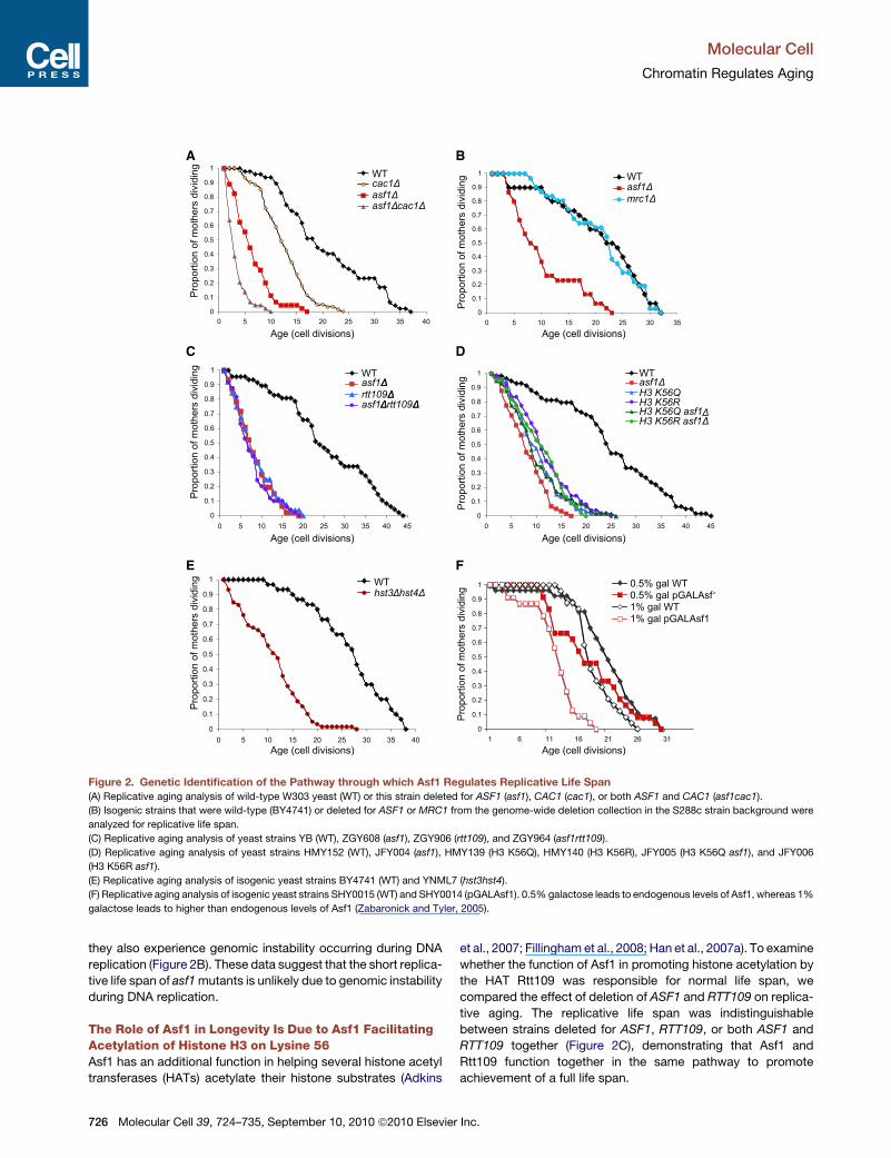
Figure 3. Aging Cells Have Reduced Histone Protein Levels
(A) RNA levels for H3 (HHT2 and HHT1), H4 (HHF2 and HHF1), H2A (HTA1 and HTA2), H2B (HTB1 and HTB2), and tubulin (TUB) from strain BY4741 (WT) or this
strain deleted forASF1 (asf1D) orRTT109 (rtt109D). RNA levels were normalized to total DNA content, and the RNA levels were normalized to 1 for youngWTRNA
(data prior to normalizing to 1 are given in Figure S3C). Representative results are shown.
(B) Histone H3 protein and tubulin levels in wild-type strain BY4741 were measured by western analysis of equivalent amounts of total protein extracts from
biochemically separated wild-type yeast. Below is shown a Coomassie-stained gel of the same amounts of the same total yeast protein extracts analyzed in
the western analyses. The western analyses used infrared secondary antibodies, and the images were taken in the linear range of detection.
(C) Total (T), soluble (S), and pellet (P) proteins were isolated from strain UCC5181 (WT) or this strain deleted for HIR1 (hir1D), ASF1 (asf1D), or RTT109 (rtt109D),
which were growing asynchronously of mixed age or were aged via the ‘‘mother enrichment program.’’ The sameDNA equivalents of each fraction were analyzed
for histone H3 protein levels by western blotting, wherein nonchromatin-bound histones reside in the soluble fraction and chromatinized histones reside in the
pellet fraction. An asterisk denotes a likely proteolytic cleavage product of H3 that arises upon overhandling of the protein. Tubulin was assayed to demonstrate
the efficiency of the fractionation. Below are shown amido black staining of the membranes used for the western analyses. The western analyses used infrared
secondary antibodies, and the images were taken in the linear range of detection.
Molecular Cell
Chromatin Regulates Aging
728 Molecular Cell 39, 724–735, September 10, 2010 ª2010 Elsevier Inc.

Molecular Cell
Chromatin Regulates Aging
To determine whether the decrease in histone protein levels in
the short-lived asf1 and rtt109 mutants and during the normal
aging process reflects a loss of free histones or chromatinized
histones, we performed chromatin fractionations. We analyzed
the same DNA equivalents of proteins for the total, supernatant
(free proteins), and pellet (chromatin proteins) fractions to enable
direct comparison of the distribution of the histones between the
free and chromatinized forms. In order to isolate sufficient
numbers of aged cells for this analysis, we utilized a new system
developed by Dan Gottschling’s lab, termed the ‘‘mother
enrichment program’’ (Lindstrom and Gottschling, 2009).
Coupled with the traditional biochemical isolation of old cells,
this system enabled us to easily isolate much older cells that
had reached �50% of the maximum life span for each strain.
As before, we observe a greatly reduced total amount of histones
during aging (Figure 3C). Furthermore, we find that, in both
young and old wild-type and hir1 cells, the vast majority of
histone H3 is chromatinized (Figure 3C). Quite unexpectedly,
we find that, in the asf1 and rtt109 short-lived mutants, a signifi-
cant number of the histones are free, even in the mixed age
(asynch) population (Figure 3C). This may reflect a defect in chro-
matin assembly in the absence of Rtt109 and Asf1 (Li et al.,
2008). To provide further evidence for reduced histone occu-
pancy on the DNA during aging, we performed chromatin immu-
noprecipitation (ChIP) analysis of histone H3. Again, we utilized
the mother enrichment program (Lindstrom and Gottschling,
2009) to isolate old cells (median 30 generations) for this
analysis. At all areas examined, including the active Actin
gene, the silent HML locus, the rDNA, and telomere proximal
loci, we observed a 50%–75% reduction in histone occupancy
in the aged population in comparison to themixed age asynchro-
nous culture (Figures 3D and S4F).
Supplying Extra Histone Proteins Increases LongevityWe hypothesized that the increase in histone transcript levels
that normally occurs during aging serves to protect cells from
premature aging, whereas the reduced histone expression in
the short-lived mutants is a cause of their shortened life span.
To test this hypothesis, we investigated whether ectopic expres-
sion of extra histones could extend the life span of the short-
lived asf1 mutants. A high copy number plasmid encoding all
four core histones extended themedian life span of asf1mutants
by 65% (Figure 3E). Overexpression of the H2A/H2B pair of
histones from a galactose-inducible promoter also significantly
increased the life span of asf1 mutants (Figure 3F). Overexpres-
sion of H3/H4 did not significantly extend the life span of the
asf1 mutant (Figures S5A and S5B), which is not too surprising
given that these cells are lacking the major H3/H4 histone
chaperone. These data show that the shortened life span of
(D) Cells of mixed age (Asynch) or following aging via the mother enrichment prog
occupancy at the indicated DNA regions. Data shown are the average of two inde
S6B.
(E) Replicative life span of strain JFY047 and JFY048 (asf1) carrying the empty v
HTB1/HTA1 genes. The p value indicates the significance of the life span extens
(F) Replicative life span of strains JFY056 and JFY050 (asf1) carrying the empty
genes.
(G) Replicative life span of isogenic strains with the indicated gene-encoding sub
Molecu
the asf1 mutant can be partly reversed by supplying additional
histones.
Given that the reduced level of histone proteins in the asf1
mutant is partly responsible for the shortened life span of this
strain, we predicted that supplying higher levels than normal of
histone proteins would extend the life span of wild-type yeast.
To test this prediction, we analyzed the replicative life span of
the hir1mutant that has significantly higher histone protein levels
in the old cells as compared to wild-type old cells (Figure 3G).
In addition, we examined the other three components of the
Hir complex: Hir2, Hir3, Hpc2 (Green et al., 2005). Inactivation
of any component of the Hir complex extended the median repli-
cative life span by 25%–35% (Figure 3G). This extent of life span
extension is comparable to that achieved by other known indi-
vidual manipulations that result in life span extension. In
summary, mutants with low histone protein levels such as
rtt109 and asf1 mutants are short lived, whereas mutants with
high histone protein levels such as hir1, hir2, hir3, and hpc2
mutants have extended life span.
IncreasedHistone Supply Appears to ExtendReplicativeLife Span via a Pathway that Is Distinct from Other LifeSpan Extension PathwaysTo determine how the histone supply pathway of longevity
relates to other known pathways of life span extension, we per-
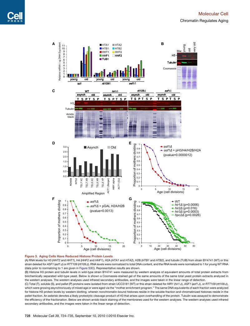
formed epistasis analyses. Deletion ofHIR1 significantly extends
the life span of the short-lived sir2 mutant (Figure 4A), indicating
that the extra histone supply pathway does not require the
Sir2 histone deacetylase to achieve life span extension. The
short life span of sir2 mutants is due to the accumulation of
ERCs because it is suppressed by deletion of FOB1 (Defossez
et al., 1999) (Figure 4B), whereby FOB1 encodes a replication
fork protein whose deletion prevents ERC formation (Defossez
et al., 1999). Of note, additional deletion of HIR1 from the
sir2fob1 double mutant led to a significant extension in life
span beyond that of the sir2fob1 double mutant (Figure 4B),
indicating that extra histone supply is unlikely to extend life
span via reducing ERC formation. An extra copy of the gene
encoding Sir2 (Sir2 OX) extends life span (Kaeberlein et al.,
1999) (Figure 4C), mediated via reduced ERC accumulation
because it is blocked by FOB1 deletion (Imai et al., 2000).
Deletion of HIR1 leads to a greater degree of life span exten-
sion than overexpression of Sir2 (Figure 4C), whereas deletion
of HIR1 and Sir2 overexpression together significantly
increased life span when compared to Sir2 overexpression
alone (Figure 4C). Collectively, these data indicate that extra
histone supply is unlikely to extend life span via the identical
pathways by which ERC accumulation and Sir2 influence life
span.
ram (aged) from strain UCC5181 were subject to ChIP analysis for histone H3
pendent experiments, whose results are shown individually in Figures S6A and
ector pRS426 or the 2 micron vector pFB1156 carrying the HHF1/HHT1 and
ion.
vector pRS315 or the vector pRO689 carrying the pGAL-driven HTB1/HTA1
units of the Hir complex deleted.
lar Cell 39, 724–735, September 10, 2010 ª2010 Elsevier Inc. 729
A
B
C
Figure 4. Increased Histone Supply Does Not Extend Life Span by
the Same Pathways as Sir2 or ERCs
(A) Replicative life span of isogenic strains with the indicated genes deleted or
containing an extra copy of Sir2 (Sir2OX).
(B) Replicative life span of isogenic strains with the indicated genes deleted.
(C) Replicative life span of isogenic strains with the indicated genes deleted
and/or containing an extra copy of Sir2 (Sir2OX).
Molecular Cell
Chromatin Regulates Aging
Life span extension due to caloric restriction (CR) is likely
mediated through the highly conserved nutrient-responsive
target of rapamycin (Tor1) kinase (Kaeberlein et al., 2005). To
investigate the relationship between the life span extension
due to increased histone supply and the life span extension
due to Tor1 deletion, we combined the hir1 and tor1 mutants.
The hir1 mutant extended life span more than the tor1 mutant
(Figure 5A). However, the double hir1tor1 mutant had a life
span that was intermediate between that of the tor1 mutant
730 Molecular Cell 39, 724–735, September 10, 2010 ª2010 Elsevier
and the hir1 mutant (Figure 5A). CR of yeast is achieved by
growth in 0.5% glucose in contrast to the 2% glucose that is
used for standard yeast growth. Although individually CR or
HIR1 deletion extends life span, CR of the hir1 mutant led to
a life span extension that was intermediate between that result-
ing from the individual manipulations, not additive (Figure 5B).
These results suggest two possibilities: either (1) deletion of
TOR1/CR and deletion of HIR1 function through the same
pathway to extend life span or, alternatively, (2) HIR1 deletion
cannot extend life span in the context of a tor1 mutant/CR.
Depletion of the 60S ribosomal subunits extends life span
(Steffen et al., 2008). Genetic evidence places CR, tor1 inhibition,
and depletion of the 60S subunits in a longevity pathway that is
partially dependent on upregulated translation of the transcrip-
tion factor Gcn4 that promotes stress resistance (Steffen et al.,
2008). To investigate whether the histone supply pathway
extends life span via the same pathways that are targeted by
deletion of TOR1 and CR, we measured Gcn4 translation and
the abundance of 60S ribosomal subunits in the mutants that
influence histone supply. Using a dual luciferase reporter
construct to measure Gcn4 translation, we observed increased
Gcn4 levels upon deletion of the gene encoding the 60S subunit
Rpl20b (Figure 5C), as observed previously (Steffen et al., 2008).
However, the isogenic short-lived rtt109 and asf1 mutants and
the long-lived hir1 mutant did not have significant alterations in
Gcn4 protein synthesis (Figure 5C), suggesting that 60S ribo-
somal subunit abundance is not drastically altered in these
strains.
To examine directly whether the abundance of the 60S ribo-
somal subunit was altered in the mutants that effect histone
supply, we performed polysome profiling. The rpl20b mutant
has a lower amount of 60S ribosomal subunit as compared to
wild-type (Figures 5D and S5C), as seen previously (Steffen
et al., 2008). We note that the amount and size of the polysomes
was lower in the rpbl20b mutant compared to wild-type, indi-
cating reduced amounts of translation in general and that each
polysome peak was a doublet, although the relevance of this is
unclear (Figure 5D). The short-lived asf1 and rtt109 mutants
had fewer polysomes than the wild-type, indicating that less
translation in general is occurring. However, no significant
change in the 60S-to-40S ratio was observedwith the short-lived
asf1 and rtt109 mutants or the long-lived hir1 mutant, as
compared to the wild-type strain (Figure 5D). Taken together,
these data indicate that extra histone supply is not influencing
life span via the same pathways of altered protein synthesis
that are influenced by inhibition of Tor1 to extend life span.
To further investigate the relationship between life span exten-
sion by extra histone supply and life span extension due to dele-
tion of TOR1 or CR, we examined total histone protein levels
during these manipulations. Surprisingly, we found that
biochemically isolated old cells from tor1 mutants or during CR
have significantly less histone proteins than old wild-type yeast
cells (Figures 5E, 5F, and S5D). These data indicate that the
pathway by which CR and TOR1 deletion leads to extended
life span is not a consequence of increased histone supply.
These data also raise the possibility that deletion of HIR1 does
not further extend the life span of tor1 mutants or during cR
because HIR1 deletion may not be sufficient to raise the histone
Inc.

A
C
E
D
B
F
Figure 5. Epistasis Analysis with the Tor1
Pathway or CR
(A) Replicative life span of isogenic strains with the
indicated genes deleted.
(B) Replicative life span of isogenic strains with or
without HIR1 deleted, grown either on 0.5%
glucose to induce CR or on 2% glucose.
(C) Schematic of the dual-luciferase reporter
plasmid used to measure Gcn4 translation levels.
The ratio of the signal from the GCN4-firefly lucif-
erase to renilla luciferase is shown for isogenic
strains from the genome-wide deletion collection
in the S288c strain background.
(D) Polysome profiles of the indicated isogenic
strains from the genome-wide deletion collection
in the S288c strain background. The position of
the 60S, 40S, and 80S ribosome and the poly-
somes are indicated.
(E) Histone H3 protein and tubulin levels were
measured by western analysis of equivalent
amounts of total protein extracts from biochemi-
cally separated young and old wild-type and tor1
mutant yeast. Below is shown the amido black-
stainedmembrane to show equivalent total protein
loading. The western analyses used infrared
secondary antibodies, and the images were taken
in the linear range of detection.
(F) As for (E) but with wild-type yeast grown in
normal conditions (2% glucose) or calorie-
restricted conditions (0.5% glucose).
Molecular Cell
Chromatin Regulates Aging
levels significantly during these manipulations. Taken together,
our analyses suggest that HIR1 deletion leads to life span exten-
sion via a pathway that is not shared with other known mecha-
nisms of life span extension.
Ectopic Expression of Histones H3 and H4 ProfoundlyIncreases Replicative Life SpanTo determine which of the core histones impart anti-aging prop-
erties, we individually integrated extra copies of the genes en-
coding H3/H4 or H2A/H2B under the control of the pGAL1/10
promoters. Overexpression of integrated genes encoding H3/
H4, but not H2A/H2B, extended the median life span of wild-
type cells by 30% (Figures 6A and S6A). Higher levels of H3/
H4 induction via addition of more galactose to more strongly
induce the promoters controlling H3/H4 resulted in life span
extension of up to 50% (Figure 6B). Of note, this degree of yeast
Molecular Cell 39, 724–735, Se
replicative life span extension is far
greater than that usually attained by
most other single manipulations in yeast.
Expression of H3/H4 in these conditions
leads to an �50% increase in the level
of total histone proteins (Figures S6B
and S6C). Overexpression of histones
H3/H4 in these conditions did not lead
to significant differences in the polysome
profile compared to wild-type cells (data
not shown), indicating that excess
histones are not drastically influencing
protein synthesis or ribosomal subunit
composition. Also, no significant changes in the cell cycle
resulted from overexpression of H3/H4 (Figure S6D). Impor-
tantly, the expression of extra histone proteins does not lead to
resistance to the oxidative damage that results from treatment
with hydrogen peroxide, replication stress due to treatment
with hydroxyurea, or DNA damage due to treatment with the al-
kylating agent methylmethane sulfonate (Figures S6E and S6F).
As such, the extended life span that results from extra histone
supply is not due to their protecting the genome from genomic
instability.
DISCUSSION
In this study, we show that aging is accompanied by a profound
loss of histone proteins. This loss of histones is causal for aging
because significant life span extension is achieved by
ptember 10, 2010 ª2010 Elsevier Inc. 731

A B
C
Figure 6. Overexpression of Histones H3/
H4 Extends Life Span
(A and B) Replicative life span of isogenic strains
that are wild-type (WT) or carrying an extra copy
of either HHT2/HHF2 or HTB1/HTA1 driven from
the galactose-inducible pGAL1/10 divergent
promoter integrated into the genome grown on
(A) 1% galactose and (B) on 2% galactose.
(C) Model for life span extension by increasing
histone supply.
Molecular Cell
Chromatin Regulates Aging
ectopically increasing histone expression in wild-type yeast via
a pathway that is distinct from other known life span extension
pathways.
Age-Dependent Changes in Chromatin StructureThe drastic decrease in the level of histone proteins on chro-
matin during the normal aging process in yeast (Figures 3B–
3D) is consistent with the loss of transcriptional silencing that
occurs in wild-type yeast during aging (Kim et al., 1996). More
recently, an age-dependent increase in levels of histone H4
K16Ac at the X elements within subtelomeric regions of yeast
chromosomes has been shown to occur due to a progressive
loss of the Sir2 deacetylase protein during aging (Dang et al.,
2009). This particular histone modification is unique in that it
prevents higher-order packaging between adjacent nucleo-
somes (Shogren-Knaak et al., 2006), implying that increased
H4 K16ac during aging would lead to a more open chromatin
structure in aged cells. Indeed, when reporter genes were in-
serted into the X elements within subtelomeric regions, the
age-dependent increase in H4 K16Ac levels led to increased
expression of these reporter genes during aging (Dang et al.,
2009). However, the mechanistic relevance of opening up the
chromatin structure of the X elements of subtelomeric regions
on aging is not clear.
Many other previous studies have also revealed transcriptional
dysregulation occurring during the aging process, and it is clear
that at least some of these transcriptional changes are due to
age-dependent alterations in the chromatin structure. For
example, DNA methylation, which is known to lead to a tighter
732 Molecular Cell 39, 724–735, September 10, 2010 ª2010 Elsevier Inc.
packaging of the chromatin structure
(Fuks, 2005), diminishes throughout the
life span of mouse liver cells, with conse-
quent expression of previously repressed
genes (Mays-Hoopes et al., 1983; Singhal
et al., 1987). Age-related mRNA changes
have also been documented extensively
in yeast (Yiu et al., 2008). Similarly, micro-
array studies of global messenger RNAs
(mRNAs) in aging animals and in mutants
considered to affect the aging process
have found many transcriptional changes
(Zahn and Kim, 2007). Furthermore, the
finding that stochastic differences in
gene expression between individuals
can influence life span in C. elegans
points to a causal role for epigenetic changes during aging
(Rea et al., 2005).
How Does Extra Histone Supply Extend Life Span?Our model for how interventions that increase histone supply
result in life span extension proposes that the resulting enlarged
soluble pool of free histones facilitates histone exchange via
stimulating the ongoing equilibrium of dynamic chromatin
assembly and disassembly (Figure 6C). The resulting higher
rate of histone exchange would accelerate the rate of removal
of posttranslationally modified and damaged histone proteins
from the DNA and would additionally enable sporadically occur-
ring nucleosome-free regions in the genome to be repackaged
into chromatin. This, in turn, would reduce inappropriate access
of proteins to the genome during aging, which would otherwise
lead to the increased transcription and genomic instability,
both of which are frequent characteristics of increased age
and conditions that exhibit accelerated aging phenotypes.
If the chromatin is packaged into a tighter structure by over-
expressing histones, one would predict that the increased
histone supply would restore the transcriptional silencing that
is normally lost during aging (Kim et al., 1996). Indeed, hir1
mutants, which have elevated histone levels, have enhanced
transcriptional silencing as compared to wild-type cells (Kauf-
man et al., 1998; Smith et al., 1999). By analogy, inactivation of
the histone deacetylase Rpd3 also leads to elevated histone
levels, extending life span in some yeast backgrounds and
increasing transcriptional silencing (Bernstein et al., 2000; Kim
et al., 1999; Rogina et al., 2002; Smith et al., 1999). Although
the increased transcriptional silencing that occurs upon

Molecular Cell
Chromatin Regulates Aging
inactivation of Rpd3 and Hir1 provides a marker for altered chro-
matin structure, it is thought that the influence of Rpd3 on the
aging process is most likely mediated through influencing the
expression of euchromatin genes (Frankel and Rogina, 2005;
Kim et al., 1999). Indeed, inactivation of Rpd3 and the concom-
itant increase in histone levels results in significant downregula-
tion of 40%of genes located within 20 kb of the telomeres (Bern-
stein et al., 2000). This is reminiscent of the induction of 40% of
genes located within 20 kb of the telomeres, which occurs upon
depletion of histone H4 (Wyrick et al., 1999).
It is clear from the increased silencing in the hir mutants and
rpd3 mutants that supplying extra histone proteins results in
a tighter chromatin structure, and we anticipate that this will be
the case not only at the silent regions, but also at other regions
of the genome. We propose a model whereby the expression
of euchromatic genes increases inappropriately during aging
due to the deterioration of the chromatin structure with age,
and the misexpression of genes contributes to the aging
process. By extension, we propose that our manipulations that
result in increased histone supply with age result in a tighter
chromatin structure, maintaining the regulated state of these
genes and leading to life span extension (Figure 6C). The next
challenge is to identify the specific transcript(s) that are downre-
gulated by the formation of a tighter chromatin structure that lead
to life span extension.
Relevance to Life Span Extension in Higher Eukaryotesand via Other PathwaysThe ultimate goal of this field is to achieve life span and health
span extension in humans. As such, it is critically important to
consider the potential relevance of our results in the yeast
system to the situation in higher eukaryotes. Consistent with
the reduction in histone proteins that we observed in aged yeast,
we have found that levels of histone H3 protein decrease in
mitotically active mouse tissues during aging (C. Das, unpub-
lished data), suggesting that histone protein loss may also occur
in mammals during aging. This could be the cause of the
elevated transcript levels that are characteristic during the aging
of renewable tissues in mice (Warren et al., 2007). By examining
the published transcriptome analyses, it is apparent that at least
one regimen of life span extension in C. elegans is accompanied
by increased histone expression (McColl et al., 2008). Further-
more, calorie restriction in mice is accompanied by increased
histone expression (Barger et al., 2008). It will be important to
determine whether the increased histone transcript levels during
these life span regimens in higher eukaryotes are mirrored in
a higher histone protein level. Indeed, we find increased histone
transcript levels of four of the six cell-cycle-regulated histone
genes in our long-lived tor1 yeast mutants (Figure S3A), whereas
histone protein levels are decreased in the old tor1 mutants and
during CR (Figures 5E and 5F), suggesting that they are trying to
compensate for the depletion of histone proteins by transcribing
more histones. Presumably, the lower level of histone proteins in
the yeast tor1 mutants and during CR (Figures 5E and 5F) is
a reflection of the greatly decreased degree of protein synthesis
that occurs during nutritional stress and in the absence of Tor1.
Our model leads us to ask why deletion of TOR1 or CR extends
life span if they have lower histone protein levels than normal in
Molecu
their old cells. Presumably, the pathway by which deletion of
TOR1 and CR extends life span, which is largely unknown, can
function without the need for extra histones to repress the key
aging transcript(s) that are inappropriately generated during
aging. This may be due to the fact that the reduced protein
synthesis of these key aging transcripts during CR or Tor1 inac-
tivation circumvents a requirement for their transcriptional
repression in old cells for extended life span. Given the conser-
vation of the mechanistic basis of aging, it will be necessary to
identify these key aging transcripts to better understand and
potentially ameliorate the age-related rise of human disease.
EXPERIMENTAL PROCEDURES
Yeast Strains and Media
Yeast strains are described in Table S1. Replicative life span assays were per-
formed on YPDwith 2%glucose or YP with 1% raffinose and 0.5%, 1%, or 2%
galactose as indicated. Plasmids expressing histones were maintained by
growth in synthetic complete media lacking the appropriate amino acid and
were supplemented with raffinose and galactose for strains carrying galactose
inducible promoter fusions.
Life Span Analysis
Replicative life span of virgin mother cells was determined as described previ-
ously (Kennedy et al., 1994). The number of mother cells analyzed for each
figure is itemized in Table S2. Yeast cells were kept overnight at 12�C to
slow cell division. Yeast were initially grown on YP glycerol plates to eliminate
yeast cells lacking mitochondria. For yeast with extended life span, the
Wilcoxon Rank-Sum Test was used to determine statistical significance.
The Wilcoxon.test function was used in the R software version 2.80, and p
values less than the significance cutoff of 0.05 are given in the figures and in
Table S2.
Isolation of Young and Old Yeast Cells
Old yeast cells were isolated as described previously by using EZ Link Sulfo-
NHL-LC-LC-Biotin (Pierce) to label cell surface proteins and MagnaBind
Streptavidin beads (Thermo Scientific) (Lin et al., 2001). The old cells gener-
ated by this protocol had divided an average eight times, as determined by cal-
cafluor staining. The ‘‘very old’’ cells were isolated by two sequential rounds of
this affinity purification and had divided an average of 15 times, as determined
by calcafluor staining.
For isolating older cells, we utilized the MEP as described in Lindstrom and
Gottschling (2009). For the chromatin fractionations, strains UC5181 and
DTY011 were grown for a total of 36 hr in the presence of estradiol, DT009,
and DT010 for 16 hr. For ChIP experiments in galactose, UC5181 was grown
for 72 hr total.
Reverse Transcriptase Real-Time PCR
RNA was analyzed by real-time RT-PCR analysis using a Roche Light Cycler
and SYBR green detection. Primer sequences are given in the Supplemental
Experimental Procedures.
Quantitative Western Blotting Analysis, ChIP, and Chromatin
Fractionations
Total protein extracts were isolated by boiling in Laemmli buffer prior to loading
gels. Anti-rat and anti-rabbit secondary antibodies were used that fluoresced
at 800 nm and 700 nm, respectively. Membranes were scanned with a Li-Cor
Odyssey scanning system, and their software was used to quantitate the
bands, using the average density method. The methods for ChIP were pub-
lished previously (Williams et al., 2008) and are detailed in the Supplemental
Experimental Procedures. The chromatin fractionation was performed using
a variation of the method published in Frei and Gasser (2000) and is detailed
in the Supplemental Experimental Procedures.
lar Cell 39, 724–735, September 10, 2010 ª2010 Elsevier Inc. 733

Molecular Cell
Chromatin Regulates Aging
Gcn4 Reporter Assay and Polysome Profiling
The analysis of Gcn4 translation used a dual reporter assay as previously
described (Steffen et al., 2008). In brief, a reporter plasmid pVW31 containing
the GCN4 promoter fused to firefly luciferase, and Renilla luciferase under the
control of a constitutive promoter was used. Cells were grown overnight in
synthetic media lacking uracil to maintain the plasmid. Before lysing, cells
were grown 2 hr in YPD to minimize GCN4 expression due to nutrient stress.
The Promega Dual-Luciferase Reporter Assay was used alongwith a Luminos-
kan Ascent to monitor expression. Firefly luciferase was normalized to Renilla
luciferase activity. For polysome profiling, cells were grown in 75 ml of YPD to
an OD of 0.8. Cultures were centrifuged and cooled with ice-cold YPD contain-
ing 100 mg/ml cycloheximide. After centrifuging again, cells were rinsed with
ice-cold ddH2O containing 100 mg/ml cycloheximide, and polysome profiles
were measured as previously described (Steffen et al., 2008).
SUPPLEMENTAL INFORMATION
Supplemental Information includes Supplemental Experimental Procedures,
six figures, and two tables and can be found with this article online at doi:10.
1016/j.molcel.2010.08.015.
ACKNOWLEDGMENTS
We thank Candice Wike for assistance and Brian Kennedy for sharing data
prior to publication. We are grateful to William Feser for aiding in the statistical
analysis. We thank Dan Gottschling, Brian Kennedy, Michael Grunstein, Allain
Verreault, Fred Winston, Valter Longo, David Botstein, David Toczyski, Zhigou
Zhang, Rohinton Kamakaka, and Namrita Dhillon for kindly providing strains
and plasmids. This work was supported by NIH grant GM64475 (to J.K.T.),
T32-GM08370 (to J.F.), and University of Colorado Cancer Center Aging Initia-
tive seed grant funding (to J.K.T.).
Received: December 23, 2009
Revised: May 4, 2010
Accepted: July 20, 2010
Published: September 9, 2010
REFERENCES
Adkins, M.W., Carson, J.J., English, C.M., Ramey, C.J., and Tyler, J.K. (2007).
The histone chaperone anti-silencing function 1 stimulates the acetylation of
newly synthesized histone H3 in S-phase. J. Biol. Chem. 282, 1334–1340.
Barger, J.L., Kayo, T., Pugh, T.D., Prolla, T.A., andWeindruch, R. (2008). Short-
term consumption of a resveratrol-containing nutraceutical mixture mimics
gene expression of long-term caloric restriction in mouse heart. Exp. Gerontol.
43, 859–866.
Bernstein, B.E., Tong, J.K., and Schreiber, S.L. (2000). Genomewide studies of
histone deacetylase function in yeast. Proc. Natl. Acad. Sci. USA 97, 13708–
13713.
Busuttil, R., Bahar, R., and Vijg, J. (2007). Genome dynamics and transcrip-
tional deregulation in aging. Neuroscience 145, 1341–1347.
Chen, C.C., Carson, J.J., Feser, J., Tamburini, B., Zabaronick, S., Linger, J.,
and Tyler, J.K. (2008). Acetylated lysine 56 on histone H3 drives chromatin
assembly after repair and signals for the completion of repair. Cell 134,
231–243.
Dang, W., Steffen, K.K., Perry, R., Dorsey, J.A., Johnson, F.B., Shilatifard, A.,
Kaeberlein, M., Kennedy, B.K., and Berger, S.L. (2009). Histone H4 lysine 16
acetylation regulates cellular life span. Nature 459, 802–807.
Defossez, P.A., Prusty, R., Kaeberlein, M., Lin, S.J., Ferrigno, P., Silver, P.A.,
Keil, R.L., and Guarente, L. (1999). Elimination of replication block protein
Fob1 extends the life span of yeast mother cells. Mol. Cell 3, 447–455.
Fillingham, J., Recht, J., Silva, A.C., Suter, B., Emili, A., Stagljar, I., Krogan,
N.J., Allis, C.D., Keogh, M.C., and Greenblatt, J.F. (2008). Chaperone control
of the activity and specificity of the histone H3 acetyltransferase Rtt109. Mol.
Cell. Biol. 28, 4342–4353.
734 Molecular Cell 39, 724–735, September 10, 2010 ª2010 Elsevier
Frankel, S., and Rogina, B. (2005). Drosophila longevity is not affected by
heterochromatin-mediated gene silencing. Aging Cell 4, 53–56.
Frei, C., and Gasser, S.M. (2000). The yeast Sgs1p helicase acts upstream of
Rad53p in the DNA replication checkpoint and colocalizes with Rad53p in
S-phase-specific foci. Genes Dev. 14, 81–96.
Fuks, F. (2005). DNA methylation and histone modifications: teaming up to
silence genes. Curr. Opin. Genet. Dev. 15, 490–495.
Gottlieb, S., and Esposito, R.E. (1989). A new role for a yeast transcriptional
silencer gene, SIR2, in regulation of recombination in ribosomal DNA. Cell
56, 771–776.
Green, E.M., Antczak, A.J., Bailey, A.O., Franco, A.A., Wu, K.J., Yates, J.R., III,
and Kaufman, P.D. (2005). Replication-independent histone deposition by the
HIR complex and Asf1. Curr. Biol. 15, 2044–2049.
Groth, A., Rocha, W., Verreault, A., and Almouzni, G. (2007). Chromatin chal-
lenges during DNA replication and repair. Cell 128, 721–733.
Han, J., Zhou, H., Horazdovsky, B., Zhang, K., Xu, R.M., and Zhang, Z. (2007a).
Rtt109 acetylates histone H3 lysine 56 and functions in DNA replication.
Science 315, 653–655.
Han, J., Zhou, H., Li, Z., Xu, R.M., and Zhang, Z. (2007b). Acetylation of lysine
56 of histone H3 catalyzed by RTT109 and regulated by ASF1 is required for
replisome integrity. J. Biol. Chem. 282, 28587–28596.
Imai, S., Armstrong, C.M., Kaeberlein, M., and Guarente, L. (2000). Transcrip-
tional silencing and longevity protein Sir2 is an NAD-dependent histone deace-
tylase. Nature 403, 795–800.
Kaeberlein, M., McVey, M., and Guarente, L. (1999). The SIR2/3/4 complex
and SIR2 alone promote longevity in Saccharomyces cerevisiae by two
different mechanisms. Genes Dev. 13, 2570–2580.
Kaeberlein, M., Powers, R.W., III, Steffen, K.K., Westman, E.A., Hu, D., Dang,
N., Kerr, E.O., Kirkland, K.T., Fields, S., and Kennedy, B.K. (2005). Regulation
of yeast replicative life span by TOR and Sch9 in response to nutrients.
Science 310, 1193–1196.
Kaufman, P.D., Kobayashi, R., and Stillman, B. (1997). Ultraviolet radiation
sensitivity and reduction of telomeric silencing in Saccharomyces cerevisiae
cells lacking chromatin assembly factor-I. Genes Dev. 11, 345–357.
Kaufman, P.D., Cohen, J.L., and Osley, M.A. (1998). Hir proteins are required
for position-dependent gene silencing in Saccharomyces cerevisiae in the
absence of chromatin assembly factor I. Mol. Cell. Biol. 18, 4793–4806.
Kennedy, B.K., Austriaco, N.R., Jr., and Guarente, L. (1994). Daughter cells of
Saccharomyces cerevisiae from old mothers display a reduced life span.
J. Cell Biol. 127, 1985–1993.
Kim, S., Villeponteau, B., and Jazwinski, S.M. (1996). Effect of replicative age
on transcriptional silencing near telomeres in Saccharomyces cerevisiae.
Biochem. Biophys. Res. Commun. 219, 370–376.
Kim, S., Benguria, A., Lai, C.Y., and Jazwinski, S.M. (1999). Modulation of life-
span by histone deacetylase genes in Saccharomyces cerevisiae. Mol. Biol.
Cell 10, 3125–3136.
Lamming, D.W., Latorre-Esteves, M., Medvedik, O., Wong, S.N., Tsang, F.A.,
Wang, C., Lin, S.J., and Sinclair, D.A. (2005). HST2 mediates SIR2-indepen-
dent life-span extension by calorie restriction. Science 309, 1861–1864.
Le, S., Davis, C., Konopka, J.B., and Sternglanz, R. (1997). Two new S-phase-
specific genes from Saccharomyces cerevisiae. Yeast 13, 1029–1042.
Li, B., Carey, M., and Workman, J.L. (2007). The role of chromatin during tran-
scription. Cell 128, 707–719.
Li, Q., Zhou, H., Wurtele, H., Davies, B., Horazdovsky, B., Verreault, A., and
Zhang, Z. (2008). Acetylation of histone H3 lysine 56 regulates replication-
coupled nucleosome assembly. Cell 134, 244–255.
Lin, S.S., Manchester, J.K., and Gordon, J.I. (2001). Enhanced gluconeogen-
esis and increased energy storage as hallmarks of aging in Saccharomyces
cerevisiae. J. Biol. Chem. 276, 36000–36007.
Lindstrom, D.L., and Gottschling, D.E. (2009). The mother enrichment
program: a genetic system for facile replicative life span analysis in Saccharo-
myces cerevisiae. Genetics 183, 413–422.
Inc.
Molecular Cell
Chromatin Regulates Aging
Luger, K., Mader, A.W., Richmond, R.K., Sargent, D.F., and Richmond, T.J.
(1997). Crystal structure of the nucleosome core particle at 2.8 A resolution.
Nature 389, 251–260.
Maas, N.L., Miller, K.M., DeFazio, L.G., and Toczyski, D.P. (2006). Cell cycle
and checkpoint regulation of histone H3 K56 acetylation by Hst3 and Hst4.
Mol. Cell 23, 109–119.
Maslov, A.Y., and Vijg, J. (2009). Genome instability, cancer and aging. Bio-
chim. Biophys. Acta 1790, 963–969.
Mays-Hoopes, L.L., Brown, A., and Huang, R.C. (1983). Methylation and rear-
rangement of mouse intracisternal a particle genes in development, aging, and
myeloma. Mol. Cell. Biol. 3, 1371–1380.
McColl, G., Killilea, D.W., Hubbard, A.E., Vantipalli, M.C., Melov, S., and
Lithgow, G.J. (2008). Pharmacogenetic analysis of lithium-induced delayed
aging in Caenorhabditis elegans. J. Biol. Chem. 283, 350–357.
Moazed, D. (2001). Enzymatic activities of Sir2 and chromatin silencing. Curr.
Opin. Cell Biol. 13, 232–238.
Myung, K., Pennaneach, V., Kats, E.S., and Kolodner, R.D. (2003). Saccharo-
myces cerevisiae chromatin-assembly factors that act during DNA replication
function in the maintenance of genome stability. Proc. Natl. Acad. Sci. USA
100, 6640–6645.
Ramey, C.J., Howar, S., Adkins, M., Linger, J., Spicer, J., and Tyler, J.K.
(2004). Activation of the DNA damage checkpoint in yeast lacking the histone
chaperone anti-silencing function 1. Mol. Cell. Biol. 24, 10313–10327.
Rea, S.L., Wu, D., Cypser, J.R., Vaupel, J.W., and Johnson, T.E. (2005).
A stress-sensitive reporter predicts longevity in isogenic populations of Cae-
norhabditis elegans. Nat. Genet. 37, 894–898.
Robert, T., Dervins, D., Fabre, F., and Gangloff, S. (2006). Mrc1 and Srs2 are
major actors in the regulation of spontaneous crossover. EMBO J. 25, 2837–
2846.
Rogina, B., Helfand, S.L., and Frankel, S. (2002). Longevity regulation by
Drosophila Rpd3 deacetylase and caloric restriction. Science 298, 1745.
Shogren-Knaak, M., Ishii, H., Sun, J.M., Pazin, M.J., Davie, J.R., and Peterson,
C.L. (2006). Histone H4-K16 acetylation controls chromatin structure and
protein interactions. Science 311, 844–847.
Singer, M.S., Kahana, A., Wolf, A.J., Meisinger, L.L., Peterson, S.E., Goggin,
C.,Mahowald,M., andGottschling, D.E. (1998). Identification of high-copy dis-
ruptors of telomeric silencing in Saccharomyces cerevisiae. Genetics 150,
613–632.
Singhal, R.P., Mays-Hoopes, L.L., and Eichhorn, G.L. (1987). DNAmethylation
in aging of mice. Mech. Ageing Dev. 41, 199–210.
Smith, J.S., Caputo, E., and Boeke, J.D. (1999). A genetic screen for ribosomal
DNA silencing defects identifies multiple DNA replication and chromatin-
modulating factors. Mol. Cell. Biol. 19, 3184–3197.
Smith, E.D., Kennedy, B.K., and Kaeberlein, M. (2007). Genome-wide identifi-
cation of conserved longevity genes in yeast and worms. Mech. Ageing Dev.
128, 106–111.
Smith, E.D., Tsuchiya, M., Fox, L.A., Dang, N., Hu, D., Kerr, E.O., Johnston,
E.D., Tchao, B.N., Pak, D.N., Welton, K.L., et al. (2008). Quantitative evidence
Molecu
for conserved longevity pathways between divergent eukaryotic species.
Genome Res. 18, 564–570.
Spector, M.S., and Osley, M.A. (1993). The HIR4-1 mutation defines a new
class of histone regulatory genes in Saccharomyces cerevisiae. Genetics
135, 25–34.
Spector, M.S., Raff, A., DeSilva, H., Lee, K., and Osley, M.A. (1997). Hir1p and
Hir2p function as transcriptional corepressors to regulate histone gene tran-
scription in the Saccharomyces cerevisiae cell cycle. Mol. Cell. Biol. 17,
545–552.
Steffen, K.K., MacKay, V.L., Kerr, E.O., Tsuchiya, M., Hu, D., Fox, L.A., Dang,
N., Johnston, E.D., Oakes, J.A., Tchao, B.N., et al. (2008). Yeast life span
extension by depletion of 60s ribosomal subunits is mediated by Gcn4. Cell
133, 292–302.
Steinkraus, K.A., Kaeberlein, M., and Kennedy, B.K. (2008). Replicative aging
in yeast: the means to the end. Annu. Rev. Cell Dev. Biol. 24, 29–54.
Sutton, A., Bucaria, J., Osley, M.A., and Sternglanz, R. (2001). Yeast ASF1
protein is required for cell cycle regulation of histone gene transcription.
Genetics 158, 587–596.
Tsuchiya, M., Dang, N., Kerr, E.O., Hu, D., Steffen, K.K., Oakes, J.A., Kennedy,
B.K., and Kaeberlein, M. (2006). Sirtuin-independent effects of nicotinamide
on life span extension from calorie restriction in yeast. Aging Cell 5, 505–514.
Tyler, J.K., Adams, C.R., Chen, S.R., Kobayashi, R., Kamakaka, R.T., and Ka-
donaga, J.T. (1999). The RCAF complex mediates chromatin assembly during
DNA replication and repair. Nature 402, 555–560.
Warren, L.A., Rossi, D.J., Schiebinger, G.R., Weissman, I.L., Kim, S.K., and
Quake, S.R. (2007). Transcriptional instability is not a universal attribute of
aging. Aging Cell 6, 775–782.
Williams, S.K., Truong, D., and Tyler, J.K. (2008). Acetylation in the globular
core of histone H3 on lysine-56 promotes chromatin disassembly during tran-
scriptional activation. Proc. Natl. Acad. Sci. USA 105, 9000–9005.
Wyrick, J.J., Holstege, F.C., Jennings, E.G., Causton, H.C., Shore, D.,
Grunstein, M., Lander, E.S., and Young, R.A. (1999). Chromosomal landscape
of nucleosome-dependent gene expression and silencing in yeast. Nature 402,
418–421.
Xu, F., Zhang, K., and Grunstein, M. (2005). Acetylation in histone H3 globular
domain regulates gene expression in yeast. Cell 121, 375–385.
Xu, F., Zhang, Q., Zhang, K., Xie, W., and Grunstein, M. (2007). Sir2 deacety-
lates histone H3 lysine 56 to regulate telomeric heterochromatin structure in
yeast. Mol. Cell 27, 890–900.
Yiu, G., McCord, A., Wise, A., Jindal, R., Hardee, J., Kuo, A., Shimogawa,M.Y.,
Cahoon, L., Wu, M., Kloke, J., et al. (2008). Pathways change in expression
during replicative aging in Saccharomyces cerevisiae. J. Gerontol. A Biol.
Sci. Med. Sci. 63, 21–34.
Zabaronick, S.R., and Tyler, J.K. (2005). The histone chaperone anti-silencing
function 1 is a global regulator of transcription independent of passage through
S phase. Mol. Cell. Biol. 25, 652–660.
Zahn, J.M., and Kim, S.K. (2007). Systems biology of aging in four species.
Curr. Opin. Biotechnol. 18, 355–359.
lar Cell 39, 724–735, September 10, 2010 ª2010 Elsevier Inc. 735
Copyright © 2022 FDOKUMEN