The histone deacetylase inhibitor entinostat (SNDX-275) induces apoptosis in Hodgkin lymphoma cells...
12
The histone deacetylase inhibitor entinostat (SNDX-275) induces apoptosis in Hodgkin lymphoma cells and synergizes with Bcl-2 family inhibitors Ad am J ona a,d, * , Noor Khaskhely a, * , Daniela Buglio a , Jessica A. Shafer b , Enrico Derenzini a , Catherine M. Bollard b , L. Jeffrey Medeiros c , Arp ad Ill es d , Yuan Ji e , and Anas Younes a a Department of Lymphoma and Myeloma, The University of Texas M.D. AndersonCancer Center, Houston, Tx., USA; b Baylor College of Medicine, Houston, Tx., USA; c Department of Hematopathology, The University of Texas M.D. Anderson Cancer Center, Houston, Tx., USA; d University of Debrecen, 3 rd Department of Internal Medicine, Debrecen, Hungary; e Department of Bioinformatics and Computational Biology, The University of Texas M.D. Anderson Cancer Center, Houston, Tx., USA (Received 22 March 2011; revised 1 June 2011; accepted 5 July 2011) Objective. Based on promising in vitro and in vivo activity of several histone deacetylase inhibitors in Hodgkin lymphoma (HL), we investigated SNDX-275, an oral class 1 isoform– selective histone deacetylase inhibitors in HL-derived cell lines. Materials and Methods. Proliferation and cell death were examined by MTS assay, Annexin V/propidium iodide, and fluorescence-activated cell sorting analysis. Gene and protein expres- sion were measured by reverse transcriptase polymerase chain reaction, Western blotting, and immunohistochemical analysis. A multiplex assay was used to determine cytokines and chemokines. Results. SNDX-275 induced cell death in a dose- and time-dependent manner with an IC 50 at the sub- and lower micromolar range at 72 hours. At the molecular level, SNDX-275 increased histone H3 acetylation, upregulated p21 expression, and activated the intrinsic apoptosis pathway by downregulating the X-linked inhibitor of apoptosis protein. SNDX-275 downregu- lated expression of antiapoptotic Bcl-2 and Bcl-xL proteins without altering Mcl-1 or Bax levels. Combination studies demonstrated that two Bcl-2 inhibitors (ABT-737 and obatoclax) significantly enhanced the effect of SNDX-275. SNDX-275 modulated the level of several cyto- kines and chemokines, including interleukin-12 p40-70, interferon-inducible protein-10, RANTES (regulated on activation, normal T expressed and secreted), interleukin-13, interleukin-4, and thymus and activation-regulated chemokine and variably induced the cancer/testis antigen expression of MAGE-A4 and survivin in HL cell lines. Conclusions. SNDX-275 has antiproliferative activity in HL cell lines, involving several mech- anisms: induction of apoptosis, regulation of cytokines and chemokines, and alteration of cancer/testis antigens. Clinical investigation of SNDX-275 alone or in combination with Bcl-2 inhibitors is warranted in patients with HL. Phase 2 studies with SNDX-275 in HL are ongoing, and future clinical studies should investigate combinations with SNDX-275. Ó 2011 ISEH - Society for Hematology and Stem Cells. Published by Elsevier Inc. Classic Hodgkin lymphoma (HL) is one of the most curable human cancers. However, treatment of patients with relapsed and refractory disease, especially those who relapse after autologous stem cell transplantation, remains challenging: the median survival for these patients is typi- cally fewer than 3 years. Several new targeted therapy agents currently being investigated in a clinical setting are showing promise [1]. Post-transcriptional modification of DNA and histones can be performed by methylation, acetylation, phosphoryla- tion, ubiquitination, and sumoylation [2]. Histone acetyla- tion and deacetylation, important epigenetic processes, are highly regulated by various enzymes, including histone acetyltransferases and histone deacetylases (HDACs) [3]. The complex of DNA and histones tends to shut down or not be expressed when deacetylated, whereas inhibition of *Drs. J ona and Khaskhely contributed equally to this work. Offprint requests to: Anas Younes, M.D., Department of Lymphoma and Myeloma, The University of Texas M.D. Anderson Cancer Center, 1515 Holcombe Boulevard, Houston, TX 77030; E-mail: ayounes@mdander- son.org Supplementary data related to this article can be found online at doi:10.1016/j.exphem.2011.07.002. 0301-472X/$ - see front matter. Copyright Ó 2011 ISEH - Society for Hematology and Stem Cells. Published by Elsevier Inc. doi: 10.1016/j.exphem.2011.07.002 Experimental Hematology 2011;39:1007–1017
-
Upload
independent -
Category
Documents
-
view
0 -
download
0
Transcript of The histone deacetylase inhibitor entinostat (SNDX-275) induces apoptosis in Hodgkin lymphoma cells...

Experimental Hematology 2011;39:1007–1017
The histone deacetylase inhibitor entinostat (SNDX-275) induces apoptosisin Hodgkin lymphoma cells and synergizes with Bcl-2 family inhibitors
�Ad�am J�onaa,d,*, Noor Khaskhelya,*, Daniela Buglioa, Jessica A. Shaferb, Enrico Derenzinia,Catherine M. Bollardb, L. Jeffrey Medeirosc, �Arp�ad Ill�esd, Yuan Jie, and Anas Younesa
aDepartment of Lymphoma and Myeloma, The University of Texas M.D. Anderson Cancer Center, Houston, Tx., USA; bBaylor College of Medicine,
Houston, Tx., USA; cDepartment of Hematopathology, The University of Texas M.D. Anderson Cancer Center, Houston, Tx., USA; dUniversity of
Debrecen, 3rd Department of Internal Medicine, Debrecen, Hungary; eDepartment of Bioinformatics and Computational Biology, The University of
Texas M.D. Anderson Cancer Center, Houston, Tx., USA
(Received 22 March 2011; revised 1 June 2011; accepted 5 July 2011)
*Drs. J�ona and Kha
Offprint requests to
Myeloma, The Unive
Holcombe Boulevard
son.org
Supplementary data
doi:10.1016/j.exphem
0301-472X/$ - see fro
doi: 10.1016/j.exph
Objective. Based on promising in vitro and in vivo activity of several histone deacetylaseinhibitors in Hodgkin lymphoma (HL), we investigated SNDX-275, an oral class 1 isoform–selective histone deacetylase inhibitors in HL-derived cell lines.
Materials and Methods. Proliferation and cell death were examined by MTS assay, AnnexinV/propidium iodide, and fluorescence-activated cell sorting analysis. Gene and protein expres-sion were measured by reverse transcriptase polymerase chain reaction, Western blotting, andimmunohistochemical analysis. A multiplex assay was used to determine cytokines andchemokines.
Results. SNDX-275 induced cell death in a dose- and time-dependent manner with an IC50 atthe sub- and lower micromolar range at 72 hours. At the molecular level, SNDX-275 increasedhistone H3 acetylation, upregulated p21 expression, and activated the intrinsic apoptosispathway by downregulating the X-linked inhibitor of apoptosis protein. SNDX-275 downregu-lated expression of antiapoptotic Bcl-2 and Bcl-xL proteins without altering Mcl-1 or Baxlevels. Combination studies demonstrated that two Bcl-2 inhibitors (ABT-737 and obatoclax)significantly enhanced the effect of SNDX-275. SNDX-275 modulated the level of several cyto-kines and chemokines, including interleukin-12 p40-70, interferon-inducible protein-10,RANTES (regulated on activation, normal T expressed and secreted), interleukin-13,interleukin-4, and thymus and activation-regulated chemokine and variably induced thecancer/testis antigen expression of MAGE-A4 and survivin in HL cell lines.
Conclusions. SNDX-275 has antiproliferative activity in HL cell lines, involving several mech-anisms: induction of apoptosis, regulation of cytokines and chemokines, and alteration ofcancer/testis antigens. Clinical investigation of SNDX-275 alone or in combination withBcl-2 inhibitors is warranted in patients with HL. Phase 2 studies with SNDX-275 in HL areongoing, and future clinical studies should investigate combinations with SNDX-275. � 2011ISEH - Society for Hematology and Stem Cells. Published by Elsevier Inc.
Classic Hodgkin lymphoma (HL) is one of the most curablehuman cancers. However, treatment of patients withrelapsed and refractory disease, especially those whorelapse after autologous stem cell transplantation, remains
skhely contributed equally to this work.
: Anas Younes, M.D., Department of Lymphoma and
rsity of Texas M.D. Anderson Cancer Center, 1515
, Houston, TX 77030; E-mail: ayounes@mdander-
related to this article can be found online at
.2011.07.002.
nt matter. Copyright � 2011 ISEH - Society for Hematolo
em.2011.07.002
challenging: the median survival for these patients is typi-cally fewer than 3 years. Several new targeted therapyagents currently being investigated in a clinical settingare showing promise [1].
Post-transcriptional modification of DNA and histonescan be performed by methylation, acetylation, phosphoryla-tion, ubiquitination, and sumoylation [2]. Histone acetyla-tion and deacetylation, important epigenetic processes,are highly regulated by various enzymes, including histoneacetyltransferases and histone deacetylases (HDACs) [3].The complex of DNA and histones tends to shut down ornot be expressed when deacetylated, whereas inhibition of
gy and Stem Cells. Published by Elsevier Inc.

1008 �A. J�ona et al./ Experimental Hematology 2011;39:1007–1017
deacetylation results in a more open chromatin, which isfunctional or expressed. This epigenetic modification playsan important role in regulating the expression of genes thatare responsible for cell proliferation, survival, angiogenesis,and immunity [4,5], thus HDACs have become attractivetargets for cancer therapy.
To date, 18 HDACs are known; they are either zinc-dependent or nicotinamide adenine dinucleotide–dependentand are grouped into one of four classes: class I (HDACs 1,2, 3, and 8), class II (HDACs 4, 5, 6, 7, 9, and 10), class III(SIRT1- SIRT7), and class IV (HDAC 11) [6,7]. Currently,two HDAC inhibitors (HDACis)dvorinostat and romidep-sindhave been approved by the Food and Drug Adminis-tration for treatment of patients with relapsed cutaneousT-cell lymphoma [8,9]. Specifically, vorinostat and romi-depsin are referred to as pan-DAC inhibitors, in contrastto mocetinostat (MGCD0103) and entinostat (SNDX-275), which inhibit only certain HDAC groups [10].
SNDX-275 (formerly MS-275) is an oral class I iso-form–selective HDACi, a synthetic benzamide derivative.It inhibits cancer cell growth with a half maximal inhibitoryconcentration (IC50) in the submicromolar range. Inhibitionof cell growth is accompanied by cell cycle arrest andinduction of the cyclin-dependent kinase inhibitor p21waf1,one of the most commonly induced genes by HDACi [11].SNDX-275 has shown promising activity, both in vitro andin vivo, against various cancer types, including colorectal,lung, ovarian, and pancreatic cancers [12], pediatric solidtumors [13], leukemia [12,14–16], prostate cancer [17],and breast cancer [18–20].
Class I selective inhibitor MGCD0103 has shown prom-ising clinical activity, but its adverse effects are significant[21,22]; thus, SNDX-275 may be a more tolerable alterna-tive for patients. In the current study, we investigated thein vitro activity and molecular mechanisms of SNDX-275in HL-derived cell lines.
Materials and methods
Cell lines, cell culture, and reagentsHuman HL-derived cell lines (HD-LM2, L-428, KM-H2),anaplastic large cell lymphoma (ALCL) cell lines (KARPAS299, SUP-M2, SUP-DHL-1), and mantle cell lymphoma cell lines(Mino, Jeko-1, SP53) were obtained from the German Collectionof Microorganisms and Cell Cultures, Department of Human andAnimal Cell Cultures, Braunschweig. The phenotypes and geno-types of these cell lines have been published previously (HD-LM2 of T-cell origin, L-428 and KM-H2 of B-cell origin) [23].Cell lines were cultured in RPMI 1640 medium supplementedwith 10% or 20% heat-inactivated fetal bovine serum (GibcoBRL, Gaithersburg, MD, USA), 1% L-glutamine, and penicillin/streptomycin in a humid environment of 5% CO2 at 37
�C.The HDACi SNDX-275 was obtained from Syndax (Waltham,
MA, USA). Gemzar (gemcitabine) was purchased from Lilly Phar-maceuticals (Indianapolis, IN, USA). The proteasome inhibitor,bortezomib (PS-341) was provided by Millennium Pharmaceuti-
cals, Inc. (Cambridge, MA, USA). Bcl-2 inhibitor obatoclax(OBT, GX-15070) was obtained from Gemin X (Malvern, PA,USA); Bcl-2 inhibitor ABT-737 was Dr. Michael Andreeff(M. D. Anderson Cancer Center). Antibodies to acetylated histoneH3; caspases 3, 8, 9; Bcl-2; Bax; HDAC-1; poly(ADP-ribose)polymerase (PARP); and X-linked inhibitor of apoptosis protein(XIAP) were purchased from Cell Signaling Technology (Beverly,MA, USA). Antibodies for p21, Bcl-xL, and Mcl-1 were purchasedfrom Santa Cruz Biotechnology (Santa Cruz, CA, USA). Antibodyto b-actin was from Sigma Chemicals Co. (St Louis, MO, USA).The Human Thirty-Plex Antibody bead kit was purchased fromInvitrogen Corporation (Camrillo, CA, USA).
In vitro proliferation assayCells were cultured in 6-, 12-, and 24-well plates at a concentrationof 0.5 � 106 cells/mL. Cell viability was assessed with the nonra-dioactive cell proliferation MTS assay by using CellTiter96AQueous
One Solution Reagent (Promega, Madison, WI, USA), as describedpreviously [24]. Briefly, 80 mL cell suspension with 20 mL CellTi-ter96AQueous One Solution Reagent were incubated in 96-wellplates for 1 hour at 37�C, 5% CO2; formazan absorbance wasmeasured at 490 nm on a mQuant plate reader equipped with KC4software (Biotek Instruments, Winooski, VT, USA). Each measure-ment was made in triplicate, and the mean value was determined.
Flow cytometryCell surface expression was determined by fluorescence-activatedcell sorter, as described previously. Apoptosis was determined byAnnexin V-FLUOS and propidium iodide (PI) double staining(Roche Molecular Biochemicals, Indianapolis, IN, USA), accord-ing to manufacturer’s instructions and our previous publications[24,25]. Data were collected on a FACSCalibur flow cytometerby using Flowjo software (Tree Star Inc, Ashland, OR, USA), asdescribed previously [24,25]. Data were analyzed with use ofFlowJo software (Tree Star Inc.), and results were shown as themean of three independent experiments.
Enzyme-linked immunosorbent assayHL cell lines were incubated with increasing doses of SNDX-275(0.1–2 mM) and dimethyl sulfoxide (DMSO) (0.1%) for 24 to72 hours before supernatants were collected and examined forinterleukin (IL)-12 p40-70, interferon-inducible protein-10 (IP-10),regulated on activation, normal T expressed and secreted(RANTES), IL-13, IL-4, and thymus and activation-regulated che-mokine (TARC) levels by Human Thirty-Plex Antibody bead kit(Invitrogen Corporation) according to manufacturer’s instructions.Each experiment was performed in triplicate, and results representmean values from three different experiments.
Western blot analysisTotal cellular proteins were extracted by incubation in lysis buffer(Cell Signaling Technology) for 30 minutes on ice and then centri-fuged to remove cellular debris. The protein in the resulting super-natant was quantified by the bicinchoninic acid method (PierceChemical Co., Rockford, IL, USA) according to manufacturer’sinstructions. Next, protein was diluted 1:2 in protein sodium do-decyl sulfate loading buffer (Cell Signaling Technology) andheated to 95�C for 5 minutes. A total of 30 mg protein was loadedonto 12% Tris-HCl sodium dodecyl sulfate polyacrylamideelectrophoresis Ready Gels (Bio-Rad, Hercules, CA, USA), trans-ferred to a nitrocellulose transfer membrane (Bio-Rad), and

1009�A. J�ona et al./ Experimental Hematology 2011;39:1007–1017
detected by using SuperSignalWest Dura Extended DurationSubstrate (Pierce Chemical Co.), as described previously [24,25].
Reverse transcriptase polymerase chain reactionCancer/testis antigen (CTA) expression was analyzed by reversetranscriptase polymerase chain reaction (RT-PCR) using anRNeasy mini kit (Qiagen, Valencia, CA, USA) and a reverse tran-scriptase system (Promega). RNA, isolated from HL-derived celllines by using Qiagen RNeasy Micro Kits (Qiagen), was used asa template for RT-PCR by using the OneStep RT-PCR Kit(Qiagen) with gene-specific primers to MAGE-A4, PRAME,SSX2, NY-ESO-1, and survivin (purchased from SABiosciences,Frederick, MA, USA) and normalized to expression of thehousekeeping gene, glyceraldehyde-3-phosphate dehydrogenase(SABiosciences). To measure CTA gene expression, cells werecultured with 0.1% DMSO, 0.5 mM or 1.0 mM SNDX-275, andvorinostat. At 72 hours after exposure, cells were harvested andanalyzed for CTA transcripts with use of the OneStep RT-PCRwith the gene-specific primers and at conditions specified by themanufacturer. In some experiments, the hypomethylating agent,5-aza-20-deoxycytidine (Decitabine; Sigma-Aldrich), was used asa positive control. PCR bands were visualized on 1% agarosegels with ethidium bromide (Sigma).
Immunohistochemical analysisCell lines were cultured in the presence of 0.1% DMSO, 0.5 mMor 1.0 mM SNDX-275, and vorinostat for 72 hours. In someexperiments, the hypomethylating agent, 5-aza-20-deoxycytidine(Decitabine; Sigma-Aldrich), was used as a positive controlafter incubation, cells were harvested and resuspended in PBS(Sigma-Aldrich), mounted onto glass slides (Shandon Cytospin4 Cytocentrifuge; Thermo Scientific, Waltham, MA, USA), andthen fixed with 4% paraformaldehyde (BD Cytofix; BD Biosci-ences, Sparks, MD, USA). Cells were subsequently permeabilizedby using 0.3% Triton-X-100 (Gibco) for 5 minutes, followed byincubation with Digest ALL1 (Zymed, San Francisco, CA,USA) for 10 minutes at 37�C. To block endogenous peroxidaseactivity, cells were treated with 3% hydrogen peroxide (Sigma).We used PowerVision kits (ImmunoVision Technologies, DalyCity, CA, USA) according to manufacturer’s recommendationfor immunohistochemical analysis. Briefly, slides were firstincubated in preblock/diluent for 30 minutes, followed by incuba-tion with MAGE-A4, PRAME, SSX2, NY-ESO-1, and survivinantibody (AbCam, Cambridge, MA, USA), and diluted 1:50 indilutant for 1 hour at room temperature. We used anti-mouse/anti-rabbit horseradish peroxidase to detect positive cells, fol-lowed by enzymatic conversion using the chromogenic substrate3,30-diaminobenzidine. A control picture of the slide was takenwithout the use of antibodies.
Statistical methods, isobologram, and Combination IndexcalculationThe effectiveness of the drugs used in the present study, and ofcombinations of these drugs, was analyzed with use of CalcusynSoftware (Biosoft, Ferguson, MO, USA). The combination indexand isobologram plot were calculated according to the Chou-Talalay method [24,25]. A Combination Index value of 1 indicatesan additive effect between two drugs. Combination Index values!1 indicate synergy, and the lower the value, the stronger thesynergy. In contrast, Combination Index values O1 indicate
antagonism. Effects of certain conditions on cell proliferation,apoptosis, and cytokine production were performed in three inde-pendent experiments in triplicate. The two-tailed Student’s t-testwas used to estimate statistical significance of the differences inresults from the three experiments. The level of significance was0.05 if marked with * and was 0.005 if marked with **.
Results
SNDX-275 shows antiproliferative activity in a dose-and time-dependent manner and increases histone H3acetylation and p21 expressionWe investigated the in vitro effect of SNDX-275 inHL-derived cell lines (HD-LM2, L-428, KM-H2), ALCLcell lines (KARPAS 299, SUP-M2, SUP-DHL-1), andmantle cell lymphoma cell lines (Mino, Jeko-1, SP53) todetermine its antiproliferative activity. These cell lineswere cultured with DMSO (0.1%) or SNDX-275 (0.1–2 mM)for 24 to 72 hours; cell viability was determined by MTSassay, which revealed antiproliferative activity in a dose-and time-dependent manner. Among HL-derived cell lines,HD-LM2 and L-428 were more sensitive. Among ALCLcell lines, KARPAS 299 showed remarkable sensitivity,whereas mantle cell lymphoma cell lines were less sen-sitive (Fig. 1A). The more sensitive cell lines (HL:HD-LM2, L-428; ALCL: KARPAS 299) had IC50 valuesin the submicromolar range, whereas the rest had valuesin the micromolar range (Fig. 1B). HL-derived cell lineswere cocultured with either DMSO (0.1%) or SNDX-275 (0.1–2 mM) for 48 hours, and Western blot analysiswas performed.
A common feature of HDACis is their ability to acety-late histones, resulting in the restoration of expression oftumor suppressor genes, such as p21 [11]. We therefore firstexamined the effect of SNDX-275 on histone H3 acetyla-tion and p21 expression. Acetylation of histone H3 was firstseen at a 0.1 mM concentration at 48 hours, which was asso-ciated with p21 expression. Expression of HDAC1 wasused as a positive control (Fig. 1C).
SNDX-275 induces apoptosis through the intrinsicapoptosis pathway by downregulating XIAPTo determine whether antiproliferative activity of SNDX-275 works through apoptosis, Annexin V/PI staining andfluorescence-activated cell sorting analysis was performedwith or without DMSO (0.1%) or SNDX-275 (1 mM) for72 hours. A representative example of three independentexperiments is shown (Fig. 2A). The average percentagesof Annexin V/PI-positive cells with SNDX-275 (HD-LM2:67.52%; L-428: 57.08%; KM-H2: 54.31%) were signifi-cantly higher than were percentages of Annexin V/PI-positive cells with or without DMSO (HD-LM2: 7.49%,7.67%; L-428: 7.34%, 8.23%; KM-H2: 3.83%, 4.33%)(**p ! 0.005) (Fig. 2B).

Figure 1. Antiproliferative activity of SNDX-275 in HL. ALCL and mantle cell lymphoma (MCL) cell lines. (A) SNDX-275 exerted its antiproliferative
effect in a dose- and time-dependent manner. All cell lines were incubated with DMSO (0.1%) or increasing doses of SNDX-275 (0.1–2 mM) for 24 to
72 hours, and cell viability was determined with use of the MTS assay. Values represent a mean of at least three experiments 6 standard error of mean.
(B) IC50 values of nine cell lines, incubated with SNDX-275 for 72 hours. (C) Molecular effects of SNDX-275 treatment in HL cell lines. HD-LM2,
L-428, and KM-H2 cells were incubated with DMSO (0.1%) or SNDX-275 (0.1–2 mM) for 48 hours, and intracellular proteins were determined by Western
blotting. SNDX-275 increased histone H3 acetylation and upregulated p21 protein expression from 0.1 mM concentration. HDAC1 was used as a positive
control. Protein loading was verified by b-actin.
1010 �A. J�ona et al./ Experimental Hematology 2011;39:1007–1017
We also analyzed several components of the apoptosispathway. Caspase-9 cleavage, observed at a 0.1 to 1.0 mMconcentration, was associated with the downregulationof XIAP and caspase 3. However, the caspase-8 levelremained unchanged, suggesting that the intrinsicapoptosis pathway is responsible for PARP cleavageand hence apoptosis. These data are in accordance withthe MTS results showing that HD-LM2 and L-428 aremore sensitive cell lines and that KM-H2 is the less sensi-tive (Fig. 2C).
SNDX-275 decreases antiapoptotic Bcl-2 protein familyexpression, Bcl-2 inhibitors enhance synergistically theeffect of SNDX-275Induction of apoptosis by HDACis has been linked to alter-ations in gene expression, resulting in downregulation ofantiapoptotic and upregulation of proapoptotic proteins[26]. Similarly, our data demonstrated that levels ofantiapoptotic Bcl-2 and Bcl-xL were decreased at a concen-tration of 2 mM SNDX-275 after 48 to 72 hours of incuba-tion. Interestingly, Mcl-1 and proapoptotic Bax remained
Figure 2. SNDX-275 induces apoptosis in HL cells lines HD-LM2, L-428, and KM-H2. (A) Cells were incubated with DMSO (0.1%) or SNDX-275 (1 mM)
for 72 hours, and apoptotic cells were determined by Annexin V-FLUOS/PI staining and fluorescence-activated cell sorting analysis. Viable cells are shown in
the lower left quadrant. Percentages in the upper right quadrant indicate Annexin V/PI-positive cells. A representative example of three independent exper-
iments is shown. (B) A summary of three independent experiments 6 standard error of mean (SEM) demonstrating ratio of Annexin V/PI-positive cells vs.
DMSO or RPMI. Differences shown are significant (**p ! 0.005). (C) Cells from HL cell lines HD-LM2, L-428, and KM-H2 were incubated with DMSO
(0.1%) or SNDX-275 (0.1–2 mM) for 48 hours, and intracellular proteins were determined by Western blotting. The intrinsic apoptosis pathway was activated
by downregulating the antiapoptotic XIAP protein, which was associated with activation of caspases 9 and 3. Protein loading was verified by b-actin.
1011�A. J�ona et al./ Experimental Hematology 2011;39:1007–1017
unchanged (Fig. 3A). The overexpression of Bcl-2 in hema-tological malignancies might be the reason for poor thera-peutic outcomes [27], which provides the rationale forcombining Bcl-2 inhibitors with SNDX-275. Hence, combi-nation studies were performed with the MTS assay usingDMSO (0.1%), SNDX-275 (0.1–2 mM), and either Bcl-2inhibitor ABT-737 (0.01–0.2 mM) or obatoclax (0.1–2mM); furthermore, either gemcitabine (0.001–0.02 mM) orbortezomib (0.001–0.02 mM) was used for 72 hours.
All three cell lines at every investigated concentrationlevel were found to be synergistic with ABT-737, thusshowing that HDACi and Bcl-2 inhibitors make a reason-able combination. Similarly, obatoclax showed synergismat every investigated concentration level in KM-H2, at
two conditions in HD-LM2 (SNDX-275 1 mM þ obatoclax1 mM and SNDX-275 2 mM þ obatoclax 2 mM), and at onecondition in L-428 (SNDX-275 2 mM þ obatoclax 2 mM).With gemcitabine and with bortezomib, much poorer syner-gism was seen. Only at one condition in L-428 (SNDX-2750.5 mM þ gemcitabine 0.005 mM) and at all conditions inKM-H2. With bortezomib, synergy could be seen at onecondition in HD-LM2 (SNDX-275 2.0 mM þ bortezomib0.02 mM) and at two conditions in KM-H2 (SNDX-2750.5 mM þ bortezomib 0.005 mM and SNDX-275 1.0 mMþ bortezomib 0.01 mM). Combinations are considered syner-gistic when Combination Index values are !1 (Table 1).
We wanted to further demonstrate that combinationswith Bcl-2 inhibitors work the same way as SNDX-275

Figure 3. Effect of SNDX-275 on Bcl-2 family proteins as a single agent and in combinations. (A) Cells from HL cell lines HD-LM2, L-428, and KM-H2
were incubated with DMSO (0.1%) or SNDX-275 (2 mM) for 48 to 72 hours, and intracellular proteins were determined by Western blotting. SNDX-275
downregulated the expression of antiapoptotic Bcl-2 and Bcl-xL, without altering the level of Mcl-1 or Bax. Protein loading was verified by b-actin.
(B) Molecular effects of incubating HL cells with combinations of SNDX-275 (2 mM) and obatoclax (2 mM) or ABT-737 (0.2 mM) for 72 hours; intracellular
proteins were determined by Western blotting. The intrinsic apoptosis pathway is involved similarly to SNDX-275 alone. Caspases 9 and 3, and PARP
cleavage can be seen; however, cleavage is weaker with obatoclax alone and in combination with SNDX-275 than with ABT-737. Protein loading was verified
by b-actin.
1012 �A. J�ona et al./ Experimental Hematology 2011;39:1007–1017
alone. Hence, we used the end concentrations of the MTSassay studies (SNDX-275: 2 mM; obatoclax: 2 mM; ABT-737: 0.2 mM) and performed Western blot analysis. Similarto results seen with use of SNDX-275 alone, caspases 9 and3, PARP cleavage was finally observed, thus confirming ourhypothesis, although weaker cleavage could be seen both inthe case of obatoclax alone and in combination withSNDX-275 than with ABT-737 (Fig. 3B).
SNDX-275 has an impact on cytokines/chemokines,which leads to a more favorable Th1 responseIt is well-known that HL has a dysregulated cytokine back-ground, which helps to maintain an environment in whicheffective immune response against Hodgkin Reed-Sternberg cells cannot be achieved [28]. Hence, to examinewhether SNDX-275 can affect this cytokine production, wemeasured the level of several major cytokines/chemokineswith a Human Thirty-Plex Antibody bead kit in the pres-ence of DMSO (0.1%) or SNDX-275 (0.1–2 mM) for48 hours. The level of cytokine IL-12 p40-70, whichpromotes Th1 cell differentiation, increased significantly.
Also, levels of chemokines IP-10 and RANTES, whichare responsible for Th1 recruitment, increased significantly.IP-10 is a ligand for CXCR3, whereas RANTES is a ligandfor both CCR3 and CCR5. IL-4, which is required for Th2cell differentiation, decreased significantly, as did IL-13,the key growth factor of Hodgkin Reed-Sternberg cells.The Th2 chemokine TARC also decreased significantly(**p ! 0.005). Collectively, these data demonstrated thatall investigated cytokines and chemokines responsible forTh1 immune response increased, whereas those requiredfor Th2 response decreased (Fig. 4).
Effect of SNDX-275 is limited to expression of CTAsPrevious studies demonstrated that epigenetic modulatoryagents, including hypomethylating drugs and HDACis, caninduce expression of CTAs (also called cancer germ-lineantigens) in a variety of tumors, but little is known of theability of HDACis to promote CTA expression in HL[29,30]. We therefore investigated the effect of SNDX-275on the expression of common CTAs. SNDX-275 slightlyinduced the expression of MAGE-A4 in L-428 and of

Table
1.SNDX-275
incombinations
SNDX-275(mM)
ABT-737(mM)
CI
SNDX-275(mM)
Obatoclax
(mM)
CI
SNDX-275(mM)
Gem
citabine(mM)
CI
SNDX-275(mM)
Bortezomib
(mM)
CI
HD-LM2
0.1
0.01
0.955
0.1
0.1
1.801
0.1
0.001
2.103
0.1
0.001
1.983
0.5
0.05
0.687
0.5
0.5
1.229
0.5
0.005
1.315
0.5
0.005
1.248
10.1
0.818
11
0.968
10.01
1.198
10.01
1.064
20.2
0.845
22
0.405
20.02
1.136
20.02
0.862
L-428
0.1
0.01
0.256
0.1
0.1
2.158
0.1
0.001
1.447
0.1
0.001
1.181
0.5
0.05
0.798
0.5
0.5
1.06
0.5
0.005
0.822
0.5
0.005
1.069
10.1
0.879
11
1.008
10.01
1.024
10.01
1.119
20.2
0.866
22
0.881
20.02
1.062
20.02
1.167
KM-H
20.1
0.01
0.1
0.1
0.1
0.186
0.1
0.001
0.098
0.1
0.001
3.761
0.5
0.05
0.494
0.5
0.5
0.126
0.5
0.005
0.014
0.5
0.005
0.996
10.1
0.669
11
0.131
10.01
0.424
10.01
0.892
20.2
0.288
22
0.23
20.02
0.176
20.02
1.65
SNDX-275had
synergisticeffectswhen
combined
withBcl-2
inhibitorsABT-737andobatoclax
inHLcells.Lesssynergycanbeseen
withgem
citabineandtheproteasomeinhibitorbortezom
ib.HLcells
wereincubated
withDMSO(0.1%),SNDX-275
(0.1–2mM),obatoclax
(0.1–2mM),ABT-737(0.01–0.2mM),bortezom
ib(0.001–0.02mM),gem
citabine(0.001–0.02mM)orthecombinationofSNDX-275
andABT-737,obatoclax,bortezom
ib,orgem
citabine,respectively,andcellviability
was
determined
at72hourswiththeMTSassay.Values
representameanofatleastthreeindependentexperim
ents6
standard
errorofmeaneach
intriplicate.Combinationsareconsidered
synergisticifCom
binationIndex
(CI)
values
are!1(show
nas
bold
inthetable).
1013�A. J�ona et al./ Experimental Hematology 2011;39:1007–1017
survivin in HD-LM2 (Fig. 5A), but had no significanteffect on SSX2 or NY-ESO-1 expression (SupplementaryFigure E1; online only, available at www.exphem.org). Incontrast, the pan HDACis vorinostat and panobinostat var-iably induced the expression of these CTAs (SupplementaryFigure E1; online only, available at www.exphem.org).Immunohistochemical analysis showed that neither a 0.5 mM(data not shown) nor a 1.0 mM concentration of SNDX-275or vorinostat induced expression of the investigated proteinsMAGE-A4, PRAME, or survivin (Fig. 5B). However, vori-nostat promoted the expression of NY-ESO-1 in the HD-LM2 cells (Supplementary Figure E1; online only, availableat www.exphem.org). When the hypomethylating agent dec-itibine was used as a control, it was more potent inducingSSX2 and NY-ESO-1 in the HD-LM2 cells (SupplementaryFigure E1; online only, available at www.exphem.org).
DiscussionBecause HDACs regulate a variety of cell functions that areinvolved in cell survival, cell cycle progression, angiogen-esis, and immunity, they are considered to be promisingtargets for cancer therapy, including lymphoma [31–41].In the current study, we provided preclinical rationale forevaluating the HDACi SNDX-275 (entinostat) in patientswith relapsed HL. SNDX-275 showed a potent antiprolifer-ative activity in a time- and dose-dependent manner, whichis comparable to our recent experience with MGCD0103[42]. SNDX-275 antiproliferative activity was primarilymediated by activating the intrinsic caspase pathway andinduction of apoptosis. A similar effect was recentlyobserved with other HDACis, including vorinostat [43]and MGCD0103 [44]. This effect was augmented by down-regulating the inhibitor of apoptosis protein, XIAP.
Targeting the Bcl-2 pathway remains of major interestbecause the overexpression of these proteins can be thereason for resistance of several hematological malignancies[27,45]. Therefore, we combined SNDX-275 with Bcl-2family inhibitor ABT-737, which is reported to bind toBcl-2, Bcl-xL, and Bcl-w in various preclinical models[46–48]. Interestingly, SNDX-275 decreased the expressionlevel of Bcl-2 and Bcl-xL, but had no significant effect onMcl-1 or Bax. These favorable effects, coupled with the re-ported mechanisms of ABT-737 and obatoclax, resulted inan enhanced caspase 3 and PARP cleavage and synergisticantiproliferative effects. In contrast, the synergistic interac-tion between SNDX-275 and gemcitibine or bortezomibwas not as obvious, perhaps because of their potent singleagent efficacy against HL cell lines.
In addition to its direct antiproliferative activity, ourresults demonstrate that SNDX-275 may also influencetumor growth and survival by altering the balance of cyto-kines and chemokines in the HL microenvironment, and byenhancing tumor antigens that may promote a favorableantitumor immune response [28,49]. SNDX-275 increased
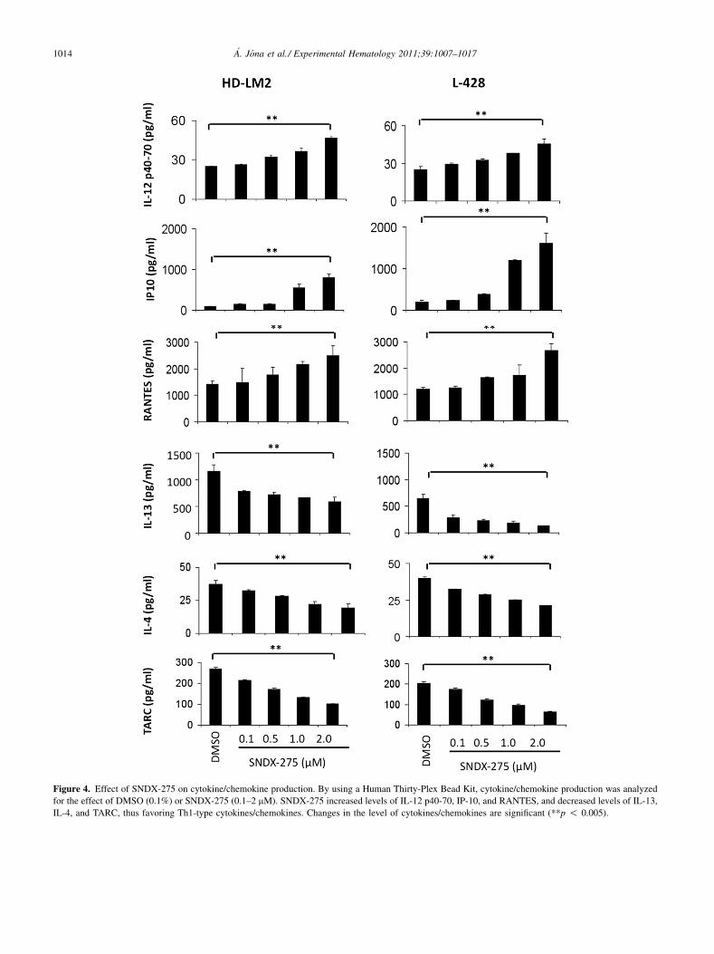
Figure 4. Effect of SNDX-275 on cytokine/chemokine production. By using a Human Thirty-Plex Bead Kit, cytokine/chemokine production was analyzed
for the effect of DMSO (0.1%) or SNDX-275 (0.1–2 mM). SNDX-275 increased levels of IL-12 p40-70, IP-10, and RANTES, and decreased levels of IL-13,
IL-4, and TARC, thus favoring Th1-type cytokines/chemokines. Changes in the level of cytokines/chemokines are significant (**p ! 0.005).
1014 �A. J�ona et al./ Experimental Hematology 2011;39:1007–1017

Figure 5. Expression of cancer/testis antigens (CTAs) in HL cell lines with HDACi SNDX-275 and vorinostat. HL cell lines HD-LM2, L-428, and KM-H2
were cultured with HDACi SNDX-275 and vorinostat at a concentration of 0.5 mM and 1.0 mM for 72 hours. CTA expression was analyzed by reverse tran-
scriptase polymerase chain reaction and immunohistochemically. (A) HDACi induced expression of MAGE-A4 in L-428 and survivin in HD-LM2 at the
RNA level. (B) At the protein level, immunohistochemical analysis showed that none of the investigated CTAs were changed.
1015�A. J�ona et al./ Experimental Hematology 2011;39:1007–1017
the production of IL-12, which is responsible for Th1 celldifferentiation, and decreased the production of IL-13 andIL-4, which promote Th2 differentiation. Furthermore,SNDX-275 altered the balance between chemokines thatare involved in attracting Th2 cells to HL microenviron-ment, including IP-10, RANTES, and TARC. Thesechanges in the microenvironment were associated with up-regulation of a variety of CTAs, although to a less degreewhen compared with other HDACis.
Collectively, our results indicate that SNDX-275 hasa favorable antiproliferative activity by direct induction ofcell death in HL cells, in addition to an indirect effect onthe microenvironment and immunity. Based on these favor-able preclinical activities, we initiated a phase II study ofSNDX-275 to determine its safety and efficacy in patientswith relapsed classical HL.
Conflict of interest disclosureNo financial interest/relationships with financial interest relatingto the topic of this article have been declared.
Funding disclosureThis research is supported in part by the National Institutesof Health (NIH) through M.D. Anderson’s Cancer CenterSupport Grant CA016672, NCI Lymphoma SPORE grant1P50CA136411-01A1, The Living Legend Fund, and thePrince Fund for Lymphoma (Anas Younes), by NIH grantP50 CA126752, the Gillson Longenbaugh Foundation,and the M.D. Anderson Foundation (Catherine M. Bollard),and by The Hungarian American Enterprise ScholarshipFund (�Ad�am J�ona).
References1. J�ona A, Younes A. Novel treatment strategies for patients with
relapsed classical Hodgkin lymphoma. Blood Rev. 2010;24:233–238.
2. Jones PA,Baylin SB. The epigenomics of cancer. Cell. 2007;128:683–692.
3. Glozak MA, Seto E. Histone deacetylases and cancer. Oncogene.
2007;26:5420–5432.
4. Heider U, Kaiser M, Sterz J, et al. Histone deacetylase inhibitors
reduce VEGF production and induce growth suppression and
apoptosis in human mantle cell lymphoma. Eur J Haematol. 2006;
76:42–50.

1016 �A. J�ona et al./ Experimental Hematology 2011;39:1007–1017
5. Brogdon JL, Xu Y, Szabo SJ, et al. Histone deacetylase activities are
required for innate immune cell control of Th1 but not Th2 effector
cell function. Blood. 2007;109:1123–1130.
6. Bolden JE, Peart MJ, Johnstone RW. Anticancer activities of histone
deacetylase inhibitors. Nat Rev Drug Discov. 2006;5:769–784.
7. Botrugno OA, Santoro F, Minucci S. Histone deacetylase inhibitors as
a new weapon in the arsenal of differentiation therapies of cancer.
Cancer Lett. 2009;280:134–144.
8. Duvic M, Talpur R, Ni X, et al. Phase 2 trial of oral vorinostat (sub-
eroylanilide hydroxamic acid, SAHA) for refractory cutaneous T-cell
lymphoma (CTCL). Blood. 2007;109:31–39.
9. Piekarz RL, Frye R, Turner M, et al. Phase II multi-institutional trial
of the histone deacetylase inhibitor romidepsin as monotherapy for
patients with cutaneous T-cell lymphoma. J Clin Oncol. 2009;27:
5410–5417.
10. Khan N, Jeffers M, Kumar S, et al. Determination of the class and
isoform selectivity of small-molecule histone deacetylase inhibitors.
Biochem J. 2008;409:581–589.
11. Gui C-Y, Ngo L, Xu WS, Richon VM, Marks PA. Histone deacetylase
(HDAC) inhibitor activation of p21WAF1 involves changes in
promoter-associated proteins, including HDAC1. PNAS. 2004;101:
1241–1246.
12. Saito A, Yamashita T, Mariko Y, et al. A synthetic inhibitor of histone
deacetylase, MS-27-275, with marked in vivo antitumor activity
against human tumors. PNAS. 1999;96:4592–4597.
13. Jaboin J, Wild J, Hamidi H, et al. MS-27-275, an inhibitor of histone
deacetylase, has marked in vitro and in vivo antitumor activity against
pediatric solid tumors. Cancer Res. 2002;62:6108–6115.
14. Gojo I, Jiemjit A, Trepel JB, et al. Phase 1 and pharmacologic study of
MS-275, a histone deacetylase inhibitor, in adults with refractory and
relapsed acute leukemias. Blood. 2007;109:2781–2790.
15. Lucas DM, Davis ME, Parthun MR, et al. The histone deacetylase
inhibitor MS-275 induces caspase-dependent apoptosis in B-cell
chronic lymphocytic leukemia cells. Leukemia. 2004;18:1207–1214.
16. Rosato RR, Almenara JA, Grant S. The histone deacetylase inhibitor
MS-275 promotes differentiation or apoptosis in human leukemia cells
through a process regulated by generation of reactive oxygen species
and induction of p21CIP1/WAF1 1. Cancer Res. 2003;63:3637–3645.
17. Yang J, Wezeman M, Zhang X, et al. Human C-reactive protein binds
activating Fcgamma receptors and protects myeloma tumor cells from
apoptosis. Cancer Cell. 2007;12:252–265.
18. Huang X, Gao L, Wang S, Lee C-K, Ordentlich P, Liu B. HDAC Inhib-
itor SNDX-275 induces apoptosis in erbB2-overexpressing breast
cancer cells via down-regulation of erbB3 expression. Cancer Res.
2009;69:8403–8411.
19. Singh TR, Shankar S, Srivastava RK. HDAC inhibitors enhance the
apoptosis-inducing potential of TRAIL in breast carcinoma. Onco-
gene. 2005;24:4609–4623.
20. Xu J, Zhou J-Y, Wei W-Z, Philipsen S, Wu GS. Sp1-mediated TRAIL
induction in chemosensitization. Cancer Res. 2008;68:6718–6726.
21. Fournel M, Bonfils C, Hou Y, et al. MGCD0103, a novel isotype-
selective histone deacetylase inhibitor, has broad spectrum antitumor
activity in vitro and in vivo. Mol Cancer Ther. 2008;7:759–768.
22. Martell RE, Garcia-Manero G, Younes A, et al. Clinical Development
of MGCD0103, An isotype-selective HDAC inhibitor: pericarditis/per-
icardial effusion in the context of overall safety and efficacy. Blood
(ASH Annual Meeting Abstracts). 2009;114:4756.
23. Drexler HG. Recent results on the biology of Hodgkin and Reed-
Sternberg cells. II. Continuous cell lines. Leuk Lymphoma. 1993;9:1–25.
24. Georgakis GV, Li Y, Rassidakis GZ, Martinez-Valdez H, Medeiros LJ,
Younes A. Inhibition of heat shock protein 90 function by 17-
allylamino-17-demethoxy-geldanamycin in Hodgkin’s lymphoma
cells down-regulates Akt kinase, dephosphorylates extracellular
signal-regulated kinase, and induces cell cycle arrest and cell death.
Clin Cancer Res. 2006;12:584–590.
25. Zheng B, Fiumara P, Li YV. MEK/ERK pathway is aberrantly active
in Hodgkin disease: a signaling pathway shared by CD30, CD40,
and RANK that regulates cell proliferation and survival. Blood.
2003;102:1019–1027.
26. Rasheed W, Bishton M, Johnstone R, Prince H. Histone deacetylase
inhibitors in lymphoma and solid malignancies. Expert Rev Anti-
cancer Ther. 2008;8:413–432.
27. Chanan-Khan A. Bcl-2 antisense therapy in hematologic malignan-
cies. Curr Opin Oncol. 2004;16:581–585.
28. Maggio E, van den Berg A, Diepstra A, Kluiver J, Visser L, Poppema
S. Chemokines, cytokines and their receptors in Hodgkin’s lymphoma
cell lines and tissues. Ann Oncol. 2002;13:52–56.
29. Karpf AR. A potential role for epigenetic modulatory drugs in the
enhancement of cancer/germ-line antigen vaccine efficacy. Epige-
netics. 2006;1:116–120.
30. Gattei V, Fonsatti E, Sigalotti L, et al. Epigenetic immunomodulation
of hematopoietic malignancies. Semin Oncol. 2005;32:503–510.
31. Marks PA, Xu WS. Histone deacetylase inhibitors: potential in cancer
therapy. J Cell Biochem. 2009;107:600–608.
32. Lane AA, Chabner BA. Histone deacetylase inhibitors in cancer
therapy. J Clin Oncol. 2009;27:5459–5468.
33. Khaskhely NM, Buglio D, Shafer J, Bollard CM, Younes A. The
histone deacetylase (HDAC) inhibitor entinostat (SNDX-275) targets
hodgkin lymphoma through a dual mechanism of immune modulation
and apoptosis induction. Blood (ASH Annual Meeting Abstracts).
2009;114:1562.
34. Hartlapp I, Pallasch C, Weibert G, Kemkers A, Hummel M, Re D.
Depsipeptide induces cell death in Hodgkin lymphoma-derived cell
lines. Leuk Res. 2009;33:929–936.
35. Gupta M, Ansell SM, Novak AJ, Kumar S, Kaufmann SH, Witzig TE.
Inhibition of histone deacetylase overcomes rapamycin-mediated
resistance in diffuse large B-cell lymphoma by inhibiting Akt
signaling through mTORC2. Blood. 2009;114:2926–2935.
36. Buglio D, Mamidipudi V, Khaskhely NM, et al. The histone deacety-
lase inhibitor MGCD0103 down regulates CD30, activates NF-Kb, and
synergizes with proteasome inhibitors by HDAC6 independent mech-
anism in Hodgkin lymphoma. Blood (ASH Annual Meeting
Abstracts). 2009;114:3735.
37. Buglio D, Georgiakis GV, Hanabuchi S, et al. Vorinostat inhibits
STAT6-mediated TH2 cytokine and TARC production and induces
cell death in Hodgkin lymphoma cell lines. Blood. 2008;112:
1424–1433.
38. Lindemann RK, Newbold A, Whitecross KF, et al. Analysis of the
apoptotic and therapeutic activities of histone deacetylase inhibitors
by using a mouse model of B cell lymphoma. PNAS. 2007;104:
8071–8076.
39. Kawamata N, Chen J, Koeffler HP. SAHA (vorinostat) suppresses
translation of cyclin D1 in mantle cell lymphoma cells. Blood.
2007;110:2667–2673.
40. Heider U, Kaiser M, Sterz J, et al. Histone deacetylase inhibitors
reduce VEGF production and induce growth suppression and apoptosis
in human mantle cell lymphoma. Eur J Haematol. 2006;76:42–50.
41. Duan H, Heckman CA, Boxer LM. Histone deacetylase inhibitors
down-regulate bcl-2 expression and induce apoptosis in t(14;18)
lymphomas. Mol Cell Biol. 2005;25:1608–1619.
42. Buglio D, Mamidipudi V, Khaskhely NM, et al. The class-I HDAC
inhibitor MGCD0103 induces apoptosis in Hodgkin lymphoma cell
lines and synergizes with proteasome inhibitors by an HDAC6-
independent mechanism. Br J Haematol;151:387–396.
43. Buglio D, Georgakis GV, Hanabuchi S, et al. Vorinostat inhibits
STAT6-mediated TH2 cytokine and TARC production and induces
cell death in Hodgkin lymphoma cell lines. Blood. 2008;112:
1424–1433.
44. Buglio D, Mamidipudi V, Khaskhely NM, et al. The histone deacety-
lase inhibitor MGCD0103 down regulates CD30, activates NF-Kb, and

1017�A. J�ona et al./ Experimental Hematology 2011;39:1007–1017
synergizes with proteasome inhibitors by HDAC6 independent mech-
anism in Hodgkin lymphoma. Blood (ASH Annual Meeting
Abstracts). 2009;114:3735.
45. Patel MP, Masood A, Patel PS, Chanan-Khan AA. Targeting the Bcl-2.
Curr Opin Oncol. 2009;21:516–523.
46. Paoluzzi L, Gonen M, Bhagat G, et al. The BH3-only mimetic ABT-
737 synergizes the antineoplastic activity of proteasome inhibitors in
lymphoid malignancies. Blood. 2008;112:2906–2916.
47. Del Gaizo Moore V, Schlis KD, Sallan SE, Armstrong SA, Letai A.
BCL-2 dependence and ABT-737 sensitivity in acute lymphoblastic
leukemia. Blood. 2008;111:2300–2309.
48. Whitecross KF, Alsop AE, Cluse LA, et al. Defining the target speci-
ficity of ABT-737 and synergistic antitumor activities in combination
with histone deacetylase inhibitors. Blood. 2009;113:1982–1991.
49. Skinnider BF, Mak TW. The role of cytokines in classical Hodgkin
lymphoma. Blood. 2002;99:4283–4297.

Supplementary Figure E1. (A) In vitro effect of HDACi or decitibine on the expression of SSX2 and NY-ESO-1 in HL cell lines as determined by the
RT-PCR method. (B) The effect of HDAC inhibitors and decitibine on NY-ESO-1 protein expression was determined by immunohistochemistry in HL
cell lines.
1017.e1 �A. J�ona et al./ Experimental Hematology 2011;39:1007–1017