
The role of acetylation in the regulation of antimicrobial ...

Cell shape regulates global histone acetylation in humanmammary epithelial cells
Johanne Le Beyec†,2, Ren Xu†,1, Sun-Young Lee1, Celeste M. Nelson1, Aylin Rizki1, JordiAlcaraz1, and Mina J. Bissell*,1Life Sciences Division, Lawrence Berkeley National Laboratory, 1 Cyclotron Road, MS 977-225A,Berkeley, CA 94720
AbstractExtracellular matrix (ECM) regulates cell morphology and gene expression in vivo; theserelationships are maintained in three-dimensional (3D) cultures of mammary epithelial cells. In thepresence of laminin-rich ECM (lrECM), mammary epithelial cells round up and undergo globalhistone deacetylation, a process critical for their functional differentiation. However, it remainsunclear whether lrECM-dependent cell rounding and global histone deacetylation are indeed part ofa common physical-biochemical pathway. Using 3D cultures as well as nonadhesive andmicropatterned substrata, here we showed that the cell ‘rounding’ caused by lrECM was sufficientto induce deacetylation of histones H3 and H4 in the absence of biochemical cues. Microarray andconfocal analysis demonstrated that this deacetylation in 3D culture is associated with a globalincrease in chromatin condensation and a reduction in gene expression. Whereas cells cultured onplastic substrata formed prominent stress fibers, cells grown in 3D lrECM or on micropatterns lackedthese structures. Disruption of the actin cytoskeleton with cytochalasin D phenocopied the lrECM-induced cell rounding and histone deacetylation. These results reveal a novel link between ECM-controlled cell shape and chromatin structure, and suggest that this link is mediated by changes inthe actin cytoskeleton.
Keywordscell morphology; chromatin structure; differentiation; histone acetylation
IntroductionCell structure and function were postulated to be intimately connected in maintenance of tissuehomeostasis [1]. Manipulating cellular and nuclear morphology in culture can induce a varietyof functional changes, including glucose uptake and metabolism [2], proliferation [3–5],apoptosis [4], differentiation and gene expression [6–10]. These morphologically drivenprocesses are controlled by the local microenvironment as exemplified in the mammary glandand mammary epithelial cells (for review see [11]). Isolated mouse mammary epithelial cellscan be induced to undergo structural and functional differentiation in three-dimensional (3D)
* Correspondence should be addressed to M.J.B. ([email protected]).†J. L. B. and R. X. contributed equally to this work.1Life Sciences Division, Lawrence Berkeley National Laboratory, 1 Cyclotron Road, MS 977 R225A, Berkeley, CA 94720, USA.2Université Pierre et Marie Curie-Paris 6, UMR S 872, Les Cordeliers, Paris, F-75006 France; INSERM, UMR S 872, Paris, F-75006France; Université Paris Descartes-Paris 5, UMR S 872, Paris, F-75006 France.Publisher's Disclaimer: This is a PDF file of an unedited manuscript that has been accepted for publication. As a service to our customerswe are providing this early version of the manuscript. The manuscript will undergo copyediting, typesetting, and review of the resultingproof before it is published in its final citable form. Please note that during the production process errors may be discovered which couldaffect the content, and all legal disclaimers that apply to the journal pertain.
NIH Public AccessAuthor ManuscriptExp Cell Res. Author manuscript; available in PMC 2008 August 15.
Published in final edited form as:Exp Cell Res. 2007 August 15; 313(14): 3066–3075.
NIH
-PA Author Manuscript
NIH
-PA Author Manuscript
NIH
-PA Author Manuscript

cultures. The acquisition of the functionally differentiated phenotype, i.e. expression of milkproteins, requires lactogenic hormones and the basement membrane protein laminin-111(laminin-1) [12,13]. The latter induces dramatic morphological changes in that cells becomerounded and actin becomes cortical. These morphological changes are necessary for expressionof some milk proteins including β-casein [9]. Artificially pre-rounding the cells by plating onthe nonadhesive substratum poly(2-hydroxyethyl methacrylate) (polyHEMA) poises them forresponsiveness to prolactin and laminin-rich ECM (lrECM)[9], whereas preventing cellrounding by treatment with the phorbol ester 12-o-tetradecanoylphorbol 13-acetate (TPA)inhibits milk protein synthesis [14]. These previous findings strongly suggested that cellrounding, mediated by reorganization of actin filaments and other cytoskeletal components,may be an important physical signal conveyed by the ECM leading to changes in geneexpression.
Cell fate and differentiated function are controlled by patterns of gene expression, which inturn are dictated by chromatin organization. The structure of chromatin is regulated by anumber of post-translational modifications at the amino-terminal tails of nucleosomal histones,including acetylation/deacetylation, phosphorylation, methylation, and ADP ribosylation[15]. The acetylation status of chromatin is dynamic, balanced by the activities of histoneacetyltransferases (HATs) and histone deacetylases (HDACs) [16]. Gene expression generallycorrelates with histone hyperacetylation at promoter regions and other regulatory cis-elements[17–19] whereas histone deacetylation represses transcription by promoting chromatincondensation into heterochromatin [20,21]. Histone deacetylation was shown to correlate withchanges in gene expression in the developing brain in rats [22], and abrogating the deacetylationby treatment with an HDAC inhibitor resulted in impaired brain development and delayedexpression of differentiation markers. Functional differentiation of many cell types isassociated with activation of specific subsets of genes, silencing of other genes, and extensiveformation of heterochromatin [23].
Acinar morphogenesis of a non-malignant human mammary epithelial cell line HMT3522-S1,is accompanied by rearrangements in chromatin structure and nuclear architecture [24–26].These reorganizations appear to be important for establishing and maintaining the normalphenotype of cells in 3D [24,25,27]. We previously found that the global level of histoneacetylation is critical for functional differentiation of mammary epithelial cells [24,28].Altering chromatin structure by increasing histone acetylation with trichostatin A induceshuman mammary epithelial cells to alter their morphology, re-enter the cell cycle, and becomedisorganized [24]; overexpressing a HAT in mouse mammary epithelial cells inhibitsproduction of endogenous β-casein in response to lrECM [28]. Conversely, treatment withlrECM induces global histone deacetylation in human and mouse mammary epithelial cells[25,28].
These previous studies showed that ECM has profound effects on cell shape and chromatinstructure, particularly histone deacetylation, but it was not understood whether these effectswere linked. In this report, we used several strategies to change cell shape, including 3D cultureon different substrata with different adhesiveness and micropatterned substrata. We find thatcell rounding per se induces global histone deacetylation and an increase in chromatincondensation, and that these processes are associated with a global reduction of geneexpression. These results reveal a process by which ECM integrates structure and function inmammary epithelial cells through a shape-dependent global histone deacetylation.
Le Beyec et al. Page 2
Exp Cell Res. Author manuscript; available in PMC 2008 August 15.
NIH
-PA Author Manuscript
NIH
-PA Author Manuscript
NIH
-PA Author Manuscript

Materials and MethodsCell culture
HMT-3522-S1 and -T4-2 human mammary epithelial cells [29,30] were grown as two-dimensional (2D) monolayers on plastic or within 3D lrECM (Matrigel, CollaborativeResearch) and maintained as previously described [31,32]. Cells were grown in 2D and 3D for10 days before harvests. For culture on nonadhesive substrata, ~4000 cells/cm2 were seededon polyHEMA (Sigma)-coated plates prepared as previously described for mouse mammaryepithelial cells [9].
MicropatterningMicropatterned substrata consisting of collagen-coated islands were created as described[33]. Briefly, elastomeric stamps containing a relief of the desired pattern were coated withtype I collagen (50 μg/mL in water; Vitrogen 100, Cohesion Technologies, Palo Alto, CA) for2 hours, washed with water, and dried under a stream of nitrogen. Flat poly(dimethylsiloxane)(PDMS; Sylgard 184, Ellsworth Adhesives, Germantown, WI) elastomer-coated substratawere UV-oxidized for 7 minutes (UVO Cleaner, Jelight Co., Irvine, CA), stamped withcollagen, blocked with 1% pluronic F108 (BASF Corp., Florham Park, NJ) in water for 1 hour,and rinsed in PBS before seeding T4-2 cells. Cells were allowed to attach to the patternedislands for approximately 30 minutes before washing away the remaining floating cells. A flatblock of PDMS was coated with collagen and stamped for the unpatterned substratum control.For measurements of projected cell area, phase contrast images of individual cells were outlinedand processed with Scion Image software.
Global gene expression analysiscDNA microarrays with ~8000 known genes spotted on poly-L-lysine-coated chips (customarrayed at Lawrence Berkeley National Laboratory using Research Genetics 8k human clones)were used. mRNA samples of interest were directly compared to each other by co-hybridizationto the same slide using dendrimer technology to label with red-Cy5 and green-Cy3(Genisphere). Total RNA (1μg) isolated with Qiagen RNEasy reagents was used for eachsample hybridized. Cells in 3D lrECM were extracted using 5mM EDTA in cold PBS todissolve the Matrigel. For each comparison, 3 independent sets of cells cultured for 10 dayswere processed, and 4 slides were hybridized. This corresponded to 3 sets of RNA fromindependent culture sets plus a dye-swap experiment in which the red and green label wasswitched for the two samples in question to account for dye-specific effects. Arrays werescanned using a Genepix scanner (Axon). Raw data for each channel (red and green) was loadedonto Genespring (Silicon Genetics) for normalization and analysis. For each chip, per-spot andper-chip intensity-dependent Lowess normalization was performed using 20% of data forsmoothing at a cutoff value for signal of 10. This was followed by normalization of each chipto the 50th percentile of the measurements taken from that chip, without extra backgroundcorrection. Differential gene expression was determined at the t-test p-value of 0.01 or lower.
Immunofluorescence analysisSamples were fixed in 2% paraformaldehyde in PBS, rinsed in 50 mM glycine in PBS, blockedin 10% goat serum, and stained with a 1:500 dilution of FITC-phalloidin (Molecular Probes)or with a 1:100 dilution of AcH4 antibody in PBS. Stained samples were imaged using a SpotRT camera attached to a Zeiss upright epifluorescence microscope or a Stanford PhotonicsXR/Mega-10 ICCD camera attached to a Zeiss spinning disk confocal microscope.
DNA was stained with DAPI and the corresponding fluorescence was measured by acquiringconfocal sections separated by 0.16 μm using a spinning disk confocal microscope. Confocal
Le Beyec et al. Page 3
Exp Cell Res. Author manuscript; available in PMC 2008 August 15.
NIH
-PA Author Manuscript
NIH
-PA Author Manuscript
NIH
-PA Author Manuscript

images were corrected for background. To assess the nuclear volume, a common intensitythreshold was established to define the edge of the nuclei in each confocal section and calculatethe enclosed area. The total fluorescence intensity was divided by the nuclear volume to obtainthe average dye spatial density, which correlates with the average chromatin packing ratio(Mascetti et al 2001). All image processing was performed using Image J software.
Western blottingTotal protein from S1 or T4-2 cells was extracted with lysis buffer [50 mM Tris (pH 7.4), 30mM NaCl, 2 % (w/v) SDS, and protease inhibitor cocktail]. After sonication, an equal amountof protein from each sample was subjected to SDS gel electrophoresis and then transferred tonitrocellulose membrane (Schleicher & Schuell). The membrane was subsequently blocked inTBST buffer [50 mM Tris (pH 8.0), 150 mM NaCl, 0.1% (v/v) Tween 20] containing 5%nonfat dried milk, then incubated in blocking buffer containing primary antibody (AcH3 andAcH4 from Upstate; total H3 from Santa Cruz). All blots were further incubated in blockingbuffer containing horseradish peroxidase-conjugated secondary antibodies and subjected toenhanced chemiluminescence (ECL) using the SuperSignal chemiluminescent substrate(Pierce, Rockford, IL). The results were quantified with AlphaEasyFC software, and studentt test were performed using SigmaPlot.
Quantitative real-time PCR analysisTotal RNA was extracted from cells with Trizol reagent (Invitrogen). cDNA was synthesizedusing Superscript first strand synthesis kit (Invitrogen) from equal amounts of RNA.Quantitative real-time PCR analysis was performed with the Lightcycler System (Roche) usingthe Lightcycler FastStart DAN Master SYBR Green I kit (Roche). The following primers wereused to amplify p21 and 18S sequences: forward primer of the p21 gene 5′-CTG GGG ATGTCC GTC AGA AC-3′ and reverse primer 5′- AGC GAG GCA CAA GGG TAC AA-3′;forward primer of the 18S gene: 5′- ACG GAC CAG AGC GAA AGC AT -3′ and reverseprimer 5′- GGA CAT CTA AGG GCA TCA CAG AC -3′. The following Lightcycler PCRamplification protocol was used: 95°C for 10 minutes, and 45 amplification cycles (95°C for5 seconds, 60°C for 10 seconds, 72°C for 5 seconds). Amplification was followed by meltingcurve analysis to verify the presence of a single PCR product [34]; 18S was amplified as areference gene using the same protocol.
Chromatin immunoprecipitation (ChIP) assayChIP assays were performed using a commercially available kit (ChIP kit; UpstateBiotechnology, Lake Placid, NY) per manufacturer instructions. For 2D conditions, 1 × 10⁷
S1 cells grown in a 100-mm dish were cross-linked with 1% formaldehyde at room temperaturefor 10 minutes. For 3D conditions, S1 cells were isolated from lrECM using PBS/EDTA andcross-linked as above. Cells were washed with PBS, resuspended and lysed in ChIP lysis buffer(1% SDS, 10mM EDTA, 50 mM Tris-HCl pH8.0). Sonicated lysates were diluted with ChIPdilution buffer and bound to protein A-agarose beads. The precleared lysates were thenimmunoprecipitated with AcH3 or AcH4 antibodies (Upstate Biotechnology), collected withprotein A-agarose beads, and washed sequentially with each of the following buffers: low saltwash buffer (0.1% SDS, 1% Triton X-100, 2 mM EDTA, 20 mM Tris-HCl pH8.0, 150 mMNaCl); high salt wash buffer (0.1% SDS, 1% Triton X-100, 2 mM EDTA, 20 mM Tris-HClpH8.0, 500 mM NaCl); LiCl buffer (0.25 M LiCl, 1% NP-40, 1% SDC, 1 mM EDTA, 10 mMTris-HCl pH8.0); TE buffer (20 mM Tris-HCl pH8.0, 1 mM EDTA pH8.0). The remainingbound p21 promoter DNA was PCR-amplified using the following primers: forward primer5′-GGT GTC TAG GTG CTC CAG GT-3′ and reverse primer 5′-GCA CTC TCC AGG AGGACA CA-3′.
Le Beyec et al. Page 4
Exp Cell Res. Author manuscript; available in PMC 2008 August 15.
NIH
-PA Author Manuscript
NIH
-PA Author Manuscript
NIH
-PA Author Manuscript

ResultsCulturing cells in 3D lrECM induces alterations in cellular morphology and global histonedeacetylation
Culturing non-malignant breast epithelial S1 cells in 3D within lrECM allows the cells to formspherical polarized structures that resemble mammary acini in vivo (Fig. 1A), whereas the cellsform monolayers when they are cultured in 2D. Immunofluorescence and western blot analysisshowed that levels of both acetylated histones H3 and H4 were reduced in 3D cultures. (Fig.1A, B, C).
Histone deacetylation is associated with chromatin condensation, and hypoacetylation ofhistones is commonly used as a marker of heterochromatin [35]. Here we used the average 4′,6-diamidino-2-phenylindole (DAPI) spatial density to assess global changes in chromatincondensation because the density of DAPI staining in nuclei increases with the chromatinpacking ratio [36,37]. We found that S1 cells in 3D lrECM had higher DAPI density than cellscultured in 2D (Fig. 1D). These data indicate that the histone deacetylation in 3D cultures isassociated with increased chromatin compaction. In addition, the tissue-like morphogenesisobserved in 3D was accompanied by a significant decrease in nuclear diameter (Fig. 1E).
T4-2, the tumorigenic derivative of S1 cells, form large non-polarized and disorganizedcolonies in 3D lrECM reminiscent of tumors in vivo, but they still respond to lrECM bychanging cell shape (Supplemental Fig A). To determine whether global histone deacetylationwas the result of acinar morphogenesis, we measured the levels of acetylated histones H3 andH4 in T4-2 cells which form disorganized structure in lrECM. Histone acetylation wasdecreased in 3D culture, whether or not the cells were polarized (Fig 1B, C), indicating thatthe reduction in histone deacetylation is a response either to ECM-induced cell rounding or toECM-induced signaling.
Cell rounding induces global histone deacetylationTo test whether changes in cell morphology could alter histone acetylation, we compared cellscultured on plastic with those cultured on the nonadhesive substratum polyHEMA, whichprevents cell attachment and spreading and allows examination of the role of cellular shapeindependently of ECM signaling [3,9]. Both S1 (Fig. 2A) and T4-2 (data not shown) cellsadopted a rounded morphology when cultured on polyHEMA. Culturing cells on polyHEMAfor 4 days also caused them to cluster together (Fig. 2A). Immunofluorescence and westernblot analysis of acetylated histones H3 and H4 demonstrated that cultivation on polyHEMAleads to histone deacetylation in both S1 and T4-2 cells (Fig. 2A, B, C). Additionally, histonedeacetylation in cells on polyHEMA is associated with chromatin condensation and a decreasein nuclear size (Fig. 2D, E). Adding lrECM to cells cultured on polyHEMA did not decreasehistone acetylation levels further (data not shown), indicating that histone deacetylationinduced by lrECM is due specifically to an effect of cell rounding or cell clustering, and thatECM ligand-induced signaling does not potentiate this effect.
To delineate the effects of cell rounding from those of cell clustering, we used a micropatterningapproach to maintain single attached cells in a rounded morphology. Cells were cultured onmicropatterned substrata that contained micrometer-sized islands of collagen surrounded bynonadhesive regions. Cells plated on these substrata could only attach to the collagen-coatedislands; individual cells cultured on small islands were prevented from spreading. Wecompared unpatterned and freely spread T4-2 cells with those patterned on 25-μm squareislands (Fig. 3A). T4-2 cells plated on micropatterned substrata remained rounded comparedto those plated on unpatterned collagen-coated substrata (Fig. 3A). Whereas control cellsformed stress fibers, patterned cells exhibited mainly cortical actin (Fig. 3A). The mean
Le Beyec et al. Page 5
Exp Cell Res. Author manuscript; available in PMC 2008 August 15.
NIH
-PA Author Manuscript
NIH
-PA Author Manuscript
NIH
-PA Author Manuscript

projected area of individual cells plated on the micropatterned substrata was significantlyreduced when compared to cells plated on tissue culture plastic (Supplemental Fig. B). Westernblot analysis showed that levels of acetylated histones H3 and H4 were reduced in patternedcells compared to unpatterned cells (Fig. 3B, C). Quantification of DAPI staining demonstratedthat patterned cells had more compacted chromatin than unpatterned cells (Fig. 3D),accompanied by a reduction in nuclear size in the patterned cells (Fig. 3E). These data showthat ECM-induced cell rounding directly controls histone deacetylation and chromatincondensation in mammary epithelial cells. It further indicates that even malignant T4-2 cellsremain sensitive to shape-induced changes in their microenvironment.
Cell shape is largely governed by the actin cytoskeleton [38]. Shape dependence thereforeimplies a requirement for a particular actin organization. Indeed, one of the striking differenceswe noticed between cells spread on plastic and those rounded in 3D lrECM or onmicropatterned substrata was the organization of the actin cytoskeleton. Both S1 and T4-2 cellscultured in 2D exhibited prominent stress fibers as well as cortical actin when stained withfluorescently-tagged phalloidin (Fig. 4A). In contrast, even though the gross architectures ofthe colonies formed by these cells differed substantially, individual S1 and T4-2 cells appearedsimilarly rounded in 3D (Fig. 1A and Supplemental Fig. A) with mainly cortical actin (Fig.4A). Cells rounded by culture on micropatterned substrata also showed prominent cortical actin(Fig 4B). To address the involvement of shape and actin organization in regulation of histoneacetylation, S1 cells grown on tissue culture plastic were treated with the actin polymerizationinhibitor cytochalasin D. Treatment with this drug disrupted actin microfilaments, induced cellrounding (Fig. 4B), and reduced the levels of acetylated histones H3 and H4 (Fig. 4C, D).Importantly, removal of the drug reversed cell morphology (data not shown) and induced anincrease in histone acetylation (Fig. 4D). Therefore, cytoskeletal changes associated with cellrounding induce global histone deacetylation.
Culture in 3D lrECM induces a global reduction in gene expressionOne question that arises from these observations is whether the histone deacetylation in 3Dculture regulates gene expression. Indeed, broad acetylation of histones H3 and H4 was shownto lead to chromatin decondensation and a structure permissive for transcription (reviewed in[39]), whereas histone deacetylation correlated with the repressed state of chromatin andalterations in patterns of gene expression [40]. To determine whether culture in 3D lrECMinduced a reduction in gene expression in addition to global histone deacetylation, weperformed microarray analysis. mRNA samples from S1 cells cultured on plastic or in 3DlrECM were fluorescently tagged and co-hybridized to cDNA microarrays, and the intensityof hybridizations were compared. Genes expressed differentially between the two conditionswere selected for analysis. We found that cells cultured in 3D lrECM showed an appreciablereduction in gene expression as compared to cells cultured on plastic (Fig. 5A; SupplementalTable.1). Of the differentially expressed genes, 162 genes were expressed at lower levels in3D, compared to 91 expressed at higher levels. This decrease in gene expression is consistentwith the global histone deacetylation observed in 3D cultures.
Acetylation of histones in the promoter regions and other regulatory cis-elements clearly affectstranscription of a large number of genes [17,18,41]. To address the question of whether thelrECM-dependent global deacetylation could specifically affect the promoter of a functionallyrelevant gene, we focused our attention on the cell cycle inhibitor p21. It was previously shownthat the expression of the p21 gene is controlled by the acetylation status of histones associatedwith its promoter [42,43]. Indeed, from our array data, the transcript level of p21 in S1 cellsin 3D was reduced to 76% of that in cells in 2D after 10 days culture (Supplemental Table 1).Quantitative real-time RT/PCR confirmed that p21 mRNA levels were reduced in 3D comparedto 2D (Fig. 5B). To determine whether this reduction in expression is correlated with reduced
Le Beyec et al. Page 6
Exp Cell Res. Author manuscript; available in PMC 2008 August 15.
NIH
-PA Author Manuscript
NIH
-PA Author Manuscript
NIH
-PA Author Manuscript

acetylation at the promoter region of the p21 gene, we performed chromatinimmunoprecipitation (ChIP) experiments. Lysates from S1 cells cultured in 2D or 3D wereimmunoprecipitated for acetylated histones H3 or H4, and the remaining associated DNA wasPCR-amplified using primers specific for the p21 promoter. Culturing the cells in 3D lrECMinduced a dramatic reduction of histone acetylation in the p21 promoter (Fig. 5C). This resultis consistent with the reduced expression of p21 observed by microarray analysis and RT/PCR.These data indicate that lrECM-induced histone deacetylation is associated with a functionalreduction of gene expression.
DiscussionThe interactions between a cell and its local microenvironment have profound effects on cellshape, cytoskeletal and nuclear matrix organization, chromatin structure, and gene expression[24,44–46]. In this study, we found that culture of human mammary epithelial cells within 3DlrECM induced cell rounding and global histone deacetylation. Histone deacetylationcorrelates with chromatin condensation and reduced gene expression. These data arecorroborated by previous studies demonstrating that lrECM-induced morphogenesis correlateswith reduced histone acetylation and increased histone methylation [24,25,28,44]. Cellsreceive at least two types of signals from laminin-rich ECM gels: biochemical signals from theengagement of cell surface receptors, and biophysical signals from the change in theirmorphology [9]. Altering cellular shape has been shown to regulate nuclear morphology andchromatin structure [37]. Using non-adhesive and micropatterned substrata as well as inhibitorsof actin polymerization, we determined that cell rounding is a sufficient stimulus to inducehistone deacetylation and chromatin condensation in the absence of biochemical signalstransduced from the ECM; notably, adding ECM to pre-rounded cells did not potentiate theshape-induced histone deacetylation.
There is increasing evidence that cell rounding inhibits cell proliferation [3,4], and that histoneacetylation levels correlate with cell cycle status [47]. We found that culturing cells on plasticand in 3D lrECM led to a similar percentage of growth-arrested cells (Supplemental Fig. C).Furthermore, global histone deacetylation was detected in T4-2 cells cultured in 3D, althoughthese cells failed to growth arrest (data not shown). Therefore, the ECM-induced reduction inhistone acetylation does not correlate with decreased proliferation in this system. S1 cellsundergo growth arrest after 10 days culture in both 2D and 3D. However, transcription levelsof the p21 gene were lower in 3D than in 2D culture, suggesting that the growth arrest in 3Dmay be regulated by other proteins or signaling pathways. Consistent with this possibility, wepreviously reported that cell cycle arrest in 3D culture was accompanied by progressivehypophosphorylation of the retinoblastoma (Rb) gene and induction of the cyclin-dependentkinase (CDK) inhibitor p27kip1[48].
Results over the past two decades appear to support the hypothesis that the ECM is dynamicallycoupled to the nucleus through the cytoskeleton and the nuclear matrix [1,49–51]. Oneattractive potential mechanism to regulate chromatin structure involves signaling from thecytoskeleton. We found that cell rounding (either in 3D lrECM or on micropatterned substrata)altered the actin cytoskeleton and prevented the formation of stress fibers, and that blockingactin polymerization/depolymerization with cytochalasin D led to histone deacetylation.Disrupting microtubules also causes cell rounding [52] and has been shown to induce histonedeacetylation [53]. These results suggest a role for low intracellular tension generated bycytoskeletal organization in the control of histone acetylation. It has been shown that disruptingactomyosin tension generation by inhibiting myosin light chain kinase, RhoA, or Rho-kinaseleads to global histone deacetylation, and conversely, increasing actomyosin contractilityenhances histone acetylation levels in gastric carcinoma cells [41]. Increasing tension byapplication of shear stress leads to histone acetylation, chromatin remodeling, and changes in
Le Beyec et al. Page 7
Exp Cell Res. Author manuscript; available in PMC 2008 August 15.
NIH
-PA Author Manuscript
NIH
-PA Author Manuscript
NIH
-PA Author Manuscript

gene expression in endothelial and embryonic stem cells [54,55]. In agreement, experimentaland theoretical evidence supports a specific role for the actin cytoskeleton in physicallyconnecting sites of cell adhesion to ECM with the nucleus [51] as well as integrating changesin cell and nuclear shape [56,57]. In this scenario it is thought that tension developed by actinbundles in spread cells deforms the nucleus to increase its projected area, while a decrease intension (as occurs in round cells) would have the opposite effects. In support of this model weobserved a systematic reduction in nuclear diameter in round cells compared to control cells.Based on the collective data described, it is tempting to speculate that cell shape regulateshistone acetylation through mechanical transmission of actin/cytoskeletal tension to thenucleus.
Histone acetylation and deacetylation are catalyzed by HATs and HDACs; therefore cellrounding could regulate histone acetylation by altering the total cellular levels or activities ofthese enzymes, changing their levels in the nucleus, or their association with the nuclear matrix.Cell rounding reduces the calibre of nuclear pore complexes and reduces the rate ofnucleocytoplasmic transport [58,59], which could directly affect the shuttling of HATs andHDACs and their relative levels within the nucleus. Biochemically, HAT and HDAC shuttlingis regulated by phosphorylation which changes their binding affinity to chaperone proteins thatusher them into or out of the nucleus [60]. Indeed, during skeletal muscle differentiationCa2+/calmodulin-dependent protein kinase (CaMK) activity regulates class II HDACphosphorylation, binding to 14-3-3 protein chaperone, and subsequent nuclear shuttling [61].Additionally, some HDACs have been found to bind to actin filaments and microtubules in thecytoplasm, potentially sequestering them away from the nuclear compartment [62].Depolymerizing portions of the cytoskeleton may release HDACs and allow them to shuttlefrom the cytoplasm into the nucleus, tipping the balance in favour of histone deacetylation. Ingeneral, regulation of HDAC and HAT activities is a large field of study which is opening anever greater array of possible mechanisms by which cell shape might regulate histoneacetylation.
Despite the fact that ECM-induced cell rounding leads to global histone deacetylation, and thatthe deacetylation is associated with a reduction in overall gene expression in 3D cultures,histones associated with genes specifically upregulated during differentiation are mostprobably acetylated locally. For example, histone H4 acetylation is increased in the promoterregion of the α-casein milk protein gene in mammary epithelial cells in the presence of collagen[63], and histone H3 and H4 acetylation is increased in the promoter region of the β-caseingene during differentiation [64,65]. It will be interesting to determine how cell shape affectsthe acetylation status of these and other differentiation-associated genes. Although globalhistone deacetylation is observed during functional differentiation of mammary epithelial cells[24,28] and is regulated by cell rounding, the latter per se may not explain all the effects ofECM on functional differentiation; indeed, the assembly of a basement membrane is requiredin addition to cell rounding for the reorganization of the nuclear protein NuMA and tissue-specific functions [24], and biochemical signals transmitted through integrin and non-integrinlaminin receptors are required for functional differentiation and β-casein expression [9,12,66,67].
We have provided strong evidence for a link between ECM-regulated cell shape, chromatinstructure and gene expression. Since ECM influences morphology and gene expression in awide variety of tissues [reviewed in [68–72]], it is most likely that cell shape-induced changesin global histone acetylation and chromatin structure are a common mechanism in regulationof tissue- and context-specific gene expression in many organs.
Le Beyec et al. Page 8
Exp Cell Res. Author manuscript; available in PMC 2008 August 15.
NIH
-PA Author Manuscript
NIH
-PA Author Manuscript
NIH
-PA Author Manuscript

Supplementary MaterialRefer to Web version on PubMed Central for supplementary material.
Acknowledgements
We thank Virginia Spencer and other members of the Bissell laboratory for helpful discussions. This work wassupported by grants from the Office of Biological and Environmental Research of the Department of Energy (DE-AC03-76SF00098 and a Distinguish Fellow Award to MJB), the National Cancer Institute (CA64786 to M.J.B. andOle William Peterson, and CA57621 to M.J.B. and Zena Werb), and the Breast Cancer Research Program (BCRP) ofthe Department of Defense (DOD) (an Innovator Award to MJB; postdoctoral fellowships DAMD17-02-1-0441 andW81XWH-04-1-0582 to RX and CMN). CMN holds a Career Award at the Scientific Interface from the BurroughsWellcome Fund.
References1. Bissell MJ, Hall HG, Parry G. How does the extracellular matrix direct gene expression? J Theor Biol
1982;99:31–68. [PubMed: 6892044]2. Bissell MJ, Farson D, Tung AS. Cell shape and hexose transport in normal and virus-transformed cells
in culture. J Supramol Struct 1977;6:1–12. [PubMed: 197315]3. Folkman J, Moscona A. Role of cell shape in growth control. Nature 1978;273:345–9. [PubMed:
661946]4. Chen CS, Mrksich M, Huang S, Whitesides GM, Ingber DE. Geometric control of cell life and death.
Science 1997;276:1425–8. [PubMed: 9162012]5. Singhvi R, Kumar A, Lopez GP, Stephanopoulos GN, Wang DI, Whitesides GM, Ingber DE.
Engineering cell shape and function. Science 1994;264:696–8. [PubMed: 8171320]6. Farmer SR, Ben-Ze’av A, Benecke BJ, Penman S. Altered translatability of messenger RNA from
suspended anchorage-dependent fibroblasts: reversal upon cell attachment to a surface. Cell1978;15:627–37. [PubMed: 719755]
7. Thomas CH, Collier JH, Sfeir CS, Healy KE. Engineering gene expression and protein synthesis bymodulation of nuclear shape. Proc Natl Acad Sci U S A 2002;99:1972–7. [PubMed: 11842191]
8. Ben-Ze’ev A, Farmer SR, Penman S. Protein synthesis requires cell-surface contact while nuclearevents respond to cell shape in anchorage-dependent fibroblasts. Cell 1980;21:365–72. [PubMed:6157481]
9. Roskelley CD, Desprez PY, Bissell MJ. Extracellular matrix-dependent tissue-specific gene expressionin mammary epithelial cells requires both physical and biochemical signal transduction. Proc NatlAcad Sci U S A 1994;91:12378–82. [PubMed: 7528920]
10. Emerman JT, Burwen SJ, Pitelka DR. Substrate properties influencing ultrastructural differentiationof mammary epithelial cells in culture. Tissue Cell 1979;11:109–19. [PubMed: 572104]
11. Bissell MJ, Rizki A, Mian IS. Tissue architecture: the ultimate regulator of breast epithelial function.Curr Opin Cell Biol 2003;15:753–62. [PubMed: 14644202]
12. Streuli CH, Bailey N, Bissell MJ. Control of mammary epithelial differentiation: basement membraneinduces tissue-specific gene expression in the absence of cell-cell interaction and morphologicalpolarity. J Cell Biol 1991;115:1383–95. [PubMed: 1955479]
13. Streuli CH, Schmidhauser C, Bailey N, Yurchenco P, Skubitz AP, Roskelley C, Bissell MJ. Lamininmediates tissue-specific gene expression in mammary epithelia. J Cell Biol 1995;129:591–603.[PubMed: 7730398]
14. Taketani Y, Oka T. Tumor promoter 12-O-tetradecanoylphorbol 13-acetate, like epidermal growthfactor, stimulates cell proliferation and inhibits differentiation of mouse mammary epithelial cells inculture. Proc Natl Acad Sci U S A 1983;80:1646–9. [PubMed: 6300862]
15. Grant PA. A tale of histone modifications. Genome Biol 2001;2:1–6.16. Davie JR. Histone modifications, chromatin structure, and the nuclear matrix. J Cell Biochem
1996;62:149–57. [PubMed: 8844394]17. Roh TY, Cuddapah S, Zhao K. Active chromatin domains are defined by acetylation islands revealed
by genome-wide mapping. Genes Dev 2005;19:542–52. [PubMed: 15706033]
Le Beyec et al. Page 9
Exp Cell Res. Author manuscript; available in PMC 2008 August 15.
NIH
-PA Author Manuscript
NIH
-PA Author Manuscript
NIH
-PA Author Manuscript

18. Bernstein BE, Humphrey EL, Erlich RL, Schneider R, Bouman P, Liu JS, Kouzarides T, SchreiberSL. Methylation of histone H3 Lys 4 in coding regions of active genes. Proc Natl Acad Sci U S A2002;99:8695–700. [PubMed: 12060701]
19. Kim TH, Barrera LO, Zheng M, Qu C, Singer MA, Richmond TA, Wu Y, Green RD, Ren B. A high-resolution map of active promoters in the human genome. Nature 2005;436:876–880. [PubMed:15988478]
20. Jenuwein T, Allis CD. Translating the histone code. Science 2001;293:1074–80. [PubMed: 11498575]21. Fraga MF, Ballestar E, Villar-Garea A, Boix-Chornet M, Espada J, Schotta G, Bonaldi T, Haydon C,
Ropero S, Petrie K, Iyer NG, Perez-Rosado A, Calvo E, Lopez JA, Cano A, Calasanz MJ, ColomerD, Piris MA, Ahn N, Imhof A, Caldas C, Jenuwein T, Esteller M. Loss of acetylation at Lys16 andtrimethylation at Lys20 of histone H4 is a common hallmark of human cancer. Nat Genet2005;37:391–400. [PubMed: 15765097]
22. Shen S, Li J, Casaccia-Bonnefil P. Histone modifications affect timing of oligodendrocyte progenitordifferentiation in the developing rat brain. J Cell Biol 2005;169:577–89. [PubMed: 15897262]
23. Francastel C, Schubeler D, Martin DI, Groudine M. Nuclear compartmentalization and gene activity.Nat Rev Mol Cell Biol 2000;1:137–43. [PubMed: 11253366]
24. Lelievre SA, Weaver VM, Nickerson JA, Larabell CA, Bhaumik A, Petersen OW, Bissell MJ. Tissuephenotype depends on reciprocal interactions between the extracellular matrix and the structuralorganization of the nucleus. Proc Natl Acad Sci U S A 1998;95:14711–6. [PubMed: 9843954]
25. Plachot C, Lelievre SA. DNA methylation control of tissue polarity and cellular differentiation in themammary epithelium. Exp Cell Res 2004;298:122–32. [PubMed: 15242767]
26. Knowles DW, Sudar D, Bator-Kelly C, Bissell MJ, Lelievre SA. Automated local bright feature imageanalysis of nuclear protein distribution identifies changes in tissue phenotype. Proc Natl Acad Sci US A 2006;103:4445–50. [PubMed: 16537359]
27. Kaminker P, Plachot C, Kim SH, Chung P, Crippen D, Petersen OW, Bissell MJ, Campisi J, LelievreSA. Higher-order nuclear organization in growth arrest of human mammary epithelial cells: a novelrole for telomere-associated protein TIN2. J Cell Sci 2005;118:1321–30. [PubMed: 15741234]
28. Pujuguet P, Radisky D, Levy D, Lacza C, Bissell MJ. Trichostatin A inhibits beta-casein expressionin mammary epithelial cells. J Cell Biochem 2001;83:660–70. [PubMed: 11746508]
29. Briand P, Petersen OW, Van Deurs B. A new diploid nontumorigenic human breast epithelial cellline isolated and propagated in chemically defined medium. In Vitro Cell Dev Biol 1987;23:181–8.[PubMed: 3558253]
30. Briand P, Nielsen KV, Madsen MW, Petersen OW. Trisomy 7p and malignant transformation ofhuman breast epithelial cells following epidermal growth factor withdrawal. Cancer Res1996;56:2039–44. [PubMed: 8616848]
31. Petersen OW, Ronnov-Jessen L, Howlett AR, Bissell MJ. Interaction with basement membrane servesto rapidly distinguish growth and differentiation pattern of normal and malignant human breastepithelial cells. Proc Natl Acad Sci U S A 1992;89:9064–8. [PubMed: 1384042]
32. Weaver VM, Petersen OW, Wang F, Larabell CA, Briand P, Damsky C, Bissell MJ. Reversion ofthe malignant phenotype of human breast cells in three-dimensional culture and in vivo by integrinblocking antibodies. J Cell Biol 1997;137:231–45. [PubMed: 9105051]
33. Tan JL, Liu W, Nelson CM, Raghavan S, Chen CS. Simple approach to micropattern cells on commonculture substrates by tuning substrate wettability. Tissue Eng 2004;10:865–72. [PubMed: 15265304]
34. Sugimoto S, Mitaka T, Ikeda S, Harada K, Ikai I, Yamaoka Y, Mochizuki Y. Morphological changesinduced by extracellular matrix are correlated with maturation of rat small hepatocytes. J CellBiochem 2002;87:16–28. [PubMed: 12210718]
35. Grunstein M. Yeast heterochromatin: regulation of its assembly and inheritance by histones. Cell1998;93:325–8. [PubMed: 9590166]
36. Mascetti G, Carrara S, Vergani L. Relationship between chromatin compactness and dye uptake forin situ chromatin stained with DAPI. Cytometry 2001;44:113–9. [PubMed: 11378861]
37. Vergani L, Grattarola M, Nicolini C. Modifications of chromatin structure and gene expressionfollowing induced alterations of cellular shape. Int J Biochem Cell Biol 2004;36:1447–61. [PubMed:15147724]
Le Beyec et al. Page 10
Exp Cell Res. Author manuscript; available in PMC 2008 August 15.
NIH
-PA Author Manuscript
NIH
-PA Author Manuscript
NIH
-PA Author Manuscript

38. Sims JR, Karp S, Ingber DE. Altering the cellular mechanical force balance results in integratedchanges in cell, cytoskeletal and nuclear shape. J Cell Sci 1992;103 ( Pt 4):1215–22. [PubMed:1487498]
39. Eberharter A, Becker PB. Histone acetylation: a switch between repressive and permissive chromatin.Second in review series on chromatin dynamics. EMBO Rep 2002;3:224–9. [PubMed: 11882541]
40. Kurdistani SK, Tavazoie S, Grunstein M. Mapping global histone acetylation patterns to geneexpression. Cell 2004;117:721–33. [PubMed: 15186774]
41. Kim YB, Yu J, Lee SY, Lee MS, Ko SG, Ye SK, Jong HS, Kim TY, Bang YJ, Lee JW. Cell adhesionstatus-dependent histone acetylation is regulated through intracellular contractility-related signalingactivities. J Biol Chem. 2005
42. Davis T, Kennedy C, Chiew YE, Clarke CL, deFazio A. Histone deacetylase inhibitors decreaseproliferation and modulate cell cycle gene expression in normal mammary epithelial cells. ClinCancer Res 2000;6:4334–42. [PubMed: 11106251]
43. Huang L, Sowa Y, Sakai T, Pardee AB. Activation of the p21WAF1/CIP1 promoter independent ofp53 by the histone deacetylase inhibitor suberoylanilide hydroxamic acid (SAHA) through the Sp1sites. Oncogene 2000;19:5712–9. [PubMed: 11126357]
44. Myers CA, Schmidhauser C, Mellentin-Michelotti J, Fragoso G, Roskelley CD, Casperson G, MossiR, Pujuguet P, Hager G, Bissell MJ. Characterization of BCE-1, a transcriptional enhancer regulatedby prolactin and extracellular matrix and modulated by the state of histone acetylation. Mol Cell Biol1998;18:2184–95. [PubMed: 9528790]
45. Beningo KA, Dembo M, Wang YL. Responses of fibroblasts to anchorage of dorsal extracellularmatrix receptors. Proc Natl Acad Sci U S A 2004;101:18024–9. [PubMed: 15601776]
46. Sidhu JS, Liu F, Omiecinski CJ. Phenobarbital responsiveness as a uniquely sensitive indicator ofhepatocyte differentiation status: requirement of dexamethasone and extracellular matrix inestablishing the functional integrity of cultured primary rat hepatocytes. Exp Cell Res 2004;292:252–64. [PubMed: 14697333]
47. Ramaswamy V, Williams JS, Robinson KM, Sopko RL, Schultz MC. Global control of histonemodification by the anaphase-promoting complex. Mol Cell Biol 2003;23:9136–49. [PubMed:14645525]
48. Fournier MV, Martin KJ, Kenny PA, Xhaja K, Bosch I, Yaswen P, Bissell MJ. Gene expressionsignature in organized and growth-arrested mammary acini predicts good outcome in breast cancer.Cancer Res 2006;66:7095–102. [PubMed: 16849555]
49. Pienta KJ, Coffey DS. Nuclear-cytoskeletal interactions: evidence for physical connections betweenthe nucleus and cell periphery and their alteration by transformation. J Cell Biochem 1992;49:357–65. [PubMed: 1429864]
50. Fey EG, Wan KM, Penman S. Epithelial cytoskeletal framework and nuclear matrix-intermediatefilament scaffold: three-dimensional organization and protein composition. J Cell Biol1984;98:1973–84. [PubMed: 6202700]
51. Maniotis AJ, Chen CS, Ingber DE. Demonstration of mechanical connections between integrins,cytoskeletal filaments, and nucleoplasm that stabilize nuclear structure. Proc Natl Acad Sci U S A1997;94:849–54. [PubMed: 9023345]
52. Flusberg DA, Numaguchi Y, Ingber DE. Cooperative control of Akt phosphorylation, bcl-2expression, and apoptosis by cytoskeletal microfilaments and microtubules in capillary endothelialcells. Mol Biol Cell 2001;12:3087–94. [PubMed: 11598193]
53. Nishiyama A, Dey A, Miyazaki J, Ozato K. Brd4 is required for recovery from antimicrotubule drug-induced mitotic arrest: preservation of acetylated chromatin. Mol Biol Cell 2006;17:814–23.[PubMed: 16339075]
54. Illi B, Nanni S, Scopece A, Farsetti A, Biglioli P, Capogrossi MC, Gaetano C. Shear stress-mediatedchromatin remodeling provides molecular basis for flow-dependent regulation of gene expression.Circ Res 2003;93:155–61. [PubMed: 12805238]
55. Illi B, Scopece A, Nanni S, Farsetti A, Morgante L, Biglioli P, Capogrossi MC, Gaetano C. Epigenetichistone modification and cardiovascular lineage programming in mouse embryonic stem cellsexposed to laminar shear stress. Circ Res 2005;96:501–8. [PubMed: 15705964]
Le Beyec et al. Page 11
Exp Cell Res. Author manuscript; available in PMC 2008 August 15.
NIH
-PA Author Manuscript
NIH
-PA Author Manuscript
NIH
-PA Author Manuscript

56. Jean RP, Gray DS, Spector AA, Chen CS. Characterization of the nuclear deformation caused bychanges in endothelial cell shape. J Biomech Eng 2004;126:552–8. [PubMed: 15648807]
57. Guilak F. Compression-induced changes in the shape and volume of the chondrocyte nucleus. JBiomech 1995;28:1529–41. [PubMed: 8666592]
58. Hansen, LK.; Ingber, DE. Regulation of nucleocytoplasmic transport by mechanical forcestransmitted through the cytoskeleton. In: Feldherr, CM., editor. Nuclear Trafficking. Academic Press;Orlando, FL: 1992.
59. Feldherr CM, Akin D. The permeability of the nuclear envelope in dividing and nondividing cellcultures. J Cell Biol 1990;111:1–8. [PubMed: 2365731]
60. Legube G, Trouche D. Regulating histone acetyltransferases and deacetylases. EMBO Rep2003;4:944–7. [PubMed: 14528264]
61. McKinsey TA, Zhang CL, Lu J, Olson EN. Signal-dependent nuclear export of a histone deacetylaseregulates muscle differentiation. Nature 2000;408:106–11. [PubMed: 11081517]
62. Waltregny D, Glenisson W, Tran SL, North BJ, Verdin E, Colige A, Castronovo V. Histonedeacetylase HDAC8 associates with smooth muscle alpha-actin and is essential for smooth musclecell contractility. Faseb J 2005;19:966–8. [PubMed: 15772115]
63. Jolivet G, Pantano T, Houdebine LM. Regulation by the extracellular matrix (ECM) of prolactin-induced alpha s1-casein gene expression in rabbit primary mammary cells: role of STAT5, C/EBP,and chromatin structure. J Cell Biochem 2005;95:313–27. [PubMed: 15778982]
64. Kabotyanski EB, Huetter M, Xian W, Rijnkels M, Rosen JM. Integration of prolactin andglucocorticoid signaling at the {beta}-casein promoter and enhancer by ordered recruitment ofspecific transcription factors and chromatin modifiers. Mol Endocrinol. 2006
65. Xu R, Spencer VA, Bissell MJ. Extracellular matrix-regulated gene expression requires cooperationof SWI/SNF and transcription factors. J Biol Chem. 2007
66. Muschler J, Lochter A, Roskelley CD, Yurchenco P, Bissell MJ. Division of labor among thealpha6beta4 integrin, beta1 integrins, and an E3 laminin receptor to signal morphogenesis and beta-casein expression in mammary epithelial cells. Mol Biol Cell 1999;10:2817–28. [PubMed:10473629]
67. Weir ML, Oppizzi ML, Henry MD, Onishi A, Campbell KP, Bissell MJ, Muschler JL. Dystroglycanloss disrupts polarity and {beta}-casein induction in mammary epithelial cells by perturbing lamininanchoring. J Cell Sci 2006;119:4047–58. [PubMed: 16968749]
68. Adams JC, Watt FM. Regulation of development and differentiation by the extracellular matrix.Development 1993;117:1183–98. [PubMed: 8404525]
69. Boudreau N, Myers C, Bissell MJ. From laminin to lamin: regulation of tissue-specific geneexpression by the ECM. Trends Cell Biol 1995;5:1–4. [PubMed: 14731421]
70. Ashkenas J, Muschler J, Bissell MJ. The extracellular matrix in epithelial biology: shared moleculesand common themes in distant phyla. Dev Biol 1996;180:433–44. [PubMed: 8954716]
71. Teller IC, Beaulieu JF. Interactions between laminin and epithelial cells in intestinal health anddisease. Expert Rev Mol Med 2001;2001:1–18. [PubMed: 14585148]
72. Ingber DE. Mechanical signaling and the cellular response to extracellular matrix in angiogenesisand cardiovascular physiology. Circ Res 2002;91:877–87. [PubMed: 12433832]
AbbreviationsECM
extracellular matrix
lrECM laminin-rich reconstituted ECM
HATs histone acetyltransferases
HDACs
Le Beyec et al. Page 12
Exp Cell Res. Author manuscript; available in PMC 2008 August 15.
NIH
-PA Author Manuscript
NIH
-PA Author Manuscript
NIH
-PA Author Manuscript

histone deacetylases
polyHEMA poly(2-hydroxyethyl methacrylate)
TPA 12-o-tetradecanoylphorbol 13-acetate
2D two-dimension(al)
3D three-dimension(al)
AcH3 and AcH4 acetylated histone H3 and H4
ChIP chromatin immunoprecipitation
Le Beyec et al. Page 13
Exp Cell Res. Author manuscript; available in PMC 2008 August 15.
NIH
-PA Author Manuscript
NIH
-PA Author Manuscript
NIH
-PA Author Manuscript

Figure 1.Culturing mammary epithelial cells in 3D lrECM induces alterations in cellular morphologyand global histone deacetylation. (A) Phase contrast and immunofluorescence images of AcH4and DAPI staining in S1 cells on tissue culture plastic (2D) or within lrECM (3D). Scale bars,50 μm; (B) Western blot analysis of AcH3 and AcH4 in S1 and T4-2 cells cultured in 2D and3D; (C) Bar graphs quantifying the relative AcH4 levels in S1 (n=4) and T4-2 (n=2) cellscultured in 2D and 3D; error bars indicate s.e.m. (*) p<0.05; (D) Quantification of nuclearDAPI staining for S1 cells cultured in 2D and 3D. (**) p<0.01; n=20; (E) Quantification ofnuclear diameter for S1 cells cultured in 2D and 3D; error bars indicate s.e.m. (**) p<0.01;n=20.
Le Beyec et al. Page 14
Exp Cell Res. Author manuscript; available in PMC 2008 August 15.
NIH
-PA Author Manuscript
NIH
-PA Author Manuscript
NIH
-PA Author Manuscript

Figure 2.Cell rounding on the nonadhesive substratum polyHEMA induces global histone deacetylation.(A) Phase contrast images of S1 cells cultured in 2D on tissue culture plastic or rounded onpolyHEMA. Immunofluorescence images of AcH4 and DAPI staining in S1 cells cultured in2D and on polyHEMA; (B) Western blot analysis of AcH3 and AcH4 in S1 and T4-2 cellscultured in 2D or on polyHEMA; (C) Bar graphs quantifying the relative AcH4 levels in S1(n=4) and T4-2 (n=2) cells cultured in 2D and on polyHEMA. (**) p<0.01; (D) Quantificationof nuclear DAPI staining in S1 cells cultured in 2D and on polyHEMA. (**) p<0.01; n=20;(E) Quantification of nuclear diameter for S1 cells cultured in 2D and on polyHEMA. (**)p<0.01; n=20.
Le Beyec et al. Page 15
Exp Cell Res. Author manuscript; available in PMC 2008 August 15.
NIH
-PA Author Manuscript
NIH
-PA Author Manuscript
NIH
-PA Author Manuscript

Figure 3.Cell rounding on micropatterned substrata induces histone deacetylation. (A) Phase contrastand immunofluorescence images of phalloidin-stained T4-2 cells cultured on unpatternedsubstratum (control) or substratum patterned with 25-μm square islands (pattern) for 24 hours.Scale bars, 50 μm; (B) Western blot analysis of AcH3 and AcH4 in T4-2 cells cultured onunpatterned and patterned substrata; (C) Bar graphs quantifying the relative AcH4 levels inT4-2 cells. (*) p<0.05, n=4; (D) Quantification of nuclear DAPI staining for T4-2 cells culturedon unpatterned and patterned substrata. (**) p<0.01; n=20; (E) Quantification of nucleardiameter for T4-2 cells cultured on unpatterned and patterned substrata. (**) p<0.01; n=20.
Le Beyec et al. Page 16
Exp Cell Res. Author manuscript; available in PMC 2008 August 15.
NIH
-PA Author Manuscript
NIH
-PA Author Manuscript
NIH
-PA Author Manuscript
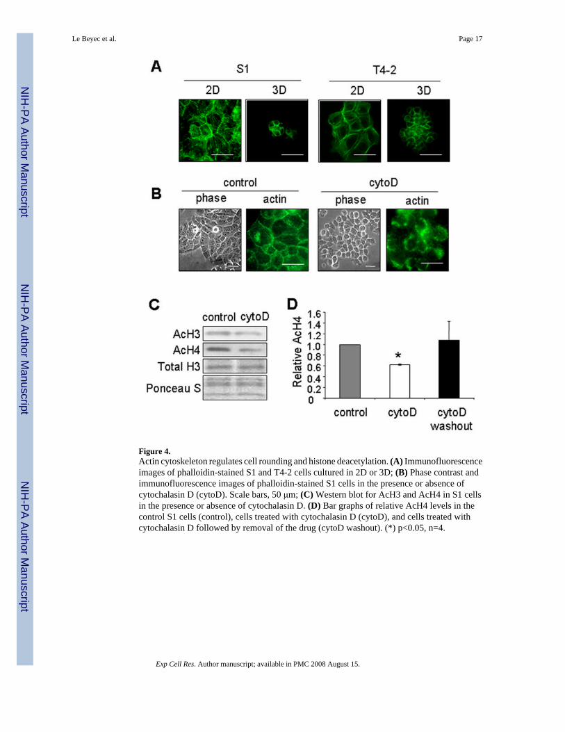
Figure 4.Actin cytoskeleton regulates cell rounding and histone deacetylation. (A) Immunofluorescenceimages of phalloidin-stained S1 and T4-2 cells cultured in 2D or 3D; (B) Phase contrast andimmunofluorescence images of phalloidin-stained S1 cells in the presence or absence ofcytochalasin D (cytoD). Scale bars, 50 μm; (C) Western blot for AcH3 and AcH4 in S1 cellsin the presence or absence of cytochalasin D. (D) Bar graphs of relative AcH4 levels in thecontrol S1 cells (control), cells treated with cytochalasin D (cytoD), and cells treated withcytochalasin D followed by removal of the drug (cytoD washout). (*) p<0.05, n=4.
Le Beyec et al. Page 17
Exp Cell Res. Author manuscript; available in PMC 2008 August 15.
NIH
-PA Author Manuscript
NIH
-PA Author Manuscript
NIH
-PA Author Manuscript
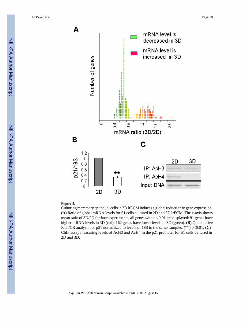
Figure 5.Culturing mammary epithelial cells in 3D lrECM induces a global reduction in gene expression.(A) Ratio of global mRNA levels for S1 cells cultured in 2D and 3D lrECM. The x-axis showsmean ratio of 3D/2D for four experiments; all genes with p< 0.01 are displayed: 91 genes havehigher mRNA levels in 3D (red); 162 genes have lower levels in 3D (green). (B) QuantitativeRT/PCR analysis for p21 normalized to levels of 18S in the same samples. (**) p<0.01; (C)ChIP assay measuring levels of AcH3 and AcH4 in the p21 promoter for S1 cells cultured in2D and 3D.
Le Beyec et al. Page 18
Exp Cell Res. Author manuscript; available in PMC 2008 August 15.
NIH
-PA Author Manuscript
NIH
-PA Author Manuscript
NIH
-PA Author Manuscript
Copyright © 2022 FDOKUMEN