Structure–Function Relationships of Antimicrobial Chemokines
Upload
khangminh22Category
view
3download
0
HAL Id: tel-01371241https://tel.archives-ouvertes.fr/tel-01371241
Submitted on 25 Sep 2016
HAL is a multi-disciplinary open accessarchive for the deposit and dissemination of sci-entific research documents, whether they are pub-lished or not. The documents may come fromteaching and research institutions in France orabroad, or from public or private research centers.
L’archive ouverte pluridisciplinaire HAL, estdestinée au dépôt et à la diffusion de documentsscientifiques de niveau recherche, publiés ou non,émanant des établissements d’enseignement et derecherche français ou étrangers, des laboratoirespublics ou privés.
The role of acetylation in the regulation of antimicrobialpeptide gene expression in the human intestine
Natalie Fischer
To cite this version:Natalie Fischer. The role of acetylation in the regulation of antimicrobial peptide gene expressionin the human intestine. Microbiology and Parasitology. Université Pierre et Marie Curie - Paris VI,2014. English. �NNT : 2014PA066276�. �tel-01371241�

Unité de Pathogénie Microbienne Moléculaire
THÈSE Présentée à
L’Université Pierre et Marie Curie
Ecole doctorale : ED515 « Complexité du Vivant »
“The role of acetylation in the regulation of antimicrobial peptide gene expression in the human intestine”
Par Natalie Fischer
Thèse de doctorat de microbiologie
Dirigée par Philippe Sansonetti
Présentée et soutenue publiquement le 23 Septembre 2014
Devant un jury composé de :
ARLET, Guillaume Professeur à l’UMPC (Président du jury)
CERF-BENSUSSAN, Nadine DRCE INSERM (Rapporteur)
AIT-SI-ALI, Slimane DR2 CNRS (Rapporteur)
SANSONETTI, Philippe Professeur au Collège de France (Directeur de Thèse)
SPERANDIO, Brice CR1 INSERM (Co-directeur de Thèse)

Acknowledgements
Acknowledgments First of all, I want to thank Philippe Sansonetti for flying me in from San Francisco to
interview and giving me the change to conduct a thesis in his laboratory. I enjoyed
very much the scientific discussions, which we shared over my project and his input
in my work. Also I want to thank him for the excellent resources concerning working
conditions as well as education, which are provided in his lab. I am very thankful to
have had the chance to work in the presence of his scientific genius and kindness
and I am grateful for his support throughout my thesis and afterwards.
I want to thank Brice Sperandio for being my close supervisor for the past years. He
guided me with the perfect balance of supervision, helped me settle in into the lab
and project in the beginning, and later trusted me to walk on my own feet. I learned a
lot from him, in the wet lab, but also concerning scientific writing and preparation of
presentations. I am thankful that he respected my opinion and critics, encouraged my
ideas and always had my best interest in mind. I am happy to say, that I think we
made a good team and he really did a great job with me.
I especially want to thank everybody who agreed to be on my thesis jury, for their
time and interest in my work. Special thanks go to Nadine Cerf-Bensussan and
Slimane Ait-Si-Ali for taking the time to read and correct my work and to Guillaume
Arlet, who kindly accepted to be the president of my jury. I am very much looking
forward to discuss my work with you!
My time at the Sansonetti lab would not have been the same without the friendly
atmosphere at PMM. I want to thank all my colleagues for their support, be it
scientific discussions, technical help in the lab or mental with the beer after work.
Especially I want to thank Emmanuel Sechet for his help in the last months, I could
have never done all those Western Blots without him.

Acknowledgements
I would not have been here if it wasn’t for the EIMID ITN program, who gladly
accepted me as a fellow. I enjoyed so much the international environment of this
program. In this regard, I want to thank especially Marco Bargagna, who was always
helpful and supportive, when it came to organizational things and deliverables. Also I
want to thank the great group of EIMID fellows, who really made these years of my
PhD special and with whom I enjoyed so many great times all around Europe.
I also want to thank my friends in Paris, especially the 12:30 lunch gang. Everybody
knows that science is a tough street and that especially during a PhD some breaking
points are reached. Thankfully I had a great support from my friends here at Pasteur
and I will never forget the times we shared in the ville de lumières. A special thanks
goes to Mathieu, who shared a very special year with me, and who has always was a
great support and source for laughter and good times.
My family deserves a great round of applause for always supporting me during all my
university education, morally and financially. I cannot thank you enough. I would not
be here today if it wasn’t for my Mum, Meikel, my uncle Dieter and my dearly missed
grandparents. This PhD title essentially goes to you!

List of Abbreviations
List of Abbreviations 1,25D3 – 1,25-dihydroxyvitamin D3 AD – Atopic dermatitis AMP – Antimicrobial peptide ANG4 – Angiogenin 4 AP1 – Activator protein 1 APC – Antigen-resenting cell APRIL – A proliferation-inducing ligand ARE – Apical recycling endosome ATF2 – Activating transcription factor 2 ATG16L1 – Autophagy related 16 like 1 ATP – Adenosine triphosphate BAFF – B-cell activating factor BMK – Big mitogen-activated protein kinase Brd4 – Bromodomain-containing protein 4 CAMP – Cathelicidin antimicrobial peptide CBP – cAMP-responsive element binding protein-binding protein CCL20 – CC-chemokine ligand 20 CCR6 – CC-chemokine receptor 6 CD – Crohn’s disease CDK9 – Cyclin-dependent kinase 9 CDRE – Caudal responsive element CFTR – Cystic fibrosis transmembrane conductance regulator CHD – Chromodomain-helicase-DNA-binding protein CNV – Copy number variation CNS1 – Conserved non-coding sequence 1 COPD – Chronic obstructive pulmonary disease CRAMP – Cathelin-related antimicrobial peptide CREB – Cyclic-AMP response element-binding protein CRS – Cryptdins-related sequence
CTD – Carboxy‐terminal domain
CTLA-4 – Cytotoxic T-lymphocyte antigen 4 CXCR4 – C-X-C chemokine receptor type 4 DC – Dendritic cell

List of Abbreviations
DEFB1 – Human beta defensin 2 DEFB2 – Human beta defensin 2 DEFB3 – Human beta defensin 3 DEFB4 – Human beta defensin 4 DNA – Deoxyribonucleic acid DNMT – DNA methyltransferases E. coli – Escherichia coli E. faecalis – Enterococcus faecalis EGFR – Epithelial growth factor receptor ER – Estrogen receptor ERE – Estrogen-responsive element ERK – Extracellular signal-regulated kinase FAE – Follicle-associated epithelia FliC – Flagella filament structural protein FOXO – Forkhead box transcription factor Foxp3 – Forkhead box P3 FRPL1 – Formyl peptide receptor-like 1 GALT – Gut-associated lymphoid tissue GAS – Group A Streptococcus GBS – Group B Streptococcus GCN5 – General control non-repressible synthesis 5 GNAT – GCN5-related N-acetyltransferases HAT – Histone acetyltransferase HBO1 – HAT bound to Orc1 hCAP18 – Human cationic antibacterial protein of 18 kDa HD 5 – Human alpha defensin 5 HD 6 – Human alpha defensin 6 HDAC – Histone deacetylase HDACi – HDAC inhibitor HDM – Histone demethylase HIF1 – Hypoxia inducible factor 1 HIP/PAP – Hepatointestinal pancreatic/pancreatitis‐ associated
protein HIV – Human immunodeficiency virus HMT – Histone methyltransferase

List of Abbreviations
HNP – Human neutrophil peptide HOTAIR – Homeobox antisense intergenic RNA HPA axis – Hypothalamus–pituitary–adrenal axis H. pylori – Helicobacter pylori
HSV – Herpes simplex virus HRE – HIF1 responsive element IAP – Intestinal alkaline phosphatase IBD – Inflammatory bowel disease IEC – Intestinal epithelial cell IEL – Intraepithelial lymphocyte IgA – Immunoglobulin A IgG – Immunoglobulin G IκBα – Inhibitor of NF-κB alpha IKK – IκB kinase IL8 – Interleukin 8 IL10 – Interleukin 10 IL1β – Interleukin 1 beta ILF – Isolated lymphoid follicles ILS – Insulin/insulin-like growth factor signaling Imd pathway – Immune deficiency pathway INF – Inflammatory gene IRAK – Interleukin-1 receptor-associated kinase IRF – Interferon response factor ISWI – Imitation SWI JDP – Jun dimerization protein JNK – c-Jun N-terminal kinases KCNN4 – Calcium-activated potassium channel protein 4 KDM2A – Lysine-specific demethylase 2A Lgr5 – Leucine-rich repeat-containing G-protein coupled receptor 5 lincRNA – Large multi-exonic intervening non-coding RNA LINoCR – LPS-inducible non-coding RNA L. monocytogenes – Listeria monocytogenes
LPS – Lipopolysaccharide LSD1 – Lysine-specific demethylase 1 LTi cells – Lymphoid tissue inducer cells

List of Abbreviations
mAchR – Muscarinic acetylcholine receptor MAL – MYD88-adaptor-like protein MAMP – Microbe-associated molecular pattern MAPK – Mitogen-activated protein kinase M cells – Microfold cells MCP – Macrophage attractant protein MDP – Muramyl dipeptide MLN – Mesenteric lymph node MOF – Males absent on the first MORF – MOZ-related factor MOZ – Monocytic leukemia zinc finger protein MSK – Mitogen- and stress activated protein kinase MSV – Multi site variations M. tuberculosis – Mycobacterium tuberculosis MUC-2 – Mucin 2 MYD88 – Myeloid differentiation primary response 88 nAchR – Nicotinic acetylcholine receptor NCoR – Nuclear receptor corepressor NEMO – NF-κB essential modulator NFAT – Nuclear factor of activated T-cells NF-κB – Nuclear factor kappa B NK – Natural killer NLR – NOD-like receptor NOD – Nucleotide-binding oligomerization domain containing
protein NuRD – Nucleosome remodeling deacetylase p300 – E1A-associated protein of 300 kDa P. aeruginosa – Pseudomonas aeruginosa PCAF – p300/CBP-associated factor PD1 – Programmed cell death protein 1 pDC – Plasmocytoid DC PDF – Plant defensin PGE2 – Prostaglandin 2 PGLYRP – Peptidoglycan recognition protein PIC – Pre-initiation complex

List of Abbreviations
PLA2s – Secreted phospholipase A2 Pol II – RNA polymerase II PP – Peyer’s patch PPAR- γ – Peroxisome proliferator-activated receptor gamma PRR – Pattern recognition receptor PSA – Polysaccharide A P-TEFb – Positive transcription elongation factor REG3 – Regenerating islet-derived protein 3 RHD – Rel homology domain RIP2 – Receptor-interacting protein 2 ROS – Reactive oxygen species RNA – Ribonucleic acid RTD – Rhesus teta defensin SAGA – Spt-Ada-Gcn5 acetyltransferase SAHA – Suberoylanilide hydroxamic acid S. aureus – Staphylococcus aureus
SCFA – Short-chain fatty acid SCTE – Stratum corneum tryptic enzyme S. epidermidi s – Staphylococcus epidermidis SFB – Segmented filamentous bacteria S. flexneri – Shigella flexneri SIGIRR – Single immunoglobulin IL1R-related molecule Sirtuins – Silent information regulator 2-related proteins SLP1 – SUN-like Protein 1 SLPI – Secretory leukocyte peptidase inhibior SMRT – Silencing mediator of retinoic acid and thyroid hormone
receptors SNP – Single nucleotide polymorphism SOCS – Suppressor of cytokine signaling SP1 – Specificity protein 1 S. pneumoniae – Streptococcus pneumoniae STAT – Signal transducer and activator of transcription S. typhimurium – Salmonella typhimurium SWI/SNF – SWItch/sucrose non fermentable TAD – Transactivation domain

List of Abbreviations
TF – Transcription factor TGFβ – Transforming growth factor beta Th cell – T helper cell Tip60 – Tat-interacting protein of 60 kDa TLR – Toll-like receptor TNFα – Tumor necrosis factor alpha TOLLIP – Toll interacting protein TRAF – TNF receptor-associated factors TRAM – TRIF-related adaptor molecule Treg cell – Regulatory T cell TRIF – TIR domain-containing adaptor protein inducing IFNβ TSA – Trichostatin A TSLP – Thymic stromal lymphopoietin TSS – Transcriptional start site UC – Ulcerative colitis UHRF1 – Ubiquitin-like, containing PHD and RING finger domains 1 VDR – Vitamin D receptor VDRE – Vitamin D responsive element Wnt – Wingless-type XPB – Xeroderma pigmentosum B XBP1 – X-box binding protein 1 XIST – X-inactive specific transcript

Table of Contents
TABLE OF CONTENTS
INTRODUCTION 1
CHAPTER I – HOMEOSTASIS IN THE HUMAN GUT 2
I.1 THE HUMAN GUT 2 I.2 THE INTESTINAL EPITHELIUM 2 I.3 THE HUMAN MICROBIOTA 5 I.4 HOW OUR GUT MICROBIOTA SHAPE OUR HEALTH 8 I.4.1 Nutrition 8 I.4.2 Development and morphogenesis of host organs and structures 12 I.4.3 Development and activity of the gut immune system 12 1.4.3.1 Development of the GALT 14
1.4.3.2 Development of lymphoid cells 14
1.4.3.3 How distinct microbiota species shape differential immune responses 16
1.4.3.4 Development of an adaptive B cell IgA response 18
1.4.3.5 Induction of mucus production 18
I.4.4 Development and activity of systemic immunity 18 I.4.5 Protection against pathogens 19 I.4.6 Brain and behavior 20 I.5 HOW WE MAINTAIN INTESTINAL HOMEOSTASIS WITH OUR MICROBIOTA 22 I.5.1 The mucus layer – an important first physical barrier 22 I.5.2 The intestinal epithelium – physical barrier, sensor and communicator 24 I.5.2.1 The intestinal epithelial layer as physical barrier 24
I.5.2.2 The intestinal epithelium as sensor of microbial signals 24
I.5.2.3 The intestinal epithelium as communicator with immune cells 30
I.5.3 Myeloid cells in intestinal tissues 30 I.5.3.1 Intestinal dendritic cells 30
I.5.3.2 Intestinal macrophages 32
I.5.4 Regulatory T cells 32 I.5.5 Other intestinal T lymphocytes 35

Table of Contents
I.5.6 Bacterial strategies to maintain tolerance 35
CHAPTER II – ANTIMICROBIAL PEPTIDES 38
II.1 ANTIMICROBIAL PEPTIDES THROUGHOUT THE KINGDOMS OF LIFE 38 II.1.1 AMPs in plants (eg. Arabidopsis thaliana) 40 II.1.2 AMPs in bacteria 42 II.1.3 AMPs in insects (eg. Drosophila melangoster) 44 II.1.3 AMPs in animals (eg. Mus musculus) 44 II.2 AMPS THROUGHOUT THE HUMAN BODY 46 II.2.1 Cathelicidins 50 II.2.2 Defensins 50 II.2.2.1 Alpha-defensins 52
II.2.2.2 Beta-defensins 52
II.2.2.4 Teta-defensins 53
II.3 EVOLUTION OF AMPS 53 II.3.1 Evolution of human defensins 54 II.3.2 Genetics of β-defensins 56 II.4 FUNCTIONS OF AMPS 58 II.4.1 Microbial killing 58 II.4.1.1 Bacterial killing 58
II.4.1.2 Anti-viral activities 59
II.4.2 Immunomodulation 60 II.4.2.1 Chemotaxis 60
II.4.2.2 Modulation of TLR signaling 60
II.4.3 Promotion of wound healing 62 II.4.4 Why are host cell membranes protected? 62 II.5 BACTERIAL RESISTANCE TO ANTIMICROBIAL PEPTIDES 64 II.5.1 Secretion of AMPs degrading enzymes 65 II.5.2 Modifications of the bacterial membrane 66 II.5.3 Trapping of AMPs 68 II.5.4 Active efflux of AMPs 68 II.5.5 Downregulation of AMPs expression 68 II.5.6 Why are AMPs still successful? 69 II.6 RELATIONSHIP BETWEEN AMPS AND HUMAN PATHOLOGIES 69

Table of Contents
II.6.1 Inflammatory bowel diseases - Crohn’s disease and ulcerative colitis 70 II.6.1.1 The role of α-defensins in IBD 72
II.6.1.2 The role of β-defensins in IBD 74
II.6.1.3 The role of LL37 in IBD 75
II.6.2 The role of AMPs in enteric infections 75 II.6.3 The role of AMPs in diseases concerning the lung 75 II.6.3.1 Cystic fibrosis 76
II.6.3.2 Asthma and chronic obstructive pulmonary disease (COPD) 76
II.6.3.3 Tuberculosis 76
II.6.4 The role of AMPs in disease of the skin 78 II.6.4.1 Atopic dermatitis 78
II.6.4.2 Acne rosacea 80
II.6.4.3 Psoriasis 80
II.6.5 The role of AMPs in disease of the oral cavity 81 II.6.6 The role of AMPs in systemic infections 81 II.6.6.1 HIV infection 81
II.6.6.2 Sepsis 82
II.7 INDUCTION OF AMPS EXPRESSION BY DIVERSE STIMULI 82 II.7.1 Host-derived inducers of AMPs expression 84 II.7.1.1 Developmental induction of AMPs expression 84
II.7.1.2 Induction of AMPs expression by cytokines 85
II.7.1.2.1 Members of the IL1 family 85
II.7.1.2.2 Th17 cytokines – IL17 and IL22 86
II.7.1.2.3 TNFα 88
II.7.1.3 Induction of AMPs expression by estrogen 88
II.7.1.4 Induction of AMPs expression by vitamin D 90
II.7.1.5 Stress hormones as repressor of AMPs expression 91
II.7.1.5.1 Glucocorticoids 91
II.7.1.5.2 Cholinergic stimulation 92
II.7.1.5.3 Physiological stress 92
II.7.1.5.4 Physical stress 93
II.7.2 Induction of AMPs expression by external stimuli 93 II.7.2.1 Induction of AMPs by bacteria 93
II.7.2.2 Induction of AMPs expression by SCFA 94

Table of Contents
II.7.2.3 Induction of AMPs expression by the amino acid isoleucine 95
CHAPTER III – REGULATION OF INDUCIBLE GENE EXPRESSION 98
III.1 STRUCTURAL ORGANIZATION OF THE HUMAN GENOME 98 III.2 THE ROLE OF CHROMATIN IN GENE ACCESSIBILITY 100 III.2.1 Histone modification 100 III.2.2 The histone code hypothesis 104 III.2.2.1 Histone marks as recruiting code 104
III.2.2.2 Histone marks encoding different physiological outcomes 104
III.3 REGULATORY PROCESSES OF INDUCIBLE GENE EXPRESSION 105 III.3.1 Transcription by the general transcription machinery 106 III.3.2 Activation of poised Pol II 106 III.3.3 Differential requirements for chromatin remodeling 108 III.3.4 Rapid activation by poised transcription factors 110 III.3.5 Enhancer motifs 110 III.3.6 DNA methylation 112 III.3.7 Removal of repressive histone marks and complexes 113 III.3.8 Activating histone marks 113 III.3.9 Non-coding regulatory RNAs 113 III.3.10 Activator complexes 114 III.4 WHAT WE KNOW ABOUT REGULATION OF AMPS GENE EXPRESSION 114 III.4.1 Transcription factors involved in the expression of AMPs 116 III.4.1.1 NF-κB 116
III.4.1.1.1 The NF-κB family 116
II.4.1.1.2 Activation of NF-κB 116
III.4.1.1.3 The role of NF-κB in DEFB2 expression 117
III.4.1.2 AP1 118
III.4.1.3 Hypoxia inducible factor 1 (HIF1) 120
III.4.1.4 Forkhead box transcription factor (FOXO) 120
III.4.1.5 Caudal 121
III.4.2 Epigenetic mechanism involved in AMPs expression 122 III.5 HAT AND HDAC ENZYMES REGULATE ACETYLATION OF (HISTONE) PROTEINS 122 III.5.1 HAT enzymes 124

Table of Contents
III.5.2 HDAC enzymes 124 III.5.3 HATs and HDACs in multi-protein complexes 125 III.5.4 Non-histone targets of HATs and HDACs 125 III.6 EPIGENETIC DRUGS – INHIBITORS OF HISTONE-MODIFYING ENZYMES 126 III.5.1 Inhibitors of DNA methylation 126 III.5.2 Inhibitors of histone methylation and demethylation 126 III.5.3 Inhibitors of histone acetylation and deacetylation 128 III.5.4 HDACis as suppressors of inflammation 128
CHAPTER IV – AIM OF THE THESIS 130
CHAPTER V – RESULTS 133
V.1 MANUSCRIPT IN PREPARATION 134 V.2 ADDITIONAL RESULTS 176 V.2.1 Additional results for challenge with the E. coli strain K12 176 V.2.1.1 Knockdown of histone deacetylases differentially influences the expression
of DEFB2 and IL8 genes upon E. coli K12 challenge 176
V.2.1.2 Differential HATs are involved in the expression of DEFB2 and IL8 180
V.2.2 Additional results for challenge with the E. coli strain LF82 182 V.2.2.1 Challenge with E. coli stain LF82 shows differential kinetics of AMPs
and INFs expression as compared to E. coli strain K12 182
V.2.2.2 LF82 shows prolonged activation of the NF-κB pathway 184
V.3 ADDITIONAL MATERIAL AND METHODS 185 V.3.1 BACTERIAL STRAINS 185 V.3.2 SIRNAS USED FOR THE KNOCKDOWN OF HDAC ENZYMES 185
CHAPTER VI – DISCUSSION 188
VI.1 REGULATION OF INDUCIBLE INFLAMMATORY GENE EXPRESSION ON THE CHROMATIN LEVEL 188 VI.1.1 Presetting the chromatin environment of NF-κB target genes 188 VI.1.2 The role of promoter-proximal paused Pol II 194 VI.1.3 Repression of inducible genes by HDAC complexes 195 VI.2 REGULATION OF INDUCIBLE INFLAMMATORY GENE EXPRESSION 196 VIA NF-ΚB 196

Table of Contents
VI.2.1 The role of NF-κB in intestinal homeostasis 196 VI.2.2 Acetylation of NF-κB regulate its function and activity 198 VI.2.3 Repression of NF-κB target genes by HDACs 200 VI.2.4 Phosphorylation of p65 and cofactor binding 202 VI.2.5 The role of NF-κB dimers in differential gene expression 203 VI.3 THE POSSIBLE ROLE OF ENHANCERS IN DEFB2 EXPRESSION 205 VI.4 THE ROLE OF SPECIFIC HISTONE MARKS IN HDACI ENHANCED DEFB2 EXPRESSION 207 VI.5 REGULATION OF INDUCIBLE INFLAMMATORY GENE EXPRESSION VIA MAPK PATHWAYS 208 VI.5.1 The role of H3S10 phosphorylation in inducible gene expression 208 VI.5.2 The role of AP1 in AMP expression 212 VI.6 THE COMBINATION OF TRANSCRIPTION FACTORS IN DEFB2 EXPRESSION 212 VI.7 HDAC INHIBITORS AS THERAPEUTIC OPTIONS FOR INFLAMMATORY AND INFECTIOUS DISEASES 213 VI.7.1 The role of HDACs in the expression of inflammatory genes 214 VI.7.1 The role of HDACs in the expression of AMP genes 215 VI.7.1 The role of natural HDAC inhibitors in intestinal homeostasis 215 VI.8 BIOLOGICAL REASONING FOR THE EPIGENETIC REGULATION OF DEFB2 EXPRESSION 217 VI.9 THERAPEUTIC PERSPECTIVE 217
CHAPTER VII – CONCLUSION 219
CHAPTER VIII – MODEL 221
CHAPTER IX – EXPERIMENTAL PERSPECTIVES 227
CHAPTER X – BIBLIOGRAPHY 228
ANNEX 291

Introduction

Introduction
1
A
B
Figure 1: The human intestine. The human intestine is divided into the small intestine (A) and the large intestine (B). Several muscular layers, covered by the submucosa and mucosa, structure the wall of the intestine. The mucous membrane in the small intestine folds into the so-called plicae circulares. The surface of these folds is furthermore covered by villi to increase the total area for absorption. The mucosa of the large intestine contains crypts but does not possess villi. It is also lined with mucus-secreting goblet cells. (Adapted from Encyclopaedia Britannica, Inc. 2003)

Introduction
2
Chapter I – Homeostasis in the human gut
I.1 The human gut The 8 meter long intestine extends from the stomach to the anus and can be divided
into the small intestine (subdivided into duodenum, jejunum and ileum) and the large
intestine (subdivided into cecum, colon and rectum) 1 (Figure 1). The predominant
task of the intestine is the processing of food coming from the stomach. The main
part of digestion and absorption of nutrients takes place in the small intestine. Folding
of the intestinal mucosa produces finger-like protrusions, the so-called villi, and
invaginations, the so-called crypts, and allows an enlargement of the surface to a
size of 250 m2 for maximized absorption (Figure 1 A). In the much shorter, but wider
large intestine we find crypts but no villi (Figure 1 B). Here water and salts are
absorbed from the remnants of the small intestine and indigestible waste products
are prepared for disposal. It is also the place with the highest density of bacteria,
which help digest fiber and produce vitamins that are absorbed by the host. The wall
of the intestinal tube is composed of four concentric tissue layers. The outmost layer
of the intestinal tube is build of connective tissue, followed by a layer of several
smooth muscle sheets, which allow the rhythmic contractions of the peristaltic to
move the contents along the intestinal tract. Underneath the muscle layer we find the
submucosa, consisting of connective tissue interspersed with nerve fibers and blood
and lymphatic vessels, followed by the mucosa, which comprises the epithelium and
the lamina propria. The lamina propria is a layer of connective tissue containing
nerve fibers, blood and lymphatic vessels. An important feature of the lamina propria
is its abundance of immune cells, such as lymphocytes and myeloid cells.
I.2 The intestinal epithelium The epithelium of the intestine is in direct contact with the lumen and its contents. It
consists of a single-cell layer of different specialized intestinal epithelial cells (IECs),
which possess a vigorous rate of renewal (3-5 days) 2 (Figure 2 A). (i) Multipotent
Leucine-rich repeat-containing G-protein coupled receptor 5 (Lgr5)-positive stem

Introduction
3
A B C
Figure 2: The intestinal epithelium. The intestinal epithelium is comprised of a variety of IECs originating from stem cells in the intestinal epithelial stem cell (IESC) niche in the crypt (A). Differentiated IECs then migrate upwards to renew the epithelium, as indicated by the dashed arrows. Microfold cells (M cells) in the follicle associated epithelium and goblet cells promote sampling of luminal antigens and live bacteria by resident dendritic cells (DCs) and intestine-resident macrophages, which reside in the underlying lamina propria (B). Furthermore, goblet cells secrete mucus covering the epithelium in a protective barrier (C). Transcytosed IgA antibodies reinforce this barrier together with antimicrobial peptides (AMPs) produced by enterocytes and/or Paneth cells, interspersed between the stem cells in the crypt 2.

Introduction
4
cells are located in the crypts of Lieberkuhn and are capable to constitute all 5 IEC
lineages, by constant proliferation and migration towards the villi tips in the small
intestine or the surface of the epithelial cuff in the colon 3,4. There, they replace
terminally differentiated cells, which undergo apoptosis and exfoliation into the
lumen. Furthermore, IECs are polarized, possessing two discrete membrane regions,
the apical and basolateral side. The apical side faces the lumen, while the
basolateral side is in contact with the underlying lamina propria. (ii) Enterocytes are
the most abundant IECs in the small intestine and are responsible for the absorption
of nutrients and the secretion of hydrolytic enzymes and immune mediators (i.e.:
Antimicrobial peptides (AMPs)) into the lumen. (iii) Hormone producing
enteroendocrine cells make up about only 1% of the cells in the epithelium 5. They
secrete various hormones, such as serotonin, vasoactive intestinal peptide and
somatostatin, which regulate fluid and electrolyte secretion, motility, blood flow and
food intake 6. (iv) Paneth cells can only be found in the crypts of the small intestine,
where they are interspersed with stem cells 7. They are well recognized for their
secretion of antimicrobial peptides, such as human alpha defensin (HD)5 and HD6,
lysozyme, secretory phospholipase A2 (PLA2s) and c-type lectins. (v) Microfold (M)
cells are preferably located in the so-called follicle-associated epithelia (FAE) right
above the gut-associated lymphoid tissues (GALT), which is comprised of mesenteric
lymph nodes (MLNs), isolated lymphoid follicles (ILFs) and Peyer’s patches (PP) 8
(Figure 2 B). Their unique morphological features, such as reduced glycocalyx, an
irregular brush border and broader microfolds instead of microvilli makes them highly
specialized for phagocytosis and transcytosis of antigens from the gut lumen across
the epithelial barrier 9. On the basolateral side of the epithelium the sampled antigens
are taken up by APCs and can be presented to T- and B-lymphocytes in the GALT.
(vi) Goblet cells secrete heavily glycosylated gel-forming mucins that build up the
layers of mucus covering the epithelium 10. Mucin 2 (MUC2) is the main component
of the mucus layer and forms a large, net-like polymer. The inner layer is firmly
adherent to the epithelial cells and approximately 50µm thick. It possesses a dense
structure, which does not allow bacteria to penetrate 11. The second non-attached
layer, which we find in the colon, is approximately 100µm thick (Figure 2 C). Its
expanded volume allows few bacterial species to adhere and use the mucins as
nutrient source 12. The abundance of goblet cells, and with it the thickness of the

Introduction
5
mucus layer increases towards the large intestine as the number of bacteria
increases and the luminal content becomes more compact. Recent results suggest a
role for goblet cells in the sampling of luminal antigens for antigen presenting cells
(APCs) in the lamina propria 13.
I.3 The human microbiota We share our world and our daily life with trillions of microscopic organism; among
them are bacteria, viruses, archae, protozoa and fungi. They inhabit next to every
terrain and water system of our planet 14. But most importantly they colonize the
surfaces of our own body, outnumbering our own somatic and germ cells by a factor
of ten. They can be found on our skin and on all mucosal surfaces that interface with
the environment, such as the gastrointestinal tract, the respiratory tract, the vagina,
the nasal and oral mucosa and the eye 15. The totality of organisms, which live in the
ecosystem of their human host are called “microbiota”. This coexistence is usually
commensal or mutually beneficial, but can also include pathogens, which can have a
damaging influence on their host. The microbial diversity harbored by the human
body has recently been explored in extensive international multicenter studies such
as the NIH Human Microbiome Project 16. Here 16S rRNA gene sequencing and
metagenomic sequencing was performed on samples from 15-18 body sites on three
separate visits of 242 healthy adults in the United States. The aim of this
extraordinary project is to understand the microbial components of the human
genetic and metabolic landscape and their contribution to healthy physiology and
predisposition to disease. The ultimate goal is to define parameters for the
manipulation of the human microbiota, which allows optimization of its performance
in the context of an individual’s physiology. The following chapters will discuss the
development and composition of the human gut microbiome and the interplay
between microbiota and the host.
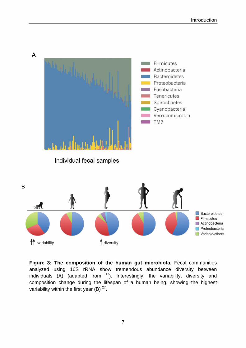
Among the mucosal tissues in the human body, the human gut harbors the
most diverse and extensive ecosystem of microbiota. In the Human Microbiome
Project the gut showed the greatest microbial diversity between individuals, but also
the lowest variability in between visits 17 (Figure 3A). Nonetheless, the composition of
the gut microbiome undergoes profound changes throughout the lifespan of an

Introduction
6
individual (Figure 3B). Although the infant has been considered sterile, it seems like
the first bacterial contact occurs already within the mother’s womb in the amniotic
fluid 18. A recent paper described the presence of a unique microbiome within the
placenta 19. After birth the colonization depends highly on the mode of delivery and
the associated contact with environmental microorganisms 20. Babies delivered by
cesarean section show lower bacterial counts and their microbiota resemble the
microbiota of the mothers skin. On the other hand the microbiota of babies born
naturally via the birth channel resemble the microbiota of the mothers vagina. Brest
feeding presents an additional mode of colonization by microorganism present in the
maternal milk. The first colonizers in the gastrointestinal tract of newborns are
facultative anaerobic bacteria such as Proteobacteria. They are thought to adjust the
highly oxidative environment for anaerobic species by decreasing the oxygen
concentration. Thereby they allow successive colonization by anaerobic
microorganisms such as Bacteroides, Actinobacteria and Firmicutes 21. During the
first year of life, the intestinal microbiota composition is simple and shows great
variability between individuals and over time. The diversity increases exponentially
throughout the first 3 years of life, where the microbiome finally reaches an “adult-
like” state 20,22.
In 2011 Arumugam et al. suggested the clustering of adult humans into three distinct
“enterotypes”, depending on high presence of Bacteroides, Prevotella, or
Ruminococcus, respectively 23. This concept caused a lot of discussions in the field
and the boundaries between the enterotypes may be fuzzier than earlier work
suggested 24. The composition of gut microbiota between individuals is highly related
to the host’s genetic disposition, diet, lifestyle, hygiene and the use of antibiotics 21,25.
500-1000 different bacterial species have been identified in the intestine, which form
a community of 1014 organisms. Firmicutes and Bacteriodetes are the two
predominant intestinal phyla 26. Members of the Firmicutes belong to Clostridia,
Mollicutes and Bacilli, including Enterococci, Lactobacilli and Lactococci.
Bacteroidetes are represented by Bacteroides species such as B. thetaiotaomicron,
B. fragilis, B. ovatus and B. caccae. Furthermore, we can find Proteobacteria,
Verrumicrobacteria, Actinobacteria, Fusobacteria, Spirochaetes and species closely
related to Cyanobacteria 28. The density and diversity of bacteria increases
Introduction
7
Figure 3: The composition of the human gut microbiota. Fecal communities analyzed using 16S rRNA show tremendous abundance diversity between individuals (A) (adapted from 17). Interestingly, the variability, diversity and composition change during the lifespan of a human being, showing the highest variability within the first year (B) 27.

Introduction
8
throughout the gastrointestinal tract from the stomach towards the colon. Most of
them are located in the lumen of the gut. Nevertheless specific bacteria expressing
lectins and glycosidases are able to adhere and inhabit the outer mucus layer and
even use it as nutrient source 12. Recent evidence suggests the existence of “crypt-
specific core microbiota”, as species of aerobic genera were found in the cecal and
colonic crypts in mice 29.
I.4 How our gut microbiota shape our health Over the recent years extensive efforts have been made towards unraveling the
various connections between the gut microbiota and host physiology. Our microbiota
provide us with a vast variability of gene products, which exceed the capabilities of
the host alone. We refer to the collectivity of 5 million bacterial genes expressed in
the intestine as the “microbiome”. The huge metabolic capacity that goes along with
those gene products has led to the view of the intestinal microbiota as an additional
organ 30. It is clear by now that intestinal microbiota are important supporters of our
health and disturbances of the equilibrium of our coexistence contribute to a wide
range of diseases, ranging from inflammation to obesity. This chapter will discuss the
influences of gut microbiota on different aspects of host development and physiology.
I.4.1 Nutrition The primary driving force behind the evolution of the mammalian gut microbiota
appears to be alterations in host diets 31. Colonization with microbiota provided a
way to enhance the host’s digestive efficiency 32. The additional metabolic capacity of
the microbiome is one of our biggest benefits from our invisible roommates. With help
of bacterial enzymes we can metabolize otherwise indigestible polysaccharides from
components of plant cell walls (including cellulose, xylan, and pectin) as well as
undigested starch 33,35,36. Bacteria convert released monosaccharides to pyruvate,
which is then further processed by fermentation in the highly anaerobic environment
of the distal intestinal lumen. The predominant end products of bacterial fermentation
in the gut are short chain fatty acids (SCFA), such as acetate, propionate, and
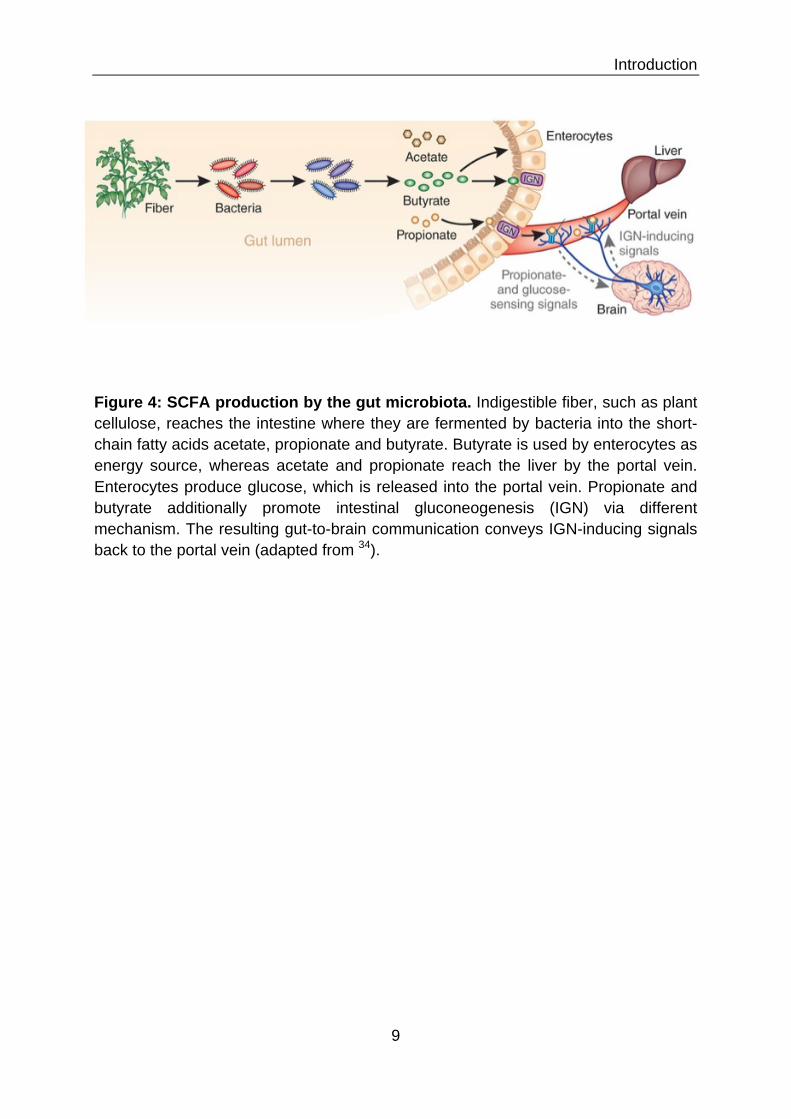
butyrate 37 (Figure 4). SCFA production represents 60–75% of the energy content of
ingested carbohydrates. They are absorbed by passive diffusion across the
Introduction
9
Figure 4: SCFA production by the gut microbiota. Indigestible fiber, such as plant cellulose, reaches the intestine where they are fermented by bacteria into the short-chain fatty acids acetate, propionate and butyrate. Butyrate is used by enterocytes as energy source, whereas acetate and propionate reach the liver by the portal vein. Enterocytes produce glucose, which is released into the portal vein. Propionate and butyrate additionally promote intestinal gluconeogenesis (IGN) via different mechanism. The resulting gut-to-brain communication conveys IGN-inducing signals back to the portal vein (adapted from 34).

Introduction
10
epithelium and diffused to different organs, or are used directly by the colonic
epithelial cells. In addition to their nutritional value, SCFA have important effects on
other aspects of gut physiology, for example uptake of water and salts, stimulation of
intestinal blood flow, proliferation and differentiation of epithelial cells 38. Interestingly,
epidemiological data suggest that dietary fiber reduces the incidence of colorectal
cancer, which could mean that microbiota produced SCFA help to maintain a healthy
gut physiology 39,40. Furthermore, our microbiota are involved in the production of
certain amino acids (up to 20% of circulating plasma lysine and threonine in adults)
and synthesis of vitamins, such as vitamin K, B12, biotin, folic acid, and pantothenate 41,42.
Research on microbiota and obesity suggest a fundamental role for microbiota in the
regulation of the host caloric energy balance and metabolic health 43 (Figure 5). A
twin study could show that obesity is associated with changes in the microbiota on
the phylum-level, reduced bacterial diversity and altered representation of bacterial
genes and metabolic pathways 44. Obese individuals show less species diversity and
dominance of Firmicutes species, compared to non-obese individuals, which harbor a
higher number of Bacteroidetes 45. Moreover, a comparison between lean and obese
mice suggests that the gut microbiota are able to influence the efficiency of energy
harvest from the diet, as well as energy use and storage 21. The microbiome of
obese mice for example is enriched for genes encoding carbohydrate-processing
enzymes and possesses a higher capacity for production of short-chain fatty acids.
Germ-free mice on the other hand show reduced total body fat, even when fed a high
fat and sugar “Western” diet 46. Overall germ-free animals require a 30% higher
caloric intake to achieve the same weight as conventional mice 47. Taking the
pathological scenario to another level, obesity often goes hand in hand with diabetes.
Both those metabolic diseases are characterized by insulin resistance and a low-
grade inflammation 48. Cani and colleagues recently identified bacterial
lipopolysaccharide (LPS) as a triggering factor for diabetes type 2 49. Mice fed with a
high-fat diet showed chronic increase of the proportion of LPS- containing microbiota
in the gut and LPS plasma concentrations. Furthermore, they could show that LPS
infusion increased markers of inflammation and insulin resistance in the liver.

Introduction
11
Figure 5: Microbiota in obese individuals. Alterations in the composition and metabolic capacity of gut microbiota in obese individuals promote adiposity and influence metabolic processes in peripheral organs, such as the control of satiety in the brain, the release of hormones from the gut, the synthesis and storage or metabolism of lipids in the adipose tissue, liver and muscle. Microbial molecules such as LPS also increase intestinal permeability, leading to systemic inflammation and insulin resistance (adapted from 32).

Introduction
12
I.4.2 Development and morphogenesis of host organs and structures The gut microbiota have a significant influence on development and morphogenesis
of host organs and physiological structures. The importance of microbiota in the
maturation and shaping of the gastrointestinal tract can best be illustrated on the
example of germ-free mice, which display considerable deficits 21. Besides altered
mucus thickness and properties, they have a reduced intestinal surface area as
compared to conventional mice 50, due to reduced villi thickness and impaired brush
border differentiation, resulting from slowed cell proliferation and turnover of epithelial
cells 51-54 (Figure 6). Furthermore, decreased intestinal peristaltic activity leads to a
prolonged transit time of food through the gastrointestinal tract 55,56. Functional
microbiota on the other hand, reduce epithelial permeability, thereby strengthening
the barrier function, and positively influence the modeling of the vascular system 52.
Additionally, to influencing gut development microbiota have also been found
to alter bone homeostasis 57. Germ-free mice have a higher bone mineral density,
reduced number of osteoclasts and lower levels of serotonin, a hormone that inhibits
bone formation 58,59. Microbial colonization promotes bone resorption via the
activation of osteoclasts. Furthermore, it leads to an increase in the number of pro-
inflammatory Th17 cells, which in turn induce osteoclastogenesis 60,61. The
suggested role for microbiota in the development of osteoporosis needs further
research efforts.
The connection between microbiota and cardiac development needs further
investigation, as it has only been described in one study so far. Germ-free mice were
found to have a smaller heart in proportion to their body weight, in comparison to
conventional mice 62.
I.4.3 Development and activity of the gut immune system Besides, influencing the morphological development of the gut, the microbiota also
shape the maturation of the local immune system. Interestingly, even single species
have been described in their characteristic shaping of particular immunological
settings.

Introduction
13
Germ-free mice Conventional mice
microbiota ! mucus thickness altered mucus properties
!!vessel density
!!AMPs & IgA production
GALT
Figure 6: Deficits in intestinal morphological and immunological development in germ-free mice. The influence of microbiota on the gut morphological and immunological development can best be illustrated in the comparison of germ-free mice with conventional raised mice. Germ-free mice show longer and thinner villi in the distal small intestine, which have a less complex vascular network. Furthermore, their intestinal crypts are less deep and contain fewer proliferating stem cells. They possess reduced mucus thickness and altered mucus properties, along with reduced secretion of AMPs and IgA. Overall germ-free mice show severe immunological underdevelopment, such as few isolated lymphoid follicles, immature Peyer's patches and immature mesenteric lymph nodes (adapted from 21).
Introduction
14
1.4.3.1 Development of the GALT Studies in germ-free mice showed that microbial colonization is essential for the
maturation of gut-associated lymphoid tissues, as these structures are
underdeveloped in germ-free mice 63 (Figure 6). A recent study could even show the
maturation of ILFs is driven by a specific bacterial factor, the peptidoglycan of the
bacterial cell wall. Bouskra et al. showed that the recognition of peptidoglycan by the
host receptor nucleotide-binding oligomerization domain containing 1 (NOD1) in
epithelial cells leads to expression of CC-chemokine ligand 20 (CCL20) and beta-
defensin 3 (DEFB3). These molecules are in turn able to bind to CC-chemokine
receptor 6 (CCR6) on lymphoid tissue inducer (LTi) cells, which then induce ILF
formation 64.
1.4.3.2 Development of lymphoid cells Furthermore, the microbiota direct the development of the lymphoid cell repertoire,
via the induction of chemokine and cytokines produced by intestinal epithelial cells.
They are important for example for the differentiation of natural killer (NK) like
RORγt+NKp46+ cells, which have been found to be scarce in germ-free mice 65.
These cells produce interleukin 22, which strengthens the intestinal barrier integrity,
by promotion of epithelial repair and production of AMPs 66. On the contrary,
microbiota are able to limit the generation of a subset of pro-inflammatory NK cells,
the so-called invariant NK T cells. Numbers of these cells are increased in germ-free
mice 67. Forkhead box P3 (Foxp3) positive regulatory CD4+ T (Treg) cells possess
anti-inflammatory properties and are important regulators of the intestinal
homeostasis 68. Conflicting observations have been reported for the influence of
microbiota colonization on the development of those cells. Multiple studies reported a
reduction of the Treg marker Foxp3 among CD4+ T cell subsets in germ-free mice 71-
73. Moreover, Treg cells from germ-free mice produce less interleukin-10 (IL10) and
are less sufficient in suppression of effector T cell proliferation. At the same time two
contradicting studies reported no change in the percentage 74 or even an increase of
CD4+ FOXP3+ cells in germ-free mice 75.

Introduction
15
Figure 7: Shaping of distinct immune responses by certain microbiota. Certain bacterial species have been identified, which are able to shape the intestinal immune environment of their host. Segmented filamentous bacteria (SFB) influence lamina propria dendritic cells (DCs) and macrophages to induce pro-inflammatory T helper 17 (Th17) cells and Th1 cells. Furthermore, bacteria-derived adenosine-triphosphate (ATP) was shown to promote the development of Th17 cells via the activation of a subset of DCs, which produce IL1#, IL6 and IL23 69.On the other hand Clostridium species and Bacteroides fragilis stimulate intestinal epithelial cells, T cells, and lamina propria DCs and macrophages to promote the development and/or the activation of anti-inflammatory regulatory T (Treg) cells (from 70).

Introduction
16
1.4.3.3 How distinct microbiota species shape differential immune responses Interestingly, individual species of the gut microbiota have been found to shape either
pro- or anti-inflammatory T cell related responses in the intestine (Figure 7). The
differentiation and expansion of anti-inflammatory Treg cells was shown to be
induced by Bacteroides fragilis polysaccharide A (PSA) via binding to Toll-like
receptor (TLR) 2 on undifferentiated CD4+ T cells 76. Furthermore, a study in germ-
free mice demonstrated expansion of Treg cells by colonization with 46 species
belonging to Clostridia, which had been isolated from conventional mouse faeces 77.
On the contrary, a pro-inflammatory influence can be seen in mice colonized with
segmented filamentous bacteria (SFB). These bacteria naturally colonizes the rodent
intestine at the time of weaning and induce a prominent Th17 and Th1 response in
their host 78,79. Thereby they stimulate the postnatal maturation of the gut immune
system and strengthen the epithelial barrier 80,81. The induced Th1 and Th17 cells
promote barrier function via recruitment and activation of macrophages and
neutrophils and the stimulation of AMPs production by epithelial cells. Conventional
mice lacking SFB show the absence of Th17 cells 78,79, elicit a lower IgA antibody
response 82 and overall weaker intestinal T cell response 80. The diminished immune
capabilities make these mice more susceptible to infection with the rodent pathogen
Citrobacter rodentium 79. The signals send by SFB by which they induce Th17 and
Th1 differentiation are not yet fully understood. Ivanov and colleagues could show
that SFB induces the acute phase protein serum amyloid A protein (SAA), which in
turn stimulates lamina propria DCs for Th17 generation 79. Recent studies point
towards a specific SFB antigen-directed mechanism 83,84. SFB are in direct contact
with the epithelium and even partially invade intestinal epithelial cells. Therefore the
close attachment to the epithelium facilitates sampling and presentation of SFB
antigens in Peyer’s patches. Moreover, cell-contact mediated signaling cannot be
excluded as a possibility. Whereas the close relation between SFB and an
immunocompetent host is generally beneficial and protective, development of
intestinal inflammation was shown in immunocompromised mice 85.

Introduction
17
Figure 8: Microbial contact influences the development of appropriate systemic immune responses. The hygiene hypothesis states that the immature immune system at birth is skewed towards a Th2 response. Contact with microbes promotes a healthy development of the systemic immune response towards a balance of Th1 and Th2 responses. If this contact is lacking due to increased hygiene in a modern western civilization the immature Th2 state persists, leading to an increased risk of asthma and other atopic diseases (adapted from 100).
Older sibling
Daycare center
Farming environment
Helminth infections
Microbial exposure
“Sterile”clean environment
Urban lifestyle
Only child
Th2
Th2 Th1
Antibiotic treatment

Introduction
18
1.4.3.4 Development of an adaptive B cell IgA response Moreover, intestinal bacteria are important for the development of adaptive immune
responses, such as the development and maturation of antibody producing B cells.
Sensing of bacterial flagellin via TLR5 receptor on dendritic cells (DCs) in the lamina
propria promotes the differentiation of B cells into IgA producing plasma cells 86. IgA
has a key role in barrier homeostasis as deficient mice show production of
microbiota-specific IgG antibodies, a sign for bacterial breach of the epithelium 87. On
the other hand, IgA is an important player in shaping the composition of the gut
microbiota 88. Mice deficient in the programmed cell death protein 1 (PD1) possess
an altered IgA repertoire, resulting in a shift in microbiota from Bacteroides and
Bifidobacterium to Enterobacteriaceae.
1.4.3.5 Induction of mucus production The constitution of the protective mucus layer is also supported by the presence of
intestinal microbiota. Germ-free rats have fewer goblet cells and therefore a thinner
mucus layer compared to conventional animals 89. A conventional mucus layer can
be developed in germ-free mice upon stimulation with bacterial products, such as
LPS or peptidoglycan. Moreover, bacteria-produced SCFAs directly activate the
expression MUC2, the main component of the mucus layer 90. MUC2 deficient mice
show bacterial overgrowth and develop spontaneous colitis 91.
I.4.4 Development and activity of systemic immunity In 1989 David Strachan formulated his much discussed “hygiene hypothesis”,
declaring that reduced exposure to microorganisms during childhood in developed
countries was the cause of increased incidence of hay fever, asthma and childhood
eczema later in life 92. This hypothesis proclaimed the idea that families with fewer
children provided less possibility for cross infections and therefore insufficient
microbial exposure. Additionally, the "hygiene revolution" of the 19th and 20th
centuries brought important public health measures such as the collection of
garbage, portable water, sanitation, vaccination and antibacterial therapeutics, which
reduced the contact with infectious but also non-infectious microbes. In accordance
with the hygiene hypothesis a lack of contact with microorganisms during
development leads to skewing of the immune response away from a Th1 and
towards a Th2 response and this imbalance leads to improper reactions related to

Introduction
19
ectopic diseases and allergies 93 (Figure 8). Later Graham Rock proclaimed the new
adapted “old friends hypothesis” 94. He discussed the coevolution of commensal
bacteria and their hosts, which were able to tolerate latent infections or carrier states.
He especially emphasizes the need for microbes in the development of our immune
system 95.
Studies comparing conventional and germ-free mice were able to highlight the
importance of microbial colonization for the maturation of the systemic immune
system. Notable deficits have been described in germ-free mice, such as reduced
germinal center size in the spleen, decreased numbers of memory CD4+ T cells,
reduced numbers of Th1 cells in both systemic and mucosal compartments and a
skewing towards a Th2-type cytokine profile 96-98. It has been postulated that the
maturating effect of microbiota beyond the gut are executed by soluble bacteria
factors that cross the epithelial barrier and enter the bloodstream. Mazmanian and
colleagues showed that monocolonization of germ-free animals with B. fragilis is
sufficient to mediate establishment of a proper Th1/Th2 balance and lymphoid
organogenesis and thereby corrects several immunologic defects found in the
absence of a bacterial microflora 97. These effects are established by PSA presented
by intestinal DCs to activate CD4+ T cells, and elicit appropriate cytokine production.
In another study Clarke and colleagues could show that peptidoglycan translocated
from the gut systemically primes the innate immune system by activation of
neutrophils in the bone marrow, enhancing their killing activity against Streptococcus
pneumoniae and Staphylococcus aureus 99. Serological peptidoglycan
concentrations correlated directly with neutrophil function. Moreover, they could show
that this effect was dependent on NOD1 signaling.
I.4.5 Protection against pathogens The gut provides niches for all kinds of bacterial species, among other
microorganisms. Nonetheless, there is a competition for space and nutrient supply.
Occupation of these niches with commensal bacteria protects our intestine from more
susceptible to infection with enteric pathogen, such as Shigella flexneri 101, Listeria
monocytogenes 102 and Salmonella typhimurium 103. Still pathogens have developed
tricks to succeed in competition with the local microbiota. In order to successfully
colonize the host Salmonella induces inflammation, which reshapes the microbiota

Introduction
20
composition, allowing the pathogen to establish an infection 104. A recent study
showed that this induction of inflammation also allows S. typhimurium to overcome
nutritional competition with the microbiota. Upon inflammation host cells release
ethanolamine, which can be used as an energy source by Salmonella but not other
competing species in the gut 105. Not to forget the administration of antibiotics does
also cause a disruption of the microbial balance in the gut and thereby leaves
dangerous room for pathogen colonization 106.
I.4.6 Brain and behavior The connection between the gut and the brain might not seem obvious; nevertheless
multiple studies in germ-free and conventional mice have shown the influences of
microbiota on brain function and behavior. There is accumulating evidence that
microbiota influence the regulation of anxiety, mood, cognition and pain in their hosts 107,108. The gut–brain axis presents a bidirectional communication system that
integrates neural (vagus and enteric nervous system), endocrine (cortisol), and
immunological (cytokines) signaling. In addition, the previously described SCFAs are
neuroactive and can also modulate brain function and behavior 109 (Figure 9).
Independent studies in different lineages of germ-free mice showed alterations in
concentrations of neurotransmitters and neurotrophic factors in the brain, resulting in
an altered stress response and less anxiety in elevated plus maze or light–dark box
tests 110-112. On the other hand, microbiota dysbiosis induced by infection with
Citrobacter rhodentium or antibiotic treatment in conventional mice can cause
anxiety-like behaviors 113. The first study to show alterations at the cognitive level
was undertaken by Gareau and colleagues. They showed deficits in simple non-
spatial and working memory tasks, such as recognition of a novel object or
spontaneous alterations in a T-maze, in germ-free mice 114. Germ-free animals
possess greater locomotor activity, linked with their resistance to obesity induction 46.
Other studies found a higher level of proteins involved in synaptogenesis in germ-
free mice, suggesting an involvement of microbiota in synaptic connectivity 111,115.
Looking at the communication in the opposite direction, from the brain to the
gut, stress and anxiety activate the hypothalamus–pituitary–adrenal (HPA) axis,
which regulates cortisol secretion. Cortisol can affect cytokine secretion by immune
cells, locally in the gut and systemically, as well as gut permeability and barrier

Introduction
21
Figure 9: The gut-brain axis communication. The bidirectional communication between gut and brain is managed via endocrine (cortisol), immune (cytokines) and neural (vagus and enteric nervous system) pathways. In direction gut-to-brain the vagus nerve, modulation of systemic tryptophan levels and production of neuroactive SCFAs are strongly implicated in relaying the influence of the gut microbiota to modulate brain and behavior. The other way around the brain can influence the composition of the gut microbiota, induce cytokine secretion and alter gut permeability and barrier function, for example in stress situations via the release of cortisol (107).

Introduction
22
function. Thereby stress and anxiety can cause changes in the intestinal microbiota,
which in turn lead to gastrointestinal disorders, including irritable bowel syndrome
(IBS) and inflammatory bowel disorder 116.
I.5 How we maintain intestinal homeostasis with our microbiota The maintenance of intestinal homeostasis is a complex interplay between the local
microbiota, the intestinal epithelium and the host immune system in the lamina
propria. The trick is to achieve a balance between tolerance and unresponsiveness
towards commensals and the readiness to launch an active immune response in the
case of bacterial breach of the epithelial barrier. A state of tolerance is maintained by
two mechanisms – ignorance towards commensal bacteria and constrain of effector
cells in the case of contact with antigens. At the same time the microbiota
commensal lifestyle supports this state of tolerance, as the majority of bacteria are
located in the lumen of the intestine at a respectable distance from the host immune
detection. Additionally, bacteria have developed special traits that help them to stay
undetected or even allow active suppression of an immune response. This part will
discuss the various mechanisms employed by the host, but also by the microbiota to
support a life together in peace and symbiosis.
I.5.1 The mucus layer – an important first physical barrier The most important and at the same time simplest mechanism to maintain tolerance
is to avoid contact. The mucus layer covering the intestinal epithelium presents a
potent physical barrier between the microbiota and the host tissue (Figure 10). Its
physical properties have already been discussed in chapter I.2. The secretion of
antimicrobial peptides by IECs and Paneth cells additionally increases the protective
capacity of this barrier. These molecules accumulate in the mucus and build up a
protective gradient towards the intestinal lumen 11,118. This allows for the control of
the numbers of mucosa-associated bacteria, but doesn’t influence the overall luminal
bacterial load. Furthermore, the mucus layer contains IgA antibodies, produced by B
cells in the lamina propria and transcytosed across the epithelium 119. IgA recognizes
and traps bacteria in the mucus layer, which allows their subsequent removal by the
peristaltic movement. It can also recruit factors of the complement system, which

Introduction
23
Figure 10: The intestinal mucus layer. In the small intestine mucus is produced by goblet cells and consists of an inner layer of ~15–30 µm and an outer layer of 100–400 µm. Additionally to the gel forming mucus layer, epithelial cells are covered with microvilli containing a high density of transmembrane cell surface mucins. In the large intestine mucus is predominantly produced by goblet cells and consists of a sterile inner layer of ~100 µm and a thick outer layer of ~700 µm (adapted from 117).
Introduction
24
promote bacterial lysis or mark bacteria for rapid phagocytosis and killing by lamina
propria macrophages in the case of tissue invasion 120.
I.5.2 The intestinal epithelium – physical barrier, sensor and communicator As the intestinal epithelium interfaces directly with the intestinal lumen and its
microbial contents its role in the maintenance of homeostasis is uncompared. IECs
employ a wide range of mechanism, which guarantee unresponsiveness and
tolerance towards the microbiota. At the same time they are at guard and ready to
alert the immune system in the case of an invasion. The following chapters will
discuss the role of the intestinal epithelium as a barrier towards intestinal contents,
sensor of microbial signals as well as pathogens and communicator towards the
underlying immune system in the lamina propria.
I.5.2.1 The intestinal epithelial layer as physical barrier The intestinal epithelial cell layer is composed of enterocytes, which are tightly linked
to their neighboring cells via gap junctions. This blocks trespassing of any microbe
across the epithelium. Moreover, the tight connection allows for gap-junctional
intercellular communication and the horizontal forwarding of immune-receptor-
mediated stimulations 121,122. This might facilitate a coordinated antimicrobial
response from the epithelium, but also offers the possibility of launching an immune
response based on single “sensor cells”.
I.5.2.2 The intestinal epithelium as sensor of microbial signals The intestinal epithelium is the main monitor and regulator of host-microbial
interactions in the gut. Despite the physical protective capacity against bacterial
translocation, microbial products can still diffuse through the mucus layer and can be
sensed by innate immune receptors of the epithelium 124. To avoid a constant
inflammatory response a certain unresponsiveness of the epithelium in the case of
contact with luminal microbial associated molecular patterns (MAMPs) has to be
guaranteed. The following part aims to discuss the various ways to achieve epithelial
unresponsiveness. The toll-like receptors (TLRs) are one of the best-studied families
of innate immune pattern recognition receptors (PRRs). They are transmembrane
receptors, localized either in the cell membrane or the membrane of intracellular
endosomes. In humans 10 different TLRs have been identified, whereas mice

Introduction
25
Figure 11: Toll-like receptor signaling. TLR5, TLR11, TLR4, and TLR2–TLR1 or TLR2–TLR6 are localized at the cell surface, whereas TLR3, TLR7–TLR8, TLR9 and TLR13 localize in endosomes. TLR4 localizes at both the plasma membrane and the endosomes. TLRs transmit their signals via various adapter molecules, such as MYD88, MYD88-adaptor-like protein (MAL), or TIR domain-containing adaptor protein inducing IFN# (TRIF) and TRIF-related adaptor molecule (TRAM). This is followed by IL1R-associated kinases (IRAKs) and TNF receptor-associated factors (TRAFs) activation. Finally, mitogen-activated protein kinases (MAPKs) p38 and Jun N-terminal kinase (JNK) activate transcription factors cyclic AMP-responsive element-binding protein (CREB) and activator protein 1 (AP1). Together with the activation of transcription factors nuclear factor-!B (NF-!B) and the interferon-regulatory factors (IRFs), this leads to the expression of pro-inflammatory cytokines (from 123).

Introduction
26
Additionally, possess TLRs 11, 12 and 13. Each TLR is specialized for the
recognition of certain microbial molecules 123,125 (Figure 11). TLR signaling is initiated
by ligand-induced dimerization of receptors. Thereby they can signal either as
homodimers, or as heterodimers, such as for example TLR2 with either TLR1 or
TLR6. Following activation, the Toll–IL1-resistence (TIR) domains of TLRs engage
TIR domain-containing adaptor proteins such as myeloid differentiation primary
response 88 (MYD88), MYD88-adaptor-like protein (MAL), or TIR domain-containing
adaptor protein inducing IFNβ (TRIF) and TRIF-related adaptor molecule (TRAM).
The signal is then transmitted via IL1R-associated kinases (IRAKs) and the adaptor
molecules TNF receptor-associated factors (TRAFs) and finally results in the
activation of the mitogen-activated protein kinases (MAPKs) and transcription factors,
which induce the expression of AMPs and pro-inflammatory cytokines and regulates
various important cellular functions. Signaling via these receptors is crucial for the
maintenance of intestinal barrier integrity, as it is involved in epithelial cell
proliferation, IgA and AMP production and maintenance of gap junctions 126-128.
Countless studies in cell lines, mice and in human tissue have aimed to
determine expression and regional and spatial localization of TLRs in the intestinal
epithelium, producing somewhat different and sometimes contradicting results 129.
The level of expression of TLRs in the intestinal epithelium is generally low and their
localization and compartmentalization differs from immune cells. Expression level
and localization differs not only between the small and the large intestine, but also
between epithelial cell types (Figure 12). Special compartmentalization is one of the
main features to prevent an unjustifiable immune response. An example for this is the
strictly basolateral localization of TLR5,which allows the detection of bacterial
flagellin only in the case of a barrier breach 130. This feature promotes both,
unresponsiveness towards commensals on the apical side and at the same time
detection of invading pathogens on the basolateral side. On the other hand TLR9,
which is the receptor for unmethylated CpG sequences in bacterial DNA, is
expressed on the apical and the basolateral side 131. But depending on the side of
the epithelium, activation elicits very distinct responses. Lee et al. showed that ligand
binding on the apical side promotes tolerance, especially by inhibition of a response
coordinated bythe transcription factor nucear factor kappa B (NF-kB). In contrast
ligand binding on the basolateral side stimulates a strong proinflammatory cytokine

Introduction
27
Figure 12: Toll-like receptor expression in the intestine. In the human small intestine the expression of TLR3, TLR4 and TLR5 has been shown on the basolateral surfaces of villus enterocytes. Enteroendocrine cells were found to express TLR1, TLR2 and TLR4, whereas their location on the apical or basolateral side is not clear. TLR9 and TLR4 are expressed in the cytoplasm of Paneth cells. In the human colon TLR3 and TLR5 are abundantly expressed on the basolateral side, whereas TLR2 and TLR4 expression is low. TLR9 is expressed apical as well as basolateral, with variable signaling outcome (adapted from 129).
Introduction
28
response by activation of transcription factors NF-κB and Jun N-terminal kinases
(JNK) 1 or JNK2, and the secretion of IL-8 132. TLR4, which is the receptor for LPS
from Gram-negative bacteria, was shown to be expressed at low protein levels in
three different human intestinal epithelial cell lines. Moreover, they lacked accessory
molecules necessary for activation, such as CD14 and MD2, which rendered them
unable to recognize of LPS 133. In a cell line derived from the mouse small intestine,
TLR4 was found to be restricted intracellular to the Golgi apparatus 134,135. Therefore
activation of TLR4 requires the presence of cytosolic LPS, which could be provided
by intracellular pathogens, but not commensal bacteria. The same is probably true
for other classes of cytosolic innate immune receptors expressed by intestinal
epithelial cells, such as Nod-like receptors (NLRs) and helicases.
Additionally, the TLR signaling pathways possess build-in negative regulators,
which allow the shut down of an immune response and the introduction of tolerance.
One member of the IRAK family, IRAK-M, is induced upon TLR stimulation and in
return negatively regulates TLR signaling 137. Signaling through IRAK proteins can
also be inhibited by Toll-interacting protein (TOLLIP), which is a suppressor of TLR2
and TLR4 signaling 138. IECs from patients with inflammatory bowel disease failed to
up regulate Tollip expression, suggesting that this may contribute to chronic
inflammation 139. Other examples are single immunoglobulin IL1R-related molecule
(SIGIRR), which is a negative regulator of TLR4 and TLR9 signaling 140 and A20, a
zinc-finger protein, which has been shown to inhibit NF-κB 141. A20-deficient mice
develop severe intestinal inflammation, suggested that this gene is critical for limiting
inflammation in the gut 142.
Besides the regulation of TLR expression and signaling, the host has other
tricks to tolerate contact with MAMPs, such as for example LPS. Fernandez and
colleagues could show a mechanism of LPS neutralization by transcytosing dimeric
IgA 143. The antibodies bind internalized LPS within the apical recycling endosome
(ARE) in murine intestinal epithelial cells. Also the zebrafish possesses an enzymatic
mechanism for LPS neutralization, the intestinal alkaline phosphatase (IAP). This
enzyme is induced during establishment of the gut microbiota and localizes to the
intestinal lumen brush border, where it detoxifies the endotoxin component of LPS 144.

Introduction
29
Figure 13: Intestinal epithelial cells secrete tolergenic factors. The gut microbiota stimulate IECs to secrete tolerogenic factors, such as thymic stromal lymphopoietin (TSLP), transforming growth factor-beta (TGF#), prostaglandin E2 (PGE2) and interleukin-10 (IL10), which directly influences the expression of pro-inflammatory cytokines by dendritic cell (DC) and macrophages lamina propria, resulting in the predominance of T regulatory lymphocytes (Tregs). In addition IECs secrete B cell and IgA promoting factors, such as APRIL (a proliferation-inducing ligand), B-cell-activating factor (BAFF) and secretory leukocyte peptidase inhibitor (SLPI). Thereby, IECs communicate with immune cells in the intestinal microenvironment and regulate the functions of both antigen-presenting cells and lymphocytes (from 136).

Introduction
30
I.5.2.3 The intestinal epithelium as communicator with immune cells Intestinal epithelial cells act as the interface between the microbiota and the gut
associated immune system. They play an important role in the communication of
signals, which help to maintain homeostasis and tolerance 145 (Figure 13). For
example they secrete a variety of factors, such as thymic stromal lymphopoietin
(TSLP), transforming growth factor beta (TGF-β), interleukin-10 (IL10), and
prostaglandin E2 (PGE2), to suppress the production of proinflammatory cytokines
and maintain a tolerogenic state of dendritic cells and macrophages in the lamina
propria 146,147. Furthermore, they produce mediators for maturation and proliferation
of primed B cells in the gut associated lymphoid tissues, such as B cell activation
factor (BAFF), proliferation-inducing ligand (APRIL) and SUN-like Protein (SLP1) 148.
I.5.3 Myeloid cells in intestinal tissues Intestinal myeloid cells, such as DCs and macrophages, are important gatekeepers
and defenders against invasion and infection. But in the maintenance of immune
homeostasis they are instructed by the intestinal epithelium to maintain a state of
tolerance (Figure 14). They can be divided into several subsets, possessing different
abilities and performing variable tasks 149. The major subsets of intestinal dendritic
cells and macrophages involved in surveillance and tolerance are discussed below.
I.5.3.1 Intestinal dendritic cells CD11b+CD103+ intestinal DCs constantly survey their microenvironment and
coordinate the initiation and polarization of adaptive immune responses. Their
hallmark activities are antigen uptake, migration and the capacity to prime T and B
cells in gut-associated lymphoid tissue 150. CD103+ DCs play an important role as
stimulators of Treg expansion via the production of TGF-β, retinoic acid 151,152, TSLP 153 and the tryptophan-catabolizing enzyme indoleamine 2,3 dioxygenase (IDO) 154.
IDO catalyzes the metabolism of tryptophan to metabolites such as kynurenine
derivatives. This causes a depletion and tryptophan starvation for effector T cells and
inhibits their proliferation, survival and activation 155. Therefore IDO is able to act on
two opposing T cell subtypes to support mucosal tolerance 156. The main activity of
CD103+ cells is maintaining the tolerogenic state, but they can also act as potent T
cell activators under inflammatory conditions, for example in models of experimental

Introduction
31
macrophage
Figure 14: Tolerogenic dendritic cells and macrophages in the intestine. IECs condition DCs and macrophages towards a tolerogenic phenotype through the production of TSLP, TGF# and retinoic acid (RA). These DCs promote the differentiation of naive CD4+ T cells into Treg cells. Expansion of converted Treg cells is induced by macrophages that are conditioned to produce IL10 by TSLP-mediated stimulation (adapted from 2).

Introduction
32
colitis 157. They are able to stimulate CD4+ and CD8+ T-cell proliferation and induce
cytotoxic T lymphocytes, therefore promoting the defense of the mucosa 158,159.
Following stimulation with flagellin, CD103+ DCs were shown to induce Th17
differentiation 86. Besides, they produced IL23, leading to induction of IL22 production
in innate lymphoid cells, which in turn activates secretion of the mouse antimicrobial
c-type lectin REG3γ (regenerating islet-derived protein 3 γ) by intestinal epithelial
cells 160. Thus CD103+ dendritic cells play a double role in maintaining intestinal
immune tolerance and promoting protective immunity.
I.5.3.2 Intestinal macrophages Intestinal macrophages are the largest reservoir of body macrophages 162. Lamina
propria resident macrophages are educated by intestinal epithelial cells to show
certain unresponsiveness towards MAMPs 163. Macrophages isolated from normal
human intestine lack CD14, a surface receptor involved in the response to LPS, and
CD89, the receptor for IgA (FcalphaR). Therefore they show reduced LPS-induced
cytokine production and LPS- and IgA-enhanced phagocytosis 164. Their abilities to
sample antigens and prime T cells complements the role of intestinal DCs, yet they
are inferior to DCs in accomplishing those tasks. CX3CR1+ intestinal macrophages
projected trans-epithelial dendrites (TEDs) extensions into the gut lumen to sense
and potentially sample its bacterial content 150. The capacity of CX3CR1+ cells to
prime naive T cells might be less efficient compared to DCs because of their faster
processing of cargo and the lack of expression of the chemokine receptor CCR7 that
is required for migration to the MLNs 165. CX3CR1+ macrophages were found to
promote the differentiation of naïve CD4+ T cells into FoxP3+ Treg cells in vitro 166.
Moreover, they produce substantial amounts of IL10 in response to the microflora,
which promotes the persistence of Foxp3 expression in Treg cells 167,168. This
highlights their role in maintenance of functional Treg cells that returned from the LNs
to the tissue.
I.5.4 Regulatory T cells Lamina propria CD4+ and CD8+ T cells provide a protective army of intestinal
defense that limits spread of bacteria in case of an epithelial barrier breach. At the
same time the activity of these effector cells presents an inherent risk of
inappropriate inflammation, especially in the case of recognition of commensals or

Introduction
33
A B
Effector T cell
Figure 15: Inhibitory mechanism of regulatory T cells. (A) Treg cells may secrete suppressor cytokines, such as IL10, TGF# and IL35, which inhibit the function of effector T cells. Moreover, Treg cells compete with effector T cells for IL2, resulting in IL2 deprivation of the effector cells and Bim-mediated apoptosis. Additionally, Treg cells may function as cytotoxic cells and directly kill effector cells in a granzyme-dependent manner. Activated Treg cells may express molecules on their cell surface (e.g., galectin-1), which can interact with receptors on effector T cells resulting in cell cycle arrest. (B) Tregs also exert inhibitory functions on APCs, for example via the expression of CTLA-4 on their surface, which mediates downregulation or prevents the upregulation of CD80 and CD86, the major costimulatory molecules on antigen-presenting cells (adapted from 161).

Introduction
34
food antigens. CD4+Foxp3+ Treg cells are important suppressors of lymphocyte-
mediated intestinal inflammation 68. As their main task is the inhibition of priming and
differentiation of effector T cells, they also have to target the activity of antigen
presenting cells. Studies in vitro have identified multiple mechanisms of Treg -
mediated suppression, some of which will be discussed below 161 (Figure 15). Yet it
is unclear if all these findings apply to in vivo situations.
IL2 is an important cytokine responsible for the development and
differentiation of T cells into effector cells 169. It is itself produced by T cells during an
infection and functions in an autocrine manner 170. Therefore IL2 is an important
target for Tregs for the inhibition of effector T cells. Several studies have reported
inhibition of IL2 mRNA induction by Tregs in effector T cells, and thereby prevention
of the autocrine T cell activation by this cytokine 171,172. Another mechanism to
interfere with this process has been described as “IL2 consumption”. Treg cells
possess the capacity to compete with effector T cells for IL2, consume it and thereby
inhibit their proliferation. In a final step this resulted in apoptosis of the effector T cell,
mediated by the proapoptotic factor Bim 173. The secretion of anti-inflammatory
cytokines, such as IL10, TGFβ and IL35 174, by Treg cells can Additionally, inhibit the
function of responder cells. Besides, they can induce ligand-receptor-mediated cell
cycle arrest and apoptosis in effector T cells, for example via the beta-galactoside
binding protein galectin-1 175. Furthermore, Treg cells were shown to express
granzyme A and can directly kill effector cells in a perforin-dependent manner, similar
to CD8+ cytotoxic cells 176.
Some Treg cells employed mechanisms primarily affect the function of APCs.
Studies showed that the Treg cell surface molecule cytotoxic T-lymphocyte antigen 4
(CTLA-4) can bind to costimulatory molecules CD80 and CD86 on the surface of
DCs and induce their downregulation 177-179. This limits the capacity of DCs to
stimulate naïve T cells via CD28. Signaling via CTLA-4 and CD80/CD86 also induces
DC expression of IDO, whose immunoregulatory mechanism have been described
above. CTLA-4 seems to be of major importance in maintenance of homeostasis.
Mice with defective CTLA-4 expression develop systemic autoimmunity shortly after
birth 180. Moreover, administration of anti-CTLA-4 antibody in an IBD model was
found to counteract the ameliorating effects of Treg cell 181.

Introduction
35
I.5.5 Other intestinal T lymphocytes Intraepithelial lymphocytes (IEL) are a specialized subset of gut T cells located at the
basolateral side of epithelial tight junctions 182. The majority of IELs are CD8+ and are
further divided into two subgroups depending on the two chains that compose their T
cell receptor: either alpha beta (αβ) or gamma delta (γδ) heterodimers. The
microbiota are important regulators of IELs. Ismail et al. showed microbiota-
dependent induction of IEL chemokines, proinflammatory cytokines, and REG3γ 183.
Moreover, γδ IELs play an important role as responders to bacteria or intestinal
injury. On one hand they express proinflammatory cytokines and chemokines that
recruit neutrophils, eosinophils, and T cells. On the other hand they promote
epithelial healing via production of keratinocyte growth factor, which stimulates
epithelial cell proliferation and restoration of barrier function 184.
Furthermore, the lamina propria and GALT are home to different subsets of
CD4+ Th cells 185. They all possess distinct cytokine profiles and fulfill different
requirements in the adaptive immune system of the gut 186. Th1 cells for example,
produce interferon γ, TNFα, and IL12 and are important for the defense against
intracellular pathogens. Th2 cells produce IL10, IL13, IL5, and IL4 and play a role in
the defense against helminthes. Th17 cells produce IL17, IL21, and IL22 and provide
protection against extracellular bacteria and parasites. In general Th activation can
result in activation and proliferation of cytotoxic T cells, activation of myeloid cells and
B cell differentiation and antibody production. To maintain a healthy gut environment
the presence of Treg cells and T effector cells needs to be balanced. Disturbance of
this balance can have negative influences on the gut microbiota as well as on the
host.
I.5.6 Bacterial strategies to maintain tolerance Besides, the hosts desire to keep the immunological homeostasis in the gut, we must
not forget the other side of the story. Naturally it is also in the highest interest of the
symbiotic bacteria colonizing our gut to maintain their living environment and stay
undetected from their hosts immune system. Therefore some of them harbor
mechanism to temper with and control the immune response of their host. For
example various strategies to manipulate the TLR downstream signal transduction
through NF-κB have been identified. Bacteroides thetaiotaomicron is able to

Introduction
36
suppress NF-κB function by enhancing the nuclear export of its subunit p65 through
a PPAR-γ-dependent pathway 187. Nonpathogenic Salmonella strains inhibit
polyubiquitination and following degradation of the NF-κB inhibitor IκB-α, thereby
preventing subsequent activation and nuclear translocation of the transcription factor 188. Lactobacillus casei induces down regulation of several components of the
proteasome, thereby protecting IκB-α degradation and NF-κB activation 189.
Additionally, commensals possess physical features that mark them as harmless.
Generally they lack virulence factors, such as adhesins and invasins. Moreover,
some species possess altered MAMPs. Bacteroidetes species for example carry
pentacetylated lipid A on their LPS, which is only poorly recognized by TLR4 190. Also
Helicobacter pylori flagellin is only mildly reactive for TLR5 in human gastric epithelial
cells 191. A similar flagellin structures could be imagined for gut commensal species.
Summary
In chapter I, we have discussed about the physiology of the human gut, the role of
the gut microbiota in various aspects of development and health of the host, and how
we maintain a peaceful and beneficial coexistence with the trillions of microbes
inhabiting our body. It has become clear that the communication between the
microbiota and the host is vital to develop and maintain a healthy organism. But also,
we must not forget that this beneficial coexistence is fragile and that the maintenance
of balance is regulated in a highly complex manner. In this regard, in completion of
chapter I.4.3 “Development and activity of the gut immune system”, it has to be
mentioned that the gut microbiota induce the expression of a broad spectrum of
AMPs in the intestine. This part of the dialogue between microbes and host is crucial
for the maintenance of homeostasis, as it controls the direct contact of bacteria with
intestinal epithelial cells. As chapter II focuses exclusively on AMPs, their expression
and characteristics, as well as their role in the bodies immune response, but also
their association with human pathologies will be discussed in great detail.
Furthermore, the capacities of microbes to evade the destructive effects of AMPs will
be described. Finally, we will review different inducers and repressors of AMPs gene
expression, in order to approach the question of how these genes are regulated in an
inducible manner.

Introduction
37
Figure 16: Antimicrobial peptides noted in the Antimicrobial Peptide Database (APD). This graph shows the percentage of antimicrobial peptides coming from different sources and noted in the APD as of 2013 192.
Introduction
38
Chapter II – Antimicrobial peptides AMPs are evolutionary conserved intrinsic antibiotics and an important part of our
innate immune system. The antimicrobial capacities of tissue, body fluids and
secretions were first described in the end of the 19th century. Many researchers
described the antibacterial activity of molecules in leucocyte extracts, blood, saliva
and tears against Gram-positive and Gram-negative bacteria 193. Already then they
were recognized for their ability to slow bacterial growth or kill invading
microorganisms through interaction with the negatively charged cell surface.
Moreover, they were thought to aid both the innate and the adaptive immunity. In the
following decades hundreds of different molecules have been identified, isolated and
described in prokaryotes, plants, invertebrates and animal species. To date, the
Antimicrobial Peptide Database (APD, http://aps.unmc.edu/AP/main.php) of the
University of Nebraska Medical Center contains 2349 antimicrobial peptides (214
bacteriocins, 305 plant AMPs, 12 fungal AMPs, and 1771 animal host defense
peptides) 194 (Figure 16).
II.1 Antimicrobial peptides throughout the kingdoms of life In order to survive and flourish in a microbe-dominated environment, plants and
animals developed an array of antimicrobial molecules, potent to fight bacteria,
viruses, fungi and protozoa 195. Even though AMPs are evolutionary conserved, the
sequence diversity in between species is remarkable, possibly reflecting the
adaptation to unique microbial environments and food sources. Despite the huge
diversity all AMPs share certain traits and structural characteristic. Most of them are
relatively short (12-50 amino acids) (Figure 17 A), derived from larger precursors and
can undergo post-translational modifications including proteolytic processing,
glycosylation or cyclization. Besides their diversity of sequences they all possess an
amphipathic structure with sections of hydrophobic and cationic amino acids (Figure
17 C, D) and carry a positive charge (Figure 17 B). According to their amino acid
composition and conformational structures AMPs can be divided into several classes:
for example α-helical peptides (eg. LL37), β-sheet structures with disulfide bonds

Introduction
39
Nr o
f AM
Ps
Nr o
f AM
Ps
Nr o
f AM
Ps
A B
C D
Peptide length Net charge
% Hydrophobic residues Protegrin Indolicin
Human !-defesin 3
Magainin 2
Figure 17: Diversity of AMP characteristics. Most AMPs have a short length of amino acids (A) and carry a positive charge (B). The majority of them possess an amphipathic structure with a cluster of hydrophilic cationic (red) and hydrophobic (green) residues (C,D) (adapted from http://aps.unmc.edu/AP/facts.php, 195)

Introduction
40
(eg. β-defensins), extended (eg. indolicidin) or loop structures (eg. cyclic θ-defensin) 196 (Figure 18).
In both animals and plants, the innate immune response is triggered after
recognition of conserved MAMPs by PRRs on multiple cell types 197,198. The
conserved production of AMPs after contact with microbes throughout all kingdoms
of life reflects the ancient origin and the importance of this type of defense response 195. Expression patterns differ between tissues and cell types, but usually AMPs are
expressed as a cocktail of different molecules from different classes. In animals,
AMPs are produced by epithelial cells and secreted onto the surface for the
protection of the host’s borders with the external microbial world. Additionally,
immune cells can produce AMPs and store them in granules, from which they are
released upon microbial contact. But in some cases they can also be secreted into
circulating fluids (e.g. the bloodstream or the hemolymph), which deliver AMPs to
infection sites. In plants, AMPs are probably not circulating, but are either
constitutively expressed in specific sensitive organs or are induced by microbes at
the site of infection and also systemically 199. The presence and expression of AMPs
in different phyla will be discussed in the next chapters on hand of certain examples
of species.
II.1.1 AMPs in plants (eg. Arabidopsis thaliana) The plant immune system employs various classes of AMPs including defensins,
thionins, lipid-transfer proteins, hevein- and knottin-like peptides, and macrocyclic
peptides as well as secondary metabolites, proteins and reactive oxygen species
(ROS) with antimicrobial activity 197,200. Expression of these molecules is upregulated
in a receptor-mediated manner, after sensing of plant danger signals upon tissue
damage or pathogen associated patterns. In vitro studies have uncovered their
involvement in biotic and abiotic stress response. Thionins possess high inhibitory
activity against fungi and bacteria including human pathogens, such as Candida
albicans. Additionally, they are toxic to mammalian, insect, and plant cells. Knottin-
like and macrocyclic AMPs exhibit insecticidal, antimicrobial, anti-human
immunodeficiency virus (HIV) and hemolytic activities. Interestingly, plant AMPs have
also been found to be involved in processes other than immunity. They mediate

Introduction
41
Figure 18: Examples for possible AMP protein structures. AMPs can be divided into multiple structural classes. Human #-defensin-2 for example possesses a mixed structure of a short "-helix and the characteristic #-sheets linked by disulfide bonds (indicated in yellow) (A). Furthermore, we can find looped structures, for example in the insect AMP thanatin (B), pure #-sheeted peptides, such as polyphemusin from the horseshoe crab (C) and the rabbit kidney defensin-1 (D), pure "-helical structures such as LL37 and magainin-2 (E) and extended structures, such as bovine indolicidin (F) (from 201).

Introduction
42
complex signaling processes between male and female gametophytes 202 and control
the density of pores in the leaves for gas exchange 203.
Plant defensins (PDFs) are the largest and best-studied class of AMPs and
wide spread throughout the plant kingdom 204. More than 300 putative defensin-like
genes were discovered in the model plant Arabidopsis thaliana, which is widely used
to study their expression and modes of action 205,206. Most of them form clusters
suggesting common origin by successive gene duplication events. Each gene
consists of two exons and a single intron in the signal peptide domain. They are
small (5kDa), basic peptides with a characteristic three-dimensional folding pattern
that is stabilized by eight disulfide-linked cysteines. PDFs are expressed in seeds,
stems, roots, leaves and floral organs. Expression of PDFs can be constitutive, for
example in sensitive tissues such as seeds, developmentally regulated or induced by
different abiotic and biotic stress factors, including cold, drought, heavy metals or
microbial pathogens 205. They are capable to inhibit growth of a broad range of fungi
and yeasts in vitro 207,208 and some of them are active against bacteria 209. A study by
DeConinck et al. showed that constitutive overexpression of the PDF AtPDF1.1 in A.
thaliana showed reduction in symptoms caused by the pathogen Cercospora beticola 210.
II.1.2 AMPs in bacteria Even some bacteria and archea produce a variety of defense peptides, the so-called
bacteriocins 211. These molecules are toxic for bacteria of the same species or
different genera and are used in combat against competitors in an ecological niche.
Protection against self-destruction is conveyed by the expression of immunity
proteins. The first bacteriocins, the E. coli produced colicine, was discovered in 1925
by Gratia. Best described today are the lanthionin-containing antibiotics “lantibiotics”,
produced by lactic acid bacteria. The structure of bacteriocins and modes of
bacterial killing are highly similar to described AMPs in other phyla. Moreover, these
peptides can be applied to a wide commercial use. The first discovered lantibiotic
Nisin A for example is used as a food preservative in over 50 countries 212. But their
application could also be of use to medicine. Bacteriocins from Staphylococcus for
example were shown to be active against multi-drug resistant S. aureus 213.

Introduction
43
Figure 19: Expression of AMPs in Drosophila melangoster. The figure illustrates the expression of AMPs at various body sites of Drosophila. Some AMPs are under the transcriptional regulation of the homeobox gene product Caudal, which is responsible for the constitutive local expression of antimicrobial peptides cecropin and drosomycin in a tissue-specific manner, for example in the reproductive tract or salivary glands. Others are induced upon microbial contact as indicated via the Toll or Imd pathway.

Introduction
44
II.1.3 AMPs in insects (eg. Drosophila melangoster) The genome of the common fruit fly Drosophila melangoster encodes 20 AMP genes,
which can be characterized into 7 different classes 214. The expression of these
genes is dependent on MAMP recognition by PRRs of the Toll or immune deficiency
(Imd) pathway 198. Mutants in PRR pathways do not express AMPs and were shown
to be highly susceptible to systemic infections 215,216. The conservation of signaling
pathways for the activation of AMPs, such as the Toll pathway, shows the ancient
origin of this defense mechanism in metazoan evolution and strengthens the use of
Drosophila as a potent model to decipher general innate immune mechanisms in
animals. Upon induction, AMPs are expressed in epithelia such as those beneath the
cuticle, in the alimentary tract, and in tracheae 198,217 (Figure 19). A specialty of
Drosophila is the expression of AMPs in the fat body, which is the equivalent of the
liver and a major immune-responsive tissue. Here, AMPs are secreted into the
hemolymph circulatory system, where they readily reach their effective
concentrations. Some AMPs are very stable due to intramolecular disulfide bonds
and can still be detected in the hemolymph several weeks after challenge 218.
Drosophila AMPs are small (<10 kDa), with the exception of the 25 kDa Attacin,
cationic, membrane-active and possess activity against Gram-negative and Gram-
positive bacteria and fungi 219.
II.1.3 AMPs in animals (eg. Mus musculus) AMPs are widely conserved and homologues of certain families can be found in
different mammalian species. This allows for the use of animal models to study the
role of AMPs in health and disease. The mouse is a commonly used model to study
cathelicidin and defensin expression. Nevertheless, the transferability of results
obtained in mice to humans is not guaranteed. Mouse intestinal Paneth cells express
a defensin family called cryptdins 220-222. Sequencing of cryptdin cDNAs
demonstrated the presence of at least 17 different mRNAs, identifying cryptdins as
the largest known defensin family. Six characterized cryptdin genes all have very
high overall nucleotide similarity (85%), suggesting gene duplication events that
occurred relatively recently in evolution. Furthermore, a recent study identified a
family of cryptdin-related sequence peptides, which exist in the form of covalent
dimers in mouse intestinal tissue 224. Interestingly, the mouse lacks myeloid

Introduction
45
A B
C D
wt CRAMP -/-
Figure 20: CRAMP deficient mice are more susceptible to group A Streptococcus infection. CRAMP -/- mice show increased susceptibility upon subcutaneous inoculation with GAS (B) as compared to wild-type (wt) mice (A). Moreover, they show higher severity of lesions as measured in the area of necrotic ulcer in individual WT (circle), CRAMP +/- (triangle), and CRAMP -/- (square) mice shown against days after infection (C). Furthermore, the bacterial load in the tissue of CRAMP deficient mice is much higher as seen in GAS bacteria cultured from tissue biopsies of WT, CRAMP -/+ and CRAMP -/- mice (D) (from 223).
Introduction
46
defensins and therefore the peptide-based antibacterial defense by recruited
neutrophils is limited 225. Several mouse beta defensins have been identified in
epithelial tissues, such as mouse β-defensin 1 (mBD-1), which is a constitutively
expressed homolog to human β-defensin 1 (DEFB1) 226,227 228, mouse β-defensin 2
(mBD-2) 229 and mouse β-defensin 3 (mBD-3), the murine homologue of human β-
defensin 2 (DEFB2) 230. The mouse also possesses a cathelicidin gene encoding the
cathelin-related antimicrobial peptide (CRAMP), which is expressed at various body
sites, such as the intestine, skin and urinary tract 231.
The mouse is a competent model to show effectiveness of AMPs in the fight
against pathogens in vivo. Mice deficient in mBD-1 were not able to achieve
clearance of Haemophilus influenzae from lungs or Staphylococcus sp. from the
urinary tract 232,233. Similarly, inactivation of the mouse cathelicidin gene increased
their susceptibility to GAS subcutaneous infection 223 (Figure 20). In another study
CRAMP-deficient mice could not maintain sterility of the urinary tract 234.
Finally, genetically manipulated mice are a useful tool to study the regulation
and expression of AMPs. A prominent example is the investigation of inflammatory
bowel diseases, which is associated with mutations in several susceptibility genes
and following decreased expression of defensins in the intestine, as will be discussed
later in this chapter 235.
II.2 AMPs throughout the human body Each interface of the human body has its characteristic repertoire of different AMPs.
Skin keratinocytes and mucosal epithelial cells produce constant low levels of AMPs,
which can be drastically increased upon certain stimuli such as infection and injury
(chapter II.7). Furthermore, tissue resident macrophages, dendritic cells, mast cells
and recruited neutrophils are capable of backing up the epithelial response with their
own AMP repertoire. Key antimicrobial peptides are cathelicidin and the defensins,
which will be described in greater depth in the scope of the presented thesis (chapter
II.2.1 and II.2.2 respectively). The discussion of all AMP families expressed in the
human body is way beyond the scope of this thesis.
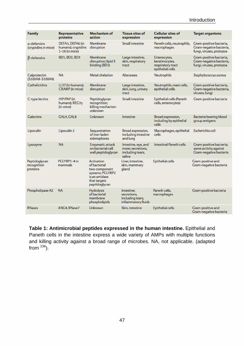
Introduction
47
Table 1: Antimicrobial peptides expressed in the human intestine. Epithelial and Paneth cells in the intestine express a wide variety of AMPs with multiple functions and killing activity against a broad range of microbes. NA, not applicable. (adapted from 236).

Introduction
48
Nonetheless, I want to briefly mention some important AMP families. For example the
S100 proteins, with psoriasin as the most prominent member and one of the major
AMPs expressed in the skin 237. The S100 proteins represent a family of small, acidic
proteins of 10–12 kDa containing two distinct EF-hand Calcium binding motifs 238.
Their expression was shown in the colon, keratinocytes and airway epithelium and
they fulfill various intra- and extracellular functions. Killing activity has been shown
against multiple pathogens, such as Escherichia coli, Klebsiella, Candida and
Staphyococcus species, but the mechanism has not been elucidated 239,240. Another
class of AMPs are the elastase inhibitors, such as elafin and secretory leukocyte
peptidase inhibitor (SLPI). They are present in granules of neutrophils, mast cells and
macrophages and are also produced by a variety of epithelial cells. After secretion
they are associated with extracellular matrix components and can kill Gram-positive
and Gram-negative bacteria and fungi via membrane permeabilization 241.
Furthermore, there exist the peptidoglycan recognition proteins (PGLYRPs) 242.
Mammals possess 4 genes of PGLYRs and they are widely distributed in leukocytes
and epithelial cells. They can kill a variety of pathogens by amidase-mediated
peptidoglycan digestion and osmotic lysis. Moreover, c-type lectins represent a large
family of receptors carrying carbohydrate recognition domains of the structural type
“C”. C-type lectins comprise the collectins, the REG proteins 243. Within the REG
family we find REG3α, REG3β and REG3γ in the Paneth cells of the mouse small
intestine and the hepatointestinal pancreatic/pancreatitis‐associated protein
(HIP/PAP) in Paneth cells of humans 244,245. C-type lectins, such as REG3γ in the
mouse, are important players in the maintenance of homeostasis by keeping the
microbiota at bay and in distance to the intestinal epithelium 246. Furthermore, the
expression of C-type lectins was shown to be induced in the mouse intestine during
pathogen infections or inflammatory conditions 247. They possess antibacterial activity
specific to Gram-positive bacteria, as they can bind to peptidoglycan in the bacterial
cell wall 248. Several AMPs expressed in the human body belong to the functional
group of iron metabolisms proteins: Lactoferrin, hepcidin and lipocalin-2 exert their
antimicrobial function via the sequestration of iron and membrane permeabilization 250-252. The major AMP families expressed in the intestine are summarized in Table 1
to provide an overview of their expression, mechanisms and target organisms (Table
1).

Introduction
49
Serine proteases
Cathelicidin Precursor
Active further processed peptides
Active LL37 peptide
Figure 21: Processing of cathelicidin. Cathelicidin is synthesized as an inactive precursor protein. It consists of an amino-terminal signal peptide (green), a central cathelin domain (purple), and the characteristic 37 amino acid long inactive carboxy-terminal AMP domain (pink). Several serine proteases have been described to cleave the AMP domain to generate active LL37, as well as additional active peptides by further cleavage of LL37 (adapted from 249).

Introduction
50
II.2.1 Cathelicidins The cathelicidins have been described in both invertebrate and vertebrate species,
including humans 253. In humans one single cathelicidin gene on chromosome 3p21.3
(CAMP) encodes the inactive 170 amino acid long precursor protein human cationic
antibacterial protein of 18 kDa (hCAP18) 254. The active mature peptide LL37, named
for the two leucines that comprise its first two N-terminal residues and its length of 37
amino acids, is released from the C-terminus of hCAP18 by protease processing 255.
Additionally, LL37 secreted onto the skin surface can be further processed into
smaller peptides, such as RK-30 and KS-30, which show even higher antimicrobial
activity, but are less immunogenic 256 (Figure 21).
LL37 is expressed and stored in the granules of myeloid cells, neutrophils,
mast cells and monocytes 254,257. Additionally, it can be secreted by epithelial cells of
the colon, urinary and respiratory tract and keratinocytes of the skin 258-261. The active
peptides generated by cathelicidin genes between species show little similarity to
each other and are referred to as a group solely based on the highly conserved N-
terminal region of the precursor protein known as the cathelin domain 253. Some of
these peptides can assume an α-helical conformation; others contain one or two
disulfide bonds. Moreover, they vary in the enrichment of amino acids, such as prolin,
arginine and tryptophane. They possess a broad range of microbial killing activity and
also exert additional functions related to host defense, which will be discussed in
more extent in following chapters. LL37 has been shown to neutralize LPS.
Furthermore, it is chemotactic for neutrophils, monocytes, mast cells and T cells. It is
also able to promote wound healing by stimulation of vascularization and re-
epithelialization 262. LL37 exerts important antibacterial action against multiple
pathogens, for example in the skin against GAS 263. In the intestine, LL37 was
described to decrease the bacterial load of Shigella in a rabbit experimental model 264.
II.2.2 Defensins Defensins are small (2-5 kDa), cysteine-rich cationic AMPs reported in invertebrates,
vertebrates and even plants 266. Mammalian defensins can be subdivided into α-, β-
and θ-defensins 265 (Figure 22). All defensins contain six highly conserved cysteine
residues, which form three pairs of intramolecular disulfide bonds. Thereby, they

Introduction
51
Figure 22: Defensin genes and peptides. The left side of the graph shows a representation of "-, #-, and &-defensin encoding genes. Below the genetic structure the resulting transcribed proteins are depicted, indicating the signal sequence (checked), pro-peptide (striped) and the mature processed peptide (blue, yellow). On the right side of the graph cysteine-bonds are indicated in the mature peptide and the three-dimensional structure is depicted 265.

Introduction
52
establish their beta-sheet structure. These bonds show specific differences between
subgroups of defensins and allow their classification. All defensins are translated as
inactive precursor proteins and activated by proteolytic processing. After secretion,
they fulfill various tasks in the immune defense and regulation of homeostasis, which
will be discussed in greater depth in the following chapters.
II.2.2.1 Alpha-defensins The α-defensins are between 29 and 35 amino acids long. Their cysteines are linked
in a 1–6, 2–4 and 3–5 pattern. Six α-defensins have been identified in humans. At
first, they were discovered in granules from neutrophils and therefore named human
neutrophil peptides (HNP) 1-4 267. Neutrophil α-defensins exhibit potent antiviral
capacities. HNP-1, HNP-2, and HNP-3 show in vitro activity against adenovirus,
papiloma virus, herpes virus, influenza virus, and cytomegalovirus, while HNP-4
inhibits HIV infection in vitro 268. Later, two additional α-defensins (HD5 and HD6)
were discovered in the Paneth cells of the small intestine, where they are stored in
intracellular vesicles and released upon stimulation, for example by MAMPs 269.
Additionally, they were found in epithelial cells from the female urogenital tract 270.
HD5 exposes effective killing activity against various bacteria 271 as well as parasites 272. HD6 showed only weak killing activity in in vitro assays, but recent work
discovered a different mechanism of action that prevents even translocation of
Salmonella in a transgenic mouse model 273. In this study HD6 was found to
polymerize and undergo ordered self-assembly into nanonets. These structures form
in vivo and are able to entrap bacteria in the lumen. Both HD5 and HD6 possess
antiviral capacities against various pathogenic viruses, such as herpes simplex virus,
human papillomavirus, adenovirus, and influenza A virus 274-277. Binding of the virus
and inhibition of viral adhesion and entry to cells exert antiviral activity.
II.2.2.2 Beta-defensins The characteristic disulfide bonds in β-defensins are located in a 1–5, 2–4 and 3–6
pattern. In humans six beta defensins with a length between 36 and 42 aminoacids
have been identified. DEFB 1-6 are all expressed in epithelial cells of different body
sites. DEFB1 is constitutively expressed while the others are inducible by a variety of
stimuli as will be discussed later. We can find expression of DEFB1 throughout the
gastrointestinal, respiratory, urinary and female genital tract 278-280 and to a lesser

Introduction
53
extend in the pancreas and liver 281. DEFB2 is expressed in the colon, skin and
urinary and respiratory tract 280-282. DEFB3 is expressed in epithelia of the skin and
colon and in high concentrations in the saliva and cervicovaginal fluids 283. DEFB4
expression was detected in testicles, stomach and the uterus 284. DEFB5 and DEFB6
are specially found in the epididymis 285. DEFB2 has shown antimicrobial activity in
vitro against Pseudomonas aeruginosa and E. coli 286. DEFB3 has potent
antibacterial activity against S. aureus 287. DEFB1, DEFB2, and DEFB3 possess also
antifungal activity against multiple Candida species 288. Some defensins showed only
weak killing activity in in vitro assays. Recent work by Schroeder et al. discovered the
necessity for biochemical activation in a reducing environment, for example in the
case of DEFB1 289.
II.2.2.4 Teta-defensins The circular θ-defensins have only been described in leukocytes of the rhesus
macaque and the olive baboon 290,291. They are formed by two hemi-α-defensins,
which each contributes three cysteines to the structure 292. θ-defensin genes are also
present in the human genome, but their expression is prevented by a premature stop
codon. Their protective capacities have been shown in various infection models 293.
Synthetic θ-defensins with sequences that correspond to those encoded within the
human pseudogenes inhibit the cellular entry of HIV, herpes simplex virus (HSV),
and influenza A virus. The same molecules protect mice from infection by Bacillus
anthracis spores. Rhesus θ-defensin 1 (RTD-1) was shown to protects mice from
coronavirus infection.
II.3 Evolution of AMPs The evolution of immunity involves direct interactions between the host and
microorganisms. Natural selection as a response to the rapid evolution of pathogens
is expected to strongly influence genes involved in these processes. Two well studied
examples for immunological evolution by gene duplication and selection are the
immunoglobulins and the major histocompatibility complex 294,295. The evolution of
human defensins is thought to have developed in a similar manner.

Introduction
54
II.3.1 Evolution of human defensins The complex evolution of defensin genes over time has lead to an exceptional cluster
of paralogous genes with varying copy numbers 296,297. In a genomics approach
Schutte et al. used a computational search tool to identify 28 new human β-defensin
genes, additional to the 4 previously described ones (DEFB1-4) 298 (Figure 23). They
are organized in five syntenic loci, with the main locus on human chromosome 8p22-
23, and at least 26 of them are actively transcribed. A comprehensive computational
search performed by Patil et al. identified a cluster encoding 10 distinct human α-
defensin genes and pseudogenes on chromosome 8p23 299. Furthermore, they
performed phylogenetic analyses, which revealed two distinct clusters, implying that
those might have independently evolved from two separate ancestral genes: One
giving rise to enteric, and another to myeloid α-defensins. On the other hand, Xiao et
al. suggested that all mammalian defensins are evolved from a common β-defensin-
like ancestor 300. α-defensins are thought to have originated from β-defensins by
gene duplication, which probably occurred after the divergence of mammals from
other vertebrate species, as they have not been identified in the latter 299. Then, θ-
defensins arose from α-defensins specific to the primate lineage. Nguyen et al.
hypothesized that the additional diversification of α- and θ–defensins reflect the need
for antiviral defense in certain mammalian and/or primate species, as it was shown
that these new defensin families can prevent virus entry into host cells 301,302.
Comparative analyses of both α- and β-defensin genes found that during the
divergence of primates episodes of purifying, positive and balancing selection have
driven the evolution of these gene families 303-305. Defensins are encoded in two exon
genes. Comparisons among first-exon sequences, which encode a signal peptide,
show little variation with an excess of synonymous substitutions. This observation is
consistent with neutral evolution or weak purifying selection. In contrast, second-exon
sequences, which encode the mature β-defensin peptide, show substantial
divergence with an excess of non-synonymous (amino-acid-altering) substitutions.
This is an indicator for the action of positive selection and implies a role for the
second exon sequence in functional diversity of peptides 296,303,306. Interestingly, the
gene encoding human DEFB1 was found to be relatively similar across primates 307.
Evidence for an evolutionary mechanism of duplication followed by diversification

Introduction
55
Gene Chr location
Gene type
Amino acid sequence of six-cysteine domain
Figure 23: Computational identification of human !-defensin genes. Chromosomal location is indicated, except where mapping was ambiguous (A). Genes are marked as known (K), if evidence exists that they are transcribed and show antimicrobial activity; related (R), if evidence exists that they are transcribed but have not been tested for antimicrobial activity; predicted (P), if no evidence exists that they are transcribed; and pseudogenes (S), if the DNA sequence is highly similar to a #-defensin gene, but the predicted amino acid sequence lacks an ORF across the six-cysteine motif. Cysteine residues are highlighted in yellow, positively charged residues in blue and other residues in red if they are represented in greater than 50% of all predicted #-defensin proteins 298.

Introduction
56
driven by positive selection has been discussed for α-defensin genes 308, bovine β-
defensin genes 309 and amphibian genes encoding AMPs 310.
Overall, the complex patterns of defensin evolution may reflect the need to
preserve their functional integrity, while at the same time favoring functional diversity
to provide responses to a wide range of pathogens, as well as activities beyond their
antimicrobial action.
II.3.2 Genetics of β-defensins The genetics of defensins are of special interest because of the relationship between
genetic variations with their expression level and susceptibility to disease. The β-
defensin cluster shows high copy number variation (CNV), ranging from 2–12 copies
per diploid genome, with 4 copies being the average in the healthy population 297.
Around 12% of the human genome is affected by CNVs 311 and the β-defensin cluster
represents one of the CNV hotspots. Just like single nucleotide polymorphisms
(SNPs), CNVs may affect gene expression and hence determine phenotypes and
pathological conditions 312. High copy numbers of β-defensin genes are associated
with the skin disease psoriasis 313. In the case of Crohn's disease conflicting results
have been found, indicating either a predisposition to disease by high or low copy
number 314,315. Whether copy number is correlated to gene expression in the case of
β-defensin is not completely clear. Hollox et al. found that the transcript level of
DEFB2 (encoded by the DEFB4 gene) is moderately correlated with its copy number
in different B-lymphoblastoid cell lines 313. And also Fellerman et al. found reduced
mRNA levels with lower copy numbers of the DEFB2-endocing gene in patients
suffering from Crohn's disease and ulcerative colitis 314.
In addition to CNV, sequence variations (multisite variations, MSVs) represent
a further level of genomic complexity. Analysis of the DEFB2-encoding gene
promoter region revealed remarkably high sequence variability (~1 MSV/-41�bp) 316.
Sequence variability has to be considered in the evaluation of disease phenotypes.
For example, an association study linked DEFB4 (encoded by the DEFB104 gene)
copy number and MSVs to prostate cancer, demonstrating an association of four
common DEFB4-encoding gene haplotypes with disease risk in two independent
patient cohorts 317.

Introduction
57
Figure 24: Mechanisms of AMPs membrane destruction. The ‘barrel-stave’ mechanism is characterized by the formation of transmembrane pores and depolarization followed by cell death. In the ‘carpet-like’ model AMPs align parallel to the outer membrane surface mainly by electrostatic interactions. After a threshold concentration of peptides has been reached, they permeate the membrane, which is followed by membrane disintegration and micellization. The ‘carpet-like’ model can also result in the formation of transient ‘toroidal’ pores, which might also lead directly to cell death (adapted from 331).

Introduction
58
II.4 Functions of AMPs The more we learn about AMPs the more it becomes clear that they are more than
just microbial killers. Additionally, to their own defense directed mechanisms against
pathogens they recruit the second wave of effector immune cells. But at the same
time as they are activating the adaptive immune response they are also controlling it
via the interaction with TLRs. And when the fight is over, they are promoters of
wound healing, cell proliferation and vascularization of the damaged tissues. The
next few chapters will discuss the multiple important actions of AMPs in more detail.
II.4.1 Microbial killing AMPs are destructive and inhibitory for many bacterial species and viruses. They
possess a range of mechanism of bacterial lysis and prevention of virus infection,
which will be discussed in this chapter. At the same time the host cells show
characteristics, which protect them from their own defense arsenal.
II.4.1.1 Bacterial killing AMPs succeed in killing many different types of Gram-positive and Gram-negative
bacteria, viruses, fungi and protozoa. Some AMPs expressed by Paneth cells, such
as lysozyme and PLA2s, carry out an enzymatic attack, hydrolyzing peptidoglycan or
phospholipids of the cell wall 318. Multiple techniques have been employed to
establish models of the molecular mechanism of cationic AMPs, such as defensins
and LL37, on their target cells 319. The first step is always the establishment of
attraction by electrostatic bonding between the AMP and structures of the bacterial
cell surface. Anionic phospholipids and phosphate groups of LPS from Gram-
negative and teichoic acids from Gram-positive bacteria attract cationic AMPs. The
mechanism by which AMPs transverse capsule polysaccharide or cell wall structures,
to reach the bacterial membranes has not really been described so far. Once they
reach the outer membrane of Gram-negative or the cytoplasmic membrane of Gram-
positive bacteria, they start to insert themselves into the lipid bilayer. A number of
models have been proposed to explain membrane permeabilization depending on
the AMP molecule and membrane structure (Figure 24). The AMP alamethicin
employs a ‘barrel-stave model’, where alpha helical peptides form a barrel structure
in the membrane with a central lumen 320. Another model employed by amphipathic
alpha helical AMPs in the ‘carpet model’. Peptides, such as ovispirin, accumulate in

Introduction
59
parallel on the bilayer surface. At high concentrations they disrupt the bilayer in a
detergent-like manner, eventually leading to the formation of micelles 321,322. The
‘toroidal-pore model’ is employed by magainins, protegrins and melittin. Here, the
polar faces of the AMP helixes associate with the polar head groups of the lipids.
This induces the lipid monolayers to bend continuously through the pore so that both
the inserted peptides and the lipid head groups line the water core 320,323. All these
models have the goal of inducing pores, leading to extensive membrane rupture,
followed by depolarization, leakage of cellular contents, disturbance of membrane
functions and finally lysis of the host cell.
Nonetheless, this is not the only killing mechanism employed by AMPs as also
intracellular targets have been identified. Mechanisms have been described for
peptide translocation across the membrane without causing membrane rupture 324,325. Once inside the target cell, AMPs can disrupt important cellular mechanism
such as synthesis of nucleic acids, cell wall components or proteins 326,327. Various
peptides have been shown effective in the inhibition of cell division in E. coli,
Salmonella and Shigella species leading to the formation of elongated filaments 328-
330. The AMP mersacidin was shown to inhibit peptidoglycan biosynthesis by
tempering with membrane-associated transglycosylation of peptidoglycan precursors 332. In fungi, histatins induce the loss of ATP from actively respiring cells, disrupt the
cell cycle and lead to the generation of reactive oxygen species 333. Pyrrhocoricin and
drosocin bind specifically to the heat-shock protein DnaK, thereby inhibits chaperone-
assisted protein folding 334.
II.4.1.2 Anti-viral activities AMPs possess potent antiviral activity, which they exert via different mechanism. β-
defensins can suppress viral infection by disruption of the viral envelopes 268.
Additionally, they can interact with viral glycoproteins and receptors and thereby
block viral binding and entry into the host cell. DEFB3 for example is an antagonist of
the HIV co-receptors CXCR4 335. Moreover, they have been shown to interfere with
cell-signaling pathways that are required for viral replication. In vitro studies have
shown that DEFB3 can inhibit HIV replication 335.

Introduction
60
II.4.2 Immunomodulation Besides, their destructive capacities towards microbes, AMPs are important
regulators of the innate and adaptive immune response. They can influence gene
expression of cytokines and chemokines and recruit effector cells, such as
neutrophils, monocytes, macrophages, immature dendritic cells and T cells, to the
infection site (Figure 25). Thereby, they are able to coordinate a united response
towards an infectious threat and mediate a fast resolution and reversion of harmful
inflammation and tissue damage.
II.4.2.1 Chemotaxis One way to achieve recruitment of effector cells is the induction of chemokine
expression by AMPs at the infection site. LL37 for example synergistically enhances
the IL1β induced production of cytokines IL6 and IL10 and macrophage chemo-
attractant proteins MCP1 and MCP3 in human peripheral blood monocytes 336.
Another way is the direct interaction of AMPs with effector cell receptors to mediate
their recruitment and activation. Interestingly, different AMPs seem to be specific for
distinct host-target cells. Human neutrophil α-defensins are chemotactic for naïve
CD4+CD45RA+ and CD8+ T cells 337, immature dendritic cells, monocytes, and
macrophages 338. Human β-defensins are also chemotactic for immature dendritic
cells and induce the migration of memory CD4+CD45RO+ T cells via the G protein-
coupled receptor CCR6 339. Another study demonstrated the capacity of DEFB2 and
DEFB3 to interact with CCR2, a chemokine receptor expressed on monocytes,
macrophages, and neutrophils 340. LL37 is chemotactic for neutrophils, monocytes
and T cells via interaction with the G protein-coupled formyl peptide receptor-like 1
(FPRL-1) 341. DEFB2 and LL37 are further recognized as chemoattractants for mast
cells 342,343.
II.4.2.2 Modulation of TLR signaling AMPs can regulate and balance the inflammatory responses towards microbes by
influencing TLR recognition of ligands and downstream signaling. Cathelicidins have
been proven especially capable of interference with TLR4 signaling. In a mouse
model of allergic contact dermatitis, cathelicidin was able to inhibited TLR4-mediated

Introduction
61
functions of AMPs that are not related to their directantimicrobial action.
Chemotactic activityUpon stimulation by microbial pathogens, local cellsrelease AMPs at the site of the infection or injury [60].In addition to inhibiting microbial growth, an additionalfunction of some of these AMPs is that they act to directlyrecruit leukocytes or induce the expression of chemokinesor cytokines including CXCL8 (IL-8), CCL2 (monocytechemoattractant protein, MCP-1) and IFN-a, therebyindirectly promoting recruitment of effector cells such asneutrophils, monocytes, macrophages, immature dendriticcells and T cells. For example, human a-defensins (HNP-1and HNP-2, but not HNP-3) have chemotactic activitymediating the recruitment of monocytes to inflammatorysites [61]. Subsequently, the defensins hBD3 and 4 havebeen reported to be chemotactic for monocytes and macro-phages [62], and hBD2 has chemoattractant activity formast cells [63]. Notably, both human a- and b-defensinsare chemotactic formemory T cells and immature dendriticcells. Human a-defensins selectively induce the migrationof human naı̈ve CD4+CD45RA+ and CD8+ cells, but notCD4+CD45RO+ memory T cells [64], whereas b-defensinsare chemotactic for immature dendritic cells and
CD4+CD45RO+ memory T cells. This chemotactic effectof human defensins on both T cells and dendritic cells ispertussis toxin-sensitive and inhibited by antibodies toCCR6 [65]. Therefore, the chemotactic activity of humandefensins has been suggested to promote cellular immuneresponses by recruiting dendritic and T cells to the site ofmicrobial invasion through interactions with the Gprotein-coupled receptor, CCR6 (Figure 3). However, struc-ture-function studies have shown conflicting data in thisprocess. On one hand, intramolecular disulfide bonding inhBD3 was reported to be required for binding and theactivation of receptors for chemotaxis, but dispensablefor its antimicrobial function [66]. On the other hand,recent data have also shown that chemoattractant activityof hBD3 is not dependent on disulfide bonds but is cysteineV-dependent [67] and CCR6 might not be a functionalreceptor for b-defensins in memory lymphocytes and den-dritic cells [68]. These contrasting resultsmight be becauseof mismatched intramolecular disulfide bonding of hBD3or different cell lines and experimental conditions used,but at any rate serve to reinforce the function of defensinsin chemotaxis and emphasize the complexity of thesesystems.
Like the b-defensins, cathelicidins have also beenshown to be chemotactic. LL37, is chemotactic for neutro-
Figure 2. Multiple functions of antimicrobial peptides in host defense. AMPs induce a variety of responses in host innate immune cells such as monocytes, macrophages,neutrophils and epithelial cells. They alter gene expression of host cells, induce production of chemokines and cytokines, promote leukocyte recruitment to the site ofinfection, influence cell differentiation and activation and block or activate TLR signaling. The outcome of the selective immunomodulation by AMPs results in innateimmune responses, leading to protection against infection, selective control of inflammation, promotion of wound healing and initiation of adaptive immune responses.Abbreviations: AMP, anti-microbial peptide; DC, dendritic cell; LPS, lipopolysaccharide; pDC, plasmacytoid dendritic cell; PMN, polymorphonucleocyte; TLR, Toll-likereceptor.
Review Trends in Immunology Vol.30 No.3
135
Epithelium
Recruitment of neutrophils
Inhibition of release of proinflammatory cytokines
functions of AMPs that are not related to their directantimicrobial action.
Chemotactic activityUpon stimulation by microbial pathogens, local cellsrelease AMPs at the site of the infection or injury [60].In addition to inhibiting microbial growth, an additionalfunction of some of these AMPs is that they act to directlyrecruit leukocytes or induce the expression of chemokinesor cytokines including CXCL8 (IL-8), CCL2 (monocytechemoattractant protein, MCP-1) and IFN-a, therebyindirectly promoting recruitment of effector cells such asneutrophils, monocytes, macrophages, immature dendriticcells and T cells. For example, human a-defensins (HNP-1and HNP-2, but not HNP-3) have chemotactic activitymediating the recruitment of monocytes to inflammatorysites [61]. Subsequently, the defensins hBD3 and 4 havebeen reported to be chemotactic for monocytes and macro-phages [62], and hBD2 has chemoattractant activity formast cells [63]. Notably, both human a- and b-defensinsare chemotactic formemory T cells and immature dendriticcells. Human a-defensins selectively induce the migrationof human naı̈ve CD4+CD45RA+ and CD8+ cells, but notCD4+CD45RO+ memory T cells [64], whereas b-defensinsare chemotactic for immature dendritic cells and
CD4+CD45RO+ memory T cells. This chemotactic effectof human defensins on both T cells and dendritic cells ispertussis toxin-sensitive and inhibited by antibodies toCCR6 [65]. Therefore, the chemotactic activity of humandefensins has been suggested to promote cellular immuneresponses by recruiting dendritic and T cells to the site ofmicrobial invasion through interactions with the Gprotein-coupled receptor, CCR6 (Figure 3). However, struc-ture-function studies have shown conflicting data in thisprocess. On one hand, intramolecular disulfide bonding inhBD3 was reported to be required for binding and theactivation of receptors for chemotaxis, but dispensablefor its antimicrobial function [66]. On the other hand,recent data have also shown that chemoattractant activityof hBD3 is not dependent on disulfide bonds but is cysteineV-dependent [67] and CCR6 might not be a functionalreceptor for b-defensins in memory lymphocytes and den-dritic cells [68]. These contrasting resultsmight be becauseof mismatched intramolecular disulfide bonding of hBD3or different cell lines and experimental conditions used,but at any rate serve to reinforce the function of defensinsin chemotaxis and emphasize the complexity of thesesystems.
Like the b-defensins, cathelicidins have also beenshown to be chemotactic. LL37, is chemotactic for neutro-
Figure 2. Multiple functions of antimicrobial peptides in host defense. AMPs induce a variety of responses in host innate immune cells such as monocytes, macrophages,neutrophils and epithelial cells. They alter gene expression of host cells, induce production of chemokines and cytokines, promote leukocyte recruitment to the site ofinfection, influence cell differentiation and activation and block or activate TLR signaling. The outcome of the selective immunomodulation by AMPs results in innateimmune responses, leading to protection against infection, selective control of inflammation, promotion of wound healing and initiation of adaptive immune responses.Abbreviations: AMP, anti-microbial peptide; DC, dendritic cell; LPS, lipopolysaccharide; pDC, plasmacytoid dendritic cell; PMN, polymorphonucleocyte; TLR, Toll-likereceptor.
Review Trends in Immunology Vol.30 No.3
135
Mig
ratio
n an
d
prol
ifera
tion
Figure 25: Immunomodulatory properties of AMPs. AMPs are able to recruit a wide range of immune cells, such as T cells, monocytes, macrophages, neutrophils and immature DCs. Furthermore, they influence the activation of TLRs and the expression of proinflammatory cytokines. Besides these mechanisms, which are involved in the resolution of an infection, they promote epithelial repair and neovasculatization (Adapted from 196).

Introduction
62
dendritic cell maturation and cytokine release 344. Moreover, cathelicidin blocked the
activation of TLR4 by its endogenous ligand hualuronan in murine bone-marrow
derived macrophages and thereby prevented the release of macrophage
inflammatory protein 2 (MIP2), which is chemotactic for polymorphonuclear
leukocytes 345. In humans, LL37 has been found to selectively suppress TNFα
release and expression of proinflammatory genes upregulated by NF-κB in
monocytes and macrophages stimulated by LPS 346. It has been proposed that
cathelicidin exerts the inhibition of receptors such as TLR4 by modifying the
membrane microdomain containing the receptor, thereby changing the receptor
function and ligand response. Furthermore, LL37, among other AMPs, can act as a
scavenger receptor for LPS, thereby preventing its binding to TLR4 347,348. This
mechanism of LPS-neutralization conveys protection against septic shock in mice 349.
II.4.3 Promotion of wound healing AMPs are not only at the front of immune defenses against invading pathogens, but
they are also involved in cleaning up the battlefield and taking care of the casualties.
Beta defensins and LL37 readily accumulate at wounds and their role in
vascularization and wound healing is widely established 350351. DEFB2 was shown to
stimulates chemotaxis, proliferation and tube formation of human endothelial cells
and speed-up wound closure 351. LL37 induces keratinocyte migration and re-
epithelialization of skin wounds via transactivation of the epithelial growth factor
receptor (EGFR) transactivation 352. Furthermore, LL37 promotes angiogenesis and
neovascularization by binding to FPRL-1 expressed on endothelial cells 353.
Additionally, LL37 shows anti-fibrotic effects by inhibiting collagen expression in
dermal fibroblasts, thereby promoting normal wound repair 354.
II.4.4 Why are host cell membranes protected? The membranes of bacteria and the eukaryotic host differ in important
characteristics, which convey protection from AMPs attack to the host cell 195,355
(Figure 26). First of all, the outer leaflet of mammalian cell membranes is exclusively
composed of electrically neutral, zwitterionic phospholipids, mainly
phosphatidylcholine and sphingomyelin. Negative charges are located to the inner
leaflet of the membrane and not exposed at the surface. Second the eukaryotic
membrane contains stabilizing cholesterol, which has been shown to reduce the

Introduction
63
Figure 26: Differences between bacterial and mammalian membranes protect host cells from AMPs attack. Cationic AMPs preferentially bind to bacterial
membranes with abundant acidic phospholipids carrying a negative charge. The
outer leaflets of mammalian cell membranes are exclusively composed of zwitterionic
phospholipids with neutral charge. The presence of cholesterol additionally protects
mammalian membranes against AMPs (adapted from 195).
Mammalian cell membrane

Introduction
64
activity of AMPs towards the lipid layer. Moreover, it was shown that the activity of
AMPs is blunted by physiological salt concentration (e.g. 150 mM NaCl), increased
presence of divalent cations and serum proteins 356. This might prevent destruction of
host cells in a physiological environment but still allow killing of bacteria, which
change their membrane structures and gene expression profile in adaptation to the
host environment 357.
The same properties, that protect healthy host cells, are aberrant in some
cancer cells and thereby make them targets for the destruction by AMPs. For
example, the membrane net negative charge is increased in malignant cells by the
accumulation of anionic molecules, such as phosphatidylserine, O-glycosylated
mucins, sialylated gangliosides and heparin sulfate 358. Especially during cell
transformation phosphatidylserine molecules will present themselves on the outer
membrane leaflet, counteracting the typical phospholipid asymmetry of the
membrane. Moreover, most cancer cells show increased membrane fluidity, making
them more susceptible for destabilization by AMPs 359. It has also been suggested
that the enlarged cell surface area of malignant cells, formed by elevated number
and distorted features of microvilli, confers a higher contact possibility with AMPs
molecules 360. Additionally, lysis of mitochondrial membranes have been described to
lead to the release of cytochrome c and eventually apoptosis 361. Many natural and
synthetic AMPs are being tested for cancer treatment 362. However, not all AMPs are
active against cancer cells and we are just beginning to understand the factors that
allow certain AMPs to recognize and lyse neoplastic cells. Understanding efficacy
and selectivity of those peptides, as well as the identification of specific targets that
are expressed and presented within a certain tumor type will be crucial in the process
of drug design in the future.
II.5 Bacterial resistance to antimicrobial peptides In order to survive, bacteria have evolved under selective pressure to stay on top of
their host’s defense mechanisms. The same is true for developing resistance to
AMPs. At the same time this presents an enormous challenge, as the targets of
AMPs are essential to bacterial integrity and survival, such as the bacterial

Introduction
65
membrane. Bacteria differ in their intrinsic susceptibility to AMPs and pathogenic
species have come up with their own remarkable protection strategies. Recent
evidence indicates that bacterial resistance to AMPs is under control of global
regulation systems, which coordinate expression of virulence factor. Those bacterial
two-component signal transduction systems usually consist of a membrane-bound
histidine kinase, which senses a specific environmental stimulus, and a response
regulator, which mediates the cellular response 363,364. These systems enable
bacteria to sense, respond, and adapt to a wide range of environmental cues
encountered in different tissue niches of their host. One example for such systems is
the PhoP/PhoQ two-component regulatory system of Salmonella enterica, which is
involved in AMP resistance 365. These mechanisms will be discussed in greater detail
in the chapter below.
Moreover, mutagenesis approaches were used to target genes of major AMP
resistance mechanisms to show that loss of the resistance phenotype translates to
decreased virulence in vivo. Direct comparisons of resistant wild-type bacteria with
AMP-sensitive mutants in in vivo infection models provided validation of the
contribution of resistance mechanism to pathogenesis and shed light on the
involvement in human disease conditions 366.
II.5.1 Secretion of AMPs degrading enzymes Destruction of AMPs can be achieved by secretion of degrading enzymes, as
demonstrated for species belonging to Staphylococci (Figure 27 D). S. aureus
metalloproteinase (aureolysin) and glutamylendopeptidase (V8 protease) are capable
of cleavage and thereby inactivation of LL37 in a time- and concentration-dependent
manner 367. The skin commensal and leading nosocomial pathogen Staphylococcus
epidermidis efficiently inactivates the anionic AMP dermcidin through its
metalloprotease sepA 368. LL37 was also shown to be degraded by bacterial
supernatants containing GAS cysteine proteinase, Proteus mirabilis metalloprotease,
or Enterococcus faecalis gelatinase 369. Even more elegant is the mechanism
employed by P. aeruginosa. The pathogen stimulates the accumulation of host
cysteine protease cathepsins B, L and S in the airway fluid, which mediate
degradation and inactivation of DEFB2 and DEFB3 370.
Introduction
66
II.5.2 Modifications of the bacterial membrane The negatively charged surface of bacteria presents a welcoming site for cationic
AMPs attack (Figure 26). So, one important protection mechanism employed by both
Gram-positive and Gram-negative bacteria is the modification of anionic surface
constituents with cationic molecules in order to repel cationic AMPs.
LPS covered Gram-negative bacteria prevent AMPs binding by the synthesis of LPS
with specific lipid A modifications (Figure 27 B). Salmonella spp. can produce hepta-
acetylated lipid A via the addition of palmitate by the bacterial factor PagP. Moreover,
it can add phosphate and phosphoethanolamine to the core polysaccharide and
modify lipid A phosphate groups with ethanolamine and 4-aminoarabinose 371. These
changes are implemented via the two-component signal transduction systems PhoP-
PhoQ and PmrA-PmrB. Interestingly, both these systems were found to be
upregulated upon the presence of subinhibitory concentrations of AMPs 372. A
PmrAB-like signal transduction system and Ara4N modification of lipid A has also
been identified in P. aeruginosa, a common airway pathogen in cystic fibrosis
patients 374. Additionally, P. aeruginosa synthesizes a hexa-acetylated lipid A with
palmitate and aminoarabinose modifications 375.
Gram-positive bacteria neutralize their surface charge by modifying teichoic
acids in their cell wall with D-alanyl groups or by including positively charged
phospholipids in the cellular membrane (Figure 27 A). Staphylococci species employ
the three-component antimicrobial peptide-sensing system to regulate synthesis of
amino acids, such as L-lysine for the decoration of membrane lipid
phosphatidylglycerol or D-alanine for the decoration of teichoic acid 376,377. D-
alanylation is achieved by gene products of the dlt operon, which was also identified
in other pathogens, such as Group B Streptococcus (GBS) 378, Bacillus subtilis 379
and L. monocytogenes 380, where it exerts the same role in AMPs resistance.
The human gastric pathogen H. pylori modifies host cholesterol by
glycosylation and incorporates it onto its surface. Cholesterol-grown H. pylori cells
showed an over 100-fold increased resistance to eight antibiotics and LL37 381.

Introduction
67
E
E C
A Gram-positive B
+ + +
+ + +
Gram-negative
Release of AMP-binding decoy molecules
E
C C
D AMP-degrading protease
Figure 27: AMP-resistance mechanism of bacteria. Bacteria have evolved several mechanisms to resist destruction by AMPs. Gram-positive bacteria for example modify their surface teichoic acid and membrane phospholipids with positive charges, to repel cationic AMPs (A). Gram-negative bacteria modify their LPS molecules with aminoarabinose or acylation of lipid (B). Furthermore, already intracellular AMPs can be extruded by efflux pumps (C) or degraded by proteases, which can also be secreted by some species (D). Some bacteria release decoy proteins to bind and block AMPs (E) (adapted from 373).

Introduction
68
II.5.3 Trapping of AMPs Pathogens have developed different mechanism to bind and thereby neutralize
AMPs (Figure 27 E). S. aureus for example secretes an AMP-binding factor called
staphylokinase. This protein was shown to directly bind HNP1 from neutrophils and
inhibit its bactericidal effects towards S. aureus 382. GAS strains expressing the
surface protein M1 exhibit increase level of resistance to LL37 as compared to
strains expressing other M proteins. Studies showed the AMP-binding capacity of the
hyper variable C-terminus of M1 protein 383. Moreover, GAS secrete an additional
protein called SIC, which can bind and inactivate LL37 and DEFB1 384.
Another astonishing mechanism is the exploitation of negatively charged
proteoglycan molecules from host epithelial cells, which work as a decoy to eliminate
AMPs. Several pathogens, including GAS, P. aeruginosa and E. faecalis, have been
shown to release proteases that degrade host cell-surface proteoglycans and release
dermatan sulfate, which binds human α-defensins 385.
II.5.4 Active efflux of AMPs Energy-driven drug efflux systems have already been accounted for resistance to
antibiotics (Figure 27 C). Now some of these systems are discovered to also expel
AMPs. Examples are the MtrCDE system of Neisseria gonorrhoeae 386, systems
encoded by the sap locus in Salmonella enterica 387 and a homologous locus in
Haemophilus influenza 388, and the QacA efflux pump in S. aureus 389.
II.5.5 Downregulation of AMPs expression Specific inhibition of AMP expression by virulence factors might be the best strategy
to obtain resistance to destruction by AMPs. H. pylori is able to temper with the
expression of gastric DEFB3, which is rapidly induced during early infection and
highly active against H. pylori in vitro. However, during prolonged infection, DEFB3
was found subsequently downregulated, in dependence of the virulence factor CagA 390.
Some Shigella species are able to shut down expression of the antibacterial
peptides LL37 and DEFB1, as observed in biopsies from patients with bacillary
dysenteries and in in vitro infections of epithelial and monocyte cells. This seems to
be mediated via the Shigella virulence plasmid 391. Another study in the human

Introduction
69
intestinal xenograft model could show that virulent S. flexneri suppress transcription
of DEFB3 in dependence of the virulence regulator MxiE 392.
II.5.6 Why are AMPs still successful? After thousands of years of coevolution with pathogens, AMPs still confer successful
broad-spectrum protection. Several important points have to be mentioned in the
discussion of AMP superiority to bacterial resistance. First of all, the development
and expression of resistance genes could involve biochemical modifications that are
metabolically too costly for maintenance. Especially considering the main targets of
cationic AMPs, the bacterial membrane and cell wall. The functional and structural
integrity of these structures are vital and therefore alterations do not allow changes in
membrane composition or lipid organization, which could provide protection against
AMPs 195,196. This might be the reason why many species have developed inducible
resistance systems that don’t have to be activated all the time.
Second, AMPs possess a relative intrinsic resistance to proteolytic cleavage
by bacterial enzymes. Proline-rich sequences, for example in cathelicidins, confer
resistance to serine proteases 393. Additionally, most AMPs possess a nondescript
sequence of amino acids and lack unique epitopes for specific bacterial protease
recognition. This implies difficulties to design a destructive enzyme, which would not
also target host proteins needed for bacterial attachment or bacterial proteins.
Additionally, the collective repertoire of different AMPs lanced by the host presents a
structural diverse cocktail of death. Even though some bacteria express AMP-
directed proteases for the elimination of one sort of peptide this does not present the
solution for survival in this case. This feature represents an important advantage
towards classical administered antibiotics, which are often expected to act on the
basis of a single compound, which can easily be overcome by bacteria. Besides, the
complexity, expression of AMPs at an infection site leads to extremely high local
concentrations that are difficult to overcome.
II.6 Relationship between AMPs and human pathologies As AMPs are at the front line of innate immunity, their expression is vital for
protection against infectious threats and control of homeostasis. Therefore,

Introduction
70
deregulation of their expression can lead to serious deficiencies in fighting microbial
invasion and controlling inflammatory processes. To date, multiple pathologies
involving decreased, but also increased AMP expression have been described 394
(Table 2). Besides, their importance in protection against deadly pathogens, such as
Shigella, Salmonella and HIV, their role in the control of commensal, which present a
constant threat of opportunistic infections, is widely recognized 395. Polymorphisms,
variations in gene copy numbers, but also deregulation and manipulation by
pathogens have been described in association with susceptibility and resistance to
infection.
II.6.1 Inflammatory bowel diseases - Crohn’s disease and ulcerative colitis In the scope of work presented in this thesis the role of AMPs in gut homeostasis and
protection is especially highlighted. An intensively studied disease condition of our
time in the gut is inflammatory bowel disease (IBD) 396,397. This disease includes two
chronic inflammatory conditions: Crohn’s disease (CD) and ulcerative colitis (UC)
(Figure 28). Both are characterized by a severe and recurrent tissue inflammation,
leading to frequent diarrhea, ulcerations and fistula formation. The main difference
between the two pathological conditions is their location and degree of inflammation.
CD can manifest itself in any part of the gastrointestinal tract, while UC is restricted to
the colon and the rectum. Inflammatory processes in CD are transmural, while
inflammation in UC is superficial and restricted to the mucosa. IBD patients suffer
from grave limitations in quality of life and treatment is based on anti-inflammatory
drugs and sometimes antibiotics. No clear etiology has been identified so far and the
complexity of the disease is just beginning to be unraveled. Various predisposing
genetic host factors have been discovered 398, but environmental factors such as
recurrent intestinal infections and smoking 399 as well play a crucial role in
development of the disease. The increased incidence of CD over the last 50 years
has also inspired a link with the hygiene hypothesis. In this regard the reduced
contact with microbes during the development of the immune system is discussed as
an explanation for an inappropriate response to gastrointestinal infection later in life 400.
The major process underlying IBD is a loss of tolerance to commensal
microbes and disturbance of the intestinal homeostasis. Alterations in the

Introduction
71
Figure 28: Pathological characteristics of inflammatory bowel diseases. These schematics and pictures compare the appearance of the colon (top panes), the histology (middle panel) and endoscopic views (bottom panel) of normal (A), Crohn’s disease (B), and ulcerative colitis (C) patients. Crohn’s disease colon is characterized by appearance of so-called “cobblestoning” of the mucosa, thickening and fissures of the wall and fat wrapping of the intestine (B). UC colon is characterized by the appearance of pseudopolyps, ulcerations and crypt distortion (C). (Adapted from Johns Hopkins Medicine, Gastroenterology & hepatology)
A B C

Introduction
72
composition of the gut microbiota may allow an overgrowth of harmful species, which
are capable of causing inflammation. At the same time failure of the host immune
responses may allow for bacterial translocation and inflammatory processes. The
question of what comes first in this scenario is still unsolved, highly debated and the
answer could be as difficult as solving the question about the egg and the hen. As
defensins are the major defender of the epithelium in the intestine their role in IBD
development is under vigorous investigation. Important differences in their local
expression and inducibility have been identified in CD and UC, which might explain a
different disease etiology. Present data suggest that while in UC β-defensins are
upregulated accordingly to an inflammatory stimulus, their expression is deregulated
in CD, which likely contributes to the disease development.
II.6.1.1 The role of α-defensins in IBD Interestingly, colonic epithelial cells from patients with UC displayed a significant
increase of HD5 and HD6 mRNA levels 402. The expression of α-defensins in the
colon might be surprising, but immunohistochemistry experiments identified
metaplastic Paneth cells, which can arise due to chronic intestinal inflammation in UC 403,404. In contrast, case-control studies of ileal CD found reduced levels of HD5 and
HD6 in patients 405,406. This reduction can be linked to mutations in multiple factors
involved in the regulation of their expression. One way to activate HD5 and HD6
expression in Paneth cells is via pattern recognition receptors. Genetic studies
suggest a role for the Nucleotide-binding oligomerization domain-containing protein 2 (NOD2) pattern recognition receptor as a major susceptibility factor in a subgroup of
CD patients 407,408. NOD2 CD-associated genetic variants lack intrinsic recognition of
its ligand muramyl dipeptide (MDP), a conserved structure in bacterial peptidoglycan 409 (Figure 29). The relationship between NOD2 genotype and α-defensin expression
in Crohn’s disease patients is under heavy debate. While two studies linked
significantly lower levels of HD5 and HD6 mRNA in ileal CD patients with NOD2
mutations 410,411 a different study claimed that there is no significant association
between α-defensin expression and NOD2 genotype 412. Simms et al. explain the
reduction in defensin expression as a result of loss of Paneth cells due to tissue
damage. Though these results seem contradicting they don’t have to be mutually
exclusive. A combination of genetic predisposition and tissue destruction by

Introduction
73
Figure 29: The relationship between NOD2, AMPs and Crohn’s disease. NOD2 plays a major role in the maintenance of intestinal homeostasis, as it detects bacteria derived peptidoglycan and activates a physiological inflammatory program via activation of kinase receptor-interacting protein 2 (RIP2) and NF-!B (left panel). This leads to the production of antimicrobial peptides and mucins, which enforce the gut barrier. In this scenario, an early Th17 cell response enhances barrier protection by inducing the production of IL22 and the REG3$. Monocytes recruited by CCL2 take up the role of barrier surveillance. In Crohn's disease mutations in the NOD2 gene, together with homeostasis perturbations, such antibiotics or infection, have been described as causes for disease (right panel). In this scenario, the protective inflammatory program is disturbed, the barrier is weakened by reduced mucins and AMPs production and eventually the microbiota composition is altered. Additionally, compensatory immune activation through other pathways then drives chronic inflammation and leads to the development of severe disease. (Adapted from 401)

Introduction
74
inflammation can be imagined. In mice, NOD2 was shown to be responsible for the
expression of intestinal cryptdins. This is underlined by the fact that NOD2-deficient
mice exhibit susceptible to bacterial infection via the oral route but not through
intravenous or intraperitoneal challenge 413.
Furthermore, mutations of proteins involved in Paneth cell function and
secretory capacities have been identified in CD patients. Cadwell et al. discovered a
risk variant of the autophagy related 16 like 1 (ATG16L1) protein, which was
accompanied by Paneth cells abnormalities in patients 414. This protein has essential
functions in granule exocytosis and therefore the peptide export from secretory cells.
Impaired AMPs secretion by Paneth cells was also linked to the X-box binding
protein 1 (XBP1) transcription factor 415. Deletion of XBP1 in a mouse model resulted
in Paneth cell dysfunction and spontaneous enteritis. Another genetic associate to
Paneth cell secretory capacity and CD has been made with the intermediate
conductance calcium-activated potassium channel protein (KCNN4) 416. Mutations in
the wingless-type (Wnt) signaling pathway, which regulates epithelial proliferation
and Paneth cell maturation are also suspected to play a role in CD etiology 417,418.
II.6.1.2 The role of β-defensins in IBD Not only α-defensins, but also β-defensins play a crucial part in IBD development.
Results for the constitutively expressed DEFB1 in this regard are contradicting. Two
patient studies found no difference in DEFB1 expression in the colon in UC 402,419,
while another patient study by Wehkamp and colleagues showed significantly
decreased DEFB1 expression in both CD and UC patients 420. As DEFB2 is inducible
by several inflammatory stimuli, it is not surprising that mRNA levels were increased
in patients suffering from IBDs compared to healthy controls. Expression was
especially high in inflamed areas of the tissue, with a significantly greater
upregulation in UC compared to CD 402,420. This goes hand in hand with a reduced
antimicrobial activity in colonic CD compared to UC 421. An in vitro study in primary
keratinocytes showed that NOD2 also mediates the induction of DEFB2 by its ligand
MDP via transcription factors NF-κB and AP1. And overexpression of the most
frequent NOD2 variant associated with Crohn’s disease in human embryonic kidney
293 cells resulted in defective induction of DEFB2 by MDP 422. So far the relationship
between NOD2 mutations and DEFB2 has not been confirmed in CD patients. A

Introduction
75
study performed by Fellermann and colleagues also linked decreased copy number
of the DEFB2 gene (<4) to a significantly lower DEFB2 expression and increased risk
for the development of colonic CD 314.
II.6.1.3 The role of LL37 in IBD Only few studies so far have investigated the role of LL37 in the development of
IBDs. Schauber et al. reported an increase in cathelicidin expression in inflamed and
non-inflamed mucosa in patients suffering from UC but not in CD 423.
II.6.2 The role of AMPs in enteric infections AMPs are at the front line of the intestinal immune response. Therefore it is not
surprising that some common enteric pathogens have developed strategies to
dampen AMPs expression. The ability of S. flexneri to turn off LL37, DEFB1 and
DEFB3 expression was shown in human polarized intestinal epithelial cells and in an
in vivo model of human intestinal xenotransplants in mice 392. S. typhimurium
downregulates expression of cryptdins and lysozyme in mice 424. On the contrary,
overexpression of human HD5 in transgenic mice protects them against oral
challenge with S. typhimurium 425. These examples underline the importance of
intestinal AMPs in the defense of the gut epithelium and therefore the host’s integrity.
II.6.3 The role of AMPs in diseases concerning the lung The epithelial surface of the lung comprises 30-50 m2, which also directly face the
environment via the connection with the nasal and oral cavity and the incoming
airflow 426. Therefore it also faces bacterial colonization and challenge. The discovery
and description of the lung microbiome is relatively recent and just beginning to be
unraveled 427. The bacterial numbers are much lower than in the gut microbiome, but
culture-independent studies found that lungs of healthy never-smokers are inhabited
by communities composed of diverse types of bacteria, viruses and fungi 428. In order
to protect the host from invasion of microorganisms, lung epithelial cells and local
inflammatory cells produce AMPs such as defensins and cathelicidin 429. The
understanding of the lung microbiome, together with the defense mechanisms, such
as AMPs, could help do decipher the disease states of the lung, which are often
associated with an infectious component.

Introduction
76
II.6.3.1 Cystic fibrosis Patients suffering from cystic fibrosis, an autosomal recessive genetic disease,
possess increased AMP levels in their respiratory tract secretions. Nonetheless, the
activity of AMPs is strongly inhibited by the unnaturally high salt concentrations
present at the apical side of the epithelium in the disease condition 430,431. This is the
result of the defective cystic fibrosis transmembrane conductance regulator (CFTR),
which regulates the transport of chloride and sodium ions across epithelial
membranes 432. Dampened AMPs activity allows for bacterial colonization and
recurrent pneumonia with low virulence organisms, such as P. aeruginosa, eventually
leading to death from lung damages. Lowering of the salt concentration of cystic
fibrosis airway fluid restored its killing activity, underlining the importance of AMPs in
fighting airway infections 430.
II.6.3.2 Asthma and chronic obstructive pulmonary disease (COPD) DEFB1 is constitutively expressed in the airway epithelium, and is upregulated in
response to infection. Several research teams have identified a role for
polymorphisms in the DEFB1 gene in obstructive airway diseases with chronic
inflammation of the respiratory tract. Asthma for example is characterized by
reversible airflow obstruction, bronchial hyperresponsiveness and bronchospasms.
Airway inflammation was shown to be involved in the etiology, pathogenesis, and
clinical course of the disease 433. Several case-control studies in cohorts of different
ethnicities have associated SNPs in the DEFB1 gene with the pathogenesis of
asthma 434. Similar observations were made in cohorts concerning COPD 435,436, a
lung disease characterized by poor airflow, sputum production and inflammatory
processes, which has prior been related to cigarette smoke or other irritants, but also
to childhood respiratory tract infections 437,438.
II.6.3.3 Tuberculosis Several studies by Rivas-Santiago and colleagues showed the involvement of AMPs
in the battle of lung tuberculosis. They found an increase in DEFB2 and LL37 in
alveolar macrophages, monocytes, neutrophils and epithelial cells when infected with
Mycobacterium tuberculosis in vitro 439,440. Lui and colleagues further investigated the
mechanism of LL37-conferred protection against M. tuberculosis. They found a

Introduction
77
crease in protease activity in the epidermis (73). The centralrole of cathelicidin is further supported by results in mice witha targeted deletion of the cathelicidin gene, Cnlp; in thesemice, increased serine protease activity does not induce inflam-mation. Thus, in patients with rosacea, an excess of AMP andabnormal processing lead to disease.
Another example of a human inflammatory skin diseaseassociated with abnormal AMP expression and activity is pso-riasis (21, 47). Cathelicidin is increased in lesional skin inpatients with psoriasis (32, 47). Psoriasis is a chronic inflam-matory skin disease, and an autoimmune reaction is suspectedto play a major role in the course of the disease. The autoan-tigens triggering inflammation in psoriasis remain unknown. Arecent study showed that LL-37 isolated from lesional skinforms complexes with human self-DNA to activate plasmacy-toid dendritic cells (pDCs) (32). pDCs do not normally re-spond to self-DNA, but binding to LL-37 converts DNA into apotent stimulus for pDC activation. Therefore, in this catheli-cidin-associated case, the response of an AMP might be nor-mal but critical to the amplification loop that results in disease.
Mature human cathelicidin (LL-37) and mouse cathelicidin(CRAMP) peptides are encoded by orthologous genes (CAMPand Cnlp, respectively) and have similar !-helical structures,antimicrobial activity spectra, and tissue distribution (75). Inmice with cathelicidin gene knockout, an increase in suscepti-bility to necrotic skin infections caused by group A streptococ-cus compared with that in wild-type mice has been reported(44). As a whole, the previously mentioned studies suggest theimportance of cathelicidin for infectious disease control inskin. In terms of clinical applications in skin infections associ-ated with burn wounds, transient cutaneous adenoviral deliv-ery of the host defense peptide hCAP-18/LL-37 exhibits sig-nificant bacterial inhibition that might be a potential adjunctfor wound treatment in the near future (9, 25). Skin infectionsusceptibility based on genetic polymorphisms or copy numberof AMP genes is not yet fully studied.
GASTROINTESTINAL INFECTIOUS DISEASES
AMP expression in the gastrointestinal tract is either con-stitutive or inducible. For example, HBD-1 is expressed con-stitutively at multiple epithelial sites, including the esophagus,
stomach, and colon, whereas !-defensins are expressed only inthe small intestine, mainly in Paneth cells (72). Defensins syn-thesized in the colon comprise HBD-1, HBD-2, HBD-3, and,in smaller amounts, HBD-4 (14, 46, 71). Cathelicidin is ex-pressed in the stomach and lower small bowel and throughoutthe colon (71, 72). Nonetheless, no microbial or inflammatorystimulus is identified as being a cathelicidin inducer in thecolon.
The healthy intestinal tract is characterized by a sensitivebalance of host AMPs and intestinal microbes. This balance isdisturbed in Crohn’s disease. Owing to insufficient expressionof HBD-2, HBD-3, and HBD-4, microbes are able to invadethe mucosa, which leads to inflammatory responses (30, 69,71). A recent study shows that an HBD-2 gene copy number of"4 is associated with diminished mucosal HBD-2 mRNA pro-duction, which predisposes an individual to colonic Crohn’sdisease (15). Because HBD-3 and HBD-4 are encoded on thesame gene locus, it is likely that the expression pattern of theseother defensins is due to the same mechanism (69). The samegroup demonstrates that genetic variants of Wnt transcriptionfactor TCF-4, which directly controls Paneth cell defensin ex-pression, are associated with small intestinal Crohn’s disease(30).
The role of AMPs in gastrointestinal infections is well doc-umented; for example, in some African adults, low !-defensinexpression could be associated with a higher risk of infectiousdiarrhea (28). Interestingly, Shigella spp. are able to downregu-late or turn off LL-37, HBD-1, HBD-3 and the gene expressionof other innate immune molecules during infection (63), pre-sumably by means of the MxiE bacterial regulator, which con-trols a regulon encompassing a set of virulence plasmid-en-coded effectors injected into host cells and regulating innatesignaling. The MxiE bacterial regulator is considered respon-sible for this dedicated regulatory process plasmid DNA, whichcan turn off several innate immunity molecules and could be animportant factor contributing to colonization by second-waveinvaders (24). On the other hand but within the same context,Salmonella enterica serovar Typhimurium downregulates basal!-defensin and lysozyme expression through the type III se-cretion system (59). Another research group reports that theprotozoan intracellular parasite Cryptosporidium parvum de-velops in epithelial cells and is an important causative agent of
TABLE 1. Diseases associated with AMP production changes
Condition Changes in AMPa Reference(s)
Atopic dermatitis 2LL-37, HBD-2, and dermcidin 22, 23, 47, 53Atopic eczema 2HBD-2, HBD-3, and LL-37 22, 23, 45, 47, 53Thermal injury Lack of HBD-2 production 49Rosacea 1LL-37 peptide form 73Psoriasis 1LL-37 21, 32, 47Infectious diarrhea 2!-defensin 28Crohn’s disease 2HBD-2 mRNA 15Diabetes type I SNPs in HBD-1;2LL-37 26Oral bacterial infection in morbus Kostmann 2LL-37,2human neutrophil peptides 51Chediak-Higashi syndrome 2AMP in neutrophil granules 17HIV-1 infection risk DEFB1 polymorphism 41, 62Tuberculosis 2mBD-3 and mBD-4; progressive disease 3, 54, 55, 57, 58
1mBD-3 and HBD-2; latent infection1LL-37 and CAMP; progressive disease
a2, diminished production; 1, augmented production.
VOL. 77, 2009 MINIREVIEW 4691
Table 2: Deregulation of AMP expression in human pathologies. Multiple and various diseases have been associated with a diminished or augmented production of AMPs. Additionally, polymorphisms have been identified for certain AMPs in pathological conditions. (Adapted from 395)

Introduction
78
TLR2/1-dependend upregulation of vitamin D receptor and the vitamin D-1–
hydroxylase genes in human macrophages, leading to induction of the antimicrobial
peptide cathelicidin and killing of intracellular bacteria. This protective mechanism
was completely abrogated by the use of siRNA against cathelicidin, which lead to
enhanced intracellular growth of mycobacteria 441. Their results were undermined by
a population study in African-American individuals, which are known to have
increased susceptibility to M. tuberculosis. Serological tests showed low 25-
hydroxyvitamin D levels, which were inefficient to induce cathelicidin messenger RNA
production 442.
II.6.4 The role of AMPs in disease of the skin The 2 m2 of skin covering our most exposed body surface are heavily colonized by
microbes 443 .The skin environment is naturally acidic with a pH of 4-4.5 due to lactic
acid in our sweat and produced by skin microbiota 444. This environment guarantees
a very specific bacterial flora and on the other hand prevents colonization with many
unwelcome guests. Additionally, AMPs such as dermcidin, psoriasin and LL37 are
the main defenders of our skin. The direct exposure to the sun promotes the vitamin
D mediated boosting of LL37 expression 445. But AMPs in the skin do not only have
protective effects; their dysregulation can lead to severe disease and even
autoimmunity.
II.6.4.1 Atopic dermatitis Keratinocytes express cathelicidin and β-defensins at low levels in non-inflamed skin,
but their expression is rapidly increased upon injury or inflammation. Additionally, the
AMP dermcidin 1 is constitutively produced in sweat glands. Atopic dermatitis (AD)
and atopic eczema are two skin diseases, which are characterized by recurrent
infections and cutaneous inflammation. AMPs induction is reduced in patients
suffering from these pathologies, leaving them vulnerable to microbial colonization 446-449. A mechanism for transcriptional control of LL37 expression in AD has been
proposed, based on the overexpression of Th2 cytokines IL4 and IL13 in AD skin 450.
A study by Howell et al. reported inhibition of TLR3-induced LL37 expression by IL4
and IL13 mediated signaling through signal transducer and activator of transcription 6
(STAT6) 451. In turn, STAT6 induces the expression of suppressor of cytokine
signaling 1 (SOCS1) and SOCS3, which inhibit NF-κB binding to consensus target

Introduction
79
Figure 30: The inflammatory cycle of psoriasis. In response to microbial infection or tissue injury, LL37 is produced locally in the skin. In the inflammatory skin condition of psioriasis LL37 forms complexes with self-DNA, which is released from dying cells. In normal skin, these DNA–LL37 complexes probably remain undetected and inconsequential. But in the presence of plasmacytoid dendritic cells (pDCs), which accumulate in the skin lesions of patients with psoriasis, these complexes are recognized by TLR9 in endosomes and trigger a strong IFN-" production, which in turn activates autoractive Th1 and Th17 cells to produce more inflammatory cytokines and feed in to the cycle of LL37 production. (Adapted from 457)
Endosome Endosome
TLR9
IFN! IFNTh1/Th17 cells
IFN" IL17 IL22

Introduction
80
sequences 452. A NF-κB binding site has been identified in the human cathelicidin
promoter 453.
II.6.4.2 Acne rosacea On the contrary, abnormally high levels of LL37 are found in the inflammatory skin
condition called acne rosacea, which is characterized by erythema (redness) caused
by dilated blood vessels across the central portion of the face. Usually, LL37
secreted onto the skin surface is further processed into smaller peptides, which
maintain the antimicrobial activity but are less immunogenic 256 (Figure 21). In this
condition posttranslational processing abnormalities lead to the occurrence of
different proteolytically processed forms of cathelicidin peptides in patients as
compared to healthy individuals 454. This is associated with an increase in stratum
corneum tryptic enzyme (SCTE), which is the key protease that cleaves hCAP18 to
active peptides in the epidermis. Yamasaki et al. could show that injection of LL37
into the skin caused erythema and vascular dilation, together with the influx of
inflammatory cells, thereby showing causality for overexpressed LL37 levels in the
disease condition. Additionally, the expression profile of LL37 may influence the local
microibiota population, which may in turn contribute to the inflammatory response.
II.6.4.3 Psoriasis Furthermore, LL37 was shown to be involved in the autoimmune inflammatory skin
disorder psoriasis. In this condition, recurrent inflammation is driven by association of
LL37 with self-DNA into aggregated and condensed structures that are delivered to
the early endocytic compartments of plasmocytoid DCs (pDCs) where they are
recognized by TLR9 455 (Figure 30). pDCs are normally sensors for viral or microbial
DNA, but inert to self-DNA. Why the combination of self-DNA and LL37 overcomes
this restriction and cause autoimmune disease is yet poorly understood 456. As a
response pDCs release Type I interferons, which trigger a cascade of myeloid DC
maturation and activation of autoreactive Th1 and Th17 cells. These T cells produce
cytokines such as interferon-γ, IL22 and IL17, which in return activate LL37
expression and thereby feed into the inflammatory cycle.

Introduction
81
II.6.5 The role of AMPs in disease of the oral cavity We can find members of prominent AMPs families in the oral cavity: defensins, LL37
and histatins. The concentration of defensins in saliva and oral tissue is linked to
copy number variations and correlations with the susceptibility to caries have been
investigated. Levels of HNP1-3 are much lower in saliva of children displaying caries
than in healthy controls 458. Deficiency in oral AMPs for example is linked to the
congenital Kostmann syndrome. Patients suffer from severe neutropenia, causing
recurrent oral infections and periodontal disease. The low numbers of neutrophils in
Kostmann syndrome patients show diminished concentrations of cathelicidin and α-
defensins 459.
II.6.6 The role of AMPs in systemic infections As mentioned before AMPs are expressed all throughout the human body and by a
broad variety of cells. The diversity of their structures and expression patterns
highlights their importance in the defense of the organism, not only on the surfaces
facing the environmental challenges, but also in the case of systemic threats, such as
HIV or sepsis.
II.6.6.1 HIV infection The activity of AMPs towards viruses is not discussed extensively in this thesis.
Nonetheless, the effectiveness of defensins in the defense against HIV can not be
neglected 460. A study in human oral epithelial cells showed HIV-induced expression
of DEFB2 and DEFB3 mRNA. The protective effect of these defensins was shown by
inhibition of virus replication and failure of HIV to infect target cells. Two mechanisms
for HIV inhibition have been proposed. One of them is binding of defensins to viral
particles, which was confirmed by electron microscopy. Additionally, defensins
induced downregulation of the HIV coreceptor CXCR4 in peripheral blood
mononuclear cells and T cells, thereby inhibition entry of the virus 461. These results
underline the importance of AMPs in mucosal protection against HIV infection and
open up questions about susceptibility, polymorphisms, and variations of copy
numbers.
Two studies in perinatally HIV infected children in Italy and Brazil reported a
significant association between a SNP in the 5'-untranslated region of the DEFB1
gene and infection if born to seropositive mothers, underlining the role of defensins in
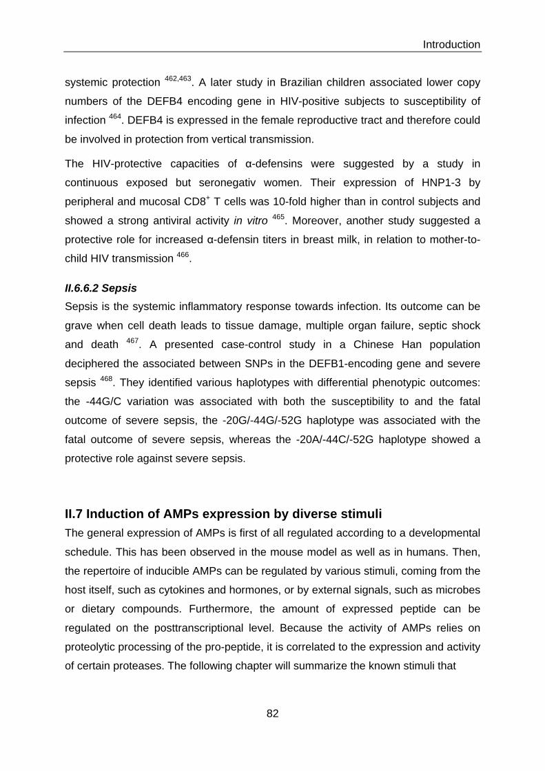
Introduction
82
systemic protection 462,463. A later study in Brazilian children associated lower copy
numbers of the DEFB4 encoding gene in HIV-positive subjects to susceptibility of
infection 464. DEFB4 is expressed in the female reproductive tract and therefore could
be involved in protection from vertical transmission.
The HIV-protective capacities of α-defensins were suggested by a study in
continuous exposed but seronegativ women. Their expression of HNP1-3 by
peripheral and mucosal CD8+ T cells was 10-fold higher than in control subjects and
showed a strong antiviral activity in vitro 465. Moreover, another study suggested a
protective role for increased α-defensin titers in breast milk, in relation to mother-to-
child HIV transmission 466.
II.6.6.2 Sepsis Sepsis is the systemic inflammatory response towards infection. Its outcome can be
grave when cell death leads to tissue damage, multiple organ failure, septic shock
and death 467. A presented case-control study in a Chinese Han population
deciphered the associated between SNPs in the DEFB1-encoding gene and severe
sepsis 468. They identified various haplotypes with differential phenotypic outcomes:
the -44G/C variation was associated with both the susceptibility to and the fatal
outcome of severe sepsis, the -20G/-44G/-52G haplotype was associated with the
fatal outcome of severe sepsis, whereas the -20A/-44C/-52G haplotype showed a
protective role against severe sepsis.
II.7 Induction of AMPs expression by diverse stimuli The general expression of AMPs is first of all regulated according to a developmental
schedule. This has been observed in the mouse model as well as in humans. Then,
the repertoire of inducible AMPs can be regulated by various stimuli, coming from the
host itself, such as cytokines and hormones, or by external signals, such as microbes
or dietary compounds. Furthermore, the amount of expressed peptide can be
regulated on the posttranscriptional level. Because the activity of AMPs relies on
proteolytic processing of the pro-peptide, it is correlated to the expression and activity
of certain proteases. The following chapter will summarize the known stimuli that

Introduction
83
JEM VOL. 205, January 21, 2008
ARTICLE
185
crobial activity against Bacillus megaterium (strain Bm11) using a standardized agar di! usion assay ( 11 ). In addition, mass spectrometry was performed on positive fractions to identify antimicrobially active substances. The presence of a large va-riety of cryptdins and CRS peptide homo- and heterodimers was con" rmed by mass spectrometric analysis in fractions 45 – 55 from adult IECs (day 28) associated with signi" cant anti-bacterial activity, as recently reported ( Fig. 2 A ) ( 11, 12 ).
In contrast, the corresponding fractions obtained from neonate IECs completely lacked antimicrobial activity and were devoid of known enteric antimicrobial peptides ( Fig. 2 B ). Antimicrobial activity in fractions 65 – 95 was associated with the presence of ribosomal proteins, known cationic molecules with antibacterial activity expected to be found in both neonate and adult tissue. Additional antimicrobial activ-ity eluting in fractions 80 – 82 from day 28 mice correlated with the elution position of a Paneth cell – derived lysozyme. To con" rm these " ndings, intestinal tissue of newborn and 6 – 28-d-old mice was stained for cryptdin 2, a prominent member of the family of Paneth cell – derived peptides expressed
expression (including Mbd 1 , 2 , 4 , and 14 ) was detected at any time point in primary IECs (not depicted). As controls, genes encoding Occludin , Zonula occludens protein 1 ( Zo-1 ), and E-cad-herin , all involved in the formation of epithelial tight junctions, as well as Angiogenin 1 , which has not been associated with an-timicrobial function, were included in the analysis. No signi" -cant changes in the expression level of these genes over the observed time period were noted ( Angiogenin 1 and E-cadherin [not depicted]). Thus, signi" cant alterations of intestinal epi-thelial antimicrobial peptide expression were detected with a marked up-regulation from day 14 and onwards.
Analysis of antibacterial activity and immunodetection of antimicrobial peptides in the developing intestine To verify the developmental changes of intestinal antimicro-bial peptide expression, detection at the protein level and quanti" cation of antibacterial activity were performed. IECs isolated from newborn (day 1) or adult (day 28) small intesti-nal tissue were homogenized, extracted, and fractionated by reverse-phase HPLC. All fractions were analyzed for antimi-
Figure 2. Comparison of antibacterial activity and antimicrobial peptide expression between neonate and adult IECs. Cationic antimicrobial peptides from 28-d-old adult (A) and 1-d-old neonate (B) mice were extracted from isolated IECs and fractionated by reverse-phase HPLC. (top) Compo-nents eluting between 10 and 50% acetonitrile (ACN). Absorbance was measured at ! = 214 nm (A 214 ). (bottom) Fractions were tested for antibacterial activity using B. megaterium Bm11 as the indicator strain in an agar diffusion assay. The killing zone diameter is indicated the ! gure. (C) Immunostaining for cryptdin 2 (red, top) and the Paneth cell marker lysozyme (red, bottom) in the small intestinal tissue of 1-, 6-, 14-, 21-, and 28-d-old mice. MFP488 phalloidin (green) and DAPI (blue) were used as counterstaining. Arrows indicate the cryptdin 2 – and lysozyme-positive Paneth cells at the bottom of the intestinal crypts. Bar, 75 µ m.
Cryptdin 2
Lysozyme
JEM VOL. 205, January 21, 2008
ARTICLE
187
sulting in a 3 – 4 log decrease of viable bacteria after 2 h was noted against selected Gram-positive and -negative intestinal commensal bacteria of the small intestine such as S. gallinaceus ,
gut commensal bacteria, as well as de! ned pathogenic bacteria known to be transmitted to the neonate organism during pas-sage through the birth canal ( 6 ). Rapid bacterial killing re-
Figure 3. CRAMP expression in IECs during postnatal development. (A) Quantitative analysis of CRAMP mRNA expression in primary IECs isolated from the small intestinal tissue of fetal, 6- and 24-h-, and 3-, 6-, 14-, 21-, and 28-d-old mice. Values represent the mean ± SD of gene expression deter-mined in total IECs from three individual mice and indicate the target/housekeeping ( Hprt1 ) gene expression ratio. n.d., not detectable. (B) Immunoblot for CRAMP in cell lysate of isolated IECs from 3- and 28-d-old mice (left) as well as in culture supernatant and cell pellet of isolated IECs from 3-d-old mice (right). Actin staining was included to demonstrate equal protein loading. (C) Immunostaining for CRAMP (red) in the small intestinal tissue of 3-, 6-, 14-, 21-, and 28-d-old mice. The FITC-conjugated lectin wheat germ agglutinin (WGA; green) binding to the mucus surface, and DAPI (blue) was used to delin-eate the anatomical structures of the mucoid surface and cell nuclei, respectively. Bar, 75 µ m. (D) RT-PCR expression analysis for the known CRAMP-cleaving enzymes proteinase 3, pancreatic elastase, and kallikrein 5 and 7 in primary IECs of 1-, 6-, 14-, and 28-d-old mice, as well total small intestinal (SI) and skin tissue as positive control. H 2 O and genomic DNA were included as negative controls for the intron-spanning primers.
CRAMP
Figure 31: Developmental induction of AMPs in new born mice. The expression of AMPs in the intestine of mice follows a developmental program. The induction of expression of some AMPs, such as cryptdin 2 and lysozyme, occurs approximately 2 weeks after birth (upper two panels). On the contrary, some AMPs, such as CRAMP are expressed already in the neonate, but then disappear between 2 and 3 weeks of age (lower panel). (Adapted from 470)

Introduction
84
have been described to induce or suppress the expression of AMPs in the human
body, but also in other species.
II.7.1 Host-derived inducers of AMPs expression First, we will focus on the host-induced expression of AMPs, discussing the
developmental regulation of AMPs expression during the pre- and post-natal phase
in the mouse model as well as in humans. Furthermore, internal host-derived AMPs-
inducing molecules, such as hormones and cytokines, will be discussed in this
chapter. Hormones take a special place in host-derived regulation of AMP
expression, as they can induce, as well as inhibit their expression. In this regard
stress hormones will be discussed as repressors of AMP expression.
II.7.1.1 Developmental induction of AMPs expression The development of the immune system of the fetus is by far not complete at the time
of birth. The transition from the mother’s womb into the open air goes along with a
huge immunological challenge posed by the microbial environment. Several
pathogens have been described to cause severe neonatal disease and mortality
through infection via the transplacental route during pregnancy, or transmission from
the mother to the newborn during passage through the birth canal 469. In these
scenarios the expression of innate defense molecules such as AMPs are of major
importance in the immunologically underdeveloped fetus or neonate. Only few
studies have been undertaken to determine the developmental onset of expression of
AMPs in neonates in humans and in the mouse model. All of them suggest a
developmental regulation of AMP expression in various tissues. In vivo studies of the
neonatal mouse small intestinal epithelium have shown that the first two weeks after
birth are dominated by high constitutive expression of CRAMP (Figure 31). This
conferred protection from the enteric pathogen L. monocytogenes 470. Interestingly,
CRAMP expression gradually declines and is absent in the small intestine in adults.
Decline of CRAMP expression goes hand in hand with increased stem cell
proliferation and the formation of intestinal crypts. Paneth cells take over the
production of AMPs, such as cryptdins after a period of 2 weeks 471,472 (Figure 31).
Developmental regulation is partly mediated by enhanced Wnt signaling which
accompanies increased epithelial proliferation during the postnatal phase 473.

Introduction
85
Compared to the postnatal establishment of AMP defense in mice, human enteric
AMP expression starts already during late gestation as observed in a study of human
fetal samples 474. Quantifiable levels of HD5 and HD6 mRNA accumulate in the small
intestine towards the end of the second trimester. Their expression was localized to
Paneth cells, which appear at 12 weeks of gestation in humans. But levels were
found to be 40-250-fold lower than those observed in adults. This can be explained
by the much lower ratio of Paneth cells in the fetal crypts, as compared with the
newborn or adult.
Developmental regulation in expression was also observed in other tissues. A
study of AMP expression in the neonatal skin found significantly elevated expression
of cathelicidins CRAMP in the skin of newborn mice and LL37 in foreskin of human
neonates when compared with adult skin 475. In the neonatal lung DEFB2 was found
to be the most prevalent AMP and its expression was less abundant in premature
compared with term or post-term tissue 476.
Interestingly, there are several AMPs based mechanism of protection for the
fetus and newborn. For example the amniotic fluid contains HNP1 to 3, lysozyme and
LL37 477,478. Additionally, the vernix caseosa, which is a lipid-rich white substance
that covers the skin of the fetus and the newborn, contains HNP1 to 3, lysozyme,
LL37, and psoriasin. Therefore these two structures protect the fetus within the
womb. After birth AMPs contained in the breast milk, such as lactoferrin, lysozyme,
and DEFB1, supplement AMPs protection 479,480. Higher DEFB1 immunoreactivity
was demonstrated in breast tissue during lactation.
II.7.1.2 Induction of AMPs expression by cytokines Cytokines are produced by a wide range of cells, including immune cells and
epithelial cells and function as important signaling molecules in cellular
communication. Their production is highly increased in response to an infectious
agent or inflammatory stimulus and it is not surprising that they are involved in the
induction of AMPs expression 481 (Figure 32).
II.7.1.2.1 Members of the IL1 family
Pro-inflammatory cytokines, such as the IL1 family members IL1α, IL1β and IL18, are
key inducers of AMPs expression. They are expressed by a number of immune cells

Introduction
86
but also by epithelial cells. Their target receptor is type I IL1 receptor, which signals
downstream via the adaptor molecules MYD88 and IRAK4 and activates several
transcription factors, such as NF-κB, AP1, JNK and p38 482. Expression of DEFB2 in
the human colon epithelial cell lines HT-29 and Caco-2 is rapidly induced by IL1α
stimulation 280. Furthermore, IL1α increased expression of skin AMPs, such as
lipocalin 2, S100A8, S100A9 and SLPI in keratinocytes 483,484. In the oral cavity IL1α
up-regulated expression of S100A7 and S100A12 in gingival keratinocytes 485.
Moreover, a study by Moon et al. showed IL1α-induced upregulation of DEFB2 in
human middle ear epithelial cells 486.
Nothing has been described about the involvement of IL1β in intestinal AMPs
expression so far. But it was shown to induce the expression of DEFB2 in bronchial,
tracheal and nasal airway epithelia 286,487,488, as well as human astrocyte cultures 489.
Furthermore, a co-culture study by Pioli and colleagues found that IL1β from blood
monocytes and uterine macrophages induces secretion DEFB2 by uterine epithelial
cells 490.
Also IL18 was found to induce expression of LL37 and DEFB2 in intestinal
epithelial cells 491.
II.7.1.2.2 Th17 cytokines – IL17 and IL22
Th17 cells are a distinct lineage of effector CD4+ T cells. Their differentiation from
naïve T cells via TGFβ is initiated by proinflammatory cytokines, such as IL6, IL1β
and TNFα 492. After, the maintenance and survival of Th17 cells is dependent on
IL23. They produce the cytokines IL17 and IL22 493. Several studies indicate that
Th17-cell-derived cytokines act as key regulators of the AMP response in vitro and in
vivo (Figure 31). IL22 is an important player in the maintenance of mucosal
homeostasis. An in vivo study in mice showed that IL22 was required for the
induction of expression of the C-type lectins REG3β and REG3γ in colonic epithelial
cells in the context of Citrobacter rodentium infection. IL22-deficient mice were highly
susceptible to infection with this murine enteropathogen, as shown by increased
tissue damage, systemic bacterial burden and mortality. Administration of exogenous
REG3γ was able to improves survival in this setting 66. Another study in a mouse
model of dextran sodium sulfate-induced acute colitis showed the importance of

Introduction
87
Balancing inflammation and homeostasis
In addition to being a crucial effector of mucosal immunity, IL-22 has been impli-cated in autoimmune inflammation and epithelial-cell proliferation. Whether IL-22 mediates inflammation and proliferation or has an anti-inflammatory protective effect may depend on the differential activation of STAT3 and the subsequent activation of either IL-21, which can further promote IL-17 production, or the suppression of cytokine signalling (SOCS) proteins, which inhibit cytokine-receptor signalling. In addition, similar to IL-17, IL-22 may have granulopoietic effects that lead to the prolif-eration of inflammatory cells, although this suggestion requires further investigation. On the one hand, IL-22 is known to induce the production of several acute-phase proteins (such as lipocalin-2), which are markers of inflammation. On the other hand, IL-22 has been shown to induce the production of high levels of LPS-binding protein (which neutralizes LPS), suggesting that IL-22 has a role in dampening inflammation32.
Another factor that requires further investigation is the role of IL-22 binding protein (IL-22BP), which inhibits IL-22 from binding to IL-22R. The balance of IL-22 and IL-22BP levels, and the mechanism by which the expression of IL-22BP affects the binding of IL-22 to IL-22R in the setting of various inflammatory disorders, is currently unclear. A greater understanding of this balance could help to determine whether IL-22 has a primarily pro- or anti-inflammatory role.
The effects of IL-22 in inflammation are probably influenced by changes in the microenvironment that are yet to be determined, but may be associated with the baseline level of colonization by commensal bacteria. Antimicrobial proteins may be both mediators and ‘end-effectors’ in this cytokine-regulated commitment to inflam-mation and proliferation, as indicated by the paradigm of the crosstalk between innate immune cells and TH17 cells. More specifi-cally, the persistent presence of commensal bacteria in the normal gut induces a constant range and level of antimicrobial-protein expression. As the innate immune system has developed a degree of tolerance to the pres-ence of commensal bacteria, introduction of new pathogenic bacteria, or changes in the quantity or distribution of the commensal flora, may tip the balance towards TH17-cell development. This can subsequently modulate the antimicrobial-protein response accordingly. The differential recognition of bacterial species by antimicrobial proteins, such as REG3 , also helps to ‘tag’ pathogens
for recognition by the immune system and alert it to changes in the quantity and quality of the commensal milieu.
A role for IL-22-driven TH17-cell-mediated antimicrobial-protein expression in inflammatory disease is most apparent in the skin. Antimicrobial peptides are highly expressed in the skin of patients with psoriasis33, and IL-22 strongly induces both the proliferation of keratinocytes and the expression of antimicrobial proteins, such as S100A7 (REFS 25,27,28,34). Moreover, neutralization of IL-22 can reduce cutane-ous acanthosis (thickening of the skin) in models of psoriasis35. A role for TH17 cells in antimicrobial responses is also supported by the finding that patients with muta-tions in STAT3 that cause Job’s syndrome (hyper-IgE syndrome) and increased susceptibility to cutaneous infections with Staphylococcus aureus and C. albicans lack antigen-specific TH17 cells in the peripheral blood36. Curiously, these patients have an exaggerated TH2-cell-associated hyper-IgE syndrome. In individuals with atopic dermatitis37, the TH2-type cytokines IL-4
and IL-13 are highly expressed in the skin, where they seem to downregulate the expression of LL37; this may explain the frequent occurrence of S. aureus infections in patients with atopic dermatitis. IL-4 and IL-13 can activate STAT6, as well as SOCS1 and SOCS3, which then inhibit both tumour-necrosis factor (TNF)- and interferon- (IFN )-mediated induction of HBD2, and HBD3 expression by keratino-cytes38. Further work is required to determine the role of IL-22 (or other activators of STAT3) in Job’s syndrome, and whether myeloid-cell or epithelial-cell expression of STAT3 contributes to the clinical phenotype of this syndrome, including the high IgE levels and susceptibility to S. aureus and C. albicans infections.
The role of TH2-type cytokines in pulmonary infection is less clear. IL-4 was recently shown to increase the transepithe-lial transport of the antimicrobial substrate thiocyanate in human bronchial epithelial cells39, which could enhance the activity of this innate immune defence mechanism in the lungs.
Nature Reviews | Immunology
Dendritic cellNeutrophil
BacteriaInfection
Inflammation
IL-1IL-6,IL-23
IL-17A,IL-17F,IL-22Macrophage
IL-1 familycytokines
Epithelialcell
Apoptoticcell
DNA
Tissue
Antimicrobial-peptide–DNA complex
Proliferation
TH17 cell
CXCR2 ligands
Antimicrobialproteins
TLR9
IL-17AR,IL-22R
CCL20
Figure 2 | Cytokine networks and antimicrobial peptides at epithelial-cell surfaces. In
response to bacterial infection, the interleukin-1 (IL-1) family cytokines, such as IL-1 , potently
induce the expression of antimicrobial proteins by the epithelium. IL-1 , together with IL-6 and
IL-23, can also induce the differentiation of T helper 17 (TH17) cells, which produce IL-17A, IL-17F
and IL-22. These cytokines further induce antimicrobial-protein expression by the epithelium. IL-17A
can also induce the production of CC-chemokine ligand 20 (CCL20), which has antimicrobial activ-
ity, recruits dendritic cells and increases the production of CXC-chemokine receptor 2 (CXCR2)
ligands that are important in neutrophil recruitment. This response is beneficial to the host during
an acute infection. However, in autoimmune diseases (such as psoriasis) cationic antimicrobial
peptides, which are present at high levels, can interact with negatively charged DNA that is released
from dying cells (cell death occurs as a result of increased cell turnover during inflammation).
Antimicrobial-peptide–DNA complexes can amplify inflammation in the skin by activating Toll-like
receptor 9 (TLR9) signalling.
PROGRESS
NATURE REVIEWS | IMMUNOLOGY VOLUME 8 | NOVEMBER 2008 | 833
Balan
cin
g in
flam
matio
n a
nd
ho
meo
stasis
In a
dd
ition
to b
ein
g a
cru
cia
l effe
cto
r of
mu
co
sal im
mu
nity
, IL-2
2 h
as b
een
imp
li-cate
d in
au
toim
mu
ne in
flam
matio
n a
nd
ep
ithelia
l-cell p
rolife
ratio
n. W
heth
er IL
-22
m
ed
iate
s infla
mm
atio
n a
nd
pro
lifera
tion
or
has a
n a
nti-in
flam
mato
ry
pro
tectiv
e e
ffect
may
dep
en
d o
n th
e d
iffere
ntia
l activ
atio
n
of S
TA
T3
an
d th
e su
bse
qu
en
t activ
atio
n o
f eith
er IL
-21
, wh
ich
can
furth
er p
rom
ote
IL
-17
pro
du
ctio
n, o
r the su
pp
ressio
n o
f cy
tok
ine sig
nallin
g (S
OC
S) p
rote
ins, w
hic
h
inh
ibit c
yto
kin
e-re
cep
tor sig
nallin
g. In
ad
ditio
n, sim
ilar to
IL-1
7, IL
-22
may
have
gra
nu
lop
oie
tic e
ffects th
at le
ad
to th
e p
rolif-
era
tion
of in
flam
mato
ry
cells, a
ltho
ug
h th
is su
gg
estio
n re
qu
ires fu
rther in
vestig
atio
n. O
n
the o
ne h
an
d, IL
-22
is kn
ow
n to
ind
uce th
e
pro
du
ctio
n o
f severa
l acu
te-p
hase
pro
tein
s (su
ch
as lip
ocalin
-2), w
hic
h a
re m
ark
ers o
f in
flam
matio
n. O
n th
e o
ther h
an
d, IL
-22
h
as b
een
sho
wn
to in
du
ce th
e p
rod
uctio
n o
f h
igh
levels o
f LP
S-b
ind
ing
pro
tein
(wh
ich
n
eu
traliz
es L
PS
), sug
gestin
g th
at IL
-22
has
a ro
le in
dam
pen
ing
infla
mm
atio
n3
2.A
no
ther fa
cto
r that re
qu
ires fu
rther
investig
atio
n is th
e ro
le o
f IL-2
2 b
ind
ing
p
rote
in (IL
-22
BP
), wh
ich
inh
ibits IL
-22
from
b
ind
ing
to IL
-22
R. T
he b
ala
nce o
f IL-2
2 a
nd
IL
-22
BP
levels, a
nd
the m
ech
an
ism b
y w
hic
h
the e
xp
ressio
n o
f IL-2
2B
P a
ffects th
e b
ind
ing
o
f IL-2
2 to
IL-2
2R
in th
e se
tting
of v
ario
us
infla
mm
ato
ry
diso
rders, is c
urre
ntly
un
cle
ar.
A g
reate
r un
dersta
nd
ing
of th
is bala
nce
co
uld
help
to d
ete
rmin
e w
heth
er IL
-22
has a
p
rimarily
pro
- or a
nti-in
flam
mato
ry
role
. T
he e
ffects o
f IL-2
2 in
infla
mm
atio
n
are
pro
bab
ly in
fluen
ced
by c
han
ges in
th
e m
icro
en
viro
nm
en
t that a
re y
et to
be
dete
rmin
ed
, bu
t may b
e a
ssocia
ted
with
the
base
line le
vel o
f co
lon
izatio
n b
y c
om
men
sal
bacte
ria. A
ntim
icro
bia
l pro
tein
s may b
e
bo
th m
ed
iato
rs an
d ‘e
nd
-effe
cto
rs’ in th
is cyto
kin
e-re
gu
late
d c
om
mitm
en
t to in
flam
-m
atio
n a
nd
pro
lifera
tion
, as in
dic
ate
d b
y th
e
para
dig
m o
f the c
rossta
lk b
etw
een
inn
ate
im
mu
ne c
ells a
nd
TH1
7 c
ells. M
ore
specifi-
cally, th
e p
ersiste
nt p
rese
nce o
f co
mm
en
sal
bacte
ria in
the n
orm
al g
ut in
du
ces a
co
nsta
nt
ran
ge a
nd
level o
f an
timic
rob
ial-p
rote
in
ex
pre
ssion
. As th
e in
nate
imm
un
e sy
stem
has
develo
ped
a d
eg
ree o
f tole
ran
ce to
the p
res-
en
ce o
f co
mm
en
sal b
acte
ria, in
trod
uctio
n o
f n
ew
path
ogen
ic b
acte
ria, o
r ch
an
ges in
the
qu
an
tity o
r distrib
utio
n o
f the c
om
men
sal
flora
, may tip
the b
ala
nce to
ward
s TH1
7-
cell d
evelo
pm
en
t. Th
is can
sub
seq
uen
tly
mo
du
late
the a
ntim
icro
bia
l-pro
tein
resp
on
se
acco
rdin
gly. T
he d
iffere
ntia
l reco
gn
ition
of
bacte
rial sp
ecie
s by a
ntim
icro
bia
l pro
tein
s, su
ch
as R
EG
3, a
lso h
elp
s to ‘ta
g’ p
ath
ogen
s
for re
co
gn
ition
by th
e im
mu
ne sy
stem
an
d
ale
rt it to c
han
ges in
the q
uan
tity a
nd
qu
ality
o
f the c
om
men
sal m
ilieu
.A
role
for IL
-22
-driv
en
TH1
7-c
ell-
med
iate
d a
ntim
icro
bia
l-pro
tein
ex
pre
ssion
in
infla
mm
ato
ry
dise
ase
is mo
st ap
pare
nt in
th
e sk
in. A
ntim
icro
bia
l pep
tides a
re h
igh
ly
ex
pre
ssed
in th
e sk
in o
f patie
nts w
ith
pso
riasis
33, a
nd
IL-2
2 stro
ng
ly in
du
ces b
oth
th
e p
rolife
ratio
n o
f kera
tino
cy
tes a
nd
the
ex
pre
ssion
of a
ntim
icro
bia
l pro
tein
s, such
as S
10
0A
7 (R
EFS
25
,27
,28
,34
). Mo
reo
ver,
neu
traliz
atio
n o
f IL-2
2 c
an
red
uce c
uta
ne-
ou
s acan
tho
sis (thic
ken
ing
of th
e sk
in) in
m
od
els o
f pso
riasis
35. A
role
for T
H1
7 c
ells
in a
ntim
icro
bia
l resp
on
ses is a
lso su
pp
orte
d
by
the fin
din
g th
at p
atie
nts w
ith m
uta
-tio
ns in
ST
AT
3 th
at c
au
se Jo
b’s sy
nd
rom
e
(hy
per-Ig
E sy
nd
rom
e) a
nd
incre
ase
d
susc
ep
tibility
to c
uta
neo
us in
fectio
ns w
ith
Sta
ph
ylo
coccu
s au
reu
s an
d C
. alb
ican
s lack
an
tigen
-specific
TH1
7 c
ells in
the p
erip
hera
l b
loo
d3
6. Cu
riou
sly, th
ese
patie
nts h
av
e a
n
ex
ag
gera
ted
TH2
-cell-a
ssocia
ted
hy
per-
IgE
syn
dro
me. In
ind
ivid
uals w
ith a
top
ic
derm
atitis
37, th
e T
H2
-typ
e c
yto
kin
es IL
-4
an
d IL
-13
are
hig
hly
ex
pre
ssed
in th
e sk
in,
wh
ere
they
seem
to d
ow
nre
gu
late
the
ex
pre
ssion
of L
L3
7; th
is may e
xp
lain
the
freq
uen
t occu
rren
ce o
f S. a
ure
us in
fectio
ns
in p
atie
nts w
ith a
top
ic d
erm
atitis. IL
-4
an
d IL
-13
can
activ
ate
ST
AT
6, a
s well a
s S
OC
S1
an
d S
OC
S3
, wh
ich
then
inh
ibit
bo
th tu
mo
ur-n
ecro
sis facto
r (TN
F)- a
nd
in
terfe
ron
- (IF
N)-m
ed
iate
d in
du
ctio
n o
f H
BD
2, a
nd
HB
D3
ex
pre
ssion
by
kera
tino
-cyte
s3
8. Fu
rther w
ork
is req
uire
d to
dete
rmin
e
the ro
le o
f IL-2
2 (o
r oth
er a
ctiv
ato
rs of
ST
AT
3) in
Job’s sy
nd
rom
e, a
nd
wh
eth
er
myelo
id-c
ell o
r ep
ithelia
l-cell e
xp
ressio
n o
f S
TA
T3
co
ntrib
ute
s to th
e c
linic
al p
hen
oty
pe
of th
is syn
dro
me, in
clu
din
g th
e h
igh
IgE
le
vels a
nd
susc
ep
tibility
to S
. au
reu
s an
d
C. a
lbica
ns in
fectio
ns.
Th
e ro
le o
f TH2
-typ
e c
yto
kin
es in
p
ulm
on
ary
infe
ctio
n is le
ss cle
ar. IL
-4 w
as
recen
tly sh
ow
n to
incre
ase th
e tra
nsep
ithe-
lial tr
an
spo
rt o
f the a
ntim
icro
bia
l sub
strate
th
ioc
yan
ate
in h
um
an
bro
nch
ial e
pith
elia
l cells
39, w
hic
h c
ou
ld e
nh
an
ce th
e a
ctiv
ity o
f th
is in
nate
imm
un
e d
efe
nce m
ech
an
ism
in
the lu
ng
s. N
atu
re R
ev
iew
s | Im
mu
no
log
y
De
nd
ritic
ce
llN
eu
tro
ph
il
Bacte
riaIn
fectio
n
Infla
mm
atio
n
IL-1
IL-6
,IL
-23
IL-17
A,
IL-17
F,
IL-22
Macro
ph
age
IL-1 fa
mily
cyto
kin
es
Ep
ith
elia
lce
ll
Ap
op
to
tic
ce
ll
DN
A
Tissue
An
tim
icro
bia
l-p
ep
tid
e–
DN
A c
om
ple
x
Pro
lifera
tio
n
TH17
ce
ll
CX
CR
2
ligan
ds
An
tim
icro
bia
lp
rote
ins
TLR9
IL-17AR,
IL-22R
CCL20
Fig
ure
2 | C
yto
kin
e n
etw
ork
s a
nd
a
ntim
ic
ro
bia
l p
ep
tid
es a
t e
pith
elia
l-c
ell su
rfa
ce
s. In
re
sp
on
se
to
ba
cte
ria
l i
nfe
ctio
n, t
he
in
te
rle
uk
in
-1
(IL
-1
) f
am
ily
cy
to
kin
es, s
uc
h a
s IL
-1
, po
te
ntly
ind
uc
e t
he
ex
pre
ssio
n o
f a
ntim
ic
ro
bia
l pro
te
in
s b
y t
he
ep
ith
eli
um
. IL
-1
, to
ge
th
er w
ith
IL
-6
an
d
IL
-2
3, c
an
als
o in
du
ce
th
e d
iffe
re
ntia
tio
n o
f T
he
lpe
r 1
7 (
TH1
7) c
ells
, wh
ich
pro
du
ce
IL
-1
7A
, IL
-1
7F
an
d IL
-2
2. T
he
se
cy
to
kin
es f
urth
er in
du
ce
an
tim
icro
bia
l-p
ro
te
in e
xp
re
ssio
n b
y t
he
ep
ith
eliu
m. IL
-1
7A
ca
n a
lso
ind
uc
e t
he
pro
du
ctio
n o
f C
C-c
he
mo
kin
e lig
an
d 2
0 (C
CL
20
), wh
ich
ha
s a
ntim
icro
bia
l ac
tiv
-
ity, r
ec
ru
its d
en
drit
ic c
ells
an
d in
cre
ase
s t
he
pro
du
ctio
n o
f C
XC
-c
he
mo
kin
e r
ec
ep
to
r 2
(C
XC
R2
)
liga
nd
s t
ha
t a
re
imp
orta
nt in
ne
utro
ph
il re
cru
itm
en
t. T
his
re
sp
on
se
is b
en
efic
ial t
o t
he
ho
st d
urin
g
an
ac
ute
in
fe
ctio
n. H
ow
ev
er, i
n a
uto
im
mu
ne
dise
ase
s (
su
ch
as p
so
ria
sis) c
atio
nic
an
tim
ic
ro
bia
l
pe
ptid
es, w
hic
h a
re
pre
se
nt a
t h
igh
lev
els
, ca
n in
te
ra
ct w
ith
ne
ga
tiv
ely
ch
arg
ed
DN
A t
ha
t is
re
lea
se
d
fro
m d
yin
g c
ells (
ce
ll d
ea
th
oc
cu
rs a
s a
re
su
lt o
f i
nc
re
ase
d c
ell t
urn
ov
er d
urin
g i
nfla
mm
atio
n).
An
tim
icro
bia
l-p
ep
tid
e–
DN
A c
om
ple
xe
s c
an
am
plif
y in
fla
mm
atio
n in
th
e s
kin
by
ac
tiv
atin
g T
oll-
like
re
ce
pto
r 9
(T
LR
9) s
ign
allin
g.
PROGRESS
NA
TU
RE
RE
VIE
WS
| IM
MU
NO
LO
GY
V
OL
UM
E 8
| NO
VE
MB
ER
20
08
| 8
33
Bala
ncin
g in
flam
matio
n a
nd
ho
meo
sta
sis
In addition to being a crucial effector of m
ucosal imm
unity, IL-22 has been impli-
cated in autoimm
une inflamm
ation and epithelial-cell proliferation. W
hether IL-22 m
ediates inflamm
ation and proliferation or has an anti-inflam
matory protective effect
may depend on the differential activation
of STAT
3 and the subsequent activation of either IL-21, w
hich can further promote
IL-17 production, or the suppression of cytokine signalling (SO
CS) proteins, w
hich inhibit cytokine-receptor signalling. In addition, sim
ilar to IL-17, IL-22 may have
granulopoietic effects that lead to the prolif-eration of inflam
matory cells, although this
suggestion requires further investigation. On
the one hand, IL-22 is known to induce the
production of several acute-phase proteins (such as lipocalin-2), w
hich are markers of
inflamm
ation. On the other hand, IL-22
has been shown to induce the production of
high levels of LPS-binding protein (which
neutralizes LPS), suggesting that IL-22 has a role in dam
pening inflamm
ation32.
Another factor that requires further
investigation is the role of IL-22 binding protein (IL-22B
P), which inhibits IL-22 from
binding to IL-22R
. The balance of IL-22 and
IL-22BP levels, and the m
echanism by w
hich the expression of IL-22B
P affects the binding of IL-22 to IL-22R
in the setting of various inflam
matory disorders, is currently unclear.
A greater understanding of this balance
could help to determine w
hether IL-22 has a prim
arily pro- or anti-inflamm
atory role. T
he effects of IL-22 in inflamm
ation are probably influenced by changes in the m
icroenvironment that are yet to be
determined, but m
ay be associated with the
baseline level of colonization by comm
ensal bacteria. A
ntimicrobial proteins m
ay be both m
ediators and ‘end-effectors’ in this cytokine-regulated com
mitm
ent to inflam-
mation and proliferation, as indicated by the
paradigm of the crosstalk betw
een innate im
mune cells and T
H 17 cells. More specifi-
cally, the persistent presence of comm
ensal bacteria in the norm
al gut induces a constant range and level of antim
icrobial-protein expression. A
s the innate imm
une system has
developed a degree of tolerance to the pres-ence of com
mensal bacteria, introduction of
new pathogenic bacteria, or changes in the
quantity or distribution of the comm
ensal flora, m
ay tip the balance towards T
H 17-cell developm
ent. This can subsequently
modulate the antim
icrobial-protein response accordingly. T
he differential recognition of bacterial species by antim
icrobial proteins, such as R
EG3
, also helps to ‘tag’ pathogens
for recognition by the imm
une system and
alert it to changes in the quantity and quality of the com
mensal m
ilieu.A
role for IL-22-driven TH 17-cell-
mediated antim
icrobial-protein expression in inflam
matory disease is m
ost apparent in the skin. A
ntimicrobial peptides are highly
expressed in the skin of patients with
psoriasis33, and IL-22 strongly induces both
the proliferation of keratinocytes and the expression of antim
icrobial proteins, such as S100A
7 (RE
FS
25
,27
,28
,34
). Moreover,
neutralization of IL-22 can reduce cutane-ous acanthosis (thickening of the skin) in m
odels of psoriasis35. A
role for TH 17 cells
in antimicrobial responses is also supported
by the finding that patients with m
uta-tions in STAT
3 that cause Job’s syndrome
(hyper-IgE syndrome) and increased
susceptibility to cutaneous infections with
Staphylococcus aureus and C. albicans lack
antigen-specific TH 17 cells in the peripheral
blood36. C
uriously, these patients have an exaggerated T
H 2-cell-associated hyper-IgE syndrom
e. In individuals with atopic
dermatitis
37, the TH 2-type cytokines IL-4
and IL-13 are highly expressed in the skin, w
here they seem to dow
nregulate the expression of LL37; this m
ay explain the frequent occurrence of S. aureus infections in patients w
ith atopic dermatitis. IL-4
and IL-13 can activate STAT
6, as well as
SOC
S1 and SOC
S3, which then inhibit
both tumour-necrosis factor (T
NF)- and
interferon- (IFN
)-mediated induction of
HB
D2, and H
BD
3 expression by keratino-cytes
38. Further work is required to determ
ine the role of IL-22 (or other activators of STA
T3) in Job’s syndrom
e, and whether
myeloid-cell or epithelial-cell expression of
STAT
3 contributes to the clinical phenotype of this syndrom
e, including the high IgE levels and susceptibility to S. aureus and C
. albicans infections.T
he role of TH 2-type cytokines in
pulmonary infection is less clear. IL-4 w
as recently show
n to increase the transepithe-lial transport of the antim
icrobial substrate thiocyanate in hum
an bronchial epithelial cells
39, which could enhance the activity of
this innate imm
une defence mechanism
in the lungs. N
ature R
eviews | Im
mu
no
logy
Den
dritic
cellN
eutro
ph
il
Bacteria
Infectio
n
Inflam
matio
n
IL-1IL-6
,IL-23
IL-17A,
IL-17F,IL-22
Macro
ph
age
IL-1 family
cytokin
es
Epith
elialcell
Ap
op
totic
cell
DN
A
Tissue
An
timicro
bial-p
eptid
e–
DN
A co
mp
lex
Proliferatio
n
TH 17 cell
CX
CR
2 ligan
ds
An
timicro
bial
pro
teins
TLR9
IL-17AR,
IL-22RCCL20
Fig
ure
2 | C
yto
kin
e n
etw
ork
s a
nd
an
tim
icro
bia
l pe
ptid
es a
t e
pit
he
lial-
ce
ll su
rfa
ce
s. In
resp
on
se
to b
ac
teria
l infe
ctio
n, th
e in
terle
uk
in-1
(IL-1
) fam
ily c
yto
kin
es, s
uc
h a
s IL
-1, p
ote
ntly
ind
uc
e th
e e
xp
ressio
n o
f an
timic
rob
ial p
rote
ins b
y th
e e
pith
eliu
m. IL
-1, to
ge
the
r with
IL-6
an
d
IL-2
3, c
an
als
o in
du
ce
the
diffe
ren
tiatio
n o
f T h
elp
er 1
7 (T
H1
7) c
ells
, wh
ich
pro
du
ce
IL-1
7A
, IL-1
7F
an
d IL
-22
. Th
ese
cy
tok
ine
s furth
er in
du
ce
an
timic
rob
ial-p
rote
in e
xp
ressio
n b
y th
e e
pith
eliu
m. IL
-17
A
ca
n a
lso
ind
uc
e th
e p
rod
uc
tion
of C
C-c
he
mo
kin
e lig
an
d 2
0 (C
CL
20
), wh
ich
ha
s a
ntim
icro
bia
l ac
tiv-
ity, re
cru
its d
en
dritic
ce
lls a
nd
inc
rea
se
s th
e p
rod
uc
tion
of C
XC
-ch
em
ok
ine
rec
ep
tor 2
(CX
CR
2)
liga
nd
s th
at a
re im
po
rtan
t in n
eu
trop
hil re
cru
itme
nt. T
his
resp
on
se
is b
en
efic
ial to
the
ho
st d
urin
g
an
ac
ute
infe
ctio
n. H
ow
ev
er, in
au
toim
mu
ne
dis
ea
se
s (s
uc
h a
s p
so
riasis
) ca
tion
ic a
ntim
icro
bia
l
pe
ptid
es, w
hic
h a
re p
rese
nt a
t hig
h le
ve
ls, ca
n in
tera
ct w
ith n
eg
ativ
ely
ch
arg
ed
DN
A th
at is re
lea
sed
from
dy
ing
ce
lls (c
ell d
ea
th o
cc
urs
as a
resu
lt of in
cre
ase
d c
ell tu
rno
ve
r du
ring
infla
mm
atio
n).
An
timic
rob
ial-p
ep
tide
–D
NA
co
mp
lex
es c
an
am
plify
infla
mm
atio
n in
the
sk
in b
y a
ctiv
atin
g T
oll-lik
e
rec
ep
tor 9
(TL
R9
) sig
na
lling
.
PROGRESS
NA
TU
RE R
EV
IEW
S | IMM
UN
OL
OG
Y
VO
LU
ME
8 | NO
VE
MB
ER
2008 | 83
3
AMPs
Figure 32: Induction of AMP expression by cytokines. Members of the IL1 family and Th17 cell-derived cytokines are important host-derived inducers of AMP expression. Bacterial infection, for example, can induce the expression of IL1# by epithelial cells and macrophages and DCs. In turn, this cytokine mediates the expression of AMPs by epithelial cells, and together with IL6 and IL23, released from macrophages and DCs, induces the differentiation of Th17 cells. Those cells then produce IL17A, IL17F and IL22, which further induce AMP expression by the epithelium. (Adapted from 481)

Introduction
88
recruitment of neutrophils upon tissue damage. These neutrophils expressed high
levels of IL22 in an IL23-dependend manner and thereby induced the upregulation of
REG3β and S100A8 in colonic epithelial cells. The protective effects of neutrophils in
this setting was shown by the fact that transfer of IL22–competent neutrophils to
Il22a-deficient mice protected the colonic epithelium from dextran sodium sulfate-
induced damage 494.
Several studies describe the role of Th17 cytokines in other tissues.
Combination of IL17 and IL22 induced the expression of DEFB2, S100A9, S100A7
and S100A8 in primary keratinocytes 493. Additionally, IL17 was shown to
synergistically increased the vitamin D-induced expression of LL37 in keratinocytes 495. This mechanism could have implications in psoriasis development, where Th17
cells are prominent. Lai and colleagues could show that IL17A can also induce the
expression of REG3α in human keratinocytes and that REG3α can act on
keratinocytes to augment their proliferation 496. The authors suggest implications in
psoriasis and wound repair. A study by Guilloteau and colleagues used a cocktail of
IL17, IL22 with IL1α, TNFα and oncostatin M to induce upregulation of DEFB2,
S100A7 in human skin explants. In vivo, intradermal injection of these five cytokines
induced expression of 12 different AMPs in the skin of mice 484. Furthermore, IL17
was able to stimulate DEFB2 expression >75 fold in primary human airway epithelial
cells 497,498.
II.7.1.2.3 TNFα
The pro-inflammatory cytokine TNFα seems to be only a weak inducer of DEFB2 in
the human intestine 280 but is capable to stimulate AMPs release in other tissues.
TNFα was shown to induce expression of DEFB2, DEFB3 and DEFB4 in human
gingival epithelial cells 499,500 and DEFB2 in human lung epithelial cells 501, corneal
epithelial cells 502 and astrocyte cultures 489. Furthermore, it stimulated expression of
lingual antimicrobial peptide, a member of the β-defensin family of peptides, in
cultured bovine tracheal epithelial cells 503.
II.7.1.3 Induction of AMPs expression by estrogen Estrogens are important regulators of female physiology. Recent epidemiological
data suggest a correlation between decrease of estrogen levels after menopause
and increased susceptibility to urinary tract infections 504. A study in pre- and

Introduction
89
A B
Figure 33: Regulation of cathelicidin expression by vitamin D. The expression of cathelicidin is regulated by two distinct 1,25D3-dependent pathways in keratinocytes and monocytes. In the skin, keratinocytes are activated upon injury by TGF# or TLR2/6 ligands, which then leads to induction of CYP27B1. As a consequence 25D3 is converted to 1,25D3, which binds to the VDR and induces cathelicidin, TLR2, and CD14 (A). The thereby increased TLR2 expression acts as a positive feedback for further keratinocytes activation and cathelicidin expression. In contrast, circulating macrophages are activated by TLR2/1 agonists, which induce the expression of the VDR and CYP27B1 (B). In consequence, CYP27B1 converts 25D3 to 1,25D3 and subsequently increases cathelicidin. (Adapted from 512)
Keratinocyte Macrophage

Introduction
90
postmenopausal women investigated the role of estradiol in urinary AMPs expression 505. A 2-week supplementation of estrogen significantly increased the expression of
AMPs, such as DEFB1, DEFB2, psoriasin, RNase 7, and cathelicidin, in urinary
epithelial cells. Additionally, estrogen promoted the expression of cell-cell contact–
associated proteins, thereby strengthening the epithelial integrity and barrier function.
Another study showed that estradiol stimulates a five fold increase in DEFB2 expression in human uterine epithelial cells 506. Responsiveness of tissues to
estrogen is mediated by two estrogen receptors (ER), ERa and ERb, and by the
estrogen-responsive elements (ERE) located in the promoter regions of target genes.
EREs have been reported in the promoters for DEFB1, DEFB2, and psoriasin 507.
II.7.1.4 Induction of AMPs expression by vitamin D A special case of induction is described for LL37. Its promoter contains a vitamin D
responsive element (VDRE). This consensus sequence can be bound by the vitamin
D receptor, which is thereby able to act as a transcription factor. Activation of this
process in achieved by its ligand 1,25-dihydroxyvitamin D3 (1,25D3), the hormonal
form of vitamin D3. Binding of ligand-stimulated vitamin D receptor was shown to
induce LL37 expression in human keratinocytes, monocytes and neutrophils 508,509
and in vivo in human skin 510 (Figure 33). The induction of LL37 by vitamin D in
intestinal epithelial cells has not been documented.
Vitamin D3 is produced in the skin from the cholesterol precursor 7-
dehydrocholesterol under the influence of UVB light 511. The activation of vitamin D3
to 1,25D3 requires 2 major hydroxylation steps, the first by 25-hydroxylase
(CYP27A1) and the second by 1α-hydroxylase (CYP27B1). A negative feedback
mechanism initializes the induction of 24-hydroxylase (CYP24A1) and degradation of
1,25D3 to limit the level of this bioactive hormone.
Recent studies investigated the relations between infection, injury and vitamin
D induced LL37 expression. They were able to demonstrate that stimulation of TLRs
on human macrophages induces CYP27B1 for the conversion of 25D3 to active
1,25D3 and increases the expression of the vitamin D receptor (VDR) for the
activation of LL37 expression 442 (Figure 33 B). These results indicate a link between
infection and the activation of LL37 expression via vitamin D. A follow up study in

Introduction
91
keratinocytes showed that injury is able to induce CYP27B1 and thereby increase
levels of 1,25D3 in the skin (Figure 33 A). Additionally, factors involved in
the wound repair such as TGF-β induced 1,25D3-dependent immune responses.
Furthermore, they could show a 1,25D3-dependent increase of TLR2 and CD14
receptors. These findings describe a sort of feedback loop which allows the activation
of AMP immune responses upon injury and at the same time increases the level of
detection of microbes and thereby responsiveness in the skin immune system in a
vitamin D dependent manner 512.
II.7.1.5 Stress hormones as repressor of AMPs expression Sustained stress is a common health threat in our society nowadays and negatively
influences various physiological processes 513. Also the immune system is under the
control of the stress response. While acute stress is thought to be immunoenhancing
in scope of the “fight-and-flight” syndrome, sustained stress leads to the impairment
of effective immune responses. Stress can be communicated via three major
pathways: The catecholamine pathway via adrenergic stimulation, the glucocorticoid
pathways via activation of the hypothalamic-pituitary-adrenal axis, and the cholinergic
pathway via acetylcholine. Several studies have been performed to shed light on the
influences of stress onto AMPs expression and evidence for the involvement of each
of these pathways has been described. No intestine-specific studies have been
undertaken so far.
II.7.1.5.1 Glucocorticoids
Glucocorticoids are steroid hormones expressed by the adrenal cortex and cortisol is
the best-known member of this family. They signal via the glucocorticoid receptor,
which can directly act as a transcription factor. Their anti-inflammatory capacities are
well described and they are widely used in medicine to treat inflammatory disorders.
A study in human primary bronchial epithelial cells showed the inhibition of inducible
DEFB3 after pretreatment with glucocorticoids before infection with heat-killed P.
aeruginosa 514. Also the expression of skin antimicrobial peptides in the frog Rana
esculenta was completely blocked after administration of glucocorticoids 515.This
went hand in hand with an increase of viable mouth bacteria. A mechanism where

Introduction
92
glucocorticoids induce the NF-κB inhibitor IκBα to block AMPs gene transcription was
presumed in this scenario.
II.7.1.5.2 Cholinergic stimulation
Cholinergic receptors are ligand-gated ion channels, which are activated by the
neurotransmitter acetylcholine. Additionally, the nicotinic acetylcholine receptor
(nAchR) can be activated by nicotine and the muscarinic acetylcholine receptor
(mAchR) can be activated by muscarine. Cholinergic activation was shown to inhibit
local proinflammatory responses and anti-inflammatory effects on immune cells and
epithelia have been studied 516-518. Activation of the nAchR by topic nicotine
administration reduced AMPs activity in the skin of mice and increased their
susceptibility to methicillin-resistant S. aureus infection 519. These results
demonstrate possible mechanisms for the increased risk for infections followed
prolonged stress or nicotine exposure, which both activate the nAchR pathway.
II.7.1.5.3 Physiological stress
One group investigated the effect of physiological stress, such as combined
insomnia, noise, and crowding, in a mouse model of cutaneous infection 520.
Cutaneous barrier function was severely decreased by physiological stress as well as
administration of endogenous glucocorticoids. Both CRAMP and mBD3 expression
was significantly reduced, which lead to increased severity of group A Streptococcus
pyogenes skin infection. They hypothesized that an inhibition in epidermal lipid
synthesis decreased the secretion of lamellar bodies, which are important carriers for
skin AMPs also in humans. The effects could be reversed by pharmacological
blockade of stress hormone action, as well as topical administration of physiologic
lipids.
The response to stress in cattle and the relationship to pneumonia has also
been investigated. Transportation, weaning, and commingling results in elevated
levels of stress hormones, such as glucocorticoids and catecholamines. Results from
a study by Mitchell et al. indicate that glucocorticoids inhibited expression of AMPs
such as tracheal antimicrobial peptide and lingual antimicrobial peptide in bovine
tracheal epithelial cells 521.

Introduction
93
II.7.1.5.4 Physical stress
Two human in vivo studies proclaimed a reduction in innate immune responses in
elite athletes by physical stress, signaling via the HPA axis and cortisol release. They
reported reduced levels of LL37 and DEFB2 in saliva from individuals and this
correlated with the occurrence of upper respiratory tract infections 522,523.
II.7.2 Induction of AMPs expression by external stimuli Various external stimuli have been described to induce the expression of AMPs.
Among them are bacteria and bacterial products as well as dietary compounds,
which will be discussed in the following chapter.
II.7.2.1 Induction of AMPs by bacteria Bacterial stimuli are important for the development of our immune system as
described in chapter I. Germ-free mice are a useful tool to study the overall role of
microbial signals in AMPs expression. Some intestinal AMPs are expressed
independently of the microbiota, such as lysozyme or PLA2s. Others require bacterial
signals for their expression and are absent in germ-free mice, such as members of
the cryptdins-related sequence family of peptides, angiogenin 4 (ANG4) and REG3γ 247,524,525.
In an infectious setting AMPs have been shown to be inducible by several
gram-positive and gram-negative bacteria. DEFB2 expression in human colon
epithelial cell lines is rapidly induced by infection with enteroinvasive bacteria, such
as Salmonella and E. coli species 280. In the same study human fetal intestinal
xenografts were used to how DEFB2 induction by intraluminal infection with
Salmonella.
Induction of AMPs expression by bacteria has also been described in other
tissues. P. aeruginosa infection for example stimulated DEFB2 expression in A549
lung epithelial cell line 286. Infection of keratinocytes with S. aureus upregulated
expression of DEFB2, DEFB3 and LL37 526. In gastric epithelial cells infection with H.
pylori induced expression of DEFB2. In accordance an increased DEFB2 level was
also found in biopsies from patients with H. pylori-positive gastritis 527.

Introduction
94
Also, bacterial molecules alone are able to induce expression of AMPs via innate
receptor signaling pathways. Two different studies described the ability of flagella
filament structural protein (FliC) in the supernatant of Salmonella cultures to induce
DEFB2 expression in the intestinal epithelial cell line Caco-2 cells in a NF-κB
dependent manner 528,529. Vora and colleagues showed that LPS and peptidoglycan
stimulate DEFB2 expression in a TLR4- and TLR2/TLR6-dependent manner in
intestinal epithelial cells 530. Furthermore, LPS was found to induce DEFB2 in human
astrocyte cultures 489. Moreover, several studies reported LPS and LTA mediated
activation of DEFB2 expression in airway epithelial cells via TLR4 and TLR2
respectively 531-533.
Activation of TLRs converges in the signal cascades of MAP kinases and NF-
κB. The role of these two pathways in AMPs expression will be discussed in greater
depth in the following chapters.
II.7.2.2 Induction of AMPs expression by SCFA SCFA are produced in vast amounts by fermentation processes of the intestinal
microbiota. Their involvement in several gut physiological processes has been
described in chapter I. Several studies demonstrated their immune modulating role
via the induction of cathelicidin in the intestine. Cell differentiation seems to be a key
determinant of LL37 expression, as observed in human colonic biopsies 260,534. In
vitro studies in human colonic cell lines identified butyrate, isobutyrate and
propionate as inducers of cell differentiation and LL37 expression 534. The use of
specific inhibitors showed the involvement of several signaling pathways in the
butyrate-mediated expression of LL37, such as the vitamin D pathway, MAPK
pathways and TGFβ signaling 534,535. Although it seems like different MAP kinases
are involved in different colonic cell lines and other cell types such as lung epithelial
cells and keratinocytes 536,537. In vivo studies of a rabbit model of Shigella infection
showed that butyrate treatment significantly increased the expression levels of the
rabbit homologue to LL37, CAP18 (Figure 34). This lead also to reduction in the
bacterial load and amelioration of clinical symptoms and inflammation in the colon 264. Furthermore, treatment with the butyrate stimulated DEFB2 expression in colonic
epithelial cells 538.
Introduction
95
Figure 34: Colonic expression of the rabbit cathelicidin CAP18 upon Shigella infection and SCFA treatment. Imunohistochemical staining showed localization of CAP18 (brown) almost exclusively located in the surface epithelium in healthy rabbits (A). On the contrary, Shigella-infection led to the disappearance of CAP18 on the surface and in crypts (arrowheads) (B). On the other hand, CAP18 staining was increased in inflammatory cells in the lamina propria upon infection (arrows). Treatment of infected rabbits with butyrate lead to the reappearance of CAP18 staining in the surface epithelium and disappearance in the lamina propria 264.

Introduction
96
II.7.2.3 Induction of AMPs expression by the amino acid isoleucine The essential amino acid isoleucine has been described in one study as an inducer
of β-defensins in bovine kidney epithelial cells in a NF-κB dependent manner 539.
Summary
In Chapter II, we have discussed the existence of AMPs throughout the kingdoms of
life, their characteristics and properties. We have learned that they are not only
capable of killing a multitude of microbes, but also important in the activation of the
adaptive immune response, tissue repair, and restitution. In a bigger picture, we have
discussed the involvement of AMPs in human pathologies and the detrimental effects
of the deregulation of their expression. Their importance for intestinal homeostasis is
indisputable, as illustrated on the example of IBDs. As we learn more about these
multitalented peptides we wonder how we can manipulate their expression? In this
regard, we have discussed their inducibility by host molecules, as well as bacterial
factors and nutritional components. To go deeper into the regulation of inducible
AMPs expression we will take a look on the basics of genomic organization and
general gene expression. Different mechanisms, which allow for rapid and
distinguished stimulus-induced gene expression, will be discussed. Following this
part of general inducible gene expression we will take a look on what is known about
the regulation of AMPs gene expression by transcription factors and epigenetic
mechanism. In this regard, we will focus on the role of acetylation of histone tails in
gene expression, and we will learn about enzymes, which are involved in acetylation
and deacetylation of histones and other proteins.

Introduction
97
Figure 35: Structural organization of DNA. Each cell contains double stranded DNA of a total length of 2 meters. In order to fit this length into the nucleus it needs to be tightly packed into a structure called chromatin. This structure consists of so-called nucleosomes, which contain a complex of eight core histone molecules wrapped with 147 base pairs of 1.7 turns of DNA. Additionally, histone H1 clamps the DNA to the histone core, supports the next level of packaging into highly condensed fibers. (Adapted from “Life: The Science of Biology, 7th Edition, 2003” and 540)
Nucleosome
Introduction
98
Chapter III – Regulation of inducible gene expression As discussed in chapter II, AMPs genes can be either expressed constitutively or
induced by a wide variety of host-derived or external stimuli. Deciphering the
mechanism, which govern the inducibility of certain AMPs is important to understand
the basis of their regulation. Also the question, whether AMPs and INFs are
regulated in the same ways in response to external stimuli is intriguing. In order to go
deeper into the stimulus-induced expression of AMPs, we have to understand the
basic mechanisms, which allow inducible gene expression. External signals such as
growth factors, stresses, cytokines or pharmacological agents activate pathways,
which lead to the rapid expression of primary response genes. Those are specified
as being directly connected to the signaling machinery and do not require protein
synthesis for their expression 541. To understand the regulation of expression of
inducible genes, it is important to consider the organization of the human genome
into chromatin. The following chapters are aiming to describe the molecular
organization of DNA in eukaryotic cells and the complex mechanisms, which regulate
accessibility of the stored genetic information.
III.1 Structural organization of the human genome The human genome contains around 20 000 protein-coding genes, encoded in a total
length of over 3 billion base pairs of DNA 542. In order to be packed into the confined
space of the nucleus of eukaryotic cells, the DNA double strand is highly compacted
into a structure called chromatin 543 (Figure 36). But this compacted structure is still
very dynamic to allow for important cellular processes such as transcription, repair, or
cell division. The packing of DNA is achieved by wrapping of a piece of 147
nucleotide pairs (1.7 turns) around an octamer of core histone proteins (2 copies
each of histones H2A, H2B, H3, and H4) to form a so-called nucleosome 544,545
(Figure 35). This basic unit of chromatin is repeated every 200 base pairs and further
stabilized and compacted by histone H1. In mitosis, the chromatin reaches its highest
level of condensation into the chromosomes, which are then ready for cell division.
During interphase, the DNA exists in two chromatin states of different levels of
compaction.
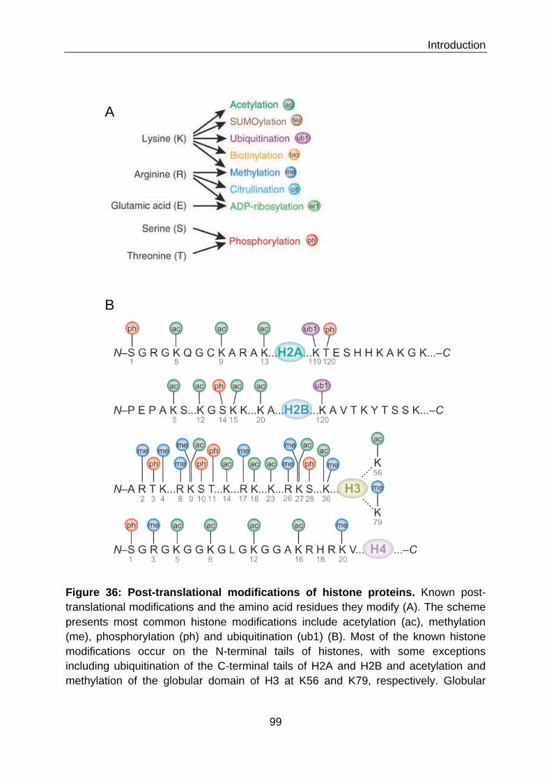
Introduction
99
A
B
Figure 36: Post-translational modifications of histone proteins. Known post-translational modifications and the amino acid residues they modify (A). The scheme presents most common histone modifications include acetylation (ac), methylation (me), phosphorylation (ph) and ubiquitination (ub1) (B). Most of the known histone modifications occur on the N-terminal tails of histones, with some exceptions including ubiquitination of the C-terminal tails of H2A and H2B and acetylation and methylation of the globular domain of H3 at K56 and K79, respectively. Globular

Introduction
100
domains of each core histone are represented as colored ovals. (Adapted from 546 and 547)
Heterochromatin is the denser of the two structures and is generally associated with
inactive gene expression. This compact structure can generally be found at the
centromere and telomeres of the chromosome. The structure of euchromatin in
contrast is much more relaxed and contains most of the active genes.
III.2 The role of chromatin in gene accessibility
As mentioned previously, the compacted structure of DNA still has to be highly
flexible and is constantly exposed to remodeling processes in order to allow gene
expression, DNA replication, repair or recombination. Remodeling is orchestrated by
chromatin-remodeling complexes 548,549. Facilitated by the use of energy from ATP
hydrolysis, nucleosomes can be repositioned, replaced by histone-variants or
expelled to loosen up the chromatin structure and increase accessibility. So far, five
families of chromatin remodelers have been identified in eukaryotes: SWI/SNF
(SWItch/Sucrose NonFermentable), ISWI (Imitation SWI), Mi-2/NuRD (nucleosome
remodeling deacetylase) complex, INO80 and SWR1. They all possess a common
ATPase domain. But each remodeler complex has unique protein domains and
consists of different subunits possessing histone-modifying capacities. Therefore,
their functions can be variable and either activating or repressive. In order to recruit
these complexes to target genes, specific signals are set for the recognition by
interacting domains. These signals occur in the form of post-translational modification
marks on core histones, which are set by a large variety of histone-modifying
enzymes.
III.2.1 Histone modification The N-terminal tails of core histones protrude from the nucleosome structure and are
subjected to a vast array of covalent post-translational modifications (Figure 35 and
36). Among them are mono-, di-, or trimethylation of lysine, mono- or dimethylation of
arginine, acetylation of lysine, phosphorylation of serine, threonine or tyrosine,
ubiquitylation of lysine, sumoylation of lysine, and many more 550 (Figure 36).
Occurrence of these modifications has been extensively mapped in yeast 551, plants 552, drosophila 553, nematodes 554, humans 555 and a variety of human cell lines 557.

Introduction
101
A
B
Figure 37: Characteristic chromatin marks of promoters and gene bodies of active or inactive genes. Histone modifications may serve as 'switches' as promoters — from active to poised to inactive — and contribute to fine-tuning of expression levels (A). At gene bodies, they discriminate between active and inactive conformations. In addition, exons in active genes have higher nucleosome occupancy and thus more histone H3 lysine 36 trimethylation (H3K36me3) and H3K79me2-modified histones than introns (B). (Adapted from 556)

Introduction
102
Numerous histone modifying enzyme families, adding and removing covalent groups,
have been characterized 558. Modifications are always reversible and highly dynamic,
just like the chromatin structure itself. Many studies have been undertaken to
characterize the functions of these vast possibilities of modifications. Some
modifications directly influence the interaction of DNA with histones. Acetylation for
example neutralizes the negative charge of the lysine residue and phosphorylation of
lysine even adds a negative charge. Both effectively reduce the positive charge of
histone proteins and thereby the attracting force to the negatively charged DNA
strand. Therefore these modifications decompact the chromatin structure. By now,
we know that promoters, as well as gene bodies, are characterized by the
occurrence of specific chromatin marks, according to their state of activation (Figure
37). Certain marks have been directly associated with transcriptional activation, such
as tri-methylation of histone H3 lysine 4 (H3K4me3) on transcriptional start sites,
H3K36me3 throughout actively transcribed genes, and H3K4me1 in enhancer
regions 559-561. On the opposite, there are marks associated with repression of
transcription, such as H3K9me3, H3K27me3, and H4K12ac 562.
Furthermore, histone marks can act as docking sites for chromatin-associated
factors such as remodeling complexes. Many distinct domains have been
characterizes for protein-histone docking (Figure 38 A). Acetylated lysine for example
can be bound by the bromodomain and methylated lysine by the chromodomain of
interaction partners 563,564. But they can also act in the opposite way, by inhibiting the
binding of transcription repressive complexes. For example, H3K4me3 inhibits
binding of the NuRD complex 565. In some cases, cooperation of marks is needed for
the effective recruitment of chromatin interacting factors.
Another level of complexity is added by several possibilities of cross talk
between histone modifications 566 (Figure 38 B). There can be competitive
antagonism, for example in the case of lysine methylation or acetylation, often
resulting in opposite outcomes concerning gene transcription. Another possibility is
trans-regulation, where one mark obligatory precedes the occurrence of another
mark. On the other hand, one mark can also be disruptive for the occurrence of an
adjacent modification.

Introduction
103
A
B
Figure 38: Specific binding of histone marks by protein domains and crosstalk in between marks. Examples of proteins with domains that specifically bind to modified histones as shown (A). Histone modifications can positively or negatively affect other modifications. A positive effect is indicated by an arrowhead and a negative effect is indicated by a flat head (B). 550
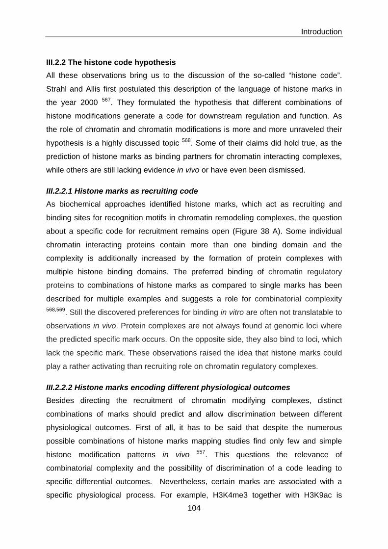
Introduction
104
III.2.2 The histone code hypothesis All these observations bring us to the discussion of the so-called “histone code”.
Strahl and Allis first postulated this description of the language of histone marks in
the year 2000 567. They formulated the hypothesis that different combinations of
histone modifications generate a code for downstream regulation and function. As
the role of chromatin and chromatin modifications is more and more unraveled their
hypothesis is a highly discussed topic 568. Some of their claims did hold true, as the
prediction of histone marks as binding partners for chromatin interacting complexes,
while others are still lacking evidence in vivo or have even been dismissed.
III.2.2.1 Histone marks as recruiting code As biochemical approaches identified histone marks, which act as recruiting and
binding sites for recognition motifs in chromatin remodeling complexes, the question
about a specific code for recruitment remains open (Figure 38 A). Some individual
chromatin interacting proteins contain more than one binding domain and the
complexity is additionally increased by the formation of protein complexes with
multiple histone binding domains. The preferred binding of chromatin regulatory
proteins to combinations of histone marks as compared to single marks has been
described for multiple examples and suggests a role for combinatorial complexity 568,569. Still the discovered preferences for binding in vitro are often not translatable to
observations in vivo. Protein complexes are not always found at genomic loci where
the predicted specific mark occurs. On the opposite side, they also bind to loci, which
lack the specific mark. These observations raised the idea that histone marks could
play a rather activating than recruiting role on chromatin regulatory complexes.
III.2.2.2 Histone marks encoding different physiological outcomes Besides directing the recruitment of chromatin modifying complexes, distinct
combinations of marks should predict and allow discrimination between different
physiological outcomes. First of all, it has to be said that despite the numerous
possible combinations of histone marks mapping studies find only few and simple
histone modification patterns in vivo 557. This questions the relevance of
combinatorial complexity and the possibility of discrimination of a code leading to
specific differential outcomes. Nevertheless, certain marks are associated with a
specific physiological process. For example, H3K4me3 together with H3K9ac is

Introduction
105
found in the promotor region of actively transcribed promoters across all studied
species. But whether their role in these processes is really essential is questionable
as a study by Lenstra et al. showed. They found that mutation of H3K4 or the K4
methylase had almost no effect on gene expression in yeast 570. Another study by
Martin and colleagues showed that the loss of different acetylation marks on histone
3 is indistinguishable by the yeast cell and moreover shows only modest differences
in gene expression 571.
Therefore the histone code in vivo is still mainly undeciphered and remains a
highly interesting field of study. The main challenge will be to bridge the discrepancy
between observed in vitro binding of protein complexes to certain combinations of
marks and the discrimination and relevant function of mark combinations in vivo.
III.3 Regulatory processes of inducible gene expression Now, that we have understood how the human genome is organized into chromatin
and how the components of this structure can be modified and remodeled, we will
take a look at the regulatory processes, which allow for inducible gene expression.
These processes allows the cell to rapidly react to external stimuli such as stress,
infection or developmental cues and to produce new proteins according to the
environmental changes. Several genes can be expressed synergistically to produce
the suitable response. Nevertheless, there are many levels of selectivity of
expression, which tailor the response to a specific stimulus. Mechanisms of
regulation can be cell-specific, signal-specific and gene-specific. This chapter will
describe the rapid induction of the inflammatory response as an example of inducible
gene expression. The inflammatory gene repertoire is activated within minutes upon
stimulation. The expression of these genes has to be tightly orchestrated and has to
include negative feedback loops in order to prevent tissue damage by ongoing
inflammation. Much effort has been made in detailing the sensing, signal
transmission and transcriptional and post-transcriptional regulatory mechanisms of
the inflammatory response. Transcription factors and chromatin add an additional
level of complexity to this regulation. On the transcriptional level, inflammatory
response genes can be divided into two classes: primary response genes and
secondary response genes. Primary response genes are genes which are rapidly
induced and do not require de novo protein synthesis 541. Secondary response

Introduction
106
genes, on the other hand, require de novo synthesis of transcription or signaling
factors and their induction is therefore slower. Differences between these two groups
of genes will be discussed as follows. But first of all, the basic processes of general
transcription will be described.
III.3.1 Transcription by the general transcription machinery In the initial step, transcriptional activators bind sequence-specific near the target
gene (Figure 39 a). They then recruit large multi-subunit co-activator complexes and
nucleosome-remodeling complexes, which facilitate the access to the promotor and
the recruitment of the general transcription machinery (Figure 39 b). Multiple
enzymes facilitate access by repositioning or ejection of nucleosomes and
modifications of histone proteins, as discussed later in this chapter. The pre-initiation
complex (PIC) is formed on the promotor by recruited general transcription factors
(TF) IIA, TFIIB, TFIID, TFIIE, TFIIF and TFIIH and the RNA polymerase II (Pol II)
(Figure 39 c). In the next steps, CDK7 in human TFIIH phosphorylates the serine 5
(S5) position of the Pol II carboxy‐terminal domain (CTD). At the same time, another
subunit of TFIIH, the DNA helicase XPB (xeroderma pigmentosum B), remodels the
PIC at the transcription start site to introduce a single-stranded DNA template of 11–
15 bases into the active site of Pol II (Figure 39 d). Then, Pol II clears the promotor
and transcribes about 20-40 nucleotides into the gene before it is halted at the
promotor-proximal-pause site 572. Now, a second phosphorylation of Pol II at the S2
position of the CTD by CDK9 (Cyclin-dependent kinase 9), a subunit of human P‐
TEFb (positive transcription elongation factor), is required for continued productive
elongation (Figure 39 e). This phosphorylation creates binding sites for chromatin
modifying enzymes and nucleosome remodeling complexes, which facilitate the
continuing passage of Pol II and the transcription of the full length gene 573.
III.3.2 Activation of poised Pol II
Even though signal-dependent Pol II recruitment and transcription initiation is
considered as an important mechanism for induction of gene expression, it has been
shown that Pol ll is already poised at many inducible genes in genome-wide
analysesin human and murine embryonic stem cells and Drosophila cells 575-577.

Introduction
107
Activator
Nucleosome- remodelling complex
GTFs
RNA polymerase II
Nucleosome- remodelling complex
Nucleosome- remodelling complex
Nature Reviews | Genetics
GTFs
a
H2A
H2B
Target gene
Target gene
b
d
e
CDK7
S5P
P P
Recognition site TSS
Target gene
H3
H4
c
GTFsAc
tivat
or-d
epen
dent
recr
uitm
ent
Prom
oter
cle
aran
ceRe
leas
e fr
om p
rom
oter
-pr
oxim
al p
ausin
g
Co-activator
Activator
Activator
RNA polymerase II
XPB
S2S5 RNA polymerase II
P-TEFb
Nucleosome- remodelling complex
PICTarget gene
Target gene
Co-activator
General transcription machineryRNA polymerase II together with the general transcription factors TFIIA, TFIIB, TFIID, TFIIE, TFIIF and TFIIH.
Transcriptional activatorA sequence-specific DNA-binding protein that increases the rate of transcription by recruiting RNA polymerase II, either directly in prokaryotes or through co-activators in eukaryotes.
these initial stages of the transcription cycle. Although the mechanisms involved in the chosen examples may not always be observed in all other cases of inducible gene expression, we hope to provide a broad over-view of the principles involved in inducible activation of transcription.
Activator-dependent recruitmentGene activation involves a multistep recruitment process that consists of several potential rate-limiting steps during the initial stages of the transcription cycle (reviewed in REF. 8) (FIG. 1). During the initial steps of gene induction, transcriptional activators bind to specific DNA sequences near target genes and recruit transcrip-tional co-activators and components of the transcription
machinery to these genes through protein–protein interactions. These steps result in formation of the pre-initiation complex (PIC) on the promoter9,10. For the purposes of this Review, these first three steps can be regarded as a single rate-determining process, which we refer to as activator-dependent recruitment (FIG. 1a–c). An additional level of regulation is required for polymerase to proceed to productive transcription elongation (FIG. 1d,e). Although all of the steps in the transcription cycle are subject to regulation11, we focus in this Review on those steps that are most important for inducible gene expression: activator-dependent recruitment resulting in PIC formation; activation of the PIC and transcription initiation; and release of paused polymerase into productive elongation.
Figure 1 | Early steps in the transcription cycle. a | Promoter selection is determined by the interaction of one or more transcriptional activator(s) with specific DNA sequences (recognition sites) near target genes. Activators then recruit components of the transcription machinery to these genes through protein–protein interactions. b | Activation of gene expression is induced by the sequential recruitment of large multi-subunit protein co-activator complexes (shown in purple and pink) through binding to activators. Activators also recruit ATP-dependent nucleosome-remodelling complexes, which move or displace histones at the promoter, facilitating the rapid recruitment and assembly of co-activators and the general transcription machinery. c | Together, co-activators and nucleosome remodellers facilitate the rapid recruitment of RNA polymerase II (Pol II) and the general transcription factors (GTFs) TFIIA, TFIIB, TFIID, TFIIE, TFIIF and TFIIH to form the pre-initiation complex (PIC) on the core promoter9. These first three steps (a–c) constitute acti-vator-dependent recruitment. d | After PIC assembly, CDK7 in human TFIIH (Kin28 in yeast) phosphorylates the serine 5 (S5) position of the Pol II carboxy-terminal domain (CTD). At the same time, another subunit of TFIIH, the DNA helicase XPB (Rad25 in yeast), remodels the PIC, and 11–15 bases of DNA at the transcription start site (TSS) is unwound to introduce a single-stranded DNA template into the active site of Pol II83. Pol II then dissociates from some of the GTFs and transitions into an early elongation stage of transcription83. This step is often referred to as promoter escape or clearance but is not sufficient for efficient passage of Pol II into the remainder of the gene. e | Following promoter clearance, Pol II transcribes 20 – 40 nucleotides into the gene and halts at the promoter-proximal pause site. Efficient elongation by Pol II requires a second phosphorylation event at the S2 position of the Pol II CTD by CDK9,a subunit of human P-TEFb (Ctk1 in yeast)8,104. Phosphorylation of the CTD creates binding sites for proteins that are important for mRNA processing and transcription through chromatin such as the histone H3 lysine 36 (H3K36) methylase SET2 (REF. 104). Nucleosome remodellers also facilitate passage of Pol II during the elongation phase of transcription. The transcription cycle continues with elongation of the transcript by Pol II, followed by termination and re-initiation of a new round of transcription (not shown).
REVIEWS
NATURE REVIEWS | GENETICS VOLUME 11 | JUNE 2010 | 427
© 20 Macmillan Publishers Limited. All rights reserved10
Figure 39: Initiation of transcription by Pol II. Transcription is initiated by sequence specific recruitment of activators (a), subsequent recruitment of co‐activator and nucleosome remodeling complexes (b), general transcription factors, and Pol II to the target promoter (c). CDK7 mediated phosphorylation at S5 activates Pol II and XPB mediated remodeling of the PIC leads to introduction of a single‐stranded DNA template into the active site of Pol II (d). Pol II clears the promoter and transcribes 20 – 40 nucleotides into the gene until an promoter‐proximal pause site. Then, Pol II undergoes a second phosphorylation at the S2 position of the CTD by P‐TEFb, which initiates productive elongation (e). 574

Introduction
108
Furthermore, concrete examples have been described, such as the heat-shock
genes in Drosophila melanogaster and the gene encoding the mammalian
transcription factor c-myc 575,578. In those cases the signal-dependent step is not Pol
II recruitment, but the transition from Pol II initiation to Pol II elongation 572.
Hargreaves and colleagues could show that LPS induced acetylation of H4K5, 8 and
12 leads to the recruitment of P-TEFb. This in turn mediates the secondary
phosphorylation of paused Pol II and thereby the transition to elongation 579. It has
been suggested that the localization of paused Pol II at inactive inducible genes
additionally facilitates the synchronized expression of several genes, which act
together in the response to a single stimulus 580.
III.3.3 Differential requirements for chromatin remodeling The importance of the chromatin structure in the processes of gene expression has
become quite clear. A simple difference in kinetics and inducibility of gene expression
can be made by the accessibility of the promoter, for example in the case of primary
versus secondary response genes. It was found that promoters of primary response
genes contain CpG islands, which generally destabilize the assembly of
nucleosomes 581,582. In vitro experiments could show that the presence of
nucleosomes inhibits formation of the PIC and subsequent transcription 583.
Furthermore, CpG islands contain more binding sites for constitutive transcription
factors such as specificity protein 1 (SP1) 581. These features might allow rapid
transcriptional activation without the requirement for chromatin remodeling.
On the other hand, most secondary response genes, but also late primary
response genes were found to be dependent on chromatin remodeling induced by
LPS stimulation in macrophages, reflected by the differences in NF-κB binding
kinetics to different target gene promoters 584. Also later studies by Ramirez-Carrozzi
et al. showed a gene variable requirement for the remodeling complex SWI/SNF in
the same setting 581. Recent genome wide studies in yeast found that promoters of
inducible genes have a higher density of nucleosomes, highlighting the role of
remodeling complexes in gene induction 585.

Introduction
109
Nature Reviews | Immunology
Cytoplasm
Nucleus
TLR4
LPS
NF- BIRF
Class I transcription factors
Class III transcription factors
Chromatinremodelling
Primary response genes
Class II transcriptionfactors
0.5–2 hours
Secondaryresponse genes
2–8 hours
Macrophage-specific geneexpression
PU.1
C/EBP
ATF3
C/EBP
RUNX1 IRF8
and/or facilitates its removal from the promoters of target genes12,13. However, positive feed-forward mechanisms might ensure the sustained activation of these transcrip-tion factors and their participation in subsequent waves of gene induction; for example, the production of tumour necrosis factor (TNF) triggered by LPS stimulation seems to be crucial for autocrine signalling and induction of a second wave of NF-!B activation14,15.
The second category of transcription factors (class II) are synthesized de novo after LPS stimulation. It is esti-mated that approximately 50 proteins make up this group, many of which have poorly defined functions in the context of the LPS-induced transcriptional response. These transcription factors regulate subsequent waves of gene expression after the primary response genes, and they can do so over a prolonged period of time5. The activity of these transcription factors is often subject to positive feedback control, and because these proteins are transcriptionally upregulated, a general principle here seems to be transcriptional autoregulation. For exam-ple, the amplification of the LPS-induced transcrip-tional response by CCAAT/enhancer-binding protein-" (C/EBP") requires its autoinduction16. The stable upreg-ulation of expression of these transcription factors could also enable the reprogramming of macrophage func-tions. For this reason, we speculate that some transcrip-tion factors in this category might function as master regulators of distinct functional modules.
The third category of transcription factors (class III) consists of lineage-specific transcriptional regulators, the expression of which is turned on during macro-phage differentiation. Notable members of this group
include PU.1 (also known as SPI1) and C/EBP#, as well as runt-related transcription factor 1 (RUNX1) and IRF8 (REFS 17,18). Although none of these transcrip-tion factors is exclusive to macrophages (for example, PU.1 is also expressed by B cells), they are induced dur-ing macrophage differentiation and their combinato-rial expression specifies the macrophage phenotype. These proteins turn on constitutively expressed genes in macrophages, remodel chromatin at inducible genes and silence genes that are associated with alternative cell fates. In mature macrophages, these transcription fac-tors mediate cell type-specific responses to inflamma-tory signals and other stimuli, presumably by conferring a permissive chromatin state on macrophage-specific inducible genes. A unique mode of action of at least some of the transcription factors in this category is the organization of cell type-specific, higher order chroma-tin structure or chromosomal domains. In particular, RUNX1 has been shown to ‘anchor’ specific genomic loci to the nuclear matrix (the architectural scaffold of the nucleus) to assemble domains of active or inactive chromatin19 that could determine the macrophage-specific patterns of active, silenced and inducible genes on a global scale.
The transcription factors of the three categories men-tioned above do not act independently, but function coordinately to control the LPS-induced transcriptional response. Using a systems biology approach, Aderem and colleagues have identified some of the regulatory circuits that are shedding light on the logic of this combinatorial control4,16,20. They have found, using global kinetic profil-ing, that LPS-induced gene expression in macrophages
Figure 1 | Lipopolysaccharide (LPS)-induced primary and secondary response genes are regulated by three
categories of transcription factors. The first category (class I) consists of transcription factors that are activated
post-translationally by Toll-like receptor (TLR) signalling, often at the step of nuclear translocation. Examples include
nuclear factor-!B (NF-!B) and interferon-regulatory factor (IRF) proteins. These transcription factors control the
induction of the primary response genes. The second category (class II) is comprised of transcription factors that are
induced during the primary response, such as CCAAT/enhancer-binding protein-" (C/EBP"), which control induction of
the secondary response genes. A third category of transcription factors (class III), which includes PU.1, C/EBP#,
runt-related transcription factor 1 (RUNX1) and IRF8, is not directly targeted by pro-inflammatory signals but is induced
during macrophage differentiation, and has a key role in specifying macrophage-specific patterns of inducible gene
expression. ATF3, activating transcription factor 3.
REVIEWS
694 | OCTOBER 2009 | VOLUME 9 www.nature.com/reviews/immunol
Figure 40: Inflammatory signals induce expression of primary and secondary response genes by different categories of transcription factors. Class I transcription factors are poised in the cytosol and activated post-translationally upon inflammatory signaling. These transcription factors, such as NF-!B or interferon-regulatory factors (IRFs), control the induction of the primary response genes (left panel). Class II transcription factors are induced during the primary response, such as CCAAT/enhancer-binding protein-% (C/EBP%), which control induction of the secondary response genes (right panel). 586

Introduction
110
III.3.4 Rapid activation by poised transcription factors
Besides their more accessible promoter structure, another reason for the fast
induction of primary response genes is their regulation via transcription factors that
are already present in the cell cytosol in a poised state. Most inflammatory
transcription factors are constitutively expressed and activated by signal-dependent
post-translational modifications, commonly phosphorylation or dephosphorylation of
the factors themselves or of inhibitory proteins. Usually these transcription factors are
retained in the cytoplasm in the basal state and translocate to the nucleus upon
signal-dependent activation. The first transcription factor discovered to directly induce
primary response genes was NF-κB. LPS was shown to stimulate the post-
transcriptional activation of cytosolic NF-κB, followed by sequence-specific DNA
binding activity and gene transcription 587 (Figure 40, left panel). Multiple transcription
factors, which are induced by inflammatory stimuli and activated via post-translational
modifications have been discovered thereafter: for example AP1 588, cyclic-AMP
response element-binding protein (CREB) 589, E2F 590, and NFAT (Nuclear factor of
activated T-cells) 591. It should be mentioned that this first round of transcription
factors induces the expression and de novo synthesis of a second round of
transcription factors involved in the expression of secondary response genes (Figure
40, right panel). Thereby, the inflammatory response can be adapted in a second
level of regulation 586.
III.3.5 Enhancer motifs As mentioned above, transcription factors bind to specific DNA motifs located at the
transcription start site in the gene promotor. This is one way to promote specificity in
gene activation. Another way is via the involvement of distant enhancer motifs 592.
These DNA motifs can be located upstream or downstream of the gene or even on a
different chromosome. By looping of DNA, they can come into close contact with the
gene of interest and can help to deliver transcription factors to their binding sites in
the promotor region (Figure 41). Enhancer motifs bind enhancer proteins, which help
to recruit Pol II and the general transcription machinery. Whole complexes of multiple
inducible transcription factors, which activate transcription synergistically have been
described, such as the so-called enhanceosome 593.
Introduction
111
Figure 41: Action of distant enhancers on gene expression. Enhancers can be located at a great distance from their target gene. Regulatory transcription factors (activators) bind to the enhancer sequence and form the enhanceosome (1). They induce looping of DNA, which brings the enhancer close to the target gene promoter (2). Furthermore, they interact with specific coactivators and chromatin remodeling complexes, to transform the chromatin environment for the recruitment of the general transcription factors and Pol II to the promoter by the multiprotein complex Mediator, which is essential for activator-dependent transcription (3). (Pearson Education Inc.)

Introduction
112
The enhanceosome was described in the activation of the interferon β gene, an
important protein for fighting viral infection. It consists of several different DNA-
binding proteins, such as ATF2 (Activating transcription factor 2)/c-Jun, interferon
response factors (IRFs) and NF-κB. This protein complex recruits histone
acetyltransferases (HATs) and remodeling complexes leading to nucleosome
replacement and recruitment of the general transcription machinery 594. Gene
expression is only activated if the stimulus activates all transcription factors
associated with the enhanceosome 595. This example emphasizes the importance for
synergy in the specificity of the transcriptional outcome. It can be speculated that
such a “transcription factor code” could also direct specificity for other inducible
genes.
III.3.6 DNA methylation Methylation of DNA on cytosines within CpG dinucleotides is another level of
regulation of gene expression 596. DNA methylation in mammals is achieved by three
distinct phylogenic DNA methyltransferases (DNMT) 1, 3a and 3b 597,598. DNA
methylation patterns show a tight correlation with chromatin structure, as active
regions of the chromatin are associated with hypomethylated DNA, whereas
chromatin with hypermethylated DNA is inactive 597. Recent evidence has shown that
there is a connection between DNA methylation and histone modifications and that
specific DNA methylation patterns could be directed by chromatin modification.
Several histone modifying enzymes, such as the histone deacetylases (HDAC) 1 and
HDAC2, interact with DNA methylating enzymes, such as DNMT1 and 3a, and recruit
them to specific targets 599. Thereby, the repressive abilities of histone methylation
and DNA methylation can be coordinated at target genes 600. Furthermore, some
histone interacting proteins show preferences for binding to chromatin associated
with methylated or non-methylated DNA 601. There has been a report for enhanced
binding of chromatin interacting protein Ubiquitin-like, containing PHD and RING
finger domains 1 (UHRF1) to nucleosomes with methylated DNA. An opposing
observation has been made for the lysine-specific demethylase 2A (KDM2A), which
only binds to H3K9me3 when the connected DNA is not methylated.

Introduction
113
III.3.7 Removal of repressive histone marks and complexes As mentioned earlier, several studies have identified the presence of repressive
histone marks H3K9-me3, H3K27-me3 and H2AK119-ub at the promoters of
inducible genes 602-604 (Figure 39). These marks were rapidly lost upon LPS
stimulation in macrophages. Removal of repressive marks upon stimulation required
the recruitment of demethylases and deubiquitinase to target genes. Furthermore,
repressive protein complexes, such as NCoR (nuclear receptor corepressor) and
SMRT (silencing mediator of retinoic acid and thyroid hormone receptors), have been
found associated with primary response genes 605,606. These repressor complexes
are often associated with HDACs, which additionally help to maintain a silenced
chromatin state. Removing such repressing marks and complexes presents another
level of gene activation upon stimulation.
III.3.8 Activating histone marks Besides, repressing gene transcription, histone marks can also work in the opposite
way. The promoters of primary response genes show a high level basic of
transcription activating histone marks, such as H3K4me3 and H3ac 579,607 (Figure
39). Promoters of secondary response genes on the other hand only subsequently
acquire these marks upon LPS stimulation.
III.3.9 Non-coding regulatory RNAs There is emerging evidence for the delivery and control of histone marks by non-
coding regulatory RNAs, which have been shown to interact with and direct the
actions of histone modifying enzymes 608,609. Recent studies have discovered the
huge abundance of large multi-exonic intervening non-coding RNAs (lincRNAs).
Their sequences are highly evolutionary conserved speaking for their functionality.
Guttman et al. characterized their association with functional gene sets of immune
response, response to pathogens, biotic stimuli, cell surface receptor, and
transmembrane signaling 610. Moreover, they have been identified as players in the
regulation of gene expression. LincRNAs were found located near genes encoding
transcription factors, and other neighboring genes were strongly biased towards
those encoded transcription factors 610. The two lincRNAs homeobox antisense
intergenic RNA (HOTAIR) and X-inactive specific transcript (XIST) were found
associated with chromatin remodeling complexes and were shown to represses gene

Introduction
114
expression 611,612. On the other hand, Lefevre et al. could show the activating activity
of the noncoding RNA LINoCR (LPS-inducible non-coding RNA) on transcription of
the chicken lysozyme gene upon stimulation with LPS. Expression of LINoCR is
correlated with IκB kinase (IKK) recruitment, the subsequent phosphorylation of
histone H3 at serine 10 (H3S10-P) together with H3K9-ac in the transcribed region
and the eviction of the enhancer-blocking protein CTCF 613.
III.3.10 Activator complexes A special role in activation of gene expression is played by transcriptional co-
regulators, which lack DNA-binding specificity and are recruited to target genes
through protein-protein interaction or interaction with histone marks (Figure 40 A).
They possess histone-modifying and chromatin-remodeling activities and thereby aid
transcription factors in the induction of gene expression. The transcription factor NF-
κB for example cooperates with its co-activators CBP/p300 (CREB-binding
protein/E1A-associated protein of 300kDa). They execute HAT activity and recruit the
remodeling complex SWI/SNF and other factors 614. Two other histone
acetyltransferases, PCAF (p300-CBP-associated factor) and GCN5 (general control
non-repressive 5) were shown to direct transcription elongation factors to target
genes 579. The role of these complexes will be discussed in greater detail in chapter
VI. Of special interest for gene activation is the role of histone acetylation by HAT
complexes, as will be discussed in the following chapter.
III.4 What we know about regulation of AMPs gene expression As we have discussed in chapter II.7, multiple and various internal and external
stimuli are involved in the induction and repression of AMPs expression in the human
body. If we consider the above-discussed mechanism, which govern inducible gene
expression, we must say that relatively little is known of their influence in the
expression of AMPs. This section summarizes the knowledge we have concerning
the activation of AMPs expression by stimulus-induced transcription factors, in
mammals as well as in Drosophila. The second part will give a short outlook on the
involvement of epigenetic regulatory mechanism.

Introduction
115
Ankyrin-repeats
DNA binding Dimerization, I!B binding
A
B
Target gene
expression
Figure 42: The NF-"B family members and activation of the NF-"B pathway. The mammalian NF-!B/Rel family of transcription factors comprises 5 members: RelA/p65 (RelA), RelB, c-Rel, NF-!B1 (p105 cleaved to p50) and NF-!B2 (p100 cleaved to p52) (A). They all contain REL-homology domains (RHDs), which mediate DNA-binding, dimerization and binding of the inhibitors I!B. Only p65, c-REL and RelB contain carboxy-terminal transactivation domains (TADs) and possess transcriptional activity. The p105 and p100 proteins contain ankyrin-repeats as well as glycine-rich regions (GRRs). Phosphorylation of p65 at serines S276, S311, S529 and/or S536 is required for optimal transcriptional activity. Acetylation of p65 at lysines K122, K123, K218, K221 and K310 regulates distinct functions of NF-!B, including DNA binding, and I!B association. The leucine zipper (LZ) of RelB is required for transactivation by RelB. In the simplest model of NF-!B activation, an inflammatory stimulus activates signal transduction pathways that induce the activation of the IKK complex (B). This results in the phosphorylation of I!B proteins and consequently their degradation by the proteasome. Released NF-!B dimers translocate to the nucleus and bind !B sites in the promoters or enhancers of target genes, which leads to their transcription. Green phosphate: negative regulation of the phosphorylated protein; Yellow phosphate: indicates activation of the phosphorylated protein; Ub, ubiquitin. (Adapted from 618 and 619)

Introduction
116
III.4.1 Transcription factors involved in the expression of AMPs It has already been mentioned that the expression of AMPs is mediated via
inflammatory receptors, such as TLRs, and signaling pathways, such as the MAPK
pathway. Most of these pathways converge in the activation of transcription factors
such as NF-κB or AP1. Interestingly, not only inflammatory pathways, but also
situations related to stress and nutrition activate AMP expression and some
transcription factors acting in those situations have been described, such as HIF1
and the Drosophila transcription factors FOXO and caudal. The following chapter
summarizes the knowledge about transcription factors acting on AMP promoters.
III.4.1.1 NF-κB The transcription factor NF-κB can be activated by a range of stimuli, including
various pro-inflammatory cytokines, growth factors, DNA-damaging agents and
MAMPs. Its role in transcriptional activation is versatile as it is involved in multiple
cellular processes, such as inflammatory and immune responses, apoptosis, cell
proliferation and differentiation, and tumorigenesis 615,616.
III.4.1.1.1 The NF-κB family
There are five members belonging to the nuclear factor kappa B transcription factor
family: p65 (RelA), RelB, c-Rel, NF-κB1 (p105 cleaved to p50) and NF-κB2 (p100
cleaved to p52) 617 (Figure 42 A). They all contain an aminoterminal Rel homology
domain (RHD), which is essential for DNA binding, dimerization and interaction with
the IκB family of NF-κB inhibitors. Additionally, RelA, c-Rel and RelB contain
transactivation domains (TADs) within the carboxy-teminal region, which facilitate
interaction with various components of the basal transcription apparatus and co-
activators. The active transcription factors are formed by homo- or heterodimers of
different family members, whereby the typical complex consists of p50 and p65.
Combinations of p50/p65, p50/c-Rel, p65/p65, and p65/c-Rel are all transcriptionally
active, whereas p50 or p52 homodimers are transcriptionally repressive.
II.4.1.1.2 Activation of NF-κB
In unstimulated cells, NF-κB is kept in the cytosol by binding to a complex of
members of the IκB family of cytoplasmic inhibitors, which prevent translocation into
the nucleus 620 (Figure 42 B). Upon stimulation, the IκB-kinase (IKK) complex is

Introduction
117
activated and mediates site-specific phosphorylation of the IκBs, which is followed by
their ubiquitination and proteasomal degradation. NF-κB is released and translocates
into the nucleus, where is recruits co-activators and binds to target gene consensus
sequences for gene transcription 621. Additionally, its activity and target-gene
specificity can be directed by post-translational modification, such as phosphorylation
and acetylation and modification of the target gene surrounding chromatin
environment 619 (Figure 42 A). These characteristics make it an interesting target in
the context of epigenetic/post-translational regulation. Finally, it induces the
transcription of its own repressor IκBα, which mediates shuttling out of the nucleus
and terminates the transcriptional response in a way of negative feedback 622,623.
III.4.1.1.3 The role of NF-κB in DEFB2 expression
Several studies showed that induction of DEFB2 expression in IECs upon bacterial
challenge, is dependent on NF-κB. This was observed for example upon infection
with enteroinvasive Salmonella enterica serovar Dublin or enteroinvasive E. coli 280,
stimulation with S. enterica serovar Enteritidis flagellum filament protein 528, or
stimulation by LPS 530.
Furthermore, infection of gastric cells with H. pylori resulted in NF-κB-
dependent induction of DEFB2 624. Also, the stimulation of DEFB2 expression by
cytokines was shown to be mediated by NF-κB. For example IL1β induced
expression in lung epithelial cells A549 or corneal epithelial cells 502,625. A study in
human airway epithelium showed the importance of the NF-κB binding sites in the
DEFB2 promotor for the expression upon LTA stimulation 532.
The promoter region of the DEFB2 gene contains several binding sites for NF-
κB 286,626 (Figure 51). Wehkamp et al. investigated the importance of the two binding
sites (at positions -205 to -186 and -596 to -572) for the expression of DEFB2 in
IECs upon infection with E. coli Nissle 1917 via a site-directed mutagenesis approach 627. They found that mutation of the first proximal binding site almost completely
abolished the induction of the DEFB2 promoter, whereas mutation of the second
potential binding site showed no significant effect. Additionally, they used the specific
NF-κB inhibitor helenalin, which blocked E. coli Nissle 1917-mediated DEFB2
induction. Two other studies also identified these two NF-κB sites as essential for
LPS-dependent DEFB2 induction in macrophages and HeLa cells 628,629.

Introduction
118
III.4.1.2 AP1 Another important example for cellular signaling induced by various stimuli is the
activation of the MAPK phosphorylation cascades, for example by stress,
inflammation or growth factors (Figure 43). In mammals, 4 MAPK cascades have
been described, consisting of different subtypes and responding to different stimuli:
the extracellular signal-regulated kinase (ERK 1 and ERK 2) pathways, the JNK
(JNK1, JNK2 and JNK3) pathway, the p38 (p38α, p38β, p38δ and p38γ) pathway and
the big mitogen-activated protein kinase (BMK1/ERK5) pathway 630. Downstream of
subsequent phosphorylation events, activation of the MAP kinase pathways results in
the phosphorylation of transcription factors and coregulators bound to their regulatory
elements in the promoters of target genes. One of them is AP1, which is a homo- or
heterodimer of proteins belonging to the c-Fos, c-Jun, ATF and JDP (Jun
dimerization protein) families 631.
The promotor region of DEFB2 contains several AP1 binding sites 286,626.
Several studies have described the involvement of MAPK pathways and of AP1 in
the expression of DEFB2 by the use of specific inhibitors of those pathways or site-
directed mutagenesis of the promotor region. Interestingly, the results are not always
the same. A study in IECs by Wehkamp et al. showed the involvement of the JNK
pathway (but not p38 or ERK1/2 pathways) and AP1 in DEFB2 expression upon
infection with the probiotic E. coli Nissle 1917 627. Another study in IECs showed the
involvement of JNK pathway and AP1 in LPS-TLR4 mediated up-regulation of
DEFB2 530. On the contrary, Ogushi et al. showed that Salmonella FliC stimulation in
IECs increased phosphorylation of p38 and ERK1/2 (but not JNK) and results in AP1-
mediated transcription of DEFB2 632.
In other cell types, similar results for the involvement of different MAPK
pathways were obtained. Krisanaprakornkit et al. showed that Fusobacterium
nucleatum induces DEFB2 in human gingival epithelial cells via the p38 and JNK
kinase pathways 634. A study in the lung epithelial cell line A549 demonstrated the
involvement of p38, JNK (but not ERK1/2) in the induction of DEFB2 expression
upon IL1β treatment 625. The same findings were obtained in a study in corneal
epithelial cells 502. IL1α on the other hand was found to upregulates DEFB2

Introduction
119
Figure 43: Mammalian MAPK pathways. Schematic overview of the mammalian mitogen-activated protein kinase (MAPK) pathways and some of the downstream substrates. Color code as follows: red, MAPKKs; orange, MAPKs; yellow, downstream kinases; green, transcription factors; light blue, nucleosomal proteins; dark blue, coregulators 633.
Introduction
120
transcription via the activation of the ERK1/2 signaling pathway in human middle ear
epithelial cells 486.
III.4.1.3 Hypoxia inducible factor 1 (HIF1) The environment in the intestine is characterized by a steep oxygen gradient from the
anaerobic lumen across the epithelium into the highly vascularized sub-epithelium.
IECs are uniquely resistant to this ‘‘physiological hypoxia’’ and have developed
measures to cope with this challenging situation to maintain their absorptive and
barrier properties 635. The main responder to changing oxygen conditions and
hypoxia is the transcription factor HIF1, which is a heterodimer of an α- and a β-
subunit 636. HIF1α is an important regulator of intestinal homeostasis as it is
responsible for example for the expression of mucin-3, and the intestinal trefoil
factors 637,638. A recent study by Kelly et al. established a link between HIF1α and
AMPs expression in the intestine 639. They found that mice with a targeted intestinal
epithelial HIF1α knockout showed decreased expression of a cluster of 11 defensin-
related genes. Interestingly, studies in these mice identified HIF1α as a protective
factor of experimental colitis 640. Knockdown of both HIF1 subunits in two human
intestinal epithelial cell lines revealed the regulation of DEFB1 (but not DEFB2 or 3)
by HIF1α via binding to the a single HIF1 responsive element (HRE) in the promotor
region. This study indicates a role for HIF1α in the constitutive expression of DEFB1
and thereby the maintenance of homeostasis in the hypoxic environment of the
intestine.
III.4.1.4 Forkhead box transcription factor (FOXO) The transpiration factor FOXO is responsible for the expression of genes involved in
cell growth, proliferation and differentiation and has a pivotal role in adapting
metabolism to nutrient conditions 641. An interesting study by Becker et al. deciphered
a new cross-regulatory mechanism between metabolism and innate immunity in
Drosophila. Here, AMP expression upon infection is regulated by two distinct
signaling cascades, the Toll and IMD pathways, as described in chapter II.1.1.
Becker et al. found that mutants of the insulin/insulin-like growth factor signaling (ILS)
cascade showed an upregulation of AMPs in epithelia and the fat body in non-
infected conditions. The same effect was observed by starvation in tissue culture
cells, larvae and adult flies. FOXO is the signal transducer of the ILS cascade and is

Introduction
121
activated when energy levels are low and ILS is reduced. Sequence analysis of the
regulatory regions identified highly conserved FOXO/forkhead consensus binding
sites in most drosophila AMP genes, especially in the drosomycin promoter, which
contains five putative FOXO binding sites. Moreover, electrophoretic mobility band
shift and supershift assays showed that FOXO binds directly to the FOXO sites in the
drosomycin promoter. Knockout of FOXO verified its role in the induction of AMPs
genes in the above-described conditions. Furthermore, they could show that the
regulation is independent of the pathogen-responsive innate immunity pathways by
the use of mutants, which render those pathways unresponsive. As FOXO is
evolutionarily conserved across species it was interesting to see that regulation of
defensins in several human cell lines, such as lung, gut, kidney and skin cells, is also
ILS-dependent. This indicates that the FOXO-dependent regulation of AMPs genes is
conserved and may help maintaining and strengthening the defense barrier, in
particular when animals are suffering from energy shortage or stress.
III.4.1.5 Caudal In Drosophila, similar to the human body, the first line of defense consists of the local
expression of AMPs in barrier epithelia, which are in direct contact with
microorganisms. This local AMP expression can also either be constitutive or
inducible in a tissue-specific manner. κB sites and Rel family transcription factors
(Dif and Relish) are essential for inducible expression of all AMPs during systemic
and inducible local immune responses 219. On the other hand, a study by Ruy et al.
described the involvement of the transcription factor caudal, which is known for its
function in anteroposterior body axis formation, in the constitutive expression of a
subset of AMP genes in Drosophila epithelia, such as the salivary glands and the
ejaculatory duct, via the binding to caudal responsive elements (CDRE) in their
promoter region 642. The role of this transcription factor is highly tissue specific, as a
later study by the same group identified caudal as a repressor of commensal-
mediated NF-κB-inducible AMP expression in the fly gut epithelium, and thereby
regulator of gut microbiota composition and homeostasis 643. Decrease of caudal
expression via siRNA knockdown led to overexpression of AMPs, which in turn
altered the commensal population within the intestine and finally led to gut cell
apoptosis and mortality.

Introduction
122
III.4.2 Epigenetic mechanism involved in AMPs expression The most prominent connection between epigenetic regulatory mechanism and
AMPs gene expression that has been made concerns HDAC enzymes. HDACs are
important regulators of inflammation and immunity 644. Therefore it is not surprising
that they are involved as well in the regulation of AMP expression. Still, only very little
has been described in this regard. The first reports about the involvement of HDACs
in AMPs expression were concerning natural HDAC inhibitors. The bacteria produced
short chain fatty acid butyrate is such an inhibitor, and was found to induce the
expression of LL37 in IECs in vitro and in the rabbit colon 264,534. In a follow up study
Schauber and colleagues compared the effects of butyrate to the HDAC inhibitor
TSA and found that they both induce expression of LL37 in colonic, gastric and
hepatocellular carcinoma cells 645. Furthermore, treatment with the butyrate and
another dietary HDACi, sulforaphane, stimulated DEFB2 expression in colonic
epithelial cells 538. These data are moreover especially relevant concerning the
intestine, as it is naturally exposed to HDAC inhibitors coming from the diet and
produced by the microbiota.
One recent study was able to single out HDAC1 as the sole HDAC repsonsible
for the control of DEFB1 expression in the lung. They found a 2-fold increase in
DEFB1 expression after treatment of lung epithelial cell lines with the HDAC inhibitor
TSA and in a follow up by knockdown of HDAC1 646. ChIP experiments could show
an increase in acetylation of H3 and the transcription-promoting histone mark H3K4-
me3 at the DEFB1 promoter upon specific HDAC1 inhibitor treatment, highlighting
the epigenetic component in the regulation of the DEFB1 expression in the lung.
Collectively these results give us a hint on the involvement of enzymes
mediating protein/histone acetylation and deacetylation in the expression of AMPs.

Introduction
123
Acetylation by HATs
Figure 44: Acetylation and deacetylation of histones by HAT and HDAC enzymes. Histone acetylation is mediated by the addition of an acetyl-group from acetyl-CoA on lysine resides by HAT enzymes. Addition of acetyl groups results in a more open, transcriptionally permissive chromatin conformation (right). Removal of acetyl groups is mediated by HDACs and leads to a condensed, transcriptionally repressive chromatin conformation (left). (Adapted from 648 and 649)

Introduction
124
III.5 HAT and HDAC enzymes regulate acetylation of (histone) proteins
In scope of the presented thesis, the focus in epigenetic regulation is laid on the
families of HAT and HDAC enzymes and their action on histone and non-histone
proteins. The acetylation of histones was discovered in the 60s 647. Since then, it has
become one of the most studied post-translational modification in histones and is
broadly associated with activation of gene expression. Two opposing enzyme
families regulate acetylation and deacetylation were identified almost 30 years later:
the HATs and the HDACs 650 (Figure 44).
III.5.1 HAT enzymes HAT enzymes transferred acetyl groups from acetyl-coenzym A to the ε-amino group
of lysine side chains (Figure 44). They can be classified into two groups: type-A and
type-B. The type-A HATs can be categorized into at least three separate families:
GCN5-related N-acetyltransferases (GNATs), MYST (named after its four founding
members MOZ Ybf2 (Sas3), Sas2, and Tip60) and CBP/p300 families 650. Type-B
HATs are predominantly cytoplasmic and acetylate newly synthesized free histones
for their deposition into the chromatin structure 651. In humans there are two GCN5-
like proteins, PCAF and GCN5 and five members of the MYST family, MOZ
(monocytic leukemia zinc finger protein), MORF (MOZ-related factor), HBO1 (HAT
bound to Orc1), Tip60 (Tat-interacting protein of 60 kDa), and MOF (males absent on
the first) and p300/CBP.
III.5.2 HDAC enzymes HDAC enzymes are commonly regarded as repressors of gene transcription, as they
remove neutralizing acetyl groups and restore the tight binding of DNA to the
chromatin structure (Figure 44). 18 classical HDAC enzymes have been described,
categorized into 4 classes. All classical HDACs require Zn+ for deacetylase activity.
Class I HDACs (HDAC1, 2, 3 and 8) are generally localized to the nucleus and, with
the exception of HDAC3, lack a nuclear export signal 652. They have been most
widely studied as histone modifiers and transcriptional repressors. The class II
enzymes are subdivided into class IIa (HDAC4, 5, 7 and 9) and IIb (HDAC6 and 10) 653. IIa HDACs possess N-terminal domains that interact with transcription factors.
Introduction
125
They also possess C-terminal nuclear export signals, which enable shuttling between
the nucleus and cytoplasm. These enzymes primarily control gene expression by
recruiting corepressors or coactivators. Class IIb HDACs are distinguished from the
class IIa subfamily in possessing tandem deacetylase domains, although the second
domain of HDAC10 is reported to be nonfunctional 654. HDAC6 is unique amongst the
classical HDAC family in that it is predominantly cytoplasmic, whereas HDAC10 is
found in both the nucleus and cytoplasm. The class III HDACs, also called sirtuins
(silent information regulator 2-related proteins), are comprised of seven members
(SIRT1-7) and require NAD+ as cofactor for their activity 655. Class IV only has one
member, the HDAC 11.
III.5.3 HATs and HDACs in multi-protein complexes HAT and HDAC enzymes are often found in multi-protein complexes together with
other histone modifying enzymes. The SAGA (Spt-Ada-Gcn5 acetyltransferase)
complex is a well studied example, which is conserved between yeast and humans,
and contains enzymatic modules for acetylation and deubiquitination 656. HDACs on
the other hand are found in complexes with demethylases, for example HDAC1/2 is
part of multiple different complexes in cooperation with histone demethylases 657.
Furthermore, non-catalytic subunits in a complex can help to target the catalytic
subunits to a genomic region and influence their substrate specificity 658. HAT
complexes for example have been found to use methylated histones as docking sites
for their histone acetylation activity 659. Besides, many HATs themselves contain
bromodomains for acetyl-lysine recognition 563.
III.5.4 Non-histone targets of HATs and HDACs Both these enzyme families don’t possess great substrate specificity and no
consensus motifs have been described so far. It is not surprising that by now many
non-histone targets for acetylation and deacetylation by these enzymes have been
identified 660,661. Among those targets are transcription factors, cytoskeletal proteins,
molecular chaperones, nuclear import factors and even viral proteins. One interesting
non-histone target of HATs and HDACs is the transcription factor NF-κB, whose
transcriptional activity is widely regulated by acetylation of lysine residues 662. The
role of acetylation in NF-κB activity will be discussed in more detail in chapter VI.
Acetylation of proteins can have various effects, such as stimulation or disruption of

Introduction
126
DNA binding, regulation of protein-protein interaction or promotion of protein stability 663. The fact that different HATs and HDACs have the same target makes their
functional studies additionally difficult. The cell cycle regulator p53 for example was
found to be acetylated by p300, PCAF and two members of the MYST family
respectively 664.
III.6 Epigenetic drugs – inhibitors of histone-modifying enzymes Epigenetic regulation of gene expression has been related to specific disease states,
such as for example cancer, neurological disorders and autoimmune diseases 665.
Therefore the possibilities of treatment of certain diseases with epigenetic drugs is
being highly investigated 666. Whole libraries of natural and synthetic drugs, which
target a variety of histone modifying enzymes are being characterized and some of
these drugs are tested already in clinical trials (Figure 45). The following chapter will
give a brief overview of the epigenetic drugs related to the presented work.
III.5.1 Inhibitors of DNA methylation Methylation of DNA can be inhibited by selective drugs, for example by 5-azacytidine,
which is a catalytic inhibitor of DNMT1 667. It incorporates into DNA and traps DNMT1
in the progressing fork, resulting in passive demethylation of the nascent DNA strand 668. This approach is especially studied in the field of cancer research as it is
proposed that inhibition of DNMT1 activates aberrantly methylated tumor suppressor
genes and thereby arrests tumor growth 669,670.
III.5.2 Inhibitors of histone methylation and demethylation As mentioned earlier, histone methylation can have activating or suppressing effects.
Inhibitors of histone methyltransferases (HMTs) could prevent silencing of tumor
suppressor genes by methylation of H3K9 and H3K27 672-675 (Figure 45). On the
other hand, histone demethylases (HDMs), such as the lysine-specific demethylase 1
(LSD1), are also interesting targets 676. The nonselective monoamine oxidase
inhibitor pargyline for example was found to inhibit LSD1 677,678 (Figure 45). LSD1
demethylates H3K4Me2, which is a hallmark of active genes. Inhibition of histone
demethylation could result in the activation of potential tumor suppressor genes.

Introduction
127
Figure 45: Drugs inhibiting enzymes involved in epigenetic modifications. This figure shows the most important epigenetic drugs classified depending on their particular epigenetic targets 671.

Introduction
128
III.5.3 Inhibitors of histone acetylation and deacetylation HATs have been implicated in several diseases such as cancer, asthma, COPD,
Alzheimer’s, and viral infections. Small-molecule inhibitors such as bisubstrate,
garcinol, curcumin or anacardic acid have been identified but lack potency, specificity
or the ability to penetrate the cell 679,680 (Figure 45). On the other hand, a large
number of structurally diverse classes of HDAC inhibitors (HDACis) have been
identified, such as hydroxamic acid-derived compounds (Trichostatin A (TSA),
Suberoylanilide Hydroxamic Acid (SAHA)), short-chain fatty acids (sodium butyrate),
benzamides, cyclic tetrapeptides and thiolates 681 (Figure 45). The classic HDACis
TSA and SAHA inhibit class 1 and class 2 HDACs. HDACis are currently investigated
for treatment of cancer, due to their antiproliferative activity, and mental disorders 666,682. Moreover, these sort of drugs exhibit pleiotropic effects on a variety of cell
functions, such as the regulation of the cell cycle, DNA recombination and repair,
extrinsic and intrinsic apoptosis pathways, angiogenesis, autophagy and
senescence, and the regulation of ROS concentration 682,683. All the known HDACis
block one class or several classes of HDACs and thus should have a global effect on
gene expression. Surprisingly, comprehensive microarray gene expression
experiments revealed that only a relatively small proportion of genes are up or
downregulated following treatment with HDACis 684-686. This can be explained by the
fact that histone modifying enzymes, such as HDACs are targeted to specific genes
and the inhibitor will only influence genes that are targets of HDACs. Moreover, it
indicates the prevalence of non-transcriptional targets of HDACis 682,687.
III.5.4 HDACis as suppressors of inflammation HDACs play important roles in the regulation of inflammation and immunity 644.
Several in vivo studies have shown the anti-inflammatory properties of HDACis for
example in experimental colitis models in mice 688-691. A study by Leoni et al. found
that oral administration of SAHA to mice prior to LPS stimulation dose-dependently
reduced circulating inflammatory cytokines TNFα, IL1β, IL6 and IFNγ 688. Similar
effects were observed in vitro in human peripheral blood mononuclear cells and
mouse macrophages, where additionally the production of nitric oxide was
suppressed. Further studies in different mouse models of experimental colitis indicate
a protective effect of the HDAC inhibitors SAHA and valporic acid, as oral

Introduction
129
administration ameliorated colonic proinflammatory cytokine expression and disease
severity and thereby even prevent inflammation associated carcinogenesis 690,691.
Summary
In chapter III, we have discussed about the basics of genomic organization and gene
expression. We have learned about the importance of histone proteins and the post-
translational modification of their tails, which have a variety of functions in gene
expression. Furthermore, we have discussed important studies, concerning the
differential induction of gene expression by various stimuli. In this regard, we have
learned that epigenetic mechanisms are able to separate genes in different clusters
of expression and inducibility. We have focused on the knowledge of genetic and
epigenetic regulation of AMPs. In this regard we have discussed transcription factors
as well as HDAC enzymes and this was followed by a description of the enzymes
involved in acetylation and deacetylation of histone and non-histone proteins. Finally,
we have taken a look at drugs, which are able to inhibit enzymes involved in
epigenetic modifications.
The epigenetic regulation of (inducible) gene expression opens up a whole new field
of medical treatment options. As we learned epigenetic drugs are already on the
market for cancer treatment and are in the process of being tested for the treatment
of inflammatory disorders. In this regards, HDAC inhibitors present an interesting
option for the prevention of inflammation, as the work of Glauben and Leoni
demonstrated. We know that the dietary HDAC inhibitor butyrate induces AMP
expression in the intestine, but we don’t know the mechanisms, by which acetylation
and other possible epigenetic modification are involved in their expression.
Therefore, we set out in this presented work to explore the influence of epigenetic
regulatory mechanism on the expression of AMPs in the intestine, by the use of
epigenetic drugs, such as HDAC inhibitors.
Aim of the Thesis
130
Chapter IV – Aim of the Thesis
Antimicrobial peptides are an important part of the innate immune system and
vigorous defenders of the body’s epithelial surfaces. Especially in the intestine, they
are essential for the maintenance of homeostasis and balance with the local
microbiota, and defense against intruding pathogens. They are not only capable to
annihilate a multitude of different microorganism, but also are chemoattractants for
adaptive immune cells and mediate wound healing and tissue regeneration. Their
role in human pathologies has been widely described and deregulation of their
expression or function can have detrimental effects on human health.
AMPs expression can be either constitutive or inducible and many internal and
external stimuli have been described, which are capable to induce AMPs expression,
in different tissues and cell types, via cellular signaling pathways. Still, the regulation
of AMPs gene expression remains widely undiscovered. Few transcription factors
involved in AMPs expression have been described, but are also in concordance with
expression of INFs genes, such as the transcription factor NF-κB.
Besides signaling pathways and transcription factor activation, epigenetic
mechanisms are major regulators of inducible gene expression. Chromatin
remodeling and post-translational modifications of histones can impact the
accessibility of promoters or enhancers, and the recruitment of cofactors. In this
regard, enzymes, which mediate deacetylation of histones (but also cellular proteins)
have been implicated in the expression of AMPs genes, and repression of INFs
genes by previous studies.
The aim of the presented thesis was to decipher the epi/genetic components
in the regulation of AMPs gene expression in the human intestine, with a special
interest in regulation by de/acetylation. In the light of previous studies, regarding
inhibition of histone deacetylases and inflammatory gene expression, we were
additionally interested to explore the possibility of a disconnection of the AMP
response from the inflammatory response via epigenetic regulatory mechanisms.
The set up of an in vitro model of intestinal epithelial cells, challenged with E.
coli strains, as a stimulus to induce AMPs and INFs gene expression, was employed
to study AMPs gene regulation.

Aim of the Thesis
131

132
Results

Results
133
Chapter V – Results
The following chapter summarizes the main findings obtained during the course of
the presented thesis project. This section is divided into two parts: In chapter V.1 the
manuscript in preparation is presented, followed by a chapter of additional results in
V.2.
The manuscript in preparation (V.1) describes the role of acetylation in the
expression of antimicrobial peptides, in comparison to inflammatory genes. The
results present the influence of histone deacetylase inhibitors on the expression of
DEFB2 and IL8 genes, in the context of intestinal epithelial cell challenge with the
commensal E. coli strain K12. Furthermore, results aiming to decipher the
involvement of HDAC and HAT enzymes, histone marks, MAPK pathways and the
transcription factor NF-κB, are presented.
The additional results section (V.2) illustrates results obtained with the
pathobiontic E. coli strain LF82, which was used all along the experimental
procedures, additionally to the K12 strain (V.2.1 and V.2.2). Furthermore, additional
siRNA knockdown data are presented, concerning the involvement of HDAC (V.2.3)
and HAT (V.2.4) enzymes.

Results
134
V.1 Manuscript in preparation Inhibition of histone deacetylases enhances expression of antimicrobial peptide genes upon a bacterial challenge
Natalie Fischer1,2, Emmanuel Sechet1,2, Robin Carl Friedman1,2, Philippe J.
Sansonetti1,2,3,# and Brice Sperandio1,2,#
1Unité de Pathogénie Microbienne Moléculaire, Institut Pasteur, 75724 Paris cedex
15, France
2INSERM U786, Institut Pasteur, 75724 Paris cedex 15, France
3Microbiologie et Maladies Infectieuses, Collège de France, 75231 Paris cedex 05,
France
#Correspondence: [email protected] (B.S.), [email protected]
(P.J.S.)
Running title: HDAC inhibition enhances expression of antimicrobial peptides
Keywords: intestinal mucosa, epithelial cells, antimicrobial peptides, epigenetic
regulations, acetylation, Escherichia coli
Introduction
Remodeling of chromatin between relatively “open” and “closed” forms has a key role
in epigenetic regulation of gene expression. Such remodeling results from modifying
the structure of nucleosomes, the fundamental units of chromatin, which comprise
approximately two turns of DNA wound around a histone octamer. A range of
modifications of the amino-terminal “tails” of histone proteins are involved in this
process, including acetylation, methylation, phosphorylation, poly-ADP ribosylation,

Results
135
ubiquitinylation, sumoylation, carbonylation and glycosylation (1). These
modifications can also occur within the “globular” domain of histones, which forms
extensive contacts with the DNA. The consequence of such modifications on gene
expression depends on the amino acid residues targeted and their close
environment. Perturbed balance between these modifications leads to aberrant gene
expression (2). Recent evidence proves the impact of histone modifications on
regulation of the innate immune response and the expression of associated defence
genes (3, 4).
Among histone modifications, acetylation and deacetylation play a crucial role in
transcriptional regulation of genes (5). The acetylation status of histone proteins is
determined by the opposing actions of histone acetyl-transferases (HATs) and
histone deacetylases (HDACs). HATs add acetyl groups to the ε-amino group of
lysine residues of nucleosomal histones, while HDACs remove those acetyl groups.
In most of the cases, there is a positive correlation between the level of histone
acetylation and transcriptional activity. Acetylation of histones by HATs promotes a
relaxed structure of the chromatin by negating the positive charge interacting with
negatively charged DNA, allowing transcriptional activation. Conversely, HDACs can
act as transcription repressors, due to histone deacetylation, and consequently
promote chromatin condensation.
In humans, 18 HDACs have been identified and classified, based on their homology
to yeast HDACs (6). Class I HDACs (HDAC1, HDAC2, HDAC3 and HDAC8) are
related to the yeast RPD3 deacetylase. Class II HDACs (HDAC4, HDAC5, HDAC6,
HDAC7, HDAC9 and HDAC10) have homology with the yeast HDA1. All class I and
II HDACs are zinc-dependent proteins and their enzymatic activity can be inhibited by
compounds such as Trichostatin A (TSA) or Suberoylanilide Hydroxamic Acid
(SAHA) (7). Members of the Class III, named Sirtuins, require NAD+ for their
enzymatic activity. Class IV HDAC is represented by HDAC11 and has conserved
residues in the catalytic region shared by both class I and II enzymes (8). On the
other hand, HATs are classified based on their cellular localization to nuclear HATs
(type A) and cytoplasmic HATs (type B) (9). Several nuclear HATs have been
identified, whereas only one cytoplasmic HAT has been described so far.
Additionally, HATs have been devided into five families based on their primary
structure homology. The three families that have been studied extensively are the

Results
136
GNAT family (GCN5, PCAF), the MYST family (Tip60), and the well-known
p300/CBP family (p300, CBP).
Despite their appellation, a huge number of non-histone proteins have been identified
as substrates for both HATs and HDACs. Many of these proteins are transcription
factors involved in the regulation of gene expression, including the transcription factor
NF-κB, which regulates a wide range of host genes involved in the host innate
immune response (10, 11). Reversible acetylation of the p65 subunit regulates
diverse functions of NF-κB, including DNA binding and transcriptional activity, as well
as its ability to associate with the sequester IκBα (12). Seven acetylated lysines have
been identified within p65, including lysines K122, K123, K218, K221, K310, K314
and K315. The majority of these residues are acetylated by the HAT p300/CBP (13).
For example, acetylation of K310 has been shown to be required for full
transcriptional activity of NF-κB, but does not affect DNA binding (14). Conversely,
several HDACs, including HDAC1, HDAC3 and SIRT1, have been found to
specifically deacetylate p65 and thereby negatively regulate the transcriptional
activity of NF-κB (12).
Most genes involved in the innate immune response are inducible genes, whose
expression has to be highly regulated and must be able to be rapidly and specifically
activated in response to diverse stimuli (15). This is the case at the human intestinal
mucosal surface, where epithelial cells have to manage the expression of multiple
genes, including antimicrobial and pro-inflammatory genes. These two groups of
genes often have to be synchronously activated to protect the epithelium and keep
commensal bacteria at bay and far from the surface. However, it is likely that such
genes with different biological functions should have distinct requirements for
regulation. Genes encoding pro-inflammatory mediators should be express at the
appropriate level to avoid a detrimental effect on the tissue physiology. On the other
hand, genes encoding antimicrobial peptides should be expressed at a basal level to
provide suitable protection of the mucosa against the resident microbiota, or over-
expressed under critical circumstances, such as infectious situations.
In this study, we conducted an in-depth analysis aiming to compare the regulation of
the two classes of genes encoding antimicrobial peptides (DEFB2, DEFB3, LL37)
and pro-inflammatory mediators (IL1B, IL8, CCL20) in human intestinal epithelial
cells. Using the Escherichia coli K12 commensal bacteria as an inducer, we reveal

Results
137
that induction of the DEFB2 antimicrobial gene expression is greatly enhanced upon
inhibition of histone deacetylases enzymes, while transcription of the IL8 pro-
inflammatory gene is not impacted. We show that this mechanism relies on an
increased strength of the DEFB2 promoter, which ultimately leads to enhanced
secretion of the DEFB2 peptide. We investigated the molecular mechanism
underlying this observation at the chromatin level and highlight post-translational
modifications of the histone H3 protein by acetylation and phosphorylation, at specific
lysine and serine residues. We demonstrate that NF-κB is also acetylated on a
specific lysine residue, in part by p300, and that both NF-κB and p300 mediates the
enhanced induction of the DEFB2 gene expression upon inhibition of histone
deacetylases. Finally, we identify additional genes of the antimicrobial defence and
the epithelial restitution that exhibit a similar expression pattern. This work highlights
existence of differential regulatory mechanisms occurring between antimicrobial and
pro-inflammatory genes that take place at the epigenetic level and that are
dependent on the acetylation process, in intestinal epithelial cells.
Materials and Methods
Antibodies
We used the rabbit polyclonal antibodies anti-NF-κB p65 subunit (SC-109, Santa
Cruz), anti- NF-κB p65 acetyl K310 (ab52175, Abcam), anti-NF-κB p105/p50 (3035,
Cell Signalling), anti-IκBα (SC-371, Santa Cruz), anti-histone H3 (ab1791, Abcam),
anti-acetyl-histone H3 K9/14/18/23/27 (ab47915, Abcam), anti-acetyl-histone H3 K4
(07-539, Millipore), anti-acetyl-histone H3 K9 (ab4441, Abcam), anti-acetyl-histone
H3 K14 (07-353, Millipore), anti-acetyl-histone H3 K18 (ab1191, Abcam), anti-acetyl-
histone H3 K23 (07-355, Millipore), anti-acetyl-histone H3 K27 (ab4729, Abcam),
anti-acetyl-histone H3 K56 (07-677-I, Millipore), anti-histone H4 (ab7311, Abcam),
anti-acetyl-histone H4 K5/8/12/16 (06-866, Millipore), anti-acetyl-histone H4 K5 (07-
327, Millipore), anti-acetyl-histone H4 K8 (07-328, Millipore), anti-acetyl-histone H4
K12 (07-595, Millipore), anti-histone H3 phospho-S10 (ab5176, Abcam), anti-
phospho-SAPK/JNK (T183/Y185) MAPK (9251, Cell Signalling), and anti-actin
(A2066, Sigma). We used the rabbit monoclonal antibodies anti-p38 MAPK (8690,
Results
138
Cell Signalling), anti-phospho-p38 (T180/Y182) MAPK (4511, Cell Signalling), anti-
p44/42 MAPK (4695, Cell Signalling), anti-phospho-p44/42 (T202/Y204) MAPK
(4370, Cell Signalling). We used the mouse monoclonal antibody anti-KAT3B/p300
(ab14984, Abcam), the sheep antibody anti-mouse-IgG-POX (NXA931, GE
Healthcare), and the goat antibody anti-rabbit-IgG-POX (GAR/IgG(H+L)/PO, Nordic
Immunology).
Cell culture
The Caco-2 human intestinal epithelial cell line, subclone TC7 (16), was cultured with
DME (Invitrogen) supplemented with 10% decomplemented Fetal Bovine Serum
(FBS, Invitrogen), 1% non-essential amino acids (NEAA, Invitrogen), 100 U/ml
penicillin and 100 µg/ml streptomycin (Invitrogen), at 37°C and 10% CO2. Cells were
split two times per week, using Versene solution (Invitrogen).
Bacterial challenges
The Escherichia coli K12 bacterial strain was grown in LB medium (Sigma) at 37°C.
For challenge experiments, cells were grown at confluence in 6-well plates (1.5 × 10⁶
cells/well) for 48 h at 37°C and 10% CO2. Bacterial challenges were performed using
supplemented DME without antibiotics directly with over night bacterial cultures, at a
multiplicity of infection (MOI) of 10 bacteria per cell, for indicated times. For
experiments involving pharmacological inhibitors, cells were pretreated for 16 h over-
night and washed with supplemented DME without antibiotics before infection.
Inhibitors
We used the pharmacological inhibitors Trichostatin A (T1952, Sigma),
Suberoylanilide Hydroxamic Acid (SC-220139, Santa Cruz Biotechnology), Pargyline
Hydrochloride (P8013, Sigma), 5-Azacitidine (A1287, Sigma), BMS-345541 (B9935,
Sigma), C646 (SML0002, Sigma).
Transfections
The RNAi Max reagent (Invitrogen) was used to transfect cells with a final
concentration of 25 nM siGENOME SMARTpool siRNAs (Thermo Scientific) targeting
p65 (M-003533-02-0005), p300 (M-003486-04-0005), or control scramble siRNAs (D-
001210-01-50). Transfections were performed in OptiMEM medium (Invitrogen)

Results
139
supplemented with 1% NEAA and 5% FBS. Knockdowns were assessed after 48 h
using qRT-PCR and immunobloting analysis.
qRT-PCR
RNA was isolated using the RNeasy Mini kit and the RNase free DNase kit (Qiagen).
RT-PCR reactions were performed over night on RNA samples using the SuperScript
II reverse transcriptase (Invitrogen) and the oligo(dT)18 primers (Thermo Scientific),
as recommended by the suppliers. Gene-specific primers were designed and
purchased from Sigma (DEFB2 exon GCCATGAGGGTCTTGTATCTC /
TTAAGGCAGGTAACAGGATCG, DEFB2 intron/exon
AGCAGTGCCAGTTTCCATGTCA / TAAATCCGCAGCGGGCTTCTTT, DEFB3
TTTGGTGCCTGTTCCAGGTCAT / GCCGCCTCTGACTCTGCAATAATA, LL37
AAGGAAGCTGTGCTTCGTGCTA / AATCCTCTGGTGACTGCTGTGT, IL1B
TACGATCACTGAACTGCACGCT / TCTTTCAACACGCAGGACAGGT, IL8 exon
AAGAAACCACCGGAAGGAACCA / ATTTCTGTGTTGGCGCAGTGTG, IL8
intron/exon ACTGAGGTCAAGGGCTAGGAGAAT /
AGCTCTGCCAGCTACTTCCTTT, CCL20 AAGAGTTTGCTCCTGGCTGCTT /
GCAGTCAAAGTTGCTTGCTGCT, B2M ATTGCTATGTGTCTGGGTTTCA /
AAGACAAGTCTGAATGCTCCAC). The qRT-PCR reactions were carried out in a 20
µl final volume containing 8 µl of cDNA (diluted at 1/100), 2 µl of primers (0,2 µM
each), and 10 µl of Power SYBR Green mix (Applied Biosystems). Reactions were
run on an ABI 7900HT instrument (Applied Biosystems) with conditions of the
recommended universal thermal cycling parameters. Each sample reaction was run
in duplicate on the same plate. Relative gene expression quantification was
performed using the comparative Ct method. Data were normalized to the β-2-
microglobulin (B2M) housekeeping gene expression.
ELISA
We used the Enzyme-linked Immunosorbant Assay (ELISA) kits for DEFB2 (900-
K72, PeproTech), and IL8 (900-K18, PeproTech). Absorbance was measured on a
M200PRO fluorimeter (Tecan).

Results
140
Immunoblotting
Total cell lysates were harvested by removing growth medium and adding NP40 lysis
buffer [25 mM Tris HCl (pH 7.5), 1 mM EDTA, 0.1 mM EGTA, 5 mM MgCl2, 1%
NP40, 10% Glycerol, 150 mM NaCl] supplemented by a cocktail of protease
inhibitors [Sodium Orthovanadate (Sigma), 4-(2-Aminoethyl)benzenesulfonyl fluoride
hydrochloride (Sigma), COMPLETE (Roche)]. Samples were diluted with Blue juice
buffer [1M Tris HCl, 20% Glycerol, 6% SDS, 0.02% Bromophenol Blue, 10% β-
Mercaptoethanol] and boiled for 5 min. Denatured proteins were loaded on 7.5%,
10% or 12% acrylamide Mini PROTEAN TGX precast gels (BioRad). Separated
proteins were transferred onto a PVDF membrane using the iBlot Gel Transfer
System (Invitrogen). Membranes were blocked with 3% Albumin from Bovine Serum
(BSA, Sigma) or 5% milk (Regilait), at room temperature, prior to incubation with
primary antibodies, over night at 4°C, in 1% BSA or 5% milk. Incubation with the
secondary horseradish peroxidase-conjugated IgG antibody was performed at room
temperature. Blots were developed using the SuperSignal West Dura Extended
Duration Substrate solution (Thermo Scientific) and the ChemiDoc XRS System
(BioRad).
Cytotoxicity measurement
Cytotoxic effects of bacterial infection and pharmacological inhibitor treatment were
evaluated by measurement of lactate dehydrogenase (LDH) release, using the
CytoTox 96 non-radioactive Cytotoxicity Assay (Promega).
RNA sequencing
RNA libraries were created with Illumina TruSeq stranded PolyA+ mRNA kits
sequenced on 2 Illumina HiSeq lanes, 3 samples multiplexed per lane, with paired-
end 50 base pair reads. Reads were mapped to human genome hg19 (GRCh37)
using TopHat 2.0.11, with Gencode 19 human genes as a transcriptome guide and
maximum of 50 multihits. Reads mapping to Gencode genes were counted with
the featureCounts software of the Subread package. Read counts were normalized
using the DESeq 1.14 estimateSizeFactors function. Genes having less than 10
reads in any condition were filtered out, and remaining transcripts had a pseudocount
of 5 added to down-weight poorly expressed transcripts in fold-change calculations.

Results
141
Results
Antimicrobial and pro-inflammatory genes are induced in cells challenged with commensal bacteria
To analyse the expression of inducible genes involved in the innate immune
response in intestinal epithelial cells exposed to commensal bacteria, such as the
genes encoding antimicrobial peptides (DEFB2, DEFB3, LL37) (Fig. 1A) and pro-
inflammatory mediators (IL1B, IL8, CCL20) (Fig. 1B), we challenged the human
colonic epithelial Caco-2 cells, subclone TC7, with the Escherichia coli K12 strain.
Experiments were performed on confluent cell monolayers using a ratio of ten
bacteria per cells. RNA was harvested at different time points after challenge and
analyzed by qRT-PCR for expression of the selected set of genes.
In non-challenged cells, we observed a basal expression of the beta-defensins
DEFB2, DEFB3, cathelicidin LL37, interleukins IL1B, IL8, and chemokine CCL20
genes (unpublished data). Challenge of cells with E. coli was followed by strong
transcriptional induction of the DEFB2, IL8, IL1B, and CCL20 genes, whereas the
expression of DEFB3 and LL37 genes was only weakly induced throughout the
experiment (Fig. 1A-B). Cellular viability, as assessed by lactate dehydrogenase
assay, remained the same for cells challenged or not (unpublished data).
Collectively, these results demonstrate that major genes encoding antimicrobial
peptides and pro-inflammatory mediators are expressed and induced together in
intestinal epithelial cells exposed to commensal bacteria.
Induction of antimicrobial peptide gene expression is enhanced upon inhibition of histone deacetylases
To investigate the epigenetic mechanisms taking part in the induction of genes of the
innate immune response, we tested the impact of inhibition of several chromatin
modifying enzymes on the expression of antimicrobial and pro-inflammatory genes.
Dose response assays were carried out over-night on confluent cell monolayers with
inhibitors of DNA methylation (Azacytidine), histone demethylation (Pargyline), and

Results
142
histone deacetylation (Trichostatin A, Suberoylanilide Hydroxamic Acid). The
inhibitor-containing medium was removed, and E. coli challenges were performed to
induce expression of innate immune genes. RNA was extracted at 2h after challenge
and analyzed by qRT-PCR for expression of genes encoding antimicrobial peptides
and pro-inflammatory mediators.
In our model, we found that inhibition of DNA methylases or histone demethylases
had no effect on the induction of expression of the set of antimicrobial and pro-
inflammatory genes, whatever the inhibitor concentration used (unpublished data). In
contrast, inhibition of histone deacetylase enzymes impacted the induction of
expression of the two classes of genes differently (Fig. 2 and S1). Induction of
antimicrobial DEFB2 and LL37 gene expression was significantly enhanced in a
concentration dependent manner (Fig. 2A), whereas induction of pro-inflammatory
gene expression was insensitive to the inhibitor (Fig. 2B). Upon treatment with 5 µM
TSA prior to infection, the bacteria-induced expression of DEFB2 was additionally
increased more than 1000-fold compared with less than 100-fold without inhibitor
treatment (Fig. 2A). In contrast, induction of the IL8 gene expression was similar for
all tested TSA concentrations (Fig. 2B). Interestingly, this pattern of expression upon
inhibition of histone deacetylases was also observed in non-challenged cells, as
previously described for the expression of the cathelicidin LL37 gene, in colonic cells
treated with the histone deacetylase inhibitor sodium butyrate (Fig. 2A) (17). As a
control, cellular viability was only affected for cells treated with the highest
concentration of pharmacological inhibitor, as assessed by lactate dehydrogenase
assay (unpublished data, Fig. S1). Together, these results show that inhibition of
histone deacetylase enzymes has a differential impact on the expression of genes
encoding antimicrobial peptides and pro-inflammatory mediators in intestinal
epithelial cells.
Strength of the DEFB2 gene promoter, maturation of DEFB2 pre-messengers, and translation of the DEFB2 messengers are increased upon inhibition of histone deacetylases
To further characterize the enhanced induction of the DEFB2 gene expression
observed upon inhibition of histone deacetylase enzymes, we proceeded to a

Results
143
detailed analyze of the transcription of this gene after treatment of confluent cells with
5 µM TSA prior to E. coli challenge.
To study the strength of the DEFB2 gene promoter, we quantified DEFB2 pre-mRNA
at different time points by qRT-PCR, using a couple of primers matching at the
junction of an intron and an exon of the gene sequence (Fig. 3A). Kinetic and
magnitude of DEFB2 pre-mRNA transcription were significantly increased, with more
than 1 Log of difference, within the first hour post-challenge in TSA treated cells
compared to non-treated cells. This indicates that inhibition of histone deacetylases
increased the strength of the DEFB2 gene promoter. Interestingly, this difference was
dampened at the later time point and returned to normal infection-induced level at 6h
post-challenge, suggesting that a modification occuring at the promoter moderated its
activity. By comparison, IL8 pre-mRNA transcription was not influenced by histone
deacetylase inhibition (Fig. 3A).
To analyze the maturation of DEFB2 gene pre-mRNA transcripts, we quantified the
amount of mature DEFB2 mRNA by qRT-PCR, using a couple of primers matching in
the first exon of the gene sequence (Fig. 3A). As observed for DEFB2 pre-mRNA, the
kinetic and magnitude of DEFB2 mRNA detection were significantly increased in TSA
treated cells compared to non-treated cells, reflecting the splicing process of pre-
mRNA into mRNA. Conversely, IL8 mRNA levels remained unchanged upon inhibitor
treatment (Fig. 3A).
To investigate translation of DEFB2 mRNA into protein, cells were challenged with E.
coli for 6h. Bacteria were then killed by gentamicin treatment, and DEFB2 peptide
secreted in the supernatant of cells was quantified after 24h, by ELISA (Fig. 3B). In
supernatants of non-challenged cells, the DEFB2 peptide was detected in small
amounts after inhibition of histone deacetylases alone, showing that induction of the
DEFB2 gene expression observed subsequently to the TSA treatment (Fig. 2A) was
enough to stimulate the peptide synthesis and secretion. In supernatants of cells
challenged with E. coli, dosage of the DEFB2 peptide revealed a concentration
reaching 104 pg/ml for TSA treated cells, after 24h, compared to 102 pg/ml for non-
treated cells. By comparison, secretion of the interleukin IL8 was found to be identical
in both conditions (Fig. 3B). As a control, cellular viability was tested and found
similar for challenged cells treated or not with TSA, as assessed by lactate
dehydrogenase assay (unpublished data). Collectively, these results show that

Results
144
inhibition of histone deacetylase enzymes enhances activity of the DEFB2 gene
promoter and the DEFB2 gene transcription, increasing ultimately the DEFB2 peptide
synthesis and secretion.
Phosphorylation of histone H3 is enhanced upon inhibition of histone deacetylases
Based on these observations, we sought to determine the molecular mechanism
governing the enhanced induction of the DEFB2 gene expression upon inhibition of
the histone deacetylases. Several studies have suggested that accessibility of
transcription factors to chromatin on the promoter of genes of the innate immune
response requires MAPK-induced phosphorylation of histone H3 at the serine 10
residue (3, 18). We therefore investigated the effect of inhibition of histone
deacetylases on histone H3 phosphorylation and MAPKs activation in E. coli
challenged cells (Fig. 4).
Strikingly, using an antibody against histone H3 phosphorylated at serine 10 residue,
we observed that TSA treatment induced a strong increase of this phosphorylation
mark, in non-challenged as well as challenged cells (Fig. 4).
By immunoblot, using an antibody detecting phosphorylated T202/Y204 residues on
Erk, we found that TSA treatment alone reduced the basal level of Erk
phosphorylation in non-challenged cells (Fig. 4). This TSA-dependent decrease of
Erk phosphorylation was reverted in cells challenged with E. coli, as shown by the
increased level of Erk phosphorylation observed in treated and challenged cells, as
soon as 1 hour post-challenge (Fig. 4).
Conversely, the basal level of p38 phosphorylation was not impacted by the TSA
treatment in non-challenged cells, as assessed by the use of an antibody detecting
phosphorylated T180/Y182 residues on p38 (Fig. 4). Increased form of
phosphorylated p38 was observed upon E. coli challenge, in TSA treated as well as
non-treated cells, with a maximum reached between 1-3h post-challenge (Fig. 4).
Together, these data show that upon inhibition of histone deacetylases, the Erk and
p38 MAPKs are activated upon a challenge with E. coli, and that phosphorylation of
histone H3 at the serine 10 residue is dramatically enhanced, suggesting activation
of an alternative pathway in charge of this histone modifying process.

Results
145
Inhibition of histone deacetylases increases acetylation of specific histone H3 lysine residues
Histone modifications exert their effects via two main mechanisms. The first involves
the electrostatic modification directly influencing the overall structure of chromatin,
the second involves the access modification regulating the binding of transcription
factors and co-regulators. Among these modifications, besides phosphorylation, also
histone acetylation has the potential to disrupt the interaction between histones and
DNA, thereby facilitating DNA access by transcription factors. Therefore, we
investigated the consequence of histone deacetylase inhibition on acetylation level of
the histone H3 and H4 proteins at different lysine residues.
By immunoblot, using specific antibodies, we identified two classes of lysine residues
that were differentially impacted by the TSA treatment, in non-challenged cells as
well as E. coli challenged cells. The first class included lysines constitutively
acetylated and poorly affected by the TSA treatment, such as the residues H3K4,
H3K14, H3K18, H3K23, H4K5, H4K8, H4K12, and H4K16 (Fig. 5 and Fig. S2). The
second class included lysines greatly acetylated upon the TSA treatment alone, like
the residues H3K9, H3K27, and H3K56 (Fig. 5 and Fig. S2). Together, these results
indicate that single lysine residues from the histone H3 and H4 are differently
impacted upon inhibition of histone deacetylase enzymes.
NF-κB mediates the enhanced induction of DEFB2 gene expression upon inhibition of histone deacetylases
A current model suggests that post-translational modifications of histone H3 by
phosphorylation at the serine 10 residue, and acetylation of surrounding lysine
residues, accounts for a histone structure that favors the binding of chromatin-
remodeling enzymes, which increase the promoter accessibility for the NF-κB
transcription factor on a specific set of genes of the innate immune response (18).
The promoter of DEFB2 as well as the promoter of IL8 possess NF-κB binding sites
(19, 20). We therefore investigated the role of this transcription factor in the induction
of expression of these two genes upon inhibition of histone deacetylase enzymes.
Using a pool of siRNA to target the expression of the p65 subunit, we found that
transcription of the DEFB2 as well as the IL8 gene was dependent on NF-κB in TSA

Results
146
treated as well as non-treated cells challenged with E. coli (Fig. 6A). In both
conditions, upon an efficacy of p65 silencing reaching 80 %, expression of the
DEFB2 and IL8 genes was reduced at least 10 fold compared to cells transfected
with the scramble siRNA negative control.
To confirm these results by an alternative approach, we tested the impact of the
chemical inhibitor BMS-345541 on the expression of the DEFB2 and IL8 genes in
treated and challenged cells. This chemical is a specific inhibitor of the NF-κB
pathway targeting the inhibitor kappa B kinase (IKK), the kinase complex that
controls activation of the NF-κB transcription factor (21). Without BMS-345541
treatment, DEFB2 expression was enhanced more than 1 Log in TSA treated cells
compared to non-treated cells, as soon as 1 hour post-challenge, as previously
observed (Fig. 3A and 6B). Strikingly, expression of the DEFB2 and IL8 genes was
completely abolished upon treatment by the BMS-345541 inhibitor, both in cells
treated or non-treated with TSA (Fig. 6B). As a control, cellular viability was not
affected in all tested conditions, as assessed by lactate dehydrogenase assay
(unpublished data). Collectively, these results demonstrate that NF-κB is essential for
induction of the DEFB2 and IL8 gene expression, as well as for the enhanced
induction of the DEFB2 gene expression observed upon inhibition of the histone
deacetylase enzymes.
NF-κB is acetylated upon inhibition of histone deacetylases
The NF-κB transcription factor exists in homo- or hetero-dimeric complexes
consisting of different members of the REL family of proteins. In resting cells, p50-
p65 is present in the cytoplasm in an inactive form, bound to inhibitory proteins
known as IκBs, including IκBα. Upon stimulation, such as a bacterial challenge, the
IκBs are phosphorylated by IKK, ubiquitinated and degraded. Degradation of IκBα
results in the release of the p50-p65 dimer, which translocates into the nucleus,
followed by specific upregulation of gene expression (11). Since we found NF-κB to
be essential for the induction of DEFB2 gene expression, we sought to determine the
impact of inhibition of histone deacetylases on the NF-κB complex. Using an antibody
detecting the p65 protein, we found that TSA treatment had no significant impact on
the protein level of this subunit of NF-κB, in non-challenged cells as well as in E. coli

Results
147
challenged cells (Fig. 7). Conversely, we detected a decreased level of the p50
protein and its precursor p105 upon TSA treatment, as assessed by the use of an
antibody recognizing the two forms of the protein (Fig. 7). Interestingly, we observed
the degradation of IκBα in non-challenged cells upon TSA treatment (Fig. 7),
providing evidence for activation of the NF-κB pathway by this histone deacetylase
inhibitor. This observation could explain the induction of the DEFB2 gene expression
and DEFB2 peptide secretion detected in non-challenged cells subsequently to a
TSA treatment (Fig. 2A and Fig. 3B). In comparison, we observed the resynthesis of
IκBα in challenged cells, as soon as 2 hours post-challenge (Fig. 7). This
reappearance of the NF-κB inhibitor could be an explanation for the observed
decrease of DEFB2 pre-mRNA synthesis after 3h post-challenge (Fig. 3).
Recent studies indicate that post-translational modifications of NF-κB, especially of
the p65 subunit, play a critical role in fine-tuning the activity of this transcription
factor, adding another layer of complexity to the transcriptional regulation of NF-κB.
Among them, reversible acetylation of p65 regulates diverse functions of this
transcription factor, including its ability to associate with IκBα (12). To investigate
whether NF-κB is post-translationaly modified by acetylation upon inhibition of
histone deacetylases, we used an antibody detecting acetylation of the lysine K310
residue of the p65 subunit. By immunoblot, we found that TSA treatment dramatically
induced acetylation of p65 on this residue (Fig. 7). Acetylation of this lysine residue
was only detected within the first hour following the treatment of cells with TSA,
suggesting a highly dynamic reversibility of the acetylation mark. Together, these
results show that inhibition of histone deacetylase enzymes activates the NF-κB
pathway, as suggested by the degradation of IκBα, and leads to a reversible
acetylation of the p65 subunit.
Histone acetyltransferase p300 takes part in NF-κB acetylation and mediates induction of DEFB2 gene expression
Acetylation mostly occurs in the nucleus, where most of the enzymes mediating this
modification reside. These enzymes include histone acetyltransferases and histone
deacetylases, which mediate the addition or removal of the acetyl group to and from
lysine residues. Recently, it has been shown that the p65 subunit of NF-κB is

Results
148
acetylated at the lysine K310 residue by the histone acetyltransferase p300, and that
acetylation of this lysine is required for the transcriptional activity of NF-κB (14, 22).
We therefore investigated the role of p300 on acetylation of the p65 subunit of NF-κB
and the induction of the DEFB2 gene expression.
Using a pool of siRNA to target expression of the p300 histone acetyltransferase in
cells challenged with E. coli, we found that the p65 protein was less acetylated on the
lysine K310 residue, compared to cells transfected with the scramble siRNA negative
control (Fig. 8A). Interestingly, this observation correlated with a significantly
decreased transcription of the DEFB2 gene, upon p300 knockdown (Fig. 8B). This
result was confirmed by an alternative approach, using the C646 chemical inhibitor,
which is specific for the p300 histone acetyltransferase (Fig. 8C) (23). Dose response
assays showed that transcription of the DEFB2 gene was reduced more than 1 log at
the highest concentration of the C646 inhibitor, as compared to non-treated cells. On
the other hand, expression of the IL8 gene was less impacted by the decreased
amount or activity of the p300 histone acetyltransferase (Fig. 8B-C). As a control,
cellular viability was not affected in all of these conditions, as assessed by lactate
dehydrogenase assay (unpublished data). Collectively, these data demonstrate that
the histone acetyltransferase p300 is involved in the acetylation process of the p65
subunit of NF-κB in our model, and mediates induction of the DEFB2 gene
expression upon a bacterial challenge.
Expression of additional genes from the antimicrobial defence and epithelial restitution is enhanced upon inhibition of histone deacetylases
We finally hypothesized that the enhanced induction of the DEFB2 gene expression
observed upon inhibition of histone deacetylase enzymes might reflect a global and
differential regulatory mechanism existing between genes from the antimicrobial
defence and the inflammation process. We therefore investigated whether other
genes from the innate immune response exhibit a pattern of expression similar to the
DEFB2 gene upon inhibition of histone deacetylases. For this purpose, we performed
a transcriptomic analysis of cells treated or not with TSA and challenged with E. coli.
RNA was extracted from cells and analyzed by RNA sequencing, using the Illumina
technology. The fold-change calculations representative of the gene expression were

Results
149
determined and presented on a scatter-plot (Fig. 9A). Among all the genes, we
specifically focused our attention on those from the innate immune response (Fig.
9B-C). Validation of RNAseq data was performed on a set of genes, using qRT-PCR
(unpublished data). From this cluster of genes, we identified a total of 58 genes
whose expression was significantly modulated in cells upon the E. coli challenge.
Interestingly, we identified 19 genes whose expression was enhanced more than one
Log2 in cells treated with TSA compared to non-treated cells, including the DEFB2
gene (Fig. 9B). Those were genes belonging to the antimicrobial defence, such as
PGLYRP1, PGLYRP2, PGLYRP3, and PGLYRP4 encoding the four human
peptidoglycan recognition proteins, which target bacterial membranes, PLA2G7
encoding the phospholipase A2, which hydrolyses bacterial membrane
phospholipids, and LCN2 encoding the lipocalin iron sequestration protein.
Remarkably, besides genes from the antimicrobial defence, expression of other
genes belonging to the epithelial restitution, such as TGFB2, TSLP, and IL7, was
also enhanced upon inhibition of histone deacetylases (Fig. 9B). TGFB2 is a member
of the transforming growth factor beta family of cytokines involved in the promotion of
the intestinal homeostasis. TSLP is a hemopoietic cytokine promoting T helper type 2
cell responses that are associated with immunity to moderate intestinal inflammation.
IL7 is produced locally by intestinal epithelial cells, and may serve as a regulatory
factor for intestinal mucosal lymphocytes. By comparison, expression of pro-
inflammatory genes was quite similar in challenged cells treated or not with TSA, as
observed for the IL1B and IL8 gene and most of genes encoding chemokines and
cytokines (Fig. 9C). Collectively, these results show that expression of many genes
from the antimicrobial defence and the epithelial restitution is enhanced upon
inhibition of histone deacetylase enzymes, whereas expression of pro-inflammatory
genes is only minimally impacted.
Discussion
The human intestinal epithelium achieves a protective barrier function between the
host and its microbiota. This barrier protects against invasion and systemic
dissemination of colonizing microorganisms. Among the numerous factors that

Results
150
participate in establishment and maintenance of epithelial homeostasis, antimicrobial
peptides play an important regulatory role. They are ubiquitously expressed by
epithelial cells throughout the intestinal tract and keep in check the resident and
transient bacterial populations (24). These effectors participate in the innate immune
response to commensals under stady-state conditions, a situation of tolerance
actively maintained by commensals themselves (25). Epithelial cells mediate and
orchestrate this dialogue mainly through signal transduction pathways, which on one
hand result in activation of histone modifying enzymes and remodeling complexes
(26). These pathways induce expression of hundreds of genes with different
functions and therefore different regulatory requirements (27). Because these genes
are induced by the same pathways, their expression has to be regulated by gene-
specific manners rather than signal-specific mechanisms. As such, in macrophages,
genes encoding pro-inflammatory mediators are transiently inactivated to limit tissue
damage, whereas genes encoding antimicrobial peptides, which do not affect tissue
physiology remain inducible to provide continuous protection (28). This process is an
adaptive element of the innate immune response based on epigenetic mechanisms.
One link between bacterial sensing and its effect on histones has been described for
the MAPKs cascades, whose activation leads to phosphorylation of histone H3 on
the serine 10 residue (H3S10). Both, the Erk and p38 kinases have been shown to
activate the effector kinases MSK1 and MSK2 that directly phosphorylate H3S10 at
the promoter of activated genes (29). We observed occurrence of this histone mark
in cells challenged with the E. coli commensal bacteria. But unexpectedly, we also
found a strong and durable phosphorylation of H3S10 in non-challenged cells upon
inhibition of the histone deacetylase enzymes by TSA treatment. This occurred
without significant activation of the MAPKs Erk and p38, as observed by their
phosphorylation level. Therefore, we can speculate that the TSA inhibitor itself, or its
inhibitory action on the histone deacetylase enzymes, are able to activate an
alternative pathway in charge of the H3S10 phosphorylation. One potential candidate
is the IκB kinase (IKK) pathway that can mediate phosphorylation of H3S10 and
activate transcription of NF-κB-responsive genes (30, 31). IKK phosphorylates and
targets the NF-κB-sequester IκBs regulatory proteins for proteasomal degradation. In
agreement with this hypothesis, we observed a decrease in the protein level of IκBα
upon TSA treatment in non-challenged cells, highlighting an activation of the IKK

Results
151
pathway that might promote H3S10 phosphorylation. Furthermore, the current
hypothesis is that phosphorylation of H3S10 is a predisposing mark for histone
acetylation, a mark for active transcription (32). As we observed a strong increase in
the acetylation of the H3K9, H3K27, and H3K56 residues upon TSA treatment, it is
also tempting to speculate about the role of these modifications in the enhanced
induction of the antimicrobial peptide gene expression. Whether these histone
phosphorylation and acetylation marks occur differently at the DEFB2 and IL8
promoters will need to be deeply investigated to determine whether there is a
correlation between their appearance and the expression of antimicrobial peptide
encoding genes at varying degrees.
Phosphorylation of H3S10 and acetylation of H3 surrounding lysine residues are
highlighted in a current model as discrete modifications promoting chromatin
remodeling at the promoter of specific genes of the innate immune response, which
allow a precise recruitment of NF-κB (18). Besides this chromatin-conditioned
control, the NF-κB activity is subjected to other regulatory mechanisms. Among
these, acetylation of specific lysine residues of the NF-κB subunits play distinct roles
in the regulation of its DNA-binding ability, its transcriptional activity and duration of
its actions (11). Interestingly, these post-translational modifications resemble not only
to those amending histone proteins, but also shares the same key enzymes, such as
HATs and HDACs. Recent studies showed that inhibition of HDAC enzymes by TSA
treatment prolonged the presence of the p65 NF-κB subunit in the nucleus and
enhanced its binding activity to the DNA, possibly by its increased acetylation (33,
34). We observed a strong acetylation of the p65 subunit on the lysine K310 residue
upon inhibition of HDAC enzymes in our model. Thus, it is tempting to hypothesize
that the increased acetylation of NF-κB strengthens its nuclear presence and activity,
which would explain, to a certain extent, the enhanced basal expression as well as
increased induction of the DEFB2 gene expression observed upon TSA treatment
and E. coli challenge. Moreover, several studies have shown that upon DNA binding,
NF-κB can recruits HATs to the target gene promoter where these enzymes change
the acetylation profiles of histone proteins and function as co-activators for gene
transcription (35, 36). Among them, p300 has been shown to interact with the
transcriptional activation domain of p65. As we demonstrated that p300 takes part in
the NF-κB p65 subunit acetylation process and in the induction of the DEFB2 gene
Results
152
expression, it would be of interest to investigate whether p300, p65, and p65
acetylated on the lysine K310 residue are recruited to a same extent at the DEFB2
and IL8 gene promoters. This would give more insight into the role of the
transcription factor NF-κB, the HAT/co-activator p300, and the acetylation status of
the p65 subunit as well as histone proteins, in the differential induction of the DEFB2
and IL8 gene expression observed upon HDACs inhibition.
Several HDAC inhibitors have already been evaluated as therapeutic compounds
with activities in cancer therapies (37). Recently, molecules derived from those
inhibitors have been shown to have applications beyond cancer therapies, based on
their additional properties (38). Apart from applications in oncology, a considerable
research effort has been aimed at evaluating the potential of these inhibitor
molecules as therapeutics for neurodegenerative disorders, cardiac hypertrophy, and
asthma. However, except for HIV, hepatitis and malaria infection, the therapeutic
potential of HDAC inhibitors has not been largely investigated for treatment of
infectious diseases. One example is the HDAC inhibitor sodium butyrate, which can
stimulate production of the endogenous cathelicidin antimicrobial peptide in the
intestine and promote the clearance of the Shigella enteropathogen, leading to
clinical improvement of chronic infection (39). Along the same line, we have
demonstrated the properties of the HDAC inhibitor TSA to enhance induction of the
DEFB2 gene expression without impacting the IL8 basal transcription upon a
bacterial challenge. This observation highlights the possibility to disconnect the
expression of antimicrobial peptide genes from those encoding pro-inflammatory
mediators at the epigenetic level. Moreover, we found that inhibition of HDAC
chromatin-modifying enzymes has a positive impact on the induction of expression of
additional genes from the epithelial defence and restitution pathways in response to a
bacterial challenge, without modifying pro-inflammatory gene expression. This
suggests a potential role in immune therapy for these inhibitor molecules through
epigenetic modifications. Understanding the coordinated interplay between
epigenetic regulation, gene expression, and environment, will allow the translation of
this fundamental knowledge into the development of innovative pharmacoepigenetic
treatments.

Results
153
Acknowledgments
The research leading to these results has received funding from the European Union
Seventh Framework Programme under the Grant Agreement EIMID ITN No 264388.
Philippe J. Sansonetti is supported by the European Research Councli
(HOMEOEPITH project), and by the Howard Hughes Medical Institute. The authors
have no conflicting financial interests.
Figure Legends
Figure 1. Expression of antimicrobial and pro-inflammatory genes upon challenge with E. coli. Transcriptional expression of genes encoding the
antimicrobial peptide beta-defensin DEFB2 and DEFB3, the cathelicidin LL37 (A), and the pro-inflammatory mediators interleukins IL1B and IL8, and the chemokine
CCL20 (B), in cells challenged with the Escherichia coli K12 commensal strain. After
mRNA extraction and RT reactions, qRT-PCR was performed on each sample, for
each time point, with specific primers to determine the relative expression of genes
using the comparative Ct method. Values are presented on a logarithmic scale as the
ratio of gene expression in challenged cells compared to non-challenged cells.
Experiments were performed at a MOI of 10 bacteria per cell. N = 3 independent
experiments. Error bars represent the SD.
Figure 2. Expression of antimicrobial and pro-inflammatory genes upon inhibition of histone deacetylases. Transcriptional expression of the DEFB2,
DEFB3, and LL37 antimicrobial genes (A), and IL1B, IL8, and CCL20 pro-
inflammatory genes (B), in cells treated for 16 hours with increasing concentrations
of trichostatin A (0-50 µM TSA), and challenged for 2 hours with the E. coli K12
strain. After mRNA extraction and RT reactions, qRT-PCR was performed on each
sample with specific primers to determine the relative expression of genes using the
comparative Ct method. Values are presented on a logarithmic scale as the ratio of
gene expression in treated and challenged cells (black bars), or treated and non-

Results
154
challenged cells (white bars), compared to non-treated and non-challenged cells.
Experiments were performed at a MOI of 10 bacteria per cell. N = 3 independent
experiments. Error bars represent the SD. *, P < 0,01 for treated cells, challenged or
not, compared to non-treated and non-challenged cells.
Figure 3. Expression of the beta-defensin DEFB2 and the interleukin IL8 at the pre-mRNA, mRNA, and protein levels, upon inhibition of histone deacetylases. (A) Kinetic of expression of the DEFB2 and IL8 genes in cells treated or non-treated
for 16 hours with 5 µM TSA, and challenged with the E. coli K12 strain. After RNA
extraction at each time point and RT reactions, qRT-PCR was performed on each
sample with specific primers to determine the relative expression of pre-mRNA
(dashed lines) and mRNA (solid lines) of genes using the comparative Ct method.
Values are presented on a logarithmic scale as the ratio of gene expression in
treated and challenged cells (black lines), or non-treated and challenged cells (grey
lines), compared to non-treated and non-challenged cells. Experiments were
performed at a MOI of 10 bacteria per cell. N = 3 independent experiments. Error
bars represent the SD. (B) ELISA measurement of the amount of DEFB2 and IL8
proteins detected in supernatants of cells treated or non-treated for 16 hours with 5
µM TSA, and challenged or non-challenged for 6 hours with the E. coli K12 strain.
Bacterial challenges were stopped at 6 hours by addition of gentamicin, and
supernatants were collected 24 hours after the beginning of the challenge.
Experiments were performed at a MOI of 10 bacteria per cell. Values are presented
on a logarithmic scale in picogram of protein per milliliter. N = 3 independent
experiments. NC: non-challenged cells ; K12: cells challenged with the E. coli K12
strain. Black bars: 5 µM TSA treated cells ; grey bars: non-treated cells. Error bars
represent the SD. *, P < 0,01 for treated cells compared to non-treated cells.
Figure 4. Phosphorylation of MAPKs Erk and p38, and histone H3 on serine 10 residue, upon inhibition of histone deacetylases. Immunoblot analysis of the
phosphorylation mark of the Erk1/2 and p38 MAPKs, and the histone H3 protein on
serine 10 residue, in cells treated for 16 hours with 5 µM TSA, and challenged with
the E. coli K12 strain. After cell lysis at the indicated time points, western-blots were

Results
155
performed using specific antibodies directed against proteins or post-translational
modification marks. Experiments were performed at a MOI of 10 bacteria per cell. N
= 2 independent experiments. NC: non-challenged cells ; K12: cells challenged with
the E. coli K12 strain. “P” prefix: phosphorylation.
Figure 5. Acetylation of histone H3 lysine residues upon inhibition of histone deacetylases. Immunoblot analysis of histone H3 protein acetylation mark on lysine
residues K4, K9, K14, K18, K23, K27 and K56, in cells treated for 16 hours with 5 µM
TSA, and challenged with the E. coli K12 strain. After cell lysis at the indicated time
points, western-blots were performed using specific antibodies directed against
histone post-translational modification marks. Experiments were performed at a MOI
of 10 bacteria per cell. N = 2 independent experiments. NC: non-challenged cells ;
K12: cells challenged with the E. coli K12 strain. “Ac” prefix: acetylation.
Figures 6. Implication of NF-κB in expression of the DEFB2 and IL8 genes upon inhibition of histone deacetylases. (A) Transcriptional expression of the DEFB2
and IL8 genes in cells transfected with siRNA against the p65 NF-κB subunit, treated
or non-treated for 16 hours with 5 µM TSA, and challenged for 2 hours with the E.
coli K12 strain. After mRNA extraction and RT reactions, qRT-PCR was performed
on each sample with specific primers to determine the relative expression of genes
using the comparative Ct method. Values are presented on a logarithmic scale as the
ratio of gene expression in challenged cells, treated or non-treated, compared to non-
challenged and non-treated cells. Experiments were performed at MOI of 10 bacteria
per cell. N = 3 independent experiments. Error bars represent the SD. *, P < 0,01 for
cells transfected with p65 siRNA, compared to cells transfected with scramble siRNA.
Black bars: p65 siRNA transfected cells ; white bars: scramble siRNA transfected
cells (SC). (B) Kinetic of expression of the DEFB2 and IL8 genes in cells inhibited for
the NF-κB pathway by treatment with the BMS-345541 IKK inhibitor, treated or non-
treated for 16 hours with 5 µM TSA, and challenged with the E. coli K12 strain. After
mRNA extraction and RT reactions, qRT-PCR was performed on each sample with
specific primers to determine the relative expression of genes using the comparative
Ct method. Values are presented on a logarithmic scale for NF-κB inhibited cells
Results
156
(dashed lines), and NF-κB non-inhibited cells (solid lines), as the ratio of gene
expression in treated and challenged cells (black lines), or non-treated and
challenged cells (grey lines), compared to non-treated and non-challenged cells.
Experiments were performed at a MOI of 10 bacteria per cell. N = 3 independent
experiments. Error bars represent the SD.
Figures 7. Acetylation of the NF-κB p65 subunit on serine residue K310 upon inhibition of histone deacetylases. Immunoblot analysis of the p65, p50, and its
precursor p105 subunit proteins of the NF-κB transcription factor complex, the IκBα
sequestration protein, and the acetylation mark of the p65 subunit on serine residue
K310 (Ac-p65), in cells treated for 16 hours with 5 µM TSA, and challenged with the
E. coli K12 strain. After cell lysis at the indicated time points, western-blots were
performed using specific antibodies directed against proteins or post-translational
modification marks. Experiments were performed at a MOI of 10 bacteria per cell. N
= 2 independent experiments. NC: non-challenged cells ; K12: cells challenged with
the E. coli K12 strain. “Ac” prefix: acetylation.
Figures 8. Implication of p300 in acetylation of NF-κB and induction of the
DEFB2 gene expression. (A) Immunoblot analysis of the p300 histone
acetyltranferase protein, and the acetylation mark of the p65 subunit on serine
residue K310 (Ac-p65), in cells transfected with siRNA against the histone
acetyltransferase p300, and challenged for 1 hours with the E. coli K12 strain. After
cell lysis at the indicated time points, wester-blots were performed using specific
antibodies directed against proteins or post-translational modification marks.
Experiments were performed at a MOI of 10 bacteria per cell. N = 2 independent
experiments. NC: non-challenged cells ; K12: cells challenged with the E. coli K12
strain ; SC: scramble siRNA transfected cells ; p300: p300 siRNA transfected cells.
“Ac” prefix: acetylation. (B) Transcriptional expression of the DEFB2 and IL8 genes in
cells transfected with siRNA directed against the histone acetyltransferase p300, and
challenged for 2 hours with the E. coli K12 strain. After mRNA extraction and RT
reactions, qRT-PCR was performed on each sample with specific primers to
determine the relative expression of genes using the comparative Ct method. Values

Results
157
are presented on a logarithmic scale as the ratio of gene expression in challenged
cells compared to non-challenged cells. Experiments were performed at MOI of 10
bacteria per cell. N = 3 independent experiments. Error bars represent the SD. *, P <
0,01 for cells transfected with p300 siRNA, compared to cells transfected with
scramble siRNA. Black bars: p300 siRNA transfected cells ; white bars: scramble
siRNA transfected cells (SC). (C) Transcriptional expression of the DEFB2 and IL8
genes in cells treated with increasing concentrations of the p300 inhibitor C646 (0-50
µM C646), and challenged for 2 hours with the E. coli K12 strain. After mRNA
extraction and RT reactions, qRT-PCR was performed on each sample with specific
primers to determine the relative expression of genes using the comparative Ct
method. Values are presented on a logarithmic scale, as the ratio of gene expression
in challenged cells, compared to non-challenged cells. Experiments were performed
at a MOI of 10 bacteria per cell. N = 3 independent experiments. Error bars represent
the SD. *, P < 0,01 for cells treated with C646, compared to non-treated cells.
Figure 9. Transcriptomic analysis of intestinal epithelial cells upon inhibition of histone deacetylases. (A) Transcriptional expression of the whole genome of cells
treated or non-treated for 16 hours with 5 µM TSA, and challenged for 1 hour with the
E. coli K12 strain. After mRNA extraction, RNA sequencing was performed on each
sample to determine the number of read per gene. Values are presented on a
logarithmic scale as the ratio of gene expression in challenged cells compared to
non-challenged cells (X axis) versus gene expression in treated and challenged cells
compared to non-treated and non-challenged cells (Y axis). Experiments were
performed at a MOI of 10 bacteria per cell. (B-C) Focus on the transcriptional
expression of genes from the antimicrobial defence and epithelial restitution clusters
(B), and the transcriptional expression of genes from the pro-inflammatory pathway
cluster (C). Values are presented on a logarithmic scale as the ratio of gene
expression in challenged cells compared to non-challenged cells (white bars), and
treated and challenged cells compared to non-treated and non-challenged cells
(black bars).

Results
158
Supplemental figure 1. Expression of antimicrobial peptides and pro-inflammatory mediators upon inhibition of histone deacetylases by suberoylanilide hydroxamic acid. (A) Transcriptional expression of the DEFB2,
DEFB3, and LL37 antimicrobial genes, and IL1B, IL8, and CCL20 pro-inflammatory
genes, in cells treated for 16 hours with increasing concentrations of suberoylanilide
hydroxamic acid (0-500 µM SAHA), and challenged for 2 hours with the E. coli K12
strain. After mRNA extraction and RT reactions, qRT-PCR was performed on each
sample with specific primers to determine the relative expression of genes using the
comparative Ct method. Values are presented on a logarithmic scale as the ratio of
gene expression in treated and challenged cells (black bars), or treated and non-
challenged cells (white bars), compared to non-treated and non-challenged cells.
Experiments were performed at a MOI of 10 bacteria per cell. N = 3 independent
experiments. Error bars represent the SD. *, P < 0,01 for treated cells, challenged or
not, compared to non-treated and non-challenged cells. (B) ELISA measurement of
the amount of DEFB2 and IL8 proteins detected in supernatants of cells treated or
non-treated for 16 hours with 5 µM SAHA, and challenged or non-challenged for 6
hours with the E. coli K12 strain. Bacterial challenges were stopped at 6 hours by
addition of gentamicin, and supernatants were collected 24 hours after the beginning
of the challenge. Experiments were performed at a MOI of 10 bacteria per cell.
Values are presented on a logarithmic scale in picogram of protein per milliliter. N = 3
independent experiments. NC: non-challenged cells ; K12: cells challenged with the
E. coli K12 strain. Black bars: 5 µM SAHA treated cells ; grey bars: non-treated cells.
Error bars represent the SD. *, P < 0,01 for treated cells compared to non-treated
cells. (C) Cellular viability of cells treated for 16 hours with increasing concentrations
of suberoylanilide hydroxamic acid (0-500 µM SAHA), and challenged for 2 hours
with E. coli K12. Lactate dehydrogenase assays were performed with the
supernatant of treated and challenged cells. Values represent the percentage of cell
death. N = 3 independent experiments. Error bars represent the SD.
Supplemental figure 2. Acetylation of histone H4 lysine residues upon inhibition of histone deacetylases. Immunoblot analysis of histone H4 protein
acetylation mark on lysine residues K5, K8, K12 and K16, in cells treated for 16
hours with 5 µM TSA, and challenged with the E. coli K12 strain. After cell lysis at the

Results
159
indicated time points, western-blots were performed using specific antibodies
directed against histone post-translational modification marks. Experiments were
performed at a MOI of 10 bacteria per cell. N = 2 independent experiments. NC: non-
challenged cells ; K12: cells challenged with the E. coli K12 strain. “Ac” prefix:
acetylation.

Results
160
Figures
Figure 1:
0 2 4 610-1
100
101
102
103
Time (h)
IL1B
exp
ress
ion
ratio
0 2 4 610-1
100
101
102
103
104
Time (h)
CCL2
0 ex
pres
sion
ratio
0 2 4 610-1
100
101
102
103
104
Time (h)
DEFB
2 ex
pres
sion
ratio
A
0 2 4 610-1
100
101
102
103
104
Time (h)
IL8
expr
essi
on ra
tio
0 2 4 610-1
100
101
Time (h)
LL37
exp
ress
ion
ratio
B
0 2 4 610-1
100
101
Time (h)
DEFB
3 ex
pres
sion
ratio
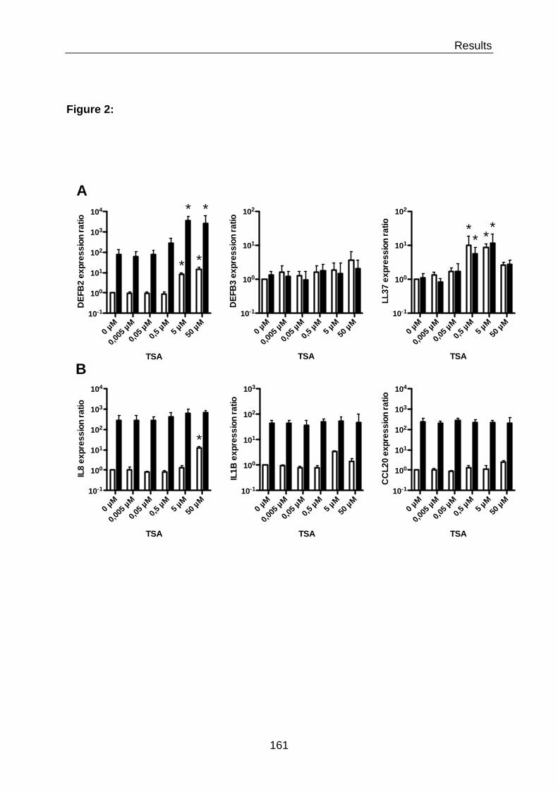
Results
161
Figure 2:
0 µM
0,005
µM
0,05 µ
M0,5
µM5 µ
M50
µM10-1
100
101
102
103
104
TSA
DEF
B2
expr
essi
on ra
tio
* *
**
0 µM
0,005
µM
0,05 µ
M0,5
µM5 µ
M50
µM10-1
100
101
102
TSA
DEF
B3
expr
essi
on ra
tio
0 µM
0,005
µM
0,05 µ
M0,5
µM5 µ
M50
µM10-1
100
101
102
TSA
LL37
exp
ress
ion
ratio *
* **
0 µM
0,005
µM
0,05 µ
M0,5
µM5 µ
M50
µM10-1
100
101
102
103
104
TSA
IL8
expr
essi
on ra
tio
*
0 µM
0,005
µM
0,05 µ
M0,5
µM5 µ
M50
µM10-1
100
101
102
103
TSA
IL1B
exp
ress
ion
ratio
0 µM
0,005
µM
0,05 µ
M0,5
µM5 µ
M50
µM10-1
100
101
102
103
104
TSA
CC
L20
expr
essi
on ra
tio
A
B

Results
162
Figure 3:
0 2 4 610-1
100
101
102
103
104
Time (h)
IL8
expr
essi
on ra
tio
0 2 4 610-1
100
101
102
103
104
Time (h)
DEFB
2 ex
pres
sion
ratio
A
NCK12
100
101
102
103
104
105
DEFB
2 pr
otei
n (p
g/m
l)
*
**
B
NCK12
100
101
102
103
104
105
IL8
prot
ein
(pg/
ml)

Results
163
Figure 4:
P-H3S10
Time (h) 0 1 3 6
- TSA + TSA
NC - TSA + TSA
K12
0 1 3 6 0 1 3 6 0 1 3 6
Actin
Erk1/2
P-Erk1/2
p38
P-p38

Results
164
Figure 5:
H3
Ac-H3
Ac-H3K14
Time (h) 0 1 2 3
- TSA + TSA
NC - TSA + TSA
K12
0 1 2 3 0 1 2 3 0 1 2 3
Ac-H3K18
Actin
Ac-H3K4
Ac-H3K23
Ac-H3K9
Ac-H3K27
Ac-H3K56

Results
165
Figure 6:
0 2 4 610-1
100
101
102
103
104
105
Time (h)
DEF
B2
expr
essi
on ra
tio
0 2 4 610-2
10-1
100
101
102
103
Time (h)
IL8
expr
essi
on ra
tio
B
SCp65 SC
p65100
101
102
103
104
105
DEF
B2
expr
essi
on ra
tio
- TSA + TSA
*
*
SCp65 SC
p65100
101
102
103
IL8
expr
essi
on ra
tio
- TSA + TSA
**
A

Results
166
Figure 7:
p65
Ac-p65 (K310)
I!B"
Time (h) 0 1 2 3
- TSA + TSA
NC - TSA + TSA
K12
0 1 2 3 0 1 2 3 0 1 2 3
p105
p50
Actin

Results
167
Figure 8:
p300
Actin
K12
Ac-p65 (K310)
SC p300
A
SCp30
0100
101
102
103
DEF
B2
expr
essi
on ra
tio
*
B
SCp30
0100
101
102
103
IL8
expr
essi
on ra
tio
0 µM
10 µM
25 µM
50 µM
100
101
102
103
C646
IL8
expr
essi
on ra
tio *
0 µM
10 µM
25 µM
50 µM
100
101
102
103
C646
DEF
B2
expr
essi
on ra
tio
*
*
C

Results
168
Figure 9:
A
−! −" # " ! $ % &#−$
−!
−"
#
"
!
$
%
&#
&"
log2(K12 vs. NI)
log2
(K12
, TSA
vs.
NI)
-2 0 2 4 6 8 10
Log2(K12/NI)
-4
-2
0
2
4
6
8
1
0
Log2
(K12
+TSA
/NI)
-1 0 1 2 3 4 5
NODALBMP6BMP3FGF2MSTN
GDF11IL7
GRNTSLP
TGFB2LGALS8S100A7
LCN2PLA2G7
PGLYRP4PGLYRP3PGLYRP2PGLYRP1
DEFB2
Log2 (fold change)-2 0 2 4 6 8 10
XCL2XCL1
CX3CL1CXCL16CXCL5CXCL3CXCL2CCL28CCL20CCL16CCL15CCL14CCL11CCL2IL23AIL17DIL17CIL17BIL12A
IL8IL1B
Log2 (fold change)
Defence & Restitution Inflammation B C

Results
169
Supplementary Figure 1:
0 µM
0,05 µ
M0,5
µM5 µ
M50
µM
500 µ
M0
20
40
60
80
100
SAHA
% C
ytot
oxic
ity
0 µM
0,05 µ
M0,5
µM5 µ
M50
µM
500 µ
M10-1
100
101
102
103
104
SAHA
DEF
B2
expr
essi
on ra
tio **
*
*
*
*
A
0 µM
0,05 µ
M0,5
µM5 µ
M50
µM
500 µ
M10-1
100
101
102
SAHA
DEF
B3
expr
essi
on ra
tio
0 µM
0,05 µ
M0,5
µM5 µ
M50
µM
500 µ
M10-1
100
101
102
SAHA
LL37
exp
ress
ion
ratio
* ** *
0 µM
0,05 µ
M0,5
µM5 µ
M50
µM
500 µ
M10-1
100
101
102
103
104
SAHA
IL8
expr
essi
on ra
tio
*
0 µM
0,05 µ
M0,5
µM5 µ
M50
µM
500 µ
M10-1
100
101
102
103
SAHA
IL1B
exp
ress
ion
ratio
0 µM
0,05 µ
M0,5
µM5 µ
M50
µM
500 µ
M10-1
100
101
102
103
104
SAHA
CC
L20
expr
essi
on ra
tio*
C
NCK12
100
101
102
103
104
105
DEF
B2
prot
ein
(pg/
ml)
*
*
B
NCK12
100
101
102
103
104
105
IL8
prot
ein
(pg/
ml)

Results
170
Supplementary Figure 2:
H4
Ac-H4
Ac-H4K8
Time (h) 0 1 2 3
- TSA + TSA
NC - TSA + TSA
K12
0 1 2 3 0 1 2 3 0 1 2 3
Actin
Ac-H4K5
Ac-H4K12
Ac-H4K16

Results
171
References
1. Nightingale, K. P., L. P. O'Neill, and B. M. Turner. 2006. Histone modifications:
signalling receptors and potential elements of a heritable epigenetic code. Curr. Opin.
Genet. Dev. 16: 125–136.
2. Kouzarides, T. 2007. SnapShot: Histone-modifying enzymes. Cell 128: 802.
3. Arbibe, L., D. W. Kim, E. Batsche, T. Pedron, B. Mateescu, C. Muchardt, C.
Parsot, and P. J. Sansonetti. 2007. An injected bacterial effector targets chromatin
access for transcription factor NF-kappaB to alter transcription of host genes involved
in immune responses. Nat. Immunol. 8: 47–56.
4. Eskandarian, H. A., F. Impens, M.-A. Nahori, G. Soubigou, J.-Y. Coppée, P.
Cossart, and M. A. Hamon. 2013. A role for SIRT2-dependent histone H3K18
deacetylation in bacterial infection. Science 341: 1238858.
5. Lehrmann, H., L. L. Pritchard, and A. Harel-Bellan. 2002. Histone
acetyltransferases and deacetylases in the control of cell proliferation and
differentiation. Adv. Cancer Res. 86: 41–65.
6. Marks, P. A., and M. Dokmanovic. 2005. Histone deacetylase inhibitors: discovery
and development as anticancer agents. Expert Opin Investig Drugs 14: 1497–1511.
7. Codd, R., N. Braich, J. Liu, C. Z. Soe, and A. A. H. Pakchung. 2009. Zn(II)-
dependent histone deacetylase inhibitors: suberoylanilide hydroxamic acid and
trichostatin A. Int. J. Biochem. Cell Biol. 41: 736–739.
8. Minucci, S., and P. G. Pelicci. 2006. Histone deacetylase inhibitors and the
promise of epigenetic (and more) treatments for cancer. Nat. Rev. Cancer 6: 38–51.
9. Baldwin, A. S. 1996. The NF-kappa B and I kappa B proteins: new discoveries and
insights. Annu. Rev. Immunol. 14: 649–683.
10. Kouzarides, T. 2000. Acetylation: a regulatory modification to rival
phosphorylation? EMBO J. 19: 1176–1179.
11. Ghizzoni, M., H. J. Haisma, H. Maarsingh, and F. J. Dekker. 2011. Histone
acetyltransferases are crucial regulators in NF-κB mediated inflammation. Drug
Discov. Today 16: 504–511.

Results
172
12. Huang, B., X.-D. Yang, A. Lamb, and L.-F. Chen. 2010. Posttranslational
modifications of NF-kappaB: another layer of regulation for NF-kappaB signaling
pathway. Cell. Signal. 22: 1282–1290.
13. Kiernan, R., V. Brès, R. W. M. Ng, M.-P. Coudart, S. El Messaoudi, C. Sardet, D.-
Y. Jin, S. Emiliani, and M. Benkirane. 2003. Post-activation turn-off of NF-kappa B-
dependent transcription is regulated by acetylation of p65. J. Biol. Chem. 278: 2758–
2766.
14. Chen, L.-F., Y. Mu, and W. C. Greene. 2002. Acetylation of RelA at discrete sites
regulates distinct nuclear functions of NF-kappaB. EMBO J. 21: 6539–6548.
15. Weake, V. M., and J. L. Workman. 2010. Inducible gene expression: diverse
regulatory mechanisms. Nat. Rev. Genet. 11: 426–437.
16. Chantret, I., A. Rodolosse, A. Barbat, E. Dussaulx, E. Brot-Laroche, A.
Zweibaum, and M. Rousset. 1994. Differential expression of sucrase-isomaltase in
clones isolated from early and late passages of the cell line Caco-2: evidence for
glucose-dependent negative regulation. J. Cell. Sci. 107 ( Pt 1): 213–225.
17. Schauber, J., C. Svanholm, S. Termén, K. Iffland, T. Menzel, W. Scheppach, R.
Melcher, B. Agerberth, H. Lührs, and G. H. Gudmundsson. 2003. Expression of the
cathelicidin LL-37 is modulated by short chain fatty acids in colonocytes: relevance of
signalling pathways. Gut 52: 735–741.
18. Saccani, S., S. Pantano, and G. Natoli. 2002. p38-Dependent marking of
inflammatory genes for increased NF-kappa B recruitment. Nat. Immunol. 3: 69–75.
19. Wehkamp, J., J. Harder, K. Wehkamp, B. Wehkamp-von Meissner, M. Schlee, C.
Enders, U. Sonnenborn, S. Nuding, S. Bengmark, K. Fellermann, J. M. Schröder,
and E. F. Stange. 2004. NF-kappaB- and AP-1-mediated induction of human beta
defensin-2 in intestinal epithelial cells by Escherichia coli Nissle 1917: a novel effect
of a probiotic bacterium. Infect. Immun. 72: 5750–5758.
20. Hoffmann, E., O. Dittrich-Breiholz, H. Holtmann, and M. Kracht. 2002. Multiple
control of interleukin-8 gene expression. J. Leukoc. Biol. 72: 847–855.
21. Burke, J. R., M. A. Pattoli, K. R. Gregor, P. J. Brassil, J. F. MacMaster, K. W.
McIntyre, X. Yang, V. S. Iotzova, W. Clarke, J. Strnad, Y. Qiu, and F. C. Zusi. 2003.
BMS-345541 is a highly selective inhibitor of I kappa B kinase that binds at an
Results
173
allosteric site of the enzyme and blocks NF-kappa B-dependent transcription in mice.
J. Biol. Chem. 278: 1450–1456.
22. Yang, X.-D., E. Tajkhorshid, and L.-F. Chen. 2010. Functional interplay between
acetylation and methylation of the RelA subunit of NF-kappaB. Mol. Cell. Biol. 30:
2170–2180.
23. Bowers, E. M., G. Yan, C. Mukherjee, A. Orry, L. Wang, M. A. Holbert, N. T.
Crump, C. A. Hazzalin, G. Liszczak, H. Yuan, C. Larocca, S. A. Saldanha, R.
Abagyan, Y. Sun, D. J. Meyers, R. Marmorstein, L. C. Mahadevan, R. M. Alani, and
P. A. Cole. 2010. Virtual ligand screening of the p300/CBP histone acetyltransferase:
identification of a selective small molecule inhibitor. Chem. Biol. 17: 471–482.
24. Salzman, N. H., M. A. Underwood, and C. L. Bevins. 2007. Paneth cells,
defensins, and the commensal microbiota: a hypothesis on intimate interplay at the
intestinal mucosa. Semin. Immunol. 19: 70–83.
25. Rescigno, M., and P. Borrow. 2001. The host-pathogen interaction: new themes
from dendritic cell biology. Cell 106: 267–270.
26. Tato, C. M., and C. A. Hunter. 2002. Host-pathogen interactions: subversion and
utilization of the NF-kappa B pathway during infection. Infect. Immun. 70: 3311–3317.
27. Huang, Q., D. Liu, P. Majewski, L. C. Schulte, J. M. Korn, R. A. Young, E. S.
Lander, and N. Hacohen. 2001. The plasticity of dendritic cell responses to
pathogens and their components. Science 294: 870–875.
28. Foster, S. L., D. C. Hargreaves, and R. Medzhitov. 2007. Gene-specific control of
inflammation by TLR-induced chromatin modifications. Nature 447: 972–978.
29. Clayton, A. L., and L. C. Mahadevan. 2003. MAP kinase-mediated
phosphoacetylation of histone H3 and inducible gene regulation. FEBS Lett. 546: 51–
58.
30. Yamamoto, Y., U. N. Verma, S. Prajapati, Y.-T. Kwak, and R. B. Gaynor. 2003.
Histone H3 phosphorylation by IKK-alpha is critical for cytokine-induced gene
expression. Nature 423: 655–659.
31. Anest, V., J. L. Hanson, P. C. Cogswell, K. A. Steinbrecher, B. D. Strahl, and A.
S. Baldwin. 2003. A nucleosomal function for IkappaB kinase-alpha in NF-kappaB-
dependent gene expression. Nature 423: 659–663.

Results
174
32. Cheung, P., C. D. Allis, and P. Sassone-Corsi. 2000. Signaling to chromatin
through histone modifications. Cell 103: 263–271.
33. Duan, J., J. Friedman, L. Nottingham, Z. Chen, G. Ara, and C. Van Waes. 2007.
Nuclear factor-kappaB p65 small interfering RNA or proteasome inhibitor bortezomib
sensitizes head and neck squamous cell carcinomas to classic histone deacetylase
inhibitors and novel histone deacetylase inhibitor PXD101. Mol. Cancer Ther. 6: 37–
50.
34. Katsura, T., S. Iwai, Y. Ota, H. Shimizu, K. Ikuta, and Y. Yura. 2009. The effects
of trichostatin A on the oncolytic ability of herpes simplex virus for oral squamous cell
carcinoma cells. Cancer Gene Ther. 16: 237–245.
35. Gerritsen, M. E., A. J. Williams, A. S. Neish, S. Moore, Y. Shi, and T. Collins.
1997. CREB-binding protein/p300 are transcriptional coactivators of p65. Proc. Natl.
Acad. Sci. U.S.A. 94: 2927–2932.
36. Sheppard, K. A., D. W. Rose, Z. K. Haque, R. Kurokawa, E. McInerney, S.
Westin, D. Thanos, M. G. Rosenfeld, C. K. Glass, and T. Collins. 1999.
Transcriptional activation by NF-kappaB requires multiple coactivators. Mol. Cell.
Biol. 19: 6367–6378.
37. Bolden, J. E., M. J. Peart, and R. W. Johnstone. 2006. Anticancer activities of
histone deacetylase inhibitors. Nat Rev Drug Discov 5: 769–784.
38. Ververis, K., and T. C. Karagiannis. 2011. Potential non-oncological applications
of histone deacetylase inhibitors. Am J Transl Res 3: 454–467.
39. Raqib, R., P. Sarker, P. Bergman, G. Ara, M. Lindh, D. A. Sack, K. M. Nasirul
Islam, G. H. Gudmundsson, J. Andersson, and B. Agerberth. 2006. Improved
outcome in shigellosis associated with butyrate induction of an endogenous peptide
antibiotic. Proc. Natl. Acad. Sci. U.S.A. 103: 9178–9183.

Results
175

Additional Results
176
V.2 Additional Results This chapter is divided into two parts: The first part concerns results obtained with the
E. coli strain K12, in addition to the results presented in the manuscript in preparation
(V.2.1). The second part contains results obtained with an additional bacterial strain,
E. coli LF82 (V.2.2). The additional results section will be followed by a short section
of additional methods (V.3)
V.2.1 Additional results for challenge with the E. coli strain K12
The previously presented manuscript in preparation contains the most striking results
concerning the expression and regulation of the DEFB2 and IL8 genes upon
challenge of intestinal epithelial cells with the E. coli strain K12. In this section those
results are completed with observations concerning the knockdown of HDAC
(V.2.1.1) and HAT (V.2.1.2) enzymes prior to bacterial challenge.
V.2.1.1 Knockdown of histone deacetylases differentially influences the expression of DEFB2 and IL8 genes upon E. coli K12 challenge As we observed that the inhibition of the classical histone deacetylases by the use of
the pan-inhibitor TSA resulted in the strong increase of expression of AMP genes
upon bacterial challenge, we wanted to investigate the involvement of single HDAC
enzymes or HDAC classes in this process. Therefore, intestinal epithelial cells were
transfected with siRNAs targeting either single HDAC enzymes, a combination to
target a single class or a combination of several classes. After knockdown for 48h,
cells were challenged with the E. coli strain K12 for 2h and the expression of the
DEFB2 and IL8 genes was evaluated using qRT-PCR. Interestingly, we did not
observe an increased DEFB2 expression upon knockdown of a single HDAC
enzyme, not a single HDAC class (Figure A1 A, upper panel), nor knockdown of
combination of classes, or all classical HDAC enzymes together (Figure A1 B, upper
panel). Moreover, knockdown of some HDAC enzymes significantly decrease the
expression of DEFB2 upon E. coli challenge, with the exception of single knockdown
of HDAC1 and most of HDACs of class IIa. On the contrary, knockdown of most
single HDAC enzymes significantly increased the expression of IL8 upon E. coli

Additional Results
177
SC
HDAC1
HDAC2
HDAC3
HDAC8
HDAC4
HDAC5
HDAC7
HDAC9
HDAC6
HDAC100
200
400
600
DEF
B2
expr
essi
on ra
tio
Class I Class IIbClass IIa
** * * * *
A
SC
HDAC1
HDAC2
HDAC3
HDAC8
HDAC4
HDAC5
HDAC7
HDAC9
HDAC6
HDAC100
2000
4000
6000
IL8
expr
essi
on ra
tio
Class I Class IIbClass IIa
*
* * * * * *
SC
Class I
Class I
Ia
Class I
Ib
Class I
+ IIa
Class I
+ IIb
Class I
Ia + I
Ib All0
100
200
300
400
500
DEF
B2
expr
essi
on ra
tio
* * * **
*
B
SC
Class I
Class I
Ia
Class I
Ib
Class I
+ IIa
Class I
+ IIb
Class I
Ia + I
Ib All0
1000
2000
3000
IL8
expr
essi
on ra
tio
*
Figure A1: Influence of HDAC enzyme knockdown on the expression of DEFB2 and IL8 upon challenge with E. coli K12. Transcriptional expression of the DEFB2 and IL8 genes in cells transfected with siRNA directed against single histone deacetylases (A) or in a combination to target groups of histone deacetylases (B), and challenged for 3 hours with the E. coli K12 strain. After mRNA extraction and RT reactions, qRT-PCR was performed on each sample with specific primers to determine the relative expression of genes using the comparative Ct method. Values are presented on a logarithmic scale as the ratio of gene expression in challenged cells compared to non-challenged cells. Experiments were performed at MOI of 10 bacteria per cell. N = 3 independent experiments. Error bars represent the SD. *, P < 0,01 for cells transfected with HDAC targeting siRNA, compared to cells transfected with scramble siRNA. Black bars: HDAC class I siRNA transfected cells; dark grey bars: HDAC class II siRNA transfected cells; light grey bars: HDAC class III siRNA transfected cells; white bars: scramble siRNA transfected cells (SC).

Additional Results
178
challenge, especially HDAC1 (Figure A1 A, lower panel). Interestingly, this effect is
lost in the knockdown of HDAC classes, except class I, or combination of HDAC
classes (Figure A1 B, lower panel). At this point we can conclude that the effect of
increased expression of AMPs upon histone deacetylase inhibitor treatment is not
mediated by a single HDAC and cannot be obtained upon knockdown of HDAC
enzymes. As these enzymes play such versatile roles in the cell and act together not
only with each other, but also with other proteins within protein complexes, it can be
imagined the difficulty of deciphering their explicit role in gene expression. This will
be discussed in more detail in chapter VI.

Additional Results
179
Figure A2: Influence of HAT enzyme knockdown on the expression of DEFB2 and IL8 upon challenge with E. coli K12. Transcriptional expression of the DEFB2 (A) and IL8 (B) genes in cells transfected with siRNA directed against single histone acetyltransferases, and challenged for 3 hours with the E. coli K12 strain. After mRNA extraction and RT reactions, qRT-PCR was performed on each sample with specific primers to determine the relative expression of genes using the comparative Ct method. Values are presented on a logarithmic scale as the ratio of gene expression in challenged cells compared to non-challenged cells. Experiments were performed at MOI of 10 bacteria per cell. N = 3 independent experiments. Error bars represent the SD. *, P < 0,01 for cells transfected with HAT targeting siRNA, compared to cells transfected with scramble siRNA. Black bars: HAT siRNA transfected cells; white bars: scramble siRNA transfected cells (SC).
SCp30
0CBP
PCAF0
100
200
300
400
500
600
SCp30
0CBP
PCAF0
500
1000
1500
2000
DE
FB2
expr
essi
on ra
tio
IL8
expr
essi
on ra
tio
*
* *
A B

Additional Results
180
V.2.1.2 Differential HATs are involved in the expression of DEFB2 and IL8 Since we discovered that acetylation seems to play a big role in the expression of
AMPs, and we investigated already the role of HDAC enzymes, we wanted to know
more about the involvement of different HAT enzymes. Therefore, we performed
siRNA transfection experiments of intestinal epithelial cells to knockdown HAT
enzymes belonging to different families, prior to challenge with E. coli strains. As
already described in the presented manuscript, the knockdown of p300 had a
significant effect on the bacteria-stimulated DEFB2 expression and reduced it
significantly (Figure A2 A), while having no effect on the expression of IL8 (Figure A2
B). Knockdown of other HATs, such as CBP and PCAF, did not influence the induced
DEFB2 expression (Figure A2 A). On the contrary, we observed a significant
increase in bacteria-induced IL8 expression upon knockdown of CBP and PCAF
(Figure A2 B). These results are interesting, as they present the involvement of
different HAT enzymes in the expression of different gene, and moreover of two
HATs which share such a high sequence homology as p300 and CBP. Furthermore,
the data show a differential effect of different HATs in the stimulation or repression of
expression of these genes, as knockdown can either lead to increased or decreased
inducibility.

Additional Results
181
0 2 4 610-1
100
101
102
103
104
Time (h)
DEF
B2
expr
essi
on ra
tio
0 2 4 610-1
100
101
102
Time (h)
DEF
B3
expr
essi
on ra
tio
0 2 4 610-1
100
101
Time (h)
LL37
exp
ress
ion
ratio
0 2 4 610-1
100
101
102
103
104
Time (h)
IL8
expr
essi
on ra
tio
0 2 4 610-1
100
101
102
103
Time (h)
IL1B
exp
ress
ion
ratio
0 2 4 610-1
100
101
102
103
104
Time (h)
CC
L20
expr
essi
on ra
tio
A
B
Figure A3: Expression of antimicrobial and pro-inflammatory genes upon challenge with E. coli LF82. Transcriptional expression of genes encoding the antimicrobial peptide beta-defensin DEFB2 and DEFB3, the cathelicidin LL37 (A), and the pro-inflammatory mediators interleukins IL1B and IL8, and the chemokine CCL20 (B), in cells challenged with the Escherichia coli LF82 strain. After mRNA extraction and RT reactions, qRT-PCR was performed on each sample, for each time point, with specific primers to determine the relative expression of genes using the comparative Ct method. Values are presented on a logarithmic scale as the ratio of gene expression in challenged cells compared to non-challenged cells. Experiments were performed at a MOI of 10 bacteria per cell. N = 3 independent experiments. Error bars represent the SD.

Additional Results
182
V.2.2 Additional results for challenge with the E. coli strain LF82 In the beginning of the thesis project we choose to work with two E. coli strains, in
order to compare a commensal strain and a possible pathogenic strain. All
experiments presented with the E. coli strain K12 were additionally performed in
parallel with the E. coli strain LF82. This strain was isolated from a Crohn’s disease
patient 692 and is considered a pathobiontic strain, as it can adhere to, and invade
cells and induces a strong TNFα response in macrophages 693. We were surprised to
not observe major differences in the expression or regulation of AMPs and INFs, with
or without pre-treatment of epigenetic inhibitors, upon challenge of intestinal epithelial
cells with these two strains throughout all presented experiments. Other results
obtained in this comparative study were similar and are therefore not shown.The few
differences we observed are presented in this section for challenge with the LF82
strain.
V.2.2.1 Challenge with E. coli stain LF82 shows differential kinetics of AMPs and INFs expression as compared to E. coli strain K12 For some of the examined AMPs and INFs genes we could observe differences in
the kinetics and magnitude of expression after intestinal epithelial cell challenge with
the E. coli strain LF82 as compared to K12. For the expression of DEFB2 we observe
a slightly slower kinetic of expression within the first hour after challenge with LF82
as compared to K12, which then increases 2 log fold until 2h post challenge, to the
same level as observed with K12 (Figure A3 A, left panel). The induction of DEFB3
expression, on the other hand, is significantly higher upon challenge with LF82 as
compared to K12, especially after 3h post challenge (Figure A3 A, middle panel). For
the expression of INFs genes, we observe a slower kinetic of induction for all three
examined genes, within the first hour post challenge with LF82, as compared to K12
(Figure A3 B). Then, the level of INFs gene induction rises quickly to the same level
as compared to K12 at 2h post challenge, and seems to even slightly exceed the
level achieved with K12 challenge at 6h post challenge. These observations,
concerning the slower kinetics of DEFB2, IL8, IL1β and CCL20 gene expression with
LF82, especially within the first hour of challenge, could indicate mechanism applied
by this pathobiontic strain, which allow for a restrain of the host response.

Additional Results
183
Figures A4: Acetylation of the NF-"B p65 subunit on serine residue K310 upon inhibition of histone deacetylases. Immunoblot analysis of the p65, p50, and its precursor p105 subunit proteins of the NF-!B transcription factor complex, the I!B" sequestration protein, and the acetylation mark of the p65 subunit on serine residue K310 (Ac-p65), in cells treated for 16 hours with 5 µM TSA, and challenged with the E. coli LF82 strain. After cell lysis at the indicated time points, western-blots were performed using specific antibodies directed against proteins or post-translational modification marks. Experiments were performed at a MOI of 10 bacteria per cell. N = 2 independent experiments. NC: non-challenged cells ; LF82: cells challenged with the E. coli LF82 strain. “Ac” prefix: acetylation.
p65
Ac-p65 (K310)
I!B"
Time (h) 0 1 2 3
- TSA + TSA
NI
0 1 2 3
p105
p50
Actin
- TSA + TSA
LF82
0 1 2 3 0 1 2 3

Additional Results
184
V.2.2.2 LF82 shows prolonged activation of the NF-κB pathway Besides the differences in kinetics and level of AMPs and INFs genes expression
between the two E. coli strains, we observed a difference in the activation of NF-κB.
The Western blot analysis of proteins participating in the NF-κB pathway after
treatment of intestinal epithelial cells with histone deacetylate inhibitor and challenge
with E. coli strains revealed some differences between challenge with the K12 and
the LF82 strain. As already discussed the treatment with TSA decreased the protein
level of the NF-κB inhibitor IκBα and the subunit p50/p105. Interestingly, the timing of
re-synthesis of this proteins differed between K12 and LF82. After challenge with K12
the protein level of IκBα was observed back at basal level after 2h, while after
challenge with LF82 it was only observed back at basal level after 3h post challenge
(Figure A4). Additionally, challenge with LF82 after TSA pre-treatment, but not K12,
lead to a continuing decreased level of the p50 protein and its precursor p105, which
recovered after 3h post challenge. These observations indicate that the pathobiontic
strain induces a longer activation of the NF-κB pathway and additionally influences
NF-κB subunits differently as compared to the commensal K12. This could explain
the higher and prolonged expression of INFs genes at later time points post
challenge, as compared to K12 (Figure A3 B).

Additional Material and Methods
185
V.3 Additional Material and Methods
V.3.1 Bacterial strains The Escherichia coli LF82 bacterial strain 694 was grown in LB medium (Sigma) at
37°C. For challenge experiments, cells were grown at confluence in 6-well plates (1.5
× 106 cells/well) for 48 h at 37°C and 10% CO2. Bacterial challenges were performed
using supplemented DME without antibiotics directly with over night bacterial
cultures, at a multiplicity of infection (MOI) of 10 bacteria per cell, for indicated times.
For experiments involving pharmacological inhibitors, cells were pretreated for 16 h
over-night and washed with supplemented DME without antibiotics before infection.
V.3.2 siRNAs used for the knockdown of HDAC enzymes The RNAi Max reagent (Invitrogen) was used to transfect cells with a final
concentration of 25 nM siGENOME SMARTpool siRNAs (Thermo Scientific) targeting
HDAC 1 (M-003493-02-0020), HDAC 2 (M-003495-02-0005), HDAC 3 (M-003496-
02-0005), HDAC 4 (M-003497-03-0005), HDAC 5 (M-003498-02-0005), HDAC 6 (M-
003499-00-0005), HDAC 7 (M-009330-02-0005), HDAC 8 (M-003500-02-0005),
HDAC 9 (M-005241-03-0005), HDAC 10 (M-004072-00-0005), p300 (M-003486-04-
0005), CBP (M-003477-02-0005), PCAF (M-005055-00-0005) or control scramble
siRNAs (D-001210-01-50). Transfections were performed in OptiMEM medium
(Invitrogen) supplemented with 1% NEAA and 5% FBS. Knockdowns were assessed
after 48 h using qRT-PCR and immunobloting analysis.

186
Discussion

Discussion
187

Discussion
188
Chapter VI – Discussion
This chapter aims to discuss the presented results for the (epi)genetic regulation of
DEFB2 expression in the context of regulatory mechanism of inducible inflammatory
gene expression. In more detail the role of the transcription factor NF-κB, the histone
acetyltransferase p300 as well as the specific histone marks H3S10-P in association
with MAPK pathways, H3K9-Ac, H3K27-Ac and H3K56-Ac will be discussed.
Following the discussion there will be a short conclusion on hypothesis, followed by a
brief perspective on the experimental level.
VI.1 Regulation of inducible inflammatory gene expression on the chromatin level
The inducible response to an inflammatory stimulus is complex and results in the
orchestrated activation of many target genes with various functions, such as to alert
the body to infection, fight invading pathogens, and repair damaged tissues. The
outcomes of activation of those different genes can have opposing influence on the
host physiology and homeostasis and therefore they have different regulatory
requirements. Especially potentially harmful pro-inflammatory cytokines, which can
have deleterious effects on the tissue when expressed at high levels over a long time
period, have to be differentially regulated than antimicrobial peptides and tissue
repair factors. Control of gene expression via the same signaling pathways, such as
TLRs, NF-κB and MAPK, does not allow for differentiation in target gene expression
and fine tuning of the defense response. Therefore another program of regulatory
mechanism is needed to allow a single gene to be expressed or silenced according
to its function, irrespective of other genes induced by the same signaling pathway.
Evidence is increasing that epigenetic modifications mediate this level of regulation at
individual promoters.
VI.1.1 Presetting the chromatin environment of NF-κB target genes The transcription factor NF-κB is one of the key regulators of the inflammatory and
immune response. The κB-binding site is estimated to be found 1.4x10⁴ times in the
human genome (extrapolated from study by Martone and colleagues 695) and these

Discussion
189
Figure 46: Requirements for chromatin remodeling on primary and secondary response genes. The figure depicts the selective requirement for SWI/SNF complexes at secondary response and late primary response genes, with Mi-2/NuRD negatively influencing these same sets of genes. Early primary response genes do not appear to be regulated by either SWI/SNF or Mi-2/NuRD complexes 581.

Discussion
190
sites regulate two to three hundred NF-κB targeted genes. The number of p65
molecules entering the nucleus after a stimulus is estimated to be 1.5x10⁵ 696. With
this abundance it could be expected that NF-κB quickly finds its way to its binding
sites and activated gene transcription. Yet this is not the case, and the kinetics of
recruited p65 to target genes differs widely, between 10 minutes up to 2 hours 584.
This highlights the importance of additional pathways and coregulators to fine-tune
NF-κB target gene expression. Nuclear presence is not enough and the targeted
recruitment of p65 to a promoter integrates multiple inputs from simultaneously
activated signaling pathways.
Two studies by Ramirez-Carrozzi and colleagues and Saccani and colleagues
have shown that TLR-signaling induces recruitment of NF-κB and expression
inflammatory genes in two waves, the primary response genes, which are further
divided into early (eg. TNFα, MIP2) and late genes (eg. RANTES, IFNβ), and
secondary response genes (eg. IL12b, IL6, Nos2) 581,584 (Figure 46). Moreover,
several studies could show that there seems to be a presetting on the chromatin level
for primary inducible genes. Ramirez-Carrozzi and colleagues could show that the
expression of early primary response genes does not require chromatin remodeling
by the SWI/SNF complex, in contrast to the expression of late primary response
genes and secondary response genes 581 (Figure 46). The basal chromatin
environment of primary induced genes is characterized by high levels of histone
acetylation as well as occurrence of specific activating marks, such as H3S10-P,
H3K9-Ac, H3K14-Ac and H3K4-me3, and association with promoter-proximal paused
Pol II 579,607,697. Overall the histone acetylation level is correlated with gene
expression and several studies have described specific acetylation patterns at NF-κB
target gene promoters in different cell lines upon inflammatory stimuli, with the main
mediating HATs being p300, CBP and PCAF 698. Interestingly, Saccani and
colleagues found that different stimuli such as LPS vs TNFα, don’t lead to
hyperacetylation and activation of the same NF-κB target genes, highlighting the fact
that there is differential chromatin modifying regulation probably mediated by different
enzymes and cofactors.
Nothing has been described so far about the occurrence of these marks on the
DEFB2 promoter and ChIP studies will give more insights into the chromatin state
before and after stimulation. We can assume that the treatment with HDAC inhibitors

Discussion
191
require a change in chromatin structure to be activated [32].These studies imply nucleosome structure is a crucialcontroller of the kinetics of NF!B-regulated genes. Inaddition, switching of NF!B dimers can further regulateappropriate timing of inflammatory gene expression indendritic cells [33]. Rapidly activated p50/p65 dimers aregradually replaced over the period of hours by slowlyactivated p52/RelB dimers. While these slowly activateddimers maintain expression of some chemokine genes, theyinhibit expression of IL-12p40, a cytokine important inregulating adaptive immunity. Lomvardas et al. found thata nucleosome positioned over the start site of the IFN"promoter imposes stringent requirements for activation onIFN", so that this gene is only activated in the correctcontext, for example in response to viral infection, and not inresponse to TNF# [34]. In contrast, some IFN# target genesare constitutively remodeled so that they can be quicklyinduced [35]. Thus, gene-specific control mechanisms ensurethat TLR-induced genes are expressed with correct timingand in response to the correct stimulus.
Histone modifications also regulate inflammatory genes.A spatio-temporal pattern of histone modifications at theIFN" promoter recruits transcription factors and the remo-deling enzyme Brg1 [36]. Saccani and others have shown thatinduction of some groups of early primary response inflam-matory genes by TLR signaling are marked by phosphoryla-tion of histone 3 at serine 10 (H3S10), methylation at H3R17,methylation at H3K4, and acetylation at H3K9/H3K14 [37–
39]. Silencing of subsets of these genes, on the other hand, isassociated with methylation of H3K9 [40]. At secondaryresponse genes, the pattern of histone modifications maydepend on recruitment of histone modifying enzymes byprimary response gene products [39]. Finally, Brogdon et al.asked if histone modifications have functional consequencesfor the immune response by treating dendritic cells andmacrophages with the histone deacetylase inhibitor LAQ824[41]. This treatment led to multiple gene-specific effects,including a decrease in transcription of Th1-inducingcytokines and an increase in Th2-inducing cytokines, imply-ing that histone deacetylation may play opposing roles atthese two groups of genes.
The above data suggest that distinct sets of genesdownstream of TLR4 can be regulated at the level ofindividual promoters by modifications to chromatin. Canchromatin modifications allow selective control of differentfunctional modules of the TLR-induced inflammatoryresponse? LPS tolerance is an ideal system for addressingthis question, as we know that the proinflammatory cytokinemodule becomes silenced in tolerant cells, presumably toprevent tissue damage. Indeed, McCall's group has found thatsilencing of IL-1" and TNF# in an LPS-tolerant macrophage-like cell line is due to chromatin modifications at thepromoters of these genes [42,43]. Therefore, we can use thephenomenon of tolerance to look at what happens to otherfunctional groups of genes, specifically antimicrobial effec-tors. Although proinflammatory cytokines are silenced,
Figure 3 LPS tolerance is an example of gene-specific regulation of the inflammatory response. Following LPS stimulation of naivemacrophages, both class Tand class NT promoters recruit transcription factors and display increased histone acetylation, histone H3K4trimethylation, and accessibility (represented by yellow starburst). In tolerant macrophages stimulated with LPS, class T promotersare deacetylated and inaccessible. Class NT promoters, on the other hand, are inducibly reacetylated, remethylated, and madeaccessible with faster kinetics.
12 S.L. Foster, R. Medzhitov
Figure 47: Induction of LPS tolerance in macrophages by chromatin modification. Following LPS stimulation of naive macrophages, both tolerizable (class T) and non-tolerizable (class NT) promoters recruit transcription factors and display increased histone acetylation, histone H3K4 trimethylation, and accessibility. In tolerant macrophages, class T promoters are deacetylated and inaccessible. Class NT promoters, on the other hand, are inducibly reacetylated, remethylated, and made accessible with faster kinetics upon a second stimulation with LPS 701.

Discussion
192
transforms the DEFB2 promotor into an early accessible state, which enhances the
NF-κB-dependent inducibility of its expression upon stimulation. Especially the
kinetics and magnitude of transcription of pre-mRNA at the promoter is significantly
enhanced within the first two hours, as shown by qPCR analysis. In fact Saccani and
colleagues mention that these sort of promoters are able to recruit p65 without any
stimulus, which could explain the increased basal expression in non-stimulated TSA
treated cells 584. Why this does not happen at the IL8 promotor is still unanswered. It
can be envisioned that there is a differential protective mechanism, which inhibits
such scenarios for IL8, as a boost of its expression beyond the normal level could
have detrimental effects.
Interestingly, the occurrence of high levels of H3-Ac and H3K4-me3 correlated
with high presence of CpG islands in those gene promoters 607, indicating a pre-
disposing mechanism inscribed in the promoter sequence of primary response
genes. Moreover, it was shown that CpG rich sequences have a lower affinity for
nucleosome occupancy, thereby allowing for faster recruitment of the general
transcription machinery and cofactors without the need for chromatin remodeling 607.
A simple analysis of the DEFB2 sequence shows that there is not a high abundance
of CpG in the promoter region of this gene. The nucleosome density could be
assessed by DNAse I hypersensitivity assays.
Moreover, the classification by chromatin environment is dependent on the cell
type. IL6 was shown to be a remodeling dependent secondary response gene in
murine macrophages, while its promoter was highly accessible in unstimulated
murine embryonic fibroblasts and did not change upon stimulation. This suggests
that the requirements for gene expression can be adjusted on the chromatin level
according to the need in a specific cell type. Therefore it should be considered that
the epigenetic regulation of the DEFB2 gene might be different in epithelial cells and
immune cells, according to their different localization and roles in immune
surveillance and defense.
Furthermore, also negative regulation of gene expression has been attributed
to chromatin alterations. For example, the induction of LPS-tolerance in
macrophages, and thereby the shut down of a subset of pro-inflammatory genes
towards a second stimulation by LPS, was shown to be regulated via chromatin
modifications. The major features of tolerized inflammatory genes was the loss of H4

Discussion
193
142 Cell 138, 129–145, July 10, 2009 ª2009 Elsevier Inc.
Figure 48: Model of LPS-induced primary response gene transcription. At the basal level Sp1 recruits Pol II and induced the generation of unspliced unstable transcripts. Upon LPS stimulation NF-!B and GCN5 are recruited, and acetylation of H4K5/K8/K12 is established. Those marks bind the cofactor Brd4, which in turn recruits P-TEFb to mediate the secondary phosphorylation of paused Pol II and thereby initiates productive transcription of target genes. Squares indicate acetylated residues, circles indicate methylated residues, and stars indicate phosphorylated residues 579.

Discussion
194
acetylation and H3K4-me3, accompanied by a block of nucleosomal remodeling and
loss of recruitment of nuclear p65 699,700 (Figure 47). On the other hand, a group of
non-tolerized antimicrobial genes maintained active chromatin structures and were
still inducible upon a second stimulus, highlighting the separation of gene subsets
simply be chromatin features 699. Moreover, products of the first LPS-stimulation were
shown to be responsible for the chromatin modifications, which on one hand shut
down tolerized genes and on the other hand primed non-tolerized genes to even be
induced with increased kinetics and magnitude. In addition, these data highlight the
possibility to separate the expression of two sets of genes with different functions on
the epigenetic level. The described boost in gene expression by chromatin alterations
can be compared to our observation for the increase in kinetics and magnitude of
DEFB2 gene expression upon TSA pre-treatment. We can speculate that the same
kind of chromatin priming occurs in our case.
VI.1.2 The role of promoter-proximal paused Pol II As mentioned above, many primary response genes are characterized by a basal
level of paused Pol II at their promoter regions 579. The constitutive transcription
factor Sp1 was reported to be required for basal Pol II recruitment and induced the
generation of unspliced unstable transcripts. This was shown to be necessary for the
maintenance of the active chromatin state at primary response genes. Furthermore,
Hargreaves and colleagues found that LPS stimulation of macrophages leads to the
recruitment of HATs GCN5 and PCAF to primary response gene promoters. This
correlates with the establishment of acetylation marks H4K5/K8/K12, which are in
turn able to bind the cofactor Brd4 (Bromodomain-containing protein 4). Brd4 recruits
P-TEFb to mediate the secondary phosphorylation of paused Pol II and thereby
initiates productive transcription of target genes (Figure 48). This is possible to be
related to the recruitment of NF-κB by Brd4, as suggested by Huang and colleagues 702. And indeed inhibition of NF-κB abolished all the events downstream of initiation
by LPS-treatment, emphasizing its role in the generation of functional full length
transcripts 579. The presence of promoter-proximal paused Pol II at the DEFB2
promoter in the basal state could be investigated by ChIP experiments. Furthermore,
we could not observe a role for GNC5 and PCAF in the DEFB2 expression in our
model, but the role of p300 in our setting might be redundant. The presence of
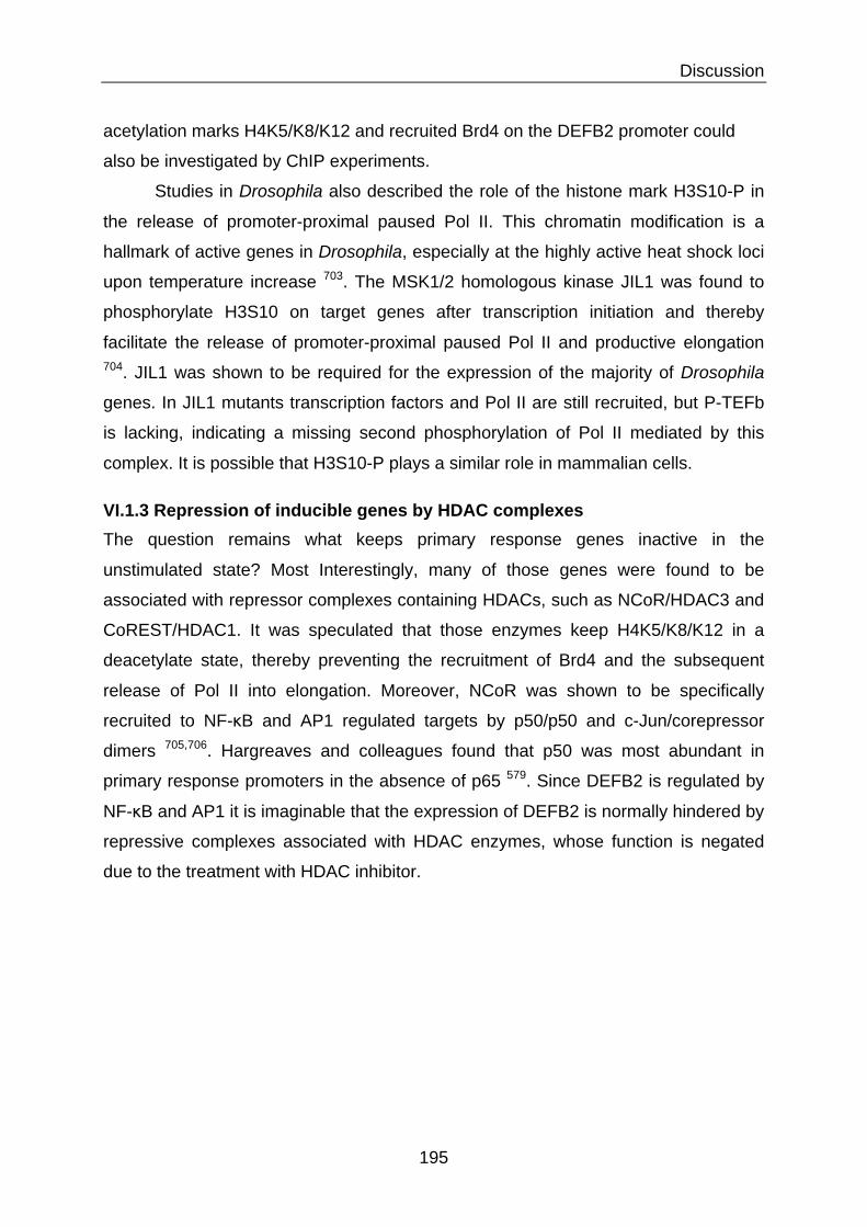
Discussion
195
acetylation marks H4K5/K8/K12 and recruited Brd4 on the DEFB2 promoter could
also be investigated by ChIP experiments.
Studies in Drosophila also described the role of the histone mark H3S10-P in
the release of promoter-proximal paused Pol II. This chromatin modification is a
hallmark of active genes in Drosophila, especially at the highly active heat shock loci
upon temperature increase 703. The MSK1/2 homologous kinase JIL1 was found to
phosphorylate H3S10 on target genes after transcription initiation and thereby
facilitate the release of promoter-proximal paused Pol II and productive elongation 704. JIL1 was shown to be required for the expression of the majority of Drosophila
genes. In JIL1 mutants transcription factors and Pol II are still recruited, but P-TEFb
is lacking, indicating a missing second phosphorylation of Pol II mediated by this
complex. It is possible that H3S10-P plays a similar role in mammalian cells.
VI.1.3 Repression of inducible genes by HDAC complexes The question remains what keeps primary response genes inactive in the
unstimulated state? Most Interestingly, many of those genes were found to be
associated with repressor complexes containing HDACs, such as NCoR/HDAC3 and
CoREST/HDAC1. It was speculated that those enzymes keep H4K5/K8/K12 in a
deacetylate state, thereby preventing the recruitment of Brd4 and the subsequent
release of Pol II into elongation. Moreover, NCoR was shown to be specifically
recruited to NF-κB and AP1 regulated targets by p50/p50 and c-Jun/corepressor
dimers 705,706. Hargreaves and colleagues found that p50 was most abundant in
primary response promoters in the absence of p65 579. Since DEFB2 is regulated by
NF-κB and AP1 it is imaginable that the expression of DEFB2 is normally hindered by
repressive complexes associated with HDAC enzymes, whose function is negated
due to the treatment with HDAC inhibitor.

Discussion
196
VI.2 Regulation of inducible inflammatory gene expression
via NF-κB As already mentioned, NF-κB is on of the main transcription factors regulating the
stimulus induced expression of inflammatory and immune genes. Its activity can be
regulated on multiple levels: First by activation in the cytoplasm, then by post-
translational modifications, by interaction with cofactors, and nonetheless, by the
possibility of the formation of different dimers. All these possibilities will be discussed
after a brief introduction into the important role of this factor, not only in inflammation
and defense, but also in homeostasis of the gut.
VI.2.1 The role of NF-κB in intestinal homeostasis The transcription factor NF-κB is one of the major regulators of inflammatory gene
expression. Therefore, dysregulation of its activity can lead to a range of
inflammatory pathologies. The role of NF-κB in physiology and disease has been
widely studied in genetic mouse models possessing cell-specific increased or
inhibited NF-κB activity (as complete KO mice suffer from severe inflammation and
early post-natal death) 707. Unexpected observations have been made in mice
carrying alterations in NF-κB expression in IECs. One would expect that
overexpressed NF-κB would cause severe inflammation, but only mild mucosal
inflammation was observed upon sustained increased NF-κB activity in IECs.
Besides, these mice were strongly sensitized to intestinal challenges such as
chemical induction of colitis with dextran sulfate sodium, inflammation triggered by
LPS or TNF, and infection with enteric pathogens 708,709. On the other hand, inhibition
of NF-κB activity should have an anti-inflammatory effect, as widely observed for
immune cells. On the contrary, IEC-specific knockout of members of the IκB kinase
complex and thereby blocking of NF-κB activation lead to spontaneous intestinal
inflammation 710. Knockout of either IKKα or IKKβ did not lead to development of
inflammation and only partially inhibited NF-κB activation, probably due to functional
redundancy. But knockout of a combination of IKKα and IKKβ, or of the NF-κB
essential modulator (NEMO) lead to development of severe chronic colitis, due to
death of IECs, disruption of the epithelial barrier and bacterial invasion of the
mucosa. Interestingly, IEC-NEMO knockout mice showed decreased expression of
defensins, which has also been related to Crohn’s disease in humans 314. Moreover,

Discussion
197
that phosphorylation of S536 represents an active mark for canonicalNF-!B activation, it has to be noted that phosphorylation of S536could also be involved in non-canonical NF-!B activation which isindependent of I!B" degradation. As such, the expression of genesregulated by this mechanism appears to be different with respect tothose regulated by I!B"-mediated NF-!B canonical activation [22,28].
2.1.3. Phosphorylation of serine 468Although RelA phosphorylation is often associated with the en-
hanced transcriptional activation of NF-!B, phosphorylation at certainresidues can result in decreased transcriptional activity of NF-!B. S468is one of the residues whose phosphorylation negatively modulatestranscriptional activation of NF-!B. S468 of RelA within the TAD canbe phosphorylated by three different kinases including GSK3#, IKK#and IKK$ under unstimulated or stimulated conditions. S468 is con-stitutively phosphorylated by GSK3# without any stimuli, and thisphosphorylation negatively regulates the basal NF-!B activity [29].S468 is also inducibly phosphorylated by IKK# or IKK$ in responseto TNF-" and IL-1#, and phosphorylation of S468 by these kinasesmoderately reduces the stimulated NF-!B activity by enhancing the
binding of the COMMD1-containing E3 ligase complex for the deg-radation of NF-!B [30–32]. Surprisingly, phosphorylation of S468 byIKK$ in T cells enhances the transcriptional activation of NF-!B inresponse to T cell co-stimulation [21]. These different outcomessuggest that phosphorylation of S468 regulates the transcriptionalactivity of NF-!B in a context-dependent manner, but the mechanismunderlying this requires further investigation.
2.1.4. Phosphorylation of RelA at other residuesOther residues that are phosphorylated and have been implicated
in NF-!B regulation include S205, S281, S311, S529, T254, T435 andT505 (Fig. 1A). Anrather et al. found that S205 and S281 within theRHDwere phosphorylated after LPS stimulation [33]. Phosphorylationof these serines by an unknown kinase(s) moderately regulates thetranscriptional activity of some NF-!B target genes. S311 is induciblyphosphorylated by PKC% in a TNF-"-dependent manner [34]. Similarto phosphorylationof S276 andS536, phosphorylation of S311enhancesthe transcriptional activity of NF-!B by enhancing the recruitment ofCBP to the promoter of IL-6 [34]. Phosphorylation of S529 by caseinkinase 2 (CK2) has also been described to moderately enhance the
Fig. 1. Posttranslational modifications of RelA by phosphorylation, acetylation and methylation. (A) Regulation of RelA phosphorylation by various kinases and phosphatases.Schematic representation shows seven serine phosphorylation sites and three threonine phosphorylation sites identified in RelA. Most of the sites are located in the Rel homologydomain (RHD) and the transactivation domains (TAD1 and TAD2). Several sites are phosphorylated by more than one kinase and some kinases phosphorylate numerous sites. Thekinases for S205, S281, T254 and T435 are unknown right now. Three phosphatases have been shown to dephosphorylate RelA. (B) Reversible acetylation of RelA. RelA is acetylatedby p300/CBP or PCAF at multiple lysines. Lysines 218, 221, 310, 314 and 315 are acetylated by p300/CBP, whereas lysines 122 and 123 are acetylated by both p300/CBP and PCAF.These acetylated sites are selectively deacetylated by HDAC1, HDAC3 or SIRT1, respectively. (C) Regulation of RelA by reversible lysine methylation. NSD1monomethylates K218 anddimethylates K221; these methylated lysines are demethylated by FBXL11. Set9 monomethylates K37, K314 and K315. Whether Set9-mediated methylation of K37, K314 and K315is subject to demethylation is not known.
1284 B. Huang et al. / Cellular Signalling 22 (2010) 1282–1290
Acetyltransferase:
Deacetylase:
p65
Function: Promoter selectivity
!Transcriptional activity
"DNA binding "!Trancriptinal activity
!!DNA binding (K221) "! IkB binding (K221/K218)
DNA
Figure 49: Acetylation of p65 and its different outcomes on NF-"B activity. The p65 subunit of transcription factor NF-!B is reversible acetylated on several lysine residues (K) by HATs PCAF and p300/CBP. Acetylation of these lysines can have various influences on the activity of NF-!B as indicated. Acetylations are removed by HDAC1 or 3 and SIRT1. (Adapted from 662)

Discussion
198
humans carrying a mutation in the NEMO gene show colitis as one of the symptoms 711. IEC-specific knockout of the NF-κB subunit p65 lead to spontaneous intestinal
pathology and early death in only 10% of mice and did not affect intestinal health of
adult mice 712. This implicates that other subunits can partly compensate for the loss
of p65.
These findings highlight the role of NF-κB as a major messenger between
microbial communication via PRRs and the epithelial response, which regulates
barrier integrity and homeostasis 713. The question of how NF-κB can play this dual
role as trigger of inflammation and maintenance of tissue homeostasis is highly
interesting. Many studies focused on the ability of NF-κB to differentially regulate
target gene expression, with the help of alterations on the chromatin level. This will
be discussed in greater depth in the following chapters.
VI.2.2 Acetylation of NF-κB regulate its function and activity On a first level, the stimulus-induced activation of cytoplasmic NF-κB is tightly
regulated by its inhibitor IκB (Figure 35). Once activated and translocated to the
nucleus, a second level of regulation kicks in: the reversible addition of extensive
post-translational modifications. Especially the subunit p65 can be phosphorylated,
acetylated, methylated and ubiquitinated on various residues. These modifications
are in cross-talk with each other, mediate the binding of cofactors or corepressor,
regulate the strength of DNA binding and the duration of NF-κB nuclear activity as
well as target gene specific transcription 662. This post-translational “NF-κB code”
resembles not only the scheme used by histone modifications, but also shares some
of its key enzymes, such as HATs and HDACs.
As NF-κB is one of the key transcription factors responsible for DEFB2
expression, and acetylation seems to play a major role in the regulation of this gene,
we were highly interested in the regulation of p65 by acetylation. So far 7 lysine
residues acetylated by p300/CBP and PCAF have been identified within the p65
subunit: lysines 122, 123, 218, 221, 310, 314 and 315 (Figure 49). Multiple studies
have aimed to allocate distinct functions to these lysine-acetylation marks. Chen and
colleagues found that acetylation of K221 enhances the DNA binding of NF-κB 714.
Together, acetylation at K221 and K218 prolong NF-κB nuclear activity by blocking
its association with the inhibitor IκB, which is one of the NF-κB target genes, and is

Discussion
199
rapidly produced in a negative-feedback loop. On the contrary, acetylation of K122
and K123 were shown to negatively regulates NF-κB-mediated transcription by
reducing p65 binding to κB elements on DNA, and thereby facilitate its removal by
IκB and removal from the nucleus 715.
A special role in transcriptional activity is allocated to acetylation of K310.
Acetylation of this residue was shown to be required for full transcriptional activity of
NF-κB and functions on two regulatory levels. First, Huang and colleagues could
show that p300-mediated acetylation of K310 subsequently recruits the coactivator
Brd4, which in turn mediates phosphorylation of Pol II via P-TEFb and stimulates
transcription of NF-κB target genes 702. Furthermore, they could show a differential
recruitment of p300 as well as differential requirement for P-TEFb recruitment to
target gene promoters after TNF stimulation of human lung carcinoma cells. How this
differential recruitment is regulated remains undescribed, but a scenario involving
acetylated histone marks as described by Hargreaves and colleagues can be
envisioned 579. Secondly, acetylation of K310 inhibits methylation of K314/315 by the
methyltransferase Set9, which would lead to ubiquitination and degradation of
chromatin associated p65 716. Several studies could show that TSA treatment
prolonged nuclear presence of p65 and enhanced its DNA binding activity, possibly
by enhances acetylation of this factor 717-719. We could show by Western blot that
overnight pretreatment with TSA strongly enhances the acetylation of p65 on K310 in
IECs. Acetylation of other residues is expected, but could not be shown due to the
lack of availability of commercial antibodies. It can be hypothesized that the increase
acetylation of p65 enhanced its presence and activity in the nucleus and this could
partly explain the strong increase of DEFB2 expression upon HDACi treatment. If this
is the case in our model will have to be investigated by immunofluorescent staining
for p65 or Western blot of nuclear extracts. ChIP analysis of the recruitment of p65
and p65K310-Ac to the DEFB2 and IL8 promoters will give more insight into the role
of NF-κB in the differential expression of those genes upon HDAC inhibitor treatment.
At this point we can only speculate about the differential influence of p65
acetylation on DEFB2 and IL8 gene expression. In this regard, two studies described
the role of acetylation of p65 K314 and K315 in the differential regulation of
expression of specific sets of NF-κB target genes in response to TNFα stimulation 720,721. They identified a few genes, which showed either increased or decreased

Discussion
200
expression upon site-specific mutations creating acetylation mutants and subsequent
whole genome microarray analysis after TNFα stimulation in mouse embryonic
fibroblasts. AMPs were not described in this data set. The acetylation of lysines,
which convey promotor specific targeting, such as K314 or K315 can be envisioned,
but could not be investigated in the presented work due to lack of available
antibodies.
The p50 subunit can be acetylated on lysines 431, 440 and 441 by p300 722,723. Biological relevance for p50 acetylation is less clear. Acetylation is increased
after LPS or TNFα stimulation by p300 and has been associated with increased
promotor binding and transcriptional activity. We did not investigate p50 acetylation in
the presented work.
VI.2.3 Repression of NF-κB target genes by HDACs Additionally, to the role of acetylation by HATs, also the role of deacetylation by
HDACs in the regulation of NF-κB activity has to be considered. HDAC3 was shown
to be responsible for the removal of acetylations on all known lysines of p65 and is a
switch for IκB-dependent termination of NF-κB signaling 717 (Figure 49). HDAC1 and
SIRT1 have been additionally reported to deacetylate K310 and decrease p65
transcriptional activity 724,725. Moreover, Ashburner and colleagues found that the low
level of nuclear p65 in unstimulated cells is associated with the corepressor complex
HDAC1/HDAC2, via binding of HDAC1 with the Rel homology domain, and thereby
unable to bind to DNA target sequences 726 (Figure 50). Furthermore, they showed
that TSA treatment results in an increase in both basal and induced expression of an
integrated NF-κB-dependent reporter gene, as well as the NF-κB target gene IL8, in
mouse embryonic fibroblasts and HeLa cells. This indicates that nuclear HDAC-
bound p65 is released and transcriptionally active upon HDACi treatment. This
observation could also account for the increased DEFB2 expression upon HDACi
treatment in the basal state.
But not only nuclear p65 sequestration by HDACs is keeping target gene
expression in unstimulated cells in check, there is also the report of binding of
nuclear p50 dimers which recruit HDAC1 to the promoter regions of a subset of NF-
κB target genes 628,727. The p50 subunit is predominant in the nucleus, as it cannot be
bound and exported by the inhibitor IκB.

Discussion
201
Molecular Cell634
Figure 6. Phosphorylation Determines Whether Nuclear NF-!B Associates with Histone Acetylase or Deacetylase
In unstimulated cells (left), transcriptionally inactive nuclear NF-!B consists of p50 or p65 homodimers bound to HDAC-1, and while p50-HDAC-1 binds to DNA, unphosphorylated p65-HDAC-1 complexes do not. In contrast, signal-induced transcriptionally active NF-!B enteringthe nucleus (right) is phosphorylated and associated with CBP/p300 and can displace p50-HDAC-1 complexes from DNA. This mechanisminsures that only signal-induced NF-!B drives NF-!B-dependent gene expression.
was increased in resting p50"/" cells. It therefore ap- dependent p65 acetylation that is reversed by HDAC-3(Chen et al., 2001) and concluded that deacetylated p65pears that p50 homodimers recruit HDAC-1 to inactivate
the promoters of a defined subset of NF-!B-regulated binds to I!B# and is exported from the nucleus causingdownregulation of signal-induced NF-!B activity. How-genes.
The effects of HDAC-1 and CBP/p300 underscore the ever, the effect of HDAC-3 on acetylated histones asso-ciated with transcriptionally active p65-dependentimportance of acetylation in regulating NF-!B activity,
although the identity of CBP/p300 targets remains to be genes was not examined, and the level of acetylatedp65 was very low, suggesting that the full complementfully determined. CBP/p300 can acetylate the four core
histones, loosening chromatin and facilitating transcrip- of nuclear p65 is not acetylated. Thus, it appears thatthe major effect of CBP/p300 on NF-!B-dependent tran-tion (Struhl, 1998). In addition to our findings with IL-6,
histones associated with other NF-!B-dependent genes scription is via acetylation of histones or other proteinsin the chromatin remodeling and transcriptional appa-are also acetylated following stimulation (El Kharroubi et
al., 1998; Senger et al., 2000). Alternative targets include ratus.In summary, we have shown that p65 phosphorylationp53, where acetylation is important for CBP-mediated
p53-dependent transcription, (Gu and Roeder, 1997) al- determines whether nuclear NF-!B associates withHDAC-1 (inactive) or CBP/p300 (active) and that p50-though unlike p53 and despite many attempts, we have
not detected p65 acetylation by CBP/p300 (unpublished HDAC-1 represses NF-!B-dependent gene expressionin resting cells. Such a regulatory mechanism (Figuredata). Intriguingly, a recent report demonstrated p300-
HDAC1 HDAC1/2
Unstimulated cells
Stimulation by LPS/TNF!/"
Figure 50: Repression of p65 and NF-"B target genes by HDAC complexes. In unstimulated cells (left), p50 or p65 homodimers are bound to HDAC1. While only p50/HDAC1 can bind to DNA at NF-!B target genes, both complexes are transitionally inactive. Inflammatory stimuli induce phosphorylation of cytoplasmic p65/p50 heterodimers and their release form I!B. Those activated dimers translocate into the nucleus and are able to associate with coactivators CDB/p300 and to displace p50-HDAC-1 complexes from DNA. This mechanism insures that only signal-induced, phosphorylated NF-!B drives target gene expression. (Adapted from 727)

Discussion
202
Nuclear p50 homodimers were also found associated with HDAC1 and on the
contrary to p65/HDAC1 they can bind to specific NF-κB target genes and repress
their expression in unstimulated cells. Upon a stimulus cytosolic p65/p50 shuttles to
the nucleus and is able to replace the repressive p50/HDAC1 complex to activate
gene expression as its affinity for the consensus-binding site is much higher 728. This
replacement was shown for example for the INFβ promotor upon virus infection 729.
The importance for p50/HDAC1 mediated repression was emphasized by treatment
of murine blood peripheral lymphocytes with the HDACi TSA (20-100 nM), which
induced the expression of IL6 and iNOS in unstimulated cells 727. Additionally, TSA
treatment increased the acetylation of H4 on the IL6 promoter. ChIP experiments
showed binding of p50/HDAC1 to the IL6 promoter in unstimulated cells.
Furthermore, the inducing effect of TSA on IL6 and iNOS expression was lost in p50-
/- mice. It can be envisioned that p50/HDAC1 associates and repressed the DEFB2
promoter in unstimulated cells and that this repression is revoked upon TSA
treatment. This could explain the increase of basal expression of DEFB2 in non-
infected cells. If the p50 subunit plays a role in this expression could be investigated
by targeted knockdown of this protein. The presence of p50/HDAC1 complex and
p50 alone at the DEFB2 and IL8 promoter will have to be evaluated by ChIP.
VI.2.4 Phosphorylation of p65 and cofactor binding
Not only acetylation directs the activity of NF-κB, but also phosphorylation plays an
important role for the interaction with coactivators. Stimulus-induced phosphorylation
of p65 at S276 and S536 by PKA or MAPK was shown to be necessary for efficient
recruitment of the coactivators CBP/p300 and enhances acetylation of K310 730,731
(Figure 49). The ability to bind coactivators is lost in a knock-in mouse mutant, which
carries a non-phosphorylatable version of p65 732. ChIP assays revealed normal
recruitment of this p65 mutant to promoters of target genes such as IL6 and IκBα, but
reduced recruitment of HATs and therefore less acetylation of H3. This went hand in
hand with the observation of increase recruitment of HDAC3 to those promoters.
Interestingly, the expression of target genes was differentially influenced: Certain
genes were completely blocked (e.g. MIP2), while others were relatively unaffected
(e.g. IκBα and Cox2) or partially affected (e.g. TNFα, MCP1 and IL6). This study
shows the differential requirement for cofactors such as p300 at NF-κB target genes,
and thereby suggests that different cofactors are specific for the expression of certain

Discussion
203
target genes. This goes hand in hand with our observation that DEFB2 shows a
stronger requirement for p300 as compared to IL8, indicated by the 10-fold stronger
reduction in expression upon chemical inhibition of p300.
VI.2.5 The role of NF-κB dimers in differential gene expression The NF-κB family consists of five members, which can form multiple hetero and
homo dimers. Studies performed in knockout mice and cells lacking individual NF-κB
proteins showed that each NF-κB family member has a specific role in the induction
of target genes 617,620. Some dimers are uniquely required for the expression of
specific target genes. The expression of IL12p40 in LPS stimulated murine
macrophages for example specifically requires c-Rel homodimer binding 733 and
p52:RelB dimers are necessary at the promoters of CXCL13 and CCL19 734. The
involvement of p65/p50 at the DEFB2 promoter has already been describe, whereas
nothing is known about the binding of other dimers. siRNA knockdown of p65 did not
completely abolish the expression of DEFB2 in IECs. This could on one hand be due
to incomplete knockdown and transcriptional activity of the p65 residual pool or it
could be that other subunits play a role in the scenario of DEFB2 expression. The
chemical inhibitor BMS-345541 blocks the activation of IKK and therefore, as well the
canonical as the non-canonical NF-κB pathway 735. The complete inhibition of DEFB2
expression upon inhibitor treatment shows therefore that NF-κB is essential, but does
not allow differentiation between subunits. The investigation of recruitment of
different dimers upon TSA treatment and infection on the DEFB2 and IL8 promoter
by ChIP could give important insights into the role of different NF-κB dimers in this
setting. It is possible that different dimers mediate the differentiation between DEFB2
and IL8 upon HDACi treatment. The question what conveys dimer specific regulation
of distinct target genes is not easy to be solved and certain possibilities will be
discussed here.
It can be imagined that preferable binding to specific derivates of the canonical
κB site play a role, as well as affinity to shared binding sequences. A recent study by
Siggers and colleagues re-evaluated DNA-binding site recognized by different NF-κB
dimers in a protein-binding microarray containing more than 3000 potential κB
binding sites 736. Overall, they were able to identify a differential preference for kB
site lengths for c-Rel and RelA homodimers (9 bp), heterodimers (10 bp), and p50
and p52 homodimers (11 bp). Moreover, binding was directed by a single consensus

Discussion
204
half site. For homodimers, they found that c-Rel recognizes all κB-binding sites with a
much higher affinity than p65 homodimers, despite highly correlated binding profiles.
Therefore, to outcompete c-Rel, p65 homodimers need to rely on unique alternative
mechanisms, such as coactivator interactions. p50 and p52 homodimers were found
to recognize very similar sequences and show differential sequence preference as
compared to other dimers.
Surprising were the results for heterodimers, where they found highly
overlapping binding specificity of all NF-κB heterodimers and considerable potential
for competitive binding. These results indicate that selective function of each
heterodimer may not be achieved via dimer-specific recognition of κB motifs but likely
by alternative mechanisms, such as dimer-specific interactions with co-regulatory
proteins and other transcription factors. Nevertheless, it cannot be excluded that
relatively small differences in DNA-binding specificities can have important functional
consequences.
In addition to the overlapping recognition of binding sites, relatively small
affinity differences between heterodimers were observed. It is not known if an affinity
threshold to activate gene expression exists, but high-affinity binding sites for NF-κB
and other factors were shown to lead to stronger transcription than low-affinity sites 737. The role of affinity is Additionally, questioned by a study from Bosisio and
colleagues, who showed that while in vitro NF-κB dimers have a very high affinity for
κB sites and generate very stable complexes with a half-life of 45 minutes, the in vivo
situation is completely different. They found that NF-κB is only transiently
immobilized and the complete turnover of NF-κB on activated chromatin occurs in
less than 30 seconds 738. The fast dynamics of NF-κB binding and unbinding
generates a dynamic equilibrium between promoter-bound and nucleoplasmic NF-κB
dimers and oscillations in promoter occupancy and transcriptional activity.
Others also implied the role of transcription factor kinetics and different
activation dynamics in target gene specificity. For example, c-Rel and p65 complexes
have been reported to be differentially exported from the nucleus and may be subject
to differential degradation as the NF-κB response is attenuated 739,740.
One possibility for dimer specific recruitment of cofactors is the observation
that different dimers adapt different conformations when bound to target gene

Discussion
205
sequences. p50 homodimers for example were found to change confirmation upon
binding to certain target gene motifs and only then to induce their transcription 741. A
later study by Leung and colleagues strengthens the theory that the consensus-
binding site may only weakly determine the ability of a particular dimer to bind.
Instead, it rather affects the configuration of the bound dimer and thereby determines
which coactivators will form productive interactions 742. Furthermore, it was shown
that the same promoter needed coactivators for the induction in response to one
stimulus, but not to another, such as the need of IκBξ at the IL6 promoter stimulated
by LPS versus TNFα 743. Additionally, to the investigation of actual recruitment of
different subunits, it could be interesting to compare the sequences of the κB binding
sites at the DEFB2 and IL8 promoter. Differences in sequence might explain
differential requirement for cofactors, such as for example p300.
VI.3 The possible role of enhancers in DEFB2 expression Promoters are located at the 5’ ends of genes, surrounding the transcriptional start
site (TSS), and mark the initiation point of transcription. Enhancers on the other hand
contribute to the activation of their target genes from positions upstream,
downstream or within a target or neighboring gene 744. Some enhancers are even
located thousands of kilo bases away from their target or even on a different
chromosome, yet they are able to regulate timing and levels of the expression of their
targets.
Distal control regions have been shown to play a role in the expression of
inducible immune genes, such as IL2 and IL4 in T cells 745,746. Furthermore, the LPS-
dependent expression of IL12p40 in murine macrophages by the c-Rel homodimer
requires induced nucleosome remodeling at the promoter and distal enhancer region
at the same time 747,748. It is possible that DEFB2 is partly regulated by such
enhancer elements, which are activated upon acetylation after HDACi treatment.
The identification of enhancers throughout the genome has been incredibly
aided by the discovery of specific enhancer-associated chromatin signatures, such
as the histone mark H3K4me1 combined with low amounts of H3K4me3 749.
Additionally, constitutive as well as inducible localization of p300 to those domains
was shown in untreated and IFNγ stimulated HeLa cells 749, and LPS stimulated

Discussion
206
murine macrophages 750. A consecutive study Additionally, identified frequent
acetylation of H3K27 at active enhancers 561. Furthermore, studies with other HDACi
have shown that they can influence the level of H3K27 acetylation. ChIP analysis of
butyrate treated T cells showed a marked specific increase in H3K27-Ac at the Foxp3
promoter and its intronic enhancer CNS1 (conserved non-coding sequence 1) 751.
The fact that we observe the involvement of p300 in DEFB2 expression, as well as
the induced increase in H3K27-Ac upon HDACi treatment points towards the
possibility of enhancer involvement. Nothing has been described so far in this regard
for the DEFB2 gene.
One example for the regulation of gene expression by a proximal promoter
and a distal enhancer in NF-κB target genes has been described for the monocyte
chemo attractant protein 1 gene (MCP1) in response to TNFα. In unstimulated cells,
only the distal enhancer region is accessible to transcription factors. Upon stimulation
with TNFα, NF-κB binds to the distal enhancer region and recruits CBP and p300 752.
Those HATs modify and open the local chromatin structure until it can form a stable
interaction with the proximal promoter element. In this scenario, an increase in
histone acetylation at both the distal and proximal regions as well as within the
intervening sequences have been described 753. This interaction finally enables
binding of Sp1 and the general transcription machinery at the promoter as well as
enhances the binding of the expression coactivator CARM1 to the distal enhancer
region. This study shows a model in which two independent chromatin states exist at
the promoter and the distal enhancer, thereby allowing for prevention of inappropriate
gene activation at the promoter and rapid induction through the distal enhancer at the
same time.
In Drosophila, the affinity of binding sites for the NF-κB protein Dorsal
correlates with the activity of gene transcription. Enhancers bearing low affinity
binding sites, require higher amounts of Dorsal for their activation, which sets a kind
of threshold for gene transcription 754. It could be envisioned that a DEFB2 enhancer,
which contains a low affinity p65-binding site, is activated by the increased nuclear
presence of acetylated p65 upon HDACi treatment.

Discussion
207
VI.4 The role of specific histone marks in HDACi enhanced DEFB2
expression
As we observe a strong increase in the acetylation of H3K9, H3K27 and H3K56 upon
TSA treatment, it is intriguing to speculate about their role in the enhanced DEFB2
expression. Moreover, it has been described that p300 is able to acetylate all these
marks 755,756. Bedford and colleagues even showed that knockout of CBP/p300 in
mouse embryonic fibroblasts leads to the loss of all H3K27-Ac 757. As already
mentioned, H3K27-Ac, H3K9-Ac and p300 are prominent marks of enhancers 561,757.
Additionally, H3K27-Ac and H3K9–Ac are commonly found surrounding the TSS on
promoters of active genes 555. A study by Agalioti and colleagues showed that H3K9-
Ac together with H3K14-Ac is critical for the recruitment of the general transcription
factor TFIID at the IFNβ promoter upon virus infection of HeLa cells and therefore the
initiation of transcription 758. Furthermore, acetylation of those marks prevents their
competitive methylation, which would turn them into the repressive marks H3K27-
me3 and H3K9-me3 759. All these findings support the possibility of an activating role
of these marks at the DEFB2 promoter and possible enhancer. If they occur there
specifically enhanced after TSA treatment will have to be established in ChIP
experiments.
On the contrary, not so much is known about the function of K56 acetylation,
which lies not on the tail, but the globular core domain of histone H3. Recent work in
yeast showed that H3K56-Ac has a critical role in packaging DNA into chromatin
following DNA replication and is involved in DNA damage repair 760,761. Another study
showed that H3K65-Ac is involved in promoter chromatin disassembly in yeast,
which is also a widely used mechanism to regulate transcriptional induction in
eukaryotic cells. They suggest that there is an equilibrium of H3/H4 disassembly and
reassembly at promoters that is regulated by the level of H3K56 acetylation to
facilitate rapid changes in gene expression 762. Not much has been described about
the role of this histone mark in eukaryotic cells so far. Das and colleagues could
show that H3K56-Ac is also involved in DNA repair in Drosophila and human HeLa
cells. The level of this marks increase at repair sites in a concentration dependent
manner with the treatment of DNA damaging agents 756. Moreover, they could show
that CBP and p300 are responsible for the acetylation of H3K56 in human cells. Work

Discussion
208
coming from Schneider and colleagues Furthermore, indicates that this mark is
associated with actively transcribed genes and could be involved with the elongating
Pol II in yeast and Drosophila 763. One study in human adipocytes set out to locate
H3K56-Ac over the complete genome. They also found this mark primarily located
around TSS and Additionally, reported that it was associated with a generally higher
expression of those genes 764. Interestingly, they found a 87% overlap of H3K56-Ac
with the results for H3K27Ac coming from another study in adipocytes 765. It can be
imagined that the acetylation of this core lysine is also involved in transcriptional
initiation in eukaryotic cells and could support the expression of DEFB2 in our model.
VI.5 Regulation of inducible inflammatory gene expression via MAPK pathways
MAP kinases are important transmitters of inflammatory stimuli and are activated
concurrently with NF-κB. Moreover, they were found in promoter-bound transcription
complexes, together with Pol II and general transcription factors, and can activate
those via phosphorylation 633. They not only mediate recruitment of remodeling
complexes and histone modifying enzymes, but are even capable of directly
modifying chromatin themselves 766. All these features make them an interesting
player in the regulation of inducible gene expression 767.
VI.5.1 The role of H3S10 phosphorylation in inducible gene expression One of the outcomes of stimulus induced MAPK signaling is the direct downstream
phosphorylation of histone 3 at the serine 10 residue (H3S10-P) (Figure 50).
Different stress stimuli were shown to induce this mark at selective promoters of
primary response genes and thereby regulate inducible gene expression in mouse
fibroblasts. The signaling cascade was shown to go via ERK or p38 followed by
MSK1/MSK2 activation, which directly phosphorylate S10 766,768. Inflammatory
stimuli, such as LPS, as well activate p38 and downstream kinases to phosphorylate
H3S10 in human primary DCs. This in turn enhances the accessibility of NF-κB
binding sites in a selected subset of target genes (for example IL6, IL8, IL12p40,
MCP1) 697. But not all genes were found to be activated in the same manner.
Moreover, there was also p38-independent phosphorylation of H3S10 as well as

Discussion
209
Review265
Figure 2. Duality of Histone H3 Phosphorylation in Interphase and Metaphase Cells
(Left) Schematic representation of the sequential H3 phosphorylation and acetylation events seen upon mitogen (EGF) stimulation. Activationof MAP kinase or p38 pathways results in the activation of mitogenic H3 kinases (Rsk-2 or Msk1) and leads to rapid and transient phosphorylation(red P) of H3 at serine 10. This H3 phosphorylation is coupled to acetylation of nearby residues (green Ac’s, acetylation of Lys9 and Lys14are highlighted, but additional sites may also be involved). Together, these events can facilitate transcription of the immediate-early genes.The blue protrusions represent the N-terminal tails of the core histones. (Right) Schematic drawing of a mitotic chromosome with representativenucleosomes from the chromosome arm region. During mitosis, not yet well defined cell cycle signals activate mitotic H3 kinases (Ipl1 inyeast or NIMA in A. nidulans) which in turn phosphorylate the N-terminal tails of H3 (red P’s on extended blue tails) at Ser10. Ser28 is alsophosphorylated during mitosis; however, the identity of the kinase responsible for this has not been determined. These phosphorylation eventslikely contribute to the chromosome condensation process.
gest dynamic interplay between different modifications CBP or other HATs can directly interact with phosphory-lated H3. In that respect, it is noteworthy that CBP wasthat occur on the same histone tail, a phenomenon that
adds yet another layer of complexity to the regulation found to interact with Rsk-2 (Nakajima et al., 1996), andprotein complexes with multiple histone modifying-of gene expression through histone modifications.
Another model of how phosphorylated—and/or acet- enzymes may be targeted to histones at the same time.The role of histone modification in mediating pro-ylated—H3 may promote gene activation is that these
modifications may serve as recognition sites for recruit- tein–protein interaction is better illustrated by a struc-ture known as bromodomain that binds acetyl-lysines.ment of transcription factors or regulatory complexes.
An informative paradigm for phospho-dependent pro- Bromodomains are found in a number of nuclear his-tone acetyltransferases and transcription regulators (re-tein binding is exemplified by SH2 domains found in
many elements of receptor tyrosine kinase–mediated viewed in Jeanmougin et al., 1997; Winston and Allis,1999). A direct interaction between bromodomains andsignal transduction pathways (reviewed in Hunter, 2000).
The discovery of SH2 and other phospho-tyrosine bind- acetyl-lysines was initially suggested by NMR structuralstudies of PCAF’s bromodomain and confirmed by ining motifs was an important breakthrough that provided
a mechanistic basis for the ability of phosphorylation vitro peptide binding and mutagenesis experiments(Dhalluin et al., 1999). More recent crystallographic anal-to recruit and target enzymes involved in propagating
signaling cascades. At present, much less is known yses of the double bromodomain of human TAFII250showed that these modules have the capacity to bindabout factors that may have phosphoserine binding mo-
tifs, and as yet, no nuclear factors have been identified histones that are multiply acetylated (Jacobson et al.,2000; Figure 5A). Interestingly, the distance betweenthat specifically bind to phosphorylated H3 molecules.
However, phosphoserine-specific binding proteins are the two binding pockets ideally suits interaction withacetylated lysines that are seven amino acids apart, aknown. For example, the transcription coactivator CBP
(CREB binding protein) was first identified as a nuclear configuration that is seen on H4 in vivo (K5, K8, K12,and K16 of the N-terminal tail are known to be acetylatedfactor that specifically binds to CREB phosphorylated
at Ser133, and this phosphate-dependent interaction [Thorne et al., 1990]). Furthermore, in vitro peptide bind-ing assays showed that multiply acetylated H4 peptideswith CBP is critical for the stimulus-induced activation
property of CREB (Chrivia et al., 1993; reviewed in De bind to the double bromodomain with much greateraffinity than unacetylated peptides. Given that TAFII250Cesare et al., 1999; and Shaywitz and Greenberg, 1999).
Given that CBP itself has HAT activity, one intriguing is a component of TFIID, these findings immediatelysuggest a model whereby acetylation of histones, partic-possibility that merits further investigation is whether
Inflammatory stimuli
Review265
Figure 2. Duality of Histone H3 Phosphorylation in Interphase and Metaphase Cells
(Left) Schematic representation of the sequential H3 phosphorylation and acetylation events seen upon mitogen (EGF) stimulation. Activationof MAP kinase or p38 pathways results in the activation of mitogenic H3 kinases (Rsk-2 or Msk1) and leads to rapid and transient phosphorylation(red P) of H3 at serine 10. This H3 phosphorylation is coupled to acetylation of nearby residues (green Ac’s, acetylation of Lys9 and Lys14are highlighted, but additional sites may also be involved). Together, these events can facilitate transcription of the immediate-early genes.The blue protrusions represent the N-terminal tails of the core histones. (Right) Schematic drawing of a mitotic chromosome with representativenucleosomes from the chromosome arm region. During mitosis, not yet well defined cell cycle signals activate mitotic H3 kinases (Ipl1 inyeast or NIMA in A. nidulans) which in turn phosphorylate the N-terminal tails of H3 (red P’s on extended blue tails) at Ser10. Ser28 is alsophosphorylated during mitosis; however, the identity of the kinase responsible for this has not been determined. These phosphorylation eventslikely contribute to the chromosome condensation process.
gest dynamic interplay between different modifications CBP or other HATs can directly interact with phosphory-lated H3. In that respect, it is noteworthy that CBP wasthat occur on the same histone tail, a phenomenon that
adds yet another layer of complexity to the regulation found to interact with Rsk-2 (Nakajima et al., 1996), andprotein complexes with multiple histone modifying-of gene expression through histone modifications.
Another model of how phosphorylated—and/or acet- enzymes may be targeted to histones at the same time.The role of histone modification in mediating pro-ylated—H3 may promote gene activation is that these
modifications may serve as recognition sites for recruit- tein–protein interaction is better illustrated by a struc-ture known as bromodomain that binds acetyl-lysines.ment of transcription factors or regulatory complexes.
An informative paradigm for phospho-dependent pro- Bromodomains are found in a number of nuclear his-tone acetyltransferases and transcription regulators (re-tein binding is exemplified by SH2 domains found in
many elements of receptor tyrosine kinase–mediated viewed in Jeanmougin et al., 1997; Winston and Allis,1999). A direct interaction between bromodomains andsignal transduction pathways (reviewed in Hunter, 2000).
The discovery of SH2 and other phospho-tyrosine bind- acetyl-lysines was initially suggested by NMR structuralstudies of PCAF’s bromodomain and confirmed by ining motifs was an important breakthrough that provided
a mechanistic basis for the ability of phosphorylation vitro peptide binding and mutagenesis experiments(Dhalluin et al., 1999). More recent crystallographic anal-to recruit and target enzymes involved in propagating
signaling cascades. At present, much less is known yses of the double bromodomain of human TAFII250showed that these modules have the capacity to bindabout factors that may have phosphoserine binding mo-
tifs, and as yet, no nuclear factors have been identified histones that are multiply acetylated (Jacobson et al.,2000; Figure 5A). Interestingly, the distance betweenthat specifically bind to phosphorylated H3 molecules.
However, phosphoserine-specific binding proteins are the two binding pockets ideally suits interaction withacetylated lysines that are seven amino acids apart, aknown. For example, the transcription coactivator CBP
(CREB binding protein) was first identified as a nuclear configuration that is seen on H4 in vivo (K5, K8, K12,and K16 of the N-terminal tail are known to be acetylatedfactor that specifically binds to CREB phosphorylated
at Ser133, and this phosphate-dependent interaction [Thorne et al., 1990]). Furthermore, in vitro peptide bind-ing assays showed that multiply acetylated H4 peptideswith CBP is critical for the stimulus-induced activation
property of CREB (Chrivia et al., 1993; reviewed in De bind to the double bromodomain with much greateraffinity than unacetylated peptides. Given that TAFII250Cesare et al., 1999; and Shaywitz and Greenberg, 1999).
Given that CBP itself has HAT activity, one intriguing is a component of TFIID, these findings immediatelysuggest a model whereby acetylation of histones, partic-possibility that merits further investigation is whether
p50 p50 p65
I!B
P
I!B -P
proteasomal degradation
p50 p50 p65
NF-!B
Figure 51: MAPK-signaling induced H3 phosphorylation on serine 10 and following acetylation events. Activation of MAP kinase pathways results in the activation of mitogenic H3 kinases (eg. MSK1/MSK2) and leads to rapid and transient phosphorylation of H3 at serine 10. H3S10-P promotes the recruitment of HATs and subsequent acetylation of nearby residues H3K9 and H3K14. Together, these events can facilitate transcription of the immediate-early genes by concurrent NF-!B activation. (Adapted from 770)

Discussion
210
gene activation without prior H3S10 phosphorylation, indicating a differential
regulation and requirement of this histone mark in the activation of NF-κB target
genes 697.
Additionally, other groups could show that H3S10-P can influence the
surrounding histone residues and thereby direct specific outcomes. For example
H3S10-P was found to increase the binding and activity of several HATs, including
p300, and thereby promotes subsequent acetylation of H3K14 and H3K9, which also
correlates with transcriptional activation of targeted promoters 769 771 (Figure 51).
Interestingly, this depends on the target gene, as not all HATs have increased activity
towards H3S10-P, and the same HAT may require H3 phosphorylation for maximal
activity on some promoters but not on others 769.
As already mentioned, we observe a strong increase in general H3S10-P upon
TSA pre-treatment in our model. If TSA activates MAPK in our model and if so, which
one, needs to be established by Western Blot. Furthermore, the involvement of
different MAPK could be investigated by the use of specific inhibitors. Other studies
showed that also IKKα can mediate phosphorylation of H3S10 at NF-κB-responsive
promoters upon cytokine stimulation 772,773. As we see a decrease in the protein level
of the NF-κB inhibitor IκB by Western blot upon TSA treatment in non-infected cells
this could indicate an activation of the IKK complex, which might target H3S10. If this
mark occurs specific at the DEFB2 promoter and correlates with the presence of a
certain MAPK or IKK could be investigated by ChIP analysis.
Furthermore, it can be envisioned that H3S10-P recruits p300 to the DEFB2
promoter. Also this possibility will have to be established by ChIP experiments.
Additionally, recruited p300 could further facilitate remodeling of chromatin at the
DEFB2 promoter and allow for enhanced binding of NF-κB. The possible discrepancy
between DEFB2 and IL8 could be mediated via differential recruitment of kinases, as
it was shown in yeast where the MAPK Hog1 is recruited by sequence specific stress
related transcription factors to individual target genes 774.
The importance of H3S10-P dependent regulation of gene expression in the
immune response is underlined by the fact that pathogens have evolved mechanisms
to inhibit H3S10-P and thereby gain advantage over the host’s protective force. This
is the case for example for Shigella flexneri, which uses the secreted effector OspF

Discussion
211
Fig. 1. Nucleotide sequence of 5 0-flanking region of hBD-2 gene. Putative transcription factor binding sites (threshold score, >0.82) are underlined,and the TATA-like box is indicated.
Fig. 2. Promoter activity of hBD-2. (Left) Schematic diagram of the five hBD-2 reporter constructs containing promoter fragments of di!erentlengths cloned into the pSEAP2-Basic vector. The numbers in the names of the constructs indicate their respective lengths in nucleotides. (Right) Therelative SEAP value is indicated as a fold increase in the SEAP activity for each construct relative to that without LPS for the pro 39 construct. LPS-induced value, the cells were incubated with LPS; basal promoter value, the cells were incubated without LPS. Error bars indicate SD of threeindependent assays.
40 J. Mineshiba et al. / FEMS Immunology and Medical Microbiology 45 (2005) 37–44
Figure 52: Putative transcription factor binding sites of the DEFB2 gene. This scheme shows the nucleotide sequence of the 5‘-flanking region of the DEB2 gene and putative transcription factor binding sites (underlined), and the TATA-like box as determined by computational analysis 629.

Discussion
212
to dephosphorylate p38 and ERK and thereby block the downstream phosphorylation
of S10 at promoter of a subset of genes involved in immune response 775. Especially
IL8 expression was impaired in this scenario. Also Listeria LLO was shown to
decrease H3S10-P, the involved signaling cascade has not been identified 776.
VI.5.2 The role of AP1 in AMP expression The transcription factor AP1 has been implicated in the expression of LL37 and
DEFB2. Interestingly, some studies were able to link HDAC inhibitors to the induction
of AP1. A study by Nepelska and colleagues investigated the effect of bacterial
culture supernatants on signaling pathways in IECs. They found that the amount of
butyrate in the supernatant correlated with the induction of the AP1 pathway and that
butyrate activates AP1 via ERK1/2 777. Furthermore, they found that TSA activates
AP1 in a luciferase reporter cell line of IECs. In a step further, a study in lung
epithelial cells showed that butyrate increases cathelicidin expression in an AP1
dependent manner, as indicated by mutation of the AP1 binding site in the
cathelicidin promoter 537. Blockage of all three MAPK pathways inhibited the effect of
butyrate on cathelicidin expression. On the contrary, a study by Schauber and
colleagues describe that butyrate and TSA treatment induce the expression of LL37
in IECs and that this induction was completely blocked by inhibition of the MEK/ERK
pathway, but independent on AP1 534. The link between HDAC inhibitors and
MAPK/AP1 activation seems to be not clear yet. Since DEFB2 possesses an AP1
binding site, there is a possibility that this transcription factor plays a role in the TSA-
induced overexpression of DEFB2 (Figure 51).
VI.6 The combination of transcription factors in DEFB2 expression We could show by knockdown and chemical inhibition that p65 is necessary for the
expression of DEFB2 in IECs upon E. coli stimulation. This is in concordance with
data coming from other groups 627. Still we cannot exclude that other transcription
factors play a role in DEFB2 expression, as there are multiple putative binding sites
located in the 5’-flanking region (Figure 52). A study by van Essen and colleagues
showed that binding of p65 is necessary for the recruitment of many secondary
transcription factors, such as AP1, Sp1 or ATF, to some NF-κB target promoters 778.
This can also be the case for p65 bound to distal regulatory regions via

Discussion
213
communication through looping of DNA. It is likely that p65 achieves the recruitment
of secondary TFs to NF-κB target genes by directed chromatin alterations, such as
histone acetylation 753. Considering the functional diversity of NF-κB target genes the
specific recruitment of secondary transcription factors could be one mechanism to
allow a fine-tuning of the response on selected target genes. Additionally, it could
prolong transcription by other factors after the IκB mediated removal of p65 from the
promoter. As Wehkamp and colleagues mutated alternatively the two NF-κB and the
one AP1 binding sites in the DEFB2 promoter in IECs they showed that the binding
of AP1 is not sufficient for gene transcription in the absence of NF-κB binding. We
can speculate that the binding of p65 is also necessary in our case for the
recruitment of secondary TFs such as AP1 and that they work in synergy to increase
the DEFB2 expression upon HDACi treatment.
VI.7 HDAC inhibitors as therapeutic options for inflammatory and infectious diseases
HDACs are important regulators of inflammation and innate as well as adaptive
immunity 644. They play key roles in TLR and IFN signaling pathways, regulation of
macrophage and DC activity, antigen presentation, Th cell polarization and
lymphocyte development and function. Interestingly, they can act as positive or
negative regulators of TLR signaling, which is reflected in the opposing outcomes of
HDAC inhibition on downstream target gene expression 779,780. Both HATs and
HDACs are implicated in inflammatory diseases. For example, asthma and COPD
are associated with decreased HDAC expression and activity 781. In COPD patients,
H4 acetylation of IL8 promoter is increased, indicating a role of overexpression of this
inflammatory cytokine in the pathology 782. Diabetes on the other hand is linked to
over activity of the HAT p300 stimulated by glucose leading to increased histone H3
acetylation at TNFα and COX2 promoters 783,784. Few HDAC inhibitors are already
used as anti-cancer agents and many more are in clinical trials for this purpose.
Animal studies show potential for the use in treatment of inflammatory disorders but
this new development of treatment is just starting 785.

Discussion
214
The importance for histone acetylation in infectious diseases is underlined by the fact
that several pathogens are able to induce specific deacetylation of target genes
involved in the immune response due to yet unknown underlying mechanisms.
Mycobacterium tuberculosis for example is able to induce histone deacetylation at
the promoters of genes involved in antigen presentation and thereby repress their
transcription 786. The intracellular pathogen Anaplasma phagocytophilum was directly
shown to activate the expression of genes encoding HDAC1 and HDAC2, which
correlates with the decrease in H3 acetylation levels at the promoter of key immunity
genes and their transcriptional repression 787.
To assess the therapeutic potential of HDAC inhibitors in the treatment of
diseases with an inflammatory or infectious component it is important to understand
the underlying regulatory mechanism of their actions. Additionally, the understanding
of the effects of natural dietary HDAC inhibitors, such as butyrate, can help to
decipher their role in the maintenance of homeostasis and health.
VI.7.1 The role of HDACs in the expression of inflammatory genes Several studies have shown the anti-inflammatory properties of HDAC inhibitors.
Leoni and colleagues for example found that suberoylanilide hydroxamic acid
(SAHA) suppresses the production of pro-inflammatory cytokines TNFα, IL1β, IL12,
IL6 and IFNγ induced by LPS stimulation in vitro, in human peripheral blood
mononuclear cells and in vivo, in a mouse model 688. They later repeated these
results with another synthetic HDAC inhibitor, ITF2357 689. Two subsequent papers
by Glauben and colleagues took these observations further into pathological
conditions and linked the histone acetylation state to experimental colitis. They used
the HDACis valproic acid (VPA) and SAHA to study their effect in dextran sulfate
sodium- and trinitrobenzene sulfonic acid-induced colitis in mice, where oral
administration of both drugs decreased disease severity 690. This went hand in hand
with a dose-dependent suppression of proinflammatory cytokines in vivo, induction of
apoptosis in lamina propria lymphocytes, as well as a local increase in histone H3
acetylation. Again, these results were reproduced with ITF2357, where they
Additionally, showed the prevention of inflammation-induced tumourigenesis by
administration of the HDAC inhibitor 691.
Discussion
215
More remarkably was the finding that HDAC inhibitors are not generally suppressing
a pro-inflammatory response, but are able to regulate subsets of genes differentially.
Leoni and colleagues for example did not find a suppression of IL8 by HDACi upon
LPS stimulation 688. Brogdon and colleagues treated dendritic cells and macrophages
with the HDAC inhibitor LAQ824 before stimulation with LPS. The inhibitor alone
induced both up and downregulation of genes in non stimulated macrophages, but
selectively reduced or inhibited only a subset of LPS upregulated genes 780.
Specifically, it inhibited DC-controlled Th1 activation but not Th2 activation and
migration. Furthermore, macrophage- and DC-mediated monocyte chemotaxis was
inhibited, but not neutrophil chemotaxis. These results indicates that HDACs play
differential roles in the regulation of expression of selected target genes and even
whole physiological programs, and more understanding of their actions is needed to
assess targeted treatment options.
VI.7.1 The role of HDACs in the expression of AMP genes
Very little has been described about the role of single HDAC enzymes in the
expression of specific genes. Interestingly, one of the few studies describes the role
of HDAC1 in AMP expression in the lung. Both, DEFB1 polymorphisms 435,436 and
HDAC deregulation 782 are associated with the pathogenesis of COPD, where a
chronic inflammation contributes to pathology. Treatment of lung epithelial cell lines
with the HDAC inhibitor TSA for 24 hours increased DEFB1 expression 2-fold. More
specifically, siRNA experiments showed an increase in DEFB1 expression upon
knockdown of HDAC1. ChIP experiments could show an increase in acetylation of
H3 and the transcription-promoting histone mark H3K4-me3 at the DEFB1 promoter
upon specific HDAC1 inhibitor treatment 646. The connection between DEFB1
expression and HDAC1 in the lung indicates a direct link between epigenetic
regulation of AMP expression and disease. Moreover, this study shows the
involvement of HDACs in AMP gene expression, as described in our model of IECs.
VI.7.1 The role of natural HDAC inhibitors in intestinal homeostasis
It is interesting to speculate about the role of natural HDACis, such as butyrate, in the
maintenance of AMP expression in the intestine. In vitro studies in human colonic cell
lines as well as in vivo studies in the rabbit model identified butyrate as an inducer of
cathelicidin expression 264,534. Additionally, treatment with the butyrate and another

Discussion
216
dietary HDACi, sulforaphane, stimulated DEFB2 expression in colonic epithelial cells 538. Besides, the role of SCFA in AMP stimulation, their influence on the local immune
surveillance in the intestine is just being unraveled. Recent studies discuss directly
their function as HDAC inhibitors in the maintenance of homeostasis. Chang and
colleagues describe that treatment with butyrate in vitro or oral butyrate
administration in vivo downregulated expression of proinflammatory cytokines, such
as IL6, IL12 and nitric oxide by intestinal lamina propria macrophages 788. This effect
was attributed directly to the HDAC inhibiting activity in concordance with the effects
of TSA. They suggest that this mechanism of rendering lamina propria macrophages
hyporesponsive via commensal produced butyrate could play an important role in
homeostasis maintenance in the gut. This hypothesis is supported by a study coming
from Arpaia and colleagues, who showed that microbiota-generated SCFAs promote
the differentiation of peripheral Tregs in vitro and in vivo and the accumulation of
Tregs in the colon 751. Comparison with the HDACi TSA lead them to the conclusion
that those effects rely on the HDAC inhibiting capacity of butyrate and propionate.
ChIP analysis of butyrate treated T cells showed a marked increase in H3K27
acetylation on the Foxp3 promoter and the intronic enhancer CNS1 (conserved non-
coding sequence 1), but the mRNA level was not influenced. Instead, the protein
concentration of Foxp3 was increased, which could be due to acetylation-mediated
increase in stability of this factor 789. Furthermore, butyrate primed DCs for the
increased induction of Treg development. This activity was also correlated with the
HDACi function. A comparison of TSA and butyrate treated DCs showed remarkably
similar gene expression profiles, with a systemic repression of LPS response genes
and the DC activity-regulating gene Relb. Others had shown before that knockdown
of Relb promotes the ability of DCs to differentiate Tregs 790. Together, a combination
of differential DC activation, increased Foxp3 stability, and acetylation on the gene
level of Foxp3 by HDAC inhibition can be envisioned to play a role in Treg generation
by SCFA.

Discussion
217
VI.8 Biological reasoning for the epigenetic regulation of DEFB2
expression
The question about the biological relevance of these regulatory mechanisms for AMP
genes is intriguing. The requirement for stimulus-induced recruitment of transcription
and co-factors, establishment of histone marks and chromatin remodeling before
expression of a gene allows for a highly selective activation. Considering the versatile
roles of DEFB2 in defense, but also in recruitment of immune cells, it would make
sense to have the expression and inducibility of this gene tightly controlled.
Moreover, it can be envisioned that the epigenetic control seen by acetylation in our
model, mediates a gradual inducibility and expression of the DEFB2 gene, which can
be adjusted according to the situation. In the basal state of homeostasis in the
intestine, the DEFB2 gene remains unexpressed and other constitutively expressed
AMPs regulate the interaction with gut commensals. In the case of a barrier breach
DEFB2 expression is activated dependent on the inducing signal. This activation
could possibly be fine-tuned and enhanced by epigenetic mechanism according to
the type of commensal or pathogen, the amount of bacteria and the grade and
duration of inflammation. In this scenario, it would make sense to enhance the
antimicrobial response separated from the inflammatory response, which eventually
needs to be shut down to avoid detrimental effects on the tissue. While a short period
of expression can be enough for certain chemokines and cytokines to fulfill their role
in recruitment and activation of immune cells, the expression of AMPs needs to be
enhances and prolonged in certain situations, as they directly kill invading pathogens.
VI.9 Therapeutic perspective
If we consider the findings concerning the anti-inflammatory capacity of HDAC
inhibitors and combine them with our findings on their boosting activity on AMPs and
restitution gene expression, they seem to be promising candidates for the treatment
of inflammatory conditions, where they at the same time could improve the
antimicrobial defense and tissue repair. Further research will have to be undertaken
to unravel the target gene specificity and enzyme specific effects on host physiology
to better understand their mode of action and develop targeted treatment options.

Discussion
218
The fact that many of the above mentioned substances are already used in cancer
trials or even approved by the FDA is of advantage for the exploration of their
therapeutic potential.
Conclusion
219
Chapter VII – Conclusion Considering the possibilities for the regulation of inducible gene expression
discussed above and the specific role of acetylation in the expression of DEFB2 in
our model, we can imagine several scenarios by which pre-treatment with HDACi
increases the kinetics and magnitude of its expression:
Regarding the role of HDACs and their inhibition by TSA we can imagine that
the DEFB2 promoter is occupied with repressive HDAC-containing complexes in
unstimulated cells. HDACi pre-treatment removes those complexes and primes the
promoter for enhances expression.
A second point in this regard is the possible release of nuclear p65 from
inhibiting HDAC1/2 complexes as well as the increased and prolonged nuclear
location and facilitated promoter binding of acetylated p65 by HDAC inhibition. The
role of nuclear p50 released from HDAC1 in transcription also has to be considered.
Concerning the chromatin state at the DEFB2 promoter it is possible, that
HDACi treatment increases acetylation of the site at specific residues, which on one
hand open up the structure and on the other hand recruit important coactivators and
the transcription machinery.
Furthermore, the same can be expected for a possible enhancer region, which
becomes accessible upon HDACi treatment and increases the transcriptional activity
and output of the DEFB2 promoter.
As for the role of p300, we can imagine its action on two sites, which are
important for DEFB2 expression: For once due to the acetylation of p65 and on the
other hand due to acetylation of the promoter and possible enhancer region.
Interesting is the role of H3S10-P, which is highly increased upon HDACi
treatment. Whether this mark is phosphorylated via the activation of MAPK pathways
or IKK is still an open question. Its role in recruitment of p300 and downstream
acetylation of H3K9 and H3K14 as well as in transcriptional elongation could be
important for the enhanced the DEFB2 expression upon HDACi treatment.
In regards of MAPK activation, we have to consider a possible role for AP1, as
well as synergistic action with NF-κB.

Conclusion
220
The question of what distinguished the acetylation-dependent regulation of
DEFB2 from the activation of IL8 is unsolved for now, but could be answered by
additional ChIP experiments. It is likely that it is the result of targeted promoter or
enhancer specific recruitment of cofactors, possibly via epigenetic modifications,
which positively influence the DEFB2 expression but are absent in IL8 expression.

Model
221
DEFB2
promoter inactive and deacetylated by HDACs
! !"#$"%&'(
!"#$
HDAC1 !"#$
1. Unstimulated cell
NF-"B bound by inhibitor and deacetylated by HDACs
-Ac
-Ac
!"#$ !#%$
&'($
IL8
"B site is blocked by repressive complex
DEFB2 DEFB2 !"#$
HDAC1
HDAC1
HDAC1 !"#$
HDAC1
deacetylated by HDACs
-Ac -Ac
-Ac
"B site is blocked by B site is blocked by repressive complex
Chapter VIII – Model
The presented results and discussion lead to the following models describing the
influence of TSA on unstimulated cells and the activity of DEFB2 and IL8 gene
expression in the circumstance of TSA treatment with and without bacterial
challenge.
In unstimulated cells, the DEFB2 and IL8 promoter are inactive. We speculate that
repressive complexes of p50/HDAC1 block the !B site at the DEFB2 promoter.
Furthermore, the chromatin environment is maintained in a deacetylated state by the
activity of HDACs. NF-!B is kept in the cytosol by its inhibitor and is kept
deacetylated by HDAC enzymes.

Model
222
DEFB2 ! !"#$"%&'(DEFB2 DEFB2 ! !"#$"%&'(
2. Bacterial challenge
DEFB2
!"##$
H3 S10
!%&$ !&#$
-K31
0Ac
-K31
0Ac
bacteria
MAPK
'(($
!"#$ !#%$
&'($
P
')*$ -P
Phosphorylation and degradation
activation
+Ac
!"##$
IL8 IL8
!"##$
)
H3 S10
!%&$ !&#$
-K31
0Ac
phosphorylation
phosphorylation )
Gene is transcribed
Gene is transcribed
Upon bacterial challenge, several signaling pathways are activated. For once IKK
complex phosphorylated I!B for degradation and thereby releases activated NF-!B,
which is acetylated by p300. Additionally, MAPKs are activated and moderately
phosphorylate H3S10, which leads to possible modifications of the chromatin
environment and recruitment of cofactors, such as p300. Both the DEFB2 and IL8
promoter are activated for transcription by NF-!B and possibly other factors.

Model
223
DEFB2
promoter ! !"#$"%&'(
!"#$
HDAC1 !"#$
3.A Unstimulated cell + TSA TSA
!""#
!"#$ !#%$
&'($
P
!$%# -P
Phosphorylation and degradation
activation
inhibition and removal of repressive complexes
-Ac
-Ac inhibition
IL8
phosphorylation
DEFB2 DEFB2 ! !"#$"%&'(
!"#$
HDAC1
HDAC1
HDAC1 !"#$
HDAC1
inhibition and removal of inhibition and removal of repressive complexes repressive complexes
-Ac
Upon TSA treatment of unstimulated cells, all classical HDACs are inhibited.
Repressive complexes are removed from the DEFB2 promoter. Acetylation of
histones as well as p65 becomes possible. Furthermore, TSA activates the IKK
complex to phosphorylate and degrade I!B and release NF-!B.

Model
224
DEFB2 ! !"#$"%&'(
TSA
!""#activation
$%&&#
)(
H3 S10
+Ac
*%(*%(
+Ac
H3 K9
H3 K56
*%(
H3 K9
*%(
H3 K27 Acetylation and activation
of enhancer Phosphorylation followed by acetylation
and activation of promoter
IL8
+&,&%-.&('&%'/012&"1(34(50"$6&6($"7(89::((
13(1#&(;!<=>(8'3231&'#
+ other kinase ?
3.B Unstimulated cell + TSA
DEFB2 DEFB2 ! !"#$"%&'(DEFB2
$%&&#
)(
+Ac
*%(*%(
+Ac
!"#$"%&'(
*%(
!"#$"%&'(
*%(
13(1#&(;!<=>(8'3231&'
We hypothesize that TSA activates the IKK complex and possible other kinases,
which are selectively recruited to the DEFB2 promoter and lead to a massive
phosphorylation of H3S10-P. This mark recruits p300, which mediates de novo
acetylation of H3K9, H3K27 and H3K56 specifically at the DEFB2 promoter.
Furthermore, we suggest the existence of a distal enhancer element, which could be
activated by p300, indicated by acetylation of H3K9 and H3K27.

Model
225
DEFB2 ! !"#$"%&'(
!"##$
)
H3 S10
*%(*%(
H3 K9
H3 K56
*%(
H3 K9
*%(
H3 K27
!%&$ !&#$
-K31
0Ac
!%&$ !&#$
-Ac
!"##$
Recruitment of additional cofactors (?)
IL8
gene is transcribed
3.C Unstimulated cell + TSA
DEFB2 DEFB2 ! !"#$"%&'(DEFB2
!"##$
) *%(*%(
!"#$"%&'(
*%(
!"#$"%&'(
*%(
!%&$ !&#$
-K31
0Ac
-K31
0Ac
Recruitment of additional cofactors (?)
We propose that the increase in basal DEFB2 expression upon TSA treatment in
unchallenged cells is mediated by NF-!B, which is selectively recruited to the
modified DEFB2 promoter, but not to the same extend to the IL8 promoter in this
case. Furthermore, it is possible that the activated enhancer facilitates the
recruitment of additional cofactors and transcription factors.

Model
226
DEFB2 ! !"#$"%&'( ! !"#$"%&'(
4. TSA followed by bacterial challenge
DEFB2 DEFB2
!"##$
)
H3 S10
*%(*%(
H3 K9
H3 K56
!"#$"%&'(
*%(
H3 K9
!"#$"%&'(
*%(
H3 K27
!%&$ !&#$
-K31
0Ac
-K31
0Ac
Recruitment of additional cofactors (?)
bacteria '(($
!"#$ !#%$
&'($
P
')*$ -P
Phosphorylation and degradation
ACTIVATION
+Ac !"##$
*+,-.*,-/0($+,$-../0+1-2$!-345-67$
7894$-7$:;<($
TSA +
IL8 IL8
!"##$
)
H3 S10
!%&$ !&#$
-K31
0Ac
Gene is transcribed normally
Finally, in the case of bacterial challenge of TSA-pre-treated cells the activation of
NF-!B and other signaling pathways is significantly enhanced. Together with the pre-
setting of the DEFB2 promoter by TSA the bacterial signals lead to an enhanced
expression of DEFB2, while IL8 is expressed at a normal level as seen upon
infection.
Experimental perspectives
227
Chapter IX – Experimental perspectives The discussion of the presented results in a bigger picture raises many unsolved
questions regarding the regulation of DEFB2 expression on epigenetic as well as
genetic levels. It becomes more and more clear that these two levels of regulation as
tightly intertwined and cannot be regarded separately. The interplay between factors
of the transcription machinery and chromatin modifying enzymes is essential for
inducible gene expression. The question if underlying programs for the recruitment of
these factors exist in the DNA sequence is intriguing and cannot be answered
straightforward at this moment.
As for the regulation of DEFB2 expression, in general ChIP experiments will
have to be performed to examine the localization of factors such as p300, HDAC1/2,
p65, p65K310-Ac, p50, AP1, Brd4, p-TEFb and Pol II as well as occurrence of
specific histone marks H3S10-P, H3K9-Ac, H3K14-Ac, H3K27-Ac and H3K56-Ac in
the basal state, upon HDACi pre-treatment and upon infection.
Comparison with the promoter of IL8, whose expression is not boosted by
HDACi treatment in our setting, will give a possible negative control and allow
assessment of DEFB2 specific regulatory mechanisms.
Additionally, the investigation of our observations in human ex vivo tissue would be
an important validation of our findings.

Bibliography
228
Chapter X – Bibliography 1. Gray H. Henry Gray's Anatomy. 1958.
2. Peterson LW, Artis D. Intestinal epithelial cells: regulators of barrier function and immune homeostasis. Nature Reviews Immunology [Internet]. 2014 March;14(3):141–153.
3. Sato T, Vries RG, Snippert HJ, van de Wetering M, Barker N, Stange DE, van Es JH, Abo A, Kujala P, Peters PJ, et al. Single Lgr5 stem cells build crypt-villus structures in vitro without a mesenchymal niche. Nature. 2009 May 14;459(7244):262–265.
4. Clevers H. The intestinal crypt, a prototype stem cell compartment. Cell. 2013 July 18;154(2):274–284.
5. Schonhoff SE, Giel-Moloney M, Leiter AB. Minireview: Development and differentiation of gut endocrine cells. Endocrinology. 2004 June;145(6):2639–2644.
6. Moran GW, Leslie FC, Levison SE, Worthington J, McLaughlin JT. Enteroendocrine cells: neglected players in gastrointestinal disorders? Therapeutic advances in gastroenterology. 2008 July;1(1):51–60.
7. Clevers HC, Bevins CL. Paneth cells: maestros of the small intestinal crypts. Annual review of physiology. 2013;75:289–311.
8. Forchielli ML, Walker WA. The role of gut-associated lymphoid tissues and mucosal defence. The British journal of nutrition. 2005 April;93 Suppl 1:S41–8.
9. Mabbott NA, Donaldson DS, Ohno H, Williams IR, Mahajan A. Microfold (M) cells: important immunosurveillance posts in the intestinal epithelium. Mucosal Immunology. 2013 July;6(4):666–677.
10. Johansson MEV, Sjövall H, Hansson GC. The gastrointestinal mucus system in health and disease. Nature reviews. Gastroenterology & hepatology. 2013 June;10(6):352–361.
11. Johansson MEV, Phillipson M, Petersson J, Velcich A, Holm L, Hansson GC. The inner of the two Muc2 mucin-dependent mucus layers in colon is devoid of bacteria. Proceedings of the National Academy of Sciences. 2008 September 30;105(39):15064–15069.
12. Johansson MEV, Larsson JMH, Hansson GC. The two mucus layers of colon are organized by the MUC2 mucin, whereas the outer layer is a legislator of host-microbial interactions. Proceedings of the National Academy of Sciences. 2011 March 15;108 Suppl 1:4659–4665.
13. McDole JR, Wheeler LW, McDonald KG, Wang B, Konjufca V, Knoop KA, Newberry RD, Miller MJ. Goblet cells deliver luminal antigen to CD103+ dendritic cells in the small intestine. Nature. 2012 March 15;483(7389):345–349.

Bibliography
229
14. Lozupone CA, Knight R. Global patterns in bacterial diversity. Proceedings of the National Academy of Sciences of the United States of America. 2007 July 3;104(27):11436–11440.
15. Dethlefsen L, McFall-Ngai M, Relman DA. An ecological and evolutionary perspective on human-microbe mutualism and disease. Nature. 2007 October 18;449(7164):811–818.
16. Human Microbiome Project Consortium. A framework for human microbiome research. Nature. 2012 June 14;486(7402):215–221.
17. Consortium THMP. Structure, function and diversity of the healthy human microbiome. Nature. 2013 April 9;486(7402):207–214.
18. DiGiulio DB, Romero R, Amogan HP, Kusanovic JP, Bik EM, Gotsch F, Kim CJ, Erez O, Edwin S, Relman DA. Microbial prevalence, diversity and abundance in amniotic fluid during preterm labor: a molecular and culture-based investigation. PLoS ONE. 2008;3(8):e3056.
19. Aagaard K, Ma J, Antony KM, Ganu R, Petrosino J, Versalovic J. The placenta harbors a unique microbiome. Science translational medicine. 2014 May 21;6(237):237ra65.
20. Dominguez-Bello MG, Costello EK, Contreras M, Magris M, Hidalgo G, Fierer N, Knight R. Delivery mode shapes the acquisition and structure of the initial microbiota across multiple body habitats in newborns. Proceedings of the National Academy of Sciences. 2010 June 29;107(26):11971–11975.
21. Sommer F, Bäckhed F. The gut microbiota — masters of host development and physiology. Nature Reviews Microbiology. 2013 February 25;11(4):227–238.
22. Palmer C, Bik EM, DiGiulio DB, Relman DA, Brown PO. Development of the human infant intestinal microbiota. PLoS biology. 2007 July;5(7):e177.
23. Arumugam M, Raes J, Pelletier E, Le Paslier D, Yamada T, Mende DR, Fernandes GR, Tap J, Bruls T, Batto J-M, et al. Enterotypes of the human gut microbiome. Nature. 2011 May 12;473(7346):174–180.
24. Yong E. Gut microbial “enterotypes” become less clear-cut. Nature News. 2012.
25. Spor A, Koren O, Ley R. Unravelling the effects of the environment and host genotype on the gut microbiome. Nature Reviews Microbiology. 2011 April;9(4):279–290.
26. Ley RE, Lozupone CA, Hamady M, Knight R, Gordon JI. Worlds within worlds: evolution of the vertebrate gut microbiota. Nature Reviews Microbiology. 2008 October;6(10):776–788.
27. Kostic AD, Howitt MR, Garrett WS. Exploring host-microbiota interactions in animal models and humans. Genes & Development. 2013 April 16;27(7):701–718.
28. Duerkop BA, Vaishnava S, Hooper LV. Immune Responses to the Microbiota at

Bibliography
230
the Intestinal Mucosal Surface. Immunity. 2009 September 18;31(3):368–376.
29. Pedron T, Mulet C, Dauga C, Frangeul L, Chervaux C, Grompone G, Sansonetti PJ. A Crypt-Specific Core Microbiota Resides in the Mouse Colon. Mbio. 2012;3(3):–.
30. Gill SR, Pop M, Deboy RT, Eckburg PB, Turnbaugh PJ, Samuel BS, Gordon JI, Relman DA, Fraser-Liggett CM, Nelson KE. Metagenomic analysis of the human distal gut microbiome. Science. 2006 June 2;312(5778):1355–1359.
31. Hooper LV, Midtvedt T, Gordon JI. How host-microbial interactions shape the nutrient environment of the mammalian intestine. Annual review of nutrition. 2002;22:283–307.
32. Tremaroli V, Bäckhed F. Functional interactions between the gut microbiota and host metabolism. Nature. 2012 September 13;489(7415):242–249.
33. Salyers AA, Gherardini F, O'Brien M. Utilization of xylan by two species of human colonic Bacteroides. Applied and environmental microbiology. 1981 April;41(4):1065–1068.
34. Brüssow H, Parkinson SJ. You are what you eat. Nature Biotechnology. 2014 March;32(3):243–245.
35. Salyers AA, Vercellotti JR, West SE, Wilkins TD. Fermentation of mucin and plant polysaccharides by strains of Bacteroides from the human colon. Applied and environmental microbiology. 1977 February;33(2):319–322.
36. Martens EC, Lowe EC, Chiang H, Pudlo NA, Wu M, McNulty NP, Abbott DW, Henrissat B, Gilbert HJ, Bolam DN, et al. Recognition and degradation of plant cell wall polysaccharides by two human gut symbionts. PLoS biology. 2011 December;9(12):e1001221.
37. Macfarlane S, Macfarlane GT. Regulation of short-chain fatty acid production. The Proceedings of the Nutrition Society. 2003 February;62(1):67–72.
38. Scheppach W. Effects of short chain fatty acids on gut morphology and function. Gut. 1994 January;35(1 Suppl):S35–8.
39. Clausen MR, Bonnén H, Mortensen PB. Colonic fermentation of dietary fibre to short chain fatty acids in patients with adenomatous polyps and colonic cancer. Gut. 1991 August;32(8):923–928.
40. Wong JMW, de Souza R, Kendall CWC, Emam A, Jenkins DJA. Colonic health: fermentation and short chain fatty acids. Journal of clinical gastroenterology. 2006 March;40(3):235–243.
41. Albert MJ, Mathan VI, Baker SJ. Vitamin B12 synthesis by human small intestinal bacteria. Nature. 1980 February 21;283(5749):781–782.
42. Hill MJ. Intestinal flora and endogenous vitamin synthesis. European journal of cancer prevention : the official journal of the European Cancer Prevention Organisation (ECP). 1997 March;6 Suppl 1:S43–5.
Bibliography
231
43. Turnbaugh PJ, Gordon JI. The core gut microbiome, energy balance and obesity. The Journal of physiology. 2009 September 1;587(Pt 17):4153–4158.
44. Turnbaugh PJ, Hamady M, Yatsunenko T, Cantarel BL, Duncan A, Ley RE, Sogin ML, Jones WJ, Roe BA, Affourtit JP, et al. A core gut microbiome in obese and lean twins. Nature. 2009 January 22;457(7228):480–484.
45. Ley RE, Turnbaugh PJ, Klein S, Gordon JI. Microbial ecology: human gut microbes associated with obesity. Nature. 2006 December 21;444(7122):1022–1023.
46. Bäckhed F, Manchester JK, Semenkovich CF, Gordon JI. Mechanisms underlying the resistance to diet-induced obesity in germ-free mice. Proceedings of the National Academy of Sciences of the United States of America. 2007 January 16;104(3):979–984.
47. Wostmann BS, Larkin C, Moriarty A, BRUCKNER-KARDOSS E. Dietary intake, energy metabolism, and excretory losses of adult male germfree Wistar rats. Laboratory animal science. 1983 February;33(1):46–50.
48. Dandona P, Aljada A, Bandyopadhyay A. Inflammation: the link between insulin resistance, obesity and diabetes. Trends in Immunology. 2004 January;25(1):4–7.
49. Cani PD, Amar J, Iglesias MA, Poggi M, Knauf C, Bastelica D, Neyrinck AM, Fava F, Tuohy KM, Chabo C, et al. Metabolic endotoxemia initiates obesity and insulin resistance. Diabetes. 2007 July;56(7):1761–1772.
50. GORDON HA, BRUCKNER-KARDOSS E. Effect of normal microbial flora on intestinal surface area. The American journal of physiology. 1961 July;201:175–178.
51. ABRAMS GD, BAUER H, SPRINZ H. Influence of the normal flora on mucosal morphology and cellular renewal in the ileum. A comparison of germ-free and conventional mice. Laboratory Investigation. 1963 March;12:355–364.
52. Reinhardt C, Bergentall M, Greiner TU, Schaffner F, Ostergren-Lundén G, Petersen LC, Ruf W, Bäckhed F. Tissue factor and PAR1 promote microbiota-induced intestinal vascular remodelling. Nature. 2012 March 29;483(7391):627–631.
53. Banasaz M, Norin E, Holma R, Midtvedt T. Increased enterocyte production in gnotobiotic rats mono-associated with Lactobacillus rhamnosus GG. Applied and environmental microbiology. 2002 June;68(6):3031–3034.
54. Alam M, Midtvedt T, Uribe A. Differential cell kinetics in the ileum and colon of germfree rats. Scandinavian journal of gastroenterology. 1994 May;29(5):445–451.
55. Husebye E, Hellström PM, Midtvedt T. Intestinal microflora stimulates myoelectric activity of rat small intestine by promoting cyclic initiation and aboral propagation of migrating myoelectric complex. Digestive diseases and sciences. 1994 May;39(5):946–956.
56. Samuel BS, Shaito A, Motoike T, Rey FE, Bäckhed F, Manchester JK, Hammer RE, Williams SC, Crowley J, Yanagisawa M, et al. Effects of the gut microbiota on host adiposity are modulated by the short-chain fatty-acid binding G protein-coupled

Bibliography
232
receptor, Gpr41. Proceedings of the National Academy of Sciences. 2008 October 28;105(43):16767–16772.
57. Sjögren K, Engdahl C, Henning P, Lerner UH, Tremaroli V, Lagerquist MK, Bäckhed F, Ohlsson C. The gut microbiota regulates bone mass in mice. Journal of bone and mineral research : the official journal of the American Society for Bone and Mineral Research. 2012 June;27(6):1357–1367.
58. Bliziotes M, Eshleman A, Burt-Pichat B, Zhang X-W, Hashimoto J, Wiren K, Chenu C. Serotonin transporter and receptor expression in osteocytic MLO-Y4 cells. Bone. 2006 December;39(6):1313–1321.
59. Yadav VK, Ryu J-H, Suda N, Tanaka KF, Gingrich JA, Schütz G, Glorieux FH, Chiang CY, Zajac JD, Insogna KL, et al. Lrp5 controls bone formation by inhibiting serotonin synthesis in the duodenum. Cell. 2008 November 28;135(5):825–837.
60. Kong YY, Feige U, Sarosi I, Bolon B, Tafuri A, Morony S, Capparelli C, Li J, Elliott R, McCabe S, et al. Activated T cells regulate bone loss and joint destruction in adjuvant arthritis through osteoprotegerin ligand. Nature. 1999 November 18;402(6759):304–309.
61. Sato K, Suematsu A, Okamoto K, Yamaguchi A, Morishita Y, Kadono Y, Tanaka S, Kodama T, Akira S, Iwakura Y, et al. Th17 functions as an osteoclastogenic helper T cell subset that links T cell activation and bone destruction. The Journal of experimental medicine. 2006 November 27;203(12):2673–2682.
62. Wostmann BS, BRUCKNER-KARDOSS E, Pleasants JR. Oxygen consumption and thyroid hormones in germfree mice fed glucose-amino acid liquid diet. The Journal of nutrition. 1982 March;112(3):552–559.
63. Round JL, Mazmanian SK. The gut microbiota shapes intestinal immune responses during health and disease. Nature Reviews Immunology. 2009 May;9(5):313–323.
64. Bouskra D, Brézillon C, Bérard M, Werts C, Varona R, Boneca IG, Eberl G. Lymphoid tissue genesis induced by commensals through NOD1 regulates intestinal homeostasis. Nature. 2008 November 27;456(7221):507–510.
65. Sanos SL, Bui VL, Mortha A, Oberle K, Heners C, Johner C, Diefenbach A. RORgammat and commensal microflora are required for the differentiation of mucosal interleukin 22-producing NKp46+ cells. Nature Immunology. 2009 January;10(1):83–91.
66. Zheng Y, Valdez PA, Danilenko DM, Hu Y, Sa SM, Gong Q, Abbas AR, Modrusan Z, Ghilardi N, de Sauvage FJ, et al. Interleukin-22 mediates early host defense against attaching and effacing bacterial pathogens. Nature Medicine. 2008 March;14(3):282–289.
67. Olszak T, An D, Zeissig S, Vera MP, Richter J, Franke A, Glickman JN, Siebert R, Baron RM, Kasper DL, et al. Microbial exposure during early life has persistent effects on natural killer T cell function. Science. 2012 April 27;336(6080):489–493.

Bibliography
233
68. Barnes MJ, Powrie F. Regulatory T cells reinforce intestinal homeostasis. Immunity. 2009 September 18;31(3):401–411.
69. Atarashi K, Nishimura J, Shima T, Umesaki Y, Yamamoto M, Onoue M, Yagita H, Ishii N, Evans R, Honda K, et al. ATP drives lamina propria T(H)17 cell differentiation. Nature. 2008 October 9;455(7214):808–812.
70. Kamada N, Seo S-U, Chen GY, Nuñez G. Role of the gut microbiota in immunity and inflammatory disease. Nature Reviews Immunology. 2013 May;13(5):321–335.
71. Ostman S, Rask C, Wold AE, Hultkrantz S, Telemo E. Impaired regulatory T cell function in germ-free mice. European journal of immunology. 2006 September;36(9):2336–2346.
72. Ishikawa H, Tanaka K, Maeda Y, Aiba Y, Hata A, Tsuji NM, Koga Y, Matsumoto T. Effect of intestinal microbiota on the induction of regulatory CD25+ CD4+ T cells. Clinical and Experimental Immunology. 2008 July;153(1):127–135.
73. Strauch UG, Obermeier F, Grunwald N, Gürster S, Dunger N, Schultz M, Griese DP, Mähler M, Schölmerich J, Rath HC. Influence of intestinal bacteria on induction of regulatory T cells: lessons from a transfer model of colitis. Gut. 2005 November;54(11):1546–1552.
74. Zaph C, Du Y, Saenz SA, Nair MG, Perrigoue JG, Taylor BC, Troy AE, Kobuley DE, Kastelein RA, Cua DJ, et al. Commensal-dependent expression of IL-25 regulates the IL-23-IL-17 axis in the intestine. Journal of Experimental Medicine. 2008 September 29;205(10):2191–2198.
75. Ivanov II, Frutos R de L, Manel N, Yoshinaga K, Rifkin DB, Sartor RB, Finlay BB, Littman DR. Specific microbiota direct the differentiation of IL-17-producing T-helper cells in the mucosa of the small intestine. Cell Host and Microbe. 2008 October 16;4(4):337–349.
76. Round JL, Lee SM, Li J, Tran G, Jabri B, Chatila TA, Mazmanian SK. The Toll-like receptor 2 pathway establishes colonization by a commensal of the human microbiota. Science. 2011 May 20;332(6032):974–977.
77. Atarashi K, Tanoue T, Shima T, Imaoka A, Kuwahara T, Momose Y, Cheng G, Yamasaki S, Saito T, Ohba Y, et al. Induction of colonic regulatory T cells by indigenous Clostridium species. Science. 2011 January 21;331(6015):337–341.
78. Gaboriau-Routhiau V, Rakotobe S, Lécuyer E, Mulder I, Lan A, Bridonneau C, Rochet V, Pisi A, De Paepe M, Brandi G, et al. The key role of segmented filamentous bacteria in the coordinated maturation of gut helper T cell responses. Immunity. 2009 October 16;31(4):677–689.
79. Ivanov II, Atarashi K, Manel N, Brodie EL, Shima T, Karaoz U, Wei D, Goldfarb KC, Santee CA, Lynch SV, et al. Induction of intestinal Th17 cells by segmented filamentous bacteria. Cell. 2009 October 30;139(3):485–498.
80. Talham GL, Jiang HQ, Bos NA, Cebra JJ. Segmented filamentous bacteria are potent stimuli of a physiologically normal state of the murine gut mucosal immune

Bibliography
234
system. Infection and immunity. 1999 April;67(4):1992–2000.
81. Lee YK, Mazmanian SK. Microbial Learning Lessons: SFB Educate the Immune System. Immunity. 2014 April 17;40(4):457–459.
82. Klaasen HL, Van der Heijden PJ, Stok W, Poelma FG, Koopman JP, Van den Brink ME, Bakker MH, Eling WM, Beynen AC. Apathogenic, intestinal, segmented, filamentous bacteria stimulate the mucosal immune system of mice. Infection and immunity. 1993 January;61(1):303–306.
83. Lécuyer E, Rakotobe S, Lengliné-Garnier H, Lebreton C, Picard M, Juste C, Fritzen R, Eberl G, McCoy KD, Macpherson AJ, et al. Segmented filamentous bacterium uses secondary and tertiary lymphoid tissues to induce gut IgA and specific T helper 17 cell responses. Immunity. 2014 April 17;40(4):608–620.
84. Goto Y, Panea C, Nakato G, Cebula A, Lee C, Diez MG, Laufer TM, Ignatowicz L, Ivanov II. Segmented filamentous bacteria antigens presented by intestinal dendritic cells drive mucosal th17 cell differentiation. Immunity. 2014 April 17;40(4):594–607.
85. Stepankova R, Powrie F, Kofronova O, Kozakova H, Hudcovic T, Hrncir T, Uhlig H, Read S, Rehakova Z, Benada O, et al. Segmented filamentous bacteria in a defined bacterial cocktail induce intestinal inflammation in SCID mice reconstituted with CD45RBhigh CD4+ T cells. Inflammatory Bowel Diseases. 2007 October;13(10):1202–1211.
86. Uematsu S, Fujimoto K, Jang MH, Yang B-G, Jung Y-J, Nishiyama M, Sato S, Tsujimura T, Yamamoto M, Yokota Y, et al. Regulation of humoral and cellular gut immunity by lamina propria dendritic cells expressing Toll-like receptor 5. Nature Immunology. 2008 July;9(7):769–776.
87. Macpherson AJ, Gatto D, Sainsbury E, Harriman GR, Hengartner H, Zinkernagel RM. A primitive T cell-independent mechanism of intestinal mucosal IgA responses to commensal bacteria. Science. 2000 June 23;288(5474):2222–2226.
88. Kawamoto S, Tran TH, Maruya M, Suzuki K, Doi Y, Tsutsui Y, Kato LM, Fagarasan S. The inhibitory receptor PD-1 regulates IgA selection and bacterial composition in the gut. Science. 2012 April 27;336(6080):485–489.
89. Sharma R, Schumacher U. The influence of diets and gut microflora on lectin binding patterns of intestinal mucins in rats. Laboratory Investigation. 1995 October;73(4):558–564.
90. Burger-van Paassen N, Vincent A, Puiman PJ, van der Sluis M, Bouma J, Boehm G, van Goudoever JB, van Seuningen I, Renes IB. The regulation of intestinal mucin MUC2 expression by short-chain fatty acids: implications for epithelial protection. The Biochemical journal. 2009 June 1;420(2):211–219.
91. Petersson J, Schreiber O, Hansson GC, Gendler SJ, Velcich A, Lundberg JO, Roos S, Holm L, Phillipson M. Importance and regulation of the colonic mucus barrier in a mouse model of colitis. American journal of physiology. Gastrointestinal and liver physiology. 2011 February;300(2):G327–33.
Bibliography
235
92. Strachan DP. Hay fever, hygiene, and household size. BMJ (Clinical research ed.). 1989 November 18;299(6710):1259–1260.
93. Romagnani S. The increased prevalence of allergy and the hygiene hypothesis: missing immune deviation, reduced immune suppression, or both? Immunology. 2004 July;112(3):352–363.
94. Rook GAW, Martinelli R, Brunet LR. Innate immune responses to mycobacteria and the downregulation of atopic responses. Current opinion in allergy and clinical immunology. 2003 October;3(5):337–342.
95. Rook GA. Regulation of the immune system by biodiversity from the natural environment: an ecosystem service essential to health. Proceedings of the National Academy of Sciences. 2013 November 12;110(46):18360–18367.
96. BAUER H, HOROWITZ RE, LEVENSON SM, POPPER H. The response of the lymphatic tissue to the microbial flora. Studies on germfree mice. The American journal of pathology. 1963 April;42:471–483.
97. Mazmanian SK, Liu CH, Tzianabos AO, Kasper DL. An immunomodulatory molecule of symbiotic bacteria directs maturation of the host immune system. Cell. 2005 July 15;122(1):107–118.
98. Smith K, McCoy KD, Macpherson AJ. Use of axenic animals in studying the adaptation of mammals to their commensal intestinal microbiota. Seminars in immunology. 2007 April;19(2):59–69.
99. Clarke TB, Davis KM, Lysenko ES, Zhou AY, Yu Y, Weiser JN. Recognition of peptidoglycan from the microbiota by Nod1 enhances systemic innate immunity. Nature Medicine. 2010 February;16(2):228–231.
100. Gern JE, Busse WW. Relationship of viral infections to wheezing illnesses and asthma. Nature Reviews Immunology. 2002 February;2(2):132–138.
101. SPRINZ H, KUNDEL DW, DAMMIN GJ, HOROWITZ RE, SCHNEIDER H, FORMAL SB. The response of the germfree guinea pig to oral bacterial challenge with Escherichia coli and Shigella flexneri. The American journal of pathology. 1961 December;39:681–695.
102. Zachar Z, Savage DC. Microbial interference and colonization of the murine gastrointestinal tract by Listeria monocytogenes. Infection and immunity. 1979 January;23(1):168–174.
103. Nardi RM, Silva ME, Vieira EC, Bambirra EA, Nicoli JR. Intragastric infection of germfree and conventional mice with Salmonella typhimurium. Brazilian journal of medical and biological research = Revista brasileira de pesquisas médicas e biológicas / Sociedade Brasileira de Biofísica ... [et al.]. 1989;22(11):1389–1392.
104. Stecher B, Robbiani R, Walker AW, Westendorf AM, Barthel M, Kremer M, Chaffron S, Macpherson AJ, Buer J, Parkhill J, et al. Salmonella enterica serovar typhimurium exploits inflammation to compete with the intestinal microbiota. PLoS biology. 2007 October;5(10):2177–2189.

Bibliography
236
105. Thiennimitr P, Winter SE, Winter MG, Xavier MN, Tolstikov V, Huseby DL, Sterzenbach T, Tsolis RM, Roth JR, Bäumler AJ. Intestinal inflammation allows Salmonella to use ethanolamine to compete with the microbiota. Proceedings of the National Academy of Sciences. 2011 October 18;108(42):17480–17485.
106. Brandl K, Plitas G, Mihu CN, Ubeda C, Jia T, Fleisher M, Schnabl B, DeMatteo RP, Pamer EG. Vancomycin-resistant enterococci exploit antibiotic-induced innate immune deficits. Nature. 2008 October 9;455(7214):804–807.
107. Cryan JF, Dinan TG. Mind-altering microorganisms: the impact of the gut microbiota on brain and behaviour. Nature reviews. Neuroscience. 2012 October;13(10):701–712.
108. Collins SM, Surette M, Bercik P. The interplay between the intestinal microbiota and the brain. Nature Reviews Microbiology. 2012 November;10(11):735–742.
109. Grenham S, Clarke G, Cryan JF, Dinan TG. Brain-gut-microbe communication in health and disease. Frontiers in physiology. 2011;2:94.
110. Neufeld KM, Kang N, Bienenstock J, Foster JA. Reduced anxiety-like behavior and central neurochemical change in germ-free mice. Neurogastroenterology and motility : the official journal of the European Gastrointestinal Motility Society. 2011 March;23(3):255–64– e119.
111. Diaz Heijtz R, Wang S, Anuar F, Qian Y, Björkholm B, Samuelsson A, Hibberd ML, Forssberg H, Pettersson S. Normal gut microbiota modulates brain development and behavior. Proceedings of the National Academy of Sciences of the United States of America. 2011 February 15;108(7):3047–3052.
112. Clarke G, Grenham S, Scully P, Fitzgerald P, Moloney RD, Shanahan F, Dinan TG, Cryan JF. The microbiome-gut-brain axis during early life regulates the hippocampal serotonergic system in a sex-dependent manner. Molecular psychiatry. 2013 June;18(6):666–673.
113. Lyte M, Li W, Opitz N, Gaykema RPA, Goehler LE. Induction of anxiety-like behavior in mice during the initial stages of infection with the agent of murine colonic hyperplasia Citrobacter rodentium. Physiology & behavior. 2006 October 30;89(3):350–357.
114. Gareau MG, Wine E, Rodrigues DM, Cho JH, Whary MT, Philpott DJ, Macqueen G, Sherman PM. Bacterial infection causes stress-induced memory dysfunction in mice. Gut. 2011 March;60(3):307–317.
115. Diamond B, Huerta PT, Tracey K, Volpe BT. It takes guts to grow a brain: Increasing evidence of the important role of the intestinal microflora in neuro- and immune-modulatory functions during development and adulthood. BioEssays : news and reviews in molecular, cellular and developmental biology. 2011 August;33(8):588–591.
116. Reber SO. Stress and animal models of inflammatory bowel disease--an update on the role of the hypothalamo-pituitary-adrenal axis. Psychoneuroendocrinology. 2012 January;37(1):1–19.

Bibliography
237
117. McGuckin MA, Lindén SK, Sutton P, Florin TH. Mucin dynamics and enteric pathogens. Nature Reviews Microbiology. 2011 April;9(4):265–278.
118. Meyer-Hoffert U, Hornef MW, Henriques-Normark B, Axelsson L-G, Midtvedt T, Putsep K, Andersson M. Secreted enteric antimicrobial activity localises to the mucus surface layer. Gut. 2008 June;57(6):764–771.
119. Macpherson AJ, Geuking MB, McCoy KD. Immune responses that adapt the intestinal mucosa to commensal intestinal bacteria. Immunology. 2005 June;115(2):153–162.
120. Fagarasan S, Honjo T. Intestinal IgA synthesis: regulation of front-line body defences. Nature Reviews Immunology. 2003 January;3(1):63–72.
121. Dolowschiak T, Chassin C, Ben Mkaddem S, Fuchs TM, Weiss S, Vandewalle A, Hornef MW. Potentiation of epithelial innate host responses by intercellular communication. PLoS pathogens. 2010;6(11):e1001194.
122. Kasper CA, Sorg I, Schmutz C, Tschon T, Wischnewski H, Kim ML, Arrieumerlou C. Cell-cell propagation of NF-κB transcription factor and MAP kinase activation amplifies innate immunity against bacterial infection. Immunity. 2010 November 24;33(5):804–816.
123. O'Neill LAJ, Golenbock D, Bowie AG. The history of Toll-like receptors - redefining innate immunity. Nature Reviews Immunology. 2013 June;13(6):453–460.
124. Pott J, Hornef M. Innate immune signalling at the intestinal epithelium in homeostasis and disease. Nature Publishing Group. 2012 July 17;13(8):684–698.
125. Takeda K, Kaisho T, Akira S. T OLL-L IKER ECEPTORS. Annual review of immunology. 2003 April;21(1):335–376.
126. Rakoff-Nahoum S, Paglino J, Eslami-Varzaneh F, Edberg S, Medzhitov R. Recognition of commensal microflora by toll-like receptors is required for intestinal homeostasis. Cell. 2004 July 23;118(2):229–241.
127. Shang L, Fukata M, Thirunarayanan N, Martin AP, Arnaboldi P, Maussang D, Berin C, Unkeless JC, Mayer L, Abreu MT, et al. Toll-like receptor signaling in small intestinal epithelium promotes B-cell recruitment and IgA production in lamina propria. Gastroenterology. 2008 August;135(2):529–538.
128. Cario E. Barrier-protective function of intestinal epithelial Toll-like receptor 2. Mucosal Immunology. 2008 November;1 Suppl 1:S62–6.
129. Abreu MT. Toll-like receptor signalling in the intestinal epithelium: how bacterial recognition shapes intestinal function. Nature Publishing Group. 2010 February;10(2):131–144.
130. Gewirtz AT, Navas TA, Lyons S, Godowski PJ, Madara JL. Cutting edge: bacterial flagellin activates basolaterally expressed TLR5 to induce epithelial proinflammatory gene expression. Journal of immunology (Baltimore, Md. : 1950). 2001 August 15;167(4):1882–1885.

Bibliography
238
131. Chabot S, Wagner JS, Farrant S, Neutra MR. TLRs regulate the gatekeeping functions of the intestinal follicle-associated epithelium. Journal of immunology (Baltimore, Md. : 1950). 2006 April 1;176(7):4275–4283.
132. Lee J, Mo J-H, Katakura K, Alkalay I, Rucker AN, Liu Y-T, Lee H-K, Shen C, Cojocaru G, Shenouda S, et al. Maintenance of colonic homeostasis by distinctive apical TLR9 signalling in intestinal epithelial cells. Nature cell biology. 2006 December;8(12):1327–1336.
133. Abreu MT, Vora P, Faure E, Thomas LS, Arnold ET, Arditi M. Decreased expression of Toll-like receptor-4 and MD-2 correlates with intestinal epithelial cell protection against dysregulated proinflammatory gene expression in response to bacterial lipopolysaccharide. Journal of immunology (Baltimore, Md. : 1950). 2001 August 1;167(3):1609–1616.
134. Hornef MW, Frisan T, Vandewalle A, Normark S, Richter-Dahlfors A. Toll-like receptor 4 resides in the Golgi apparatus and colocalizes with internalized lipopolysaccharide in intestinal epithelial cells. The Journal of experimental medicine. 2002 March 4;195(5):559–570.
135. Hornef MW, Normark BH, Vandewalle A, Normark S. Intracellular recognition of lipopolysaccharide by toll-like receptor 4 in intestinal epithelial cells. The Journal of experimental medicine. 2003 October 20;198(8):1225–1235.
136. Artis D. Epithelial-cell recognition of commensal bacteria and maintenance of immune homeostasis in the gut. Nature Reviews Immunology. 2008 June;8(6):411–420.
137. Kobayashi K, Hernandez LD, Galán JE, Janeway CA, Medzhitov R, Flavell RA. IRAK-M is a negative regulator of Toll-like receptor signaling. Cell. 2002 July 26;110(2):191–202.
138. Zhang G, Ghosh S. Negative regulation of toll-like receptor-mediated signaling by Tollip. The Journal of biological chemistry. 2002 March 1;277(9):7059–7065.
139. Steenholdt C, Andresen L, Pedersen G, Hansen A, Brynskov J. Expression and function of toll-like receptor 8 and Tollip in colonic epithelial cells from patients with inflammatory bowel disease. Scandinavian journal of gastroenterology. 2009;44(2):195–204.
140. Wald D, Qin J, Zhao Z, Qian Y, Naramura M, Tian L, Towne J, Sims JE, Stark GR, Li X. SIGIRR, a negative regulator of Toll-like receptor-interleukin 1 receptor signaling. Nature Immunology. 2003 September;4(9):920–927.
141. Song HY, Rothe M, Goeddel DV. The tumor necrosis factor-inducible zinc finger protein A20 interacts with TRAF1/TRAF2 and inhibits NF-kappaB activation. Proceedings of the National Academy of Sciences of the United States of America. 1996 June 25;93(13):6721–6725.
142. Turer EE, Tavares RM, Mortier E, Hitotsumatsu O, Advincula R, Lee B, Shifrin N, Malynn BA, Ma A. Homeostatic MYD88-dependent signals cause lethal inflamMation in the absence of A20. Journal of Experimental Medicine. 2008

Bibliography
239
February 18;205(2):451–464.
143. Fernandez MI, Pedron T, Tournebize R, Olivo-Marin JC, Sansonetti PJ, Phalipon A. Anti-inflammatory role for intracellular dimeric immunoglobulin a by neutralization of lipopolysaccharide in epithelial cells. Immunity. 2003 June;18(6):739–749.
144. Bates JM, Akerlund J, Mittge E, Guillemin K. Intestinal alkaline phosphatase detoxifies lipopolysaccharide and prevents inflammation in zebrafish in response to the gut microbiota. Cell Host and Microbe. 2007 December 13;2(6):371–382.
145. Goto Y, Kiyono H. Epithelial barrier: an interface for the cross-communication between gut flora and immune system. Immunological reviews. 2012 January;245(1):147–163.
146. Sansonetti PJ, Medzhitov R. Learning Tolerance while Fighting Ignorance. Cell. 2009 August;138(3):416–420.
147. Rimoldi M, Chieppa M, Salucci V, Avogadri F, Sonzogni A, Sampietro GM, Nespoli A, Viale G, Allavena P, Rescigno M. Intestinal immune homeostasis is regulated by the crosstalk between epithelial cells and dendritic cells. Nature Immunology. 2005 May;6(5):507–514.
148. Cerutti A, Rescigno M. The Biology of Intestinal Immunoglobulin A Responses. Immunity. 2008 June;28(6):740–750.
149. Farache J, Zigmond E, Shakhar G, Jung S. Contributions of dendritic cells and macrophages to intestinal homeostasis and immune defense. Immunology and cell biology. 2013 March;91(3):232–239.
150. Rescigno M, Rotta G, Valzasina B, Ricciardi-Castagnoli P. Dendritic cells shuttle microbes across gut epithelial monolayers. Immunobiology. 2001 December;204(5):572–581.
151. Coombes JL, Siddiqui KRR, Arancibia-Cárcamo CV, Hall J, Sun C-M, Belkaid Y, Powrie F. A functionally specialized population of mucosal CD103+ DCs induces Foxp3+ regulatory T cells via a TGF-beta and retinoic acid-dependent mechanism. The Journal of experimental medicine. 2007 August 6;204(8):1757–1764.
152. Sun C-M, Hall JA, Blank RB, Bouladoux N, Oukka M, Mora JR, Belkaid Y. Small intestine lamina propria dendritic cells promote de novo generation of Foxp3 T reg cells via retinoic acid. The Journal of experimental medicine. 2007 August 6;204(8):1775–1785.
153. Spadoni I, Iliev ID, Rossi G, Rescigno M. Dendritic cells produce TSLP that limits the differentiation of Th17 cells, fosters Treg development, and protects against colitis. Mucosal Immunology. 2012 March;5(2):184–193.
154. Matteoli G, Mazzini E, Iliev ID, Mileti E, Fallarino F, Puccetti P, Chieppa M, Rescigno M. Gut CD103+ dendritic cells express indoleamine 2,3-dioxygenase which influences T regulatory/T effector cell balance and oral tolerance induction. Gut. 2010 May;59(5):595–604.

Bibliography
240
155. Frumento G, Rotondo R, Tonetti M, Damonte G, Benatti U, Ferrara GB. Tryptophan-derived catabolites are responsible for inhibition of T and natural killer cell proliferation induced by indoleamine 2,3-dioxygenase. The Journal of experimental medicine. 2002 August 19;196(4):459–468.
156. Mellor AL, Munn DH. IDO expression by dendritic cells: tolerance and tryptophan catabolism. Nature Reviews Immunology. 2004 October;4(10):762–774.
157. Laffont S, Siddiqui KRR, Powrie F. Intestinal inflammation abrogates the tolerogenic properties of MLN CD103+ dendritic cells. European journal of immunology. 2010 July;40(7):1877–1883.
158. Fujimoto K, Karuppuchamy T, Takemura N, Shimohigoshi M, Machida T, Haseda Y, Aoshi T, Ishii KJ, Akira S, Uematsu S. A new subset of CD103+CD8alpha+ dendritic cells in the small intestine expresses TLR3, TLR7, and TLR9 and induces Th1 response and CTL activity. The Journal of Immunology. 2011 June 1;186(11):6287–6295.
159. Jaensson E, Uronen-Hansson H, Pabst O, Eksteen B, Tian J, Coombes JL, Berg P-L, Davidsson T, Powrie F, Johansson-Lindbom B, et al. Small intestinal CD103+ dendritic cells display unique functional properties that are conserved between mice and humans. Journal of Experimental Medicine. 2008 September 1;205(9):2139–2149.
160. Kinnebrew MA, Buffie CG, Diehl GE, Zenewicz LA, Leiner I, Hohl TM, Flavell RA, Littman DR, Pamer EG. Interleukin 23 production by intestinal CD103(+)CD11b(+) dendritic cells in response to bacterial flagellin enhances mucosal innate immune defense. Immunity. 2012 February 24;36(2):276–287.
161. Shevach EM. Mechanisms of foxp3+ T regulatory cell-mediated suppression. Immunity. 2009 May;30(5):636–645.
162. Lee SH, Starkey PM, Gordon S. Quantitative analysis of total macrophage content in adult mouse tissues. Immunochemical studies with monoclonal antibody F4/80. The Journal of experimental medicine. 1985 March 1;161(3):475–489.
163. Zigmond E, Jung S. Intestinal macrophages: well educated exceptions from the rule. Trends in Immunology. 2013 April 1;34(4):162–168.
164. Smith PD, Smythies LE, Mosteller-Barnum M, Sibley DA, Russell MW, Merger M, Sellers MT, Orenstein JM, Shimada T, Graham MF, et al. Intestinal macrophages lack CD14 and CD89 and consequently are down-regulated for LPS- and IgA-mediated activities. Journal of immunology (Baltimore, Md. : 1950). 2001 September 1;167(5):2651–2656.
165. Schulz O, Jaensson E, Persson EK, Liu X, Worbs T, Agace WW, Pabst O. Intestinal CD103+, but not CX3CR1+, antigen sampling cells migrate in lymph and serve classical dendritic cell functions. Journal of Experimental Medicine. 2009 December 21;206(13):3101–3114.
166. Denning TL, Wang Y-C, Patel SR, Williams IR, Pulendran B. Lamina propria macrophages and dendritic cells differentially induce regulatory and interleukin 17-

Bibliography
241
producing T cell responses. Nature Immunology. 2007 October;8(10):1086–1094.
167. Ueda Y, Kayama H, Jeon SG, Kusu T, Isaka Y, Rakugi H, Yamamoto M, Takeda K. Commensal microbiota induce LPS hyporesponsiveness in colonic macrophages via the production of IL-10. International immunology. 2010 December;22(12):953–962.
168. Murai M, Turovskaya O, Kim G, Madan R, Karp CL, Cheroutre H, Kronenberg M. Interleukin 10 acts on regulatory T cells to maintain expression of the transcription factor Foxp3 and suppressive function in mice with colitis. Nature Immunology. 2009 November;10(11):1178–1184.
169. Cantrell DA, Smith KA. The interleukin-2 T-cell system: a new cell growth model. Science. 1984 June 22;224(4655):1312–1316.
170. Smith KA. Interleukin-2: inception, impact, and implications. Science. 1988 May 27;240(4856):1169–1176.
171. Thornton AM, Shevach EM. CD4+CD25+ immunoregulatory T cells suppress polyclonal T cell activation in vitro by inhibiting interleukin 2 production. The Journal of experimental medicine. 1998 July 20;188(2):287–296.
172. Takahashi T, Kuniyasu Y, Toda M, Sakaguchi N, Itoh M, Iwata M, Shimizu J, Sakaguchi S. Immunologic self-tolerance maintained by CD25+CD4+ naturally anergic and suppressive T cells: induction of autoimmune disease by breaking their anergic/suppressive state. International immunology. 1998 December;10(12):1969–1980.
173. Pandiyan P, Zheng L, Ishihara S, Reed J, Lenardo MJ. CD4+CD25+Foxp3+ regulatory T cells induce cytokine deprivation-mediated apoptosis of effector CD4+ T cells. Nature Immunology. 2007 December;8(12):1353–1362.
174. Collison LW, Workman CJ, Kuo TT, Boyd K, Wang Y, Vignali KM, Cross R, Sehy D, Blumberg RS, Vignali DAA. The inhibitory cytokine IL-35 contributes to regulatory T-cell function. Nature. 2007 November 22;450(7169):566–569.
175. Garín MI, Chu C-C, Golshayan D, Cernuda-Morollón E, Wait R, Lechler RI. Galectin-1: a key effector of regulation mediated by CD4+CD25+ T cells. Blood. 2007 March 1;109(5):2058–2065.
176. Grossman WJ, Verbsky JW, Barchet W, Colonna M, Atkinson JP, Ley TJ. Human T regulatory cells can use the perforin pathway to cause autologous target cell death. Immunity. 2004 October;21(4):589–601.
177. Serra P, Amrani A, Yamanouchi J, Han B, Thiessen S, Utsugi T, Verdaguer J, Santamaria P. CD40 ligation releases immature dendritic cells from the control of regulatory CD4+CD25+ T cells. Immunity. 2003 December;19(6):877–889.
178. Misra N, Bayry J, Lacroix-Desmazes S, Kazatchkine MD, Kaveri SV. Cutting edge: human CD4+CD25+ T cells restrain the maturation and antigen-presenting function of dendritic cells. Journal of immunology (Baltimore, Md. : 1950). 2004 April 15;172(8):4676–4680.

Bibliography
242
179. Onishi Y, Fehervari Z, Yamaguchi T, Sakaguchi S. Foxp3+ natural regulatory T cells preferentially form aggregates on dendritic cells in vitro and actively inhibit their maturation. Proceedings of the National Academy of Sciences. 2008 July 22;105(29):10113–10118.
180. Wing K, Onishi Y, Prieto-Martin P, Yamaguchi T, Miyara M, Fehervari Z, Nomura T, Sakaguchi S. CTLA-4 control over Foxp3+ regulatory T cell function. Science. 2008 October 10;322(5899):271–275.
181. Read S, Greenwald R, Izcue A, Robinson N, Mandelbrot D, Francisco L, Sharpe AH, Powrie F. Blockade of CTLA-4 on CD4+CD25+ regulatory T cells abrogates their function in vivo. Journal of immunology (Baltimore, Md. : 1950). 2006 October 1;177(7):4376–4383.
182. Cheroutre H, Lambolez F, Mucida D. The light and dark sides of intestinal intraepithelial lymphocytes. Nature Reviews Immunology. 2011 July;11(7):445–456.
183. Ismail AS, Behrendt CL, Hooper LV. Reciprocal interactions between commensal bacteria and gamma delta intraepithelial lymphocytes during mucosal injury. The Journal of Immunology. 2009 March 1;182(5):3047–3054.
184. Yang H, Antony PA, Wildhaber BE, Teitelbaum DH. Intestinal intraepithelial lymphocyte gamma delta-T cell-derived keratinocyte growth factor modulates epithelial growth in the mouse. Journal of immunology (Baltimore, Md. : 1950). 2004 April 1;172(7):4151–4158.
185. Smith PM, Garrett WS. The gut microbiota and mucosal T cells. Frontiers in microbiology. 2011;2:111.
186. Zhu J, Paul WE. CD4 T cells: fates, functions, and faults. Blood. 2008 September 1;112(5):1557–1569.
187. Kelly D, Campbell JI, King TP, Grant G, Jansson EA, Coutts AGP, Pettersson S, Conway S. Commensal anaerobic gut bacteria attenuate inflammation by regulating nuclear-cytoplasmic shuttling of PPAR-γ and RelA. Nature Immunology. 2003 December 21;5(1):104–112.
188. Neish AS, Gewirtz AT, Zeng H, Young AN, Hobert ME, Karmali V, Rao AS, Madara JL. Prokaryotic regulation of epithelial responses by inhibition of IkappaB-alpha ubiquitination. Science. 2000 September 1;289(5484):1560–1563.
189. Tien M-T, Girardin SE, Regnault B, Le Bourhis L, Dillies M-A, Coppée J-Y, Bourdet-Sicard R, Sansonetti PJ, Pedron T. Anti-inflammatory effect of Lactobacillus casei on Shigella-infected human intestinal epithelial cells. Journal of immunology (Baltimore, Md. : 1950). 2006 January 15;176(2):1228–1237.
190. Coats SR, Do CT, Karimi-Naser LM, Braham PH, Darveau RP. Antagonistic lipopolysaccharides block E. coli lipopolysaccharide function at human TLR4 via interaction with the human MD-2 lipopolysaccharide binding site. Cellular microbiology. 2007 May;9(5):1191–1202.
191. Lee SK, Stack A, Katzowitsch E, Aizawa SI, Suerbaum S, Josenhans C.

Bibliography
243
Helicobacter pylori flagellins have very low intrinsic activity to stimulate human gastric epithelial cells via TLR5. Microbes and Infection. 2003 December;5(15):1345–1356.
192. Wang G. Database-Guided Discovery of Potent Peptides to Combat HIV-1 or Superbugs. Pharmaceuticals. 2013 June;6(6):728–758.
193. SKARNES RC, WATSON DW. Antimicrobial factors of normal tissues and fluids. Bacteriological reviews. 1957 December;21(4):273–294.
194. Wang Z, Wang G. APD: the Antimicrobial Peptide Database. Nucleic Acids Research. 2004 January 1;32(Database issue):D590–2.
195. Zasloff M. Antimicrobial peptides of multicellular organisms. Nature. 2002 January 24;415(6870):389–395.
196. Lai Y, Gallo RL. AMPed up immunity: how antimicrobial peptides have multiple roles in immune defense. Trends in Immunology. 2009 March;30(3):131–141.
197. Sels J, Mathys J, De Coninck BMA, Cammue BPA, De Bolle MFC. Plant pathogenesis-related (PR) proteins: a focus on PR peptides. Plant physiology and biochemistry : PPB / Société française de physiologie végétale. 2008 November;46(11):941–950.
198. Lemaitre B, Hoffmann J. The host defense of Drosophila melanogaster. Annual review of immunology. 2007;25:697–743.
199. Maróti G, Kereszt A, Kondorosi E, Mergaert P. Natural roles of antimicrobial peptides in microbes, plants and animals. Research in microbiology. 2011 May;162(4):363–374.
200. Odintsova T, Egorov T. Plant Antimicrobial Peptides. In: Signaling and Communication in Plants. Vol. 16. Signaling and Communication in Plants. Berlin, Heidelberg: Springer Berlin Heidelberg; 2012. pp. 107–133.
201. Jenssen H, Hamill P, Hancock REW. Peptide antimicrobial agents. Clinical microbiology reviews. 2006 July;19(3):491–511.
202. Amien S, Kliwer I, Márton ML, Debener T, Geiger D, Becker D, Dresselhaus T. Defensin-like ZmES4 mediates pollen tube burst in maize via opening of the potassium channel KZM1. PLoS biology. 2010;8(6):e1000388.
203. Sugano SS, Shimada T, Imai Y, Okawa K, Tamai A, Mori M, Hara-Nishimura I. Stomagen positively regulates stomatal density in Arabidopsis. Nature. 2010 January 14;463(7278):241–244.
204. Stotz HU, Thomson JG, Wang Y. Plant defensins: defense, development and application. Plant signaling & behavior. 2009 November;4(11):1010–1012.
205. Lay FT, Anderson MA. Defensins--components of the innate immune system in plants. Current protein & peptide science. 2005 February;6(1):85–101.
206. Silverstein KAT, Graham MA, Paape TD, VandenBosch KA. Genome

Bibliography
244
organization of more than 300 defensin-like genes in Arabidopsis. Plant physiology. 2005 June;138(2):600–610.
207. Aerts AM, Thevissen K, Bresseleers SM, Sels J, Wouters P, Cammue BPA, François IEJA. Arabidopsis thaliana plants expressing human beta-defensin-2 are more resistant to fungal attack: functional homology between plant and human defensins. Plant cell reports. 2007 August;26(8):1391–1398.
208. Thevissen K, Warnecke DC, François IEJA, Leipelt M, Heinz E, Ott C, Zähringer U, Thomma BPHJ, Ferket KKA, Cammue BPA. Defensins from insects and plants interact with fungal glucosylceramides. The Journal of biological chemistry. 2004 February 6;279(6):3900–3905.
209. da Rocha Pitta MG, da Rocha Pitta MG, Galdino SL. Development of novel therapeutic drugs in humans from plant antimicrobial peptides. Current protein & peptide science. 2010 May;11(3):236–247.
210. De Coninck BMA, Sels J, Venmans E, Thys W, Goderis IJWM, Carron D, Delauré SL, Cammue BPA, De Bolle MFC, Mathys J. Arabidopsis thaliana plant defensin AtPDF1.1 is involved in the plant response to biotic stress. The New phytologist. 2010 September;187(4):1075–1088.
211. Nishie M, Nagao J-I, Sonomoto K. Antibacterial peptides “bacteriocins”: an overview of their diverse characteristics and applications. Biocontrol science. 2012 March;17(1):1–16.
212. Cotter PD, Hill C, Ross RP. Bacteriocins: developing innate immunity for food. Nature Reviews Microbiology. 2005 October;3(10):777–788.
213. Nascimento JS, Ceotto H, Nascimento SB, Giambiagi-Demarval M, Santos KRN, Bastos MCF. Bacteriocins as alternative agents for control of multiresistant staphylococcal strains. Letters in applied microbiology. 2006 March;42(3):215–221.
214. Tzou P, De Gregorio E, Lemaitre B. How Drosophila combats microbial infection: a model to study innate immunity and host-pathogen interactions. Current opinion in microbiology. 2002 February;5(1):102–110.
215. Hedengren M, Asling B, Dushay MS, Ando I, Ekengren S, Wihlborg M, Hultmark D. Relish, a central factor in the control of humoral but not cellular immunity in Drosophila. Molecular Cell. 1999 November;4(5):827–837.
216. Lemaitre B, Nicolas E, Michaut L, Reichhart JM, Hoffmann JA. The dorsoventral regulatory gene cassette spätzle/Toll/cactus controls the potent antifungal response in Drosophila adults. Cell. 1996 September 20;86(6):973–983.
217. Tzou P, Ohresser S, Ferrandon D, Capovilla M, Reichhart JM, Lemaitre B, Hoffmann JA, Imler JL. Tissue-specific inducible expression of antimicrobial peptide genes in Drosophila surface epithelia. Immunity. 2000 November;13(5):737–748.
218. Uttenweiler-Joseph S, Moniatte M, Lagueux M, Van Dorsselaer A, Hoffmann JA, Bulet P. Differential display of peptides induced during the immune response of Drosophila: a matrix-assisted laser desorption ionization time-of-flight mass

Bibliography
245
spectrometry study. Proceedings of the National Academy of Sciences of the United States of America. 1998 September 15;95(19):11342–11347.
219. Imler J-L, Bulet P. Antimicrobial peptides in Drosophila: structures, activities and gene regulation. Chemical immunology and allergy. 2005;86:1–21.
220. Eisenhauer PB, Harwig SS, Lehrer RI. Cryptdins: antimicrobial defensins of the murine small intestine. Infection and immunity. 1992 September;60(9):3556–3565.
221. Ouellette AJ, Hsieh MM, Nosek MT, Cano-Gauci DF, Huttner KM, Buick RN, Selsted ME. Mouse Paneth cell defensins: primary structures and antibacterial activities of numerous cryptdin isoforms. Infection and immunity. 1994 November;62(11):5040–5047.
222. Huttner KM, Selsted ME, Ouellette AJ. Structure and diversity of the murine cryptdin gene family. Genomics. 1994 February;19(3):448–453.
223. Nizet V, Ohtake T, Lauth X, Trowbridge J, Rudisill J, Dorschner RA, Pestonjamasp V, Piraino J, Huttner K, Gallo RL. Innate antimicrobial peptide protects the skin from invasive bacterial infection. Nature. 2001 November 22;414(6862):454–457.
224. Hornef MW, Pütsep K, Karlsson J, Refai E, Andersson M. Increased diversity of intestinal antimicrobial peptides by covalent dimer formation. Nature Immunology. 2004 July 4;5(8):836–843.
225. Eisenhauer PB, Lehrer RI. Mouse neutrophils lack defensins. Infection and immunity. 1992 August;60(8):3446–3447.
226. Bals R, Goldman MJ, Wilson JM. Mouse beta-defensin 1 is a salt-sensitive antimicrobial peptide present in epithelia of the lung and urogenital tract. Infection and immunity. 1998 March;66(3):1225–1232.
227. Huttner KM, Kozak CA, Bevins CL. The mouse genome encodes a single homolog of the antimicrobial peptide human beta-defensin 1. FEBS Letters. 1997 August 11;413(1):45–49.
228. Morrison GM, Davidson DJ, Kilanowski FM, Borthwick DW, Crook K, Maxwell AI, Govan JR, Dorin JR. Mouse beta defensin-1 is a functional homolog of human beta defensin-1. Mammalian genome : official journal of the International Mammalian Genome Society. 1998 June;9(6):453–457.
229. Morrison GM, Davidson DJ, Dorin JR. A novel mouse beta defensin, Defb2, which is upregulated in the airways by lipopolysaccharide. FEBS Letters. 1999 January 8;442(1):112–116.
230. Bals R, Wang X, Meegalla RL, Wattler S, Weiner DJ, Nehls MC, Wilson JM. Mouse beta-defensin 3 is an inducible antimicrobial peptide expressed in the epithelia of multiple organs. Infection and immunity. 1999 July;67(7):3542–3547.
231. Gallo RL, Kim KJ, Bernfield M, Kozak CA, Zanetti M, Merluzzi L, Gennaro R. Identification of CRAMP, a cathelin-related antimicrobial peptide expressed in the

Bibliography
246
embryonic and adult mouse. The Journal of biological chemistry. 1997 May 16;272(20):13088–13093.
232. Moser C, Weiner DJ, Lysenko E, Bals R, Weiser JN, Wilson JM. beta-Defensin 1 contributes to pulmonary innate immunity in mice. Infection and immunity. 2002 June;70(6):3068–3072.
233. Morrison G, Kilanowski F, Davidson D, Dorin J. Characterization of the mouse beta defensin 1, Defb1, mutant mouse model. Infection and immunity. 2002 June;70(6):3053–3060.
234. Chromek M, Slamová Z, Bergman P, Kovács L, Podracká L, Ehrén I, Hökfelt T, Gudmundsson GH, Gallo RL, Agerberth B, et al. The antimicrobial peptide cathelicidin protects the urinary tract against invasive bacterial infection. Nature Medicine. 2006 June;12(6):636–641.
235. Mizoguchi A. Animal models of inflammatory bowel disease. Progress in molecular biology and translational science. 2012;105:263–320.
236. Gallo RL, Hooper LV. Epithelial antimicrobial defence of the skin and intestine. Nature Publishing Group. 2012 July 1;12(7):503–516.
237. Madsen P, Rasmussen HH, Leffers H, Honoré B, Dejgaard K, Olsen E, Kiil J, Walbum E, Andersen AH, Basse B. Molecular cloning, occurrence, and expression of a novel partially secreted protein “psoriasin” that is highly up-regulated in psoriatic skin. The Journal of investigative dermatology. 1991 October;97(4):701–712.
238. Marenholz I, Heizmann CW, Fritz G. S100 proteins in mouse and man: from evolution to function and pathology (including an update of the nomenclature). Biochemical and Biophysical Research Communications. 2004 October 1;322(4):1111–1122.
239. Gläser R, Harder J, Lange H, Bartels J, Christophers E, Schröder J-M. Antimicrobial psoriasin (S100A7) protects human skin from Escherichia coli infection. Nature Immunology. 2005 January;6(1):57–64.
240. Lee KC, Eckert RL. S100A7 (Psoriasin)--mechanism of antibacterial action in wounds. The Journal of investigative dermatology. 2007 April;127(4):945–957.
241. Williams SE, Brown TI, Roghanian A, Sallenave J-M. SLPI and elafin: one glove, many fingers. Clinical Science. 2006 January 1;110(1):21.
242. Dziarski R. Peptidoglycan recognition proteins (PGRPs). Molecular Immunology. 2004 February;40(12):877–886.
243. Zelensky AN, Gready JE. The C-type lectin-like domain superfamily. The FEBS journal. 2005 December;272(24):6179–6217.
244. Lasserre C, Simon MT, Ishikawa H, Diriong S, Nguyen VC, Christa L, Vernier P, Brechot C. Structural organization and chromosomal localization of a human gene (HIP/PAP) encoding a C-type lectin overexpressed in primary liver cancer. European journal of biochemistry / FEBS. 1994 August 15;224(1):29–38.

Bibliography
247
245. Cash HL, Whitham CV, Hooper LV. Refolding, purification, and characterization of human and murine RegIII proteins expressed in Escherichia coli. Protein expression and purification. 2006 July;48(1):151–159.
246. Vaishnava S, Yamamoto M, Severson KM, Ruhn KA, Yu X, Koren O, Ley R, Wakeland EK, Hooper LV. The antibacterial lectin RegIIIgamma promotes the spatial segregation of microbiota and host in the intestine. Science. 2011 October 14;334(6053):255–258.
247. Cash HL, Whitham CV, Behrendt CL, Hooper LV. Symbiotic bacteria direct expression of an intestinal bactericidal lectin. Science. 2006 August 25;313(5790):1126–1130.
248. Lehotzky RE, Partch CL, Mukherjee S, Cash HL, Goldman WE, Gardner KH, Hooper LV. Molecular basis for peptidoglycan recognition by a bactericidal lectin. Proceedings of the National Academy of Sciences. 2010 April 27;107(17):7722–7727.
249. Izadpanah A, Gallo RL. Antimicrobial peptides. Journal of the American Academy of Dermatology. 2005 March;52(3 Pt 1):381–90– quiz 391–2.
250. Legrand D, Elass E, Pierce A, Mazurier J. Lactoferrin and host defence: an overview of its immuno-modulating and anti-inflammatory properties. Biometals : an international journal on the role of metal ions in biology, biochemistry, and medicine. 2004 June;17(3):225–229.
251. Kemna EHJM, Tjalsma H, Willems HL, Swinkels DW. Hepcidin: from discovery to differential diagnosis. Haematologica. 2008 January;93(1):90–97.
252. Borregaard N, Cowland JB. Neutrophil gelatinase-associated lipocalin, a siderophore-binding eukaryotic protein. Biometals : an international journal on the role of metal ions in biology, biochemistry, and medicine. 2006 April;19(2):211–215.
253. Zanetti M, Gennaro R, Romeo D. Cathelicidins: a novel protein family with a common proregion and a variable C-terminal antimicrobial domain. FEBS Letters. 1995 October 23;374(1):1–5.
254. Cowland JB, Johnsen AH, Borregaard N. hCAP-18, a cathelin/pro-bactenecin-like protein of human neutrophil specific granules. FEBS Letters. 1995 July 10;368(1):173–176.
255. Sørensen OE, Follin P, Johnsen AH, Calafat J, Tjabringa GS, Hiemstra PS, Borregaard N. Human cathelicidin, hCAP-18, is processed to the antimicrobial peptide LL-37 by extracellular cleavage with proteinase 3. Blood. 2001 June 15;97(12):3951–3959.
256. Murakami M, Lopez-Garcia B, Braff M, Dorschner RA, Gallo RL. Postsecretory processing generates multiple cathelicidins for enhanced topical antimicrobial defense. Journal of immunology (Baltimore, Md. : 1950). 2004 March 1;172(5):3070–3077.
257. Di Nardo A, Vitiello A, Gallo RL. Cutting edge: mast cell antimicrobial activity is

Bibliography
248
mediated by expression of cathelicidin antimicrobial peptide. Journal of immunology (Baltimore, Md. : 1950). 2003 March 1;170(5):2274–2278.
258. Frohm M, Agerberth B, Ahangari G, Stâhle-Bäckdahl M, Lidén S, Wigzell H, Gudmundsson GH. The expression of the gene coding for the antibacterial peptide LL-37 is induced in human keratinocytes during inflammatory disorders. The Journal of biological chemistry. 1997 June 13;272(24):15258–15263.
259. Kim ST, Cha HE, Kim DY, Han GC, Chung Y-S, Lee YJ, Hwang YJ, Lee H-M. Antimicrobial peptide LL-37 is upregulated in chronic nasal inflammatory disease. Acta oto-laryngologica. 2003 January;123(1):81–85.
260. Hase K, Eckmann L, Leopard JD, Varki N, Kagnoff MF. Cell differentiation is a key determinant of cathelicidin LL-37/human cationic antimicrobial protein 18 expression by human colon epithelium. Infection and immunity. 2002 February;70(2):953–963.
261. Bals R, Wang X, Zasloff M, Wilson JM. The peptide antibiotic LL-37/hCAP-18 is expressed in epithelia of the human lung where it has broad antimicrobial activity at the airway surface. Proceedings of the National Academy of Sciences of the United States of America. 1998 August 4;95(16):9541–9546.
262. Zanetti M. Cathelicidins, multifunctional peptides of the innate immunity. Journal of Leukocyte Biology. 2004 January;75(1):39–48.
263. Dorschner RA, Pestonjamasp VK, Tamakuwala S, Ohtake T, Rudisill J, Nizet V, Agerberth B, Gudmundsson GH, Gallo RL. Cutaneous injury induces the release of cathelicidin anti-microbial peptides active against group A Streptococcus. The Journal of investigative dermatology. 2001 July;117(1):91–97.
264. Raqib R, Sarker P, Bergman P, Ara G, Lindh M, Sack DA, Nasirul Islam KM, Gudmundsson GH, Andersson J, Agerberth B. Improved outcome in shigellosis associated with butyrate induction of an endogenous peptide antibiotic. Proceedings of the National Academy of Sciences of the United States of America. 2006 June 13;103(24):9178–9183.
265. Selsted ME, Ouellette AJ. Mammalian defensins in the antimicrobial immune response. Nature Immunology. 2005 June;6(6):551–557.
266. Thomma BPHJ, Cammue BPA, Thevissen K. Plant defensins. Planta. 2002 December;216(2):193–202.
267. Ganz T, Selsted ME, Szklarek D, Harwig SS, Daher K, Bainton DF, Lehrer RI. Defensins. Natural peptide antibiotics of human neutrophils. Journal of Clinical Investigation. 1985 October;76(4):1427–1435.
268. Klotman ME, Chang TL. Defensins in innate antiviral immunity. Nature Reviews Immunology. 2006 June;6(6):447–456.
269. Porter EM, Liu L, Oren A, Anton PA, Ganz T. Localization of human intestinal defensin 5 in Paneth cell granules. Infection and immunity. 1997 June;65(6):2389–2395.

Bibliography
249
270. Quayle AJ, Porter EM, Nussbaum AA, Wang YM, Brabec C, Yip KP, Mok SC. Gene expression, immunolocalization, and secretion of human defensin-5 in human female reproductive tract. The American journal of pathology. 1998 May;152(5):1247–1258.
271. Ericksen B, Wu Z, Lu W, Lehrer RI. Antibacterial activity and specificity of the six human {alpha}-defensins. Antimicrobial agents and chemotherapy. 2005 January;49(1):269–275.
272. Leitch GJ, Ceballos C. A role for antimicrobial peptides in intestinal microsporidiosis. Parasitology. 2009 February;136(2):175–181.
273. Chu H, Pazgier M, Jung G, Nuccio S-P, Castillo PA, de Jong MF, Winter MG, Winter SE, Wehkamp J, Shen B, et al. Human α-defensin 6 promotes mucosal innate immunity through self-assembled peptide nanonets. Science. 2012 July 27;337(6093):477–481.
274. Buck CB, Day PM, Thompson CD, Lubkowski J, Lu W, Lowy DR, Schiller JT. Human alpha-defensins block papillomavirus infection. Proceedings of the National Academy of Sciences of the United States of America. 2006 January 31;103(5):1516–1521.
275. Dugan AS, Maginnis MS, Jordan JA, Gasparovic ML, Manley K, Page R, Williams G, Porter E, O'Hara BA, Atwood WJ. Human alpha-defensins inhibit BK virus infection by aggregating virions and blocking binding to host cells. The Journal of biological chemistry. 2008 November 7;283(45):31125–31132.
276. Doss M, White MR, Tecle T, Gantz D, Crouch EC, Jung G, Ruchala P, Waring AJ, Lehrer RI, Hartshorn KL. Interactions of alpha-, beta-, and theta-defensins with influenza A virus and surfactant protein D. The Journal of Immunology. 2009 June 15;182(12):7878–7887.
277. Wang A, Chen F, Wang Y, Shen M, Xu Y, Hu J, Wang S, Geng F, Wang C, Ran X, et al. Enhancement of antiviral activity of human alpha-defensin 5 against herpes simplex virus 2 by arginine mutagenesis at adaptive evolution sites. Journal of Virology. 2013 March;87(5):2835–2845.
278. McCray PB, Bentley L. Human airway epithelia express a beta-defensin. American journal of respiratory cell and molecular biology. 1997 March;16(3):343–349.
279. Valore EV, Park CH, Quayle AJ, Wiles KR, McCray PB, Ganz T. Human beta-defensin-1: an antimicrobial peptide of urogenital tissues. Journal of Clinical Investigation. 1998 April 15;101(8):1633–1642.
280. O'Neil DA, Porter EM, Elewaut D, Anderson GM, Eckmann L, Ganz T, Kagnoff MF. Expression and regulation of the human beta-defensins hBD-1 and hBD-2 in intestinal epithelium. Journal of immunology (Baltimore, Md. : 1950). 1999 December 15;163(12):6718–6724.
281. Ganz T. Defensins in the urinary tract and other tissues. The Journal of infectious diseases. 2001 March 1;183 Suppl 1:S41–2.

Bibliography
250
282. Sørensen OE, Thapa DR, Rosenthal A, Liu L, Roberts AA, Ganz T. Differential regulation of beta-defensin expression in human skin by microbial stimuli. Journal of immunology (Baltimore, Md. : 1950). 2005 April 15;174(8):4870–4879.
283. Ghosh SK, Gerken TA, Schneider KM, Feng Z, McCormick TS, Weinberg A. Quantification of human beta-defensin-2 and -3 in body fluids: application for studies of innate immunity. Clinical chemistry. 2007 April;53(4):757–765.
284. García JR, Krause A, Schulz S, Rodríguez-Jiménez FJ, Klüver E, Adermann K, Forssmann U, Frimpong-Boateng A, Bals R, Forssmann WG. Human beta-defensin 4: a novel inducible peptide with a specific salt-sensitive spectrum of antimicrobial activity. FASEB journal : official publication of the Federation of American Societies for Experimental Biology. 2001 August;15(10):1819–1821.
285. Yamaguchi Y, Nagase T, Makita R, Fukuhara S, Tomita T, Tominaga T, Kurihara H, Ouchi Y. Identification of multiple novel epididymis-specific beta-defensin isoforms in humans and mice. Journal of immunology (Baltimore, Md. : 1950). 2002 September 1;169(5):2516–2523.
286. Harder J, Meyer-Hoffert U, Teran LM, Schwichtenberg L, Bartels J, Maune S, Schroder JM. Mucoid Pseudomonas aeruginosa, TNF-alpha, and IL-1beta, but not IL-6, induce human beta-defensin-2 in respiratory epithelia. American journal of respiratory cell and molecular biology. 2000 June;22(6):714–721.
287. Chen X, Niyonsaba F, Ushio H, Okuda D, Nagaoka I, Ikeda S, Okumura K, Ogawa H. Synergistic effect of antibacterial agents human beta-defensins, cathelicidin LL-37 and lysozyme against Staphylococcus aureus and Escherichia coli. Journal of dermatological science. 2005 November;40(2):123–132.
288. Aerts AM, François IEJA, Cammue BPA, Thevissen K. The mode of antifungal action of plant, insect and human defensins. Cellular and Molecular Life Sciences. 2008 July;65(13):2069–2079.
289. Schroeder BO, Wu Z, Nuding S, Groscurth S, Marcinowski M, Beisner J, Buchner J, Schaller M, Stange EF, Wehkamp J. Reduction of disulphide bonds unmasks potent antimicrobial activity of human b-defensin 1. Nature. 2012 April 12;469(7330):419–423.
290. Garcia AE, Osapay G, Tran PA, Yuan J, Selsted ME. Isolation, synthesis, and antimicrobial activities of naturally occurring theta-defensin isoforms from baboon leukocytes. Infection and immunity. 2008 December;76(12):5883–5891.
291. Tran D, Tran P, Roberts K, Osapay G, Schaal J, Ouellette A, Selsted ME. Microbicidal properties and cytocidal selectivity of rhesus macaque theta defensins. Antimicrobial agents and chemotherapy. 2008 March;52(3):944–953.
292. Tang YQ, Yuan J, Osapay G, Osapay K, Tran D, Miller CJ, Ouellette AJ, Selsted ME. A cyclic antimicrobial peptide produced in primate leukocytes by the ligation of two truncated alpha-defensins. Science. 1999 October 15;286(5439):498–502.
293. Lehrer RI, Cole AM, Selsted ME. θ-Defensins: cyclic peptides with endless

Bibliography
251
potential. Journal of Biological Chemistry. 2012 August 3;287(32):27014–27019.
294. Ota T, Sitnikova T, Nei M. Evolution of vertebrate immunoglobulin variable gene segments. Current topics in microbiology and immunology. 2000;248:221–245.
295. Hughes AL, Yeager M. Natural selection and the evolutionary history of major histocompatibility complex loci. Frontiers in bioscience : a journal and virtual library. 1998 May 26;3:d509–16.
296. Semple CAM, Rolfe M, Dorin JR. Duplication and selection in the evolution of primate beta-defensin genes. Genome biology. 2003;4(5):R31.
297. Abu Bakar S, Hollox EJ, Armour JAL. Allelic recombination between distinct genomic locations generates copy number diversity in human beta-defensins. Proceedings of the National Academy of Sciences. 2009 January 20;106(3):853–858.
298. Schutte BC, Mitros JP, Bartlett JA, Walters JD, Jia HP, Welsh MJ, Casavant TL, McCray PB. Discovery of five conserved beta -defensin gene clusters using a computational search strategy. Proceedings of the National Academy of Sciences of the United States of America. 2002 February 19;99(4):2129–2133.
299. Patil A, Hughes AL, Zhang G. Rapid evolution and diversification of mammalian alpha-defensins as revealed by comparative analysis of rodent and primate genes. Physiological genomics. 2004 December 15;20(1):1–11.
300. Xiao Y, Hughes AL, Ando J, Matsuda Y, Cheng J-F, Skinner-Noble D, Zhang G. A genome-wide screen identifies a single beta-defensin gene cluster in the chicken: implications for the origin and evolution of mammalian defensins. BMC Genomics. 2004 August 13;5(1):56.
301. Nguyen TX, Cole AM, Lehrer RI. Evolution of primate theta-defensins: a serpentine path to a sweet tooth. Peptides. 2003 November;24(11):1647–1654.
302. Wang W, Owen SM, Rudolph DL, Cole AM, Hong T, Waring AJ, Lal RB, Lehrer RI. Activity of alpha- and theta-defensins against primary isolates of HIV-1. Journal of immunology (Baltimore, Md. : 1950). 2004 July 1;173(1):515–520.
303. Semple CAM, Taylor K, Eastwood H, Barran PE, Dorin JR. Beta-defensin evolution: selection complexity and clues for residues of functional importance. Biochemical Society transactions. 2006 April;34(Pt 2):257–262.
304. Hollox EJ, Armour JAL. Directional and balancing selection in human beta-defensins. BMC evolutionary biology. 2008;8:113.
305. Das S, Nikolaidis N, Goto H, McCallister C, Li J, Hirano M, Cooper MD. Comparative genomics and evolution of the alpha-defensin multigene family in primates. Molecular biology and evolution. 2010 October;27(10):2333–2343.
306. Maxwell AI, Morrison GM, Dorin JR. Rapid sequence divergence in mammalian beta-defensins by adaptive evolution. Molecular Immunology. 2003 November;40(7):413–421.

Bibliography
252
307. Del Pero M, Boniotto M, Zuccon D, Cervella P, Spanò A, Amoroso A, Crovella S. Beta-defensin 1 gene variability among non-human primates. Immunogenetics. 2002 February;53(10-11):907–913.
308. Hughes AL, Yeager M. Coordinated amino acid changes in the evolution of mammalian defensins. Journal of molecular evolution. 1997 June;44(6):675–682.
309. Hughes AL. Evolutionary diversification of the mammalian defensins. Cellular and Molecular Life Sciences. 1999 October 1;56(1-2):94–103.
310. Duda TF, Vanhoye D, Nicolas P. Roles of diversifying selection and coordinated evolution in the evolution of amphibian antimicrobial peptides. Molecular biology and evolution. 2002 June;19(6):858–864.
311. Feuk L, Carson AR, Scherer SW. Structural variation in the human genome. Nature Reviews Genetics. 2006 February;7(2):85–97.
312. Henrichsen CN, Chaignat E, Reymond A. Copy number variants, diseases and gene expression. Human Molecular Genetics. 2009 April 15;18(R1):R1–8.
313. Hollox EJ, Huffmeier U, Zeeuwen PLJM, Palla R, Lascorz J, Rodijk-Olthuis D, van de Kerkhof PCM, Traupe H, de Jongh G, Heijer den M, et al. Psoriasis is associated with increased beta-defensin genomic copy number. Nature Genetics. 2008 January;40(1):23–25.
314. Fellermann K, Stange DE, Schaeffeler E, Schmalzl H, Wehkamp J, Bevins CL, Reinisch W, Teml A, Schwab M, Lichter P, et al. A chromosome 8 gene-cluster polymorphism with low human beta-defensin 2 gene copy number predisposes to Crohn disease of the colon. The American Journal of Human Genetics. 2006 September;79(3):439–448.
315. Bentley RW, Pearson J, Gearry RB, Barclay ML, McKinney C, Merriman TR, Roberts RL. Association of higher DEFB4 genomic copy number with Crohn's disease. The American journal of gastroenterology. 2010 February;105(2):354–359.
316. Groth M, Wiegand C, Szafranski K, Huse K, Kramer M, Rosenstiel P, Schreiber S, Norgauer J, Platzer M. Both copy number and sequence variations affect expression of human DEFB4. Genes and Immunity. 2010 September;11(6):458–466.
317. Huse K, Taudien S, Groth M, Rosenstiel P, Szafranski K, Hiller M, Hampe J, Junker K, Schubert J, Schreiber S, et al. Genetic variants of the copy number polymorphic beta-defensin locus are associated with sporadic prostate cancer. Tumour biology : the journal of the International Society for Oncodevelopmental Biology and Medicine. 2008;29(2):83–92.
318. Harwig SS, Tan L, Qu XD, Cho Y, Eisenhauer PB, Lehrer RI. Bactericidal properties of murine intestinal phospholipase A2. Journal of Clinical Investigation. 1995 February;95(2):603–610.
319. Brogden KA. Antimicrobial peptides: pore formers or metabolic inhibitors in bacteria? Nature Reviews Microbiology. 2005 March;3(3):238–250.

Bibliography
253
320. Yang L, Harroun TA, Weiss TM, Ding L, Huang HW. Barrel-stave model or toroidal model? A case study on melittin pores. Biophysical journal. 2001 September;81(3):1475–1485.
321. Bechinger B. The structure, dynamics and orientation of antimicrobial peptides in membranes by multidimensional solid-state NMR spectroscopy. Biochimica et biophysica acta. 1999 December 15;1462(1-2):157–183.
322. Oren Z, Shai Y. Mode of action of linear amphipathic alpha-helical antimicrobial peptides. Biopolymers. 1998;47(6):451–463.
323. Matsuzaki K, Murase O, Fujii N, Miyajima K. An antimicrobial peptide, magainin 2, induced rapid flip-flop of phospholipids coupled with pore formation and peptide translocation. Biochemistry. 1996 September 3;35(35):11361–11368.
324. Park CB, Yi KS, Matsuzaki K, Kim MS, Kim SC. Structure-activity analysis of buforin II, a histone H2A-derived antimicrobial peptide: the proline hinge is responsible for the cell-penetrating ability of buforin II. Proceedings of the National Academy of Sciences of the United States of America. 2000 July 18;97(15):8245–8250.
325. Casteels P, Ampe C, Jacobs F, Tempst P. Functional and chemical characterization of Hymenoptaecin, an antibacterial polypeptide that is infection-inducible in the honeybee (Apis mellifera). The Journal of biological chemistry. 1993 April 5;268(10):7044–7054.
326. Park CB, Kim HS, Kim SC. Mechanism of action of the antimicrobial peptide buforin II: buforin II kills microorganisms by penetrating the cell membrane and inhibiting cellular functions. Biochemical and Biophysical Research Communications. 1998 March 6;244(1):253–257.
327. Patrzykat A, Friedrich CL, Zhang L, Mendoza V, Hancock REW. Sublethal concentrations of pleurocidin-derived antimicrobial peptides inhibit macromolecular synthesis in Escherichia coli. Antimicrobial agents and chemotherapy. 2002 March;46(3):605–614.
328. Shi J, Ross CR, Chengappa MM, Sylte MJ, McVey DS, Blecha F. Antibacterial activity of a synthetic peptide (PR-26) derived from PR-39, a proline-arginine-rich neutrophil antimicrobial peptide. Antimicrobial agents and chemotherapy. 1996 January;40(1):115–121.
329. Subbalakshmi C, Sitaram N. Mechanism of antimicrobial action of indolicidin. FEMS microbiology letters. 1998 March 1;160(1):91–96.
330. Salomón RA, Farías RN. Microcin 25, a novel antimicrobial peptide produced by Escherichia coli. Journal of bacteriology. 1992 November;174(22):7428–7435.
331. Papo N, Shai Y. Host defense peptides as new weapons in cancer treatment. Cellular and Molecular Life Sciences. 2005 April;62(7-8):784–790.
332. Brötz H, Bierbaum G, Leopold K, Reynolds PE, Sahl HG. The lantibiotic mersacidin inhibits peptidoglycan synthesis by targeting lipid II. Antimicrobial agents
Bibliography
254
and chemotherapy. 1998 January;42(1):154–160.
333. Kavanagh K, Dowd S. Histatins: antimicrobial peptides with therapeutic potential. The Journal of pharmacy and pharmacology. 2004 March;56(3):285–289.
334. Kragol G, Lovas S, Varadi G, Condie BA, Hoffmann R, Otvos L. The antibacterial peptide pyrrhocoricin inhibits the ATPase actions of DnaK and prevents chaperone-assisted protein folding. Biochemistry. 2001 March 13;40(10):3016–3026.
335. Feng Z, Dubyak GR, Lederman MM, Weinberg A. Cutting edge: human beta defensin 3--a novel antagonist of the HIV-1 coreceptor CXCR4. Journal of immunology (Baltimore, Md. : 1950). 2006 July 15;177(2):782–786.
336. Yu J, Mookherjee N, Wee K, Bowdish DME, Pistolic J, Li Y, Rehaume L, Hancock REW. Host defense peptide LL-37, in synergy with inflammatory mediator IL-1beta, augments immune responses by multiple pathways. Journal of immunology (Baltimore, Md. : 1950). 2007 December 1;179(11):7684–7691.
337. Yang D, Chertov O, Oppenheim JJ. Participation of mammalian defensins and cathelicidins in anti-microbial immunity: receptors and activities of human defensins and cathelicidin (LL-37). Journal of Leukocyte Biology. 2001 May;69(5):691–697.
338. Yang D, Biragyn A, Kwak LW, Oppenheim JJ. Mammalian defensins in immunity: more than just microbicidal. Trends in Immunology. 2002 June;23(6):291–296.
339. Yang D, Chertov O, Bykovskaia SN, Chen Q, Buffo MJ, Shogan J, Anderson M, Schroder JM, Wang JM, Howard OM, et al. Beta-defensins: linking innate and adaptive immunity through dendritic and T cell CCR6. Science. 1999 October 15;286(5439):525–528.
340. Röhrl J, Yang D, Oppenheim JJ, Hehlgans T. Human beta-defensin 2 and 3 and their mouse orthologs induce chemotaxis through interaction with CCR2. The Journal of Immunology. 2010 June 15;184(12):6688–6694.
341. De Yang, Chen Q, Schmidt AP, Anderson GM, Wang JM, Wooters J, Oppenheim JJ, Chertov O. LL-37, the neutrophil granule- and epithelial cell-derived cathelicidin, utilizes formyl peptide receptor-like 1 (FPRL1) as a receptor to chemoattract human peripheral blood neutrophils, monocytes, and T cells. The Journal of experimental medicine. 2000 October 2;192(7):1069–1074.
342. Niyonsaba F, Iwabuchi K, Matsuda H, Ogawa H, Nagaoka I. Epithelial cell-derived human beta-defensin-2 acts as a chemotaxin for mast cells through a pertussis toxin-sensitive and phospholipase C-dependent pathway. International immunology. 2002 April;14(4):421–426.
343. Niyonsaba F, Iwabuchi K, Someya A, Hirata M, Matsuda H, Ogawa H, Nagaoka I. A cathelicidin family of human antibacterial peptide LL-37 induces mast cell chemotaxis. Immunology. 2002 May;106(1):20–26.
344. Di Nardo A, Braff MH, Taylor KR, Na C, Granstein RD, McInturff JE, Krutzik S, Modlin RL, Gallo RL. Cathelicidin antimicrobial peptides block dendritic cell TLR4

Bibliography
255
activation and allergic contact sensitization. Journal of immunology (Baltimore, Md. : 1950). 2007 February 1;178(3):1829–1834.
345. Morioka Y, Yamasaki K, Leung D, Gallo RL. Cathelicidin antimicrobial peptides inhibit hyaluronan-induced cytokine release and modulate chronic allergic dermatitis. The Journal of Immunology. 2008 September 15;181(6):3915–3922.
346. Mookherjee N, Brown KL, Bowdish DME, Doria S, Falsafi R, Hokamp K, Roche FM, Mu R, Doho GH, Pistolic J, et al. Modulation of the TLR-mediated inflammatory response by the endogenous human host defense peptide LL-37. Journal of immunology (Baltimore, Md. : 1950). 2006 February 15;176(4):2455–2464.
347. Larrick JW, Hirata M, Balint RF, Lee J, Zhong J, Wright SC. Human CAP18: a novel antimicrobial lipopolysaccharide-binding protein. Infection and immunity. 1995 April;63(4):1291–1297.
348. Rosenfeld Y, Shai Y. Lipopolysaccharide (Endotoxin)-host defense antibacterial peptides interactions: role in bacterial resistance and prevention of sepsis. Biochimica et biophysica acta. 2006 September;1758(9):1513–1522.
349. Bals R, Weiner DJ, Moscioni AD, Meegalla RL, Wilson JM. Augmentation of innate host defense by expression of a cathelicidin antimicrobial peptide. Infection and immunity. 1999 November;67(11):6084–6089.
350. Heilborn JD, Nilsson MF, Kratz G, Weber G, Sørensen O, Borregaard N, Ståhle-Bäckdahl M. The cathelicidin anti-microbial peptide LL-37 is involved in re-epithelialization of human skin wounds and is lacking in chronic ulcer epithelium. The Journal of investigative dermatology. 2003 March;120(3):379–389.
351. Baroni A, Donnarumma G, Paoletti I, Longanesi-Cattani I, Bifulco K, Tufano MA, Carriero MV. Antimicrobial human beta-defensin-2 stimulates migration, proliferation and tube formation of human umbilical vein endothelial cells. Peptides. 2009 February;30(2):267–272.
352. Tokumaru S, Sayama K, Shirakata Y, Komatsuzawa H, Ouhara K, Hanakawa Y, Yahata Y, Dai X, Tohyama M, Nagai H, et al. Induction of keratinocyte migration via transactivation of the epidermal growth factor receptor by the antimicrobial peptide LL-37. Journal of immunology (Baltimore, Md. : 1950). 2005 October 1;175(7):4662–4668.
353. Koczulla R, Degenfeld von G, Kupatt C, Krötz F, Zahler S, Gloe T, Issbrücker K, Unterberger P, Zaiou M, Lebherz C, et al. An angiogenic role for the human peptide antibiotic LL-37/hCAP-18. Journal of Clinical Investigation. 2003 June;111(11):1665–1672.
354. Park HJ, Cho DH, Kim HJ, Lee JY, Cho BK, Bang SI, Song SY, Yamasaki K, Di Nardo A, Gallo RL. Collagen synthesis is suppressed in dermal fibroblasts by the human antimicrobial peptide LL-37. The Journal of investigative dermatology. 2009 April;129(4):843–850.
355. Matsuzaki K. Why and how are peptide-lipid interactions utilized for self-defense? Magainins and tachyplesins as archetypes. Biochimica et biophysica acta.

Bibliography
256
1999 December 15;1462(1-2):1–10.
356. Maisetta G, Di Luca M, Esin S, Florio W, Brancatisano FL, Bottai D, Campa M, Batoni G. Evaluation of the inhibitory effects of human serum components on bactericidal activity of human beta defensin 3. Peptides. 2008 January;29(1):1–6.
357. Dorschner RA, Lopez-Garcia B, Peschel A, Kraus D, Morikawa K, Nizet V, Gallo RL. The mammalian ionic environment dictates microbial susceptibility to antimicrobial defense peptides. The FASEB Journal. 2006 January;20(1):35–42.
358. Schweizer F. Cationic amphiphilic peptides with cancer-selective toxicity. European journal of pharmacology. 2009 December 25;625(1-3):190–194.
359. Sok M, Sentjurc M, Schara M. Membrane fluidity characteristics of human lung cancer. Cancer letters. 1999 May 24;139(2):215–220.
360. Chan SC, Hui L, Chen HM. Enhancement of the cytolytic effect of anti-bacterial cecropin by the microvilli of cancer cells. Anticancer research. 1998 November;18(6A):4467–4474.
361. Mai JC, Mi Z, Kim SH, Ng B, Robbins PD. A proapoptotic peptide for the treatment of solid tumors. Cancer Research. 2001 November 1;61(21):7709–7712.
362. Gaspar D, Veiga AS, Castanho MARB. From antimicrobial to anticancer peptides. A review. Frontiers in microbiology. 2013;4:294.
363. Stock AM, Robinson VL, Goudreau PN. Two-component signal transduction. Annual review of biochemistry. 2000;69:183–215.
364. Mascher T, Helmann JD, Unden G. Stimulus perception in bacterial signal-transducing histidine kinases. Microbiology and molecular biology reviews : MMBR. 2006 December;70(4):910–938.
365. Ernst RK, Guina T, Miller SI. Salmonella typhimurium outer membrane remodeling: role in resistance to host innate immunity. Microbes and Infection. 2001 November;3(14-15):1327–1334.
366. Antimicrobial Peptide Resistance Mechanisms of Human Bacterial Pathogens. 2005 July 6:1–17.
367. Sieprawska-Lupa M, Mydel P, Krawczyk K, Wójcik K, Puklo M, Lupa B, Suder P, Silberring J, Reed M, Pohl J, et al. Degradation of human antimicrobial peptide LL-37 by Staphylococcus aureus-derived proteinases. Antimicrobial agents and chemotherapy. 2004 December;48(12):4673–4679.
368. Lai Y, Villaruz AE, Li M, Cha DJ, Sturdevant DE, Otto M. The human anionic antimicrobial peptide dermcidin induces proteolytic defence mechanisms in staphylococci. Molecular microbiology. 2007 January;63(2):497–506.
369. Schmidtchen A, Frick I-M, Andersson E, Tapper H, Björck L. Proteinases of common pathogenic bacteria degrade and inactivate the antibacterial peptide LL-37. Molecular microbiology. 2002 October;46(1):157–168.

Bibliography
257
370. Taggart CC, Greene CM, Smith SG, Levine RL, McCray PB, O'Neill S, McElvaney NG. Inactivation of human beta-defensins 2 and 3 by elastolytic cathepsins. Journal of immunology (Baltimore, Md. : 1950). 2003 July 15;171(2):931–937.
371. Gunn JS, Ryan SS, Van Velkinburgh JC, Ernst RK, Miller SI. Genetic and functional analysis of a PmrA-PmrB-regulated locus necessary for lipopolysaccharide modification, antimicrobial peptide resistance, and oral virulence of Salmonella enterica serovar typhimurium. Infection and immunity. 2000 November;68(11):6139–6146.
372. Bader MW, Navarre WW, Shiau W, Nikaido H, Frye JG, McClelland M, Fang FC, Miller SI. Regulation of Salmonella typhimurium virulence gene expression by cationic antimicrobial peptides. Molecular microbiology. 2003 October;50(1):219–230.
373. Bahar AA, Ren D. Antimicrobial peptides. Pharmaceuticals. 2013;6(12):1543–1575.
374. Moskowitz SM, Ernst RK, Miller SI. PmrAB, a two-component regulatory system of Pseudomonas aeruginosa that modulates resistance to cationic antimicrobial peptides and addition of aminoarabinose to lipid A. Journal of bacteriology. 2004 January;186(2):575–579.
375. Ernst RK, Yi EC, Guo L, Lim KB, Burns JL, Hackett M, Miller SI. Specific lipopolysaccharide found in cystic fibrosis airway Pseudomonas aeruginosa. Science. 1999 November 19;286(5444):1561–1565.
376. Li M, Lai Y, Villaruz AE, Cha DJ, Sturdevant DE, Otto M. Gram-positive three-component antimicrobial peptide-sensing system. Proceedings of the National Academy of Sciences of the United States of America. 2007 May 29;104(22):9469–9474.
377. Peschel A, Jack RW, Otto M, Collins LV, Staubitz P, Nicholson G, Kalbacher H, Nieuwenhuizen WF, Jung G, Tarkowski A, et al. Staphylococcus aureus resistance to human defensins and evasion of neutrophil killing via the novel virulence factor MprF is based on modification of membrane lipids with l-lysine. The Journal of experimental medicine. 2001 May 7;193(9):1067–1076.
378. Poyart C, Pellegrini E, Marceau M, Baptista M, Jaubert F, Lamy M-C, Trieu-Cuot P. Attenuated virulence of Streptococcus agalactiae deficient in D-alanyl-lipoteichoic acid is due to an increased susceptibility to defensins and phagocytic cells. Molecular microbiology. 2003 September;49(6):1615–1625.
379. Cao M, Helmann JD. The Bacillus subtilis extracytoplasmic-function sigmaX factor regulates modification of the cell envelope and resistance to cationic antimicrobial peptides. Journal of bacteriology. 2004 February;186(4):1136–1146.
380. Abachin E, Poyart C, Pellegrini E, Milohanic E, Fiedler F, Berche P, Trieu-Cuot P. Formation of D-alanyl-lipoteichoic acid is required for adhesion and virulence of Listeria monocytogenes. Molecular microbiology. 2002 January;43(1):1–14.

Bibliography
258
381. McGee DJ, George AE, Trainor EA, Horton KE, Hildebrandt E, Testerman TL. Cholesterol enhances Helicobacter pylori resistance to antibiotics and LL-37. Antimicrobial agents and chemotherapy. 2011 June;55(6):2897–2904.
382. Jin T, Bokarewa M, Foster T, Mitchell J, Higgins J, Tarkowski A. Staphylococcus aureus resists human defensins by production of staphylokinase, a novel bacterial evasion mechanism. Journal of immunology (Baltimore, Md. : 1950). 2004 January 15;172(2):1169–1176.
383. Lauth X, Köckritz-Blickwede von M, McNamara CW, Myskowski S, Zinkernagel AS, Beall B, Ghosh P, Gallo RL, Nizet V. M1 protein allows Group A streptococcal survival in phagocyte extracellular traps through cathelicidin inhibition. Journal of Innate Immunity. 2009;1(3):202–214.
384. Frick I-M, Akesson P, Rasmussen M, Schmidtchen A, Björck L. SIC, a secreted protein of Streptococcus pyogenes that inactivates antibacterial peptides. The Journal of biological chemistry. 2003 May 9;278(19):16561–16566.
385. Schmidtchen A, Frick IM, Björck L. Dermatan sulphate is released by proteinases of common pathogenic bacteria and inactivates antibacterial alpha-defensin. Molecular microbiology. 2001 February;39(3):708–713.
386. Veal WL, Nicholas RA, Shafer WM. Overexpression of the MtrC-MtrD-MtrE efflux pump due to an mtrR mutation is required for chromosomally mediated penicillin resistance in Neisseria gonorrhoeae. Journal of bacteriology. 2002 October;184(20):5619–5624.
387. Parra-Lopez C, Lin R, Aspedon A, Groisman EA. A Salmonella protein that is required for resistance to antimicrobial peptides and transport of potassium. The EMBO Journal. 1994 September 1;13(17):3964–3972.
388. Mason KM, Munson RS, Bakaletz LO. A mutation in the sap operon attenuates survival of nontypeable Haemophilus influenzae in a chinchilla model of otitis media. Infection and immunity. 2005 January;73(1):599–608.
389. Kupferwasser LI, Skurray RA, Brown MH, Firth N, Yeaman MR, Bayer AS. Plasmid-mediated resistance to thrombin-induced platelet microbicidal protein in staphylococci: role of the qacA locus. Antimicrobial agents and chemotherapy. 1999 October;43(10):2395–2399.
390. Bauer B, Pang E, Holland C, Kessler M, Bartfeld S, Meyer TF. The Helicobacter pylori virulence effector CagA abrogates human β-defensin 3 expression via inactivation of EGFR signaling. Cell Host and Microbe. 2012 June 14;11(6):576–586.
391. Islam D, Bandholtz L, Nilsson J, Wigzell H, Christensson B, Agerberth B, Gudmundsson G. Downregulation of bactericidal peptides in enteric infections: a novel immune escape mechanism with bacterial DNA as a potential regulator. Nature Medicine. 2001 February;7(2):180–185.
392. Sperandio B, Regnault B, Guo J, Zhang Z, Stanley SL, Sansonetti PJ, Pedron T. Virulent Shigella flexneri subverts the host innate immune response through manipulation of antimicrobial peptide gene expression. Journal of Experimental

Bibliography
259
Medicine. 2008 May 12;205(5):1121–1132.
393. Shinnar AE, Butler KL, Park HJ. Cathelicidin family of antimicrobial peptides: proteolytic processing and protease resistance. Bioorganic chemistry. 2003 December;31(6):425–436.
394. Guaní-Guerra E, Santos-Mendoza T, Lugo-Reyes SO, Terán LM. Antimicrobial peptides: General overview and clinical implications in human health and disease. Clinical Immunology. 2010 April 1;135(1):1–11.
395. Rivas-Santiago B, Serrano CJ, Enciso-Moreno JA. Susceptibility to Infectious Diseases Based on Antimicrobial Peptide Production. Infection and immunity. 2009 October 16;77(11):4690–4695.
396. Podolsky DK. Inflammatory bowel disease. The New England journal of medicine. 2002 August 8;347(6):417–429.
397. Jäger S, Stange EF, Wehkamp J. Inflammatory bowel disease: an impaired barrier disease. Langenbeck's Archives of Surgery. 2012 November 18;398(1):1–12.
398. Khor B, Gardet A, Xavier RJ. Genetics and pathogenesis of inflammatory bowel disease. Nature. 2011 June 15;474(7351):307–317.
399. Halfvarson J, Jess T, Magnuson A, Montgomery SM, Orholm M, Tysk C, Binder V, Järnerot G. Environmental factors in inflammatory bowel disease: a co-twin control study of a Swedish-Danish twin population. Inflammatory Bowel Diseases. 2006 October;12(10):925–933.
400. Gent AE, Hellier MD, Grace RH, Swarbrick ET, Coggon D. Inflammatory bowel disease and domestic hygiene in infancy. Lancet. 1994 March 26;343(8900):766–767.
401. Philpott DJ, Sorbara MT, Robertson SJ, Croitoru K, Girardin SE. NOD proteins: regulators of inflammation in health and disease. Nature Reviews Immunology. 2014 January;14(1):9–23.
402. Fahlgren A, Hammarström S, Danielsson A, Hammarström M-L. Increased expression of antimicrobial peptides and lysozyme in colonic epithelial cells of patients with ulcerative colitis. Clinical and Experimental Immunology. 2003 January;131(1):90–101.
403. Cunliffe RN, Rose FR, Keyte J, Abberley L, Chan WC, Mahida YR. Human defensin 5 is stored in precursor form in normal Paneth cells and is expressed by some villous epithelial cells and by metaplastic Paneth cells in the colon in inflammatory bowel disease. Gut. 2001 February;48(2):176–185.
404. Symonds DA. Paneth cell metaplasia in diseases of the colon and rectum. Archives of pathology. 1974 June;97(6):343–347.
405. Wehkamp J, Salzman NH, Porter E, Nuding S, Weichenthal M, Petras RE, Shen B, Schaeffeler E, Schwab M, Linzmeier R, et al. Reduced Paneth cell alpha-defensins in ileal Crohn's disease. Proceedings of the National Academy of Sciences

Bibliography
260
of the United States of America. 2005 December 13;102(50):18129–18134.
406. Elphick D, Liddell S, Mahida YR. Impaired luminal processing of human defensin-5 in Crohn's disease: persistence in a complex with chymotrypsinogen and trypsin. The American journal of pathology. 2008 March;172(3):702–713.
407. Hugot JP, Chamaillard M, Zouali H, Lesage S, Cézard JP, Belaiche J, Almer S, Tysk C, O'Morain CA, Gassull M, et al. Association of NOD2 leucine-rich repeat variants with susceptibility to Crohn's disease. Nature. 2001 May 31;411(6837):599–603.
408. Ogura Y, Bonen DK, Inohara N, Nicolae DL, Chen FF, Ramos R, Britton H, Moran T, Karaliuskas R, Duerr RH, et al. A frameshift mutation in NOD2 associated with susceptibility to Crohn's disease. Nature. 2001 May 31;411(6837):603–606.
409. Inohara N, Ogura Y, Fontalba A, Gutierrez O, Pons F, Crespo J, Fukase K, Inamura S, Kusumoto S, Hashimoto M, et al. Host recognition of bacterial muramyl dipeptide mediated through NOD2. Implications for Crohn's disease. The Journal of biological chemistry. 2003 February 21;278(8):5509–5512.
410. Wehkamp J, Harder J, Weichenthal M, Schwab M, Schäffeler E, Schlee M, Herrlinger KR, Stallmach A, Noack F, Fritz P, et al. NOD2 (CARD15) mutations in Crohn's disease are associated with diminished mucosal alpha-defensin expression. Gut. 2004 November;53(11):1658–1664.
411. Bevins CL, Stange EF, Wehkamp J. Decreased Paneth cell defensin expression in ileal Crohn's disease is independent of inflammation, but linked to the NOD2 1007fs genotype. Gut. 2009 June;58(6):882–3– discussion 883–4.
412. Simms LA, Doecke JD, Walsh MD, Huang N, Fowler EV, Radford-Smith GL. Reduced alpha-defensin expression is associated with inflammation and not NOD2 mutation status in ileal Crohn's disease. Gut. 2008 July;57(7):903–910.
413. Kobayashi KS, Chamaillard M, Ogura Y, Henegariu O, Inohara N, Nuñez G, Flavell RA. Nod2-dependent regulation of innate and adaptive immunity in the intestinal tract. Science. 2005 February 4;307(5710):731–734.
414. Cadwell K, Liu JY, Brown SL, Miyoshi H, Loh J, Lennerz JK, Kishi C, Kc W, Carrero JA, Hunt S, et al. A key role for autophagy and the autophagy gene Atg16l1 in mouse and human intestinal Paneth cells. Nature. 2008 November 13;456(7219):259–263.
415. Kaser A, Lee A-H, Franke A, Glickman JN, Zeissig S, Tilg H, Nieuwenhuis EES, Higgins DE, Schreiber S, Glimcher LH, et al. XBP1 links ER stress to intestinal inflammation and confers genetic risk for human inflammatory bowel disease. Cell. 2008 September 5;134(5):743–756.
416. Simms LA, Doecke JD, Roberts RL, Fowler EV, Zhao ZZ, McGuckin MA, Huang N, Hayward NK, Webb PM, Whiteman DC, et al. KCNN4 gene variant is associated with ileal Crohn's Disease in the Australian and New Zealand population. The American journal of gastroenterology. 2010 October;105(10):2209–2217.

Bibliography
261
417. Koslowski MJ, Kübler I, Chamaillard M, Schaeffeler E, Reinisch W, Wang G, Beisner J, Teml A, Peyrin-Biroulet L, Winter S, et al. Genetic variants of Wnt transcription factor TCF-4 (TCF7L2) putative promoter region are associated with small intestinal Crohn's disease. PLoS ONE. 2009;4(2):e4496.
418. Koslowski MJ, Teltschik Z, Beisner J, Schaeffeler E, Wang G, Kübler I, Gersemann M, Cooney R, Jewell D, Reinisch W, et al. Association of a functional variant in the Wnt co-receptor LRP6 with early onset ileal Crohn's disease. PLoS genetics. 2012;8(2):e1002523.
419. Wehkamp J, Fellermann K, Herrlinger KR, Baxmann S, Schmidt K, Schwind B, Duchrow M, Wohlschläger C, Feller AC, Stange EF. Human beta-defensin 2 but not beta-defensin 1 is expressed preferentially in colonic mucosa of inflammatory bowel disease. European journal of gastroenterology & hepatology. 2002 July;14(7):745–752.
420. Wehkamp J, Harder J, Weichenthal M, Mueller O, Herrlinger KR, Fellermann K, Schroeder JM, Stange EF. Inducible and constitutive beta-defensins are differentially expressed in Crohn's disease and ulcerative colitis. Inflammatory Bowel Diseases. 2003 July;9(4):215–223.
421. Nuding S, Fellermann K, Wehkamp J, Stange EF. Reduced mucosal antimicrobial activity in Crohn's disease of the colon. Gut. 2007 September;56(9):1240–1247.
422. Voss E, Wehkamp J, Wehkamp K, Stange EF, Schröder JM, Harder J. NOD2/CARD15 mediates induction of the antimicrobial peptide human beta-defensin-2. The Journal of biological chemistry. 2006 January 27;281(4):2005–2011.
423. Schauber J, Rieger D, Weiler F, Wehkamp J, Eck M, Fellermann K, Scheppach W, Gallo RL, Stange EF. Heterogeneous expression of human cathelicidin hCAP18/LL-37 in inflammatory bowel diseases. European journal of gastroenterology & hepatology. 2006 June;18(6):615–621.
424. Salzman NH, Chou MM, de Jong H, Liu L, Porter EM, Paterson Y. Enteric salmonella infection inhibits Paneth cell antimicrobial peptide expression. Infection and immunity. 2003 March;71(3):1109–1115.
425. Salzman NH, Ghosh D, Huttner KM, Paterson Y, Bevins CL. Protection against enteric salmonellosis in transgenic mice expressing a human intestinal defensin. Nature. 2003 April 3;422(6931):522–526.
426. Hasleton PS. The internal surface area of the adult human lung. Journal of anatomy. 1972 September;112(Pt 3):391–400.
427. Beck JM, Young VB, Huffnagle GB. The microbiome of the lung. Translational research : the journal of laboratory and clinical medicine. 2012 October;160(4):258–266.
428. Charlson ES, Bittinger K, Haas AR, Fitzgerald AS, Frank I, Yadav A, Bushman FD, Collman RG. Topographical continuity of bacterial populations in the healthy human respiratory tract. American journal of respiratory and critical care medicine.

Bibliography
262
2011 October 15;184(8):957–963.
429. Hiemstra PS. Defensins and cathelicidins in inflammatory lung disease: beyond antimicrobial activity. Biochemical Society transactions. 2006 April;34(Pt 2):276–278.
430. Smith JJ, Travis SM, Greenberg EP, Welsh MJ. Cystic fibrosis airway epithelia fail to kill bacteria because of abnormal airway surface fluid. Cell. 1996 April 19;85(2):229–236.
431. Bals R, Wang X, Wu Z, Freeman T, Bafna V, Zasloff M, Wilson JM. Human beta-defensin 2 is a salt-sensitive peptide antibiotic expressed in human lung. Journal of Clinical Investigation. 1998 September 1;102(5):874–880.
432. Riordan JR, Rommens JM, Kerem B, Alon N, Rozmahel R, Grzelczak Z, Zielenski J, Lok S, Plavsic N, Chou JL. Identification of the cystic fibrosis gene: cloning and characterization of complementary DNA. Science. 1989 September 8;245(4922):1066–1073.
433. Ishmael FT. The inflammatory response in the pathogenesis of asthma. The Journal of the American Osteopathic Association. 2011 November;111(11 Suppl 7):S11–7.
434. Levy H, Raby BA, Lake S, Tantisira KG, Kwiatkowski D, Lazarus R, Silverman EK, Richter B, Klimecki WT, Vercelli D, et al. Association of defensin beta-1 gene polymorphisms with asthma. The Journal of allergy and clinical immunology. 2005 February;115(2):252–258.
435. Matsushita I, Hasegawa K, Nakata K, Yasuda K, Tokunaga K, Keicho N. Genetic variants of human beta-defensin-1 and chronic obstructive pulmonary disease. Biochemical and Biophysical Research Communications. 2002 February 15;291(1):17–22.
436. Hu R-C, Xu Y-J, Zhang Z-X, Ni W, Chen S-X. Correlation of HDEFB1 polymorphism and susceptibility to chronic obstructive pulmonary disease in Chinese Han population. Chinese medical journal. 2004 November;117(11):1637–1641.
437. Sethi S. Bacterial infection and the pathogenesis of COPD. Chest. 2000 May;117(5 Suppl 1):286S–91S.
438. Sethi JM, Rochester CL. Smoking and chronic obstructive pulmonary disease. Clinics in chest medicine. 2000 March;21(1):67–86– viii.
439. Rivas-Santiago B, Hernandez-Pando R, Carranza C, Juarez E, Contreras JL, Aguilar-Leon D, Torres M, Sada E. Expression of cathelicidin LL-37 during Mycobacterium tuberculosis infection in human alveolar macrophages, monocytes, neutrophils, and epithelial cells. Infection and immunity. 2008 March;76(3):935–941.
440. Rivas-Santiago B, Schwander SK, Sarabia C, Diamond G, Klein-Patel ME, Hernandez-Pando R, Ellner JJ, Sada E. Human {beta}-defensin 2 is expressed and associated with Mycobacterium tuberculosis during infection of human alveolar epithelial cells. Infection and immunity. 2005 August;73(8):4505–4511.

Bibliography
263
441. Liu PT, Stenger S, Tang DH, Modlin RL. Cutting edge: vitamin D-mediated human antimicrobial activity against Mycobacterium tuberculosis is dependent on the induction of cathelicidin. Journal of immunology (Baltimore, Md. : 1950). 2007 August 15;179(4):2060–2063.
442. Liu PT, Stenger S, Li H, Wenzel L, Tan BH, Krutzik SR, Ochoa MT, Schauber J, Wu K, Meinken C, et al. Toll-like receptor triggering of a vitamin D-mediated human antimicrobial response. Science. 2006 March 24;311(5768):1770–1773.
443. Grice EA, Segre JA. The skin microbiome. Nature Reviews Microbiology. 2011 April;9(4):244–253.
444. Lambers H, Piessens S, Bloem A, Pronk H, Finkel P. Natural skin surface pH is on average below 5, which is beneficial for its resident flora. International journal of cosmetic science. 2006 October;28(5):359–370.
445. Zasloff M. Sunlight, vitamin D, and the innate immune defenses of the human skin. The Journal of investigative dermatology. 2005 November;125(5):xvi–xvii.
446. Rieg S, Steffen H, Seeber S, Humeny A, Kalbacher H, Dietz K, Garbe C, Schittek B. Deficiency of dermcidin-derived antimicrobial peptides in sweat of patients with atopic dermatitis correlates with an impaired innate defense of human skin in vivo. Journal of immunology (Baltimore, Md. : 1950). 2005 June 15;174(12):8003–8010.
447. Ong PY, Ohtake T, Brandt C, Strickland I, Boguniewicz M, Ganz T, Gallo RL, Leung DYM. Endogenous antimicrobial peptides and skin infections in atopic dermatitis. The New England journal of medicine. 2002 October 10;347(15):1151–1160.
448. Kisich KO, Carspecken CW, Fiéve S, Boguniewicz M, Leung DYM. Defective killing of Staphylococcus aureus in atopic dermatitis is associated with reduced mobilization of human beta-defensin-3. The Journal of allergy and clinical immunology. 2008 July;122(1):62–68.
449. Hata TR, Kotol P, Boguniewicz M, Taylor P, Paik A, Jackson M, Nguyen M, Kabigting F, Miller J, Gerber M, et al. History of eczema herpeticum is associated with the inability to induce human β-defensin (HBD)-2, HBD-3 and cathelicidin in the skin of patients with atopic dermatitis. The British journal of dermatology. 2010 September;163(3):659–661.
450. Hamid Q, Boguniewicz M, Leung DY. Differential in situ cytokine gene expression in acute versus chronic atopic dermatitis. Journal of Clinical Investigation. 1994 August;94(2):870–876.
451. Howell MD, Gallo RL, Boguniewicz M, Jones JF, Wong C, Streib JE, Leung DYM. Cytokine milieu of atopic dermatitis skin subverts the innate immune response to vaccinia virus. Immunity. 2006 March;24(3):341–348.
452. Ohmori Y, Hamilton TA. Interleukin-4/STAT6 represses STAT1 and NF-kappa B-dependent transcription through distinct mechanisms. The Journal of biological chemistry. 2000 December 1;275(48):38095–38103.

Bibliography
264
453. Pestonjamasp VK, Huttner KH, Gallo RL. Processing site and gene structure for the murine antimicrobial peptide CRAMP. Peptides. 2001 October;22(10):1643–1650.
454. Yamasaki K, Di Nardo A, Bardan A, Murakami M, Ohtake T, Coda A, Dorschner RA, Bonnart C, Descargues P, Hovnanian A, et al. Increased serine protease activity and cathelicidin promotes skin inflammation in rosacea. Nature Medicine. 2007 August;13(8):975–980.
455. Lande R, Gregorio J, Facchinetti V, Chatterjee B, Wang Y-H, Homey B, Cao W, Wang Y-H, Su B, Nestle FO, et al. Plasmacytoid dendritic cells sense self-DNA coupled with antimicrobial peptide. Nature. 2007 October 4;449(7162):564–569.
456. Gilliet M, Lande R. Antimicrobial peptides and self-DNA in autoimmune skin inflammation. Current opinion in immunology. 2008 August;20(4):401–407.
457. Baumgarth N, Bevins CL. Autoimmune disease: skin deep but complex. Nature. 2007 October 4;449(7162):551–553.
458. Dale BA, Tao R, Kimball JR, Jurevic RJ. Oral antimicrobial peptides and biological control of caries. BMC oral health. 2006;6 Suppl 1:S13.
459. Pütsep K, Carlsson G, Boman HG, Andersson M. Deficiency of antibacterial peptides in patients with morbus Kostmann: an observation study. Lancet. 2002 October 12;360(9340):1144–1149.
460. Garzino-Demo A. Chemokines and defensins as HIV suppressive factors: an evolving story. Current pharmaceutical design. 2007;13(2):163–172.
461. Quiñones-Mateu ME, Lederman MM, Feng Z, Chakraborty B, Weber J, Rangel HR, Marotta ML, Mirza M, Jiang B, Kiser P, et al. Human epithelial beta-defensins 2 and 3 inhibit HIV-1 replication. AIDS (London, England). 2003 November 7;17(16):F39–48.
462. Braida L, Boniotto M, Pontillo A, Tovo PA, Amoroso A, Crovella S. A single-nucleotide polymorphism in the human beta-defensin 1 gene is associated with HIV-1 infection in Italian children. AIDS (London, England). 2004 July 23;18(11):1598–1600.
463. Milanese M, Segat L, Pontillo A, Arraes LC, de Lima Filho JL, Crovella S. DEFB1 gene polymorphisms and increased risk of HIV-1 infection in Brazilian children. AIDS (London, England). 2006 August 1;20(12):1673–1675.
464. Milanese M, Segat L, Arraes LC, Garzino-Demo A, Crovella S. Copy number variation of defensin genes and HIV infection in Brazilian children. Journal of acquired immune deficiency syndromes (1999). 2009 March 1;50(3):331–333.
465. Trabattoni D, Caputo SL, Maffeis G, Vichi F, Biasin M, Pierotti P, Fasano F, Saresella M, Franchini M, Ferrante P, et al. Human alpha defensin in HIV-exposed but uninfected individuals. Journal of acquired immune deficiency syndromes (1999). 2004 April 15;35(5):455–463.
Bibliography
265
466. Kuhn L, Trabattoni D, Kankasa C, Semrau K, Kasonde P, Lissoni F, Sinkala M, Ghosh M, Vwalika C, Aldrovandi GM, et al. Alpha-defensins in the prevention of HIV transmission among breastfed infants. Journal of acquired immune deficiency syndromes (1999). 2005 June 1;39(2):138–142.
467. Cohen J. The immunopathogenesis of sepsis. Nature. 2002 December;420(6917):885–891.
468. Chen Q-X, Lv C, Huang L-X, Cheng B-L, Xie G-H, Wu S-J, Fang X-M. Genomic variations within DEFB1 are associated with the susceptibility to and the fatal outcome of severe sepsis in Chinese Han population. Genes and Immunity. 2007 May 17;8(5):439–443.
469. Camacho-Gonzalez A, Spearman PW, Stoll BJ. Neonatal infectious diseases: evaluation of neonatal sepsis. Pediatric clinics of North America. 2013 April;60(2):367–389.
470. Ménard S, Förster V, Lotz M, Gütle D, Duerr CU, Gallo RL, Henriques-Normark B, Pütsep K, Andersson M, Glocker EO, et al. Developmental switch of intestinal antimicrobial peptide expression. Journal of Experimental Medicine. 2008 January 21;205(1):183–193.
471. Bry L, Falk P, Huttner K, Ouellette A, Midtvedt T, Gordon JI. Paneth cell differentiation in the developing intestine of normal and transgenic mice. Proceedings of the National Academy of Sciences of the United States of America. 1994 October 25;91(22):10335–10339.
472. van Es JH, Jay P, Gregorieff A, van Gijn ME, Jonkheer S, Hatzis P, Thiele A, van den Born M, Begthel H, Brabletz T, et al. Wnt signalling induces maturation of Paneth cells in intestinal crypts. Nature cell biology. 2005 April;7(4):381–386.
473. Kim B-M, Mao J, Taketo MM, Shivdasani RA. Phases of canonical Wnt signaling during the development of mouse intestinal epithelium. Gastroenterology. 2007 August;133(2):529–538.
474. Mallow EB, Harris A, Salzman N, Russell JP, DeBerardinis RJ, Ruchelli E, Bevins CL. Human enteric defensins. Gene structure and developmental expression. The Journal of biological chemistry. 1996 February 23;271(8):4038–4045.
475. Dorschner RA, Lin KH, Murakami M, Gallo RL. Neonatal skin in mice and humans expresses increased levels of antimicrobial peptides: innate immunity during development of the adaptive response. Pediatric research. 2003 April;53(4):566–572.
476. Starner TD, Agerberth B, Gudmundsson GH, McCray PB. Expression and activity of beta-defensins and LL-37 in the developing human lung. Journal of immunology (Baltimore, Md. : 1950). 2005 February 1;174(3):1608–1615.
477. Yoshio H, Tollin M, Gudmundsson GH, Lagercrantz H, Jornvall H, Marchini G, Agerberth B. Antimicrobial polypeptides of human vernix caseosa and amniotic fluid: implications for newborn innate defense. Pediatric research. 2003 February;53(2):211–216.
Bibliography
266
478. Levy O. Innate immunity of the newborn: basic mechanisms and clinical correlates. Nature Reviews Immunology. 2007 May;7(5):379–390.
479. Jia HP, Starner T, Ackermann M, Kirby P, Tack BF, McCray PB. Abundant human beta-defensin-1 expression in milk and mammary gland epithelium. The Journal of pediatrics. 2001 January;138(1):109–112.
480. May JT. Antimicrobial factors and microbial contaminants in human milk: recent studies. Journal of paediatrics and child health. 1994 December;30(6):470–475.
481. Kolls JK, McCray PB, Chan YR. Cytokine-mediated regulation of antimicrobial proteins. Nature Reviews Immunology. 2008 November;8(11):829–835.
482. Weber A, Wasiliew P, Kracht M. Interleukin-1 (IL-1) Pathway. Science Signaling. 2010 January 19;3(105):cm1–cm1.
483. Bando M, Hiroshima Y, Kataoka M, Shinohara Y, Herzberg MC, Ross KF, Nagata T, Kido J-I. Interleukin-1alpha regulates antimicrobial peptide expression in human keratinocytes. Immunology and cell biology. 2007 October;85(7):532–537.
484. Guilloteau K, Paris I, Pedretti N, Boniface K, Juchaux F, Huguier V, Guillet G, Bernard F-X, Lecron J-C, Morel F. Skin Inflammation Induced by the Synergistic Action of IL-17A, IL-22, Oncostatin M, IL-1{alpha}, and TNF-{alpha} Recapitulates Some Features of Psoriasis. The Journal of Immunology. 2010 March 24.
485. Hiroshima Y, Bando M, Kataoka M, Inagaki Y, Herzberg MC, Ross KF, Hosoi K, Nagata T, Kido J-I. Regulation of antimicrobial peptide expression in human gingival keratinocytes by interleukin-1α. Archives of oral biology. 2011 August;56(8):761–767.
486. Moon S-K, Lee H-Y, Li J-D, Nagura M, Kang S-H, Chun Y-M, Linthicum FH, Ganz T, Andalibi A, Lim DJ. Activation of a Src-dependent Raf-MEK1/2-ERK signaling pathway is required for IL-1alpha-induced upregulation of beta-defensin 2 in human middle ear epithelial cells. Biochimica et biophysica acta. 2002 June 12;1590(1-3):41–51.
487. Singh PK, Jia HP, Wiles K, Hesselberth J, Liu L, Conway BA, Greenberg EP, Valore EV, Welsh MJ, Ganz T, et al. Production of beta-defensins by human airway epithelia. Proceedings of the National Academy of Sciences of the United States of America. 1998 December 8;95(25):14961–14966.
488. Tsutsumi-Ishii Y, Nagaoka I. Modulation of human beta-defensin-2 transcription in pulmonary epithelial cells by lipopolysaccharide-stimulated mononuclear phagocytes via proinflammatory cytokine production. Journal of immunology (Baltimore, Md. : 1950). 2003 April 15;170(8):4226–4236.
489. Hao HN, Zhao J, Lotoczky G, Grever WE, Lyman WD. Induction of human beta-defensin-2 expression in human astrocytes by lipopolysaccharide and cytokines. Journal of neurochemistry. 2001 May;77(4):1027–1035.
490. Pioli PA, Weaver LK, Schaefer TM, Wright JA, Wira CR, Guyre PM. Lipopolysaccharide-induced IL-1 beta production by human uterine macrophages up-regulates uterine epithelial cell expression of human beta-defensin 2. Journal of

Bibliography
267
immunology (Baltimore, Md. : 1950). 2006 June 1;176(11):6647–6655.
491. McDonald V, Pollok RCG, Dhaliwal W, Naik S, Farthing MJG, Bajaj-Elliott M. A potential role for interleukin-18 in inhibition of the development of Cryptosporidium parvum. Clinical and Experimental Immunology. 2006 September;145(3):555–562.
492. Mangan PR, Harrington LE, O'Quinn DB, Helms WS, Bullard DC, Elson CO, Hatton RD, Wahl SM, Schoeb TR, Weaver CT. Transforming growth factor-beta induces development of the T(H)17 lineage. Nature. 2006 May 11;441(7090):231–234.
493. Liang SC, Tan X-Y, Luxenberg DP, Karim R, Dunussi-Joannopoulos K, Collins M, Fouser LA. Interleukin (IL)-22 and IL-17 are coexpressed by Th17 cells and cooperatively enhance expression of antimicrobial peptides. The Journal of experimental medicine. 2006 October 2;203(10):2271–2279.
494. Zindl CL, Lai J-F, Lee YK, Maynard CL, Harbour SN, Ouyang W, Chaplin DD, Weaver CT. IL-22-producing neutrophils contribute to antimicrobial defense and restitution of colonic epithelial integrity during colitis. Proceedings of the National Academy of Sciences. 2013 July 30;110(31):12768–12773.
495. Peric M, Koglin S, Kim S-M, Morizane S, Besch R, Prinz JC, Ruzicka T, Gallo RL, Schauber J. IL-17A enhances vitamin D3-induced expression of cathelicidin antimicrobial peptide in human keratinocytes. The Journal of Immunology. 2008 December 15;181(12):8504–8512.
496. Lai Y, Li D, Li C, Muehleisen B, Radek KA, Park HJ, Jiang Z, Li Z, Lei H, Quan Y, et al. The antimicrobial protein REG3A regulates keratinocyte proliferation and differentiation after skin injury. Immunity. 2012 July 27;37(1):74–84.
497. Kao C-Y, Chen Y, Thai P, Wachi S, Huang F, Kim C, Harper RW, Wu R. IL-17 markedly up-regulates beta-defensin-2 expression in human airway epithelium via JAK and NF-kappaB signaling pathways. Journal of immunology (Baltimore, Md. : 1950). 2004 September 1;173(5):3482–3491.
498. Huang F, Kao C-Y, Wachi S, Thai P, Ryu J, Wu R. Requirement for both JAK-mediated PI3K signaling and ACT1/TRAF6/TAK1-dependent NF-kappaB activation by IL-17A in enhancing cytokine expression in human airway epithelial cells. Journal of immunology (Baltimore, Md. : 1950). 2007 November 15;179(10):6504–6513.
499. Krisanaprakornkit S, Kimball JR, Weinberg A, Darveau RP, Bainbridge BW, Dale BA. Inducible expression of human beta-defensin 2 by Fusobacterium nucleatum in oral epithelial cells: multiple signaling pathways and role of commensal bacteria in innate immunity and the epithelial barrier. Infection and immunity. 2000 May;68(5):2907–2915.
500. Vankeerberghen A, Nuytten H, Dierickx K, Quirynen M, Cassiman J-J, Cuppens H. Differential induction of human beta-defensin expression by periodontal commensals and pathogens in periodontal pocket epithelial cells. Journal of periodontology. 2005 August;76(8):1293–1303.
501. Kota S, Sabbah A, Chang TH, Harnack R, Xiang Y, Meng X, Bose S. Role of

Bibliography
268
human beta-defensin-2 during tumor necrosis factor-alpha/NF-kappaB-mediated innate antiviral response against human respiratory syncytial virus. The Journal of biological chemistry. 2008 August 15;283(33):22417–22429.
502. McDermott AM, Redfern RL, Zhang B, Pei Y, Huang L, Proske RJ. Defensin expression by the cornea: multiple signalling pathways mediate IL-1beta stimulation of hBD-2 expression by human corneal epithelial cells. Investigative ophthalmology & visual science. 2003 May;44(5):1859–1865.
503. Russell JP, Diamond G, Tarver AP, Scanlin TF, Bevins CL. Coordinate induction of two antibiotic genes in tracheal epithelial cells exposed to the inflammatory mediators lipopolysaccharide and tumor necrosis factor alpha. Infection and immunity. 1996 May;64(5):1565–1568.
504. Dielubanza EJ, Schaeffer AJ. Urinary tract infections in women. The Medical clinics of North America. 2011 January;95(1):27–41.
505. Lüthje P, Brauner H, Ramos NL, Ovregaard A, Gläser R, Hirschberg AL, Aspenström P, Brauner A. Estrogen supports urothelial defense mechanisms. Science translational medicine. 2013 June 19;5(190):190ra80.
506. Fahey JV, Wright JA, Shen L, Smith JM, Ghosh M, Rossoll RM, Wira CR. Estradiol selectively regulates innate immune function by polarized human uterine epithelial cells in culture. Mucosal Immunology. 2008 May 14;1(4):317–325.
507. Tang S, Han H, Bajic VB. ERGDB: Estrogen Responsive Genes Database. Nucleic Acids Research. 2004 January 1;32(Database issue):D533–6.
508. Wang T-T, Nestel FP, Bourdeau V, Nagai Y, Wang Q, Liao J, Tavera-Mendoza L, Lin R, Hanrahan JW, Mader S, et al. Cutting edge: 1,25-dihydroxyvitamin D3 is a direct inducer of antimicrobial peptide gene expression. Journal of immunology (Baltimore, Md. : 1950). 2004 September 1;173(5):2909–2912.
509. Gombart AF, Borregaard N, Koeffler HP. Human cathelicidin antimicrobial peptide (CAMP) gene is a direct target of the vitamin D receptor and is strongly up-regulated in myeloid cells by 1,25-dihydroxyvitamin D3. The FASEB Journal. 2005 July;19(9):1067–1077.
510. Weber G, Heilborn JD, Chamorro Jimenez CI, Hammarsjo A, Törmä H, Stahle M. Vitamin D induces the antimicrobial protein hCAP18 in human skin. The Journal of investigative dermatology. 2005 May;124(5):1080–1082.
511. Brown AJ, Dusso A, Slatopolsky E. Vitamin D. The American journal of physiology. 1999 August;277(2 Pt 2):F157–75.
512. Schauber J, Dorschner RA, Coda AB, Büchau AS, Liu PT, Kiken D, Helfrich YR, Kang S, Elalieh HZ, Steinmeyer A, et al. Injury enhances TLR2 function and antimicrobial peptide expression through a vitamin D-dependent mechanism. Journal of Clinical Investigation. 2007 March;117(3):803–811.
513. Glaser R. Stress-associated immune dysregulation and its importance for human health: a personal history of psychoneuroimmunology. Brain, behavior, and

Bibliography
269
immunity. 2005 January;19(1):3–11.
514. Duits LA, Rademaker M, Ravensbergen B, van Sterkenburg MAJA, van Strijen E, Hiemstra PS, Nibbering PH. Inhibition of hBD-3, but Not hBD-1 and hBD-2, mRNA Expression by Corticosteroids. Biochemical and Biophysical Research Communications. 2001 January;280(2):522–525.
515. Simmaco M, Boman A, Mangoni ML, Mignogna G, Miele R, Barra D, Boman HG. Effect of glucocorticoids on the synthesis of antimicrobial peptides in amphibian skin. FEBS Letters. 1997 October 27;416(3):273–275.
516. Bernik TR, Friedman SG, Ochani M, DiRaimo R, Susarla S, Czura CJ, Tracey KJ. Cholinergic antiinflammatory pathway inhibition of tumor necrosis factor during ischemia reperfusion. Journal of vascular surgery. 2002 December;36(6):1231–1236.
517. De Rosa MJ, Dionisio L, Agriello E, Bouzat C, Esandi MDC. Alpha 7 nicotinic acetylcholine receptor modulates lymphocyte activation. Life sciences. 2009 September 9;85(11-12):444–449.
518. Pavlov VA, Ochani M, Gallowitsch-Puerta M, Ochani K, Huston JM, Czura CJ, Al-Abed Y, Tracey KJ. Central muscarinic cholinergic regulation of the systemic inflammatory response during endotoxemia. Proceedings of the National Academy of Sciences of the United States of America. 2006 March 28;103(13):5219–5223.
519. Radek KA, Elias PM, Taupenot L, Mahata SK, O'Connor DT, Gallo RL. Neuroendocrine Nicotinic Receptor Activation Increases Susceptibility to Bacterial Infections by Suppressing Antimicrobial Peptide Production. Cell Host and Microbe. 2010 April 22;7(4):277–289.
520. Aberg KM, Radek KA, Choi E-H, Kim D-K, Demerjian M, Hupe M, Kerbleski J, Gallo RL, Ganz T, Mauro T, et al. Psychological stress downregulates epidermal antimicrobial peptide expression and increases severity of cutaneous infections in mice. Journal of Clinical Investigation. 2007 November;117(11):3339–3349.
521. Mitchell GB, Al-Haddawi MH, Clark ME, Beveridge JD, Caswell JL. Effect of corticosteroids and neuropeptides on the expression of defensins in bovine tracheal epithelial cells. Infection and immunity. 2007 March;75(3):1325–1334.
522. Usui T, Yoshikawa T, Orita K, Ueda S-Y, Katsura Y, Fujimoto S, Yoshimura M. Changes in salivary antimicrobial peptides, immunoglobulin A and cortisol after prolonged strenuous exercise. European journal of applied physiology. 2011 September;111(9):2005–2014.
523. Usui T. Comparison of salivary antimicrobial peptides and upper respiratory tract infections in elite marathon runners and sedentary subjects. 2012 June 6:1–7.
524. Putsep K. Germ-free and Colonized Mice Generate the Same Products from Enteric Prodefensins. Journal of Biological Chemistry. 2000 September 28;275(51):40478–40482.
525. Hooper LV, Stappenbeck TS, Hong CV, Gordon JI. Angiogenins: a new class of microbicidal proteins involved in innate immunity. Nature Immunology. 2003

Bibliography
270
March;4(3):269–273.
526. Menzies BE, Kenoyer A. Staphylococcus aureus infection of epidermal keratinocytes promotes expression of innate antimicrobial peptides. Infection and immunity. 2005 August;73(8):5241–5244.
527. Bajaj-Elliott M, Fedeli P, Smith GV, Domizio P, Maher L, Ali RS, Quinn AG, Farthing MJG. Modulation of host antimicrobial peptide (beta-defensins 1 and 2) expression during gastritis. Gut. 2002 September;51(3):356–361.
528. Ogushi K, Wada A, Niidome T, Mori N, Oishi K, Nagatake T, Takahashi A, Asakura H, Makino S, Hojo H, et al. Salmonella enteritidis FliC (flagella filament protein) induces human beta-defensin-2 mRNA production by Caco-2 cells. The Journal of biological chemistry. 2001 August 10;276(32):30521–30526.
529. Takahashi A, Wada A, Ogushi K, Maeda K, Kawahara T, Mawatari K, Kurazono H, Moss J, Hirayama T, Nakaya Y. Production of beta-defensin-2 by human colonic epithelial cells induced by Salmonella enteritidis flagella filament structural protein. FEBS Letters. 2001 November 23;508(3):484–488.
530. Vora P, Youdim A, Thomas LS, Fukata M, Tesfay SY, Lukasek K, Michelsen KS, Wada A, Hirayama T, Arditi M, et al. Beta-defensin-2 expression is regulated by TLR signaling in intestinal epithelial cells. Journal of immunology (Baltimore, Md. : 1950). 2004 November 1;173(9):5398–5405.
531. Becker MN, Diamond G, Verghese MW, Randell SH. CD14-dependent lipopolysaccharide-induced beta-defensin-2 expression in human tracheobronchial epithelium. The Journal of biological chemistry. 2000 September 22;275(38):29731–29736.
532. Wang X, Zhang Z, Louboutin J-P, Moser C, Weiner DJ, Wilson JM. Airway epithelia regulate expression of human beta-defensin 2 through Toll-like receptor 2. The FASEB Journal. 2003 September;17(12):1727–1729.
533. Hertz CJ, Wu Q, Porter EM, Zhang YJ, Weismüller K-H, Godowski PJ, Ganz T, Randell SH, Modlin RL. Activation of Toll-like receptor 2 on human tracheobronchial epithelial cells induces the antimicrobial peptide human beta defensin-2. Journal of immunology (Baltimore, Md. : 1950). 2003 December 15;171(12):6820–6826.
534. Schauber J, Svanholm C, Termén S, Iffland K, Menzel T, Scheppach W, Melcher R, Agerberth B, Lührs H, Gudmundsson GH. Expression of the cathelicidin LL-37 is modulated by short chain fatty acids in colonocytes: relevance of signalling pathways. Gut. 2003 May;52(5):735–741.
535. Schwab M, Reynders V, Shastri Y, Loitsch S, Stein J, Schröder O. Role of nuclear hormone receptors in butyrate-mediated up-regulation of the antimicrobial peptide cathelicidin in epithelial colorectal cells. Molecular Immunology. 2007 March;44(8):2107–2114.
536. Sayama K, Komatsuzawa H, Yamasaki K, Shirakata Y, Hanakawa Y, Ouhara K, Tokumaru S, Dai X, Tohyama M, Dijke Ten P, et al. New mechanisms of skin innate immunity: ASK1-mediated keratinocyte differentiation regulates the expression of

Bibliography
271
beta-defensins, LL37, and TLR2. European journal of immunology. 2005 June;35(6):1886–1895.
537. Kida Y, Shimizu T, Kuwano K. Sodium butyrate up-regulates cathelicidin gene expression via activator protein-1 and histone acetylation at the promoter region in a human lung epithelial cell line, EBC-1. Molecular Immunology. 2006 May;43(12):1972–1981.
538. Schwab M, Reynders V, Loitsch S, Steinhilber D, Schröder O, Stein J. The dietary histone deacetylase inhibitor sulforaphane induces human β-defensin-2 in intestinal epithelial cells. Immunology. 2008 October;125(2):241–251.
539. Fehlbaum P, Rao M, Zasloff M, Anderson GM. An essential amino acid induces epithelial beta -defensin expression. Proceedings of the National Academy of Sciences of the United States of America. 2000 November 7;97(23):12723–12728.
540. Levenson JM, Sweatt JD. Epigenetic mechanisms in memory formation. Nature reviews. Neuroscience. 2005 February;6(2):108–118.
541. Herschman HR. Primary response genes induced by growth factors and tumor promoters. Annual review of biochemistry. 1991;60:281–319.
542. International Human Genome Sequencing Consortium. Finishing the euchromatic sequence of the human genome. Nature. 2004 October 21;431(7011):931–945.
543. Tremethick DJ. Higher-order structures of chromatin: the elusive 30 nm fiber. Cell. 2007 February 23;128(4):651–654.
544. Luger K, Mäder AW, Richmond RK, Sargent DF, Richmond TJ. Crystal structure of the nucleosome core particle at 2.8 A resolution. Nature. 1997 September 18;389(6648):251–260.
545. Stockdale C, Bruno M, Ferreira H, Garcia-Wilson E, Wiechens N, Engeholm M, Flaus A, Owen-Hughes T. Nucleosome dynamics. Biochemical Society symposium. 2006;(73):109–119.
546. Latham JA, Dent SYR. Cross-regulation of histone modifications. Nature Structural & Molecular Biology. 2007 November 5;14(11):1017–1024.
547. Bhaumik SR, Smith E, Shilatifard A. Covalent modifications of histones during development and disease pathogenesis. Nature Structural & Molecular Biology. 2007 November;14(11):1008–1016.
548. Clapier CR, Cairns BR. The biology of chromatin remodeling complexes. Annual review of biochemistry. 2009;78:273–304.
549. Petty E, Pillus L. Balancing chromatin remodeling and histone modifications in transcription. Trends in genetics : TIG. 2013 November;29(11):621–629.
550. Bannister AJ, Kouzarides T. Regulation of chromatin by histone modifications. Cell Research. 2011 March;21(3):381–395.

Bibliography
272
551. Sinha I, Buchanan L, Rönnerblad M, Bonilla C, Durand-Dubief M, Shevchenko A, Grunstein M, Stewart AF, Ekwall K. Genome-wide mapping of histone modifications and mass spectrometry reveal H4 acetylation bias and H3K36 methylation at gene promoters in fission yeast. Epigenomics. 2010 June;2(3):377–393.
552. Roudier F, Ahmed I, Bérard C, Sarazin A, Mary-Huard T, Cortijo S, Bouyer D, Caillieux E, Duvernois-Berthet E, Al-Shikhley L, et al. Integrative epigenomic mapping defines four main chromatin states in Arabidopsis. The EMBO Journal. 2011 May 18;30(10):1928–1938.
553. Kharchenko PV, Alekseyenko AA, Schwartz YB, Minoda A, Riddle NC, Ernst J, Sabo PJ, Larschan E, Gorchakov AA, Gu T, et al. Comprehensive analysis of the chromatin landscape in Drosophila melanogaster. Nature. 2011 March 24;471(7339):480–485.
554. Gerstein MB, Lu ZJ, Van Nostrand EL, Cheng C, Arshinoff BI, Liu T, Yip KY, Robilotto R, Rechtsteiner A, Ikegami K, et al. Integrative analysis of the Caenorhabditis elegans genome by the modENCODE project. Science. 2010 December 24;330(6012):1775–1787.
555. Wang Z, Zang C, Rosenfeld JA, Schones DE, Barski A, Cuddapah S, Cui K, Roh T-Y, Peng W, Zhang MQ, et al. Combinatorial patterns of histone acetylations and methylations in the human genome. Nature Genetics. 2008 July;40(7):897–903.
556. Zhou VW, Goren A, Bernstein BE. Charting histone modifications and the functional organization of mammalian genomes. Nature Reviews Genetics. 2011 January;12(1):7–18.
557. Ernst J, Kheradpour P, Mikkelsen TS, Shoresh N, Ward LD, Epstein CB, Zhang X, Wang L, Issner R, Coyne M, et al. Mapping and analysis of chromatin state dynamics in nine human cell types. Nature. 2012 April 12;473(7345):43–49.
558. Marmorstein R, Trievel RC. Histone modifying enzymes: structures, mechanisms, and specificities. Biochimica et biophysica acta. 2009 January;1789(1):58–68.
559. Schneider R, Bannister AJ, Myers FA, Thorne AW, Crane-Robinson C, Kouzarides T. Histone H3 lysine 4 methylation patterns in higher eukaryotic genes. Nature cell biology. 2004 January;6(1):73–77.
560. Bannister AJ, Schneider R, Myers FA, Thorne AW, Crane-Robinson C, Kouzarides T. Spatial distribution of di- and tri-methyl lysine 36 of histone H3 at active genes. The Journal of biological chemistry. 2005 May 6;280(18):17732–17736.
561. Heintzman ND, Hon GC, Hawkins RD, Kheradpour P, Stark A, Harp LF, Ye Z, Lee LK, Stuart RK, Ching CW, et al. Histone modifications at human enhancers reflect global cell-type-specific gene expression. Nature. 2009 May 7;459(7243):108–112.
562. Kim J, Kim H. Recruitment and biological consequences of histone modification of H3K27me3 and H3K9me3. ILAR journal / National Research Council, Institute of

Bibliography
273
Laboratory Animal Resources. 2012 December;53(3-4):232–239.
563. Mujtaba S, Zeng L, Zhou M-M. Structure and acetyl-lysine recognition of the bromodomain. Oncogene. 2007 August 13;26(37):5521–5527.
564. Jones DO, Cowell IG, Singh PB. Mammalian chromodomain proteins: their role in genome organisation and expression. BioEssays : news and reviews in molecular, cellular and developmental biology. 2000 February;22(2):124–137.
565. Zegerman P, Canas B, Pappin D, Kouzarides T. Histone H3 lysine 4 methylation disrupts binding of nucleosome remodeling and deacetylase (NuRD) repressor complex. The Journal of biological chemistry. 2002 April 5;277(14):11621–11624.
566. Suganuma T, Workman JL. Crosstalk among Histone Modifications. Cell. 2008 November;135(4):604–607.
567. Strahl BD, Allis CD. The language of covalent histone modifications. Nature. 2000 January 6;403(6765):41–45.
568. Rando OJ. Combinatorial complexity in chromatin structure and function: revisiting the histone code. Current Opinion in Genetics & Development. 2012 April;22(2):148–155.
569. Wang Z, Patel DJ. Combinatorial readout of dual histone modifications by paired chromatin-associated modules. Journal of Biological Chemistry. 2011 May 27;286(21):18363–18368.
570. Lenstra TL, Benschop JJ, Kim T, Schulze JM, Brabers NACH, Margaritis T, van de Pasch LAL, van Heesch SAAC, Brok MO, Groot Koerkamp MJA, et al. The specificity and topology of chromatin interaction pathways in yeast. Molecular Cell. 2011 May 20;42(4):536–549.
571. Martin AM, Pouchnik DJ, Walker JL, Wyrick JJ. Redundant roles for histone H3 N-terminal lysine residues in subtelomeric gene repression in Saccharomyces cerevisiae. Genetics. 2004 July;167(3):1123–1132.
572. Saunders A, Core LJ, Lis JT. Breaking barriers to transcription elongation. Nature Reviews Molecular Cell Biology. 2006 August;7(8):557–567.
573. Fuda NJ, Ardehali MB, Lis JT. Defining mechanisms that regulate RNA polymerase II transcription in vivo. Nature. 2009 September 10;461(7261):186–192.
574. Weake VM, Workman JL. Inducible gene expression:diverse regulatory mechanisms. Nature Publishing Group [Internet]. 2010 April 27;11(6):426–437. Available from: http://www.nature.com/doifinder/10.1038/nrg2781
575. Lis JT, Mason P, Peng J, Price DH, Werner J. P-TEFb kinase recruitment and function at heat shock loci. Genes & Development. 2000 April 1;14(7):792–803.
576. Muse GW, Gilchrist DA, Nechaev S, Shah R, Parker JS, Grissom SF, Zeitlinger J, Adelman K. RNA polymerase is poised for activation across the genome. Nature

Bibliography
274
Genetics. 2007 December;39(12):1507–1511.
577. Guenther MG, Levine SS, Boyer LA, Jaenisch R, Young RA. A chromatin landmark and transcription initiation at most promoters in human cells. Cell. 2007 July 13;130(1):77–88.
578. Bentley DL, Groudine M. A block to elongation is largely responsible for decreased transcription of c-myc in differentiated HL60 cells. Nature. 1986 June;321(6071):702–706.
579. Hargreaves DC, Horng T, Medzhitov R. Control of inducible gene expression by signal-dependent transcriptional elongation. Cell. 2009 July 10;138(1):129–145.
580. Boettiger AN, Levine M. Synchronous and stochastic patterns of gene activation in the Drosophila embryo. Science. 2009 July 24;325(5939):471–473.
581. Ramirez-Carrozzi VR, Nazarian AA, Li CC, Gore SL, Sridharan R, Imbalzano AN, Smale ST. Selective and antagonistic functions of SWI/SNF and Mi-2beta nucleosome remodeling complexes during an inflammatory response. Genes & Development. 2006 February 1;20(3):282–296.
582. Segal E, Fondufe-Mittendorf Y, Chen L, Thåström A, Field Y, Moore IK, Wang J-PZ, Widom J. A genomic code for nucleosome positioning. Nature. 2006 August 17;442(7104):772–778.
583. Knezetic JA, Luse DS. The presence of nucleosomes on a DNA template prevents initiation by RNA polymerase II in vitro. Cell. 1986 April 11;45(1):95–104.
584. Saccani S, Pantano S, Natoli G. Two waves of nuclear factor kappaB recruitment to target promoters. The Journal of experimental medicine. 2001 June 18;193(12):1351–1359.
585. Cairns BR. The logic of chromatin architecture and remodelling at promoters. Nature. 2009 September 10;461(7261):193–198.
586. Medzhitov R, Horng T. Transcriptional control of the inflammatory response. Nature Reviews Immunology. 2009 October;9(10):692–703.
587. Sen R, Baltimore D. Inducibility of kappa immunoglobulin enhancer-binding protein Nf-kappa B by a posttranslational mechanism. Cell. 1986 December 26;47(6):921–928.
588. Rauscher FJ, Voulalas PJ, Franza BR, Curran T. Fos and Jun bind cooperatively to the AP-1 site: reconstitution in vitro. Genes & Development. 1988 December;2(12B):1687–1699.
589. Montminy MR, Bilezikjian LM. Binding of a nuclear protein to the cyclic-AMP response element of the somatostatin gene. Nature. 1987 July;328(6126):175–178.
590. Kovesdi I, Reichel R, Nevins JR. Identification of a cellular transcription factor involved in E1A trans-activation. Cell. 1986 April 25;45(2):219–228.

Bibliography
275
591. Shaw JP, Utz PJ, Durand DB, Toole JJ, Emmel EA, Crabtree GR. Identification of a putative regulator of early T cell activation genes. Science. 1988 July 8;241(4862):202–205.
592. Pennacchio LA, Bickmore W, Dean A, Nobrega MA, Bejerano G. Enhancers: five essential questions. Nature Reviews Genetics. 2013 April;14(4):288–295.
593. Thanos D, Maniatis T. Virus induction of human IFN beta gene expression requires the assembly of an enhanceosome. Cell. 1995 December 29;83(7):1091–1100.
594. Lomvardas S, Thanos D. Modifying gene expression programs by altering core promoter chromatin architecture. Cell. 2002 July 26;110(2):261–271.
595. Doyle S, Vaidya S, O'Connell R, Dadgostar H, Dempsey P, Wu T, Rao G, Sun R, Haberland M, Modlin R, et al. IRF3 mediates a TLR3/TLR4-specific antiviral gene program. Immunity. 2002 September;17(3):251–263.
596. Bird A. DNA methylation patterns and epigenetic memory. Genes & Development. 2002 January 1;16(1):6–21.
597. Razin A, Cedar H. Distribution of 5-methylcytosine in chromatin. Proceedings of the National Academy of Sciences of the United States of America. 1977 July;74(7):2725–2728.
598. Okano M, Xie S, Li E. Cloning and characterization of a family of novel mammalian DNA (cytosine-5) methyltransferases. Nature Genetics. 1998 July;19(3):219–220.
599. Fuks F, Burgers WA, Brehm A, Hughes-Davies L, Kouzarides T. DNA methyltransferase Dnmt1 associates with histone deacetylase activity. Nature Genetics. 2000 January;24(1):88–91.
600. Fuks F, Hurd PJ, Wolf D, Nan X, Bird AP, Kouzarides T. The methyl-CpG-binding protein MeCP2 links DNA methylation to histone methylation. The Journal of biological chemistry. 2003 February 7;278(6):4035–4040.
601. Bartke T, Vermeulen M, Xhemalce B, Robson SC, Mann M, Kouzarides T. Nucleosome-interacting proteins regulated by DNA and histone methylation. Cell. 2010 October 29;143(3):470–484.
602. Saccani S, Natoli G. Dynamic changes in histone H3 Lys 9 methylation occurring at tightly regulated inducible inflammatory genes. Genes & Development. 2002 September 1;16(17):2219–2224.
603. Zhou W, Wang X, Rosenfeld MG. Histone H2A ubiquitination in transcriptional regulation and DNA damage repair. The International Journal of Biochemistry & Cell Biology. 2009 January;41(1):12–15.
604. De Santa F, Totaro MG, Prosperini E, Notarbartolo S, Testa G, Natoli G. The histone H3 lysine-27 demethylase Jmjd3 links inflammation to inhibition of polycomb-mediated gene silencing. Cell. 2007 September 21;130(6):1083–1094.

Bibliography
276
605. Hermanson O, Jepsen K, Rosenfeld MG. N-CoR controls differentiation of neural stem cells into astrocytes. Nature. 2002 October 31;419(6910):934–939.
606. Ghisletti S, Huang W, Jepsen K, Benner C, Hardiman G, Rosenfeld MG, Glass CK. Cooperative NCoR/SMRT interactions establish a corepressor-based strategy for integration of inflammatory and anti-inflammatory signaling pathways. Genes & Development. 2009 March 15;23(6):681–693.
607. Ramirez-Carrozzi VR, Braas D, Bhatt DM, Cheng CS, Hong C, Doty KR, Black JC, Hoffmann A, Carey M, Smale ST. A unifying model for the selective regulation of inducible transcription by CpG islands and nucleosome remodeling. Cell. 2009 July 10;138(1):114–128.
608. Tsai M-C, Manor O, Wan Y, Mosammaparast N, Wang JK, Lan F, Shi Y, Segal E, Chang HY. Long noncoding RNA as modular scaffold of histone modification complexes. Science. 2010 August 6;329(5992):689–693.
609. Moazed D. Small RNAs in transcriptional gene silencing and genome defence. Nature. 2009 January 22;457(7228):413–420.
610. Guttman M, Amit I, Garber M, French C, Lin MF, Feldser D, Huarte M, Zuk O, Carey BW, Cassady JP, et al. Chromatin signature reveals over a thousand highly conserved large non-coding RNAs in mammals. Nature. 2009 March 12;458(7235):223–227.
611. Rinn JL, Kertesz M, Wang JK, Squazzo SL, Xu X, Brugmann SA, Goodnough LH, Helms JA, Farnham PJ, Segal E, et al. Functional demarcation of active and silent chromatin domains in human HOX loci by noncoding RNAs. Cell. 2007 June 29;129(7):1311–1323.
612. Zhao J, Sun BK, Erwin JA, Song J-J, Lee JT. Polycomb proteins targeted by a short repeat RNA to the mouse X chromosome. Science. 2008 October 31;322(5902):750–756.
613. Lefevre P, Witham J, Lacroix CE, Cockerill PN, Bonifer C. The LPS-induced transcriptional upregulation of the chicken lysozyme locus involves CTCF eviction and noncoding RNA transcription. Molecular Cell. 2008 October 10;32(1):129–139.
614. Huang Z-Q, Li J, Sachs LM, Cole PA, Wong J. A role for cofactor-cofactor and cofactor-histone interactions in targeting p300, SWI/SNF and Mediator for transcription. The EMBO Journal. 2003 May 1;22(9):2146–2155.
615. Oeckinghaus A, Ghosh S. The NF-kappaB family of transcription factors and its regulation. Cold Spring Harbor perspectives in biology. 2009 October;1(4):a000034.
616. Bhatt D, Ghosh S. Regulation of the NF-κB-Mediated Transcription of Inflammatory Genes. Frontiers in immunology. 2014;5:71.
617. Ghosh S, May MJ, Kopp EB. NF-kappa B and Rel proteins: evolutionarily conserved mediators of immune responses. Annual review of immunology. 1998;16:225–260.

Bibliography
277
618. Ghosh S, Hayden MS. New regulators of NF-kappaB in inflammation. Nature Reviews Immunology. 2008 November;8(11):837–848.
619. Chen L-F, Greene WC. Shaping the nuclear action of NF-κB. Nature Reviews Molecular Cell Biology. 2004 May;5(5):392–401.
620. Baldwin AS. The NF-kappa B and I kappa B proteins: new discoveries and insights. Annual review of immunology. 1996;14:649–683.
621. Sheppard KA, Rose DW, Haque ZK, Kurokawa R, McInerney E, Westin S, Thanos D, Rosenfeld MG, Glass CK, Collins T. Transcriptional activation by NF-kappaB requires multiple coactivators. Molecular and Cellular Biology. 1999 September;19(9):6367–6378.
622. Sun SC, Ganchi PA, Ballard DW, Greene WC. NF-kappa B controls expression of inhibitor I kappa B alpha: evidence for an inducible autoregulatory pathway. Science. 1993 March 26;259(5103):1912–1915.
623. Arenzana-Seisdedos F, Thompson J, Rodriguez MS, Bachelerie F, Thomas D, Hay RT. Inducible nuclear expression of newly synthesized I kappa B alpha negatively regulates DNA-binding and transcriptional activities of NF-kappa B. Molecular and Cellular Biology. 1995 May;15(5):2689–2696.
624. Wada A, Ogushi K, Kimura T, Hojo H, Mori N, Suzuki S, Kumatori A, Se M, Nakahara Y, Nakamura M, et al. Helicobacter pylori-mediated transcriptional regulation of the human beta-defensin 2 gene requires NF-kappaB. Cellular microbiology. 2001 February;3(2):115–123.
625. Jang B-C, Lim K-J, Paik J-H, Kwon Y-K, Shin S-W, Kim S-C, Jung T-Y, Kwon TK, Cho J-W, Baek W-K, et al. Up-regulation of human beta-defensin 2 by interleukin-1beta in A549 cells: involvement of PI3K, PKC, p38 MAPK, JNK, and NF-kappaB. Biochemical and Biophysical Research Communications. 2004 July 30;320(3):1026–1033.
626. Liu L, Wang L, Jia HP, Zhao C, Heng HH, Schutte BC, McCray PB, Ganz T. Structure and mapping of the human beta-defensin HBD-2 gene and its expression at sites of inflammation. Gene. 1998 November 19;222(2):237–244.
627. Wehkamp J, Harder J, Wehkamp K, Meissner BWV, Schlee M, Enders C, Sonnenborn U, Nuding S, Bengmark S, Fellermann K, et al. NF- B- and AP-1-Mediated Induction of Human Beta Defensin-2 in Intestinal Epithelial Cells by Escherichia coli Nissle 1917: a Novel Effect of a Probiotic Bacterium. Infection and immunity. 2004 September 22;72(10):5750–5758.
628. Tsutsumi-Ishii Y, Nagaoka I. NF-kappa B-mediated transcriptional regulation of human beta-defensin-2 gene following lipopolysaccharide stimulation. Journal of Leukocyte Biology. 2002 January;71(1):154–162.
629. Mineshiba J, Myokai F, Mineshiba F, Matsuura K, Nishimura F, Takashiba S. Transcriptional regulation of beta-defensin-2 by lipopolysaccharide in cultured human cervical carcinoma (HeLa) cells. FEMS immunology and medical microbiology. 2005 July 1;45(1):37–44.

Bibliography
278
630. Kyriakis JM, Avruch J. Mammalian mitogen-activated protein kinase signal transduction pathways activated by stress and inflammation. Physiological reviews. 2001 April;81(2):807–869.
631. Hess J. AP-1 subunits: quarrel and harmony among siblings. Journal of Cell Science. 2004 December 1;117(25):5965–5973.
632. Ogushi K-I, Wada A, Niidome T, Okuda T, Llanes R, Nakayama M, Nishi Y, Kurazono H, Smith KD, Aderem A, et al. Gangliosides act as co-receptors for Salmonella enteritidis FliC and promote FliC induction of human beta-defensin-2 expression in Caco-2 cells. The Journal of biological chemistry. 2004 March 26;279(13):12213–12219.
633. Edmunds JW. MAP kinases as structural adaptors and enzymatic activators in transcription complexes. Journal of Cell Science. 2004 September 1;117(17):3715–3723.
634. Krisanaprakornkit S, Kimball JR, Dale BA. Regulation of human beta-defensin-2 in gingival epithelial cells: the involvement of mitogen-activated protein kinase pathways, but not the NF-kappaB transcription factor family. Journal of immunology (Baltimore, Md. : 1950). 2002 January 1;168(1):316–324.
635. Colgan SP, Taylor CT. Hypoxia: an alarm signal during intestinal inflammation. Nature reviews. Gastroenterology & hepatology. 2010 May;7(5):281–287.
636. Smith TG, Robbins PA, Ratcliffe PJ. The human side of hypoxia-inducible factor. British journal of haematology. 2008 May;141(3):325–334.
637. Louis NA, Hamilton KE, Canny G, Shekels LL, Ho SB, Colgan SP. Selective induction of mucin-3 by hypoxia in intestinal epithelia. Journal of Cellular Biochemistry. 2006 December 15;99(6):1616–1627.
638. Furuta GT, Turner JR, Taylor CT, Hershberg RM, Comerford K, Narravula S, Podolsky DK, Colgan SP. Hypoxia-inducible factor 1-dependent induction of intestinal trefoil factor protects barrier function during hypoxia. The Journal of experimental medicine. 2001 May 7;193(9):1027–1034.
639. Kelly CJ, Glover LE, Campbell EL, Kominsky DJ, Ehrentraut SF, Bowers BE, Bayless AJ, Saeedi BJ, Colgan SP. Fundamental role for HIF-1. Mucosal Immunology. 2013 March 6:1–9.
640. Karhausen J, Furuta GT, Tomaszewski JE, Johnson RS, Colgan SP, Haase VH. Epithelial hypoxia-inducible factor-1 is protective in murine experimental colitis. Journal of Clinical Investigation. 2004 October;114(8):1098–1106.
641. Gross DN, van den Heuvel APJ, Birnbaum MJ. The role of FoxO in the regulation of metabolism. Oncogene. 2008 April 7;27(16):2320–2336.
642. Ryu J-H, Nam K-B, Oh C-T, Nam H-J, Kim S-H, Yoon J-H, Seong J-K, Yoo M-A, Jang I-H, Brey PT, et al. The homeobox gene Caudal regulates constitutive local expression of antimicrobial peptide genes in Drosophila epithelia. Molecular and Cellular Biology. 2004 January;24(1):172–185.

Bibliography
279
643. Ryu JH, Kim SH, Lee HY, Bai JY, Nam YD, Bae JW, Lee DG, Shin SC, Ha EM, Lee WJ. Innate Immune Homeostasis by the Homeobox Gene Caudal and Commensal-Gut Mutualism in Drosophila. Science. 2008 February 8;319(5864):777–782.
644. Shakespear MR, Halili MA, Irvine KM, Fairlie DP, Sweet MJ. Histone deacetylases as regulators of inflammation and immunity. Trends in Immunology. 2011 July;32(7):335–343.
645. Schauber J, Iffland K, Frisch S, Kudlich T, Schmausser B, Eck M, Menzel T, Gostner A, Lührs H, Scheppach W. Histone-deacetylase inhibitors induce the cathelicidin LL-37 in gastrointestinal cells. Molecular Immunology. 2004 July;41(9):847–854.
646. Kallsen K, Andresen E, Heine H. Histone deacetylase (HDAC) 1 controls the expression of beta defensin 1 in human lung epithelial cells. PLoS ONE. 2012;7(11):e50000.
647. PHILLIPS DM. The presence of acetyl groups of histones. The Biochemical journal. 1963 May;87:258–263.
648. Rodd AL, Ververis K, Karagiannis TC. Current and Emerging Therapeutics for Cutaneous T-Cell Lymphoma: Histone Deacetylase Inhibitors. Lymphoma. 2012;2012(2):1–10.
649. Korzus E. Manipulating the brain with epigenetics. Nature neuroscience. 2010 April;13(4):405–406.
650. Yang XJ, Seto E. HATs and HDACs: from structure, function and regulation to novel strategies for therapy and prevention. Oncogene. 2007 August 13;26(37):5310–5318.
651. Parthun MR. Hat1: the emerging cellular roles of a type B histone acetyltransferase. Oncogene. 2007 August 13;26(37):5319–5328.
652. Yang X-J, Seto E. The Rpd3/Hda1 family of lysine deacetylases: from bacteria and yeast to mice and men. Nature Reviews Molecular Cell Biology. 2008 March;9(3):206–218.
653. Martin M, Kettmann R, Dequiedt F. Class IIa histone deacetylases: regulating the regulators. Oncogene. 2007 August 13;26(37):5450–5467.
654. Guardiola AR, Yao T-P. Molecular cloning and characterization of a novel histone deacetylase HDAC10. The Journal of biological chemistry. 2002 February 1;277(5):3350–3356.
655. de Ruijter AJM, van Gennip AH, Caron HN, Kemp S, van Kuilenburg ABP. Histone deacetylases (HDACs): characterization of the classical HDAC family. The Biochemical journal. 2003 March 15;370(Pt 3):737–749.
656. Koutelou E, Hirsch CL, Dent SYR. Multiple faces of the SAGA complex. Current opinion in cell biology. 2010 June;22(3):374–382.

Bibliography
280
657. Hayakawa T, Nakayama J-I. Physiological roles of class I HDAC complex and histone demethylase. Journal of biomedicine & biotechnology. 2011;2011:129383.
658. Kimura A, Matsubara K, Horikoshi M. A decade of histone acetylation: marking eukaryotic chromosomes with specific codes. Journal of biochemistry. 2005 December;138(6):647–662.
659. Lee KK, Workman JL. Histone acetyltransferase complexes: one size doesn't fit all. Nature Reviews Molecular Cell Biology. 2007 April;8(4):284–295.
660. Glozak MA, Sengupta N, Zhang X, Seto E. Acetylation and deacetylation of non-histone proteins. Gene. 2005 December;363:15–23.
661. Spange S, Wagner T, Heinzel T, Krämer OH. Acetylation of non-histone proteins modulates cellular signalling at multiple levels. The International Journal of Biochemistry & Cell Biology. 2009 January;41(1):185–198.
662. Huang B, Yang X-D, Lamb A, Chen L-F. Posttranslational modifications of NF-kappaB: another layer of regulation for NF-kappaB signaling pathway. Cellular signalling. 2010 September;22(9):1282–1290.
663. Kouzarides T. Acetylation: a regulatory modification to rival phosphorylation? The EMBO Journal. 2000 March 15;19(6):1176–1179.
664. Tang Y, Zhao W, Chen Y, Zhao Y, Gu W. Acetylation is indispensable for p53 activation. Cell. 2008 May 16;133(4):612–626.
665. Portela A, Esteller M. Epigenetic modifications and human disease. Nature Biotechnology. 2010 October;28(10):1057–1068.
666. Szyf M. Epigenetics, DNA Methylation, and Chromatin Modifying Drugs. Annual Review of Pharmacology and Toxicology. 2009 February;49(1):243–263.
667. Jones PA, Taylor SM. Cellular differentiation, cytidine analogs and DNA methylation. Cell. 1980 May;20(1):85–93.
668. Wu JC, Santi DV. On the mechanism and inhibition of DNA cytosine methyltransferases. Progress in clinical and biological research. 1985;198:119–129.
669. Brueckner B, Kuck D, Lyko F. DNA methyltransferase inhibitors for cancer therapy. Cancer journal (Sudbury, Mass.). 2007 January;13(1):17–22.
670. Singh V, Sharma P, Capalash N. DNA methyltransferase-1 inhibitors as epigenetic therapy for cancer. Current cancer drug targets. 2013 May;13(4):379–399.
671. Rodríguez-Paredes M, Esteller M. Cancer epigenetics reaches mainstreamoncology. Nature Medicine. 2011 March 7:1–10.
672. Nguyen CT, Weisenberger DJ, Velicescu M, Gonzales FA, Lin JCY, Liang G, Jones PA. Histone H3-lysine 9 methylation is associated with aberrant gene silencing in cancer cells and is rapidly reversed by 5-aza-2'-deoxycytidine. Cancer Research. 2002 November 15;62(22):6456–6461.

Bibliography
281
673. Schlesinger Y, Straussman R, Keshet I, Farkash S, Hecht M, Zimmerman J, Eden E, Yakhini Z, Ben-Shushan E, Reubinoff BE, et al. Polycomb-mediated methylation on Lys27 of histone H3 pre-marks genes for de novo methylation in cancer. Nature Genetics. 2007 February;39(2):232–236.
674. Kubicek S, O'Sullivan RJ, August EM, Hickey ER, Zhang Q, Teodoro ML, Rea S, Mechtler K, Kowalski JA, Homon CA, et al. Reversal of H3K9me2 by a Small-Molecule Inhibitor for the G9a Histone Methyltransferase. Molecular Cell. 2007 February;25(3):473–481.
675. Greiner D, Bonaldi T, Eskeland R, Roemer E, Imhof A. Identification of a specific inhibitor of the histone methyltransferase SU(VAR)3-9. Nature chemical biology. 2005 August;1(3):143–145.
676. Shi Y, Lan F, Matson C, Mulligan P, Whetstine JR, Cole PA, Casero RA, Shi Y. Histone demethylation mediated by the nuclear amine oxidase homolog LSD1. Cell. 2004 December 29;119(7):941–953.
677. Metzger E, Wissmann M, Yin N, Müller JM, Schneider R, Peters AHFM, Günther T, Buettner R, Schüle R. LSD1 demethylates repressive histone marks to promote androgen-receptor-dependent transcription. Nature. 2005 August 3.
678. Huang Y, Vasilatos SN, Boric L, Shaw PG, Davidson NE. Inhibitors of histone demethylation and histone deacetylation cooperate in regulating gene expression and inhibiting growth in human breast cancer cells. Breast Cancer Research and Treatment. 2011 March 31;131(3):777–789.
679. Dekker FJ, Haisma HJ. Histone acetyl transferases as emerging drug targets. Drug Discovery Today. 2009 October;14(19-20):942–948.
680. Furdas SD, Kannan S, Sippl W, Jung M. Small molecule inhibitors of histone acetyltransferases as epigenetic tools and drug candidates. Archiv der Pharmazie. 2012 January;345(1):7–21.
681. Eot-Houllier G, Fulcrand G, Magnaghi-Jaulin L, Jaulin C. Histone deacetylase inhibitors and genomic instability. Cancer letters. 2009 February 18;274(2):169–176.
682. Bolden JE, Peart MJ, Johnstone RW. Anticancer activities of histone deacetylase inhibitors. Nature Reviews Drug Discovery. 2006 September;5(9):769–784.
683. Bose P, Dai Y, Grant S. Histone deacetylase inhibitor (HDACI) mechanisms of action: Emerging insights. Pharmacology & therapeutics. 2014 April 24.
684. Glaser KB, Staver MJ, Waring JF, Stender J, Ulrich RG, Davidsen SK. Gene expression profiling of multiple histone deacetylase (HDAC) inhibitors: defining a common gene set produced by HDAC inhibition in T24 and MDA carcinoma cell lines. Molecular Cancer Therapeutics. 2003 February;2(2):151–163.
685. Mariadason JM, Corner GA, Augenlicht LH. Genetic reprogramming in pathways of colonic cell maturation induced by short chain fatty acids: comparison with trichostatin A, sulindac, and curcumin and implications for chemoprevention of

Bibliography
282
colon cancer. Cancer Research. 2000 August 15;60(16):4561–4572.
686. Chiba T, Yokosuka O, Fukai K, Kojima H, Tada M, Arai M, Imazeki F, Saisho H. Cell growth inhibition and gene expression induced by the histone deacetylase inhibitor, trichostatin A, on human hepatoma cells. Oncology. 2004;66(6):481–491.
687. Johnstone RW, Licht JD. Histone deacetylase inhibitors in cancer therapy: is transcription the primary target? Cancer Cell. 2003 July;4(1):13–18.
688. Leoni F, Zaliani A, Bertolini G, Porro G, Pagani P, Pozzi P, Donà G, Fossati G, Sozzani S, Azam T, et al. The antitumor histone deacetylase inhibitor suberoylanilide hydroxamic acid exhibits antiinflammatory properties via suppression of cytokines. Proceedings of the National Academy of Sciences of the United States of America. 2002 March 5;99(5):2995–3000.
689. Leoni F, Fossati G. The Histone Deacetylase Inhibitor ITF2357 Reduces Production of Pro-Inflammatory Cytokines In Vitro and Systemic Inflammation In Vivo
. Molecular Medicine. 2005;11(1-12):1.
690. Glauben R, Batra A, Fedke I, Zeitz M, Lehr HA, Leoni F, Mascagni P, Fantuzzi G, Dinarello CA, Siegmund B. Histone hyperacetylation is associated with amelioration of experimental colitis in mice. Journal of immunology (Baltimore, Md. : 1950). 2006 April 15;176(8):5015–5022.
691. Glauben R, Batra A, Stroh T, Erben U, Fedke I, Lehr HA, Leoni F, Mascagni P, Dinarello CA, Zeitz M, et al. Histone deacetylases: novel targets for prevention of colitis-associated cancer in mice. Gut. 2008 May;57(5):613–622.
692. Miquel S, Peyretaillade E, Claret L, de Vallée A, Dossat C, Vacherie B, Zineb EH, Segurens B, Barbe V, Sauvanet P, et al. Complete Genome Sequence of Crohn's Disease-Associated Adherent-Invasive E. coli Strain LF82 Ahmed N, editor. PLoS ONE. 2010 September 17;5(9):e12714.
693. Wine E, Ossa JC, Gray-Owen SD, Sherman PM. Adherent-invasive Escherichia coli, strain LF82 disrupts apical junctional complexes in polarized epithelia. BMC Microbiology. 2009;9(1):180.
694. Darfeuille-Michaud A, Boudeau J, Bulois P, Neut C, Glasser A-L, Barnich N, Bringer M-A, Swidsinski A, Beaugerie L, Colombel J-F. High prevalence of adherent-invasive Escherichia coli associated with ileal mucosa in Crohn’s disease. Gastroenterology. 2004 August;127(2):412–421.
695. Martone R, Euskirchen G, Bertone P, Hartman S, Royce TE, Luscombe NM, Rinn JL, Nelson FK, Miller P, Gerstein M, et al. Distribution of NF-kappaB-binding sites across human chromosome 22. Proceedings of the National Academy of Sciences of the United States of America. 2003 October 14;100(21):12247–12252.
696. Hottiger MO, Felzien LK, Nabel GJ. Modulation of cytokine-induced HIV gene expression by competitive binding of transcription factors to the coactivator p300. The EMBO Journal. 1998 June 1;17(11):3124–3134.

Bibliography
283
697. Saccani S, Pantano S, Natoli G. p38-dependent marking of inflammatory genes for increased NF-κB recruitment. Nature Immunology. 2001 December 17;3(1):69–75.
698. Ghizzoni M, Haisma HJ, Maarsingh H, Dekker FJ. Histone acetyltransferases are crucial regulators in NF-κB mediated inflammation. Drug Discovery Today. 2011 June;16(11-12):504–511.
699. Foster SL, Hargreaves DC, Medzhitov R. Gene-specific control of inflammation by TLR-induced chromatin modifications. Nature. 2007 May 30.
700. Chan C, Li L, McCall CE, Yoza BK. Endotoxin tolerance disrupts chromatin remodeling and NF-kappaB transactivation at the IL-1beta promoter. Journal of immunology (Baltimore, Md. : 1950). 2005 July 1;175(1):461–468.
701. Foster SL, Medzhitov R. Gene-specific control of the TLR-induced inflammatory response. Clinical Immunology. 2009 January 1;130(1):7–15.
702. Huang B, Yang X-D, Zhou M-M, Ozato K, Chen L-F. Brd4 coactivates transcriptional activation of NF-kappaB via specific binding to acetylated RelA. Molecular and Cellular Biology. 2009 March;29(5):1375–1387.
703. Nowak SJ, Corces VG. Phosphorylation of histone H3 correlates with transcriptionally active loci. Genes & Development. 2000 December 1;14(23):3003–3013.
704. Ivaldi MS, Karam CS, Corces VG. Phosphorylation of histone H3 at Ser10 facilitates RNA polymerase II release from promoter-proximal pausing in Drosophila. Genes & Development. 2007 November 1;21(21):2818–2831.
705. Baek SH, Ohgi KA, Rose DW, Koo EH, Glass CK, Rosenfeld MG. Exchange of N-CoR corepressor and Tip60 coactivator complexes links gene expression by NF-kappaB and beta-amyloid precursor protein. Cell. 2002 July 12;110(1):55–67.
706. Perissi V, Aggarwal A, Glass CK, Rose DW, Rosenfeld MG. A corepressor/coactivator exchange complex required for transcriptional activation by nuclear receptors and other regulated transcription factors. Cell. 2004 February 20;116(4):511–526.
707. Pasparakis M, Luedde T, Schmidt-Supprian M. Dissection of the NF-kappaB signalling cascade in transgenic and knockout mice. Cell death and differentiation. 2006 May;13(5):861–872.
708. Vlantis K, Wullaert A, Sasaki Y, Schmidt-Supprian M, Rajewsky K, Roskams T, Pasparakis M. Constitutive IKK2 activation in intestinal epithelial cells induces intestinal tumors in mice. Journal of Clinical Investigation. 2011 July;121(7):2781–2793.
709. Guma M, Stepniak D, Shaked H, Spehlmann ME, Shenouda S, Cheroutre H, Vicente-Suarez I, Eckmann L, Kagnoff MF, Karin M. Constitutive intestinal NF-κB does not trigger destructive inflammation unless accompanied by MAPK activation. Journal of Experimental Medicine. 2011 August 29;208(9):1889–1900.

Bibliography
284
710. Nenci A, Becker C, Wullaert A, Gareus R, van Loo G, Danese S, Huth M, Nikolaev A, Neufert C, Madison B, et al. Epithelial NEMO links innate immunity to chronic intestinal inflammation. Nature. 2007 March 29;446(7135):557–561.
711. Pai S-Y, Levy O, Jabara HH, Glickman JN, Stoler-Barak L, Sachs J, Nurko S, Orange JS, Geha RS. Allogeneic transplantation successfully corrects immune defects, but not susceptibility to colitis, in a patient with nuclear factor-kappaB essential modulator deficiency. The Journal of allergy and clinical immunology. 2008 December;122(6):1113–1118.e1.
712. Steinbrecher KA, Harmel-Laws E, Sitcheran R, Baldwin AS. Loss of epithelial RelA results in deregulated intestinal proliferative/apoptotic homeostasis and susceptibility to inflammation. Journal of immunology (Baltimore, Md. : 1950). 2008 February 15;180(4):2588–2599.
713. Pasparakis M. Role of NF-κB in epithelial biology. Immunological reviews. 2012 March;246(1):346–358.
714. Chen L-F, Mu Y, Greene WC. Acetylation of RelA at discrete sites regulates distinct nuclear functions of NF-kappaB. The EMBO Journal. 2002 December 2;21(23):6539–6548.
715. Kiernan R. Post-activation Turn-off of NF-kappa B-dependent Transcription Is Regulated by Acetylation of p65. Journal of Biological Chemistry. 2002 November 4;278(4):2758–2766.
716. Yang XD, Tajkhorshid E, Chen LF. Functional Interplay between Acetylation and Methylation of the RelA Subunit of NF- B. Molecular and Cellular Biology. 2010 April 7;30(9):2170–2180.
717. Chen LF. Duration of Nuclear NF-kappa B Action Regulated by Reversible Acetylation. Science. 2001 August 31;293(5535):1653–1657.
718. Duan J, Friedman J, Nottingham L, Chen Z, Ara G, Van Waes C. Nuclear factor-kappaB p65 small interfering RNA or proteasome inhibitor bortezomib sensitizes head and neck squamous cell carcinomas to classic histone deacetylase inhibitors and novel histone deacetylase inhibitor PXD101. Molecular Cancer Therapeutics. 2007 January;6(1):37–50.
719. Katsura T, Iwai S, Ota Y, Shimizu H, Ikuta K, Yura Y. The effects of trichostatin A on the oncolytic ability of herpes simplex virus for oral squamous cell carcinoma cells. Cancer gene therapy. 2009 March;16(3):237–245.
720. Rothgiesser KM, Fey M, Hottiger MO. Acetylation of p65 at lysine 314 is important for late NF-κB-dependent gene expression. BMC Genomics. 2010;11(1):22.
721. Buerki C, Rothgiesser KM, Valovka T, Owen HR, Rehrauer H, Fey M, Lane WS, Hottiger MO. Functional relevance of novel p300-mediated lysine 314 and 315 acetylation of RelA/p65. Nucleic Acids Research. 2008 January 10;36(5):1665–1680.
722. Deng W-G, Zhu Y, Wu KK. Up-regulation of p300 binding and p50 acetylation in

Bibliography
285
tumor necrosis factor-alpha-induced cyclooxygenase-2 promoter activation. The Journal of biological chemistry. 2003 February 14;278(7):4770–4777.
723. Furia B, Deng L, Wu K, Baylor S, Kehn K, Li H, Donnelly R, Coleman T, Kashanchi F. Enhancement of nuclear factor-kappa B acetylation by coactivator p300 and HIV-1 Tat proteins. The Journal of biological chemistry. 2002 February 15;277(7):4973–4980.
724. Liu Y, Smith PW, Jones DR. Breast cancer metastasis suppressor 1 functions as a corepressor by enhancing histone deacetylase 1-mediated deacetylation of RelA/p65 and promoting apoptosis. Molecular and Cellular Biology. 2006 December;26(23):8683–8696.
725. Yeung F, Hoberg JE, Ramsey CS, Keller MD, Jones DR, Frye RA, Mayo MW. Modulation of NF-kappaB-dependent transcription and cell survival by the SIRT1 deacetylase. The EMBO Journal. 2004 June 16;23(12):2369–2380.
726. Ashburner BP, Westerheide SD, Baldwin AS. The p65 (RelA) Subunit of NF- B Interacts with the Histone Deacetylase (HDAC) Corepressors HDAC1 and HDAC2 To Negatively Regulate Gene Expression. Molecular and Cellular Biology. 2001 October 15;21(20):7065–7077.
727. Zhong H, May MJ, Jimi E, Ghosh S. The phosphorylation status of nuclear NF-kappa B determines its association with CBP/p300 or HDAC-1. Molecular Cell. 2002 March;9(3):625–636.
728. Phelps CB, Sengchanthalangsy LL, Malek S, Ghosh G. Mechanism of kappa B DNA binding by Rel/NF-kappa B dimers. The Journal of biological chemistry. 2000 August 11;275(32):24392–24399.
729. Senger K, Merika M, Agalioti T, Yie J, Escalante CR, Chen G, Aggarwal AK, Thanos D. Gene repression by coactivator repulsion. Molecular Cell. 2000 October;6(4):931–937.
730. Zhong H, Voll RE, Ghosh S. Phosphorylation of NF-kappa B p65 by PKA stimulates transcriptional activity by promoting a novel bivalent interaction with the coactivator CBP/p300. Molecular Cell. 1998 April;1(5):661–671.
731. Chen L-F, Williams SA, Mu Y, Nakano H, Duerr JM, Buckbinder L, Greene WC. NF-kappaB RelA phosphorylation regulates RelA acetylation. Molecular and Cellular Biology. 2005 September;25(18):7966–7975.
732. Dong J, Jimi E, Zhong H, Hayden MS, Ghosh S. Repression of gene expression by unphosphorylated NF-kappaB p65 through epigenetic mechanisms. Genes & Development. 2008 May 1;22(9):1159–1173.
733. Sanjabi S, Hoffmann A, Liou HC, Baltimore D, Smale ST. Selective requirement for c-Rel during IL-12 P40 gene induction in macrophages. Proceedings of the National Academy of Sciences of the United States of America. 2000 November 7;97(23):12705–12710.
734. Bonizzi G, Bebien M, Otero DC, Johnson-Vroom KE, Cao Y, Vu D, Jegga AG,

Bibliography
286
Aronow BJ, Ghosh G, Rickert RC, et al. Activation of IKKalpha target genes depends on recognition of specific kappaB binding sites by RelB:p52 dimers. The EMBO Journal. 2004 October 27;23(21):4202–4210.
735. Madonna G, Ullman CD, Gentilcore G, Palmieri G, Ascierto PA. NF-κB as potential target in the treatment of melanoma. Journal of translational medicine. 2012;10:53.
736. Siggers T, Chang AB, Teixeira A, Wong D, Williams KJ, Ahmed B, Ragoussis J, Udalova IA, Smale ST, Bulyk ML. Principles of dimer-specific gene regulation revealed by a comprehensive characterization of NF-κB family DNA binding. Nature Immunology. 2012 January;13(1):95–102.
737. Mauxion F, Jamieson C, Yoshida M, Arai K, Sen R. Comparison of constitutive and inducible transcriptional enhancement mediated by kappa B-related sequences: modulation of activity in B cells by human T-cell leukemia virus type I tax gene. Proceedings of the National Academy of Sciences of the United States of America. 1991 March 15;88(6):2141–2145.
738. Bosisio D, Marazzi I, Agresti A, Shimizu N, Bianchi ME, Natoli G. A hyper-dynamic equilibrium between promoter-bound and nucleoplasmic dimers controls NF-kappaB-dependent gene activity. The EMBO Journal. 2006 February 22;25(4):798–810.
739. Tam WF, Wang W, Sen R. Cell-specific association and shuttling of IkappaBalpha provides a mechanism for nuclear NF-kappaB in B lymphocytes. Molecular and Cellular Biology. 2001 July;21(14):4837–4846.
740. Saccani S, Marazzi I, Beg AA, Natoli G. Degradation of promoter-bound p65/RelA is essential for the prompt termination of the nuclear factor kappaB response. The Journal of experimental medicine. 2004 July 5;200(1):107–113.
741. Fujita T, Nolan GP, Ghosh S, Baltimore D. Independent modes of transcriptional activation by the p50 and p65 subunits of NF-kappa B. Genes & Development. 1992 May;6(5):775–787.
742. Leung TH, Hoffmann A, Baltimore D. One nucleotide in a kappaB site can determine cofactor specificity for NF-kappaB dimers. Cell. 2004 August 20;118(4):453–464.
743. Yamamoto M, Yamazaki S, Uematsu S, Sato S, Hemmi H, Hoshino K, Kaisho T, Kuwata H, Takeuchi O, Takeshige K, et al. Regulation of Toll/IL-1-receptor-mediated gene expression by the inducible nuclear protein IkappaBzeta. Nature. 2004 July 8;430(6996):218–222.
744. Ong C-T, Corces VG. Enhancer function: new insights into the regulation of tissue-specific gene expression. Nature Reviews Genetics. 2011 April;12(4):283–293.
745. Kim HP, Kelly J, Leonard WJ. The basis for IL-2-induced IL-2 receptor alpha chain gene regulation: importance of two widely separated IL-2 response elements. Immunity. 2001 July;15(1):159–172.

Bibliography
287
746. Agarwal S, Avni O, Rao A. Cell-type-restricted binding of the transcription factor NFAT to a distal IL-4 enhancer in vivo. Immunity. 2000 June;12(6):643–652.
747. Weinmann AS, Plevy SE, Smale ST. Rapid and selective remodeling of a positioned nucleosome during the induction of IL-12 p40 transcription. Immunity. 1999 December;11(6):665–675.
748. Zhou L, Nazarian AA, Xu J, Tantin D, Corcoran LM, Smale ST. An inducible enhancer required for Il12b promoter activity in an insulated chromatin environment. Molecular and Cellular Biology. 2007 April;27(7):2698–2712.
749. Heintzman ND, Stuart RK, Hon G, Fu Y, Ching CW, Hawkins RD, Barrera LO, Van Calcar S, Qu C, Ching KA, et al. Distinct and predictive chromatin signatures of transcriptional promoters and enhancers in the human genome. Nature Genetics. 2007 March;39(3):311–318.
750. Ghisletti S, Barozzi I, Mietton F, Polletti S, De Santa F, Venturini E, Gregory L, Lonie L, Chew A, Wei C-L, et al. Identification and characterization of enhancers controlling the inflammatory gene expression program in macrophages. Immunity. 2010 March 26;32(3):317–328.
751. Arpaia N, Campbell C, Fan X, Dikiy S, van der Veeken J, deRoos P, Liu H, Cross JR, Pfeffer K, Coffer PJ, et al. Metabolites produced by commensal bacteria promote peripheral regulatory T-cell generation. Nature. 2013 November 13:1–6.
752. Teferedegne B, Green MR, Guo Z, Boss JM. Mechanism of action of a distal NF-kappaB-dependent enhancer. Molecular and Cellular Biology. 2006 August;26(15):5759–5770.
753. Boekhoudt GH, Guo Z, Beresford GW, Boss JM. Communication between NF-kappa B and Sp1 controls histone acetylation within the proximal promoter of the monocyte chemoattractant protein 1 gene. Journal of immunology (Baltimore, Md. : 1950). 2003 April 15;170(8):4139–4147.
754. Jiang J, Levine M. Binding affinities and cooperative interactions with bHLH activators delimit threshold responses to the dorsal gradient morphogen. Cell. 1993 March 12;72(5):741–752.
755. Szerlong HJ, Prenni JE, Nyborg JK, Hansen JC. Activator-dependent p300 acetylation of chromatin in vitro: enhancement of transcription by disruption of repressive nucleosome-nucleosome interactions. Journal of Biological Chemistry. 2010 October 15;285(42):31954–31964.
756. Das C, Lucia MS, Hansen KC, Tyler JK. nature07861. Nature. 2009 April 29;459(7243):113–117.
757. Bedford DC, Brindle PK. Is histone acetylation the most important physiological function for CBP and p300? Aging. 2012 April;4(4):247–255.
758. Agalioti T, Chen G, Thanos D. Deciphering the transcriptional histone acetylation code for a human gene. Cell. 2002 November 1;111(3):381–392.

Bibliography
288
759. Tie F, Banerjee R, Stratton CA, Prasad-Sinha J, Stepanik V, Zlobin A, Diaz MO, Scacheri PC, Harte PJ. CBP-mediated acetylation of histone H3 lysine 27 antagonizes Drosophila Polycomb silencing. Development (Cambridge, England). 2009 September;136(18):3131–3141.
760. Chen C-C, Carson JJ, Feser J, Tamburini B, Zabaronick S, Linger J, Tyler JK. Acetylated lysine 56 on histone H3 drives chromatin assembly after repair and signals for the completion of repair. Cell. 2008 July 25;134(2):231–243.
761. Li Q, Zhou H, Wurtele H, Davies B, Horazdovsky B, Verreault A, Zhang Z. Acetylation of histone H3 lysine 56 regulates replication-coupled nucleosome assembly. Cell. 2008 July 25;134(2):244–255.
762. Williams SK, Truong D, Tyler JK. Acetylation in the globular core of histone H3 on lysine-56 promotes chromatin disassembly during transcriptional activation. Proceedings of the National Academy of Sciences. 2008 July 1;105(26):9000–9005.
763. Schneider J, Bajwa P, Johnson FC, Bhaumik SR, Shilatifard A. Rtt109 is required for proper H3K56 acetylation: a chromatin mark associated with the elongating RNA polymerase II. The Journal of biological chemistry. 2006 December 8;281(49):37270–37274.
764. Lo KA, Bauchmann MK, Baumann AP, Donahue CJ, Thiede MA, Hayes LS, Etages des SAG, Fraenkel E. Genome-wide profiling of H3K56 acetylation and transcription factor binding sites in human adipocytes. PLoS ONE. 2011;6(6):e19778.
765. Mikkelsen TS, Xu Z, Zhang X, Wang L, Gimble JM, Lander ES, Rosen ED. Comparative epigenomic analysis of murine and human adipogenesis. Cell. 2010 October 1;143(1):156–169.
766. Clayton AL, Mahadevan LC. MAP kinase-mediated phosphoacetylation of histone H3 and inducible gene regulation. FEBS Letters. 2003 July;546(1):51–58.
767. Yang S-H, Sharrocks AD, Whitmarsh AJ. Transcriptional regulation by the MAP kinase signaling cascades. Gene. 2003 November 27;320:3–21.
768. Thomson S, Clayton AL, Hazzalin CA, Rose S, Barratt MJ, Mahadevan LC. The nucleosomal response associated with immediate-early gene induction is mediated via alternative MAP kinase cascades: MSK1 as a potential histone H3/HMG-14 kinase. The EMBO Journal. 1999 September 1;18(17):4779–4793.
769. Lo WS, Trievel RC, Rojas JR, Duggan L, Hsu JY, Allis CD, Marmorstein R, Berger SL. Phosphorylation of serine 10 in histone H3 is functionally linked in vitro and in vivo to Gcn5-mediated acetylation at lysine 14. Molecular Cell. 2000 June;5(6):917–926.
770. Cheung P, Allis CD, Sassone-Corsi P. Signaling to chromatin through histone modifications. Cell. 2000 October 13;103(2):263–271.
771. Cheung P, Tanner KG, Cheung WL, Sassone-Corsi P, Denu JM, Allis CD. Synergistic coupling of histone H3 phosphorylation and acetylation in response to epidermal growth factor stimulation. Molecular Cell. 2000 June;5(6):905–915.

Bibliography
289
772. Yamamoto Y, Verma UN, Prajapati S, Kwak Y-T, Gaynor RB. Histone H3 phosphorylation by IKK-alpha is critical for cytokine-induced gene expression. Nature. 2003 June 5;423(6940):655–659.
773. Anest V, Hanson JL, Cogswell PC, Steinbrecher KA, Strahl BD, Baldwin AS. A nucleosomal function for IkappaB kinase-alpha in NF-kappaB-dependent gene expression. Nature. 2003 June 5;423(6940):659–663.
774. Alepuz PM, Jovanovic A, Reiser V, Ammerer G. Stress-induced map kinase Hog1 is part of transcription activation complexes. Molecular Cell. 2001 April;7(4):767–777.
775. Arbibe L, Kim DW, Batsche E, Pedron T, Mateescu B, Muchardt C, Parsot C, Sansonetti PJ. An injected bacterial effector targets chromatin access for transcription factor NF-κB to alter transcription of host genes involved in immune responses. Nature Immunology. 2006 December 10;8(1):47–56.
776. Hamon MA, Batsche E, Regnault B, Tham TN, Seveau S, Muchardt C, Cossart P. Histone modifications induced by a family of bacterial toxins. Proceedings of the National Academy of Sciences of the United States of America. 2007 August 14;104(33):13467–13472.
777. Nepelska M, Cultrone A, Béguet-Crespel F, Le Roux K, Doré J, Arulampalam V, Blottière HM. Butyrate Produced by Commensal Bacteria Potentiates Phorbol Esters Induced AP-1 Response in Human Intestinal Epithelial Cells Blachier F, editor. PLoS ONE. 2012 December 27;7(12):e52869.
778. van Essen D, Engist B, Natoli G, Saccani S. Two modes of transcriptional activation at native promoters by NF-kappaB p65. PLoS biology. 2009 March 31;7(3):e73.
779. Halili MA, Andrews MR, Labzin LI, Schroder K, Matthias G, Cao C, Lovelace E, Reid RC, Le GT, Hume DA, et al. Differential effects of selective HDAC inhibitors on macrophage inflammatory responses to the Toll-like receptor 4 agonist LPS. Journal of Leukocyte Biology. 2010 June;87(6):1103–1114.
780. Brogdon JL, Xu Y, Szabo SJ, An S, Buxton F, Cohen D, Huang Q. Histone deacetylase activities are required for innate immune cell control of Th1 but not Th2 effector cell function. Blood. 2007 February 1;109(3):1123–1130.
781. Ito K, Caramori G, Lim S, Oates T, Chung KF, Barnes PJ, Adcock IM. Expression and activity of histone deacetylases in human asthmatic airways. American journal of respiratory and critical care medicine. 2002 August 1;166(3):392–396.
782. Ito K, Ito M, Elliott WM, Cosio B, Caramori G, Kon OM, Barczyk A, Hayashi S, Adcock IM, Hogg JC, et al. Decreased histone deacetylase activity in chronic obstructive pulmonary disease. The New England journal of medicine. 2005 May 12;352(19):1967–1976.
783. Chen S, Feng B, George B, Chakrabarti R, Chen M, Chakrabarti S. Transcriptional coactivator p300 regulates glucose-induced gene expression in

Bibliography
290
endothelial cells. American journal of physiology. Endocrinology and metabolism. 2010 January;298(1):E127–37.
784. Miao F, Gonzalo IG, Lanting L, Natarajan R. In vivo chromatin remodeling events leading to inflammatory gene transcription under diabetic conditions. The Journal of biological chemistry. 2004 April 23;279(17):18091–18097.
785. Dinarello C, Fossati G. Molecular Medicine. 2011;17(5-6):1.
786. Wang Y, Curry HM, Zwilling BS, Lafuse WP. Mycobacteria inhibition of IFN-gamma induced HLA-DR gene expression by up-regulating histone deacetylation at the promoter region in human THP-1 monocytic cells. Journal of immunology (Baltimore, Md. : 1950). 2005 May 1;174(9):5687–5694.
787. Garcia-Garcia JC, Barat NC, Trembley SJ, Dumler JS. Epigenetic silencing of host cell defense genes enhances intracellular survival of the rickettsial pathogen Anaplasma phagocytophilum. PLoS pathogens. 2009 June;5(6):e1000488.
788. Chang PV, Hao L, Offermanns S, Medzhitov R. The microbial metabolite butyrate regulates intestinal macrophage function via histone deacetylase inhibition. Proceedings of the National Academy of Sciences. 2014 February 11;111(6):2247–2252.
789. van Loosdregt J, Vercoulen Y, Guichelaar T, Gent YYJ, Beekman JM, van Beekum O, Brenkman AB, Hijnen D-J, Mutis T, Kalkhoven E, et al. Regulation of Treg functionality by acetylation-mediated Foxp3 protein stabilization. Blood. 2010 February 4;115(5):965–974.
790. Zhu H-C, Qiu T, Liu X-H, Dong W-C, Weng X-D, Hu C-H, Kuang Y-L, Gao R-H, Dan C, Tao T. Tolerogenic dendritic cells generated by RelB silencing using shRNA prevent acute rejection. Cellular immunology. 2012;274(1-2):12–18.

291
Annex
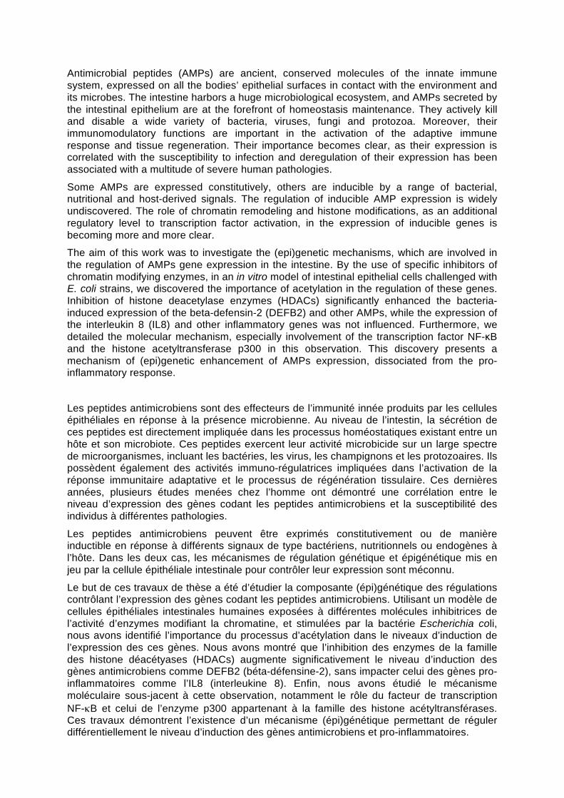
Antimicrobial peptides (AMPs) are ancient, conserved molecules of the innate immune system, expressed on all the bodies’ epithelial surfaces in contact with the environment and its microbes. The intestine harbors a huge microbiological ecosystem, and AMPs secreted by the intestinal epithelium are at the forefront of homeostasis maintenance. They actively kill and disable a wide variety of bacteria, viruses, fungi and protozoa. Moreover, their immunomodulatory functions are important in the activation of the adaptive immune response and tissue regeneration. Their importance becomes clear, as their expression is correlated with the susceptibility to infection and deregulation of their expression has been associated with a multitude of severe human pathologies.
Some AMPs are expressed constitutively, others are inducible by a range of bacterial, nutritional and host-derived signals. The regulation of inducible AMP expression is widely undiscovered. The role of chromatin remodeling and histone modifications, as an additional regulatory level to transcription factor activation, in the expression of inducible genes is becoming more and more clear.
The aim of this work was to investigate the (epi)genetic mechanisms, which are involved in the regulation of AMPs gene expression in the intestine. By the use of specific inhibitors of chromatin modifying enzymes, in an in vitro model of intestinal epithelial cells challenged with E. coli strains, we discovered the importance of acetylation in the regulation of these genes. Inhibition of histone deacetylase enzymes (HDACs) significantly enhanced the bacteria-induced expression of the beta-defensin-2 (DEFB2) and other AMPs, while the expression of the interleukin 8 (IL8) and other inflammatory genes was not influenced. Furthermore, we detailed the molecular mechanism, especially involvement of the transcription factor NF-κB and the histone acetyltransferase p300 in this observation. This discovery presents a mechanism of (epi)genetic enhancement of AMPs expression, dissociated from the pro-inflammatory response.
Les peptides antimicrobiens sont des effecteurs de l’immunité innée produits par les cellules épithéliales en réponse à la présence microbienne. Au niveau de l’intestin, la sécrétion de ces peptides est directement impliquée dans les processus homéostatiques existant entre un hôte et son microbiote. Ces peptides exercent leur activité microbicide sur un large spectre de microorganismes, incluant les bactéries, les virus, les champignons et les protozoaires. Ils possèdent également des activités immuno-régulatrices impliquées dans l’activation de la réponse immunitaire adaptative et le processus de régénération tissulaire. Ces dernières années, plusieurs études menées chez l’homme ont démontré une corrélation entre le niveau d’expression des gènes codant les peptides antimicrobiens et la susceptibilité des individus à différentes pathologies.
Les peptides antimicrobiens peuvent être exprimés constitutivement ou de manière inductible en réponse à différents signaux de type bactériens, nutritionnels ou endogènes à l’hôte. Dans les deux cas, les mécanismes de régulation génétique et épigénétique mis en jeu par la cellule épithéliale intestinale pour contrôler leur expression sont méconnu.
Le but de ces travaux de thèse a été d’étudier la composante (épi)génétique des régulations contrôlant l’expression des gènes codant les peptides antimicrobiens. Utilisant un modèle de cellules épithéliales intestinales humaines exposées à différentes molécules inhibitrices de l’activité d’enzymes modifiant la chromatine, et stimulées par la bactérie Escherichia coli, nous avons identifié l’importance du processus d’acétylation dans le niveaux d’induction de l’expression des ces gènes. Nous avons montré que l’inhibition des enzymes de la famille des histone déacétyases (HDACs) augmente significativement le niveau d’induction des gènes antimicrobiens comme DEFB2 (béta-défensine-2), sans impacter celui des gènes pro-inflammatoires comme l’IL8 (interleukine 8). Enfin, nous avons étudié le mécanisme moléculaire sous-jacent à cette observation, notamment le rôle du facteur de transcription NF-κB et celui de l’enzyme p300 appartenant à la famille des histone acétyltransférases. Ces travaux démontrent l’existence d’un mécanisme (épi)génétique permettant de réguler différentiellement le niveau d’induction des gènes antimicrobiens et pro-inflammatoires.
Copyright © 2022 FDOKUMEN