Nanocarrier Drug Delivery Systems: Characterization ... - MDPI
Upload
khangminh22Category
view
3download
0
Citation: Saddiq, A.A.; Tag, H.M.;
Doleib, N.M.; Salman, A.S.; Hagagy, N.
Antimicrobial, Antigenotoxicity, and
Characterization of Calotropis procera
and Its Rhizosphere-Inhabiting
Actinobacteria: In Vitro and In Vivo
Studies. Molecules 2022, 27, 3123.
https://doi.org/10.3390/
molecules27103123
Academic Editors: Monika
Waksmundzka-Hajnos and
Miroslaw Hawryl
Received: 7 April 2022
Accepted: 11 May 2022
Published: 13 May 2022
Publisher’s Note: MDPI stays neutral
with regard to jurisdictional claims in
published maps and institutional affil-
iations.
Copyright: © 2022 by the authors.
Licensee MDPI, Basel, Switzerland.
This article is an open access article
distributed under the terms and
conditions of the Creative Commons
Attribution (CC BY) license (https://
creativecommons.org/licenses/by/
4.0/).
molecules
Article
Antimicrobial, Antigenotoxicity, and Characterizationof Calotropis procera and Its Rhizosphere-InhabitingActinobacteria: In Vitro and In Vivo StudiesAmna A. Saddiq 1,*, Hend M. Tag 2,3, Nada M. Doleib 2,4, Asmaa S. Salman 2,5 and Nashwa Hagagy 2,6,*
1 Department of Biology, College of Science, University of Jeddah, Jeddah 21959, Saudi Arabia2 Department of Biology, College of Science and Arts at Khulis, University of Jeddah,
Jeddah 21959, Saudi Arabia; [email protected] (H.M.T.); [email protected] (N.M.D.);[email protected] (A.S.S.)
3 Zoology Department, Faculty of Science, Suez Canal University, Ismailia 41522, Egypt4 Department of Microbiology, Biotechnology and Biochemistry, Faculty of Applied and Industrial Sciences,
University of Bahri, Kartoum 13311, Sudan5 Genetics & Cytology Department, Biotechnology Research Institute, National Research Center,
Giza 12622, Egypt6 Botany and Microbiology Department, Faculty of Science, Suez Canal University, Ismailia 41522, Egypt* Correspondence: [email protected] (A.A.S.); [email protected] (N.H.)
Abstract: Calotropis procera (C. procera) is a wild shrub that is a medicinal plant found in abundancethroughout Saudi Arabia. In this study, we investigated the phytochemical composition and antigeno-toxic properties of the ethanolic extract of C. procera, in addition to the antimicrobial activity of theplant and its rhizospheric actinobacteria effects against pathogenic microorganisms. Soil-extractmedium supplemented with glycerol as a carbon source and starch–casein agar medium was used forisolation of actinobacteria from rhizosphere. From the plant, a total of 31 compounds were identifiedusing gas chromatography/mass spectrometry (GC–MS). The main components were α-amyrin(39.36%), lupeol acetate (17.94%), phytol (13.32%), hexadecanoic acid (5.55%), stigmasterol (3.16%),linolenic acid (3.04%), and gombasterol A (2.14%). C. procera plant extract’s antimicrobial activitywas investigated using an agar well-diffusion assay and minimum inhibitory concentration (MIC)against six pathogenic microbial strains. The plant extract of C. procera was considered significantlyactive against Staphylococcus aureus, Klebsiella pneumonia, and Escherichia coli, with inhibition zones of18.66 mm, 21.26 mm, and 21.93 mm, respectively. The plant extract was considered to be a moderateinhibitor against Bacillus subtilis, with MIC ranging from 0.60–1.50 mg/mL. On the other hand, the iso-lated actinobacteria were considered to be a moderate inhibitor against S. aureus (MIC of 86 µg/mL),and a potent inhibitor, strain CALT_2, against Candida albicans (MIC of 35 µg/mL). The 16S rRNAgene sequence analysis showed that the potential strains belonged to the genus Streptomyces. Theeffect of C. procera extract against cyclophosphamide (CP)-induced genotoxicity was examined byevaluating chromosome abnormalities in mouse somatic cells and DNA fragmentation assays. Thecurrent study revealed that oral pretreatment of C. procera (50, 100, and 200 mg/kg b.w.) for 1, 7,and 14 days to cyclophosphamide-treated animals significantly reduced chromosomal abnormali-ties as well as DNA fragmentation in a dose-dependent manner. Moreover, C. procera extract hadantimicrobial and antigenotoxic effects against CP-induced genotoxicity.
Keywords: Calotropis procera; actinobacteria; antimicrobial activity; genotoxicity; DNA fragmentation;chromosomal aberration
1. Introduction
Calotropis procera (Aiton) Dryand is a perennial, soft-wooded shrub, belonging tothe Apocynaceae family and subfamily Asclepiadaceae. This evergreen, xerophytic plantthrives in dry and semiarid environments. In different regions of the world, it is known
Molecules 2022, 27, 3123. https://doi.org/10.3390/molecules27103123 https://www.mdpi.com/journal/molecules

Molecules 2022, 27, 3123 2 of 19
by numerous common names such as apple of Sodom, calotrope, wild cotton, Indianmilkweed, gigantic milkweed, and rubber tree, and in Saudi Arabia, it is called “Ushar”. InNorth Africa, the Middle East, South Asia, and Southeast Asia, it has long been utilized intraditional medicinal applications. Since antiquity, it has been used for fuel, fiber, feed, andlumber [1–3].
Within salt-stressed environments, endophytic bacteria such as Virgibacillus koreensisand Pseudomonas stutzeri have been found to be associated with C. procera, which mayhelp it survive under harsh conditions. In addition, endophytic fungal species, such asPhaeoramularia calotropidis, Curvularia hawaiiensis, Guignardia bidwellii, Alternaria alternata,Cochliobolus hawaiiensis, Aspergillus spp., Mucor circinelloides, Fusarium spp., Chaetomium spp.,and Penicillium spp., provide protection for the plant against pathogens and pests [4,5].
Flavonoids, terpenoids, alkaloids, tannins, saponins, cardiac glycosides, and steroidshave been found in several portions of the plant, according to several studies [6–9]. Fattyacid ethyl esters (21.4%), palmitic acid esters (10.2%), amino acids (8.1%), and linoleic acids(7.4%) are the principal phytochemical groups found in C. procera leaf extracts [10].
In several plant and animal cells, including human cells, C. procera causes acute toxicity.As a result, numerous plant parts, particularly latex, have been tested against various cancercell lines [11–13]. Similarly, the plant’s antibacterial and anthelmintic properties are beingexplored in pharmacology. The toxicity–bioactivity relationship of C. procera, on the otherhand, has yet to be well examined. According to a few studies, the plant causes acutecardiotoxicity and hepatotoxicity [14]. These toxic effects have not been investigated indetail, and additional research is needed to confirm the therapeutic potential of C. procera.The search for environmentally friendly prototypes to replace chemically manufacturedpharmaceuticals is on the rise. As a result, several studies have been conducted on theplant species described in traditional medical systems. The pharmacological capabilitiesof C. procera have been used to treat a variety of various human infections in the past,including colds, fevers, leprosy, rheumatism, asthma, indigestion, eczema, elephantiasis,diarrhea, dysentery, and skin diseases [8]. In Saudi Arabia, a decoction of abovegroundportions is used to cure fever, joint pain, constipation, and muscle spasms [6]. In BurkinaFaso, the plant is also used to treat mental conditions [15]. Secondary metabolites andcardiotonic compounds found in C. procera are responsible for the therapeutic properties ofthe plant [16,17].
In previous studies, Mossa et al. and Garabadu et al. [6,18] found that the extracts ofthe aboveground plant sections of C. procera have high antipyretic, antidepressive, analgesic,and neuromuscular-blocking activities. Antibacterial activity was observed in extracts fromthe bark and leaves against Pseudomonas aeruginosa, Klebsiella pneumoniae, Escherichia coli,and Bacillus subtilis [17]. The extracts of both aerial portions of C. procera and its endophyticbacteria, Bacillus siamensis, have been demonstrated to have a broad antibacterial range [16].C. procera leaf extracts also significantly lower blood glucose levels, demonstrating theirantihyperglycemic potential [19]. Although the pharmaceutical and industrial applicationsof the plant have attracted considerable interest, the plant’s biological and ecologicalcharacteristics (especially those focusing on adaptations or plasticity) have received littlestudy in general. Furthermore, the toxicity–bioactivity relationship of C. procera has notbeen well studied, which is important for verifying its therapeutic properties. Evaluatingthese fundamental aspects could help C. procera become more commercially viable andopen it up to new applications. Moreover, filling in these knowledge gaps could benefita better understanding of its invasive behavior and potential future biodiversity and/orenvironmental problems [20].
In this study, the antimicrobial activity of C. procera extract was evaluated, along withthe antimicrobial activity of its rhizosphere-inhabiting actinobacteria, which was evaluatedfor the first time, to our knowledge. Moreover, the present investigation aimed to evaluatethe cytogenetic bioactivity of C. procera leaves to ameliorate the cytogenetic alterations andDNA damage induced by cyclophosphamide (CP), an alkylating agent used as a potent

Molecules 2022, 27, 3123 3 of 19
anti-inflammatory and immunosuppressive cytostatic and cytotoxic drug to treat diversemedical problems such as neoplasia.
2. Results2.1. Characterization and Identification of the Potential Actinobacterial Isolates
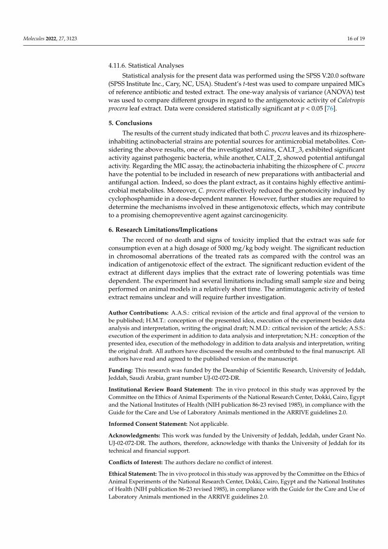
Based on colony shape, color, and texture, a total of 17 isolates were selected forpreliminary screening of antimicrobial activity. Only four isolates, designated as CALT_1,CALT_2, CALT_3, and CALT_4, showed potential activity and were selected for furtherinvestigation. A 16S rRNA gene-sequence analysis showed that the potential actinobacte-rial strains were affiliated within genus Streptomyces, with similarity ≥ 97%, and closelyrelated to Streptomyces coeruleorubidus, Streptomyces maritimus, Streptomyces carminius, andunclassified species within the same genus, as shown in Figure 1. The 16S rRNA genedata were deposited under the accession numbers MT742093-MT742096 in the NCBI andGenBank nucleotide sequence databases.
Molecules 2022, 27, x FOR PEER REVIEW 3 of 22
a potent anti-inflammatory and immunosuppressive cytostatic and cytotoxic drug to treat diverse medical problems such as neoplasia.
2. Results 2.1. Characterization and Identification of the Potential Actinobacterial Isolates
Based on colony shape, color, and texture, a total of 17 isolates were selected for pre-liminary screening of antimicrobial activity. Only four isolates, designated as CALT_1, CALT_2, CALT_3, and CALT_4, showed potential activity and were selected for further investigation. A 16S rRNA gene-sequence analysis showed that the potential actinobacte-rial strains were affiliated within genus Streptomyces, with similarity ≥ 97%, and closely related to Streptomyces coeruleorubidus, Streptomyces maritimus, Streptomyces carminius, and unclassified species within the same genus, as shown in Figure 1. The 16S rRNA gene data were deposited under the accession numbers MT742093-MT742096 in the NCBI and Gen-Bank nucleotide sequence databases.
Figure 1. Neighbor-joining tree (partial sequences ~950 bp) showing the phylogenetic relationships of actinobacterial 16S rRNA gene sequences of potential strains to closely related (S ≥ 97%) se-quences from the GenBank database.
2.2. Antimicrobial Activity In the present study, the extract from C. procera was examined for antimicrobial ac-
tivity quantitatively at a concentration of 10 mg/mL (100 µg) by zone inhibition on an agar plate (Table 1). The results revealed that the ethanolic extract of C. procera possessed po-tential antibacterial activity. C. procera leaf extract showed significant activity against all tested microorganisms compared with the standard antibiotics gentamycin and ketocon-azole. The most antibacterial activity was recorded in S. aureus and K. pneumoniae, fol-lowed by B. subtilis and E. coli. The antifungal activity of leaf extract of C. procera was also significant against the tested pathogenic fungi C. albicans and A. fumigatus compared with ketoconazole. Regarding the effectiveness of the tested plant extract against Gram-nega-tive bacteria, the results revealed that the significant inhibition zone of 21.93 ± 1.71 mm
Figure 1. Neighbor-joining tree (partial sequences ~950 bp) showing the phylogenetic relationshipsof actinobacterial 16S rRNA gene sequences of potential strains to closely related (S ≥ 97%) sequencesfrom the GenBank database.
2.2. Antimicrobial Activity
In the present study, the extract from C. procera was examined for antimicrobial activityquantitatively at a concentration of 10 mg/mL (100 µg) by zone inhibition on an agar plate(Table 1). The results revealed that the ethanolic extract of C. procera possessed potentialantibacterial activity. C. procera leaf extract showed significant activity against all testedmicroorganisms compared with the standard antibiotics gentamycin and ketoconazole.The most antibacterial activity was recorded in S. aureus and K. pneumoniae, followed byB. subtilis and E. coli. The antifungal activity of leaf extract of C. procera was also significantagainst the tested pathogenic fungi C. albicans and A. fumigatus compared with ketoconazole.Regarding the effectiveness of the tested plant extract against Gram-negative bacteria, theresults revealed that the significant inhibition zone of 21.93 ± 1.71 mm was observedagainst E. coli which was comparatively insignificant compared to the positive control(23.40 ± 2.42 mm). In addition, C. procera showed potent activity against K. pneumoniae(ZOI = 21.26), which was not significantly different from that of the reference antibiotic.These values fall within the range considered to be highly sensitive when compared to thecontrol antibiotic.

Molecules 2022, 27, 3123 4 of 19
Table 1. Zone of inhibition (mm) of ethanolic extract of C. procera leaves and rhizosphere-inhabitingactinobacterial isolates against pathogens tested using agar well-diffusion assay.
Antimicrobial Activity against Pathogens (Inhibition Zone in mm)
Treatments(100 µg/mL)
Fungi (Yeast) Gram-Positive Gram-Negative
C. albicans A. fumigatus S. aureus B. subtilis E. coli K. pneumonia
C. procera 21.00 ± 2.64 a 12.00 ± 0.52 a 18.66 ± 1.17 a 16.23 ± 3.80 a 21.93 ± 1.71 a 21.26 ± 3.16 a
CALT_1 10.43 ± 0.93 b 7.17 ± 0.95 b 8.16 ± 0.76 c 7.43 ± 0.75 b 7.83 ± 0.76 b 9.94 b ± 0.81 b
CALT_2 13.77 ± 1.36 b 10.10 ± 1.85 b 12.30 ± 1.30 b 11.22 ± 0.96 c 8.76 ± 0.92 b 10.80 ± 0.43 b
CALT_3 7.20 ± 0.82 c 8.10 ± 2.25 b 12.10 ± 2.01 c 11.37 ± 1.30 c 9.23 ± 0.25 b 9.00 ± 0.60 b
CALT_4 10.90 ± 0.90 b 11.4 ± 1.00 a 7.00 ± 0.10 b 6.50 ± 0.50 b 8.63 ± 0.66 b 8.47 ± 0.62 b
Positive control 20.33 ± 1.52 a 11.16 ± 0.76 a 16.53 ± 1.50 a 20.07 ± 4.20 a 23.40 ± 2.42 a 20.20 ± 1.72 a
Negative control NI NI NI NI NI NI
Values are mean ± SD; NI denotes no inhibition. Positive control for fungi: 25 µg/mL ketoconazole; positivecontrol for bacteria: 25 µg/mL. Negative control: 50% of ethanol. For each column, the same letter shows thatthe difference between the means was not statistically significant. However, different letters show statisticallysignificant differences (p < 0.05) between the corresponding treatments.
The minimum inhibitory concentration (MIC) results of C. procera extract againstdifferent pathogenic microorganisms are shown in Figures 2–4. The ethanolic extract ofC. procera leaves showed significantly high MIC values for both C. albicans and A. fumigatuscompared with the actinobacterial extract from strains CALT_1, CALT_2, CALT_3, andCALT_4 (Figure 2). Figure 3 displays the MIC for the tested extract against Gram-positivebacteria; the current results showed that CALT_1, CALT_2, CALT_3, and CALT_4 weremore effective than the C. procera leaf extract. Regarding the effectiveness of tested extractsagainst Gram-negative bacteria, C. procera extract revealed potent antimicrobial activityagainst E. coli as compared with CALT_1, CALT_2, CALT_3, and CALT_4, while CALT_2exhibited more effectiveness against Klebsiella pneumoniae.
Molecules 2022, 27, x FOR PEER REVIEW 5 of 22
Figure 2. Minimum inhibitory concentrations (µg/mL) of C. procera ethanolic extract and actinobac-teria isolate extracts against yeast and fungi. Values are mean ± SD; different letters (a, b, c ,d) indi-cate significant differences.
ab
c
b
d
c
a
b
c
b
d
c
0.0
20.0
40.0
60.0
80.0
100.0
120.0
140.0
C. procera CALT_1 CALT_2 CALT_3 CALT_4 Positivecontrol
Min
imum
Inhi
bito
ry C
once
ntra
tion
(µg/
mL)
Treatments
Candida albicansAspergillus fumigatus
Figure 2. Minimum inhibitory concentrations (µg/mL) of C. procera ethanolic extract and actinobacteriaisolate extracts against yeast and fungi. Values are mean ± SD; different letters (a, b, c, d) indicatesignificant differences.

Molecules 2022, 27, 3123 5 of 19Molecules 2022, 27, x FOR PEER REVIEW 6 of 22
Figure 3. Minimum inhibitory concentrations (µg/mL) of C. procera ethanolic extract and actinobac-terial isolate extracts against Gram-positive bacteria. Values are mean ± SD; different letters (a, b, c, d) indicate significant differences (p ≤ 0.05) between both plant and actinobacteria isolate extracts for the same pathogen according to one-way ANOVA test.
a
bc
c
d
c
a
bc
c d
c
0.0
20.0
40.0
60.0
80.0
100.0
120.0
140.0
160.0
180.0
C. procera CALT_1 CALT_2 CALT_3 CALT_4 Positivecontrol
Min
imum
Inhi
bito
ry C
once
ntra
tion
(µg/
mL)
Treatments
Escherichia coliKlebsiella pneumonia
Figure 3. Minimum inhibitory concentrations (µg/mL) of C. procera ethanolic extract and actinobacterialisolate extracts against Gram-positive bacteria. Values are mean ± SD; different letters (a, b, c, d)indicate significant differences (p ≤ 0.05) between both plant and actinobacteria isolate extracts forthe same pathogen according to one-way ANOVA test.
Molecules 2022, 27, x FOR PEER REVIEW 7 of 22
Figure 4. Minimum inhibitory concentrations (µg/mL) of C. procera ethanolic extract and actinobac-terial isolates extract against Gram-negative bacteria. Values are mean ± SD; different letters (a, b, c, d) indicate significant differences (p ≤ 0.05) between both plant and actinobacteria isolate extracts for the same pathogen according to one-way ANOVA test.
Regarding the antimicrobial activity of rhizosphere-inhabiting actinobacteria, out of 17 actinobacterial isolates screened for antimicrobial activity, 4 exhibited potential antimi-crobial activities against tested pathogens, with inhibitory zone diameters ranging from 6.5 to 13.7 mm shown by strain CALT_4 against B. subtilis and strain CALT_2 against Can-dida albicans, respectively, as shown in Table 1. The extract of strain CALT_2 showed the most potent activity against tested pathogens, with MIC values of 35 µg/mL against A. fumigatus as shown in Figure 2, 56 µg/mL against B. subtilis as shown in Figure 3, and 54 µg/mL against K. pneumoniae as shown in Figure 4.
According to Figure 2, the MIC results revealed that the ethanolic extract of C. procera leaves showed significantly high MIC values against both C. albicans and A. fumigatus as compared with the actinobacteria extracts CALT_1, CALT_2, CALT_3, and CALT_4. Fig-ure 3 displays the MICs for the tested extracts against Gram-positive bacteria, and the current results showed that CALT_1, CALT_2, CALT_3, and CALT_4 were more effective than the C. procera leaf extract. Regarding the effectiveness of the tested extracts against Gram-negative bacteria, C. procera extract revealed potent antimicrobial activity against E. coli as compared with strains CALT_1, CALT_2, CALT_3, and CALT_4, while CALT_2 exhibited more effectiveness against Klebsiella pneumoniae.
2.3. Gas Chromatography–Mass Spectrometry (GC–MS) A phytochemical study was carried out by GC–MS analysis of the C. procera leaf ex-
tract. The chromatogram identified 31 phytochemicals as constituents (Figure 5), of which α-amyrin was the major compound (39.36%) identified at retention time 63.63 min, fol-lowed by lupeol acetate (17.94%) at retention time 64.65 min, phytol (13.32%) at retention time 36.71 min, hexadecanoic acid (5.55%) at retention time 32.41 min, stigmasterol
a
b bb
c
d
ab
c b
d
d
0.0
20.0
40.0
60.0
80.0
100.0
120.0
140.0
160.0
180.0
C. procera CALT_1 CALT_2 CALT_3 CALT_4 Positivecontrol
Min
imum
Inhi
bito
ry C
once
ntra
tion
(µg/
mL)
Treatments
Staphylococcus aureusBacillus subtilis
Figure 4. Minimum inhibitory concentrations (µg/mL) of C. procera ethanolic extract and actinobacterialisolates extract against Gram-negative bacteria. Values are mean ± SD; different letters (a, b, c, d)indicate significant differences (p ≤ 0.05) between both plant and actinobacteria isolate extracts forthe same pathogen according to one-way ANOVA test.

Molecules 2022, 27, 3123 6 of 19
Regarding the antimicrobial activity of rhizosphere-inhabiting actinobacteria, outof 17 actinobacterial isolates screened for antimicrobial activity, 4 exhibited potential an-timicrobial activities against tested pathogens, with inhibitory zone diameters rangingfrom 6.5 to 13.7 mm shown by strain CALT_4 against B. subtilis and strain CALT_2 againstCandida albicans, respectively, as shown in Table 1. The extract of strain CALT_2 showedthe most potent activity against tested pathogens, with MIC values of 35 µg/mL againstA. fumigatus as shown in Figure 2, 56 µg/mL against B. subtilis as shown in Figure 3, and54 µg/mL against K. pneumoniae as shown in Figure 4.
According to Figure 2, the MIC results revealed that the ethanolic extract of C. proceraleaves showed significantly high MIC values against both C. albicans and A. fumigatusas compared with the actinobacteria extracts CALT_1, CALT_2, CALT_3, and CALT_4.Figure 3 displays the MICs for the tested extracts against Gram-positive bacteria, and thecurrent results showed that CALT_1, CALT_2, CALT_3, and CALT_4 were more effectivethan the C. procera leaf extract. Regarding the effectiveness of the tested extracts againstGram-negative bacteria, C. procera extract revealed potent antimicrobial activity againstE. coli as compared with strains CALT_1, CALT_2, CALT_3, and CALT_4, while CALT_2exhibited more effectiveness against Klebsiella pneumoniae.
2.3. Gas Chromatography–Mass Spectrometry (GC–MS)
A phytochemical study was carried out by GC–MS analysis of the C. procera leafextract. The chromatogram identified 31 phytochemicals as constituents (Figure 5), ofwhich α-amyrin was the major compound (39.36%) identified at retention time 63.63 min,followed by lupeol acetate (17.94%) at retention time 64.65 min, phytol (13.32%) at retentiontime 36.71 min, hexadecanoic acid (5.55%) at retention time 32.41 min, stigmasterol (3.16%)at retention time 55.61 min, and linolenic acid (3.04%) at retention time 38.12 min. Theremaining constituent chemical compounds were present in proportions of less than 2%.The components and their retention times, molecular formulas, and molecular weightsare summarized in Table 2. Figure 6 shows the demonstrated hit spectrum and chemicalstructure of the major compounds present in C. procera leaf extract. The analysis of extractfrom Streptomyces sp. strain CALT_2 by GC–MS led to the detection of three compoundson the basis of retention time and mass analysis (Table 3). The following compounds wereidentified: (i) hexadecanoic acid, (ii) stigmasterol, (iii) α-amyrin. Spectra and chemicalstructure are presented in Figure 6.
Molecules 2022, 27, x FOR PEER REVIEW 9 of 22
62.67 0.61 1,2-Dilinoleoyl-Sn-Glycero-3-Phosph Oethan-olamine
C41H74NO8P 739
63.63 39.36 α-Amyrin C30H50O 426 64.65 17.94 Lupeol Acetate C32H52O2 468 64.97 0.27 Methyl Commate D C31H50O4 486
a RT: retention time of the compounds based on GC–MS peaks; compounds are listed in order of their elution from a DB5/MS column.
Figure 5. Chromatogram of ethanolic extract of C. procera leaves by gas chromatography–mass spec-trometry (GC–MS) analysis. The GC–MS spectrum at retention time 0-65 min represents the main compounds.
Table 3. Identification of metabolites components produced by Streptomyces sp. strain CALT_2 us-ing gas chromatography–mass spectrometry (GC–MS) analysis.
RT a (min) Area % Compound Name Molecular Formula Molecular Weight 32.41 44.48 Hexadecanoic acid C17H34O2 270 55.61 18.48 Stigmasterol C29H48O 412 63.63 39.8 α-Amyrin C30H50O 426
a RT: retention time of the compounds based on GC–MS peaks; compounds are listed in order of their elution from a DB5/MS column.
Figure 5. Chromatogram of ethanolic extract of C. procera leaves by gas chromatography–massspectrometry (GC–MS) analysis. The GC–MS spectrum at retention time 0-65 min represents themain compounds.

Molecules 2022, 27, 3123 7 of 19Molecules 2022, 27, x FOR PEER REVIEW 10 of 22
Figure 6. Hit spectra and chemical structures of the major phytochemical constituents of C. procera leaf extract. Chemical structure taken from ChemSpider web site, (accessed on 20 October 2021): hexadecanoic acid, phytol, stigmasterol, linolenic acid, α-amyrin, lupeol acetate.
Figure 6. Hit spectra and chemical structures of the major phytochemical constituents of C. proceraleaf extract. Chemical structure taken from ChemSpider web site, (accessed on 20 October 2021):hexadecanoic acid, phytol, stigmasterol, linolenic acid, α-amyrin, lupeol acetate.

Molecules 2022, 27, 3123 8 of 19
Table 2. Identification of phytocomponents of ethanolic extract of C. procera leaves using GC–MS analysis.
RT a (min) Area % Compound Name Molecular Formula Molecular Weight
27.95 0.91 Neophytadiene C20H38 278
29.3 0.38 8-Heptadecyne, 1-Bromo- C17H31Br 314
29.88 1.26 2-Pentadecanone C18H36O 268
31.86 0.87 Tert-Hexadecanethiol C16H34S 258
32.41 5.55 Hexadecanoic Acid C17H34O2 270
35.93 0.55 7,9-Di-Tert-Butyl-1-oxaspiro (4,5) DeCa-6,9-Diene-2,8-Dione C17H24O3 276
36.71 13.32 Phytol C20H40O 296
37.12 1.2317-Octadecenoic Acid,
C19H36O2 296Methyl Ester
37.48 0.95 9,12-Octadecadienoic Acid (Z,Z)-,Methyl Ester C19H34O2 294
38.12 3.04 α-Linolenic acid C19H32O2 292
38.43 0.899-Octadecenoic Acid (Z)-,
C20H38O2 310Ethyl Ester
38.78 0.89 Linoleic Acid Ethyl Ester C20H36O2 308
43.69 0.75À-D-Glucopyranoside, Methyl
C16H32BNO6Si 3732-(Acetylamino)-2-Deoxy-3-O-(TrimEthylsilyl)-, cyclic butylboronate
48.51 0.5 Promecarb C16H16N2O5 316
49.06 0.31
14-Hydroxy-14-Methyl-Hex
C18H34O3 298Adec-15-Enoic Acid Methyl
Ester
52.4 0.54 Methanesulfonic Acid C26H43DO4S 453
52.72 0.53 À-Tocospiro A C29H50O4 462
55.26 0.63 Picrotin C15H18O7 310
55.61 3.16 Stigmasterol C29H48O 412
56.63 1.61 Boroxin, C21H12B3F9O3 516
57.9 0.68 Tetrakis (4-Methylphenyl) Thieno3,2-BThiophene C34H28S2 500
58.47 0.52 Astilbin C21H22O11 450
59.28 0.67Thieno3,4-CPyridine,
C31H21NS 4391,3,4,7-Tetraphenyl-
60.17 1.42 Nicotiflorin C27H30O15 594
60.34 0.9 Momordicinin C30H46O2 438
60.73 2.14 Gombasterol A C28H48O7 496
61.96 0.66 25-Hydroxy-24-Epi-Brassinolide C28H48O7 496
62.67 0.61 1,2-Dilinoleoyl-Sn-Glycero-3-Phosph Oethanolamine C41H74NO8P 739
63.63 39.36 α-Amyrin C30H50O 426
64.65 17.94 Lupeol Acetate C32H52O2 468
64.97 0.27 Methyl Commate D C31H50O4 486a RT: retention time of the compounds based on GC–MS peaks; compounds are listed in order of their elutionfrom a DB5/MS column.

Molecules 2022, 27, 3123 9 of 19
Table 3. Identification of metabolites components produced by Streptomyces sp. strain CALT_2 usinggas chromatography–mass spectrometry (GC–MS) analysis.
RT a (min) Area % Compound Name Molecular Formula Molecular Weight
32.41 44.48 Hexadecanoic acid C17H34O2 270
55.61 18.48 Stigmasterol C29H48O 412
63.63 39.8 α-Amyrin C30H50O 426a RT: retention time of the compounds based on GC–MS peaks; compounds are listed in order of their elutionfrom a DB5/MS column.
2.4. In Vivo Studies of C. proceraAcute Toxicity Test
The toxicity of the ethanolic extract of C. procera was assessed at different doses ofup to 5000 mg/kg. It was observed that the tested extract did not cause any mortality orchanges in the behavior of the treated mice from the beginning of administration until14 days. No changes were observed in food or water intake, which confirmed that theextract was safe and did not cause any toxicity, even at high doses.
2.5. Chromosomal Aberrations in Bone Marrow Cells
Table 4 shows the total counts and percentages of chromosomal aberrations in controland C. procera-treated animals. The percentage of chromosome aberrations in animalstreated with single and repeated doses of C. procera was not significantly different from thatin the control animals (Table 4). No significant reduction in chromosomal abnormalitiesinduced by CP was observed after a single treatment with C. procera. Repeated treatmentwith C. procera for 7 and 14 days caused a significant (p < 0.01) reduction in the percentage ofchromosomal abnormalities induced by CP (Table 5). The percentage of reduction reached42.85% and 58.3% after pretreatment with C. procera for 7 and 14 days, respectively. Table 5illustrates the protective effect of C. procera in reducing the different types of aberrations.
Table 4. Number and mean percentages of different chromosomal aberrations in bone marrow cellsof mice after treatment with different doses of C. procera for 1, 7, and 14 days.
GroupsC. proceraTreatment
Day(s)
No. of Metaphases with Total Chromosomal Aberrations
Gap Frag. and/orBreak Del. Gap + (Frag.
and/or Break)Excluding Gaps
Mean ± S.E.Including Gaps
Mean ± S.E.
Control
1
7 12 — 4 3.2 ± 0.23 4.6 ± 0.3C. procera 50 mg/kg 9 10 1 4 3.0 ± 0.33 4.8 ± 0.3
C. procera 100 mg/kg 7 9 — 5 2.8 ± 0.21 4.2 ± 0.32C. procera 200 mg/kg 9 11 1 3 3.0 ± 0.2 4.8 ± 0.5
Control7
8 9 — 4 2.6 ± 0.24 4.2 ± 0.22C. procera 50 mg/kg 8 10 — 4 2.8 ± 0.3 4.4 ± 0.3
C. procera 100 mg/kg 11 9 — 3 2.4 ± 0.22 4.6 ± 0.32C. procera 200 mg/kg 9 11 1 2 2.8 ± 0.2 4.6 ± 0.22
Control14
11 10 — 3 2.6 ± 0.2 4.8 ± 0.34C. procera 50 mg/kg 12 11 — 1 2.4 ± 0.23 4.8 ± 0.24
C. procera 100 mg/kg 9 10 — 4 2.8 ± 0.23 4.6 ± 0.2C. procera 200 mg/kg 7 9 — 3 2.4 ± 0.2 3.8 ± 0.2
The total number of scored metaphases was 500 (5 animals/group). Frag. = fragment; Del = deletion.

Molecules 2022, 27, 3123 10 of 19
Table 5. Number and mean percentage of the different types of chromosomal aberrations in bonemarrow cells of mice after treatment with C. procera 200 mg/kg for 1, 7, and 14 days alone or incombination with cyclophosphamide 20 mg/kg.
GroupsC. proceraTreatment
day(s)
No. of Metaphases with Total Chromosomal Aberrations Inhibition %
GapFrag.
and/orBreak
Del Rt
Gap +(Frag.
and/orBreak)
End PolyExcluding
GapsMean ± S.E.
IncludingGaps
Mean ± S.E.
Control (nontreated)1
9 10 1 — 2 — — 2.6 ± 0.4 4.4 ± 0.228.3 aCP 20 42 7 4 20 3 8 16.8 ± 0.33 a 20.8 ± 0.36 a
C. procera + CP 21 40 6 3 21 2 5 15.4 ± 0.44 a 19.6 ± 0.4 a
Control7
10 9 1 — 1 — — 2.2 ± 0.22 4.2 ± 0.2142.85 bCP 20 42 7 4 20 3 8 16.8 ± 0.33 a 20.8 ± 0.36 a
C. procera + CP 15 28 2 — 15 3 — 9.6 ± 0.86 b 12.6 ± 0.8 b
Control14
9 11 — — 3 — — 2.8 ± 0.3 4.6 ± 0.358.3 bCP 20 42 7 4 20 3 8 16.8 ± 0.33 a 20.8 ± 0.36 a
C. procera + CP 16 24 2 — 9 — — 7.0 ± 0.35 b 10.2 ± 0.43 b
The total number of scored metaphases is 500 (5 animals/group). Frag. = fragment; Del = deletion;Rt. = Robertsonian translocation; End. = endomitosis; Poly. = polyploidy. a Significant at the 0.05 level (one-wayANOVA test) compared with control (nontreated). b Significant at the 0.05 level (one-way ANOVA test) comparedwith treatment.
2.6. DNA Fragmentation
Administration of single and repeated doses of C. procera caused no significant DNAfragmentation (Table 6). Pretreatment with a repeated dose of C. procera significantly(p < 0.01) decreased the percentage of DNA fragmentation induced by CP in liver cells(Table 7). The percentage of DNA fragmentation was reduced to 5.34% and 4.29% (p < 0.01)after pretreatment with C. procera for 7 and 14 days, respectively, compared with 8.77% forthe groups treated only with CP.
Table 6. DNA fragmentation in mouse liver cells after treatment with different doses of C. procera for1, 7, and 14 days.
Groups Days DNA Fragmentation
Control
1
2.92 ± 0.2
C. procera (50 mg/kg) 3.7 ± 0.46
C. procera (100 mg/kg) 3.15 ± 0.23
C. procera (200 mg/kg) 3.22 ± 0.03
Control
7
3.33 ± 0.29
C. procera (50 mg/kg) 2.98 ± 0.3
C. procera (100 mg/kg) 3.32 ± 0.34
C. procera (200 mg/kg) 3.17 ± 0.29
Control
14
3.2 ± 0.25
C. procera (50 mg/kg) 3.1 ± 0.23
C. procera (100 mg/kg) 3.32 ± 0.33
C. procera (200 mg/kg) 3.41 ± 0.2

Molecules 2022, 27, 3123 11 of 19
Table 7. DNA fragmentation in mouse liver cells after treatment with C. procera 200 mg/kg for 1, 7and 14 days alone or in combination with cyclophosphamide 20 mg/kg.
Groups Days DNA Fragmentation DNA Fragmentation Inhibition %
Control
1
2.97 ± 0.27
CP 8.77 ± 0.37 a
C. procera + CP 8.58 ± 0.38 b 2.1 b
Control7
3.07 ± 0.22
C. procera + CP 5.34 ± 0.3 b 39.11 b
Control14
2.29 ± 0.2
C. procera + CP 4.29 ± 0.23 b 51.08 b
a Significant at the 0.05 level (one-way ANOVA test) compared with control (nontreated). b Significant at the 0.05level (one-way ANOVA test) compared with CP.
3. Discussion
The current study investigated the in vitro antimicrobial activity against some pathogenicmicroorganisms and the in vivo antigenotoxicity of C. procera leaf ethanolic extract. Theevolving resistance of pathogenic microbes to currently existing antimicrobial agents re-quires new antimicrobial agents. The use of medicinal plants as a natural alternative isthe primary research field for overcoming drug resistance to infectious agents. Scientistsstill need to assess medicinal plants’ effectiveness against microbes [21–23]. Several activ-ities have been attributed to C. procera, including antibacterial [24], antifungal [25], andantitumoral [26], which indicate the pronounced biological potential of this genus. Inthe present study, the phytochemical constituents of the C. procera ethanolic extract fromleaves were evaluated. Of these, 39.36% were α-amyrin esters, suggesting that they may bechemical markers for C. procera. Lupeol acetate, phytol, hexadecanoic acid, stigmasterol,and linolenic acid were also identified. The current finding of many medicinal plantsfor bioactive potential has generated a growing interest in the bioactive potential of theirsoil-inhabiting microbes. The antibacterial activity of Streptomyces sp. strain CALT_2, anactinobacterium found in the rhizosphere of C. procera, was investigated in this study. TheGC–MS analysis of the bacterial extract identified three chemicals that were also foundin the leaf extract, but their relative concentrations in the bacterial extract were higher,implying that microbial communities may contribute to the principal active componentgenerated in plant tissue.
The results of this study revealed that the hydroethanolic extract of C. procera leavesshowed potent antifungal activity against Candida albicans and Aspergillus fumigatus com-pared with the standard drug, ketoconazole, a broad-spectrum antifungal medication.Moreover, C. procera exhibited potent antibacterial activity against Gram-positive bacteriaS. aureus and Gram-negative bacteria K. pneumoniae compared with gentamycin, an amino-glycoside antibiotic effective against a wide range of pathogenic bacteria. Previous studieson the antipathogenic activity of the methanolic extract of C. procera leaves have shownits potential against S. aureus and S. typhi [27]. This finding was in agreement with that ofthe current study. The plant extract’s ability to destroy or inhibit the growth of pathogenicmicrobes with excellent efficiency indicates the presence of bioactive secondary metabolitesthat have been considered to be antimicrobial agents [28]. Moreover, Thenmozhi et al. [29]stated that the antibacterial activity of plants is due to the secondary metabolites they formfor protection against pests, herbivores, and microbial infections.
The distinct antimicrobial activity of C. procera could be attributed to the presenceof α-amyrin, a pentacyclic triterpene. Triterpenes have important antimicrobial prop-erties, as reported in previous studies [30,31]. Previous results supported our findings.Johann et al. [32] investigated the antifungal activity of amyrin against Candida species. Singhand Dubey [33] demonstrated that β-amyrin acetate isolated from Heliotropium marifolumshowed potent activity against Penicillium chrysogenum, E. coli, and K. pneumoniae. Accord-

Molecules 2022, 27, 3123 12 of 19
ing to Awolola et al. [34], lupeol acetate exhibited moderate antimicrobial activity againstS. aureus. Saha et al. [35] demonstrated that phytol was a potent antimicrobial agent.
On the other hand, among the species with the most potential for producing biolog-ically active compounds are those of Streptomyces, which also play a significant role inthe protection of plants against pathogens [36]. However, the antimicrobial potential ofactinobacteria inhabiting rhizosphere soil from this plant has not been investigated previ-ously. In this investigation, four species belonging to genus Streptomyces isolated from therhizosphere of a common medicinal plant in Saudi Arabia, C. procera, showed significantantimicrobial activity against two Gram-negative bacteria (E. coli and Klebsiella pneumonia),two Gram-positive bacteria (S. aureus and B. subtilis), one yeast (Candida albicans), andone fungus (Aspergillus fumigatus). The results indicated that Streptomyces carminius strainCALT_4 showed the least inhibitory activity (6.5 ± 0.50 mm) against B. subtilis, whileStreptomyces sp. strain CALT_2 showed the most inhibitory activity against C. albicans; ingeneral, all potential strains showed a significant variation in inhibitory activity againsttested pathogens. However, the potential strains showed more inhibitory activity againstGram-positive than Gram-negative bacteria. In addition, the MICs of their extracts revealedthat they had good inhibitory activity against Gram-positive, Gram-negative, and fungalpathogens. The MIC values ranged between 35 and 86 µg/mL (Figures 2–4), and the resultsobtained were higher than those obtained from species of Streptomyces against testedpathogens by previous studies [37,38]. Further studies are needed in this unexplored desertarea in regard to the discovery of novel microorganisms, especially Actinobacteria, and totheir biotechnological applications in several fields.
An acute toxicity test of the C. procera ethanolic extract revealed that animals thatreceived doses of up to 5000 mg/kg of the extract did not die or display the appearance ofany signs of toxicity, indicating that the LD50 was higher than 5 g/kg. Based on previousstudies [39], it has been proven that substances with a half-lethal dose higher than 5 g/kgare nontoxic substances, and therefore, the toxicity of C. procera was not detected.
Mutations caused by carcinogens in somatic cells contribute to genetic instability,which is an essential feature of carcinogenesis. Antigenotoxic agents prevent the de-velopment of DNA adducts, activate DNA repair mechanisms, and have antioxidantfunctions [40]. The present results indicated that the mean percentage of chromosomalaberrations induced with 20 mg CP/kg b.wt. reached 16.8% (p > 0.01), compared with2.6% for the control. Additionally, treatment with CP induced 8.77% DNA fragmentationin liver cells compared with 2.97% in control animals. CP is characterized by its inactiveform; once it reaches the liver, it converts to active metabolites and generates reactiveoxygen species [41,42], which induces genetic alterations and chromosomal breakages,rearrangements, aneuploidies, and other mutagenic effects [43,44].
C. procera, as a natural extract, was examined to minimize CP’s genotoxicity in thebone marrow and liver cells of mice. The results revealed that pretreatment with C. procerasignificantly decreased the percentage of chromosomal aberrations and DNA fragmen-tation induced by CP. This activity may be due to some bioactive secondary metabolitesobserved in the GC–MS analysis of C. procera leaf extracts, such as α-amyrin, lupeol ac-etate, and linolenic acid. α- and β-amyrins have been documented to have antitumor,anti-inflammatory [45], and antioxidant properties [46]. Lupeol compounds have beenobserved to exert antioxidant action [47,48]. Prasad et al. [49] stated that mice treatedwith lupeol had significantly reduced aberrant cells, micronuclei, and cytotoxicity inducedby benzo(a)pyrene and increased mitotic indices. Using the comet assay, Blasi et al. [50]demonstrated that linoleic acid was an effective antigenotoxic compound against ethylmethanesulfonate in human hepatoma (HepG2) cells.
4. Materials and Methods4.1. Reagents and Chemicals
All of the chemicals and reagents used were analytical grade. Ethyl alcohol and ethylacetate were used for preparing the extract from plant and bacteria respectively. Mueller–

Molecules 2022, 27, 3123 13 of 19
Hinton (MH) broth and starch–casein agar medium were acquired from Himedia (Mumbai,India). Also acquired were Sabouraud’s agar from (Oxoid, Lenexa, KS, USA) and potatodextrose agar from (Difco, Göteborg, Sweden).
4.2. Sample Collection and Identification of Plant
The leaves of C. procera and the soil from its rhizosphere were collected in January2020 from the college campus of the University of Jeddah, Khulais Governorate, SaudiArabia. The selected plant was identified and authenticated by Prof. Amna Saddiq,Biology Department, Faculty of Science, and Jeddah University on the basis of taxonomiccharacters with a voucher number UJH#012020. The leaf parts were cut into small piecesand shade-dried for seven days. The dried, carved fragments were powdered using amechanical grinder and stored in an airtight container. Collection and processing of soilfrom rhizosphere were performed according to method described by McPherson et al. [51].
4.3. Preparation of C. procera Leaf Extract
A total of two hundred grams of shade-dried Calotropis procera leaves was coarselypowdered, charged into aspirator bottles, and allowed to soak in hydroethanol (75%)(1:3 plant material to solvent) for 72 h at room temperature. The extracts were filtered,and the pooled ethanolic extract was evaporated under reduced pressure using a rotaryevaporator (Bioevopak Co., Ltd., Jinan, China), followed by lyophilization using a labo-ratory lyophilizer until complete dryness [52]. The extract was preserved at −20 ◦C foruse in this study. The extractable components obtained from Calotropis procera leaf ac-counted for 7.16%. The extract yield (g/100 g) was calculated using the following equation:yield (%) = (W1 × 100)/W2, where W1 is the weight of the extract residue obtained aftersolvent removal and W2 is the weight of raw material collected.
4.4. Isolation of Rhizosphere Inhabiting Actinobacteria
Soil samples were first pretreated; soil pretreatment was required for inhibiting oreliminating unwanted microorganisms. Moist heat treatment was employed for the selec-tion of various actinobacteria groups. One gram of soil sample was serially diluted at 1:10to 1:1000 in sterile saline solution. Soil extract medium [53] with glycerol as carbon sourceand starch–casein agar medium [54] were used for isolation of actinobacteria. The pH ofthe media used was set to 7.2. Cycloheximide and nystatin (0.050 mg/mL) were added tothe medium as antifungal agents [55,56]. Nalidixic acid (10 mg/L) was also used to inhibitthe bacteria capable of overcrowding without affecting the growth of actinobacteria [57,58]Plates were incubated at 28 ◦C, and the number of colonies was determined after 7–14 days.The purified colonies were maintained on to starch–casein slants and kept in 20% glycerolat −20 ◦C as stock cultures.
4.5. Primary Screening of Isolated Actinobacteria
A total of seventeen pure isolates were screened for antimicrobial activity by theagar disc method described by Thakur et al. [59]. The potential isolates were selected forsecondary metabolite extraction with ethyl acetate according to the methods described byChakraborty et al. [60] for secondary screening by the agar well-diffusion method describedby Kadriye et al. [61].
4.6. Characterization of the Isolates
According to Bergey’s Manual of Determinative Bacteriology [62], the purified isolateswere identified to the genus level after direct microscopic observation at 1000× magnifica-tion for the aerial and substrate mycelial growth on coverslips inserted in starch–caseinagar medium [63]. In addition, the colors of aerial and substrate mycelia and the diffusiblepigments produced were visually determined.

Molecules 2022, 27, 3123 14 of 19
4.7. Molecular identification of Actinomycetes Isolates
Genomic DNA was extracted from four actinomycetal strains that showed the bestantimicrobial activity according to the method described by Hong et al. [64]. The 16S rRNAgene was amplified with a set of bacteria-universal primers (Invitrogen, USA); the primers27F (5-GAGTTTGATCCTGGCTCA-3) and 1498R (5-ACGGCTACCTTGTTACGACTT-3),which are complementary to the conserved regions at the 5- and 3- ends of the E. coli16S rRNA gene [65]; 3 mM MgCl2; 3 mM dNTPs; 5 µL of Taq buffer; and 1 U Taq DNApolymerase (Invitrogen, Waltham, MA, USA). PCR amplification was performed on acycler PCR machine (Bio-Rad Laboratories, Segrate, Italy), with the initial denaturation at94 ◦C for 5 min, followed by 50 cycles of amplification (94 ◦C for 1 min, 54 ◦C for 1 min,and 72 ◦C for 2 min) and an extension step (72 ◦C for 5 min). Of each PCR product,50 ng/µL was used to prepare the samples, which were delivered to MacroGen Companyin Korea (http://www.dna.macrogen.com, accessed on 20 October 2021) following theirspecifications. The sequences were analyzed using BLAST (http://www.ncbi.nlm.nih.gov/BLAST, accessed on 20 October 2021) to preliminarily identify the strains. The clusteranalysis was performed using the MEGAX (10.1.8) software package.
4.8. In Vitro Antimicrobial Activity
The antimicrobial activity of both plant extracts and selected actinobacterial iso-lates extracts against the Gram-negative bacteria E. coli (ATCC 25955) and K. pneumonia(ATCC 13883), the Gram-positive bacteria B. subtilis (NRRL B-543) and S. aureus (ATCC25923), the unicellular fungus C. albicans (ATCC 10231), and the filamentous fungusAspergillus fumigatus (ATCC 1022) was determined by agar well-diffusion assay accordingto the method described by Balouiri et al. [66] based on the measurement of the diameterof the inhibition zone in mm. Mueller–Hinton agar (Merck) was used for the growth ofbacterial test strains at 37 ◦C for 24 h, Sabouraud’s agar (Oxoid) for growth of unicellularfungi (yeast) at 30 ◦C for 24 h, and potato dextrose agar (Difco) for growth of the fungalstrain at 28 ◦C for 48 h. Both plant and actinobacterial extracts of 10 mg/mL concentrationwere prepared in dimethyl sulfoxide (DMSO). Wells containing the same volume of DMSO(1%) served as negative controls. At the same time, the standard antibiotic ketoconazole(25 µg/mL) for fungi and gentamycin (12 µg/mL) for actinobacteria were used as positivecontrols. All treatments were performed in triplicate.
4.9. MIC Test
MICs for both ethanolic extract of C. procera leaves and ethyl acetate extract of acti-nobacteria isolates against test microbes, previously mentioned, were determined by theserial dilution method from 20 to 120 µg/mL (as 6 successive concentrations) according tothe method described by Zgoda and Porter [67]. MIC values were recorded as the lowestconcentration of the extract that inhibited the growth of the test pathogens [68].
4.10. Determination of Bioactive Compounds by Gas Chromatography–Mass Spectrometry Analysis
Analysis of the C. procera leaves and the most potential actinobacterial strain (CALT_2)extracts was carried out at the National Research Center in Cairo, Egypt using a GC–MSspectrometer (THERMO Scientific TRACE 1310 Gas chromatograph, Waltham MA, USA)with an ISQ Single Quadrupole Mass Spectrometer. The GC–MS scheme had a DB5/MScolumn (J & W Scientific, 30 m× 0.25 mm i.d., 0.25µm film thickness) (Agilent, Santa Clara,CA, United States). Helium was used as the carrier gas at a flow rate of 1.5 mL/min anda split ratio of 1:10. Temperature programming was applied (50 ◦C for 1 min, 150 ◦C for1 min, 250 ◦C for 5 min, and 290 ◦C for 10 min). The injector and detector were maintainedat 250 ◦C. Diluted samples (1:10 diethyl ether, v/v) of 5 µL were injected. The totalrunning time was 65 min. Mass spectra were obtained by electron ionization (EI) at 70 eVusing a spectral range of m/z 40–450. The identity of each compound was determined bycomparing its retention index with the spectra documented in the Wiley 9 database13.

Molecules 2022, 27, 3123 15 of 19
4.11. In Vivo Antigenotoxic Activity of C. procera4.11.1. Experimental Animals
Swiss albino male mice 8–10 weeks old with an average weight of 27.5 ± 2.5 g werepurchased from the National Research Center Animal House (Dokki, Cairo, Egypt). Theanimals were fed throughout the experimental period with a special powdered diet (protein:160.4 g/kg, fat: 36.3 kg, fiber: 41 g/kg, with 12.1 MJ of metabolized energy) containingno inorganic sorbents purchased from Meladco Feed Co. (Aubor City, Cairo, Egypt)and housed in filter-top polycarbonate cages in a room free from any source of chemicalcontamination, artificially illuminated (12 h dark/light cycle), and thermally controlled(25 ± 1 ◦C). All animals were received humane care in compliance with the Animal Careguidelines as approved by the Committee of the National Research Center, Dokki, Cairo,Egypt and the National Institutes of Health (ethical code: NRC-NIH-86-23-1985).
4.11.2. Determination of LD50 of C. procera Ethanolic Extract in Male Mice
Lorke’s method for determining acute toxicity (LD50) was used in this investigation [69].The investigation included 2 phases. Nine mice were randomly divided into three groupsof three mice each and given 10, 100, and 1000 mg extract/kg body weight orally in thefirst phase. The protocol was repeated in the second phase of the trial with three mice, eachrandomly divided into three groups of one mouse and administered 1600, 2900, or 5000 mgextract/kg body weight. At all phases, animals were observed for signs of adverse effectsand mortality [70].
4.11.3. Experimental Design
The animals were divided randomly into 11 groups (10 mice/group) after one week ofacclimatization. Positive control animals were treated intraperitoneally with CP (20 mg/kg).Three groups of animals were treated orally using a gavage tube (gauge = 20) with50 mg/kg of C. procera for 1, 7, and 14 days; three groups of animals were treated orallywith 100 mg/kg of C. procera for 1, 7, and 14 days; and three groups of animals were treatedorally with a high dose of C. procera (200 mg/kg b.wt) for 1, 7 and 14 days. The selecteddoses of the C. procera extract administered to the mice were according to the prescription ofprevious studies that used C. procera for as therapeutic agent for other disorders [70,71]. Atthe end of the treatment period, half of each treatment group (5 animals) was dissected forliver samples, and the other half was injected i.p. with colchicines 2 h before sacrifice. Bonemarrow from the femur of each animal was obtained for the chromosomal aberration assay.
4.11.4. Chromosome Abnormalities in Somatic Cell
Chromosome preparations from bone marrow were performed according to Rastrick [72].Regarding the chromosomal abnormality of metaphases, one hundred well-spread patternswere analyzed per mouse. Metaphases with gaps, chromosome or chromatid breakage,and fragments were recorded.
4.11.5. DNA Fragmentation Assay
DNA content was calorimetrically detected as described by Sahota et al. [73]. Hepatictissue was dissociated in hypotonic lysis buffer (10 mM Tris, 1 mM EDTA, 0.2% Triton X-100,pH 8.0) and incubated for 30 min at 48 ◦C, and the intact chromatin (pellet) was separatedfrom DNA fragments (supernatant) by centrifugation for 15 min at 12,000× g. The pelletwas resuspended in a lysis buffer. Samples were precipitated with 10% trichloroacetic acidat 48 ◦C, pelleted at 4000 rpm for 10 min, mixed with 5% trichloroacetic acid, and boiled for15 min, and DNA content was quantified using diphenylamine reagent. The percentage ofDNA fragmentation was expressed using the following formula [74,75]:
% DNA fragmentation = (O.D. of Supernatant/(O.D. of supernatant + O.D. of pellet)) × 100
Molecules 2022, 27, 3123 16 of 19
4.11.6. Statistical Analyses
Statistical analysis for the present data was performed using the SPSS V.20.0 software(SPSS Institute Inc., Cary, NC, USA). Student’s t-test was used to compare unpaired MICsof reference antibiotic and tested extract. The one-way analysis of variance (ANOVA) testwas used to compare different groups in regard to the antigenotoxic activity of Calotropisprocera leaf extract. Data were considered statistically significant at p < 0.05 [76].
5. Conclusions
The results of the current study indicated that both C. procera leaves and its rhizosphere-inhabiting actinobacterial strains are potential sources for antimicrobial metabolites. Con-sidering the above results, one of the investigated strains, CALT_3, exhibited significantactivity against pathogenic bacteria, while another, CALT_2, showed potential antifungalactivity. Regarding the MIC assay, the actinobacteria inhabiting the rhizosphere of C. procerahave the potential to be included in research of new preparations with antibacterial andantifungal action. Indeed, so does the plant extract, as it contains highly effective antimi-crobial metabolites. Moreover, C. procera effectively reduced the genotoxicity induced bycyclophosphamide in a dose-dependent manner. However, further studies are required todetermine the mechanisms involved in these antigenotoxic effects, which may contributeto a promising chemopreventive agent against carcinogenicity.
6. Research Limitations/Implications
The record of no death and signs of toxicity implied that the extract was safe forconsumption even at a high dosage of 5000 mg/kg body weight. The significant reductionin chromosomal aberrations of the treated rats as compared with the control was anindication of antigenotoxic effect of the extract. The significant reduction evident of theextract at different days implies that the extract rate of lowering potentials was timedependent. The experiment had several limitations including small sample size and beingperformed on animal models in a relatively short time. The antimutagenic activity of testedextract remains unclear and will require further investigation.
Author Contributions: A.A.S.: critical revision of the article and final approval of the version tobe published; H.M.T.: conception of the presented idea, execution of the experiment besides dataanalysis and interpretation, writing the original draft; N.M.D.: critical revision of the article; A.S.S.:execution of the experiment in addition to data analysis and interpretation; N.H.: conception of thepresented idea, execution of the methodology in addition to data analysis and interpretation, writingthe original draft. All authors have discussed the results and contributed to the final manuscript. Allauthors have read and agreed to the published version of the manuscript.
Funding: This research was funded by the Deanship of Scientific Research, University of Jeddah,Jeddah, Saudi Arabia, grant number UJ-02-072-DR.
Institutional Review Board Statement: The in vivo protocol in this study was approved by theCommittee on the Ethics of Animal Experiments of the National Research Center, Dokki, Cairo, Egyptand the National Institutes of Health (NIH publication 86-23 revised 1985), in compliance with theGuide for the Care and Use of Laboratory Animals mentioned in the ARRIVE guidelines 2.0.
Informed Consent Statement: Not applicable.
Acknowledgments: This work was funded by the University of Jeddah, Jeddah, under Grant No.UJ-02-072-DR. The authors, therefore, acknowledge with thanks the University of Jeddah for itstechnical and financial support.
Conflicts of Interest: The authors declare no conflict of interest.
Ethical Statement: The in vivo protocol in this study was approved by the Committee on the Ethics ofAnimal Experiments of the National Research Center, Dokki, Cairo, Egypt and the National Institutesof Health (NIH publication 86-23 revised 1985), in compliance with the Guide for the Care and Use ofLaboratory Animals mentioned in the ARRIVE guidelines 2.0.
Molecules 2022, 27, 3123 17 of 19
References1. Al Sulaibi, M.A.M.; Thiemann, C.; Thiemann, T. Chemical constituents and uses of Calotropis procera and Calotropis gigantea—A
review (Part I—The plants as material and energy resources). Open Chem. J. 2020, 7, 1–15. [CrossRef]2. Batool, H.; Hussain, M.; Hameed, M.; Ahmad, R. A review on Calotropis procera its phytochemistry and traditional uses. Big Data
Agric. 2020, 2, 29–31. [CrossRef]3. Al-Quwaie, D.A.H. Bacterial community dynamics with rhizosphere of Calotropis procera and Senna alexandrina desert plants in
Saudi Arabia. Bioinformation 2020, 16, 567–578. [CrossRef]4. Nascimento, T.L.; Oki, Y.; Lima, D.M.M.; Almeida-Cortez, J.S.; Fernandes, G.W.; Souza-Motta, C.M. Biodiversity of endophytic
fungi in different leaf ages of Calotropis procera and their antimicrobial activity. Fungal Ecol. 2015, 14, 79–86. [CrossRef]5. Rani, R.; Sharma, D.; Chaturvedi, M.; Yadav, J.P. Antibacterial activity of twenty different endophytic fungi isolated from
Calotropis procera and time kill assay. Clin. Microbiol. 2017, 6, 1000280. [CrossRef]6. Mossa, J.S.; Tariq, M.; Mohsin, A.; Ageel, A.M.; Al-Yahya, M.A.; Al-Said, M.S.; Rafatullah, S. Pharmacological studies on aerial
parts of Calotropis procera. Am. J. Chin. Med. 1991, 19, 223–231. [CrossRef] [PubMed]7. Moustafa, A.M.Y.; Ahmed, S.H.; Nabil, Z.I.; Hussein, A.A.; Omran, M.A. Extraction and phytochemical investigation of
Calotropis procera: Effect of plant extracts on the activity of diverse muscles. Pharm. Biol. 2010, 48, 1080–1190. [CrossRef]8. Al-Rowaily, S.L.; Abd-ElGawad, A.M.; Assaeed, A.M.; Elgamal, A.M.; El Gendy, A.E.N.G.; Mohamed, T.A.; Dar, B.A.;
Mohamed, T.K.; Elshamy, A.I. Essential oil of Calotropis procera: Comparative chemical profiles, antimicrobial activity, andallelopathic potential on weeds. Molecules 2020, 25, 5203. [CrossRef]
9. El-Seedi, H.R. Antimicrobial triterpenes from Poulsenia armata miq. standl. Nat. Prod. Res. 2005, 19, 197–202. [CrossRef]10. Pattnaik, P.K.; Kar, D.; Chhatoi, H.; Shahbazi, S.; Ghosh, G.; Kuanar, A. Chemometric profile & antimicrobial activities of leaf
extract of Calotropis procera and Calotropis gigantea. Nat. Prod. Res. 2017, 31, 1954–1957.11. Ibrahim, S.R.M.; Mohamed, G.A.; Shaala, L.A.; Banuls, L.M.Y.; Kiss, R.; Youssef, D.T.A. Calotroposides H–N, new cytotoxic
oxypregnane oligoglycosides from the root bark of Calotropis procera. Steroids 2015, 96, 63–72. [CrossRef] [PubMed]12. Viana, C.A.; Ramos, M.V.; Filho, J.D.B.M.; Lotufo, L.V.C.; Figueiredo, I.S.T.; de Oliveira, J.S.; Mastroeni, P.; Lima-Filho, J.V.;
Alencar, N.M. Cytotoxicity against tumor cell lines and anti-inflammatory properties of chitinases from Calotropis procera latex.Naunyn Schmiedebergs Arch. Pharmacol. 2017, 390, 1005–1013. [CrossRef] [PubMed]
13. Al-Qahtani, M.A.M.; Farah, M.A.; Abou-Tarboush, F.M.; Al-Anazi, K.M.; AlHarbi, N.O.; Ali, M.A.; Hailan, W.A. Anticancereffects of Calotropis procera latex extract in MCF-7 breast cancer cells. Pharmacogn. Mag. 2020, 16, 550–556.
14. de Lima, J.M.; de Freitas, F.J.C.; Amorim, R.N.L.; Câmara, A.C.L.; Batista, J.S.; Soto-Blanco, B. Clinical and pathological effects ofCalotropis procera exposure in sheep and rats. Toxicon 2011, 57, 183–185. [CrossRef]
15. Kinda, P.T.; Nacoulma, A.P.; Guenné, S.; Compaoré, M.; Djandé, A.; Lagnika, L.; Kiendrébéogo, M. The metabolomic study ofCalotropis procera Ait. from Burkina Faso, based on chemical functional groups profiling using FTIR. J. Complement. Integr. Med.2020, 17, 20190134. [CrossRef]
16. Hagaggi, N.S.A.; Mohamed, A.A. Plant–bacterial endophyte secondary metabolite matching: A case study. Arch. Microbiol. 2020,202, 2679–2687. [CrossRef]
17. Mehmood, T.; Arshad, H.; Nawaz, S.; Ullah, A.; Hafeez, A.; Anwar, F.; Ahmad, M.M.; Iqbal, M. Pharmaceutical potential andphenolics profiling of leaves and bark of Calotropis procera in relation to extraction solvents. Pharm. Chem. J. 2020, 54, 631–641.[CrossRef]
18. Garabadu, D.; Srivastava, N.; Murti, Y. Calotropis procera attenuates chronic unpredictable mild stress-induced depression inexperimental animals. Metab. Brain Dis. 2019, 34, 1635–1647. [CrossRef]
19. Nadeem, M.; Mumtaz, M.W.; Danish, M.; Rashid, U.; Mukhtar, H.; Anwar, F.; Raza, S.A. Calotropis procera: UHPLC-QTOF-MS/MSbased profiling of bioactives, antioxidant and anti-diabetic potential of leaf extracts and an insight into molecular docking. J. FoodMeas. Charact. 2019, 13, 3206–3220. [CrossRef]
20. Kaur, A.; Batish, D.R.; Kaur, S.; Chauhan, B.S. An Overview of the Characteristics and Potential of Calotropis procera from Botanical,Ecological, and Economic Perspectives. Front. Plant Sci. 2021, 12, 2021. [CrossRef]
21. Dhama, K.; Tiwari, R.; Chakraborty, S.; Saminathan, M.; Kumar, A.; Karthik, K.; Rahal, A. Evidence based antibacterial potentialsof medicinal plants and herbs countering bacterial pathogens especially in the era of emerging drug resistance: An integratedupdate. Int. J. Pharmacol. 2014, 10, 1–43. [CrossRef]
22. Sevindik, M.; Akgul, H.; Pehlivan, M.; Selamoglu, Z. Determination of therapeutic potential of Mentha longifolia ssp. longifolia.Fresen Environ. Bull. 2017, 26, 4757–4763.
23. Mohammed, F.S.; Karakas, M.; Akgül, H.; Sevindik, M. Medicinal properties of Allium calocephalum collected from GaraMountain (Iraq). Fresen Environ. Bull. 2019, 28, 7419–7426.
24. Geraldo, Y.; Leandro, L.; Silva, A.R.; Campina, F.; Araújo, A.C.; Freitas, P.; Coutinho, H. Evaluation of the antibacterialand modulatory activities of ethanolic excract of Calotropis procera (Aiton) WT Aiton against multiresistant bacterial strains:Antibacterial effect of C. Procera. Anales de Biología. 2021, 43, 205–209. [CrossRef]
25. Han, H.L.; Kwon, C.W.; Choi, Y.; Chang, P.S. Antifungal activity of α-helical propeptide SnuCalCpI15 derived fromCalotropis procera R. Br. against food spoilage yeasts. Food Control 2022, 133, 108628. [CrossRef]
26. Vahidi, R.; Abbasloo, E.; Safi, S.; Bolourchian, M. Bcl2-dependent antineoplastic effects of Calotropis procera root extract againstcanine mammary tumor cells. Vet. Res. Forum. 2021, 12, 197–202.

Molecules 2022, 27, 3123 18 of 19
27. Kumar, A.; Dandapat, S.; Kumar, M.; Sinha, M.P. Antipathogenic efficacy and aemolytic activity of Calotropis procera leaves. WorldJ. Zool. 2013, 8, 366–370.
28. Adnan, M.; Patel, M.; Deshpande, S.; Alreshidi, M.; Siddiqui, A.J.; Reddy, M.N.; De Feo, V. Effect of Adiantum philippense extracton biofilm formation, adhesion with its antibacterial activities against foodborne pathogens, and characterization of bioactivemetabolites: An in vitro-in silico approach. Front. Microbiol. 2020, 11, 823. [CrossRef]
29. Thenmozhi, M.; Sivaraj, R. Phytochemical Analysis and Antimicrobial Activity of Polyalthia Longifolia. Mater. Methods Int. J.Pharma Bio. Sci. 2010, 1, 6288–6299.
30. Hernández-Vázquez, L.; Palazón Barandela, J.; Navarro-Ocaña, A. The pentacyclic triterpenes α, β-amyrins: A review of sourcesand biological activities. In Rao, Venketeshwer. Phytochemicals: A Global Perspective of Their Role in Nutrition and Health; Rao, V., Ed.;IntechOpen: London, UK, 2012; Chapter 23; pp. 487–502. ISBN 978-953-51-4317-8.
31. Rodrigues, V.G.; Duarte, L.P.; Silva, G.D.; Silva, F.C.; Góes, J.V.; Takahashi, J.A.; Vieira Filho, S.A. Evaluation of antimicrobialactivity and toxic potential of extracts and triterpenes isolated from Maytenus imbricata. Química Nova 2012, 35, 1375–1380.[CrossRef]
32. Johann, S.; Soldi, C.; Lyon, J.P.; Pizzolatti, M.G.; Resende, M.A. Antifungal activity of the amyrin derivatives and in vitro inhibitionof Candida albicans adhesion to human epithelial cells. Lett. Appl. Microbiol. 2007, 45, 148–153. [CrossRef] [PubMed]
33. Singh, B.; Dubey, M.M. Estimation of triterpenoids from Heliotropium marifolium Koen. ex Retz. in vivo and in vitro.I. Antimicrobial screening. Phyther. Res. Int. J. Devoted Pharm. Toxicol. Eval. Nat. Prod. Deriv. 2001, 15, 231–234.
34. Wolola, G.V.; Koorbanally, N.A.; Chenia, H.; Shode, F.O.; Baijnath, H. Antibacterial and anti-biofilm activity of flavonoids andtriterpenes isolated from the extracts of Ficus sansibarica Warb. subsp. Aansibarica Extracts. Afr. J. Tradit. Complement. Altern. Med.2014, 11, 124–131.
35. Saha, M.; Bandyopadhyay, P.K. In vivo and in vitro antimicrobial activity of phytol, a diterpene molecule, isolated and character-ized from Adhatoda vasica Nees. (Acanthaceae), to control severe bacterial disease of ornamental fish, Carassius auratus, causedby Bacillus licheniformis PK. Microb. Pathog. 2020, 141, 103977. [CrossRef] [PubMed]
36. Brader, G.; Compant, S.; Mitter, B.; Trognitz, F.; Sessitsch, A. Metabolic potential of endophytic bacteria. Curr. Opin. Biotechnol.2014, 27, 30–37. [CrossRef] [PubMed]
37. Ndonde, M.J.M.; Semu, E. Preliminary characterization of some Streptomyces species from four Tanzanian soils and theirantimicrobial potential against selected plant and animal pathogenic bacteria. World J. Microbiol. Biotechnol. 2000, 16, 595–599.[CrossRef]
38. Saadoun, I.; Gharaibeh, R. The Streptomyces flora of Badia region of Jordan and its potential as a source of antibiotics activeagainst antibiotic-resistant bacteria. J. Arid Environ. 2003, 53, 365–371. [CrossRef]
39. Oboh, G.; Akomolafe, T.L.; Adefegha, S.A.; Adetuyi, A.O. Inhibition of cyclophosphamide-induced oxidative stress in rat brainby polar and non-polar extracts of Annatto (Bixa orellana) seeds. Exp. Toxicol. Pathol. 2011, 63, 257–262. [CrossRef]
40. Stankiewicz, A.; Skrzydlewska, E. Protection against cyclophosphamide-induced renal oxidative stress by amifostine: The role ofantioxidative mechanisms. Toxicol. Mech. Methods 2003, 13, 301–308. [CrossRef]
41. Gamal-Eldeen, A.M.; Abo-Zeid, M.A.M.; Ahmed, E.F. Anti-genotoxic effect of the Sargassum dentifolium extracts: Prevention ofchromosomal aberrations, micronuclei, and DNA fragmentation. Exp. Toxicol. Pathol. 2013, 65, 27–34. [CrossRef]
42. Ben-Yehuda, D.; Krichevsky, S.; Caspi, O.; Rund, D.; Polliack, A.; Abeliovich, D.; Zelig, O.; Yahalom, V.; Paltiel, O.; Or, R.; et al.Microsatellite instability and p53 mutations in therapy-related leukemia suggest mutator phenotype. Blood 1996, 88, 4296–4303.[CrossRef] [PubMed]
43. Da Silva, K.A.B.S.; Paszcuk, A.F.; Passos, G.F.; Silva, E.S.; Bento, A.F.; Meotti, F.C.; Calixto, J.B. Activation of cannabinoidreceptors by the pentacyclic triterpene α, β-amyrin inhibits inflammatory and neuropathic persistent pain in mice. Pain 2011, 152,1872–1887. [CrossRef] [PubMed]
44. Karen, C.B.; de Oliveira, H.; Zonta, M.U.; Mariano, F.C.M.; de Araújo, A.C.C.; Gonçalves, J.E.; Laverde, A., Jr.; Barion Romagnolo, M.;Andrea Linde, G.; Cristiani Gazim, Z. Antioxidant activity of α and β-amyrin isolated from Myrcianthes pungens leaves. Nat.Prod. Res. 2020, 34, 1777–1781. [CrossRef] [PubMed]
45. Nagaraj, M.; Sunitha, S.; Varalakshmi, P. Effect of lupeol, a pentacyclic triterpene, on the lipid peroxidation and antioxidant statusin rat kidney after chronic cadmium exposure. J. Appl. Toxicol. Int. J. 2000, 20, 413–417. [CrossRef]
46. Senthilkumar, N.; Badami, S.; Dongre, S.H.; Bhojraj, S. Antioxidant and hepatoprotective activity of the methanol extract ofCareya arborea bark in Ehrlich ascites carcinoma-bearing mice. J. Nat. Med. 2008, 62, 336–339. [CrossRef] [PubMed]
47. Parthipan, B.; Suky, M.G.T.; Mohan, V.R. GC-MS analysis of phytocomponents in Pleiospermium alatum (Wall. ex Wight & Arn.)Swingle, (Rutaceae). J. Pharmacogn. Phytochem. 2015, 4, 216–222.
48. Uddin, N.; Hasan, M.R.; Hossain, M.M.; Sarker, A.; Hasan, A.N.; Islam, A.M.; Rana, M.S. In vitro α–amylase inhibitory activityand in vivo hypoglycemic effect of methanol extract of Citrus macroptera Montr. fruit. Asian Pac. J. Trop. Biomed. 2014, 4, 473–479.[CrossRef]
49. Prasad, S.; Kumar Yadav, V.; Srivastava, S.; Shukla, Y. Protective effects of lupeol against benzo [a] pyrene induced clastogenicityin mouse bone marrow cells. Mol. Nutr. Food Res. 2008, 52, 1117–1120. [CrossRef]
50. Blasi, F.; Dominici, L.; Moretti, M.; Villarini, M.; Maurelli, S.; Simonetti, M.S.; Damiani, P.; Cossignani, L. In vitro genotoxic-ity/antigenotoxicity testing of some conjugated linoleic acid isomers using comet assay. Eur. J. Lipid Sci. Technol. 2012, 114,1016–1024. [CrossRef]

Molecules 2022, 27, 3123 19 of 19
51. McPherson, M.R.; Wang, P.; Marsh, E.L.; Mitchell, R.B.; Schachtman, D.P. Isolation and Analysis of Microbial Communities in Soil,Rhizosphere, and Roots in Perennial Grass Experiments. Journal of visualized experiments. JoVE J. Vis. Exp. 2018, 24, e57932.[CrossRef]
52. Rani, R.; Sharma, D.; Chaturvedi, M.; Yadav, J.P. Phytochemical Analysis, Antibacterial and Antioxidant Activity ofCalotropis procera and Calotropis gigantea. Nat. Prod. J. 2019, 9, 47–60. [CrossRef]
53. Barakate, M.; Ouhdouch, Y.; Oufdou, K.; Beaulieu, C. Characterization of rhizospheric soil streptomycetes from Moroccan habitatsand their antimicrobial activities. World J. Microbiol. Biotechnol. 2002, 18, 49–54. [CrossRef]
54. KÜSTER, E.; WILLIAMS, S.T. Selection of Media for Isolation of Streptomycetes. Nature 1964, 202, 928–929. [CrossRef] [PubMed]55. Porter, J.N.; Wilhelm, J.J.; Tresner, H.D. Method for the Preferential Isolation of Actinomycetes from Soils. Appl. Microbiol. 1960, 8,
174–178. [CrossRef] [PubMed]56. Phillips, G.B.; Hanel, E. Control of Mold Contaminants on Solid Media by the Use of Actidione. J. Bacteriol. 1950, 60, 104–105.
[CrossRef]57. Araragi, M. Actinomycete flora of tropical upland farm soils on the basis of genus composition and antagonistic property. Soil Sci.
Plant Nutr. 1979, 25, 513–521. [CrossRef]58. You, K.; Park, Y.A. New method for the selective isolation of actinomycetes from soil. Biotechnol. Tech. 1996, 10, 541–546.
[CrossRef]59. Thakur, D.; Yadav, A.; Gogoi, B.K.; Bora, T.C. Isolation and screening of Streptomyces in soil of protected forest areas from the
states of Assam and Tripura, India, for antimicrobial metabolites. J. Mycol. Médicale. 2007, 17, 242–249. [CrossRef]60. Chakraborty, B.; Kumar, R.S.; Almansour, A.I.; Gunasekaran, P.; Nayaka, S.V. Bioprospection and secondary metabolites profiling
of marine Streptomyces levis strain KS46. Saudi J. Biol. Sci. 2022, 29, 667–679. [CrossRef]61. Kadriye, O.; Semiha, C.A.; Orcun, K.; Atac, U.; Esin, H.K.; Erdal, B. Diversity and antibiotic-producing potential of cultivable
marine-derived actinomycetes from coastal sediments of Turkey. J. Soils Sediments 2013, 13, 1493–1501. [CrossRef]62. Holt, J.G.; Krieg, N.R.; Sneath, P.H.; Stanley, J.T.; William, S.T. Regular, Nonsporing Gram-positive rods. In Bergey’s Manual of
Determinative Bacteriology, 9th ed.; Williams & Wilkins Co.: Baltimore, MD, USA, 1994; p. 566.63. Kawato, M.; Shinobu, R. A simple technique for the microscopical observation, memoirs of the Osaka University Liberal Arts and
Education. Nat. Sci. 1959, 8, 114.64. Hong, K.; Gao, A.H.; Xie, Q.Y.; Gao, H.; Zhuang, L.; Lin, H.P.; Yu, H.P.; Li, J.; Yao, X.S.; Goodfellow, M.; et al. Actinomycetes for
marine drug discovery isolated from mangrove soils and plants in China. Mar. Drugs 2009, 7, 24–44. [CrossRef] [PubMed]65. Reddy, G.S.; Aggarwal, R.K.; Matsumoto, G.I.; Shivaji, S. Arthrobacter flavus sp. nov., a psychrophilic bacterium isolated from a
pond in McMurdo Dry Valley, Antarctica. Int. J.Syst. Evol. Microbiol. 2000, 50, 1553–1561. [CrossRef] [PubMed]66. Balouiri, M.; Sadiki, M.; Ibnsouda, S.K. Methods for in vitro evaluating antimicrobial activity: A review. J Pharm Anal. 2016, 6,
71–79. [CrossRef]67. Zgoda, J.R.; Porter, J.R.A. Convenient microdilution method for screening natural products against bacteria and fungi. Pharm. Biol.
2001, 39, 221–225. [CrossRef]68. Ostrosky, E.A.; Mizumoto, M.K.; Lima, M.E.; Kaneko, T.M.; Nishikawa, S.O.; Freitas, B.R. Methods for evaluation of the
antimicrobial activity and determination of minimum inhibitory concentration (MIC) of plant extracts. Rev. Bra-Sileira Farmacogn.2008, 18, 301–307. [CrossRef]
69. Lorke, D.A. new approach to practical acute toxicity testing. Arch. Toxicol. 1983, 54, 275–287. [CrossRef]70. Chinedu, E.; Arome, D.; Ameh, F. A new method for determining acute toxicity in animal models. Toxicol. Int. 2013, 20, 224.
[CrossRef]71. Saba, A. Anti-Inflammatory and Analgesic Activities of Ethanolic Leaf Extract of Calotropis procera. Afr. J. Biomed. Res. 2011, 14,
203–208.72. Obese, E.; Ameyaw, E.O.; Biney, R.P.; Adakudugu, E.A.; Woode, E. Neuropharmacological Assessment of the Hydroethanolic
Leaf Extract of Calotropis procera (Ait). R. Br. (Apocynaceae) in Mice. Scientifica 2021, 2021, 5551380. [CrossRef]73. Rastrick, J.M. A method for the positive identification of erythropoietic cells in chromosome preparations of bone marrow. Br. J.
Haematol. 1969, 16, 185–192. [CrossRef] [PubMed]74. Sahota, R.S.; Morgan, S.L.; Creek, K.E.A. pyrolysis-gas chromatographic/mass spectrometric method for measuring the DNA
content of cultured mammalian cells. J. Anal. Appl. Pyrolysis 1992, 24, 107–122. [CrossRef]75. Kluza, J.; Lansiaux, A.; Wattez, N.; Mahieu, C.; Osheroff, N.; Bailly, C. Apoptotic response of HL-60 human leukemia cells to the
antitumor drug TAS-103. Cancer Res. 2000, 60, 4077–4084. [PubMed]76. Field, A. Discovering Statistics Using IBM SPSS Statistics; Sage: London, UK, 2013.
Copyright © 2022 FDOKUMEN


![Synthesis, Characterization and Antimicrobial activity of 2-(5-Mercapto-3-subsituted-1,5-dihydro-[1,2,4]Triazole’](https://static.fdokumen.com/doc/165x107/6317d6eab6c3e3926d0e1092/synthesis-characterization-and-antimicrobial-activity-of-2-5-mercapto-3-subsituted-15-dihydro-124triazole.jpg)