Synthesis, characterization, and in vitro antimicrobial and antitumor activities of some...
11
Synthesis, characterization, and in vitro antimicrobial activity of organotin(IV) complexes with triazolo-pyrimidine ligands containing exocyclic oxygen atoms M. Assunta Girasolo a , Clelia Di Salvo a , Domenico Schillaci b , Giampaolo Barone a , Arturo Silvestri a , Giuseppe Ruisi a, * a Dipartimento di Chimica Inorganica e Analitica Stanislao Cannizzaro, Universita ` di Palermo, Viale delle Scienze, Edificio 17, I-90128 Palermo, Italy b Dipartimento di Chimica e Tecnologia Farmaceutiche, Universita ` di Palermo, via Archirafi 32, I-90123 Palermo, Italy Received 18 May 2005; received in revised form 20 July 2005; accepted 20 July 2005 Available online 2 September 2005 Abstract Tri-organotin(IV) complexes of the triazolo-pyrimidine derivatives 4,5-dihydro-5-oxo-[1,2,4]triazolo-[1,5a]pyrimidine (5HtpO), 4,7-dihydro-5-methyl-7-oxo-[1,2,4]triazolo-[1,5a]pyrimidine (HmtpO), and 4,5,6,7-tetrahydro-5,7-dioxo-[1,2,4]triazolo-[1,5a]pyrimi- dine (H 2 tpO 2 ), and the diorganotin derivative n-Bu 2 Sn(tpO 2 ), were synthesized and characterized by means of infrared and 119 Sn Mo ¨ ssbauer spectroscopy. In all the complexes obtained the triazolopyrimidines act as multidentate ligands producing polymeric structures. A trigonal bipyramidal arrangement of the ligands around the tin atom is proposed for triorganotin(IV) derivatives, with organic groups on the equatorial plane and bridging anionic ligands. DFT calculations were performed on the structure of H 2 tpO 2 and on its mono- an di-anions, to investigate their harmonic vibra- tional modes. The observed trend of the experimental and calculated carbonyl stretching frequencies furnishes a support for the interpretation of the structure of the organotin(IV) complexes obtained with this ligand. The structure of n-Bu 2 Sn(tpO 2 ) was elucidated by quantum chemical calculations, performed on a model system of the polymeric complex by a two layers ONIOM method. The combined experimental and theoretical results obtained support for a trans-n-Bu 2 distorted octahedral geometry, with the tpO 2 2 units acting as bis-chelate ligands bridging the diorganotin(IV) moieties, and with the N(1)O(7) and N(4)O(5) chelating groups in the equatorial plane showing a cis-O 2 , or cis-N 2 , coordination. In vitro antimicrobial tests were performed on n-Bu 3 Sn(HtpO 2 ) and Ph 3 Sn(HtpO 2 ), and a good antifungal and antibiofilm activ- ity was observed, in particular for n-Bu 3 Sn(HtpO 2 ). Ó 2005 Elsevier B.V. All rights reserved. Keywords: Triazolopyrimidine; Organotin(IV); Mo ¨ ssbauer; DFT calculations; Antimicrobial activity 1. Introduction The coordination chemistry of heterocyclic ligands is a field of increasing interest. Between them, 1,2,4-tri- azole-[1,5-a]pyrimidine (tp) derivatives are particularly studied because they are isomers of purines, and their coordination compounds can be considered as model systems for metal–ligand interactions observed in bio- logical systems [1]. Triazolopyrimidines are versatile li- gands for metal ions, the resulting complexes giving rise to a variety of structures including homo- and het- ero-polynuclear compounds [1,2]. Further interest in the field comes from the search of new compounds with antimicrobial activity [1]. Many transition metal com- plexes of triazolopyrimidines were so far studied, while 0022-328X/$ - see front matter Ó 2005 Elsevier B.V. All rights reserved. doi:10.1016/j.jorganchem.2005.07.072 * Corresponding author. Tel.: +39 091 489369; fax: +39 091 427584. E-mail address: [email protected] (G. Ruisi). Journal of Organometallic Chemistry 690 (2005) 4773–4783 www.elsevier.com/locate/jorganchem
-
Upload
independent -
Category
Documents
-
view
0 -
download
0
Transcript of Synthesis, characterization, and in vitro antimicrobial and antitumor activities of some...
Journal of Organometallic Chemistry 690 (2005) 4773–4783
www.elsevier.com/locate/jorganchem
Synthesis, characterization, and in vitro antimicrobial activityof organotin(IV) complexes with triazolo-pyrimidine ligands
containing exocyclic oxygen atoms
M. Assunta Girasolo a, Clelia Di Salvo a, Domenico Schillaci b, Giampaolo Barone a,Arturo Silvestri a, Giuseppe Ruisi a,*
a Dipartimento di Chimica Inorganica e Analitica Stanislao Cannizzaro, Universita di Palermo, Viale delle Scienze, Edificio 17, I-90128 Palermo, Italyb Dipartimento di Chimica e Tecnologia Farmaceutiche, Universita di Palermo, via Archirafi 32, I-90123 Palermo, Italy
Received 18 May 2005; received in revised form 20 July 2005; accepted 20 July 2005Available online 2 September 2005
Abstract
Tri-organotin(IV) complexes of the triazolo-pyrimidine derivatives 4,5-dihydro-5-oxo-[1,2,4]triazolo-[1,5a]pyrimidine (5HtpO),4,7-dihydro-5-methyl-7-oxo-[1,2,4]triazolo-[1,5a]pyrimidine (HmtpO), and 4,5,6,7-tetrahydro-5,7-dioxo-[1,2,4]triazolo-[1,5a]pyrimi-dine (H2tpO2), and the diorganotin derivative n-Bu2Sn(tpO2), were synthesized and characterized by means of infrared and 119SnMossbauer spectroscopy. In all the complexes obtained the triazolopyrimidines act as multidentate ligands producing polymericstructures.
A trigonal bipyramidal arrangement of the ligands around the tin atom is proposed for triorganotin(IV) derivatives, with organicgroups on the equatorial plane and bridging anionic ligands.
DFT calculations were performed on the structure of H2tpO2 and on its mono- an di-anions, to investigate their harmonic vibra-tional modes. The observed trend of the experimental and calculated carbonyl stretching frequencies furnishes a support for theinterpretation of the structure of the organotin(IV) complexes obtained with this ligand.
The structure of n-Bu2Sn(tpO2) was elucidated by quantum chemical calculations, performed on a model system of the polymericcomplex by a two layers ONIOM method. The combined experimental and theoretical results obtained support for a trans-n-Bu2distorted octahedral geometry, with the tpO2�
2 units acting as bis-chelate ligands bridging the diorganotin(IV) moieties, and with theN(1)O(7) and N(4)O(5) chelating groups in the equatorial plane showing a cis-O2, or cis-N2, coordination.
In vitro antimicrobial tests were performed on n-Bu3Sn(HtpO2) and Ph3Sn(HtpO2), and a good antifungal and antibiofilm activ-ity was observed, in particular for n-Bu3Sn(HtpO2).� 2005 Elsevier B.V. All rights reserved.
Keywords: Triazolopyrimidine; Organotin(IV); Mossbauer; DFT calculations; Antimicrobial activity
1. Introduction
The coordination chemistry of heterocyclic ligands isa field of increasing interest. Between them, 1,2,4-tri-azole-[1,5-a]pyrimidine (tp) derivatives are particularlystudied because they are isomers of purines, and their
0022-328X/$ - see front matter � 2005 Elsevier B.V. All rights reserved.
doi:10.1016/j.jorganchem.2005.07.072
* Corresponding author. Tel.: +39 091 489369; fax: +39 091 427584.E-mail address: [email protected] (G. Ruisi).
coordination compounds can be considered as modelsystems for metal–ligand interactions observed in bio-logical systems [1]. Triazolopyrimidines are versatile li-gands for metal ions, the resulting complexes givingrise to a variety of structures including homo- and het-ero-polynuclear compounds [1,2]. Further interest inthe field comes from the search of new compounds withantimicrobial activity [1]. Many transition metal com-plexes of triazolopyrimidines were so far studied, while
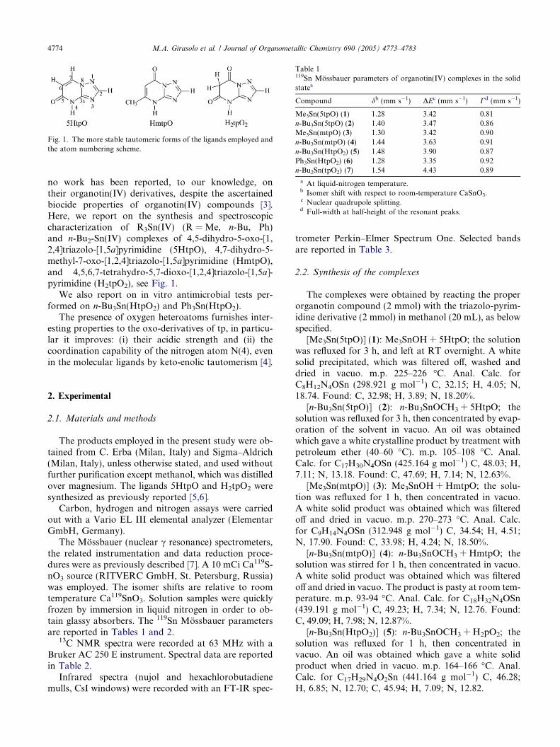
Fig. 1. The more stable tautomeric forms of the ligands employed andthe atom numbering scheme.
Table 1119Sn Mossbauer parameters of organotin(IV) complexes in the solidstatea
Compound db (mm s�1) DEc (mm s�1) Cd (mm s�1)
Me3Sn(5tpO) (1) 1.28 3.42 0.81n-Bu3Sn(5tpO) (2) 1.40 3.47 0.86Me3Sn(mtpO) (3) 1.30 3.42 0.90n-Bu3Sn(mtpO) (4) 1.44 3.63 0.91n-Bu3Sn(HtpO2) (5) 1.48 3.90 0.87Ph3Sn(HtpO2) (6) 1.28 3.35 0.92n-Bu2Sn(tpO2) (7) 1.54 4.43 0.89
a At liquid-nitrogen temperature.b Isomer shift with respect to room-temperature CaSnO3.c Nuclear quadrupole splitting.d Full-width at half-height of the resonant peaks.
4774 M.A. Girasolo et al. / Journal of Organometallic Chemistry 690 (2005) 4773–4783
no work has been reported, to our knowledge, ontheir organotin(IV) derivatives, despite the ascertainedbiocide properties of organotin(IV) compounds [3].Here, we report on the synthesis and spectroscopiccharacterization of R3Sn(IV) (R = Me, n-Bu, Ph)and n-Bu2-Sn(IV) complexes of 4,5-dihydro-5-oxo-[1,2,4]triazolo-[1,5a]pyrimidine (5HtpO), 4,7-dihydro-5-methyl-7-oxo-[1,2,4]triazolo-[1,5a]pyrimidine (HmtpO),and 4,5,6,7-tetrahydro-5,7-dioxo-[1,2,4]triazolo-[1,5a]-pyrimidine (H2tpO2), see Fig. 1.
We also report on in vitro antimicrobial tests per-formed on n-Bu3Sn(HtpO2) and Ph3Sn(HtpO2).
The presence of oxygen heteroatoms furnishes inter-esting properties to the oxo-derivatives of tp, in particu-lar it improves: (i) their acidic strength and (ii) thecoordination capability of the nitrogen atom N(4), evenin the molecular ligands by keto-enolic tautomerism [4].
2. Experimental
2.1. Materials and methods
The products employed in the present study were ob-tained from C. Erba (Milan, Italy) and Sigma–Aldrich(Milan, Italy), unless otherwise stated, and used withoutfurther purification except methanol, which was distilledover magnesium. The ligands 5HtpO and H2tpO2 weresynthesized as previously reported [5,6].
Carbon, hydrogen and nitrogen assays were carriedout with a Vario EL III elemental analyzer (ElementarGmbH, Germany).
The Mossbauer (nuclear c resonance) spectrometers,the related instrumentation and data reduction proce-dures were as previously described [7]. A 10 mCi Ca119S-nO3 source (RITVERC GmbH, St. Petersburg, Russia)was employed. The isomer shifts are relative to roomtemperature Ca119SnO3. Solution samples were quicklyfrozen by immersion in liquid nitrogen in order to ob-tain glassy absorbers. The 119Sn Mossbauer parametersare reported in Tables 1 and 2.
13C NMR spectra were recorded at 63 MHz with aBruker AC 250 E instrument. Spectral data are reportedin Table 2.
Infrared spectra (nujol and hexachlorobutadienemulls, CsI windows) were recorded with an FT-IR spec-
trometer Perkin–Elmer Spectrum One. Selected bandsare reported in Table 3.
2.2. Synthesis of the complexes
The complexes were obtained by reacting the properorganotin compound (2 mmol) with the triazolo-pyrim-idine derivative (2 mmol) in methanol (20 mL), as belowspecified.
[Me3Sn(5tpO)] (1): Me3SnOH + 5HtpO; the solutionwas refluxed for 3 h, and left at RT overnight. A whitesolid precipitated, which was filtered off, washed anddried in vacuo. m.p. 225–226 �C. Anal. Calc. forC8H12N4OSn (298.921 g mol�1) C, 32.15; H, 4.05; N,18.74. Found: C, 32.98; H, 3.89; N, 18.20%.
[n-Bu3Sn(5tpO)] (2): n-Bu3SnOCH3 + 5HtpO; thesolution was refluxed for 3 h, then concentrated by evap-oration of the solvent in vacuo. An oil was obtainedwhich gave a white crystalline product by treatment withpetroleum ether (40–60 �C). m.p. 105–108 �C. Anal.Calc. for C17H30N4OSn (425.164 g mol�1) C, 48.03; H,7.11; N, 13.18. Found: C, 47.69; H, 7.14; N, 12.63%.
[Me3Sn(mtpO)] (3): Me3SnOH + HmtpO; the solu-tion was refluxed for 1 h, then concentrated in vacuo.A white solid product was obtained which was filteredoff and dried in vacuo. m.p. 270–273 �C. Anal. Calc.for C9H14N4OSn (312.948 g mol�1) C, 34.54; H, 4.51;N, 17.90. Found: C, 33.98; H, 4.24; N, 18.50%.
[n-Bu3Sn(mtpO)] (4): n-Bu3SnOCH3 + HmtpO; thesolution was stirred for 1 h, then concentrated in vacuo.A white solid product was obtained which was filteredoff and dried in vacuo. The product is pasty at room tem-perature. m.p. 93-94 �C. Anal. Calc. for C18H32N4OSn(439.191 g mol�1) C, 49.23; H, 7.34; N, 12.76. Found:C, 49.09; H, 7.98; N, 12.87%.
[n-Bu3Sn(HtpO2)] (5): n-Bu3SnOCH3 + H2pO2; thesolution was refluxed for 1 h, then concentrated invacuo. An oil was obtained which gave a white solidproduct when dried in vacuo. m.p. 164–166 �C. Anal.Calc. for C17H29N4O2Sn (441.164 g mol�1) C, 46.28;H, 6.85; N, 12.70; C, 45.94; H, 7.09; N, 12.82.
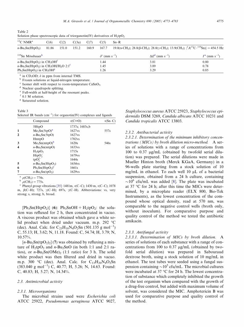
Table 2Solution phase spectroscopic data of triorganotin(IV) derivatives of H2tpO2
13C NMRa C(6) C(2) C(3a) C(7) C(5) Sn–R
n-Bu3Sn(HtpO2) 81.86 151.0 151.2 160.9 167.7 19.0(a-CH2); 28.8(b-CH2); 28.0(c-CH2); 13.9(CH3); j1J(13C–119Sn)j = 454.5 Hz
119Sn Mossbauerb dc (mm s�1) DEd (mm s�1) Ce (mm s�1)
n-Bu3Sn(HtpO2) in CH3OHf 1.44 3.81 0.80n-Bu3Sn(HtpO2) in CH3OH/H2O 2:1f 1.45 3.89 0.78Ph3Sn(HtpO2) in CH3OHg 1.26 3.29 0.85
a in CD3OD; d in ppm from internal TMS.b Frozen solutions at liquid-nitrogen temperature.c Isomer shift with respect to room-temperature CaSnO3.d Nuclear quadrupole splitting.e Full-width at half-height of the resonant peaks.f 0.1 M solution.g Saturated solution.
Table 3Selected IR bands (cm�1) for organotin(IV) complexes and ligands
Compound m(C@O) m(Sn–C)
5HtpO 1737s; 1683s,b1 Me3Sn(5tpO)a 1627vs 557s2 n-Bu3Sn(5tpO) 1627vs
HmtpO 1702vs3 Me3Sn(mtpO)b 1620s 546s4 n-Bu3Sn(mtpO) 1633vs
H2tpO2 1715sHtpO�
2 1679vstpO2�
2 1644s5 n-Bu3Sn(HtpO2) 1654vs6 Ph3Sn(HtpO2)
c 1661s7 n-Bu2Sn(tpO2) 1629vs
a q(CH3) = 774s.b q(CH3) = 775s.c Phenyl group vibrations [35]: 1481m, m(C–C); 1430 m, m(C–C); 1078
m, b(C–H); 727s, c(C–H); 697s, c(C–H). Abbreviations: vs, verystrong; s, strong; b, broad.
M.A. Girasolo et al. / Journal of Organometallic Chemistry 690 (2005) 4773–4783 4775
[Ph3Sn(HtpO2)] (6): Ph3SnOH + H2tpO2: the solu-tion was refluxed for 2 h, then concentrated in vacuo.A viscous product was obtained which gave a white so-lid product when dried under vacuum. m.p. 292 �C(dec). Anal. Calc. for C23H18N4O2Sn (501.135 g mol�1)C, 55.13; H, 3.62; N, 11.18. Found: C, 54.74; H, 3.79; N,10.57%.
[n-Bu2Sn(tpO2)2] (7) was obtained by refluxing a mix-ture of H2tpO2 and n-Bu2SnO (in both 1:1 and 2:1 ra-tios), or n-Bu2Sn(OMe)2 (1:1 ratio) for 3 h. The solidwhite product was then filtered and dried in vacuo.m.p. 300 �C (dec). Anal. Calc. for C13H20N4O2Sn(383.040 g mol�1) C, 40.77; H, 5.26; N, 14.63. Found:C, 40.83; H, 5.27; N, 14.34%.
2.3. Antimicrobial activity
2.3.1. Microorganisms
The microbial strains used were Escherichia coli
ATCC 25922, Pseudomonas aeruginosa ATCC 9027,
Staphylococcus aureus ATCC 25923, Staphylococcus epi-dermidis DSM 3269, Candida albicans ATCC 10231 andCandida tropicalis ATCC 13803.
2.3.2. Antibacterial activity
2.3.2.1. Determination of the minimum inhibitory concen-
trations (MICs) by broth dilution micro-method. A ser-ies of solutions with a range of concentrations from100 to 0.37 lg/mL (obtained by twofold serial dilu-tion) was prepared. The serial dilutions were made inMueller Hinton broth (Merck KGaA, Germany) in a96-wells plate starting from a stock solution of 10mg/mL in ethanol. To each well 10 lL of a bacterialsuspension, obtained from a 24 h culture, containing�106 cfu/mL was added [8]. The plate was incubatedat 37 �C for 24 h; after this time the MICs were deter-mined, by a microplate reader (ELX 800, Bio-TekInstruments), as the lowest concentration of the com-pound whose optical density, read at 570 nm, wascomparable to the negative control wells (broth only,without inoculum). For comparative purpose andquality control of the method we tested the antibioticamikacin.
2.3.3. Antifungal activity
2.3.3.1. Determination of MICs by broth dilution. Aseries of solutions of each substance with a range of con-centrations from 100 to 0.37 lg/mL (obtained by two-fold serial dilution) was prepared in Sabourauddextrose broth, using a stock solution of 10 mg/mL inethanol. The test tubes were seeded using a fungal sus-pension containing �105 cfu/mL. The microbial cultureswere incubated at 37 �C for 24 h. The lowest concentra-tion of substance which completely inhibited the growthof the test organism when compared with the growth ofa drug-free control, but added with maximum volume ofsolvent, was considered the MIC. Amphotericin B wasused for comparative purpose and quality control ofthe method.
4776 M.A. Girasolo et al. / Journal of Organometallic Chemistry 690 (2005) 4773–4783
2.3.4. Inhibition of germ tube formation
Sabouraud dextrose broth containing 20% foetal bo-vine serum was used as medium in germ tube formationassay. 1 mL of a fungal suspension in 0.9% NaCl,whose transmittance at 570 nm was about 15%, wasseeded in the medium described before, supplementedwith several concentrations ranging from 3 to 0.78lg/mL of n-Bu3Sn(HtpO2) (5), its organotin precursorn-Bu3SnOCH3, and Ph3Sn(HtpO2) (6) (that showedthe best activity on C. albicans) and incubated at 37�C in shaking bath for 3 h. After this incubation timewe determined, by counting in a hematocytometer,the number of cells with germ tube and we could calcu-late the inhibition percentages by comparing the samplewith a drug-free growth control, but added withmaximum concentration of solvent used to dissolvethe substances, undergone to the same experimentalconditions.
2.3.5. Biofilms susceptibility testing
C. albicans ATCC 10231 was cultivated in yeastnitrogen base (YNB) medium (Sigma) with 50 mM glu-cose at 37 �C for 24 h. After this time it was diluted inthe same medium to an optical density of 0.8 at 520nm. [9]. 100 lL of such suspension were inoculated inwells of a 96-well plates at 37 �C; after 24 h the wellswere washed three times with 200 lL of sterile phos-phate-buffered saline (PBS). The plates were air driedat 37 �C and to each well, except in the case of positive(growth) controls, 100 lL broth Sabouraud were added,supplemented with concentrations ranging from 25 to0.78 lg/mL, the MIC of n-Bu3Sn(HtpO2) determinedon planktonic form of the same strain. The plates wereincubated at 37 �C for 24 h; after this incubation timethe medium was removed, the plates were air driedand then each well was filled with 100 lL of PBS adding20 lL of a 5 mg/mL MTT (methylthiazotetrazolium)solution and incubated for 4 h at 37 �C. The insolublepurple formazan, obtained by cleavage of MTT madeby dehydrogenase enzymes of living cells, was dissolvedwith a mixture of acidic isopropyl alcohol and Triton X-100 (Sigma). The optical density of each well was readby a microplate reader (ELX 800, Bio-Tek Instruments)at 570 nm with background subtraction at 630 nm.Comparison of optical density of positive control wellswith those of sample enabled the calculation of inhibi-tion percentages of our substance at severalconcentrations.
Biological activity data are reported in Tables 7 and 8and in Fig. 5.
2.4. Quantum chemical calculations
DFT calculations, with full geometry optimization,were performed on the 4,5,6,7-tetrahydro-5,7-dioxo-[1,2,4]triazolo-[1,5a]-pyrimidine (H2tpO2) ligand, and
on the two mono-anionic and on the di-anionic forms(see Fig. 3), by the hybrid B3LYP method and usingthe 6-31++G(D,P) basis set [10]. A vibration analysiswas performed within the harmonic approximation,and the unscaled frequency values were compared withthe corresponding experimental data. A two layersONIOM calculation [11], with full geometry optimiza-tion, was performed on two isomeric trans-di-n-butyloctahedral model complexes of di-n-butyltin(IV) withthe tpO2�
2 ligand, mimicking a truncated mono-dimen-sional polymer with cis-O2 and trans-O2 coordination,respectively (see Fig. 4). The DFT B3lyp level, withthe DZVP basis set [12], was used for the higher layer,including the central di-n-butyltin moiety and the twocoordinated tpO2�
2 ligands; the semiempirical AM1method was used for the lower layer. To neutralize theoverall charge of the system, the external ligands wereconsidered monoanionic. All calculations were per-formed by the Gaussian98 program package [13]. The119Sn Mossbauer nuclear quadrupole splitting was cal-culated by the calibration formula DEcalcd = 0.93 Æ V ±0.58 mm s�1 [14], where V ¼ V zz½1þ 1
3ðV xx�V yy
V zzÞ2�1=2 and
where Vxx, Vyy, Vzz, following the condition jVzzj PjVyyjP jVxxj, are the eigenvalues of the diagonalizedelectric field gradient tensor, calculated at the same levelof theory of the higher layer [14].
3. Results and discussion
3.1. 5HtpO derivatives
The most basic positions for the ligand in the molec-ular or anionic form appear to be, on the basis of semi-empirical [5] and ab initio calculations [15], N(3) andN(4), respectively (Fig. 1).
X-ray structures of some transition metal complexesshow that in most cases the ligand, in his neutral enolicor anionic form, bridges two metal ions through N(3),N(4) atoms [16,4,17]; only two exceptions are known,[Cu2(phen)2(5tpO)2(H2O)2](NO3)2 Æ 4H2O [16] where5tpO� bridges two Cu2+ ions through N(3) and O(5)atoms, and [Ni(NCS)2(5HtpO)2 Æ (H2O)2] [18], charac-terized by an all-trans octahedral structure with two5HtpO ligands coordinated through the N atom in posi-tion 3. The ionization of the ligand is normally associ-ated to a shift of the m(C@O) absorption to lowerwavenumbers, while an extra-shift has been attributedto the involvement of O(5) in coordination [16]. In fact,in the above mentioned copper complex [16], where thetriazolopyrimidine ligand binds the copper ions throughN(3) and O(5), m(C@O) is at 1637 cm�1, while in com-plexes where the ligand is N(3), N(4) coordinated,m(C@O) appears near 1650 cm�1 [16].
In the Me3Sn(5tpO) and n-Bu3Sn(5tpO) complexesthe m(C@O) absorption is observed at 1627 cm�1
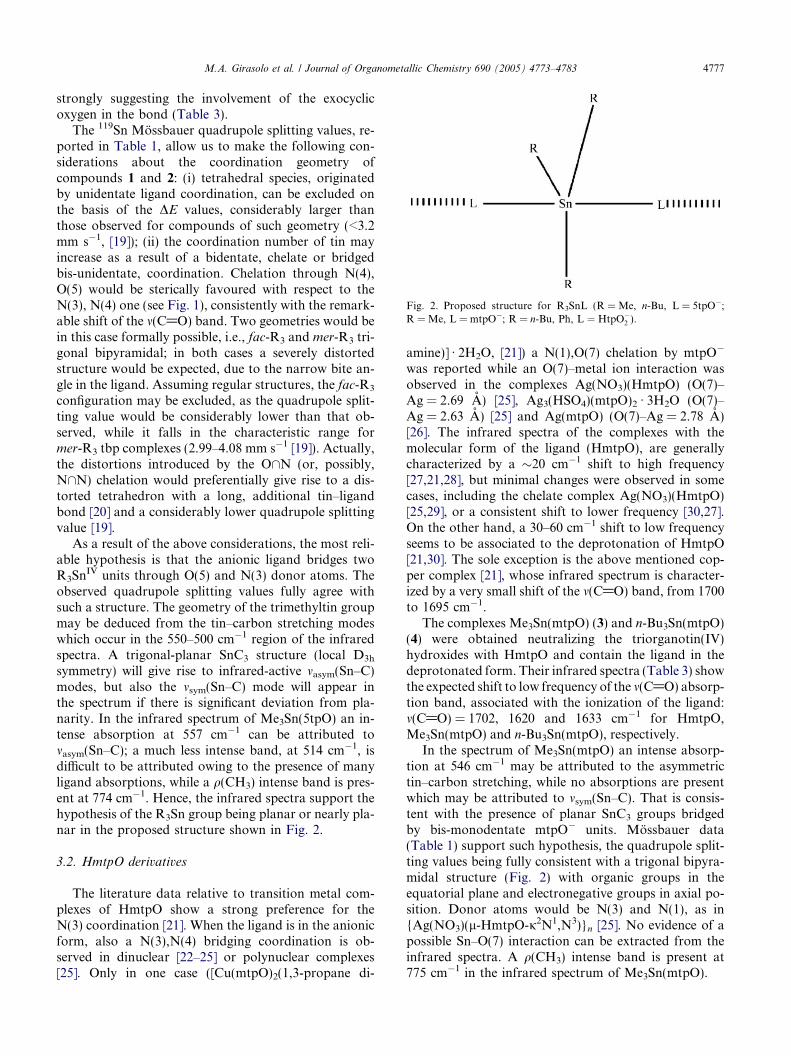
Fig. 2. Proposed structure for R3SnL (R = Me, n-Bu, L = 5tpO�;R = Me, L = mtpO�; R = n-Bu, Ph, L ¼ HtpO�
2 ).
M.A. Girasolo et al. / Journal of Organometallic Chemistry 690 (2005) 4773–4783 4777
strongly suggesting the involvement of the exocyclicoxygen in the bond (Table 3).
The 119Sn Mossbauer quadrupole splitting values, re-ported in Table 1, allow us to make the following con-siderations about the coordination geometry ofcompounds 1 and 2: (i) tetrahedral species, originatedby unidentate ligand coordination, can be excluded onthe basis of the DE values, considerably larger thanthose observed for compounds of such geometry (<3.2mm s�1, [19]); (ii) the coordination number of tin mayincrease as a result of a bidentate, chelate or bridgedbis-unidentate, coordination. Chelation through N(4),O(5) would be sterically favoured with respect to theN(3), N(4) one (see Fig. 1), consistently with the remark-able shift of the m(C@O) band. Two geometries would bein this case formally possible, i.e., fac-R3 and mer-R3 tri-gonal bipyramidal; in both cases a severely distortedstructure would be expected, due to the narrow bite an-gle in the ligand. Assuming regular structures, the fac-R3
configuration may be excluded, as the quadrupole split-ting value would be considerably lower than that ob-served, while it falls in the characteristic range former-R3 tbp complexes (2.99–4.08 mm s�1 [19]). Actually,the distortions introduced by the O\N (or, possibly,N\N) chelation would preferentially give rise to a dis-torted tetrahedron with a long, additional tin–ligandbond [20] and a considerably lower quadrupole splittingvalue [19].
As a result of the above considerations, the most reli-able hypothesis is that the anionic ligand bridges twoR3Sn
IV units through O(5) and N(3) donor atoms. Theobserved quadrupole splitting values fully agree withsuch a structure. The geometry of the trimethyltin groupmay be deduced from the tin–carbon stretching modeswhich occur in the 550–500 cm�1 region of the infraredspectra. A trigonal-planar SnC3 structure (local D3h
symmetry) will give rise to infrared-active masym(Sn–C)modes, but also the msym(Sn–C) mode will appear inthe spectrum if there is significant deviation from pla-narity. In the infrared spectrum of Me3Sn(5tpO) an in-tense absorption at 557 cm�1 can be attributed tomasym(Sn–C); a much less intense band, at 514 cm�1, isdifficult to be attributed owing to the presence of manyligand absorptions, while a q(CH3) intense band is pres-ent at 774 cm�1. Hence, the infrared spectra support thehypothesis of the R3Sn group being planar or nearly pla-nar in the proposed structure shown in Fig. 2.
3.2. HmtpO derivatives
The literature data relative to transition metal com-plexes of HmtpO show a strong preference for theN(3) coordination [21]. When the ligand is in the anionicform, also a N(3),N(4) bridging coordination is ob-served in dinuclear [22–25] or polynuclear complexes[25]. Only in one case ([Cu(mtpO)2(1,3-propane di-
amine)] Æ 2H2O, [21]) a N(1),O(7) chelation by mtpO�
was reported while an O(7)–metal ion interaction wasobserved in the complexes Ag(NO3)(HmtpO) (O(7)–Ag = 2.69 A) [25], Ag3(HSO4)(mtpO)2 Æ 3H2O (O(7)–Ag = 2.63 A) [25] and Ag(mtpO) (O(7)–Ag = 2.78 A)[26]. The infrared spectra of the complexes with themolecular form of the ligand (HmtpO), are generallycharacterized by a �20 cm�1 shift to high frequency[27,21,28], but minimal changes were observed in somecases, including the chelate complex Ag(NO3)(HmtpO)[25,29], or a consistent shift to lower frequency [30,27].On the other hand, a 30–60 cm�1 shift to low frequencyseems to be associated to the deprotonation of HmtpO[21,30]. The sole exception is the above mentioned cop-per complex [21], whose infrared spectrum is character-ized by a very small shift of the m(C@O) band, from 1700to 1695 cm�1.
The complexes Me3Sn(mtpO) (3) and n-Bu3Sn(mtpO)(4) were obtained neutralizing the triorganotin(IV)hydroxides with HmtpO and contain the ligand in thedeprotonated form. Their infrared spectra (Table 3) showthe expected shift to low frequency of the m(C@O) absorp-tion band, associated with the ionization of the ligand:m(C@O) = 1702, 1620 and 1633 cm�1 for HmtpO,Me3Sn(mtpO) and n-Bu3Sn(mtpO), respectively.
In the spectrum of Me3Sn(mtpO) an intense absorp-tion at 546 cm�1 may be attributed to the asymmetrictin–carbon stretching, while no absorptions are presentwhich may be attributed to msym(Sn–C). That is consis-tent with the presence of planar SnC3 groups bridgedby bis-monodentate mtpO� units. Mossbauer data(Table 1) support such hypothesis, the quadrupole split-ting values being fully consistent with a trigonal bipyra-midal structure (Fig. 2) with organic groups in theequatorial plane and electronegative groups in axial po-sition. Donor atoms would be N(3) and N(1), as in{Ag(NO3)(l-HmtpO-j2N1,N3)}n [25]. No evidence of apossible Sn–O(7) interaction can be extracted from theinfrared spectra. A q(CH3) intense band is present at775 cm�1 in the infrared spectrum of Me3Sn(mtpO).
4778 M.A. Girasolo et al. / Journal of Organometallic Chemistry 690 (2005) 4773–4783
3.3. H2tpO2 derivatives
The most stable tautomers of the ligand in the molec-ular form (H2tpO2) and in its monoanionic ðHtpO�
2 Þand dianionic form ðtpO2�
2 Þ, as derived from theoreticalcalculations [6, this work] are shown in Fig. 3. The firstionization of the ligand involves the strongly acidic C(6)methylene group (pKa1 = 2.9) [6], while the second ion-ization (pKa2 = 9.9) concerns the N(4) hydrogen [6].
The infrared spectra of the ligand (Table 3) are char-acterized by a strong, broad absorption due to stretch-ing vibrations of both the carbonyl groups, centred at1715, 1679 and 1644 cm�1 in the spectra of H2tpO2,NaHtpO2 and Na2tpO2, respectively.
The greater stability of the C(6) compared to the N(4)first deprotonation was confirmed by our DFT calcula-tions, showing that the HtpO�
2 isomer A is more stable,of 31.9 kJ/mol, than HtpO�
2 B (see Fig. 3).While the neutral and monoanionic form of the
H2tpO2 ligand have been previously characterized bothexperimentally and by semi-empirical quantum chemicalcalculations [6], the structure of the dianionic form hasnot been up to date considered. Quite obviously, thering planarity is preserved in all the systems investigated.The enolic character, hence the coordination strengthtoward hard metals, of the oxygen hetero-atoms in-creases by increasing the negative charge of the mole-cule. This can be observed in Table 4, by consideringthe increasing trend in the C–O bond length (of about0.06 A) and, qualitatively, in the Mulliken negativecharge on the oxygen atoms, by going from H2tpO2 to
Fig. 3. Structures of the neutral, mono- and di-anionic H2tpO2 ligand,
tpO2�2 . This result is also supported by the monotonous
lowering of the calculated IR frequency of the stretchingbands of the carbonyl groups (see Table 5), in agreementwith the experimental IR data relative to NaHtpO2,Na2tpO2 and n-Bu2Sn(tpO2), compared to the isolatedligand (see Table 3). Following the results of our calcu-lations, this trend is to be attributable mainly to theincreasing of the enolic character of the carbonyl groupsfollowing the deprotonations of the hydrogen atoms,respectively, on C(6) and N(4).
A previous theoretical study [6] showed that the exo-cyclic oxygens, O(7) and O(5), are the primary coordina-tion sites for HtpO�
2 . The O(7),N(1) bidentatecoordination, supported by the formation of a five mem-bered chelate ring, is, however, preferred to other possi-ble coordination modes.
The O(7),N(1) coordination mode was observed in[Mn(HtpO2)2(H2O)2] [31] where the ligand has a geom-etry virtually identical to the one present in its sodiumsalt. Its infrared spectrum is characterized by the pres-ence of the carbonyl group stretching band at 1652cm�1. Comparing this value with that observed forNaHtpO2, the m(C@O) shift seems rather to be deter-mined by the ionization of the ligand, than by metalcoordination.
Triorganotin complexes, n-Bu3Sn(HtpO2) (5) andPh3Sn(HtpO2) (6), exhibit similar infrared spectra, withthe m(C@O) band centred at 1654 and 1661 cm�1,respectively. O(7),N(1) bidentate coordination can be,however, ruled out on the basis of the 119Sn Mossbauerquadrupole splitting values, which strongly suggest
obtained after full geometry optimization at DFT level (see text).
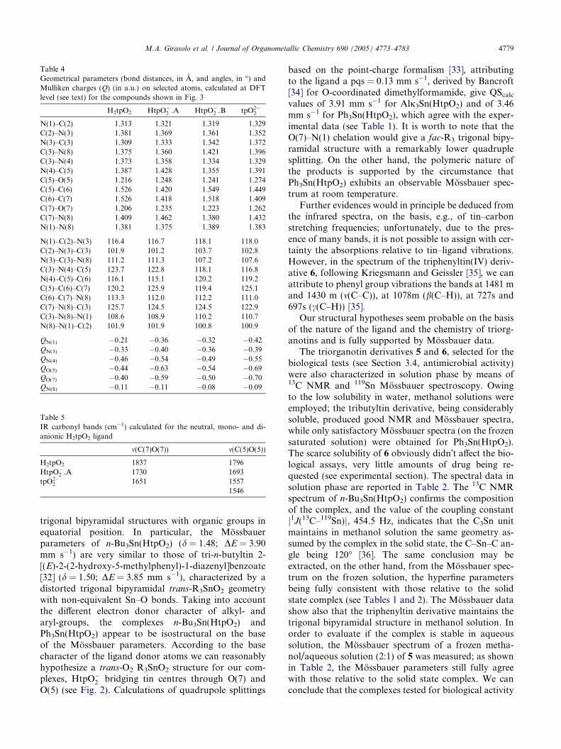
Table 4Geometrical parameters (bond distances, in A, and angles, in �) andMulliken charges (Q) (in a.u.) on selected atoms, calculated at DFTlevel (see text) for the compounds shown in Fig. 3
H2tpO2 HtpO�2 A HtpO�
2 B tpO2�2
N(1)–C(2) 1.313 1.321 1.319 1.329C(2)–N(3) 1.381 1.369 1.361 1.352N(3)–C(3) 1.309 1.333 1.342 1.372C(3)–N(8) 1.375 1.360 1.421 1.396C(3)–N(4) 1.373 1.358 1.334 1.329N(4)–C(5) 1.387 1.428 1.355 1.391C(5)–O(5) 1.216 1.248 1.241 1.274C(5)–C(6) 1.526 1.420 1.549 1.449C(6)–C(7) 1.526 1.418 1.518 1.409C(7)–O(7) 1.206 1.235 1.223 1.262C(7)–N(8) 1.409 1.462 1.380 1.432N(1)–N(8) 1.381 1.375 1.389 1.383
N(1)–C(2)–N(3) 116.4 116.7 118.1 118.0C(2)–N(3)–C(3) 101.9 101.2 103.7 102.8N(3)–C(3)–N(8) 111.2 111.3 107.2 107.6C(3)–N(4)–C(5) 123.7 122.8 118.1 116.8N(4)–C(5)–C(6) 116.1 115.1 120.2 119.2C(5)–C(6)–C(7) 120.2 125.9 119.4 125.1C(6)–C(7)–N(8) 113.3 112.0 112.2 111.0C(7)–N(8)–C(3) 125.7 124.5 124.5 122.9C(3)–N(8)–N(1) 108.6 108.9 110.2 110.7N(8)–N(1)–C(2) 101.9 101.9 100.8 100.9
QN(1) �0.21 �0.36 �0.32 �0.42QN(3) �0.33 �0.40 �0.36 �0.39QN(4) �0.46 �0.54 �0.49 �0.55QO(5) �0.44 �0.63 �0.54 �0.69QO(7) �0.40 �0.59 �0.50 �0.70QN(8) �0.11 �0.11 �0.08 �0.09
Table 5IR carbonyl bands (cm�1) calculated for the neutral, mono- and di-anionic H2tpO2 ligand
m(C(7)O(7)) m(C(5)O(5))
H2tpO2 1837 1796HtpO�
2 A 1730 1693tpO2�
2 1651 15571546
M.A. Girasolo et al. / Journal of Organometallic Chemistry 690 (2005) 4773–4783 4779
trigonal bipyramidal structures with organic groups inequatorial position. In particular, the Mossbauerparameters of n-Bu3Sn(HtpO2) (d = 1.48; DE = 3.90mm s�1) are very similar to those of tri-n-butyltin 2-[(E)-2-(2-hydroxy-5-methylphenyl)-1-diazenyl]benzoate[32] (d = 1.50; DE = 3.85 mm s�1), characterized by adistorted trigonal bipyramidal trans-R3SnO2 geometrywith non-equivalent Sn–O bonds. Taking into accountthe different electron donor character of alkyl- andaryl-groups, the complexes n-Bu3Sn(HtpO2) andPh3Sn(HtpO2) appear to be isostructural on the baseof the Mossbauer parameters. According to the basecharacter of the ligand donor atoms we can reasonablyhypothesize a trans-O2 R3SnO2 structure for our com-plexes, HtpO�
2 bridging tin centres through O(7) andO(5) (see Fig. 2). Calculations of quadrupole splittings
based on the point-charge formalism [33], attributingto the ligand a pqs = 0.13 mm s�1, derived by Bancroft[34] for O-coordinated dimethylformamide, give QScalcvalues of 3.91 mm s�1 for Alj3Sn(HtpO2) and of 3.46mm s�1 for Ph3Sn(HtpO2), which agree with the exper-imental data (see Table 1). It is worth to note that theO(7)–N(1) chelation would give a fac-R3 trigonal bipy-ramidal structure with a remarkably lower quadruplesplitting. On the other hand, the polymeric nature ofthe products is supported by the circumstance thatPh3Sn(HtpO2) exhibits an observable Mossbauer spec-trum at room temperature.
Further evidences would in principle be deduced fromthe infrared spectra, on the basis, e.g., of tin–carbonstretching frequencies; unfortunately, due to the pres-ence of many bands, it is not possible to assign with cer-tainty the absorptions relative to tin–ligand vibrations.However, in the spectrum of the triphenyltin(IV) deriv-ative 6, following Kriegsmann and Geissler [35], we canattribute to phenyl group vibrations the bands at 1481 mand 1430 m (m(C–C)), at 1078m (b(C–H)), at 727s and697s (c(C–H)) [35].
Our structural hypotheses seem probable on the basisof the nature of the ligand and the chemistry of triorg-anotins and is fully supported by Mossbauer data.
The triorganotin derivatives 5 and 6, selected for thebiological tests (see Section 3.4, antimicrobial activity)were also characterized in solution phase by means of13C NMR and 119Sn Mossbauer spectroscopy. Owingto the low solubility in water, methanol solutions wereemployed; the tributyltin derivative, being considerablysoluble, produced good NMR and Mossbauer spectra,while only satisfactory Mossbauer spectra (on the frozensaturated solution) were obtained for Ph3Sn(HtpO2).The scarce solubility of 6 obviously didn�t affect the bio-logical assays, very little amounts of drug being re-quested (see experimental section). The spectral data insolution phase are reported in Table 2. The 13C NMRspectrum of n-Bu3Sn(HtpO2) confirms the compositionof the complex, and the value of the coupling constantj1J(13C–119Sn)j, 454.5 Hz, indicates that the C3Sn unitmaintains in methanol solution the same geometry as-sumed by the complex in the solid state, the C–Sn–C an-gle being 120� [36]. The same conclusion may beextracted, on the other hand, from the Mossbauer spec-trum on the frozen solution, the hyperfine parametersbeing fully consistent with those relative to the solidstate complex (see Tables 1 and 2). The Mossbauer datashow also that the triphenyltin derivative maintains thetrigonal bipyramidal structure in methanol solution. Inorder to evaluate if the complex is stable in aqueoussolution, the Mossbauer spectrum of a frozen metha-nol/aqueous solution (2:1) of 5 was measured; as shownin Table 2, the Mossbauer parameters still fully agreewith those relative to the solid state complex. We canconclude that the complexes tested for biological activity
4780 M.A. Girasolo et al. / Journal of Organometallic Chemistry 690 (2005) 4773–4783
maintain in solution the same structure they have in thesolid state.
The diorganotin derivative, n-Bu2Sn(tpO2) (7) wasobtained reacting n-Bu2SnO with H2tpO2 (in 1:1 and1:2 ratios), or n-Bu2Sn(OMe)2 with H2tpO2 (1:1 ratio).In any case a compound is obtained whose Mossbauerspectrum is characterized by a very wide doublet (DE-mean = 4.44 mm s�1; Cmean = 0.91 mm s�1) typical oftrans-R2 octahedral structures. The complex exhibits ob-servable Mossbauer spectrum at room temperature sug-gesting a polymeric nature. On the other hand,considering the stoichiometry of the compound, only apolymeric structure would justify the observed nuclearquadrupole splitting, as only a bridged coordination ofthe ligand would allow an octahedral arrangement of li-gand atoms around tin.
A trans-R2 octahedral structure implies that the li-gand chelates two organotin(IV) moieties. A donoratoms couple would be obviously O(7)–N(1), while theother one could be either O(5)–N(4) or N(4)–N(3).Moreover, there is a consequent ambiguity about themutual disposition of the chelating ligands in the equa-
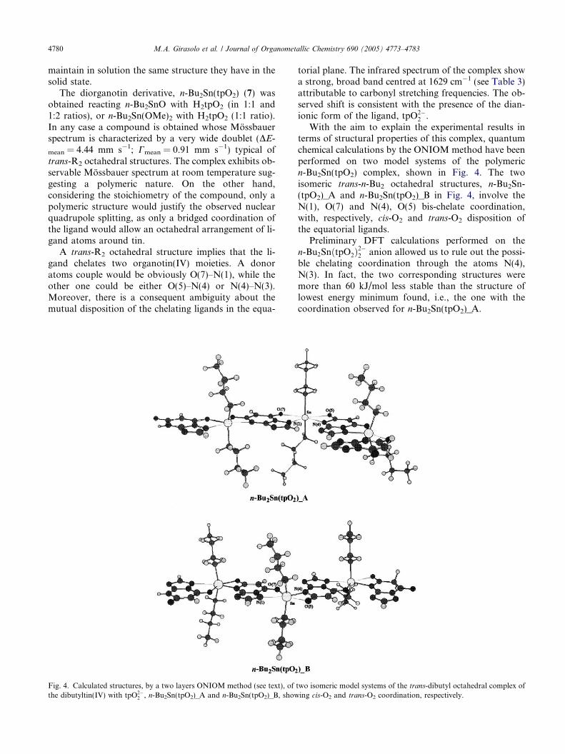
Fig. 4. Calculated structures, by a two layers ONIOM method (see text), ofthe dibutyltin(IV) with tpO2�
2 , n-Bu2Sn(tpO2)_A and n-Bu2Sn(tpO2)_B, show
torial plane. The infrared spectrum of the complex showa strong, broad band centred at 1629 cm�1 (see Table 3)attributable to carbonyl stretching frequencies. The ob-served shift is consistent with the presence of the dian-ionic form of the ligand, tpO2�
2 .With the aim to explain the experimental results in
terms of structural properties of this complex, quantumchemical calculations by the ONIOM method have beenperformed on two model systems of the polymericn-Bu2Sn(tpO2) complex, shown in Fig. 4. The twoisomeric trans-n-Bu2 octahedral structures, n-Bu2Sn-(tpO2)_A and n-Bu2Sn(tpO2)_B in Fig. 4, involve theN(1), O(7) and N(4), O(5) bis-chelate coordination,with, respectively, cis-O2 and trans-O2 disposition ofthe equatorial ligands.
Preliminary DFT calculations performed on then-Bu2SnðtpO2Þ
2�2 anion allowed us to rule out the possi-
ble chelating coordination through the atoms N(4),N(3). In fact, the two corresponding structures weremore than 60 kJ/mol less stable than the structure oflowest energy minimum found, i.e., the one with thecoordination observed for n-Bu2Sn(tpO2)_A.
two isomeric model systems of the trans-dibutyl octahedral complex ofing cis-O2 and trans-O2 coordination, respectively.
M.A. Girasolo et al. / Journal of Organometallic Chemistry 690 (2005) 4773–4783 4781
Among the two possible isomeric structures consid-ered (see Fig. 4), the cis-O2 coordination, correspondingto n-Bu2Sn(tpO2)_A, is more stable of about 18 kJ/molcompared to the trans-O2 coordination, correspondingto n-Bu2Sn(tpO2)_B. Moreover, the calculated nuclearquadrupole splitting value relative to n-Bu2Sn(tpO2)_Ais in much better agreement with the experimental valueof DE, compared to the one calculated for n-Bu2Sn-(tpO2)_B (see Table 6). These results strongly supportthe hypothesis of trans-di-n-butyl, cis-O2 distorted octa-hedral coordination geometry for the investigatedcompound.
A further support to such coordination geometry alsocomes from the polymeric structure obtained in the solidstate for the complex NaHtpO2 [6]. The X-ray crystallo-graphic analysis showed the involvement of a similarcoordination of the metal atom through N(1), O(7),O(5) and of the protonated N(4) atom through hydro-gen bond to a coordinated water molecule. It is probablethat the hard character of sodium and tin(IV) leads tothe preferential coordination of the highly negativelycharged oxygen atoms compared to the N(3) atom.
Table 6Relevant geometrical parameters (bond distances, in A, and angles,in �) relative to the central tin environment of the two isomer modelsystems, n-Bu2Sn(tpO2)_A and n-Bu2Sn(tpO2)_B, shown in Fig. 4,calculated by a two layer ONIOM method (see text). Their relativeenergy and the calculated nuclear quadrupole splitting values are alsoreported
n-Bu2Sn(tpO2)_A n-Bu2Sn(tpO2)_B
Sn–C (average) 2.175 2.173Sn–N(1) 2.299 2.701Sn–O(7) 2.411 2.161Sn–N(4) 2.325 2.249Sn–O(5) 2.412 2.412C(5)–O(5) 1.271 1.289C(7)–O(7) 1.269 1.273
C–Sn–C 150.7 142.8N(1)–Sn–O(7) 71.4 68.7N(1)–Sn–O(5) 147.3 150.7N(1)–Sn–N(4) 90.9 152.2N(4)–Sn–O(5) 56.5 57.1N(4)–Sn–O(7) 162.2 83.5O(5)–Sn–O(7) 141.3 140.4
Energy (kJ/mol) 0.0 18.5DEcalcd (mm s�1) 4.15 3.78
Table 7Antimicrobial activity in vitro, MIC values in lg/mL
Compound E. coli
ATCC25922P. aeruginosa
ATCC9027S. a
AT
n-Bu3SnOCH3 >100 >100 >105 n-Bu3Sn(HtpO2) >100 100 1
Ph3SnOH >100 >100 >106 Ph3Sn(HtpO2) >25 >25
Amikacin 10 5Amphotericin B
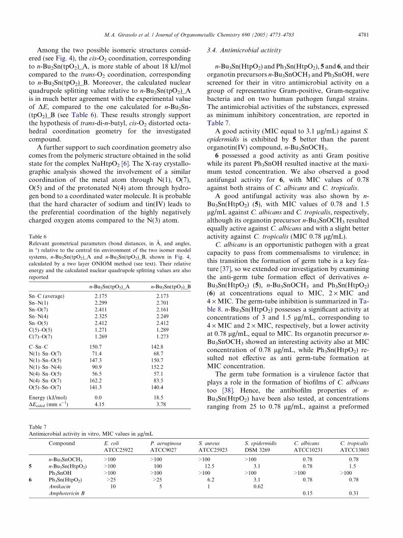
3.4. Antimicrobial activity
n-Bu3Sn(HtpO2) and Ph3Sn(HtpO2), 5 and 6, and theirorganotin precursors n-Bu3SnOCH3 and Ph3SnOH, werescreened for their in vitro antimicrobial activity on agroup of representative Gram-positive, Gram-negativebacteria and on two human pathogen fungal strains.The antimicrobial activities of the substances, expressedas minimum inhibitory concentration, are reported inTable 7.
A good activity (MIC equal to 3.1 lg/mL) against S.epidermidis is exhibited by 5 better than the parentorganotin(IV) compound, n-Bu3SnOCH3.
6 possessed a good activity as anti Gram positivewhile its parent Ph3SnOH resulted inactive at the maxi-mum tested concentration. We also observed a goodantifungal activity for 6, with MIC values of 0.78against both strains of C. albicans and C. tropicalis.
A good antifungal activity was also shown by n-Bu3Sn(HtpO2) (5), with MIC values of 0.78 and 1.5lg/mL against C. albicans and C. tropicalis, respectively,although its organotin precursor n-Bu3SnOCH3 resultedequally active against C. albicans and with a slight betteractivity against C. tropicalis (MIC 0.78 lg/mL).
C. albicans is an opportunistic pathogen with a greatcapacity to pass from commensalisms to virulence; inthis transition the formation of germ tube is a key fea-ture [37], so we extended our investigation by examiningthe anti-germ tube formation effect of derivatives n-Bu3Sn(HtpO2) (5), n-Bu3SnOCH3 and Ph3Sn(HtpO2)(6) at concentrations equal to MIC, 2 · MIC and4 · MIC. The germ-tube inhibition is summarized in Ta-ble 8. n-Bu3Sn(HtpO2) possesses a significant activity atconcentrations of 3 and 1.5 lg/mL, corresponding to4 · MIC and 2 · MIC, respectively, but a lower activityat 0.78 lg/mL, equal to MIC. Its organotin precursor n-Bu3SnOCH3 showed an interesting activity also at MICconcentration of 0.78 lg/mL, while Ph3Sn(HtpO2) re-sulted not effective as anti germ-tube formation atMIC concentration.
The germ tube formation is a virulence factor thatplays a role in the formation of biofilms of C. albicanstoo [38]. Hence, the antibiofilm properties of n-Bu3Sn(HtpO2) have been also tested, at concentrationsranging from 25 to 0.78 lg/mL, against a preformed
ureus
CC25923S. epidermidis
DSM 3269C. albicans
ATCC10231C. tropicalis
ATCC13803
0 >100 0.78 0.782.5 3.1 0.78 1.50 >100 >100 >1006.2 3.1 0.78 0.781 0.62
0.15 0.31

Table 8Inhibition of germ tube formation in C. albicans ATCC 10231
Compound Concentration
4 · MIC 2 · MIC MIC
5 n-Bu3Sn(HtpO2) 92.1 ± 0.7 90 ± 5 35.1 ± 0.76 Ph3Sn(HtpO2) 83 ± 4 35 ± 5 ns
n-Bu3SnOCH3 85 ± 3 75 ± 3 68 ± 2
ns: not significant; the values are the mean of at least three independentdeterminations; coefficient of variation was less than 15%.
Fig. 5. In vitro inhibition percentages on C. albicans ATCC10231biofilm by n-Bu3Sn(HtpO2) (5). Values are the average of at least threeindependent determinations. Coefficient of variation was less than15%.
4782 M.A. Girasolo et al. / Journal of Organometallic Chemistry 690 (2005) 4773–4783
biofilm of C. albicans. Although the MIC value wasgood against the planktonic form of C. albicans, we re-ported a weak activity at concentration equal to MICagainst biofilm of the same strain, in fact we only ob-served an interesting antibiofilm effect at concentrations16 · MIC or more (see Fig. 5).
Candida albicans infections often involve the forma-tion of biofilms on implanted devices or on tissue sur-faces, such infections are difficult to treat, becausebiofilms are resistant to a wide range of current anti-fungal agents [39]. There is therefore an urgent need todevelop new agents and we think that n-Bu3Sn(HtpO2)might represent a lead compound for developing anti-fungal and antibiofilm agents.
Acknowledgment
The financial support of Universita di Palermo, Italy,is gratefully acknowledged.
References
[1] J.M. Salas, M.A. Romero, M.P. Sanchez, M. Quiros, Coord.Chem. Rev. 193–195 (1999) 1119.
[2] J.G. Haasnot, Coord. Chem. Rev. 200–202 (2000) 131.[3] See for example: (a) L. Pellerito, L. Nagy, Coord. Chem. Rev. 224
(2002) 111;
(b) M. Nath, R. Yadav, G. Eng, P. Musingarimi, Appl.Organomet. Chem. 13 (1999) 29;(c) M. Nath, S. Pokharia, G. Eng, X. Song, A. Kumar, M.Gielen, R. Willem, M. Biesemans, Appl. Organomet. Chem. 18(2004) 460;(d) P. Novak, A. Lycka, I. Cısarova, V. Buchta, L. Silva, L.Kolarova, A. Ruzicka, J. Holecek, Appl. Organomet. Chem. 19(2005) 500.
[4] M. Abul Haj, M. Quiros, J.M. Salas, R. Faure, J. Chem. Soc.,Dalton Trans. (2001) 1798.
[5] M. Abul Haj, J.M. Salas, M. Quiros, J. Molina, R. Faure, J. Mol.Struct. 519 (2000) 165.
[6] S. Orihuela, M.P. Sanchez, M. Quiros, J. Molina, R. Faure, J.Mol. Struct. 415 (1997) 285.
[7] R. Barbieri, G. Ruisi, A. Silvestri, A.M. Giuliani, A. Barbieri, G.Spina, F. Pieralli, F. Del Giallo, J. Chem. Soc., Dalton Trans.(1995) 467.
[8] L.H. Lennette, E.H. Spaulding, P. Truant, in: Manual of ClinicalMicrobiology, second ed., American Society for Microbiology,Washington, DC, 1974 (Chapter 42).
[9] M.A.S. Alem, J.L. Douglas, Antimicrob. Agents Chemother. 48(2004) 41.
[10] (a) W.J. Hehre, R. Ditchfield, J.A. Pople, J. Chem. Phys. 56(1972) 2257;(b) T. Clark, J. Chandrasekhar, P.V.R. Schleyer, J. Comput.Chem. 4 (1983) 294;(c) R. Krishnam, J.S. Binkley, R. Seeger, J.A. Pople, J. Chem.Phys. 72 (1980) 650;(d) P.M.W. Gill, B.G. Johnson, J.A. Pople, M.J. Frisch, Chem.Phys. Lett. 197 (1992) 499.
[11] S. Dapprich, I. Komaromi, K.S. Byun, K. Morokuma, M.J.Frisch, J. Mol. Struct. (Theochem) 462 (1999) 1.
[12] (a) N. Godbout, D.R. Salahub, J. Andzelm, E. Wimmer, Can. J.Chem. 70 (1992) 560;(b) C. Sosa, J. Andzelm, B.C. Elkin, E. Wimmer, K.D. Dobbs,D.A. Dixon, J. Phys. Chem. 96 (1992) 6630.
[13] M.J. Frisch, G.W. Trucks, H.B. Schlegel, G.E. Scuseria, M.A.Robb, J.R. Cheeseman, V.G. Zakrzewski, J.A. Montgomery Jr.,R.E. Stratmann, J.C. Burant, S. Dapprich, J.M. Millam, A.D.Daniels, K.N. Kudin, M.C. Strain, O. Farkas, J. Tomasi, V.Barone, M. Cossi, R. Cammi, B. Mennucci, C. Pomelli, C.Adamo, S. Clifford, J. Ochterski, G.A. Petersson, P.Y. Ayala, Q.Cui, K. Morokuma, D.K. Malick, A.D. Rabuck, K. Raghava-chari, J.B. Foresman, J. Cioslowski, J.V. Ortiz, A.G. Baboul,B.B. Stefanov, G. Liu, A. Liashenko, P. Piskorz, I. Komaromi,R. Gomperts, R.L. Martin, D.J. Fox, T. Keith, M.A. Al-Laham,C.Y. Peng, A. Nanayakkara, M. Challacombe, P.M.W. Gill, B.Johnson, W. Chen, M.W. Wong, J.L. Andres, C. Gonzalez, M.Head-Gordon, E.S. Replogle, J.A. Pople, GAUSSIAN-98, RevisionA.8, Gaussian, Inc., Pittsburgh, PA, 1998.
[14] G. Barone, A. Silvestri, G. Ruisi, G. La Manna, Chem. Eur. J.,accepted (DOI: doi:10.1002/chem.200401156).
[15] J.A. Dobado, S. Grigoleit, J. Molina Molina, J. Chem. Soc.,Perkin Trans. 2 (2000) 1675.
[16] M. Abul Haj, M. Quiros, J.M. Salas, J.A. Dobado, J. MolinaMolina, G. Basallote, M.A. Manez, Eur. J. Inorg. Chem. (2002)811.
[17] M. Abul-Haj, M. Quiros, J.M. Salas, Polyhedron 23 (2004)2373.
[18] M. Abul-Haj, M. Quiros, J.M. Salas, Acta Crystallogr., Sect. C56 (2000) 934.
[19] R. Barbieri, F. Huber, L. Pellerito, G. Ruisi, A. Silvestri, in: P.J.Smith (Ed.), Chemistry of Tin, Chapman & Hall, London, 1998,p. 496 (Chapter 14).
[20] See for example: (a) C. Ma, F. Li, Q. Jiang, R. Zhang, J.Organomet. Chem. 689 (2004) 96;(b) C. Ma, Q. Jiang, R. Zhang, Polyhedron 23 (2004) 779.
M.A. Girasolo et al. / Journal of Organometallic Chemistry 690 (2005) 4773–4783 4783
[21] J.A.R. Navarro, M.A. Romero, J.M. Salas, J. Molina, E.R.T.Tiekink, Inorg. Chim. Acta 274 (1998) 53.
[22] J.A.R. Navarro, M.A. Romero, J.M. Salas, J. Chem. Soc, DaltonTrans. (1997) 1001.
[23] J.A.R. Navarro, M.A. Romero, J.M. Salas, M. Quiros, Inorg.Chem. 36 (1997) 3277.
[24] J.A.R. Navarro, M.A. Romero, J.M. Salas, M. Quiros, J. ElBahrauni, J. Molina, Inorg. Chem. 35 (1996) 7829.
[25] J.A.R. Navarro, M.A. Romero, J.M. Salas, R. Faure, X. Solans,J. Chem. Soc, Dalton Trans. (1997) 2321.
[26] D.L. Smith, H.R. Luss, Photogr. Sci. Eng. 20 (1976) 184.[27] M.H.B. Bol, E.J. Dirks, J.G. Haasnot, J. Reedijk, Inorg. Chim.
Acta 180 (1991) 33.[28] J.A.R. Navarro, J.M. Salas, M.A. Romero, R. Vilaplana, F.
Gonzalez-Vılchez, R. Faure, J. Med. Chem. 41 (1998) 332.[29] J.A.R. Navarro, M.A. Romero, J.M. Salas, M. Quiros, E.R.T.
Tiekink, Inorg. Chem. 36 (1997) 4988.
[30] E.J. Dirks, J.G. Haasnot, A.J. Kinneging, J. Reedijk, Inorg.Chem. 26 (1987) 1902.
[31] S. Orihuela, M.P. Sanchez, M. Quiros, D. Martın, R. Faure,Polyhedron 17 (1998) 2477.
[32] R. Willem, I. Verbruggen, M. Gielen, M. Biesemans, B. Mahieu,T.S. Basu Baul, E.R.T. Tiekink, Organometallics 17 (1998) 5758.
[33] G.M. Bancroft, R.H. Platt, Adv. Inorg. Chem. Radiochem. 15(1972) 59.
[34] G.M. Bancroft, V.G. Kumar Das, T.K. Sham, M.G. Clark, J.Chem. Soc., Dalton Trans. (1976) 643.
[35] Von H. Kriegsmann, H. Geissler, Z. Anorg. Allg. Chem. 323(1963) 170.
[36] J. Holecek, M. Navdornık, K. Handlır, A. Lycka, J. Organomet.Chem. 315 (1986) 299.
[37] R.A. Calderone, W.A. Fonzi, Trends Microbiol. 9 (2001) 327.[38] G.S. Baillie, L.J. Douglas, J. Med. Microbiol. 48 (1999) 671.[39] L.J. Douglas, Trends Microbiol. 11 (2003) 30.