Antimicrobial Susceptibility Testing: A Comprehensive Review ...
26
Citation: Gajic, I.; Kabic, J.; Kekic, D.; Jovicevic, M.; Milenkovic, M.; Mitic Culafic, D.; Trudic, A.; Ranin, L.; Opavski, N. Antimicrobial Susceptibility Testing: A Comprehensive Review of Currently Used Methods. Antibiotics 2022, 11, 427. https://doi.org/10.3390/ antibiotics11040427 Academic Editor: Eleonora Nicolai Received: 28 February 2022 Accepted: 18 March 2022 Published: 23 March 2022 Publisher’s Note: MDPI stays neutral with regard to jurisdictional claims in published maps and institutional affil- iations. Copyright: © 2022 by the authors. Licensee MDPI, Basel, Switzerland. This article is an open access article distributed under the terms and conditions of the Creative Commons Attribution (CC BY) license (https:// creativecommons.org/licenses/by/ 4.0/). antibiotics Review Antimicrobial Susceptibility Testing: A Comprehensive Review of Currently Used Methods Ina Gajic 1, * , Jovana Kabic 1 , Dusan Kekic 1 , Milos Jovicevic 1 , Marina Milenkovic 2 , Dragana Mitic Culafic 3 , Anika Trudic 4,5 , Lazar Ranin 1 and Natasa Opavski 1 1 Institute of Microbiology and Immunology, Faculty of Medicine, University of Belgrade, 11000 Belgrade, Serbia; [email protected] (J.K.); [email protected] (D.K.); [email protected] (M.J.); [email protected] (L.R.); [email protected] (N.O.) 2 Department of Microbiology and Immunology, Faculty of Pharmacy, University of Belgrade, 11000 Belgrade, Serbia; [email protected] 3 Faculty of Biology, University of Belgrade, 11000 Belgrade, Serbia; [email protected] 4 Faculty of Medicine, University of Novi Sad, 21000 Novi Sad, Serbia; [email protected] 5 Institute for Pulmonary Diseases of Vojvodina, Sremska Kamenica, 21204 Novi Sad, Serbia * Correspondence: [email protected]; Tel.: +381-629-726-331 Abstract: Antimicrobial resistance (AMR) has emerged as a major threat to public health globally. Accurate and rapid detection of resistance to antimicrobial drugs, and subsequent appropriate antimicrobial treatment, combined with antimicrobial stewardship, are essential for controlling the emergence and spread of AMR. This article reviews common antimicrobial susceptibility testing (AST) methods and relevant issues concerning the advantages and disadvantages of each method. Although accurate, classic technologies used in clinical microbiology to profile antimicrobial susceptibility are time-consuming and relatively expensive. As a result, physicians often prescribe empirical antimicrobial therapies and broad-spectrum antibiotics. Although recently developed AST systems have shown advantages over traditional methods in terms of testing speed and the potential for providing a deeper insight into resistance mechanisms, extensive validation is required to translate these methodologies to clinical practice. With a continuous increase in antimicrobial resistance, additional efforts are needed to develop innovative, rapid, accurate, and portable diagnostic tools for AST. The wide implementation of novel devices would enable the identification of the optimal treatment approaches and the surveillance of antibiotic resistance in health, agriculture, and the environment, allowing monitoring and better tackling the emergence of AMR. Keywords: antimicrobial susceptibility testing; antimicrobial resistance; methods 1. The Emergence of Antimicrobial Resistance and Overlooked Pandemic Antimicrobial resistance (AMR) remains the world’s most urgent public health con- cern [1]. According to the World Health Organization (WHO), Geneva, Switzerland, antibiotic resistance is rising to dangerously high levels in all parts of the world, leading to increased morbidity and mortality [2]. Hence, the six leading mortality-causing pathogens—Escherichia coli, Staphylococcus aureus, Klebsiella pneumoniae, Streptococcus pneumoniae, Acinetobacter bauman- nii, and Pseudomonas aeruginosa—were responsible for 929,000 deaths attributable to AMR and 3.57 million deaths associated with AMR in 2019 [3]. This number could rise to 10 million by 2050 according to estimates by the WHO [4]. Furthermore, the SARS-CoV-2 pandemic has exacerbated the existing global crisis of AMR, mostly due to the mis- and over-use of antibi- otics, treatments that induce immunosuppression, and prolonged hospitalisation [5]. Besides, during the COVID-19 pandemic, limited ability to work with AMR partnerships, decreases in funding, and reduced availability of nursing, medical, and public health staff affected AMR surveillance, prevention, and control [6]. In addition, increased use of disinfectants, including hand sanitisers and surface cleaners, is anticipated to cause increased rates of antimicrobial Antibiotics 2022, 11, 427. https://doi.org/10.3390/antibiotics11040427 https://www.mdpi.com/journal/antibiotics
-
Upload
khangminh22 -
Category
Documents
-
view
2 -
download
0
Transcript of Antimicrobial Susceptibility Testing: A Comprehensive Review ...
�����������������
Citation: Gajic, I.; Kabic, J.; Kekic, D.;
Jovicevic, M.; Milenkovic, M.; Mitic
Culafic, D.; Trudic, A.; Ranin, L.;
Opavski, N. Antimicrobial
Susceptibility Testing: A
Comprehensive Review of Currently
Used Methods. Antibiotics 2022, 11,
427. https://doi.org/10.3390/
antibiotics11040427
Academic Editor: Eleonora Nicolai
Received: 28 February 2022
Accepted: 18 March 2022
Published: 23 March 2022
Publisher’s Note: MDPI stays neutral
with regard to jurisdictional claims in
published maps and institutional affil-
iations.
Copyright: © 2022 by the authors.
Licensee MDPI, Basel, Switzerland.
This article is an open access article
distributed under the terms and
conditions of the Creative Commons
Attribution (CC BY) license (https://
creativecommons.org/licenses/by/
4.0/).
antibiotics
Review
Antimicrobial Susceptibility Testing: A Comprehensive Reviewof Currently Used MethodsIna Gajic 1,* , Jovana Kabic 1 , Dusan Kekic 1, Milos Jovicevic 1, Marina Milenkovic 2, Dragana Mitic Culafic 3 ,Anika Trudic 4,5, Lazar Ranin 1 and Natasa Opavski 1
1 Institute of Microbiology and Immunology, Faculty of Medicine, University of Belgrade,11000 Belgrade, Serbia; [email protected] (J.K.); [email protected] (D.K.);[email protected] (M.J.); [email protected] (L.R.); [email protected] (N.O.)
2 Department of Microbiology and Immunology, Faculty of Pharmacy, University of Belgrade,11000 Belgrade, Serbia; [email protected]
3 Faculty of Biology, University of Belgrade, 11000 Belgrade, Serbia; [email protected] Faculty of Medicine, University of Novi Sad, 21000 Novi Sad, Serbia; [email protected] Institute for Pulmonary Diseases of Vojvodina, Sremska Kamenica, 21204 Novi Sad, Serbia* Correspondence: [email protected]; Tel.: +381-629-726-331
Abstract: Antimicrobial resistance (AMR) has emerged as a major threat to public health globally.Accurate and rapid detection of resistance to antimicrobial drugs, and subsequent appropriateantimicrobial treatment, combined with antimicrobial stewardship, are essential for controlling theemergence and spread of AMR. This article reviews common antimicrobial susceptibility testing (AST)methods and relevant issues concerning the advantages and disadvantages of each method. Althoughaccurate, classic technologies used in clinical microbiology to profile antimicrobial susceptibilityare time-consuming and relatively expensive. As a result, physicians often prescribe empiricalantimicrobial therapies and broad-spectrum antibiotics. Although recently developed AST systemshave shown advantages over traditional methods in terms of testing speed and the potential forproviding a deeper insight into resistance mechanisms, extensive validation is required to translatethese methodologies to clinical practice. With a continuous increase in antimicrobial resistance,additional efforts are needed to develop innovative, rapid, accurate, and portable diagnostic toolsfor AST. The wide implementation of novel devices would enable the identification of the optimaltreatment approaches and the surveillance of antibiotic resistance in health, agriculture, and theenvironment, allowing monitoring and better tackling the emergence of AMR.
Keywords: antimicrobial susceptibility testing; antimicrobial resistance; methods
1. The Emergence of Antimicrobial Resistance and Overlooked Pandemic
Antimicrobial resistance (AMR) remains the world’s most urgent public health con-cern [1]. According to the World Health Organization (WHO), Geneva, Switzerland, antibioticresistance is rising to dangerously high levels in all parts of the world, leading to increasedmorbidity and mortality [2]. Hence, the six leading mortality-causing pathogens—Escherichiacoli, Staphylococcus aureus, Klebsiella pneumoniae, Streptococcus pneumoniae, Acinetobacter bauman-nii, and Pseudomonas aeruginosa—were responsible for 929,000 deaths attributable to AMR and3.57 million deaths associated with AMR in 2019 [3]. This number could rise to 10 million by2050 according to estimates by the WHO [4]. Furthermore, the SARS-CoV-2 pandemic hasexacerbated the existing global crisis of AMR, mostly due to the mis- and over-use of antibi-otics, treatments that induce immunosuppression, and prolonged hospitalisation [5]. Besides,during the COVID-19 pandemic, limited ability to work with AMR partnerships, decreases infunding, and reduced availability of nursing, medical, and public health staff affected AMRsurveillance, prevention, and control [6]. In addition, increased use of disinfectants, includinghand sanitisers and surface cleaners, is anticipated to cause increased rates of antimicrobial
Antibiotics 2022, 11, 427. https://doi.org/10.3390/antibiotics11040427 https://www.mdpi.com/journal/antibiotics

Antibiotics 2022, 11, 427 2 of 26
resistance in pathogenic microbes in the coming years [7]. Replacement of first-line antibioticsby more expensive medications, a longer duration of illness, and treatment-related to AMRincreases healthcare costs as well as the economic burden on patients and societies [2]. TheWorld Bank estimates that drug-resistant infections could cause a global economic crisis,leading to 28 million people who could be pushed into extreme poverty every year by 2050,with an overall cost to the global economy of USD 1 trillion per year [8].
Throughout their evolution, bacteria have developed versatile resistance mechanismsto antibiotics. The four main mechanisms of AMR are enzymatic inactivation of antimicro-bial compounds, alteration of a drug target, reduced permeability of the outer membrane,and active drug efflux [9]. Hydrolases (e.g., beta-lactamases encoding by bla genes, suchas extended-spectrum beta-lactamases, ESBL; cephalosporinases; and carbapenemases),passivation, and modified enzymes are three of the most important drug-inactivatingenzymes. An altered target site is a major cause of Gram-positive bacteria’s drug resistance(e.g., PBP2a in methicillin-resistant S. aureus, MRSA by the acquisition of the mecA gene andother homologues), as well as polymyxin-resistant bacteria. The membrane permeability isa key in the level of susceptibility to antibiotics in some bacteria, such as Enterobacterales.Modification of the bacterial envelope by decreasing the porin production or increasing theexpression of efflux pump systems (e.g., M phenotype in Streptococcus spp. encoding bymefA gene) has been reported [10].
The causes of antimicrobial resistance are complex and multifaceted. In countrieswhere antibiotics are sold without a prescription or used as growth-promoting substances orprophylactic additives in livestock farming, antibiotic-resistant bacteria develop especiallyfast [2]. Administration of antibiotics to patients with suspected moderate to severe bacterialinfections has been deemed inappropriate in at least half of the cases [11]. Antimicrobialstewardship (AMS) is one of the key strategies for combatting resistance. Implementationof such programs is therefore recommended across the globe [12].
The present review provides an updated overview of the various antimicrobial sus-ceptibility testing (AST) methods that are currently used or potentially applicable in theforeseeable future, as well as their advantages and disadvantages.
2. The Rationale for Performing Susceptibility Testing
The choice of the best therapeutic option for the treatment of bacterial infections relieson the results of AST, a part of the routine work of all clinical microbiological laboratories.These reports provide insight into local patterns of antimicrobial susceptibility, helpingphysicians to choose the most effective antibiotic therapy [13]. For instance, if the AMR rateof a pathogen is above 20%, that drug should not be administered as a single empiric ther-apy for infection treatment [14]. Evaluation of the effectiveness of prevention and infectioncontrol measures relies as well on the results of AST, e.g., monitoring of resistant pathogenssuch as MRSA (methicillin-resistant Staphylococcus aureus), VRE (vancomycin-resistantenterococci), extended-spectrum beta-lactamase (ESBL)- and carbapenemase-producing En-terobacterales, carbapenem-resistant Acinetobacter baumannii (CRAB), carbapenem-resistantPseudomonas aeruginosa (CRPA), colistin-resistant bacteria, etc. [15]. Finally, surveillance ofantimicrobial resistance is based on routine clinical antimicrobial susceptibility data frommicrobiological laboratories. Numerous AMR surveillance systems exist, of which theWHOs Global Antimicrobial Resistance and Use Surveillance System (GLASS), EuropeanAntimicrobial Resistance Surveillance Network (EARS-Net), and Antibiotic ResistanceLaboratory Network (AR Lab Network) of the Centers for Disease Control and Preventionare the most recognizable networks of national surveillance systems providing informationon the actual burden of resistance at the international level. Policymakers and healthadministrators revise the recommendations for empirical treatment for community orhospital-acquired infections according to the local, national, and international AMR data.In addition, prevention and infection control measures are implemented based on the samedata as a part of AMS programs [16,17]. Likewise, continuous monitoring provides earlywarnings of emerging threats and identifies long-term resistance trends.
Antibiotics 2022, 11, 427 3 of 26
Although resistance surveillance at the national and international levels is of greatbenefit to public health, knowledge of the local resistance rates is of even greater practicalimportance to physicians. An antibiogram represents a convenient and widely availablemeasurement of an institution’s pathogens and susceptibilities [18]. Therefore, it is in-creasingly suggested that there is the necessity to create local (hospital or institutional)antibiograms specific for each hospital and even ward, annually. This principle appliesespecially to certain hospital departments where resistance rates are high, such as intensivecare units. Additionally, this is particularly relevant for secondary and tertiary hospitalsthat treat chronically ill patients who have already received multiple antibiotic courses andthus increase antimicrobial selective pressure. Klinker et al. provide the rationale for whyhospital AMS programs should implement alternative antibiograms, including combinationand syndromic antibiograms, in addition to traditional antibiograms [18]. A combinationantibiogram is used to determine in vitro rates of susceptibility to potential antibacterialcombination regimens consisting of a first-choice antibiotic plus alternatives. A syndromicantibiogram displays the likelihood of adequate coverage for a specific infection syndrome,considering the weighted incidence of pathogens causing that syndrome. It was developedby Hebert et al. [19] as a weighted-incidence syndromic combination antibiogram. Whilecombination antibiograms are useful in determining combined empiric antibiotic regimensfor multidrug-resistant pathogens [20], syndromic antibiograms provide effective antibiotictherapy for a specific infectious syndrome, such as hospital- and ventilator-associatedpneumonia [21]. The Clinical and Laboratory Standards Institute (CLSI) has developedguidelines (M39-A4) [22] to provide a standardised template for the preparation of institu-tional antibiograms. In a retrospective study by Puzniak et al. [23], the utility of combinationantibiograms in identifying optimal anti-P. aeruginosa drug regimens in US hospitals wasevaluated. They found that adding an aminoglycoside to backbone antibiotic, such asextended-spectrum cephalosporin, carbapenem, or piperacillin-tazobactam, resulted inhigher susceptibility rates than adding a fluoroquinolone. They concluded that local insti-tutional use of combination antibiograms ensures optimisation and timely administrationof appropriate empiric therapy of infections caused by difficult-to-treat pathogens.
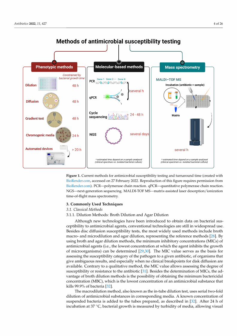
Clinical laboratories currently employ several AST methods depending on the equip-ment and laboratory test menu that they provide. Conventional AST based on phenotypictesting examines the bacterial response in the presence of an antimicrobial agent. Classicalculture-dependent methods (e.g., a disk diffusion test, gradient diffusion method) arefirmly established in the diagnostic routine, and their main limitation is that the results areobtained for most clinically important bacteria within at least 18–24 h or 48 h, includingprior bacterial isolation and identification. The turnaround time is prolonged for anaerobesor some slow-growing fastidious bacteria such as the HACEK group (Haemophilus species,Aggregatibacter species, Cardiobacterium hominis, Eikenella corrodens, and Kingella species),Brucella spp. etc. [24]. For many years, clinical laboratories have been equipped withautomated systems based on microdilution trays to provide faster results (6–24 h afterinitial isolation). However, the time required to obtain the results is similar in comparisonwith the broth microdilution (BMD) method [25]. Molecular AST is based on the detectionof resistance determinants in bacterial isolates or directly in clinical specimens by molecularmethods with a turnaround time of approximately 1–6 h [26]. Besides high costs, majordrawbacks of molecular methods are detection of the resistance genes targeted only bythe known probes and overestimating resistance because the resistance gene is not neces-sarily associated with the expression of a resistance phenotype. Because of a significantrise in multi- and pan-drug-resistant infections, there is an urgent need for a more rapidand reliable test to improve infection diagnosis and support evidence-based antimicrobialprescribing [27]. The currently used methods for AST are summarised in Figure 1.
Antibiotics 2022, 11, 427 4 of 26
Antibiotics 2022, 11, x FOR PEER REVIEW 4 of 27
antimicrobial prescribing [27]. The currently used methods for AST are summarised in Figure 1.
Figure 1. Current methods for antimicrobial susceptibility testing and turnaround time (created with BioRender.com., accessed on 27 February 2022. Reproduction of this figure requires permission from BioRender.com). PCR—polymerase chain reaction. qPCR—quantitative polymerase chain re-action. NGS—next-generation sequencing. MALDI-TOF MS—matrix-assisted laser desorption/ion-ization time-of-flight mass spectrometry.
3. Commonly Used Techniques 3.1. Classical Methods 3.1.1. Dilution Methods: Broth Dilution and Agar Dilution
Although new technologies have been introduced to obtain data on bacterial suscep-tibility to antimicrobial agents, conventional technologies are still in widespread use. Be-sides disc diffusion susceptibility tests, the most widely used methods include broth macro- and microdilution and agar dilution, representing the reference methods [28]. By using broth and agar dilution methods, the minimum inhibitory concentrations (MICs) of antimicrobial agents (i.e., the lowest concentration at which the agent inhibits the growth of microorganisms) can be determined [29,30]. The MIC value serves as the basis for as-sessing the susceptibility category of the pathogen to a given antibiotic, of organisms that give ambiguous results, and especially when no clinical breakpoints for disk diffusion are available. Contrary to a qualitative method, the MIC value allows assessing the degree of susceptibility or resistance to the antibiotic [31]. Besides the determination of MICs, the advantage of broth dilution methods is the possibility of obtaining the minimum bacteri-cidal concentration (MBC), which is the lowest concentration of an antimicrobial sub-stance that kills 99.9% of bacteria [32].
Figure 1. Current methods for antimicrobial susceptibility testing and turnaround time (created withBioRender.com, accessed on 27 February 2022. Reproduction of this figure requires permission fromBioRender.com). PCR—polymerase chain reaction. qPCR—quantitative polymerase chain reaction.NGS—next-generation sequencing. MALDI-TOF MS—matrix-assisted laser desorption/ionizationtime-of-flight mass spectrometry.
3. Commonly Used Techniques3.1. Classical Methods3.1.1. Dilution Methods: Broth Dilution and Agar Dilution
Although new technologies have been introduced to obtain data on bacterial sus-ceptibility to antimicrobial agents, conventional technologies are still in widespread use.Besides disc diffusion susceptibility tests, the most widely used methods include brothmacro- and microdilution and agar dilution, representing the reference methods [28]. Byusing broth and agar dilution methods, the minimum inhibitory concentrations (MICs) ofantimicrobial agents (i.e., the lowest concentration at which the agent inhibits the growthof microorganisms) can be determined [29,30]. The MIC value serves as the basis forassessing the susceptibility category of the pathogen to a given antibiotic, of organisms thatgive ambiguous results, and especially when no clinical breakpoints for disk diffusion areavailable. Contrary to a qualitative method, the MIC value allows assessing the degree ofsusceptibility or resistance to the antibiotic [31]. Besides the determination of MICs, the ad-vantage of broth dilution methods is the possibility of obtaining the minimum bactericidalconcentration (MBC), which is the lowest concentration of an antimicrobial substance thatkills 99.9% of bacteria [32].
The macrodilution method, also known as the in-tube dilution test, uses serial two-folddilution of antimicrobial substances in corresponding media. A known concentration ofsuspended bacteria is added to the tubes prepared, as described in [32]. After 24 h ofincubation at 37 ◦C, bacterial growth is measured by turbidity of media, allowing visual
Antibiotics 2022, 11, 427 5 of 26
determination of MIC values. Another macrodilution method is the time-kill methodol-ogy. This test allows monitoring of the effect of different concentrations of antimicrobialsubstances by examining the rate at which antimicrobials lead to bacterial death—i.e., thebactericidal activity of antimicrobial agents is determined depending on the concentrationand time. Bacterial viability is determined by counting colonies on agar plates at regulartime points for 24 h [33]. The rate of bacterial growth is monitored via changes in logCFU/mL during the first 24 h time-kill test. Based on the results, experimental curveswhich represent the absence of growth or the killing effect can be constructed and give usinsight into the interaction between the bacteria and the antimicrobial agent. The data canbe further analysed using different mathematical models [30,34,35].
The BMD method is standardised, accurate, and inexpensive. Since it is performedin 96-well microtiter plates, it allows the testing of several antimicrobial substances ina row and eight series of two-fold dilutions of antimicrobial agents in one plate. Afterthe dilutions are made, each well is inoculated with standardised bacterial inoculum andincubated for at least 16–24 h. Although this procedure is used as a reference method, ithas been improved by the addition of a resazurin colour redox indicator. Resazurin isa blue colour that turns into pink, fluorescent resorufin in the presence of metabolicallyactive bacterial cells. The reduction of resazurin to fluorescent resorufin can be measuredfluorimetrically [27,32,36–38]. Nowadays, there are several commercially available easy toperform BMD systems such as MBD Sensititre System (Thermo Fisher Scientific, Waltham,MA, USA) and ComASP Colistin (Liofilhem, Roseto degli Abruzzi, Italy), formerly Sen-siTest Colistin. The MBD Sensititre System can be performed manually or automatically.ComASP Colistin is a compact panel containing the antibiotic colistin in seven two-foldserial dilutions and allows for four samples to be tested simultaneously with the BMDmethod [39].
The agar dilution method involves adding different concentrations of antimicrobialsubstances to the non-selective medium before solidification [40]. Afterwards, the stan-dardised bacterial inoculum is spotted on the agar surface. Following overnight incubation,plates are evaluated visually, determining whether growth has occurred at the inoculatedsites. The lowest concentration of antibiotics that prevent bacterial growth is considered tobe the MIC. This method allows simultaneous testing of different bacterial strains [41].
3.1.2. Antimicrobial Gradient Method
The gradient strip test is a combination of disk-diffusion and dilution method of AST,having advantageous properties of both methods. It allows the MIC to be determined whilekeeping it simple and easy to use. The method is based on the diffusion of an antibioticthrough agar with a continuous gradient. A concordance of the susceptibility categories andMIC values obtained by gradient test and BMD method, a “gold standard” recommendedby the European Committee on Antimicrobial Susceptibility Testing (EUCAST) and CLSI,were observed [28,42]. For example, the new ceftazidime–avibactam and ceftolozane–tazobactam gradient tests (Etests, bioMérieux, Marcy-l’Étoile, France) have shown a highcategorical agreement between gradient test and BMD, of 96% and 94%, respectively [43,44].On the other hand, for some antibiotics, such as colistin and tigecycline [45], controversialresults have been obtained. Some agar-related factors, i.e., the content of divalent cations,can affect the diffusion of colistin, resulting in false susceptibility. Consequently, BMDremains the only appropriate method for MIC determination for certain antibiotics [46].Currently, a few commercial gradient strip tests, such as Etest (bioMérieux, France), MICTest Strip (Liofilchem, Roseto degli Abruzzi, Italy), M.I.C.Evaluator (Thermo Scientific,Waltham, MA, USA), and Ezy MIC Strip (HiMedia Laboratories, Mumbai, India), areavailable [47]. They can be used for susceptibility testing of microorganisms to antibioticsand antifungals [48–50].
A gradient strip test is performed according to the manufacturer’s instructions: a shortplastic or paper strip impregnated with antibiotic is placed on inoculated agar (Figure 2).On the standardised 100 mm Petri dish, two strips may be placed, while on the larger

Antibiotics 2022, 11, 427 6 of 26
150 mm Petri dish, up to six antibiotics may be tested simultaneously. The MIC of a testedagent is determined by the intersection of a zone of inhibition with the strip and measuredusing labelled concentrations on the strip. If the intersection is between two values on ascale, a higher value is reported as MIC. In addition, if beta-haemolysis is present on theplate, careful examination of the strip is required since the reporting of the intersection ofhaemolysis leads to false higher MICs values. Automated systems for reading the resultsof gradient tests are also available (ADAGIO Automated System, Bio-Rad Laboratories,Hercules, CA, USA).
Antibiotics 2022, 11, x FOR PEER REVIEW 6 of 27
available [47]. They can be used for susceptibility testing of microorganisms to antibiotics and antifungals [48–50].
A gradient strip test is performed according to the manufacturer’s instructions: a short plastic or paper strip impregnated with antibiotic is placed on inoculated agar (Fig-ure 2). On the standardised 100 mm Petri dish, two strips may be placed, while on the larger 150 mm Petri dish, up to six antibiotics may be tested simultaneously. The MIC of a tested agent is determined by the intersection of a zone of inhibition with the strip and measured using labelled concentrations on the strip. If the intersection is between two values on a scale, a higher value is reported as MIC. In addition, if beta-haemolysis is present on the plate, careful examination of the strip is required since the reporting of the intersection of haemolysis leads to false higher MICs values. Automated systems for read-ing the results of gradient tests are also available (ADAGIO Automated System, Bio-Rad Laboratories, Hercules, CA, USA).
Figure 2. Disk diffusion and gradient test of various bacterial isolates. (A)—Antimicrobial suscepti-bility of Streptococcus pyogenes showing iMLS phenotype, using disk diffusion method. (B)—Anti-microbial susceptibility of extended-spectrum beta-lactamase-producing Pseudomonas aeruginosa, using disk diffusion method. (C)—Gradient test of Enterococcus spp. iMLS phenotype—inducible macrolide, lincosamide, and streptogramin phenotype.
A variation of gradient tests exists for the detection of various AMR phenotypes. Currently, Etests for phenotypic detection of ESBL production in enterobacteria are avail-able, such as strips with cefotaxime+clavulanic acid, ceftazidime with clavulanic acid, and cefepime with clavulanic acid [51]. The gradient tests for ESBL detection are two-sided strips that contain antibiotic on one end, while on the other is the same antibiotic with clavulanic acid. Reduction in MIC equal to or greater than eight times by the combination of antibiotic and clavulanate refers to ESBL production [52]. Similar to the double-disk synergy test, the phantom zone below the clavulanic end also indicates a positive result. Identification of metallo-beta-lactamase (MBL)-producing bacteria can be carried out us-ing a gradient test. These tests contain carbapenem antibiotic on one side of the strip and the same carbapenem with EDTA on the other side. Imipenem with EDTA for detection of MBL in Acinetobacter spp. and Pseudomonas spp. is available, although sensitivity and specificity may vary [53,54]. For detection of AmpC beta-lactamase-producing enterobac-teria, Etest impregnated with cefotetan on one end and cefotetan–cloxacillin on the other end can be used [55]. Gradient tests with a predefined gradient of vancomycin and teicoplanin on each side of the strip can be used for the detection of glycopeptide re-sistance in Staphylococcus aureus [56]. Since these tests are easy to perform, they could be used as “screening” tests for the detection of emerging resistance patterns among clini-cally relevant bacteria.
Figure 2. Disk diffusion and gradient test of various bacterial isolates. (A)—Antimicrobialsusceptibility of Streptococcus pyogenes showing iMLS phenotype, using disk diffusion method.(B)—Antimicrobial susceptibility of extended-spectrum beta-lactamase-producing Pseudomonas aerugi-nosa, using disk diffusion method. (C)—Gradient test of Enterococcus spp. iMLS phenotype—induciblemacrolide, lincosamide, and streptogramin phenotype.
A variation of gradient tests exists for the detection of various AMR phenotypes.Currently, Etests for phenotypic detection of ESBL production in enterobacteria are avail-able, such as strips with cefotaxime+clavulanic acid, ceftazidime with clavulanic acid, andcefepime with clavulanic acid [51]. The gradient tests for ESBL detection are two-sidedstrips that contain antibiotic on one end, while on the other is the same antibiotic withclavulanic acid. Reduction in MIC equal to or greater than eight times by the combinationof antibiotic and clavulanate refers to ESBL production [52]. Similar to the double-disksynergy test, the phantom zone below the clavulanic end also indicates a positive result.Identification of metallo-beta-lactamase (MBL)-producing bacteria can be carried out usinga gradient test. These tests contain carbapenem antibiotic on one side of the strip and thesame carbapenem with EDTA on the other side. Imipenem with EDTA for detection of MBLin Acinetobacter spp. and Pseudomonas spp. is available, although sensitivity and specificitymay vary [53,54]. For detection of AmpC beta-lactamase-producing enterobacteria, Etestimpregnated with cefotetan on one end and cefotetan–cloxacillin on the other end can beused [55]. Gradient tests with a predefined gradient of vancomycin and teicoplanin on eachside of the strip can be used for the detection of glycopeptide resistance in Staphylococcusaureus [56]. Since these tests are easy to perform, they could be used as “screening” testsfor the detection of emerging resistance patterns among clinically relevant bacteria.
Plenty of advantages of gradient tests are known: simple performance, flexibility inthe testing of any combination of antibiotics, and the fact that they do not require expertiseand special technologies. Moreover, their use is suited when only a couple of antibioticsare needed to be tested. The price of each strip is relatively high, compared with the priceof disks; therefore, gradient tests are usually used to test only a few antibiotics per strain.The incubation length of 16–24 h for gradient tests may represent a disadvantage, as morerapid automated systems are available with the reliable determination of MIC.

Antibiotics 2022, 11, 427 7 of 26
3.1.3. Disk Diffusion Test
Since its development in 1940, the disk diffusion (DD) test has remained the mostwidely used routine AST in clinical microbiological laboratories [57]. It has been stan-dardised to test the susceptibility of the most common and clinically relevant bacteria thatcause human diseases [42,58]. The standardisation is a continuous process, and DD formany microorganisms/antimicrobials is an ongoing process [59]. The method is based onplacing different antibiotic-impregnated disks on previously inoculated agar with bacterialsuspension. The antibiotic diffuses radially outward through the agar medium, producingan antibiotic concentration gradient. After the inhibition zones are established within 24 hof incubation at 35 ± 1 ◦C, the zone diameters of each tested antibiotic are measured by thenaked eye or using an automated system [60]. Obtained results should be interpreted andcategorised according to the recommended clinical breakpoint of the standard in use [42,61].Disk diffusion is the most widely used AST method in microbiology laboratories becauseof its low cost and ease of performance and applicability of numerous bacterial species andantibiotics [32]. The choice of antibiotic disks is flexible and enables the clinical laboratoryto make different combinations according to the bacterial species and the type of samplethe isolate was obtained from [62]. Simple interpretation allows the detection of atypicalphenotypes and visibility of contamination. However, the main disadvantages are theinability to determine the MIC and delays in getting the results. Reduction in turnaroundtime and timely treatment are of great importance for critically ill patients. In addition,the biological properties of lag and log phase of bacterial growth and their expression onantibiotic influence should be considered [63]. Nevertheless, methods to reduce incuba-tion time for DD were suggested decades ago [64–66]. A revival of that idea led to thedevelopment of automated systems (WASPLab, Copan, Murrieta, CA, USA and BD Kiestra,Becton, Dickinson and Company, Franklin Lakes, NJ, USA) for an acceleration of AST byDD [67,68]. The automatisation of the AST by DD leads to a shortening of the requiredtime to obtain results and produce the final report [68]. EUCAST has defined a methodol-ogy of disk diffusion rapid AST (RAST), which is performed directly from positive bloodculture bottles, with breakpoints for short incubations of 4, 6, and 8 h [69–71]. RAST canbe implemented in routine laboratories without major investments. The method has beenvalidated for a limited number of bacterial species and antibiotics so far. Furthermore, thecombined use of a MALDI-TOF MS for the identification of bacteria and RAST directlyfrom positive blood bottles enables reporting AST results within less than 24 h, whichsignificantly reduced the turnaround time compared with the 24–48 h needed for culturingand classical AST methods, such as DD [72]. The predictive value of direct DD testing frompositive blood cultures has been reported to have an important influence on AMS [73].
Quality control testing of media and antibiotics is of great importance for ensuringthat disk diffusion is providing accurate and reliable results [59,74]. In some cases, the DDmethod is more reliable than MIC determination. For instance, in the case of detectingpenicillinase-producing S. aureus strains, the inhibition zone diameter combined with theEUCAST-based zone edge test is the most sensitive and specific phenotypic method [42,75].The DD method can be used for screening of susceptibility to a larger number of antibioticsor a whole class of antibiotics; detection of certain important resistance phenotypes, suchas ESBL, carbapenemases, inducible resistance to macrolides (Figure 2); or the presenceof a heteroresistant population of bacterial species in a sample that cannot be detected byother phenotypic AST methods. The above-mentioned suggests that the DD method willremain a widely used AST method in the future.
3.1.4. Chromogenic Agar Media for Detection of Antimicrobial-Resistant Bacteria
Since the introduction of the first chromogenic media for the detection of antibiotic-resistant bacteria 20 years ago, a variety of different media for the detection of clinicallyimportant resistant pathogens—such as MRSA, VRE, and ESBL- and carbapenemases-producing or colistin-resistant Gram-negative bacteria—have been developed [76–80].
Antibiotics 2022, 11, 427 8 of 26
The main purpose of the development of chromogenic media was to enable morerapid detection and identification of resistant microorganisms. The target organisms arecharacterised by specific enzyme systems that metabolise the substrates to release thechromogen. The chromogen can then be visually detected by direct observation of a distinctcolour change in the medium. Thus, these selective and differential media enable targetpathogens to grow as coloured colonies. Compared with the use of conventional culturemedia, the use of chromogenic agar often reduces the costs and labour time [81]. Theirprimary use is for screening of patients colonised with various pathogens, and thereforethey are valuable in infection prevention [82] and control of hospital-acquired infections.The sensitivity and specificity of chromogenic media depend on the manufacturer and thetype of microorganism detected; thus, additional identification confirmation of the resistantbacteria is sometimes needed. Because of their wide applicability, new chromogenic mediaare being developed [83,84].
3.1.5. Colourimetric Tests for Detection of Antimicrobial-Resistant Bacteria
Colourimetric tests represent phenotypic methods developed for the detection of AMR.They are based upon the bacterial enzymes hydrolysing test component, which is detectedby the changes in pH values and the colour of chromogenic substances. Briefly, bacterialsuspension or bacterial lysate suspension is added to a detection solution containingantibiotic and pH indicator dyes, such as phenol red, and incubated for a short period oftime, no longer than a couple of hours. The pH of the detection solution changes due tothe growth of antibiotic-resistant bacteria, or bacterial enzymes activity, and subsequently,the colour of the solution changes, which can then be visually observed. These tests wereshown to be fast, easy to perform and interpret, and highly sensitive and specific. A goodexample of such colourimetric tests is Carba NP (bioMérieux, Marcy-l’Étoile, France), whichdetects carbapenemase-producing bacteria. The test gives reliable results in 30 min to 2 h,making it the quick and easy way to control carbapenemase producers [85].
4. Current Technologies for Rapid AST4.1. Automated and Semi-Automated Devices Based on Microdilution Susceptibility Testing
Clinical microbiology laboratories are under increasing pressure to provide fast andreliable microbial identification (ID) and AST [86]. Automated and semi-automated devicesfor bacterial ID and AST are worthy of the task and have significantly improved laboratoryefficiency. Nowadays, automation has been successfully implemented in most clinicalmicrobiological laboratories to reduce turnaround times, increase efficiency, and improvecost-effectiveness [86,87]. Various test systems—such as the VITEK 2 (bioMérieux, France),MicroScan Walkaway (Dade-Behring MicroScan, Deerfield, IL, USA), and Phoenix system(BD Diagnostic Systems, Baltimore, MD, USA)—have been widely used over the lastdecades. These instruments, using optical systems for measuring subtle changes, determinebacterial growth and antimicrobial susceptibility [88] and can produce results in a shortertime (6–12 h) than conventional manual assessment [36].
• VITEK 2 Systems—The first generation of VITEK system with a turnaround time of 13 hwas developed for enumeration and identification of bacteria and yeasts in 1973. TheVITEK 2 System, the next-generation of an instrument, is a BMD-based AST system thatuses 64-well plastic cards containing 17–20 antimicrobial agents. If the bacterial isolateis not previously identified, one card is used for bacterial identification (ID card) andthe other for antimicrobial susceptibility testing (AST card). Two Vitek 2 instrumentsare available with test card (ID and AST) capacities of 60 cards (Vitek 2) and 120 cards(Vitek 2 XL). Results are reported in 4–18 h, containing MIC and category of susceptibility,whereas the detection of AMR is facilitated by the Advanced Expert System (AES). Thecurrently available Vitek 2 Compact instruments can use 15, 30, and 60 cards. Themain advantage of the Vitek 2 system with computer software is the determination ofsusceptibility of clinically important resistant pathogens, such as Staphylococcus aureusand Enterococcus faecalis, to an additional four to ten antibiotics [86,89,90].

Antibiotics 2022, 11, 427 9 of 26
• Phoenix System—The Phoenix System is widely accepted and used in clinical mi-crobiology laboratories for identification testing (ID) and antimicrobial susceptibilitytesting (AST). The principle of determining the susceptibility is based on the use ofan oxidation-reduction indicator (resazurin dye or Alamar blue) and the detection ofbacterial growth in the presence of various concentrations of the antimicrobial agent.In the Phoenix instrument, a maximum of 100 tests can be performed by using PhoenixID/AST combination panels (51 for ID and 85 for AST). The instrument performsautomatic reading at 20 min intervals during incubation for up to 18 h and providesaccurate and rapid susceptibility results with easy workflow for the laboratory worker.In 2014, the new panel for susceptibility of Gram-negative bacteria was introduced forthe Phoenix system to be used in combination with the BD Bruker MALDI-TOF [91].
• MicroScan WalkAway plus System—The MicroScan WalkAway plus System providesaccurate and rapid identification and susceptibility results for a wide range of Gram-positive and Gram-negative aerobic bacteria. The instrument utilises three types ofpanel configurations: combo panels, breakpoint combo panels, and MIC panels. Thereare two types of system: 40- and 96-panel capacity models. The panels are manuallyinoculated, rehydrated by the RENOK inoculator, and read automatically. The resultsare obtained after 4.5–18 h by reading of rapid panels [91].
• MicroScan AutoScan 4—The AutoScan 4 is a semiautomated instrument mostly usedin smaller laboratories or for the testing of supplemental antimicrobial agents. Theinstrument provides simplified ID/AST testing in a highly reliable and affordablepackage. The system uses the off-line incubation of the conventional MicroScan ASTpanels. The panels are manually inoculated or with the MicroScan Renok instrumentand read automatically [91].
• MicroScan WalkAway System—The first generation of the MicroScan WalkAwaySystem available on the market is the AutoSCAN-3. The new versions of instrumentsAuto-ACAN-4 and AutoSCAN-WalkAway are improved and use dry panels that donot need refrigeration. The AutoSCAN-WalkAway system detects bacterial enzymaticactivity and can process 96 panels at the same [86].
Each of the above-mentioned systems has inherent advantages and limitations, andthe results vary widely by antimicrobial drugs, software versions, and cards used. Hence,some of the systems are not reliable for correct categorisation of susceptibility profiles forcertain drugs, leading to wrong classifications of susceptibility categories [92]. It seemsthat low inoculum size has a major influence on the outcome of these systems, with falsesusceptibilities being reported. Additionally, software updates and synchronisation ofbreakpoints according to the current standards are mandatory. Thus, it is incumbent uponthe instrument manufacturer to keep pace with the breakpoint updates and make relevantimprovements, such as extending the detection limit and verifying the performance of theAST system with the revised breakpoints internally, to avoid the problem of uncategorisedresults [93]. Panels usually contain only several concentrations of each antimicrobial agent,and the resulting MIC is not always given as an exact value. In contrast, classical BMD con-tains a wide range of doubling dilution antimicrobial concentrations for the determinationof the MIC, thus obtaining the more precise value. In addition, according to the previouslypublished reports, many of the resistance phenotypes are not easily detected using theautomated susceptibility testing methods so prevalent in today’s clinical laboratories [94].Nonetheless, interestingly, the ability of automated systems to detect inducible resistanceto clindamycin in 524 isolates of Staphylococcus spp. revealed sensitivity and specificity of100% and 99.6%, respectively, for Phoenix, and 91.1% and 99.8%, respectively, for Vitek2 [95]. The multicentre evaluation showed that categorical agreement between the Phoenixsystem and a BMD reference method for 2013 streptococcal isolates including Streptococcuspneumoniae, viridans group streptococci, and beta-haemolytic Streptococcus groups A, B, C,and G ranged from 92% to 100%, with one exception for viridans streptococci and penicillin,which was 87% [96]. However, according to the results of the evaluation of ASTs obtained

Antibiotics 2022, 11, 427 10 of 26
using Vitek 2, Phoenix, and MicroScan, caution should be taken for AST of Stenotrophomonasmaltophilia, as a high rate of errors may be observed [97].
4.2. Molecular-Based Techniques for Resistance Detection
Molecular AST directly detects specific resistance genes, as well as mutations in andexpression of these genes. These molecular methods have been developed and tested as analternative for or complementary to conventional AST and are generally faster than classicculture-based assays, with the test results available within one to a few hours [98] (Figure 3).Most of the molecular AST methods fall into one of the three categories: amplification-based,hybridization-based, or sequence-based. In amplification-based methods, the target genesequence is amplified to allow detection; in hybridization-based techniques, hybridizednucleic acid probes target gene sequences allowing detection; and in sequence-basedapproaches, genome sequences are analysed to detect resistance-conferring mutations orresistance genes.
Antibiotics 2022, 11, x FOR PEER REVIEW 10 of 27
BMD contains a wide range of doubling dilution antimicrobial concentrations for the de-termination of the MIC, thus obtaining the more precise value. In addition, according to the previously published reports, many of the resistance phenotypes are not easily de-tected using the automated susceptibility testing methods so prevalent in today’s clinical laboratories [94]. Nonetheless, interestingly, the ability of automated systems to detect inducible resistance to clindamycin in 524 isolates of Staphylococcus spp. revealed sensitiv-ity and specificity of 100% and 99.6%, respectively, for Phoenix, and 91.1% and 99.8%, respectively, for Vitek 2 [95]. The multicentre evaluation showed that categorical agree-ment between the Phoenix system and a BMD reference method for 2013 streptococcal isolates including Streptococcus pneumoniae, viridans group streptococci, and beta-haemo-lytic Streptococcus groups A, B, C, and G ranged from 92% to 100%, with one exception for viridans streptococci and penicillin, which was 87% [96]. However, according to the re-sults of the evaluation of ASTs obtained using Vitek 2, Phoenix, and MicroScan, caution should be taken for AST of Stenotrophomonas maltophilia, as a high rate of errors may be observed [97].
4.2. Molecular-Based Techniques for Resistance Detection Molecular AST directly detects specific resistance genes, as well as mutations in and
expression of these genes. These molecular methods have been developed and tested as an alternative for or complementary to conventional AST and are generally faster than classic culture-based assays, with the test results available within one to a few hours [98] (Figure 3). Most of the molecular AST methods fall into one of the three categories: ampli-fication-based, hybridization-based, or sequence-based. In amplification-based methods, the target gene sequence is amplified to allow detection; in hybridization-based tech-niques, hybridized nucleic acid probes target gene sequences allowing detection; and in sequence-based approaches, genome sequences are analysed to detect resistance-confer-ring mutations or resistance genes.
Figure 3. The basic workflow of molecular-based techniques for antimicrobial susceptibility testing. The routes from a clinical specimen to a final result are indicated by arrows (created with Figure 3. The basic workflow of molecular-based techniques for antimicrobial susceptibility testing. Theroutes from a clinical specimen to a final result are indicated by arrows (created with BioRender.com,accessed on 27 February 2022. Reproduction of this figure requires permission from BioRender.com).
4.2.1. Polymerase Chain Reaction
The most widely used nucleic acid amplification-based method for the detection ofspecific resistance genes is polymerase chain reaction (PCR). Both real-time and conven-tional PCR rely on the amplification of nucleic acid sequences that encode resistance toan antibiotic. New PCR-based methods are being developed for the detection of geneticdeterminants of resistance to a variety of antibiotics for various bacterial species, as ourknowledge about the genetic basis of antibiotic resistance increases [99]. Multiplex assaysfor simultaneous testing of multiple genetic determinants in various bacterial species havealso been developed, i.e., multiplex assays for identifying numerous cephalosporinase-and carbapenemase-encoding genes, such as blaKPC, blaNDM, blaIMP, blaVIM, blaAmpC, blaTEM,blaSHV, and blaOXA, or mecA gene-encoding methicillin resistance in MRSA [100,101]. Op-

Antibiotics 2022, 11, 427 11 of 26
Gen, Inc. (Rockville, MD, USA) has recently released the multiplex-based Acuitas® AMRGene Panel that detects 28 genetic AMR markers, covering select drugs in nine classes ofantibiotics, from 26 different pathogens [102]. The advantage of this test in comparisonwith other commercially available molecular tests is that it also detects non-beta-lactamresistance genes and those for what would be considered “last-resort antibiotics”, suchas colistin.
Real-time PCR (quantitative PCR, qPCR) is one of the most ubiquitous methods foundthroughout clinical microbiology. Although costlier, qPCR offers several advantages overconventional PCR, including the measurement of data in real-time, greater sensitivity,reduced risk of carryover contamination, and greater amenity to multiplexing. Furtheradvantages are that many systems are partially or even completely automated, such asGeneXpert® Instrument Systems (Cepheid Corp., Sunnyvale, CA, USA) and BD MAXSystem platform (Becton Dickinson, Franklin Lakes, NJ, USA), which are easily operatedand can be used for the detection of carbapenemases, ESBLs, MRSA, VRE, etc. [103–105].The downside is that they are limited to using test assays only from specific manufacturers,with GeneXpert® Instrument Systems requiring GeneXpert assays (Cepheid Corp., Sun-nyvale, CA, USA) and BD MAX System using Check-Points® qPCR assays (Wageningen,The Netherlands). Tests based on qPCR can also be used for phenotypic differentiation ofresistant and susceptible strains due to its ability to measure genome copy numbers duringbacterial growth in the presence of antibiotics [106]. The major disadvantage is that thesystem cannot provide information about the mechanism of resistance and that it requiresthe previous culture, meaning that the primary clinical samples cannot be used.
4.2.2. DNA-Microarrays
DNA-microarrays are used to identify the presence of specific nucleic acid sequencesusing complementary short oligonucleotides immobilised on a solid surface. Since theseoligonucleotides can be assembled onto solid surfaces in close proximity, this method coulddetect numerous sequences in a single assay, which would allow simultaneous, in paralleldetection of different pathogens and detection of vast numbers of different resistance genes,as well as detecting numerous distinct mechanisms of resistance or variants of a singlemechanism present in bacterial isolates, as opposed to PCR-based approaches [107]. TheVerigene system (Luminex Corporation, Austin, TX, USA) has developed Blood CultureMultiplex Microarray-Based Molecular assays for rapid diagnostics of 12 Gram-positiveand 9 Gram-negative bacteria, along with their associated resistance genes (i.e., mecA, vanA,vanB, blaKPC, blaNDM, blaIMP, and blaVIM) [108]. In addition to qPCR-based assays, Check-Points® has also developed the CHECK-MDR CT103 DNA microarray for the detectionof the clinically most prevalent ESBLs and carbapenemases, as well as mobile colistinresistance (mcr) genes in Gram-negative bacteria [109]. Both of these microarray tests haveshown high sensitivity (94–100%) and specificity (94–98%), and CHECK-MDR CT103 DNAmicroarray also showed the ability to discriminate between carbapenemase and ESBLvariants of GES-type beta-lactamase [108,109].
The advantages of currently available molecular-based methods are that they are direct,rapid, highly sensitive, and specific, thus potentially allowing the earlier administrationof targeted therapy [98]. Furthermore, for some methods, direct clinical samples can beused. However, it should be noted that the presence of a resistance marker does notalways have to correlate with phenotypic resistance. Additionally, the extent and intensityof gene expression are important parameters, as some genes need different expressionlevels to produce resistance. A potential solution to this issue would be the use of reversetranscription qPCR, which relies on the measurement of gene transcripts (RNA levels)instead of the presence of a gene [110,111]. Another drawback is that these methods canonly detect resistances that are searched for, and not novel or uncharacterised mechanismsof resistance, which could lead to false-negative results and inappropriate classification ofresistant isolates as susceptible. A final consideration is that these methods are not capableof defining MIC values. As such, these methods have to be validated against phenotypic
Antibiotics 2022, 11, 427 12 of 26
data to be useful, and extensive resistance marker databases and innovative bioinformaticsmethodologies are mandatory requirements. Nevertheless, molecular-based AST methodsare a safe, efficient, and reliable screening tool in clinical settings. As experience with thesetests grows, and as data are gathered on their efficacy and clinical impact, they will likelybe more widely adopted.
4.2.3. Whole-Genome Sequencing in Antimicrobial Susceptibility Testing
As DNA sequencing technology and bioinformatics pipelines for genome assemblyand analysis advance, the possibility of using these techniques for the detection of an-tibiotic resistance opens. Applying whole-genome sequencing (WGS) would essentiallyenable the detection of all genes involved in AMR, which would help make comprehen-sive databases of all species-specific resistance factors (i.e., CARD-Comprehensive Antibi-otic Resistance Database—https://card.mcmaster.ca, ResFinder—https://cge.cbs.dtu.dk/services/ResFinder (accessed on 27 February 2022)) and make in silico AMR detectionpossible. Recent studies showed high concordance between the resistance profiles obtainedusing WGS and those obtained using phenotypic susceptibility testing, demonstrating thatdata obtained from genome sequences can correlate well with phenotypic resistance insome cases [112,113]. In addition to genome-based resistome analyses, RNA-mediated tran-scriptomic approaches have also been described [114,115]. Despite all of the advantages,WGS is not routinely performed in clinical practice. Considering the turnaround times ofWGS, the existence of unknown resistance mechanisms, and the elevated cost comparedwith traditional and emerging techniques, the use of WGS for AST is not yet part of routinepractice in clinical microbiology [116,117].
4.3. Mass Spectrometry
Matrix-assisted laser desorption ionization-time of flight mass spectrometry (MALDI-TOF MS) was discovered in the 1980s and introduced into the microbiological routine as aneffective tool for bacterial and yeast identification about 15 years ago. It has been appliedto classify the specific bacterial protein contents and their matching protein biomarkersbecause of its rapid turnaround time, low sample volume requirements, and per-samplecosts [118]. Several MALDI-TOF MS-based methods have been proposed for rapid detec-tion of antimicrobial resistance, including monitoring antibiotic modification by bacterialculture (e.g., beta-lactam hydrolysis [65,119]), acetylation of fluoroquinolones [120], directdetection of proteins involved in specific resistance mechanisms [121,122], and detectionof stable isotope labelling that requires expensive, isotopically labelled media [123,124].The hydrolysis of the target beta-lactam antibiotic, as shown by peak disappearance, isused to detect beta-lactamase-producing bacteria using MALDI-TOF MS. As a result, theassay for detecting carbapenemase production [125] automatically determines sensitivityor resistance depending on the degree of antibiotic hydrolysis. The method had 98% sensi-tivity and 100% specificity after 30 min of incubation of bacteria with the antibiotic, withboth reaching 100% after 60 min of incubation [126,127]. However, beta-lactam resistance isonly recognized when it is mediated by beta-lactamases; alternative resistance mechanismshave not been elucidated; therefore, other tests should confirm negative results. Anotherassay for the detection of carbapenemases is a rapid and novel method using detonationnanodiamonds (DNDs) as a platform for the concentration and extraction of A. baumanniicarbapenemase-associated proteins before MALDI-TOF MS analysis [128]. The sensitivityand the specificity of the proposed platform could reach 96% and 73%, as compared withtraditional imipenem susceptibility testing, and 100% compared with PCR results. Thismethod may detect the carbapenemases produced by A. baumannii in 90 min and does notrequire the addition of a carbapenemase substrate, as other mass spectrometric methodsdo. It is efficient for detecting other carbapenemase-producing bacteria.
MALDI Biotyper-Antibiotic Susceptibility Test Rapid Assay (MBT-ASTRA) is an al-ternative MS-based method for AST which utilises semi-quantitative MALDI-TOF MS tomeasure the relative growth rates of bacterial isolates exposed to antibiotics compared

Antibiotics 2022, 11, 427 13 of 26
with untreated controls during a short incubation step. A software tool calculates andcompares the area under the curves (AUCs) of spectra of bacteria either exposed or notto an antibiotic [129]. In this method, if the microbial strain is susceptible, the AUC ofthe bacterial suspension with the antibiotic will be reduced compared with that withoutantibiotics, whereas with a resistant strain the AUCs with or without antibiotics will becomparable. The main advantage of the MBT-ASTRA is that the assay does not dependon the resistance mechanism and is utilisable with any antibiotic [130]. Moreover, it doesnot require specialised media or instrumentation, beyond the MALDI-TOF mass spec-trometer. However, a drawback of the MBT-ASTRA assay is that the concentration ofantibiotics used and the incubation time must be optimised for each species and antibioticcombination [129].
MBT-Resist assay, based on the detection of peak shift after stable isotope labelling, isan approach that uses the following principle: bacteria are grown in parallel in two distinctculture mediums, one containing 12C as a carbon component and the other containing13C [131]. The system compares the mass spectrum of bacteria grown on an isotope-labelledmedium with antibiotics to the mass spectrum of the same strain grown on an unlabelledmedium without antibiotics. Resistant strains can thrive in the presence of antibiotics,incorporating 13C into the polypeptide, causing a shift in the peak to a higher m/z in themass spectrum [131].
Antibiotic resistance by direct-on-target microdroplet growth assay (DOT-MGA) isa novel approach for detecting antimicrobial susceptibility in bacteria treated with break-point concentrations of antibiotic on the target plate of MALDI-TOF MS [132]. The bestperformance was obtained by recovering bacteria from positive blood cultures and after a4 h incubation of microdroplets with or without meropenem at the breakpoint concentra-tion. Under these conditions, 96.3% validity, 91.7% sensitivity, and 100% specificity wereachieved. Recently, a screening panel for the detection of ESBL and AmpC beta-lactamaseactivity was developed [133]. Compared with the PCR results, positive percentage agree-ment values for ESBL, AmpC, and ESBL + AmpC resistance were 94.4%, 94.4%, and 100%,and negative percentage agreement values were 100%, 93.7%, and 100%, respectively. Theaccuracy of the DOT-MGA achieved results incomparable with those of the BMD assay,with a time saving of about 14 h, and higher than combination disk tests.
According to Yoon et al., due to the great speed and simple application, MALDI-TOF MS would be the most suitable for endemic AMR clinical strains in specific set-tings, i.e., MRSA, VRE, CRAB, CRPA, and ESBL-, AmpC-, and carbapenemase-producingEnterobacterales [134].
The advantages and disadvantages of the commonly used methods of antimicrobialsusceptibility testing were summarised in Table 1.
Table 1. Advantages and disadvantages of the common methods of antimicrobial susceptibility testing.
Method Advantage Disadvantage Comments
Broth dilution Well-standardised Time-consuming Quantitative **Harmonised Individual mistakesCommercially available tests are easy to perform
Agar Dilution Well-standardised Time-consuming Quantitative
Suitable for testing a large number of isolates Limited concentration ofantimicrobial agents
Possible automationin part
Disk diffusion Simple to perform Time-consuming Qualitative *Low cost No MIC value
Simple and fast interpretation The inability for someantibiotics to be tested
The high number of test antibiotics per testHigh flexibility in antibiotic selectionDetection of resistance patternsMass use and the possibility of automatisationA number of a different use (AST, identification, screening, etc.)Detection of heteroresistant population or contamination

Antibiotics 2022, 11, 427 14 of 26
Table 1. Cont.
Method Advantage Disadvantage Comments
Gradient test Convenient and flexible Relatively expensive Quantitative
Simple to perform Relativelylong incubation
Does not require expertiseDetection of resistance patterns
Automatedsystems Simple to perform Relatively expensive Semi-quantitative ***
Chromogenicmedia Mass use and the possibility of automatisation Not completely
susceptible and specific
Qualitative with nointerpretationcriteria (S, I, R)
Simple to perform Time-consuming
Simple and fast interpretation Limited spectra orsingle antibioticRelatively expensiveScreening only orrequired confirmatoryidentificationNo MIC value
MALDI-TOF MS Rapid turnaround time High cost of theMALDI-TOF MS
Simple to perform
Need furtheroptimisation for eachspecies and antibioticcombination
Low sample volume requirements No MIC valueLow per-sample costs
Genetic methods Rapid Limited spectra QualitativeHighly accurate Limited throughput Semi-quantitativeSensitive High costReproducibleIncreased ability to detect slow-growing ornon-cultivable organisms
Genomic methods Highly accurate High cost QualitativeSensitive Time-consumingIncreased ability to detect slow-growing ornon-cultivable organisms
Challenginginterpretation of results
* Qualitative; results are expressed as susceptible (S), susceptible, increased exposure (I), or resistant (R) basedon established criteria from EUCAST. ** Quantitative; results are expressed as minimal inhibitory concentration(MIC) for each drug. Susceptibility reports should include interpretation of MIC, such as S, I, or R. *** Semi-quantitative; results are expressed as MIC using three to four antimicrobial dilutions for each drug. Precise MICvalues cannot be established if the MIC falls below or above the three to four dilutions used in the test panel.Susceptibility reports include interpretation of breakpoint MIC as S, I, or R. MALDI-TOF MS—matrix-assistedlaser desorption/ionization time-of-flight mass spectrometry.
5. Selection of Antimicrobial Drugs for Susceptibility Testing, Interpretation,and Reporting
Since there are a large number of antibiotics in use, it would be irrational and pointlessto test the susceptibility of isolates to all of them. Although each laboratory should establishits own panels of antibiotics depending on the bacterial isolate, for which susceptibility istested, there are some common rules regarding the choice of antibiotics [36]. The numberand selection of antimicrobials tested are primarily dependent on the organism isolated,infection site, type of infection (community or hospital-acquired), comorbidities, patient’sage, and gender, but also the institution’s formulary, physician requests, and the automatedpanel or other testing methodology used [135].
Interpretation of AST results and reporting of bacterial susceptibility categories toantibiotics is based on the breakpoints published by the two most commonly used systemsworldwide: CLSI and EUCAST [42,61]. CLSI provides recommendations for agents that areimportant to test routinely (group A) and those that may be tested or reported selectivelybased on the institution’s formulary (groups B and C) [136]. Accurate identification ofbacteria is crucial in the choice of antibiotics [137]. It is well known that resistance mecha-

Antibiotics 2022, 11, 427 15 of 26
nisms have not been observed in some bacterial species so far, e.g., continued penicillinsusceptibility of Streptococcus pyogenes [26].
It is also known that some bacterial species are intrinsically resistant to particularantibiotics or antibiotic classes. It is systematically presented in the EUCAST document“Intrinsic Resistance and Unusual Phenotypes” [138]. Therefore, it is unnecessary to testcertain bacterial species for activity in vitro, but microbiologists should report intrinsicresistance to clinicians. This document also indicates when to avoid the use of antimicrobialagents that are likely to result in treatment failure.
Exceptional or unusual resistance phenotypes are also described in the EUCASTdocument [138], and knowledge of those phenotypes allows microbiologists to furtherinvestigate the obtained results. Examples of such phenotypes are penicillin-resistant groupA streptococci or vancomycin-resistant staphylococci. These results should be confirmedand checked before reporting. Organisms with unusual phenotypes could be seen due tothe emergence of antibiotic resistance, but it is more likely that an error occurred duringthe strain identification, antibacterial susceptibility testing, or when the mixed culturewas tested [139].
Due to the similar in vitro activities of some antimicrobials belonging to the sameclass, a representative antibiotic should be tested to predict susceptibility to other classmembers [27]. Thus, for example, in beta-haemolytic streptococci, the susceptibility topenicillins can be inferred from benzylpenicillin susceptibility results [42]. Susceptibilityof staphylococci to beta-lactams can be assessed by susceptibility to cefoxitin, while sus-ceptibility of staphylococci and streptococci to macrolides and fluoroquinolones could bepredicted using erythromycin and norfloxacin, respectively [135]. In enterococci, ampicillinis used as an indicator of resistance to ampicillin, amoxicillin, and piperacillin with andwithout beta-lactamase inhibitor. It is not commonly used in Gram-negative bacteria dueto their more heterogeneity in resistance.
Until recently, both EUCAST and CLSI use “susceptible” (S), “intermediate” (I), and“resistant” (R) categories and also shared their definitions. While categories S and R stilldo not differ in interpretation in both standards and are easily understood, category Iwas not clear since it represented few definitions into one: uncertain therapeutic effect,appropriate to use when antibiotic is physiologically concentrated at the site of infection,appropriate to use a high dosage of the drug, etc. [140]. The above-described interpretationof the category I led clinicians to either avoid or misuse certain antibiotics [140]. Conse-quently, in June 2018 the EUCAST Steering Committee changed the definitions of ASTcategories to susceptible, standard dosing regimen (S), susceptible, with increased exposure(I), and resistant (R) [141]. This is done to emphasise the close relationship between thesusceptibility of the organism and the exposure of the organism to antibiotics at the siteof infection [142], encouraging physicians to use antibiotics from the I category at theappropriate dosage. EUCAST breakpoints are based on dose and mode of administrationas indicated in rationale documents and files of the recommended AST breakpoints [143].Tables of recommended doses for antibiotics, standard dose regimen for S, and high dosesfor the new I category were also published [42]. This relatively new rule still needs to beimplemented in healthcare professional training and clinical use [31].
Antimicrobial susceptibility categories help clinicians to predict the outcome of an-tibiotic treatment and provide them information on the recommended dosage. For someinfections that require high doses of antibiotics for reaching therapeutic concentrations (e.g.,meningitis), EUCAST listed meningitis breakpoints that are different from breakpointsfor non-meningeal isolates [144]. Furthermore, for the treatment of infections caused byPseudomonas spp., an increased antibiotic concentration on the site of infection is required.Therefore, according to the EUCAST recommendations, the majority of isolates belong tothe category “Susceptible, increased exposure” when evaluating susceptibility to fluoro-quinolones, penicillins, and third and fourth generations of cephalosporins, aztreonam,and imipenem [42].

Antibiotics 2022, 11, 427 16 of 26
The principle of the automated systems for AST is to interpret the results of MICsto the susceptibility category that corresponds to built-in standards (e.g., EUCAST, CLSI).This allows concurrent reporting of categories of susceptibility, values of MICs, and evensome resistance phenotypes, providing valuable information for patient treatment. It alsoallows clinicians and pharmacologists to determine the appropriate dosage for each patient,which represents an individual or personalised medicine. Such an approach enables theestimation of the degree of resistance or susceptibility to the tested agent [31]. However,this assumption should not be based on a direct comparison of the MIC values obtainedduring testing, nor under the assumption that lower MIC values indicate greater sensitivityof tested bacteria in all cases. Additionally, MICs values can indicate lower effectiveness ofthe antibiotic, despite the susceptibility category. For example, the MIC value of 2 mg/Lfor vancomycin predicts the risk of treatment failure in tested S. aureus strains, even thoughit is still in the susceptible category [145].
Although both EUCAST/CLSI standards are constantly being improved and publishedannually, there are still limitations in reporting. Thus, interpretations of susceptibility tosome common pathogens (e.g., Acinetobacter spp. and Stenotrophomonas maltophilia) and keyantibiotics are lacking, resulting in the absence of AST reporting.
Overall, the general recommendation is to report susceptibility to narrow-spectrumantibiotics whenever possible and to avoid reporting of broad-spectrum antibiotics thatpose higher AMR selective pressure (carbapenems, fluoroquinolones) [146,147]. Selectivereporting of AST results is recognized as a useful tool for antibiotic stewardship. It impliesperforming AST according to usual practices, but the results are reported to the physicianonly for a few antibiotics recognised as a first-line choice [14]. Therefore, microbiologistsplay an essential role in AMS [148,149]. As Tebano G et al. [14] propose, selective report-ing might achieve the following: firstly, reducing unnecessary antibiotic prescriptions incases of the detection of contaminants or members of the normal microbiota. Secondly,reducing inappropriate antibiotic prescriptions by persuading clinicians to choose narrow-over broad-spectrum antibiotics. Both goals could be obtained by reducing the number ofreported antibiotics or selective exclusion of certain antibiotics from the reporting whereappropriate. Some countries established their National Antimicrobial Committee to ad-dress the challenges of AMR [150,151]. These committees adapt the recommendationsof EUCAST/CLSI in accordance with the epidemiological specificities of their countriesand provide laboratory guidelines for antibiotic selection, testing, and reporting for theirnational-level use. In conclusion, evidence-based data shows that selective reporting leadsto improved antibiotic usage, reducing both unnecessary and inappropriate prescribing. Itcan be concluded that, although numerous interventions have been proposed to reduceantibiotic consumption and bacterial resistance, much effort is still needed, especially inmiddle- and low-income countries. Therefore, wider and more consistent support in theimplementation of these strategies is highly needed on a global scale [152].
6. Quality Assurance in Antimicrobial Susceptibility Testing
Soon after the introduction of AST in clinical laboratories, the need for standardizationof the process was recognized. In the past, several of the European national antimicrobialbreakpoint Committees developed their own AST standards. Consequently, the nationalCommittees initiated the standardisation of AST performance and interpretation accordingto the EUCAST recommendations [153]. A much older and widely used standard is theAmerican CLSI, formerly NCCLS [61]. Although the process of harmonization of these twoorganizations is ongoing, there are still significant differences.
All AST needs to be evaluated in order to provide a certain quality of the results. Forconventional methods, manufacturers of antibiotic disks or tablets, gradient tests, andcommercial microdilutions plates with a predetermined concentration of antibiotics have toprovide a certain quality of the products, as do suppliers of automated or semi-automatedtests. In addition, the microbiology laboratories are responsible for the adequate perfor-mance of the antimicrobial susceptibility tests. The control of the quality of performance is
Antibiotics 2022, 11, 427 17 of 26
ensured through internal and external controls. Internal quality assessment should be pro-vided daily or less frequently if the performance is consistent. External quality assessmenttests the overall performance of the laboratory by a collection of bacterial strains of undis-closed susceptibility, usually sent by central international laboratories such as NEQUAS inthe UK. After the results are sent back, a detailed evaluation of the participating laboratoryperformance is provided by the central laboratory [154].
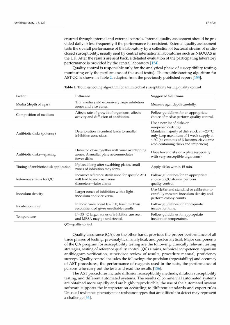
Quality control is responsible only for the analytical phase of susceptibility testing,monitoring only the performance of the used test(s). The troubleshooting algorithm forAST QC is shown in Table 2, adapted from the previously published report [155].
Table 2. Troubleshooting algorithm for antimicrobial susceptibility testing quality control.
Factor Influence Suggested Solutions
Media (depth of agar) Thin media yield excessively large inhibitionzones and vice versa. Measure agar depth carefully.
Composition of medium Affects rate of growth of organisms; affectsactivity and diffusion of antibiotics.
Follow guidelines for an appropriatechoice of media; perform quality control.
Antibiotic disks (potency) Deterioration in content leads to smallerinhibition zone sizes.
Use a new lot of disks orunopened cartridge.Maintain majority of disk stock at −20 ◦C,only keep maximum of 1 week supply at4 ◦C (be cautious of β-lactams, clavulanicacid-containing disks and imipenem).
Antibiotic disks—spacingDisks too close together will cause overlappingzones. A smaller plate accommodatesfewer disks
Place fewer disks on a plate (especiallywith very susceptible organisms)
Timing of antibiotic disk application If placed long after swabbing plates, smallzones of inhibition may form. Apply disks within 15 min.
Reference strains for QCIncorrect reference strain used for specific ASTwill lead to incorrect zonediameters—false alarm.
Follow guidelines for an appropriatechoice of QC strains; performquality control.
Inoculum density Larger zones of inhibition with a lightinoculum and vice versa.
Use McFarland standard or calibrator tocarefully measure inoculum density andperform colony counts.
Incubation time In most cases, ideal 16–18 h; less time thanrecommended gives unreliable results.
Follow guidelines for appropriateincubation time.
Temperature If <35 ◦C larger zones of inhibition are seenand MRSA may go undetected.
Follow guidelines for appropriateincubation temperature.
QC—quality control.
Quality assurance (QA), on the other hand, provides the proper performance of allthree phases of testing: pre-analytical, analytical, and post-analytical. Major componentsof the QA program for susceptibility testing are the following: clinically relevant testingstrategies, testing of reference quality control (QC) strains, technical competency, organismantibiogram verification, supervisor review of results, procedure manual, proficiencysurveys. Quality control includes the following: the precision (repeatability) and accuracyof AST procedures, the performance of reagents used in the tests, the performance ofpersons who carry out the tests and read the results [156].
The AST procedures include diffusion susceptibility methods, dilution susceptibilitytesting, and different automated systems. The results of commercial automated systemsare obtained more rapidly and are highly reproducible; the use of the automated systemsoftware supports the interpretation according to different standards and expert rules.Unusual resistance phenotype or resistance types that are difficult to detect may representa challenge [36].

Antibiotics 2022, 11, 427 18 of 26
As for automated antimicrobial susceptibility systems, which are used more andmore frequently in microbiological laboratories, usually, every device has its QC pro-cedure. The most commonly used automated systems for antimicrobial susceptibilityare VITEK 2 System (bioMérieux, France), BD Phoenix System (BD Diagnostic Systems,Baltimore, MD, USA), MicroScan WalkAway SI (Siemens Healthcare Diagnostics, WestSacramento, CA, USA), and TREK Sensititre (ARIS 2X, Trek Diagnostic Systems, Cleveland,OH, USA). Quality controls of such devices are not included in standards such as EUCASTor CLSI [61,156,157]. Agencies that provide the clearance of commercial systems for generaluse, such as the FDA for the USA, should use reference methods before the clearance foruse is given.
To evaluate or compare different susceptibility tests, usually these types of errors areconsidered: very major error, major error, and minor error. Very major error refers to thecharacterisation of the resistant isolate as susceptible. A major error is made when a sus-ceptible isolate is characterised as resistant. When an intermediate isolate is characterisedas susceptible or resistant, as well as when susceptible or resistant isolate is reported asintermediate, a minor error is made. Errors in antimicrobial susceptibility testing shouldbe monitored by laboratories and carefully analysed. Potential problems either with theidentification or susceptibility testing may be revealed [158].
For evaluation of new susceptibility tests or devices, a certain number of susceptibleas well as resistant strains should be tested against different antibiotics. Additionally,all types of the above-mentioned errors should be evaluated [36]. For new antimicrobialsusceptibility devices, acceptable performance according to FDA considers that majorerrors should not exceed 3% based on the number of susceptible organisms tested [159].The acceptable numbers of very major discrepancies rate should be 7.5% or less (for theupper 95% confidence limit) and 1.5% or less (for the lower 95% confidence limit), based onthe number of resistant organisms tested. Additionally, growth failure rates in the systemhave to be less than 10%.
The above-mentioned criteria may not be identical for acceptable accuracy comparedwith the international standards on susceptibility test device evaluation [160].
7. Near-Future Perspectives for Antimicrobial Susceptibility Testing
In the last decades, several innovative approaches for AST have been developed. Someof the most promising platforms for the forthcoming period—MALDI-TOF, flow cytometry,and isothermal microcalorimetry—will be briefly described.
Application of MALDI-TOF MS as routine rapid AST requires additional validation,such as standardised protocols, test kits, and software [161]. Further research efforts areneeded to refine and optimise MALDI-TOF MS-based assays to obtain accurate and reliableresults in the shortest possible time. A major focus of future research in this field will be toachieve the standardisation of methods and simultaneous susceptibility testing of microbesto various classes of antimicrobials [162]. So far, only two commercially available kits withsoftware for automated interpretation of spectra have been authorised in Europe to detectcarbapenemase activity or resistance toward third-generation cephalosporins in clinicalmicrobiology laboratories.
By using flow cytometry, the changes in morphology, physiological and metabolicactivity, and survival of microorganisms can be monitored after exposure to antimicrobials.The effects of treatment can be seen within a few hours, suggesting a strong potential forAST. Using nuclear dyes that do not penetrate the cell walls of healthy organisms, theamount of dying and dead cells can be rapidly determined. In order to determine theemission spectrum, the cells must go through a flow channel and be excited by a laser torelease dye [163]. Nevertheless, the future challenge is to improve the ability of a system todistinguish cellular damage caused by -cidal vs. -static antibiotics, as well as to address theissue of autofluorescence of certain species of bacteria. In addition, an enormous amount ofwork is required for the verification of the clinical database and the method itself.

Antibiotics 2022, 11, 427 19 of 26
Isothermal microcalorimetry is a dynamic technique allowing the measurement ofheat generated by the metabolism of actively growing cells. This technique is not new;however, it has been successfully adapted for AST of bacteria [164]. Additional advantagesof this technique are that it requires a small culture volume, the testing is performed insealed ampules, and monitoring during testing does not require manual manipulation.The completed analysis can also provide information on the -static vs. -cidal activity ofan agent.
Up to now, various alternative approaches to combat AMR have been proposed.For instance, epigenetic agents in modulating antibiotic susceptibility [165], agents thatinhibit virulence-conferring factors such as multidrug efflux inhibitors [166], or targets’essential processes for bacterial survival, such as cell division inhibitor [167], have beenrecently developed. If such antibacterial agents become licensed for treatment, antimicrobialsusceptibility techniques should be updated along with the new strategies developed tocounter AMR.
8. Conclusions
The timely administration of appropriate antimicrobial therapy based on accurateAST is widely recognised as a cornerstone of the management of infectious diseases. De-spite the shortcomings of the traditional AST methods, such as BMD and DD, their useis required in clinical practice to obtain the correct results according to the standardisedprotocols of EUCAST and CLSI, or for comparison against the results of novel techniques.Molecular-based methods are rapid, efficient, and reliable, with high specificity and sensi-tivity. Nonetheless, additional efforts are needed to bring closer NGS technology to routinemicrobiology. MALDI-TOF MS appears to be a promising highly specific system withreduced time and low cost of consumables. However, this method is not yet validated forall species and antimicrobials, and the acquisition costs of the system and its maintenanceare high. Hence, further improvement in the currently used and novel AST methods andinstruments is mandatory for speeding up the determination of antimicrobial efficacy inclinical microbiology laboratories in the foreseeable future.
Author Contributions: Conceptualisation, I.G.; manuscript writing, all authors; review and editing,I.G., N.O., M.M., D.M.C., A.T. and L.R.; figure creation: I.G., J.K., M.J. and D.K. All authors have readand agreed to the published version of the manuscript.
Funding: This review received no external funding.
Data Availability Statement: Not applicable.
Conflicts of Interest: The authors declare no conflict of interest.
References1. Holmes, A.H.; Moore, L.S.P.; Sundsfjord, A.; Steinbakk, M.; Regmi, S.; Karkey, A.; Guerin, P.J.; Piddock, L.J.V. Understanding the
mechanisms and drivers of antimicrobial resistance. Lancet 2016, 387, 176–187. [CrossRef]2. WHO. Antibiotic Resistance. Available online: https://www.who.int/news-room/fact-sheets/detail/antibiotic-resistance
(accessed on 23 February 2022).3. Murray, C.J.; Ikuta, K.S.; Sharara, F.; Swetschinski, L.; Robles Aguilar, G.; Gray, A.; Han, C.; Bisignano, C.; Rao, P.; Wool, E.; et al.
Global burden of bacterial antimicrobial resistance in 2019: A systematic analysis. Lancet 2022, 399, 629–655. [CrossRef]4. WHO. New Report Calls for Urgent Action to Avert Antimicrobial Resistance Crisis. Available online: https://www.
who.int/news/item/29-04-2019-new-report-calls-for-urgent-action-to-avert-antimicrobial-resistance-crisis (accessed on 23February 2022).
5. Mirzaei, R.; Goodarzi, P.; Asadi, M.; Soltani, A.; Aljanabi, H.A.A.; Jeda, A.S.; Dashtbin, S.; Jalalifar, S.; Mohammadzadeh, R.;Teimoori, A.; et al. Bacterial co-infections with SARS-CoV-2. IUBMB Life 2020, 72, 2097–2111. [CrossRef] [PubMed]
6. Tomczyk, S.; Taylor, A.; Brown, A.; de Kraker, M.E.A.; El-Saed, A.; Alshamrani, M.; Hendriksen, R.S.; Jacob, M.; Löfmark, S.;Perovic, O.; et al. Impact of the COVID-19 pandemic on the surveillance, prevention and control of antimicrobial resistance: Aglobal survey. J. Antimicrob. Chemother. 2021, 76, 3045–3058. [CrossRef] [PubMed]
7. Bengoechea, J.A.; Bamford, C.G. SARS-CoV-2, bacterial co-infections, and AMR: The deadly trio in COVID-19? EMBO Mol. Med.2020, 12, e12560. [CrossRef] [PubMed]

Antibiotics 2022, 11, 427 20 of 26
8. By 2050, Drug-Resistant Infections Could Cause Global Economic Damage on Par with 2008 Financial Crisis. Available on-line: https://www.worldbank.org/en/news/press-release/2016/09/18/by-2050-drug-resistant-infections-could-cause-global-economic-damage-on-par-with-2008-financial-crisis (accessed on 23 February 2022).
9. Zhu, Y.; Huang, W.E.; Yang, Q. Clinical perspective of antimicrobial resistance in bacteria. Infect. Drug Resist. 2022, 15, 735–746.[CrossRef]
10. Munita, J.M.; Arias, C.A. Mechanisms of antibiotic resistance. Microbiol. Spectr. 2016, 4, 464–472. [CrossRef] [PubMed]11. Tacconelli, E. Antimicrobial use: Risk driver of multidrug resistant microorganisms in healthcare settings. Curr. Opin. Infect. Dis.
2009, 22, 352–358. [CrossRef] [PubMed]12. Barlam, T.F.; Cosgrove, S.E.; Abbo, L.M.; Macdougall, C.; Schuetz, A.N.; Septimus, E.J.; Srinivasan, A.; Dellit, T.H.;
Falck-Ytter, Y.T.; Fishman, N.O.; et al. Executive Summary: Implementing an Antibiotic Stewardship Program: Guidelines bythe Infectious Diseases Society of America and the Society for Healthcare Epidemiology of America. Clin. Infect. Dis. 2016, 62,1197–1202. [CrossRef] [PubMed]
13. Van Belkum, A.; Bachmann, T.T.; Lüdke, G.; Lisby, J.G.; Kahlmeter, G.; Mohess, A.; Becker, K.; Hays, J.P.; Woodford, N.;Mitsakakis, K.; et al. Developmental roadmap for antimicrobial susceptibility testing systems. Nat. Rev. Microbiol. 2018, 17, 51–62.[CrossRef]
14. Tebano, G.; Mouelhi, Y.; Zanichelli, V.; Charmillon, A.; Fougnot, S.; Lozniewski, A.; Thilly, N.; Pulcini, C. Selective reporting ofantibiotic susceptibility testing results: A promising antibiotic stewardship tool. Expert Rev. Anti. Infect. Ther. 2020, 18, 251–262.[CrossRef] [PubMed]
15. WHO. Monitoring and Evaluation of the Global Action Plan on Antimicrobial Resistance: Framework and RecommendedIndicators. Available online: https://apps.who.int/iris/handle/10665/325006 (accessed on 23 February 2022).
16. Iskandar, K.; Molinier, L.; Hallit, S.; Sartelli, M.; Hardcastle, T.C.; Haque, M.; Lugova, H.; Dhingra, S.; Sharma, P.; Islam, S.; et al.Surveillance of antimicrobial resistance in low- and middle-income countries: A scattered picture. Antimicrob. Resist. Infect.Control 2021, 10, 63. [CrossRef]
17. WHO. Antimicrobial Stewardship Interventions: A Practical Guide; World Health Organization: Geneva, Switzerland, 2021.18. Klinker, K.P.; Hidayat, L.K.; DeRyke, C.A.; DePestel, D.D.; Motyl, M.; Bauer, K.A. Antimicrobial stewardship and antibiograms:
Importance of moving beyond traditional antibiograms. Ther. Adv. Infect. Dis. 2021, 8, 1–9. [CrossRef] [PubMed]19. Hebert, C.; Ridgway, J.; Vekhter, B.; Brown, E.C.; Weber, S.G.; Robicsek, A. Demonstration of the weighted-incidence syndromic
combination antibiogram: An empiric prescribing decision Aid. Infect. Control Hosp. Epidemiol. 2012, 33, 381–388. [CrossRef]20. Khalili, H.; Izadpanah, M. Antibiotic regimens for treatment of infections due to multidrug-resistant Gram-negative pathogens:
An evidence-based literature review. J. Res. Pharm. Pract. 2015, 4, 105. [CrossRef]21. Kalil, A.C.; Metersky, M.L.; Klompas, M.; Muscedere, J.; Sweeney, D.A.; Palmer, L.B.; Napolitano, L.M.; O’Grady, N.P.; Bartlett, J.G.;
Carratalà, J.; et al. Management of adults with hospital-acquired and ventilator-associated pneumonia: 2016 clinical practiceguidelines by the Infectious Diseases Society of America and the American Thoracic Society. Clin. Infect. Dis. 2016, 63, e61–e111.[CrossRef] [PubMed]
22. Clinical and Laboratory Standard Institute. M39-A4: Analysis and Presentation of Cumulative Antimicrobial Susceptibility Test Data:Approved Guideline, 4th ed.; Clinical and Laboratory Standard Institute: Wayne, PA, USA, 2014; ISBN 610.688.0700.
23. Puzniak, L.; DePestel, D.D.; Srinivasan, A.; Ye, G.; Murray, J.; Merchant, S.; Andrew DeRyke, C.; Gupta, V. A combinationantibiogram evaluation for Pseudomonas aeruginosa in respiratory and blood sources from intensive care unit (ICU) and Non-ICUSettings in U.S. Hospitals. Antimicrob. Agents Chemother. 2019, 63, e02564-18. [CrossRef] [PubMed]
24. Khan, Z.A.; Siddiqui, M.F.; Park, S. Current and emerging methods of antibiotic susceptibility testing. Diagnostics 2019, 9, 49.[CrossRef] [PubMed]
25. Syal, K.; Mo, M.; Yu, H.; Iriya, R.; Jing, W.; Guodong, S.; Wang, S.; Grys, T.E.; Haydel, S.E.; Tao, N. Current and emergingtechniques for antibiotic susceptibility tests. Theranostics 2017, 7, 1795–1805. [CrossRef]
26. Jenkins, S.G.; Schuetz, A.N. Current concepts in laboratory testing to guide antimicrobial therapy. Mayo Clin. Proc. 2012, 87,290–308. [CrossRef] [PubMed]
27. Spencer, D.C.; Paton, T.F.; Mulroney, K.T.; Inglis, T.J.J.; Sutton, J.M.; Morgan, H. A fast impedance-based antimicrobial susceptibilitytest. Nat. Commun. 2020, 11, 5328. [CrossRef] [PubMed]
28. Clinical and Laboratory Standards Institute. M07: Methods for Dilution Antimicrobial Susceptibility Tests for Bacteria that GrowAerobically, 11th ed.; Clinical and Laboratory Standards Institute: Wayne, PA, USA, 2018; ISBN 1562388363.
29. Golus, J.; Sawicki, R.; Widelski, J.; Ginalska, G. The agar microdilution method—A new method for antimicrobial susceptibilitytesting for essential oils and plant extracts. J. Appl. Microbiol. 2016, 121, 1291–1299. [CrossRef] [PubMed]
30. Vasilijevic, B.; Mitic-Culafic, D.; Djekic, I.; Markovic, T.; Kneževic-Vukcevic, J.; Tomasevic, I.; Velebit, B.; Nikolic, B. Antibacterialeffect of Juniperus communis and Satureja montana essential oils against Listeria monocytogenes in vitro and in wine marinated beef.Food Control 2019, 100, 247–256. [CrossRef]
31. Kowalska-Krochmal, B.; Dudek-Wicher, R. The minimum inhibitory concentration of antibiotics: Methods, interpretation, clinicalrelevance. Pathogens 2021, 10, 165. [CrossRef] [PubMed]
32. Balouiri, M.; Sadiki, M.; Ibnsouda, S.K. Methods for in vitro evaluating antimicrobial activity: A review. J. Pharm. Anal. 2016, 6,71–79. [CrossRef] [PubMed]
Antibiotics 2022, 11, 427 21 of 26
33. Ferro, B.E.; Van Ingen, J.; Wattenberg, M.; Van Soolingen, D.; Mouton, J.W. Time-kill kinetics of antibiotics active against rapidlygrowing mycobacteria. J. Antimicrob. Chemother. 2015, 70, 811–817. [CrossRef]
34. Mueller, M.; De La Peña, A.; Derendorf, H. Issues in pharmacokinetics and pharmacodynamics of anti-infective agents: Killcurves versus MIC. Antimicrob. Agents Chemother. 2004, 48, 369–377. [CrossRef]
35. Olajuyigbe, O.O.; Afolayan, A.J. In vitro antibacterial and time-kill evaluation of the Erythrina caffra Thunb. extract againstbacteria associated with diarrhoea. Sci. World J. 2012, 2012, 738314. [CrossRef] [PubMed]
36. Jorgensen, J.H.; Ferraro, M.J. Antimicrobial susceptibility testing: A review of general principles and contemporary practices.Clin. Infect. Dis. 2009, 49, 1749–1755. [CrossRef] [PubMed]
37. Nikolic, B.; Vasilijevic, B.; Mitic-Culafic, D.; Lesjak, M.; Vukovic-Gacic, B.; Dukic, N.M.; Kneževic-Vukcevic, J. Screening of theantibacterial effect of Juniperus sibirica and Juniperus sabina essential oils in a microtitre plate-based MIC assay. Bot. Serb. 2016, 40,43–48. [CrossRef]
38. Foerster, S.; Desilvestro, V.; Hathaway, L.J.; Althaus, C.L.; Unemo, M. A new rapid resazurin-based microdilution assay forantimicrobial susceptibility testing of Neisseria gonorrhoeae. J. Antimicrob. Chemother. 2017, 72, 1961–1968. [CrossRef] [PubMed]
39. Galani, I.; Adamou, P.; Karaiskos, I.; Giamarellou, H.; Souli, M. Evaluation of ComASPTM Colistin (formerly SensiTestTM Colistin),a commercial broth microdilution-based method to evaluate the colistin minimum inhibitory concentration for carbapenem-resistant Klebsiella pneumoniae isolates. J. Glob. Antimicrob. Resist. 2018, 15, 123–126. [CrossRef]
40. Wiegand, I.; Hilpert, K.; Hancock, R.E.W. Agar and broth dilution methods to determine the minimal inhibitory concentration(MIC) of antimicrobial substances. Nat. Protoc. 2008, 3, 163–175. [CrossRef]
41. Mirajkar, C.J.; Gebhart, C.J. Comparison of agar dilution and antibiotic gradient strip test with broth microdilution for susceptibil-ity testing of swine Brachyspira species. J. Vet. Diagn. Investig. 2016, 28, 133–143. [CrossRef] [PubMed]
42. The European Committee on Antimicrobial Susceptibility Testing. Breakpoint Tables for Interpretation of MICs and ZoneDiameters, Version 12.0. Available online: http://www.eucast.org/clinical_breakpoints/ (accessed on 23 February 2022).
43. Shields, R.K.; Clancy, C.J.; Pasculle, A.W.; Press, E.G.; Haidar, G.; Hao, B.; Chen, L.; Kreiswirth, B.N.; Nguyen, M.H. Verifi-cation of Ceftazidime-Avibactam and Ceftolozane-Tazobactam Susceptibility Testing Methods against Carbapenem-ResistantEnterobacteriaceae and Pseudomonas aeruginosa. J. Clin. Microbiol. 2018, 56, e01093-17. [CrossRef] [PubMed]
44. Humphries, R.M.; Hindler, J.A.; Magnano, P.; Wong-Beringer, A.; Tibbetts, R.; Miller, S.A. Performance of ceftolozane-tazobactametest, MIC test strips, and disk diffusion compared to reference broth microdilution for-Lactam-Resistant Pseudomonas aeruginosaisolates. J. Clin. Microbiol. 2018, 56, e01633-17. [CrossRef] [PubMed]
45. Gajic, I.; Ranin, L.; Kekic, D.; Opavski, N.; Smitran, A.; Mijac, V.; Jovanovic, S.; Hadnadjev, M.; Travar, M.; Mijovic, G. Tigecyclinesusceptibility of multidrug-resistant Acinetobacter baumannii from intensive care units in the western Balkans. Acta Microbiol.Immunol. Hung. 2020, 67, 176–181. [CrossRef] [PubMed]
46. Satlina, M.J. The search for a practical method for colistin susceptibility testing: Have we found it by going back to the future? J.Clin. Microbiol. 2018, 57, e01608-18. [CrossRef] [PubMed]
47. Jönsson, A.; Jacobsson, S.; Foerster, S.; Cole, M.J.; Unemo, M. Performance characteristics of newer MIC gradient strip testscompared with the Etest for antimicrobial susceptibility testing of Neisseria gonorrhoeae. APMIS 2018, 126, 822–827. [CrossRef][PubMed]
48. Miftahussurur, M.; Fauzia, K.A.; Nusi, I.A.; Setiawan, P.B.; Syam, A.F.; Waskito, L.A.; Doohan, D.; Ratnasari, N.; Khomsan, A.;Adnyana, I.K.; et al. E-test versus agar dilution for antibiotic susceptibility testing of Helicobacter pylori: A comparison study.BMC Res. Notes 2020, 13, 22. [CrossRef]
49. Akcali, S.; Cicek, C.; Surucuoglu, S.; Ozbakkaloglu, B. E-Test: An Alternative Method for Susceptibility Testing of Mycobacteriumtuberculosis. Med. Princ. Pract. 2005, 14, 264–267. [CrossRef] [PubMed]
50. Dannaoui, E.; Espinel-Ingroff, A. Antifungal Susceptibly Testing by Concentration Gradient Strip Etest Method for Fungal Isolates:A Review. J. Fungi 2019, 5, 108. [CrossRef] [PubMed]
51. Pereckaite, L.; Tatarunas, V.; Giedraitiene, A. Current antimicrobial susceptibility testing for beta-lactamase-producing Enterobac-teriaceae in clinical settings. J. Microbiol. Methods 2018, 152, 154–164. [CrossRef] [PubMed]
52. Garrec, H.; Drieux-Rouzet, L.; Golmard, J.L.; Jarlier, V.; Robert, J. Comparison of nine phenotypic methods for detection ofextended-spectrum β-lactamase production by Enterobacteriaceae. J. Clin. Microbiol. 2011, 49, 1048–1057. [CrossRef] [PubMed]
53. Guzel, M.; Afsar, Y.; Akdogan, D.; Moncheva, P.; Hristova, P.; Erdem, G. Evaluation of metallo-beta-lactamase productionin multiple antibiotic-resistant Pseudomonas spp. and Acinetobacter baumannii strains. Biotechnol. Biotechnol. Equip. 2018, 32,1285–1290. [CrossRef]
54. Martinez-Martinez, L.; Cantón Spain, R.; Stefani, S.; Skov, R.; Glupczynski, Y.; Nordmann, P.; Wootton, M.; Miriagou, V.;Skov Simonsen, G.; Zemlickova, H.; et al. EUCAST Guidelines for Detection of Resistance Mechanisms and Specific Resistances ofClinical and/or Epidemiological Importance. Available online: https://www.eucast.org/fileadmin/src/media/PDFs/EUCAST_files/Resistance_mechanisms/EUCAST_detection_of_resistance_mechanisms_170711.pdf (accessed on 27 February 2022).
55. Peter-Getzlaff, S.; Polsfuss, S.; Poledica, M.; Hombach, M.; Giger, J.; Böttger, E.C.; Zbinden, R.; Bloemberg, G.V. Detection of AmpCbeta-lactamase in Escherichia coli: Comparison of three phenotypic confirmation assays and genetic analysis. J. Clin. Microbiol.2011, 49, 2924–2932. [CrossRef]

Antibiotics 2022, 11, 427 22 of 26
56. Yusof, A.; Engelhardt, A.; Karlsson, A.; Bylund, L.; Vidh, P.; Mills, K.; Wootton, M.; Walsh, T.R. Evaluation of a new etestvancomycin-teicoplanin strip for detection of glycopeptide-intermediate Staphylococcus aureus (GISA), in particular, heterogeneousGISA. J. Clin. Microbiol. 2008, 46, 3042–3047. [CrossRef] [PubMed]
57. Heatley, N.G. A method for the assay of penicillin. Biochem. J. 1944, 38, 61. [CrossRef] [PubMed]58. Clinical and Laboratory Standards Institute. M02: Performance Standards for Antimicrobial Disk Susceptibility Tests, 13th ed.; Clinical
and Laboratory Standards Institute: Wayne, PA, USA, 2018; ISBN 1562387863.59. The European Committee on Antimicrobial Susceptibility Testing. Routine and Extended Internal Quality Control for MIC
Determination and Disk Diffusion as Recommended by EUCAST, Version 12.0. Available online: http://www.eucast.org/ast_of_bacteria/quality_control/ (accessed on 23 February 2022).
60. Le Page, S.; Dubourg, G.; Rolain, J.-M. Evaluation of the Scan® 1200 as a rapid tool for reading antibiotic susceptibility testing bythe disc diffusion technique. J. Antimicrob. Chemother. 2016, 71, 3424–3431. [CrossRef]
61. Clinical and Laboratory Standards Institute. M100: Performance Standards for Antimicrobial Susceptibility Testing, 32nd ed.; Clinicaland Laboratory Standards Institute: Wayne, PA, USA, 2022; ISBN 9781684400331.
62. Hombach, M.; Zbinden, R.; Böttger, E.C. Standardisation of disk diffusion results for antibiotic susceptibility testing using thesirscan automated zone reader. BMC Microbiol. 2013, 13, 225. [CrossRef]
63. Idelevich, E.A.; Hoy, M.; Görlich, D.; Knaack, D.; Grünastel, B.; Peters, G.; Borowski, M.; Becker, K. Rapid phenotypic detection ofmicrobial resistance in gram-positive bacteria by a real-time laser scattering method. Front. Microbiol. 2017, 8, 1064. [CrossRef][PubMed]
64. Jorgensen, J.H.; Barry, A.L.; Traczewski, M.M.; Sahm, D.F.; McElmeel, M.L.; Crawford, S.A. Rapid automated antimicrobialsusceptibility testing of Streptococcus pneumoniae by use of the bioMerieux VITEK 2. J. Clin. Microbiol. 2000, 38, 2814–2818.[CrossRef] [PubMed]
65. Coyle, M.B.; McGonagle, L.A.; Plorde, J.J.; Clausen, C.R.; Schoenknecht, F.D. Rapid antimicrobial susceptibility testing of isolatesfrom blood cultures by direct inoculation and early reading of disk diffusion tests. J. Clin. Microbiol. 1984, 20, 473–477. [CrossRef][PubMed]
66. Liberman, D.F.; Robertson, R.G. Evaluation of a rapid Bauer-Kirby antibiotic susceptibility determination. Antimicrob. AgentsChemother. 1975, 7, 250–255. [CrossRef]
67. Van den Bijllaardt, W.; Buiting, A.G.; Mouton, J.W.; Muller, A.E. Shortening the incubation time for antimicrobial susceptibilitytesting by disk diffusion for Enterobacteriaceae: How short can it be and are the results accurate? Int. J. Antimicrob. Agents 2017,49, 631–637. [CrossRef] [PubMed]
68. Hombach, M.; Jetter, M.; Blöchliger, N.; Kolesnik-Goldmann, N.; Böttger, E.C. Fully automated disc diffusion for rapid antibioticsusceptibility test results: A proof-of-principle study. J. Antimicrob. Chemother. 2017, 72, 1659–1668. [CrossRef] [PubMed]
69. Jonasson, E.; Matuschek, E.; Kahlmeter, G. The EUCAST rapid disc diffusion method for antimicrobial susceptibility testingdirectly from positive blood culture bottles. J. Antimicrob. Chemother. 2020, 75, 968–978. [CrossRef] [PubMed]
70. The European Committee on Antimicrobial Susceptibility Testing. Zone Diameter Breakpoints for Rapid Antimicrobial Suscepti-bility Testing (RAST) Directly from Blood Culture Bottles, Version 4.0. Available online: http://www.eucast.org/rapid_ast_in_blood_cultures/breakpoints_for_short_incubation/ (accessed on 23 February 2022).
71. The European Committee on Antimicrobial Susceptibility Testing. Screening for ESBL and Carbapenemases in E. coli and K.pneumoniae for Epidemiological Purposes as Part of the RAST Procedure. EUCAST Guidelines for Detection of ResistanceMechanisms and Specific Resistance of Clinical and/or Epidemiological Importance. Available online: http://www.eucast.org/resistance_mechanisms/ (accessed on 23 February 2022).
72. Idelevich, E.A.; Schüle, I.; Grünastel, B.; Wüllenweber, J.; Peters, G.; Becker, K. Rapid identification of microorganisms frompositive blood cultures by MALDI-TOF mass spectrometry subsequent to very short-term incubation on solid medium. Clin.Microbiol. Infect. 2014, 20, 1001–1006. [CrossRef]
73. Savage, T.J.; Rao, S.; Joerger, J.; Ozonoff, A.; McAdam, A.J.; Sandora, T.J. Predictive value of direct disk diffusion testing frompositive blood cultures in a children’s hospital and its utility in antimicrobial stewardship. J. Clin. Microbiol. 2021, 59, e02445-20.[CrossRef]
74. Ahman, J.; Matuschek, E.; Kahlmeter, G. The quality of antimicrobial discs from nine manufacturers-EUCAST evaluations in 2014and 2017. Clin. Microbiol. Infect. 2019, 25, 346–352. [CrossRef]
75. Aubry, B.; Lemarié, C.; Chenouard, R.; Kempf, M.; Eveillard, M.; Pailhoriès, H. Performance of penicillinase detection tests inStaphylococcus epidermidis: Comparison of different phenotypic methods. BMC Microbiol. 2020, 20, 240. [CrossRef]
76. Merlino, J.; Leroi, M.; Bradbury, R.; Veal, D.; Harbour, C. New chromogenic identification and detection of Staphylococcus aureusand methicillin-resistant S. aureus. J. Clin. Microbiol. 2000, 38, 2378–2380. [CrossRef] [PubMed]
77. Ledeboer, N.A.; Das, K.; Eveland, M.; Roger-Dalbert, C.; Mailler, S.; Chatellier, S.; Dunne, W.M. Evaluation of a novel chromogenicagar medium for isolation and differentiation of vancomycin-resistant Enterococcus faecium and Enterococcus faecalis isolates. J.Clin. Microbiol. 2007, 45, 1556–1560. [CrossRef] [PubMed]
78. Glupczynski, Y.; Berhin, C.; Bauraing, C.; Bogaerts, P. Evaluation of a New Selective Chromogenic Agar Medium for Detection ofExtended-Spectrum-Lactamase-Producing Enterobacteriaceae. J. Clin. Microbiol. 2007, 45, 501–505. [CrossRef]
79. Samra, Z.; Bahar, J.; Madar-Shapiro, L.; Aziz, N.; Israel, S.; Bishara, J. Evaluation of CHROMagar KPC for rapid detection ofcarbapenem-resistant Enterobacteriaceae. J. Clin. Microbiol. 2008, 46, 3110–3111. [CrossRef] [PubMed]
Antibiotics 2022, 11, 427 23 of 26
80. Gordon, N.C.; Wareham, D.W. Evaluation of CHROMagar Acinetobacter for detection of enteric carriage of multidrug-resistantAcinetobacter baumannii in samples from critically ill patients. J. Clin. Microbiol. 2009, 47, 2249–2251. [CrossRef]
81. Perry, J.D. A decade of development of chromogenic culture media for clinical microbiology in an era of molecular diagnostics.Clin. Microbiol. Rev. 2017, 30, 449–479. [CrossRef] [PubMed]
82. Kekic, D.; Gajic, I.; Opavski, N.; Kojic, M.; Vukotic, G.; Smitran, A.; Boskovic, L.; Stojkovic, M.; Ranin, L. Trends in molecularcharacteristics and antimicrobial resistance of group B streptococci: A multicenter study in Serbia, 2015–2020. Sci. Rep. 2021,11, 540. [CrossRef] [PubMed]
83. Tierney, D.; Copsey, S.D.; Morris, T.; Perry, J.D. A new chromogenic medium for isolation of Bacteroides fragilis suitable forscreening for strains with antimicrobial resistance. Anaerobe 2016, 39, 168–172. [CrossRef]
84. Abdul Momin, M.H.F.; Bean, D.C.; Hendriksen, R.S.; Haenni, M.; Phee, L.M.; Wareham, D.W. CHROMagar COL-APSE: Aselective bacterial culture medium for the isolation and differentiation of colistin-resistant Gram-negative pathogens. J. Med.Microbiol. 2017, 66, 1554–1561. [CrossRef] [PubMed]
85. Nordmann, P.; Poirel, L.; Dortet, L. Rapid Detection of Carbapenemase-producing Enterobacteriaceae. Emerg. Infect. Dis. 2012,18, 1503. [CrossRef]
86. Benkova, M.; Soukup, O.; Marek, J. Antimicrobial susceptibility testing: Currently used methods and devices and the near futurein clinical practice. J. Appl. Microbiol. 2020, 129, 806–822. [CrossRef]
87. Sader, H.S.; Fritsche, T.R.; Jones, R.N. Accuracy of three automated systems (MicroScan WalkAway, VITEK, and VITEK 2) forsusceptibility testing of Pseudomonas aeruginosa against five broad-spectrum beta-lactam agents. J. Clin. Microbiol. 2006, 44,1101–1104. [CrossRef] [PubMed]
88. Fredborg, M.; Andersen, K.R.; Jørgensen, E.; Droce, A.; Olesen, T.; Jensen, B.B.; Rosenvinge, F.S.; Sondergaard, T.E. Real-timeoptical antimicrobial susceptibility testing. J. Clin. Microbiol. 2013, 51, 2047–2053. [CrossRef]
89. Ligozzi, M.; Bernini, C.; Bonora, M.G.; De Fatima, M.; Zuliani, J.; Fontana, R. Evaluation of the VITEK 2 system for identificationand antimicrobial susceptibility testing of medically relevant gram-positive cocci. J. Clin. Microbiol. 2002, 40, 1681–1686. [CrossRef]
90. Spanu, T.; Sanguinetti, M.; Ciccaglione, D.; D’Inzeo, T.; Romano, L.; Leone, F.; Fadda, G. Use of the VITEK 2 System for rapididentification of clinical isolates of Staphylococci from bloodstream infections. J. Clin. Microbiol. 2003, 41, 4259. [CrossRef][PubMed]
91. Evangelista, A.T.; Karlowsky, J.A. Automated and Manual Systems for Antimicrobial Susceptibility Testing of Bacteria. In Manualof Commercial Methods in Clinical Microbiology; John Wiley & Sons, Inc.: Hoboken, NJ, USA, 2016; pp. 414–432. [CrossRef]
92. Zhou, M.; Wang, Y.; Liu, C.; Kudinha, T.; Liu, X.; Luo, Y.; Yang, Q.; Sun, H.; Hu, J.; Xu, Y.-C. Comparison of five commonlyused automated susceptibility testing methods for accuracy in the China Antimicrobial Resistance Surveillance System (CARSS)hospitals. Infect. Drug Resist. 2018, 11, 1347–1358. [CrossRef]
93. Bulik, C.C.; Fauntleroy, K.A.; Jenkins, S.G.; Abuali, M.; LaBombardi, V.J.; Nicolau, D.P.; Kuti, J.L. Comparison of meropenem MICsand susceptibilities for carbapenemase-producing Klebsiella pneumoniae isolates by various testing methods. J. Clin. Microbiol.2010, 48, 2402. [CrossRef]
94. Woodford, N.; Eastaway, A.T.; Ford, M.; Leanord, A.; Keane, C.; Quayle, R.M.; Steer, J.A.; Zhang, J.; Livermore, D.M. Comparisonof BD Phoenix, Vitek 2, and MicroScan Automated Systems for detection and inference of mechanisms responsible for carbapenemresistance in Enterobacteriaceae. J. Clin. Microbiol. 2010, 48, 2999. [CrossRef] [PubMed]
95. Buchan, B.W.; Anderson, N.W.; Ledeboer, N.A. Comparison of BD Phoenix and bioMérieux Vitek 2 automated systems for thedetection of macrolide-lincosamide-streptogramin B resistance among clinical isolates of Staphylococcus. Diagn. Microbiol. Infect.Dis. 2012, 72, 291–294. [CrossRef] [PubMed]
96. Richter, S.S.; Howard, W.J.; Weinstein, M.P.; Bruckner, D.A.; Hindler, J.F.; Saubolle, M.; Doern, G.V. Multicenter evaluation of theBD Phoenix Automated Microbiology System for antimicrobial susceptibility testing of Streptococcus species. J. Clin. Microbiol.2007, 45, 2863. [CrossRef] [PubMed]
97. Khan, A.; Arias, C.A.; Abbott, A.; Dien Bard, J.; Bhatti, M.M.; Humphries, R.M. Evaluation of the Vitek 2, Phoenix, and MicroScanfor antimicrobial susceptibility testing of Stenotrophomonas maltophilia. J. Clin. Microbiol. 2021, 59, e0065421. [CrossRef]
98. Schumacher, A.; Vranken, T.; Malhotra, A.; Arts, J.J.C.; Habibovic, P. In vitro antimicrobial susceptibility testing methods: Agardilution to 3D tissue-engineered models. Eur. J. Clin. Microbiol. Infect. Dis. 2018, 37, 187–208. [CrossRef]
99. Sekyere, J.O.; Asante, J. Emerging mechanisms of antimicrobial resistance in bacteria and fungi: Advances in the era of genomics.Future Microbiol. 2018, 13, 241–262. [CrossRef]
100. Gajic, I.; Jovicevic, M.; Milic, M.; Kekic, D.; Opavski, N.; Zrnic, Z.; Dacic, S.; Pavlovic, L.; Mijac, V. Clinical and molecularcharacteristics of OXA-72-producing Acinetobacter baumannii ST636 outbreak at a neonatal intensive care unit in Serbia. J. Hosp.Infect. 2021, 112, 54–60. [CrossRef] [PubMed]
101. Rocchetti, T.T.; Martins, K.B.; Martins, P.Y.F.; de Oliveira, R.A.; Mondelli, A.L.; Fortaleza, C.M.C.B.; de Lourdes Ribeiro de Souzada Cunha, M. Detection of the mecA gene and identification of Staphylococcus directly from blood culture bottles by multiplexpolymerase chain reaction. Braz. J. Infect. Dis. 2018, 22, 99–105. [CrossRef] [PubMed]
102. Simner, P.; Musser, K.; Mitchell, K.; Wise, M.; Lewis, S.; Yee, R.; Bergman, Y.; Good, C.; Abdelhamed, A.; Li, H.; et al. MulticenterEvaluation of the Acuitas AMR Gene Panel for Detection of an Extended Panel of Antimicrobial Resistance Genes among BacterialIsolates. J. Clini. Microbiol. 2022, 60, e0209821. [CrossRef]

Antibiotics 2022, 11, 427 24 of 26
103. Findlay, J.; Hopkins, K.L.; Meunier, D.; Woodford, N. Evaluation of three commercial assays for rapid detection of genes encodingclinically relevant carbapenemases in cultured bacteria. J. Antimicrob. Chemother. 2015, 70, 1338–1342. [CrossRef] [PubMed]
104. Cepheid|Molecular Testing of Healthcare Associated Infections. Available online: https://www.cepheid.com/en_US/tests/Healthcare-Associated-Infections (accessed on 16 March 2022).
105. Confirmation-Check-Points Health. Available online: https://check-pointshealth.com/confirmation/ (accessed on 16March 2022).
106. Matsuda, K. PCR-based detection methods for single-nucleotide polymorphism or mutation: Real-time PCR and its substantialcontribution toward technological refinement. Adv. Clin. Chem. 2017, 80, 45–72. [CrossRef] [PubMed]
107. Charnock, C.; Samuelsen, Ø.; Nordlie, A.L.; Hjeltnes, B. Use of a commercially available microarray to characterize antibiotic-resistant clinical isolates of Klebsiella pneumoniae. Curr. Microbiol. 2018, 75, 163–172. [CrossRef]
108. Kim, J.-S.; Kang, G.-E.; Kim, H.-S.; Kim, H.S.; Song, W.; Lee, K.M. Evaluation of Verigene Blood Culture Test Systems for RapidIdentification of Positive Blood Cultures. Biomed Res. Int. 2016, 2016, 1081536. [CrossRef]
109. Cunningham, S.A.; Vasoo, S.; Patel, R. Evaluation of the Check-Points Check MDR CT103 and CT103 XL Microarray Kits by Useof Preparatory Rapid Cell Lysis. J. Clin. Microbiol. 2016, 54, 1368–1371. [CrossRef] [PubMed]
110. Gomes, A.É.I.; Stuchi, L.P.; Siqueira, N.M.G.; Henrique, J.B.; Vicentini, R.; Ribeiro, M.L.; Darrieux, M.; Ferraz, L.F.C. Selectionand validation of reference genes for gene expression studies in Klebsiella pneumoniae using Reverse Transcription Quantitativereal-time PCR. Sci. Rep. 2018, 8, 9001. [CrossRef] [PubMed]
111. Shifman, O.; Steinberger-Levy, I.; Aloni-Grinstein, R.; Gur, D.; Aftalion, M.; Ron, I.; Mamroud, E.; Ber, R.; Rotem, S. A rapidantimicrobial susceptibility test for determining Yersinia pestis susceptibility to doxycycline by RT-PCR quantification of RNAmarkers. Front. Microbiol. 2019, 10, 754. [CrossRef]
112. Hendriksen, R.S.; Bortolaia, V.; Tate, H.; Tyson, G.H.; Aarestrup, F.M.; McDermott, P.F. Using genomics to track global antimicrobialresistance. Front. Public Health 2019, 7, 242. [CrossRef] [PubMed]
113. Faksri, K.; Kaewprasert, O.; Ong, R.T.H.; Suriyaphol, P.; Prammananan, T.; Teo, Y.Y.; Srilohasin, P.; Chaiprasert, A. Comparisonsof whole-genome sequencing and phenotypic drug susceptibility testing for Mycobacterium tuberculosis causing MDR-TB andXDR-TB in Thailand. Int. J. Antimicrob. Agents 2019, 54, 109–116. [CrossRef]
114. Yang, X.; Hashemi, M.M.; Andini, N.; Li, M.M.; Kuang, S.; Carroll, K.C.; Wang, T.H.; Yang, S. RNA markers for ultra-rapidmolecular antimicrobial susceptibility testing in fluoroquinolone-treated Klebsiella pneumoniae. J. Antimicrob. Chemother. 2020, 75,1747–1755. [CrossRef]
115. Bhattacharyya, R.P.; Bandyopadhyay, N.; Ma, P.; Son, S.S.; Liu, J.; He, L.L.; Wu, L.; Khafizov, R.; Boykin, R.; Cerqueira, G.C.; et al.Simultaneous detection of genotype and phenotype enables rapid and accurate antibiotic susceptibility determination. Nat. Med.2019, 25, 1858–1864. [CrossRef] [PubMed]
116. Rossen, J.W.A.; Friedrich, A.W.; Moran-Gilad, J. Practical issues in implementing whole-genome-sequencing in routine diagnosticmicrobiology. Clin. Microbiol. Infect. 2018, 24, 355–360. [CrossRef] [PubMed]
117. Ellington, M.J.; Ekelund, O.; Aarestrup, F.M.; Canton, R.; Doumith, M.; Giske, C.; Grundman, H.; Hasman, H.; Holden, M.T.G.;Hopkins, K.L.; et al. The role of whole genome sequencing in antimicrobial susceptibility testing of bacteria: Report from theEUCAST Subcommittee. Clin. Microbiol. Infect. 2017, 23, 2–22. [CrossRef] [PubMed]
118. Ho, Y.P.; Reddy, P.M. Advances in mass spectrometry for the identification of pathogens. Mass. Spectrom. Rev. 2011, 30, 1203–1224.[CrossRef]
119. Oviaño, M.; de la Luna Ramírez, C.; Barbeyto, L.P.; Bou, G. Rapid direct detection of carbapenemase-producing Enterobacteriaceaein clinical urine samples by MALDI-TOF MS analysis. J. Antimicrob. Chemother. 2017, 72, 1350–1354. [CrossRef]
120. Oviaño, M.; Rodríguez-Martínez, J.M.; Pascual, Á.; Bou, G. Rapid detection of the plasmid-mediated quinolone resistancedeterminant AAC(6’)-Ib-cr in Enterobacteriaceae by MALDI-TOF MS analysis. J. Antimicrob. Chemother. 2017, 72, 1074–1080.[CrossRef] [PubMed]
121. Camara, J.E.; Hays, F.A. Discrimination between wild-type and ampicillin-resistant Escherichia coli by matrix-assisted laserdesorption/ionization time-of-flight mass spectrometry. Anal. Bioanal. Chem. 2007, 389, 1633–1638. [CrossRef] [PubMed]
122. Griffin, P.M.; Price, G.R.; Schooneveldt, J.M.; Schlebusch, S.; Tilse, M.H.; Urbanski, T.; Hamilton, B.; Ventera, D. Use of matrix-assisted laser desorption ionization-time of flight mass spectrometry to identify vancomycin-resistant enterococci and investigatethe epidemiology of an outbreak. J. Clin. Microbiol. 2012, 50, 2918–2931. [CrossRef] [PubMed]
123. Sparbier, K.; Lange, C.; Jung, J.; Wieser, A.; Schubert, S.; Kostrzewa, M. MALDI biotyper-based rapid resistance detection bystable-isotope labeling. J. Clin. Microbiol. 2013, 51, 3741–3748. [CrossRef] [PubMed]
124. Jung, J.S.; Eberl, T.; Sparbier, K.; Lange, C.; Kostrzewa, M.; Schubert, S.; Wieser, A. Rapid detection of antibiotic resistance basedon mass spectrometry and stable isotopes. Eur. J. Clin. Microbiol. Infect. Dis. 2014, 33, 949–955. [CrossRef]
125. Oviaño, M.; Barba, M.J.; Fernández, B.; Ortega, A.; Aracil, B.; Oteo, J.; Campos, J.; Bou, G. Rapid detection of OXA-48-producingEnterobacteriaceae by matrix-assisted laser desorption ionization-time of flight mass spectrometry. J. Clin. Microbiol. 2016, 54,754–759. [CrossRef] [PubMed]
126. Akyar, I.; Kaya Ayas, M.; Karatuna, O. Performance evaluation of MALDI-TOF MS MBT STAR-BL versus in-house Carba NPtesting for the rapid detection of carbapenemase activity in Escherichia coli and Klebsiella pneumoniae Strains. Microb. Drug Resist.2019, 25, 985–990. [CrossRef]
Antibiotics 2022, 11, 427 25 of 26
127. Oviaño, M.; Gato, E.; Bou, G. Rapid detection of KPC-Producing Enterobacterales susceptible to imipenem/relebactam by usingthe MALDI-TOF MS MBT STAR-Carba IVD assay. Front. Microbiol. 2020, 11, 328. [CrossRef] [PubMed]
128. Chang, K.C.; Chung, C.Y.; Yeh, C.H.; Hsu, K.H.; Chin, Y.C.; Huang, S.S.; Liu, B.R.; Chen, H.A.; Hu, A.; Soo, P.C.; et al. Directdetection of carbapenemase-associated proteins of Acinetobacter baumannii using nanodiamonds coupled with matrix-assistedlaser desorption/ionization time-of-flight mass spectrometry. J. Microbiol. Methods 2018, 147, 36–42. [CrossRef] [PubMed]
129. Sparbier, K.; Schubert, S.; Kostrzewa, M. MBT-ASTRA: A suitable tool for fast antibiotic susceptibility testing? Methods 2016, 104,48–54. [CrossRef]
130. Maxson, T.; Taylor-Howell, C.L.; Minogue, T.D. Semi-quantitative MALDI-TOF for antimicrobial susceptibility testing inStaphylococcus aureus. PLoS ONE 2017, 12, e0183899. [CrossRef] [PubMed]
131. Demirev, P.A.; Hagan, N.S.; Antoine, M.D.; Lin, J.S.; Feldman, A.B. Establishing drug resistance in microorganisms by massspectrometry. J. Am. Soc. Mass Spectrom. 2013, 24, 1194–1201. [CrossRef]
132. Idelevich, E.A.; Becker, K.; Reischl, U. New microbiological techniques in the diagnosis of bloodstream infections. Dtsch. Arztebl.Int. 2018, 115, 822–832. [CrossRef] [PubMed]
133. Correa-Martínez, C.L.; Idelevich, E.A.; Sparbier, K.; Kostrzewa, M.; Becker, K. Rapid Detection of extended-spectrum β-lactamases(ESBL) and AmpC β-lactamases in Enterobacterales: Development of a screening panel using the MALDI-TOF MS-based direct-on-target microdroplet growth assay. Front. Microbiol. 2019, 10, 13. [CrossRef]
134. Yoon, E.J.; Jeong, S.H. MALDI-TOF mass spectrometry technology as a tool for the rapid diagnosis of antimicrobial resistance inbacteria. Antibiotics 2021, 10, 982. [CrossRef] [PubMed]
135. Wilcox, M.H.; Dmitrieva, N.; Gales, A.C.; Petukhova, I.; Al-Obeid, S.; Rossi, F.; M Blondeau, J. Susceptibility testing and reportingof new antibiotics with a focus on tedizolid: An international working group report. Future Microbiol. 2017, 12, 1523–1532.[CrossRef]
136. Truong, W.R.; Hidayat, L.; Bolaris, M.A.; Nguyen, L.; Yamaki, J. The antibiogram: Key considerations for its development andutilization. JAC-Antimicrob. Resist. 2021, 3, dlab060. [CrossRef] [PubMed]
137. Giuliano, C.; Patel, C.R.; Kale-Pradhan, P.B. A guide to bacterial culture identification and results interpretation. Pharm. Ther.2019, 44, 192–200.
138. The European Committee on Antimicrobial Susceptibility Testing. Intrinsic Resistance and Unusual Phenotypes, Version 3.3.Available online: http://www.eucast.org/expert_rules_and_intrinsic_resistance/ (accessed on 23 February 2022).
139. Livermore, D.M.; Winstanley, T.G.; Shannon, K.P. Interpretative reading: Recognizing the unusual and inferring resistancemechanisms from resistance phenotypes. J. Antimicrob. Chemother. 2001, 48 (Suppl. 1), 87–102. [CrossRef] [PubMed]
140. Kahlmeter, G.; Giske, C.G.; Kirn, T.J.; Sharp, S.E. Point-Counterpoint: Differences between the European Committee on Antimi-crobial Susceptibility Testing and Clinical and Laboratory Standards Institute Recommendations for reporting antimicrobialsusceptibility results. J. Clin. Microbiol. 2019, 57, e01129-19. [CrossRef] [PubMed]
141. The European Committee on Antimicrobial Susceptibility Testing. Breakpoint Tables for Interpretation of MICs and ZoneDiameters, Version 9.0. Available online: https://www.eucast.org/ast_of_bacteria/previous_versions_of_documents/ (accessedon 23 February 2022).
142. European Committee on Antimicrobial Susceptibility Testing. To Clinical Colleagues: On Recent Changes in Clini-cal Microbiology Susceptibility Reports—New Interpretation of Susceptibility Categories S, I and R. Available online:https://www.eucast.org/fileadmin/src/media/PDFs/EUCAST_files/Guidance_documents/To_clinical_colleagues_on_recent_changes_in_clinical_microbiology_susceptibility_reports_9_July2021.pdf (accessed on 27 February 2022).
143. Brown, D.; Cantón, R.; Dubreuil, L.; Gatermann, S.; Giske, C.; MacGowan, A.; Martínez-Martínez, L.; Mouton, J.; Skov, R.;Steinbakk, M.; et al. Widespread implementation of EUCAST breakpoints for antibacterial susceptibility testing in Europe.Eurosurveillance 2015, 20, 21008. [CrossRef]
144. European Committee on Antimicrobial Susceptibility Testing. General Consultation on Meningitis Breakpoints. Availableonline: https://www.eucast.org/fileadmin/src/media/PDFs/EUCAST_files/Consultation/2020/Meningitis_breakpoint_consultation_20200901.pdf (accessed on 27 February 2022).
145. Van Hal, S.J.; Lodise, T.P.; Paterson, D.L. The clinical significance of vancomycin minimum inhibitory concentration in Staphylo-coccus aureus infections: A systematic review and meta-analysis. Clin. Infect. Dis. 2012, 54, 755–771. [CrossRef] [PubMed]
146. Graham, M.; Walker, D.A.; Haremza, E.; Morris, A.J. RCPAQAP audit of antimicrobial reporting in Australian and New Zealandlaboratories: Opportunities for laboratory contribution to antimicrobial stewardship. J. Antimicrob. Chemother. 2018, 74, 251–255.[CrossRef] [PubMed]
147. Langford, B.J.; Seah, J.; Chan, A.; Downing, M.; Johnstone, J.; Matukas, L.M. Antimicrobial stewardship in the microbiologylaboratory: Impact of selective susceptibility reporting on ciprofloxacin utilization and susceptibility of gram-negative isolates tociprofloxacin in a hospital setting. J. Clin. Microbiol. 2016, 54, 2343–2347. [CrossRef] [PubMed]
148. Ramsamy, Y.; Muckart, D.J.J.; Han, K.S.S. Microbiological surveillance and antimicrobial stewardship minimise the need forultrabroad-spectrum combination therapy for treatment of nosocomial infections in a trauma intensive care unit: An audit of anevidence-based empiric antimicrobial policy. S. Afr. Med. J. 2013, 103, 371. [CrossRef] [PubMed]
149. Chetty, S.; Reddy, M.; Ramsamy, Y.; Naidoo, A.; Essack, S. Antimicrobial stewardship in South Africa: A scoping review of thepublished literature. JAC-Antimicrob. Resist. 2019, 1, dlz060. [CrossRef]

Antibiotics 2022, 11, 427 26 of 26
150. Cantón, R.; Oliver, A.; Alós, J.I.; de Benito, N.; Bou, G.; Campos, J.; Calvo, J.; Canut, A.; Castillo, J.; Cercenado, E.; et al.Recommendations of the Spanish Antibiogram Committee (COESANT) for selecting antimicrobial agents and concentrations forin vitro susceptibility studies using automated systems. Enferm. Infecc. Microbiol. Clin. 2020, 38, 182–187. [CrossRef]
151. Thursky, K.A.; Hardefeldt, L.Y.; Rajkhowa, A.; Ierano, C.; Bishop, J.; Hawes, L.; Biezen, R.; Saha, S.K.; Dowson, L.;Bailey, K.E.; et al. Antimicrobial stewardship in Australia: The role of qualitative research in programme development.JAC-Antimicrob. Resist. 2021, 3, dlab166. [CrossRef]
152. Lim, J.M.; Singh, S.R.; Duong, M.C.; Legido-Quigley, H.; Hsu, L.Y.; Tam, C.C. Impact of national interventions to promoteresponsible antibiotic use: A systematic review. J. Antimicrob. Chemother. 2020, 75, 14–29. [CrossRef] [PubMed]
153. Matuschek, E.; Brown, D.F.J.; Kahlmeter, G. Development of the EUCAST disk diffusion antimicrobial susceptibility testingmethod and its implementation in routine microbiology laboratories. Clin. Microbiol. Infect. 2014, 20, O255–O266. [CrossRef][PubMed]
154. Kahlmeter, G.; Brown, D. Chapter 9: Laboratory Control of Antimicrobial Therapy. In Antibiotic and Chemotherapy E-Book;Finch, R.G., Greenwood, D., Whitley, R.J., Norrby, S.R., Eds.; Elsevier Health Sciences: Amsterdam, The Netherlands, 2010;pp. 115–122. ISBN 0702047651.
155. Committee on Trade and Investment (CTI); Sub-Committee on Standards and Conformance (SCSC). Laboratory Guide: Methodolo-gies for Antimicrobial Susceptibility Testing; APEC: Canberra, Australia, 2020. Available online: https://www.apec.org/Publications/2020/05/Laboratory-Guide---Methodologies-for-Antimicrobial-Susceptibility-Testing (accessed on 14 March 2022).
156. Karatuna, O. Quality Assurance in Antimicrobial Susceptibility Testing. In Latest Research into Quality Control; IntechOpen:London, UK, 2012. [CrossRef]
157. The European Committee on Antimicrobial Susceptibility Testing. Disk Diffusion Test Manual, Version 10.0. Available online:http://www.eucast.org/ast_of_bacteria/disk_diffusion_methodology/ (accessed on 23 February 2022).
158. Procop, G.W.; Church, D.L.; Hall, G.S.; Janda, W.M.; Koneman, E.W.; Schreckenberger, P.C.; Woods, G.L. (Eds.) AntimicrobialSusceptibility Testing. In Koneman’s Color Atlas and Textbook of Diagnostic Microbiology; Jones & Bartlett Learning: Burlington, MA,USA, 2020; pp. 1574–1719. ISBN 9781284322378.
159. FDA. Antimicrobial Susceptibility Test (AST) Systems—Class II Special Controls Guidance for Industry and FDA; FDA: Silver Spring,MD, USA, 2007. Available online: https://www.fda.gov/medical-devices/guidance-documents-medical-devices-and-radiation-emitting-products/antimicrobial-susceptibility-test-ast-systems-class-ii-special-controls-guidance-industry-and-fda (accessedon 27 February 2022).
160. ISO 20776-2:2021; Clinical Laboratory Testing and In Vitro Diagnostic Test Systems—Susceptibility Testing of Infectious Agentsand Evaluation of Performance of Antimicrobial Susceptibility Test Devices—Part 2: Evaluation of Performance of AntimicrobialSusceptibility Test Devices Against Reference Broth Micro-Dilution. International Organization for Standardization—ISO: Geneva,Switzerland, 2021.
161. Maurer, F.P.; Christner, M.; Hentschke, M.; Rohde, H. Advances in rapid identification and susceptibility testing of bacteria in theclinical microbiology laboratory: Implications for patient care and antimicrobial stewardship programs. Infect. Dis. Rep. 2017, 9,18–27. [CrossRef] [PubMed]
162. Florio, W.; Baldeschi, L.; Rizzato, C.; Tavanti, A.; Ghelardi, E.; Lupetti, A. Detection of antibiotic-resistance by MALDI-TOF massspectrometry: An expanding area. Front. Cell. Infect. Microbiol. 2020, 10, 616. [CrossRef] [PubMed]
163. Soejima, T.; Minami, J.; Iwatsuki, K. The exclusive use of flow cytometry to evaluate the antibiotic-susceptibility. Biochim. Biophys.Acta-Gen. Subj. 2012, 1820, 1980–1986. [CrossRef] [PubMed]
164. Von Ah, U.; Wirz, D.; Daniels, A. Isothermal micro calorimetry—A new method for MIC determinations: Results for 12 antibioticsand reference strains of E. coli and S. aureus. BMC Microbiol. 2009, 9, 106. [CrossRef] [PubMed]
165. Ghosh, D.; Veeraraghavan, B.; Elangovan, R.; Vivekanandan, P. Antibiotic resistance and epigenetics: More to it than meets theeye. Antimicrob. Agents Chemother. 2020, 64, e02225-19. [CrossRef]
166. Marshall, R.L.; Lloyd, G.S.; Lawler, A.J.; Element, S.J.; Kaur, J.; Ciusa, M.L.; Ricci, V.; Tschumi, A.; Kühne, H.; Alderwick, L.J.; et al.New multidrug efflux inhibitors for gram-negative bacteria. MBio 2020, 11, e01340-20. [CrossRef] [PubMed]
167. Den Blaauwen, T.; Andreu, J.M.; Monasterio, O. Bacterial cell division proteins as antibiotic targets. Bioorg. Chem. 2014, 55, 27–38.[CrossRef]