Antimicrobial Activity of Terminalia bellerica Leaf and Stem Collected from Two Different Sites
Upload
khangminh22Category
view
1download
0
DEVELOPMENT OF ANTIMICROBIAL ACTIVITY
OF Senna alata Linn NIOSOMAL GEL
A Dissertation Submitted to
THE TAMILNADU Dr. M.G.R. MEDICAL UNIVERSITY,
CHENNAI-600 032
In partial fulfilment of the requirement for the award of the degree of
MASTER OF PHARMACY
IN
PHARMACEUTICS
Submitted by
C.PONMUDI
Reg No: 261710102
Under the guidance of
A.ABIRAMI, M.pharm.,(Ph.D.).,
Asst. Professor
DEPARTMENT OF PHARMACEUTICS
K.M.COLLEGE OF PHARMACY
UTHANGUDI, MADURAI-625107
MAY-2019
CERTIFICATE
This is to certify that the dissertation entitled “DEVELOPMENT OF ANTIMICROBIAL
ACTIVITY OF Senna alata Linn NIOSOMAL GEL submitted by Mrs.C.PONMUDI
(Reg. No: 261710102) in partial fulfilment for the award of MASTER OF PHARMACY
IN PHARMACEUTICS under The Tamilnadu Dr.M.G.R Medical University, Chennai,
done at K.M.COLLEGE OF PHARMACY, Madurai-625107, is a bonafide work
carried out by her under my guidance and supervision during the academic year
2018-2019. The dissertation partially or fully has not been submitted for any other
degree or diploma of this university or other universities.
GUIDE H.O.D
Mrs.ABIRAMI. M.pharm., (Ph.D) Dr.S.MOHAMED HALITH, M.pharm. Ph.D
Asst.Professor, Professor & Head,
Department of Pharmaceutics, Department of Pharmaceutics,
K.M.college of pharmacy, K.M.college of pharmacy,
Uthangudi, Madurai-625107 Uthangudi, Madurai-625107
PRINCIPAL
Dr.M.SUNDARAPANDIAN, M.pharm, Ph.D.,
Principal,
K.M.college of pharmacy,
Uthangudi, Madurai-625 107
CERTIFICATE
This is to certify that the dissertation entitled “DEVELOPMENT OFANTIMICROBIAL
ACTIVITY OF Senna alata Linn NIOSOMAL GEL submitted by Mrs.C.PONMUDI
(Reg. No: 261710102) K.M.COLLEGE OF PHARMACY, Madurai-625107 in partial
fulfilment of the university rules and regulation for the award of MASTER OF
PHARMACY IN PHARMACEUTICS under my guidance and supervision during the
academic year 2018-2019. The dissertation partially or fully has not been submitted
for any other degree or diploma of this university or other universities.
GUIDE H.O.D
Mrs.ABIRAMI. M.pharm., (Ph.D) Dr.S.MOHAMED HALITH, M.pharm. Ph.D
Asst.Professor, Professor & Head,
Department of Pharmaceutics, Department of Pharmaceutics,
K.M.college of pharmacy, K.M.college of pharmacy,
Uthangudi, Madurai-625107 Uthangudi, Madurai-625107
PRINCIPAL
Dr.M.SUNDARAPANDIAN, M.pharm, Ph.D
Principal,
K.M.college of pharmacy,
Uthangudi, Madurai-625 107
ACKNOWLEDGEMENT
AN EXPRESSION OF GRATITUDE
“Praise be to god
The secret of success is undaunted order, motivation, dedication,
confidence on self and above the blessing of god. I bow in reverence to
almighty for bestowing upon me all kindness that has helped me throughout the
journey of my life. The key to joy and true success is found in blessed
thankfulness hereby take this opportunity to acknowledge all those who have
helped me in the completion of this dissertation work.
Let me first thank almighty for giving me life and my Parents for
educating me and keeping my requirement in priority at all situations. Without
their unconditional support and encouragement it would have been impossible
to pursue my interest.
I express my heartfelt thanks to honourable Chairman
Prof.M.Nagarajan, M. Pharm., M.B.A., DMS (I.M), DMS (B.M), K.M College of
pharmacy, Madurai for his encouragement and providing qualified staffs to
complete my thesis work in such a calibre.
I am greatly indebted to thank Dr.M.Sundarapandian, M.Pharm, Ph.D
Principal, K.M College of pharmacy, Madurai for his support and constant
encouragement during my course of study.
It is a genuine pleasure to express my deep sense of thanks and
gratitude to my mentor, philosopher and guide Prof.Dr.S.MOHAMED HALITH.,
M.Pharm. Ph.D Professor& Head Dept of Pharmaceutics K.M College of
pharmacy, Madurai. His dedication and keen interest above all his
overwhelming attitude to help his student had been solely and mainly
responsible for completing my work. His timely advice meticulous scrutiny
advice and scientific approach have helped me to very great to accomplish this
task.
It is a genuine pleasure to express my deep sense of thanks and
gratitude to my guide Mrs.Abirami M.Pharm. (Ph.D) Asst. Professor,
Department of Pharmaceutics K.M College of pharmacy, Madurai. Her
dedication and keen interest above all his overwhelming attitude to help his
student had been solely and mainly responsible for completing my work.
I express my sincere thanks to Dr.Hariharan, and Mrs.Sathyapriya
Asst.Professor, Department of Pharmaceutics K.M College of pharmacy,
Madurai for their help and support during my work.
I wish to express a special thanks to Prof.M.Prakash for the support to
complete this work successfully in time.
I express my sincere thanks to Mrs.Shanthi, Mrs.Aruna and
Mrs.Priyadharshini Asst. Professor, Department of Pharmaceutics K.M
College of pharmacy, Madurai for their help and support during my work.
It is my duty to say a special word of thanks to Mrs.Shanthi B.A.,
M.L.I.S.c. M.Phil., Librarian Mrs.Lathakalayanasundari for their timely help
during this work. A Special work of thanks to all the professor and Assistant
professor of all departments for their kind holly hortatory constant
encouragement and expertise during this course.
I wish to express a special thanks to Mrs.Vellamal and Mrs.Jeyanthi
lab Assistant for their constant valuable support.
I express my deep sense gratitude to my beloved friends Mrs.Sathiya
priya Mrs.Annal thamaraiselvi, for their constant valuable support.
I wish to express a very special thanks to Mr.A.Elangovan my
husband to encouragement and all time support for me.
I express my deep sense gratitude to my friends Miss.Rajalakshmi,
Mr.Asharudeen, Mr.Stromhawk Ajith, Mr.Samsudeen, for their constant
valuable support
To my family, I won’t be this stronger without you as my inspiration to
my Mr.K. Chellam, Mrs.C.sumathi my beloved parents! You’re the reason why
I keep pushing; I keep facing all the struggles, pains, and hardships. I LOVE
YOU SO MUCH...
THANK YOU ALL
CONTENTS
SL.NO. TITLE PAGE NO.
1 INTRODUCTION 1
2 LITERATURE REVIEW 26
3 RESEARCH ENVISAGED
AIM OF THE WORK 44
PLAN OF THE WORK 45
4 MATERIALS AND INSTRUMENTS
MATERIALS 47
INSTRUMENTS USED 48
PLANT PROFILE 49
EXCIPIENT PROFILE 57
5 EXPERIMENTAL INVESTIGATION 60
6 RESULT AND DISCUSSION 77
7 CONCLUSION 122
8 BIBLIOGRAPHY
9 ERRATA
INTRODUCTION

LITERATURE REVIEW

RESEARCH ENVISAGED

MATERIALS AND
INSTRUMENTS
PLANT PROFILE
EXPERIMENTAL
INVESTIGATION

RESULTS AND
DISCUSSION

CONCLUSION

BIBLIOGRAPHY

Dedicated to our beloved Parents, Husband &
HOD, Guide, and God

ERRATA
1
2
3
4
5
6
7
8
9
10
11
12
13
14
15
16
17
18
19
20
INTRODUCTION
Dept Of Pharmaceutics, K.M.COLLEGE OF PHARMACY,MADURAI. 1
1. INTRODUCTION OF NOVEL DRUG DELIVERY SYSTEM
The aim of NDDS is to provide a therapeutic amount of drug to the
appropriate site in the body to accomplish promptly and then maintain the
desired drug concentration. The drug- delivery system should deliver drug at a
rate control by the necessity of the body over a specified term of treatment.1
These two aspects most important to drug delivery are as Follows,
I. Spatial Drug Delivery: Targeting a drug to a particular organ or tissue.
II. Temporal Drug Delivery: The drug delivery rate to the target tissue is
controlled.
The prime areas of research and development for NDDS are:
• Liposomes
• Niosomes
• Nanoparticles
• Transdermal drug delivery
• Implants
• Oral systems
• Micro encapsulation / Microcapsules
1.1. Novel drug delivery system can be divided into classes. 2
1. Sustained release drug delivery system.
2. Controlled release drug delivery system.
1.1.1. Sustained release drug delivery system
It is a pharmaceutical dosage form formulated to retard the release of a
therapeutic effect such that its look in the systemic circulation is delayed and/ or
prolonged and the plasma profile is sustained in duration. The onset of its
pharmaceutical action is often slow, and the duration of its therapeutic effect is
sustained. (e g : coated granules)
INTRODUCTION
Dept Of Pharmaceutics, K.M.COLLEGE OF PHARMACY,MADURAI. 2
1.1.2. Controlled release drug delivery system
This system has a meaning that goes beyond the scope of sustained
drug action. It manifests a predictability and reproducibility in the drug release
kinetics. The release of drug Substances from a controlled release drug
delivery system gains at a rate profile that is not only predictable kinetically but
also reproduced from one unit to another.3
They are classified as follows
1. Rate pre-programmed drug delivery system
2. Activation modulated drug delivery system
3. Feedback regulated drug delivery system
4. Site targeting drug delivery system
Site targeting drug delivery system
Delivery of drugs to the targeted site (tissue) is complex, and it is consists
of multiple steps of diffusion and partitioning. It is an uncontrolled release
of drugs from the drug delivery system, but the path of drug release
should be in control.
To get read of uncontrolled drug release, drug delivery system should be
site targeting specific. It is divided into three parts.
First order targeting: -
Drugs carrier releases the drugs at the targeted site such as organ,
tissue, cavity, etc.
Second order targeting: -
Carrier releases the drugs in the specific cell such as tumours cells not to
the normal cells. This is also called as the selective drug delivery system.
Third order targeting: -
Carrier releases the drugs to the intracellular site of targeted cells.
INTRODUCTION
Dept Of Pharmaceutics, K.M.COLLEGE OF PHARMACY,MADURAI. 3
Site targeting drug delivery system also classified as
(1) Passive targeting:
In this, drugs carrier releases the drug at the particular site due to the
cause of physicochemical or pharmacological signal.
(2) Active targeting:
It is also called as ligand-mediated targeting. In this ligand (drugs) are
present on nanoparticle surface and interact with the cells or diseased
cell.
Ligand molecules are selected with the interaction of infected cell, and it
should not disturb the healthy cells.
It is designed the specific ligand for specific diseased cells. Some
physicochemical properties may affect the interaction of ligands cell
binding, as the ligand density, the size of nanoparticles and choice of
targeting ligand for cells.
Example of active targeting is the use of the monoclonal antibody for the
treatment of cancer 4
1.2.1. Advantage of Novel Drug Delivery system:
1. Reduce the number and frequency of doses required to maintain the
desired therapeutic response.
2. Reduction in the total amount of drug administered over the period of drug
treatment.
3. Reduced blood level oscillation characteristic of multiple dosing of
conventional dosage forms.
4. Reduction in the incidence and severity of both local and systemic side
effects related to high speak plasma drug concentration.
5. Protection from first pass metabolism & GIT degradation.
6. Maximizing availability with minimum dose.
7. Safety margin of high potency drugs can be increased.
8. Targeting the drug molecule towards the tissue or organ reduces the
toxicity to the normal tissues.
9. Improved patient compliance.5
INTRODUCTION
Dept Of Pharmaceutics, K.M.COLLEGE OF PHARMACY,MADURAI. 4
1.2.2. Disadvantage of Novel Drug Delivery system:
1. Administration of sustained release medication does not have prompt
termination of therapy.
2. The various physiological factors such as gastro – intestinal pH, enzyme
activities, gastric and intestinal transit rate, food and severity of patient’s
disease which interfere with the abortion of drug form the system.
3. The drug with low biological half lift can’t be formulated SR formulation.
4. The potent drugs are unlike to formulate in such systems.
5. The physician has less flexibility in adjusting dosage regimen.
6. Sustained release forms are designed for normal population
1.3. NIOSOMES
• Paul Ehrlich, in 1909, initiated the development for targeted drug delivery,
a drug delivery mechanism that would target directly to diseased cell
• The first niosome formulations were developed and patented by L’Oreal in
1975 Niosomes were first utilized in drug delivery for anticancer drugs.
• The main goal of a site specific DDS is not only increase the selectivity
and drug therapeutic index, but also to reduce toxicity of the drug 6
Definition
A niosome is a non-ionic surfactant –based liposome. Niosomes are
formed mostly by cholesterol incorporation as an excipient. Other excipients can
also be used. Niosomes have more penetrating capability than emulsions. They
are structurally similar to liposomes in having a bilayer, however, the materials
used to prepare niosomes make them more stable and thus niosomes offer
many more advantages over liposomes. The sizes of niosomes are microscopic
and lie in nanometric scale, the size ranges 10nm-100nm 7.
Advantages of Niosomes
1. The application of vesicular systems in cosmetics and for therapeutic
purpose may offer several advantages.
2. They improve the therapeutic performance of the drug molecules by
delayed clearance from the circulation, protecting the drug from
biological environment and restricting effects to target cells.

INTRODUCTION
Dept Of Pharmaceutics, K.M.COLLEGE OF PHARMACY,MADURAI. 5
3. Niosomal dispersion in an aqueous phase can be emulsified in a non-
aqueous phase to regulate the delivery
4. Rate of drug administer normal vesicle in external non-aqueous phase.
5. They are osmotically active and stable, as well as they increase the
stability of entrapped drug.
6. Handling and storage of surfactants requires no special conditions.
7. They improve oral bioavailability of poorly absorbed drugs and
enhance skin penetration of drugs.
8. They can be made to reach the site of action by oral, parenteral as well
as topical routes
Disadvantages of Niosomes
1. Physical instability
2. Aggregation
3. Fusion
4. Leaking of entrapped drug Hydrolysis of encapsulated drugs which
limiting the shelf life of the dispersion
1.3.1. Compositions of Niosomes: 8-10
The two major components used for the preparation of niosomes are,
1. Cholesterol
2. Non-ionic surfactants
1. Cholesterol
Cholesterol is used to provide rigidity and proper shape, conformation to the
niosomes preparations.
2. Non-ionic surfactants
The role surfactants play a major role in the formation of niosomes The following
non-ionic surfactants are generally used for the preparation of niosomes.
INTRODUCTION
Dept Of Pharmaceutics, K.M.COLLEGE OF PHARMACY,MADURAI. 6
E.g.
• Spans (span 60, 40, 20, 85, 80)
• Tweens (tween 20, 40, 60, 80) and
• Brjs (brj 30, 35, 52, 58, 72, 76).
The non-ionic surfactants possess a hydrophilic head and a hydrophobic tail.
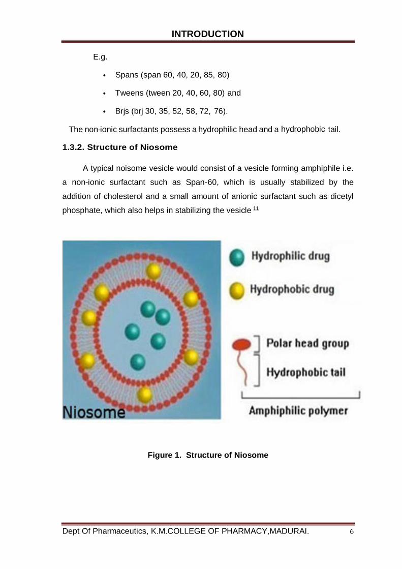
1.3.2. Structure of Niosome
A typical noisome vesicle would consist of a vesicle forming amphiphile i.e.
a non-ionic surfactant such as Span-60, which is usually stabilized by the
addition of cholesterol and a small amount of anionic surfactant such as dicetyl
phosphate, which also helps in stabilizing the vesicle 11
Figure 1. Structure of Niosome
INTRODUCTION
Dept Of Pharmaceutics, K.M.COLLEGE OF PHARMACY,MADURAI. 7
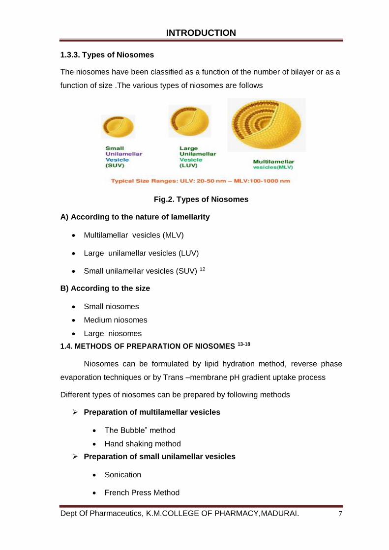
1.3.3. Types of Niosomes
The niosomes have been classified as a function of the number of bilayer or as a
function of size .The various types of niosomes are follows
Fig.2. Types of Niosomes
A) According to the nature of lamellarity
Multilamellar vesicles (MLV)
Large unilamellar vesicles (LUV)
Small unilamellar vesicles (SUV) 12
B) According to the size
Small niosomes
Medium niosomes
Large niosomes
1.4. METHODS OF PREPARATION OF NIOSOMES 13-18
Niosomes can be formulated by lipid hydration method, reverse phase
evaporation techniques or by Trans –membrane pH gradient uptake process
Different types of niosomes can be prepared by following methods
Preparation of multilamellar vesicles
The Bubble” method
Hand shaking method
Preparation of small unilamellar vesicles
Sonication
French Press Method
INTRODUCTION
Dept Of Pharmaceutics, K.M.COLLEGE OF PHARMACY,MADURAI. 8
Preparation of large unilamellar vesicles
Reverse phase Evaporation Method
Ethanol Injection Method
Ether Injection Method
Homogenization
Trans membranes pH gradient (inside acidic) Drug Uptake Process:
or Remote Loading Technique
Dehydration / Rehydration of small unilamellar vesicles
Detergent Removal Method
Multiple Membrane Extrusion Method
Preparation of multilamellar vesicles
The Bubble” method
It is novel technique for the one step preparation of liposomes
and niosomes without the use of organic solvents. The bubbling unit consists of
round-bottomed flask with three necks positioned in water bath to control the
temperature. Water-cooled reflux and thermometer is positioned in the first and
second neck and nitrogen supply through the third neck. Cholesterol and
surfactant are dispersed together in this buffer (PH 7.4) at 70°C, the dispersion
mixed for 15 seconds with high shear homogenizer and immediately afterwards
“bubbled” at 70°C using nitrogen gas
Preparation of small unilamellar vesicles
Sonication
A typical method of production of the vesicles is by sonication of solution
as described by Cable. In this method an aliquot of drug solution in buffer is
added to the surfactant/cholesterol mixture in a 10-ml glass vial. The mixture is
probe sonicated at 60°C for 3 minutes using a sonicator with a titanium probe to
yield niosomes.
INTRODUCTION
Dept Of Pharmaceutics, K.M.COLLEGE OF PHARMACY,MADURAI. 9
Preparation of large unilamellar vesicles
Ether Injection Method
This method provides a means of making niosomes by slowly
introducing a solution of surfactant dissolved in diethyl ether into
warm water maintained at 60°C.
The surfactant mixture in ether is injected through 14-gauge
needle into an aqueous solution of material.
Vaporization of ether leads to formation of single layered
vesicles.
Depending upon the conditions used the diameter of the vesicle
range from 50 to 1000 nm.
Multiple membrane extrusion method
Mixture of surfactant, cholesterol and dicetyl phosphate in chloroform is
made into thin film by evaporation. The film is hydrated with aqueous drug
polycarbonate membranes, solution and the resultant suspension extruded
through which are placed in series for up to 8 passages. It is a good method for
controlling niosome size.19
Micro fluidization Method
Micro fluidization is a recent technique used to prepare unilamellar
vesicles of defined size distribution. This is based on submerged jet principle in
which two fluidized streams interact at ultra-high velocities, in precisely defined
micro channels within the interaction chamber. The impingement of thin liquid
sheet along a common front is arranged such that the energy supplied to the
system remains within the area of niosomes formation. The result is a greater
uniformity, smaller size and better reproducibility of niosomes formed.
1.5. SEPARATION OF UNENTRAPPED DRUG
The removal of unentrapped solute from the vesicles can be
accomplished by various techniques, which include: 20
INTRODUCTION
Dept Of Pharmaceutics, K.M.COLLEGE OF PHARMACY,MADURAI. 10
1. Dialysis
The aqueous niosomal dispersion is dialyzed in a dialysis tubing against
phosphate buffer or normal saline or glucose solution.21
2. Gel Filtration
The unentrapped drug is removed by gel filtration of niosomal dispersion
through a Sephadex-G-50 column and elution with phosphate buffered saline or
normal saline.
3. Centrifugation
The niosomal suspension is centrifuged and the supernatant is separated.
The pellet is washed and then resuspended to obtain a niosomal suspension
free from unentrapped drug.22
1.6. Characterization of Niosomes 23-27
a. Scanning electron microscopy
Particle size of niosomes is very important characteristic. The surface
morphology (roundness, smoothness, and formation of aggregates) and the size
distribution of niosomes were studied by Scanning Electron Microscopy (SEM).
Niosomes were sprinkled on to the double- sided tape that was affixed on
aluminium stubs. The aluminium stub was placed in the vacuum chamber of a
scanning electron microscope. The samples were observed for morphological
characterization using a gaseous secondary electron detector.
b. Optical Microscopy
The niosomes were mounted on glass slides and viewed under a
microscope with a magnification of 1200X for morphological observation after
suitable dilution. The photomicrograph of the preparation also obtained from the
microscope by using a digital SLR camera. Niosomes were sprinkled on to the
double- sided tape that was affixed on aluminium stubs.

INTRODUCTION
Dept Of Pharmaceutics, K.M.COLLEGE OF PHARMACY,MADURAI. 11
c. Measurement of vesicle size
The vesicle dispersions were diluted about 100 times in the same medium
used for their preparation. Vesicle size was measured on a particle size analyzer.
The apparatus consists of a He-Ne laser beam focused with a minimum power of
5 mW using a Fourier lens to a point at the center of multielement detector and a
small volume sample holding cell (Su cell).
d. Entrapment efficiency
Entrapment efficiency of the niosomal dispersion in can be done by
separating the unentrapped drug by dialysis centrifugation or gel filtration as
described above and the drug remained entrapped in niosomes is determined by
complete vesicle disruption using 50% n-propanol or 0.1% Triton X-100 and
analyzing the resultant solution by appropriate assay method for the drug
f. osmotic shock
The change in the vesicle size can be determined by osmotic studies.
Niosomes formulations are incubated with hypotonic, isotonic, hypertonic
solutions for 3 hours. Then the changes in the size of vesicles in the formulations
are viewed under optical microscopy.
g. Stability studies
To determine the stability of niosomes, the optimized batch was stored in
airtight sealed vials at different temperatures. Surface characteristics and
percentage drug retained in niosomes and niosomes derived from proniosomes
were selected as parameters for evaluation of the stability, since instability of the
formulation would reflect in drug leakage and a decrease. In the percentage drug
retained. The niosomes were sample at regular intervals of time (0,1,2, and
3months), observed for color change, surface characteristics and tested for the
percentage drug retained after being hydrated to form niosomes and analyzed by
suitable analytical methods (UV spectroscopy, HPLC methods etc).
INTRODUCTION
Dept Of Pharmaceutics, K.M.COLLEGE OF PHARMACY,MADURAI. 12
h. Zeta potential analysis
Zeta potential analysis is done for determining the colloidal properties of
the prepared formulations. The suitably diluted niosomes derived from
proniosomes dispersion was determined using zeta potential analyzer based on
electrophoretic light scattering and laser Doppler velocimetry method. The
temperature was set at 25°C. Charge on vesicles and their mean zeta potential
values with standard deviation of measurements were obtained directly from the
measurement.
1.7. Factors Affecting Formation of Niosomes
Nature of surfactants
Structure of surfactants
Membrane composition
Nature of encapsulated drug
Bilayer formation
Number of lamellae
Membrane rigidity
Entrapment efficiency (EE)
Cholesterol contents
Nature of surfactants
A surfactant used for preparation of niosomes must have a hydrophilic
head and hydrophobic tail. The hydrophobic tail may consist of one or two alkyl
or perfluoro alkyl groups or in some cases a single steroidal group.28
The ether type surfactants with single chain alkyl as hydrophobic tail is
more toxic than corresponding dialkyl ether chain .The ester type surfactants are
chemically less stable than ether type surfactants and the former is less toxic
than the latter due to ester-linked surfactant degraded by esterase to
triglycerides and fatty acid in vivo .The surfactants with alkyl chain length from
C12-C18 are suitable for preparation of niosome.29,30
INTRODUCTION
Dept Of Pharmaceutics, K.M.COLLEGE OF PHARMACY,MADURAI. 13
Structure of surfactants
The geometry of vesicle to be formed from surfactants is affected by its
structure, which is related to critical packing parameters. On the basis of critical
packing parameters of surfactants can predicate geometry of vesicle to be
formed. Critical packing parameters can be defined using following equation,
CPP (Critical Packing Parameters) =v/lc* a0 where v = hydrophobic group
volume, lc = the critical hydrophobic group length, a0 = the area of hydrophilic
head group. From the critical packing parameter value type of miceller structure
formed can be ascertained as given below, If CPP < ½ then formation of
spherical micelles, If ½ < CPP < 1 formation of bilayer micelles, If CPP > 1
formation inverted micelles.31
Membrane composition
The stable niosomes can be prepared with addition of different additives
along with surfactants and drugs. Niosomes formed have a number of
morphologies and their permeability and stability properties can be altered by
manipulating membrane characteristics by different additives. In case of
polyhedral niosomes formed from C16G2, the shape of these polyhedral
niosome remains unaffected by adding low amount of solulan C24 (cholesterol
poly-24-oxyethylene ether), which prevents aggregation due to development of
steric hindrance.31,32
Nature of encapsulated drug
The physico-chemical properties of encapsulated drug influence charge
and rigidity of the niosome bilayer. The drug interacts with surfactant head
groups and develops the charge that creates mutual repulsion between
surfactant bilayers and hence increases vesicle size.33
Temperature of hydration
Hydration temperature influences the shape and size of the niosome. For
ideal condition it should be above the gel to liquid phase transition temperature of
system. Temperature change of niosomal system affects assembly of surfactants
into vesicles and also induces vesicle shape transformation.34
INTRODUCTION
Dept Of Pharmaceutics, K.M.COLLEGE OF PHARMACY,MADURAI. 14
Bilayer formation
Assembly of non-ionic surfactants to form bilayer vesicle is characterized
by X-cross formation under light polarization microscopy.35
Number of lamellae
It is determined by using NMR spectroscopy, small angle X-ray scattering
and electron microscopy.36
Membrane rigidity
Membrane rigidity can be measured by means of mobility of fluorescence
probe as function of temperature.37,38
Entrapment efficiency (EE)
The entrapment efficiency (EE) is expressed as
EE = amount entrapped / total amount Added × 100
It is determined after separation of unentrapped drug, on complete vesicle
disruption by using about 1ml of 2.5% sodium lauryl sulphate, briefly
homogenized and centrifuged and supernatant assayed for drug after suitable
dilution.39
Entrapment efficiency is affected by following factors.
Surfactants
The chain length and hydrophilic head of non-ionic surfactants affect
entrapment efficiency, such as stearyl chain C18 non-ionic surfactant vesicles
show higher entrapment efficiency than lauryl chain C12 non-ionic surfactant
vesicles. The tween series surfactants bearing a long alkyl chain and a large
hydrophilic moiety in the combination with cholesterol at1:1 ratio has highest
entrapment efficiency for water soluble drugs.40
HLB value of surfactants affects entrapment efficiency, such as HLB value
of 14 to 17 is not suitable for niosomes but HLB value of 8.6 has highest
INTRODUCTION
Dept Of Pharmaceutics, K.M.COLLEGE OF PHARMACY,MADURAI. 15
entrapment efficiency and entrapment efficiency decreases with decrease in HLB
value from 8.6 to 1.7 30.41
Cholesterol contents
The incorporation of cholesterol into bilayer composition of niosome
induces membrane stabilizing activity and decreases the leakiness of membrane.
Hence, incorporation of cholesterol into bilayer increases entrapment
efficiency. The permeability of vesicle bilayer to 5, 6-carboxy fluorescein (CF) is
reduced by 10 times due to incorporation of cholesterol.42
1.8. EVALUATION OF NIOSOMES
a) Entrapment efficiency
After preparing niosomal dispersion, unentrapped drug is separated by
dialysis, centrifugation, or gel filtration as described above and the drug
remained entrapped in niosomes is determined by complete vesicle disruption
using 50% n-propanol or 0.1% Triton X-100 and analysing the resultant solution
by appropriate assay method for the drug. Where, 43
Entrapment efficiency (EE) = (Amount entrapped total amount) x 100
b) Vesicle diameter
Niosomes, similar to liposomes, assume spherical shape and so their
diameter can be determined using light microscopy, photon correlation
microscopy and freeze fracture electron microscopy. Freeze thawing (keeping
vesicles suspension at –20°C for 24 hrs. and then heating to ambient
temperature) of niosomes increases the vesicle diameter, which might be
attributed to fusion of vesicles during the cycle
c) In-vitro release
A method of in-vitro release rate study includes the use of dialysis tubing.
A dialysis sac is washed and soaked in distilled water. The vesicle suspension is
pipetted into a bag made up of the tubing and sealed. The bag containing the
vesicles is placed in 200 ml of buffer solution in a 250 ml beaker with constant
INTRODUCTION
Dept Of Pharmaceutics, K.M.COLLEGE OF PHARMACY,MADURAI. 16
shaking at 25°C or 37°C. At various time intervals, the buffer is analysed for the
drug content by an appropriate assay method Methods for the evaluation of
niosomes:
Evaluation parameter Method 44
Morphology : SEM, TEM, freeze fracture technique
Size distribution : polydispersity index
Particle size analyzer : Dynamic light scattering
Viscosity : Ostwald viscometer
Membrane thickness : X-ray scattering analysis Thermal analysis
DSC Turbidity : UV-Visible diode array spectrophotometer
Entrapment efficacy : Centrifugation, dialysis, gel chromatography
In-vitro release study : Dialysis membrane Permeation
In-vitro methods for niosomes
In vitro drug release can be done by 45
1. Dialysis tubing
2. Reverse dialysis
3. Franz diffusion cell
Dialysis tubing
In vitro drug release could be achieved by using dialysis tubing. The niosomes is
placed in prewashed dialysis tubing which can be hermetically sealed. The
dialysis sac is then dialyzed against a suitable dissolution medium at room
temperature; the samples are withdrawn from the medium at suitable intervals,
centrifuged and analyzed for drug content using suitable method (U.V.
spectroscopy, HPLC etc). The maintenance of sink condition is essential.
INTRODUCTION
Dept Of Pharmaceutics, K.M.COLLEGE OF PHARMACY,MADURAI. 17
Reverse dialysis
In this technique a number of small dialysis as containing 1ml of dissolution
medium are placed in proniosomes. The proniosomes are then displaced into the
dissolution medium. The direct dilution of the proniosomes is possible with this
method; however the rapid release cannot be quantified using this method.46
Franz diffusion cell
The in vitro diffusion studies can be performed by using Franz diffusion cell.
Proniosomes is placed in the donor chamber of a Franz diffusion cell fitted with a
cellophane membrane. The proniosomes is then dialyzed against a suitable
dissolution medium at room temperature; the samples are withdrawn from the
medium at suitable intervals, and analyzed for drug content using suitable
method (U.V spectroscopy, HPLC, etc) .the maintenance of sink condition is
essential.
1.9. Applications of niosomes 47
1. The application of niosomes technology is widely varied and can be
used to treat a number of diseases.
2. The following are a few uses of niosomes which are either proven or
under research. It is used as Drug Targeting.
3. It is used as Anti-neoplastic Treatment i.e. Cancer Disease.
4. It is used as Leishmaniasis i.e. Dermal and Mucocutaneous infections
e.g. Sodium stribogluconate.
5. It is used act as Delivery of Peptide Drugs.
6. It is used in Studying Immune Response.
7. Niosomes as Carriers for Haemoglobin.Transdermal Drug Delivery
Systems Utilizing Niosomes
8. It is used in ophthalmic drug delivery Other Applications: Niosomes can
also be utilized for sustained drug release and localized drug action to
greatly increase the safety and efficacy of many drugs.
9. Toxic drugs which need higher doses can possibly be delivered safely
using niosomal encapsulation.

INTRODUCTION
Dept Of Pharmaceutics, K.M.COLLEGE OF PHARMACY,MADURAI. 18
1.10. MECHANISM OF DRUG PERMEABILITY FROM NIOSOME
The are several mechanisms explain the ability of niosomes to modulated drug
transfer across skin including
I. Adsorption and fusion on the surface of skin facilitate drug permeation
II. The vesicles act as penetration enhancer to reduce the barrier
properties of stratum corneum
III. The lipid bilayers of niosomes act as a rate limiting membrane barrier
for drugs
The possible mechanism for niosomal enhancement of the permeability of
drugs is structure modification of the stratum corneum. It has been
reported that the intercellular lipid barrier in the stratum corneum would be
dramatically looser and more permeable following treatment with
niosomes .The both lipids and non-ionic surfactants in the niosomes can
act as penetration enhancers, which are useful for increasing the
permeation of many drug. The fusion of niosome vesicles to the surface of
the skin results in higher flux of the drug due to direct transfer of drug from
vesicles to the skin
1.11. DISEASE OVER VIEW
SKIN DISEASES
Skin disease is a very common symptom of systemic lupus erythematosus
(SLE), or lupus. Rashes are often the first visible indication of the disease.48
Skin diseases undermine their aspiration eroding confidence and
demaging relationship social life self-image and self-esteem. Sleep disturbance
and severe itching may increase Stress and further lowers the quality of life
eczema and psoriasis can cause embrassement anxiety anger or depression
Acne can devastate life of teenager at a time when the formation of relationship
so important
The art of diagnosis in dermatology in the past was particularly
emphasized by the visual experience of the skin and the skin lesions. It is true
that some skin lesion can be diagnosed on the sight with the high degree of
INTRODUCTION
Dept Of Pharmaceutics, K.M.COLLEGE OF PHARMACY,MADURAI. 19
confidence but even in such cases systematic approach is indispensable for a
good dermatologist not to miss other important skin lesions, when dermatology
first evolved from general internal medicine in the nineteenth century
There is usually some good reason for tropical treatment ,the drug is
irritant or sensitizing or does not adequately penetrate the skin The base In
which drug is applied to the skin is important
Bacterial skin infections
Bacterial Skin Infections or Pyoderma Bacterial skin infections or
pyoderma are common in most developing countries.49
Generally these infections arise as primary infections of the skin known as
impetigo or as secondary infections of other lesions such as scabies or insect
bites. The usual bacterial causes are Group A streptococci or Staphylococcus
aureus.50
Bacterial infections are common in communities. In many cases, no
bacteriological confirmation is available from cultures, but surveys show that
Group a streptococci account for a substantial number of cases which is not
often the case in similar infections in temperate climates, where S. aureus
dominates. This finding carries implications for the selection of treatment options.
The reasons for this finding are not clear, although humidity and heat are
associated with increased risk of bacterial skin infection. In addition to these
superficial infections, S. aureus also causes folliculitis or hair follicle infections
and abscesses. Rarer causes of skin infection in developing countries include
cutaneous diphtheria and anthrax, as well as necrotizing infection caused by
Vibrio vulnificus. Bacterial infection causes irritation and some discomfort.51
In some cases, the infection penetrates deep down through the epidermis,
causing a necrotic ulcer a condition known as ecthyma. However, some
evidence suggests that streptococcal infection may cause additional long-term
damage through the development of prolonged proteinuria, as described earlier
in relation to scabies.52

INTRODUCTION
Dept Of Pharmaceutics, K.M.COLLEGE OF PHARMACY,MADURAI. 20
Fungal Skin Infections
Fungal infections that affect the skin and adjacent structures are common
in all environments. They include infections such as ringworm or
dermatophytosis; superficial candidiasis and infections caused by lipophilic
yeasts and Malassezia species;
Fungal infection are termed mycoses and in general can be divided in to
superficial infection and systemic infections many of the fungi that can cause
mycoses live in association with man as commensals are present in his
environment. The serious superficial infections were relatively uncommon and
systematic infections very common. 53
Fungi are the most common parasites led by the ubiquitous yeast,
candida albicans. Candida organisms are natural residents of the body, but they
are normally found in harmless proportion. The body’s natural defence against
fungal infection is its resident population of normal bacterial flora which prevents
invasion by foreigners. But yeast is the impervious to the antibiotics that wipe out
bacteria when friendly bacteria wiped out along with unfriendly one by these
drugs.
Fungal infections can also affect other parts of the body. Onchomycosis
fungal disease of the toe nails and finger nails particularly affected the elderly
one has increased by a factor in the last 20 year.
Common fungal diseases
Candidiasis
Pityriasis versicolor
Tinea pedis
Tinea manuum
Tinea unguium
Tinea capitis

INTRODUCTION
Dept Of Pharmaceutics, K.M.COLLEGE OF PHARMACY,MADURAI. 21
1.12. INTRODUCTION OF SENNA ALATA LINN
Senna alata Linn. (English: Ringworm shrub, Sanskrit: Dadrughna,
Hindi: Dadmurdan) is an erect shrub growing up to 3-4.5 m in height.54
It shows large paripinnately compound leaves. The flowers are golden
yellow in colour and appear in racemes.55
Pods are flat, dark brown with many seeds. The plant is cosmopolitan in
distribution.
It is found wild as well as the cultivated ornamental plant throughout
India. Various parts of S. alata are used for diverse healing actions
PHARMACOLOGICAL ACTIVITIES
The research data indicate that Senna alata possesses enormous
pharmacological values which support its various traditional uses for the
management of health problems. The most important are: 56
Antimalarial activity:
Senna alata L. leaves exhibited in vitro antimalarial activities against
Plasmodium falciparum in ethylene Glycol-water 3:7 solvent; with IC50 values
below 1µg/ml value of 17.270µg/ml. The antimalarial activity of this plant is
doubtless due to its alkaloids, quinones and terpenes. In fact, in the
chemotherapy of malaria, most molecules belong to the class of alkaloids these
studies have provided scientific evidence for traditional usage of Senna alata as
effective remedy for the treatment of malaria in many African countries.57
Anti-cancer and antitumor activity:
Study designed to investigate the sub-acute toxicity, in vivo antioxidant
and antitumor activity of aqueous ethanol extract of Senna alata L on bearing
carcinomatous cells on Nude mice the results of in vivo antitumor activity of
extract of Senna alata observed. Hence, the leaf extract of Senna alata can be
used to cure the breast cancer. Evaluated leaf extracts of Senna alata for their
potential antitumor properties in vitro MTT assays were used to examine the
cytotoxic effects of crude extracts on five human cancer cell lines, namely MCF-
7, derived from a breast carcinoma, SK-BR-3, another breast carcinoma, T24 a
bladder carcinoma, Col 2, a colorectal carcinoma, and A549, a non- small cell
INTRODUCTION
Dept Of Pharmaceutics, K.M.COLLEGE OF PHARMACY,MADURAI. 22
lung adenocarcinoma. Hexane extracts showed remarkable cytotoxicity against
MCF-7, T24, and Col 2 in a dose-dependent manner. This plant extract had also
proved its effectiveness against Leukaemia cells.58
Anti-inflammatory activity:
The anti-inflammatory mechanism of a hexane extract of Senna alata L
was investigated in Complete Freund’s Adjuvant (CFA) arthritis, as a chronic
model of inflammation
The CFA model was created by the injection of 0.5ml CFA into the
synovial cavity of the right knee joint of hind leg of rats. Changes in knee
joint swelling, cartilage integrity and synovial fluid leukocyte counts were
assessed in response Senna alata L to treatment.59
Anthelmintic activity:
Alcohol extract from the leaves of Senna alata L and Typha angustifolia
were investigated for their anthelmintic activity against Pheretima posthumous
and Arcadia galls In conclusion, the traditional claim of leaves of Senna alata L
as anthelmintic have been confirmed as the leaf extracts displayed activity
against the worms used in the study.60
Antioxidant activity:
Sarkar et al carried out a study to evaluate in vivo the antioxidant and
antitumor activity of the aqueous-EtOH extract of leaves of Senna alata L.
Antioxidant potential was found to be more in extract as compared to control.
The results of this study clearly indicated that the extracts of Senna alata L could
be used as a potential source of natural antioxidant agents. The antioxidant
activity of crude methanol extracts from the leaves, flowers and pods of Senna
alata.61
Hepatoprotective and hepatocurative activities:
Hepatoprotective activity of the Infusion of the dried leaves of Senna alata
(ICA) was studied against Paracetamol induced hepatic injury in albino rats Pre-
treatment of the Infusion (ICA) reduced the biochemical markers of hepatic injury
like serum glutamate pyruvate transaminase (SGPT), serum oxaloacetate
transaminase (SGOT), alkaline phosphatase (ALP), total bilirubin and gamma
glutamate trans peptidase (GGTP). Histopathological observations also revealed
that pre-treatment with ICA protected the animals from Paracetamol induced liver
INTRODUCTION
Dept Of Pharmaceutics, K.M.COLLEGE OF PHARMACY,MADURAI. 23
damage. The results indicate that the leaves of Senna alata possess the
hepatoprotective activity. This property may be attributed to the flavonoids
present in the leaves of this plant. The effect of oral administration of aqueous
extract of leaves of Senna alata in various doses (2.5-20.0 mg/kg) for seven
days, on hepatic induced damage by administration of 45% of ethanol (20ml/kg)
and CCl4 (0.1 ml/kg) in rats of the use of Senna alata in the traditional medicine
for the treatment of cirrhosis and hepatitis.62
Antibacterial and antifungal activities:
Antimicrobial studies showed that the isolated compound from Senna
alata seeds successfully inhibited Pseudomonas aeruginosa, Klebsiella
pneumonia, Escherichia coli, Staphylococcus aureus Candida albicans and
Aspergillus Niger. Chemical investigation of the bioactive constituents from the
seeds of Senna alata resulted in the isolation of a new cannabinoid alkaloid (4-
butylamine 10-methyl-6hydroxy cannabinoid dronabinol). The antimicrobial
observation of the above compound against these pathogens showed that the
bioactive compound could be responsible for the activity of the plant and its use
in traditional medicine. These findings also justify the use of Senna alata in the
treatment of skin infections such as eczemas, ringworms, boils, carbuncles,
breast abscess, infantile impetigo, sores and wound in herbal medicine and its
use as an ingredient in the formulation of medicated and antiseptic soaps.63
Cardio protective activity:
Research work designed to evaluate the cardio protective potential of the
Senna alata leaves against doxorubicin induced cardiac toxicity in rats Senna
alata leaves methanolic extract has significant cardio protective activity.
Preliminary phytochemical investigation of Senna alata leaves methanolic
extracts shows presence of flavonoids, tannins, glycosides. Thus, the strong
antioxidant and cardio protective effect of the extract could be attributed to the
presence of bioactive constituents present in the extract. 64
Anti-diabetic and antihyperlipidemic activities:
A study aimed to depict the therapeutic effect of Senna alata leaf aqueous
extract on oxidative stress in aorta as well as heart of streptozotoc in
hyperglycaemic rats was undertaken by Two days after diabetes induction effect.

INTRODUCTION
Dept Of Pharmaceutics, K.M.COLLEGE OF PHARMACY,MADURAI. 24
Thus, one of the possible antidiabetic mechanisms of action of Senna alata is by
inhibiting carbohydrate digestion.65
Bronchorelaxant activity:
Aqueous-ethanolic extract of Senna alata (AECal) and its derived fractions
obtained through liquid-liquid fractionation were evaluated for their
bronchorelaxant effect activity of rats tracheas in the presence of tested
materials, as well as its modifications with different inhibitors and blockers, was
isometrically recorded. In animals pre-treated with the extract, the percentage of
Induced DNA damage decreased. The results suggest that (1) muscarinic
receptors contribute at least in part to the relaxant effects of AECal; (2) AECal
interferes with membrane polarization. Evaluation of isolated bioactive molecules
from plant extract eliciting tracheorelaxant effect may give new investigational
and treatment tools in broncho respiratory pharmacology. This study provides
sound mechanistic basis for the use of Senna alata in asthma-induced
bronchospasm.66
Antiviral activity:
Mohamed et al investigated the antiviral activity of five extracts (methanol,
chloroform, ethyl acetate, n-butanol, and aqueous) from Senna alata leaves
against rotavirus (RV) infection in vitro and in vivo). In vitro, all extracts prevented
the cytopathic effect (CPE) of RV, as demonstrated in an MTT colorimetric and
karber methods, with therapeutic index (IT) ranged from 22.8 to 0.02 and
reduction in virus titres ranged from 4.25 and 0.25 log10TCID50. The methanol
extract was the stronger than the other extracts against RV replication. In vivo
antiviral activity of the methanol extract against rotavirus was evaluated, using a
mouse model. Orally administered methanol extract at 100 mg and 50 mg /kg
body weight/day significantly reduced virus yield in the small intestine as well as
it reduced mortality, severity and duration of diarrhoea after infection for 7 days.
In addition to this, extract protects the intestinal tissues from damage resulted
from RV infection when compared with the untreated infected control. These
results clearly shows that in vitro and in vivo infection with RV can be effectively
treated by the methanol extract of Senna alata leaves. The antiviral activity of
methanol extract of Senna alata may be attributed to the presence of Saponins.
The anti-rotavirus activity of Saponins has been well documented in vivo. Thus,

INTRODUCTION
Dept Of Pharmaceutics, K.M.COLLEGE OF PHARMACY,MADURAI. 25
the present study has shown that the methanol extract of Senna alata recovered
the rotavirus gastroenteritis by coordinating antiviral and anti-inflammatory
effects.67
Reported toxicity:
Investigated the acute and sub-acute toxicities of hydro-ethanolic extract
of leaves of Senna alata in Swiss mice and Wister albino rats The mice were
divided into 6 groups of 10 animals and each group received once by intra
gastric gavages 0, 4, 8, 12, 16, 20 times 1000 mg/kg dose Med. Sci. of extract.
Distilled water served as the control. For the sub-acute toxicity, three groups
of 10 rats (5 males and 5 females) were treated per so with distilled water
(control), 500 or 1000 mg/kg of extract every 48 h for 26 days. At the end of
treatment blood sample and 20% liver homogenates were collected for
biochemical analyses. The results indicated that the medium lethal dose (LD50)
was about 18.50 g/kg of body weight. Significant variation (P<0.05) of the body
weight was observed after 26 days of treatment, in some biochemical index of
serum and 20% liver homogenates (glutathione , alkaline phosphatase (APL),
aspartate aminotransferase (AST)), haematological parameters (platelet) also in
the female relative weight of heart of rat. Some of parameter investigated in this
study showed dose responsive. The histo pathological study of the liver did not
show any features after the treatment but, the extract seems to ameliorate the
liver architecture. In another study, no death and no clinically significant changes
were recorded in mice which consumed this plant extract. The maximum non-
lethal dose was more than 1687.5 mg/kg in animals. No significant changes were
observed in body weight, tissues morphology, biochemical and haematological
parameters at doses above or equal to 2779.5 mg/kg body weight.68
LITERATURE REVIEW
Dept Of Pharmaceutics, K.M.COLLEGE OF PHARMACY,MADURAI. 26
2. LITERATURE REVIEW
2.1 STUDIES ON SENNA ALATA LEAF
Neil Alejandra Pinarok, N.A.A et al., (2017) Developed Senna alata L. leaf
extract was used to develop an antifungal ointment with two different formulations
namely; Simple ointment and Emulsifying ointment using Jatropha curcas L. seed
oil as base.. In vitro antifungal assay established that Senna alata L. ointment
whether simple or emulsifying has antifungal effect against common fungi. Stability
tests of the processed ointments have stable characteristics in normal conditions,
but slightly changes when exposed to higher temperatures other than normal
temperatures, especially its pH, and weight. Jatropha curcas seed oil extract can
be used to manufacture cosmetic products.69
Vigbedor Bright Yaw et al.,( 2015) Investigated the stem bark of A. Africana and
the leaves of Senna alata L. for their antimalarial activity against the 3D7 strain of
the Plasmodium falciparum parasite. by employing the WHO micro test assay
(Mark III). Compared with Artesunate-amodiaquine. The standard drug,
Artesunate-amodiaquine was the most active with an Inhibitory Concentration
(IC50) of 0.313μg/ml this was followed by Afzelia Africana, and Senna alata with
IC50 values of 2.954μg/ml, and 17.270μg/ml respectively.70
Raphael M. Mordi et al., (2016) Evaluated the antimicrobial properties of the
ethanol and aqueous extracts of senna alata leaves.by Agar diffusion method the
muella-hinton agar for the bacteria and potato dextrose agar for the fungi.
phytochemical screening revealed the presence of cardiac glycoside, reduced
sugar in equal concentration in both extracts while flavonoids, terpenoids,
Saponins and phenolic were in slightly higher concentrations in the ethanol extract
than in the aqueous extract. The antimicrobial effects produced by the extracts
were dose dependent at the tested doses; 1000mg/ml, 500mg/ml and 250mg/ml.71
Addai mensah donkor et al., (2016) studied antibacterial activities of the extracts
of medicinal plants, Senna alata, Ricinus communis and Lannea barter. Bacteria
strains such as Staphylococcus aureus, Pseudomonas aeruginosa, Klebsiella
pneumonia and Escherichia coli are known wound and skin disease causing
bacteria.. Phytochemical screening of the crude extracts of Senna alata, revealed

LITERATURE REVIEW
Dept Of Pharmaceutics, K.M.COLLEGE OF PHARMACY,MADURAI. 27
the presence of some bioactive components, including tannins, Anthraquinones,
Saponins, flavonoids and alkaloids. The extracts showed inhibition on test
organisms and were in the range of 12.0 ± 2.83mm to 36.0 ± 2.12mm.The MIC
values obtained for the entire test ranged from 3.13mg/ml to 12.50mg/ml and MBC
values were found to be 200mg/ml minimum and 400mg/ml maximum.72
Senga Kitumbe et al., (2016) formulated a dermal ointment from the whole leaves
of Senna alata Linn in order to improve the traditional preparation used against
dermal diseases. In comparison with the aqueous extract of S. alata leaves
medicinal properties, gentamicin and ketoconazole discs were used as controls.
the herbal ointment demonstrated higher antifungal activity than antibacterial
activity based on the zones of inhibition recorded for all the concentrations. The
chromatographic fingerprints established and quantitative analyses conducted in
this investigation are worth considering for the quality control of the herbal
ointment formulated.73
M. S.Rahman et al.,(2015) isolated isoflavone from the leaves of Senna alata , 2,
5, 7, 4′-tetrahydroxy isoflavone with the help of column and thin layer
chromatography by using a gradient mixture of organic solvents with increasing
polarity. The compound was characterized on the basis of UV, IR, and 1H-NMR,
13C-NMR and Mass spectrometry and confirmed the compound belonged to the
isoflavone series.74
Dr. D. Victor Arokia Doss et al., (2015) prepared Senna alata juice of fresh
leaves of recognized by local healers as a remedy for parasitic skin disease and is
used in the treatment of many eruptive and pustular skin condition by simply
rubbing the crushed leaves either alone or mixed with oil on the skin. Root is taken
in Nigerian and Guinea with rock salt and other dry medicinal plants is taken in
Nigeria thrice weekly on an empty stomach for effective treatment of chronic
gonorrhoea. the existence of bioactive compounds with free radical scavenging
activity, anticancer property and antidiabetic activities.75
Meenu priya et al., (2014) carried out the phytochemical screening and bioassay
of Senna alata leaf extract and its skin hyperpigmentation activity. Medicinal plants
of Indian origin play a significant role in treating a broad spectrum of human

LITERATURE REVIEW
Dept Of Pharmaceutics, K.M.COLLEGE OF PHARMACY,MADURAI. 28
diseases natural products seem a reliable alternate that’s considered safe and
effective. The phytochemical screening and analysis of Senna alata leaf extract
against hyperpigmentation of skin due to spray allergy and under eye darkness.
The efficacy of hydro ethanolic extract of Senna alata applied to under eye dark
circles and hyper pigmented under arms due to regular usage of body spray. The
results are highly promising on regular application twice a day for 20 days. Visible
difference is observed within one week of application and normal skin color is
obtained within a month’s time.76
Rama Swamy Nanna et al., (2015) evaluated anti-diabetic activity and
antihyperlipidemic evaluation of leaf extracts of senna alata in alloxan induced
diabetic rats Senna alata is an ethno medicinal plant belonging to the family
Fabaceae. A number of bioactive constituents which contribute to the medicinal
properties are attributed to the species. The anti-diabetic activity attributed to the
species and also to evaluate the anti hyperlipidemic potential of the Species using
various standard experimental models available. Acute toxicity studies of aqueous
leaf extracts of S. alata were performed up to a dose of 2500mg/kg bodyweight of
rats and a dose of 200 mg/kg was selected. Aqueous leaf extracts of S. alata
showed a significant (P<0.01) anti diabetic and anti hyperlipidemic potential in
alloxan induced diabetic rats within 15 days of induction of diabetes and the
antidiabetic potential of the species may be due to the presence of flavonoids.77
Pratiwi Wikaningtyasthe et al,.(2015) evaluated antibacterial activity of senna
alata leaf extract and fraction towards MRSA (methicillin resistant staphylococcus
aureus) and its mode of action. The increasing prevalence of healthcare-
associated MRSA infections need new approach to overcome of growing
problems. Medicinal plants are promising as the most valuable resources for
antibiotic development. Senna alata had shown antibacterial effect in Str.
pyogenes and S. aureus the antibacterial activity calculated based on Minimum
Inhibitory Concentration (MIC) using Mueller Hinton broth in micro dilution method
and the mode of action was conducted using Scanning Electron Microscopy
(SEM). Phytochemical screening of dried Senna alata leaf and its extract showed
the presence of flavonoid, alkaloid, Saponins, Quinone, tannin, and sterol. The
antibacterial activity showed the MIC value of the extract against MRSA was
LITERATURE REVIEW
Dept Of Pharmaceutics, K.M.COLLEGE OF PHARMACY,MADURAI. 29
512µg/mL. The ethyl acetate fraction showed the best MIC value at 256µg/mL. The
SEM observation of MRSA treated by ethyl acetate fraction of Senna alata showed
membrane shrinkage. Senna alata was promising to be developed as antibacterial
agent especially MRSA strain.78
Mulham alfatama et al., (2012) formulated Microencapsulation of Senna alata
and its antifungal and antimicrobial activities. This extracts S. alata, followed by
microencapsulation by solvent-evaporation using biodegradable PLGA. The
microsphere is envisaged to release the extract in a controlled manner allowing
more convenient mode of administration when treating related skin infection.
Several variables had been investigated including different types of surfactants
and buffer systems and different homogenization times for primary emulsion. It
was found that, most of the surfactants employed resulted in low encapsulation
efficiency (<10%). However with 3% poly vinyl alcohol (PVA), slightly higher
encapsulation efficiency of 11% was achieved, with size distribution of 7-13 µm.
The encapsulation efficiencies were significantly improved (45-64.4%) when the
hardening tank was buffered to pH 7, with minimal effect on particle size.
Additionally, in-vitro controlled release formulation of S. alata microsphere was
demonstrated for duration of almost a month.79
Selvi Vet al., (2012), studied on antimicrobial activities from flower extract of
Senna alata Linn. The plant materials to prevent and treat infectious diseases
successfully over the years has attracted the attention of scientist’s worldwide. The
medicinal usefulness of the plant Senna alata Linn. S. alata is an ornamental shrub
or tree growing up to 12 m high and widely available in the tropics, These trees are
used to treat diarrhoea, dysentery and other gastrointestinal problems. The
macerated juices of the young fresh leaves are used to treat eye infections and
parasitic diseases. The decoction of the stem bark and roots are used to treat
urinary tract infections, bronchitis and asthma. the microbial activity of methanol,
chloroform and petroleum ether extracts of flowers of Senna alata Evaluated for
potential antimicrobial activity against bacterial and fungal strains. The fungal
isolates tested include: Epidermatophyton floccose, Microsporium gypseum and
Trichophyton mentagrophyte and the Bacterial isolates tested include: Escherichia
coli. The antimicrobial activity was determined the extracts using agar well diffusion
LITERATURE REVIEW
Dept Of Pharmaceutics, K.M.COLLEGE OF PHARMACY,MADURAI. 30
method. The flowers were shade dried and then homogenized to fine powder by a
mechanical grinder. They were extracted using different solvents such as by
methanol, chloroform and petroleum ether by soxhlet apparatus. The zone of
inhibition was measured.80
Silver Ighosotu et al., (2013) investigated the effects of Senna alata L. aqueous
leaf extract on the germination of Corchorus olitorius the effect of different
concentrations of Senna alata crude aqueous leaf extract on the germination of
Corchorus olitorius S. alata aqueous leaf extract (10, 30, 50, 75 and 100% C) used
in this study caused significant (P < 0.05) decrease in the total percentage and
germination rate of C. olitorius. Pre-soaking of C. olitorius seeds in C. alata crude
aqueous leaf extract led to an increase in the lag-phase period preceding
germination of the seeds in a concentration-dependent manner. The total
percentage and rate of germination decreased as extract concentration increased.
All concentrations caused a consistent decrease in absolute rate and percentages
of germination compared to the control.81
Mohammed I Ali et al., (2017) studied Leaf extracts of Senna alata L traditionally
used for treatment of a variety of diseases. Chloroform fraction of leaves was
evaluated for its potential antitumor properties in vitro. MTT assay was used to
examine the cytotoxic effect on three human cancer cell lines namely HepG2,
MDA-MB-231 and Caco2. Chloroform fraction showed remarkable cytotoxicity
against HepG2 cells with IC50 = 37.4 µg/ml at exposure time 48 h. The fraction
exhibited weak anti-proliferative effect on Caco2 and MDA-MB-231 cells (8.2% and
11.6% respectively), with IC50 values >100 µg/ml. DPPH free radical scavenging
activity of the fraction (100 µg/ml) revealed weak antioxidant activity (7.8%).
Further bioassay-guided fractionation of the cytotoxic fraction led to the isolation
and characterization of three Anthraquinones (rhein, aloe-emodin and emodin).82
R. P. Senthil kumar et al.,(2013) carried out phytochemical screening and
antibacterial evaluation of the leaf, flower and seed coat extracts of Senna alata L
Preliminary studies on the phytochemistry and extract of diethyl ether, chloroform
and acetone of Senna alata L leaf, flower and seed coat were examined for
antimicrobial properties. Extract tested against clinical isolated of Pseudomonas
sps, Escherichia coli, Staphylococcus sps, Shigella boydii and Salmonella sps. The
LITERATURE REVIEW
Dept Of Pharmaceutics, K.M.COLLEGE OF PHARMACY,MADURAI. 31
zone of inhibitions produced by the extracts in disc diffusion assay against the test
microorganisms, the produced zones that measured in mm. Preliminary
phytochemical analysis showed that the extracts contained tannin, flavonoid,
terpenoids, cardiac glycosides, steroids and terpenoids, absence of alkaloids.83
Panichayupakaranant et al., (2004) isolated the antioxidant constituent from
Senna alata L. leaves using DPPH radical scavenging assay The DPPH radical
scavenging assay-guided isolation, the methanol extract of C. alata leaves was
separated by silica gel vacuum chromatography and Sephadex LH-20 gel filtration
chromatography afford a light yellowish powder (CA1), which was identified as
kaempferol. This compound exhibited antioxidant activity (ED50 9.99 µM) that was
six times stronger than that of BHT (ED50 57.41 µM) and fifty eight times stronger
than that of emodin (ED50 578.87 µM).84
Da FL, Keugni et al, (2018) Evaluated anti-inflammatory and protective effect of
Senna alata Linn Leaves.The anti-inflammatory and protective effect of
dichloromethane extract of Senna alata Linn. leaves (CF-AECal) has been studied
according to Freund’s adjuvant-induced arthritic rat models. CF-AECal 50 mg/Kg,
100 mg/Kg and dexamethasone 1 mg/Kg were the doses of drugs receipt by rats.
CF-AECal 100 mg/Kg showed a substantial anti-inflammatory and protective effect.
CF-AECal 50 and 100 mg/Kg produced antioxidant effects in Freund's adjuvant-
induced paw oedema. Indeed, antioxidant enzymes are maintained more or less
that their normal rate. These antioxidant effects protect the liver, the kidney and
the spleen highly (p< 0.001). CF-AECal 50 and 100 mg/Kg lead to a significant
decline in the rate of serum enzymes (AST, PAL and CREA) and a decrease of the
serum concentration of total bilirubin and protein compared to animals of the
control group ignited. At the doses of the extract used, the leukocyte infiltration is
reduced in the cutaneous cloths of the paws of rats.85
Ikechukwu et al., (2014) Investigated The efficacy of crude extracts of Senna
alata in the improvement of vegetative and reproductive growth in Celosia
argentea. Fresh leaves of S. alata were blended with a homogenizer in 1 litre of
distilled water. The resultant green paste was filtered under suction. Different
concentrations (75%, 50%, 40%, 30%, 25%, 12%, 10%, and 5%) were prepared
from the 100% crude extract. Seeds of C. argentea were pre-soaked in these

LITERATURE REVIEW
Dept Of Pharmaceutics, K.M.COLLEGE OF PHARMACY,MADURAI. 32
different concentrations including a control (0%) and planted out after 24 hours.
Results showed that seedling height, leaf area, dry weight and leaf area ratio were
promoted and enhanced by pre-soaking seeds in the extract. Seedlings raised
from seeds pre-soaked in water (control) however, flowered earlier (8 weeks) than
the treatments (10 weeks in 75% and 100%). Pre-soaking seeds of C. argentea in
crude extracts of S. alata before planting is recommended for optimum production
of the leafy vegetable. The procedure is cheap and easily implementable by
resource-poor farmers who are the main growers of C. argentea.86
Chris. A. Alalor et al,. (2012) Evaluated antibacterial activity of Herbal ointments
formulated with Methanolic extract of Senna alata LThe preliminary in vitro
antibacterial activity of the methanolic extracts of sun-dried leaves of Senna alata
against Staphylococcus aureus, Bacillus subtilis, Escherichia coli and
Pseudomonas aeruginosa using the Agar cup plate method. Herbal ointments
were prepared by incorporating the methanolic extract of Senna alata (10 % w/w)
into aqueous cream, emulsifying ointment and simple ointment bases and
evaluated for their in vitro antibacterial efficacy. The formulation containing Senna
alata extract in aqueous cream showed comparatively better antibacterial activity
than the other formulations in the following order: aqueous cream > emulsifying
ointment > simple ointment > crude extract. The herbal ointment also compared
favourably with a commercial brand of Gentamicin ointment. has high potential as
antibacterial agent when formulated as ointment for topical use and could therefore
explain the successes claimed in the folk use of the plant in the treatment of
common skin conditions.87
Okooboh et al., (2013) studied phytochemistry and antimicrobial activity of the leaf
of senna alata linn. The leaves of were extracted with n-hexane and ethyl acetate
using the soxhlet method. Preliminary phytochemical screening of the extracts
revealed the presence of free Anthraquinones, flavonoids, steroids and Saponins.
The n-hexane crude extract exhibited some antibacterial activity against Yersinia
enterocolitica, streptococcus pneumonia and salmonella typhi. Antifungal activities
against Microsporium audouini and Trichophyton meritagrophyta were also
exhibited. A synergic test of n-hexane and ethyl acetate extracts showed an
improved sensitivity against Shigella sormei and strep. Pneumonia. A confirmatory

LITERATURE REVIEW
Dept Of Pharmaceutics, K.M.COLLEGE OF PHARMACY,MADURAI. 33
phytochemical analysis performed on the most mobile TLC isolate (RF 0.94) from
the n-hexane extract revealed the presence of steroidal Saponins. This was found
to be active against strep. Pneumonia.88
Adlis Santoni et al,. (2016) Performed structure elucidation of anthraquinone
derivate from senna alata Linn and Antioxidant Activity This plant has traditionally
been used as an anthelmintic, thrush. Laxative, anti-parasitic, herpes, syphilis,
scabies and other skin diseases. The isolated anthraquinone compound from the
leaves of the plant. Extraction was done by soxhletation method, while purification
was done by vacuum liquid chromatography with silica gel stationary phase and
gradually eluted using Step Gradient Polarity (SGP) method by using the solvent
n-hexane, ethyl acetate and methanol. Structure elucidation done by ultraviolet,
infrared and 1H-NMR spectroscopy. Compound was isolated from ethyl acetate
fraction as an orange powder The isolated compound is anthraquinone derivate.
Testing the antioxidant activity of the ethyl acetate fraction shown IC50 = 310 µg/
mL and classified as active antioxidants.89
Douye V et al., 2014 studied antimicrobial activity of ethanol extract of senna alata
leaves against some selected microorganism. Senna alata is an underutilized
shrub growing in many parts of the world and is known for its traditional use in the
treatment of dermotophytes and other related ailment. The ethanol extract of
Senna alata leaves was evaluated against some dermotophytes (Malassezia
pachydermatis Malassezia furfure and Malassezia restrict, Malassezia globosa)
and gastrointestinal bacterial pathogens (Salmonella Typhi, Escherichia coli,
Proteus mirabilis, Pseudomonas aeruginosa, and Klebsiella spp). The extract was
tested using well diffusion technique against pathogens and found effective against
the selected pathogens. The highest zone of inhibition was observed against
Klebsiella spp (27.4mm), followed by S. Typhi (26mm) and P. auriginosa (26mm),
P. mirabilis (21.7mm) and E. coli (19.5mm). In case of fungi, it was most effective
against M. globosa (19.7mm) and M. pachydermatis (17mm). It was not effective
against M. furfure and M. restrict a. Thus, S. alata is a good antimicrobial agent
against bacterial and fungal pathogens of humans.90
K. kavipriya et al., (2018) detected FTIR and GCMS analysis of bioactive
Phytocompounds in methanolic leaf extract of Senna alata by using soxhlet
LITERATURE REVIEW
Dept Of Pharmaceutics, K.M.COLLEGE OF PHARMACY,MADURAI. 34
apparatus. FTIR and GCMS analysis were done to this plant extract to find out the
bioactive Phytocompounds. The FTIR results of this plant extract showed peaks
indicate the presence of the bioactive compounds such as sulphates,
sulphonamides, sulfones, sulfonyl chlorides, sulphates, sulphonamides, alkanes,
aromatic, aromatic, alkenes, ester, alkenes, ketenes, isocyanides, isothiocyanates,
acetylene, nitrile, phosphine, phosphine, aldehyde, alkane, amide, alcohol and
alcohol. The GCMS results showed peaks. The retention time (RT) of all these
peaks indicate the presence of functional group such as 1-Butanol, 3-methyl-1,6-
Anhydro-.beta.-D-glucopyranose (laevoglucose), 3-O-Methyl-dglucose, Oxirane,
10-Methyl-E-11-tridecen-1-ol propionate, l-(+)-Ascorbic acid 2,6-dihexadecanoate,
(R)-(-)-14-Methyl-8-hexadecyn-1-ol, Oleic Acid , Vitamin E acetate and 1,2-
Bis(trimethylsilyl)benzene.91
Kudatarkar NM et al., (2018) studied Pharmacological Screening of Senna alata
leaves on Colorectal Cancer. Senna alata extract (CAE) was standardized by
HPTLC. Upon oral administration of 2 g/kg, Senna alata leaves extract (CAE) to
female rats, mortality was not observed, and animals did not show any signs and
symptoms of toxicity. Upon treatment for four weeks the rats did not show any
cumulative toxicity profiles. Haematological parameters showed significant
differences between the groups. In ACF count and colon length by weight ratio,
there was significant difference found. In histopathological examination, it was
found that there was formation of dysplasia in standard and post treatment and
malignancy was found in disease control and co-treatment group.92
Solomon Oluwole Oladeji et al., (2016) obtained Mass spectroscopic and
phytochemical screening of phenolic compounds in the leaf extract of Senna alata
using soxhlet apparatus and the concentrated extracts were purified using column
chromatography; the fractions were eluted and screened for their phytochemical
and the mass spectroscopic analysis was performed using a mass
spectrophotometer. The antimicrobial activity was carried out using agar disc
diffusion method. The phytochemical analysis revealed the presence of important
secondary metabolites such as anthraquinone, flavonoid and Saponins while
steroids was absent in the leaf extracts. The molecular ions of 250, 250, and 222
were obtained from the mass spectra. This showed the presence of methaqualone,

LITERATURE REVIEW
Dept Of Pharmaceutics, K.M.COLLEGE OF PHARMACY,MADURAI. 35
cinnamic acid and isoquinoline. Ethanolic extracts showed a higher antimicrobial
activity when compared with the methanolic extracts but less activity when
compared with the standard used (amoxicillin).The presence of these
phytochemicals could be responsible for the observed antifungal and antibacterial
activities on the susceptible organisms studied of the plant and also can be a
natural source of antimicrobial substances of high importance.93
G.R.Nalini et al., 2017 compared of antifungal sensitivity of senna alata leaf and
flower extracts against Candida albicans. The plant Senna alata is a shrub that has
various uses ranging from mild to severe infectious and non-infectious diseases.
The study was comparing the antifungal efficiency of the methanolic leaf and
flower extracts of Senna alata against Candida albicans. The leaves and flowers or
the plant were collected, dried, extracted by continuous hot percolation method
using methanol. The extract was used for antifungal sensitivity testing by well
diffusion method at different concentrations. At 20 percent, the zone of inhibition
for the leaf was 1.90 ± 1.14 and for flower was 1.10±1.58. At 40% level, the zone
of inhibition for leaf was 4.10 ± 0.64 and for flower was 2.80±0.84. While at 60%
level, zone of inhibition for leaf extract and flower was 5.80 ± 0.31 and 3.60 ± 0.54,
respectively and at 80% level, the zone of inhibition for leaf extract and flower was
7.90 ± 1.10 and 4.40 ± 1.10, respectively. Whereas, the pure extract of leaf
depicted 8.80 ± 0.46 and flower depicted 6.20 ± 0.68 zone of inhibition. the leaf
extract has better anti-fungal properties than the flower due to the presence of
more phyto-pigment anthraquinone in leaves than flower petals.94
Mitra Prasenjit et al., (2016) Isolated and characterization of a compound from
the leaves of senna alata linn.by solvent extraction, acid hydrolysis,
chromatography followed by crystallization.Infra-red spectroscopy, mass
spectroscopy and nuclear magnetic resonance studies revealed that the isolated
compound was chemically 1, 3, 5-trihydroxy-7-methylanthracene-9, 10-Dione.95
M. Wuthi-udomlert S et al., (2003) studied antifungal activities of senna alata
extracts using different methods of extraction Senna alata (L.) Roxb. Has been
used for a long time for the treatment of tinea versicolor and ringworm infections.
Conventional methods using various solvents and soxhlet extraction yielded
fractions with different properties. Lyophilisation of the aqueous extract might limit
LITERATURE REVIEW
Dept Of Pharmaceutics, K.M.COLLEGE OF PHARMACY,MADURAI. 36
the ingredients obtained. However, both methods differed in their yields: in the
obtained percentage, appearance, properties and time and cost consumed. In
addition, sonication was an alternative to acquire the active ingredients from the
plants. the extracts were investigated for their antifungal activities. On the basis of
inhibitory zone, activities against dermotophytes and Candida albicans 36 and 26
clinical isolates, respectively, were established by an agar diffusion method. The
extracts A, B and C (20 mg, each), D and E (80 µg, each) inhibited the
dermotophytes by 13.8, 9.9, 21.9, 8.2 and 7.5 mm and C. albicans by 18.8, 10.7,
14.1, 10.1 and 7.2 mm, respectively. From TLC, the crude ethanol and ethanol
sonicated extracts (A and D) of S. alata were shown to contain rhein
(anthraquinone aglycones), while the lyophilized water extract (C), contained some
polar compounds, which might be anthraquinone glycosides.96
Timothy SY et al., (2012) studied antifungal activity of aqueous and ethanolic leaf
extracts of Senna alata Linn Is an important medicinal plant as well as ornamental
flowering plant. The leaf decoction of Senna alata has been used to treat infectious
diseases in north eastern Nigeria. This study was embarked upon so as to
evaluate the safety and efficacy of Senna alata in the management of fungal
infectious diseases. The leaves of the plant were collected, dried and extracted
using water and 95% ethanol. The extracts were used for evaluating antifungal
activity against five clinical isolates of pathogenic fungi. The result of this study
showed a dose dependent antifungal activity of both aqueous and ethanolic leaf
extracts on the five selected clinical isolates of pathogenic fungi. The extracts
inhibited the growth of Candida albicans, Microsporium canis and Trichophyton
mentagrophyte better than the ketoconazole 200 mg used as a positive control
(p<0.05). The minimum inhibitory concentration of the water leaf extract of Senna
alata for Candida albicans, Aspergillus niger, Penicillium notatum, Microsporium
canis and Trichophyton mentagrophyte were 26.90 mg, 32.40 mg, 29.50 mg, 30.30
mg and 27.80 mg respectively, while that of ethanol leaf extract of Senna alata for
Candida albicans, Aspergillus niger, Penicillium notatum, Microsporium canis and
Trichophyton mentagrophyte were 5.60 mg, 3.50 mg, 4.90 mg, 12.60 mg and 9.80
mg respectively. Senna alata has been found to exhibit a greater antifungal activity
against some human pathogenic fungi and this has justified the traditional use of
this plant in managing fungal diseases.97

LITERATURE REVIEW
Dept Of Pharmaceutics, K.M.COLLEGE OF PHARMACY,MADURAI. 37
Oke D. G et al., (2018) Performed studies on the laxative properties of Senna
alata L. and Hollandia yoghurt experiments on Senna alata aqueous extract and
Hollandia yoghurt were done to determine active compounds responsible for their
laxative properties and to further establish sample vulnerability to microbial attack.
Phytochemical screening was performed on test substrates to analyse for
alkaloids, flavonoids, tannins, Saponins, terpenoids and steroids. Escherichia coli
(Gram negative), Micrococcus species (Gram positive), Klebsiella pneumoniae
(Gram negative), Enterococcus species (Gram negative) and Salmonella species
(Gram negative) were test bacteria while Penicillium species, Trichophyton
species, Rhizopus species, Fusarium oxysporium and Aspergillus niger were test
fungi used for microbial assays. The results show that alkaloids, tannins, Saponins,
terpenoids and steroids were found in S. alata while only alkaloids, terpenoids and
steroid were found in Hollandia yoghurt. S. alata had activity against Penicillium
spp., Trichophyton spp. and Rhizopus spp., while Hollandia yoghurt showed no
antifungal activity. Aqueous extract of S. alata and Hollandia yoghurt are however
together active against test bacteria of which, K. pneumonia is common.98
Mansuang Wuthi-udomlert et al., (2010) evaluated the antifungal activity of
anthraquinone derivatives of senna alata. Leaf powder was used to obtain five
extracts which contain anthraquinone compounds in different forms i.e.
anthraquinone aglycones extract, anthraquinone glycoside extract, anthraquinone
aglycones from glycosidic fraction, crude ethanol extract, and anthraquinone
aglycones from crude ethanol extract. All extracts were tested against clinical
strain of dermotophytes: Trichophyton rub rum, T. mentagrophyte, Epidermophyton
floccosum, and Microsporium gypseum by diffusion and broth dilution techniques
to find out the active form for antifungal activity. Thin layer chromatography was
developed to demonstrate the fingerprints of chemical constituents of each extract.
best in vitro antifungal activity of anthraquinone aglycones from glycosidic fraction
qualitatively and quantitatively, compared to other extracts99
Funmilayo Adelowo et al., (2017) reviewed of the phytochemical analysis of
bioactive compounds in Senna alata the major problem man is facing is the
growing rate of the activities of microbes. Virtually everything that surrounds man,
even his environment are contaminated and polluted with these microbes. The use
LITERATURE REVIEW
Dept Of Pharmaceutics, K.M.COLLEGE OF PHARMACY,MADURAI. 38
of medicinal plants for the treatment of bacteria and fungi that caused related
diseases gave rise to the introduction of antibiotics or natural drugs. The proper
use of Senna alata as herbal medicine, their curative and therapeutic effects
should be studied. It is of great importance to determine the chemical components
(phytochemicals) and the ethno biological view of Senna alata extracts.100
U. Igweand FK et al., (2015), investigated the leaf essential oil of senna alata linn
and its antimicrobial activity The chemical constituents of the essential oil
(extracted with isopropanol) of the leaves of Senna alata Linn were characterized
using Gas Chromatography-Mass Spectrometry (GC/MS) technique and seven
compounds were identified which include (6Z)-7,11-dimethyl-3-
methylidenedodeca-1,6,10triene (2.42%), 4a,8-dimethyl-2-(prop-1-en-2-yl)-
1,2,3,4,4a,5,6,8a-octahydronaphthalene (3.80%), 4,4,7a-trimethyl-5,6,7,7a-
tetrahydro-1-benzofuran-2(4H)-one (2.91 %), 3,7-dimethylocta-1,6-diene (3.94%),
hexadecanoic acid methyl ester (8.59%), hexadecanoic acid (3.31%) and
octadecanoic acid methyl ester (75.03%). The extract exhibited marked
antimicrobial activity against Staphylococcus aureus, streptococcus faecal is,
Escherichia coli and Proteus mirabilis. The sensitivity of each test microorganism
to the extract was determined using the Disc Diffusion Technique. The presence of
these compounds in the leaves of S. alatamight is responsible for its antimicrobial
activity as well as its use in the treatment of dermal diseases and other infections
in herbal medicine in Nigeria.101
R.M. Kolawole et al., (2015) Evaluated-MS analysis and antifungal activity of
Senna alata Linn using standard agar well diffusion and broth dilution techniques,
The different extracts showed reasonable zone of inhibitions but to varying degree
of efficacies. Of the different extracts tested, ethanolic extract displayed the
highest activity as reflected in their mean zone of inhibition ranging from 73.6mm to
167.4mm. This was followed by the activity of the chloroform extract that ranges
from average zone of inhibition of 38-91mm. The aqueous extract showed the
least mean zones of inhibition that ranges from 33-57mm. This also corroborated
by the MIC and the MBC values. The GC-MS analysis of ethanolic extract led to
identification of compounds including xylene, alcohol, aldehydes, alkanes, alkenes,
fatty alcohol, acetic acid, ketones and ester. The compounds were identified by
LITERATURE REVIEW
Dept Of Pharmaceutics, K.M.COLLEGE OF PHARMACY,MADURAI. 39
comparing their retention time and peak area with that of literature and by
interpretation of mass spectra. Also, Senna alata Linn was also found containing
Saponins, alkaloids, tannins, phlobatannins, Anthraquinones, cardenolides,
steroidal ring and flavonoids. It can thus be inferred that Senna alata Linn possess
good antifungal activity and such activities might be ascribed to the presence of
the phytochemicals and some of the chemical constituents.102
S.Mohideen et al., (2005) investigated pharmacognosy of Senna alata Linn, is
commonly known as Seemai Agathi in Tamil is well known for its various medicinal
properties in Indian systems of medicine. Various parts of this plant are used as
vermicide, astringent, purgative, expectorant and to treat skin diseases. The
present work deals with the anatomy, quantitative microscopy, physical constants
and fluorescence analysis of the plant leaves.103
Pharkphoom Panichayupakaranant et al, (2009) developed Quantitative HPLC
determination and extraction of anthraquinone in Senna alata Leaves. A reversed-
phase high-performance liquid chromatographic method the simultaneous
determination of four Anthraquinones: rhein, aloe-emodin, emodin, and
chrysophanol in Senna alata leaves. The method involves the use of a TSK-gel
ODS80Tm column (5 µm, 4.6× ×150 mm) at 25°C with the mixture of methanol
and 2% aqueous acetic acid (70:30, v/v) as the mobile phase and detection at 254
nm. The parameters of linearity, precision, accuracy, and specificity of the method
were evaluated. The recovery of the method is 100.3–100.5%, and linearity (r2 >
0.9998) was obtained for all Anthraquinones. A high degree of specificity as well
as repeatability and reproducibility (relative standard deviation values less than
5%) were also achieved. The solvent for extraction of Anthraquinones from S.
alata leaves was examined in order to increase the anthraquinone content of the
leaf extract. It was found that a solution of 5% hydrochloric acid (v/v), 5% ferric
chloride (w/v), and 15% water in methanol (v/v) was capable of increasing the
anthraquinone content in the leaf extract up to 1.67% (w/w).104
Simplice Joel Ndendoung tats m et al., (2017) isolated the Antibacterial-guided
isolation of constituents from Senna alata leaves with a particular reference
against Multi-Drug-Resistant Vibrio cholera and Shigella Flexner Senna alata is
widely used in Cameroon for the treatment of several infections which include
LITERATURE REVIEW
Dept Of Pharmaceutics, K.M.COLLEGE OF PHARMACY,MADURAI. 40
gonorrhoea, gastro-intestinal and skin diseases. Extraction of plant material was
done with methanol, follow by partition with hexane. Separation and purification of
compounds was done using a combination of chromatographic techniques.
Isolated compounds were identified by means of spectroscopic methods and
comparison with literature data. The antibacterial activity of extracts, fractions and
compounds was assessed by evaluating the minimum inhibitory concentration
(MIC) and minimum bactericidal concentration (MBC) against Multi-Drug-Resistant
(MDR) Vibrio cholera and Shigella flexneri. Three secondary metabolites namely
kaempferol, lute Olin and aloe emodin were isolated from methanol residue active
extract. The antibacterial results showed that the Me OH residue extract, fractions
A and C, as well as compounds 1-3 exhibited variables MIC values, depending on
the bacterial strains. Aloe-emodin (MIC = 4 to 128 μg/mL) exhibited the highest
antibacterial activity against MDR Vibrio cholera and Shigella flexneri.105
Olivier Tene Tcheghebe et al., (2017) studied Ethno botanical uses
phytochemical and pharmacological profiles, and toxicity of Senna alata L.ancient
references for utilization of herbs, plants and other natural substances in clinical
treatments. This knowledge is common among the people living in Senna alata is
one of these plants largely used by traditional health practitioners with
effectiveness. In the folkloric medicine, haemorrhoids, intestinal parasitosis, breast
cancers. Pharmacological tests anti-cancer and antitumor, anti-inflammatory,
hepatocurative, antibacterial and antifungal, bronchorelaxant and antiviral
activities. Some bioactive co Anthraquinones, tannins, flavonoids, Saponins,
alkaloids, steroids, triterpenoid, significantly present in the plant in traditional
medicine. it has been scientifically proved that safe till a dose of 16875 mg/kg body
weight. The literature on the ethno botanical uses, phytochemical and
pharmacological profiles, as well as the toxicity of Senna alata .106
Dr.p.l.Rajagopal et al., (2014) Review of Phytopharmacological Senna alata linn
Senna alata (Guajava) is a beautiful flowering shrub that grows about 1 to 2 m in
height. It produces pretty yellow flowers in a column that resemble yellow
candlesticks earning its common name candlestick or candle bush. It is native to
the Amazon Rainforest and can be found in Peru, Brazil, French Guiana, Guyana,
LITERATURE REVIEW
Dept Of Pharmaceutics, K.M.COLLEGE OF PHARMACY,MADURAI. 41
Suriname, Venezuela and Colombia. Due to its beauty, it has been cultivated
around the world as an ornamental plant.107
CA Alalor et al., (2012) investigated the antibacterial activity of the aqueous and
methanol extracts of sundried leaves of Senna alata . Testing the extracts against
some pathogenic Escherichia coli, staphylococcus aureus, bacillus subtilis, and
pseudomonas aeruginosa using the agar cup plate method. The minimum
inhibitory concentration (MIC) of the aqueous extracts against susceptible test
organism was determined using the agar dilution method. The plant part can be
used to treat infections caused by aureus and B subtilis which were susceptible.
The Invitro findings justify the use of the extract of Senna alata in traditional
medicine practice for the treatment of some external skin infections. Senna alata
has shown to be a very versatile plant and can be a viable alternative as an
antibacterial agent in the future either alone or in combination with other medicinal
plants, if formulated into appropriate pharmaceutical dosage forms .It exists for in
vivo research studies with preformulation testing, pharmaceutical dosage
formulation and development, pharmacokinetics, safety and efficacy in patients.108
2.2. STUDY ON NIOSOMAL FORMULATION
Onochie et al., (2013) formulated benzylpencillin niosomes by using thin film
hydration technique. The encapsulation efficiency was found to be highest in batch A
with value of 82.42 %. Batches B and C exhibited slow release, oral stability and
good bioavailability in vivo. For in vitro and in vivo studies, batch B containing span
80, tween 65 and cholesterol was particularly stable and released its drug content in
a controlled manner. The Cmax for the batches were higher than that of pure drug
which has value of 55.04 mg/ml in vivo. The batches were high against the test
microorganisms and exhibited antimicrobial activities greater than the unformulated
drug against S.typhi, P.vulgaris and Ps. Aereuginosa.109
Katare O.P. et al., (2001) prepared and in vitro evaluated the liposomal and
niosomal delivery system for ant psoriatic drug Dithranol. The in vitro permeation
study using mouse abdominal skin shows significantly enhanced permeation with
vesicles as indicated by flux Dithranol from liposomes and niosomes as compared
with the cream base. The entrapment of drug in vesicles reduces the dose and dose
dependent side effect like irritation and staining.110
LITERATURE REVIEW
Dept Of Pharmaceutics, K.M.COLLEGE OF PHARMACY,MADURAI. 42
Jia - you. Fang et al., (2001) studied the skin permeation and partitioning of
antibacterial agent Enoxacin in liposomes and niosomes after tropical application.
The in vitro study through mouse skin shows that the niosomal formulation as a
higher stability after 48 hours incubation when compared to liposomes. The tropical
liposomal and niosomal formulation of Enoxacin modulate drug delivery without
significant toxicity.111
Anna M. fadda et al., (2006) formulated and evaluated the in vitro cutaneous
delivery of vesicle incorporated Tretinoin. The permeation study was carried out
using both Tretinoin saturated and non-saturated vesicular formulations and also
compared the results with liposomes formulation. The formulations which are
saturated with tretinoin have shown to give higher cutaneous drug retention than
both liposome and commercial formulation.112
Ibrahim. A. alsarra et al., (2004) formulated proniosomes using different non-ionic
surfactant and studied the in vitro release using rabbit skin. The in vitro results
shows that proniosomes prepared with span 60 provided a higher Ketorolac flux
across the when compared with tween 20 proniosomes. About 99% of drug was
encapsulated in each formulation. The proniosomes was a promising carrier for the
Ketorolac and other drugs especially due to their simple production and facile up.113
Yi.Hung Tsai et al., (2000) formulated the proniosomes of Estradiol using different
span surfactant and elevated the in vitro skin permeation from various proniosomes
gel formulated across rat skin. Niosome suspension and proniosomes gel showed
different behaviour in modulating transdermal delivery of estradiol across the skin.114
H.m shreedevi et al., (2016) incorporated Stavudine into niosomes by using ether
injection method. The physiochemical characterisation and in vitro permeation
studies of the prepared vesicles by dialysis membrane to get the idea of drug
release. The developed niosome formulation of Stavudine has shown great potential
in the treatment of HIV by providing a prolonged release profile.115
Ahmad usama et al., (2016) developed and characterized niosome-based delivery
system of Meloxicam for in vitro performance. Niosome were prepared by reverse-
phrase evaporation method. Results showed high encapsulation efficiency was
obtained with ranged from about 81.93%. The highest entrapment efficiency was
LITERATURE REVIEW
Dept Of Pharmaceutics, K.M.COLLEGE OF PHARMACY,MADURAI. 43
obtained with 1:1 surfactant: cholesterol ratio and 15 mg drug loading, so niosomes
prepared by this ratio were selected for further studies.116
Tamizharasi et al., (2009) formulated and evaluated Gliclazide loaded niosomes to
improve the oral bioavailability of the drug. The positive values of zeta potential
indicated that the Gliclazide niosomes were stabilizing by electrostatic repulsive
forces. The niosomes formulation could be a promising delivery system Gliclazide
with improved bioavailability and prolonged drug release profile.117
P. Aravinth kumar et al., (2013) studied Pregabalin in niosome for achieving
prolonged release & longer duration of action. Niosomes containing Pregabalin was
formulated using two surfactants such as span 40 & span 60 and evaluated for
various parameters. Invitro release studies showed that the percentage amount of
free drug release was 99.04% within 2.5 hours. Storage under refrigerated condition
showed greater stability with 97.23% of drug at the end of 3 months.118
Ranga Priya et al.,(2013) developed niosomes of Zidovudine by ether injection
method by varying ratios of span 80 and span 20 with cholesterol in ratio of 1:1,2:1
and 3:1and dicetyl phosphate added to the formulation to prevent aggregation of
vesicle. The niosome with span and cholesterol in ratio of 2:1 had high drug release
profile were found to be best formulation and they increase the therapeutic
effectiveness of Zidovudine on prolong the therapeutic action of drug.119
Naresh ahuja et al., (2008) prepared Lansoprazole niosome are by reverse phase
evaporation method using homogenizers. Non-ionic surfactant based vesicles used
as vesicles for drug formulation have been found to reduce the systemic toxicity of
many drugs. Niosomes are characterized for its size range, entrapment efficiency
and in –vitro release of drug.120
Alok kumar srinivastav et al.,(2014) formulated and evaluate Ofloxacin niosomes,
it is a potent second generation flouroquinalone active against a broad range of
gram positive and gram negative aerobic and anaerobic bacteria. The Ofloxacin
niosomes were prepared by lipid film hydration method. The prepared Ofloxacin
niosomes order with non-Fickian diffusion mechanism. Hence, the thin film hydration
technique is an optimized technique for the preparation of niosomes.121

AIM OF THE WORK
Dept Of Pharmaceutics, K.M.COLLEGE OF PHARMACY,MADURAI. 44
3.1. AIM OF THE WORK
Skin is the most sensitive organ in the human body. Infection of the skin is
caused by various pathogens such as bacteria, fungi and virus. Among the
pathogens, fungi are the most causative organisms causing skin infections.
Fungal infections represent an important paradigm in immunology, as they
can result from either a lack of recognition by the immune system or over
activation of the inflammatory response.
However, plant based medicines are of interest in this context because they
comprise safer or more effective substitutes for synthetically produced
antimicrobial agents.
Seemai Agathi also called as Vandukolli is the best medicinal plant against
bacteria, fungi, virus and parasite.
The botanical name of Seemai Agathi is senna alata which belongs to the
family Caesalpiniaceae.
This medicinal plant has got several uses including skin infections caused by
bacteria, fungi etc.
The study of antimicrobial activity of prepared niosome gel. It is against both
bacterial, and fungal skin infection.
Niosomes are one of the carrier mediated drug delivery system, so site
specific drug delivery could be achieved.
If it is given in niosome entrapped form, bioavailability of the drug could be
improved. By entrapment of drug in niosomes, dose also could be reduced.
Niosomes formed from self-assembly of hydrated synthetic nonionic
surfactant monomers capable of entrapping variety of drugs. The size of these
vesicles is in the nanometer range. This size range offers the decisive
advantage of this class of pharmaceutical dosage forms as it allows drug
targeting which often is not possible with free drug.

AIM OF THE WORK
Dept Of Pharmaceutics, K.M.COLLEGE OF PHARMACY,MADURAI. 45
Senna Alalta .L niosomes are prolonging the circulation of entrapped drug
and altering its organ distribution and metabolic stability.
The aim of this work was using niosomal formulation could minimize the drug
dose, reduce the toxic side effects and early recovery than the other
conventional formulations of Senna Alalta .L. Hence Senna Alalta .L niosomal
gel formulation was formulated and investigated for the feasibility of using
niosome as a drug delivery system.
The aim of the study was to investigate the feasibility of using niosome as a
transdermal drug delivery system for Senna Alalta .L.
Niosomes offer a versatile vesicle delivery concept with potential for delivering
drug via transdermal route. This is possible when niosome is hydrated with
water from skin on topical application under occulusive condition. Topically
applied niosomes can increase residence time of drug in the stratum corneum
and epidermis, while reducing the systemic absorption of the drug.
It also improves the horny layer properties both by reducing transepidermal
water loss and by increasing smoothness via replenishing lost skin lipids.
Better targeting of drugs to the infected organs can be achieved by niosomal
gel formulation due to the presence non-ionic surfactants with lipids. The
presence of nonionic surfactants increase the permeability of Fluconazole
through the biological membrane and also reduces the systemic toxicity of
anti-infective drugs. Thus the therapeutic index of the Senna Alalta .L can be
improved when given in niosomal gel formulation.

PLAN OF THE WORK
Dept Of Pharmaceutics, K.M.COLLEGE OF PHARMACY,MADURAI. 46
3.2. PLAN OF WORK
Collection of leaf of Senna alata leaves
Selection of Senna alata leaves
Collection of the Senna alata leaves
Drying of the Senna alata leaves
Storage and Preservation of Senna alata leaves
Grinding of the Senna alata leaves
Pharmacognosy of senna alata leaves
Macroscopical character
Microscopical character
Extraction of senna alata leaves
Ethanolic extraction senna alata leaves
Drying of extract
Phytochemical analysis of senna alata leaves
Formulation of senna alata L.Niosome Gel
Evaluation of senna alata L.Niosome Gel
Invitro Evaluation of senna alata L.Niosome Gel
Stability study of Niosomes
Release kinetic studies
Scanning Electron Microscopy of Senna Alata L.Niosomal gel
Evaluation of Antimicrobial Activity of Senna Alata L.Niosomal gel
formulation
MATERIALS AND METHODS
Dept Of Pharmaceutics, K.M.COLLEGE OF PHARMACY,MADURAI. 47
4. MATERIALS AND METHODS
4.1 MATERIALS USED
MATERIALS SOURCE
Senna Alata Linn. Leafs : Ullaganeri mellur
Main road Madurai
Cholesterol : S.D.Fine Chem Ltd, Boisar
Potassium Dihydrogen Phosphate : S.D.Fine Chem Ltd, Boisar
Disodium Hydrogen Phosphate : S.D.Fine Chem Ltd, Boisar
Sodium Chloride : S.D.Fine Chem Ltd, Boisar
Chloroform : S.D.Fine Chem Ltd, Boisar
Methanol : S.D.Fine Chem Ltd, Boisar
Sabourand Glucose Agar : SRL CULTURE MEDIA PRODUCT
Agar Agar (Bacto) : S.D.Fine Chem Ltd, Boisar
Carbopol 940 : S.D.Fine Chem Ltd, Boisar
MATERIALS AND METHODS
Dept Of Pharmaceutics, K.M.COLLEGE OF PHARMACY,MADURAI. 48
4.2. INSTRUMENTS USED
1. Rotary Flash Evaporator : Equitron
2. Probe sonicator : Bandelin, Germany
3. Single Beam UV – Visible : 1201, Shimadzu Corporation,
4. Spectrophotometer Japan
5. Stability Chamber of 120 litres : Oswald, Mumbai
Capacity
6. Zeta sizer : Malvern
7. Single pan electronic balance : Shimadzu Corporation,
8. Magnetic Stirrer : Remi
9. pH meter : ELICO LI 127
10. Hot Air Oven : Biochemi (Universal)
11. Autoclave : KEMI
12. 12.Laminar Air Flow Bench : Klenzaids, Mumbai
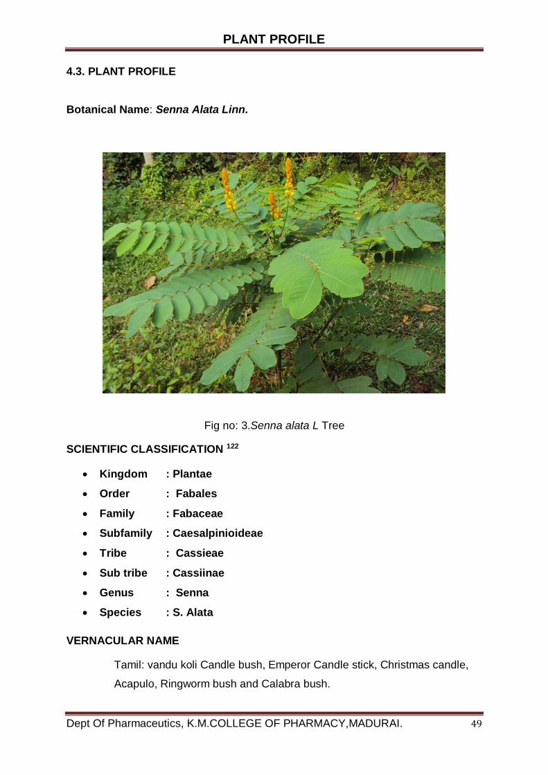
PLANT PROFILE
Dept Of Pharmaceutics, K.M.COLLEGE OF PHARMACY,MADURAI. 49
4.3. PLANT PROFILE Botanical Name: Senna Alata Linn.
Fig no: 3.Senna alata L Tree
SCIENTIFIC CLASSIFICATION 122
Kingdom : Plantae
Order : Fabales
Family : Fabaceae
Subfamily : Caesalpinioideae
Tribe : Cassieae
Sub tribe : Cassiinae
Genus : Senna
Species : S. Alata
VERNACULAR NAME
Tamil: vandu koli Candle bush, Emperor Candle stick, Christmas candle,
Acapulo, Ringworm bush and Calabra bush.
PLANT PROFILE
Dept Of Pharmaceutics, K.M.COLLEGE OF PHARMACY,MADURAI. 50
4.3.1. BOTONICAL DESCRIPTION
Senna alata is a shrub with usually an average height of between 1 and 5metres and
has horizontally spread branches. Its leaves are par pinnate of between 30 to 60 cm
long and consisting of 8 to 20 pairs of leaflets. Each leaflet is oblong or elliptic oblong
and rounded at both ends. Its flowers are dense in auxiliary racemes, about 20 to50
cm long and 3 to 4 cm broad. The inflorescence looks like a yellow candle. The plant
fruits are a thick, flattened with wings and glabrous pods. They grow well in full sun
in a wide range of soils that retain moisture adequately. The species is easy to grow
from the seed.
They can either be sown directly or started in a nursery and distributed all over the
country up to 1,500 m above sea level; they are most often cultivated for medicinal
purpose.123
Leaf
Leaf Arrangement : Alternate Leaf
Leaf Venation : Pinnate Leaf
Leaf Persistence : Evergreen
Leaf Type : Odd Pinnately compound
Leaf Blade : 5 - 10 cm
Leaf Shape : Oblong
Leaf Margins : Entire
Leaf Textures : Smooth, Coarse
Leaf Scent : No Fragrance
Color (growing season) : Green Color
(Changing season) : Green
Flower
Flower Showiness : True
Flower Size Range : 10 – 20
Flower Type : Spike Flower
Sexuality : Monoecious (Bisexual)
Flower Scent : No Fragrance
Flower Color : Yellow, Orange

PLANT PROFILE
Dept Of Pharmaceutics, K.M.COLLEGE OF PHARMACY,MADURAI. 51
Seasons : Summer, Fall Trunk Trunk
Susceptibility to Breakage : Generally resists breakage
Number of Trunks : Multi-Trunked Trunk Esthetical
Fruit
Values : Smooth Fruit
Fruit Type : Legume Fruit
Showiness : False
Fruit Size Range : 7 - 10
Fruit Colours : Brown
Seasons : Summer, Fall
4.3.2. Distribution:
It is a native to the Amazon Rainforest and can be found in Peru, Brazil,
French Guiana, Guyana, Suriname, Venezuela and Colombia. Due to its beauty, it
has been cultivated around the world as an ornamental plant and has naturalized in
many tropical regions in the world including tropical Africa, tropical Asia, Australia,
Mexico, the Caribbean islands, Melanesia, Polynesia, Hawaii and widely distributed
throughout different parts of India like Chhattisgarh, Maharashtra, West Bengal,
Andhra Pradesh etc.124
4.3.3. Worldwide Ethno medicinal Uses:
India- as an antidote, bactericide, diuretic, fungicide, insecticide, pesticide,
purgative, Vermifuge, for asthma, bronchitis, constipation, dysentery, eczema,
herpes, intestinal Parasites, rheumatism, skin disorders, snakebite, stomach
ache, venereal diseases etc.
Africa- as an abortifacient, laxative, for parturition, scurvy.
Brazil- for anaemia, constipation, dermatitis, dyspepsia, fevers, hydropsy,
liver Problems, menstrual disorders, skin problems, venereal disease, as a
diuretic, Emmenagogues, laxative and as a purgative.
Cuba- as a diuretic, diaphoretic, laxative, against herpes, skin infections.
Ghana- as an abortifacient, insecticide, purgative, vermifuge, for ascites,
PLANT PROFILE
Dept Of Pharmaceutics, K.M.COLLEGE OF PHARMACY,MADURAI. 52
Craw, eczema, gonorrhoea, herpes, leprosy, mycosis, parturition, Ringworm,
shingles, skin problems, sores, wounds etc.
Haiti - as a depurative, diaphoretic, insecticide, tonic, vulnerary, for
amygdalitis, Herpes, itch, measles, psoriasis, sore (throat), tonic, skin
problems, prurigo, sores, Wounds etc.
Java- for herpes, itch, ringworm, scabies, syphilis, as a larvaecide etc.
Mexico- as a diaphoretic, diuretic, insecticide, purgative; for fever,
rheumatism, Ringworms, skin infections, snakebite, syphilis etc.
Peru- as a diuretic, insecticide, laxative, vermifuge, for acaries, hepatitis,
herpes, Intestinal parasites, ringworm, skin problems, snakebite, urinary
infections etc.
Samoa - as a purgative, for ringworms, skin problems, snakebite etc.
Trinidad - as a bactericide, laxative, vermifuge, for diarrhoea, eczema,
herpes, venereal Diseases, vitiligo etc.
Venezuela- as a diuretic; for itch, skin problems
Elsewhere- as an antidote, bactericide, diuretic, fungicide, insecticide,
pesticide, Purgative, vermifuge, for asthma, bronchitis, constipation,
dysentery, eczema, herpes, Intestinal parasites, rheumatism, skin disorders,
snakebite, stomach ache, venereal Diseases etc.
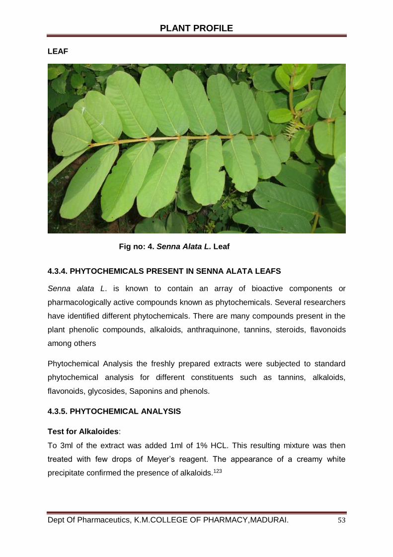
PLANT PROFILE
Dept Of Pharmaceutics, K.M.COLLEGE OF PHARMACY,MADURAI. 53
LEAF
Fig no: 4. Senna Alata L. Leaf
4.3.4. PHYTOCHEMICALS PRESENT IN SENNA ALATA LEAFS
Senna alata L. is known to contain an array of bioactive components or
pharmacologically active compounds known as phytochemicals. Several researchers
have identified different phytochemicals. There are many compounds present in the
plant phenolic compounds, alkaloids, anthraquinone, tannins, steroids, flavonoids
among others
Phytochemical Analysis the freshly prepared extracts were subjected to standard
phytochemical analysis for different constituents such as tannins, alkaloids,
flavonoids, glycosides, Saponins and phenols.
4.3.5. PHYTOCHEMICAL ANALYSIS
Test for Alkaloides:
To 3ml of the extract was added 1ml of 1% HCL. This resulting mixture was then
treated with few drops of Meyer’s reagent. The appearance of a creamy white
precipitate confirmed the presence of alkaloids.123
PLANT PROFILE
Dept Of Pharmaceutics, K.M.COLLEGE OF PHARMACY,MADURAI. 54
Test for Tannins:
Two drops of 5% FeCl3 was added to 1ml of the plant extract. The appearance of a
dirty green precipitate indicated the presence of tannins
Test for Flavonoids:
To 1ml of the extract was added 3 drops of ammonia solution (NH3+) followed by
0.5ml of concentrated HCl. The resultant pale brown colouration of the entire mixture
indicated the presence of flavonoids
Test for Steroids:
To 1ml of the plant extract was added 1ml of concentrated tetraoxosulphate (vi) acid
(H2SO4). A red colouration confirmed the presence of steroids
Test for Resins:
To 5ml of the extract was added 5ml of copper acetate solution. The mixture was
shaken vigorously and allowed to separate. The appearance of a reddish-brown
precipitate indicated the presence of resins.
4.3.6. Traditional uses
S.alata L. leaves and flowers have long been traditionally used as laxative and
antifungal agents
Use as laxative
1. Eight leaves are sun dried and powder an infusion is made from the
powdered leaves and taken before bed time
2. A few flowers cooked in boiling water and taken with the a special sauce
called Namprik
3. Three to five branches with leaves are boiled with water (1500 ml). The
decoction is boiled until about one third of water used is obtained. Salt is
added to the infusion to give it a salty taste. One glass of the decoction is
taken.
PLANT PROFILE
Dept Of Pharmaceutics, K.M.COLLEGE OF PHARMACY,MADURAI. 55
Treatment of ringworm or Tinea versicolor
1. Fresh leaves are pounded and the juice obtained is applied over the infected
area.
2. Three to four fresh leaves are pounded. Lemon juice is then added. The
obtained juice applied over the infected area.
4.3.7. ETHNOMEDICAL USES
Stems
Treatment of yaws, ringworm, Tinea versicolor, constipation, urinary stone,
anthelmintic and cardio tonic
Leaves
Treatment of skin diseases, urinary stone, ringworm, Tinea versicolor, laxative,
cardio tonic, and Expectorant.
Flowers
Laxatives and improvement of appearance and texture of skin
Pod
As an anthelmintic
Seed
Treatment of skin diseases, constipation and as an anthelmintic.
Whole plant
As an anthelmintic and antipyretic
Not specific part
Treatment of skin diseases, haemorrhoids, chronic gastrointestinal aliments of
children between the ages of 5 and 13 years characterized by marked malnutrition,
usually associated with intestinal parasitism.

PLANT PROFILE
Dept Of Pharmaceutics, K.M.COLLEGE OF PHARMACY,MADURAI. 56
4.3.8. CHEMICAL SUBSTANCE
Plant part:
Leaves
Flavonoid
Glycoside
Anthraquinone
Anthraquinone glycoside
Polyphenol
Root
Anthraquinone
Stem
Flavonoid
Glycoside
Anthraquinone
Anthrone
Sterol
Fruit
Anthraquinone
Seed
Polyalcohol
Carbohydrate
Flavonoid
Glycoside
Sterol
EXCIPIENT PROFILE
Dept Of Pharmaceutics, K.M.COLLEGE OF PHARMACY,MADURAI. 57
4.4. EXCIPIENT PROFILE
4.4.1 CARBOPOL
Carbopol polymers are polymers of acrylic acid cross-linked with polyalkenyl ethers
or divinyl glycol. They are primary produced from polymer having particles of about
0.2 to 6.0 micron in diameter. Carbomer-940 is to be a polymer of acrylic acid cross-
linked with alkyl-sucrose: CH = CHCH2-O-sucrose.Carbomer-940P is the
pharmaceutical grade of Carbomer-940.125
Molecular weights for Carbomers - 940:
Approx. 500,000 to 4,000,000.g
Colour: White, light, acidic, hygroscopic powder.
Particle size:
Flocculated powder having a median diameter of 2 to 7 microns.
Solubility / swelling properties:
They are insoluble due to their cross linked nature and high molecular weight. They
get swell in water and some polar solvents, producing viscous dispersions.
Topical Applications:
Carbopol 940 is very well suited to aqueous formulations of the topical dosage
forms such as hydrogel. Many commercial topical products available today have
been formulated with these polymers. They provide the following plentiful advantage
to topical formulations126
Safe & Effective
Carbopol polymers have a long antiquity of safe and effective use in topical gels,
creams and ointments. They are also supported by board toxicology studies
Non Sensitizing
Carbopol polymers have been shown to have extremely low irritancy properties and
are non-sensitizing with repeat usage.
EXCIPIENT PROFILE
Dept Of Pharmaceutics, K.M.COLLEGE OF PHARMACY,MADURAI. 58
Biocompatible
Carbopol polymers provide a magnificent vehicle for drug delivery. Due to their high
molecular weight, they are not able to penetrate the skin or affect the therapeutic
efficacy of the drug. Superior thickening, suspending, & emulsification properties for
Topical preparations.
Plant Profile
Products with a wide range of viscosities and flow properties have been successfully
formulated and commercialized.
Advantages with use of Carbopol gels as a vehicle
Good rheological properties on the application site.127
Substitute to oil-based ointment formulations
Viscosity is high at low concentration
Compatibility
Better bio adhesive properties
Good thermal stability
Uses:
In dermatological formulation Carbopol used as excellent vehicle. They can be used
in condition when controlled pattern is desired in drug delivery system. These
polymers are anionic polymers that need naturalization to become jellified.

EXCIPIENT PROFILE
Dept Of Pharmaceutics, K.M.COLLEGE OF PHARMACY,MADURAI. 59
4.4.2. SODIUM CARBOXYMETHYL CELLULOSE
Synonyms: 128
Sodium cellulose glycolate, Na CMC, CMC, cellulose gum, sodium CMC; INS No.
466
Chemical names:
Sodium salt of carboxymethyl ether of cellulose
Chemical formula:
[C6H7O2 (OH)x(OCH2COONa)y]n where n is the degree of polymerization x = 1.50
to 2.80 y = 0.2 to 1.50 x + y = 3.0 (y = degree of substitution)
Structural formula
Where R = H or CH2COONa
Formula weight
Structural unit with a degree of substitution of 0.20: 178.14
Structural unit with a degree of substitution of 1.50: 282.18
Macromolecules: greater than about 17,000 (n about 100) 129
Description
White or slightly yellowish, almost odourless hygroscopic granules, powder or fine
fibres
FUNCTIONAL USES
Thickening agent, stabilizer, suspending agent
EXPERIMENTAL INVESTIGATION
Dept Of Pharmaceutics, K.M.COLLEGE OF PHARMACY,MADURAI. 60
EXPERIMENTAL INVESTIGATION
5. EXPERIMENTAL INVESTIGATION
5.1 COLLECTION AND IDENTIFICATION OF PLANT MATERIALS
Collection and Identification of the plants
The fresh leaves of Senna alata L. was collected in the November to February
month of from Ullaganeri mellur main road Madurai. The plant was identified and
authenticated by Dr.D.Stephen, Assistant professor, Department of Botany The
American College, Madurai.
Drying of the plant part
The collected plants were washed with water and unwanted materials were
discarded. Collected plants were air and sun dried for 14 days.
Storage and Preservation of plant part
Most plant parts from desired plants have undergone a period of storage before they
were finally used for research purpose in the laboratory. During this period many
undesirable changes may occur in the plant parts if they were not properly stored
and preserved against the reabsorption of moisture, oxidation, excessive heat,
humidity, direct sunlight, growth of molds and bacteria and infestation by insects and
rodents. Proper storage and preservation of plant parts are thus are very important
factors in maintaining a high degree of quality in them. All efforts towards proper
storage should be geared to protect the drugs from all the above deteriorating factors
and agents.
Grinding of the plant parts
The Fresh leaves were air dried and then dried in a hot air oven at 50o C for 5 hours.
The dried leaves were ground with an electronic mill and passed through a sieve (20
mesh).
The fresh leaves was used for the study of morphological and microscopically
characters; Whereas the dried leaves powder was used for determination of powder
microscopy, physicochemical characterization and phytochemical analysis.

EXPERIMENTAL INVESTIGATION
Dept Of Pharmaceutics, K.M.COLLEGE OF PHARMACY,MADURAI. 61
5.2.1 MACROSCOPIC CHARACTERS
The leaves of Senna alata L. was subjected to microscopic studies which
comprised of organoleptic characteristics viz. colour, odour, appearance,taste,shape
texture,fracture ,etc. of the drug This parameters are considered as quite useful in
quality control of the crude drug and were evaluated as per standard WHO
guidelines
The observation has been tabulated in the following table
Table No : 1
Leaflet shape oblong; obovate
Leaf odour characteristic odour
Leaf taste Bitterish unpleasant taste
Leaf colour Green
Texture Coarse
Leaflet margin Entire
The fresh leafs is green colour, Leaf arrangement: alternate , Leaf type: even
Pinnately compound, Leaflet margin: entire, Leaflet shape: oblong; obovate, Leaflet
venation: pinnate, Leaf type and persistence: evergreen, Leaflet blade length: 2 to 4
inches, Leaf colour: green . The dried leaf is light green colour
5.2.2. MICROSCOPIC CHARACTERISTICS OF SENNA ALATA LEAF
Fresh leaves of Senna Alata L. were selected for the microscopically studies.
Microscopic sections were cut by free hand sectioning. Numerous temporary and
permanent mounts of the microscopically sections of the leaf were made and
examined microscopically. Histochemcal reactions were applied with staining
reagents on transverse sections and on leaves powder by reported methods.
Photomicrographs of the microscopically sections were taken with the help of Digital
microscope,
EXPERIMENTAL INVESTIGATION
Dept Of Pharmaceutics, K.M.COLLEGE OF PHARMACY,MADURAI. 62
5.2.3. PHYSICOCHEMICAL EVALUATION OF SENNA ALATA LEAF
Analysis of Physicochemical constants of the powder leaves has been done to
evaluate the quality and purity of the drug. Various Physicochemical parameters like
Moisture contents, foreign organic matters. Ash Values and Extract Values were
calculated as per WHO Guidelines. The information collected from these test was
useful for standardization and obtaining the quality standards.
5.2.4. EXTRACTION PROCESS OF SENNA ALATA LEAF
SOXHLET EXTRACTION
The powdered leaves of Senna alata L. (10.0 g) were extracted with 300 ml of 80%
ethanol in a soxhlet apparatus. The extraction was continued until the extraction was
exhausted. Each extract was then combined, filtered and evaporated to dryness on a
hot water bath to yield a soxhlet crude extract (2.56 g). The extraction method which
promoted the extract containing the maximum content of total Anthraquinones would
be choosen as the appropriate method for further extracting the leaf samples of
Senna alata L. Collected from various locations.
Drying of extract:
Using rotary evaporator, the methanolic extract of plant was evaporated at 55-60
degree Celsius temperature and a rotation speed of 160-180 rpm for 1 month. After
this drying process, a slurry concentration were obtained, which were kept in small
50 ml beakers for further drying. During transfer to the beaker the extracts were
rinsed by acetone.
5.2.5. PHYTOCHEMICAL INVESTIGATIONS OF SENNA ALATA LEAF
The qualitative chemical test carried out for the identification of the nature different
phytoconstituents present in the powdered crude drug. The tests were carried out by
using standard conventional protocols.
Determination of absorbance maximum (max) 130
Senna alata L .was dissolved in phosphate buffer saline pH 7.4 solutions with 20
g/ml concentration was prepared by suitable dilution.
EXPERIMENTAL INVESTIGATION
Dept Of Pharmaceutics, K.M.COLLEGE OF PHARMACY,MADURAI. 63
Senna alata L drug in solution was scanned in UV spectrophotometer from 200 to
400 nm using phosphate buffer saline pH 7.4 as blank. Absorbance maximum was
determined as 298 nm. The drug was later quantified by measuring the absorbance
at 240 nm in phosphate buffer saline pH7.4.
Standard curve for Senna alata L (by UV method)
Isolation of emodin
Ethanol plant fraction was applied on precoated silica gel G plate and run in solvent
system chloroform and methanol (80:20) shown three different distinct spots. Then
ethyl acetate fraction was subjected for column chromatography using silica gel G
and Chloroform and methanol (80:20) as a mobile phase. Compound flavonoid was
isolated.
Preparation of primary stock solution
Senna alata L 100 mg was weighed and dissolved in phosphate buffer saline pH7.4
in a 100 ml volumetric flask. The flask was shaken and volume was made up to the
mark with phosphate buffer saline pH 7.4 to give a solution containing 1000g/ml.
Preparation of secondary stock solution
From the primary stock solution, pipette out 2ml and placed into 100ml
volumetric flask. The volumetric was made up to mark with phosphate buffer saline
pH 7.4 give a stock solution containing 20g/ml.
Preparation of sample solution
Appropriate volumes of aliquots (1to10ml) from standard Senna alata L secondary
stock solution were transferred to different volumetric flasks of 10 ml capacity. The
volume was adjusted to the mark with phosphate buffer saline pH7.4 to obtain
concentration of 10,20,30,40,50,60,70,80,90,and 100g/ml. Absorbance of each
solution against phosphate buffer saline pH7.4 as blank were measured at 298 nm
and the graph of absorbance against concentration were plotted shown fig no:
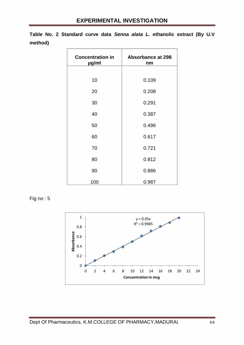
EXPERIMENTAL INVESTIGATION
Dept Of Pharmaceutics, K.M.COLLEGE OF PHARMACY,MADURAI. 64
Table No. 2 Standard curve data Senna alata L. ethanolic extract (By U.V
method)
Fig no : 5
Concentration in
µg/ml
Absorbance at 298
nm
10
20
30
40
50
60
70
80
90
100
0.109
0.208
0.291
0.387
0.496
0.617
0.721
0.812
0.886
0.987
y = 0.05xR² = 0.9985
0
0.2
0.4
0.6
0.8
1
0 2 4 6 8 10 12 14 16 18 20 22 24
Ab
sorb
ance
Concentration in mcg
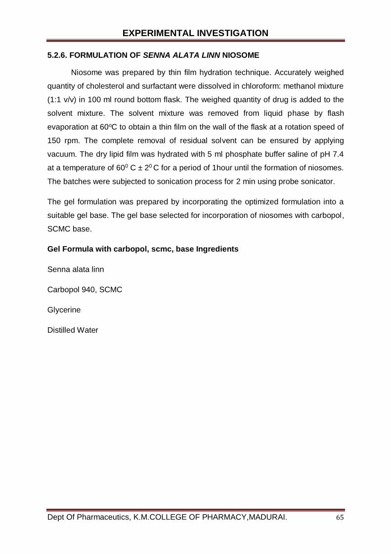
EXPERIMENTAL INVESTIGATION
Dept Of Pharmaceutics, K.M.COLLEGE OF PHARMACY,MADURAI. 65
5.2.6. FORMULATION OF SENNA ALATA LINN NIOSOME
Niosome was prepared by thin film hydration technique. Accurately weighed
quantity of cholesterol and surfactant were dissolved in chloroform: methanol mixture
(1:1 v/v) in 100 ml round bottom flask. The weighed quantity of drug is added to the
solvent mixture. The solvent mixture was removed from liquid phase by flash
evaporation at 60oC to obtain a thin film on the wall of the flask at a rotation speed of
150 rpm. The complete removal of residual solvent can be ensured by applying
vacuum. The dry lipid film was hydrated with 5 ml phosphate buffer saline of pH 7.4
at a temperature of 600 C ± 20 C for a period of 1hour until the formation of niosomes.
The batches were subjected to sonication process for 2 min using probe sonicator.
The gel formulation was prepared by incorporating the optimized formulation into a
suitable gel base. The gel base selected for incorporation of niosomes with carbopol,
SCMC base.
Gel Formula with carbopol, scmc, base Ingredients
Senna alata linn
Carbopol 940, SCMC
Glycerine
Distilled Water
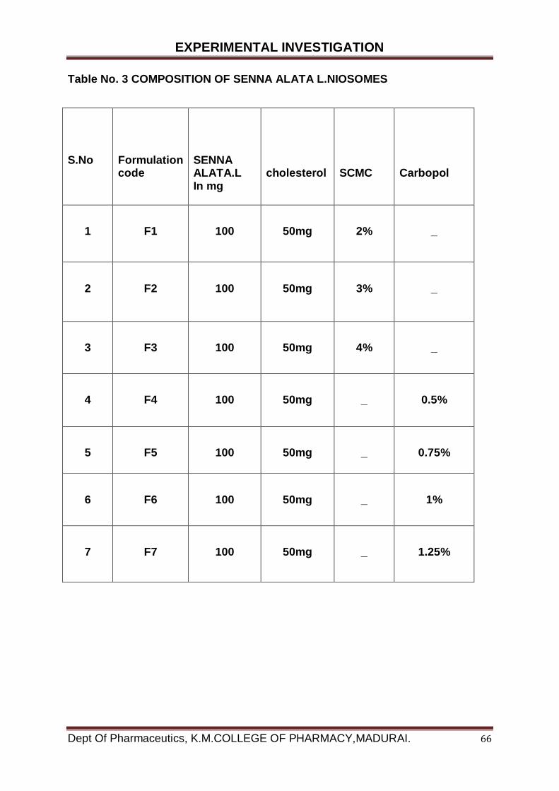
EXPERIMENTAL INVESTIGATION
Dept Of Pharmaceutics, K.M.COLLEGE OF PHARMACY,MADURAI. 66
Table No. 3 COMPOSITION OF SENNA ALATA L.NIOSOMES
S.No
Formulation code
SENNA ALATA.L In mg
cholesterol
SCMC
Carbopol
1
F1
100
50mg
2%
_
2
F2
100
50mg
3%
_
3
F3
100
50mg
4%
_
4
F4
100
50mg
_
0.5%
5
F5
100
50mg
_
0.75%
6
F6
100
50mg
_
1%
7
F7
100
50mg
_
1.25%

EXPERIMENTAL INVESTIGATION
Dept Of Pharmaceutics, K.M.COLLEGE OF PHARMACY,MADURAI. 67
5.2.7. EVALUATION OF SENNA ALATA L. NIOSOMES
DRUG & POLYMER COMPATABILITY STUDIES BY FTIR:
Pressed pellet technique was used to handle the sample in FTIR
spectrometer. In this technique a pinch of sample was mixed with potassium bromide
and the mixture was pressed with special discs under high pressure into a
transparent pellet and then inserted in to special holder of IR spectrometer.
IR spectrums for pure drug alone and physical mixture of drug and polymers
are taken. The spectrum of physical mixture was compare with spectrum of pure
drug. Bands seen in pure drug also recognized in physical mixture, hence there was
to no significant interaction between drug and excipients
Removal of unentrapped drug from niosome
The unentrapped drug from niosome was removed by dialysis method. Niosome
suspension was placed in 3cm × 8cm long dialysis bag whose molecular weight cut
off was 12,000. The dialysis bag was then placed in 250 ml beaker containing
phosphate buffer saline of pH 7.4 with constant stirring by means of a magnetic
stirrer. Dialysis was carried out for 24 hour by replacing the buffer with fresh for
every 3 hours.
Size Analysis
By optical microscopy
A drop of niosome suspension was placed on a glass slide and it was diluted.
A cover slip was placed over the diluted niosome suspension and evaluated the
average vesicle size and shape by an ordinary optical microscope using a
recalibrated ocular eye piece micro meter.
By scanning electron microscopy
The optimized formulation was morphologically characterized by scanning
electron microscopy. The sample for SEM analysis was mounted on the specimen
stub was placed on to a carbon coated grid to leave a thin film before drying of this
film on the grid. It was negatively stained with 1% of phosphor tungstic acid (PTA).

EXPERIMENTAL INVESTIGATION
Dept Of Pharmaceutics, K.M.COLLEGE OF PHARMACY,MADURAI. 68
For this ,a drop of staining solution was added on to the film and the excess of the
solution was drained of the filter paper. The grid was allowed to air dry thoroughly
and was then visualized using a scanning electron microscope with an accelerating
voltage of 80kv.
Percentage encapsulation of drug
Vesicles containing senna alata L were separated from encapsulated drug by
dialysis. Senna alata linn Niosomal preparation of 0.5 ml was taken after dialysis. To
this 0.5ml of 10% triton X- 100 was added and incubated for 1hour .The triton X-100
was added to lyse the vesicles in order to release the encapsulated senna alata linn.
Then it was diluted with phosphate buffer saline solution (pH 7.4) and filtered through
whatmann filter buffer and triton X-100 mixture as blank. From the absorbance value,
the concentration of drug in mcg/ml was found using the standard curve
Amount of drug entrapped (mg) Percentage drug loading (PDL) = x 100 Total amount of drug added (mg)
5.2.8. IN VITRO CHARACTERISATION
Niosomal preparation was taken in a dialysis membrane of 5 cm length and
suitably suspended in a beaker containing 200 ml of diffusion medium (Phosphate
buffer saline pH 7.4). The medium was maintained at a temperature of 37 ±0.5 C. It
was stirred by means of magnetic stirrer at a constant speed. Sample of 1ml
(diffusion medium) was withdrawn at every 24 hours and replaced the diffusion
medium, so that the volume of diffused medium was maintained constant at
200ml.The samples were measured spectrophotometrically at 266nm. The release
was compared with a marketed senna alata L gel.
5.2.9. STABILITY STUDIES OF SENNA ALATA L NIOSOMES
The optimized formulation F6 was subjected to stability study for one month at 40 C
(refrigerator), room temperature and at 450C/75% RH. At the interval of 30 days,
samples of niosomal formulation were taken and evaluated for the entrapment
efficiency and in vitro release of drug Entrapment efficiency of optimized formulation
F6 kept at 40C shows a release rate of 95.5% after 30 days of stability study. The
EXPERIMENTAL INVESTIGATION
Dept Of Pharmaceutics, K.M.COLLEGE OF PHARMACY,MADURAI. 69
entrapment efficiency of formulation got decreased on exposure to higher
temperature. The percentage entrapment of formulation kept at room temperature
and at 450C/75%RH were 92% and 81% respectively after 30 days of stability study
5.3. RELEASE KINETICS STUDIES
For estimation of the kinetic and mechanism of drug release, in vitro drug
release study of niosomes were fitted with various kinetic equation like were used to
described the release kinetic. The zero order release states that drug release rate
was independent of its concentration. The first order release describes, the releases
rate from the system was concentration dependent. Higuchi described the releases
of drug from insoluble matrix as a square root of time dependent process was based
on Fickian diffusion.
1. Zero order - Cumulative % drug release versus time.
2. First order -Log cumulative % drug remaining versus time.
3. Higuchi’s model -Cumulative % drug released versus square root of time.
4. Korsmeyer equation / Peppa’s model – Log cumulative per cent drug
released versus log time.
a. Zero order kinetics
Zero order release would be predicted by the following equation:
At = A0-K0t
Where
At = Drug release at time’s’
A0 = Initial drug concentration.
K0 = Zero- order rate constant (hr-1)
Where the data is plotted as cumulative per cent drug release versus
time, if the plot is linear then the data obeys Zero –order kinetics and its slope
is equal to Zero order release constant K0.

EXPERIMENTAL INVESTIGATION
Dept Of Pharmaceutics, K.M.COLLEGE OF PHARMACY,MADURAI. 70
b. First order kinetics:
First order release could be predicted by the following equation:
Log C = log C0 – Kt / 2.303
Where,
C = Amount of drug remained at time’s’
C0 = Initial amount of drug
K = First order rate constant (hr-1)
When the data plotted as log cumulative per cent drug remaining versus time,
yield a straight line, indicating that the release follows first order kinetics. The
constant ‘Kt’ can be obtained by multiplying 2.303 with the slope value.
c. Higuchi’s model:
Drug release from the matrix devices by diffusion has been described
below Higuchi’s classical diffusion equation:
Q = [D€ / T (2A - €Cs) Cs t] 1/2
Where,
Q = Amount of drug release at time‘t’
D = Diffusion coefficient of the drug in the matrix.
A = Total amount of drug in unit volume of matrix.
Cs = Solubility of drug in the matrix.
€ = Porosity of the matrix.
EXPERIMENTAL INVESTIGATION
Dept Of Pharmaceutics, K.M.COLLEGE OF PHARMACY,MADURAI. 71
T = Tortuosity.
t = Time (hrs. at which q amount of drug is released.)
Above equation can be simplified assumed that ‘D’, ‘Cs’ and ‘A’ are constants. Then
the equation becomes
Q = Kt1/2
According to the equation, if cumulative drug release versus square root of
time yields a straight line, indicating that the drug was released by diffusion
mechanism.
d. Korsmeyer equation / Peppa’s model:
Korsmeyer described a simple relationship to find the mechanism of drug
release from polymeric system. To study the mechanism of drug releases from the
Nanoparticles formulation, the release data was fitted to the Korsmeyer –Peppa’s
law equation, which was used to describe the drug release behaviour systems.
Mt / Mα = Ktη
Where,
Mt / Mα = Fraction of drug released at time‘t’
K = Constant
n = Diffusion exponent related to the mechanism of the release.
Above equation can be simplified as follows by applying log on both sides,
Log Mt / Mα = Log K + n Log t
In the above equation, “n” value was used to characterize the various
release mechanism as mentioned in the table below

EXPERIMENTAL INVESTIGATION
Dept Of Pharmaceutics, K.M.COLLEGE OF PHARMACY,MADURAI. 72
Table No: 4 Diffusion exponent and solute release mechanism for cylindrical
shape
S.No
Diffusion
Exponent (n) Overall solute
diffusion mechanism
1
0.45
Fickian diffusion
2
0.45<n<0.89
Anomalous (non-Fickian )
Diffusion
3
0.89
Case - 2transport
4
n>0.89
Super case-2 transport
EXPERIMENTAL INVESTIGATION
Dept Of Pharmaceutics, K.M.COLLEGE OF PHARMACY,MADURAI. 73
5.4. Morphology
5.4.1. Binocular microscopy and Scanning Electron Microscopy (SEM)
The niosomes morphology, surface, appearance and shape of the
nanoparticles was analysed by Scanning Electron Microscopy (SEM) at different
magnifications. A few mg of prepared niosomes was gold coated using a Hitachi
HVSJGB Vacuum evaporator. Coated samples were viewed and photographed in a
Hitachi S-450 SEM operated at 20 k v
5.5. Evaluation of Antimicrobial Activity of Senna Alata L.niosomal
gel formulation
The antimicrobial activity of senna alata L niosomal formulation was carried
out by agar plate method with test organism.
Materials required
Organism used
candida albicans, spores
Aspergillus niger
Staphylococcus aureus
Pseudomonas aeruginosa,
Escherichia coli
Standard used
Fluconazole, ciprofloxin
Medium used
MGYP medium
Nutrient Agar medium
Potato Dextrose Agar medium MGYP medium
Medium used for broth culture
MGYP broth
Nutrient Agar broth
Potato Dextrose Agar broth
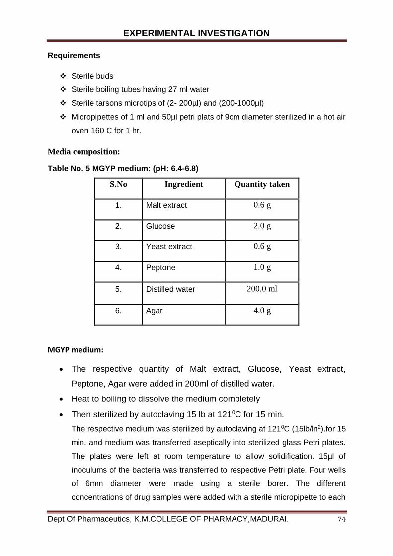
EXPERIMENTAL INVESTIGATION
Dept Of Pharmaceutics, K.M.COLLEGE OF PHARMACY,MADURAI. 74
Requirements
Sterile buds
Sterile boiling tubes having 27 ml water
Sterile tarsons microtips of (2- 200µl) and (200-1000µl)
Micropipettes of 1 ml and 50µl petri plats of 9cm diameter sterilized in a hot air
oven 160 C for 1 hr.
Media composition:
Table No. 5 MGYP medium: (pH: 6.4-6.8)
S.No Ingredient Quantity taken
1. Malt extract 0.6 g
2. Glucose 2.0 g
3. Yeast extract 0.6 g
4. Peptone 1.0 g
5. Distilled water 200.0 ml
6. Agar 4.0 g
MGYP medium:
The respective quantity of Malt extract, Glucose, Yeast extract,
Peptone, Agar were added in 200ml of distilled water.
Heat to boiling to dissolve the medium completely
Then sterilized by autoclaving 15 lb at 1210C for 15 min.
The respective medium was sterilized by autoclaving at 1210C (15lb/ln2).for 15
min. and medium was transferred aseptically into sterilized glass Petri plates.
The plates were left at room temperature to allow solidification. 15µl of
inoculums of the bacteria was transferred to respective Petri plate. Four wells
of 6mm diameter were made using a sterile borer. The different
concentrations of drug samples were added with a sterile micropipette to each
EXPERIMENTAL INVESTIGATION
Dept Of Pharmaceutics, K.M.COLLEGE OF PHARMACY,MADURAI. 75
of the cups. The plates were maintained on sight place for 2 hours to allow the
diffusion of the solution into the medium. The Petri dishes are kept inverted
position in incubator at 370C ±10C for 24 hours. The diameter of zone of
inhibition surrounding each of the wells was recorded.
Table No. 6 Nutrient Agar medium: (pH: 7-7.5)
S.No Ingredient Quantity taken
1. Beef extract 2.0 g
2. NaCl 1.0 g
3. Peptone 2.0 g
4. Distilled water 200.0 ml
5. Agar 4.0 g
Nutrient Agar medium:
The respective quantity of Beef extract, NaCl, Peptone, Agar adds in 200ml of
distilled water.
Heat to boiling to dissolve the medium completely
Then sterilized by autoclaving 15 lb at 1210C for 15 min.
The respective medium was sterilized by autoclaving at 1210C (15lb/ln2).for 15 min.
and medium was transferred aseptically into sterilized glass Petri plates. The plates
were left at room temperature to allow solidification. 15µl of inoculums of the bacteria
was transferred to respective Petri plate. Four wells of 6mm diameter were made
using a sterile borer. The different concentrations of drug samples were added with a
sterile micropipette to each of the cups. The plates were maintained on sight place
for 2 hours to allow the diffusion of the solution into the medium. The Petri dishes are
kept inverted position in incubator at 370C ±10C for 24 hours. The diameter of zone
of inhibition surrounding each of the wells was recorded.
EXPERIMENTAL INVESTIGATION
Dept Of Pharmaceutics, K.M.COLLEGE OF PHARMACY,MADURAI. 76
Table No.7 Potato Dextrose Agar medium: (pH: 6-7)
S.No Ingredient Quantity taken
1. Pieces of potato 40.0 g
2. Dextrose 4.0 g
3. Yeast extract 0.02 g
4. Distilled water 200.0 ml
5. Agar 4.0 g
Potato Dextrose Agar medium:
40.0 g of peeled potatoes are cut into small pieces and suspended in 200.0 ml
of distilled water.
Steamed for 30 min. decant the extract or filter through muslin cloth and make
the final volume to 200.0 ml.
Add 4.0 g of dextrose, 0.02 g of yeast extract and 4.0 g of agar.
The respective medium was sterilized by autoclaving at 1210C (15lb/ln2).for 15 min.
and medium was transferred aseptically into sterilized glass Petri plates. The plates
were left at room temperature to allow solidification.15µl of inoculums of the bacteria
and fungi was transferred to respective Petri plate. Four wells of 6mm diameter were
made using a sterile borer. The different concentrations of drug samples were added
with a sterile micropipette to each of the cups. The plates were maintained on sight
place for 2 hours to allow the diffusion of the solution into the medium. The Petri
dishes are kept inverted position in incubator at 280C for 48 hours. The diameter of
zone of inhibition surrounding each of the wells was recorded.

RESULTS AND DISCUSSION
Dept Of Pharmaceutics, K.M.COLLEGE OF PHARMACY,MADURAI. 77
RESULTS AND DISCUSSION
6. RESULTS AND DISCUSSION
6.1. COLLECTION AND IDENTIFICATION OF PLANT MATERIALS
Collection and identification of plant
The fresh leaves of Senna alata was collected in the November to February month of
from Ullaganeri mellur main road Madurai. The plant was identified and
authenticated by Dr.D.Stephen, Assistant professor, Department of Botany The
American College, Madurai.
Drying of the plant part
The collected plants were washed with water and unwanted materials were
discarded. Collected plants were air and sun dried for 14 days.
Fig. No.6 Dried Senna alata leaves
Storage and Preservation of plant part
Most plant parts from desired plants have undergone a period of storage
before they were finally used for research purpose in the laboratory. During this
period many undesirable changes may occur in the plant parts if they were not

RESULTS AND DISCUSSION
Dept Of Pharmaceutics, K.M.COLLEGE OF PHARMACY,MADURAI. 78
properly stored and preserved against the reabsorption of moisture, oxidation,
excessive heat, humidity, direct sunlight, growth of molds and bacteria and
infestation by insects and rodents. Proper storage and preservation of plant parts are
thus are very important factors in maintaining a high degree of quality in them. All
efforts towards proper storage should be geared to protect the drugs from all the
above deteriorating factors and agents.
Grinding of the plant parts
The dried small pieces of plant parts were grinded into small fine particles by
a grinder machine from. The powder was stored in an air tight container and kept in
a cool dark and place until analysis commenced The dried plant powder as identified
and authenticated by Dr.D.Stephen, Assistant professor, Department of Botany The
American College, Madurai.
6.2.1 MACROSCOPIC CHARACTERS OF SENNA ALATA LEAF
Senna alata is a shrub with usually an average height of between 1 and
5metres and has horizontally spread branches. Its leaves are par pinnate of between
30 to 60 cm long and consisting of 8 to 20 pairs of leaflets. Each leaflet is oblong or
elliptic oblong and rounded at both ends. Its flowers are dense in auxiliary racemes,
about 20 to50 cm long and 3 to 4 cm broad. The inflorescence looks like a yellow
candle. The plant fruits are a thick, flattened with wings and glorious pods. They
grow well in full sun in a wide range of soils that retain moisture adequately. The
species is easy to grow from the seed.
They can either be sown directly or started in a nursery and distributed all over the
country up to 1,500 m above sea level; they are most often cultivated for medicinal
purposes
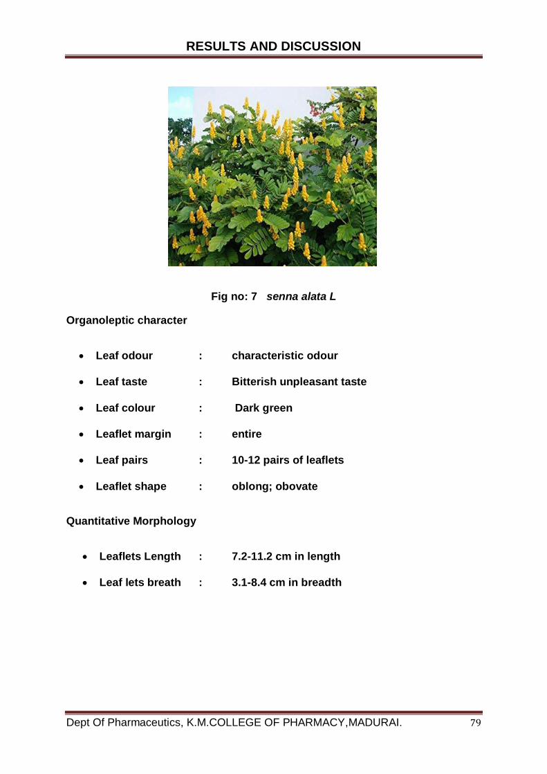
RESULTS AND DISCUSSION
Dept Of Pharmaceutics, K.M.COLLEGE OF PHARMACY,MADURAI. 79
Fig no: 7 senna alata L
Organoleptic character
Leaf odour : characteristic odour
Leaf taste : Bitterish unpleasant taste
Leaf colour : Dark green
Leaflet margin : entire
Leaf pairs : 10-12 pairs of leaflets
Leaflet shape : oblong; obovate
Quantitative Morphology
Leaflets Length : 7.2-11.2 cm in length
Leaf lets breath : 3.1-8.4 cm in breadth

RESULTS AND DISCUSSION
Dept Of Pharmaceutics, K.M.COLLEGE OF PHARMACY,MADURAI. 80
6.2.2. MICROSCOPIC CHARACTERISTICS
Powder Microscopy of leaf
The powder preparation leaf shows the following inclusion:
Fig no: Powder Microscopy of leaf
Fig No: 8
Fig No: 9 Powder microscopy

RESULTS AND DISCUSSION
Dept Of Pharmaceutics, K.M.COLLEGE OF PHARMACY,MADURAI. 81
6.2.3. PHYSICOCHEMICAL EVALUATION OF SENNA ALATA LEAF
Ash Values: Air dried powdered samples of the whole plants of Cassia alata was
investigated for total ash, acid insoluble ash and water-soluble ash values by
following procedure.
Total Ash Value:
About 2gm of the Cassia alata was weighed accurately and spread as a fine layer at
the bottom of a tarred silica crucible. The crucible was incinerated at 500 – 600 ºC in
a muffle furnace until free from carbon. Then the crucible was cooled & weighed, the
percentage total ash content was calculated.
Acid Insoluble Ash Value:
The ash obtained from the total ash was boiled with 25ml of HCl for 5 min. The
insoluble ash was collected on ash less filter paper, washed with hot water. The filter
paper along with the residue was transferred to a tarred silica crucible and ignited at
a temperature not exceeding 600 ºC to constant weight. The percentage of the acid
insoluble ash was calculated.
Water-Soluble Ash Value:
The total ash obtained was boiled with 25ml of water for 5 min. The insoluble matter
is collected on an ash-less filter paper, washed with hot water and ignited for 15 min
at a temperature not exceeding 450ºC. The weights of insoluble matter were
subtracted from the weight of total ash. The difference in weight represents the water
soluble ash. The percentage of water-soluble ash was calculated.
Extractive Values
Chloroform Soluble Extractive Value:
15gm of coarsely powdered air-dried cassia alata was accurately weighed, in a
glass-stopper conical flask. 100 ml of chloroform was added shaken vigorously and
vigorously intermediately for 24 hours and reweighed the flask. The flask
evaporated, cooled and weighted
RESULTS AND DISCUSSION
Dept Of Pharmaceutics, K.M.COLLEGE OF PHARMACY,MADURAI. 82
Alcohol soluble extractive value
15gm of coarsely powdered air-dried cassia alata was accurately weighed, in a
glass-stopper conical flask. 100 ml of alcohol (90%v/v) is added shaken vigorously
intermediately for 24 hours and reweighed the flask. The flask evaporated, cooled
and weighted.
Physical constants
Ash Values
i) Total ash – 7.84%
ii) Acid – insoluble ash-Nil
iii) Water soluble ash – 7.59%
Extractive values
Chloroform – 7.4%
Alcohol – 5.8 %

RESULTS AND DISCUSSION
Dept Of Pharmaceutics, K.M.COLLEGE OF PHARMACY,MADURAI. 83
6.2.4. EXTRACTION PROCESS
SOXHLET EXTRACTION
The powdered leaves of S. alata (10.0 g) were extracted with 300 ml of 80% ethanol
in a soxhlet apparatus. The extraction was continued until the extraction was
exhausted. Each extract was then combined, filtered and evaporated to dryness on a
hot water bath to yield a soxhlet crude extract (2.56 g). The extraction method which
promoted the extract containing the maximum content of total Anthraquinones would
be choosen as the appropriate method for further extracting the leaf samples of S.
alata collected from various locations.
Fig No: 10 soxhlet apparatus
RESULTS AND DISCUSSION
Dept Of Pharmaceutics, K.M.COLLEGE OF PHARMACY,MADURAI. 84
Drying of extract:
Using rotary evaporator, the methanolic extract of plant was evaporated at 55-60
degree Celsius temperature and a rotation speed of 160-180 rpm for 1 month. After
this drying process, a slurry concentration were obtained, which were kept in small
50 ml beakers for further drying. During transfer to the beaker the extracts were
rinsed by acetone.
6.2.5. PHYTOCHEMICAL INVESTIGATIONS SENNA ALATA LEAF
The qualitative chemical test carried out for the identification of the nature different
phytoconstituents present in the powdered crude drug. The tests were carried out by
using standard conventional protocols
Phytochemicals Present in Senna alata
Senna alata is known to contain an array of bioactive components or
pharmacologically active compounds known as phytochemicals.
Several researchers have identified different phytochemicals. There are many
compounds present in the plant phenolic compounds, alkaloids,
anthraquinone, tannins, steroids, flavonoids among others
Phytochemical Analysis of Senna alata
Phytochemical screening is done to ascertain the scientific assessment of the
claim of the therapeutic potency.
The healing potency of these phytochemicals have taken a new dimension
since there have been recent interest by the Scientists on what constitutes
each medicinal plants.
There have been different measures or steps taken for the analysis of these
phytochemicals.
Senna alata are reported to contain a variety of secondary or bioactive
compounds, known as phytochemicals.

RESULTS AND DISCUSSION
Dept Of Pharmaceutics, K.M.COLLEGE OF PHARMACY,MADURAI. 85
They screened the leaves extracts for its phytochemicals, antibacterial and
antifungal activities. The methanolic extract showed the highest activity than
the ethanolic and petroleum ether extracts
This may be due to the effect of polarity of the solvent. The unidentified active
components purified from preparative thin layer chromatography exhibited low
activities against Mucor, Rhizopus and Aspergillus Niger at concentration of
70µg/ml while higher activity was exhibited against all the test bacteria and
fungi at 860µg/ml.
Senna alata leaf decoction has been used to treat infectious diseases in north
eastern Nigeria. Timothy etal. Screened the leaves extract for their activity
against infectious diseases.
In the attempt to perform this, the leaves of the plant were collected, dried and
extracted using water and 95% ethanol and evaluated against five clinical
isolates of pathogenic fungi for their antifungal activity.

RESULTS AND DISCUSSION
Dept Of Pharmaceutics, K.M.COLLEGE OF PHARMACY,MADURAI. 86
Table No: 8 Phytochemical analysis
TEST
OBSERVATION
RESULT
Test for Alkaloides: To 3ml of the extract was added 1ml of 1% HCL. This resulting mixture was then treated with few drops of Meyer’s reagent
The appearance of a creamy white precipitate confirmed the presence of alkaloids
Test for Tannins: Two drops of 5% FeCl3 was added to 1ml of the plant extract
The appearance of a dirty green precipitate indicated the presence of tannins
Test for Flavonoids:
To 1ml of the extract was added 3 drops of ammonia solution (NH3+) followed by .5ml of concentrated HCl
The resultant pale brown colouration of the entire mixture indicated the presence of flavonoids
Test for Steroids: To 1ml of the plant extract was added 1ml of concentrated tetraoxosulphate (vi) acid (H2SO4).
A red colouration confirmed the presence of steroids.
Test For Glycosides Hydrochloric acid (2M) was added to the sample and the mixture was heated on a hot water bath for 15 minutes, then cooled and filtered. The filtrate was extracted with chloroform. The chloroform layer was separated and shaken with 10% potassium hydroxide solution.
The upper aqueous layer becomes pink
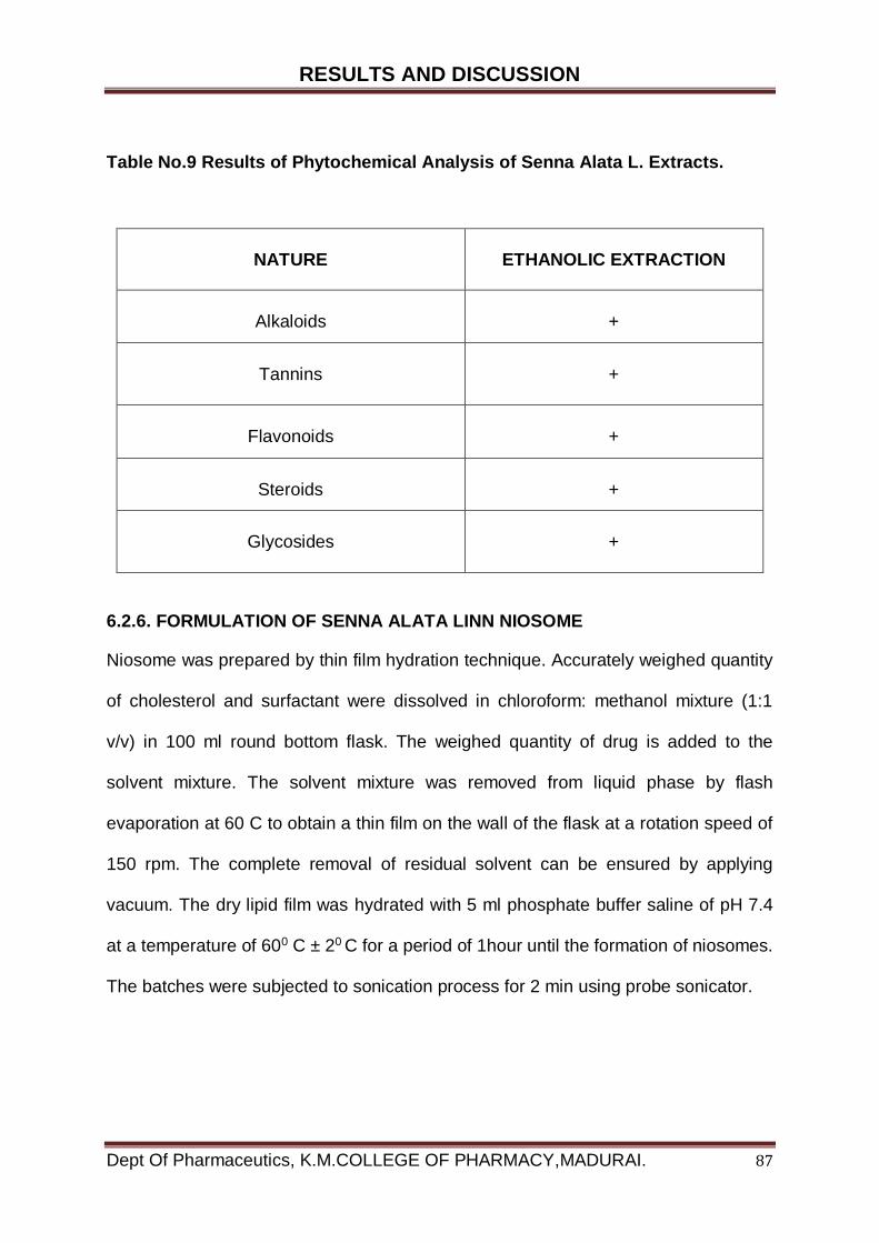
RESULTS AND DISCUSSION
Dept Of Pharmaceutics, K.M.COLLEGE OF PHARMACY,MADURAI. 87
Table No.9 Results of Phytochemical Analysis of Senna Alata L. Extracts.
NATURE
ETHANOLIC EXTRACTION
Alkaloids
+
Tannins
+
Flavonoids
+
Steroids
+
Glycosides
+
6.2.6. FORMULATION OF SENNA ALATA LINN NIOSOME
Niosome was prepared by thin film hydration technique. Accurately weighed quantity
of cholesterol and surfactant were dissolved in chloroform: methanol mixture (1:1
v/v) in 100 ml round bottom flask. The weighed quantity of drug is added to the
solvent mixture. The solvent mixture was removed from liquid phase by flash
evaporation at 60 C to obtain a thin film on the wall of the flask at a rotation speed of
150 rpm. The complete removal of residual solvent can be ensured by applying
vacuum. The dry lipid film was hydrated with 5 ml phosphate buffer saline of pH 7.4
at a temperature of 600 C ± 20 C for a period of 1hour until the formation of niosomes.
The batches were subjected to sonication process for 2 min using probe sonicator.
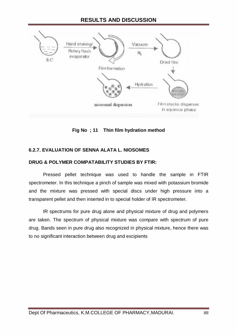
RESULTS AND DISCUSSION
Dept Of Pharmaceutics, K.M.COLLEGE OF PHARMACY,MADURAI. 88
Fig No ; 11 Thin film hydration method
6.2.7. EVALUATION OF SENNA ALATA L. NIOSOMES
DRUG & POLYMER COMPATABILITY STUDIES BY FTIR:
Pressed pellet technique was used to handle the sample in FTIR
spectrometer. In this technique a pinch of sample was mixed with potassium bromide
and the mixture was pressed with special discs under high pressure into a
transparent pellet and then inserted in to special holder of IR spectrometer.
IR spectrums for pure drug alone and physical mixture of drug and polymers
are taken. The spectrum of physical mixture was compare with spectrum of pure
drug. Bands seen in pure drug also recognized in physical mixture, hence there was
to no significant interaction between drug and excipients
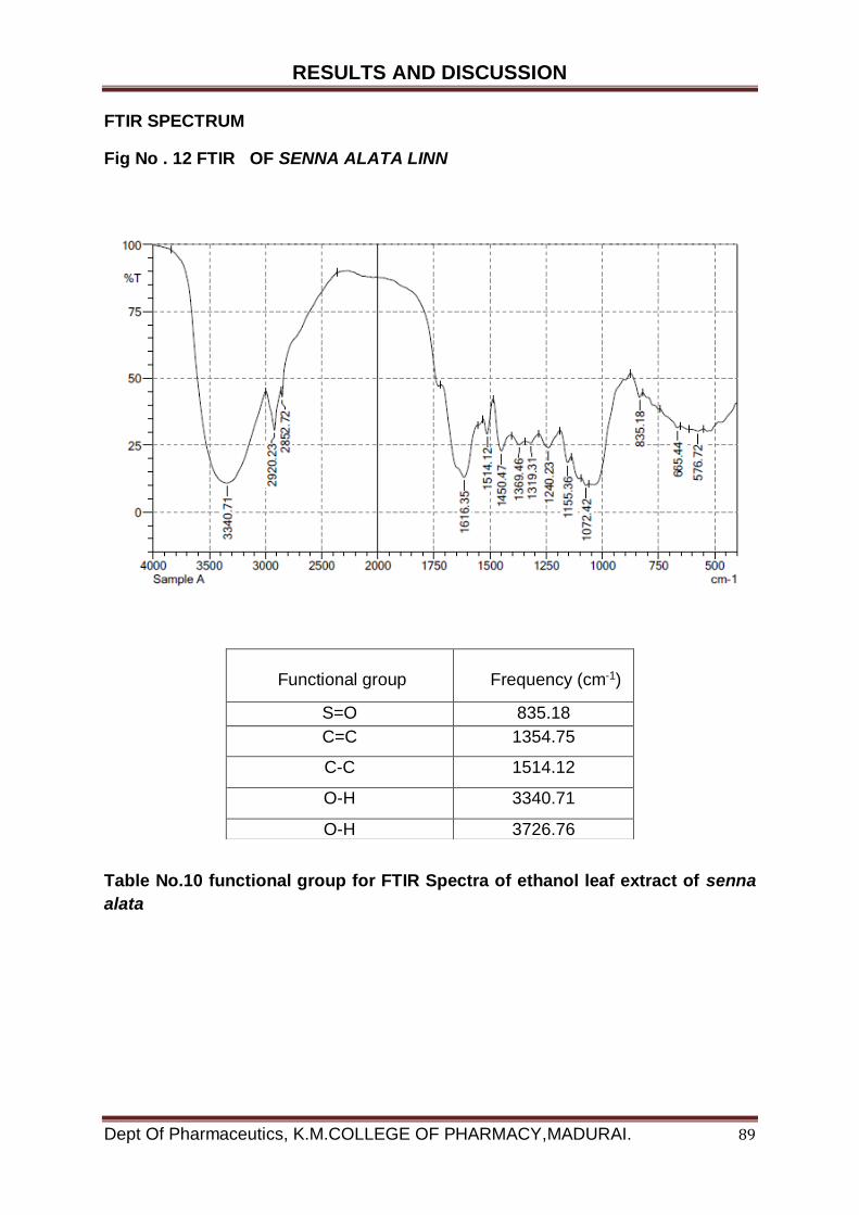
RESULTS AND DISCUSSION
Dept Of Pharmaceutics, K.M.COLLEGE OF PHARMACY,MADURAI. 89
FTIR SPECTRUM
Fig No . 12 FTIR OF SENNA ALATA LINN
Table No.10 functional group for FTIR Spectra of ethanol leaf extract of senna
alata
Functional group
Frequency (cm-1)
S=O 835.18
C=C 1354.75
C-C 1514.12
O-H 3340.71
O-H 3726.76
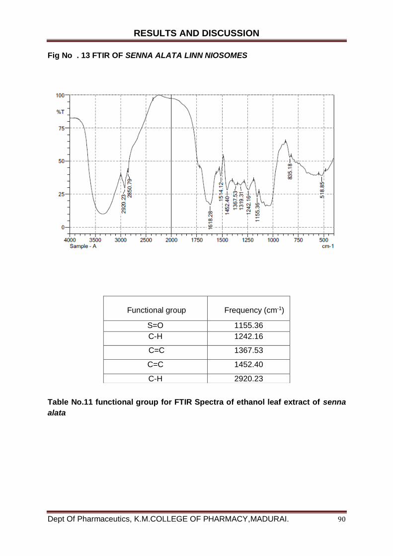
RESULTS AND DISCUSSION
Dept Of Pharmaceutics, K.M.COLLEGE OF PHARMACY,MADURAI. 90
Fig No . 13 FTIR OF SENNA ALATA LINN NIOSOMES
Table No.11 functional group for FTIR Spectra of ethanol leaf extract of senna
alata
Functional group
Frequency (cm-1)
S=O 1155.36
C-H 1242.16
C=C 1367.53
C=C 1452.40
C-H 2920.23
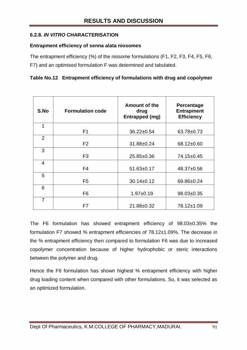
RESULTS AND DISCUSSION
Dept Of Pharmaceutics, K.M.COLLEGE OF PHARMACY,MADURAI. 91
6.2.8. IN VITRO CHARACTERISATION
Entrapment efficiency of senna alata niosomes
The entrapment efficiency (%) of the niosome formulations (F1, F2, F3, F4, F5, F6,
F7) and an optimised formulation F was determined and tabulated.
Table No.12 Entrapment efficiency of formulations with drug and copolymer
S.No
Formulation code
Amount of the
drug Entrapped (mg)
Percentage Entrapment Efficiency
1
F1
36.22±0.54
63.78±0.73
2 F2
31.88±0.24
68.12±0.60
3 F3
25.85±0.36
74.15±0.45
4 F4
51.63±0.17
48.37±0.56
5 F5
30.14±0.12
69.86±0.24
6 F6
1.97±0.19
98.03±0.35
7 F7
21.88±0.32
78.12±1.09
The F6 formulation has showed entrapment efficiency of 98.03±0.35% the
formulation F7 showed % entrapment efficiencies of 78.12±1.09%. The decrease in
the % entrapment efficiency then compared to formulation F6 was due to increased
copolymer concentration because of higher hydrophobic or steric interactions
between the polymer and drug.
Hence the F6 formulation has shown highest % entrapment efficiency with higher
drug loading content when compared with other formulations. So, it was selected as
an optimized formulation.
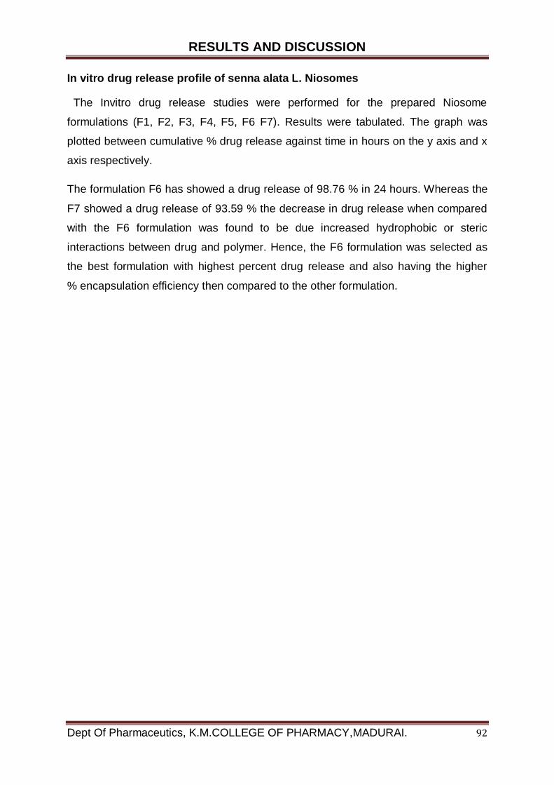
RESULTS AND DISCUSSION
Dept Of Pharmaceutics, K.M.COLLEGE OF PHARMACY,MADURAI. 92
In vitro drug release profile of senna alata L. Niosomes
The Invitro drug release studies were performed for the prepared Niosome
formulations (F1, F2, F3, F4, F5, F6 F7). Results were tabulated. The graph was
plotted between cumulative % drug release against time in hours on the y axis and x
axis respectively.
The formulation F6 has showed a drug release of 98.76 % in 24 hours. Whereas the
F7 showed a drug release of 93.59 % the decrease in drug release when compared
with the F6 formulation was found to be due increased hydrophobic or steric
interactions between drug and polymer. Hence, the F6 formulation was selected as
the best formulation with highest percent drug release and also having the higher
% encapsulation efficiency then compared to the other formulation.
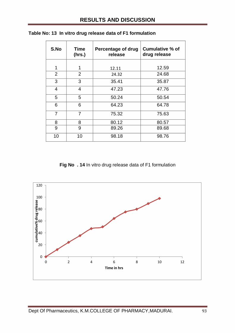
RESULTS AND DISCUSSION
Dept Of Pharmaceutics, K.M.COLLEGE OF PHARMACY,MADURAI. 93
Table No: 13 In vitro drug release data of F1 formulation
S.No
Time (hrs.)
Percentage of drug
release
Cumulative % of drug release
1
1
12.11
12.59
2 2 24.32
24.68
3 3 35.41 35.87
4 4 47.23 47.76
5 5 50.24 50.54
6 6 64.23 64.78
7 7 75.32 75.63
8 8 80.12 80.57
9 9 89.26 89.68
10 10 98.18 98.76
Fig No . 14 In vitro drug release data of F1 formulation
0
20
40
60
80
100
120
0 2 4 6 8 10 12
cum
ula
tive
% d
rug
rele
ase
Time in hrs
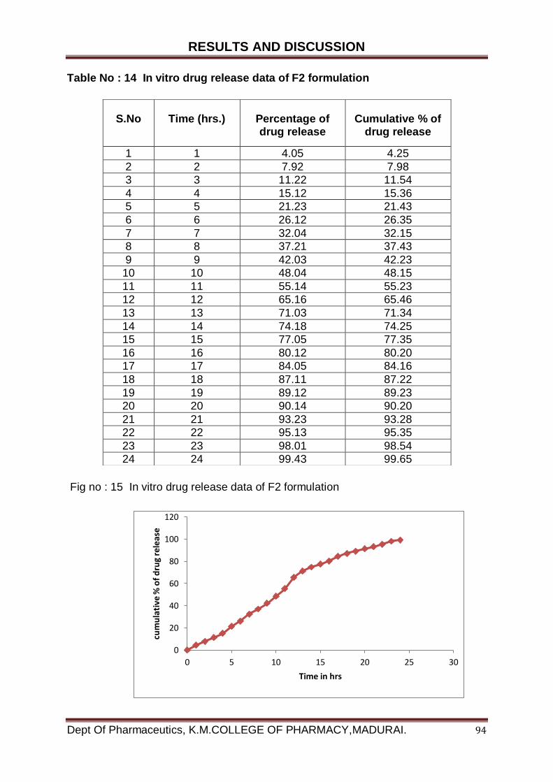
RESULTS AND DISCUSSION
Dept Of Pharmaceutics, K.M.COLLEGE OF PHARMACY,MADURAI. 94
Table No : 14 In vitro drug release data of F2 formulation
Fig no : 15 In vitro drug release data of F2 formulation
S.No
Time (hrs.)
Percentage of drug release
Cumulative % of
drug release
1 1 4.05 4.25
2 2 7.92 7.98
3 3 11.22 11.54
4 4 15.12 15.36
5 5 21.23 21.43
6 6 26.12 26.35
7 7 32.04 32.15
8 8 37.21 37.43
9 9 42.03 42.23
10 10 48.04 48.15
11 11 55.14 55.23
12 12 65.16 65.46
13 13 71.03 71.34
14 14 74.18 74.25
15 15 77.05 77.35
16 16 80.12 80.20
17 17 84.05 84.16
18 18 87.11 87.22
19 19 89.12 89.23
20 20 90.14 90.20
21 21 93.23 93.28
22 22 95.13 95.35
23 23 98.01 98.54
24 24 99.43 99.65
0
20
40
60
80
100
120
0 5 10 15 20 25 30
cum
ula
tive
% o
f d
rug
rele
ase
Time in hrs

RESULTS AND DISCUSSION
Dept Of Pharmaceutics, K.M.COLLEGE OF PHARMACY,MADURAI. 95
Table No .15 In vitro drug release data of F3 formulation
S.No Time (hrs.) Percentage of drug release
Cumulative % of drug release
1 1 12.02 12.15
2 2 17.13 17.20
3 3 24.05 24.23
4 4 32.13 32.65
5 5 39.15 39.34
6 6 47.02 47.25
7 7 55.32 55.54
8 8 63.06 63.35
9 9 72.14 72.43
10 10 79.23 79.33
11 11 86.12 86.42
12 12 90.52 90.56
13 13 97.14 97.35
14 14 99.01 99.30
Fig no : 16 In vitro drug release data of F3 formulation
0
20
40
60
80
100
120
0 5 10 15
cum
ula
tive
% o
f d
rug
rele
ase
Time in hrs
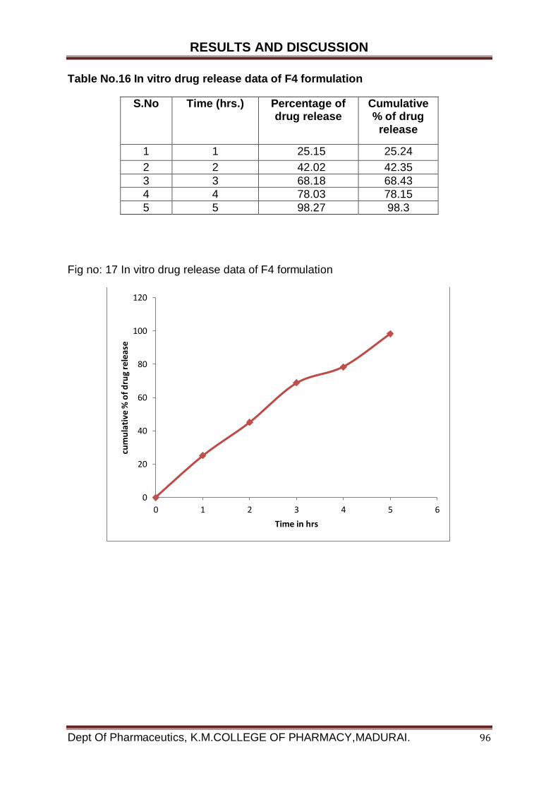
RESULTS AND DISCUSSION
Dept Of Pharmaceutics, K.M.COLLEGE OF PHARMACY,MADURAI. 96
Table No.16 In vitro drug release data of F4 formulation
S.No Time (hrs.) Percentage of drug release
Cumulative % of drug
release
1 1 25.15 25.24
2 2 42.02 42.35
3 3 68.18 68.43
4 4 78.03 78.15
5 5 98.27 98.3
Fig no: 17 In vitro drug release data of F4 formulation
0
20
40
60
80
100
120
0 1 2 3 4 5 6
cum
ula
tive
% o
f d
rug
rele
ase
Time in hrs
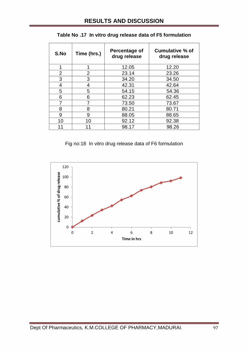
RESULTS AND DISCUSSION
Dept Of Pharmaceutics, K.M.COLLEGE OF PHARMACY,MADURAI. 97
Table No .17 In vitro drug release data of F5 formulation
S.No Time (hrs.) Percentage of drug release
Cumulative % of drug release
1 1 12.05 12.20
2 2 23.14 23.26
3 3 34.20 34.50
4 4 42.31 42.64
5 5 54.15 54.36
6 6 62.23 62.45
7 7 73.50 73.67
8 8 80.21 80.71
9 9 88.05 88.65
10 10 92.12 92.38
11 11 98.17 98.26
Fig no:18 In vitro drug release data of F6 formulation
0
20
40
60
80
100
120
0 2 4 6 8 10 12
cum
ula
tive
% o
f d
rug
rele
ase
Time in hrs
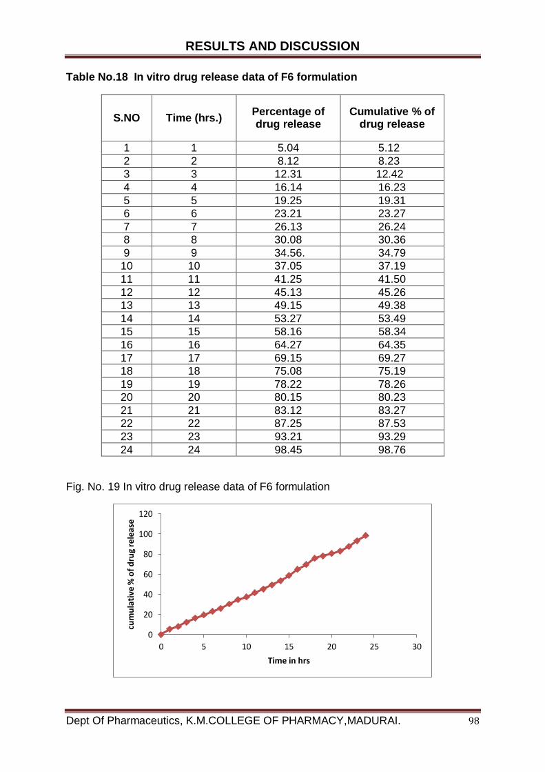
RESULTS AND DISCUSSION
Dept Of Pharmaceutics, K.M.COLLEGE OF PHARMACY,MADURAI. 98
Table No.18 In vitro drug release data of F6 formulation
S.NO Time (hrs.) Percentage of drug release
Cumulative % of drug release
1 1 5.04 5.12
2 2 8.12 8.23
3 3 12.31 12.42
4 4 16.14 16.23
5 5 19.25 19.31
6 6 23.21 23.27
7 7 26.13 26.24
8 8 30.08 30.36
9 9 34.56. 34.79
10 10 37.05 37.19
11 11 41.25 41.50
12 12 45.13 45.26
13 13 49.15 49.38
14 14 53.27 53.49
15 15 58.16 58.34
16 16 64.27 64.35
17 17 69.15 69.27
18 18 75.08 75.19
19 19 78.22 78.26
20 20 80.15 80.23
21 21 83.12 83.27
22 22 87.25 87.53
23 23 93.21 93.29
24 24 98.45 98.76
Fig. No. 19 In vitro drug release data of F6 formulation
0
20
40
60
80
100
120
0 5 10 15 20 25 30
cum
ula
tive
% o
f d
rug
rele
ase
Time in hrs
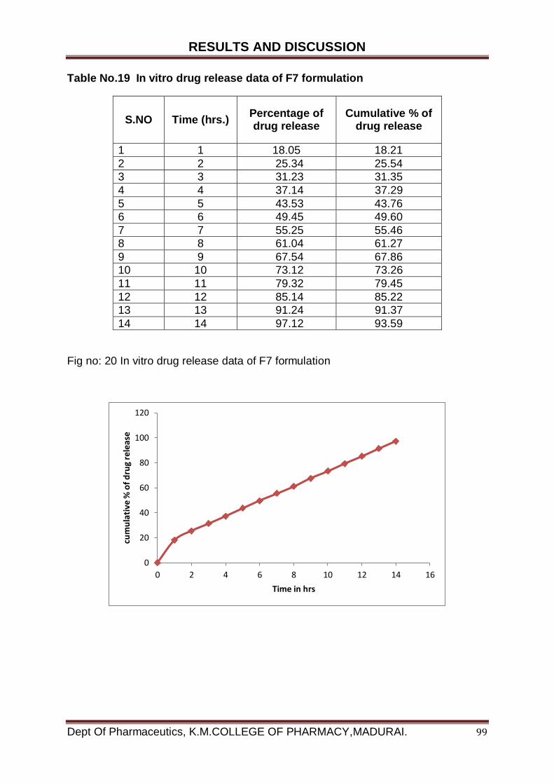
RESULTS AND DISCUSSION
Dept Of Pharmaceutics, K.M.COLLEGE OF PHARMACY,MADURAI. 99
Table No.19 In vitro drug release data of F7 formulation
S.NO Time (hrs.) Percentage of drug release
Cumulative % of drug release
1 1 18.05 18.21
2 2 25.34 25.54
3 3 31.23 31.35
4 4 37.14 37.29
5 5 43.53 43.76
6 6 49.45 49.60
7 7 55.25 55.46
8 8 61.04 61.27
9 9 67.54 67.86
10 10 73.12 73.26
11 11 79.32 79.45
12 12 85.14 85.22
13 13 91.24 91.37
14 14 97.12 93.59
Fig no: 20 In vitro drug release data of F7 formulation
0
20
40
60
80
100
120
0 2 4 6 8 10 12 14 16
cum
ula
tive
% o
f d
rug
rele
ase
Time in hrs
RESULTS AND DISCUSSION
Dept Of Pharmaceutics, K.M.COLLEGE OF PHARMACY,MADURAI. 100
6.2.9. Stability Study
The optimized formulation F6 was subjected to stability study for one month at 40 C
(refrigerator), room temperature and at 450C/75% RH. At the interval of 30 days,
samples of niosomal formulation were taken and evaluated for the entrapment
efficiency and in vitro release of drug Entrapment efficiency of optimized formulation
F6 kept at 40C shows a release rate of 95.5% after 30 days of stability study. The
entrapment efficiency of formulation got decreased on exposure to higher
temperature. The percentage entrapment of formulation kept at room temperature
and at 450C/75%RH were 92% and 81% respectively after 30 days of stability study
The in vitro release of optimize formulation F6 shows that the niosomal formulation
are more stable at 40 C (Refrigerator) when compared to room temperature and at
450C/75%RH (stability chamber). The niosomal formulation kept at 40 C showed a
cumulative release of 89.89% after 30 days of stability studies.
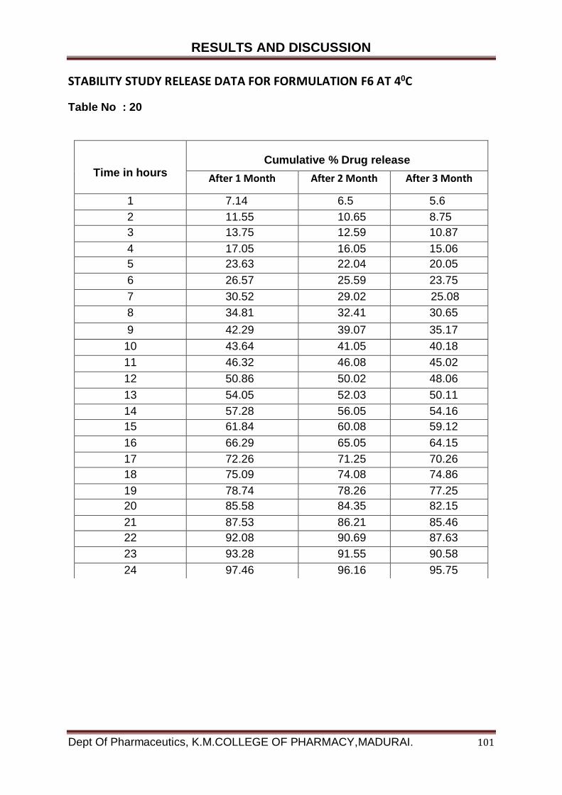
RESULTS AND DISCUSSION
Dept Of Pharmaceutics, K.M.COLLEGE OF PHARMACY,MADURAI. 101
STABILITY STUDY RELEASE DATA FOR FORMULATION F6 AT 40C
Table No : 20
Time in hours
Cumulative % Drug release
After 1 Month After 2 Month After 3 Month
1 7.14 6.5 5.6
2 11.55 10.65 8.75
3 13.75 12.59 10.87
4 17.05 16.05 15.06
5 23.63 22.04 20.05
6 26.57 25.59 23.75
7 30.52 29.02 25.08
8 34.81 32.41 30.65
9 42.29 39.07 35.17
10 43.64 41.05 40.18
11 46.32 46.08 45.02
12 50.86 50.02 48.06
13 54.05 52.03 50.11
14 57.28 56.05 54.16
15 61.84 60.08 59.12
16 66.29 65.05 64.15
17 72.26 71.25 70.26
18 75.09 74.08 74.86
19 78.74 78.26 77.25
20 85.58 84.35 82.15
21 87.53 86.21 85.46
22 92.08 90.69 87.63
23 93.28 91.55 90.58
24 97.46 96.16 95.75

RESULTS AND DISCUSSION
Dept Of Pharmaceutics, K.M.COLLEGE OF PHARMACY,MADURAI. 102
Fig No. 21 STABILITY STUDY RELEASE DATA FOR FORMULATION F6 AT 40C
`
0
20
40
60
80
100
120
0 5 10 15 20 25 30
cum
ula
tive
% d
rug
rele
ase
Time in hrs
Series1
Series2
Series3
1 month
2 month
3 month
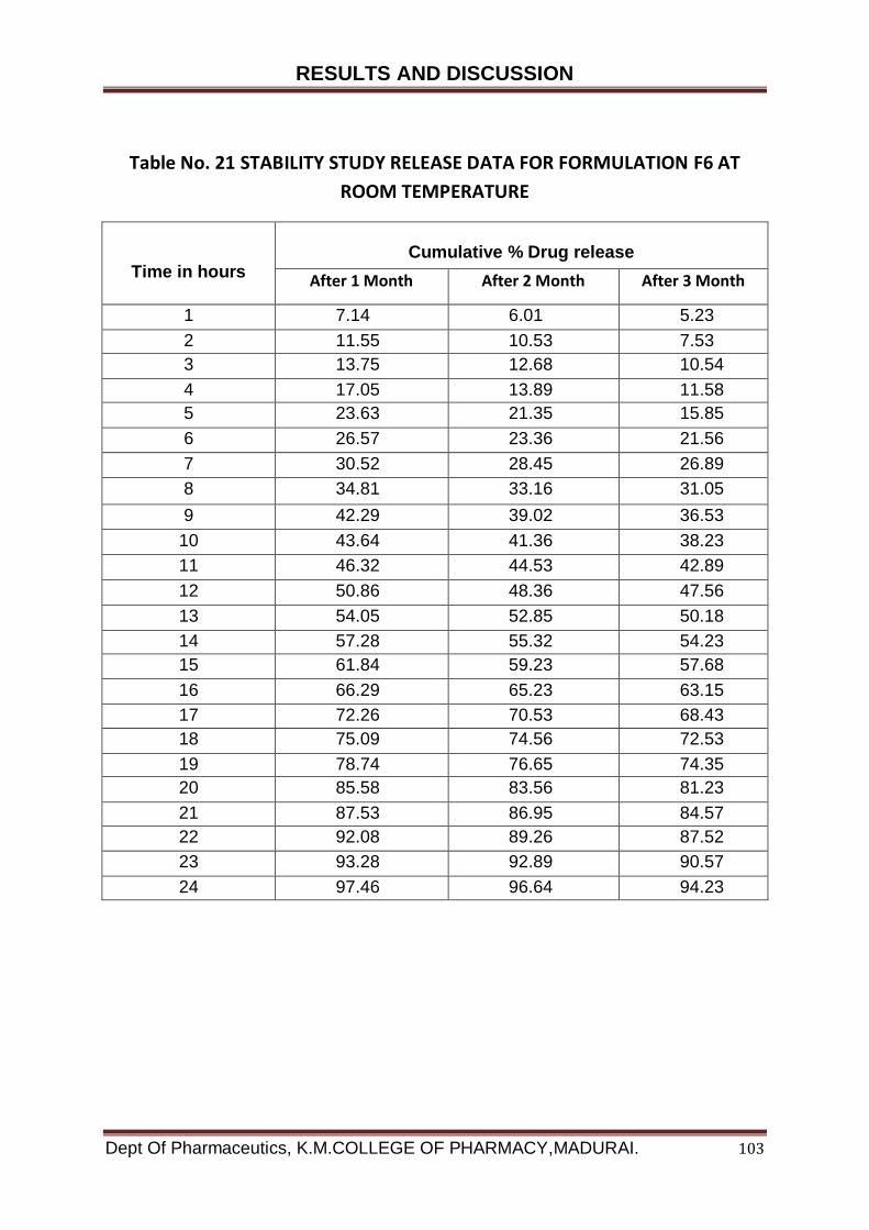
RESULTS AND DISCUSSION
Dept Of Pharmaceutics, K.M.COLLEGE OF PHARMACY,MADURAI. 103
Table No. 21 STABILITY STUDY RELEASE DATA FOR FORMULATION F6 AT
ROOM TEMPERATURE
Time in hours
Cumulative % Drug release
After 1 Month After 2 Month After 3 Month
1 7.14 6.01 5.23
2 11.55 10.53 7.53
3 13.75 12.68 10.54
4 17.05 13.89 11.58
5 23.63 21.35 15.85
6 26.57 23.36 21.56
7 30.52 28.45 26.89
8 34.81 33.16 31.05
9 42.29 39.02 36.53
10 43.64 41.36 38.23
11 46.32 44.53 42.89
12 50.86 48.36 47.56
13 54.05 52.85 50.18
14 57.28 55.32 54.23
15 61.84 59.23 57.68
16 66.29 65.23 63.15
17 72.26 70.53 68.43
18 75.09 74.56 72.53
19 78.74 76.65 74.35
20 85.58 83.56 81.23
21 87.53 86.95 84.57
22 92.08 89.26 87.52
23 93.28 92.89 90.57
24 97.46 96.64 94.23

RESULTS AND DISCUSSION
Dept Of Pharmaceutics, K.M.COLLEGE OF PHARMACY,MADURAI. 104
STABILITY STUDY RELEASE DATA FOR FORMULATION F6 AT ROOM
TEMPERATURE
Fig no: 22
0
20
40
60
80
100
120
0 5 10 15 20 25 30
cum
ula
tive
% o
f d
rug
rele
ase
Time in hrs
Series1
Series2
Series3
1 month
2 month
3 month
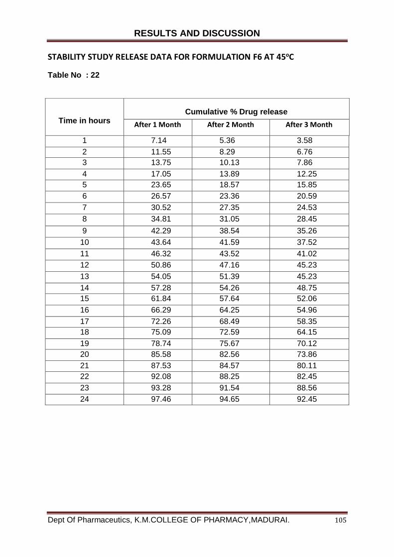
RESULTS AND DISCUSSION
Dept Of Pharmaceutics, K.M.COLLEGE OF PHARMACY,MADURAI. 105
STABILITY STUDY RELEASE DATA FOR FORMULATION F6 AT 45oC
Table No : 22
Time in hours
Cumulative % Drug release
After 1 Month After 2 Month After 3 Month
1 7.14 5.36 3.58
2 11.55 8.29 6.76
3 13.75 10.13 7.86
4 17.05 13.89 12.25
5 23.65 18.57 15.85
6 26.57 23.36 20.59
7 30.52 27.35 24.53
8 34.81 31.05 28.45
9 42.29 38.54 35.26
10 43.64 41.59 37.52
11 46.32 43.52 41.02
12 50.86 47.16 45.23
13 54.05 51.39 45.23
14 57.28 54.26 48.75
15 61.84 57.64 52.06
16 66.29 64.25 54.96
17 72.26 68.49 58.35
18 75.09 72.59 64.15
19 78.74 75.67 70.12
20 85.58 82.56 73.86
21 87.53 84.57 80.11
22 92.08 88.25 82.45
23 93.28 91.54 88.56
24 97.46 94.65 92.45

RESULTS AND DISCUSSION
Dept Of Pharmaceutics, K.M.COLLEGE OF PHARMACY,MADURAI. 106
STABILITY STUDY RELEASE DATA FOR FORMULATION F6 AT 450C
Fig No : 23
0
20
40
60
80
100
120
0 5 10 15 20 25 30
cum
ula
tive
% o
f d
rug
rele
ase
Time
Series1
Series2
Series3
1 month
2 month
3 month
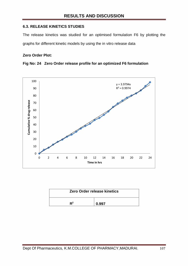
RESULTS AND DISCUSSION
Dept Of Pharmaceutics, K.M.COLLEGE OF PHARMACY,MADURAI. 107
6.3. RELEASE KINETICS STUDIES
The release kinetics was studied for an optimised formulation F6 by plotting the
graphs for different kinetic models by using the in vitro release data
Zero Order Plot:
Fig No: 24 Zero Order release profile for an optimized F6 formulation
Zero Order release kinetics
R2
0.997
y = 3.9794xR² = 0.9974
0
10
20
30
40
50
60
70
80
90
100
0 2 4 6 8 10 12 14 16 18 20 22 24
Cu
mu
lati
ve %
dru
g re
leas
e
Time in hrs
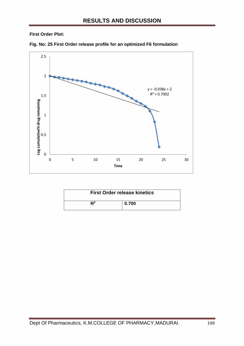
RESULTS AND DISCUSSION
Dept Of Pharmaceutics, K.M.COLLEGE OF PHARMACY,MADURAI. 108
First Order Plot:
Fig. No: 25 First Order release profile for an optimized F6 formulation
First Order release kinetics
R2 0.700
y = -0.038x + 2R² = 0.7002
0
0.5
1
1.5
2
2.5
0 5 10 15 20 25 30
Log
cum
ula
tive
% d
rug
rem
ain
ing
Time

RESULTS AND DISCUSSION
Dept Of Pharmaceutics, K.M.COLLEGE OF PHARMACY,MADURAI. 109
Higuchi Plot:
Fig No: 26 Higuchi release profile for an optimized F6 formulation
Higuchi release kinetics
R2 0.956
y = 24.963x - 34.421R² = 0.9557
-20
0
20
40
60
80
100
120
0 1 2 3 4 5 6
Cu
mu
lati
ve %
dru
g re
leas
e
sqrt of time in hrs
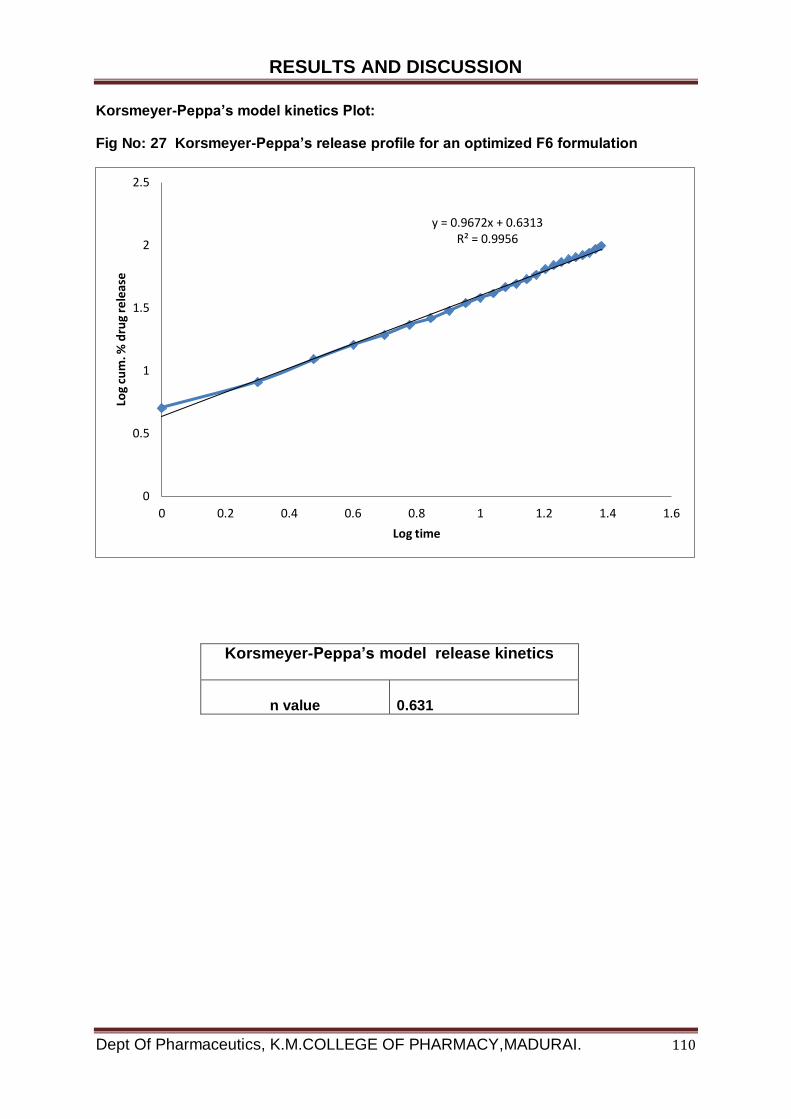
RESULTS AND DISCUSSION
Dept Of Pharmaceutics, K.M.COLLEGE OF PHARMACY,MADURAI. 110
Korsmeyer-Peppa’s model kinetics Plot:
Fig No: 27 Korsmeyer-Peppa’s release profile for an optimized F6 formulation
Korsmeyer-Peppa’s model release kinetics
n value
0.631
y = 0.9672x + 0.6313R² = 0.9956
0
0.5
1
1.5
2
2.5
0 0.2 0.4 0.6 0.8 1 1.2 1.4 1.6
Log
cum
. % d
rug
rele
ase
Log time
RESULTS AND DISCUSSION
Dept Of Pharmaceutics, K.M.COLLEGE OF PHARMACY,MADURAI. 111
Zero Order plot:
Zero order plot of a freeze dried formulation F4 was to be linear with a
regression value of 0.997, which signifies that the drug was released in a controlled
manner from the nanoparticles during the release study.
First Order plot:
First Order plot was made by plotting log remaining cumulative % drug
release against time and the regression value was found to be 0.700 which indicates
that drug release was not followed the first order rate kinetics.
Higuchi plot:
Higuchi plot was found to be linear with a regression value of 0.956, which
indicates that diffusion was one of the mechanisms of the drug release from
nanoparticles matrices.
Korsmeyer-Peppa’s Plot:
The type of in vitro mechanisms of drug release was best explained by
Korsmeyer – Peppa’s plot. The plot was found to be linear with R2 value 0.996 and
diffusion exponent ‘n’ value was 0.631 According to Korsmeyer-Peppa’s equation,
mechanisms of drug based on ‘n’ which indicates that mechanism of drug release
from copolymer matrices was followed non Fickian diffusion.
So, the kinetic studies of an optimized F6 formulation with different kinetic models
has showed that the senna alata L. release profile was best fitted with zero order plot
and release mechanism was found to be non Fickian diffusion based on ‘n’ value
from Korsmeyer-Peppa’s plot.
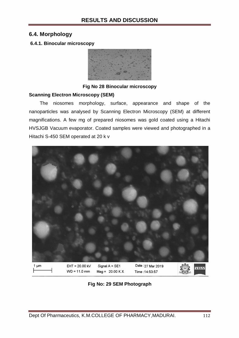
RESULTS AND DISCUSSION
Dept Of Pharmaceutics, K.M.COLLEGE OF PHARMACY,MADURAI. 112
6.4. Morphology
6.4.1. Binocular microscopy
Fig No 28 Binocular microscopy
Scanning Electron Microscopy (SEM)
The niosomes morphology, surface, appearance and shape of the
nanoparticles was analysed by Scanning Electron Microscopy (SEM) at different
magnifications. A few mg of prepared niosomes was gold coated using a Hitachi
HVSJGB Vacuum evaporator. Coated samples were viewed and photographed in a
Hitachi S-450 SEM operated at 20 k v
Fig No: 29 SEM Photograph
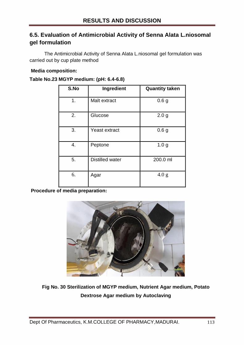
RESULTS AND DISCUSSION
Dept Of Pharmaceutics, K.M.COLLEGE OF PHARMACY,MADURAI. 113
6.5. Evaluation of Antimicrobial Activity of Senna Alata L.niosomal
gel formulation
The Antimicrobial Activity of Senna Alata L.niosomal gel formulation was
carried out by cup plate method
Media composition:
Table No.23 MGYP medium: (pH: 6.4-6.8)
S.No Ingredient Quantity taken
1. Malt extract 0.6 g
2. Glucose 2.0 g
3. Yeast extract 0.6 g
4. Peptone 1.0 g
5. Distilled water 200.0 ml
6. Agar 4.0 g
Procedure of media preparation:
Fig No. 30 Sterilization of MGYP medium, Nutrient Agar medium, Potato
Dextrose Agar medium by Autoclaving

RESULTS AND DISCUSSION
Dept Of Pharmaceutics, K.M.COLLEGE OF PHARMACY,MADURAI. 114
MGYP medium:
The respective quantity of Malt extract, Glucose, Yeast extract, Peptone, Agar
were added in 200ml of distilled water.
Heat to boiling to dissolve the medium completely
Then sterilized by autoclaving 15 lb at 1210C for 15 min.
The respective medium was sterilized by autoclaving at 1210C (15lb/ln2).for 15
min. and medium was transferred aseptically into sterilized glass Petri
plates. The plates were left at room temperature to allow solidification. 15µl of
inoculums of the bacteria was transferred to respective Petri plate. Four wells
of 6mm diameter were made using a sterile borer. The different
concentrations of drug samples were added with a sterile micropipette to each
of the cups. The plates were maintained on sight place for 2 hours to allow the
diffusion of the solution into the medium. The Petri dishes are kept inverted
position in incubator at 370C ±10C for 24 hours. The diameter of zone of
inhibition surrounding each of the wells was recorded.
Table No.24 Nutrient Agar medium: (pH: 7-7.5)
S.No Ingredient Quantity taken
1. Beef extract 2.0 g
2. NaCl 1.0 g
3. Peptone 2.0 g
4. Distilled water 200.0 ml
5. Agar 4.0 g

RESULTS AND DISCUSSION
Dept Of Pharmaceutics, K.M.COLLEGE OF PHARMACY,MADURAI. 115
Nutrient Agar medium:
The respective quantity of Beef extract, NaCl, Peptone, Agar adds in 200ml of
distilled water.
Heat to boiling to dissolve the medium completely
Then sterilized by autoclaving 15 lb at 1210C for 15 min.
The respective medium was sterilized by autoclaving at 1210C (15lb/ln2).for 15 min.
and medium was transferred aseptically into sterilized glass Petri plates. The plates
were left at room temperature to allow solidification. 15µl of inoculums of the bacteria
was transferred to respective Petri plate. Four wells of 6mm diameter were made
using a sterile borer. The different concentrations of drug samples were added with a
sterile micropipette to each of the cups. The plates were maintained on sight place
for 2 hours to allow the diffusion of the solution into the medium. The Petri dishes are
kept inverted position in incubator at 370C ±10C for 24 hours. The diameter of zone
of inhibition surrounding each of the wells was recorded.
Table No.25 Potato Dextrose Agar medium: (pH: 6-7)
S.No Ingredient Quantity taken
1. Pieces of potato 40.0 g
2. Dextrose 4.0 g
3. Yeast extract 0.02 g
4. Distilled water 200.0 ml
5. Agar 4.0 g
Potato Dextrose Agar medium:
40.0 g of peeled potatoes are cut into small pieces and suspended in 200.0 ml
of distilled water.

RESULTS AND DISCUSSION
Dept Of Pharmaceutics, K.M.COLLEGE OF PHARMACY,MADURAI. 116
Steamed for 30 min. decant the extract or filter through muslin cloth and make
the final volume to 200.0 ml.
Add 4.0 g of dextrose, 0.02 g of yeast extract and 4.0 g of agar.
The respective medium was sterilized by autoclaving at 1210C (15lb/ln2).for 15 min.
and medium was transferred aseptically into sterilized glass Petri plates. The plates
were left at room temperature to allow solidification..15µl of inoculums of the bacteria
and fungi was transferred to respective Petri plate. Four wells of 6mm diameter were
made using a sterile borer. The different concentrations of drug samples were added
with a sterile micropipette to each of the cups. The plates were maintained on sight
place for 2 hours to allow the diffusion of the solution into the medium. The Petri
dishes are kept inverted position in incubator at 280C for 48 hours. The diameter of
zone of inhibition surrounding each of the wells was recorded.
Preparation of inoculums:
Fungal inoculum:
The inoculums was prepared by inoculate the Spores of in Potato dextrose broth
and Candida albicans, Aspergillus niger MGYP broth and incubated at 370C for 24
hrs.
Bacterial inoculums:
The inoculums was prepared by inoculate the Spores of Escherichia coli,
pseudomonas aureus, Staphylococcus aureus in Nutrient broth and incubated at
370C for 24 hrs.
Preparation of sample:
Stock solution: 100mg of drug is dissolved in 10ml of methanol to produce
10000µg concentration.
Sample: From this stock solution different dilution were prepared in the
concentration rang of 1,2,3,4,5,6,7,8,9,10 mg/ml in 10ml of volumetric flask.
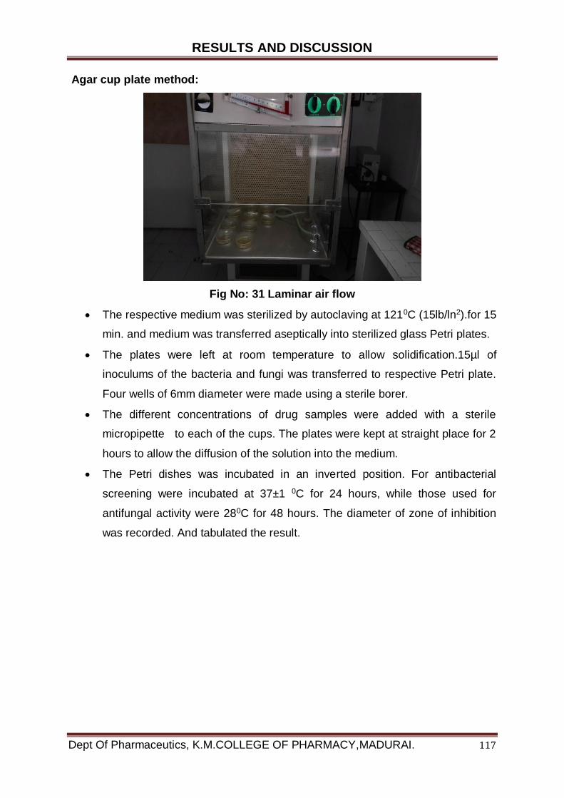
RESULTS AND DISCUSSION
Dept Of Pharmaceutics, K.M.COLLEGE OF PHARMACY,MADURAI. 117
Agar cup plate method:
Fig No: 31 Laminar air flow
The respective medium was sterilized by autoclaving at 1210C (15lb/ln2).for 15
min. and medium was transferred aseptically into sterilized glass Petri plates.
The plates were left at room temperature to allow solidification.15µl of
inoculums of the bacteria and fungi was transferred to respective Petri plate.
Four wells of 6mm diameter were made using a sterile borer.
The different concentrations of drug samples were added with a sterile
micropipette to each of the cups. The plates were kept at straight place for 2
hours to allow the diffusion of the solution into the medium.
The Petri dishes was incubated in an inverted position. For antibacterial
screening were incubated at 37±1 0C for 24 hours, while those used for
antifungal activity were 280C for 48 hours. The diameter of zone of inhibition
was recorded. And tabulated the result.

RESULTS AND DISCUSSION
Dept Of Pharmaceutics, K.M.COLLEGE OF PHARMACY,MADURAI. 118
Determination of Zone of Inhibition of Senna alata niosomal gel by agar Cup
Plate method:
Fungal and bacterial strain:
The bacterial and fungal strains employed in the study were obtained from the
National collection of industrial microorganisms (NCIM) Pune.
Table No.26 Fungal strain:
S. No. Name of organism Maintenance Media
1. Aspergillus niger Potato Dextrose Agar
2. Candida albicans MGYP
3. Candida albicans MGYP
Table No.27 Bacterial strain:
S. No. Name of organism Maintenance Media
1. Pseudomonas aeruginosa Nutrient Agar
2. Staphylococcus aureus Nutrient Agar
3. Escherichia coli Nutrient Agar

RESULTS AND DISCUSSION
Dept Of Pharmaceutics, K.M.COLLEGE OF PHARMACY,MADURAI. 119
Antifungal and Antibacterial Evaluation
Table No. 28 Antifungal activity
Name of the fungal pathogens
Senna Alata L. Diameter of zone of inhibition (mm)
Concentration (mg/mL)
25mg (mg/mL)
50
(mg/mL)
75
(mg/mL)
100
(mg/mL)
Aspergillus niger
16
22
23
25
Candida albicans 8 12 14 17
Candida albicans 14 16 18 21
Fluconazole (500 μg/ml)
18
22
23
26
Fig No: 32 Antifungal activity Determination of Zone of Inhibition of Senna
alata niosomal gel by agar Cup Plate method:
Aspergillus niger
Candida albicans Candida albicans
RESULTS AND DISCUSSION
Dept Of Pharmaceutics, K.M.COLLEGE OF PHARMACY,MADURAI. 120
Table No. 29 Antibacterial activity
Name of the Bacterial pathogens
Senna Alata L. Diameter of zone of inhibition
(mm) Concentration (mg/mL)
25mg (mg/mL)
50
(mg/mL)
75
(mg/mL)
100
(mg/mL)
Escherichia coli
8
11
13
15
Staphylococcus aureus 9.55
12
16
19
Pseudomonas aeruginosa
9
12 14 16
Ciprofloxacin 50µg
20
25
28
32
Fig No: 33 Antibacterial activity Determination of Zone of Inhibition of Senna
alata niosomal gel by agar Cup Plate method:
Escherichia coli
Staphylococcus aureus Pseudomonas aeruginosa
RESULTS AND DISCUSSION
Dept Of Pharmaceutics, K.M.COLLEGE OF PHARMACY,MADURAI. 121
Discussion:
Traditionally medicinal plants have been used for many years as topical and internal
preparation in the treatment of fungal and bacterial disease. Many investigations are
being carried out throughout the world to discover plant products to inhibit the
clinically important fungal pathogens.
The World Health Organization (WHO) estimates that plant extracts or their active
constituents are used as folk medicine in traditional therapies of 80% of the world's
population. Each and every country has their own indigenous system of medicine
and many of the formulations were not validated for their purported claims and it is
the need of the hour to scientifically prove the claimed effects to spread those
systems to gain acceptance at the global level.
This study shows that Senna alata has antibacterial & Antifungal activity against
Staphylococcus aureus, and has high potential as antibacterial agent when
formulated as for topical use and could therefore explain the successes claimed in
the folk use of the plant in the treatment of common skin conditions.
The potency of the Senna alata niosome gel against the bacterial and fungal
commonest etiologic agent of boils, carbuncles, infantile impetigo and wounds.
The senna alata niosome gel is better penetrating to the skin & soft smoothening,
active against bacterial and fungal
The results also revealed that the extracts incorporated into the gel bases showed
better activity than that of the crude extract of Senna alata.
This implied that there might have been better diffusion of drug for the niosome gel
than for the crude extract.
Skin disease commonly found on the hands, face and in deep layers of the skin
CONCLUSION
Dept Of Pharmaceutics, K.M.COLLEGE OF PHARMACY,MADURAI. 122
7. CONCLUSION
An effort was made to formulate the Senna alata Niosomes and
incorporate the Niosomes into the gel.
From the results of the present experiments it may be concluded that
formulation F6 showing high percentage of entrapment and desired sustained
release of Senna alata Niosome gel. Hence F6 formulation was optimized one.
The optimized formulation F6 was found to follow zero order release
pattern which was revealed by the linearity shown from the plot of time versus
concentration.
Niosomes were stable for 4 weeks at 40 C and affirm the drug leakage
increased the higher temperature. Optimized formulation was released from the
Niosomal gel formulation in a sustained manner for 24 Hours.
The present work revels that the senna alata niosomal gel produces
anticipated synergistic effect against most of the tested fungal strain.
So, my work revels that the formulated Antimicrobial niosomal gel will
shows the benefit to society against opportunistic fungal and bacterial infection.
Antimicrobial activity indicates that the gel preparation of the optimized
formulation F6 Thus, the aim of the project was achieved by optimizing the
formulation Parameters.
Antimicrobial sensitivity screening, the ethanolic extract of Senna alata
niosomal gel showed excellent activity and Most of these organisms are natural
flora of the skin.
Hence, plant based medicines that inhibit their growth without harming the
host represent potential therapeutic agent.

BIBLIOGRAPHY
Dept Of Pharmaceutics, K.M.COLLEGE OF PHARMACY,MADURAI.
1. Aarti jag tap, Formulation and Evaluation of niosome entrapped pentoxifylline
using in vivo bronchodilator activity in guniea pigs ; India journal of
pharmaceutics 2001;63(1);49-54.
2. Aarti jag tap, Study of antiparkinsonisms activity of plain and niosomal
pentoxifylline; India journal of pharmaceutics 2001; 63 (3); 239-243.
3. Abbas paradakht, Invitro study of polyethylene alkyl ether niosomes for
delivery of insulin; International journal of pharmaceutics 2007; 328; 130-141.
4. Bertrand, N.; Wu, J.; Xu, X.; Kamaly, N.; Farokhzad, O.C., Cancer
nanotechnology: the impact of passive and active targeting in the era of
modern cancer biology. Advanced drug delivery reviews 2014; 66: 2-25.
5. Ajith Singh design and development of niosomal delivery system; Pharma
time, 2004; 36: 11-13.
6. Anna m.fadda niosomes carries for Tretinoin III. Study into the in vitro
cutaneous delivery of vesicle incorporated Tretinoin; International journal of
pharmaceutics, 2006; 311; 11-19.
7. Biju S.S. vesicular system; an over view Indian journal of pharmaceutical
science, 2006; 68(2); 141-153.
8. Hu C., Rhodes D.G., Proniosomes: a novel drug carrier preparation. Int. J.
Pharm. 1999; 185: 23-35.
9. Yoshioka T., Stemberg B.,Florence A.T., Preparation and properties of
vesicles (niosomes) of sorbitan monoesters (Span 20, 40, 60, and 80) and a
sorbitan triester (Span 85). Int. J Pharm.1994; 105: 1-6.
10. Yoshida H., Lehr, C.M., Kok W., Junginger H.E., Verhoef J.C., Bouwistra J.A,.
Niosomes for oral delivery of peptide drugs. J.Control Rel.1992; 21: 145-153.
11. http//en.wikipedia.org/wiki/Niosomes, structure of niosomes. -
12. Roberson A., Cummings J., Wilmot N., Florence AT., The distribution of
doxorubicin in mice following administration in niosomes. J. Pharm.
Pharmacol. 1988; 40: 337-342.
13. Satturwar, P.M., Fulzele, S.V., Nande, V.S., Khandare, J.N., Formulation and
evaluation of ketoconazole Niosomes. Indian J.Pharm, 2002; 64 (2): 155-158.
14. Vyas S.P., Khar R, K., Niosomes Targeted and Controlled Drug delivery,
2008; 249 – 279.
BIBLIOGRAPHY
Dept Of Pharmaceutics, K.M.COLLEGE OF PHARMACY,MADURAI.
15. Gibaldi .M and Perrier D; Pharmacokinetics, second edition, New York,
Marcel Dekker, Inc., 1982; 127-134.
16. Namdeo, A., Jain, N.K., Niosomal delivery of 5-fluorouracil. J.
Microencapsulate. 1999; 16 (6): 731 – 740.
17. Bhaskaran S., Panigrahi L., Formulation and Evaluation of Niosomes using
Different Non-ionic Surfactant. Ind J Pharm Sci. 2002; 63: 1-6.
18. Balasubramaniam A., Formulation and In-Vivo Evaluation of Niosome
Encapsulated Daunorubicin Hydrochloride. Drug Dev and Ind.Pharm.2002;
3(2): 1181-84. 21
19. Schreiber H., Liposomes and niosomes as topical drug carriers: dermal and
transdermal delivery. J. Controlled Release. 1985; 30: 863-868.
20. Buck ton G., Harwood, Interfacial phenomena in Drug Delivery and Targeting
Academic Publishers, Switzerland. 1995; 154-155.
21. Hunter C.A., Dolan T.F., Coombs G.H., Baillie A.J, Vesicular systems
(niosomes and liposomes) for delivery of sodium stribogluconate in
experimental murine visceral Leishmaniasis. M.pharm. Pharmacol. 1988; 40
(3): 161-165.
22. Bairwa N. K., Choudhary Deepika., Pro niosome: A review, Asian Journal of
Biochemical and Pharmaceutical Research. 2011; 2 (1): 690-694.
23. Suzuki K., Sokan K., The Application of Liposome’s to Cosmetics. Cosmetic
and Toiletries. 1990; 105: 65-78.
24. Chauhan S and Laurence MJ. The Preparation of Polyoxyethylene Containing
Non-Ionic Surfactant Vesicles. J Pharm. Pharmacol.1989; 41:6.
25. Yoshioka T, Sternberg B and Florence AT. Preparation and Properties of
Vesicles (Niosomes) of Sorbitan Monoesters (Span 20, 40, 60, and 80) and a
Sorbitan Triester (Span 85). Int J Pharm. 1994; 105:1-6.
26. Gayatri DS, Venkatesh P and Udupa N. Niosomal Sumatriptan Succinate for
Nasal Administration. Int J Pharm Sci. 2000; 62 (6):479-481.
27. Hu C and Rhodes D.G. Proniosomes: a novel drug carrier preparation. Int J
Pharm.1999; 185:23-35.
28. B.L. Silver Ed., the Physical Chemistry of Membranes, Alan & Unwin and
Soloman Press. New York, USA.1985; 209-230.
BIBLIOGRAPHY
Dept Of Pharmaceutics, K.M.COLLEGE OF PHARMACY,MADURAI.
29. Ozer AY. A Novel Drug Delivery System Non -ionic Surfactant Vesicles. Euro
J Pharm Bio pharm.1991; 75.
30. Uchegbu FI and Vyas PS. Non-Ionic Surfactant Based Vesicles (Niosomes) in
Drug Delivery. Int J Pharm.1998; 33.
31. Hunter CA.Vesicular System (Niosomes and Liposomes) for Delivery of
Sodium Stribogluconate in Experimental Murine Visceral Leishmaniasis. J
Pharm. Pharmacol. 1988; 161.
32. Nasseri B and Florence AT. Microtubules Formed by Capillary Extrusion and
Fusion of Surfactant Vesicles. Int J of Pharm.2003; 91.
33. Ozer AY. A Novel Drug Delivery System Non -ionic Surfactant Vesicles. Euro
J Pharm Bio pharm.1991; 75.
34. Bhaskaran S and Panigrahi L. Formulation and Evaluation of Niosomes using
Different Non -ionic Surfactant. Ind J Pharm Sci. 2002; 63.
35. Arunothayanun P. The effect of Processing Variables on the Physical
Characteristics of Non -ionic Surfactant Vesicles (Niosomes) formed from
Hexadecyl Diglycerol ether. Int J Pharm. 2000; 7.
36. Stafford S, Ballie AJ and Florence AT. Drug effect on the Size of Chemically
Defined Non -ionic Surfactant Vesicles. J Pharm Pharmacol.1988; 26.
37. Uchegbu IF and Florence AT. Non -Ionic Surfactant Vesicles (niosomes)
Physical and Pharmaceutical chemistry. Adv. Colloid Interface Sci.1995; 1.
38. Manosroi A. Characterization of Vesicles Prepared with Various Non-ionic
Surfactants mixed with Cholesterol. Colloids and surfaces Biointerfaces.2003;
129.
39. Balasubramaniam A. Formulation and In-Vivo Evaluation of Niosome
Encapsulated Daunorubicin Hydrochloride. Drug Dev and Ind Pharm.2002;
1181.
40. Shahiwala A and Misra A. Studies in Topical Application of Niosomal
Entrapped Nimesulide. J Pharma Sci. 2002; 220.
41. Baillie AJ. The preparation and Properties of Niosomes-Non -ionic Surfactant
Vesicles. J Pharm. Pharmacol.1985; 863.
42. Schreier H., Liposomes and niosomes as topical drug carriers: dermal and
transdermal delivery. J. Controlled Release.1985; 30: 863-868.
BIBLIOGRAPHY
Dept Of Pharmaceutics, K.M.COLLEGE OF PHARMACY,MADURAI.
43. Buckton G., Harwood, Interfacial phenomena in Drug Delivery and Targeting
Academic Publishers, Switzerland. 1995, 154-155.
44. Bairwa N. K., Choudhary Deepika., Proniosomes: A review, Asian Journal of
Biochemical and Pharmaceutical Research. 2011; 2 (1): 690-694.
45. Suzuki K., Sokan K., the Application of Liposome’s to Cosmetics. Cosmetic
and Toiletries. 1990; 105: 65-78.
46. Chandraprakash KS, Udupa N., Uma Devi P., Pillai GK., Formulation and
Evaluation of Methotrexate Niosomes. Ind J Pharm.Sci.1992; 54(5): 197.
47. V.C.Okore, A A.Attama, K.C.Ofokansl, C.O.Esimome1 and E.B.Onuigbo
formulation and evaluation of niosomes. Indian journal of pharmaceutical
science, 2011; 73, (3):323-328.
48. Bulto, T., F.H.Maskel,and, G.Fisseha.Skin Lesions in Resettled and
Indigenous Populations in Gambela, with Special Emphasis on the
Epidemiology of Tropical Ulcer. “Ethiopian Medical Journal 1993; 31: 75–82.
49. Carapetis, J. R., B. J. Currie, and E. L. Kaplan. 1999. “Epidemiology and
Prevention of Group a Streptococcal Infection: Acute Respiratory Tract
Infections, Skin Infections, and Their Sequelae at the Close of the 20th
Century. Clinical Infectious Diseases 1999; 28: 205–10.
50. Daroczy, J. Antiseptic Efficacy of Local Disinfecting PovidoneIodine (Beta
dine) Therapy in Chronic Wounds of Lymphedematous Patients.
“Dermatology 2002; 204: 75–78.
51. Eells, L. D., P. M. Mertz, Y. Piovanetti, G. M. Pekoe, and W. H. Eaglestein..
“Topical Antibiotic Treatment of Impetigo with Mupirocin.” Archives of
Dermatology 1986; 122: 1273–76.
52. Lopez-Gomez, S., A. Del Palacio, J. Van Cutsem, M. Soledad Cuetara, L.
Iglesias, and A. Rodriguez-Noriega. 1994. “Itraconazole versus Griseofulvin in
the Treatment of Tinea Capitis: A Double-Blind Randomized Study in
Children.” International Journal of Dermatology 1994; 33: 743–47.
53. Baan siddhi J, Pechaaply D, 1988. Botanical report of some Thai medicinal
plants, part I, Bangkok: Department of Medical Sciences.1988.; p.8-9.
54. Ali-Emmanuel NM, Moudachirou AJ, Akakpo J, Quetin-Leclercq Activities in
vitro antibacterial Cassia alata, Lantana camara and Mitracarpus scaber on

BIBLIOGRAPHY
Dept Of Pharmaceutics, K.M.COLLEGE OF PHARMACY,MADURAI.
Dermatophilus congolensis isolated in Benin. Revue Élev. Méd. V ét. Pays
Trop.,2003; 55: 183-187
55. Anandan R, Jayakar B, Manavalan R, Hepatoprotective Activity of the Infusion
of the Dried Leaves of Cassia Alata Linn. Biomed. Pharmacol. J.; 2009;
2(1):113-116.
56. Archana P, Samantha T, Mahitha B, Chamundeswari, Rama SN Preliminary
phytochemical screening from leaf and seed extracts of Senna alata. Roxb-an
Ethno medicinal plant. International Journal of Pharmaceutical and Biological
Research .2012; 3(3): 82-89.
57. Blytt HJ, Guscar TK Butler LG Ant nutritional effects and ecological
significance of dietary condensed tannins may not be due to binding and
inhibiting digestive enzymes. J Chem Ecol, 1988; 14: 1455-1465.
58. Dakuyo ZP, Sekhoacha MP, Matsabisa MG, Nikièma JB.,Ouedraogo JB,
Ouedraogo GA.Evaluation of the antiplasmodial activity and lethality of the
leaf extract of Cassia alata. (Fabaceae). Pak. J. Biol.Sci., 2016; 19: 171-
178.2016.
59. Demirezer LO, Kuruüzüm-Uz A, Bergere I, Schiewe HJ, Zeeck A,The
structures of antioxidant and cytotoxic agents from natural source:
Anthraquinones and tannins from roots of Rumex patientia. Phytochemistry,
2001; 58: 1213-1217.
60. Doughari JH, Okafor B, Antimicrobial activity of Senna alata Linn. East and
Central African Journal of Pharmaceutical Sciences, 2007; 1: 1721.
61. Effraim KD, Sodipo OA, Jacks TW, Antihepatotoxic activity of aqueous of
Cassia alata (Linn) leaves against carbon tetrachloride induced liver damage
in rats. Pakistan Vet J. 1999; (3):111-114.
62. Federicci E, Palazzino G, Nicoletti M, Caleffi C, Antiplasmodial activity of the
alkaloids of Paschiera fuchsia folia. Plantae. Med. 2000; 66(1): 93-95.
63. Ibrahim D, Osman H, Antimicrobial activity of Cassia alata from Malaysia. J
Ethnopharmacol, 1995; 45(3), 151-156.
64. Igwe OU, Onwu FK, Leaf essential oil of Senna alata Linn from South East
Nigeria and its antimicrobial activity. International Journal of Research in
Pharmacy and Chemistry. 2015;5(1) 27-33;

BIBLIOGRAPHY
Dept Of Pharmaceutics, K.M.COLLEGE OF PHARMACY,MADURAI.
65. Jyothirmai M, Himavysnavi K, Himabindu N, Bhargavi C, Ravi KA, Chinna EM
Phytochemical evaluation of Cassia alata, Techolospermum jasminoides and
Caesalpiniaceae sappan.Indian Journal of Research in Pharmacy and
Biotechnology, 2015 ; 3(6) : 467
66. Kayembe JS, Taba KM, Ntumba K, Kazadi TK, In vitro Antimalarial Activity of
11 Terpenes Isolated from Ocimum gratissimum and Cassia alata Leaves.
Screening of Their Binding Affinity with Haemin. Journal of Plant Studies;
2012; 1 (2): 168-172.
67. Khare CP (2007). Indian Medicinal Plants: An Illustrated Dictionary. Springer-
Verlag Berlin Heidelberg London 2007; 126-133.
68. Lewis A, Levy A, Anti-inflammatory activities of Cassia alata extract in
Complete Freund’s adjuvant arthritis in rats. West Indian Med J. 2011; 60(6):
615-621.
69. Neil Alejandro A. Pinarok, Lylah Daisy A. Acorin, Delbert A. Dala, Jenny
Rose U. Kruse, Maricar T. Obina, Loida A. Rapada, Debbie Joyce R. Voloso,
Rey Niel C. Salac, Aljon Victor Nibalvos Development of Cassia alata Linn.
(Akapulko) ointment using Jatropha curcas Linn. (tuba-tuba) oil as base.
International Journal of Advanced Scientific and Technical Research 2017;
(7)259-276.
70. Vigbedor Bright Yaw, Osafo Acquah Samuel, Ben Adu Gyan B, Lotsi Bertha
In vitro antimalarial activity of the ethanol extracts of Afzelia Africana and
Cassia alata commonly used as herbal remedies for malaria in Ghana
International Journal of Novel Research in Life Sciences, 2015;(6):10-16.
71. Raphael M. Mordi; Amalawa N. Osula; Clara L. Igeleke; Emmanuel E.
Odjadjare; F. O. J. Oboh; Esosa O Uwadiae The evaluation of the
antimicrobial property of Cassia alata leaves in Benin City, Nigeria
Department of Basic Sciences., Biomedicine and Nursing 2016;2(3) 14-23
72. Addai Mensah Donkor Richard Mosobil Jennifer suurbaar In Vitro
Bacteriostatic and Bactericidal Activities of Senna alata, Ricinus communis
and Lannea barteri extracts Against Wound and Skin Disease Causing
Bacteria Journal of Analytical & Pharmaceutical Research J Anal Pharm Res
2016; 3(1): 00046

BIBLIOGRAPHY
Dept Of Pharmaceutics, K.M.COLLEGE OF PHARMACY,MADURAI.
73. Senga Kitumbe , Nkanga Isalomboto , Ngoro Vero, Welo Unya , Opota Onya
Formulation Of A Herbal Dermal Ointment Using Whole Leaves Of Cassia
Alata Linn. International Journal of Ayurvedic and Herbal Medicine,2016;6:
2355–2363
74. M. S.Rahman, M. S. Yeasmin M. N. Begum studies on the isolation of 2, 5, 7,
4′ ′′ tetrahydroxy isoflavone from the leaves of cassia alata, Asian Journal of
Pharmaceutical Analysis and Medicinal Chemistry.2015; 3(2):64 - 70.
75. Victor Arokia Doss, Mohanasundaram Sugumar and Prasad Maddisetty, P.N.,
Bioactive compounds with multiple therapeutic values of Senna alata (l.), an
ornamental shrub from the botanical family of Fabaceae International Journal
of Current Research, 2015; 7,12: 23892-23896
76. J. Meenu priya, A. Sahaya Vinisha and P. Priya Phytochemical screening and
bioassay of Cassia alata leaf extract to study its skin hyperpigmentation
activity World Journal of Pharmaceutical Sciences,2014;2321-3310;
77. Rama Swamy Nanna, Archana Pamulaparthi, Vamshi Ramana Prathap,
Mahitha Banala experimental Evaluation of anti-diabetic activity and anti
hyperlipidemic evaluation of leaf extracts of senna alata in alloxan induced
diabetic rats European Journal of Pharmaceutical and Medical Research
2015;2(4):227-237.
78. Pratiwi Wikaningtyas and Elin Yulinah Sukandar The antibacterial activity
study of senna alata leaf extract and fraction towards mrsa (methicillin
resistant staphylococcus aureus) and its mode of action World Journal of
Pharmacy and Pharmaceutical Sciences, 2015; 126-133.
79. Mulham alfatama, kausar Ahmad, farahidah moham microencapsulation of
cassia alata: fabrication and characterization ED International Journal of
Pharmacy and Pharmaceutical Sciences, 2012; 0975-1491.
80. Selvi V, I. Isaivani and S. Karpagam Studies on antimicrobial activities from
flower extract of Cassia alata Linn. Int J CURR SCI 2012; 299- 303.
81. Silver Ighosotu and Nyerhovwo J, Tonukari., the effects of Cassia alata L.
aqueous leaf extract on the germination of Corchorus olitorius Journal of
Agricultural Biotechnology and Sustainable Development 2013; 5(1),1-5.
82. Mohammed I. Ali, Ahmed M. Aboul-Enein, Samy M. Mohamed, Faten M.
Abou elella, Magdy M. D. Mohammed, Ahmed R. Hamed., Phytochemical,
BIBLIOGRAPHY
Dept Of Pharmaceutics, K.M.COLLEGE OF PHARMACY,MADURAI.
cytotoxicity and antioxidant investigation of Cassia alata leaves growing in
Egypt, Journal of Innovations in Pharmaceutical and Biological Sciences
(JIPBS) 2017 ; 2349-2759.
83. R.P.Senthil kumar, V. Malaya man and S. Sindhuja Phytochemical screening
and antibacterial evaluation of the leaf, flower and seed coat extracts of
Cassia alata L, Journal of Chemical and Pharmaceutical
Research,2013;5(11):740-744
84. Panichayupakaranant, P. and Kaewsuwan, S. Bioassay-guided isolation of
the antioxidant constituent from Cassia alata L. leaves Songklanakarin J. Sci.
Technol., 2004; 26(1): 103-107.
85. Da FL, Keugni, Belemtougri, Fotio, and Dimo. Protective Effect of
Dichloromethane Extract of Cassia alata (Linn.) Leaves on Complete Freund's
Adjuvant-Induced Inflammation in Rats. Research Journal of Pharmaceutical,
Biological and Chemical Sciences 2018; 229-238.
86. Ikechukwu O. Agbagwa Crude Extracts of Senna alata (L.) Roxb. Mimics
Plant Growth Hormones in Promotion of Vegetative and Reproductive Growth
in Celosia argentea L American Journal of Plant Sciences, 2014; 1919-1925.
87. Chris. A. Alalor, Cecilia. Igwilo, Chukwuemeka. Azubuik Evaluation of the
Antibacterial activity of Herbal ointments formulated with Methanolic extract of
Cassia alata L, Asian Journal of Biomedical and Pharmaceutical Sciences
2012;15-19.
88. Okooboh, Asotie Matthew, Gambo, Nanbol Ndam Phytochemistry and
Antimicrobial Activity of the Leaf of cassia Alata linn. Chemistry and Materials
Research ,2013;96-101
89. Adlis Santoni, Mai Efdi and Ismail Structure Elucidation of Anthraquinone
Derivate from Cassia Alata Linn and Antioxidant Activity Journal of Chemical
and Pharmaceutical Research, 2016; 8(10):117-120
90. Douye V. Zige Elijah I. Ohimain Biobelemoye Nengimonyo Antimicrobial
Activity of Ethanol Extract of Senna Alata Leaves against Some Selected
Microorganism. Greener Journal of Microbiology and Antimicrobials, 2014; 26-
31.
BIBLIOGRAPHY
Dept Of Pharmaceutics, K.M.COLLEGE OF PHARMACY,MADURAI.
91. K. kavipriya and M. chandran FTIR and GCMS Analysis of Bioactive
Phytocompounds in Methanolic Leaf Extract of Cassia Alata Biomedical &
Pharmacology Journal , 2018; 11(1):141-147
92. Kudatarkar NM and Nayak YK Pharmacological Screening of Cassia alata
leaves on Colorectal Cancer iMedPub Journals, 2018; 1-7.
93. Solomon Oluwole Oladeji, Funmilayo Enitan Adelowo and Kehinde Abraham
Ode lade Mass spectroscopic and phytochemical screening of phenolic
compounds in the leaf extract of Senna alata (L.) Roxb. Brazilian Journal of
Biological Sciences, 2016;210-219.
94. G.R.Nalini, Azad S.A.K, Cherryl D.M Documentation and Comparison of
Antifungal Sensitivity of Cassia alata Leaf and Flower Extracts against
Candida albicans International Journal of Pharmacy and Pharmaceutical
Sciences ,2017; 192-197.
95. Mitra Prasenjit, Ghosh Tanaya, Gupta Sumanta, Basu Basudeb and Mitra
Prasanta Kumar,Isolation and Characterization of a Compound from the
Leaves of Cassia Alata Linn. ECRONICON open acess chemistry
2016;2(2):138-144
96. M. Wuthi-udomlert S. Prathanturarug N. Soonthornchareonnon, Antifungal
Activities of Senna alata extracts Using Different Methods of Extraction Act a
Hort. 2003;597.
97. Timothy SY, Wazis CH, Adati RG and Maspalma ID Antifungal Activity of
Aqueous and Ethanolic Leaf Extracts of Cassia Alata Linn Journal of Applied
Pharmaceutical Science, 2012; 02 (07); 182-185.
98. Oke D. G., Oluranti O. O. and Akande J. a Preliminary studies on the laxative
properties of Senna alata L. and Hollandia yoghurt, African Journal of
Biotechnology, 2018; 431-432.
99. Mansuang Wuthi-udomlert, Pavena Kupittayanant and Wandee Gritsanapan
in vitro evaluation of antifungal activity of anthraquinone derivatives of senna
alata J. Healtb Res, 2010;24(3) 117-122.
100. Funmilayo Adelowo, Oluwole Oladeji an Overview of the Phytochemical
Analysis of Bioactive Compounds in Senna alata , Advances in Biochemistry
,2017; 5(5):102-109.
BIBLIOGRAPHY
Dept Of Pharmaceutics, K.M.COLLEGE OF PHARMACY,MADURAI.
101. U. Igweand FK. Onwu leaf essential oil of senna alata linn from south east
Nigeria and its antimicrobial activity international journal of research in
pharmacy and chemistry IJRPC 2015, 5(1), 27-33
102. R.M. Kolawole, B.T. Thomas, J.B. Folorunso and A. Oluwadun GC-MS
Analysis and Antifungal Activity of Senna alata Linn ,International Journal of
Microbiological Research ,2015;6 (2): 100-107.
103. S.Mohideen, e.sasikala, and p.aruh.aj, Pharmacognosy of Cassia Alata Linn –
leaves ancient life of science, 2005; 192-198.
104. Pharkphoom Panichayupakaranant, Apirak Sakunpak, and Athip
Sakunphueak Quantitative HPLC Determination and Extraction of
Anthraquinones in Senna alata Leaves Journal of Chromatographic Science,
2009; 47.
105. Simplice Joel Ndendoung tatsim, jean-de-dieu tamokou, virginie tankeu
tsague, marc lamsho, prodipta sarkar, Prasanta kumar bar and Michael
Spitteler., Antibacterial-guided isolation of constituents from Senna alata
leaves with a particular reference against Multi-Drug-Resistant Vibrio cholera
and Shigella Flexner. International journal of biological and chemical
sciences, 2017; 46-53.
106. Olivier Tene Tcheghebe, Viviane Raïssa, Francis Ngouafong, Tatong Kamsu,
Tchuente Ethno botanical uses phytochemical and pharmacological profiles,
and toxicity of Cassia alata L. Medicine and Medical Sciences 2017;4(2):16-
24.
107. P.l.rajagopal, majetha,. k.r.sreejith, s.s. Kiron, s.aneeshiad, dr.k.premaletha
phytopharmacological review on cassia alata linn journal of international
academic research for multidisciplinary impact factor ,2014;1 .393, 2320-
5083.
108. CA Alalor C.I. Igwilo and E.jeroh journal of pharmacy and allied health science
evaluation of the antibacterial properties of aqueous and methanolic extracts
of cassia alata, journal of pharmacy and allied health science 2012; 16-24.
109. Onochie.I.T.O 1, Nwakile C.D UmeyorC.E, UronnachiE.M. OsonwaU.E
Attama. A. and EsiomeC.O Formulation and evaluation of niosomes of Benzyl
penicillin. Journal of applied Pharmaceutical science, 2013; 3 (12): 066-071.
BIBLIOGRAPHY
Dept Of Pharmaceutics, K.M.COLLEGE OF PHARMACY,MADURAI.
110. Katare O.P (2001) prepared and in vitro evaluated the liposomal and niosomal
delivery system for ant psoriatic drug dithranol, International Journal of
Pharmaceutics ,2001;228,43-52.
111. Jia - you. Fang. Effect of the skin permeation and partitioning of antibacterial
agent enoxacin in liposomes and niosomes, International Journal of
Pharmaceutics 2001
112. Anna m.fadda niosomes carries for Tretinoin III. Study into the in vitro
cutaneous delivery of vesicle incorporated Tretinoin; international journal of
pharmaceutics, 2006; 311: 11-19.
113. Ibrahim. A. Alsarra, proniosomes as a drug carrier for transdermal delivery of
ketorolac; European Journal of pharmaceutics and Bio pharmaceutics,
2005;59: 485 – 490.
114. Yi.Hung Tsai Niosome suspension and proniosomes gel showed different
behaviour in modulating transdermal delivery of estradiol across the
skin.2000;3(2) 1-9.
115. H. M. shreedevi, J. Adlin Jino Nesalin, T. Tamizh Mani. Development and
evaluation of stavudine niosome by ether injection method. International
Journal of Pharma Sciences and Research, 2016; 7 (1) : 0975-9492.,
116. Ahmad Usama, Gihan Fetih, and Tahani, EL-Faham, Performance of
meloxicam Niosomal gel Formulations for Transdermal Drug Delivery .British
Journal of Pharmaceutical research, 2016;12(2): 1-14,
117. Tamizharasi S, Dubey A, Rathi V ,Rathi JC, Development and
characterization of Niosomal Drug Delivery of Gliclazide. J young pharm.
2009; 1(3):205-209.
118. P.Aravind Kumar, Ranjit Singh, K. Karthick and K.S.G. Arulkumaran.
Formulation and evaluation of sustained released niosomes containing
pregabalin. International Journal of Pharmacy & Life sciences, 2013; 4 (7):
2770- 2774.
119. Ranga Priya M, Natrajan R, and Rajendran NN. Formulation and Evaluation
of Zidovudine Loaded Niosomes. Research and Reviews: Journal of
pharmaceutics and nanotechnology, 2013.1(1): 12- 18.
BIBLIOGRAPHY
Dept Of Pharmaceutics, K.M.COLLEGE OF PHARMACY,MADURAI.
120. Naresh Ahuja, Vipin Saini, Vijay kumar Bishnoi, Atul Garg, Monika Hisoria
Joyati Sharma and kunul nepali. Formulation and Evaluation of Lansoprazole
niosome, 2008; 1 (3): 561-563.
121. Alok kumar Srivasta v, Priyanka Das, To study the Formulation of Niosome of
Ofloxacin and its Evaluation for Efficacy Anti-Microbial Activity International
Journal of Innovative Research in Science, Engineering and Technology,
2014; 3(12).
122. Agrawal, V., Desai, S. centrifugally accelerated thin layer chromatography for
isolation of marker compounds and bioactive, Journal of Pharmacognosy and
Phytochemistry, 2015;3(6):145- 149.
123. Ahmed, F & Urooj, A. Traditional uses, medicinal properties, and
phytopharmacological of Senna alata: A review, Pharmaceutical Biology,
Information Healthcare, Pharmaceutical biology, 2010; 48(6):672-681.
124. Fans worth and Bunyapraphatsara Thai ethno medical text book 1992; 45-58.
125. Lubrizol, carbopol controlled release drug development, Ind, pharm, 2006; 32
: 95-106.
126. Yusef., study of topical application of carbopol 1993;1-15.
127. Liu et al., 2008 advantages of carbopol 2008;4(2):24-30
128. Prepared at the 28th JECFA published in 1984;31/2 : 52.
129. Metals and arsenic specifications revised at the 55th JECFA,2000;
130. Z. Markovi,N. Manojlovi, and S. Zlatanovi. Electronic Absorption Spectra of
Substituted Anthraquinones and Their Simulation Using ZINDO/S Method,
Journal of the Serbian Society for Computational Mechanics, 2008; 2:73-79.
Copyright © 2022 FDOKUMEN