Single-Stranded DNA Transposition Is Coupled to Host Replication

RESEARCH ARTICLE
Enhanced Amphiphilic Profile of a Shortβ-Stranded Peptide Improves ItsAntimicrobial ActivityGiorgia Manzo1, Mariano A. Scorciapino1, ParveshWadhwani2, Jochen Bürck2, NicolaPietro Montaldo3, Manuela Pintus3, Roberta Sanna1, Mariano Casu1, Andrea Giuliani4,Giovanna Pirri4, Vincenzo Luca5, Anne S. Ulrich2,6, Andrea C. Rinaldi3*
1 Department of Chemical and Geological Sciences, University of Cagliari, Cittadella Universitaria, I-09042Monserrato (CA), Italy, 2 Institute of Biological Interfaces (IBG-2), Karlsruhe Institute of Technology (KIT),POB 3640, 76021 Karlsruhe, Germany, 3 Department of Biomedical Sciences, University of Cagliari,Cittadella Universitaria, I-09042 Monserrato (CA), Italy, 4 Research & Development Unit, Spider BiotechS.r.l., I-10010 Colleretto Giacosa (TO), Italy, 5 Dipartimento di Scienze Biochimiche, “A. Rossi Fanelli”,Istituto Pasteur-Fondazione Cenci Bolognetti, Sapienza Università di Roma, Rome, Italy, 6 Institute ofOrganic Chemistry, Karlsruhe Institute of Technology (KIT), Fritz-Haber-Weg 6, 76131 Karlsruhe, Germany
AbstractSB056 is a novel semi-synthetic antimicrobial peptide with a dimeric dendrimer scaffold. Ac-
tive against both Gram-negative and -positive bacteria, its mechanism has been attributed
to a disruption of bacterial membranes. The branched peptide was shown to assume a β-
stranded conformation in a lipidic environment. Here, we report on a rational modification of
the original, empirically derived linear peptide sequence [WKKIRVRLSA-NH2, SB056-lin].
We interchanged the first two residues [KWKIRVRLSA-NH2, β-SB056-lin] to enhance the
amphipathic profile, in the hope that a more regular β-strand would lead to a better antimi-
crobial performance. MIC values confirmed that an enhanced amphiphilic profile indeed sig-
nificantly increases activity against both Gram-positive and -negative strains. The
membrane binding affinity of both peptides, measured by tryptophan fluorescence, in-
creased with an increasing ratio of negatively charged/zwitterionic lipids. Remarkably, β-
SB056-lin showed considerable binding even to purely zwitterionic membranes, unlike the
original sequence, indicating that besides electrostatic attraction also the amphipathicity of
the peptide structure plays a fundamental role in binding, by stabilizing the bound state.
Synchrotron radiation circular dichroism and solid-state 19F-NMR were used to characterize
and compare the conformation and mobility of the membrane bound peptides. Both SB056-
lin and β-SB056-lin adopt a β-stranded conformation upon binding POPC vesicles, but the
former maintains an intrinsic structural disorder that also affects its aggregation tendency.
Upon introducing some anionic POPG into the POPCmatrix, the sequence-optimized β-
SB056-lin forms well-ordered β-strands once electro-neutrality is approached, and it aggre-
gates into more extended β-sheets as the concentration of anionic lipids in the bilayer is
raised. The enhanced antimicrobial activity of the analogue correlates with the formation of
these extended β-sheets, which also leads to a dramatic alteration of membrane integrity as
PLOSONE | DOI:10.1371/journal.pone.0116379 January 24, 2015 1 / 18
OPEN ACCESS
Citation: Manzo G, Scorciapino MA, Wadhwani P,Bürck J, Montaldo NP, Pintus M, et al. (2015) En-hanced Amphiphilic Profile of a Short β-StrandedPeptide Improves Its Antimicrobial Activity. PLoSONE 10(1): e0116379. doi:10.1371/journal.pone.0116379
Academic Editor: Hendrik W. van Veen, Universityof Cambridge, UNITED KINGDOM
Received: May 12, 2014
Accepted: December 5, 2014
Published: January 24, 2015
Copyright: © 2015 Manzo et al. This is an open ac-cess article distributed under the terms of theCreative Commons Attribution License, which permitsunrestricted use, distribution, and reproduction in anymedium, provided the original author and source arecredited.
Data Availability Statement: All NMR files and otherrelevant experimental data are available from theUniCA Eprints database (http://veprints.unica.it/).
Funding: ACR is the recipient of a grant from theSardinia Regional Government (www.regione.sarde-gna.it/), L.R. 7/2007, bando 2009, grant number:CRP-17385. The Italian Ministry of Education, Univer-sity and Research (MIUR) is acknowledged for a fel-lowship to MAS. The Sardinia Regional Governmentis acknowledged for the Ph.D. scholarship providedto GM (P.O.R. Sardegna F.S.E. Operational Pro-gramme of the Autonomous Region of Sardinia,

shown by 31P-NMR. These findings are generally relevant for the design and optimization of
other membrane-active antimicrobial peptides that can fold into amphipathic β-strands.
IntroductionNowadays, many important pathogens have developed multi-drug resistance and are able toevade treatment with multiple antimicrobial classes, covering most, sometimes all, clinically us-able antibiotics [1], [2]. The higher mortality rates and the increased lengths and costs of hospi-talization are calling for the urgent development of new antibiotics [3]. Over the last twodecades, much interest has focused on natural compounds known as antimicrobial peptides(AMP) or host defence peptides. This is a wide group of molecules expressed by multicellularorganisms as effectors of the innate immune system. They are characterized by a wide spec-trum of antimicrobial activity, ranging from Gram-positive to Gram-negative bacteria, fromfungi to enveloped viruses and protozoa [4–6]. Despite a large variety in primary sequencesand secondary structures, AMPs generally share a cationic character and their length usuallydoes not exceed 50 residues. With a large proportion of hydrophobic amino acids, many ofthem also tend to assume globally amphipathic folds with clearly distinguishable hydrophilicand hydrophobic faces. While conventional antibiotics interact with specific bacterial targets(e.g., enzymes), allowing the pathogens to develop resistance relatively easily, AMPs usually actby physically destroying or permeabilizing the microbial plasma membrane through interac-tions with the lipids. This makes AMPs and their derivatives particularly suitable as novel anti-microbial drugs, because target substitution/modification, and thus resistance, is less likely tooccur [7], [8].
However, despite intense research on AMPs, only a handful of peptides have been clinicallyapproved yet. There are multiple hurdles that limit the direct development of a naturally occur-ring AMP into an applicable antibiotic. These include high manufacturing costs, susceptibilityto protease degradation, and a reduced activity in the presence of salts at physiological concen-trations [3], [9], [10]. Given the inherent limitations of using naturally occurring AMPs, twogeneral approaches have emerged to overcome these obstacles, namely the modification ofexisting peptide sequences to make them proteolytically more stable, and the de novo synthesisof peptides and/or the design of synthetic molecules mimicking the properties and activities ofnatural AMPs (see [11] and references therein). Especially dendrimeric peptides have receivedmuch attention [12], [13] as branched macromolecules consisting of a core and a certain num-ber of covalently attached functional units [14]. Usually, dendrimeric peptides display in-creased activity compared to their monomeric counterparts, probably because of the higherlocal concentration of the bioactive units [15]. Moreover, they have a greater stability towardsproteases due to steric hindrance, thus increasing the peptides’ pharmacokinetic properties[16].
Starting from a linear AMP sequence, originally identified by selecting a random phage li-brary against Escherichia coli cells, a rational modification and optimization process led to thetetra-branched peptide known as SB041 [17]. This compound was found to be especially activeagainst Gram-negative strains, and able to strongly bind E. coli and Pseudomonas aeruginosa li-popolysaccharide (LPS) in vitro. Further modifications of the primary sequence led to SB056, anovel AMP with a dimeric dendrimer scaffold. SB056 is highly active against Gram-negativebacteria, with a potency comparable to that of Colistin and Polymyxin B, but it shows a broaderspectrum of activity, with a certain activity also against Gram-positive bacteria [18]. A
Improved Activity of a Short β-Stranded Antimicrobial Peptide
PLOS ONE | DOI:10.1371/journal.pone.0116379 January 24, 2015 2 / 18
European Social Fund 2007–2013). The OrdineNazionale dei Biologi is acknowledged for the fellow-ship provided to VL. The funders had no role in studydesign, data collection and analysis, decision to pub-lish, or preparation of the manuscript.
Competing Interests: AG and GP are minor share-holders of Spider Biotech S.r.l. This does not alter theauthors’ adherence to PLOS ONE policies on sharingdata and materials.
biophysical characterization by circular dichroism (CD), nuclear magnetic resonance (NMR)and molecular dynamics (MD) simulations, combined with membrane affinity assays by lipidmonolayer surface pressure experiments, revealed that this interesting peptide was indeedmembrane-active by folding into a β-type conformation in lipidic environments [18]. Theprimary sequence [WKKIRVRLSA] of the peptidic part of the dendrimeric SB056 reveals astriking pattern of alternating hydrophilic and hydrophobic amino acids, with the exception ofthe first two residues that shall now be manipulated here (Fig. 1A). This sequence is largelycompatible with an extended β-strand conformation on the membrane surface, such that thehydrophobic residues are embedded in the membrane and the hydrophilic residues are direct-ed towards the lipid-water interface.
From such structural point of view, SB056 is reminiscent of the model peptide [KIGAKI]3that had been specifically designed as an amphiphilic β-strand [19]. This regular peptide se-quence exhibits a high antimicrobial activity, and it has also been suggested as a model systemfor studying membrane-induced amyloid formation. Recently, using electron microscopy andCD analysis, it has been shown to assemble as cross-β-sheet amyloid-like fibrils, both in solu-tion and in the membrane-bound state [20], [21]. Solid-state 19F-NMR showed that it formsimmobilized β-stranded aggregates on the lipid bilayer surface. The antimicrobial activity ofthe KIGAKI-type peptides thus seems to be related to the formation of amphipathic β-strandedaggregates on the bilayer surface, which in turn was found to be strictly dependent on the pep-tide length [22]. A series of peptides composed of KIGAKI repeats with lengths varying be-tween 6 and 30 residues showed that the β-content of the bound molecules increasedsigmoidally with peptide length, with a midpoint around 10–12 amino acids [22]. Peptides lon-ger than 10 amino acids readily form β-structures, while much less β-conformation is observedin the shorter ones.
The peptidic part of the SB056 dendrimer contains only 10 residues and represents thelower limit for β-strand formation upon membrane binding. In the present work we have ratio-nally optimized the profile of the linear SB056 sequence (SB065-lin) by interchanging the firsttwo residues to obtain the perfectly regular amphipathic β-SB056-lin [KWKIRVRLSA-NH2](Fig. 1B). Before investigating any effects in the context of the dimeric dendrimer scaffold, herewe have focused on a comparison of the two linear analogues (SB056-lin and β-SB056-lin) inorder to find out whether the strategy of improving the amphiphilic profile would really
Figure 1. Schematic representation of the peptides. (A)Original SB056-lin, and (B) sequence-optimizedβ-SB056-lin. Yellow circles indicate hydrophobic residues, blue ones positively charged amino acids, andcyan indicates the polar serine residue.
doi:10.1371/journal.pone.0116379.g001
Improved Activity of a Short β-Stranded Antimicrobial Peptide
PLOS ONE | DOI:10.1371/journal.pone.0116379 January 24, 2015 3 / 18
enhance the formation of β-stranded structures in lipid bilayers, and whether this feature couldbe used to improve also the antimicrobial activity. Various complementary biophysical tech-niques are applied to systematically characterize the two SB056 linear analogues.
Materials and Methods
Peptide synthesisSB056-lin [WKKIRVRLSA-NH2] and β-SB056-lin [KWKIRVRLSA-NH2] were synthesized ona rink amide resin with an amidated C-terminus. A standard solid-phase peptide Fmoc/OtBu(9-fluorenylmethyoxy-carbonyl) strategy was employed as reported [18]. The crude peptideswere checked by analytical RP-HPLC on a Jupiter Proteo analytical C12 column (4.6×250 mm)supplied by Phenomenex (Torrance, CA, USA), using 0.1% trifluoroacetic acid (TFA)/H2O assolvent A and 0.1% TFA/MeCN as solvent B, the concentration of the latter going from 5% to95% V/V over 14 min at a flow rate of 1.0 mL/min. Peptides were purified on a Jupiter Proteosemi-preparative C12 column (10×250 mm) as the major peak. MALDI-TOF mass spectrome-try (Bruker Daltonik, Bremen, Germany) was performed using sinapinic acid. For solid-state19F-NMR analysis, the reporter group L-3-(trifluoromethyl)-bicyclopent-[1.1.1]-1-ylglycine(CF3-Bpg) was substituted into the position of Val6 [23], [24]. For CD and solid-state NMR in-vestigations, these peptides were purified on a Vydac C18 preparative column using a fluorine-free solvent mixture composed of water/acetonitrile, supplemented with 5 mMHCl as previ-ously described [20], [25].
Minimum Inhibitory Concentration determinationThe minimum inhibitory concentration (MIC) of SB056-lin and β-SB056-lin was determinedagainst two Gram-negative and two Gram-positive reference bacterial strains: E. coli ATCC25922, P. aeruginosa ATCC 27853, Staphylococcus aureus ATCC 25923 and Enterococcusfaecalis ATCC 29212, using a modified two-fold broth dilution assay (AlamarBlue assay) [26].Bacteria were grown in Mueller-Hinton (MH) broth at 37°C with continuous shaking at200 rpm, up to an optical density at 550 nm of 2.0. Then, the culture was diluted with MHbroth up to a final bacterial concentration of 106 CFU/mL. MIC values were determined ina sterile 96-well polystyrene microtiter plate (total volume 100 μL), where the peptide wasadded to each well by following a serial dilution of the stock solution (in ethanol/MilliQ water1/1 v/v). Finally, 50 μL of bacterial suspension were inoculated into each well (to a final bacteri-al concentration of 5�105 CFU/mL). The plates were incubated for 22 h at 37°C, and bacterialgrowth was observed with the AlamarBlue assay [26]. The MIC value (μg/mL) corresponds tothe peptide concentration at which no bacterial growth was observed.
Hemolytic activityThe hemolytic activity of the peptides was determined using fresh human erythrocytes fromhealthy donors. Blood was centrifuged and the erythrocytes were washed three times with 0.9%NaCl. Peptides dissolved in water were added to the erythrocyte suspension (5%, v/v), at a finalconcentration ranging from 0.25 to 64 μg/mL in a final volume of 100 μl. Samples were incu-bated with agitation at 37°C for 40 min. The release of hemoglobin was monitored by measur-ing the absorbance (Abs) of the supernatant at 415 nm. Control for zero hemolysis (blank)consisted of erythrocytes suspended in 0.9% NaCl. Hypotonically lysed erythrocytes (in water)were used as a standard for 100% hemolysis. The percentage of hemolysis was calculated usingthe following equation: % hemolysis = [(Abs sample–Abs blank)/(Abs total lysis–Abs blank)] ×100. The results are the mean of three independent experiments.
Improved Activity of a Short β-Stranded Antimicrobial Peptide
PLOS ONE | DOI:10.1371/journal.pone.0116379 January 24, 2015 4 / 18
Preparation of lipid vesiclesThe lipids POPC (1-palmitoyl-2-oleoyl-sn-glycero-3-phosphocholine) and POPG (1-palmi-toyl-2-oleoyl-sn-glycero-3-phospho-(10-rac-glycerol), sodium salt) were purchased fromAvanti Polar Lipids (Alabaster, AL, USA) and used without further purification. Large unila-mellar vesicles (LUV) and small unilamellar vesicles (SUV) were used for fluorescence spec-troscopy and CD analysis, respectively. In order to vary the negative surface charge of theliposomes, we employed mixtures of the zwitterionic POPC and the anionic POPG at differentmolar ratios, namely 0% POPG, 25% POPG, 50% POPG, 75% POPG, and 100% POPG.Weighed amounts of POPC and POPG were dissolved in chloroform/methanol (1/1 v/v). Thesolvent was evaporated under a gentle stream of nitrogen, followed by vacuum overnight. Theresulting lipid film was hydrated with 10 mM phosphate buffer (PB, pH 7.4) and multilamellarvesicles (MLV) were formed by vortexing 5× 1 min, followed by 5 freeze-thaw cycles. After-wards, LUVs were prepared by extrusion, by passing the MLV dispersion 11× through twopolycarbonate filters (Whatman, Avanti polar lipids, Inc.) with pore size of 400 nm and then100 nm, using an Avanti mini-extruder (Avanti polar lipids, Inc.). SUVs were obtained by soni-cation of the MLV dispersion 4× 4 min in a high-power ultrasonic bath with a beaker-shapedsonotrode (UTR 200, Hielscher, Germany). The water in the ultrasonic bath was cooled downto room temperature (with ice) between the sonication steps to avoid overheating.
Fluorescence spectroscopyLipid binding assays were performed by fluorescence spectroscopy with an LS55 LuminescenceSpectrometer (Perkin-Elmer, Waltham, MA, USA) equipped with a thermostated cuvette hold-er. LUVs with different POPC/POPG molar ratios were prepared as described above. Peptidewas added at a final concentration of 1 μM to the buffered liposomal dispersion, and aliquotsof the liposome stock solution were diluted to obtain the following lipid/peptide molar ratios:0, 10, 15, 20, 30, 40, 60, 80, 100, 150, 200, 400. The intrinsic tryptophan fluorescence was mea-sured at 27°C (i.e. well above lipid phase transition temperature of −2°C) by recording theemission spectrum between 300 and 450 nm. The excitation wavelength was set to 280 nm,and beam entry and exit slit width was set to 5 mm. The LUV dispersion before peptide addi-tion yielded the reference spectrum for background subtraction.
Circular dichroism spectroscopySynchrotron radiation (SR) CD was used to investigate the secondary structure of the peptides,because the strong background absorption of the unsaturated lipids and the unfavorable scat-tering of the SUVs would have lead to poor spectral quality if conventional CD had been used.SRCD spectra were collected at the UV-CD12 beamline of the ANKA storage ring (KIT, Ger-many). The beamline components and its experimental end-station have been described in de-tail [27]. Following the closure of SRS (Daresbury Laboratory, UK) in 2008, the beamline thathad been formerly known under the name of “CD12”, was transferred and adapted to ANKA.Weighed amounts of peptide were dissolved in 10 mM PB (pH 7.0) to obtain stock solutions ata concentration of 25 mg/mL. SUVs at different POPC/POPG ratios were prepared as de-scribed above. SRCD samples were prepared by adding a proper aliquot of peptide stock solu-tion to the liposome dispersion to obtain a final lipid/peptide molar ratio of 25 (~1.6 mMpeptide, ~40 mM lipids). Measurements were performed on a 3 μL aliquot of the sample in a“Birkbeck-type” demountable CaF2 cell [28] with a path length of 13.1 μm (HELLMA,Müllheim, Germany), acquiring the spectrum between 260 and 180 nm at 0.5 nm intervals.Spectra were recorded at 20°C, i.e. well above lipid phase transition temperature (−2°C), usinga cell holder thermostated by Peltier elements. A scan rate of 14 nm/min, 1 nm spectral
Improved Activity of a Short β-Stranded Antimicrobial Peptide
PLOS ONE | DOI:10.1371/journal.pone.0116379 January 24, 2015 5 / 18
bandwidth, 0.3 s lock-in time constant, and 1.5 s dwell time were applied. Three spectra wereacquired and averaged for each sample. The average spectrum was corrected by subtracting thecorresponding spectrum of SUV dispersion in the absence of peptide.
Conventional CD spectroscopy was also employed for the sake of comparison. Spectra wererecorded on a J-815 spectropolarimeter (JASCO, Groß-Umstadt, Germany). Similarly to theSRCD experiments, peptide samples were prepared by adding a proper aliquot of a peptidestock solution to the liposome dispersion to obtain a final lipid/peptide molar ratio of 25(~40 μM peptide, ~1.0 mM lipids). Measurements were performed in quartz glass cells(Suprasil, Hellma) of 1 mm path length between 260 and 180 nm at 0.1 nm intervals. Spectrawere recorded at 20°C using a water-thermostated rectangular cell holder. Three repeat scansat a scan-rate of 10 nm min−1, 8 s response time and 1 nm bandwidth were averaged for eachsample and for the baseline of the corresponding peptide-free sample. After subtracting thebaseline from the sample spectra, CD data were processed with the adaptive smoothingmethod, which is part of the Jasco Spectra Analysis software.
Solid-state NMR analysisAll solid-state NMR measurements of the CF3-Bpg labeled peptides were performed on a Bru-ker Avance 500 MHz spectrometer (Bruker Biospin, Karlsruhe, Germany). Macroscopicallyoriented samples were prepared as described previously [20], [29]. Peptides (0.2 to 0.5 mg)were dissolved in methanol/water, while the lipids were solubilized in chloroform/methanol(1:1). Different amounts of lipid were employed to obtain the desired lipid-to-peptide ratio, byadding the clear lipid solution to the clear peptide solution. The resulting solution was vor-texed, sonicated for 1 min, and uniformly spread over 10–25 thin glass plates (9 mm ×7.5 mm). The plates were dried under air, and residual solvent was removed under vacuumovernight. The glass plates with the dry peptide-lipid films were stacked and hydratedovernight at 48°C in a humid chamber using saturated K2SO4 solution (96% humidity). Thehydrated stack was wrapped in parafilm and thin polyethylene film to avoid drying of thesample. All experiments were performed at 308 K. Solid state 19F-NMR experiments were per-formed with a flat-coil 19F/1H probe, using an anti-ringing sequence with a 90° pulse of 2.5 μs,and with a relaxation delay time of 1s, 500 kHz spectral width, and proton decoupling usingttpm20 [30]. Usually 7000 to 50000 scans were recorded, and each spectrum was referenced toa 100 mMNaF solution for which the 19F-NMR signal was set to −119.5 ppm. 31P-NMR wasused to check the quality of the phospholipid orientation in the samples, using a Hahn echosequence with phase cycling. Typically, 128 to 512 scans were recorded with a relaxation delaytime of 2 s, and with a spectral width of 100 kHz. Protons were decoupled using ttpm20.
Results and Discussion
Antimicrobial activity and hemolysisTable 1 shows the MIC values determined for both peptides (SB056-lin and β-SB056-lin)against four standard bacteria strains, the Gram-negative E. coli and P. aeruginosa, and theGram-positive S. aureus and E. faecalis. Values from the literature [18] for Polymyxin B andColistin are reported for comparison. In the present work, the original SB056-lin sequence[WKKIRVRLSA-NH2] has been optimized by inverting the first two residues to give a perfectlyregular amphiphilic sequence [KWKIRVRLSA-NH2]. This strategy was based on the expecta-tion that better membrane binding and a more favorable β-strand assembly would lead toenhanced disruption of the lipid bilayer, as has been reported recently for various AMPs in avesicle fusion assay combined with CD analysis [31]. The MIC values clearly show that thesequence optimization significantly increased the antimicrobial activity against both
Improved Activity of a Short β-Stranded Antimicrobial Peptide
PLOS ONE | DOI:10.1371/journal.pone.0116379 January 24, 2015 6 / 18

Gram-positive and Gram-negative bacteria. The simple amino acid interchange lead to a 3–4fold increase in activity of β-SB056-lin, bringing it close to that of Polymyxin B and Colistinwhich are conventionally used against Gram-negative strains. While the two conventional anti-biotics are essentially inactive against Gram-positive bacteria, β-SB056-lin shows a very broadspectrum of activity, being able to inhibit their growth almost as well as in the case of Gram-negative bacteria.
Table 2 reports the measured lytic activity of SB056-lin and β-SB056-lin against human redblood cells. While the hemolytic activity of the original SB056-lin is very low at any tested con-centration (max 4% at 64 μg/mL), sequence regularization led to a slight increase of cytotoxici-ty for β-SB056-lin at high concentrations (max 11% at 64 μg/mL), while negligible hemolyticactivity was maintained at peptide concentrations close to the MIC values for both E. coli andS. aureus (Table 1). These findings are in line with the results of the membrane binding affinityassay, that clearly demonstrated an enhanced affinity of β-SB056-lin also for zwitterionic lipidbilayers (see below).
Peptide binding examined by fluorescence spectroscopyTryptophan fluorescence is readily used to monitor the binding of peptides to lipid modelmembranes, as the tryptophan side chain tends to enter a more hydrophobic environment.Typically, this leads to a blue shift and an increase in the quantum yield of fluorescence [32],[33]. We have investigated the two SB056 analogues in the presence of LUVs prepared with dif-ferent molar ratios of zwitterionic/anionic lipids, given that bacterial membranes are character-ized by a much higher content of anionic lipids than eukaryotic ones [4], [34]. Furthermore,the content of anionic lipids in Gram-negative bacteria is around 30%, while it is 70% or morein Gram-positive ones [35], [36]. POPC was chosen as the uncharged phospholipid, and thecorresponding phosphoglycerol (POPG) was employed as the anionic one.
Table 1. MIC values of SB056-lin and β-SB056-lin against two Gram-negative and two Gram-positive bacterial strains.
MIC (μg/mL) Microorganism/Strain
E. coli P. aeruginosa S. aureus E. faecalis
ATCC 25922 ATCC 27853 ATCC 25923 ATCC 29212
SB056-lin 64 64 64 256
β-SB056-lin 4 16 8 64a Polymyxin B 0.25–2 0.5–2 128 >128a Colistin 0.25–2 0.5–2 >128 >128
Values from the literature for Polymyxin B and Colistin are presented for comparison [18].a Ranges obtained against different strains.
doi:10.1371/journal.pone.0116379.t001
Table 2. Hemolytic activities of SB056-lin and β-SB056-lin.
% Hemolysis
Peptide Peptide concentration (μg/mL)
64 32 16 8 4 2 1 0.5 0.25
SB056-lin 4.1 ± 1.2 2.1 ± 0.1 2.0 ± 1.6 2.0 ± 1.4 2.0 ± 1.1 0.0 ± 0.0 ND ND ND
β-SB056-lin 11.1 ± 0.9 10.9 ± 1.9 6.4 ± 1.6 5.9 ± 0.2 2.4 ± 1.1 2.1 ± 0.5 2.0 ± 1.2 ND ND
ND = Not determined. Values are means of three independent measurements, ± SD.
doi:10.1371/journal.pone.0116379.t002
Improved Activity of a Short β-Stranded Antimicrobial Peptide
PLOS ONE | DOI:10.1371/journal.pone.0116379 January 24, 2015 7 / 18

The fluorescence intensity is known to depend on many different factors that are hard totake into account quantitatively. For instance, upon binding the tryptophan residues could suf-fer from fluorescence self-quenching if peptide oligomerization occurs. Another source ofquenching might be the interaction between tryptophan and cationic groups on the peptide[37], like the tryptophan-flanking lysines in the peptides under investigation. Moreover,quenching could also be due to the charged head group of POPG interacting with the trypto-phan π-orbitals [32]. All these contributions depend on the specific secondary structureadopted by the peptide, on its oligomerization state, and on its alignment in the bilayer. In thepresent case, accurate structural information about the vesicle-bound state is not available, theposition of tryptophan differs for the two peptides, and the POPG content of the lipid vesicleswas systematically varied. Considering all these factors, even if the fluorescence intensity as afunction of the lipid/peptide ratio ([L]/[P]) could in principle be related to the peptide bindingconstant [33], no attempt was made to estimate the latter. However, the tryptophan blue shiftcan still provide important qualitative information about the relative binding strength/affinityof different peptides for the same membrane model. Similarly, the relative binding affinities ofa peptide for differently charged membranes can be evaluated. The tryptophan emission wave-length (λ) usually decreases with increasing [L]/[P] until saturation is reached [32], [33]. Al-though the maximum difference (Δλmax) between saturation and starting λ (i.e. the wavelengthin the absence of lipids) depends on several factors (similarly to fluorescence intensity), thehigher the peptide binding affinity, the lower the saturation [L]/[P].
Fig. 2 shows the results obtained for the two systems investigated here. The absolute valueof Δλ is plotted as a function of the [L]/[P] molar ratio. As expected, the binding affinity in-creases with increasing POPG content in the vesicles, indicating that the interaction of the cat-ionic peptides with the membrane increases with the anionic character of the latter.Comparison of the two SB056 analogues shows interesting differences, even though the twopeptides bear exactly the same net charge (+5). SB056-lin has a marked binding affinity only
Figure 2. Membrane binding curves. The blue shift of tryptophan fluorescence emission |Δλ| is plotted as a function of [L]/[P] for (A) SB056-lin, and (B) β-SB056-lin, in the presence of differently charged POPC/POPG LUVs. The lower the [L]/[P] needed to reach saturation, the higher is the peptide bindingaffinity.
doi:10.1371/journal.pone.0116379.g002
Improved Activity of a Short β-Stranded Antimicrobial Peptide
PLOS ONE | DOI:10.1371/journal.pone.0116379 January 24, 2015 8 / 18
for the two highest POPG contents investigated (i.e. 50% and 75%), whereas a comparabletryptophan fluorescence blue-shift was observed already at 25% POPG for the optimizedβ-SB056-lin. Even more surprisingly, β-SB056-lin shows considerable binding even to pure POPCliposomes, whereas SB056-lin has essentially no affinity for this uncharged membrane.
These results suggest that, even though electrostatics play a major role, also the folding andaggregation preferences of the particular sequence can significantly affect the binding proper-ties. Electrostatic interactions are usually required to bring the peptide from the aqueous envi-ronment close to the membrane. However, peptide binding (i.e. the equilibrium between thebound- and unbound-states) will be enhanced by a favorable amphiphilic profile and bypeptide-peptide interactions, such as β-sheet aggregation in the bound state. In our case, the al-ternating pattern of charged/polar and hydrophobic residues along the sequence of β-SB056-lin conveys an ideal amphiphilic character when bound as an extended β-strand, which canexplain its high affinity even for pure POPC.
Circular dichroism spectroscopy—peptide secondary structureThe secondary structure adopted by the two peptides in the presence of differently chargedPOPC/POPG vesicles was investigated by CD spectroscopy. Fig. 3A, for instance, shows theconventional CD lineshapes obtained for SB056-lin and β-SB056-lin in the presence of POPC/POPG (1/1 mol/mol SUVs). The positive band at ~200 nm and the negative band at ~220 nmare visible and indicate a β-type conformation for both peptides. As expected, the spectrum ofthe optimized β-SB056-lin is much more intense, indicating a more characteristic and regularβ-strand conformation than for the original SB056-lin analogue. However, this example clearlysuffers from severe spectral distortions that are commonly encountered on conventional CDinstruments when measuring peptide/lipid vesicle samples at wavelengths<200 nm, whichcan make the resulting lineshapes very difficult to interpret and possibly even render the tech-nique useless in some cases. These distortions are mostly due to an insufficient intensity of theincident radiation below 190 nm, and due to severe scattering artifacts even when small unila-mellar liposomes are employed (see CD section in Materials and Methods).
Synchrotron radiation CD (SRCD) is based on the same basic principle as conventional CD,but it offers numerous advantages that have been described in detail [38–42]. At wavelengthsbelow 200 nm the photon flux in SRCD is orders of magnitude more intense than what is typi-cally available in conventional instruments, allowing spectra to be obtained well into the vacu-um ultraviolet region. Since additional electronic transitions occur in that wavelength region,these data also contain more information than conventional CD spectra. In addition, the great-er intensity of the synchrotron light source provides a better signal-to-noise ratio (S/N) in thespectra collected with SRCD instruments. Fig. 3B, for instance, shows two spectra recorded atthe UV-CD12 beamline of the ANKA synchrotron, to be compared to those in Fig. 3A ob-tained with conventional CD instrumentation. The significant improvement in S/N achievedwith SRCD is evident, as these spectra are free from distortions below 190 nm.
Fig. 3C and 3D show the SRCD spectra obtained for SB056-lin and β-SB056-lin, respective-ly, in the presence of differently charged POPC/POPG vesicles. The data show that both pep-tides have a clear tendency to assume a β-type conformation in the presence of lipid vesicles.This is in agreement with a previous report on the folding propensity of SB056 peptides [18].The characteristic positive band at 195–200 nm and the negative one at 215–220 nm areclearly distinguishable in most of the spectra. However, a relatively low intensity and deviationsfrom a canonical β-sheet spectrum are sometimes observed, which can be attributed to pep-tide-peptide interactions and absorption flattening (see below). These data suggest that (i)structural order and/or peptide aggregation depends on the negative charge in the bilayer, (ii)
Improved Activity of a Short β-Stranded Antimicrobial Peptide
PLOS ONE | DOI:10.1371/journal.pone.0116379 January 24, 2015 9 / 18

Figure 3. Circular Dichroism analysis. (A) Conventional CD spectra of both SB056-lin and β-SB056-lin in the presence of POPC/POPG (1/1 mol/mol)SUVs. (B) SRCD spectra of both SB056-lin and β-SB056-lin in the presence of POPC/POPG (1/1 mol/mol) SUVs. SRCD spectra of (C) SB056-lin and(D) β-SB056-lin in the presence of differently charged SUVs.
doi:10.1371/journal.pone.0116379.g003
Improved Activity of a Short β-Stranded Antimicrobial Peptide
PLOS ONE | DOI:10.1371/journal.pone.0116379 January 24, 2015 10 / 18

the two SB056 analogues respond differently to a variation in membrane charge, and (iii) theyfold/aggregate in different ways.
In contrast to all other spectra, the spectrum of SB056-lin in pure POPC vesicles (Fig. 3C)shows an intense negative band at ~198 nm and weak positive ellipticities around 225 nm. Thenegative band can be attributed to a random coil conformation, even though both spectral fea-tures are also similar to those characteristic of a polyproline II (PPII) secondary structure [38].A small blue-shift of the negative band from ~200 nm, an increase in the negative mean residueellipticity, which is typically observed in PPII peptides, and a small but pronounced positiveband around 225 nm, usually serve as criteria to distinguish between random coil and PPIIconformation in conventional CD spectra. Here, synchrotron radiation allows us to obtain reli-able CD spectra below 190 nm and thereby adds another feature to discriminate both confor-mations. While PPII peptides are usually characterized by negative ellipticities in the range of180–190 nm, a random coil conformation is distinguished by small positive ellipticities around180 nm [38]. Only by using SRCD, we were able to confirm that SB056-lin is predominantlyunstructured as a random coil in the presence of pure POPC vesicles (Fig. 3C). This finding isfully compatible with the negligible binding observed with fluorescence spectroscopy.
Although peptide binding was found to increase with increasing the negative charge in thebilayer, CD suggests that folding of SB056-lin is poor in all cases investigated here, up to 100%POPG content in the vesicles (Fig. 3C). The spectral lineshape indicates some kind of β-typestructure, with a positive band at ~195 nm and a negative one at ~217 nm, but the relatively lowamplitude of these bands suggests that the formation of regular β-strands on the bilayer can beruled out. It appears plausible that increasing the negative charge of the vesicle favors binding,such that more and more SB056-lin monomers accumulate on the vesicle surface. However,they seem to maintain an intrinsic conformational disorder due to their non-optimized sequence.
In contrast, the sequence-optimized β-SB056-lin peptide exhibits a strong binding affinityeven for pure POPC vesicles. Accordingly, its corresponding SRCD spectrum (Fig. 3D) is clearlynot indicative of a random coil structure. Also in this case, a β-type conformation can be inferred,though regular β-sheets can be excluded. A dramatic difference is also observed compared to thenon-optimized analogue once the negative charge on the vesicles is being increased. The SRCDspectrum obtained in the presence of 25% POPG (Fig. 3D) is very intense and clearly indicatesthat the peptide folds into well-ordered β-strands. When the whole system approaches electro-neutrality, which corresponds to 25% POPG, the maximum spectral intensity is observed.
When the negative charge of the vesicles is increased further, thus moving to an excess ofanionic charges in the whole peptide-lipid mixture, deviations from the canonical β-sheet spec-tral lineshape are again observed (Fig. 3D). Decreasing intensity and broadening of both thepositive and the negative CD bands leads to a shift in the x-axis intersection point towardshigher wavelengths, and both bands also exhibit a slight red shift. These changes can be attrib-uted to absorption flattening and differential scattering phenomena. Absorption flattening is aconsequence of the non-random distribution of chromophores within the sample, i.e. to differ-ential absorption from different portions of the sample. In peptide-membrane samples it oc-curs when the peptide chromophores are sequestered in discrete regions of the sample withhigh local density. The extent of absorption flattening is proportional to the concentration ofabsorbers in each particle [43]. Thus, with increasing binding, more and more peptides seem toform extended β-pleated aggregates on the bilayer and thereby enhance the absorption flatten-ing effects in the CD spectrum. Differential scattering of the incident radiation becomes moreand more important with increasing aggregate size [44]. Thus, the more extended the β-sheets,the more severe is the loss in spectral intensity.
The present CD/SRCD data clearly show a different behavior for the two SB056 analogues.The non-optimized SB056-lin is electrostatically attracted and binds in not so well-ordered
Improved Activity of a Short β-Stranded Antimicrobial Peptide
PLOS ONE | DOI:10.1371/journal.pone.0116379 January 24, 2015 11 / 18

β-strands. The optimized β-SB056-lin, on the other hand, forms regular β-strands thatassemble into extended β-sheets, whose size increases with the negative charge of the bilayer.
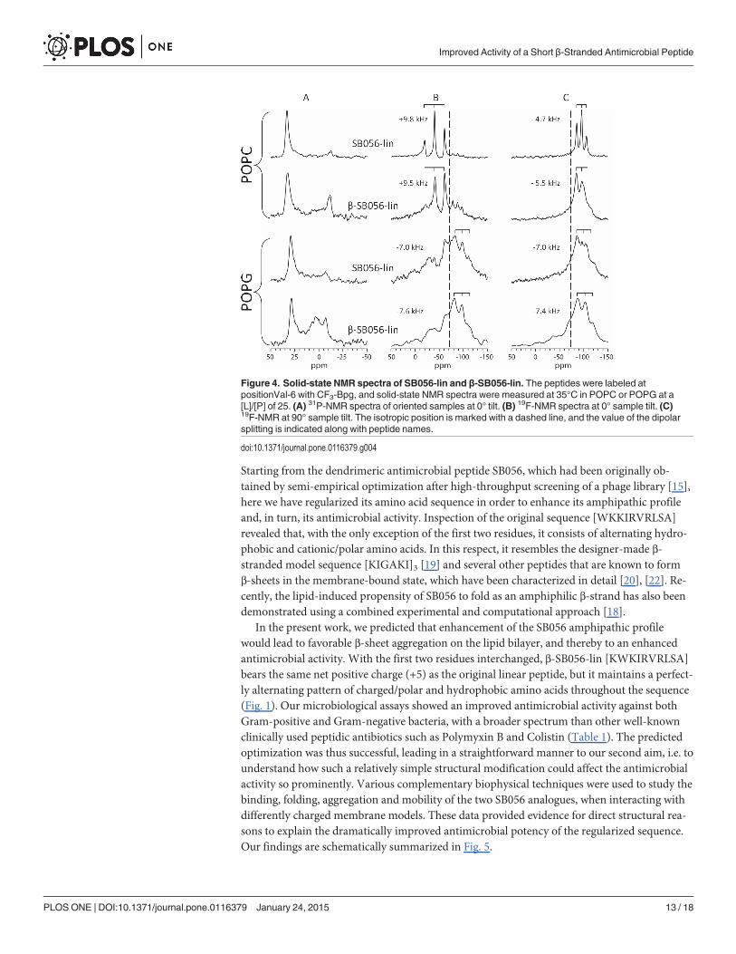
Solid-State NMR analysisSolid-state NMR is particularly well suited to study peptide-lipid interactions, as demonstratedfor various α-helical peptides, for unstructured membrane-bound peptides, and for monomericβ-strands and oligomeric β-sheets [20], [21], [29], [45–47]. Theoretical details about solid-stateNMR have been described previously in detail ([48–50] and references therein). In particular,the high sensitivity of 19F-NMR allows us to readily distinguish binding, folding, aggregation,and transitions into amyloid-like fibrils in the presence of membranes. It has been previouslydemonstrated that peptide structure and function remain unperturbed when a single hydropho-bic residue in a membrane active peptide is selectively replaced with (CF3-Bpg or CF3-Phg)[51–53]. Indeed, also in the present case the 19F-labeled [WKKIR(CF3-Bpg)RLSA] and [KWKIR(CF3-Bpg)RLSA] exhibit MIC values similar to those of the unlabeled peptides (data not shown).
19F-NMR analysis relies on the direct read-out of the dipolar splitting of the 19F-labeledreporter group on the peptide, which reveals the alignment and mobility of the CF3-Bpg sidechain that is rigidly attached to the backbone, and which can therefore reflect the behaviour ofthe entire (folded) molecule. Especially in the case of β-stranded peptides, several characteristicscenarios can be readily distinguished based on a single 19F-labeled position [20], [21], [29]. Forexample, an isotropic signal is usually indicative of a highly mobile peptide in a disordered statethat is not bound to the lipid bilayer at all. The largest possible splitting of +17 kHz, on the otherhand, indicates that the CF3-Bpg side chain is inserted straight into the membrane and immobi-lized parallel to the lipid acyl chains. This is a signature of oligomeric β-strands that are assem-bled on the membrane surface in an amyloid-like manner. Any splitting in between, that isreduced by a factor of 2 upon tilting the sample by 90°, indicates that the peptide is free to under-go lateral diffusion in the bilayer and has not aggregated, thus reflecting a monomeric or loweroligomeric state. In contrast, a dipolar splitting of −7.5 kHz with a powder-like lineshape meansthat the peptide has aggregated without preferential orientation and is immobilized. At the sametime, solid-state 31P-NMR can be used to assess the influence of the peptide on the phospholipidbilayer by observing any perturbance induced by the peptide. A higher antimicrobial activity of aβ-stranded peptide may be expected to correlate with a greater perturbance of the membrane, ashas been previously proposed based on a vesicle fusion assays [31].
Our 31P-NMR results show that SB056-lin induces no significant bilayer perturbation in POPCat a [L]/[P] of 25. 19F-NMR exhibits a well-defined triplet that gets reduces by a factor of 2 upontilting the sample, indicating that the peptide is mobile and most likely in a monomeric state(Fig. 4). Under the same conditions, β-SB056-lin is also mobile, but 31P-NMR shows considerablebilayer perturbance (Fig. 4). When anionic POPG is added to the POPCmatrix, the binding-aggregation equilibrium becomes more complex, and the NMR spectra cannot be easily deconvo-luted (data not shown). In pure POPG, finally, SB056-lin causes only slight perturbation, whileβ-SB056-lin induces massive disturbance in the lipid bilayer at a [L]/[P] of 25. 19F-NMR of bothSB056-lin and β-SB056-lin shows powder spectra, suggesting that the peptides have aggregated(Fig. 4). This membrane perturbing effect can be directly correlated with theMIC values measured-of the two peptides, given the presence of negatively charged lipids in bacterial membranes.
Summary and ConclusionsAntimicrobial peptides in general, and dendrimeric analogues in particular, represent an at-tractive research avenue for developing novel antibiotics [54]. However, there are still manyopen questions concerning their mode of interaction with bacterial membranes [55], [56].
Improved Activity of a Short β-Stranded Antimicrobial Peptide
PLOS ONE | DOI:10.1371/journal.pone.0116379 January 24, 2015 12 / 18
Starting from the dendrimeric antimicrobial peptide SB056, which had been originally ob-tained by semi-empirical optimization after high-throughput screening of a phage library [15],here we have regularized its amino acid sequence in order to enhance its amphipathic profileand, in turn, its antimicrobial activity. Inspection of the original sequence [WKKIRVRLSA]revealed that, with the only exception of the first two residues, it consists of alternating hydro-phobic and cationic/polar amino acids. In this respect, it resembles the designer-made β-stranded model sequence [KIGAKI]3 [19] and several other peptides that are known to formβ-sheets in the membrane-bound state, which have been characterized in detail [20], [22]. Re-cently, the lipid-induced propensity of SB056 to fold as an amphiphilic β-strand has also beendemonstrated using a combined experimental and computational approach [18].
In the present work, we predicted that enhancement of the SB056 amphipathic profilewould lead to favorable β-sheet aggregation on the lipid bilayer, and thereby to an enhancedantimicrobial activity. With the first two residues interchanged, β-SB056-lin [KWKIRVRLSA]bears the same net positive charge (+5) as the original linear peptide, but it maintains a perfect-ly alternating pattern of charged/polar and hydrophobic amino acids throughout the sequence(Fig. 1). Our microbiological assays showed an improved antimicrobial activity against bothGram-positive and Gram-negative bacteria, with a broader spectrum than other well-knownclinically used peptidic antibiotics such as Polymyxin B and Colistin (Table 1). The predictedoptimization was thus successful, leading in a straightforward manner to our second aim, i.e. tounderstand how such a relatively simple structural modification could affect the antimicrobialactivity so prominently. Various complementary biophysical techniques were used to study thebinding, folding, aggregation and mobility of the two SB056 analogues, when interacting withdifferently charged membrane models. These data provided evidence for direct structural rea-sons to explain the dramatically improved antimicrobial potency of the regularized sequence.Our findings are schematically summarized in Fig. 5.
Figure 4. Solid-state NMR spectra of SB056-lin and β-SB056-lin. The peptides were labeled atpositionVal-6 with CF3-Bpg, and solid-state NMR spectra were measured at 35°C in POPC or POPG at a[L]/[P] of 25. (A) 31P-NMR spectra of oriented samples at 0° tilt. (B) 19F-NMR spectra at 0° sample tilt. (C)19F-NMR at 90° sample tilt. The isotropic position is marked with a dashed line, and the value of the dipolarsplitting is indicated along with peptide names.
doi:10.1371/journal.pone.0116379.g004
Improved Activity of a Short β-Stranded Antimicrobial Peptide
PLOS ONE | DOI:10.1371/journal.pone.0116379 January 24, 2015 13 / 18

Improved Activity of a Short β-Stranded Antimicrobial Peptide
PLOS ONE | DOI:10.1371/journal.pone.0116379 January 24, 2015 14 / 18

The membrane binding affinity of both peptides was found to increase with increasing theanionic/zwitterionic lipid ratio in the vesicles, as expected for cationic peptides. However, thesequence-optimized β-SB056-lin showed a remarkable binding even to pure POPC bilayers,unlike the original SB056-lin. This observation indicates that, besides electrostatics, also theglobal amphipathicity of the folded peptide can be sufficient to stabilize the bound state, lead-ing to dramatic differences in peptide behavior after interchanging just two residues out of ten.On the other hand, the greater affinity of the sequence-regularized analogue for zwitterionicmembranes is also responsible for its greater cytotoxicity against human erythrocytes, an aspectthat should be taken into consideration when any further development of these peptides intotherapeutically valuable compounds will be attempted. When the proportion of anionic lipidsin the target membrane is increased, more and more peptide monomers were found to bind.Both SB056-lin and β-SB056-lin adopt a β-stranded conformation, as expected. However, theformer maintains a considerable degree of structural disorder, given that the position of thepositive charges and the hydrophobic residue on the N-terminus cannot be accommodated ina straight amphipathic β-strand. Since the peptide is relatively short, an adaptation of the firsttwo residues to a regular amphiphilic pattern has a profound effect on the overall peptide con-formation and self-assembly in the membrane bound state.
When the peptide-lipid system approaches electro-neutrality (around 25% POPG in ourcase), the amphipathically-enhanced β-SB056-lin is seen to form very well-ordered β-strands.They self-assemble into more and more extended β-sheets when the proportion of anionic lip-ids is further increased in the sample. At the same time, the mobility of these extended peptideaggregates decreases, as shown by 19F-NMR. Thus, the enhanced antimicrobial activity of theregularized analogue appears to be correlated with the formation of such extended β-sheets onthe bacterial membrane. Although elucidation of the detailed mode of action will still requirefurther investigations, it is plausible that such highly positively charged peptides can induce an-ionic lipid clustering in the bilayer [57]. Therefore, a high local concentration of membrane-bound β-stranded peptides that can assemble into extended β-sheets seems to be the basis ofthe antimicrobial action of this class of peptides. The strong membrane perturbing effects thathave been seen above by 31P-NMR and in earlier lipid fusion assays [31] can be attributed tothis kind of self-assembly, with dramatic effects of membrane permeability, stability and/ordepolarization.
AcknowledgmentsWe acknowledge the Synchrotron Light Source ANKA for provision of instruments at theirbeamlines, and we would like to thank Bianca Posselt and Siegmar Roth (Institute of BiologicalInterfaces, IBG-2, KIT) for technical assistance during the SRCDmeasurements at thebeamline UV-CD12 and Andrea Eisele and Kerstin Scheubeck for their assistance in peptidesynthesis.
Author ContributionsConceived and designed the experiments: GMMASMP PW JB VL ASU ACR. Performed theexperiments: GMMASMP PW JB NPMMP RS VL. Analyzed the data: GMMASMP PW JB
Figure 5. Summary of peptide-lipid interactions. The proportion of anionic lipids in the vesicles is increased from top to bottom. The behavior of theoriginal SB056-lin peptide is represented on the left hand side, and the sequence-optimized β-SB056-lin on the right. Black arrows of different length andthickness are used to indicate the different binding equilibria. SB056-lin binds only to anionic bilayers, and in a not so well-ordered β-stranded conformation.The sequence optimized β-SB056-lin, on the other hand, forms regular β-strands that self-assemble into extended β-sheets when the negative charge of thebilayer exceeds electro-neutrality of the peptide-lipid system.
doi:10.1371/journal.pone.0116379.g005
Improved Activity of a Short β-Stranded Antimicrobial Peptide
PLOS ONE | DOI:10.1371/journal.pone.0116379 January 24, 2015 15 / 18
MC VL ASU ACR. Contributed reagents/materials/analysis tools: AG GP PW JB ASU. Wrotethe paper: MAS PW JB VL ASU ACR.
References1. Opar A (2007) Bad drugs need more drugs. Nature Reviews Drug Discovery 6: 943–944.
2. Tew GN, Scott RW, Klein ML, DegradoWF (2009) De novo design of antimicrobial polymers,foldamers, and small molecules: from discovery to practical applications. Accounts of ChemicalResearch 43: 30–39.
3. Rotem S, Mor A (2009) Antimicrobial peptide mimics for improved therapeutic properties. Biochimica etBiophysica Acta 1788: 1582–1592. doi: 10.1016/j.bbamem.2008.10.020 PMID: 19028449
4. Zasloff M (2002) Antimicrobial peptides of multicellular organisms. Nature 415: 389–395. doi: 10.1038/415389a PMID: 11807545
5. Hancock REW, Rozek A (2002) Role of membranes in the activities of antimicrobial cationic peptides.FEMSMicrobiology Letters 206: 143–149. doi: 10.1111/j.1574-6968.2002.tb11000.x PMID: 11814654
6. Giuliani A, Pirri G, Bozzi A, Di Giulio A, Aschi M, et al. (2008) Antimicrobial peptides: natural templatesfor synthetic membrane-active compounds. Cellular and Molecular Life Sciences 65: 2450–2460. doi:10.1007/s00018-008-8188-x PMID: 18661101
7. Strandberg E, Ulrich AS (2004) NMRmethods for studying membrane-active antimicrobial peptides.Concepts in Magnetic Resonance A 23: 89–120.
8. Shai Y (2006) Mode of action of membrane active antimicrobial peptides. Biopolymers 66: 236–248.
9. Peschel A, Sahl HG (2006) The co-evolution of host cationic antimicrobial peptides and microbial resis-tance. Nature Reviews Microbiology 4: 529–536. doi: 10.1038/nrmicro1441 PMID: 16778838
10. Diamond G, Beckloff N, Weinberg A, Kisich KO (2009) The roles of antimicrobial peptides in innate hostdefense. Current Pharmaceutical Design 15: 2377–2392. doi: 10.2174/138161209788682325 PMID:19601838
11. Scorciapino MA, Rinaldi AC (2012) Antimicrobial peptidomimetics: reinterpreting nature to deliver inno-vative therapeutics. Frontiers in Immunology 3: 1–4. doi: 10.3389/fimmu.2012.00171 PMID: 22798960
12. Rosa Borges A, Schengrund CL (2005) Dendrimers and antivirals: a review. Current Drug Targets. In-fectious Disorders 5: 247–254. doi: 10.2174/1568005054880127 PMID: 16181143
13. Mintzer MA, Dane EL, O’Toole GA, Grinstaff MW (2012) Exploiting dendrimer multivalency to combatemerging and re-emerging infectious diseases. Molecular Pharmaceutics 9: 342–354. doi: 10.1021/mp2005033 PMID: 22126461
14. Sadler K, Tam JP (2002) Peptide dendrimers: applications and synthesis. Reviews in Molecular Bio-technology 90: 195–229. PMID: 12071226
15. Pini A, Giuliani A, Falciani C, Runci Y, Ricci C, et al. (2005) Antimicrobial activity of novel dendrimericpeptides obtained by phage display selection and rational modification. Antimicrobial Agents and Che-motherapy 7: 2665–2672.
16. Giuliani A, Rinaldi AC (2011) Beyond natural antimicrobial peptides: multimeric peptides and otherpeptidomimetic approaches. Cellular and Molecular Life Sciences 68: 2255–2266. doi: 10.1007/s00018-011-0717-3 PMID: 21598022
17. Bruschi M, Pirri G, Giuliani A, Nicoletto SF, Baster I, et al. (2010) Synthesis, characterization, antimicro-bial activity and LPS-interaction properties of SB041, a novel dendrimeric peptide with antimicrobialproperties. Peptides 31: 1459–1467. doi: 10.1016/j.peptides.2010.04.022 PMID: 20438783
18. Scorciapino MA, Pirri G, Vargiu AV, Ruggerone P, Giuliani A, et al. (2012) A novel dendrimeric peptidewith antimicrobial properties: structure-function analysis of SB056. Biophysical Journal 102: 1039–1048. doi: 10.1016/j.bpj.2012.01.048 PMID: 22404926
19. Blazyk J, Wiegand R, Klein J, Hammer J, Epand RM, et al. (2001) A novel linear amphipathic β-sheetcationic antimicrobial peptide with enhanced selectivity for bacterial lipids. The Journal of BiologicalChemistry 276: 27899–27906.
20. Wadhwani P, Strandberg E, Heidenreich N, Bürck J, Fanghänel S, et al. (2012) Self-assembly of flexi-ble β-strands into immobile amyloid-like β-sheets in membranes as revealed by solid-state19F NMR.Journal of the American Chemical Society 134: 6512–6515. doi: 10.1021/ja301328f PMID: 22452513
21. Wadhwani P, Reichert J, Strandberg E, Bürck J, Misiewicz J, et al. (2013) Stereochemical effects onthe aggregation and biological properties of the fibril-forming peptide [KIGAKI]3 in membranes. Physi-cal Chemistry Chemical Physics 15: 8962–8971. doi: 10.1039/c3cp50896j PMID: 23652359
Improved Activity of a Short β-Stranded Antimicrobial Peptide
PLOS ONE | DOI:10.1371/journal.pone.0116379 January 24, 2015 16 / 18
22. Meier M, Seelig J (2008) Length dependence of the coil$ β-sheet transition in a membrane environ-ment. Journal of the American Chemical Society 130: 1017–1024. doi: 10.1021/ja077231r PMID:18163629
23. Mykhailiuk PK, Afonin S, Chernega AN, Rusanov EB, Platonov MO, et al. (2006) Conformationallyrigid trifluoromethyl-substituted α-amino acid designed for peptide structure analysis by solid state19F-NMR, Angewandte Chemie International Edition 45: 5659–5661.
24. Mykhailiuk PK, Voievoda NM, Afonin S, Ulrich AS, Komarov IV (2010) An optimized protocol for themultigram synthesis of 3-(trifluoromethyl)bicyclopent-[1.1.1]-1-ylglycine, Journal of Fluorine Chemistry131: 217–220.
25. Afonin S, Glaser RW, Berditchevskaia M, Wadhwani P, Gührs K-H, et al. (2003) 4-Fluorophenylglycineas a label for 19F NMR structure analysis of membrane-associated peptides. ChemBioChem 4: 1151–1163.
26. Sarker SD, Nahar L, Kumarasamy Y (2007) Microtitre plate-based antibacterial assay incorporatingresazurin as an indicator of cell growth, and its application in the in vitro antibacterial screening of phyto-chemicals. Methods 42: 321–324.
27. Clarke DT, Jones G (2004) CD12: a new high-flux beamline for ultraviolet and vacuum-ultraviolet circu-lar dichroism on the SRS, Daresbury. Journal of Synchrotron Radiation 11: 142–149.
28. Wien F, Wallace BA (2005) Calcium fluoride micro cells for synchrotron radiation circular dichroismspectroscopy. Applied Spectroscopy 59: 1109–1113.
29. Wadhwani P, Bürck J, Strandberg E, Mink C, Afonin S, et al. (2008) Using a sterically restrictive aminoacid as a 19F NMR label to monitor and to control peptide aggregation in membranes. Journal of theAmerican Chemical Society 130: 16515–16517.
30. Bennett AE, Rienstra CM, Auger M, Lakshmi KV, Griffin RG (1995) Heteronuclear decoupling in rotat-ing solids. Journal of Chemical Physics 103: 6951–6958.
31. Wadhwani P, Reichert J, Bürck J, Ulrich AS (2012) Antimicrobial and cell-penetrating peptides inducelipid vesicle fusion by folding and aggregation. European Biophysics Journal 41: 177–187.
32. Zhao H, Kinnunen PKJ (2002) Binding of the antimicrobial peptide temporin L to liposomes assessedby Trp fluorescence. Journal of Biological Chemistry 277: 25170–25177.
33. Christiaens B, Symoens S, Vanderheyden S, Engelborghs Y, Joliot A, et al. (2002) Tryptophan fluores-cence study of the interaction of penetratin peptides with model membranes. European Journal of Bio-chemistry 269: 2918–2926.
34. Epand RF, Ramamoorthy A, Epand RM (2006) Membrane lipid composition and the interaction of par-daxin: the role of cholesterol. Protein and Peptide Letters 13: 1–5.
35. Epand RM, Epand RF (2009) Domains in bacterial membranes and the action of antimicrobial agents.Molecular Biosystems 5: 580–587.
36. ShawN (1974) Lipid composition as a guide to the classification of bacteria. Advances in Applied Micro-biology 17: 63–108.
37. Breukink E, Van Kraaij C, Demel RA, Siezen RJ, Kuipers OP, et al. (1997) The C-terminal region ofnisin is responsible for the initial interaction of nisin with the target membrane. Biochemistry 36: 6968–6976.
38. Miles AJ, Wallace BA (2006) Synchrotron radiation circular dichroism spectroscopy of proteins and ap-plications in structural and functional genomics. Chemical Society Reviews 35: 39–51.
39. Wallace BA (2009) Protein characterization by synchrotron radiation circular dichroism spectroscopy.Quarterly Reviews of Biophysics 42: 317–370.
40. Wallace BA, Janes RW (eds.) (2009). Modern techniques for circular dichroism and synchrotron radia-tion circular dichroism spectroscopy. IOS Press, Amsterdam, The Netherlands.
41. Wallace BA, Janes RW (2010) Synchrotron radiation circular dichroism (SRCD) spectroscopy—An en-hanced method for examining protein conformations and protein interactions. Biochemical SocietyTransactions 38: 861–873.
42. Bürck J, Roth S, Windisch D, Wadhwani P, Moss D, et al. (2014) UV-CD12: Synchrotron radiation circu-lar dichroism beamline at ANKA. Journal of Synchrotron Radiation, in revision
43. Wallace BA, Teeters CL (1987) Differential absorption flattening optical effects are significant in the cir-cular dichroism spectra of large membrane fragments. Biochemistry 26: 65–70.
44. Bustamante C, Tinoco IJr, Maestre MF (1983) Circular differential scattering can be an important part ofthe circular dichroism of macromolecules. Proceedings of the National Academy of Science USA 80:3568–3572.
Improved Activity of a Short β-Stranded Antimicrobial Peptide
PLOS ONE | DOI:10.1371/journal.pone.0116379 January 24, 2015 17 / 18

45. Glaser RW, Sachse C, Dürr UHN, Wadhwani P, Ulrich AS (2004) Orientation of the antimicrobial pep-tide PGLa in lipid membranes determined from 19F-NMR dipolar couplings of 4-CF3-phenylglycine la-bels. Journal of Magnetic Resonance 168: 153–163.
46. Afonin SE, Grage SL, Ieronimo M, Wadhwani P, Ulrich AS (2008) Temperature-dependent transmem-brane insertion of the amphiphilic peptide PGLa in lipid bilayers observed by solid state 19F-NMR, Jour-nal of the American Chemical Society 130: 16512–16514.
47. Maisch D, Wadhwani P, Afonin S, Böttcher C, Koksch B, et al. (2009) Chemical labeling strategy with(R)- and (S)-trifluoromethylalanine for solid state 19F-NMR analysis of peptaibols in membranes, Jour-nal of the American Chemical Society 131: 15596–15597.
48. Ulrich AS (2005) Solid state 19F NMRmethods for studying biomembranes. Progress in Nuclear Mag-netic Resonance Spectroscopy 46: 1–21.
49. Grage S, Ulrich AS, Strandberg E, Sachse C, Berditchevskaia M, et al. (2005) Solid-state 19F-nuclearmagnetic resonance analysis of membrane-active peptides. In: Ramamoorthy A ( ed.), NMR Spectros-copy of Biological Solids. CRC Press, Boca Raton, FL, USA. Pp. 215–236.
50. Saitô H, Ando I, Naito A (2006) Solid state NMR spectroscopy for biopolymers—Principles and applica-tions. Springer, Dordrecht, The Netherlands.
51. Strandberg E, Kanithasen N, Tiltak D, Bürck J, Wadhwani P, et al. (2008) Solid-state NMR analysiscomparing the designer-made antibiotic MSI-103 with its parent peptide PGLa in lipid bilayers. Bio-chemistry 47: 2601–2616.
52. Salwiczek M, Mikhailiuk PK, Afonin S, Komarov IV, Ulrich AS, et al. (2010) Compatibility of the confor-mationally rigid CF3-Bpg side chain with the hydrophobic coiled-coil interface. Amino Acids 39: 1589–1593.
53. Tkachenko AN, Mykhailiuk PK, Afonin S, Radchenko DS, Kubyshkin VS, et al. (2013) A 19F NMR labelto substitute polar amino acids in peptides: a CF3-substituted analogue of serine and threonine.Angewandte Chemie International Edition 52: 1486–1489.
54. Tam JP, Lu YA, Yang JL (2002) Antimicrobial dendrimeric peptides. European Journal of Biochemistry269: 923–932.
55. Dathe M, Wieprecht T (1999) Structural features of helical antimicrobial peptides: their potential to mod-ulate activity on model membranes and biological cells. Biochimica et Biophysica Acta 1462: 71–87.
56. Melo MN, Castanho MA (2012) The mechanism of action of antimicrobial peptides: lipid vesicles vs.bacteria. Frontiers in Immunology 3: 236.
57. Wadhwani P, Epand RF, Heidenreich N, Bürck J, Ulrich AS, et al. (2012) Membrane-active peptidesand the clustering of anionic lipids. Biophysical Journal, 103: 265–274.
Improved Activity of a Short β-Stranded Antimicrobial Peptide
PLOS ONE | DOI:10.1371/journal.pone.0116379 January 24, 2015 18 / 18
Copyright © 2022 FDOKUMEN