Gene.iobio: an interactive web tool for versatile, clinically ...
Upload
khangminh22Category
view
1download
0
International Journal of
Molecular Sciences
Review
Self-Assembly of Amphiphilic Compounds as a VersatileTool for Construction of Nanoscale Drug Carriers
Ruslan Kashapov *, Gulnara Gaynanova, Dinar Gabdrakhmanov, Denis Kuznetsov, Rais Pavlov,Konstantin Petrov, Lucia Zakharova and Oleg Sinyashin
A.E. Arbuzov Institute of Organic and Physical Chemistry, FRC Kazan Scientific Center of RAS, Arbuzov street 8,Kazan 420088, Russia; [email protected] (G.G.); [email protected] (D.G.); [email protected] (D.K.);[email protected] (R.P.); [email protected] (K.P.); [email protected] (L.Z.); [email protected] (O.S.)* Correspondence: [email protected]; Tel.: +7-(843)-273-22-93; Fax: +7-(843)-273-22-53
Received: 21 August 2020; Accepted: 19 September 2020; Published: 22 September 2020�����������������
Abstract: This review focuses on synthetic and natural amphiphilic systems prepared fromstraight-chain and macrocyclic compounds capable of self-assembly with the formation of nanoscaleaggregates of different morphology and their application as drug carriers. Since numerous biologicalspecies (lipid membrane, bacterial cell wall, mucous membrane, corneal epithelium, biopolymers,e.g., proteins, nucleic acids) bear negatively charged fragments, much attention is paid to cationiccarriers providing high affinity for encapsulated drugs to targeted cells. First part of the review isdevoted to self-assembling and functional properties of surfactant systems, with special attentionfocusing on cationic amphiphiles, including those bearing natural or cleavable fragments. Further,lipid formulations, especially liposomes, are discussed in terms of their fabrication and applicationfor intracellular drug delivery. This section highlights several features of these carriers, includingnoncovalent modification of lipid formulations by cationic surfactants, pH-responsive properties,endosomal escape, etc. Third part of the review deals with nanocarriers based on macrocycliccompounds, with such important characteristics as mucoadhesive properties emphasized. In thissection, different combinations of cyclodextrin platform conjugated with polymers is considered asdrug delivery systems with synergetic effect that improves solubility, targeting and biocompatibilityof formulations.
Keywords: amphiphile; cationic surfactants; drug delivery; liposome; endosomal escape; macrocycle;polymer; mucoadhesion
1. Self-Assembly of Amphiphilic Compounds
Self-assembling systems based on amphiphilic compounds find wide application in differentfundamental and practical areas due to their unique ability to form nanoscale assemblies withgradients of polarity, viscosity, electric charge and other characteristics [1–3]. In an aqueous medium,such nanosized aggregates possess a nonpolar interior capable of entrapping guest molecules, therebydramatically changing their properties [2–4]. This phenomenon is responsible for the wide rangeof applications of amphiphilic compounds in cosmetics, food industry, pharmacy, drug and genedelivery, etc. [5–8]. Meanwhile, for these reasons they are strongly required to meet the green chemistrycriteria. Therefore, the design of environmentally friendly amphiphilic compound (surfactants,macrocycles, polymers) is a challenging task from the viewpoint of the development of supramolecularmultifunctional systems with tunable characteristics.
Int. J. Mol. Sci. 2020, 21, 6961; doi:10.3390/ijms21186961 www.mdpi.com/journal/ijms

Int. J. Mol. Sci. 2020, 21, 6961 2 of 47
1.1. Cationic Surfactants Bearing Cleavable Fragments
Among amphiphilic compounds, special attention is received by cationic surfactants, which isrelated to both their fundamental and practical relevance. Researchers have succeeded in designingand synthesizing a variety of homologous series of cationic surfactants differing in the structure ofhead groups, which allows for determining the role of the polar fragment in the physicochemicalproperties and functional activity of supramolecular systems based on cationic surfactants in termsof structure–properties–activity relationships. Numerous fundamental approaches and practicalapplications involving cationic surfactants have recently been demonstrated in the fields of biochemistry,nanomedicine, pharmacy, catalysis, corrosion protection, oil recovery, food, cosmetics, etc. [2].These beneficial results were achieved despite the well-known toxicity of cationic surfactants, sincemuch effort has been undertaken to overcome this limitation. Therefore, the design of novel cationicsurfactants addressing this problem is of importance. To this end: (i) cationic surfactants with specificstructures, including cleavable, gemini, and biocompatible surfactants have been designed; (ii) mixedcompositions with less toxic nonionic surfactants are used; (iii) modifications with hydrotropic agentsare carried out. Some of these studies devoted to synthesis and use of novel amphiphilic compoundsanswering biotechnological criteria are discussed below, with emphasis on cationic surfactants.
Recently, much attention has been received by the so-called cleavable surfactants bearing ester,amide, disulfide or other degradable fragments [9]. While researchers initially focused on ester quatsbearing quaternized nitrogens in the polar group [10], more diverse amphiphilic structures have beendesigned, including those containing a carbamate moiety [11], gemini analogs [12], or amphiphilicmatrices bearing two cleavable groups [13]. The structural characteristics of ester quats are ofimportance in their surface activity, with synergetic hydrolysis-driven effects being observed forderivatives in which the carbonyl groups are bridged by an oxygen atom with a quarternized nitrogenatom [10]. Series of gemini surfactants bearing ester and amide groups were synthesized andsystematically studied in [14], with exploration of their degradability, surface activity, foaming andantimicrobial properties.
A series of publications [15–18] focused on carbamate-bearing surfactants in single and mixedsystems, including those containing additionally an imidazolium moiety.
For these carbamate surfactants, critical micelle concentration (CMC) values were shown to belower compared to conventional cationic surfactants with trimethylammonium (TMA) head groupsand to decrease with an increase in the number of carbon atoms (n) in the alkyl tail as follows:lg (CMC) = 0.563–0.257 × n [15]. A series of carbamate surfactants (Figure 1a) was tested for theirsolubilization capacity toward hydrophobic probes and antimicrobial activity, which confirm them asbiodegradable micellar nanocontainers with improved functional efficacy. The hexadecyl derivative wasfurther investigated in composition with nonionic surfactants, with negative deviation revealed fromideal mixture behavior [13,16]. These mixed systems demonstrated effective solubilization propertiestoward the anti-inflammatory drug meloxicam, with a balance achieved between the low toxicitycharacteristics due to the presence of nonionic surfactants and the solubilization activity maintainedby cationic surfactants. In [17] novel formulations for the herbicide Lontrel® were developed with theuse of a series of cationic surfactants. Carbamate-bearing surfactants were found to show the highestactivity, with a 3-fold increase in the Lontrel® concentration observed in the plants tested and improvedwetting ability compared to an unformulated preparation. These results could be further enhancedby increasing the temperature. The biomedical functionality of carbamate-bearing surfactants couldbe significantly modified by the introduction of an imidazolium fragment [18]. For a homologousseries of imidazolium-containing amphiphiles bearing carbamate groups (Figure 1b), antimicrobialproperties within the concentration range characterized by low hemolytic activity are documented.In addition, membranotropic ability is revealed, which is more pronounced in the case of lowerhomologs. A valuable property of cationic surfactants is their affinity to negatively charged biologicalspecies, which is responsible, for example, for their application as nonviral vectors. The complexationactivity of imidazolium amphiphiles with carbamate fragments toward DNA decamer and bovine

Int. J. Mol. Sci. 2020, 21, 6961 3 of 47
serum albumin (BSA) macromolecules was verified by a variety of techniques and treated in terms oftheir electrostatic interaction and hydrogen bonding ability [18].
Int. J. Mol. Sci. 2020, 21, x FOR PEER REVIEW 3 of 46
verified by a variety of techniques and treated in terms of their electrostatic interaction and hydrogen
bonding ability [18].
Figure 1. Chemical formulas of carbamate-containing amphiphiles bearing ammonium (a) and
imidazolium (b) moieties.
1.2. Amphiphilic Compounds with Natural Fragment
Among the wide spectrum of recently synthesized amphiphilic compounds a special place is
occupied by amphiphiles bearing natural fragments in their structures (Figure 2). The main idea
behind the design of this type of amphiphilic compounds is based on principles of biomimetics, that
allow constructing environmentally and physiologically friendly supramolecular systems of
nanoscale dimensions for a number of practical applications in industry and biomedicine.
Figure 2. Schematic illustration of selected amphiphilic compounds bearing natural fragments and
their benefits.
Since the term “natural fragment” is quite common and could include a lot of different structural
fragments, it is practically impossible to overview all possible combinations in the framework of this
review. Therefore, only selected amphiphiles containing natural moieties are discussed below,
namely: (i) amphiphilic compounds bearing amino acid fragments; (ii) hydrophobized derivatives of
saccharides of different types; (iii) salts of bile acids and their derivatives and (iv) nucleolipids. In line
with the biomimetic approach, the introduction of these residues is assumed to improve the
biocompatibility and reduce the toxic characteristics of amphiphilic molecules, to contribute to
complementary interactions when using in vivo assays, to allow for realizing various aggregate
morphological structures (Figure 3), etc.
1.2.1. Amphiphilic Compounds Bearing Amino Acid Fragments
One of the most popular routes for modification of amphiphilic compounds and making systems
on their basis more biocompatible and bioavailable is the introduction of amino acid fragments into
Figure 1. Chemical formulas of carbamate-containing amphiphiles bearing ammonium (a) and imidazolium(b) moieties.
1.2. Amphiphilic Compounds with Natural Fragment
Among the wide spectrum of recently synthesized amphiphilic compounds a special place isoccupied by amphiphiles bearing natural fragments in their structures (Figure 2). The main ideabehind the design of this type of amphiphilic compounds is based on principles of biomimetics,that allow constructing environmentally and physiologically friendly supramolecular systems ofnanoscale dimensions for a number of practical applications in industry and biomedicine.
Int. J. Mol. Sci. 2020, 21, x FOR PEER REVIEW 3 of 46
verified by a variety of techniques and treated in terms of their electrostatic interaction and hydrogen
bonding ability [18].
Figure 1. Chemical formulas of carbamate-containing amphiphiles bearing ammonium (a) and
imidazolium (b) moieties.
1.2. Amphiphilic Compounds with Natural Fragment
Among the wide spectrum of recently synthesized amphiphilic compounds a special place is
occupied by amphiphiles bearing natural fragments in their structures (Figure 2). The main idea
behind the design of this type of amphiphilic compounds is based on principles of biomimetics, that
allow constructing environmentally and physiologically friendly supramolecular systems of
nanoscale dimensions for a number of practical applications in industry and biomedicine.
Figure 2. Schematic illustration of selected amphiphilic compounds bearing natural fragments and
their benefits.
Since the term “natural fragment” is quite common and could include a lot of different structural
fragments, it is practically impossible to overview all possible combinations in the framework of this
review. Therefore, only selected amphiphiles containing natural moieties are discussed below,
namely: (i) amphiphilic compounds bearing amino acid fragments; (ii) hydrophobized derivatives of
saccharides of different types; (iii) salts of bile acids and their derivatives and (iv) nucleolipids. In line
with the biomimetic approach, the introduction of these residues is assumed to improve the
biocompatibility and reduce the toxic characteristics of amphiphilic molecules, to contribute to
complementary interactions when using in vivo assays, to allow for realizing various aggregate
morphological structures (Figure 3), etc.
1.2.1. Amphiphilic Compounds Bearing Amino Acid Fragments
One of the most popular routes for modification of amphiphilic compounds and making systems
on their basis more biocompatible and bioavailable is the introduction of amino acid fragments into
Figure 2. Schematic illustration of selected amphiphilic compounds bearing natural fragments andtheir benefits.
Since the term “natural fragment” is quite common and could include a lot of different structuralfragments, it is practically impossible to overview all possible combinations in the framework ofthis review. Therefore, only selected amphiphiles containing natural moieties are discussed below,namely: (i) amphiphilic compounds bearing amino acid fragments; (ii) hydrophobized derivativesof saccharides of different types; (iii) salts of bile acids and their derivatives and (iv) nucleolipids.In line with the biomimetic approach, the introduction of these residues is assumed to improvethe biocompatibility and reduce the toxic characteristics of amphiphilic molecules, to contributeto complementary interactions when using in vivo assays, to allow for realizing various aggregatemorphological structures (Figure 3), etc.

Int. J. Mol. Sci. 2020, 21, 6961 4 of 47
Int. J. Mol. Sci. 2020, 21, x FOR PEER REVIEW 4 of 46
their chemical structures [19]. These synthetic amino acid-based surfactants are an alternative to
ordinary antimicrobial compounds, because they can be obtained from renewable raw materials.
Figure 3. Schematic representation of nature-inspired amphiphiles and their possible various
morphological structures.
Amino acid-based amphiphiles are promising in obtaining drugs based on surfactant–protein
complexes [19] and can be used in oil recovery [20], as nonviral vectors in gene therapy [21], in
cosmetics [22], etc. The main requirements for the resulting amphiphiles are multifunctionality, low
toxicity and biodegradability [23].
It is well known that in accordance with one of the conventional classifications of amino acids
from the viewpoint of their charge characteristics provided by the nature of their substituents, they
could be divided into four big groups [24]: (1) nonpolar amino acids; (2) polar amino acids; (3) polar
acidic amino acids (negatively charged); (4) polar basic amino acids (positively charged). Since the
charge of an amphiphilic compound determines its properties to a great extent, the corresponding
derivatives of amino acids should be discussed from this perspective.
The main attention of researchers is focused on the application of amphiphilic compounds
bearing nonpolar amino acids. Although there are several examples of studies dealing with the
synthesis of this kind of compounds [25], the majority of investigations are dedicated to discussion
of their physicochemical properties in solutions and potential applications in biomedicine. For
example, hydrophobized phenylalanine and valine derivatives demonstrated high cytotoxicity
against the MCF-7 and HEK cell lines [26], pH-dependent self-assembly behavior [27] and could be
used for construction of worm-like aggregates for ibuprofen delivery [28]. As a separate line of
investigation, the interaction of these amphiphiles with biological species should be mentioned:
glycine and sarcosinate derivatives are approved as effective agents for complexation with
carrageenans of various types [29] and myoglobin [30]. Besides, compositions based on amphiphiles
with amino acid fragments exhibit controlled morphological [31] and rheological [32] behavior,
valuable in oil recovery processes, for antimicrobial activity and suitable biodegradability [33].
Alongside with these reports, there are systematic investigations of the role of amino acid structures
in various properties: it has been shown that variation of the structure of amino acid (proline,
phenylalanine, isoleucine) could be used as a tool for regulating of the stability of emulsions based
on them and antibacterial activity against Staphylococcus aureus and Bacillus subtilis strains [34].
Amphiphiles bearing polar amino acids and polar acidic amino acids (negatively charged) have
attracted less attention. Polar amino acid derivatives are represented by serine- and cysteine-
containing amphiphiles exhibiting a more than one order of magnitude lower hemolytic activity in
comparison with conventionally used sodium dodecyl sulphate [35]. They demonstrated biomedical
potential, which can be exemplified by their study as nonviral vectors for delivery of DNA into HeLa
Figure 3. Schematic representation of nature-inspired amphiphiles and their possible variousmorphological structures.
1.2.1. Amphiphilic Compounds Bearing Amino Acid Fragments
One of the most popular routes for modification of amphiphilic compounds and making systemson their basis more biocompatible and bioavailable is the introduction of amino acid fragments intotheir chemical structures [19]. These synthetic amino acid-based surfactants are an alternative toordinary antimicrobial compounds, because they can be obtained from renewable raw materials.
Amino acid-based amphiphiles are promising in obtaining drugs based on surfactant–proteincomplexes [19] and can be used in oil recovery [20], as nonviral vectors in gene therapy [21],in cosmetics [22], etc. The main requirements for the resulting amphiphiles are multifunctionality,low toxicity and biodegradability [23].
It is well known that in accordance with one of the conventional classifications of amino acids fromthe viewpoint of their charge characteristics provided by the nature of their substituents, they could bedivided into four big groups [24]: (1) nonpolar amino acids; (2) polar amino acids; (3) polar acidicamino acids (negatively charged); (4) polar basic amino acids (positively charged). Since the charge ofan amphiphilic compound determines its properties to a great extent, the corresponding derivatives ofamino acids should be discussed from this perspective.
The main attention of researchers is focused on the application of amphiphilic compounds bearingnonpolar amino acids. Although there are several examples of studies dealing with the synthesisof this kind of compounds [25], the majority of investigations are dedicated to discussion of theirphysicochemical properties in solutions and potential applications in biomedicine. For example,hydrophobized phenylalanine and valine derivatives demonstrated high cytotoxicity against theMCF-7 and HEK cell lines [26], pH-dependent self-assembly behavior [27] and could be used forconstruction of worm-like aggregates for ibuprofen delivery [28]. As a separate line of investigation,the interaction of these amphiphiles with biological species should be mentioned: glycine andsarcosinate derivatives are approved as effective agents for complexation with carrageenans of varioustypes [29] and myoglobin [30]. Besides, compositions based on amphiphiles with amino acid fragmentsexhibit controlled morphological [31] and rheological [32] behavior, valuable in oil recovery processes,for antimicrobial activity and suitable biodegradability [33]. Alongside with these reports, there aresystematic investigations of the role of amino acid structures in various properties: it has been shownthat variation of the structure of amino acid (proline, phenylalanine, isoleucine) could be used asa tool for regulating of the stability of emulsions based on them and antibacterial activity againstStaphylococcus aureus and Bacillus subtilis strains [34].

Int. J. Mol. Sci. 2020, 21, 6961 5 of 47
Amphiphiles bearing polar amino acids and polar acidic amino acids (negatively charged) haveattracted less attention. Polar amino acid derivatives are represented by serine- and cysteine-containingamphiphiles exhibiting a more than one order of magnitude lower hemolytic activity in comparisonwith conventionally used sodium dodecyl sulphate [35]. They demonstrated biomedical potential,which can be exemplified by their study as nonviral vectors for delivery of DNA into HeLa cancercells with high gene transfection efficiency (up to 50%) [36]. Their application in the synthesis ofcatalytic active modified silver nanoparticles for p-nitrophenol reduction was documented in [37].The synthesis of a new cationic double-tailed cysteine-based surfactant is given in [38]. For thisamphiphile, the aggregation characteristics and the ability to bind to BSA were evaluated. It wasshown that there are various intermolecular interactions between the components depending on thesurfactant concentration.
Investigations of amino acid derivatives belonging to the negatively charged amino acid classare exemplified herein by arginine-containing amphiphilic compounds documented as additivesfor modification of physicochemical properties of oil [39], potential antimicrobial compounds activeagainst Micrococcus luteus, Bacillus subtilis, Staphylococcus aureus strains [40] and agents capable to bindwith heparin [41], which activity could be regulated by selecting appropriate alkyl tail length for theamphiphile. Compositions based on amphiphilic lysine derivatives were documented as drug deliveryvehicles containing encapsulated 5-fluorouracil [42] for cancer treatment as well as the component fordesign of coating against protein adsorption on nanoparticles [43]. In [44] promising stimuli-responsivedouble-tailed lysine-based surfactants were obtained.
Below the Krafft point, the amphiphiles self-organized into tubular structures of variousmorphologies forming hydrogels at low surfactant concentration; above the Krafft point, there was atransition from tubular structures to micelles or vesicles, depending on the structure of the surfactant,and this transition was thermo-reversible in the physiological temperature range. The synthesis ofhistidine-based surfactants with high antimicrobial activity against Gram-positive and Gram-negativebacteria is given in [45], with some of them displaying no destructive effect on red blood cells.The work [46] is devoted to the synthesis of arginine-based surfactants and the study of their hemolyticactivity. It was found that this amphiphile protected human red blood cells from hypotonic lysisover a wide range of concentrations. Dicationic histidine derivatives were documented as promisingagents for antibacterial treatment against new generations of microorganisms like methicillin-resistantStaphylococcus aureus [47]. The authors of [48] investigated the surface-active and micelle-formingproperties of three anionic dicarboxylic amino acid-based surfactants in combination with the cationiccompound cethyltrimethylammonium bromide (CTAB). It was shown that maximum number of thecationic surfactant CTAB molecules on the micellar surface leads to the formation of close-packedmicellar structures [49]. The CMC values were significantly lower than predicted, which indicates theexistence of associative interactions between the components.
Several examples of amino acid-based amphiphiles and their properties are given in Table 1.Analysis of these values allows one to conclude that the aggregation and antimicrobial properties ofthis type of amphiphiles could be significantly enhanced by the introduction of additional head groupsand hydrophobic tails into the chemical structures of molecules, as well as through covalent linking ofacyclic NH-fragments. The latter could be due to the increase of the hydrogen bonding contribution toaggregation processes and destruction of bacterial cell wall due to additional amine fragments.

Int. J. Mol. Sci. 2020, 21, 6961 6 of 47
Table 1. CMC values and minimal inhibitory concentrations (MIC,µM) against Staphylococcus epidermidisstrains for several amino acid-based surfactants.
Chemical Structure CMC, mM MIC, µM
Int. J. Mol. Sci. 2020, 21, x FOR PEER REVIEW 5 of 46
cancer cells with high gene transfection efficiency (up to 50%) [36]. Their application in the synthesis
of catalytic active modified silver nanoparticles for p-nitrophenol reduction was documented in [37].
The synthesis of a new cationic double-tailed cysteine-based surfactant is given in [38]. For this
amphiphile, the aggregation characteristics and the ability to bind to BSA were evaluated. It was
shown that there are various intermolecular interactions between the components depending on the
surfactant concentration.
Investigations of amino acid derivatives belonging to the negatively charged amino acid class
are exemplified herein by arginine-containing amphiphilic compounds documented as additives for
modification of physicochemical properties of oil [39], potential antimicrobial compounds active
against Micrococcus luteus, Bacillus subtilis, Staphylococcus aureus strains [40] and agents capable to
bind with heparin [41], which activity could be regulated by selecting appropriate alkyl tail length
for the amphiphile. Compositions based on amphiphilic lysine derivatives were documented as drug
delivery vehicles containing encapsulated 5-fluorouracil [42] for cancer treatment as well as the
component for design of coating against protein adsorption on nanoparticles [43]. In [44] promising
stimuli-responsive double-tailed lysine-based surfactants were obtained.
Below the Krafft point, the amphiphiles self-organized into tubular structures of various
morphologies forming hydrogels at low surfactant concentration; above the Krafft point, there was a
transition from tubular structures to micelles or vesicles, depending on the structure of the surfactant,
and this transition was thermo-reversible in the physiological temperature range. The synthesis of
histidine-based surfactants with high antimicrobial activity against Gram-positive and Gram-
negative bacteria is given in [45], with some of them displaying no destructive effect on red blood
cells. The work [46] is devoted to the synthesis of arginine-based surfactants and the study of their
hemolytic activity. It was found that this amphiphile protected human red blood cells from hypotonic
lysis over a wide range of concentrations. Dicationic histidine derivatives were documented as
promising agents for antibacterial treatment against new generations of microorganisms like
methicillin-resistant Staphylococcus aureus [47]. The authors of [48] investigated the surface-active and
micelle-forming properties of three anionic dicarboxylic amino acid-based surfactants in combination
with the cationic compound cethyltrimethylammonium bromide (CTAB). It was shown that
maximum number of the cationic surfactant CTAB molecules on the micellar surface leads to the
formation of close-packed micellar structures [49]. The CMC values were significantly lower than
predicted, which indicates the existence of associative interactions between the components.
Several examples of amino acid-based amphiphiles and their properties are given in Table 1.
Analysis of these values allows one to conclude that the aggregation and antimicrobial properties of
this type of amphiphiles could be significantly enhanced by the introduction of additional head
groups and hydrophobic tails into the chemical structures of molecules, as well as through covalent
linking of acyclic NH-fragments. The latter could be due to the increase of the hydrogen bonding
contribution to aggregation processes and destruction of bacterial cell wall due to additional amine
fragments.
Table 1. CMC values and minimal inhibitory concentrations (MIC, μM) against Staphylococcus
epidermidis strains for several amino acid-based surfactants.
Chemical Structure CMC, mM MIC, μM
0.7 [47] 5.0 [47] 0.7 [47] 5.0 [47]
Int. J. Mol. Sci. 2020, 21, x FOR PEER REVIEW 6 of 46
0.3 [40] 17 [40]
1.5 [45] 35 [45]
1.0 [33] 450 [33]
1.0 [33] 450 [33]
1.2.2. Sugar-Based Amphiphilic Compounds
Another promising approach to design substances meeting biomedical application criteria is the
functionalization of amphiphiles by a saccharide moiety [50], that makes it possible to prepare sugar-
based surfactants, such as alkylpolyglycosides, alkylglucamines, etc. Among the obvious advantages
of these substances are their biodegradability, low toxicity and low cost. This research direction looks
so promising, that researchers have used significant theoretical approaches to predict their
aggregation parameters [51]. An overview of the reports dedicated to sugar-containing amphiphiles
allows one to deduce that these compounds could be classified by two ways from the viewpoint of
the degree of oligomerization of the sugar moiety inserted in the chemical structure of an amphiphilic
compound. The first classification takes into account the number of furanose and/or pyranose
moieties in the structure and therefore the compounds can be divided into three groups: (1)
amphiphiles bearing monosaccharide fragments; (2) amphiphilic compounds containing
disaccharide moieties; (3) hydrophobized derivatives of trisaccharides. Another classification is
based on the nature of the head group and the number of hydrophobic tails and head groups of
amphiphiles, so these derivatives could be divided into: (i) monomeric nonionic surfactants, (ii)
nonionic gemini surfactants and (iii) cationic surfactants and other amphiphiles with a sugar
fragment. Herein, we consider some general properties for each of the six mentioned groups.
The majority of monosaccharide amphiphilic compounds are represented by glucose
derivatives. Interestingly, the selection of functionalization of glucose-containing amphiphilic
compounds and using of various approaches to molecule design could become a useful tool for
tailoring desired practical properties. For example: (i) significant foamability and high emulsion
stability for toluene/water system could be achieved in the case of application of amido- and/or
alkoxy derivatives of this hexose [52]; (ii) introduction of a trisiloxane fragment into the glucose
structure is responsible for superspreading properties on hydrophobic surfaces like parafilm [53]; (iii)
the transition from monomeric amphiphiles to their gemini analogues containing two glucose
fragments and additional OH-groups is key for the construction of vehicles for biomedicine purposes
capable of effectively encapsulating the drug of a broad spectrum, resveratrol (encapsulation
0.3 [40] 17 [40]
Int. J. Mol. Sci. 2020, 21, x FOR PEER REVIEW 6 of 46
0.3 [40] 17 [40]
1.5 [45] 35 [45]
1.0 [33] 450 [33]
1.0 [33] 450 [33]
1.2.2. Sugar-Based Amphiphilic Compounds
Another promising approach to design substances meeting biomedical application criteria is the
functionalization of amphiphiles by a saccharide moiety [50], that makes it possible to prepare sugar-
based surfactants, such as alkylpolyglycosides, alkylglucamines, etc. Among the obvious advantages
of these substances are their biodegradability, low toxicity and low cost. This research direction looks
so promising, that researchers have used significant theoretical approaches to predict their
aggregation parameters [51]. An overview of the reports dedicated to sugar-containing amphiphiles
allows one to deduce that these compounds could be classified by two ways from the viewpoint of
the degree of oligomerization of the sugar moiety inserted in the chemical structure of an amphiphilic
compound. The first classification takes into account the number of furanose and/or pyranose
moieties in the structure and therefore the compounds can be divided into three groups: (1)
amphiphiles bearing monosaccharide fragments; (2) amphiphilic compounds containing
disaccharide moieties; (3) hydrophobized derivatives of trisaccharides. Another classification is
based on the nature of the head group and the number of hydrophobic tails and head groups of
amphiphiles, so these derivatives could be divided into: (i) monomeric nonionic surfactants, (ii)
nonionic gemini surfactants and (iii) cationic surfactants and other amphiphiles with a sugar
fragment. Herein, we consider some general properties for each of the six mentioned groups.
The majority of monosaccharide amphiphilic compounds are represented by glucose
derivatives. Interestingly, the selection of functionalization of glucose-containing amphiphilic
compounds and using of various approaches to molecule design could become a useful tool for
tailoring desired practical properties. For example: (i) significant foamability and high emulsion
stability for toluene/water system could be achieved in the case of application of amido- and/or
alkoxy derivatives of this hexose [52]; (ii) introduction of a trisiloxane fragment into the glucose
structure is responsible for superspreading properties on hydrophobic surfaces like parafilm [53]; (iii)
the transition from monomeric amphiphiles to their gemini analogues containing two glucose
fragments and additional OH-groups is key for the construction of vehicles for biomedicine purposes
capable of effectively encapsulating the drug of a broad spectrum, resveratrol (encapsulation
1.5 [45] 35 [45]
Int. J. Mol. Sci. 2020, 21, x FOR PEER REVIEW 6 of 46
0.3 [40] 17 [40]
1.5 [45] 35 [45]
1.0 [33] 450 [33]
1.0 [33] 450 [33]
1.2.2. Sugar-Based Amphiphilic Compounds
Another promising approach to design substances meeting biomedical application criteria is the
functionalization of amphiphiles by a saccharide moiety [50], that makes it possible to prepare sugar-
based surfactants, such as alkylpolyglycosides, alkylglucamines, etc. Among the obvious advantages
of these substances are their biodegradability, low toxicity and low cost. This research direction looks
so promising, that researchers have used significant theoretical approaches to predict their
aggregation parameters [51]. An overview of the reports dedicated to sugar-containing amphiphiles
allows one to deduce that these compounds could be classified by two ways from the viewpoint of
the degree of oligomerization of the sugar moiety inserted in the chemical structure of an amphiphilic
compound. The first classification takes into account the number of furanose and/or pyranose
moieties in the structure and therefore the compounds can be divided into three groups: (1)
amphiphiles bearing monosaccharide fragments; (2) amphiphilic compounds containing
disaccharide moieties; (3) hydrophobized derivatives of trisaccharides. Another classification is
based on the nature of the head group and the number of hydrophobic tails and head groups of
amphiphiles, so these derivatives could be divided into: (i) monomeric nonionic surfactants, (ii)
nonionic gemini surfactants and (iii) cationic surfactants and other amphiphiles with a sugar
fragment. Herein, we consider some general properties for each of the six mentioned groups.
The majority of monosaccharide amphiphilic compounds are represented by glucose
derivatives. Interestingly, the selection of functionalization of glucose-containing amphiphilic
compounds and using of various approaches to molecule design could become a useful tool for
tailoring desired practical properties. For example: (i) significant foamability and high emulsion
stability for toluene/water system could be achieved in the case of application of amido- and/or
alkoxy derivatives of this hexose [52]; (ii) introduction of a trisiloxane fragment into the glucose
structure is responsible for superspreading properties on hydrophobic surfaces like parafilm [53]; (iii)
the transition from monomeric amphiphiles to their gemini analogues containing two glucose
fragments and additional OH-groups is key for the construction of vehicles for biomedicine purposes
capable of effectively encapsulating the drug of a broad spectrum, resveratrol (encapsulation
1.0 [33] 450 [33]
1.2.2. Sugar-Based Amphiphilic Compounds
Another promising approach to design substances meeting biomedical application criteria is thefunctionalization of amphiphiles by a saccharide moiety [50], that makes it possible to prepare sugar-basedsurfactants, such as alkylpolyglycosides, alkylglucamines, etc. Among the obvious advantages of thesesubstances are their biodegradability, low toxicity and low cost. This research direction looks so promising,that researchers have used significant theoretical approaches to predict their aggregation parameters [51].An overview of the reports dedicated to sugar-containing amphiphiles allows one to deduce that thesecompounds could be classified by two ways from the viewpoint of the degree of oligomerization of thesugar moiety inserted in the chemical structure of an amphiphilic compound. The first classification takesinto account the number of furanose and/or pyranose moieties in the structure and therefore the compoundscan be divided into three groups: (1) amphiphiles bearing monosaccharide fragments; (2) amphiphiliccompounds containing disaccharide moieties; (3) hydrophobized derivatives of trisaccharides. Anotherclassification is based on the nature of the head group and the number of hydrophobic tails and headgroups of amphiphiles, so these derivatives could be divided into: (i) monomeric nonionic surfactants,

Int. J. Mol. Sci. 2020, 21, 6961 7 of 47
(ii) nonionic gemini surfactants and (iii) cationic surfactants and other amphiphiles with a sugar fragment.Herein, we consider some general properties for each of the six mentioned groups.
The majority of monosaccharide amphiphilic compounds are represented by glucose derivatives.Interestingly, the selection of functionalization of glucose-containing amphiphilic compounds and usingof various approaches to molecule design could become a useful tool for tailoring desired practicalproperties. For example: (i) significant foamability and high emulsion stability for toluene/water systemcould be achieved in the case of application of amido- and/or alkoxy derivatives of this hexose [52];(ii) introduction of a trisiloxane fragment into the glucose structure is responsible for superspreadingproperties on hydrophobic surfaces like parafilm [53]; (iii) the transition from monomeric amphiphilesto their gemini analogues containing two glucose fragments and additional OH-groups is key forthe construction of vehicles for biomedicine purposes capable of effectively encapsulating the drugof a broad spectrum, resveratrol (encapsulation efficiency reaches 90%) [54], and amino acids withdemonstrated of chiral specificity toward certain D- or L-isomers [55]; (iv) thermotropic liquid crystallinebehavior can be achieved in the case of sulfur-containing glucose derivatives [56]. The utilization ofother monosaccharides as building blocks for amphiphilic compounds is also well-known, howeverthese reports are significantly fewer: mannose derivatives are documented as agents for regulating theaggregation state of β-lactoglobulin [57], and a xylopyranose-based amphiphilic compound showsitself as a material for the construction of liquid crystals [58].
Disaccharide derivatives cover amphiphiles mainly containing lactose, maltose and sucrosefragments in their structures. The presence of a lactose moiety in the molecule provides bothindustrial and biotechnological significance to these compositions. These compounds could beused as efficient micellar catalysts of Ullmann C-S coupling reactions in water (the yield of productcould reach 90% by selecting an appropriate alkyl tail length) [59]; highly-effective antibacterialagents against a wide spectrum of microorganisms [60]; coatings interrupting undesired adsorptionprocesses [61]; compounds for condensation with DNA, that could be accurately tuned by selectionof the counterion [62]; a basis of a vehicle for ketoprofen delivery [63]. Maltose and sucroseamphiphilic derivatives were recommended as components of microemulsions—potential alternativefuel [64], regulators of wettability of various solid surfaces like quartz and polyethylene [65] andmodifiers of adsorption parameters at the air/liquid interface of systems [66]. Herein, crocin, a naturalbolaamphiphile with two disaccharide moieties, should be separately mentioned. Recently thiscarotenoid was used for micellar catalysis of oxidation processes [67]. Besides the abovementioneddisaccharides, a sophorose amphiphilic derivative was reported as a building block for the constructionof unique supramolecular architectures, namely vesicle-in-vesicle units with diameters up to 200 nm [68].
There are only a couple of recent applications dealing with trisaccharide amphiphilic compounds,nevertheless compositions based on them show themselves to be biocompatible and suitable forbiotechnological purposes. For example, raffinose and melezitose amphiphilic derivatives wasdocumented as effective solubilizers for polycyclic aromatic hydrocarbons possessing acceptablehemolytic activity, which could be reduced by shortening the amphiphile alkyl tail [69] as well asagents allowing one to maintain the activity of enzymes during 20 days after its thermal treatment [70].
A series of alkyltriazole glycosides (ATGs) with different length of spacer linking the sugarand triazole moieties were synthesized, and their surface-active properties and phase behavior werestudied [71]. It was shown that the ATG derivative with the shortest spacer length forms a bicontinuouscubic phase and the position of the triazole bond in ATG doesn’t significantly affect the surface activity.
In [72] the synthesis of lactose-based non-ionic surfactants and their analysis in vitro wereaccomplished. The cytotoxic activity of these amphiphiles toward two cell lines, colonic epitheliumCaco-2 and airway epithelium Calu-3, was studied. It was shown that cell apoptosis occurred at a lowconcentration of the surfactants.
The effect of three different mannose-based surfactants included in the lipid bilayer on thephysicochemical parameters of the system was studied in [73]. It was shown that the mannosederivatives differ in properties from other alkylmonosaccharides due to their structural features.

Int. J. Mol. Sci. 2020, 21, 6961 8 of 47
For example, octyl and dodecyl derivatives integrate into hydrophilic regions of the lipid bilayer,while a hexadecyl derivative modifies the internal and external regions of the model membrane.
In [74] the effect of octyl-D-glucopyranoside-based surfactants on the environment wasinvestigated. The study showed that they were completely biodegradable with different utilizationefficiencies depending on the functional group. It is important to note that these amphiphiles weretoxic towards Gram-negative bacteria cells due to a decrease in zeta potential, thereby increasing thepermeability of the cell membrane.
Gemini surfactants containing two hydrophilic head groups and two hydrophobic tails exhibitbeneficial physicochemical properties, in particular, they have significantly reduced CMC valuesand increased surface activity compared to monomeric amphiphiles. In the works of Liu et al. [75],gemini alkyl glucosides were synthesized and studied. In addition to increasing the surface activity,interesting self-assembly features were observed. The gemini dodecyl O-glucoside-based surfactantsdemonstrated high pass-through capacity for vesicles loaded with (+)-catechin and (–)-epigallocatechin.The incorporation of these drugs into the vesicle made it possible to strengthen the vesicular bilayer inthe low concentration range.
In [76] glucono-δ-lactone-based gemini surfactants were studied. For these compounds, a decreasein CMC and more efficient adsorption at the air-water interface were observed compared to monomericderivatives. Depending on the length of the alkyl tail and concentration, they can form both largeaggregates (vesicles), and small aggregates of different sizes (micelles).
Cationic surfactants are mentioned above to be an important class of chemical compounds,which find wide practical application as emulsifiers in cosmetology, bactericidal agents, corrosioninhibitors, antifoams and in medicine. At present, many studies are focused on the increase ofbiocompatibility and biodegradability of cationic surfactants. To achieve these goals, sugar-basedcationic surfactants are obtained. Glucose-based cationic surfactants containing an ester group wereobtained in [77], and their surface properties and antimicrobial activity were studied. These amphiphilesare characterized by a decrease in surface tension and CMC in comparison with conventional surfactants,as well as excellent compatibility with sodium dodecyl sulfate.
In [78] reverse micelles formed by sugar-based surfactants with dicarboxylate moieties as acounterion were used to extract BSA. Under optimal conditions, almost complete dissolution of BSAin the micelles of surfactants with a glucosylammonium fragment was observed, while 70–80% ofBSA was dissolved in micelles of surfactants with a lactosylammonium head group. The influence ofvarious system parameters on the morphology of aggregates of cationic gemini surfactants containingan isosorbide fragment was studied in [79]. With a change in pH or by heating the system, aggregateswere rearranged from micelles to vesicles, and vice versa, and in the presence of salts a transition fromvesicles to micelles was observed.
Analysis of existing representatives of sugar-based amphiphiles has demonstrated that keycriteria responsible for the best aggregation characteristics (the lowest CMC values) of sugar-basedsurfactants are the presence of more than one hydrophobic tail in the structure and the transitionfrom monosaccharide to oligosaccharide derivatives. If the first mentioned criterion is very similar toconventional surfactants and has the same nature, the second one could be due to allowing for moreintermolecular hydrogen bonds between the OH-groups of additional saccharide moieties. Severalexamples of amphiphiles with superior self-assembling propensity are listed in Table 2.

Int. J. Mol. Sci. 2020, 21, 6961 9 of 47
Table 2. Several examples of sugar-based amphiphiles with the lowest CMC values.
Chemical Structure CMC, µM
Int. J. Mol. Sci. 2020, 21, x FOR PEER REVIEW 8 of 46
alkyl glucosides were synthesized and studied. In addition to increasing the surface activity,
interesting self-assembly features were observed. The gemini dodecyl O-glucoside-based surfactants
demonstrated high pass-through capacity for vesicles loaded with (+)-catechin and (–)-
epigallocatechin. The incorporation of these drugs into the vesicle made it possible to strengthen the
vesicular bilayer in the low concentration range.
In [76] glucono-δ-lactone-based gemini surfactants were studied. For these compounds, a
decrease in CMC and more efficient adsorption at the air-water interface were observed compared to
monomeric derivatives. Depending on the length of the alkyl tail and concentration, they can form
both large aggregates (vesicles), and small aggregates of different sizes (micelles).
Cationic surfactants are mentioned above to be an important class of chemical compounds,
which find wide practical application as emulsifiers in cosmetology, bactericidal agents, corrosion
inhibitors, antifoams and in medicine. At present, many studies are focused on the increase of
biocompatibility and biodegradability of cationic surfactants. To achieve these goals, sugar-based
cationic surfactants are obtained. Glucose-based cationic surfactants containing an ester group were
obtained in [77], and their surface properties and antimicrobial activity were studied. These
amphiphiles are characterized by a decrease in surface tension and CMC in comparison with
conventional surfactants, as well as excellent compatibility with sodium dodecyl sulfate.
In [78] reverse micelles formed by sugar-based surfactants with dicarboxylate moieties as a
counterion were used to extract BSA. Under optimal conditions, almost complete dissolution of BSA
in the micelles of surfactants with a glucosylammonium fragment was observed, while 70–80% of
BSA was dissolved in micelles of surfactants with a lactosylammonium head group. The influence of
various system parameters on the morphology of aggregates of cationic gemini surfactants
containing an isosorbide fragment was studied in [79]. With a change in pH or by heating the system,
aggregates were rearranged from micelles to vesicles, and vice versa, and in the presence of salts a
transition from vesicles to micelles was observed.
Analysis of existing representatives of sugar-based amphiphiles has demonstrated that key
criteria responsible for the best aggregation characteristics (the lowest CMC values) of sugar-based
surfactants are the presence of more than one hydrophobic tail in the structure and the transition
from monosaccharide to oligosaccharide derivatives. If the first mentioned criterion is very similar to
conventional surfactants and has the same nature, the second one could be due to allowing for more
intermolecular hydrogen bonds between the OH-groups of additional saccharide moieties. Several
examples of amphiphiles with superior self-assembling propensity are listed in Table 2.
Table 2. Several examples of sugar-based amphiphiles with the lowest CMC values.
Chemical Structure CMC, μM
29.0 [80]
33.0 [76]
29.0 [80]
Int. J. Mol. Sci. 2020, 21, x FOR PEER REVIEW 8 of 46
alkyl glucosides were synthesized and studied. In addition to increasing the surface activity,
interesting self-assembly features were observed. The gemini dodecyl O-glucoside-based surfactants
demonstrated high pass-through capacity for vesicles loaded with (+)-catechin and (–)-
epigallocatechin. The incorporation of these drugs into the vesicle made it possible to strengthen the
vesicular bilayer in the low concentration range.
In [76] glucono-δ-lactone-based gemini surfactants were studied. For these compounds, a
decrease in CMC and more efficient adsorption at the air-water interface were observed compared to
monomeric derivatives. Depending on the length of the alkyl tail and concentration, they can form
both large aggregates (vesicles), and small aggregates of different sizes (micelles).
Cationic surfactants are mentioned above to be an important class of chemical compounds,
which find wide practical application as emulsifiers in cosmetology, bactericidal agents, corrosion
inhibitors, antifoams and in medicine. At present, many studies are focused on the increase of
biocompatibility and biodegradability of cationic surfactants. To achieve these goals, sugar-based
cationic surfactants are obtained. Glucose-based cationic surfactants containing an ester group were
obtained in [77], and their surface properties and antimicrobial activity were studied. These
amphiphiles are characterized by a decrease in surface tension and CMC in comparison with
conventional surfactants, as well as excellent compatibility with sodium dodecyl sulfate.
In [78] reverse micelles formed by sugar-based surfactants with dicarboxylate moieties as a
counterion were used to extract BSA. Under optimal conditions, almost complete dissolution of BSA
in the micelles of surfactants with a glucosylammonium fragment was observed, while 70–80% of
BSA was dissolved in micelles of surfactants with a lactosylammonium head group. The influence of
various system parameters on the morphology of aggregates of cationic gemini surfactants
containing an isosorbide fragment was studied in [79]. With a change in pH or by heating the system,
aggregates were rearranged from micelles to vesicles, and vice versa, and in the presence of salts a
transition from vesicles to micelles was observed.
Analysis of existing representatives of sugar-based amphiphiles has demonstrated that key
criteria responsible for the best aggregation characteristics (the lowest CMC values) of sugar-based
surfactants are the presence of more than one hydrophobic tail in the structure and the transition
from monosaccharide to oligosaccharide derivatives. If the first mentioned criterion is very similar to
conventional surfactants and has the same nature, the second one could be due to allowing for more
intermolecular hydrogen bonds between the OH-groups of additional saccharide moieties. Several
examples of amphiphiles with superior self-assembling propensity are listed in Table 2.
Table 2. Several examples of sugar-based amphiphiles with the lowest CMC values.
Chemical Structure CMC, μM
29.0 [80]
33.0 [76] 33.0 [76]
Int. J. Mol. Sci. 2020, 21, x FOR PEER REVIEW 9 of 46
8.0 [69]
9.0 [69]
1.2.3. Bile Salts and Derivatives of Bile Acids–Natural Amphiphiles and Their Artificially Designed
Counterparts
Salts of bile acids are derived from steroid acids contained in the bile of mammals. Their main
difference from conventional amphiphiles is the presence of a bulky scaffold, which predetermines
their unique properties. For example, bile salts are known as effective transporters of lipids [81],
regulators of the rate of crystallization [82] and components of compositions aimed at drug delivery
[83]. The most widespread bile salts considered in recent reports are cholate, deoxycholate and
taurodeoxycholate. Sodium cholate is known for its capability to act as a transdermal enhancer [84]
and additive to mono- and dicationic surfactant solutions significantly modifying their
morphological behavior [85] and release profile of cargo encapsulated in self-assembled drug
nanovehicles [86]. Sodium deoxycholate was reported as part of biamphiphiles, ionic compounds
with both cationic and anionic amphiphilic nature, which aggregate shape could be accurately
controlled [87]; micellar catalyst for the synthesis of 1,2,3-triazole derivatives; their characteristics is
superior in comparison with those of conventional surfactants CTAB and Triton-X-100 [88]. Sodium
taurodeoxyholate was documented as a compound for regulation of solubility of steroid drug in
surfactant micellar solutions and biorelevant media [89] and as a component of composition for
albendazole delivery for the treatment of lung parasites [90].
Further attempts of bile acids functionalization have been presented in recent reports. There are
a couple of reports discussing p-tertbutylbenzoyl derivatives of cholic acid, which when mixed with
a copolymer and in a covalent conjugation with tripeptide demonstrate fabrication of
thermoresponsive supramolecular structures [91] and twisted nanoribbons [92] as well as dimeric
bile acid derivatives acting as organogelators for the design of hybrid nanomaterials [93].
1.2.4. Nucleolipids (Amphiphilic Derivatives of Nucleobases)
A special place among amphiphiles bearing natural fragments is occupied by the so-called
nucleolipids–hydrophobic derivatives of nucleobases, nucleosides and nucleotides. Interest in these
compounds is mostly due to the nitrogenous bases (adenine, uracil, thymine, guanine) inserted in
their chemical structure and capable of being involved in additional specific interactions. As an
example, nucleolipids was known by their capability to form films at air/liquid interfaces [94],
coatings for surface hydrophobization [95], liquid crystalline mesophases [96], as well as they have
potential for the design of highly-sensitive sensors for nucleobases [97] and inorganic ions [98].
Besides, the specific architecture of nucleolipid molecules and their biocompatibility allows their use
for biomedical purposes as neurotracers [99], drugs against glioblastoma [100] and carriers for
potential anti-malarial drugs [101]. Systematic studies [102–105] have focused on charged
representatives of this type of amphiphilic compounds (Figure 4), exemplified by a series of cationic
8.0 [69]
Int. J. Mol. Sci. 2020, 21, x FOR PEER REVIEW 9 of 46
8.0 [69]
9.0 [69]
1.2.3. Bile Salts and Derivatives of Bile Acids–Natural Amphiphiles and Their Artificially Designed
Counterparts
Salts of bile acids are derived from steroid acids contained in the bile of mammals. Their main
difference from conventional amphiphiles is the presence of a bulky scaffold, which predetermines
their unique properties. For example, bile salts are known as effective transporters of lipids [81],
regulators of the rate of crystallization [82] and components of compositions aimed at drug delivery
[83]. The most widespread bile salts considered in recent reports are cholate, deoxycholate and
taurodeoxycholate. Sodium cholate is known for its capability to act as a transdermal enhancer [84]
and additive to mono- and dicationic surfactant solutions significantly modifying their
morphological behavior [85] and release profile of cargo encapsulated in self-assembled drug
nanovehicles [86]. Sodium deoxycholate was reported as part of biamphiphiles, ionic compounds
with both cationic and anionic amphiphilic nature, which aggregate shape could be accurately
controlled [87]; micellar catalyst for the synthesis of 1,2,3-triazole derivatives; their characteristics is
superior in comparison with those of conventional surfactants CTAB and Triton-X-100 [88]. Sodium
taurodeoxyholate was documented as a compound for regulation of solubility of steroid drug in
surfactant micellar solutions and biorelevant media [89] and as a component of composition for
albendazole delivery for the treatment of lung parasites [90].
Further attempts of bile acids functionalization have been presented in recent reports. There are
a couple of reports discussing p-tertbutylbenzoyl derivatives of cholic acid, which when mixed with
a copolymer and in a covalent conjugation with tripeptide demonstrate fabrication of
thermoresponsive supramolecular structures [91] and twisted nanoribbons [92] as well as dimeric
bile acid derivatives acting as organogelators for the design of hybrid nanomaterials [93].
1.2.4. Nucleolipids (Amphiphilic Derivatives of Nucleobases)
A special place among amphiphiles bearing natural fragments is occupied by the so-called
nucleolipids–hydrophobic derivatives of nucleobases, nucleosides and nucleotides. Interest in these
compounds is mostly due to the nitrogenous bases (adenine, uracil, thymine, guanine) inserted in
their chemical structure and capable of being involved in additional specific interactions. As an
example, nucleolipids was known by their capability to form films at air/liquid interfaces [94],
coatings for surface hydrophobization [95], liquid crystalline mesophases [96], as well as they have
potential for the design of highly-sensitive sensors for nucleobases [97] and inorganic ions [98].
Besides, the specific architecture of nucleolipid molecules and their biocompatibility allows their use
for biomedical purposes as neurotracers [99], drugs against glioblastoma [100] and carriers for
potential anti-malarial drugs [101]. Systematic studies [102–105] have focused on charged
representatives of this type of amphiphilic compounds (Figure 4), exemplified by a series of cationic
9.0 [69]
1.2.3. Bile Salts and Derivatives of Bile Acids—Natural Amphiphiles and Their ArtificiallyDesigned Counterparts
Salts of bile acids are derived from steroid acids contained in the bile of mammals. Their maindifference from conventional amphiphiles is the presence of a bulky scaffold, which predetermines theirunique properties. For example, bile salts are known as effective transporters of lipids [81], regulatorsof the rate of crystallization [82] and components of compositions aimed at drug delivery [83]. The mostwidespread bile salts considered in recent reports are cholate, deoxycholate and taurodeoxycholate.Sodium cholate is known for its capability to act as a transdermal enhancer [84] and additive to mono-and dicationic surfactant solutions significantly modifying their morphological behavior [85] andrelease profile of cargo encapsulated in self-assembled drug nanovehicles [86]. Sodium deoxycholatewas reported as part of biamphiphiles, ionic compounds with both cationic and anionic amphiphilicnature, which aggregate shape could be accurately controlled [87]; micellar catalyst for the synthesis of1,2,3-triazole derivatives; their characteristics is superior in comparison with those of conventionalsurfactants CTAB and Triton-X-100 [88]. Sodium taurodeoxyholate was documented as a compound

Int. J. Mol. Sci. 2020, 21, 6961 10 of 47
for regulation of solubility of steroid drug in surfactant micellar solutions and biorelevant media [89]and as a component of composition for albendazole delivery for the treatment of lung parasites [90].
Further attempts of bile acids functionalization have been presented in recent reports. There are acouple of reports discussing p-tertbutylbenzoyl derivatives of cholic acid, which when mixed with acopolymer and in a covalent conjugation with tripeptide demonstrate fabrication of thermoresponsivesupramolecular structures [91] and twisted nanoribbons [92] as well as dimeric bile acid derivativesacting as organogelators for the design of hybrid nanomaterials [93].
1.2.4. Nucleolipids (Amphiphilic Derivatives of Nucleobases)
A special place among amphiphiles bearing natural fragments is occupied by the so-callednucleolipids–hydrophobic derivatives of nucleobases, nucleosides and nucleotides. Interest in thesecompounds is mostly due to the nitrogenous bases (adenine, uracil, thymine, guanine) inserted intheir chemical structure and capable of being involved in additional specific interactions. As anexample, nucleolipids was known by their capability to form films at air/liquid interfaces [94], coatingsfor surface hydrophobization [95], liquid crystalline mesophases [96], as well as they have potentialfor the design of highly-sensitive sensors for nucleobases [97] and inorganic ions [98]. Besides,the specific architecture of nucleolipid molecules and their biocompatibility allows their use forbiomedical purposes as neurotracers [99], drugs against glioblastoma [100] and carriers for potentialanti-malarial drugs [101]. Systematic studies [102–105] have focused on charged representativesof this type of amphiphilic compounds (Figure 4), exemplified by a series of cationic nucleolipidsbearing a pyrimidine moiety, which allows revealing quantitative structure–activity relationships.For example, it has been established that these nucleolipids have unique aggregation behavior inaqueous solutions [102], solubilization properties toward model hydrophobic compounds [103],marked membranotropic properties [104] and substrate-specific effects on the catalysis of ecotoxicantsof various hydrophobicity [105].
Int. J. Mol. Sci. 2020, 21, x FOR PEER REVIEW 10 of 46
nucleolipids bearing a pyrimidine moiety, which allows revealing quantitative structure–activity
relationships. For example, it has been established that these nucleolipids have unique aggregation
behavior in aqueous solutions [102], solubilization properties toward model hydrophobic
compounds [103], marked membranotropic properties [104] and substrate-specific effects on the
catalysis of ecotoxicants of various hydrophobicity [105].
Figure 4. Some representatives of cationic pyrimidine-based nucleolipids [102–105].
A comparative analysis of collected data for nucleolipids of various structure (Table 3) was
conducted in order to establish the functional significance in their amphiphilic structures. The main
features responsible for increase in aggregation and solubilization properties of pyrimidine-based
nucleolipids are the introduction of a hydrophobic tail into the uracil residue, the growth of
hydrophobicity of the cyclic head group, as well as transition from acyclic derivatives to macrocyclic
ones and insertion of additional nucleobases and hydrophobic tails to the chemical structure of the
molecule.
Table 3. CMC values and solubilization power S toward hydrophobic Orange OT for nucleolipids of
various structures investigated in our reports.
Chemical Structure CMC,
mM
103·S,
moldye/molamphiphille
15.0 [106] -
3.0 [107] -
0.05 [108] -
>100 [106]
Figure 4. Some representatives of cationic pyrimidine-based nucleolipids [102–105].
A comparative analysis of collected data for nucleolipids of various structure (Table 3) wasconducted in order to establish the functional significance in their amphiphilic structures. The mainfeatures responsible for increase in aggregation and solubilization properties of pyrimidine-basednucleolipids are the introduction of a hydrophobic tail into the uracil residue, the growth ofhydrophobicity of the cyclic head group, as well as transition from acyclic derivatives to macrocyclicones and insertion of additional nucleobases and hydrophobic tails to the chemical structure ofthe molecule.

Int. J. Mol. Sci. 2020, 21, 6961 11 of 47
Table 3. CMC values and solubilization power S toward hydrophobic Orange OT for nucleolipids ofvarious structures investigated in our reports.
Chemical Structure CMC, mM103·S,
moldye/molamphiphille
Int. J. Mol. Sci. 2020, 21, x FOR PEER REVIEW 10 of 46
nucleolipids bearing a pyrimidine moiety, which allows revealing quantitative structure–activity
relationships. For example, it has been established that these nucleolipids have unique aggregation
behavior in aqueous solutions [102], solubilization properties toward model hydrophobic
compounds [103], marked membranotropic properties [104] and substrate-specific effects on the
catalysis of ecotoxicants of various hydrophobicity [105].
Figure 4. Some representatives of cationic pyrimidine-based nucleolipids [102–105].
A comparative analysis of collected data for nucleolipids of various structure (Table 3) was
conducted in order to establish the functional significance in their amphiphilic structures. The main
features responsible for increase in aggregation and solubilization properties of pyrimidine-based
nucleolipids are the introduction of a hydrophobic tail into the uracil residue, the growth of
hydrophobicity of the cyclic head group, as well as transition from acyclic derivatives to macrocyclic
ones and insertion of additional nucleobases and hydrophobic tails to the chemical structure of the
molecule.
Table 3. CMC values and solubilization power S toward hydrophobic Orange OT for nucleolipids of
various structures investigated in our reports.
Chemical Structure CMC,
mM
103·S,
moldye/molamphiphille
15.0 [106] -
3.0 [107] -
0.05 [108] -
>100 [106]
15.0 [106] -
Int. J. Mol. Sci. 2020, 21, x FOR PEER REVIEW 10 of 46
nucleolipids bearing a pyrimidine moiety, which allows revealing quantitative structure–activity
relationships. For example, it has been established that these nucleolipids have unique aggregation
behavior in aqueous solutions [102], solubilization properties toward model hydrophobic
compounds [103], marked membranotropic properties [104] and substrate-specific effects on the
catalysis of ecotoxicants of various hydrophobicity [105].
Figure 4. Some representatives of cationic pyrimidine-based nucleolipids [102–105].
A comparative analysis of collected data for nucleolipids of various structure (Table 3) was
conducted in order to establish the functional significance in their amphiphilic structures. The main
features responsible for increase in aggregation and solubilization properties of pyrimidine-based
nucleolipids are the introduction of a hydrophobic tail into the uracil residue, the growth of
hydrophobicity of the cyclic head group, as well as transition from acyclic derivatives to macrocyclic
ones and insertion of additional nucleobases and hydrophobic tails to the chemical structure of the
molecule.
Table 3. CMC values and solubilization power S toward hydrophobic Orange OT for nucleolipids of
various structures investigated in our reports.
Chemical Structure CMC,
mM
103·S,
moldye/molamphiphille
15.0 [106] -
3.0 [107] -
0.05 [108] -
>100 [106]
3.0 [107] -
Int. J. Mol. Sci. 2020, 21, x FOR PEER REVIEW 10 of 46
nucleolipids bearing a pyrimidine moiety, which allows revealing quantitative structure–activity
relationships. For example, it has been established that these nucleolipids have unique aggregation
behavior in aqueous solutions [102], solubilization properties toward model hydrophobic
compounds [103], marked membranotropic properties [104] and substrate-specific effects on the
catalysis of ecotoxicants of various hydrophobicity [105].
Figure 4. Some representatives of cationic pyrimidine-based nucleolipids [102–105].
A comparative analysis of collected data for nucleolipids of various structure (Table 3) was
conducted in order to establish the functional significance in their amphiphilic structures. The main
features responsible for increase in aggregation and solubilization properties of pyrimidine-based
nucleolipids are the introduction of a hydrophobic tail into the uracil residue, the growth of
hydrophobicity of the cyclic head group, as well as transition from acyclic derivatives to macrocyclic
ones and insertion of additional nucleobases and hydrophobic tails to the chemical structure of the
molecule.
Table 3. CMC values and solubilization power S toward hydrophobic Orange OT for nucleolipids of
various structures investigated in our reports.
Chemical Structure CMC,
mM
103·S,
moldye/molamphiphille
15.0 [106] -
3.0 [107] -
0.05 [108] -
>100 [106]
0.05 [108] -
Int. J. Mol. Sci. 2020, 21, x FOR PEER REVIEW 10 of 46
nucleolipids bearing a pyrimidine moiety, which allows revealing quantitative structure–activity
relationships. For example, it has been established that these nucleolipids have unique aggregation
behavior in aqueous solutions [102], solubilization properties toward model hydrophobic
compounds [103], marked membranotropic properties [104] and substrate-specific effects on the
catalysis of ecotoxicants of various hydrophobicity [105].
Figure 4. Some representatives of cationic pyrimidine-based nucleolipids [102–105].
A comparative analysis of collected data for nucleolipids of various structure (Table 3) was
conducted in order to establish the functional significance in their amphiphilic structures. The main
features responsible for increase in aggregation and solubilization properties of pyrimidine-based
nucleolipids are the introduction of a hydrophobic tail into the uracil residue, the growth of
hydrophobicity of the cyclic head group, as well as transition from acyclic derivatives to macrocyclic
ones and insertion of additional nucleobases and hydrophobic tails to the chemical structure of the
molecule.
Table 3. CMC values and solubilization power S toward hydrophobic Orange OT for nucleolipids of
various structures investigated in our reports.
Chemical Structure CMC,
mM
103·S,
moldye/molamphiphille
15.0 [106] -
3.0 [107] -
0.05 [108] -
>100 [106] >100 [106]Int. J. Mol. Sci. 2020, 21, x FOR PEER REVIEW 11 of 46
3.0 [102] 1.6 [102]
1.9 [103] 9.4 [103]
2.0 [109] 21 [109]
3.4 [104] 1.4 [104]
0.9 [110] -
1.0 [111] 7.8 [111]
0.4 [112] 12.9 [112]
1.3. Silicone-Based Surfactants
Silicone-based surfactants are new effective amphiphiles with advanced characteristics. They are
widely used in the leather industry [113], agriculture [114], as cosmetic emulsifiers, fabric softeners
[115], etc. In addition, cationic silicone-based gemini surfactants are in accordance with the principles
3.0 [102] 1.6 [102]
Int. J. Mol. Sci. 2020, 21, x FOR PEER REVIEW 11 of 46
3.0 [102] 1.6 [102]
1.9 [103] 9.4 [103]
2.0 [109] 21 [109]
3.4 [104] 1.4 [104]
0.9 [110] -
1.0 [111] 7.8 [111]
0.4 [112] 12.9 [112]
1.3. Silicone-Based Surfactants
Silicone-based surfactants are new effective amphiphiles with advanced characteristics. They are
widely used in the leather industry [113], agriculture [114], as cosmetic emulsifiers, fabric softeners
[115], etc. In addition, cationic silicone-based gemini surfactants are in accordance with the principles
1.9 [103] 9.4 [103]
Int. J. Mol. Sci. 2020, 21, x FOR PEER REVIEW 11 of 46
3.0 [102] 1.6 [102]
1.9 [103] 9.4 [103]
2.0 [109] 21 [109]
3.4 [104] 1.4 [104]
0.9 [110] -
1.0 [111] 7.8 [111]
0.4 [112] 12.9 [112]
1.3. Silicone-Based Surfactants
Silicone-based surfactants are new effective amphiphiles with advanced characteristics. They are
widely used in the leather industry [113], agriculture [114], as cosmetic emulsifiers, fabric softeners
[115], etc. In addition, cationic silicone-based gemini surfactants are in accordance with the principles
2.0 [109] 21 [109]
Int. J. Mol. Sci. 2020, 21, x FOR PEER REVIEW 11 of 46
3.0 [102] 1.6 [102]
1.9 [103] 9.4 [103]
2.0 [109] 21 [109]
3.4 [104] 1.4 [104]
0.9 [110] -
1.0 [111] 7.8 [111]
0.4 [112] 12.9 [112]
1.3. Silicone-Based Surfactants
Silicone-based surfactants are new effective amphiphiles with advanced characteristics. They are
widely used in the leather industry [113], agriculture [114], as cosmetic emulsifiers, fabric softeners
[115], etc. In addition, cationic silicone-based gemini surfactants are in accordance with the principles
3.4 [104] 1.4 [104]
Int. J. Mol. Sci. 2020, 21, x FOR PEER REVIEW 11 of 46
3.0 [102] 1.6 [102]
1.9 [103] 9.4 [103]
2.0 [109] 21 [109]
3.4 [104] 1.4 [104]
0.9 [110] -
1.0 [111] 7.8 [111]
0.4 [112] 12.9 [112]
1.3. Silicone-Based Surfactants
Silicone-based surfactants are new effective amphiphiles with advanced characteristics. They are
widely used in the leather industry [113], agriculture [114], as cosmetic emulsifiers, fabric softeners
[115], etc. In addition, cationic silicone-based gemini surfactants are in accordance with the principles
0.9 [110] -
Int. J. Mol. Sci. 2020, 21, x FOR PEER REVIEW 11 of 46
3.0 [102] 1.6 [102]
1.9 [103] 9.4 [103]
2.0 [109] 21 [109]
3.4 [104] 1.4 [104]
0.9 [110] -
1.0 [111] 7.8 [111]
0.4 [112] 12.9 [112]
1.3. Silicone-Based Surfactants
Silicone-based surfactants are new effective amphiphiles with advanced characteristics. They are
widely used in the leather industry [113], agriculture [114], as cosmetic emulsifiers, fabric softeners
[115], etc. In addition, cationic silicone-based gemini surfactants are in accordance with the principles
1.0 [111] 7.8 [111]

Int. J. Mol. Sci. 2020, 21, 6961 12 of 47
Table 3. Cont.
Chemical Structure CMC, mM103·S,
moldye/molamphiphille
Int. J. Mol. Sci. 2020, 21, x FOR PEER REVIEW 11 of 46
3.0 [102] 1.6 [102]
1.9 [103] 9.4 [103]
2.0 [109] 21 [109]
3.4 [104] 1.4 [104]
0.9 [110] -
1.0 [111] 7.8 [111]
0.4 [112] 12.9 [112]
1.3. Silicone-Based Surfactants
Silicone-based surfactants are new effective amphiphiles with advanced characteristics. They are
widely used in the leather industry [113], agriculture [114], as cosmetic emulsifiers, fabric softeners
[115], etc. In addition, cationic silicone-based gemini surfactants are in accordance with the principles
0.4 [112] 12.9 [112]
1.3. Silicone-Based Surfactants
Silicone-based surfactants are new effective amphiphiles with advanced characteristics. They arewidely used in the leather industry [113], agriculture [114], as cosmetic emulsifiers, fabric softeners [115],etc. In addition, cationic silicone-based gemini surfactants are in accordance with the principles ofgreen chemistry due to low toxicity and quick decomposition of the Si-O-Si bond [116]. Importantly,silicone-based amphiphiles reduce surface tension better than hydrocarbon surfactants.
The presence of a tetrasiloxane fragment in the structure of cationic gemini surfactants canpotentially improve the biocompatibility and various characteristics of amphiphiles. A series ofnew cationic gemini surfactants with a tetrasiloxane fragment (Figure 5a) was synthesized in [117].The formation of various aggregates (vesicles, spherical micelles and rod-like micelles) depending onthe nature of the spacer between the head groups was observed.
Int. J. Mol. Sci. 2020, 21, x FOR PEER REVIEW 12 of 46
of green chemistry due to low toxicity and quick decomposition of the Si-O-Si bond [116].
Importantly, silicone-based amphiphiles reduce surface tension better than hydrocarbon surfactants.
The presence of a tetrasiloxane fragment in the structure of cationic gemini surfactants can
potentially improve the biocompatibility and various characteristics of amphiphiles. A series of new
cationic gemini surfactants with a tetrasiloxane fragment (Figure 5a) was synthesized in [117]. The
formation of various aggregates (vesicles, spherical micelles and rod-like micelles) depending on the
nature of the spacer between the head groups was observed.
Two tetrasiloxane-based imidazolium gemini surfactants with methylene spacer groups (Figure
5b) were synthesized and characterized in [118]. The results showed that with an increase in the
length of the spacer, the ability of the surfactants to form micelles increases, while the effectiveness
of reducing the surface tension decreases.
Figure 5. Chemical formulas of cationic tetrasiloxane gemini surfactants.
A series of organosilicon bis-quaternary amphiphiles was prepared in [119]. It was shown that
with an increase in the length of the siloxane spacer, the CMC decreases, and the adsorption
characteristics improve. The antimicrobial activity of these compounds was also characterized. Other
three cationic silicone surfactants with chloride counterions (Figure 6a) were synthesized in [120]. It
was shown that all obtained surfactants have a high surface activity and at the same time a low degree
of counterion binding. In [121] pyridine- (Figure 6b) and piperidine-based (Figure 6c) silicone
surfactants were synthesized and their properties were investigated. The effect of inorganic salts
(NaCl, NaBr, NaI, Na2SO4) on aggregation of these surfactants was studied.
Figure 6. Chemical structures of cationic ammonium- (a), pyridine- (b) and piperidine-based (c)
silicone surfactants.
The authors of [122] obtained three cationic silicone-based surfactants with pyridinium and
ammonium fragments and studied the influence of steric factors on the surface activity, adsorption
at the interface, thermodynamic characteristics, and aggregation of these amphiphiles. In another
work, a series of cationic silicone-based gemini surfactants containing hydrophobic substituents of
various lengths (CnH2n+1, n = 8,10,12,14,16,18) was obtained in [123]. It was shown that the morphology
of aggregates changes from spherical to rod-like, dumbbell-like and finally string dumbbell-like
associates with the increase of surfactant concentration (Figure 7).
Figure 5. Chemical formulas of cationic tetrasiloxane gemini surfactants bearing ammonium (a) andimidazolium (b) groups.
Two tetrasiloxane-based imidazolium gemini surfactants with methylene spacer groups (Figure 5b)were synthesized and characterized in [118]. The results showed that with an increase in the length ofthe spacer, the ability of the surfactants to form micelles increases, while the effectiveness of reducingthe surface tension decreases.
A series of organosilicon bis-quaternary amphiphiles was prepared in [119]. It was shownthat with an increase in the length of the siloxane spacer, the CMC decreases, and the adsorptioncharacteristics improve. The antimicrobial activity of these compounds was also characterized. Otherthree cationic silicone surfactants with chloride counterions (Figure 6a) were synthesized in [120].It was shown that all obtained surfactants have a high surface activity and at the same time a lowdegree of counterion binding. In [121] pyridine- (Figure 6b) and piperidine-based (Figure 6c) silicone

Int. J. Mol. Sci. 2020, 21, 6961 13 of 47
surfactants were synthesized and their properties were investigated. The effect of inorganic salts (NaCl,NaBr, NaI, Na2SO4) on aggregation of these surfactants was studied.
Int. J. Mol. Sci. 2020, 21, x FOR PEER REVIEW 12 of 46
of green chemistry due to low toxicity and quick decomposition of the Si-O-Si bond [116].
Importantly, silicone-based amphiphiles reduce surface tension better than hydrocarbon surfactants.
The presence of a tetrasiloxane fragment in the structure of cationic gemini surfactants can
potentially improve the biocompatibility and various characteristics of amphiphiles. A series of new
cationic gemini surfactants with a tetrasiloxane fragment (Figure 5a) was synthesized in [117]. The
formation of various aggregates (vesicles, spherical micelles and rod-like micelles) depending on the
nature of the spacer between the head groups was observed.
Two tetrasiloxane-based imidazolium gemini surfactants with methylene spacer groups (Figure
5b) were synthesized and characterized in [118]. The results showed that with an increase in the
length of the spacer, the ability of the surfactants to form micelles increases, while the effectiveness
of reducing the surface tension decreases.
Figure 5. Chemical formulas of cationic tetrasiloxane gemini surfactants.
A series of organosilicon bis-quaternary amphiphiles was prepared in [119]. It was shown that
with an increase in the length of the siloxane spacer, the CMC decreases, and the adsorption
characteristics improve. The antimicrobial activity of these compounds was also characterized. Other
three cationic silicone surfactants with chloride counterions (Figure 6a) were synthesized in [120]. It
was shown that all obtained surfactants have a high surface activity and at the same time a low degree
of counterion binding. In [121] pyridine- (Figure 6b) and piperidine-based (Figure 6c) silicone
surfactants were synthesized and their properties were investigated. The effect of inorganic salts
(NaCl, NaBr, NaI, Na2SO4) on aggregation of these surfactants was studied.
Figure 6. Chemical structures of cationic ammonium- (a), pyridine- (b) and piperidine-based (c)
silicone surfactants.
The authors of [122] obtained three cationic silicone-based surfactants with pyridinium and
ammonium fragments and studied the influence of steric factors on the surface activity, adsorption
at the interface, thermodynamic characteristics, and aggregation of these amphiphiles. In another
work, a series of cationic silicone-based gemini surfactants containing hydrophobic substituents of
various lengths (CnH2n+1, n = 8,10,12,14,16,18) was obtained in [123]. It was shown that the morphology
of aggregates changes from spherical to rod-like, dumbbell-like and finally string dumbbell-like
associates with the increase of surfactant concentration (Figure 7).
Figure 6. Chemical structures of cationic ammonium- (a), pyridine- (b) and piperidine-based (c)silicone surfactants.
The authors of [122] obtained three cationic silicone-based surfactants with pyridinium andammonium fragments and studied the influence of steric factors on the surface activity, adsorption atthe interface, thermodynamic characteristics, and aggregation of these amphiphiles. In another work,a series of cationic silicone-based gemini surfactants containing hydrophobic substituents of variouslengths (CnH2n+1, n = 8,10,12,14,16,18) was obtained in [123]. It was shown that the morphology ofaggregates changes from spherical to rod-like, dumbbell-like and finally string dumbbell-like associateswith the increase of surfactant concentration (Figure 7).Int. J. Mol. Sci. 2020, 21, x FOR PEER REVIEW 13 of 46
Figure 7. Possible models of morphological transition between aggregates of silicone-based
surfactants at different concentration. Reprinted with permission from [123]. Copyright 2017 Elsevier.
In [124] a series of functional silicone-based surfactants were obtained and the solubilizing
ability of these amphiphiles with respect to the disperse dye C.I. Disperse Blue 56 was studied. Two
new silicone-based carboxylate surfactants with various hydrophobic silicone-containing fragments
were obtained in [125]. Both surfactants showed a strong decrease in the surface tension of water.
TEM results showed that these amphiphiles formed spherical aggregates above their CMC.
A four-step synthesis and characterization of a new silicone-containing polyurethane surfactant
are given in [126]. The resulting polymers have high molecular weight and exhibit excellent surface-
active properties. New silicone-containing surfactants based on polyesters with various hydrophobic
groups were synthesized and characterized in [127]. The presence of a methylene group between the
silicone atoms increases the Lifshitz–Van der Waals forces of the carbosilane hydrophobic groups,
thereby increasing the CMC value and the area occupied by one surfactant molecule. Degradable and
thermosensitive microgels based on a three-component system containing a silicone component were
obtained in [128]. They were of spherical shape with a narrow size distribution and stable in an acidic
buffer solution, but decomposed in neutral and base solutions, and capable of loading and releasing
doxorubicin (DOX).
The above literature survey demonstrates that the synthesis of novel amphiphilic compounds
and investigation of their properties and practical applications have attracted much attention from
researchers. Many efforts have been directed on the improvement of the surface activity and
adsorption characteristics of surfactants and solubilization capacity of their assemblies, since these
are basic properties responsible for the bio- and nanotechnological potential of supramolecular
systems. In the case of surfactants with natural or cleavable fragments special attention has been paid
to their applications in pharmacy and medicine due to their reduced toxicity, biocompatibility and
diverse morphology, with most interest being attracted by cationic surfactants. These amphiphilic
compounds are further considered as building blocks and modifiers for nanocarriers capable of
enhancing their efficacy as drug delivery systems.
2. Self-assembled Amphiphilic Systems as Smart Drug Nanocarriers
As illustrated above, amphiphilic systems are of particular interest in biomedical applications
[2], which can be supported by their use as hydrotropic additives [129], nanocontainers for poorly
soluble drugs [130–133], antimicrobial preparations [134,135], complexing agents for negatively
charged biomolecules such as DNA and BSA [136–140], etc. They have been successfully used for the
fabrication of drug and gene delivery systems demonstrating numerous advantages compared to
non-formulated drugs, i.e., improved bioavailability, higher stability and prolonged circulation in
bioenvironment, lower side effects, etc. [141]. Although various types of nanocarriers have been
Figure 7. Possible models of morphological transition between aggregates of silicone-based surfactantsat different concentration. Reprinted with permission from [123]. Copyright 2017 Elsevier.
In [124] a series of functional silicone-based surfactants were obtained and the solubilizing abilityof these amphiphiles with respect to the disperse dye C.I. Disperse Blue 56 was studied. Two newsilicone-based carboxylate surfactants with various hydrophobic silicone-containing fragments wereobtained in [125]. Both surfactants showed a strong decrease in the surface tension of water. TEM resultsshowed that these amphiphiles formed spherical aggregates above their CMC.
A four-step synthesis and characterization of a new silicone-containing polyurethane surfactant aregiven in [126]. The resulting polymers have high molecular weight and exhibit excellent surface-activeproperties. New silicone-containing surfactants based on polyesters with various hydrophobic groupswere synthesized and characterized in [127]. The presence of a methylene group between the silicone

Int. J. Mol. Sci. 2020, 21, 6961 14 of 47
atoms increases the Lifshitz–Van der Waals forces of the carbosilane hydrophobic groups, therebyincreasing the CMC value and the area occupied by one surfactant molecule. Degradable andthermosensitive microgels based on a three-component system containing a silicone component wereobtained in [128]. They were of spherical shape with a narrow size distribution and stable in an acidicbuffer solution, but decomposed in neutral and base solutions, and capable of loading and releasingdoxorubicin (DOX).
The above literature survey demonstrates that the synthesis of novel amphiphilic compoundsand investigation of their properties and practical applications have attracted much attention fromresearchers. Many efforts have been directed on the improvement of the surface activity and adsorptioncharacteristics of surfactants and solubilization capacity of their assemblies, since these are basicproperties responsible for the bio- and nanotechnological potential of supramolecular systems. In thecase of surfactants with natural or cleavable fragments special attention has been paid to theirapplications in pharmacy and medicine due to their reduced toxicity, biocompatibility and diversemorphology, with most interest being attracted by cationic surfactants. These amphiphilic compoundsare further considered as building blocks and modifiers for nanocarriers capable of enhancing theirefficacy as drug delivery systems.
2. Self-Assembled Amphiphilic Systems as Smart Drug Nanocarriers
As illustrated above, amphiphilic systems are of particular interest in biomedical applications [2],which can be supported by their use as hydrotropic additives [129], nanocontainers for poorly solubledrugs [130–133], antimicrobial preparations [134,135], complexing agents for negatively chargedbiomolecules such as DNA and BSA [136–140], etc. They have been successfully used for the fabricationof drug and gene delivery systems demonstrating numerous advantages compared to non-formulateddrugs, i.e., improved bioavailability, higher stability and prolonged circulation in bioenvironment,lower side effects, etc. [141]. Although various types of nanocarriers have been recently designed thereare problems preventing efficient transporting of cargo into targeted cells. Among crucial challenges aretrafficking in bioenvironment, intracellular uptake and endosomal escape. Therefore, much attentionis paid to engineering the nanoparticles addressing these tasks. Some representatives or pioneerexamples are summarized in Table 4.
Importantly, the physicochemical properties, e.g., size, shape, charge characteristics of nanocarriersare key factors determining their biological fate, especially the primary interaction with cellmembranes [173,174]. In this context, much effort has been made to design novel effective formulationsand their surface functionalization. The size of nanoparticles is documented to affect the formationof corona proteins, with both quantity and quality of protein guided, which essentially controls theopsonization process. Generally, small particles are less opsonized; therefore, they are characterizedby longer circulation times and lower accumulation in liver and spleen. Meanwhile the particleshape rather than particle size has a significant impact on the macrophage uptake, biodistribution andtargeted delivery. Phagocytosis rates are shown to increase in the series prolate ellipsoid < sphere <
oblate ellipsoid [173]. Deviation from spherical shape is responsible for hydrodynamic characteristics,orientation in the blood stream and angle between nanoparticle and membrane, with these factorsstrongly correlating with trafficking of nanocarriers. Surface charge plays a significant role influencingamount of plasma proteins adsorbed on the nanocarriers, thereby controlling the circulation time.Increase in charge density regardless of the sign of charge results in an increase in opsonizationprocess and decrease in circulation time compared to neutral nanoparticles. Noteworthy, morphologyof nanocarriers is significant factor influencing intracellular uptake [175], with the size 50–100 nmconsidered as optimal for endocytic pathway.

Int. J. Mol. Sci. 2020, 21, 6961 15 of 47
Table 4. Examples of novel successful approaches that utilize nanocarriers to solve typicalchemotherapeutic and drug delivery problems.
FormulationDescription Problem to Be Solved Solution Ref
DOXliposomes
fast clearance by thereticuloendothelial system
Modification of liposomeswith PEG-conjugated lipids,
which hinder their recognitionby macrophages. This
provides prolonged circulationtime of pegylated liposomes.
[142]
DOXliposomes
passively targeted versusligand-targeted liposomes
testing
Liposomes modified by folatedemonstrated enhanced
biodistribution infolate-expressing tumors.
[143]
Paclitaxelliposomes
improvement of loadingcapacity of hydrophobic
drug
Incorporation of triglycerideincreased the fluidity and
lamellarity of the liposomesthereby resulting in sharp
increase in concentration ofdrug loaded.
[144]
DOXliposomes
targeted delivery andcellular uptake
Protonation/deprotonationequilibria to switch the
peptide between an“anchored” inactive positionand active targeting position
within tumor medium.
[145]
Irinotecanliposomes PEG dilemma
PEG-shedding in lower pHachieved by attaching the PEG
chain to the lipids via imidebond.
[146]
DOXliposomes endosomal escape
DC-Cholesterol protonation inendosome media addspositive charge to the
membrane and facilitatesliposome-endosome fusion.
[147]
Porphyrinfumed
silica–liposomenanocomposite
triggered releaseProtonation of the fumed silica
surface releases liposomalcargo.
[148]
DOXcerasomes triggered release
Cerasomes were preparedwith addition of
thermosensitive DPPC andDMPC lipids.
[149]
DOXliposome endosomal escape
Liposomes based on POPCand malachite green derivative
carrying a long alkyl chainexhibited fusogenicity
following UV irradiation
[150]
DOXpolymer
nanoparticlesendosomal escape
Proton sponge effect bybinding protons present in theendo-lysosomes on the tertiary
nitrogen atoms in theN,N-dimethylaminoethyl
methacrylate units.
[151]

Int. J. Mol. Sci. 2020, 21, 6961 16 of 47
Table 4. Cont.
FormulationDescription Problem to Be Solved Solution Ref
Paclitaxelmixed micelles endosomal escape
Poly(β-amino ester)-mediatedendosomal escape through
proton-sponge effect.[152]
DOXniosomes targeted delivery
The glucosamine anchoredDOX- loaded targeted
niosomes showed the longercirculation in plasma with
significantly higherbioavailability
[153]
Olanzapinesolid lipid
nanoparticlesovercoming the BBB
The formulated nanoparticleswith olanzapine showed a
significant increase in relativebioavailability, i.e., 23-fold inthe brain compared to pure
olanzapine suspension.
[154]
DNAliposome endosomal escape
Cationic lipids destabilizenegatively charged endosomal
membranes throughion-pairing mechanism,
causing a phase inversion.
[155,156]
Polymernanoparticles endosomal escape
For polymers (e.g., PEI,chitosan, PAMAM dendrimer)
bearing ionogenic groupscapable of being protonated at
acidic pH additionalmechanism is assumed
referred to as proton-spongeeffect.
[157,158]
Amphotericin Bchitosan
nanoparticlesoral and targeted delivery
An orally active nanomedicinebased on an amphiphilic
polymer nanoparticle withmucoadhesive properties
provides a relativeAmphotericin B oral
bioavailability of 24.7%.
[159]
Telmisartannanoparticles
instability due toaggregation, poor
permeation through cornea
Addition of hydroxypropylmethylcellulose to γ-CD–drug
complex[160]
Celecoxibnanoparticles low drug solubility
Addition of hydroxypropylmethylcellulose to CD–drug
complexes[161]
Celecoxibnano- and
microparticles
low drug solubility, poormucoadhesion andcytocompatibility
Addition of hyaluronic acid torandomly methylatedβ-CD–drug complex
[162]
Piroxicamtablets
weak drug release, poorpermeation through buccal
epithelium
Complexation with CDs wasused to provide controlled
drug release in vitro, and theadditional combination with
chitosan increased thepermeation of the drug across
buccal mucosa.
[163]

Int. J. Mol. Sci. 2020, 21, 6961 17 of 47
Table 4. Cont.
FormulationDescription Problem to Be Solved Solution Ref
Simvastatinnanoparticles
low drug solubility, pureabsorption and permeationthrough intestinal mucosa,
fast drug release
The drug-loaded nanoparticlessuspensions were prepared by
ionotropic gelation methodusing chitosan, sodium
tripolyphosphate, β-CD andcoated with Eudragit L100.
[164]
Eugenolnano- and
microparticlesfast drug release
Drug release from electrostaticCD–chitosan aggregates was
prolonged due to aggregationability in contrast to
CD–chitosan conjugate.
[165]
Itraconazolemicelles
low drug solubility, cornealtissue irritation from
pluronics
Modification of the pluronicsmicelles through the
incorporation of β-CD andpolyethylene oxide.
[166]
Econazolenano- and
microparticles
instability due toaggregation, low
bioavailability
Combination ofhydroxypropyl-β-CD with
Tween 80 protected theencapsulated drug from
aggregation. The suspensionin chitosan acidic solution
increased the drugbioavailability.
[167]
Clotrimazolenanofibers
fast drug release, poormucoadhesion
Drug-loadedpolyvinylpyrrolidone/
hydroxypropyl-β-CD fiberwas coated with
chitosan-cysteine/polyvinylalcohol.
[168]
Camptothecinnanocapsules
low drug loading, fast drugrelease, intestinal
permeability
Addition of chitosanglutamate (PROTASAN™ UP
G 113) to amphiphilicCD–drug complex
[169]
Timolol maleatecompositions
pure permeation through thebovine cornea
Addition ofhydroxypropyl-β-CD tobioadhesive polymers
[170]
DOXmesoporous silica
nanoparticlespoor mucoadhesion Functionalization of
nanoparticles by thiol groups [171]
Docetaxelnanoparticles
low drug loading, fast drugrelease, intestinal
permeability
The anionic emulsionpolymerization of
isobutylcyanoacrylate wascarried out in a solution of
methyl-β-CD/drug inclusioncomplex.
[172]
2.1. Endosomal Escape
Usually nanoparticles cannot passively pass through the hydrophobic cell membrane. Thus, the cellmembrane presents the first barrier to their penetration into the targeted intracellular compartment.At the same time, it is well known that several active intracellular uptake pathways that could beused for nanoparticle internalization are available at the cell membrane. During the first step ofintracellular uptake, the cell membrane forms an invagination to take up a portion of extracellular

Int. J. Mol. Sci. 2020, 21, 6961 18 of 47
fluid including cargo molecules in it. This active intracellular transport, which is called endocytosis,for cargos smaller than 200 nm can be basically classified into three major categories: receptor-mediatedclathrin-dependent endocytosis, caveolar pathway, and the less understood panoply of clathrin- andcaveolae-independent pathways [176].
Receptor-mediated clathrin-dependent endocytosis is the mechanism used to internalize cargointo the cells via binding to their specific receptors on the surface of the cell membrane. After formationof these ligand-receptor complexes, the cytosolic protein named clathrin-1 polymerizes on the innersurface of cell membrane and the cargo is engulfed through the formation of clathrin-coated vesicles.
Caveolae-mediated endocytosis is the mechanism of active intracellular transport which involvesmembrane invaginations called caveolae. Caveolae are formed by assembly of membrane proteinscaveolins. It is important to note that endocytosis via caveolae remains poorly characterized, and it isnot clear whether caveolae-mediated endocytosis is constitutive, as clathrin-dependent pathway.
There are also a number of pathways for endocytosis, where specific proteins that coat vesicles,such as clathrin or caveolins, were not identified. It is known that endocytosis occurs in cells,when clathrin-dependent endocytosis was inhibited by hypotonic shock, cytosolic potassium depletion,or acidification of cytosol [177]. However, it is not clear under which physiologically normal conditionsclathrin- or caveolin-independent endocytosis occurs. Thus, this pathway of internalization is still farfrom being understood and requires further research to be used for targeted delivery systems.
This information about the main pathways for endocytosis of nanoparticles has been successfullyused to develop new nanocarriers for targeted delivery of drugs. For example, mechanisms ofreceptor-mediated endocytosis can be used for targeted delivery of anticancer drugs. It has been shownthat density of certain receptors that are internalized via clathrin-dependent endocytosis is much higherin cancer cells than in normal cells. Functionalization of nanoparticles by the ligands to these receptorscan promote both specific binding to the particular cancer cells as well as the intracellular uptakeof nanoparticles. The commonly used ligands for anticancer targeted drug delivery are transferrinreceptor ligands [178], folate receptor [179], EGFR [180]—just to name a few.
Following internalization, the next crucial step for targeted drug delivery is the intracellulartrafficking for localization in the target cellular compartment. It is important to note that in mostcases, initially, the intracellular localization of the nanoparticles is not optimal. After internalization ofnanoparticles via clathrin-dependent endocytosis or clathrin- and caveolin-independent endocytosis,they will first fuse with early endosomes, which will transform into late endosomes upon the maturation.The late endosomes will then fuse with lysosomes and then nanoparticles will be digested by thehydrolytic enzymes activated at acidic pH occurring in these microdomains. Thus, to reach the targetedintracellular compartment, nanoparticles must be able to escape this endosome-lysosome pathway,bypassing the degradation process.
Over the past two decades, to prevent the endosomal degradation different strategies have beendeveloped. It was shown that endosomal escape of nanoparticles into the cytoplasm can be promotedby the incorporation of cell membrane penetrating peptides [181] or pH-dependent fusogenic agentsinto the nanocarrier [182] (Figure 8). Meanwhile, these and other effective approaches have beengeneralized in the recent literature [156,157]. One of the effective ways for facilitating endosomalescape is the use of endosomolytic agents, that can mediate endosomal escape due to fusion ordestabilization of endosomal membrane. These agents may be exemplified by peptides of both anionicand cationic nature. Anionic peptide agents can be designed by the introduction of amino acid residuei.e., aspartic acid and glutamatic acid to the peptide sequence. Due to combination of amino- andcarboxylic groups these molecules demonstrate pH dependent behavior, being positively chargedunder acidic conditions and negatively charged under basic conditions. Cationic endosomolyticagents are designed by modifying peptides with arginine, lysine or histidine moieties, which arepositively charged at both physiological and acidic conditions or undergo charge transition uponthe lowering in pH (e.g., histidine). These pH-dependent agents demonstrate fusogenic behavior

Int. J. Mol. Sci. 2020, 21, 6961 19 of 47
in acidic microenvironment due to electrostatic interaction with negatively charged membranes,inducing membrane fusion and endosomal escape.
Int. J. Mol. Sci. 2020, 21, x FOR PEER REVIEW 17 of 46
Following internalization, the next crucial step for targeted drug delivery is the intracellular
trafficking for localization in the target cellular compartment. It is important to note that in most
cases, initially, the intracellular localization of the nanoparticles is not optimal. After internalization
of nanoparticles via clathrin-dependent endocytosis or clathrin- and caveolin-independent
endocytosis, they will first fuse with early endosomes, which will transform into late endosomes
upon the maturation. The late endosomes will then fuse with lysosomes and then nanoparticles will
be digested by the hydrolytic enzymes activated at acidic pH occurring in these microdomains. Thus,
to reach the targeted intracellular compartment, nanoparticles must be able to escape this endosome-
lysosome pathway, bypassing the degradation process.
Over the past two decades, to prevent the endosomal degradation different strategies have been
developed. It was shown that endosomal escape of nanoparticles into the cytoplasm can be promoted
by the incorporation of cell membrane penetrating peptides [181] or pH-dependent fusogenic agents
into the nanocarrier [182] (Figure 8). Meanwhile, these and other effective approaches have been
generalized in the recent literature [156,157]. One of the effective ways for facilitating endosomal
escape is the use of endosomolytic agents, that can mediate endosomal escape due to fusion or
destabilization of endosomal membrane. These agents may be exemplified by peptides of both
anionic and cationic nature. Anionic peptide agents can be designed by the introduction of amino
acid residue i.e., aspartic acid and glutamatic acid to the peptide sequence. Due to combination of
amino- and carboxylic groups these molecules demonstrate pH dependent behavior, being positively
charged under acidic conditions and negatively charged under basic conditions. Cationic
endosomolytic agents are designed by modifying peptides with arginine, lysine or histidine moieties,
which are positively charged at both physiological and acidic conditions or undergo charge transition
upon the lowering in pH (e.g., histidine). These pH-dependent agents demonstrate fusogenic
behavior in acidic microenvironment due to electrostatic interaction with negatively charged
membranes, inducing membrane fusion and endosomal escape.
Figure 8. Examples of cytoplasmic delivery via endosomal escape. Three main strategies are available
for nanoparticles to break through and escape endosomal barriers. (a) Membrane-disrupting surface
modifications and mechanisms (e.g., poly(ethyleneimine) PEI; cell-penetrating peptides (CPPs); and
lipid fusion with endosomal membrane); (b) pH-responsive materials (e.g., hydrazone bonds); and
Figure 8. Examples of cytoplasmic delivery via endosomal escape. Three main strategies areavailable for nanoparticles to break through and escape endosomal barriers. (a) Membrane-disruptingsurface modifications and mechanisms (e.g., poly(ethyleneimine) PEI; cell-penetrating peptides (CPPs);and lipid fusion with endosomal membrane); (b) pH-responsive materials (e.g., hydrazone bonds);and (c) enzyme-cleavable materials (e.g., ester linkages, cathepsin B cleavable peptides). Reprintedwith permission from [174]. Copyright 2019 Elsevier.
For lipid formulations such as liposomes, modification with cationic or fusogenic lipids has beenapplied. Cationic lipids destabilize negatively charged endosomal membranes through ion-pairingmechanism, which involves changes in geometry of amphiphilic matrix from cylinder to reversecone, thereby inducing the formation of reverse hexagonal phase [156]. Similar strategy can beexemplified by the construction of liposomal formulations bearing dioleoylphosphatidyl-ethanolamineand cholesterol as helper lipids, that are capable of changing the surface curvature of membranes andinitiate structural rearrangement from liquid crystalline lamellar phase to reverse hexagonal packingunder acidic conditions, thereby destabilizing endosomal membranes and allowing payloads to reachthe cytosol [157,183]. Effective way is the conjugation of lipid molecules with histidine moieties capableof charge transition behavior.
Mechanisms of endosomal escape mentioned above are postulated for other organic nanoparticles,including those based on polymeric building blocks. For polymers (e.g., PEI, chitosan, PAMAMdendrimer) bearing ionogenic groups capable of being protonated at acidic pH, additional mechanism isassumed referred to as proton-sponge effect. Generation of positively charged centers in macromoleculesis accompanied by accumulation of counterions within the endosomal compartment, thereby inducingrupture of biomembranes due to increase in osmotic pressure and swelling effect [157,158]. Meanwhile,alternative explanation is that pore formation rather than proton-sponge mechanism is responsible forthe delivery of drug from endosomal compartment to cytosol by carriers showing enhanced buffering
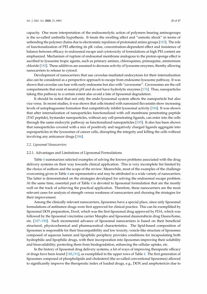
Int. J. Mol. Sci. 2020, 21, 6961 20 of 47
capacity. One more interpretation of the endosomolytic action of polymers bearing aminogroupsis the so-called umbrella hypothesis. It treats the swelling effect and “osmotic shock” in terms ofunbending the polymer chains due to electrostatic repulsion of protonated amino groups [183]. The roleof functionalization of PEI affecting its pK value, concentration-dependent effect and insistence ofbalance between efficacy in endosomal escape and cytotoxicity of formulations at high PEI content areemphasized. Mechanism of rupture of endosomal membrane analogous to the proton-sponge effect isascribed to lysosome tropic agents, such as primary amines, chloroquines, primaquine, ammoniumchloride [183]. These additives are assumed to decrease activity of lysosome enzymes, thereby allowingnanocarriers to release to cytosol.
Development of nanocarriers that use caveolae-mediated endocytosis for their internalizationalso can be considered as a perspective approach to escape from endosome-lysosome pathway. It wasshown that caveolae can fuse with early endosome but also with “caveosome”. Caveosomes are the cellcompartments that exist at neutral pH and do not have hydrolytic enzymes [176]. Thus, nanoparticlestaking this pathway to a certain extent also avoid a fate of liposomal degradation.
It should be noted that not only the endo-lysosomal system affects the nanoparticle but alsovice versa. In recent studies, it was shown that cells treated with nanosized fluvastatin show increasinglevels of autophagosome formation that competitively inhibit lysosomal activity [184]. It was shownthat after internalization of nanoparticles functionalized with cell membrane penetrating peptide(TAT peptide), bystander nanoparticles, without any cell-penetrating ligands, can enter into the cellsthrough the same endocytic pathway as functionalized nanoparticles [185]. It also has been shownthat nanoparticles covered with a mix of positively and negatively charged ligands aggregate intosupraparticles in the lysosomes of cancer cells, disrupting the integrity and killing the cells withoutinvolving any anticancer drugs [186].
2.2. Liposomal Nanocarriers
2.2.1. Advantages and Limitations of Liposomal Formulations
Table 4 summarizes selected examples of solving the known problems associated with the drugdelivery systems on their way towards clinical application. This is very incomplete list limited bythe choice of authors and the scope of the review. Meanwhile, most of the examples of the challengeovercoming given in Table 4 are representative and may be attributed to a wide variety of nanocarriers.The latter is demonstrated on the strategies developed for solving the endosomal escape problem.At the same time, essential part of Table 4 is devoted to liposomal formulation that are the mostlywell on the track of achieving the practical application. Therefore, these nanocarriers are the mostrelevant cases for analysis of strength versus weakness of nanocarriers and choosing the strategies fortheir improvement.
Among the clinically relevant nanocarriers, liposomes have a special place, since only liposomalformulations of antitumor drugs were first approved for clinical practice. This can be exemplified byliposomal DOX preparation, Doxil, which was the first liposomal drug approved by FDA, which wasfollowed by the liposomal vincristine carrier Marqibo and liposomal daunorubicin drug DaunoXome,etc. [187–190]. Such incremental advance of liposomal nanocarriers is based on their beneficialstructural, physicochemical and pharmaceutical characteristics. The lipid-based composition ofliposomes is responsible for their biocompatibility and low toxicity; vesicle-like structure of liposomescomposed of aqueous lumen and lipophilic periphery provides conditions for incapsulating bothhydrophilic and lipophilic drugs, with their incorporation into liposomes improving their solubilityand bioavailability; protecting them from biodegradation, enhancing the cellular uptake, etc.
In the history of liposomal drug delivery systems, a lot of ways of improving therapeutic efficacyof drugs have been found [188,191], as exemplified in the upper rows of Table 4. The first generation ofliposomes composed of phospholipids and cholesterol (the so-called conventional liposomes) allowedto significantly improve the therapeutic index of loaded drugs, e.g., DOX and amphotericin due to

Int. J. Mol. Sci. 2020, 21, 6961 21 of 47
affecting their pharmacokinetics, biodistribution and in vivo toxicity. However, these conventionalliposomes underwent fast clearance by the reticuloendothelial system (RES) due to opsonization byplasma proteins and uptake by macrophages. The liver and spleen were found to accumulate the largestfraction of liposomes. Eventually, the so-called stealth liposomes, modified with polyethylene glycol,were the first formulations to reach the commercial market for cancer therapy. They were the solution offast clearance problem by the RES due to reduced opsonization of the highly hydrated PEG-decoratednanocarriers, which hinder their recognition by macrophages. Along with enhanced circulation time,these nanocarriers were found to accumulate in the tumor tissue that is more vulnerable for nanoparticleaccumulation than healthy tissues. This phenomenon is referred to as enhanced permeability andretention (EPR) effect [192].
In turn, pegylated liposomes along with the stabilization effect on nanoparticles resulted in abody of concerns referred to as the PEG dilemma, with limitations on cellular uptake and furtherendosomal escape, as well as accelerated blood clearance effect (ABC) occurred. The pegylationsignificantly lowered the liposomes’ interaction with the target tissue alongside opsonins. To overcomethis problem, the next generation of liposomal formulations were developed [193,194]. Among differentapproaches to improve the cellular absorption of formulated drugs, the design of ligand-mediatednanocarriers with target molecules is being implemented. For this purpose, different peptides are usedcomplementary to such receptors as integrin, transferrin, human epidermal growth factor 2 (HER2) andothers. In the middle of the range of peptide ligands, is Arg-Gly-Asp (RGD), one of the most popularmotifs used for targeted delivery to tumor cells overexpressing integrin. A recombinant monoclonalantibody trastuzumab was used as a HER2-specific drug. The so-called immunoliposomes decoratedwith the antibody vectors have received much attention in recent literature as an improved generationof liposomal carriers [193,194]. Pegylation of nanocarriers is documented to inhibit endosomal escape,which was testified in the transfection efficacy studies. Therefore, unique approaches were neededto answer this part of the PEG dilemma, including design of pH-responsive PEG-lipids or PEG-PEIconjugates, cleavable pegylated agents bridged by pH-sensitive bonds, e.g., ester, amide, disulfide,that undergo cleavage at low pH with the PEG residue released [184,193,194].
Concerning the different aspects of incremental advances in design and practical application ofliposomal nanocarriers, a variety of liposomal drugs are on the market, and much more are in differentstages of clinical trials, which is comprehensively discussed in reviews [188–190]. This provides evidencethat all the problems occurring with liposomes can be solved without going beyond the liposomes.Meanwhile alternative nanocarriers aimed at the overcoming the limitations of liposomes are beingdeveloped. Some of such cases are given in Table 4 and exemplified by lipid formulations, e.g., solidlipid nanoparticles, vesicle-like carriers, e.g., niosomes and hybrid formulations, cerasomes [195–197].These nanocarriers tend to further improve biocompatibility, loading capacity, targeting efficiency,circulation time, and morphological stability by combining e.g., the lipid/nonionic surfactant orliposomal/silica nanoparticles features. Polymeric nanocarriers with mucoadhesive characteristics canbe alternatively involved in the case of oral administration of drugs [159]. Nevertheless, we wouldlike to adhere to the point of view emphasizing that main problems responsible for the bottleneckin the translation of liposomes to the clinical practice are common obstacles that can be faced by themajority of nanocarriers rather than liposomal formulations only [188]. They are basically relatedto manufacturing process and conjugated with the loss of stability and quality of product uponscaling of the preparation procedure, reliability and reproducibility of final drug, and regulation ofmanufacturing process by government, with intellectual property intricacies involved. The majority ofthe problems arising on the stage of manufacturing process are related to the increasing complexity ofthe composition of nanocontainers, with multistage chemical and biochemical synthesis being involved.The possibility to alleviate these problems on the research stage is probably to move towards thereduction of the complexity of chemical modification of nanocarriers and contribution of the syntheticmanipulations. Therefore, we focus on the noncovalent modification of nanocarriers, with no complexsynthesis involved. With this in mind we discuss below selected aspects related to cationic liposomes

Int. J. Mol. Sci. 2020, 21, 6961 22 of 47
(CLs), with the strategies of noncovalent surface modification of liposomes by cationic surfactantsbeing emphasized.
2.2.2. Cationic Liposomes as Drug Carriers
A survey of recent literature allows us to provide a brief outline of the urgency of the studiesfocusing on CLs. One of the most studied and recognized sphere of CLs application is their use fortransfection. Their positive charge makes them an exceptional nanocarrier for negatively chargednucleic acid sequences and is advantageous due to higher tissue uptake and endosomal escapecapabilities [198]. For a few decades, cationic liposomes have attracted attention due to their lowerimmunogenicity, little to no toxicity, when biodegradable lipids are used, and they have since beenstudied thoroughly to be used in transfection [199]. Thus, scientific interest in CLs for transfection hasnot dwindled in the past few years [198,200–204]. Another major field of CL application is their useas vaccine adjuvants due to their ability to protect the delivered vaccine, retain it at the injection site,and some degree of immunostimulation [205,206].
One of the well-known downsides of the CLs is their toxicity, which is inherent to the positivecharge; thoughtful dosage can mitigate the toxic effects, as shown in the recent work by Knudsen andco-workers [207]. Another major downside is rapid clearance from the bloodstream, resulting fromthe liposome charge [208]. Given the flexible nature of liposomes, it should be possible to avoid theseobstacles, as we will see in the following paragraphs.
Meanwhile, attempts are being made to investigate CLs as delivery vehicles. The cationicsurface charge is very beneficial when dealing with endosomal trafficking due to the negativecharge of endosome membrane [147,209]. Trementozzi et al. have characterized a ternary lipidcomposition that is able to release the contents in a triggerable manner, and addition of only 3 mol %of 1,2-dioleoyl-3-trimethylammonium propane to promote liposome-endosome affinity and fusionhas shown a 5-fold increase of formulation cytotoxicity [210]. Another use of CLs, that has beengaining popularity, is co-delivery of anticancer agents and nucleic acids, since CLs are the onlytype of liposomes that are able to encapsulate both drugs and negatively charged nucleotides well.Some anticancer agents, such as paclitaxel are object to the development of chemoresistance based onsurvivin overexpression in tumor cells. Other types of cancer cells overexpress their essential proteinsthat provide proliferation, each of which could be precisely controlled with siRNA. Cationic liposomesare the only tool that can perform well delivering both an anticancer agent and a siRNA fragment,thus providing a more complex and efficient approach to tumor treatment [201,204].
To summarize this brief discussion, cationic liposomes are of current importance in view oftheir high biomedical potentiality. Nevertheless, in some cases large positive charge of nanocarriersmay prevent their efficacy due to, e.g., non-specific membrane fusion with other than endosomalmembranes, enhanced opsonization or agglutination effects, elevated toxicity of formulations, etc.Therefore, strategies of fabrication of the partially charged liposomes and/or charge-switchablenanocarriers are of importance.
2.2.3. Cationic Liposomes Obtained by Non-Covalent Modification with Surfactants
As an alternative to the formation of mixed compositions based on phosphatidylcholines andcationic lipids, a strategy of non-covalent modification of the surface of liposomes by cationic surfactantsis actively developing (Figure 9). Among the advantages of this approach, the following can be noted:(1) commercial availability of cationic surfactants compared to cationic lipids; (2) the structuraldiversity of cationic surfactants with the ability to embed targeted or functional fragments that enhancebinding; (3) fine-tuning the properties of liposomes (substrate release rate, encapsulation and loadingefficiency) depending on the nature of the surfactant. Cationic liposomes due to their charge helpsolve the following problems: passage through the blood-brain barrier (BBB) (Figure 10); delivery ofantineoplastic agents by increasing cellular uptake; transdermal drug delivery [211–215].

Int. J. Mol. Sci. 2020, 21, 6961 23 of 47
Int. J. Mol. Sci. 2020, 21, x FOR PEER REVIEW 21 of 46
To summarize this brief discussion, cationic liposomes are of current importance in view of their
high biomedical potentiality. Nevertheless, in some cases large positive charge of nanocarriers may
prevent their efficacy due to, e.g., non-specific membrane fusion with other than endosomal
membranes, enhanced opsonization or agglutination effects, elevated toxicity of formulations, etc.
Therefore, strategies of fabrication of the partially charged liposomes and/or charge-switchable
nanocarriers are of importance.
2.2.3. Cationic Liposomes Obtained by Non-covalent Modification with Surfactants
As an alternative to the formation of mixed compositions based on phosphatidylcholines and
cationic lipids, a strategy of non-covalent modification of the surface of liposomes by cationic
surfactants is actively developing (Figure 9). Among the advantages of this approach, the following
can be noted: (1) commercial availability of cationic surfactants compared to cationic lipids; (2) the
structural diversity of cationic surfactants with the ability to embed targeted or functional fragments
that enhance binding; (3) fine-tuning the properties of liposomes (substrate release rate,
encapsulation and loading efficiency) depending on the nature of the surfactant. Cationic liposomes
due to their charge help solve the following problems: passage through the blood-brain barrier (BBB)
(Figure 10); delivery of antineoplastic agents by increasing cellular uptake; transdermal drug delivery
[211–215].
Figure 9. Schematic illustration of the building blocks and diversity of obtainable nanocarriers using
non-covalent bilayer modification.
Figure 9. Schematic illustration of the building blocks and diversity of obtainable nanocarriers usingnon-covalent bilayer modification.
Int. J. Mol. Sci. 2020, 21, x FOR PEER REVIEW 22 of 46
Figure 10. Electrostatic adsorption mediated transcytosis can be a mechanism to penetrate the BBB to
reactivate brain AChE. Reprinted with permission from [216]. Copyright 2020 Elsevier.
In the formation of modified liposomes, the degree of oligomerization of a cationic surfactant
(monomeric, gemini or trimeric) and the ratio of lipid to surfactant are crucial factors, since only at a
low surfactant concentration the incorporation of surfactant into the lipid bilayer of liposomes is
possible, while after a certain threshold value complete destruction of the lipid bilayer and the
formation of mixed surfactant-lipid micelles are observed [217–219]. The structure of the head group
of surfactants can be different.
We begin our consideration of this issue with monomeric single-tailed surfactants (Figure 11).
Pegylated cationic liposomes modified with a new arginine surfactant were described in [220], which
were used to increase the bioavailability of new 2-benzimidazolylquinoxalines derivatives poorly
soluble in water and showed highly selective cytotoxicity against a cervical cancer cell line (HeLa).
Another benzimidazole derivative, 7-(benzimidazol-2-yl)-6-(2,4-dichlorophenyl)-2-thioxo-2,3-
dihydropteridin-4(1H)-one, when encapsulated in imidazolium surfactant-modified liposomes
shows a sharp decrease in viability of diseased cells (HeLa) and increase of viability of normal cells
(Chang liver) [221]. Cationic liposomes based on 1,2-dipalmitoyl-sn-glycero-3-phosphocholine
(DPPC) and alkyltriphenylphosphonium bromides were obtained by non-covalent surface
modification with amphiphiles using varying hydrocarbon tail lengths and lipid/surfactant ratio and
showed a high degree of colocalization with mitochondria of human pancreatic cancer cell line
(PANC-1) [222]. Hybrid liposomes based on DPPC and 1-methyl-3-alkylimidazolium bromides have
been used for metronidazole encapsulation [131]. It was shown that the highest value of
encapsulation efficiency, equal to 75%, is achieved in the case of surface modification with an
octadecyl derivative. New sterically hindered phenol containing a quaternary ammonium moiety
(SHP-n-Q) was also used for incorporation into liposomes [223]. Mixed liposomes based on L-α-
phosphatidylcholine and SHP-n-Q showed long-term stability (1 year) and prolonged release of
rhodamine B. A cationic surfactant, N,N,N-triethyl-N-(12-naphthoxydodecyl)ammonium, was
inserted in liposomes based on phosphatidylcholine [224]. Modified liposomes loaded with cefepime
antibiotic can be used in bacterial infections against Escherichia coli with high inhibitory activity. The
investigation on liposomes formulated with 1,2-dimyristoyl-sn-glycero-3-phosphatidylcholine
(DMPC) and cationic surfactants: (1S, 2S)-N-hexadecyl-N-methylprolinolinium bromide, (1R, 2S)-N-
hexadecyl-N-methylprolinolinium bromide, N-hexadecyl-L-prolinolinium hydrobromide, N,N-
dihexadecyl-L-prolinolinium bromide, in the presence and in the absence of m-tetra-
hydroxyphenylchlorin is presented in [225]. Different liposome formulations have been evaluated for
use against methicillin-resistant Staphylococcus aureus bacteria and one of them has shown results
comparable to free chlorin. The traditional cationic surfactant CTAB was used in the work to impart
a positive charge to niosomes based on N-myristoylserinol and cholesterol [226]. In the increasing
presence of CTAB, the size of niosomes decreases from 300 to 85 nm, which the authors attribute to
the electrostatic repulsion of particles, and the fluidity of liposomes.
Figure 10. Electrostatic adsorption mediated transcytosis can be a mechanism to penetrate the BBB toreactivate brain AChE. Reprinted with permission from [216]. Copyright 2020 Elsevier.
In the formation of modified liposomes, the degree of oligomerization of a cationic surfactant(monomeric, gemini or trimeric) and the ratio of lipid to surfactant are crucial factors, since onlyat a low surfactant concentration the incorporation of surfactant into the lipid bilayer of liposomesis possible, while after a certain threshold value complete destruction of the lipid bilayer and theformation of mixed surfactant-lipid micelles are observed [217–219]. The structure of the head groupof surfactants can be different.
We begin our consideration of this issue with monomeric single-tailed surfactants (Figure 11). Pegylatedcationic liposomes modified with a new arginine surfactant were described in [220], which were used toincrease the bioavailability of new 2-benzimidazolylquinoxalines derivatives poorly soluble in water andshowed highly selective cytotoxicity against a cervical cancer cell line (HeLa). Another benzimidazolederivative, 7-(benzimidazol-2-yl)-6-(2,4-dichlorophenyl)-2-thioxo-2,3-dihydropteridin-4(1H)-one, when
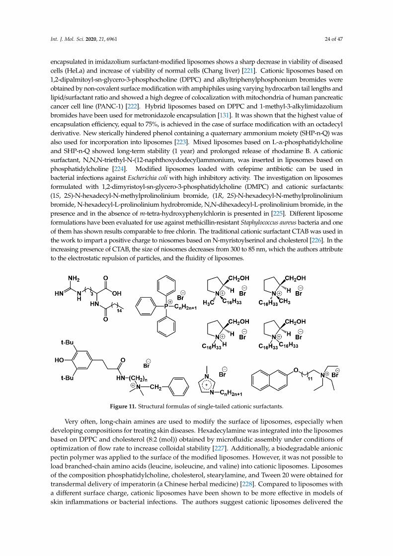
Int. J. Mol. Sci. 2020, 21, 6961 24 of 47
encapsulated in imidazolium surfactant-modified liposomes shows a sharp decrease in viability of diseasedcells (HeLa) and increase of viability of normal cells (Chang liver) [221]. Cationic liposomes based on1,2-dipalmitoyl-sn-glycero-3-phosphocholine (DPPC) and alkyltriphenylphosphonium bromides wereobtained by non-covalent surface modification with amphiphiles using varying hydrocarbon tail lengths andlipid/surfactant ratio and showed a high degree of colocalization with mitochondria of human pancreaticcancer cell line (PANC-1) [222]. Hybrid liposomes based on DPPC and 1-methyl-3-alkylimidazoliumbromides have been used for metronidazole encapsulation [131]. It was shown that the highest value ofencapsulation efficiency, equal to 75%, is achieved in the case of surface modification with an octadecylderivative. New sterically hindered phenol containing a quaternary ammonium moiety (SHP-n-Q) wasalso used for incorporation into liposomes [223]. Mixed liposomes based on L-α-phosphatidylcholineand SHP-n-Q showed long-term stability (1 year) and prolonged release of rhodamine B. A cationicsurfactant, N,N,N-triethyl-N-(12-naphthoxydodecyl)ammonium, was inserted in liposomes based onphosphatidylcholine [224]. Modified liposomes loaded with cefepime antibiotic can be used inbacterial infections against Escherichia coli with high inhibitory activity. The investigation on liposomesformulated with 1,2-dimyristoyl-sn-glycero-3-phosphatidylcholine (DMPC) and cationic surfactants:(1S, 2S)-N-hexadecyl-N-methylprolinolinium bromide, (1R, 2S)-N-hexadecyl-N-methylprolinoliniumbromide, N-hexadecyl-L-prolinolinium hydrobromide, N,N-dihexadecyl-L-prolinolinium bromide, in thepresence and in the absence of m-tetra-hydroxyphenylchlorin is presented in [225]. Different liposomeformulations have been evaluated for use against methicillin-resistant Staphylococcus aureus bacteria and oneof them has shown results comparable to free chlorin. The traditional cationic surfactant CTAB was used inthe work to impart a positive charge to niosomes based on N-myristoylserinol and cholesterol [226]. In theincreasing presence of CTAB, the size of niosomes decreases from 300 to 85 nm, which the authors attributeto the electrostatic repulsion of particles, and the fluidity of liposomes.
Int. J. Mol. Sci. 2020, 21, x FOR PEER REVIEW 23 of 46
Figure 11. Structural formulas of single-tailed cationic surfactants.
Very often, long-chain amines are used to modify the surface of liposomes, especially when
developing compositions for treating skin diseases. Hexadecylamine was integrated into the
liposomes based on DPPC and cholesterol (8:2 (mol)) obtained by microfluidic assembly under
conditions of optimization of flow rate to increase colloidal stability [227]. Additionally, a
biodegradable anionic pectin polymer was applied to the surface of the modified liposomes.
However, it was not possible to load branched-chain amino acids (leucine, isoleucine, and valine)
into cationic liposomes. Liposomes of the composition phosphatidylcholine, cholesterol,
stearylamine, and Tween 20 were obtained for transdermal delivery of imperatorin (a Chinese herbal
medicine) [228]. Compared to liposomes with a different surface charge, cationic liposomes have been
shown to be more effective in models of skin inflammations or bacterial infections. The authors
suggest cationic liposomes delivered the highest amount of the active component across the skin into
deeper regions of the epidermis [228]. The authors of another article also used stearylamine to
produce deformable cationic liposomes to increase the bioavailability of curcumin [229]. The formed
system was non-toxic and safe for skin use. It was found that cationic liposomes significantly inhibit
bacterial Staphylococcus aureus and Streptococcus pyogenes growth in vitro. Most persistent penetration
of human skin ex vivo was achieved with use of Curcumin-CLs.
Between monomeric and gemini surfactants, an intermediate position is occupied by surfactants
with two hydrophobic tails and one head group, which can form vesicles instead of micelles in a
solution and have an ability to integrate into a lipid bilayer (Figure 12). For example, mixed cationic
liposomes based on L-α-phosphatidylcholine and dihexadecylmethylhydroxy-ethylammonium
bromide were obtained in [230] with varying lipid/surfactant ratio. When the liposomes loaded with
2-PAM were intranasally administered, reactivation of brain acetylcholinesterase was observed at
12% for rats subjected to paraoxon poisoning. Liposomes based on egg phosphatidylcholine,
cholesterol, Tween 80 and didecyldimethylammonium bromide were tested to increase the
penetration of andrographolide into the brain [231] on artificial membranes (PAMPA) and
hCMEC/D3 cells. The paper discusses the mechanism of nanoparticle permeation through the
hCMEC/D3 monolayer, highlighting caveolae-mediated endocytosis as the main one. The authors of
[232] found that the presence of dimethyldioctadecylammonium bromide in the lipid bilayer of DPPC
leads to an increase in nanoparticle stiffness, which affects the penetration through the skin.
Apparently, the positive surface charge of the liposomes contributes to their retention in the stratum
corneum and upper layers of epidermis. This statement is opposite to the conclusion of the authors
of articles [228,229], where cationic liposomes with stearylamine penetrate into the deep layers of the
skin. Most likely, the structure of the surfactant has a decisive influence on the rigidity of the lipid
bilayer.
Figure 11. Structural formulas of single-tailed cationic surfactants.
Very often, long-chain amines are used to modify the surface of liposomes, especially whendeveloping compositions for treating skin diseases. Hexadecylamine was integrated into the liposomesbased on DPPC and cholesterol (8:2 (mol)) obtained by microfluidic assembly under conditions ofoptimization of flow rate to increase colloidal stability [227]. Additionally, a biodegradable anionicpectin polymer was applied to the surface of the modified liposomes. However, it was not possible toload branched-chain amino acids (leucine, isoleucine, and valine) into cationic liposomes. Liposomesof the composition phosphatidylcholine, cholesterol, stearylamine, and Tween 20 were obtained fortransdermal delivery of imperatorin (a Chinese herbal medicine) [228]. Compared to liposomes witha different surface charge, cationic liposomes have been shown to be more effective in models ofskin inflammations or bacterial infections. The authors suggest cationic liposomes delivered the

Int. J. Mol. Sci. 2020, 21, 6961 25 of 47
highest amount of the active component across the skin into deeper regions of the epidermis [228].The authors of another article also used stearylamine to produce deformable cationic liposomes toincrease the bioavailability of curcumin [229]. The formed system was non-toxic and safe for skinuse. It was found that cationic liposomes significantly inhibit bacterial Staphylococcus aureus andStreptococcus pyogenes growth in vitro. Most persistent penetration of human skin ex vivo was achievedwith use of Curcumin-CLs.
Between monomeric and gemini surfactants, an intermediate position is occupied by surfactantswith two hydrophobic tails and one head group, which can form vesicles instead of micelles ina solution and have an ability to integrate into a lipid bilayer (Figure 12). For example, mixedcationic liposomes based on L-α-phosphatidylcholine and dihexadecylmethylhydroxy-ethylammoniumbromide were obtained in [230] with varying lipid/surfactant ratio. When the liposomes loaded with2-PAM were intranasally administered, reactivation of brain acetylcholinesterase was observedat 12% for rats subjected to paraoxon poisoning. Liposomes based on egg phosphatidylcholine,cholesterol, Tween 80 and didecyldimethylammonium bromide were tested to increase the penetrationof andrographolide into the brain [231] on artificial membranes (PAMPA) and hCMEC/D3 cells.The paper discusses the mechanism of nanoparticle permeation through the hCMEC/D3 monolayer,highlighting caveolae-mediated endocytosis as the main one. The authors of [232] found that thepresence of dimethyldioctadecylammonium bromide in the lipid bilayer of DPPC leads to an increasein nanoparticle stiffness, which affects the penetration through the skin. Apparently, the positivesurface charge of the liposomes contributes to their retention in the stratum corneum and upperlayers of epidermis. This statement is opposite to the conclusion of the authors of articles [228,229],where cationic liposomes with stearylamine penetrate into the deep layers of the skin. Most likely,the structure of the surfactant has a decisive influence on the rigidity of the lipid bilayer.Int. J. Mol. Sci. 2020, 21, x FOR PEER REVIEW 24 of 46
Figure 12. Structures of cationic surfactants with double, long chains.
The next interesting block about modified liposomes is associated with gemini surfactants
(Figure 13). Modification of liposomes by new 1,4-diazabicyclo [2.2.2]octane surfactants with four
charges and two hydrophobic chains (n-DABCO-s-DABCO-n, where s = 2, 6, 12 and n = 12, 14, 16, 18)
was carried out in [233].
Figure 13. Structural formulas of gemini surfactants.
The liposomal formulation based on phosphatidylcholine and the tetradecyl gemini derivative
DABCO showed the highest antibacterial activity against Staphylococcus aureus (MIC = 7.8 μg‧mL−1)
and lowest cytotoxicity (IC50 > 125). The resulting composition was tested for transdermal drug
delivery. Also, for transdermal delivery, niosomes were obtained using lysine-based gemini
surfactant and cholesterol [234]. The most appropriate administration route and formulation are to
be used to achieve best pharmacological results.
Interactions of surfactant modified liposomes with plasma membranes are elucidated in
[235,236], where authors study how liposomes based on DMPC and a gemini surfactant ((2S,3S)-2,3-
dimethoxy-1,4-bis(N-hexadecyl-N,N-dimethylammonium)butane bromide) are internalized by cells,
using confocal microscopy and electro-rotation. The investigation on the physicochemical properties
of liposomes formulated with a DPPC or 1,2-dioleoyl-sn-glycero-3-phosphocholine (DOPC) [237],
cholesterol, and diastereomeric gemini amphiphiles (2S,3S)-2,3-dimethoxy-1,4-bis(N-hexadecyl-
N,N-dimethylammonium) butane bromide or (S,R)-2,3-dimethoxy-1,4-bis(N-hexadecyl-N,N-
dimethyl- ammonio)butane bromide and on their ability to deliver the plant alkaloid voacamine to
U-2 OS/DX osteosarcoma cells was carried out. The effect of incorporating surfactants into the lipid
bilayer on the efficiency of encapsulation depends on the nature of the lipid itself. A series of papers
by this team of authors ends up in preparation of pegylated gemini-modified liposomes [238].
In [219] the authors have studied how lysine spacer length and aggregate structure of peptide
gemini surfactants affect the interactions of the surfactants with DOPC, 1,2-dioleoyl-sn-glycero-3-
phospho-rac-(1-glycerol) sodium salt (DOPG) and DOPC/DOPG (1:1) vesicles. Derivative with butyl
spacers were found to be optimal in terms of balance of electrostatic, hydrophobic and hydrogen
bonding interactions and the most interacting with the phospholipid bilayer.
Thus, according to the increasing number of articles devoted to the production of cationic
liposomes by non-covalent surface modification with surfactants, we can say that the technique is
successful and relevant. By selecting the structure of the lipid and surfactant, the ratio of the
components, it is possible to control the efficiency of encapsulation and loading, the release rate of
the substrate, the ability of the liposomal formulation to overcome biological barriers (cancer cell
membrane, BBB, various skin levels) to obtain commercially available nanocontainers.
Figure 12. Structures of cationic surfactants with double, long chains.
The next interesting block about modified liposomes is associated with gemini surfactants(Figure 13). Modification of liposomes by new 1,4-diazabicyclo[2.2.2]octane surfactants with fourcharges and two hydrophobic chains (n-DABCO-s-DABCO-n, where s = 2, 6, 12 and n = 12, 14, 16, 18)was carried out in [233].
Int. J. Mol. Sci. 2020, 21, x FOR PEER REVIEW 24 of 46
Figure 12. Structures of cationic surfactants with double, long chains.
The next interesting block about modified liposomes is associated with gemini surfactants
(Figure 13). Modification of liposomes by new 1,4-diazabicyclo [2.2.2]octane surfactants with four
charges and two hydrophobic chains (n-DABCO-s-DABCO-n, where s = 2, 6, 12 and n = 12, 14, 16, 18)
was carried out in [233].
Figure 13. Structural formulas of gemini surfactants.
The liposomal formulation based on phosphatidylcholine and the tetradecyl gemini derivative
DABCO showed the highest antibacterial activity against Staphylococcus aureus (MIC = 7.8 μg‧mL−1)
and lowest cytotoxicity (IC50 > 125). The resulting composition was tested for transdermal drug
delivery. Also, for transdermal delivery, niosomes were obtained using lysine-based gemini
surfactant and cholesterol [234]. The most appropriate administration route and formulation are to
be used to achieve best pharmacological results.
Interactions of surfactant modified liposomes with plasma membranes are elucidated in
[235,236], where authors study how liposomes based on DMPC and a gemini surfactant ((2S,3S)-2,3-
dimethoxy-1,4-bis(N-hexadecyl-N,N-dimethylammonium)butane bromide) are internalized by cells,
using confocal microscopy and electro-rotation. The investigation on the physicochemical properties
of liposomes formulated with a DPPC or 1,2-dioleoyl-sn-glycero-3-phosphocholine (DOPC) [237],
cholesterol, and diastereomeric gemini amphiphiles (2S,3S)-2,3-dimethoxy-1,4-bis(N-hexadecyl-
N,N-dimethylammonium) butane bromide or (S,R)-2,3-dimethoxy-1,4-bis(N-hexadecyl-N,N-
dimethyl- ammonio)butane bromide and on their ability to deliver the plant alkaloid voacamine to
U-2 OS/DX osteosarcoma cells was carried out. The effect of incorporating surfactants into the lipid
bilayer on the efficiency of encapsulation depends on the nature of the lipid itself. A series of papers
by this team of authors ends up in preparation of pegylated gemini-modified liposomes [238].
In [219] the authors have studied how lysine spacer length and aggregate structure of peptide
gemini surfactants affect the interactions of the surfactants with DOPC, 1,2-dioleoyl-sn-glycero-3-
phospho-rac-(1-glycerol) sodium salt (DOPG) and DOPC/DOPG (1:1) vesicles. Derivative with butyl
spacers were found to be optimal in terms of balance of electrostatic, hydrophobic and hydrogen
bonding interactions and the most interacting with the phospholipid bilayer.
Thus, according to the increasing number of articles devoted to the production of cationic
liposomes by non-covalent surface modification with surfactants, we can say that the technique is
successful and relevant. By selecting the structure of the lipid and surfactant, the ratio of the
components, it is possible to control the efficiency of encapsulation and loading, the release rate of
the substrate, the ability of the liposomal formulation to overcome biological barriers (cancer cell
membrane, BBB, various skin levels) to obtain commercially available nanocontainers.
Figure 13. Structural formulas of gemini surfactants.
The liposomal formulation based on phosphatidylcholine and the tetradecyl gemini derivativeDABCO showed the highest antibacterial activity against Staphylococcus aureus (MIC = 7.8 µg·mL−1)and lowest cytotoxicity (IC50 > 125). The resulting composition was tested for transdermal drugdelivery. Also, for transdermal delivery, niosomes were obtained using lysine-based gemini surfactant

Int. J. Mol. Sci. 2020, 21, 6961 26 of 47
and cholesterol [234]. The most appropriate administration route and formulation are to be used toachieve best pharmacological results.
Interactions of surfactant modified liposomes with plasma membranes are elucidatedin [235,236], where authors study how liposomes based on DMPC and a gemini surfactant((2S,3S)-2,3-dimethoxy-1,4-bis(N-hexadecyl-N,N-dimethylammonium)butane bromide) are internalized bycells, using confocal microscopy and electro-rotation. The investigation on the physicochemical properties ofliposomes formulated with a DPPC or 1,2-dioleoyl-sn-glycero-3-phosphocholine (DOPC) [237], cholesterol,and diastereomeric gemini amphiphiles (2S,3S)-2,3-dimethoxy-1,4-bis(N-hexadecyl-N,N-dimethylammonium)butane bromide or (S,R)-2,3-dimethoxy-1,4-bis(N-hexadecyl-N,N-dimethyl- ammonio)butane bromide andon their ability to deliver the plant alkaloid voacamine to U-2 OS/DX osteosarcoma cells was carried out.The effect of incorporating surfactants into the lipid bilayer on the efficiency of encapsulation depends onthe nature of the lipid itself. A series of papers by this team of authors ends up in preparation of pegylatedgemini-modified liposomes [238].
In [219] the authors have studied how lysine spacer length and aggregate structureof peptide gemini surfactants affect the interactions of the surfactants with DOPC,1,2-dioleoyl-sn-glycero-3-phospho-rac-(1-glycerol) sodium salt (DOPG) and DOPC/DOPG (1:1) vesicles.Derivative with butyl spacers were found to be optimal in terms of balance of electrostatic, hydrophobicand hydrogen bonding interactions and the most interacting with the phospholipid bilayer.
Thus, according to the increasing number of articles devoted to the production of cationic liposomesby non-covalent surface modification with surfactants, we can say that the technique is successful andrelevant. By selecting the structure of the lipid and surfactant, the ratio of the components, it is possibleto control the efficiency of encapsulation and loading, the release rate of the substrate, the ability of theliposomal formulation to overcome biological barriers (cancer cell membrane, BBB, various skin levels)to obtain commercially available nanocontainers.
2.2.4. pH-Dependent Cationic Lipids
pH-responsive liposome formulations present a very vast range of benefits and have been studiedsince 1980s and especially in the last decades as candidates for second-generation drug deliverynanoparticles, but they are yet to be approved for clinical applications [239]. One way of utilizingpH sensitivity is to craft lipids that destabilize the liposome membrane when exposed to acidicenvironment, which can be found in the vicinity of tumor tissue. This concept was demonstrated in1980 by Yatvin et al. [240]. Later it was concluded that acidity can also induce fusion in pH-sensitiveliposomes, which can further contribute to the release of a substrate [241]. Such responsive liposomesremain an object of interest and are being studied with application of novel pH-sensitive lipids [242,243].
Another unique feature of such nanocontainers can help solve the problem of premature clearancefrom bloodstream [208], pH-responsive lipids and surfactants can be applied to allow vesicles thatappear almost neutral in the bloodstream, but obtain a positive charge when exposed to lowerpH environments like such in the endosomes [147]. This approach is based on protonation of the3ß-[N-(N’,N’-dimethylaminoethane)carbamoyl]cholesterol hydrochloride (DC–cholesterol) lipid inacidic media, which is common for endosome lumen, that in turn leads to liposome fusion with theendosome membrane and contributes to the endosomal escape. This result received confirmationby comparison of colocalization efficacy of DC-cholesterol liposomes in the early and late lysosomes.A different approach to utilize pH-responsiveness involves thermally treated fumed silica, silanolgroups of which are protonated in acidic media and lead to liposome collapse and rapid releaseof attached substrate, as described in [148]. One more way of exploiting in vitro and in vivo pHdifferences is attachment of cleavable PEG chains to the liposomes, that protect the nanocarriers fromquick reticuloendothelial uptake, but get cut off in the acidic media of tumors or some intracellularregions (Figure 14). Vinyl ether, hydrazine, ester, amide, imide, peptide, disulfide bonds are potentialcandidates for crafting of cleavable PEG-conjugated lipids [146,193]. This approach also helps toavoid targeting peptide degradation in circulation by exposing the depegylated liposome membrane

Int. J. Mol. Sci. 2020, 21, 6961 27 of 47
only in the tumor area [146]. This is one of the most promising ways to actively target tumors dueto the combination of enhanced permeability and retention effect and acidic extracellular pH ofcancerous tissues [244,245]. In the last decade, the researchers find more and more intelligent ways ofexploiting acidic media in bioenvironments, which must lead to the development of novel nanocarriers,that simultaneously have strong sides of pegylated and cationic liposomes.
Int. J. Mol. Sci. 2020, 21, x FOR PEER REVIEW 25 of 46
2.2.4. pH-Dependent Cationic Lipids
pH-responsive liposome formulations present a very vast range of benefits and have been
studied since 1980s and especially in the last decades as candidates for second-generation drug
delivery nanoparticles, but they are yet to be approved for clinical applications [239]. One way of
utilizing pH sensitivity is to craft lipids that destabilize the liposome membrane when exposed to
acidic environment, which can be found in the vicinity of tumor tissue. This concept was
demonstrated in 1980 by Yatvin and coworkers [240]. Later it was concluded that acidity can also
induce fusion in pH-sensitive liposomes, which can further contribute to the release of a substrate
[241]. Such responsive liposomes remain an object of interest and are being studied with application
of novel pH-sensitive lipids [242,243].
Another unique feature of such nanocontainers can help solve the problem of premature
clearance from bloodstream [208], pH-responsive lipids and surfactants can be applied to allow
vesicles that appear almost neutral in the bloodstream, but obtain a positive charge when exposed to
lower pH environments like such in the endosomes [147]. This approach is based on protonation of
the 3ß-[N-(N’,N’-dimethylaminoethane)carbamoyl]cholesterol hydrochloride (DC–cholesterol) lipid
in acidic media, which is common for endosome lumen, that in turn leads to liposome fusion with
the endosome membrane and contributes to the endosomal escape. This result received confirmation
by comparison of colocalization efficacy of DC-cholesterol liposomes in the early and late lysosomes.
A different approach to utilize pH-responsiveness involves thermally treated fumed silica, silanol
groups of which are protonated in acidic media and lead to liposome collapse and rapid release of
attached substrate, as described in [148]. One more way of exploiting in vitro and in vivo pH
differences is attachment of cleavable PEG chains to the liposomes, that protect the nanocarriers from
quick reticuloendothelial uptake, but get cut off in the acidic media of tumors or some intracellular
regions (Figure 14). Vinyl ether, hydrazine, ester, amide, imide, peptide, disulfide bonds are potential
candidates for crafting of cleavable PEG-conjugated lipids [146,193]. This approach also helps to
avoid targeting peptide degradation in circulation by exposing the de-pegylated liposome membrane
only in the tumor area [146]. This is one of the most promising ways to actively target tumors due to
the combination of enhanced permeability and retention effect and acidic extracellular pH of
cancerous tissues [244,245]. In the last decade, the researchers find more and more intelligent ways
of exploiting acidic media in bioenvironments, which must lead to the development of novel
nanocarriers, that simultaneously have strong sides of pegylated and cationic liposomes.
Figure 14. Principal mechanics of pH-sensitive PEG-shedding carriers. In the bloodstream, they
utilize the stealth effect, then they accumulate in the tumor region due to the EPR effect, where a
Figure 14. Principal mechanics of pH-sensitive PEG-shedding carriers. In the bloodstream, they utilizethe stealth effect, then they accumulate in the tumor region due to the EPR effect, where a mildly acidicpH is responsible for PEG cleavage and enhanced absorption of the unpegylated liposome.
3. Mucoadhesive Properties of Supramolecular Systems Based on Macrocycles and Polymers
In the above discussion, we focus on the beneficial applications of nanocarriers bearing cationiccharges, focusing on the family of lipid formulations modified with surfactants. At the same time,in the field of organic nanoparticles as drug delivery systems (nanomedicines) the special class ofbiomimetic transporters of macrocyclic nature and polymeric nanocarriers, especially those of cationicnature, play a significant role which remained unaddressed in the review. Therefore, the final partfocuses on supramolecular assemblies based on mixed macrocyclic and polymeric systems, with specialattention paid to cationic polymers due to their enhanced mucoadhesive properties. Among thevarious methods of delivery and modification of dosage forms, the phenomenon of mucoadhesion,i.e., the ability of the compositions to adhere to the surface of the mucous membrane (mucose) isof particular importance for increasing the drug bioavailability. In order to keep the encapsulateddrug in the tissues of the body (in the mucous membranes of the innards, digestive tract, respiratorysystem, urogenital tract) for a long period of time, the drug nanocontainer should have mucoadhesiveproperties. The lack of adherence to these tissues is the main problem observed in clinical trials withdrugs. Hence, the understanding of the mechanisms of mucoadhesion with mucus is of paramountimportance for creating effective systems for the delivery of drugs to the human body. In this line ofresearch, special attention is paid to the modification of systems by water-soluble and water-swellingpolymers, which can improve mucoadhesive properties in order to increase the therapeutic effect [246].In this section, various types of mucoadhesive systems based on macrocycles and polymers willbe considered, with their combination achieving the synergetic effect on solubility, bioavailability,targeting and mucoadhesiveness (Figure 15).

Int. J. Mol. Sci. 2020, 21, 6961 28 of 47
Int. J. Mol. Sci. 2020, 21, x FOR PEER REVIEW 26 of 46
mildly acidic pH is responsible for PEG cleavage and enhanced absorption of the unpegylated
liposome.
3. Mucoadhesive Properties of Supramolecular Systems Based on Macrocycles and Polymers
In the above discussion, we focus on the beneficial applications of nanocarriers bearing cationic
charges, focusing on the family of lipid formulations modified with surfactants. At the same time, in
the field of organic nanoparticles as drug delivery systems (nanomedicines) the special class of
biomimetic transporters of macrocyclic nature and polymeric nanocarriers, especially those of
cationic nature, play a significant role which remained unaddressed in the review. Therefore, the final
part focuses on supramolecular assemblies based on mixed macrocyclic and polymeric systems, with
special attention paid to cationic polymers due to their enhanced mucoadhesive properties. Among
the various methods of delivery and modification of dosage forms, the phenomenon of
mucoadhesion, i.e., the ability of the compositions to adhere to the surface of the mucous membrane
(mucose) is of particular importance for increasing the drug bioavailability. In order to keep the
encapsulated drug in the tissues of the body (in the mucous membranes of the innards, digestive
tract, respiratory system, urogenital tract) for a long period of time, the drug nanocontainer should
have mucoadhesive properties. The lack of adherence to these tissues is the main problem observed
in clinical trials with drugs. Hence, the understanding of the mechanisms of mucoadhesion with
mucus is of paramount importance for creating effective systems for the delivery of drugs to the
human body. In this line of research, special attention is paid to the modification of systems by water-
soluble and water-swelling polymers, which can improve mucoadhesive properties in order to
increase the therapeutic effect [246]. In this section, various types of mucoadhesive systems based on
macrocycles and polymers will be considered, with their combination achieving the synergetic effect
on solubility, bioavailability, targeting and mucoadhesiveness (Figure 15).
Figure 15. Graphic outline of the mixed macrocycle–polymer systems as mucoadhesive formulations.
It is well known that cyclodextrins (CDs) are able to increase the solubility of various lipophilic
molecules by forming relatively stable inclusion complexes. The stability of these complexes is
usually higher, when the size of the guest molecule is suitable for the size of the macrocycle cavity.
Due to the receptor action and low toxicity, CDs are widely used in the design of drug delivery
systems. Chemical modification of CDs allows the formation of derivatives with improved
physicochemical properties (good solubility, optimal size and ionic charge) as compared to natural
CDs, which can positively affect their biomedical efficiency [247]. This modification makes it possible
to include a wide range of hydrophobic, hydrophilic or amphiphilic fragments, particularly of a
polymer nature, in the macrocyclic framework.
The formation of inclusion complexes of CDs accompanied by an increase in the aqueous
solubility of drugs can lead to spontaneous self-assembly of these complexes. Encapsulation of the
drug telmisartan in the γ-CD cavity can initiate the aggregation of inclusion complexes. The γ-CD
complexes with telmisartan are able to aggregate into spherical nanoparticles with diameters from 50
Figure 15. Graphic outline of the mixed macrocycle–polymer systems as mucoadhesive formulations.
It is well known that cyclodextrins (CDs) are able to increase the solubility of various lipophilicmolecules by forming relatively stable inclusion complexes. The stability of these complexes is usuallyhigher, when the size of the guest molecule is suitable for the size of the macrocycle cavity. Due tothe receptor action and low toxicity, CDs are widely used in the design of drug delivery systems.Chemical modification of CDs allows the formation of derivatives with improved physicochemicalproperties (good solubility, optimal size and ionic charge) as compared to natural CDs, which canpositively affect their biomedical efficiency [247]. This modification makes it possible to include a widerange of hydrophobic, hydrophilic or amphiphilic fragments, particularly of a polymer nature, in themacrocyclic framework.
The formation of inclusion complexes of CDs accompanied by an increase in the aqueous solubilityof drugs can lead to spontaneous self-assembly of these complexes. Encapsulation of the drugtelmisartan in the γ-CD cavity can initiate the aggregation of inclusion complexes. The γ-CD complexeswith telmisartan are able to aggregate into spherical nanoparticles with diameters from 50 nm to500 nm [160]. However, these aggregates are metastable and are likely to precipitate. In order tomaintain the stability of the aqueous composition of eye drops, hydroxypropyl methylcellulose wasadded to the obtained complexes in two ways: polymer was added (i) during the preparation of thetelmisartan/γ-CD powder to form a ternary complex and (ii) to the environment of the formed complexto form a binary complex. The ternary complex exhibits worse mucoadhesive properties compared tothe binary system, but the ternary complex was more stable, since when stored at 4 ◦C for 6 months,there was no noticeable change in the average particle size (2.0–2.5 µm).
Hydroxypropyl methylcellulose and other water-soluble polymers (chitosan and hyaluronic acid)can be used not only for stabilization, but also to increase the solubilization of the drug as an additionalagent to CDs. The ternary CD–polymer–celecoxib complexes form joint aggregates ranging from250 to 350 nm in size and display a large solubilization capacity for the drug [161]. Eye drops ofmicrosuspension containing ternary aggregates based on randomly methylated β-CD–hyaluronicacid–celecoxib complexes showed relatively good mucoadhesion and cytocompatibility with the humanretinal pigment epithelial cell line [162]. Another work [163] demonstrated that the complexationof piroxicam with CDs can be used to ensure controlled in vitro release of the drug and increase itspenetration through the buccal mucosa, and the presence of chitosan in these complexes significantlyincreases the transport properties of the drug. Ahmed et al. also showed that the presence of anincreased amount of CDs in the mucoadhesive system with a polymer increases effectively the solubilityof poorly soluble drug as well as improves absorption and penetration of the drug through the intestinalmucosa [164].
In [248] the ability of a conjugate based on chitosan glycol and β-CD as a container of hydrophobicantitumor drugs (5-fluorouracil, DOX and vinblastine) was evaluated. This drug nanocontainer hasboth mucoadhesive properties due to the polymer and good encapsulating properties due to the

Int. J. Mol. Sci. 2020, 21, 6961 29 of 47
macrocycle. It is also possible to create a pH-sensitive carrier of antitumor drugs from chitosan glycoland carboxymethyl-β-CD [249]. Due to the mucoadhesive property of chitosan glycol, this conjugatecan interact with porcine stomach mucin, and the CD fragment having several free carboxylic acidsgroups can bind DOX in weakly basic medium (pH 7.4) and release it in acidic medium (pH 5.0).
Obviously, the introduction of CD fragments into the polymer chain of chitosan can lead to theformation of a molecular carrier with encapsulating properties for drugs, transport properties throughbiological barriers and the ability to control drug release. However, it is not always possible to solve theproblem of the low solubility of chitosan in water by crosslinking of CD with the polymer; therefore,the chitosan was quaternized [165,250]. Conjugates based on β-CD and quaternized chitosan canaggregate into large particles ranging in size from 800 to 3000 nm, where β-CD is located inside thehydrophobic core and quaternary ammonium fragments are located outside [251]. These aggregatescan solubilize eugenol, exhibit mucoadhesive properties and antimicrobial activity during drug deliverythrough the mucous membrane. It is interesting to note that the release of eugenol from aggregateswas prolonged due to aggregation ability, in contrast to the conjugate, where the macrocyclic andpolymer components are crosslinked with a citric acid spacer. However, the mucoadhesiveness of thelatter was revealed mainly due to the electrostatic interaction between the positively charged aminogroups of chitosan and negatively charged mucous membranes, as well as the hydrogen bond betweenthe carboxyl and hydroxyl groups of citrate spacers and mucus glycoprotein. Moreover, a cytotoxicitytest showed that chitosan–CD conjugates revealed less toxicity towards buccal mucosal cells than theinitial quaternized chitosan [252].
Polymeric surfactants may also be involved in the development of mucoadhesive systems.Pluronics were used to improve the delivery of itraconazole to the eyes [253]. The surfactant mixtureof two Pluronics (F127 and F68) provides the hydrophobic core needed to capture itraconazole.However, there is a likelihood of corneal tissue irritation when using these surfactants in relatively highconcentrations. Micellar dispersion formed from synperonic surfactants, polyethylene oxide and β-CDturned out to be safe, stable, mucoadhesive and effective for penetration of the drug through the rabbitcornea [166]. The sizes of these compositions were in the range of 180 to 360 nm, which is favorablefor transcorneal drug penetration through the intercellular pathways [254]. The resulting dispersionsdemonstrated the rapid rate and extent of distribution of the encapsulated itraconazole in vitro and exvivo compared to a free drug suspension. In other work [255] the magnetic nanoparticles based oniron oxide were developed by precipitation of iron salts in the presence of ammonia, and four differentcompositions based on them (particles without coating, with β-CD, with Pluronic 127 and with amixture of macrocycle and polymer) were created. The latter composition containing particles of about117 nm in size was found to be effective for binding to mucus compared to other nanoparticles studied,which was evident from the results of ex vivo absorption of these nanoparticles by the gastrointestinaltract, ovaries, pancreas, and colon.
The combination of hydroxypropyl-β-CD with Tween 80 makes it possible to stabilize thedrug econazole nitrate, protecting the encapsulated drug from aggregation for one year at roomtemperature [167]. This composition was suspended in chitosan acidic solution to increase the releaseand bioavailability of the drug. An in vivo evaluation in eyes of an albino rabbit demonstrated a higherbioavailability of the selected drug suspended in chitosan compared to similar drug suspended inisotonic buffer at pH 7.4.
Hydroxypropyl-β-CD and polyvinylpyrrolidone were also involved in the creation of rapidlydissolving nanofibers capable of encapsulating clotrimazole [256]. These nanofibers showed rapiddrug release and antifungal activity, but the treatment of oral candidiasis requires a sufficiently longperiod of time, during which the concentration of the drug exceeds the MIC. Therefore, to slow downthe release of the drug, the nanofibers were coated with additional layers of chitosan-cysteine andpolyvinyl alcohol, which also improved mucoadhesive properties [168].
CDs can be used not only to increase the water solubility of drugs, but also to improvetheir taste. Hydroxypropyl-β-CD eliminates the unpleasant taste of irsogladine maleate used for

Int. J. Mol. Sci. 2020, 21, 6961 30 of 47
treatment of stomatitis. Addition of the macrocycle to the mucoadhesive gum ghatti, consisting ofhigh-molecular-weight polysaccharides, leads to the formation of a viscous solution as a result ofaggregation due to hydrogen bonds between the macrocycle and the polymer [257].
Mucoadhesive microspheres based on a complex of γ-CD and raloxifene can be obtained usingvarious proportions of carbopol and hydroxypropyl methylcellulose. The size of the obtained particleson the percentage of polymers ranged from 3 to 15 microns, and the encapsulation efficiency ofraloxifene ranged from 81.63 to 87.73%. These microspheres had remarkable mucoadhesion andcontrolled drug release lasting up to 24 h. More than 60% of the oral dose was kept for 4 h in thestomach of Wistar rats [258].
The mixed compositions based on CDs and polymers can be used not only to encapsulate drugs,but also proteins and enzymes. In [259] microspheres consisting of chitosan, hydroxypropyl-β-CDand polyethylene glycol were successfully developed. The obtained microspheres had a diameter of6–7 µm, and the BSA encapsulation efficiency was more than 70%. The stability of primary, secondaryand tertiary structures of bound BSA was confirmed by a set of physicochemical methods. In otherwork [260] Bruton’s tyrosine kinase inhibitor, ibrutinib, was loaded into nanoparticles based on chitosanand sulfobutyl ether-β-CD due to the inclusive participation of the latter, and the activity of ibrutinibdid not disappear. These nanoparticles loaded with ibrutinib had a diameter of 280 nm and a zetapotential of +19.1 mV, which decreased to +9.2 mV after contact with negatively charged sialic acidresidues in mucin. In addition, they showed a prolonged and pH-independent release of the drug inthe mimic gastrointestinal tract.
Nanoparticles based on hydroxypropyl-β-CD and chitosan can be used in targeting various organsof the gastrointestinal tract. A study of the release of hydrophobic substrate from this compositionshowed a sharp release (more than 40%) in the gastric fluid due to the breakdown of chitosan andβ-CD inclusion complex at low pH [261]. As in the case of hydrophilic substrate, only a small amountof hydrophobic substrate is released in the intestinal fluid. In the colonic fluid, there is a sharp increasein release due to enzymatic cleavage of chitosan, as well as the decomposition of CD molecule bybacterial amylases.
Besides a wide range of different nanoparticles and microspheres, mucoadhesive micro- andnanocapsules can be obtained from macrocycles and polymers. Simvastatin microcapsules wereobtained by complexation with hydroxypropyl-β-CD, hydrophilic sodium alginate and mucilageobtained from Dillenia indica [262]. Drug release from such microcapsules reached 73% within 12 h,and this delay was due to the Dillenia polymer. In addition, Dillenia has sufficient mucoadhesiveproperties and good compatibility with the drug or any other ingredients of the composition.
Amphiphilic CDs and polymers can be used to create mucoadhesive drug nanocontainers. β-CDwith hexyl tails forms nanocapsules with poly-ε-caprolactone for encapsulation of camptothecin [169].Drug loading and cellular interaction of these nanocapsules can be regulated by coating with chitosan.The coating of the chitosan layer on nanoparticles consisting of macrocycle–drug complexes not onlyincreases the drug loading, but also decreases the release of camptothecin. The presence of a positivecharge of the obtained nanoparticles provides higher absorption due to electrostatic interaction withnegatively charged intestinal lumen epithelial cells.
The addition of hydroxypropyl-β-CD as a permeation enhancer improves the penetration of timololmaleate through the bovine cornea [170]. However, it was found that timolol formulations containingbioadhesive polymers (hyaluronic acid, chitosan, alginate) alone or together with hydroxypropyl-β-CDcan reduce drug penetration. When using polymers as stabilizers of complexes between CDs anddrugs, it is necessary to take into account the competitive binding of the polymer with the macrocycle.For example, poloxamer 407 can compete with drug molecules to form inclusion complexes withCD [263,264].
Non-covalent aggregation of macrocycles with polymers was used to develop mucoadhesivecompositions with effective encapsulating ability. Ion cross-linked nanoparticles based on oppositelycharged chitosan and sulfobutyl ether-β-CD are an interesting system for the delivery of econazole

Int. J. Mol. Sci. 2020, 21, 6961 31 of 47
nitrate to the mucous membrane of the eye [265]. Half of the drug encapsulated in these nanoparticlesis released within 8 h. In addition, the nanoparticles showed mucoadhesive properties, which allowthem to interact with the mucous membrane of the eye for an extended period of time and thus providean enhanced and controlled effect of the drug on the surface of rabbit eyes.
The use of complexation betweenα-CD molecules and polysaccharides modified with hydrophobicalkyl chains leads to the spontaneous formation of nano- or micron-sized platelets that can encapsulateantimicrobials for effective treatment of mucosal infections [266,267]. The crystal platelets have layeredstructures consisting of complexes of CDs with alkylated guests, such as polymer framework bearingalkyl chains (Figure 16). Chitosan derivatives capable of being located on the surface of aggregates canbe used as polymers in the formation of these platelets [268]. The contact area of the platelets withthe biological surface is greater compared to spherical aggregates. Therefore, the adhesion force ofthe platelets is much greater and provides better binding to mucin. The electrostatic forces betweenchitosan and mucin chains can stretch the outer chitosan layer of the platelets. If the cohesive forceof the chitosan inside the platelet is weaker than the cohesive force between chitosan and mucin,the system can break down in the intermediate layer of the platelet. As a result, the surface layer can betorn off from the platelet with the destruction of the latter, that is important when developing systemsfor targeted drug delivery with controlled release.Int. J. Mol. Sci. 2020, 21, x FOR PEER REVIEW 30 of 46
Figure 16. Schematic view of the complex formation between α-CD and alkylated polysaccharides
followed by platelet organization in water. Reprinted with permission from [268]. Copyright 2018
American Chemical Society.
In addition to crosslinking of macrocycles on flexible open-chain molecules, β-CD can be used
to modify the mesoporous silica nanoparticles with hydroxyl, amino, and thiol groups [171]. Thiol-
functionalized nanoparticles exhibit significantly higher urothelial mucoadhesiveness compared to
hydroxyl- and amino-functionalized nanoparticles, which is related with the formation of disulfide
bonds between thiol functionalized nanoparticles and cysteine subdomains of mucus glycoproteins.
Anticancer DOX can be encapsulated in these nanoparticles and slowly released in an acidic
environment.
The authors of [172] definitely proved that the mucoadhesion of the nanoparticles of methyl-β-
CD in combination with polyisobutyl cyanoacrylate and thiolated chitosan is a mandatory condition
to enhance the intestinal permeability of docetaxel. The obtained nanoparticles were spherical, had a
diameter in the range from 200 to 400 nm with a positive charge, and the encapsulation efficiency
with respect to drug was 70–80%. In vitro experiments on a model intestinal medium showed that
the docetaxel is gradually released, reaching 60% of release after 24 h and 100% after 48 h. The
intestinal permeability of drug loaded into the nanoparticles was higher than in the control ethanol
solution of this drug.
For modification of mucoadhesive nanoparticles not only CDs can be used, but also derivatives
of porphyrins, which are a promising choice for drug delivery systems in photodynamic therapy,
combining the advantages of efficient drug transport to target cells and reducing of undesirable side
effects. The surface of nanoparticles based on biodegradable poly-DL-lactide-co-glycolide and
5,10,15,20-tetrakis(m-hydroxyphenyl)porphyrin can be modified with polyethylene glycol or
chitosan [269]. The highest number of pegylated nanoparticles was found in human colon
adenocarcinoma cells. Therefore, pegylated porphyrin nanoparticles are a promising drug delivery
system for topical treatment of gastrointestinal cancer.
Summarizing the results of mucoadhesive study of mixed macrocycle–polymer systems, when
designing effective delivery systems for poorly water-soluble drugs, CDs must be added to
mucoadhesive polymers to provide good drug solubility in the aqueous medium, adequate
bioavailability and desired drug effect. CDs not only increase the solubility of the lipophilic active
substances, but also have the potential to control the rate of their release, thereby offering a promising
application for the development of sustained-release formulations. It has also been observed that to
Figure 16. Schematic view of the complex formation between α-CD and alkylated polysaccharidesfollowed by platelet organization in water. Reprinted with permission from [268]. Copyright 2018American Chemical Society.
In addition to crosslinking of macrocycles on flexible open-chain molecules, β-CD can beused to modify the mesoporous silica nanoparticles with hydroxyl, amino, and thiol groups [171].Thiol-functionalized nanoparticles exhibit significantly higher urothelial mucoadhesiveness comparedto hydroxyl- and amino-functionalized nanoparticles, which is related with the formation ofdisulfide bonds between thiol functionalized nanoparticles and cysteine subdomains of mucusglycoproteins. Anticancer DOX can be encapsulated in these nanoparticles and slowly released in anacidic environment.
The authors of [172] definitely proved that the mucoadhesion of the nanoparticles of methyl-β-CDin combination with polyisobutyl cyanoacrylate and thiolated chitosan is a mandatory condition to

Int. J. Mol. Sci. 2020, 21, 6961 32 of 47
enhance the intestinal permeability of docetaxel. The obtained nanoparticles were spherical, had adiameter in the range from 200 to 400 nm with a positive charge, and the encapsulation efficiencywith respect to drug was 70–80%. In vitro experiments on a model intestinal medium showed that thedocetaxel is gradually released, reaching 60% of release after 24 h and 100% after 48 h. The intestinalpermeability of drug loaded into the nanoparticles was higher than in the control ethanol solution ofthis drug.
For modification of mucoadhesive nanoparticles not only CDs can be used, but also derivativesof porphyrins, which are a promising choice for drug delivery systems in photodynamic therapy,combining the advantages of efficient drug transport to target cells and reducing of undesirableside effects. The surface of nanoparticles based on biodegradable poly-DL-lactide-co-glycolideand 5,10,15,20-tetrakis(m-hydroxyphenyl)porphyrin can be modified with polyethylene glycol orchitosan [269]. The highest number of pegylated nanoparticles was found in human colonadenocarcinoma cells. Therefore, pegylated porphyrin nanoparticles are a promising drug deliverysystem for topical treatment of gastrointestinal cancer.
Summarizing the results of mucoadhesive study of mixed macrocycle–polymer systems,when designing effective delivery systems for poorly water-soluble drugs, CDs must be addedto mucoadhesive polymers to provide good drug solubility in the aqueous medium, adequatebioavailability and desired drug effect. CDs not only increase the solubility of the lipophilic activesubstances, but also have the potential to control the rate of their release, thereby offering a promisingapplication for the development of sustained-release formulations. It has also been observed that toimprove mucoadhesion to mucous surfaces, the building blocks of mucoadhesive systems are thiolatedfor covalent binding to mucin subdomains. In addition, porphyrins having an affinity for cancer cellscan also be involved in the targeted action of drug formulations in the creation of mucoadhesive drugdelivery systems.
The state-of-the-art is that the creation of effective drug delivery systems can involve mucoadhesivepolymers, which can form supramolecular compositions with macrocycles. If the macrocycle in such amixture provides binding and release of drugs, then the additional presence of polymer can not onlyincrease an amount of encapsulated drug, but stabilizes the system as a whole and ensures efficientdrug delivery across various mucous membranes. In general, mixed systems based on macrocyclesand polymers perform better than individual systems.
Regarding the potential clinical relevance of mucoadhesive systems, scientists are finding waysto develop them using various approaches to improve the bioavailability of less or ineffective drugsby manipulating composition formulations. The polymer and supramolecular approaches must bejointly implemented in order to find new mucoadhesive polymers and macrocycles with additionalattributes of biodegradability, biocompatibility, non-toxicity, mucoadhesiveness for certain cells ormucous membranes for the successful delivery of molecules needed by the body. Although thephysicochemical and biological properties of pure mucoadhesive polymers and macrocycles andthe published results of clinical use allow them to be considered as promising raw materials for thepreparation of drugs with various pharmacotherapeutic actions, the ability of formulations based onthem to actually respond to medical needs has still to be demonstrated, and this imposes collaborationwith medical researchers for potential clinical translations.
4. Conclusions
To conclude, this review addresses selected topics in the field of biomimetic amphiphilic systemsincluding both fundamental and practical aspects, with studies over the last few years treated as a matterof first priority. The design and synthesis of amphiphilic building blocks is documented to attract wideattention, which is no little measure due to their practical usefulness. Since a wide variety of practicalapplications is aimed at the development of drug carriers, answering the green chemistry criteria isstrongly required. Therefore, much effort was devoted to the preparation of low-toxic, biocompatible,and degradable amphiphiles capable of self-assembling in nanoscale aggregates at low concentrations.

Int. J. Mol. Sci. 2020, 21, 6961 33 of 47
Special interest in cationic surfactants is due to their high affinity to biopolyanions and negativelycharged membranes. Biomimetic principles in the design of novel amphiphilic compounds basedon natural fragment (amino acid, sugar, bile salt, nucleobase) and environmentally friendly siliconemoiety approves oneself as a promising strategy for construction of soft supramolecular systems withdesired shape and stimuli-responsive functions. Specifically, this strategy has a great possibility fordevelopment of drug delivery systems with various morphology, tunable physicochemical propertiesand stability, which could become a useful tool for the solution of the problem of controlled releaseof cargo.
Liposomal formulations have received the main attention due to the fact that they wereamong the few carriers approved for the clinical treatment. Since numerous marketed liposomaltherapeutics are available, these formulations appeared to be of particular importance for the analysisof bench-to-bedside factors. Several generations of liposomes were designed and actively advancedtowards the market, beginning with conventional liposomes to stealth and targeted modifications,with the strategies for enhanced loading capacity, triggered release, endosomal escape, solution ofPEG dilemma, and overcoming the BBB developed. Although specificity occurs in the developmentof different family of carriers, there are many common challenges typical for each of them. This isexemplified herein by such problem as endosomal escape that was discussed in detail, including waysfor overcoming it. As alternative nanocarriers solid lipid nanoparticles, niosomes and cerasomes areidentified that are assumed to provide the better standard of toxicity, biocompatibility, loading capacityand morphological stability in the near future.
Analysis of the reasons preventing fast progress in the translation of the formulated drugsto the clinic reveals that they are mostly related to the manufacturing process and consist of theloss of product stability and quality upon scaling up of the preparation procedures, as well as inproblems with the reliability and reproducibility of the final drug. To some extent, these drawbackscan be reduced by simplification of the preparation of the nanocarriers, excluding complicatedsynthetic procedures. Therefore, much attention is paid to the publications focusing on the noncovalentmodification of nanocarriers, with the priority given to cationic surfactants, that revealed their beneficialefficacy from the viewpoint of colloidal stability, overcoming the biological barriers, cellular uptake,mitochondria-targeted drug delivery, etc. In this context, modification of nanocarriers with cationicadditives is a promising strategy, especially with the use of charge-reversal amphiphiles that allowfor preventing of lysosome degradation of encapsulated cargo. The disadvantage of cationic carriersis their toxicity that can be solved by tuning the lipid composition, with the balance between toxiccharacteristics and beneficial properties (stability, electrostatic affinity to biomembranes, antimicrobialeffect) achieved.
As a whole, the key point that should be stressed is the tendency to engineering the polycomponentformulations providing a synergetic effect. Hence, the colloidal, polymer and supramolecularapproaches must be jointly implemented in order to find new effective nanocarriers for the successfuldelivery of molecules needed by the body. From this viewpoint, macrocycle–polymer compositionsare of importance, since macrocycles exhibit selective binding of the drug resulting in enhancedsolubility, while polymer shell guarantees the stability in biomicroenvironment and improved affinityto biointerfaces. Since the physicochemical and biological properties of some pure surfactants,mucoadhesive polymers and macrocycles, as well as the published results of their clinical use, presentthem as favorable raw materials for the preparation of medicinal compositions, the mixed compositionsbased on them can be considered promising for the design of effective drug nanocarriers.
Author Contributions: R.K. formal analysis, investigation, writing—review & editing. G.G. formal analysis,writing—original draft. D.G. writing—original draft, visualization. D.K. writing—original draft. R.P. writing—originaldraft, visualization. K.P. writing—original draft, validation, conceptualization. L.Z. conceptualization, data curation,formal analysis, investigation, supervision, writing—original draft, writing—review & editing. O.S. funding acquisition,project administration, supervision. All authors have read and agreed to the published version of the manuscript.
Funding: The authors thank the Russian Science Foundation (project№ 17-73-20253) for financial support.

Int. J. Mol. Sci. 2020, 21, 6961 34 of 47
Conflicts of Interest: The authors declare no conflict of interest.
Abbreviations
CMC Critical micelle concentrationTMA TrimethylammoniumDNA Deoxyribonucleic acidDOX DoxorubicinBSA Bovine serum albuminMCF-7 Michigan cancer foundation-7HEK Human embryonic kidneyATG Alkyl triazole glycosidesTEM Transmission electron microscopyEGFR Epidermal growth factor receptorPEI PolyethyleniminePAMAM PolyamidoamineTAT Transactivator of transcriptionFDA Food and drug administrationRES Reticuloendothelial systemPEG Polyethylene glycolEPR Enhanced permeability and retentionABC Accelerated blood clearanceHER2 Human epidermal growth factor 2RGD Arginine-glycine-aspartic acidCL Cationic liposomesiRNA Small interfering RNABBB Blood-brain barrierDPPC 1,2-Dipalmitoyl-sn-glycero-3-phosphocholineSHP-n-Q Sterically hindered phenol containing a quaternary ammonium moietyDMPC 1,2-Dimyristoyl-sn-glycero-3-phosphatidylcholineCTAB Cetyltrimethylammonium bromide2-PAM 2-Pyridine aldoxime methyl chloridePAMPA Parallel artificial membrane permeability assayhCMEC/D3 Human cerebral microvascular endothelial cell line, D3 cloneDABCO 1,4-Diazabicyclo [2.2.2]octaneDOPC 1,2-Dioleoyl-sn-glycero-3-phosphocholineDOPG 1,2-Dioleoyl-sn-glycero-3-phospho-rac-(1-glycerol) sodium saltDC-cholesterol 3β-[N-(N’,N’-Dimethylaminoethane)-carbamoyl]cholesterol hydrochlorideCD Cyclodextrin
References
1. Rusanov, A.I. Micellization in Surfactant Solutions; Taylor & Francis: London, UK, 1997; 326p.2. Zakharova, L.Y.; Pashirova, T.N.; Doktorovova, S.; Fernandes, A.R.; Sanchez-Lopez, E.; Silva, A.M.; Souto, S.B.;
Souto, E.B. Cationic surfactants: Self-assembly, structure-activity correlation and their biological applications.Int. J. Mol. Sci. 2019, 20, 5534.
3. Ghosh, S.; Ray, A.; Pramanik, N. Self-assembly of surfactants: An overview on general aspects of amphiphiles.Biophys. Chem. 2020, 265, 106429.
4. Ansari, M.J. An overview of techniques for multifold enhancement in solubility of poorly soluble drugs.Curr. Issues Pharm. Med. Sci. 2019, 32, 203–209.
5. Kwa’sniewska, D.; Chen, Y.-L.; Wieczorek, D. Biological activity of quaternary ammonium salts and theirderivatives. Pathogens 2020, 9, 459.
6. Anselmo, A.C.; Mitragotri, S. Nanoparticles in the clinic: An update. Bioeng. Transl. Med. 2019, 4, e10143.

Int. J. Mol. Sci. 2020, 21, 6961 35 of 47
7. Anestopoulos, I.; Kiousi, D.; Klavaris, E.A.; Galanis, A.; Salek, K.; Euston, S.R.; Pappa, A.; Panayiotidis, M.I.Surface active agents and their health-promoting properties: Molecules of multifunctional significance.Pharmaceutics 2020, 12, 688.
8. Zhou, C.; Wang, Y. Structure–activity relationship of cationic surfactants as antimicrobial agents.Curr. Opin. Colloid Interface Sci. 2020, 45, 28–43.
9. Para, G.; Łuczyn´ski, J.; Palus, J.; Jarek, E.; Wilk, K.A.; Warszyn´ski, P. Hydrolysis driven surface activity ofesterquat surfactants. J. Colloid Interface Sci. 2016, 465, 174–182.
10. Yasa, S.R.; Kaki, S.S.; Poornachandra, Y.; Kumar, C.G.; Penumarthy, V. Synthesis, characterization,antimicrobial and biofilm inhibitory studies of new esterquats. Bioorg. Med. Chem. Lett. 2016, 26, 1978–1982.
11. Bhat, I.A.; Roy, B.; Kabir-ud-Din. Micelles of cleavable gemini surfactant induce fluorescence switching innovel probe: Industrial insight. J. Ind. Eng. Chem. 2019, 77, 60–64.
12. Wu, J.; Gao, H.; Shi, D.; Yang, Y.; Zhang, Y.; Zhu, W. Cationic gemini surfactants containing both amide andester groups: Synthesis, surface properties and antibacterial activity. J. Mol. Liq. 2020, 299, 112248.
13. Mirgorodskaya, A.B.; Kushnazarova, R.A.; Lukashenko, S.S.; Voloshina, A.D.; Lenina, O.A.; Zakharova, L.Y.;Sinyashin, O.G. Carbamate-bearing surfactants: Micellization, solubilization, and biological activity.J. Mol. Liq. 2018, 269, 203–210.
14. Kushnazarova, R.A.; Mirgorodskaya, A.B.; Lukashenko, S.S.; Voloshina, A.D.; Sapunova, A.S.; Nizameev, I.R.;Kadirov, M.K.; Zakharova, L.Y. Novel cationic surfactants bearing cleavable carbamate fragment: Tunablemorphological behavior, solubilization of hydrophobic drugs and cellular uptake study. J. Mol. Liq. 2020,318, 113894.
15. Mirgorodskaya, A.B.; Kushnazarova, R.A.; Lukashenko, S.S.; Zakharova, L.Y. Self-assembly of mixed systemsbased on nonionic and carbamate-bearing cationic surfactants as a tool for fabrication of biocompatiblenanocontainers. J. Mol. Liq. 2019, 292, 111407.
16. Mirgorodskaya, A.B.; Kushnazarova, R.A.; Lukashenko, S.S.; Nikitin, E.N.; Sinyashin, K.O.; Nesterova, L.M.;Zakharova, L.Y. Carbamate-bearing surfactants as effective adjuvants promoted the penetration of theherbicide into the plant. Colloids Surf. A 2020, 586, 124252.
17. Rusanov, A.I. Micelle Formation in Surfactant Solutions; Chemistry: St. Petersburg, Russia, 1992; 280p.18. Bordes, R.; Holmberg, K. Amino acid-based surfactants—Do they deserve more attention? Adv. Colloid
Interface Sci. 2015, 222, 79–91.19. Aslam, J.; Lone, I.H.; Radwan, N.R.E.; Siddiqui, M.F.; Parveen, S.; Alnoman, R.B.; Aslam, R. Molecular
interaction of amino acid-based gemini surfactant with human serum albumin: Tensiometric, spectroscopic,and molecular docking study. ACS Omega 2019, 4, 22152–22160.
20. Rostami, A.; Hashemi, A.; Takassi, M.A.; Zadehnazari, A. Experimental assessment of a lysine derivativesurfactant for enhanced oil recovery in carbonate rocks: Mechanistic and core displacement analysis.J. Mol. Liq. 2017, 232, 310–318.
21. Pi-Boleda, B.; Bouzas, M.; Gaztelumendi, N.; Illa, O.; Nogués, C.; Branchadell, V.; Pons, R.; Ortuño, R.M.Chiral pH-Sensitive cyclobutane β-amino acid-based cationic amphiphiles: Possible candidates for use ingene therapy. J. Mol. Liq. 2020, 297, 111856.
22. Bourkaib, M.C.; Delaunay, S.; Framboisier, X.; Hôtel, L.; Aigle, B.; Humeau, C.; Guiavarc’h, Y.; Chevalot, I.N-acylation of L-amino acids in aqueous media: Evaluation of the catalytic performances of Streptomycesambofaciens aminoacylases. Enzyme Microb. Technol. 2020, 137, 109536.
23. Wolf, J.D.; Kurpiers, M.; Baus, R.A.; Götz, R.X.; Griesser, J.; Matuszczak, B.; Bernkop-Schnürch, A.Characterization of an amino acid based biodegradable surfactant facilitating the incorporation of DNA intolipophilic delivery systems. J. Colloid Interface Sci. 2020, 566, 234–241.
24. Nelson, D.L.; Cox, M.M. Lehninger Principles of Biochemistry, 7th ed.; Macmillan Publishers Ltd.: New York,NY, USA, 2017; 1312p.
25. Bernal, C.; Guzman, F.; Illanes, A.; Wilson, L. Selective and eco-friendly synthesis of lipoaminoacid-basedsurfactants for food, using immobilized lipase and protease biocatalysts. Food Chem. 2018, 239, 189–195.
26. Katiyar, S.S.; Kushwah, V.; Dora, C.P.; Patil, R.Y.; Jain, S. Design and toxicity evaluation of novel fattyacid-amino acid-based biocompatible surfactants. AAPS PharmSciTech 2019, 20, 186.
27. Rothbauer, G.A.; Rutter, E.A.; Reuter-Seng, C.; Vera, S.; Billiot, E.J.; Fang, Y.; Billiot, F.H.; Morris, K.F. Nuclearmagnetic resonance investigation of the effect of pH on micelle formation by the amino acid-based surfactantundecyl l-phenylalaninate. J. Surfact. Deterg. 2018, 21, 139–153.

Int. J. Mol. Sci. 2020, 21, 6961 36 of 47
28. Lone, M.S.; Bhat, P.A.; Shah, R.A.; Chat, O.A.; Dar, A.A. A green pH-switchable amino acid based smartwormlike micellar system for efficient and controlled drug delivery. ChemistrySelect 2017, 2, 1144–1148.
29. Covis, R.; Vives, T.; Gaillard, C.; Benoit, M.; Benvegnu, T. Interactions and hybrid complex formation ofanionic algalpolysaccharides with a cationic glycine betaine-derived surfactant. Carbohydr. Polym. 2015,121, 436–448.
30. Mondal, S.; Raposo, M.L.; Ghosh, A.; Prieto, G.; Ghosh, S. Physicochemical and conformational studies oninteraction of myoglobin with an amino-acid based anionic surfactant, sodium N-dodecanoyl sarcosinate(SDDS). Colloids Surf. A 2019, 577, 167–174.
31. Lu, H.; Yuan, M.; Fang, B.; Wang, J.; Guo, Y. Wormlike micelles in mixed amino acid-based anionic surfactantand zwitterionic surfactant systems. J. Surfact. Deterg. 2015, 18, 589–596.
32. Chen, Z.; Zhang, P.; Sun, Y.; Wang, C.; Xu, B. Interfacial dilational rheology of sodium lauryl glycine andmixtures with conventional surfactants. J. Surfact. Deterg. 2019, 22, 1477–1485.
33. Mustahil, N.A.; Baharuddin, S.H.; Abdullah, A.A.; Reddy, A.V.B.; Mutalib, M.I.A.; Moniruzzaman, M.Synthesis, characterization, ecotoxicity and biodegradability evaluations of novel biocompatible surfaceactive lauroyl sarcosinate ionic liquids. Chemosphere 2019, 229, 349–357.
34. Sreenu, A.; Nayak, R.R.; Prasad, R.B.N.; Poornachandra, Y.; Kumar, C.G. Surface and antimicrobial propertiesof N-palmitoyl amino acid-based surfactants. J. Dispers. Sci. Technol. 2015, 36, 765–771.
35. Perinelli, D.R.; Casettari, L.; Cespi, M.; Fini, F.; Man, D.K.W.; Giorgioni, G.; Canala, S.; Lam, J.K.W.;Bonacucina, G.; Palmieri, G.F. Chemical–physical properties and cytotoxicity of N-decanoyl aminoacid-basedsurfactants: Effect of polar heads. Colloids Surf. A 2016, 492, 38–46.
36. Cardoso, A.M.; Morais, C.M.; Cruz, A.R.; Silva, S.G.; do Vale, M.L.; Marques, E.F.; Pedroso de Lima, M.C.;Jurado, A.S. New serine-derived gemini surfactants as gene delivery systems. Eur. J. Pharm. Biopharm. 2015,89, 347–356.
37. Torres, M.A.M.; Gómez, E.M.P.; Fernández, M.A.; Veglia, A.V.; Pacioni, N.L. Role of a cystine-based Geminisurfactant ligand in the synthesis of catalytic active silver nanoparticles. J. Mol. Liq. 2019, 284, 110–116.
38. Branco, M.A.; Pinheiro, L.; Faustino, C. Amino acid-based cationic gemini surfactant-protein interactions.Colloids Surf. A 2015, 480, 105–112.
39. Takassi, M.A.; Zargar, G.; Madani, M.; Zadehnazari, A. The preparation of an amino acid-based surfactantand its potential application as an EOR agent. Pet. Sci. Technol. 2017, 35, 385–391.
40. Pinazo, A.; Petrizelli, V.; Bustelo, M.; Pons, R.; Vinardell, M.P.; Mitjans, M.; Manresa, A.; Perez, L. New cationicvesicles prepared with double chain surfactants from arginine: Role of the hydrophobic group on theantimicrobial activity and cytotoxicity. Colloids Surf. B 2016, 141, 19–27.
41. Shahzadi, I.; Asim, M.H.; Dizdarevic, A.; Wolf, J.D.; Kurpiers, M.; Matuszczak, B.; Bernkop-Schnürch, A.Arginine-based cationic surfactants: Biodegradable auxiliary agents for the formation of hydrophobic ionpairs with hydrophilic macromolecular drugs. J. Colloid Interface Sci. 2019, 552, 287–294.
42. Hu, B.; Yuan, Y.; Yan, Y.; Zhou, X.; Li, Y.; Kan, Q.; Li, S. Preparation and evaluation of a novel anticancerdrug delivery carrier for 5-Fluorouracil using synthetic bola-amphiphile based on lysine as polar heads.Mater. Sci. Eng. C 2017, 75, 637–645.
43. Bracic, M.; Fras-Zemljic, L.; Perez, L.; Kogej, K.; Stana-Kleinschek, K.; Kargla, R.; Mohan, T. Protein-repellentand antimicrobial nanoparticle coatings from hyaluronic acid and a lysine-derived biocompatible surfactant.J. Mater. Chem. B 2017, 5, 3888–3897.
44. Oliveira, I.S.; Lo, M.; Araújo, M.J.; Marques, E.F. Temperature-responsive self-assembled nanostructures fromlysine-based surfactants with high chain length asymmetry: From tubules and helical ribbons to micellesand vesicles. Soft Matter 2019, 15, 3700–3711.
45. Bustelo, M.; Pinazo, A.; Manresa, M.A.; Mitjans, M.; Vinardell, M.P.; Pérez, L. Monocatenary histidine-basedsurfactants: Role of the alkyl chain length in antimicrobial activity and their selectivity over red blood cells.Colloids Surf. A 2017, 532, 501–509.
46. Fait, M.E.; Hermet, M.; Vazquez, R.; Mate, S.; Daza Millone, M.A.; Vela, M.E.; Morcelle, S.R.; Bakas, L.Volume expansion of erythrocytes is not the only mechanism responsible for the protection by arginine-basedsurfactants against hypotonic hemolysis. Colloids Surf. B 2018, 171, 134–141.
47. Pinazo, A.; Pons, R.; Bustelo, M.; Manresa, M.A.; Morán, C.; Raluy, M.; Pérez, L. Gemini histidine basedsurfactants: Characterization; surface properties and biological activity. J. Mol. Liq. 2019, 289, 111156.

Int. J. Mol. Sci. 2020, 21, 6961 37 of 47
48. Barai, M.; Mandal, M.K.; Sultana, H.; Manna, E.; Das, S.; Nag, K.; Ghosh, S.; Patra, A.; Panda, A.K. Theoreticalapproaches on the synergistic interaction between double-headed anionic amino acid-based surfactants andhexadecyltrimethylammonium bromide. J. Surfactants Deterg. 2020. [CrossRef]
49. Barai, M.; Mandal, M.K.; Karak, A.; Bordes, R.; Patra, A.; Dalai, S.; Panda, A.K. Interfacial and aggregationbehavior of dicarboxylic amino acid based surfactants in combination with a cationic surfactant. Langmuir2019, 35, 15306–15314.
50. Zheng, Y.; Zheng, M.; Ma, Z.; Xin, B.; Guo, R.; Xu, X. Sugar fatty acid esters. In Polar Lipids: Biology Chemistryand Technology; Academic Press and AOCS Press: Urbana, Italy, 2015; 568p.
51. Wang, Y.; Yan, F.; Jia, Q.; Wang, Q. Quantitative structure-property relationship for critical micellesconcentration of sugar-based surfactants using norm indexes. J. Mol. Liq. 2018, 253, 205–210.
52. Ji, S.; Shen, W.; Chen, L.; Zhang, Y.; Wu, X. Synthesis and properties of alkoxyethyl2-acetamido-2-deoxy-α-D-glucopyranoside. J. Mol. Liq. 2017, 242, 1169–1175.
53. Li, J.; Bai, Y.; Wang, W.; Tai, X.; Wang, G. Green glucamine-based trisiloxane surfactant: Surface activity,aggregate behavior, and superspreading on hydrophobic surfaces. ACS Sustain. Chem. Eng. 2019,7, 4390–4398.
54. Gan, C.; Cai, K.; Qu, X.; Li, H.; Wei, L.; Cheng, R. Glucose-based novel gemini surfactants: Surface activities,aggregation properties and a preliminary study as nanocarrier for resveratrol. J. Mol. Liq. 2019, 283, 781–787.
55. Sharma, L.; Singh, S.N. Reverse micellar encapsulation of D- and L-enantiomers of some aromatic a-aminoacids and nucleobases by glucose-derived non-ionic gemini surfactants in neat n-hexane. J. Surfact. Deterg.2015, 18, 33–39.
56. Wu, X.; Chen, L.; Fu, F.; Fan, Y.; Luo, Z. Synthesis and surface properties of alkyl β-D-thioglucopyranoside.J. Mol. Liq. 2019, 276, 282–289.
57. Fan, L.; Xie, P.; Wang, Y.; Liu, X.; Lia, Y.; Zhou, J. Influences of mannosylerythritol lipid-A on the self-assemblingstructure formation and functional properties of heat-induced β-lactoglobulin aggregates. Food Hydrocoll.2019, 96, 310–321.
58. Shen, W.; Ji, S.; Chen, L.; Zhang, Y.; Wu, X. Synthesis and properties of alkoxyethyl β-D-xylopyranoside.J. Surfact. Deterg. 2018, 21, 255–267.
59. Ge, X.; Zhang, S.; Chen, X.; Liu, X.; Qian, C. A designed bi-functional sugar-based surfactant: Micellarcatalysis for C–X coupling reaction in water. Green Chem. 2019, 21, 2771–2776.
60. Bettoschi, A.; Brisson, A.; Caltagirone, C.; Falchi, A.M.; Isaia, F.; Lippolis, V.; Loi, G.; Loi, M.; Murgia, S.;Pilia, R.; et al. Fluorescent lactose-derived catanionic aggregates: Synthesis, characterisation and potentialuse as antibacterial. RSC Adv. 2016, 6, 23340–23344.
61. Xin, Z.; Du, B.; Yan, S.; Du, S.; Zhao, C.; Sun, M.; Gao, Y. Surface modification of polyurethane via covalentimmobilization of sugar-based trisiloxane surfactants. Des. Monomers Polym. 2015, 18, 284–294.
62. Zhang, L.; Dong, Y.; Zhang, X.; Guo, X. Micellization of lactosylammonium surfactants with different counterions and their interaction with DNA. J. Chem. Eng. Data 2016, 61, 2969–2978.
63. Roig, F.; Blanzat, M.; Solans, C.; Esquena, J.; García-Celma, M.J. Hyaluronan based materials with catanionicsugar-derivedsurfactants as drug delivery systems. Colloids Surf. B 2018, 164, 218–223.
64. Kanan, K.; Fanun, M.; Wadaah, S.; Kayali, I. Formulation of microemulsions based on sugar surfactant as analternative fuel. J. Dispersion Sci. Technol. 2015, 36, 1009–1014.
65. Krawczyk, J. Solid wettability modification via adsorption of antimicrobial sucrose fatty acid esters andsome other sugar-based surfactants. Molecules 2018, 23, 1597.
66. Krawczyk, J. Thermodynamic properties of disaccharide based surfactants adsorption at the water-airinterface. Colloids Surf. A 2018, 551, 50–57.
67. Khan, Z.; Al-Thabaiti, S.A.; Malik, M.A. Biocompatible natural sugar-based surfactant assisted oxidation ofcitric acid by MnO4 in absence and presence of SDS. RSC Adv. 2016, 6, 45993–46001.
68. Baccile, N.; Griel, P.L.; Prevost, S.; Everaert, B.; Van Bogaert, I.N.A.; Roelants, S.; Soetaert, W. Glucosomes:Glycosylated vesicle-in-vesicle aggregates in water from pH-responsive microbial glycolipid. ChemistryOpen2017, 6, 526–533.
69. Carvalho, L.; Morales, J.C.; Perez-Victoria, J.M.; Perez-Victoria, I. Hemolytic activity and solubilizing capacityof raffinose and melezitose fatty acid monoesters prepared by enzymatic synthesis. Eur. J. Pharm. Biopharm.2015, 92, 139–145.

Int. J. Mol. Sci. 2020, 21, 6961 38 of 47
70. Ogawa, S.; Kawai, R.; Koga, M.; Asakura, K.; Takahashi, I.; Osanai, S. Oligosaccharide-based surfactant/citricacid buffer system stabilizes lactate dehydrogenase during freeze-drying and storage without the addition ofnatural sugar. J. Oleo Sci. 2016, 65, 525–532.
71. Han, N.S.; Heidelberg, T.; Salman, A.A. Spacer effect on triazole-linked sugar-based surfactants.J. Dispersion Sci. Technol. 2016, 38, 105–109.
72. Lucarini, S.; Fagioli, L.; Cavanagh, R.; Liang, W.; Perinelli, D.; Campana, M.; Stolnik, S.; Lam, J.K.W.;Casettari, L.; Duranti, A. Synthesis, structure–activity relationships and in vitro toxicity profile of lactose-basedfatty acid monoesters as possible drug permeability enhancers. Pharmaceutics 2018, 10, 81.
73. Sandoval-Altamirano, C.; Sánchez, S.A.; Pizarro, N.; Morales, J.; Gunther, G. Curvophilic-curvophobicbalance of monoalkyl-mannosides determines the magnitude of disturbance promoted in synthetic bilayers.J. Mol. Liq. 2019, 282, 347–355.
74. Zdarta, A.; Pacholak, A.; Smułek, W.; Zgoła-Grzeskowiak, A.; Ferlin, N.; Bil, A.; Kovensky, J.; Grand, E.;Kaczorek, E. Biological impact of octyl D-glucopyranoside based surfactants. Chemosphere 2019, 217, 567–575.
75. Feng, J.; Lin, C.; Wang, H.; Liu, S. Gemini dodecyl O-glucoside-based vesicles as nanocarriers for catechinlaurate. J. Funct. Foods 2017, 32, 256–265.
76. Gan, C.; Li, H.; Cai, K. Novel Sugar-Based Gemini Surfactants and Their Surface Properties. J. SurfactantsDeterg. 2018, 21, 859–866.
77. Gan, C.; Wang, H.; Zhao, Z.; Yin, B. Sugar-based ester quaternary ammonium compounds and their surfactantproperties. J. Surfactants Deterg. 2014, 17, 465–470.
78. Chen, L.; Dong, J.; Guo, X. Extraction of bovine serum albumin with reverse micelles from glucosylammoniumand lactosylammonium surfactants. Process Biochem. 2017, 60, 108–114.
79. Parikh, K.; Singh, S.; Kumar, S. Self assembly in an aqueous gemini surfactant containing sugar based(isosorbide) spacer. Arabian J. Chem. 2020, 13, 1848–1857.
80. Liu, S.; Sang, R.; Hong, S.; Cai, Y.; Wang, H. A novel type of highly effective nonionic gemini alkyl O-glucosidesurfactants: A versatile strategy of design. Langmuir 2013, 29, 8511–8516.
81. Macierzanka, A.; Torcello-Gómez, A.; Jungnickel, C.; Maldonado-Valderrama, J. Bile salts in digestion andtransport of lipids. Adv. Colloid Interface Sci. 2019, 274, 102045.
82. Li, N.; Mosquera-Giraldo, L.I.; Borca, C.H.; Ormes, J.D.; Lowinger, M.; Higgins, J.D.; Slipchenko, L.V.;Taylor, L.S. A comparison of the crystallization inhibition properties of bile salts. Cryst. Growth Des. 2016,16, 7286–7300.
83. Faustino, C.; Serafim, C.; Rijo, P.; Pinto Reis, C. Bile acids and bile acid derivatives: Use in drug deliverysystems and as therapeutic agents. Expert Opin. Drug Deliv. 2016, 13, 1133–1148.
84. Alvarez-Figueroa, M.J.; Muggli-Galaz, C.; González, P.M. Effect of the aggregation state of bile salts on theirtransdermal absorption enhancing properties. J. Drug Deliv. Sci. Technol. 2019, 54, 101333.
85. Padasala, S.; Patel, V.; Ray, D.; Singh, K.; Aswal, V.K.; Bahadur, P. Bile salt assisted morphological changes ofcationic gemini surfactant (12-4-12) micelles. RSC Adv. 2016, 6, 96584–96594.
86. Roy, A.; Kundu, S.; Dutta, R.; Sarkar, N. Influence of bile salt on vitamin E derived vesicles involving asurface active ionic liquid and conventional cationic micelle. J. Colloid Interface Sci. 2017, 501, 202–214.
87. Singh, G.; Singh, K.M.; Singh, O.; Kang, T.S. Hydrophobically driven morphologically diverse self-assembledarchitectures of deoxycholate and imidazolium-based biamphiphilic ionic liquids in aqueous medium.J. Phys. Chem. B 2018, 122, 12227–12239.
88. Garg, A.; Ali, A.A.; Damarla, K.; Kumar, A.; Sarma, D. Aqueous bile salt accelerated cascade synthesis of1,2,3-triazoles from arylboronic acids. Tetrahedron Lett. 2018, 59, 4031–4035.
89. Vinarov, Z.; Katev, V.; Burdzhiev, N.; Tcholakova, S.; Denkov, N. Effect of surfactant-bile interactions on thesolubility of hydrophobic drugs in biorelevant dissolution media. Mol. Pharm. 2018, 15, 5741–5753.
90. Natalini, P.M.; Razuc, M.F.; Sørlic, J.B.; Bucalá, V.; Ramírez-Rigo, M.V. The influence of surfactant on theproperties of albendazole-bile salts particles designed for lung delivery. J. Drug Deliv. Sci. Technol. 2019,53, 101162.
91. di Gregorio, M.C.; Gubitosi, M.; Travaglini, L.; Pavel, N.V.; Jover, A.; Meijide, F.; Vázquez Tato, J.; Sennato, S.;Schillén, K.; Tranchini, F.; et al. Supramolecular assembly of a thermoresponsive steroidal surfactant with anoppositely charged thermoresponsive block copolymer. Phys. Chem. Chem. Phys. 2017, 19, 1504–1515.

Int. J. Mol. Sci. 2020, 21, 6961 39 of 47
92. Travaglini, L.; Giordano, C.; D’Annibale, A.; Gubitosi, M.; di Gregorio, M.C.; Schillén, K.; Stefanucci, A.;Mollica, A.; Pavel, N.V.; Galantini, L. Twisted nanoribbons from a RGD-bearing cholic acid derivative.Colloids Surf. B 2017, 159, 183–190.
93. Chatterjee, S.; Maitra, U. Hierarchical self-assembly of photoluminescent CdS nanoparticles into a bileacid derived organogel: Morphological and photophysical properties. Phys. Chem. Chem. Phys. 2017,19, 17726–17734.
94. Arteta, M.Y.; Berti, D.; Montis, C.; Campbell, R.A.; Eriksson, C.; Clifton, L.A.; Skoda, M.W.A.; Soltwedel, O.;Koutsioubas, A.; Baglioni, P.; et al. On the formation of dendrimer/nucleolipids surface films for directedself-assembly. Soft Matter 2015, 11, 1973–1990.
95. Godeau, G.; Guittard, F.; Darmanin, T. Surfaces bearing fluorinated nucleoperfluorolipids for potentialanti-graffiti surface properties. Coatings 2017, 7, 220.
96. Zhang, D.; Liu, Q.; Visvanathan, R.; Tuchband, M.R.; Sheetah, G.H.; Fairbanks, B.D.; Clark, N.A.; Smalyukh, I.I.;Bowman, C.N. Supramolecular hydrogel prepared from thymine-containing artificial nucleolipid: Study ofassembly and lyotropic mesophases. Soft Matter 2018, 14, 7045–7051.
97. Argudo, P.G.; Muñoz, E.; Giner-Casares, J.J.; Martín-Romero, M.T.; Camacho, L. Folding of cytosine-basednucleolipid monolayer by guanine recognition at the air-water interface. J. Colloid Interface Sci. 2019,537, 694–703.
98. Nuthanakanti, A. Cytidine and ribothymidine nucleolipids synthesis, organogelation, and selective anionand metal ion responsiveness. New J. Chem. 2019, 43, 13447–13456.
99. Chaturvedi, S.; Kaul, A.; Hazari, P.P.; Jha, P.; Pal, S.; Lal, S.; Singh, B.; Barthélémy, P.; Mishra, A.K. Evaluationof BBB permeable nucleolipid (NLDPU): A di-C15-ketalised palmitone appended uridine as neuro-tracer forSPECT. Int. J. Pharm. 2019, 565, 269–282.
100. Hammerbacher, K.; Görtemaker, K.; Knies, C.; Bender, E.; Bonaterra, G.A.; Rosemeyer, H.; Kinscherf, R.Combinatorial synthesis of new pyrimidine- and purine-ß-D-ribonucleoside nucleolipids: Their distributionbetween aqueous and organic phases and their in vitro activity against human- and rat glioblastoma cellsin vitro. Chem. Biodivers. 2018, 15, e1800173.
101. Kowouvi, K.; Alies, B.; Gendrot, M.; Gaubert, A.; Vacher, G.; Gaudin, K.; Mosnier, J.; Pradines, B.;Barthelemy, P.; Grislaine, L.; et al. Nucleoside-lipid-based nanocarriers for methylene blue delivery: Potentialapplication as anti-malarial drug. RSC Adv. 2019, 9, 18844–18852.
102. Gabdrakhmanov, D.R.; Samarkina, D.A.; Semenov, V.E.; Krylova, E.S.; Reznik, V.S.; Zakharova, L.Y. Cationicsurfactant with 1,2,4-triazole- and uracil moieties as amphiphilic building blocks for supramolecularnanocontainers. J. Mol. Liq. 2016, 218, 255–259.
103. Gabdrakhmanov, D.R.; Samarkina, D.A.; Krylova, E.S.; Kapitanov, I.V.; Karpichev, Y.; Latypov, S.K.;Semenov, V.E.; Nizameev, I.R.; Kadirov, M.K.; Zakharova, L.Y. Supramolecular systems based on novelamphiphiles and a polymer: Aggregation and selective solubilization. J. Surfact. Deterg. 2019, 22, 865–874.
104. Gabdrakhmanov, D.; Samarkina, D.; Semenov, V.; Syakaev, V.; Giniyatullin, R.; Gogoleva, N.; Reznik, V.;Latypov, S.; Konovalov, A.; Pokrovsky, A.; et al. Novel dicationic pyrimidinic surfactant: Self-assembly andDNA complexation. Colloids Surf. A 2015, 480, 113–121.
105. Gabdrakhamanov, D.R.; Samarkina, D.A.; Semenov, V.E.; Saifina, L.F.; Valeeva, F.G.; Reznik, V.S.;Zakharova, L.Y. Substrate specific nanoreactors based on pyrimidine-containing amphiphiles of variousstructures for cleavage of phosphonates. Phosphorus, Sulfur Silicon Relat. Elem. 2016, 191, 1673–1675.
106. Zakharova, L.; Voronin, M.; Semenov, V.; Gabdrakhmanov, D.; Syakaev, V.; Gogolev, Y.; Giniyatullin, R.;Lukashenko, S.; Reznik, V.; Latypov, S.; et al. Supramolecular systems based on novel mono- and dicationicpyrimidinic amphiphiles and oligonucleotides: A self-organization and complexation study. ChemPhysChem2012, 13, 788–796.
107. Zakharova, L.Y.; Semenov, V.E.; Voronin, M.A.; Valeeva, F.G.; Kudryavtseva, L.A.; Giniatullin, R.K.;Reznik, V.S.; Konovalov, A.I. Supramolecular catalytic systems based on dimeric pyrimidinic surfactants andpolyethyleneimine. Mendeleev Commun. 2008, 18, 158–160.
108. Gabdrakhmanov, D.R.; Valeeva, F.G.; Zakharova, L.Y.; Giniyatullin, R.K.; Semenov, V.E.; Reznik, V.S.;Konovalov, A.I. Reactivity of phosphorus esters in supramolecular systems based on surfactants containingan uracil residue and polyethylenimine. Russ. J. Org. Chem. 2014, 50, 500–505.

Int. J. Mol. Sci. 2020, 21, 6961 40 of 47
109. Gabdrakhmanov, D.R.; Kuznetsova, D.A.; Saifina, L.F.; Shulaeva, M.M.; Semenov, V.E.; Zakharova, L.Y.Novel dicationic pyrimidine-based nucleolipid bearing piperidine head groups: Synthesis, aggregationbehavior, solubilization capacity and interaction with DNA decamer. Colloids Surf. A 2020, 599, 124853.
110. Zakharova, L.Y.; Semenov, V.E.; Voronin, M.A.; Valeeva, F.G.; Ibragimova, A.R.; Giniatullin, R.K.;Chernova, A.V.; Kharlamov, S.V.; Kudryavtseva, L.A.; Latypov, S.K.; et al. Nanoreactors based on amphiphilicuracilophanes: Self-organization and reactivity study. J. Phys. Chem. B 2007, 111, 14152–14162.
111. Gabdrakhmanov, D.R.; Samarkina, D.A.; Semenov, V.E.; Saifina, L.F.; Zakharova, L.Y. Amphiphilic macrocyclicderivative of pyrimidine: Self-assembly, solubilization and interaction with DNA decamer. Macroheterocycles2017, 10, 567–573.
112. Samarkina, D.A.; Gabdrakhmanov, D.R.; Semenov, V.E.; Valeeva, F.G.; Nikolaev, A.E.; Saifina, L.F.;Zakharova, L.Y. New amphiphilic multiheterocycle: Micelle-forming properties and effect on the reactivityof phosphorus acid esters. Russ. J. Gen. Chem. 2017, 87, 1977–1984.
113. Ma, J.; Gao, J.; Wang, H.; Lyu, B.; Gao, D. Dissymmetry gemini sulfosuccinate surfactant from vegetable oil:A kind of environmentally friendly fatliquoring agent in the leather industry. ACS Sustain. Chem. Eng. 2017,5, 10693–10701.
114. Chen, J.; Mullin, C.A. Characterization of trisiloxane surfactants from agrochemical adjuvants andpollinator-related matrices using liquid chromatography coupled to mass spectrometry. J. Agric. Food Chem.2015, 63, 5120–5125.
115. Mao, T.; Wei, Y.; Zheng, C.; Cheng, W.; Zhang, Z.; Zhu, Y.; Wang, R.; Zeng, Z. Antibacterial cotton fabricscoated by biodegradable cationic silicone softeners. J. Surfactants Deterg. 2019, 22, 1429–1443.
116. Bao, Y.; Guo, J.; Ma, J.; Li, M.; Li, X. Physicochemical and antimicrobial activities of cationic gemini surfactantswith polyether siloxane linked group. J. Mol. Liq. 2017, 242, 8–15.
117. Chen, C.-P.; Lu, F.; Tong, Q.-X. Three tetrasiloxane-tailed cationic gemini surfactants: The effect of differentspacer rigidity on surface properties and aggregation behaviors. J. Mol. Liq. 2018, 266, 504–513.
118. Zhao, X.; Liang, W.; An, D.; Ye, Z. Synthesis and properties of tetrasiloxane Gemini imidazolium surfactants.Colloid Polym. Sci. 2016, 294, 491–500.
119. Hao, C.; Cui, Y.; Yang, P.; Zhang, H.; Mao, D.; Cui, X.; Li, J. Effect of siloxane spacer length on organosiliconbi-quaternary ammonium amphiphiles. Colloids Surf. B 2015, 128, 528–536.
120. Tan, J.; Zhao, P.; Ma, D.; Feng, S.; Zhang, C. Effect of hydrophobic chains on the aggregation behavior ofcationic silicone surfactants in aqueous solution. Colloid Polym. Sci. 2013, 291, 1487–1494.
121. Fang, L.; Tan, J.; Zheng, Y.; Yang, G.; Yu, J.; Feng, S. Synthesis, aggregation behavior of novel cationic siliconesurfactants in aqueous solution and their application in metal extraction. J. Mol. Liq. 2017, 231, 134–141.
122. Tan, J.; He, Z.; Miao, Y.; Zhou, D. Effect of steric hindrance on the aggregation behavior of cationic siliconesurfactants in aqueous solutions. J. Solution Chem. 2019, 48, 891–904.
123. Bao, Y.; Guo, J.; Ma, J.; Liu, P.; Kang, Q.; Zhang, J. Cationic silicon-based gemini surfactants: Effectof hydrophobic chains on surface activity, physic-chemical properties and aggregation behaviors.J. Ind. Eng. Chem. 2017, 53, 51–61.
124. Lin, L.-H.; Wang, C.-C.; Chen, K.-M.; Lin, P.-C. Synthesis and physicochemical properties of silicon-basedgemini surfactants. Colloids Surf. A 2013, 436, 881–889.
125. Huang, Y.; Guo, M.; Tan, J.; Feng, S. Impact of molecular architecture on surface properties and aqueousstabilities of silicone based carboxylate surfactants. Langmuir 2020, 36, 2023–2029.
126. Naghash, H.J.; Daneshi, M. Synthesis and characterization of a new silicone-based polyurethane surfactant.Polym. Sci., Ser. B 2013, 55, 611–619.
127. Tan, J.; Liu, Y.; Ye, Z. Synthesis, aggregation behavior of polyether based carbosilane surfactants in aqueoussolution. J. Mol. Liq. 2019, 279, 657–661.
128. Zhou, X.; Lu, H.; Chen, F.; Kong, L.; Zhang, F.; Zhang, W.; Nie, J.; Du, B.; Wang, X. Degradable andthermo-sensitive microgels synthesized via simultaneous quaternization and siloxane condensation. Langmuir2019, 35, 6145–6153.
129. Vasilieva, E.A.; Vasileva, L.A.; Valeeva, F.G.; Karimova, T.R.; Zakharov, S.V.; Lukashenko, S.S.;Kuryashov, D.A.; Gaynanova, G.A.; Bashkirtseva, N.Y.; Zakharova, L.Y. Aggregation of pyrrolidiniumsurfactant in the presence of polymers and hydrotropes. Surf. Innov. 2020, 8, 190–199.

Int. J. Mol. Sci. 2020, 21, 6961 41 of 47
130. Samarkina, D.A.; Gabdrakhmanov, D.R.; Lukashenko, S.S.; Khamatgalimov, A.R.; Kovalenko, V.I.;Zakharova, L.Y. Cationic amphiphiles bearing imidazole fragment: From aggregation properties to potentialin biotechnologies. Colloids Surf. A 2017, 529, 990–997.
131. Kuznetsova, D.A.; Gabdrakhmanov, D.R.; Lukashenko, S.S.; Ahtamyanova, L.R.; Nizameev, I.R.;Kadirov, M.K.; Zakharova, L.Y. Novel hybrid liposomal formulations based on imidazolium-containingamphiphiles for drug encapsulation. Colloids Surf. B 2019, 178, 352–357.
132. Kashapov, R.R.; Razuvayeva, Y.S.; Ziganshina, A.Y.; Mukhitova, R.K.; Sapunova, A.S.; Voloshina, A.D.;Zakharova, L.Y. Self-assembling and biological properties of single-chain dicationic pyridinium-basedsurfactants. Colloids Surf. B 2019, 175, 351–357.
133. Kashapov, R.R.; Razuvayeva, Y.S.; Ziganshina, A.Y.; Mukhitova, R.K.; Zakharova, L.Y.N-methyl-D-glucaminocalix[4]resorcinol and its complexes with N-hexadecyl-N’-methyl viologen:Self-assembly and encapsulation activities. Colloids Surf. A 2019, 583, 124033.
134. Kuznetsova, D.A.; Gabdrakhmanov, D.R.; Lukashenko, S.S.; Voloshina, A.D.; Sapunova, A.S.; Kulik, N.V.;Nizameev, I.R.; Kadirov, M.K.; Kashapov, R.R.; Zakharova, L.Y. Supramolecular systems based on cationicimidazole-containing amphiphiles bearing hydroxyethyl fragment: Aggregation properties and functionalactivity. J. Mol. Liq. 2019, 289, 111058.
135. Kashapov, R.R.; Razuvayeva, Y.S.; Ziganshina, A.Y.; Mukhitova, R.K.; Sapunova, A.S.; Voloshina, A.D.;Syakaev, V.V.; Latypov, S.K.; Nizameev, I.R.; Kadirov, M.K.; et al. N-methyl-d-glucamine–calix[4]resorcinareneconjugates: Self-assembly and biological properties. Molecules 2019, 24, 1939.
136. Gabdrakhmanov, D.R.; Valeeva, F.G.; Samarkina, D.A.; Lukashenko, S.S.; Mirgorodskaya, A.B.; Zakharova, L.Y.The first representative of cationic amphiphiles bearing three unsaturated moieties: Self-assembly andinteraction with polypeptide. Colloids Surf. A 2018, 558, 463–469.
137. Samarkina, D.A.; Gabdrakhmanov, D.R.; Lukashenko, S.S.; Nizameev, I.R.; Kadirov, M.K.; Zakharova, L.Y.Homologous series of amphiphiles bearing imidazolium head group: Complexation with bovine serumalbumin. J. Mol. Liq. 2019, 275, 232–240.
138. Kuznetsova, D.A.; Gabdrakhmanov, D.R.; Lukashenko, S.S.; Voloshina, A.D.; Sapunova, A.S.; Kashapov, R.R.;Zakharova, L.Y. Self-assembled systems based on novel hydroxyethylated imidazolium-containingamphiphiles: Interaction with DNA decamer, protein and lipid. Chem. Phys. Lipids 2019, 223, 104791.
139. Zakharova, L.Y.; Kaupova, G.I.; Gabdrakhmanov, D.R.; Gaynanova, G.A.; Ermakova, E.A.; Mukhitov, A.R.;Galkina, I.V.; Cheresiz, S.V.; Pokrovsky, A.G.; Skvortsova, P.V.; et al. Alkyl triphenylphosphonium surfactantsas nucleic acid carriers: Complexation efficacy toward DNA decamers, interaction with lipid bilayers andcytotoxicity studies. Phys. Chem. Chem. Phys. 2019, 21, 16706–16717.
140. Gabdrakhmanov, D.R.; Vasilieva, E.A.; Voronin, M.A.; Kuznetsova, D.A.; Valeeva, F.G.; Mirgorodskaya, A.B.;Lukashenko, S.S.; Zakharov, V.M.; Mukhitov, A.R.; Faizullin, D.A.; et al. Soft nanocontainers basedon hydroxyethylated geminis: Role of spacer in self-assembling, solubilization, and complexation witholigonucleotide. J. Phys. Chem. C 2020, 124, 2178–2192.
141. Vega-Vásquez, P.; Mosier, N.S.; Irudayaraj, J. Nanoscale drug delivery systems: From medicine to agriculture.Front. Bioeng. Biotechnol. 2020, 8, 79.
142. Gabizon, A.; Shmeeda, H.; Barenholz, Y. Pharmacokinetics of pegylated liposomal Doxorubicin: Review ofanimal and human studies. Clin. Pharmacokinet. 2003, 42, 419–436.
143. Gabizon, A.; Horowitz, A.T.; Goren, D.; Tzemach, D.; Shmeeda, H.; Zalipsky, S. In vivo fate of folate-targetedpolyethylene-glycol liposomes in tumor-bearing mice. Clin. Cancer Res. 2003, 9, 6551–6559.
144. Hong, S.-S.; Kim, S.H.; Lim, S.-J. Effects of triglycerides on the hydrophobic drug loading capacity of saturatedphosphatidylcholine-based liposomes. Int. J. Pharm. 2015, 483, 142–150.
145. Zhao, Y.; Ren, W.; Zhong, T.; Zhang, S.; Huang, D.; Guo, Y.; Yao, X.; Wang, C.; Zhang, W.Q.; Zhang, X.;et al. Tumor-specific pH-responsive peptide-modified pH-sensitive liposomes containing doxorubicin forenhancing glioma targeting and anti-tumor activity. J. Control. Release 2016, 222, 56–66.
146. Juang, V.; Chang, C.H.; Wang, C.S.; Wang, H.E.; Lo, Y.L. pH-Responsive PEG-shedding and targetingpeptide-modified nanoparticles for dual-delivery of irinotecan and microRNA to enhance tumor-specifictherapy. Small 2019, 15, 1–18.
147. Rayamajhi, S.; Marchitto, J.; Nguyen, T.D.T.; Marasini, R.; Celia, C.; Aryal, S. pH-responsive cationic liposomefor endosomal escape mediated drug delivery. Colloids Surf. B 2020, 188, 110804.

Int. J. Mol. Sci. 2020, 21, 6961 42 of 47
148. Fuentes, G.V.; Doucet, E.N.; Abraham, A.; Rodgers, N.K.; Alonso, F.; Euceda, N.; Quinones, M.H.; Riascos, P.A.;Pierre, K.; Sarker, N.H.; et al. Nanocomposite liposomes for pH-controlled porphyrin release into humanprostate cancer cells. RSC Adv. 2020, 10, 17094–17100.
149. Liang, X.; Gao, J.; Jiang, L.; Luo, J.; Jing, L.; Li, X.; Jin, Y.; Dai, Z. Nanohybrid liposomal cerasomes with goodphysiological stability and rapid temperature responsiveness for high intensity focused ultrasound triggeredlocal chemotherapy of cancer. ACS Nano 2015, 9, 1280–1293.
150. Hayashi, K.; Watanabe, M.; Iwasaki, T.; Shudou, M.; Uda, R.M. Endosomal escape by photo-activated fusionof liposomes containing a malachite green derivative: A novel class of photoresponsive liposomes for drugdelivery vehicles. Photochem. Photobiol. Sci. 2019, 18, 1471–1478.
151. Lale, S.V.; Kumar, A.; Naz, F.; Bharti, A.C.; Koul, V. Multifunctional ATRP based pH responsivepolymeric nanoparticles for improved doxorubicin chemotherapy in breast cancer by proton spongeeffect/endo-lysosomal escape. Polym. Chem. 2015, 6, 2115–2132.
152. Zhao, H.; Li, Q.; Hong, Z. Paclitaxel-loaded mixed micelles enhance ovarian cancer therapy throughextracellular pH-triggered PEG detachment and endosomal escape. Mol. Pharm. 2016, 13, 2411–2422.
153. Pawar, S.; Shevalkar, G.; Vavia, P. Glucosamine-anchored doxorubicin-loaded targeted nano-niosomes:Pharmacokinetic, toxicity and pharmacodynamic evaluation. J. Drug Target. 2016, 24, 730–743.
154. Natarajan, J.; Baskaran, M.; Humtsoe, L.C.; Vadivelan, R.; Justin, A. Enhanced brain targeting efficacy ofOlanzapine through solid lipid nanoparticles. Artif. Cells Nanomed. Biotechnol. 2017, 45, 364–371.
155. Hafez, I.M.; Maurer, N.; Cullis, P.R. On the mechanism whereby cationic lipids promote intracellular deliveryof polynucleic acids. Gene Ther. 2001, 8, 1188–1196.
156. Guo, S.; Huang, L. Nanoparticles escaping RES and endosome: Challenges for siRNA delivery for cancertherapy. J. Nanomater. 2011, 2011, 742895.
157. Ahmad, A.; Khan, J.M.; Haque, S. Strategies in the design of endosomolytic agents for facilitating endosomalescape in nanoparticles. Biochimie 2019, 160, 61–75.
158. Pichon, C.; Billiet, L.; Midoux, P. Chemical vectors for gene delivery: Uptake and intracellular trafficking.Curr. Opin. Biotechnol. 2010, 21, 640–645.
159. Serrano, D.R.; Lalatsa, A.; Dea-Ayuela, M.A.; Bilbao-Ramos, P.E.; Garrett, N.L.; Moger, J.; Guarro, J.; Capilla, J.;Ballesteros, M.P.; Schätzlein, A.G.; et al. Oral particle uptake and organ targeting drives the activity ofamphotericin B nanoparticles. Mol. Pharm. 2015, 12, 420–431.
160. Muankaew, C.; Jansook, P.; Sigurdsson, H.H.; Loftsson, T. Cyclodextrin-based telmisartan ophthalmicsuspension: Formulation development for water-insoluble drugs. Int. J. Pharm. 2016, 507, 21–31.
161. Jansook, P.; Kulsirachote, P.; Loftsson, T. Cyclodextrin solubilization of celecoxib: Solid and solution statecharacterization. J. Incl. Phenom. Macrocycl. Chem. 2018, 90, 75–88.
162. Jansook, P.; Kulsirachote, P.; Asasutjarit, R.; Loftsson, T. Development of celecoxib eye drop solutionand microsuspension: A comparative investigation of binary and ternary cyclodextrin complexes.Carbohydr. Polym. 2019, 225, 115209.
163. Kontogiannidou, E.; Ferrari, M.; Deligianni, A.-D.; Bouropoulos, N.; Andreadis, D.A.; Sorrenti, M.;Catenacci, L.; Nazari, K.; Arshad, M.S.; Chang, M.-W.; et al. In Vitro and Ex Vivo Evaluation of tabletscontaining piroxicam-cyclodextrin complexes for buccal delivery. Pharmaceutics 2019, 11, 398.
164. Ahmed, A.; Yadav, H.K.S.; Manne, N.; Sureddy, V.L.; Namburi, N.B.V.; Shivakumar, H.G. Formulation andevaluation of enteric coated nanoparticulate system for poorly absorbable drug. J. Drug. Deliv. Sci. Technol.2014, 24, 50–56.
165. Sajomsang, W.; Nuchuchua, O.; Saesoo, S.; Gonil, P.; Chaleawlert-umpon, S.; Pimpha, N.; Sramala, I.;Soottitantawat, A.; Puttipipatkhachorn, S.; Ruktanonchai, U.R. A comparison of spacer on water-solublecyclodextrin grafted chitosan inclusion complex as carrier of eugenol to mucosae. Carbohydr Polym. 2013,92, 321–327.
166. Sayed, S.; Elsayed, I.; Ismail, M.M. Optimization of β-cyclodextrin consolidated micellar dispersion forpromoting the transcorneal permeation of a practically insoluble drug. Int. J. Pharm. 2018, 549, 249–260.
167. Maged, A.; Mahmoud, A.A.; Ghorab, M.M. Nano spray drying technique as a novel approach to formulatestable econazole nitrate nanosuspension formulations for ocular use. Mol Pharm. 2016, 13, 2951–2965.
168. Tonglairoum, P.; Ngawhirunpat, T.; Rojanarata, T.; Panomsuk, S.; Kaomongkolgit, R.; Opanasopit, P.Fabrication of mucoadhesive chitosan coated polyvinylpyrrolidone/cyclodextrin/clotrimazole sandwichpatches for oral candidiasis. Carbohydr. Polym. 2015, 132, 173–179.

Int. J. Mol. Sci. 2020, 21, 6961 43 of 47
169. Ünal, H.; d’Angelo, I.; Pagano, E.; Borrelli, F.; Izzo, A.; Ungaro, F.; Quaglia, F.; Bilensoy, E. Core–shell hybridnanocapsules for oral delivery of camptothecin: Formulation development, in vitro and in vivo evaluation.J. Nanopart. Res. 2015, 17, 42.
170. Rodríguez, I.; Vázquez, J.A.; Pastrana, L.; Khutoryanskiy, V.V. Enhancement and inhibition effects on thecorneal permeability of timolol maleate: Polymers, cyclodextrins and chelating agents. Int. J. Pharm. 2017,529, 168–177.
171. Zhang, Q.; Neoh, K.G.; Xu, L.; Lu, S.; Kang, E.T.; Mahendran, R.; Chiong, E. Functionalized mesoporoussilica nanoparticles with mucoadhesive and sustained drug release properties for potential bladder cancertherapy. Langmuir 2014, 30, 6151–6161.
172. Mazzaferro, S.; Bouchemal, K.; Skanji, R.; Gueutin, C.; Chacun, H.; Ponchel, G. Intestinalpermeation enhancement of docetaxel encapsulated into methyl-β-cyclodextrin/poly(isobutylcyanoacrylate)nanoparticles coated with thiolated chitosan. J. Control. Release 2012, 162, 568–574.
173. Zhao, Z.; Ukidve, A.; Krishnan, V.; Mitragotri, S. Effect of physicochemical and surface properties on in vivofate of drug nanocarriers. Adv. Drug Deliv. Rev. 2019, 143, 3–21.
174. Donahue, N.D.; Acar, H.; Wilhelm, S. Concepts of nanoparticle cellular uptake, intracellular trafficking,and kinetics in nanomedicine. Adv. Drug Deliv. Rev. 2019, 143, 68–96.
175. Stewart, M.P.; Lorenz, A.; Dahlman, J.; Sahay, G. Challenges in carrier-mediated intracellular delivery:Moving beyond endosomal barriers. Wiley Interdiscip. Rev. Nanomed. Nanobiotechnol. 2016, 8, 465–478.
176. Kumari, S.; MG, S.; Mayor, S. Endocytosis unplugged: Multiple ways to enter the cell. Cell Res. 2010,20, 256–275.
177. Conner, S.; Schmid, S. Regulated portals of entry into the cell. Nature. 2003, 422, 37–44.178. Tros de Ilarduya, C.; Düzgünes, N. Delivery of therapeutic nucleic acids via transferrin and transferrin
receptors: Lipoplexes and other carriers. Expert Opin Drug Deliv. 2013, 10, 1583–1591.179. Watanabe, K.; Kaneko, M.; Maitani, Y. Functional coating of liposomes using a folate–polymer conjugate to
target folate receptors. Int. J. Nanomed. 2012, 7, 3679–3688.180. Wang, Y.; Zhou, J.; Qiu, L.; Wang, X.; Chen, L.; Liu, T.; Di, W. Cisplatin-alginate conjugate liposomes for
targeted delivery to EGFR-positive ovarian cancer cells. Biomaterials 2014, 35, 4297–4309.181. Hoyer, J.; Neundorf, I. Peptide vectors for the nonviral delivery of nucleic acids. Acc. Chem. Res. 2012,
45, 1048–1056.182. Lia, W.; Nicol, F.; Francis, C.S., Jr. GALA: A designed synthetic pH-responsive amphipathic peptide with
applications in drug and gene delivery. Adv. Drug Deliv. Rev. 2004, 56, 967–985.183. Patel, S.; Kim, J.; Herrera, M.; Mukherjee, A.; Kabanov, A.V.; Sahay, G. Brief update on endocytosis of
nanomedicines. Adv. Drug Deliv. Rev. 2019, 144, 90–111.184. Elimam, H.; El-Say, K.M.; Cybulsky, A.V.; Khalil, H. Regulation of Autophagy Progress via Lysosomal
Depletion by Fluvastatin Nanoparticle Treatment in Breast Cancer Cells. ACS Omega 2020, 5, 15476–15486.185. Wei, Y.; Tang, T.; Pang, H. Cellular internalization of bystander nanomaterial induced by TAT-nanoparticles
and regulated by extracellular cysteine. Nat. Commun. 2019, 10, 3646–36181.186. Qiu, J.; Xia, Y. Killing cancer cells by rupturing their lysosomes. Nat. Nanotechnol. 2020, 15, 252–253.187. Anselmo, A.C.; Mitragotri, S. Nanoparticles in the clinic. Bioeng. Transl. Med. 2016, 1, 10–29.188. Sercombe, L.; Veerati, T.; Moheimani, F.; Wu, S.Y.; Sood, A.K.; Hua, S. Advances and Challenges of Liposome
Assisted Drug Delivery. Front. Pharmacol. 2015, 6, 286.189. Allen, T.M.; Cullis, P.R. Liposomal drug delivery systems: From concept to clinical applications.
Adv. Drug Deliv. Rev. 2013, 65, 36–48.190. Yingchoncharoen, P.; Kalinowski, D.S.; Richardson, D.R. Lipid-based drug delivery systems in cancer therapy:
What is available and what is yet to come. Pharmacol. Rev. 2016, 68, 701–787.191. Hossen, S.; Hossain, M.K.; Basher, M.K.; Mia, M.N.H.; Rahman, M.T.; Uddin, M.J. Smart nanocarrier-based
drug delivery systems for cancer therapy and toxicity studies: A review. J. Adv. Res. 2019, 15, 1–18.192. Klibanov, A.L.; Maruyama, K.; Torchilin, V.P.; Huang, L. Amphipathic polyethyleneglycols effectively prolong
the circulation time of liposomes. FEBS Lett. 1990, 268, 235–237.193. Fang, Y.; Xue, J.; Gao, S.; Lu, A.; Yang, D.; Jiang, H.; He, Y.; Shi, K. Cleavable PEGylation: A strategy for
overcoming the “PEG dilemma” in efficient drug delivery. Drug Deliv. 2017, 24, 22–32.

Int. J. Mol. Sci. 2020, 21, 6961 44 of 47
194. Hatakeyama, H.; Akita, H.; Harashima, H. The Polyethyleneglycol dilemma: Advantage and disadvantageof PEGylation of liposomes for systemic genes and nucleic acids delivery to tumors. Biol. Pharm. Bull. 2013,36, 892–899.
195. Yue, X.; Dai, Z. Recent advances in liposomal nanohybrid cerasomes as promising drug nanocarriers.Adv. Colloid Interface Sci. 2014, 207, 32–42.
196. Teixeira, M.C.; Carbone, C.; Souto, E.B. Beyond liposomes: Recent advances on lipid based nanostructuresfor poorly soluble/poorly permeable drug delivery. Prog. Lipid Res. 2017, 68, 1–11.
197. Bhardwaj, P.; Tripathi, P.; Gupta, R.; Pandey, S. Niosomes: A review on niosomal research in the last decade.J. Drug Deliv. Sci. Technol. 2020, 56, 101581.
198. Caillaud, M.; El Madani, M.; Massaad-Massade, L. Small interfering RNA from the lab discovery to patients’recovery. J. Control. Release 2020, 321, 616–628.
199. Yang, K.; Clifton, G.L.; Hayes, R.L. Gene therapy for central nervous system injury: The use of cationicliposomes: An invited review. J. Neurotrauma 1997, 14, 281–297.
200. Yuan, X.; Qin, B.; Yin, H.; Shi, Y.; Jiang, M.; Luo, L.; Luo, Z.; Zhang, J.; Li, X.; Zhu, C.; et al. Virus-likenonvirus cationic liposome for efficient gene delivery via endoplasmic reticulum pathway. ACS Cent. Sci.2020, 6, 174–188.
201. Yu, S.; Bi, X.; Yang, L.; Wu, S.; Yu, Y.; Jiang, B.; Zhang, A.; Lan, K.; Duan, S. Co-delivery of paclitaxel andPLK1-targeted siRNA using aptamer-functionalized cationic liposome for synergistic anti-breast cancereffects in vivo. J. Biomed. Nanotechnol. 2019, 15, 1135–1148.
202. Seraj, S.; Lee, J.; Ahn, H.J. Systemic delivery of Eg5 shRNA-expressing plasmids using PEGylatedDC-Chol/DOPE cationic liposome: Long-term silencing and anticancer effects in vivo. Biochem. Pharmacol.2019, 166, 192–202.
203. Zhang, H.; Yu, N.; Chen, Y.; Yan, K.; Wang, X. Cationic liposome codelivering PI3K pathway regulatorimproves the response of BRCA1-deficient breast cancer cells to PARP1 inhibition. J. Cell. Biochem. 2019,120, 13037–13045.
204. Zhang, C.; Zhang, S.; Zhi, D.; Zhao, Y.; Cui, S.; Cui, J. Co-delivery of paclitaxel and survivin siRNA withcationic liposome for lung cancer therapy. Colloids Surf. A 2020, 585, 124054.
205. Christensen, D.; Korsholm, K.S.; Rosenkrands, I.; Lindenstrøm, T.; Andersen, P.; Agger, E.M. Cationicliposomes as vaccine adjuvants. Expert Rev. Vaccines 2007, 6, 785–796.
206. Christensen, D.; Korsholm, K.S.; Andersen, P.; Agger, E.M. Cationic liposomes as vaccine adjuvants. ExpertRev. Vaccines 2011, 10, 513–521.
207. Knudsen, K.B.; Northeved, H.; Pramod Kumar, E.K.; Permin, A.; Gjetting, T.; Andresen, T.L.; Larsen, S.;Wegener, K.M.; Lykkesfeldt, J.; Jantzen, K.; et al. In vivo toxicity of cationic micelles and liposomes.Nanomed. Nanotechnol. Biol. Med. 2015, 11, 467–477.
208. Yan, X.; Scherphof, G.L.; Kamps, J.A.A.M. Liposome opsonization. J. Liposome Res. 2005, 15, 109–139.209. Akita, H.; Kudo, A.; Minoura, A.; Yamaguti, M.; Khalil, I.A.; Moriguchi, R.; Masuda, T.; Danev, R.;
Nagayama, K.; Kogure, K.; et al. Multi-layered nanoparticles for penetrating the endosome and nuclearmembrane via a step-wise membrane fusion process. Biomaterials 2009, 30, 2940–2949.
210. Trementozzi, A.N.; Imam, Z.I.; Mendicino, M.; Hayden, C.C.; Stachowiak, J.C. Liposome-mediatedchemotherapeutic delivery is synergistically enhanced by ternary lipid compositions and cationic lipids.Langmuir 2019, 35, 12532–12542.
211. Zakharova, L.Y.; Kashapov, R.R.; Pashirova, T.N.; Mirgorodskaya, A.B.; Sinyashin, O.G. Self-assembly strategyfor the design of soft nanocontainers with controlled properties. Mendeleev Commun. 2016, 26, 457–468.
212. Ghadiri, M.; Young, P.M.; Traini, D. Strategies to enhance drug absorption via nasal and pulmonary routes.Pharmaceutics 2019, 11, 1–20.
213. Ingvarsson, P.T.; Rasmussen, I.S.; Viaene, M.; Irlik, P.J.; Nielsen, H.M.; Foged, C. The surface charge ofliposomal adjuvants is decisive for their interactions with the Calu-3 and A549 airway epithelial cell culturemodels. Eur. J. Pharm. Biopharm. 2014, 87, 480–488.
214. Tada, R.; Hidaka, A.; Iwase, N.; Takahashi, S.; Yamakita, Y.; Iwata, T.; Muto, S.; Sato, E.; Takayama, N.;Honjo, E.; et al. Intranasal immunization with dotap cationic liposomes combined with DC-cholesterolinduces potent antigen-specific mucosal and systemic immune responses in mice. PLoS ONE 2015, 10, 1–21.
Int. J. Mol. Sci. 2020, 21, 6961 45 of 47
215. Ghaffar, K.A.; Marasini, N.; Giddam, A.K.; Batzloff, M.R.; Good, M.F.; Skwarczynski, M.; Toth, I.Liposome-based intranasal delivery of lipopeptide vaccine candidates against group A streptococcus.Acta Biomater. 2016, 41, 161–168.
216. Pavlov, R.V.; Gaynanova, G.A.; Kuznetsova, D.A.; Vasileva, L.A.; Zueva, I.V.; Sapunova, A.S.; Buzyurova, D.N.;Babaev, V.M.; Voloshina, A.D.; Lukashenko, S.S.; et al. Biomedical potentialities of cationic geminis asmodulating agents of liposome in drug delivery across biological barriers and cellular uptake. Int. J. Pharm.2020, 587, 119640.
217. Chen, Y.; Qiao, F.; Fan, Y.; Han, Y.; Wang, Y. Interactions of phospholipid vesicles with cationic and anionicoligomeric surfactants. J. Phys. Chem. B 2017, 121, 7122–7132.
218. Chen, Y.; Qiao, F.; Fan, Y.; Han, Y.; Wang, Y. Interactions of cationic/anionic mixed surfactant aggregates withphospholipid vesicles and their skin penetration ability. Langmuir 2017, 33, 2760–2769.
219. Zhang, N.; Qi, R.; Li, H.; Guan, B.; Liu, Y.; Han, Y.; Wang, Y. Interaction of phospholipid vesicles with geminisurfactants of different lysine spacer lengths. Soft Matter 2019, 15, 9458–9467.
220. Pashirova, T.N.; Zhukova, N.A.; Lukashenko, S.S.; Valeeva, F.G.; Burilova, E.A.; Sapunova, A.S.;Voloshina, A.D.; Mirgorodskaya, A.B.; Zakharova, L.Y.; Sinyashin, O.G.; et al. Multi-targeted approachby 2-benzimidazolylquinoxalines-loaded cationic arginine liposomes against cervical cancer cells in vitro.Colloids Surf. B 2019, 178, 317–328.
221. Mirgorodskaya, A.B.; Kuznetsova, D.A.; Kushnazarova, R.A.; Gabdrakhmanov, D.R.; Zhukova, N.A.;Lukashenko, S.S.; Sapunova, A.S.; Voloshina, A.D.; Sinyashin, O.G.; Mamedov, V.A.; et al. Soft nanocarriersfor new poorly soluble conjugate of pteridine and benzimidazole: Synthesis and cytotoxic activity againsttumor cells. J. Mol. Liq. 2020, 317, 114007–114015.
222. Kuznetsova, D.; Gaynanova, G.; Vasileva, L.; Sibgatullina, G.; Samigullin, D.; Sapunova, A.;Voloshina, A.; Galkina, I.; Petrov, K.; Zakharova, L. Mitochondria-targeted cationic liposomes modifiedwith alkyltriphenylphosphonium bromides loaded with hydrophilic drugs: Preparation, cytotoxicity andcolocalization assay. J. Mater. Chem. B 2019, 7, 7351–7362.
223. Pashirova, T.N.; Burilova, E.A.; Tagasheva, R.G.; Zueva, I.V.; Gibadullina, E.M.; Nizameev, I.R.; Sudakov, I.A.;Vyshtakalyuk, A.B.; Voloshina, A.D.; Kadirov, M.K.; et al. Delivery nanosystems based on sterically hinderedphenol derivatives containing a quaternary ammonium moiety: Synthesis, cholinesterase inhibition andantioxidant activity. Chem. Biol. Interact. 2019, 310, 108753.
224. Moyá, M.L.; López-López, M.; Lebrón, J.A.; Ostos, F.J.; Pérez, D.; Camacho, V.; Beck, I.; Merino-Bohórquez, V.;Camean, M.; Madinabeitia, N.; et al. Preparation and characterization of new liposomes. Bactericidal activityof cefepime encapsulated into cationic liposomes. Pharmaceutics 2019, 11, 1–12.
225. Bombelli, C.; Bordi, F.; Ferro, S.; Giansanti, L.; Jori, G.; Mancini, G.; Mazzuca, C.; Monti, D.; Ricchelli, F.;Sennato, S.; et al. New cationic liposomes as vehicles of m-tetrahydroxyphenylchlorin in photodynamictherapy of infectious diseases. Mol. Pharm. 2008, 5, 672–679.
226. Sivaramakrishna, D.; Prasad, M.D.; Swamy, M.J. A homologous series of apoptosis-inducingN-acylserinols: Thermotropic phase behavior, interaction with cholesterol and characterization of cationicN-myristoylserinol-cholesterol-CTAB niosomes. Biochim. Biophys. Acta Biomembr. 2019, 1861, 504–513.
227. Jo, M.; Park, K.M.; Park, J.Y.; Yu, H.; Choi, S.J.; Chang, P.S. Microfluidic assembly of mono-dispersed liposomeand its surface modification for enhancing the colloidal stability. Colloids Surf. A 2020, 586, 124202.
228. Lin, H.W.; Xie, Q.C.; Huang, X.; Ban, J.F.; Wang, B.; Wei, X.; Chen, Y.Z.; Lu, Z.F. Increased skin permeationefficiency of imperatorin via charged ultradeformable lipid vesicles for transdermal delivery. Int. J. Nanomed.2018, 13, 831–842.
229. Ternullo, S.; Gagnat, E.; Julin, K.; Johannessen, M.; Basnet, P.; Vanic, Ž.; Škalko-Basnet, N. Liposomes augmentbiological benefits of curcumin for multitargeted skin therapy. Eur. J. Pharm. Biopharm. 2019, 144, 154–164.
230. Pashirova, T.N.; Zueva, I.V.; Petrov, K.A.; Lukashenko, S.S.; Nizameev, I.R.; Kulik, N.V.; Voloshina, A.D.;Almasy, L.; Kadirov, M.K.; Masson, P.; et al. Mixed cationic liposomes for brain delivery of drugs by theintranasal route: The acetylcholinesterase reactivator 2-PAM as encapsulated drug model. Colloids Surf. B2018, 171, 358–367.
231. Piazzini, V.; Landucci, E.; Graverini, G.; Pellegrini-Giampietro, D.E.; Bilia, A.R.; Bergonzi, M.C. Stealth andcationic nanoliposomes as drug delivery systems to increase andrographolide BBB permeability. Pharmaceutics2018, 10, 1–19.

Int. J. Mol. Sci. 2020, 21, 6961 46 of 47
232. Rukavina, Z.; Šegvic Klaric, M.; Filipovic-Grcic, J.; Lovric, J.; Vanic, Ž. Azithromycin-loaded liposomes forenhanced topical treatment of methicillin-resistant Staphyloccocus aureus (MRSA) infections. Int. J. Pharm.2018, 553, 109–119.
233. Pashirova, T.N.; Sapunova, A.S.; Lukashenko, S.S.; Burilova, E.A.; Lubina, A.P.; Shaihutdinova, Z.M.;Gerasimova, T.P.; Kovalenko, V.I.; Voloshina, A.D.; Souto, E.B.; et al. Synthesis, structure-activity relationshipand biological evaluation of tetracationic gemini Dabco-surfactants for transdermal liposomal formulations.Int. J. Pharm. 2020, 575, 118953.
234. Muzzalupo, R.; Pérez, L.; Pinazo, A.; Tavano, L. Pharmaceutical versatility of cationic niosomes derived fromamino acid-based surfactants: Skin penetration behavior and controlled drug release. Int. J. Pharm. 2017,529, 245–252.
235. Stefanutti, E.; Papacci, F.; Sennato, S.; Bombelli, C.; Viola, I.; Bonincontro, A.; Bordi, F.; Mancini, G.; Gigli, G.;Risuleo, G. Cationic liposomes formulated with DMPC and a gemini surfactant traverse the cell membranewithout causing a significant bio-damage. Biochim. Biophys. Acta Biomembr. 2014, 1838, 2646–2655.
236. Bombelli, C.; Stringaro, A.; Borocci, S.; Bozzuto, G.; Colone, M.; Giansanti, L.; Sgambato, R.; Toccaceli, L.;Mancini, G.; Molinari, A. Efficiency of liposomes in the delivery of a photosensitizer controlled by thestereochemistry of a gemini surfactant component. Mol. Pharm. 2010, 7, 130–137.
237. Giansanti, L.; Condello, M.; Altieri, B.; Galantini, L.; Meschini, S.; Mancini, G. Influence of lipid compositionon the ability of liposome loaded voacamine to improve the reversion of doxorubicin resistant osteosarcomacells. Chem. Phys. Lipids 2019, 223, 104781.
238. Giansanti, L.; Bozzuto, G.; Fracassi, A.; Bombelli, C.; Stringaro, A.; Molinari, A.; Piozzi, A.; Sennato, S.;Mancini, G. Effect of preparation protocol on physicochemical features and biointeractions of pegylatedliposomes. Colloids Surf. A 2017, 532, 444–450.
239. Gao, W.; Chan, J.M.; Farokhzad, O.C. pH-Responsive nanoparticles for drug delivery. Mol. Pharm. 2010,7, 1913–1920.
240. Yatvin, M.B.; Kreutz, W.; Horwitz, B.A.; Shinitzky, M. pH-sensitive liposomes: Possible clinical implications.Science 1980, 210, 1253–1255.
241. Connor, J.; Yatvin, M.B.; Huang, L. pH-sensitive liposomes: Acid-induced liposome fusion.Proc. Natl. Acad. Sci. USA 1984, 81, 1715–1718.
242. Hu, F.; Yue, H.; Lu, T.; Ma, G. Cytosolic delivery of HBsAg and enhanced cellular immunity by pH-responsiveliposome. J. Control. Release 2020, 324, 460–470.
243. Hafez, I.M.; Ansell, S.; Cullis, P.R. Tunable pH-sensitive liposomes composed of mixtures of cationic andanionic lipids. Biophys. J. 2000, 79, 1438–1446.
244. Griffiths, J.R. Are cancer cells acidic? Br. J. Cancer 1991, 64, 425–427.245. Nakamura, Y.; Mochida, A.; Choyke, P.L.; Kobayashi, H. Nanodrug Delivery: Is the enhanced permeability
and retention effect sufficient for curing cancer? Bioconjug. Chem. 2016, 27, 2225–2238.246. Khutoryanskiy, V.V. Advances in mucoadhesion and mucoadhesive polymers. Macromol. Biosci. 2011,
11, 748–764.247. Méndez-Ardoy, A.; Guilloteau, N.; Di Giorgio, C.; Vierling, P.; Santoyo-González, F.; Ortiz Mellet, C.;
García Fernández, J.M. β-Cyclodextrin-based polycationic amphiphilic “click” clusters: Effect of structuralmodifications in their DNA complexing and delivery properties. J. Org. Chem. 2011, 76, 5882–5894.
248. Tan, H.; Qin, F.; Chen, D.; Han, S.; Lu, W.; Yao, X. Study of glycol chitosan-carboxymethyl β-cyclodextrins asanticancer drugs carrier. Carbohydr. Polym. 2013, 93, 679–685.
249. Tan, H.; Xue, Y.; Luana, Q.; Yao, X. Evaluation of glycol chitosan-graft-carboxymethyl β-cyclodextrinas potential pH-sensitive anticancer drug carrier by surface plasmon resonance. Anal. Methods. 2012,4, 2784–2790.
250. Gonil, P.; Sajomsang, W.; Rungsardthong, U.R.; Pimpha, N.; Sramala, I.; Nuchuchua, O.; Saesoo, S.;Chaleawlert-umpon, S.; Puttipipatkhachorn, S. Novel quaternized chitosan containing β-cyclodextrin moiety:Synthesis, characterization and antimicrobial activity. Carbohydr. Polym. 2011, 83, 905–913.
251. Sajomsang, W.; Gonil, P.; Ruktanonchai, U.R.; Pimpha, N.; Sramala, I.; Nuchuchua, O.; Saesoo, S.;Chaleawlert-umpon, S.; Puttipipatkhachorn, S. Self-aggregates formation and mucoadhesive propertyof water-soluble β-cyclodextrin grafted with chitosan. Int. J. Biol. Macromol. 2011, 48, 589–595.

Int. J. Mol. Sci. 2020, 21, 6961 47 of 47
252. Chaleawlert-umpon, S.; Nuchuchua, O.; Saesoo, S.; Gonil, P.; Ruktanonchai, U.R.; Sajomsang, W.; Pimpha, N.Effect of citrate spacer on mucoadhesive properties of a novel water-soluble cationicβ-cyclodextrin-conjugatedchitosan. Carbohydr. Polym. 2011, 84, 186–194.
253. Jaiswal, M.; Kumar, M.; Pathak, K. Zero order delivery of itraconazole via polymeric micelles incorporatedin situ ocular gel for the management of fungal keratitis. Colloids Surf. B. 2015, 130, 23–30.
254. ElMeshad, A.N.; Mohsen, A.M. Enhanced corneal permeation and antimycotic activity of itraconazoleagainst Candida albicans via a novel nanosystem vesicle. Drug Deliv. 2016, 23, 2115–2123.
255. Boya, V.N.; Lovett, R.; Setua, S.; Gandhi, V.; Nagesh, P.K.B.; Khan, S.; Jaggi, M.; Yallapu, M.M.; Chauhan, S.C.Probing mucin interaction behavior of magnetic nanoparticles. J. Colloid Interface Sci. 2017, 488, 258–268.
256. Tonglairoum, P.; Ngawhirunpat, T.; Rojanarata, T.; Kaomongkolgit, R.; Opanasopit, P. Fast-acting clotrimazolecomposited PVP/HPβCD nanofibers for oral candidiasis application. Pharm Res. 2014, 31, 1893–1906.
257. Kawano, Y.; Imamura, A.; Nakamura, T.; Akaishi, M.; Satoh, M.; Hanawa, T. Development and characterizationof oral spray for stomatitis containing irsogladine maleate. Chem. Pharm. Bull. 2016, 64, 1659–1665.
258. Jha, R.K.; Tiwari, S.; Mishra, B. Bioadhesive microspheres for bioavailability enhancement of raloxifenehydrochloride: Formulation and pharmacokinetic evaluation. AAPS PharmSciTech 2011, 12, 650–657.
259. Cho, H.; Oh, D.; Kim, D. Polysaccharides-based spray-dried microspheres for maintained stability andcontrolled release of protein. Int. J. Pharm. Investig. 2012, 42, 83–88.
260. Zhao, L.; Tang, B.; Tang, P.; Sun, Q.; Suo, Z.; Zhang, M.; Gan, N.; Yang, H.; Li, H.Chitosan/sulfobutylether-β-cyclodextrin nanoparticles for ibrutinib delivery: A potential nanoformulationof novel kinase inhibitor. J. Pharm. Sci. 2020, 109, 1136–1144.
261. Sampathkumar, K.; Loo, S.C.J. Targeted gastrointestinal delivery of nutraceuticals with polysaccharide-basedcoatings. Macromol Biosci. 2018, 18, 1700363.
262. Bal, T.; Murthy, P.N.; Sengupta, S. Preparation and evaluation of mucoadhesive simvastatin microcapsulesusing orifice gelation technique. Asian J. Pharm. 2012, 6, 74–84.
263. Nogueiras-Nieto, L.; Sobarzo-Sánchez, E.; Gómez-Amoza, J.L.; Otero-Espinar, F.J. Competitive displacementof drugs from cyclodextrin inclusion complex by polypseudorotaxane formation with poloxamer:Implications in drug solubilization and delivery. Eur. J. Pharm. Biopharm. 2012, 80, 585–595.
264. Jansook, P.; Pichayakorn, W.; Muankaew, C.; Loftsson, T. Cyclodextrin-poloxamer aggregates as nanocarriersin eye drop formulations: Dexamethasone and amphotericin B. Drug Dev. Ind. Pharm. 2016, 42, 1446–1454.
265. Mahmoud, A.A.; El-Feky, G.S.; Kamel, R.; Awad, G.E. Chitosan/sulfobutylether-β-cyclodextrin nanoparticlesas a potential approach for ocular drug delivery. Int. J. Pharm. 2011, 413, 229–236.
266. Galus, A.; Mallet, J.-M.; Lembo, D.; Cagno, V.; Djabourov, M.; Lortat-Jacob, H.; Bouchemal, K.Hexagonal-shaped chondroitin sulfate self-assemblies have exalted anti-HSV-2 activity. Carbohydr. Polym.2016, 136, 113–120.
267. Grisin, T.; Bories, C.; Bombardi, M.; Loiseau, P.M.; Rouffiac, V.; Solgadi, A.; Mallet, J.-M.; Ponchel, G.;Bouchemal, K. Supramolecular chitosan micro-platelets synergistically enhance anti-candida albicans activityof amphotericin B using an immunocompetent murine model. Pharm. Res. 2017, 34, 1067–1082.
268. Carn, F.; Nowak, S.; Chaab, I.; Diaz-Salmeron, R.; Djabourov, M.; Bouchemal, K. Autoassemblies ofα-cyclodextrin and grafted polysaccharides: Crystal structure and specific properties of the platelets.J. Phys. Chem. B. 2018, 122, 6055–6063.
269. Mahlert, L.; Anderski, J.; Mulac, D.; Langer, K. The impact of gastrointestinal mucus onnanoparticle penetration–in vitro evaluation of mucus-penetrating nanoparticles for photodynamic therapy.Eur. J. Pharm. Sci. 2019, 133, 28–39.
© 2020 by the authors. Licensee MDPI, Basel, Switzerland. This article is an open accessarticle distributed under the terms and conditions of the Creative Commons Attribution(CC BY) license (http://creativecommons.org/licenses/by/4.0/).
Copyright © 2022 FDOKUMEN