Amplification of Uncultured Single-Stranded DNA Viruses from Rice Paddy Soil
11
APPLIED AND ENVIRONMENTAL MICROBIOLOGY, Oct. 2008, p. 5975–5985 Vol. 74, No. 19 0099-2240/08/$08.000 doi:10.1128/AEM.01275-08 Copyright © 2008, American Society for Microbiology. All Rights Reserved. Amplification of Uncultured Single-Stranded DNA Viruses from Rice Paddy Soil † Kyoung-Ho Kim, 1 Ho-Won Chang, 1 Young-Do Nam, 1 Seong Woon Roh, 1 Min-Soo Kim, 1 Youlboong Sung, 2 Che Ok Jeon, 3 Hee-Mock Oh, 1 and Jin-Woo Bae 1,4 * Biological Resource Center, KRIBB, Daejeon 305-806, Korea 1 ; Environmental Research Department, Research Institute of Industrial Science and Technology, Gwangyang 545-090, Korea 2 ; Department of Life Science, Chung-Ang University, Seoul 156-756, Korea 3 ; and Environmental Biotechnology National Core Research Center, Gyeongsang National University, Jinju 660-701, Korea 4 Received 9 June 2008/Accepted 8 August 2008 Viruses are known to be the most numerous biological entities in soil; however, little is known about their diversity in this environment. In order to explore the genetic diversity of soil viruses, we isolated viruses by centrifugation and sequential filtration before performing a metagenomic investigation. We adopted multiple- displacement amplification (MDA), an isothermal whole-genome amplification method with 29 polymerase and random hexamers, to amplify viral DNA and construct clone libraries for metagenome sequencing. By the MDA method, the diversity of both single-stranded DNA (ssDNA) viruses and double-stranded DNA viruses could be investigated at the same time. On the contrary, by eliminating the denaturing step in the MDA reaction, only ssDNA viral diversity could be explored selectively. Irrespective of the denaturing step, more than 60% of the soil metagenome sequences did not show significant hits (E-value criterion, 0.001) with previously reported viral sequences. Those hits that were considered to be significant were also distantly related to known ssDNA viruses (average amino acid similarity, approximately 34%). Phylogenetic analysis showed that replication-related proteins (which were the most frequently detected proteins) related to those of ssDNA viruses obtained from the metagenomic sequences were diverse and novel. Putative circular genome components of ssDNA viruses that are unrelated to known viruses were assembled from the metagenomic sequences. In conclusion, ssDNA viral diversity in soil is more complex than previously thought. Soil is therefore a rich pool of previously unknown ssDNA viruses. Viral abundance in the environment exceeds that of bacte- ria. Viruses have a significant influence on microbial commu- nities (7, 39, 44); therefore, the ecology of environmental viral assemblages has been intensely investigated. Most of the re- search on viral diversity has been conducted in the aquatic environment (3, 6–8, 10, 12, 13, 39, 43, 44), and research on soil viral assemblages is limited compared to that on those found in aquatic environments (4, 18, 25, 49–51). Previous studies have revealed that viruses are the most numerous biological entities present in soil. Indeed, the viral abundance in soil and rhizo- sphere was previously estimated to be 1.5 10 8 g 1 (dry weight) by transmission electron microscopy (18). Virus-like particle densities were estimated to be 8.7 10 8 to 4.17 10 9 g 1 (dry weight) and 3.4 to 4.6 times more than bacteria in six Delaware soils (50); the majority of these viruses were bacte- riophages, including Sipho-, Myo-, and Podoviridae (50). Molecular analysis is essential to investigate the diversity of viral assemblages because the majority of viruses are uncul- tured due to a lack of suitable hosts, such as bacteria. Indeed, the cultivation of viruses that infect eukaryotes is not easy. A PCR-based approach is also not appropriate because there are no universally conserved genes or markers for viruses like the 16S rRNA gene for bacteria (17). Whole viral assemblage genome sequencing (viral metagenomics) recently overcame these limitations and became a promising method by which to investigate uncultured viral diversity (9, 10). By this approach, viruses are purified and concentrated by sequential filtrations and ultracentrifugation and whole viral genomes are extracted, amplified, and sequenced by shotgun cloning or pyrosequenc- ing (3, 17). The advantages of sequence-independent amplifi- cation and metagenome sequencing for characterizing novel viruses are that they are simple, fast, and without bias toward any particular viral group (16). The diversity of DNA viral assemblages has been analyzed by viral metagenomics in near- shore seawater (10), marine sediment (8), human feces (9), equine feces (11), and recently soils (18). In most previous viral metagenomic studies, linker-amplified shotgun library (LASL) techniques were used to amplify small amounts of viral DNA via PCR amplification of linker-ligated short sheared viral DNA (http://www.sci.sdsu.edu/PHAGE/LASL /index.html). Since the linkers can only be ligated to double- stranded DNA (dsDNA) and not to single-stranded DNA (ssDNA), the diversity of ssDNA viruses could not be revealed by the LASL technique (17). Recently, Angly et al. investigated marine DNA viral metagenomes from four oceanic regions by multiple-displacement amplification (MDA) and pyrosequenc- ing (3). They could acquire metagenomic sequences from both dsDNA and ssDNA viruses by using MDA as an amplification method (3). MDA is the most widely used whole-genome am- plification (WGA) method. This technique uses 29 DNA polymerase and random hexamers (14, 28), and subnanogram * Corresponding author. Mailing address: Biological Resource Cen- ter, KRIBB, Daejeon 305-806, Korea. Phone: 82 42 860 4628. Fax: 82 42 860 4677. E-mail: [email protected]. † Supplemental material for this article may be found at http://aem .asm.org/. Published ahead of print on 15 August 2008. 5975 on March 14, 2015 by guest http://aem.asm.org/ Downloaded from
Transcript of Amplification of Uncultured Single-Stranded DNA Viruses from Rice Paddy Soil
APPLIED AND ENVIRONMENTAL MICROBIOLOGY, Oct. 2008, p. 5975–5985 Vol. 74, No. 190099-2240/08/$08.00�0 doi:10.1128/AEM.01275-08Copyright © 2008, American Society for Microbiology. All Rights Reserved.
Amplification of Uncultured Single-Stranded DNA Viruses from RicePaddy Soil�†
Kyoung-Ho Kim,1 Ho-Won Chang,1 Young-Do Nam,1 Seong Woon Roh,1 Min-Soo Kim,1
Youlboong Sung,2 Che Ok Jeon,3 Hee-Mock Oh,1 and Jin-Woo Bae1,4*Biological Resource Center, KRIBB, Daejeon 305-806, Korea1; Environmental Research Department, Research Institute of
Industrial Science and Technology, Gwangyang 545-090, Korea2; Department of Life Science, Chung-Ang University,Seoul 156-756, Korea3; and Environmental Biotechnology National Core Research Center, Gyeongsang National University,
Jinju 660-701, Korea4
Received 9 June 2008/Accepted 8 August 2008
Viruses are known to be the most numerous biological entities in soil; however, little is known about theirdiversity in this environment. In order to explore the genetic diversity of soil viruses, we isolated viruses bycentrifugation and sequential filtration before performing a metagenomic investigation. We adopted multiple-displacement amplification (MDA), an isothermal whole-genome amplification method with �29 polymeraseand random hexamers, to amplify viral DNA and construct clone libraries for metagenome sequencing. By theMDA method, the diversity of both single-stranded DNA (ssDNA) viruses and double-stranded DNA virusescould be investigated at the same time. On the contrary, by eliminating the denaturing step in the MDAreaction, only ssDNA viral diversity could be explored selectively. Irrespective of the denaturing step, morethan 60% of the soil metagenome sequences did not show significant hits (E-value criterion, 0.001) withpreviously reported viral sequences. Those hits that were considered to be significant were also distantlyrelated to known ssDNA viruses (average amino acid similarity, approximately 34%). Phylogenetic analysisshowed that replication-related proteins (which were the most frequently detected proteins) related to those ofssDNA viruses obtained from the metagenomic sequences were diverse and novel. Putative circular genomecomponents of ssDNA viruses that are unrelated to known viruses were assembled from the metagenomicsequences. In conclusion, ssDNA viral diversity in soil is more complex than previously thought. Soil istherefore a rich pool of previously unknown ssDNA viruses.
Viral abundance in the environment exceeds that of bacte-ria. Viruses have a significant influence on microbial commu-nities (7, 39, 44); therefore, the ecology of environmental viralassemblages has been intensely investigated. Most of the re-search on viral diversity has been conducted in the aquaticenvironment (3, 6–8, 10, 12, 13, 39, 43, 44), and research on soilviral assemblages is limited compared to that on those found inaquatic environments (4, 18, 25, 49–51). Previous studies haverevealed that viruses are the most numerous biological entitiespresent in soil. Indeed, the viral abundance in soil and rhizo-sphere was previously estimated to be 1.5 � 108 g�1 (dryweight) by transmission electron microscopy (18). Virus-likeparticle densities were estimated to be 8.7 � 108 to 4.17 � 109
g�1 (dry weight) and 3.4 to 4.6 times more than bacteria in sixDelaware soils (50); the majority of these viruses were bacte-riophages, including Sipho-, Myo-, and Podoviridae (50).
Molecular analysis is essential to investigate the diversity ofviral assemblages because the majority of viruses are uncul-tured due to a lack of suitable hosts, such as bacteria. Indeed,the cultivation of viruses that infect eukaryotes is not easy. APCR-based approach is also not appropriate because there areno universally conserved genes or markers for viruses like the
16S rRNA gene for bacteria (17). Whole viral assemblagegenome sequencing (viral metagenomics) recently overcamethese limitations and became a promising method by which toinvestigate uncultured viral diversity (9, 10). By this approach,viruses are purified and concentrated by sequential filtrationsand ultracentrifugation and whole viral genomes are extracted,amplified, and sequenced by shotgun cloning or pyrosequenc-ing (3, 17). The advantages of sequence-independent amplifi-cation and metagenome sequencing for characterizing novelviruses are that they are simple, fast, and without bias towardany particular viral group (16). The diversity of DNA viralassemblages has been analyzed by viral metagenomics in near-shore seawater (10), marine sediment (8), human feces (9),equine feces (11), and recently soils (18).
In most previous viral metagenomic studies, linker-amplifiedshotgun library (LASL) techniques were used to amplify smallamounts of viral DNA via PCR amplification of linker-ligatedshort sheared viral DNA (http://www.sci.sdsu.edu/PHAGE/LASL/index.html). Since the linkers can only be ligated to double-stranded DNA (dsDNA) and not to single-stranded DNA(ssDNA), the diversity of ssDNA viruses could not be revealedby the LASL technique (17). Recently, Angly et al. investigatedmarine DNA viral metagenomes from four oceanic regions bymultiple-displacement amplification (MDA) and pyrosequenc-ing (3). They could acquire metagenomic sequences from bothdsDNA and ssDNA viruses by using MDA as an amplificationmethod (3). MDA is the most widely used whole-genome am-plification (WGA) method. This technique uses �29 DNApolymerase and random hexamers (14, 28), and subnanogram
* Corresponding author. Mailing address: Biological Resource Cen-ter, KRIBB, Daejeon 305-806, Korea. Phone: 82 42 860 4628. Fax: 8242 860 4677. E-mail: [email protected].
† Supplemental material for this article may be found at http://aem.asm.org/.
� Published ahead of print on 15 August 2008.
5975
on March 14, 2015 by guest
http://aem.asm
.org/D
ownloaded from
quantities to 100 ng of DNA can be amplified up to �80 �gwith relatively minimal amplification bias compared to theother WGA methods available to date (28). In contrast toPCR, MDA is an isothermal amplification method which re-quires the template to be denatured with heat or chemicalsprior to amplification (15). If the denaturing step is excluded,dsDNA has less of a chance to bind to random hexamers, sothat ssDNA is preferably amplified. Additionally, �29 poly-merase amplifies short circular DNA more efficiently than lin-ear DNA via rolling-circle replication (15, 31). Given that mostof ssDNA viruses have circular genome components, we hy-pothesized that ssDNA viral diversity could be selectively ex-amined by MDA without the denaturing step. In this study, wesequenced libraries that were constructed from viral DNAamplified by MDA and found that ssDNA viral diversity couldbe selectively investigated by MDA without the denaturingstep. This technique allowed us to demonstrate that rice paddysoil contains a diverse population of uncultured ssDNA vi-ruses. As far as we know, this is the first report to selectivelyreveal the diversity of ssDNA viruses in an environment.
MATERIALS AND METHODS
Sample collection. Soil was collected from a rice paddy field (at latitude36°23�25�N and longitude 127°20�35�E) in Daejeon, Korea, in June 2007. Morethan 1.5 kg of soil was scraped with a clean trowel from the surface of soil (0 to10 cm) that was submerged in water after rice planting. Subsamples from severaldifferent sites (0 to 30 cm apart) were mixed and transferred immediately to thelaboratory. Sample water was removed gravimetrically before plant debris andstones were detached.
Concentration of viruses in rice paddy soil. Viruses were extracted from soilsamples with potassium citrate buffer according to previously described methods(50). Wet soil (500 g) was mixed in 1 liter of 1% potassium citrate buffer (10 gpotassium citrate, 1.92 g Na2HPO4 � 12H2O, and 0.24 g KH2PO4 per liter, pH7). Viruses were extracted from the soil particles by sonication (three times for1 min each time at 300 W) plus 30 s of manual shaking. The suspension wascentrifuged at 7,000 rpm for 10 min. The supernatant was transferred to freshbottles and centrifuged at 7,000 rpm for 15 min before being sequentially filteredthrough a 0.45-�m filter and then through a 0.22-�m filter. The filtrate wasconcentrated with a 100-kDa polyethersulfone tangential-flow filter cartridge(Pellicon XL Filter; Millipore, Molsheim, France) equipped with the Labscaletangential-flow filter system (Millipore, Molsheim, France). The supernatant wasconcentrated from 850 to 60 ml. The concentrated viral suspension was filteredwith a 0.22-�m syringe filter three times. The filtrate was treated with DNase I(final concentration, 20 U/ml) at 37°C for 30 min. An aliquot not treated withDNase I was used as a negative control. Viruses were stained with Sybr gold(Molecular Probes, Inc., Eugene, OR) for quantitative analysis by epifluores-cence microscopy (EFM) as previously reported (37). Seven images taken byEFM were used to enumerate the viruses.
DNA extraction and WGA. DNA was extracted with proteinase K and phenol-chloroform/isoamyl alcohol from 10 ml of the concentrated viral suspension asdescribed previously (41). Viral DNA was amplified with the Genomiphi kit (GEHealthcare, Piscataway, NJ) according to the manufacturer’s instructions.Briefly, 1 �l DNA (6.5 ng/�l) was used in each 40-�l reaction volume. Onemicroliter of viral DNA was mixed with 19 �l of sample buffer. In order tocompare the effect of DNA denaturation by heating, one sample was heated at95°C for 3 min (designated RH) and the other sample was placed in ice for 3 minwithout heating (designated RX) before 18 �l reaction buffer and 2 �l �29 DNApolymerase were added. More than 10 �g of DNA was acquired after amplifi-cation at 30°C for 1.5 h. DNA was ethanol precipitated and digested with 5 U/�lS1 nuclease in 1X buffer (Takara, Tokyo, Japan) at 30°C for 1 h. Three aliquotsof DNA were amplified by MDA, digested, mixed, and used for cloning. PCRwas performed with 8F and 1492R bacterial universal primers with positive andnegative controls to check for bacterial DNA contamination.
Cloning and sequencing. Cloning was performed with 6.25 �g of RH DNAand 8.3 �g of RX DNA. DNA was sheared with a HydroGene machine (speedcode 3) plus sonication for 60 s. The size distribution of sheared DNAs wasviewed by agarose gel electrophoresis. DNA was ethanol precipitated, phosphor-ylated, and blunted by blunt kinase of the BKL reagent set (Takara, Tokyo,
Japan). DNAs (381 ng of RH DNA and 929 ng of RX DNA) were incubated withpUC118 vector (6 ng) and ligase mixture at 16°C for 3.5 h. Ligated DNA wasextracted with phenol and transfected into DH5 competent cells by electropo-ration. After incubation for 1 h in SOC medium (41), an equal volume of 30%glycerol was added and cultures were stored at �80°C. The insert was amplifiedby colony PCR after white-blue screening. Amplicons were sequenced with aforward primer, and a total of 396 and 389 sequences were obtained for RH andRX samples, respectively.
Analysis of viral metagenome sequences. Three different database searcheswere performed in order to analyze the clone sequences, i.e., TBLASTX andBLASTX analyses against the GenBank database and a TBLASTX analysisagainst the Phage Sequence Databank (http://scums.sdsu.edu/phage/). We down-loaded 510 complete phage and prophage genome sequences from the PhageSequence Databank and analyzed them by TBLASTX comparison with thestandalone BLAST program (2). Sequences containing any hits with an E valueof 0.001 in at least one search were considered to be “known” sequences. Inorder to classify the known sequences into biological and taxonomic groups, wecompared all of the results from three searches. Sequences were considered to beviral hits if there were any virus-positive hits within three to five of the best hitsfrom the TBLASTX and BLASTX analyses of the GenBank database. Whendifferent searches resulted in conflicting classifications, the results of theTBLAST analysis against the Phage Sequence Databank had priority. Severalbacterial hits in the TBLASTX search of GenBank were considered to be viralhits according to the results of the TBLASTX search of the Phage SequenceDatabank. The categories of the proteins were determined on the basis of theBLASTX results.
Contig assembly. Sequence assembly was performed with the SeqMan pro-gram (DNAStar, Madison, WI) with a minimum stringency of 98% identity on asequence with a minimum overlap of 20 bp. This parameter was previouslydetermined during the construction and assembly of the in silico shotgun libraryto discriminate between even closely related phage genomes (10).
Analysis of replication-related genes. Partial and full open reading frames(ORFs) containing putative replication-related genes were extracted from themetagenomic sequences that possessed significant hits with replication-relatedgenes with the ORF finder in NCBI (http://www.ncbi.nlm.nih.gov/gorf/gorf.html). Conserved domains were analyzed from these ORFs by the NCBI Con-served Domain Search (33). The ORFs containing Viral_Rep and Gemini_AL1domains were aligned with reference sequences collected from the protein family(Pfam) database (19). Alignment and tree reconstruction were performed withthe Cn3D and CDTree programs from the NCBI Conserved Domain Database(33). The block alignment algorithm of the Cn3D program was used for thealignment of the conserved domains. The parameters for reconstructing the treewith the CDTree program were as follows: alignment usage, normal alignmentonly; clustering method, neighbor joining; distance matrix, score of alignedresidue; scoring matrix, BLOSUM62.
Real-time PCR of frequently detected sequences from unamplified and am-plified DNAs. Two primer sets were designed to target contigs C005 and C112,which represent the most frequently detected contigs of 10 and 16 sequences,respectively. The sizes of the C005 and C112 amplicons were 438 and 380 bp,respectively (Fig. 1A). PCR products from viral DNA were purified and used tomake a standard curve. The PCR and real-time PCR conditions were 5 min at94°C; 33 cycles of 30 s at 94°C, 30 s at 55°C, and 30 s at 72°C; and a 10-min finalextension at 72°C. Real-time PCR was performed with the DyNAmo HS Sybrgreen quantitative PCR kit (FINNZYMES, Seoul, Korea) and the Opticon 2thermal cycler (MJ Research). Unamplified and MDA-amplified viral DNAswere used as templates.
Preparation of circular and linear DNAs. Circular DNA was prepared bycloning viral DNA into the T&A Cloning Vector (Real Biotech Corp., Taipei,Taiwan). Two plasmids, S11 and S36, containing different inserts (ca. 600 bp inlength) were selected and purified from transformed Escherichia coli strains. Theplasmids were cut at the same site by a restriction enzyme to prepare linear DNA(Fig. 2A). The Eam1105I restriction enzyme (Takara, Tokyo, Japan) was used tocut the site opposite the insert region in the circular structure to prevent terminalunderrepresentation (uneven amplification of terminal sequences during MDA)(24) by real-time PCR. The linear DNA was purified with PCR purification kits(Solgent, Seoul, Korea).
Comparison of amplification of circular and linear DNAs during MDA. Linearand circular DNAs were heated at 95°C for 3 min and cooled on ice immediatelyto denature dsDNA. Each DNA (0.037 ng/�l) was mixed with 10 ng/�l controllambda DNA and amplified by MDA without a denaturing step for 1.5 h asdescribed above. Small aliquots were collected periodically during the MDAreaction, and the amounts of amplified circular and linear DNAs were measuredby real-time PCR. Primer sets were designed for the inserted sequence of each
5976 KIM ET AL. APPL. ENVIRON. MICROBIOL.
on March 14, 2015 by guest
http://aem.asm
.org/D
ownloaded from
plasmid to produce amplicons of 391 and 378 bp for S11 and S36, respectively.Standard curves were generated with purified PCR products as templates andused to quantify the amount of DNA during MDA. PCR and real-time PCRwere performed under the same conditions, as described above.
Nucleotide sequence accession numbers. Contig and metagenome sequenceshave been deposited at DDBJ/EMBL/GenBank under the project accession no.ABQX00000000 as a whole-genome shotgun project. The version describedin this paper is the first version, ABQX01000000. The accession numbers areABQX01000001 to ABQX01000093 for contigs and ABQX01000094 toABQX01000878 for single reads.
RESULTS
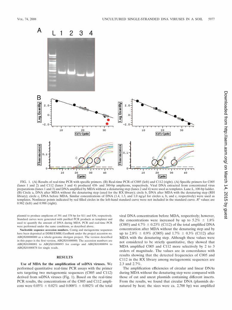
Use of MDA for the amplification of ssDNA viruses. Weperformed quantitative real-time PCR assays with the primersets targeting two metagenomic sequences (C005 and C112)derived from ssDNA viruses (Fig. 1). Based on the real-timePCR results, the concentrations of the C005 and C112 ampli-cons were 0.05% � 0.02% and 0.008% � 0.002% of the total
viral DNA concentration before MDA, respectively; however,the concentrations were increased by up to 5.2% � 1.8%(C005) and 4.7% � 0.23% (C112) of the total amplified DNAconcentration after MDA without the denaturing step and byup to 2.8% � 0.9% (C005) and 1.7% � 0.3% (C112) afterMDA with the denaturing step. Although these values werenot considered to be strictly quantitative, they showed thatMDA amplified C005 and C112 more selectively by 2 to 3orders of magnitude. The values are in concordance withresults showing that the detected frequencies of C005 andC112 in the RX library among metagenomic sequences are2.3 and 2.7%.
The amplification efficiencies of circular and linear DNAsduring MDA without the denaturing step were compared withthose of cut and uncut plasmids containing different inserts.From the results, we found that circular DNA (plasmids de-natured by heat; the sizes were ca. 2,700 bp) was amplified
FIG. 1. (A) Results of real-time PCR with specific primers. (B) Real-time PCR of C005 (left) and C112 (right). (A) Specific primers for C005(lanes 1 and 2) and C112 (lanes 3 and 4) produced 438- and 380-bp amplicons, respectively. Viral DNA extracted from concentrated viruspreparations (lanes 1 and 3) and DNA amplified by MDA without a denaturing step (lanes 2 and 4) were used as templates. Lane L, 100-bp ladder.(B) Circle a, DNA after MDA without the denaturing step (used for the RX library); circle b, DNA after MDA with the denaturing step (RHlibrary); circle c, DNA before MDA. Similar concentrations of DNA (1.4, 1.3, and 1.0 ng/�l for circles a, b, and c, respectively) were used astemplates. Nonlinear points indicated by red filled circles in the left-hand standard curve were not included in the standard curve. R2 values are0.982 (left) and 0.988 (right).
VOL. 74, 2008 UNCULTURED SINGLE-STRANDED DNA VIRUSES IN A SOIL 5977
on March 14, 2015 by guest
http://aem.asm
.org/D
ownloaded from
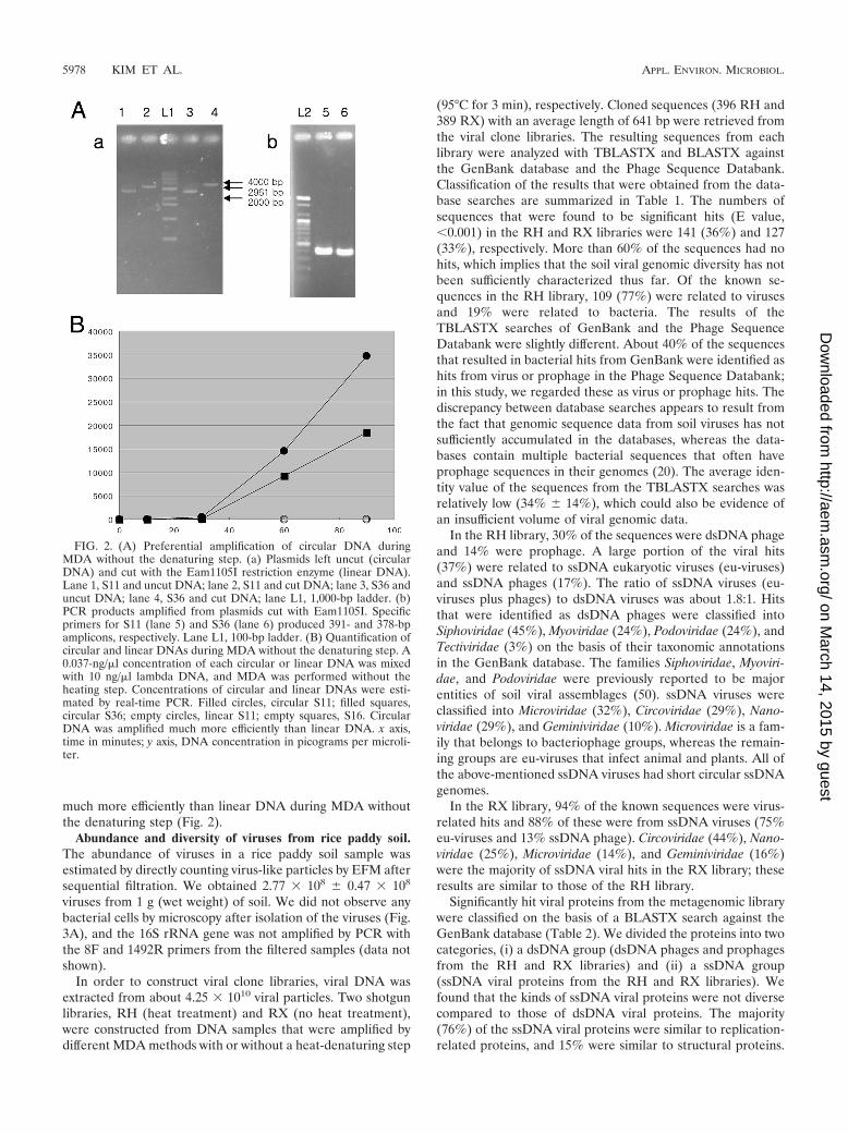
much more efficiently than linear DNA during MDA withoutthe denaturing step (Fig. 2).
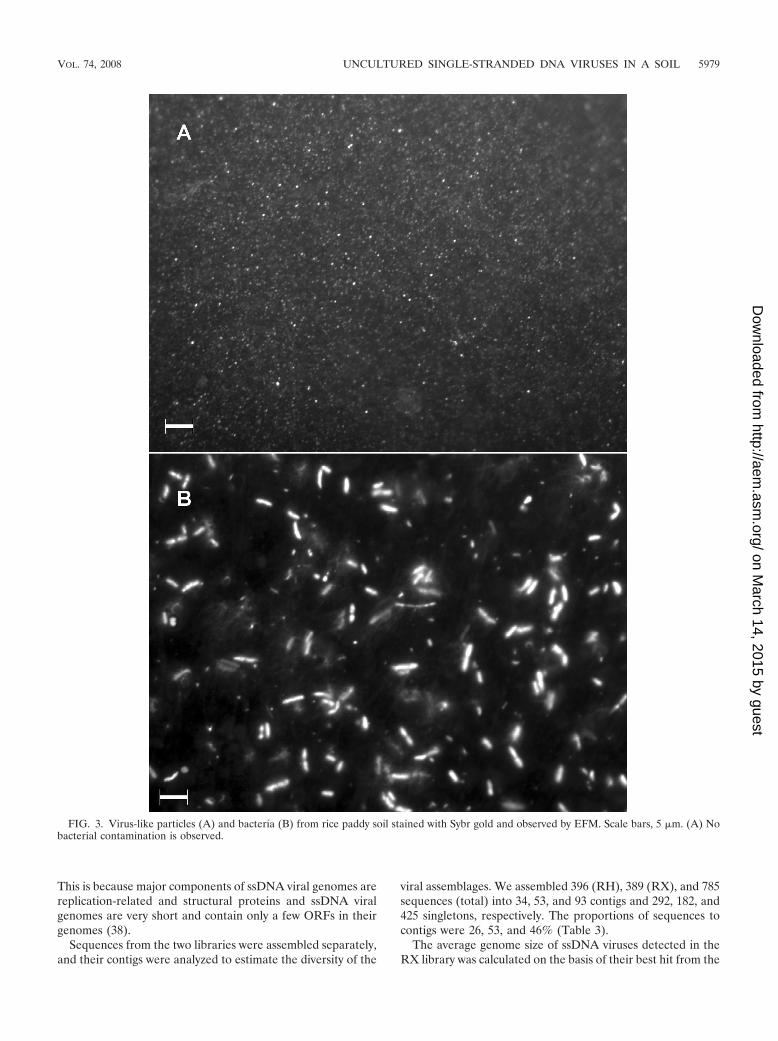
Abundance and diversity of viruses from rice paddy soil.The abundance of viruses in a rice paddy soil sample wasestimated by directly counting virus-like particles by EFM aftersequential filtration. We obtained 2.77 � 108 � 0.47 � 108
viruses from 1 g (wet weight) of soil. We did not observe anybacterial cells by microscopy after isolation of the viruses (Fig.3A), and the 16S rRNA gene was not amplified by PCR withthe 8F and 1492R primers from the filtered samples (data notshown).
In order to construct viral clone libraries, viral DNA wasextracted from about 4.25 � 1010 viral particles. Two shotgunlibraries, RH (heat treatment) and RX (no heat treatment),were constructed from DNA samples that were amplified bydifferent MDA methods with or without a heat-denaturing step
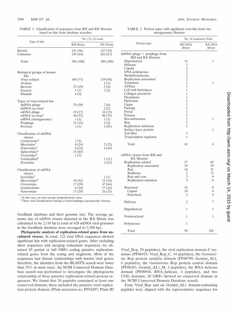
(95°C for 3 min), respectively. Cloned sequences (396 RH and389 RX) with an average length of 641 bp were retrieved fromthe viral clone libraries. The resulting sequences from eachlibrary were analyzed with TBLASTX and BLASTX againstthe GenBank database and the Phage Sequence Databank.Classification of the results that were obtained from the data-base searches are summarized in Table 1. The numbers ofsequences that were found to be significant hits (E value,0.001) in the RH and RX libraries were 141 (36%) and 127(33%), respectively. More than 60% of the sequences had nohits, which implies that the soil viral genomic diversity has notbeen sufficiently characterized thus far. Of the known se-quences in the RH library, 109 (77%) were related to virusesand 19% were related to bacteria. The results of theTBLASTX searches of GenBank and the Phage SequenceDatabank were slightly different. About 40% of the sequencesthat resulted in bacterial hits from GenBank were identified ashits from virus or prophage in the Phage Sequence Databank;in this study, we regarded these as virus or prophage hits. Thediscrepancy between database searches appears to result fromthe fact that genomic sequence data from soil viruses has notsufficiently accumulated in the databases, whereas the data-bases contain multiple bacterial sequences that often haveprophage sequences in their genomes (20). The average iden-tity value of the sequences from the TBLASTX searches wasrelatively low (34% � 14%), which could also be evidence ofan insufficient volume of viral genomic data.
In the RH library, 30% of the sequences were dsDNA phageand 14% were prophage. A large portion of the viral hits(37%) were related to ssDNA eukaryotic viruses (eu-viruses)and ssDNA phages (17%). The ratio of ssDNA viruses (eu-viruses plus phages) to dsDNA viruses was about 1.8:1. Hitsthat were identified as dsDNA phages were classified intoSiphoviridae (45%), Myoviridae (24%), Podoviridae (24%), andTectiviridae (3%) on the basis of their taxonomic annotationsin the GenBank database. The families Siphoviridae, Myoviri-dae, and Podoviridae were previously reported to be majorentities of soil viral assemblages (50). ssDNA viruses wereclassified into Microviridae (32%), Circoviridae (29%), Nano-viridae (29%), and Geminiviridae (10%). Microviridae is a fam-ily that belongs to bacteriophage groups, whereas the remain-ing groups are eu-viruses that infect animal and plants. All ofthe above-mentioned ssDNA viruses had short circular ssDNAgenomes.
In the RX library, 94% of the known sequences were virus-related hits and 88% of these were from ssDNA viruses (75%eu-viruses and 13% ssDNA phage). Circoviridae (44%), Nano-viridae (25%), Microviridae (14%), and Geminiviridae (16%)were the majority of ssDNA viral hits in the RX library; theseresults are similar to those of the RH library.
Significantly hit viral proteins from the metagenomic librarywere classified on the basis of a BLASTX search against theGenBank database (Table 2). We divided the proteins into twocategories, (i) a dsDNA group (dsDNA phages and prophagesfrom the RH and RX libraries) and (ii) a ssDNA group(ssDNA viral proteins from the RH and RX libraries). Wefound that the kinds of ssDNA viral proteins were not diversecompared to those of dsDNA viral proteins. The majority(76%) of the ssDNA viral proteins were similar to replication-related proteins, and 15% were similar to structural proteins.
FIG. 2. (A) Preferential amplification of circular DNA duringMDA without the denaturing step. (a) Plasmids left uncut (circularDNA) and cut with the Eam1105I restriction enzyme (linear DNA).Lane 1, S11 and uncut DNA; lane 2, S11 and cut DNA; lane 3, S36 anduncut DNA; lane 4, S36 and cut DNA; lane L1, 1,000-bp ladder. (b)PCR products amplified from plasmids cut with Eam1105I. Specificprimers for S11 (lane 5) and S36 (lane 6) produced 391- and 378-bpamplicons, respectively. Lane L1, 100-bp ladder. (B) Quantification ofcircular and linear DNAs during MDA without the denaturing step. A0.037-ng/�l concentration of each circular or linear DNA was mixedwith 10 ng/�l lambda DNA, and MDA was performed without theheating step. Concentrations of circular and linear DNAs were esti-mated by real-time PCR. Filled circles, circular S11; filled squares,circular S36; empty circles, linear S11; empty squares, S16. CircularDNA was amplified much more efficiently than linear DNA. x axis,time in minutes; y axis, DNA concentration in picograms per microli-ter.
5978 KIM ET AL. APPL. ENVIRON. MICROBIOL.
on March 14, 2015 by guest
http://aem.asm
.org/D
ownloaded from
This is because major components of ssDNA viral genomes arereplication-related and structural proteins and ssDNA viralgenomes are very short and contain only a few ORFs in theirgenomes (38).
Sequences from the two libraries were assembled separately,and their contigs were analyzed to estimate the diversity of the
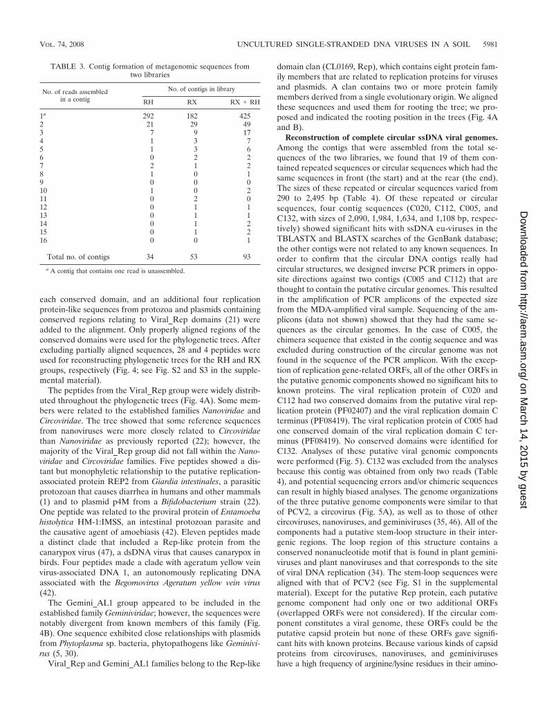
viral assemblages. We assembled 396 (RH), 389 (RX), and 785sequences (total) into 34, 53, and 93 contigs and 292, 182, and425 singletons, respectively. The proportions of sequences tocontigs were 26, 53, and 46% (Table 3).
The average genome size of ssDNA viruses detected in theRX library was calculated on the basis of their best hit from the
FIG. 3. Virus-like particles (A) and bacteria (B) from rice paddy soil stained with Sybr gold and observed by EFM. Scale bars, 5 �m. (A) Nobacterial contamination is observed.
VOL. 74, 2008 UNCULTURED SINGLE-STRANDED DNA VIRUSES IN A SOIL 5979
on March 14, 2015 by guest
http://aem.asm
.org/D
ownloaded from
GenBank database and their genome size. The average ge-nome size of ssDNA viruses detected in the RX library wascalculated to be 2,110 bp (a total of 428 ssDNA viral genomesin the GenBank database were averaged to 3,390 bp).
Phylogenetic analysis of replication-related genes from un-cultured viruses. In total, 122 viral DNA sequences showedsignificant hits with replication-related genes. After excludingshort sequences and merging redundant sequences, we ob-tained 85 partial or full ORFs coding putative replication-related genes from the contig and singletons. Most of thesequences had distant relationships with known viral genes;therefore, the identities from the BLASTX search were lowerthan 35% in most cases. An NCBI Conserved Domain Data-base search was performed to investigate the phylogeneticrelationship of these putative replication-related protein se-quences. We found that 58 peptides contained at least oneconserved domain; these included the putative viral replica-tion protein domain (Pfam accession no. PF02407, Pfam ID
Viral_Rep, 39 peptides), the viral replication domain C ter-minus (PF08419, Viral_Rep_C, 14 peptides), the Geminivi-rus Rep protein catalytic domain (PF00799, Gemini_AL1,6 peptides), the Geminivirus Rep protein central domain(PF08283, Gemini_AL1_M, 3 peptides), the RNA helicasedomain (PF00910, RNA_helicase, 4 peptides), and twoCOG domains; 28 ORFs showed no conserved domain inthe NCBI Conserved Domain Database search.
Forty Viral_Rep and six Gemini_AL1 domain-containingpeptides were aligned with the representative sequences for
TABLE 1. Classification of sequences from RH and RX librariesbased on hits from database searches
Type of hitsNo. (%) of reads
RH library RX library
Known 141 (36) 127 (33)Unknown 255 (64) 262 (67)
Total 396 (100) 389 (100)
Biological groups of knownhits
Virus related 109 (77) 119 (94)Archaea 1 (1)Bacteria 27 (19) 5 (4)Eucarya 1 (1) 2 (2)Plasmid 4 (3)
Types of virus-related hitsdsDNA phage 33 (30) 7 (6)dsDNA eu-virusa 2 (2)ssDNA phage 19 (17) 16 (13)ssDNA eu-virusa 40 (37) 90 (75)ssRNA (metagenome) 1 (1) 1 (1)Prophage 15 (14) 2 (2)Provirus 1 (1) 1 (91)
Classification of dsDNAviruses
Caudoviralesb 1 (3)Myoviridaeb 8 (24) 2 (22)Podoviridaeb 8 (24) 4 (44)Siphoviridaeb 15 (45)Tectiviridaeb 1 (3)Unclassifiedb 1 (11)Poxviridae 2 (22)
Classification of ssDNAviruses
Inoviridaeb 1 (1)Microviridaeb 19 (32) 15 (14)Circoviridae 17 (29) 47 (44)Geminiviridae 6 (10) 17 (16)Nanoviridae 17 (29) 26 (25)
a In this case, eu-virus means nonprokaryotic virus.b These viral classifications belong to bacteriophages (prokaryotic viruses).
TABLE 2. Protein types with significant viral hits from twometagenomic libraries
Protein type
No. of sequences from:
RH DNAlibrary
RX DNAlibrary
dsDNA phage � prophage fromRH and RX libraries
Hypothetical 14 1Helicase 3Capsid 2DNA polymerase 2Methyltransferase 2Replication associated 2Terminase 2 1ATPase 1Cell wall hydrolyzer 1Collagen precursor 1Deaminase 1Hydrolase 1Ligase 1Packing 1Portal 1Primase 1Recombination 1 1Rep 1Replication initiation 1Surface layer protein 1Tail fiber 1Transcription regulator 1
Total 41 4
ssDNA viruses from RH andRX libraries
Replication related 37 85Replication associated 19 39Rep 10 21Replicase 2 11Rep and coat 3 8Replication initiation 3 6
Structural 16 8Capsid 14 4Structural 2 4
Helicase 2 7
Hypothetical 1 1
Nonstructural 1 1
Polymerase 1
Total 58 102
5980 KIM ET AL. APPL. ENVIRON. MICROBIOL.
on March 14, 2015 by guest
http://aem.asm
.org/D
ownloaded from
each conserved domain, and an additional four replicationprotein-like sequences from protozoa and plasmids containingconserved regions relating to Viral_Rep domains (21) wereadded to the alignment. Only properly aligned regions of theconserved domains were used for the phylogenetic trees. Afterexcluding partially aligned sequences, 28 and 4 peptides wereused for reconstructing phylogenetic trees for the RH and RXgroups, respectively (Fig. 4; see Fig. S2 and S3 in the supple-mental material).
The peptides from the Viral_Rep group were widely distrib-uted throughout the phylogenetic trees (Fig. 4A). Some mem-bers were related to the established families Nanoviridae andCircoviridae. The tree showed that some reference sequencesfrom nanoviruses were more closely related to Circoviridaethan Nanoviridae as previously reported (22); however, themajority of the Viral_Rep group did not fall within the Nano-viridae and Circoviridae families. Five peptides showed a dis-tant but monophyletic relationship to the putative replication-associated protein REP2 from Giardia intestinales, a parasiticprotozoan that causes diarrhea in humans and other mammals(1) and to plasmid p4M from a Bifidobacterium strain (22).One peptide was related to the proviral protein of Entamoebahistolytica HM-1:IMSS, an intestinal protozoan parasite andthe causative agent of amoebiasis (42). Eleven peptides madea distinct clade that included a Rep-like protein from thecanarypox virus (47), a dsDNA virus that causes canarypox inbirds. Four peptides made a clade with ageratum yellow veinvirus-associated DNA 1, an autonomously replicating DNAassociated with the Begomovirus Ageratum yellow vein virus(42).
The Gemini_AL1 group appeared to be included in theestablished family Geminiviridae; however, the sequences werenotably divergent from known members of this family (Fig.4B). One sequence exhibited close relationships with plasmidsfrom Phytoplasma sp. bacteria, phytopathogens like Geminivi-rus (5, 30).
Viral_Rep and Gemini_AL1 families belong to the Rep-like
domain clan (CL0169, Rep), which contains eight protein fam-ily members that are related to replication proteins for virusesand plasmids. A clan contains two or more protein familymembers derived from a single evolutionary origin. We alignedthese sequences and used them for rooting the tree; we pro-posed and indicated the rooting position in the trees (Fig. 4Aand B).
Reconstruction of complete circular ssDNA viral genomes.Among the contigs that were assembled from the total se-quences of the two libraries, we found that 19 of them con-tained repeated sequences or circular sequences which had thesame sequences in front (the start) and at the rear (the end).The sizes of these repeated or circular sequences varied from290 to 2,495 bp (Table 4). Of these repeated or circularsequences, four contig sequences (C020, C112, C005, andC132, with sizes of 2,090, 1,984, 1,634, and 1,108 bp, respec-tively) showed significant hits with ssDNA eu-viruses in theTBLASTX and BLASTX searches of the GenBank database;the other contigs were not related to any known sequences. Inorder to confirm that the circular DNA contigs really hadcircular structures, we designed inverse PCR primers in oppo-site directions against two contigs (C005 and C112) that arethought to contain the putative circular genomes. This resultedin the amplification of PCR amplicons of the expected sizefrom the MDA-amplified viral sample. Sequencing of the am-plicons (data not shown) showed that they had the same se-quences as the circular genomes. In the case of C005, thechimera sequence that existed in the contig sequence and wasexcluded during construction of the circular genome was notfound in the sequence of the PCR amplicon. With the excep-tion of replication gene-related ORFs, all of the other ORFs inthe putative genomic components showed no significant hits toknown proteins. The viral replication protein of C020 andC112 had two conserved domains from the putative viral rep-lication protein (PF02407) and the viral replication domain Cterminus (PF08419). The viral replication protein of C005 hadone conserved domain of the viral replication domain C ter-minus (PF08419). No conserved domains were identified forC132. Analyses of these putative viral genomic componentswere performed (Fig. 5). C132 was excluded from the analysesbecause this contig was obtained from only two reads (Table4), and potential sequencing errors and/or chimeric sequencescan result in highly biased analyses. The genome organizationsof the three putative genome components were similar to thatof PCV2, a circovirus (Fig. 5A), as well as to those of othercircoviruses, nanoviruses, and geminiviruses (35, 46). All of thecomponents had a putative stem-loop structure in their inter-genic regions. The loop region of this structure contains aconserved nonanucleotide motif that is found in plant gemini-viruses and plant nanoviruses and that corresponds to the siteof viral DNA replication (34). The stem-loop sequences werealigned with that of PCV2 (see Fig. S1 in the supplementalmaterial). Except for the putative Rep protein, each putativegenome component had only one or two additional ORFs(overlapped ORFs were not considered). If the circular com-ponent constitutes a viral genome, these ORFs could be theputative capsid protein but none of these ORFs gave signifi-cant hits with known proteins. Because various kinds of capsidproteins from circoviruses, nanoviruses, and geminiviruseshave a high frequency of arginine/lysine residues in their amino-
TABLE 3. Contig formation of metagenomic sequences fromtwo libraries
No. of reads assembledin a contig
No. of contigs in library
RH RX RX � RH
1a 292 182 4252 21 29 493 7 9 174 1 3 75 1 3 66 0 2 27 2 1 28 1 0 19 0 0 010 1 0 211 0 2 012 0 1 113 0 1 114 0 1 215 0 1 216 0 0 1
Total no. of contigs 34 53 93
a A contig that contains one read is unassembled.
VOL. 74, 2008 UNCULTURED SINGLE-STRANDED DNA VIRUSES IN A SOIL 5981
on March 14, 2015 by guest
http://aem.asm
.org/D
ownloaded from
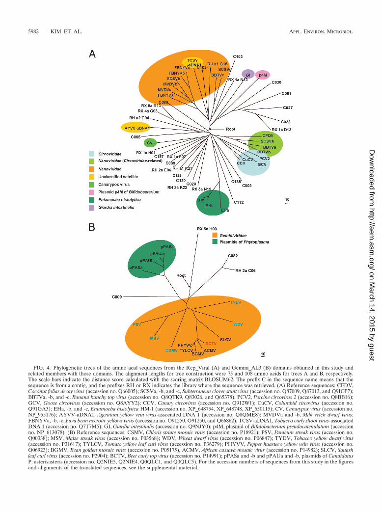
FIG. 4. Phylogenetic trees of the amino acid sequences from the Rep_Viral (A) and Gemini_AL3 (B) domains obtained in this study andrelated members with those domains. The alignment lengths for tree construction were 75 and 108 amino acids for trees A and B, respectively.The scale bars indicate the distance score calculated with the scoring matrix BLOSUM62. The prefix C in the sequence name means that thesequence is from a contig, and the prefixes RH or RX indicates the library where the sequence was retrieved. (A) Reference sequences: CFDV,Coconut foliar decay virus (accession no. Q66005); SCSVa, -b, and -c, Subterranean clover stunt virus (accession no. Q87009, Q87013, and Q9ICP7);BBTVa, -b, and -c, Banana bunchy top virus (accession no. Q8QTK9, Q83026, and Q65378); PCV2, Porcine circovirus 2 (accession no. Q8BB16);GCV, Goose circovirus (accession no. Q8AYY2); CCV, Canary circovirus (accession no. Q912W1); CuCV, Columbid circovirus (accession no.Q91GA3); EHa, -b, and -c, Entamoeba histolytica HM-1 (accession no. XP_648754, XP_648748, XP_650115); CV, Canarypox virus (accession no.NP_955176); AYVV-aDNA1, Ageratum yellow vein virus-associated DNA 1 (accession no. Q8QME8); MVDVa and -b, Milk vetch dwarf virus;FBNYVa, -b, -c, Fava bean necrotic yellows virus (accession no. O91250, O91250, and Q66862); TCSV-aDNA1, Tobacco curly shoot virus-associatedDNA 1 (accession no. Q7T7M5); GI, Giardia intestinalis (accession no. Q9NJY0); p4M, plasmid of Bifidobacterium pseudocatenulatum (accessionno. NP_613078). (B) Reference sequences: CSMV, Chloris striate mosaic virus (accession no. P18921); PSV, Panicum streak virus (accession no.Q00338); MSV, Maize streak virus (accession no. P03568); WDV, Wheat dwarf virus (accession no. P06847); TYDV, Tobacco yellow dwarf virus(accession no. P31617); TYLCV, Tomato yellow leaf curl virus (accession no. P36279); PHYVV, Pepper huasteco yellow vein virus (accession no.Q06923); BGMV, Bean golden mosaic virus (accession no. P05175), ACMV, African cassava mosaic virus (accession no. P14982); SLCV, Squashleaf curl virus (accession no. P2904); BCTV, Beet curly top virus (accession no. P14991); pPASa and -b and pPAUa and -b, plasmids of CandidatusP. asterisasteris (accession no. Q2NIE5, Q2NIE4, Q0QLC1, and Q0QLC5). For the accession numbers of sequences from this study in the figuresand alignments of the translated sequences, see the supplemental material.
5982 KIM ET AL. APPL. ENVIRON. MICROBIOL.
on March 14, 2015 by guest
http://aem.asm
.org/D
ownloaded from
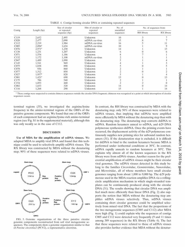
terminal regions (35), we investigated the arginine/lysinefrequency in the amino-terminal regions of the ORFs of theputative genome components. We found that one of the ORFsof each component had an arginine/lysine-rich amino-terminalregion (see Fig. S1 in the supplemental material), although thiswas only weakly so in the case of C112.
DISCUSSION
Use of MDA for the amplification of ssDNA viruses. Weadapted MDA to amplify viral DNA and found that this tech-nique could be used to selectively amplify ssDNA viruses. TheRX library was constructed by MDA without the denaturingstep; 90% of these sequences were related to ssDNA viruses.
In contrast, the RH library was constructed by MDA with thedenaturing step; only 50% of these sequences were related tossDNA viruses, thus implying that ssDNAs were amplifiedmore efficiently by MDA without the denaturing step than withthe denaturing step. The denaturing step converts dsDNA tossDNA. Random hexamers anneal to ssDNA, and �29 DNApolymerase synthesizes dsDNA. Once the priming events haveoccurred, the displacement activity of the �29 polymerase con-tinuously supplies new priming sites for unbound random hex-amers (31). If the denaturation step is excluded, it is difficultfor dsDNA to bind to the random hexamers because MDA isperformed under isothermal conditions at 30°C. In contrast,ssDNA rapidly anneals to random hexamers at 30°C. Thisexplains why almost all of the known sequences in the RXlibrary were from ssDNA viruses. Another reason for the pref-erential amplification of ssDNA viruses might be their circularviral genomes. The ssDNA viruses detected in this study be-long to the families Circoviridae, Geminiviridae, Nanoviridae,and Microviridae, all of whose members have small circulargenomes ranging from about 1,000 to 9,000 bp. The �29 poly-merase used in the MDA reaction amplifies DNA via a rolling-circle amplification mechanism in which single-stranded tem-plates can be continuously produced along with the circularDNA (31). The results showing that circular DNA was ampli-fied much more efficiently than linear DNA (Fig. 2) also sup-ports the notion that MDA without the denaturing step am-plifies ssDNA viruses selectively. Thus, ssDNA virusescontaining short circular genomes could be amplified selec-tively from mixed viral DNA. The fact that the proportions ofthe two metagenomic sequences (C005 and C112) after MDAwere high (Fig. 1) could explain why the sequences of contigsC005 and C112 were detected very frequently (9 and 11 timesamong 389 sequences) in the RX library (Table 4). The factthat these sequences were related to those of ssDNA virusesalso provides further evidence that MDA without the denatur-
TABLE 4. Contigs forming circular DNA or containing repeated sequences
Contig Length (bp)Size of circular
or repeatedsequence (bp)
Hits of circular orrepeated
sequences
No. ofassembledsequences
No. of sequences from:
RH library RX library
C119 2,652 2,495 Unknown 10 3 7C020 2,477 2,090 ssDNA eu-virus 15 8 7C112 2,219 1,984 ssDNA eu-virus 16 5 11C005 2,896a 1,634 ssDNA eu-virus 10 1 9C076 2,971a 1,230 Unknown 14 1 13C138 1,231 1,207 Unknown 6 0 6C071 1,554 1,186 Unknown 8 7 1C132 1,172 1,108 ssDNA eu-virus 2 1 1C047 1,489 1,090 Unknown 5 1 4C145 1,541 969 Unknown 4 1 3C075 1,838 890 Unknown 5 0 5C078 936 860 Unknown 2 1 1C077 1,312 850 Unknown 2 0 2C027 1,957a 820 Unknown 5 1 4C001 1,421a 690 Unknown 13 0 13C175 786 540 Unknown 2 1 1C154 1,053 440 Unknown 2 2 0C002 1,653 320 Unknown 14 0 14C116 1,268 290 Unknown 15 1 14
a These contigs were suspected to contain chimera sequences outside the circular DNA fragment; chimeras was recognized at a point at which interruption of circularsequences occurs.
FIG. 5. Genome organizations of the three putative circulargenomic components reconstructed from soil viral metagenomic se-quences. The components show a genomic organization similar to thatof Porcine circovirus2 (PCV2), a representative circovirus.
VOL. 74, 2008 UNCULTURED SINGLE-STRANDED DNA VIRUSES IN A SOIL 5983
on March 14, 2015 by guest
http://aem.asm
.org/D
ownloaded from
ing step preferentially amplifies ssDNA genomes. In addition,it was reported that circular DNA virus genomes could beamplified by a sequence-independent strategy by MDA (ormultiply primed rolling-circle amplification). The human papil-lomavirus circular genome has been amplified from DNAextracted from cell lines and bovine tissues; the concentrationof the papillomavirus DNA was increased by 2.4 � 104-fold(40). This method was also used to amplify the circular ge-nomes of various viruses, such as polyomaviruses (27), anello-virus (36), circoviruses (26), and geminiviruses (23). Consider-ing that papillomavirus and polyomavirus were dsDNA virusesand these dsDNA viruses could be amplified efficiently byMDA, the circular structure was a main reason for the pref-erential amplification of circular ssDNA viruses. In addition,the result that 50% of the significant hits related to ssDNAviruses was increased to 90% without the denaturing step im-plied that eliminating the denaturing step was necessary toinvestigate ssDNA viruses more selectively. A precise and de-tailed evaluation of the factors that affect the efficiency of theMDA technique such as the DNA length, linearity, and strandtype; the reaction time; and the denaturing step should beperformed in future studies.
Diversity of viruses in rice paddy soil. When MDA wasperformed with the denaturing step (RH library), we acquireda number of metagenomic sequences that were related to bothdsDNA and ssDNA viruses. All of the dsDNA viruses wereprokaryotic viruses (phages); the majority of them (�92%)were tailed phages, and only one was a polyhedral phage (be-longing to the family Tectiviridae). A large proportion of thesignificant hits in the RH library were related to ssDNA vi-ruses. This abundance of ssDNA viral hits is not in concordwith a previous observation suggesting that dsDNA viruses arethe major entity in a soil viral assemblage (50); preferentialamplification of circular DNA by MDA was suggested to bethe main reason for the results. The majority of ssDNA virusesdetected in this study were related to animal and plant viruses(68 to 85%) and not to phages. The proportions of animal andplant viruses were almost the same. It is suspected that themajor source of plant viruses is rice or other plants that growin the field and the source of the animal viruses seems to bewild bird feces or composted manure used as organic fertilizer.Animal and plant viruses detected in this study belong to thefamilies Circoviridae, Nanoviridae, and Geminiviridae, in whicha lot of pathogenic viruses exist (32, 45, 48); this indicates thatsoil could be a reservoir for viruses that are pathogenic toanimals and plants. Another main group of ssDNA virusesobtained in this study was Microviridae, which is a bacterio-phage family. Chp1-like ssDNA microphages belonging to thefamily Microviridae were also found to be abundant in theSargasso Sea (3).
Phylogenetic analysis of replication-related genes of uncul-tured viruses. Phylogenetic analyses of replication-relatedgenes showed that the majority of the sequences acquired inthis study represented previously unknown viruses. The viralfamily Geminviridae, containing a large number of plant-patho-genic viruses, is represented by four genera: Begomovirus, Cur-tovirus, Mastrevirus, and Topocuvirus. The sequences acquiredin this study relating to the family Geminiviridae did not fallinto any of these genera, indicating that they are new membersof Geminiviridae, the largest family of ssDNA viruses. The
majority of the sequences obtained in this study were distantlyrelated to Nanoviridae and Circoviridae but did not fall into tothe established families. Some of these sequences were closelyrelated to nonviral genomic material such as satellites, bacte-rial plasmids, genomes of protozoa, and dsDNA viruses ratherthan ssDNA viruses (Fig. 2). Nevertheless, some of these se-quences might have originated from ssDNA viruses and theymay represent new ssDNA virus families. The fact that morethan 60% of the hits in this study were nonsignificant impliesthat ssDNA viral diversity in a soil environment may be morecomplex than previously thought.
Construction of complete circular ssDNA viral genomes.The presence of a conserved stem-loop structure in the intergenicregion and two ORFs encoding a putative capsid protein and aputative Rep protein suggests that the three circular sequencesare the entire genomes or genome components of unknownssDNA viruses (some ssDNA viruses, such as Nanovirus andGeminivirus, have multiple genomic components). From thispoint of the view, the other circular sequences (derived fromcontigs) that gave no hit with any known protein in the sequencedatabases might also turn out to be completely novel viruses.Breitbart et al. pointed out the possibility of sequencing the entiregenome of uncultured viruses by using metagenomic approaches(10). Several complete genomes of RNA viruses have been re-constructed from a coastal RNA viral metagenomic study (13). Inthis report, several putative circular ssDNA viral genomes werereconstructed with a relatively small amount of sequencing (800sequence reads). These results indicate that metagenomic re-search is a useful method to uncover unknown genomic entitiesamong environmental viruses.
MDA without the denaturing step cannot displace previousmethods such as the LASL method. Rather, the work de-scribed herein points to an approach that permits the inves-tigation of ssDNA viral diversity, something that cannot beaccomplished by the LASL method. MDA has several short-comings such as chimera formation (29, 52), biased ampli-fication (24), and contamination of external DNA (52). Nev-ertheless, preferential amplification of circular ssDNA viralDNA by MDA without the denaturing step could provide anew tool to explore currently unexplored viral diversity.
Although viruses are major biological entities in soil, viral di-versity in this environment was largely unexplored. By using thenovel culture-independent metagenomic approach, we have in-vestigated the diversity of DNA viral assemblages. MDA is auseful approach for the investigation of both ssDNA and dsDNAviral diversity; this technique was also used to selectively investi-gate the diversity of ssDNA viruses. Our research also showedthat unknown short circular ssDNA viral genomes or genomecomponents can be detected without viral cultivation by sequenc-ing the metagenome and amplifying the DNA by MDA. Furtherstudies are needed to reveal the viral diversity of different soilsamples and to quantitatively analyze soil viruses. These effortswould contribute to our understanding of the role of viral assem-blages in biological soil communities.
ACKNOWLEDGMENTS
This work was supported by the Environmental Biotechnology Na-tional Core Research Center (KOSEF: R15-2003-012-02002-0) andthe 21C Frontier Microbial Genomics and Application Center Pro-gram. K.-H.K. was supported by a Korea Research Foundation grant
5984 KIM ET AL. APPL. ENVIRON. MICROBIOL.
on March 14, 2015 by guest
http://aem.asm
.org/D
ownloaded from

(MOEHRD, Basic Research Promotion Fund, KRF-2006-351-D00011).
REFERENCES
1. Adam, R. D. 2001. Biology of Giardia lamblia. Clin. Microbiol. Rev. 14:447–475.
2. Altschul, S. F., T. L. Madden, A. A. Schaffer, J. Zhang, Z. Zhang, W. Miller,and D. J. Lipman. 1997. Gapped BLAST and PSI-BLAST: a new generationof protein database search programs. Nucleic Acids Res. 25:3389–3402.
3. Angly, F. E., B. Felts, M. Breitbart, P. Salamon, R. A. Edwards, C. Carlson,A. M. Chan, M. Haynes, S. Kelley, H. Liu, J. M. Mahaffy, J. E. Mueller, J.Nulton, R. Olson, R. Parsons, S. Rayhawk, C. A. Suttle, and F. Rohwer. 2006.The marine viromes of four oceanic regions. PLoS Biol. 4:e368.
4. Ashelford, K. E., M. J. Day, and J. C. Fry. 2003. Elevated abundance ofbacteriophage infecting bacteria in soil. Appl. Environ. Microbiol. 69:285–289.
5. Bai, X., J. Zhang, A. Ewing, S. A. Miller, A. Jancso Radek, D. V. Shevchenko,K. Tsukerman, T. Walunas, A. Lapidus, J. W. Campbell, and S. A. Hogen-hout. 2006. Living with genome instability: the adaptation of phytoplasmas todiverse environments of their insect and plant hosts. J. Bacteriol. 188:3682–3696.
6. Bench, S. R., T. E. Hanson, K. E. Williamson, D. Ghosh, M. Radosovich, K.Wang, and K. E. Wommack. 2007. Metagenomic characterization of Ches-apeake Bay virioplankton. Appl. Environ. Microbiol. 73:7629–7641.
7. Bergh, O., K. Y. Borsheim, G. Bratbak, and M. Heldal. 1989. High abun-dance of viruses found in aquatic environments. Nature 340:467–468.
8. Breitbart, M., B. Felts, S. Kelley, J. M. Mahaffy, J. Nulton, P. Salamon, andF. Rohwer. 2004. Diversity and population structure of a near-shore marine-sediment viral community. Proc. Biol. Sci. 271:565–574.
9. Breitbart, M., I. Hewson, B. Felts, J. M. Mahaffy, J. Nulton, P. Salamon, andF. Rohwer. 2003. Metagenomic analyses of an uncultured viral communityfrom human feces. J. Bacteriol. 185:6220–6223.
10. Breitbart, M., P. Salamon, B. Andresen, J. M. Mahaffy, A. M. Segall, D.Mead, F. Azam, and F. Rohwer. 2002. Genomic analysis of unculturedmarine viral communities. Proc. Natl. Acad. Sci. USA 99:14250–14255.
11. Cann, A. J., S. E. Fandrich, and S. Heaphy. 2005. Analysis of the viruspopulation present in equine faeces indicates the presence of hundreds ofuncharacterized virus genomes. Virus Genes 30:151–156.
12. Casas, V., and F. Rohwer. 2007. Phage metagenomics. Methods Enzymol.421:259–268.
13. Culley, A. I., A. S. Lang, and C. A. Suttle. 2006. Metagenomic analysis ofcoastal RNA virus communities. Science 312:1795–1798.
14. Dean, F. B., S. Hosono, L. Fang, X. Wu, A. F. Faruqi, P. Bray-Ward, Z. Sun,Q. Zong, Y. Du, J. Du, M. Driscoll, W. Song, S. F. Kingsmore, M. Egholm,and R. S. Lasken. 2002. Comprehensive human genome amplification usingmultiple displacement amplification. Proc. Natl. Acad. Sci. USA 99:5261–5266.
15. Dean, F. B., J. R. Nelson, T. L. Giesler, and R. S. Lasken. 2001. Rapidamplification of plasmid and phage DNA using Phi 29 DNA polymerase andmultiply-primed rolling circle amplification. Genome Res. 11:1095–1099.
16. Delwart, E. L. 2007. Viral metagenomics. Rev. Med. Virol. 17:115–131.17. Edwards, R. A., and F. Rohwer. 2005. Viral metagenomics. Nat. Rev. Mi-
crobiol. 3:504–510.18. Fierer, N., M. Breitbart, J. Nulton, P. Salamon, C. Lozupone, R. Jones, M.
Robeson, R. A. Edwards, B. Felts, S. Rayhawk, R. Knight, F. Rohwer, andR. B. Jackson. 2007. Metagenomic and small-subunit rRNA analyses revealthe genetic diversity of bacteria, archaea, fungi, and viruses in soil. Appl.Environ. Microbiol. 73:7059–7066.
19. Finn, R. D., J. Mistry, B. Schuster-Bockler, S. Griffiths-Jones, V. Hollich, T.Lassmann, S. Moxon, M. Marshall, A. Khanna, R. Durbin, S. R. Eddy,E. L. L. Sonnhammer, and A. Bateman. 2006. Pfam: clans, web tools andservices. Nucleic Acids Res. 34:D247–D251.
20. Fouts, D. E. 2006. Phage_Finder: automated identification and classificationof prophage regions in complete bacterial genome sequences. Nucleic AcidsRes. 34:5839–5851.
21. Gibbs, M. J., V. V. Smeianov, J. L. Steele, P. Upcroft, and B. A. Efimov. 2006.Two families of rep-like genes that probably originated by interspecies re-combination are represented in viral, plasmid, bacterial, and parasitic pro-tozoan genomes. Mol. Biol. Evol. 23:1097–1100.
22. Gibbs, M. J., and G. F. Weiller. 1999. Evidence that a plant virus switchedhosts to infect a vertebrate and then recombined with a vertebrate-infectingvirus. Proc. Natl. Acad. Sci. USA 96:8022–8027.
23. Haible, D., S. Kober, and H. Jeske. 2006. Rolling circle amplification revo-lutionizes diagnosis and genomics of geminiviruses. J. Virol. Methods 135:9–16.
24. Hosono, S., A. F. Faruqi, F. B. Dean, Y. Du, Z. Sun, X. Wu, J. Du, S. F.Kingsmore, M. Egholm, and R. S. Lasken. 2003. Unbiased whole-genomeamplification directly from clinical samples. Genome Res. 13:954–964.
25. Jia, Z., R. Ishihara, Y. Nakajima, S. Asakawa, and M. Kimura. 2007. Mo-
lecular characterization of T4-type bacteriophages in a rice field. Environ.Microbiol. 9:1091–1096.
26. Johne, R., D. Fernandez-de-Luco, U. Hofle, and H. Muller. 2006. Genome ofa novel circovirus of starlings, amplified by multiply primed rolling-circleamplification. J. Gen. Virol. 87:1189–1195.
27. Johne, R., W. Wittig, D. Fernandez-de-Luco, U. Hofle, and H. Muller. 2006.Characterization of two novel polyomaviruses of birds by using multiplyprimed rolling-circle amplification of their genomes. J. Virol. 80:3523–3531.
28. Lasken, R. S., and M. Egholm. 2003. Whole genome amplification: abundantsupplies of DNA from precious samples or clinical specimens. Trends Bio-technol. 21:531–535.
29. Lasken, R. S., and T. B. Stockwell. 2007. Mechanism of chimera formationduring the multiple displacement amplification reaction. BMC Biotechnol. 7:19.
30. Liefting, L. W., M. T. Andersen, T. J. Lough, and R. E. Beever. 2006.Comparative analysis of the plasmids from two isolates of “Candidatus Phy-toplasma australiense.” Plasmid 56:138–144.
31. Lizardi, P. M., X. Huang, Z. Zhu, P. Bray-Ward, D. C. Thomas, and D. C.Ward. 1998. Mutation detection and single-molecule counting using isother-mal rolling-circle amplification. Nat. Genet. 19:225–232.
32. Mansoor, S., S. H. Khan, A. Bashir, M. Saeed, Y. Zafar, K. A. Malik, R.Briddon, J. Stanley, and P. G. Markham. 1999. Identification of a novelcircular single-stranded DNA associated with cotton leaf curl disease inPakistan. Virology 259:190–199.
33. Marchler-Bauer, A., J. B. Anderson, P. F. Cherukuri, C. DeWeese-Scott,L. Y. Geer, M. Gwadz, S. He, D. I. Hurwitz, J. D. Jackson, Z. Ke, C. J.Lanczycki, C. A. Liebert, C. Liu, F. Lu, G. H. Marchler, M. Mullokandov,B. A. Shoemaker, V. Simonyan, J. S. Song, P. A. Thiessen, R. A. Yamashita,J. J. Yin, D. Zhang, and S. H. Bryant. 2005. CDD: a conserved domaindatabase for protein classification. Nucleic Acids Res. 33:D192–D196.
34. Meehan, B. M., J. L. Creelan, M. S. McNulty, and D. Todd. 1997. Sequenceof porcine circovirus DNA: affinities with plant circoviruses. J. Gen. Virol.78(Pt. 1):221–227.
35. Niagro, F. D., A. N. Forsthoefel, R. P. Lawther, L. Kamalanathan, B. W.Ritchie, K. S. Latimer, and P. D. Lukert. 1998. Beak and feather diseasevirus and porcine circovirus genomes: intermediates between the geminivi-ruses and plant circoviruses. Arch. Virol. 143:1723–1744.
36. Niel, C., L. Diniz-Mendes, and S. Devalle. 2005. Rolling-circle amplificationof Torque teno virus (TTV) complete genomes from human and swine seraand identification of a novel swine TTV genogroup. J. Gen. Virol. 86:1343–1347.
37. Noble, R., and J. Fuhrman. 1998. Use of SYBR Green I for rapid epifluo-rescence counts of marine viruses and bacteria. Aquat. Microb. Ecol. 14:113–118.
38. Olvera, A., M. Cortey, and J. Segales. 2007. Molecular evolution of porcinecircovirus type 2 genomes: phylogeny and clonality. Virology 357:175–185.
39. Proctor, L. M., and J. A. Fuhrman. 1990. Viral mortality of marine bacteriaand cyanobacteria. Nature 343:60–62.
40. Rector, A., R. Tachezy, and M. Van Ranst. 2004. A sequence-independentstrategy for detection and cloning of circular DNA virus genomes by usingmultiply primed rolling-circle amplification. J. Virol. 78:4993–4998.
41. Sambrook, J., E. F. Fritsch, and T. Maniatis. 1989. Molecular cloning: alaboratory manual, 2nd ed. Cold Spring Harbor Laboratory Press, ColdSpring Harbor, NY.
42. Saunders, K., I. D. Bedford, and J. Stanley. 2002. Adaptation from whiteflyto leafhopper transmission of an autonomously replicating nanovirus-likeDNA component associated with ageratum yellow vein disease. J. Gen.Virol. 83:907–913.
43. Suttle, C. A. 2005. Viruses in the sea. Nature 437:356–361.44. Suttle, C. A., A. M. Chan, and M. T. Cottrell. 1990. Infection of phytoplank-
ton by viruses and reduction of primary productivity. Nature 347:467–469.45. Todd, D. 2000. Circoviruses: immunosuppressive threats to avian species: a
review. Avian Pathol. 29:373–394.46. Todd, D., J. H. Weston, D. Soike, and J. A. Smyth. 2001. Genome sequence
determinations and analyses of novel circoviruses from goose and pigeon.Virology 286:354–362.
47. Tulman, E. R., C. L. Afonso, Z. Lu, L. Zsak, G. F. Kutish, and D. L. Rock.2004. The genome of canarypox virus. J. Virol. 78:353–366.
48. Varma, A., and V. G. Malathi. 2003. Emerging geminivirus problems: aserious threat to crop production. Ann. Appl. Biol. 142:145–164.
49. Williamson, K. E., M. Radosevich, D. W. Smith, and K. E. Wommack. 2007.Incidence of lysogeny within temperate and extreme soil environments. En-viron. Microbiol. 9:2563–2574.
50. Williamson, K. E., M. Radosevich, and K. E. Wommack. 2005. Abundanceand diversity of viruses in six Delaware soils. Appl. Environ. Microbiol.71:3119–3125.
51. Williamson, K. E., K. E. Wommack, and M. Radosevich. 2003. Samplingnatural viral communities from soil for culture-independent analyses. Appl.Environ. Microbiol. 69:6628–6633.
52. Zhang, K., A. C. Martiny, N. B. Reppas, K. W. Barry, J. Malek, S. W.Chisholm, and G. M. Church. 2006. Sequencing genomes from single cells bypolymerase cloning. Nat. Biotechnol. 24:680–686.
VOL. 74, 2008 UNCULTURED SINGLE-STRANDED DNA VIRUSES IN A SOIL 5985
on March 14, 2015 by guest
http://aem.asm
.org/D
ownloaded from