PREVALENCE OF SARCOCYSTIS SP. IN STRANDED ATLANTIC WHITE-SIDED DOLPHINS (LAGENORHYNCHUS ACUTUS
6
291 Journal of Wildlife Diseases, 38(2), 2002, pp. 291–296 Wildlife Disease Association 2002 PREVALENCE OF SARCOCYSTIS SP. IN STRANDED ATLANTIC WHITE-SIDED DOLPHINS (LAGENORHYNCHUS ACUTUS) Ruth Ewing, 1,2,8 Julia Zaias, 1 M. Andrew Stamper, 3,4 Gregory D. Bossart, 5,6 and J. P. Dubey 7 1 University of Miami School of Medicine, Department of Pathology, Division of Comparative Pathology, 1600 NW 10th Avenue, Room 7101A, Miami, Florida 33136, USA 2 NOAA, National Marine Fisheries Service, Southeast Fisheries Science Center, Miami, Florida 33149, USA 3 New England Aquarium, Central Wharf, Boston, Massachusetts 02110, USA 4 Current address: EPCOT, The Living Seas, EC TRL W-251, P.O. Box 10,000 Lake Buena Vista, Florida 32830, USA 5 Miami Seaquarium, 4400 Rickenbacker CSWY, Miami, Florida 33149, USA 6 Current address: Harbor Branch Oceanographic Institute, Division of Marine Mammal Research and Conservation, 5600 US 1 North, Fort Pierce, Florida 34946, USA 7 USDA, Agricultural Research Service, Parasite Biology, Epidemiology and Systematics Laboratory, BARC-East, Beltsville, Maryland 20705, USA 8 Corresponding author: (e-mail: [email protected]) ABSTRACT: In January 1998 and 1999, two mass strandings of dolphins occurred in Wellfleet, Massachusetts. The strandings were composed of 97 and 53 animals, respectively. Tissues from 35 Atlantic white-sided dolphins (Lagenorhynchus acutus) from the 1998 stranding and 52 from the 1999 stranding were examined histologically. In the 1998 stranding, unidentified protozoal tissue cysts were seen in skeletal muscle from 11 of 28 (39%) dolphins. In addition, two dolphins had a protozoal tissue cyst in cardiac muscle. In the 1999 stranding, nine of 23 (39%) dolphins had the same protozoal tissue cysts in skeletal muscle. The identification of these protozoal tissue cysts as Sarcocystis sp. was confirmed by light and transmission electron microscopy. The high prevalence of sarcocysts in these dolphins suggests that they are likely intermediate hosts for previously undescribed Sarcocystis spp. The ultrastructure of the sarcocyst walls suggests that more than one species of Sarcocystis are present in dolphins. Key words: Atlantic white-sided dolphin, epidemiology, Lagenorhynchus acutus, protozoan, Sarcocystis. INTRODUCTION Sarcocystis spp. (Protozoa: Apicom- plexa) have an obligatory two-host (pred- ator-prey) life cycle (Dubey et al., 1989). The intermediate host, typically an herbi- vore, becomes infected by ingesting feces or water which contains infective sporo- cysts. After ingestion, the parasite under- goes asexual multiplication in several or- gans and eventually forms sarcocysts in muscles. Sarcocysts contain banana- shaped bradyzoites. When the intermedi- ate host is consumed by a carnivorous de- finitive host, bradyzoites are released from the sarcocyst and penetrate the lamina propria of the small intestine. Here they transform into gamonts. After fertilization, oocysts containing sporocysts are released into the intestinal lumen and passed out in feces (Dubey et al., 1989). Sarcocysts have been identified in many wild and domestic terrestrial mammals (Dubey et al., 1998); however, few have been documented in marine mammals. Several studies have identified sarcocysts in individual pinnipeds (Lapointe et al., 1998) and cetaceans (DeGuise et al., 1993). This study documents a relatively high prevalence of sarcocysts in Atlantic white-sided dolphins ( Lagenorhynchus acutus) associated with two mass strand- ings. METHODS In January, 1998 and 1999, members of the Northeast Regional Stranding Network re- sponded to two mass strandings of dolphins in Wellfleet, Massachusetts (4156N, 7002W). These strandings involved two cetacean spe- cies, 90 Atlantic white-sided dolphins and 17 common dolphins (Delphinus delphis). Only Atlantic white-sided dolphins were evaluated by light microscopy. The University of Miami Comparative Pathology Laboratory received tissues from 35 Atlantic white-sided dolphins from the 1998 stranding (15 males and 20 fe- males) and 50 from the 1999 stranding (31 males, 17 females, and two unknown sexes) for
-
Upload
independent -
Category
Documents
-
view
2 -
download
0
Transcript of PREVALENCE OF SARCOCYSTIS SP. IN STRANDED ATLANTIC WHITE-SIDED DOLPHINS (LAGENORHYNCHUS ACUTUS

291
Journal of Wildlife Diseases, 38(2), 2002, pp. 291–296� Wildlife Disease Association 2002
PREVALENCE OF SARCOCYSTIS SP. IN STRANDED ATLANTICWHITE-SIDED DOLPHINS (LAGENORHYNCHUS ACUTUS)
Ruth Ewing,1,2,8 Julia Zaias,1 M. Andrew Stamper,3,4 Gregory D. Bossart,5,6 and J. P. Dubey7
1 University of Miami School of Medicine, Department of Pathology, Division of Comparative Pathology,1600 NW 10th Avenue, Room 7101A, Miami, Florida 33136, USA2 NOAA, National Marine Fisheries Service, Southeast Fisheries Science Center, Miami, Florida 33149, USA3 New England Aquarium, Central Wharf, Boston, Massachusetts 02110, USA4 Current address: EPCOT, The Living Seas, EC TRL W-251, P.O. Box 10,000Lake Buena Vista, Florida 32830, USA5 Miami Seaquarium, 4400 Rickenbacker CSWY, Miami, Florida 33149, USA6 Current address: Harbor Branch Oceanographic Institute, Division of Marine Mammal Research and Conservation,5600 US 1 North, Fort Pierce, Florida 34946, USA7 USDA, Agricultural Research Service, Parasite Biology, Epidemiology and Systematics Laboratory, BARC-East,Beltsville, Maryland 20705, USA8 Corresponding author: (e-mail: [email protected])
ABSTRACT: In January 1998 and 1999, two mass strandings of dolphins occurred in Wellfleet,Massachusetts. The strandings were composed of 97 and 53 animals, respectively. Tissues from35 Atlantic white-sided dolphins (Lagenorhynchus acutus) from the 1998 stranding and 52 fromthe 1999 stranding were examined histologically. In the 1998 stranding, unidentified protozoaltissue cysts were seen in skeletal muscle from 11 of 28 (39%) dolphins. In addition, two dolphinshad a protozoal tissue cyst in cardiac muscle. In the 1999 stranding, nine of 23 (39%) dolphinshad the same protozoal tissue cysts in skeletal muscle. The identification of these protozoal tissuecysts as Sarcocystis sp. was confirmed by light and transmission electron microscopy. The highprevalence of sarcocysts in these dolphins suggests that they are likely intermediate hosts forpreviously undescribed Sarcocystis spp. The ultrastructure of the sarcocyst walls suggests thatmore than one species of Sarcocystis are present in dolphins.
Key words: Atlantic white-sided dolphin, epidemiology, Lagenorhynchus acutus, protozoan,Sarcocystis.
INTRODUCTION
Sarcocystis spp. (Protozoa: Apicom-plexa) have an obligatory two-host (pred-ator-prey) life cycle (Dubey et al., 1989).The intermediate host, typically an herbi-vore, becomes infected by ingesting fecesor water which contains infective sporo-cysts. After ingestion, the parasite under-goes asexual multiplication in several or-gans and eventually forms sarcocysts inmuscles. Sarcocysts contain banana-shaped bradyzoites. When the intermedi-ate host is consumed by a carnivorous de-finitive host, bradyzoites are released fromthe sarcocyst and penetrate the laminapropria of the small intestine. Here theytransform into gamonts. After fertilization,oocysts containing sporocysts are releasedinto the intestinal lumen and passed out infeces (Dubey et al., 1989).
Sarcocysts have been identified in manywild and domestic terrestrial mammals(Dubey et al., 1998); however, few have
been documented in marine mammals.Several studies have identified sarcocystsin individual pinnipeds (Lapointe et al.,1998) and cetaceans (DeGuise et al.,1993). This study documents a relativelyhigh prevalence of sarcocysts in Atlanticwhite-sided dolphins (Lagenorhynchusacutus) associated with two mass strand-ings.
METHODS
In January, 1998 and 1999, members of theNortheast Regional Stranding Network re-sponded to two mass strandings of dolphins inWellfleet, Massachusetts (41�56�N, 70�02�W).These strandings involved two cetacean spe-cies, 90 Atlantic white-sided dolphins and 17common dolphins (Delphinus delphis). OnlyAtlantic white-sided dolphins were evaluatedby light microscopy. The University of MiamiComparative Pathology Laboratory receivedtissues from 35 Atlantic white-sided dolphinsfrom the 1998 stranding (15 males and 20 fe-males) and 50 from the 1999 stranding (31males, 17 females, and two unknown sexes) for

292 JOURNAL OF WILDLIFE DISEASES, VOL. 38, NO. 2, APRIL 2002
histologic evaluation. Lengths of males and fe-males were similar in 1998 and 1999 so datawere pooled for analyses. The average bodylength was 210 � 31 cm and 212 � 32 cm in1998 and 1999, respectively. Mean male bodylength was 221 � 32 cm and mean female bodylength was 198 � 26 cm for both years com-bined. Findings in skeletal and cardiac muscleare reported here.
Representative skeletal muscle was not sub-mitted for every animal, nor was muscle loca-tion indicated for each specimen received in1998. Skeletal muscle samples from dolphins inthe 1999 stranding were obtained from the leftlateral epaxial muscles. All samples were sub-mitted in neutral buffered 10% formalin. Afterprocessing, the muscle was embedded in par-affin, sectioned at 5 �m, and stained with he-matoxylin and eosin (HE) for routine histopa-thology. Several samples with sarcocysts wereprocessed for transmission electron microscopy(TEM). Tissue blocks were deparaffinized in100% xylene then infiltrated and polymerizedin Spurr’s Resin. Tissues were then thick (0.5–1.0 �m) and thin (70–80 nm) sectioned on aSorvall Porter-Blum MT2 ultramicrotome.Thin sections were stained in 50% methanolicuranyl acetate and lead citrate and then viewedand photographed on a JEOL CX-100 trans-mission electron microscope.
RESULTS
Mild skeletal muscle fiber degenerationand mild multifocal infiltrates of lympho-cytes and plasma cells characterized skel-etal muscle samples. The presence of sar-cocysts within muscle bundles was a strik-ing finding in 11 of 28 (39%) and nine of23 (39%) dolphins in 1998 and 1999, re-spectively. The location of the skeletalmuscle tissue containing sarcocysts wasknown only for a few animals; these sitesincluded subcutaneous skeletal muscleand diaphragm. None of the sarcocystswere associated with inflammation.
Cardiac muscle also had mild diffusedegenerative changes. Two of 31 (6%) an-imals from the 1998 stranding had a sar-cocyst in cardiac muscle; none were seenin dolphins from the 1999 stranding. In-flammation was not associated with thesesarcocysts.
Thirty-six sarcocysts were measuredfrom 15 skeletal and one cardiac musclesections. The sarcocysts were round to oval
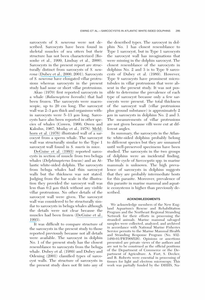
and 115.8 � 75 �m (mean � SD) in di-ameter and were located within myocytes.In 5-�m thick sections stained with H&E,the sarcocyst wall appeared �2 �m thickwith barely visible undulations or protru-sions. In 1-�m sections stained with tolu-dine blue, additional details of the sarco-cyst wall were visible. The sarcocysts fromdolphin No. 1 had a �1 �m thick sarcocystwall without recognizable villar protru-sions (Fig. 1A). The walls from the sarco-cysts in dolphins Nos. 2 and 3 were 1–2�m thick and had small conical villar pro-trusions (Fig. 1B, C). Bradyzoites were ap-proximately 5 �m long in all three sarco-cysts.
The ultrastructural appearance of thethree sarcocysts in Figure 1 is shown inFigure 2. In all three sarcocysts, the par-asitophorous vacuolar membrane waslined by an electron-dense layer (El). TheEl was interrupted at irregular distances(Fig. 2). The villar protrusions (VP) on sar-cocyst walls differed in structure. The sar-cocyst wall (Cw) in Figure 2A from dol-phin No. 1 appears smooth without anyappreciable villar protrusions. In the sar-cocyst from dolphin No. 2, the VP had aconical shape (Fig. 2B).
In the sarcocyst in dolphin No. 3, theVP were most prominent with a wide baseand a rounded villar tip (Fig. 2C). Theground substance in all sarcocysts wassmooth without microtubules or promi-nent granules.
Voucher histologic sections of musclesfrom the dolphins were deposited in theU.S. National Parasite Collection, Beltsville,Maryland as USNPC Nos. 91695(dolphin1), 91696(dolphin 2), 91697(dolphin 3).
DISCUSSION
This report is the first to demonstrate arelatively high prevalence of sarcocysts instranded Atlantic white-sided dolphins.Sarcocysts were previously described, al-though not identified, in a single Atlanticwhite-sided dolphin (DeGuise et al.,1993).
Identification of sarcocysts by light mi-

EWING ET AL.—SARCOCYSTIS IN ATLANTIC WHITE-SIDED DOLPHINS 293
FIGURE 1. Sarcocysts of Sarcocystis spp. in tissue sections of muscles of three dolphins. Note differencesin the thickness and character of the sarcocyst wall (arrowheads). Opposing arrows point to longitudinally-cutbradyzoites. A. Dolphin No. 1. Note thin sarcocyst wall without any visible villar protrusions. B. Dolphin No.2. Note small villar protrusions on the sarcocyst wall. C. Dolphin No. 3. Note small, deciduous teeth-likeprotrusions (arrows) on the sarcocyst wall. 1-�m sections stained with toludine blue. Bar � 10 �m applies inall figures.
croscopy is difficult due to variation in sar-cocyst size and shape as a result of con-tractile state of the muscle, age of sarco-cyst, type of host cell parasitized, andmethod of fixation (Dubey et al., 1989).Similarly, metrocyte and bradyzoite sizeand shape can be highly variable depend-ing on stage of division and how tightlythey are packed into the cyst. Ultrastruc-
tural characteristics of the sarcocyst wallprovide more reliable criteria for identifi-cation (Dubey et al., 1989).
Recent reports have documented en-cephalitis in sea otter (Enhydra lutris; Ro-sonke et al., 1999, Lindsay et al., 2000)and Pacific harbor seal (Phoca vitulina ri-chardsi; LaPointe et al., 1998) associatedwith S. neurona-like schizonts. However,

294 JOURNAL OF WILDLIFE DISEASES, VOL. 38, NO. 2, APRIL 2002
FIGURE 2. Transmission electron micrographs of sarcocysts from the three dolphins shown in Figure 1,respectively. Figures on the left (A–C) are low power of the sarcocysts and figures on the right (D–F) arehigh power of the sarcocyst wall (Cw). Note differences in heights of villar protrusions (VP), ground substance(Gs), and structures of bradyzoites. Opposing arrows point to longitudinally-cut bradyzoites. Also note prom-inent septa (Se) and electron-dense layer (El) lining the parasitophorous vacuolar membrane. A. Villar pro-trusions are very short. B. Villar protrusions are conical in shape. C. Villar protrusions are prominent. Uranylacetate and lead citrate stain. Bar � 2 �m A–C, bar � 0.5 �m D–F.
EWING ET AL.—SARCOCYSTIS IN ATLANTIC WHITE-SIDED DOLPHINS 295
sarcocysts of S. neurona were not de-scribed. Sarcocysts have been found inskeletal muscles of sea otters but theirstructure has not been characterized (Ro-sonke et al., 1999, Lindsay et al., 2000).Sarcocysts in the present report are struc-turally distinct from sarcocysts of S. neu-rona (Dubey et al., 2000, 2001). Sarcocystsof S. neurona have elongated villar protru-sions whereas sarcocysts in the presentstudy had none or short villar protrusions.
Akao (1970) first reported sarcocysts ina whale (Balaenoptera borealis) that hadbeen frozen. The sarcocysts were macro-scopic, up to 20 cm long. The sarcocystwall was 2–3 �m thick and organisms with-in sarcocysts were 5–13 �m long. Sarco-cysts have also been reported in other spe-cies of whales (Cowen, 1966; Owen andKakulus, 1967; Muday et al., 1978). Mehl-horn et al. (1976) illustrated wall of a sar-cocyst from a sperm whale. The sarcocystwall was structurally similar to the Type 1sarcocyst wall found in S. muris in mice.
DeGuise et al. (1993) reported sarco-cysts in section of muscle from two belugawhales (Delphinapterus leucas) and an At-lantic white-sided dolphin. The sarcocystsfrom beluga whales had thin sarcocystwalls but the thickness was not stated.Judging from the bar scale in the illustra-tion they provided the sarcocyst wall wasless than 0.2 �m thick without any visiblevillar protrusions. No other details of thesarcocyst wall were given. The sarcocystwall was considered to be structurally sim-ilar to sarcocysts in beluga whales althoughthe details were not clear because themuscles had been frozen (DeGuise et al.,1993).
It was difficult to compare structure ofthe sarcocysts in the present study to thosereported previously because not all detailswere available. The sarcocyst in dolphinNo. 1 of the present study has the closestresemblance to sarcocysts from the belugawhale. Dubey et al. (1989) and Dubey andOdening (2001) classified types of sarco-cyst walls. The structure of sarcocysts inthe present study does not fit into any of
the described types. The sarcocyst in dol-phin No. 1 has closest resemblance toType 1 sarcocyst, but in Type 1 sarcocyststhe sarcocyst wall has invaginations thatwere missing in the dolphin sarcocyst. Theclosest resemblance of the sarcocysts indolphins No. 2 and 3 is to Type 9 sarco-cysts of Dubey et al. (1989). However,Type 9 sarcocysts have prominent micro-tubules in villar protrusions that were ab-sent in the present study. It was not pos-sible to determine the prevalence of eachtype of sarcocyst because only a few sar-cocysts were present. The total thicknessof the sarcocyst wall (villar protrusionsplus ground substance) is approximately 2�m in sarcocysts in dolphins No. 2 and 3.The measurements of villar protrusionsare not given because villi were cut at dif-ferent angles.
In summary, the sarcocysts in the Atlan-tic white-sided dolphins probably belongto different species but they are unnameduntil well-preserved specimens have beenstudied. The sarcocysts in the two groupsof dolphins were an incidental finding.The life cycle of Sarcocystis spp. in marinemammals is unknown. The high preva-lence of sarcocysts in dolphins suggeststhat they are probably intermediate hostsfor this parasite. Moreover, prevalence ofthis parasite in marine mammal and aquat-ic ecosystems is higher than previously de-scribed.
ACKNOWLEDGMENTS
We acknowledge members of the New Eng-land Aquarium’s Rescue and RehabilitationProgram and the Northeast Regional StrandingNetwork for their efforts in processing thestranded animals. Marine mammal salvagedsamples were collected, analyzed, and archivedin accordance with National Marine FisheriesService permits to the Marine Mammal Healthand Stranding Response Program (No. 932-1489-01/PRT009526). Opinions or assertionspresented are private views of the authors andare not to be construed as the official positionsof the Department of Commerce or the De-partment of Agriculture. A. Fort, S. Decker,and B. Roberts were essential in processing oftissues for light and electron microscopy. Thiswork was partially funded by the DHHS, Na-

296 JOURNAL OF WILDLIFE DISEASES, VOL. 38, NO. 2, APRIL 2002
tional Institutes of Health, National Center forResearch Resources, grant T32 RR07057 (toJZ).
LITERATURE CITED
AKAO, S. 1970. A new species of Sarcocystis parasiticin the whale Balaenoptera borealis. Journal ofProtozoology 17: 290–294.
DEGUISE, S., A. LAGACE, C. GIRARD, AND P. BELAND.
1993. Intramuscular Sarcocystis in two belugawhales and an Atlantic white-sided dolphin fromthe St. Lawrence Estuary, Quebec, Canada. Jour-nal of Veterinary Diagnostic Investigation 5: 296–300.
DUBEY, J. P., C. A. SPEER, AND R. FAYER. 1989. Sar-cocystosis of animals and man. CRC Press, Inc.,Boca Raton, Florida, pp. 2–104.
, W. J. A. SAVILLE, D. S. LINDSAY, R. W. STICH,
J. F. STANEK, C. A. SPEER, B. M. ROSENTHAL, C.J. NJOKU, O. C. H. KWOK, S. K. SHEN, AND S. M.REED. 2000. Completion of the life cycle of Sar-cocystis neurona. Journal of Parasitology 86:1276–1280.
, D. S. LINDSAY, D. FRITZ, AND C. SPEER. 2001.Structure of Sarcocystis neurona sarcocysts.Journal of Parasitology 87: In press.
LAPOINTE, J. M., P. J. DUIGNAN, A. E. MARSH, F. M.GULLAND, C. BARR, D. K. NAYDEN, D. P. KING,
C. A. FARMAN, K. A. BUREK HUNTINGDON, AND
L. J. LOWENSTEIN. 1998. Meningoencephalitisdue to a Sarcocystis neurona-like protozoan inPacific harbor seals (Phoca vitulina richardsi).Journal of Parasitology 84: 1184–1189.
LINDSAY, D. S., N. J. THOMAS, AND J. P. DUBEY. 2000.Biological characterization of Sarcocystis neu-rona isolated from a southern sea otter (Enhydralutris nereis). International Journal for Parasitol-ogy 30: 617–624.
MEHLHORN, H., W. J. HARTLEY, AND A. O. HEYDORN.
1976. A comparative ultrastructural study of thecyst wall of 13 Sarcocystis species. Protistologica12: 451–467.
MUNDAY, B. L., R. W. MASON, W. J. HARTLEY, P. J. A.PRESIDENTE, AND D. OBENDORF. 1978. Sarcocys-tis and related organisms in Australian wildlife:I. Survey findings in mammals. Journal of Wild-life Diseases 14: 417–433.
OWEN, C. C., AND R. A. DADULAS. 1967. Sarcospo-ridiosis in sperm whale. Australia Journal of Sci-ence 31: 46–47.
ROSONOKE, B. J., S. R. BROWN, S. J. TORNQUIST, S. P.SNYDER, M. M. GARNER, AND L. L. BLYTHE.1999. Encephalomyelitis associated with a Sar-cocystis neurona-like organism in a sea otter.Journal of the American Veterinary Medical As-sociation 215: 1839–1842.
Received for publication 23 September 2000.