Assessment of antimicrobial activity of Catechu and its dyed substrate
11
This article appeared in a journal published by Elsevier. The attached copy is furnished to the author for internal non-commercial research and education use, including for instruction at the authors institution and sharing with colleagues. Other uses, including reproduction and distribution, or selling or licensing copies, or posting to personal, institutional or third party websites are prohibited. In most cases authors are permitted to post their version of the article (e.g. in Word or Tex form) to their personal website or institutional repository. Authors requiring further information regarding Elsevier’s archiving and manuscript policies are encouraged to visit: http://www.elsevier.com/copyright
-
Upload
independent -
Category
Documents
-
view
1 -
download
0
Transcript of Assessment of antimicrobial activity of Catechu and its dyed substrate

This article appeared in a journal published by Elsevier. The attachedcopy is furnished to the author for internal non-commercial researchand education use, including for instruction at the authors institution
and sharing with colleagues.
Other uses, including reproduction and distribution, or selling orlicensing copies, or posting to personal, institutional or third party
websites are prohibited.
In most cases authors are permitted to post their version of thearticle (e.g. in Word or Tex form) to their personal website orinstitutional repository. Authors requiring further information
regarding Elsevier’s archiving and manuscript policies areencouraged to visit:
http://www.elsevier.com/copyright

Author's personal copy
Assessment of antimicrobial activity of Catechu and its dyed substrate
Mohd Ibrahim Khan a,1, Aijaz Ahmad b,1, Shafat Ahmad Khan a, Mohd Yusuf a, Mohammad Shahid a,Nikhat Manzoor b, Faqeer Mohammad a,*
aDepartment of Chemistry, Jamia Millia Islamia, New Delhi-110025, IndiabDepartment of Biosciences, Jamia Millia Islamia, New Delhi, India
a r t i c l e i n f o
Article history:Received 16 June 2010Received in revised form8 February 2011Accepted 23 March 2011Available online 5 April 2011
Keywords:Antimicrobial activityCatechuWoollen yarn
a b s t r a c t
The present study was undertaken to evaluate the antimicrobial activity of catechu in solution and %microbial reduction of dyed wool samples against Escherichia coliMTCC 443, Staphylococcus aureusMTCC902, Candida albicans ATCC 10261 and Candida tropicalis ATCC 750, by using micro-broth dilutionmethod, disc diffusion assay and growth curve studies. The dye showed the maximum antimicrobialactivity at 20% w/v, inhibiting the microbial growth by more than 90%. In the next set of the experimentsthe antimicrobial activity of the dye was compared on woollen yarn alone and on pre-mordantedsamples. The reduction in antimicrobial activity was observed when mordanted samples were exam-ined. K/S, CIELab values and fastness properties of the dyed samples were assessed. The structuralmorphology of woollen yarn was also evaluated using scanning electron microscopy (SEM). Haemolyticactivity on human erythrocytes was studied to exclude possibility of further associated cytotoxicity. Theobserved antimicrobial characteristics and negligible cytoxicity of catechu indicate that the dye might bea promising antimicrobial agent for developing bioactive textile materials and clothing.
� 2011 Elsevier Ltd. All rights reserved.
1. Introduction
Recently therehas been increasing interest in themanufacture oftextile materials and clothing with antimicrobial properties.Consumers worldwide are looking for clothing which providegreater comfort and remain fresh andodour free in use. Clothing andother textilematerials can act as carriers formicroorganisms such aspathogenic or odour-generating bacteria, fungi and moulds (Guptaet al., 2004). These materials are often prone to attack by microor-ganisms, as these provide basic requirements such as moisture,oxygen, nutrients and temperature for microbial growth andmultiplication. This often leads to the discolouration, quality dete-rioration, objectionable odour, dermal infection, allergic responsesand other related diseases (Singh et al., 2005). Hence, there isa pressing need to provide textile materials which are resistant tomicroorganisms but retain all the usual desirable characteristics oftextiles, as these textile materials find various applications such asmasks, hospital covers, surgical gowns apart from the conventionalapparel usage (Dev et al., 2009). Many commercial products arecurrently available in the market with a range of antimicrobial
properties under different trade name for textile industry. Majorityof such products are synthetic based such as organo-metallics,phenols, quaternary ammonium salts and organosilicons (Han andYang, 2005). Although the synthetic antimicrobial agents are veryeffective against a range of microbes and give a durable effect ontextiles, they are cause of concern due to health hazards, action onnon-target microorganisms and environmental pollution (Singhet al., 2005; Joshi et al., 2009; Grillitsch et al., 2006). Natural dyesare believed to be safe because of non-toxic, non-allergic andbiodegradable nature (Ali et al., 2009; Sivakumaret al., 2011) and arebeing used extensively in colouration of textile materials (Bechtoldet al., 2003; Vankar et al., 2007; Ali and El-Mohamedy, 2010;Komboonchoo and Bechtold, 2009). Studies have been carried outfrom long time on natural extracts, and have proved that the naturalproducts have highly momentous antibiotic properties (Ahmadet al., 2010). Due to the fact that natural dyes can often inhibit thegrowth of microorganisms without toxicity (Han and Yang, 2005),considerable research work is being undertaken in the area ofproducing bioactive textiles for the protection of clothing fromcommon microbes causing cross infections (Sathianarayanan et al.,2010; Lee et al., 2009; Gupta and Laha, 2007; Prusty et al., 2010).
Cutch/Catechu/Katha is a brownnatural dye obtained chiefly fromthe heartwood of Acacia catechu, found in most of the Indian sub-Himalayas (Anonymous, 1985). The chief colouring componentpresent in the cutch is catechin (Fig. 1) having molecular formula
* Corresponding author. Tel.: þ91 9350114878 (mobile).E-mail address: [email protected] (F. Mohammad).
1 Mohd Ibrahim Khan and Aijaz Ahmad contributed equally to the study.
Contents lists available at ScienceDirect
Journal of Cleaner Production
journal homepage: www.elsevier .com/locate/ jc lepro
0959-6526/$ e see front matter � 2011 Elsevier Ltd. All rights reserved.doi:10.1016/j.jclepro.2011.03.013
Journal of Cleaner Production 19 (2011) 1385e1394

Author's personal copy
C15H14O6 (Mayer and Cook, 1943). The content of catechin in cutchvaries from 4 to 7% (Anonymous, 1985). This dye is being usedextensively in dyeing of textile materials (Bhattacharya and Shah,2000; Bhattacharyya and Lohia, 2002; Vankar and Shanker, 2008).Pharmacological studies have demonstrated that A. catechu used intraditional medicines show anti-inflammatory and anticanceractivities (Anonymous, 2006).
So far, no study has been undertaken to investigate the antimi-crobial properties of un-mordanted and pre-mordanted woollen yarndyedwith cutch. Hence, in the present study, a comparisonwasmadeto examine the antimicrobial property of dye against common path-ogenic bacteria Escherichia coli and Staphylococcus aureus and oppor-tunistic pathogenic yeasts Candida albicans and Candida tropicalis,when applied onwoollen yarn alone and on pre-mordanted samples.CIELab values of dyed samples were recorded. Fastness properties ofdyed woollen yarn samples were also examined for their durabilitywith respect towash, light and crocking. The structuralmorphologyofwoollen yarnwas evaluated by using scanning electron microscopy.
2. Materials and methods
2.1. Dye and yarn
Woollenyarnwas purchased fromMambWoollens Ltd. Bhadohi,India. Commercial sample of powdered cutch dye was obtained
from Sir Biotech India Ltd. Kanpur, India. All other chemicals usedwere of Laboratory Analytical grade.
2.2. Mordanting and dyeing
Before the application of mordant, woollen yarn was soaked inwater. The mordants were dissolved in water; liquor ratio for mor-danting was kept at 1:40. Water soaked woollen yarn samples wereimmersed inmordant solution. Temperatureof themordantbathwasraised till simmeringpoint (91e93 �C) and left at that temperature for1 hwith constant stirring. The mordanted wool samples were rinsedwith water in order to remove superfluous mordants (unused).
The mordanted wool samples were drenched in the dye bathcontaining extracted dye. For dyeing material to liquor ratio waskept at 1:40. Then temperature of the dye bath was raised tillsimmering point (91e93 �C) and left at that temperature for 1 h. Inorder to get uniform dyeing the samples were stirred regularly. Thedyed woollen yarn samples were washed with (5 gm/L) non-ionicdetergent (Safewash) and thereafter rinsed with tap water. Thesamples were dried in shade at room temperature.
2.3. Colour measurement
Dyed samples were prepared for colour measurements, whichwere carried out by following a standard procedure. CIELab and K/Svalues of the dyed samples were recorded on Gretag Macbethcolour-eye 7000 A spectrophotometer. L* corresponding tothe lightness and darkness (100 ¼ white, 0 ¼ black), a* to theredegreen coordinate (þve ¼ red, �ve ¼ green) and b* to theyelloweblue coordinate (þve ¼ yellow, �ve ¼ blue).
2.4. Fastness testing
The light fastness of the dyed wool samples were conducted ondigi light NxTm having water cooled Mercury Blended Tungstenlamp, according to test method AATCC 16E-1993. The wash fastnessof the dyed wool samples were measured in Laundrometer as perthe ISO 105-C06:1994 specifications. Dry and wet rub fastness ofthe dyed wool samples were tested using a Crockmeter as perIndian standard IS 766:1988 by mounting the fabric on panel andgiving ten strokes for both dry and wet rub fastness tests. Thesamples were assessed for staining on white adjacent fabric.
2.5. Scanning electron microscopy (SEM) of woolen samplesshowing surface morphology
SEM technique was used to investigate the surface morphologyof different woollen samples before and after the application ofmordants and dye. Samples were glued to aluminium stubs withcolloidal silver paint for conductivity and sputter coated with goldfor 3 min in an argon atmosphere. Samples were observed anddigitally imaged on a LEO 435VP Scanning Electron Microscope at10 kV accelerating voltage.
2.6. Strains and media
Stock cultures of C. albicans ATCC 10261 and C. tropicalis ATCC 750weremaintained on slants of nutrient agar (yeast extract 1%, peptone2%, D-glucose 2% and agar 2.5%) at 4 �C. Stock cultures of E. coliMTCC443andS.aureusMTCC902were cultured inMaconkyagar (HiMedia,India). To initiate growth for experimental purposes, one loop full ofcells from an agar culture was inoculated into 25 mL of respectivenutrient media and incubated at 30e37 �C for 24 h i.e., up tostationary phase (primary culture). The cells from primary culture
OH
OH
OH
OOH
OH
Fig. 1. Structure of catechin.
Fig. 2. a*b* plot of the dyed woollen yarn samples.(1) un-mordanted 20% cutch dyedwool, (2) un-mordanted 10% cutch dyed wool, (3) 5% ferrous sulphate mordanted 20%cutch dyed wool, (4) 5% ferrous sulphate mordanted 10% cutch dyed wool, (5) 1%stannous chloride mordanted 20% cutch dyed wool, (6) 1% stannous chloride mor-danted 10% cutch dyed wool, (g) 2.5% ferrous sulphate þ 0.5% stannous chloridemordanted 20% cutch dyed wool, (h) 2.5% ferrous sulphate þ 0.5% stannous chloridemordanted 10% cutch dyed wool.
M.I. Khan et al. / Journal of Cleaner Production 19 (2011) 1385e13941386

Author's personal copy
(108 cells mL�1) were re-inoculated into 100 mL fresh YPD mediumand grown for 8e10 h i.e., upto mid-log phase (106 cells mL�1).
E. coli, a gram-negative bacterium, was selected due to itspopularity of being selected as a test organism and its resistance tocommon antimicrobial agents (Yang et al., 2000). S. aureus, a path-ogenic gram-positive bacterium, was used because it is the majorcause of cross-infection in hospitals and it is the most frequentlyevaluated species. Candida isolates were selected because these arethe commencel opportunistic pathogens of the immuno-compromised patients (Shao et al., 2007).
2.7. Minimum inhibitory concentration
The Minimum Inhibitory Concentration (MIC) was defined asthe lowest concentration of the dye that causes inhibition of visiblegrowth of test microorganisms. In vitro susceptibility tests wereperformed to evaluate MICs using the standard methods describedin the guidelines of document NCCLS M27-A2 (2002) for yeasts,document NCCLS/CLSI M11-A6 (2004) guidelines for gram-nega-tive, and document CLSI M100-S15 (2005) for gram positives. Theeffect of the dye concentration (0.04e5% w/v) on the antimicrobialactivity was assessed.
2.8. Growth studies
Growth studies of the tested microorganisms were done asdescribed earlier (Khan et al., 2010) with slight modifications. Priorto testing, the test microorganisms were sub-cultured at least twiceand grown for 24 h at 35 �C on SDA plates. For growth studies,106 cells (optical density A600 ¼ 0.1) of test strains were grownaerobically in 50 mL media on automated shaker set at 35 �C withagitation of 200 rpm. A. catechu dye with final concentrations of0� MIC, 0.1� MIC, 0.5� MIC and 1� MIC for each test isolate werealso added to the cultures along with positive (0.001% w/v of flu-conazole for yeasts and 0.001% w/v of ampicillin for bacteria). Atpre-determined time points (0, 2, 4, 6, 8, 12, and 24 h) aliquots wereremoved and growth was followed turbidometrically at 595 nmusing Lab Med Spectrophotometer (USA). Optical density wasrecorded for each concentration against time (hours). OD versusconcentrations of each inhibitor is shown in Fig. 5.
2.9. Disc diffusion assay
Strains were inoculated into liquid YPD medium and grownovernight at 35 �C. The cells were then pelleted and washed threetimes with distilled water. Approximately 105 cells mL�1 were inoc-ulated in molten agar media at 40 �C and poured into 100-mm-diameter petriplates. Filter discs were kept on solid agar and dyewasspottedonthedisc. Test compounddissolved indoubledistilledwaterwith final concentrations of 1�MIC, 2�MIC, and 4�MIC, or solventcontrol (distilledwater)waspipetted onto 4-mm-diameterfilter disc.The diameter of zone of inhibition was recorded in millimetres after48 h and was compared with that of control. The type of the haloproduceddepicts fungicidal/static characteristicof test entity (Ahmadet al., 2011). The experiment was performed on two yeast and twobacteria species. Values were shown in terms of Mean � standarderror of mean (SEM) of all three respective categories.
2.10. Determination of antimicrobial activity of dyed yarn
Wool samples were dyed by the standard method prescribed fornatural dyes (Khan et al., 2006). Dyeing was carried out atsimmering point (91e93 �C) at neutral pH and a liquor ratio of 1:40,for 1 h and at two different dye concentrations (20, 10%, owf). Thedyed woollen yarn samples were washed with (5 gL�1) non-ionicdetergent (Safewash) and thereafter rinsed thoroughly with tapwater. To study the effect of mordants on antimicrobial activity,wool samples were pre-mordanted with ferrous sulphate (5% owf),stannous chloride (1% owf) and mixed ferrous sulphate and stan-nous chloride (2.5% þ 0.5% owf, respectively) and then dyed.Mordanting was carried out at simmering point (91e93 �C), atliquor ratio of 1:40 for 1 h. The mordanted wool samples wererinsed with water in order to remove superfluous mordants.
The antimicrobial activity of dyed wool specimens were evalu-ated after being subjected to one and five times washing. 0.0254m2
yarn (dyed and undyed) was introduced in the 50mL nutrient brothinoculatedwith a desiredmicrobe and incubated overnight at 37 �C.The reduction of the microbial growth by the dyed yarn wasexpressed as follows:
R ¼ B� A=A� 100
where R ¼ % reduction in microbial population; B ¼ absorbance(595 nm) of the media inoculated with microbe and undyed yarn;A¼ absorbance (595 nm) of themedia inoculatedwithmicrobe anddyed yarn. The greater the growth, the higher the turbidity, and theoptical density figure was therefore directly proportional to thenumber of microbes in the media.
2.11. Haemolytic activity
The haemolytic activity of the A. catechu dye was determined onhuman red blood cells. Human erythrocytes from healthy individ-uals were collected in tubes containing EDTA as anti-coagulant. Theerythrocytes were harvested by centrifugation for 10 m at2000 rpm at 20 �C, and washed three times in PBS. To the pellet,PBS was added to yield a 10% (v/v) erythrocytes/PBS suspension.The 10% suspension was then diluted 1:10 in PBS. From eachsuspension, 0.1 mL was added in triplicate to 100 mL of a differentdilution series of catechu dye (or fluconazole and amphotericin B)in the same buffer in Eppendorf tubes. Total haemolysis was ach-ieved with 1% Triton X-100. The tubes were incubated for 1 h at37 �C and then centrifuged for 10 min at 2000 rpm at 20 �C. Fromthe supernatant fluid, 150 mL was transferred to a flat-bottomedmicrotiter plate (BIO-RAD, iMark, US), and the absorbance wasFig. 3. K/S graph of the dyed woollen yarn samples.
M.I. Khan et al. / Journal of Cleaner Production 19 (2011) 1385e1394 1387

Author's personal copy
measured spectrophotometrically at 450 nm. The haemolysispercentage was calculated by following equation:
% hemolysis ¼ ½ðA450 of test compound treated sample
� A450 of buffer treated sampleÞ=�ðA450 of 1% Triton X� 100 treated samples
� A450 of buffer treated sample� � 100
2.12. Statistical analysis
Each experiment was performed twice and in triplicate. Resultsobtained were expressed in terms of mean � standard error (SEM).Statistical analyses were performed and P value �0.05 wasconsidered significant.
3. Results and discussion
3.1. Colour measurement
CIELab and K/S values of the dyed woollen yarn samples are giveninTable 1. From a*b* plot (Fig. 2), it can be seen that the colour gamutof wool samples dyed with cutch indicates a red yellow zone. TheL* values were higher in case of stannous chloride mordantedsamples corresponding to lighter shades.Whereas, the L*valueswerefound to be lower in case of ferrous sulphate mordanted samplescorresponding to darker shades. c* values shows tin chloride mor-danting results in brighter shades whereas ferrous sulphate increasedullness of shade. K/S value graph of the dyed samples (Fig. 3) showsthat use of mordants considerably increased dye absorption leadingto higher K/S values in case of mordanted samples than un-mor-danted ones. Stannous chloride mordant was found to have moreprominent effect on colour strength than ferrous sulphate.
3.2. Fastness testing
Light fastness testing machine with Mercury Blended Tungstenlamp has nearest approach to that of sunlight. It was observed fromthe colour fastness data (Table 2) that all the samples dyed withcatechu exhibit fairly good to good light fastness i.e. 4e5. The dyedsamples show fairly good to very goodwash fastness ratings rangingfrom 3e5 on grey scale and no staining on adjacent fabric wereobserved. The dry rub fastness of the dyed samples were foundbetween 4e5 and wet rub fastness ratings were 2e3 on grey scale.
3.3. SEM of woollen samples showing surface morphology
The surface morphological features of woollen yarn are depictedin Fig. 4. SEM photograph for the untreated wool shows a normalmorphological form and the scale clearly has been seen (Fig. 4a).
For the mordanted wool samples some particles of the mordantshave been seen on the yarn surfaces (Fig. 4bed). The un-mordantedwool sample dyedwith cutch shows the deposition of dyemoleculeon the yarn surface (Fig. 4e). The mordanted wool samples dyedwith cutch again show the deposition of dye molecule on yarnsurfaces (Fig. 4feh). From the pictures of the dyed samples it can bedemonstrated that coating on themordanted wool samples is morethan un-mordanted sample.
3.4. Antimicrobial activity of catechu in solution
Antifungal and antibacterial activities of the dye when comparedwith standard antifungal and antibacterial drugs (Fluconazole andAmpicillin) showedsignificant andpotential antimicrobialproperties.
3.4.1. Minimum inhibitory concentration (MIC)The MIC of the dye was determined against two fungal and two
bacterial species usingmicro-broth dilution method. The minimuminhibitory concentration of the dye against the fungi was observedto be as lowas 0.15%w/vwhereas for the bacteria it was observed tobe 0.62% w/v.
3.4.2. Growth studiesIn case of growth curve studies the effect of increasing
concentrations of the dye on the growth of test microbes has beenstudied. Fig. 5 depicts the growth rates of C. albicans, C. tropicalis,E. coli and S. aureus, in the presence of A. catechu dye at MIC andsub-MIC concentrations. The absorbance obtained for the growthcontrol (only organism) showed that the test cultures reached thestationary growth phase after 16 h showing a normal growthpattern. The curve depicts a lag phase in the initial phase of growth,active log phase and stationary phase. All the test microorganismisolates were found to be susceptible to the test dye. At sub-MICvalue (0.1� MIC) of the dye, the test microorganisms do not showany alterations in their growth curves, except that of E. coli showingthe extension of the lag phase by 2 h. At 0.5�MIC values of the dye,60 and 50% growth was inhibited in C. albicans and C. tropicaliswithrespect to control, respectively. The E. coli and S. aureus showed 54and 69% of growth inhibition when treated with 0.5� MIC of thedye. More than 95% inhibition was observed at MIC values of thedye by all the tested microorganisms. It was worth to note that atthe MIC values, the dye sowed the more inhibitory effect than thecommercially used antimicrobials.
3.4.3. Disc diffusion assayThe results summarized in Table 3 give the sensitivity assay,
using standard discs of A. catechu dye, ampicillin and fluconazole.All the two types of Candida isolates, E. coli and S. aureus showed
Table 1CIELab values of the dyed sample.
Dye%(Cutch)
Mordant L* a* b* c* ho K/S
20 Un-mordanted 58.46 5.23 19.68 20.37 75.13 4.3410 61.60 4.60 19.74 20.26 76.89 3.2620 5% FeSO4 56.23 4.80 13.01 13.87 69.74 6.1910 60.21 4.39 13.52 14.21 72.00 4.8120 1% SnCl2 65.64 3.96 26.75 27.04 81.57 8.6210 67.55 3.02 25.53 25.71 83.26 5.3120 2.5% FeSO4
þ 0.5% SnCl261.28 4.67 24.05 24.50 79.01 8.10
10 60.06 4.55 26.19 26.59 80.14 5.47
Table 2Fastness properties of the dyed sample.
Dye%(Cutch)
Mordant Lightfastness
Wash fastness Rub fastness
c.c. c.s. c.w. Dry Wet
20 Un-mordanted 4e5 3e4 5 5 4 2e310 4 3 5 5 3e4 220 5% FeSO4 5 4e5 5 5 4e5 2e310 5 4e5 5 5 4e5 2e320 1% SnCl2 4e5 4 5 5 4 2e310 4 3e4 5 5 4 220 2.5% FeSO4
þ 0.5% SnCl25 4 5 5 4 2
10 4e5 4 5 5 4e5 2e3
Light fastness 1 ¼ very poor, 2 ¼ poor, 3 ¼ moderate, 4 ¼ fairly good, 5 ¼ good,6 ¼ very good, 7 ¼ Excellent, 8 ¼ Outstanding.Wash fastness and rub fastness 1 ¼ Poor, 2 ¼ Moderate, 3 ¼ Fairly good, 4 ¼ good,5 ¼ Very good.
M.I. Khan et al. / Journal of Cleaner Production 19 (2011) 1385e13941388

Author's personal copy
high degree of sensitivity as is evident from zone of clearance(Fig. 6). Index of sensitivity defined as
S Zone diameterðmmÞ=concentration�mg mL�1
�
¼ clearingðmmÞ=mg
isgreater (17.33) forC. tropicalis isolates and is least (8.66) for S.aureusisolates. The most important thing noticed that the dye was moreeffective against fungus as compared to bacteria. The results showed
that, in case of control disc no zone of inhibitionwas observed, so asfar as our study is concerned, distilledwater, as a solvent, is havingnoeffect on the tested organisms. Hence we can effectively concludehere that whole of the antimicrobial effect is because of the dye.
3.5. Antimicrobial activity of catechu on substrate
Having studied the antimicrobial effect of the dye in solution,the next step was to access their effectiveness on the substrate
Fig. 4. SEMs of (a) untreated wool, (b) 5% ferrous sulphate mordanted, (c) 1% stannous chloride mordanted, (d) 2.5% ferrous sulphate þ 0.5% stannous chloride mordanted, (e) un-mordanted wool dyed with 20% cutch, (f) 5% ferrous sulphate mordanted wool dyed with 20% cutch, (g) 1% stannous chloride mordanted wool dyed with 20% cutch, (h) 2.5% ferroussulphate þ 0.5% stannous chloride mordanted wool dyed with 20% cutch.
M.I. Khan et al. / Journal of Cleaner Production 19 (2011) 1385e1394 1389

Author's personal copy
(wool). The wool samples dyed with A. catechu were used asa model system. The majority of natural dyes need a mordant inthe form of a metal salt to create an affinity between the fibre andthe pigment. These metals form a ternary complex on one sidewith the fibre and on the other side with the dye. Such a strongcoordination tendency enhances, the interaction between the fibreand the dye, resulting in high dye uptake. Therefore, the study wasalso carried on the mordanted wool samples dyed with differentconcentrations of the dye. Commercial antimicrobials (fluconazolefor the yeasts and ampicillin for the bacteria) were also studied forthe comparison. The results were summarized in Fig. 7A. It can beobserved from the Fig. 7A that wool dyed with 10% of the dye
(owf) shows slightly low extend of inhibition (65e75% in bacteriaand 80e85% in fungi). The significant enhancement in antimi-crobial activity (85e90% in bacteria and 93e95% in fungi) wasobserved when 20% dye (owf) was used. The %-age inhibitionhowever, decreases for the dye when mordanted samples wereexamined. The %-age inhibition by the yarn mordanted with 5%ferrous sulphate (owf) and dyed with 20% dye (owf) ranges from47 to 64% whereas the same figures with 1% stannous chloride(owf) mordanted wool samples were 51e73%. The %-age inhibitionby the yarn mordanted with mixed ferrous sulphate and stannouschloride (2.5% þ 0.5% owf, respectively) and dyed with 20% dye(owf) ranges from 40 to 45% in bacteria and 55e62% in fungi. Thedurability of the antimicrobial activity exhibited by the dyedwoolen yarn after subjection to five times washing is reported inFig. 7B. The results indicated <15% reduction in the antimicrobialactivity as compared to the dyed yarn subjected to one timewashing. The decrease in antimicrobial activity when mordantedsamples were used is due to the involvement of the some hydroxylgroups (the only functional groups present in the dye molecule), incoordinate bond formation with metal mordants. It should beemphasized that the functional groups present in an organicmolecule are mainly responsible for the antimicrobial activity(Venkatesan et al., 2009). The results (Fig. 7), therefore suggestthat the hydroxyl groups involved in the bond formation withmetal ions (the hydroxyl groups ortho to the chromophore usually
Fig. 5. The effect of various concentrations of the dye on the growth of C. albicans, C. tropicalis, E. coli and S. aureus. The cells were grown with 0% dye (a), 0.001% w/v of fluconazolefor yeasts and 0.001% v of ampicillin for bacteria (b), 20% solvent distilled water (c), 10% dye (d), and 20% dye (e).
Table 3Sensitivity index defined by a ratio of diameter of inhibition zone (in mm) tothe concentration (in mg mL�1) for the A. catechu dye in all the testedisolates.
Isolates A. catechu dye
Sensitivity index
Candida albicans 11.66667 � 3.511885Candida tropicalis 17.33333 � 4.50925Escherichia coli 10.33333 � 3.05505Staphylococcus aureus 8.666667 � 3.21455
M.I. Khan et al. / Journal of Cleaner Production 19 (2011) 1385e13941390

Author's personal copy
become involved in coordinate bond formation) become inactiveagainst microbes which is responsible for the decrease in anti-microbial activity in mordanted samples. On the other hand, in thecase of un-mordanted wool samples, all the functional groups areinvolved against microbes resulting in high antimicrobial activity.
This is an interesting finding and requires further in depthstudies. Hence, it may be said that the A. catechu dye can be used fordyeing woollen textiles as an alternative to very expensive,synthetic and toxic antimicrobial agents.
3.6. Haemolytic activity
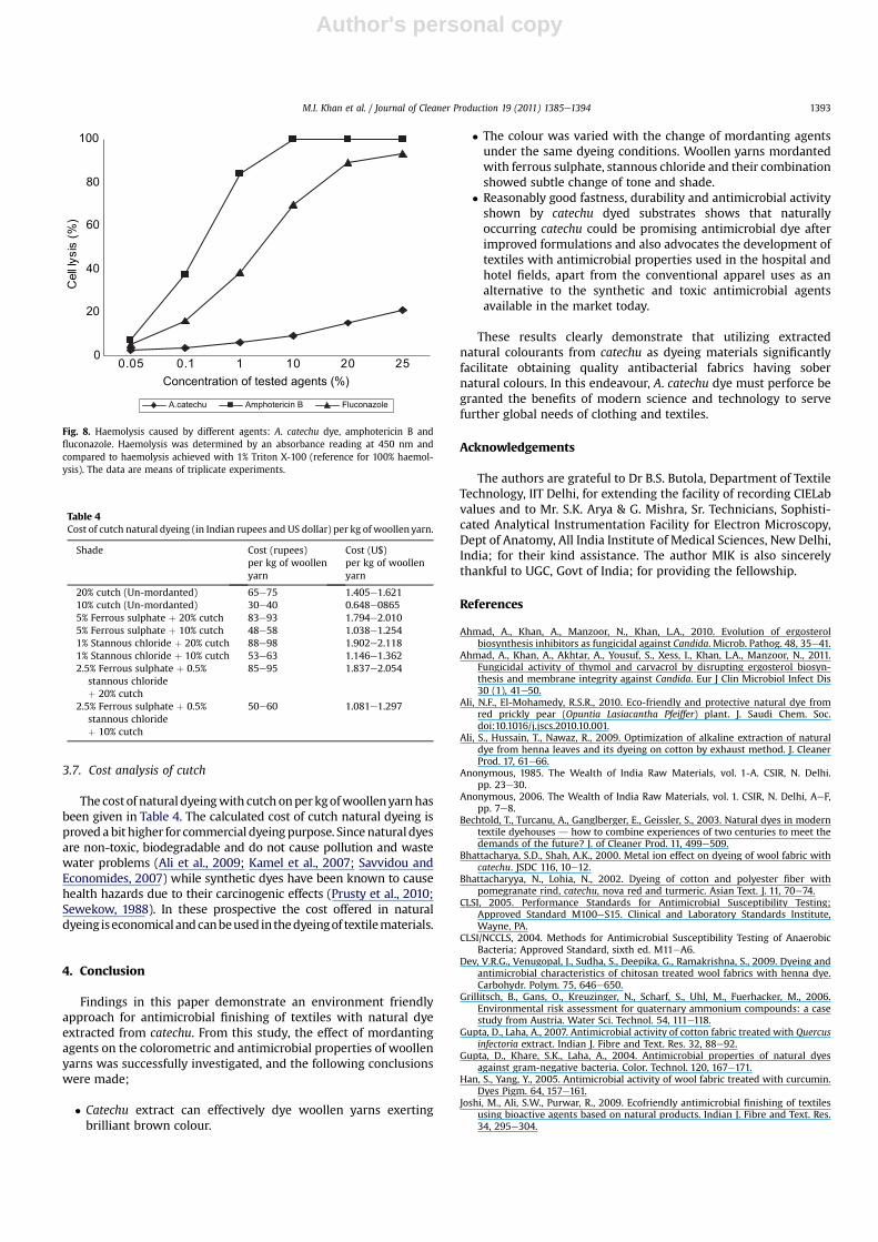
Effects of A. catechu dye, amphotericin B and fluconazole onhuman red blood cells are shown in Fig. 8. At the concentration of20% of A. catechu dye, which had profound antimicrobial effect,�20% haemolysis was observed, while as conventional antifungaltherapeutic agents showed almost 100% haemolysis. This indicatesthat A. catechu dye is significantly less toxic than the conventionalantimicrobial agents used.
Fig. 6. Disk diffusion assay of C. albicans (panel-A), C. tropicalis (panel-B), E. coli (panel-C), and S. aureus (panel-D) different concentrations of dye (1� MIC, 2� MIC, and 4� MIC).panel E describes the images for panels A, B, C and D.
M.I. Khan et al. / Journal of Cleaner Production 19 (2011) 1385e1394 1391

Author's personal copy
Fig. 7. Antimicrobial activity of the woollen yarn treated with A. catechu dye after one time washing (7A) and after five times washing (7B). Bar 1 represents the control cells withoutany treatment, 2 represents the known available antimicrobial, 3 is untreated wool, 4 & 5 represent 5% ferrous sulphate with 10 and 20% dye, 6 & 7 are 1% stannous chloride with 10and 20% dye, 8 & 9 are 2.5% ferrous sulphate þ 0.5% stannous chloride with 10 & 20% dye and 10 & 11 represent woollen yarn treated with 10 & 20% dye, respectively.
M.I. Khan et al. / Journal of Cleaner Production 19 (2011) 1385e13941392
Author's personal copy
3.7. Cost analysis of cutch
The costof natural dyeingwith cutch onperkgofwoollenyarnhasbeen given in Table 4. The calculated cost of cutch natural dyeing isproveda bit higher for commercial dyeingpurpose. Sincenatural dyesare non-toxic, biodegradable and do not cause pollution and wastewater problems (Ali et al., 2009; Kamel et al., 2007; Savvidou andEconomides, 2007) while synthetic dyes have been known to causehealth hazards due to their carcinogenic effects (Prusty et al., 2010;Sewekow, 1988). In these prospective the cost offered in naturaldyeing iseconomical andcanbeused inthedyeingof textilematerials.
4. Conclusion
Findings in this paper demonstrate an environment friendlyapproach for antimicrobial finishing of textiles with natural dyeextracted from catechu. From this study, the effect of mordantingagents on the colorometric and antimicrobial properties of woollenyarns was successfully investigated, and the following conclusionswere made;
� Catechu extract can effectively dye woollen yarns exertingbrilliant brown colour.
� The colour was varied with the change of mordanting agentsunder the same dyeing conditions. Woollen yarns mordantedwith ferrous sulphate, stannous chloride and their combinationshowed subtle change of tone and shade.
� Reasonably good fastness, durability and antimicrobial activityshown by catechu dyed substrates shows that naturallyoccurring catechu could be promising antimicrobial dye afterimproved formulations and also advocates the development oftextiles with antimicrobial properties used in the hospital andhotel fields, apart from the conventional apparel uses as analternative to the synthetic and toxic antimicrobial agentsavailable in the market today.
These results clearly demonstrate that utilizing extractednatural colourants from catechu as dyeing materials significantlyfacilitate obtaining quality antibacterial fabrics having sobernatural colours. In this endeavour, A. catechu dye must perforce begranted the benefits of modern science and technology to servefurther global needs of clothing and textiles.
Acknowledgements
The authors are grateful to Dr B.S. Butola, Department of TextileTechnology, IIT Delhi, for extending the facility of recording CIELabvalues and to Mr. S.K. Arya & G. Mishra, Sr. Technicians, Sophisti-cated Analytical Instrumentation Facility for Electron Microscopy,Dept of Anatomy, All India Institute of Medical Sciences, New Delhi,India; for their kind assistance. The author MIK is also sincerelythankful to UGC, Govt of India; for providing the fellowship.
References
Ahmad, A., Khan, A., Manzoor, N., Khan, L.A., 2010. Evolution of ergosterolbiosynthesis inhibitors as fungicidal against Candida. Microb. Pathog. 48, 35e41.
Ahmad, A., Khan, A., Akhtar, A., Yousuf, S., Xess, I., Khan, L.A., Manzoor, N., 2011.Fungicidal activity of thymol and carvacrol by disrupting ergosterol biosyn-thesis and membrane integrity against Candida. Eur J Clin Microbiol Infect Dis30 (1), 41e50.
Ali, N.F., El-Mohamedy, R.S.R., 2010. Eco-friendly and protective natural dye fromred prickly pear (Opuntia Lasiacantha Pfeiffer) plant. J. Saudi Chem. Soc.doi:10.1016/j.jscs.2010.10.001.
Ali, S., Hussain, T., Nawaz, R., 2009. Optimization of alkaline extraction of naturaldye from henna leaves and its dyeing on cotton by exhaust method. J. CleanerProd. 17, 61e66.
Anonymous, 1985. The Wealth of India Raw Materials, vol. 1-A. CSIR, N. Delhi.pp. 23e30.
Anonymous, 2006. The Wealth of India Raw Materials, vol. 1. CSIR, N. Delhi, AeF,pp. 7e8.
Bechtold, T., Turcanu, A., Ganglberger, E., Geissler, S., 2003. Natural dyes in moderntextile dyehouses d how to combine experiences of two centuries to meet thedemands of the future? J. of Cleaner Prod. 11, 499e509.
Bhattacharya, S.D., Shah, A.K., 2000. Metal ion effect on dyeing of wool fabric withcatechu. JSDC 116, 10e12.
Bhattacharyya, N., Lohia, N., 2002. Dyeing of cotton and polyester fiber withpomegranate rind, catechu, nova red and turmeric. Asian Text. J. 11, 70e74.
CLSI, 2005. Performance Standards for Antimicrobial Susceptibility Testing;Approved Standard M100eS15. Clinical and Laboratory Standards Institute,Wayne, PA.
CLSI/NCCLS, 2004. Methods for Antimicrobial Susceptibility Testing of AnaerobicBacteria; Approved Standard, sixth ed. M11eA6.
Dev, V.R.G., Venugopal, J., Sudha, S., Deepika, G., Ramakrishna, S., 2009. Dyeing andantimicrobial characteristics of chitosan treated wool fabrics with henna dye.Carbohydr. Polym. 75, 646e650.
Grillitsch, B., Gans, O., Kreuzinger, N., Scharf, S., Uhl, M., Fuerhacker, M., 2006.Environmental risk assessment for quaternary ammonium compounds: a casestudy from Austria. Water Sci. Technol. 54, 111e118.
Gupta, D., Laha, A., 2007. Antimicrobial activity of cotton fabric treated with Quercusinfectoria extract. Indian J. Fibre and Text. Res. 32, 88e92.
Gupta, D., Khare, S.K., Laha, A., 2004. Antimicrobial properties of natural dyesagainst gram-negative bacteria. Color. Technol. 120, 167e171.
Han, S., Yang, Y., 2005. Antimicrobial activity of wool fabric treated with curcumin.Dyes Pigm. 64, 157e161.
Joshi, M., Ali, S.W., Purwar, R., 2009. Ecofriendly antimicrobial finishing of textilesusing bioactive agents based on natural products. Indian J. Fibre and Text. Res.34, 295e304.
0
20
40
60
80
100
0.05 0.1 1 10 20 25Concentration of tested agents (%)
)%(
sisyllleC
A.catechu Amphotericin B Fluconazole
Fig. 8. Haemolysis caused by different agents: A. catechu dye, amphotericin B andfluconazole. Haemolysis was determined by an absorbance reading at 450 nm andcompared to haemolysis achieved with 1% Triton X-100 (reference for 100% haemol-ysis). The data are means of triplicate experiments.
Table 4Cost of cutch natural dyeing (in Indian rupees and US dollar) per kg of woollen yarn.
Shade Cost (rupees)per kg of woollenyarn
Cost (U$)per kg of woollenyarn
20% cutch (Un-mordanted) 65e75 1.405e1.62110% cutch (Un-mordanted) 30e40 0.648e08655% Ferrous sulphate þ 20% cutch 83e93 1.794e2.0105% Ferrous sulphate þ 10% cutch 48e58 1.038e1.2541% Stannous chloride þ 20% cutch 88e98 1.902e2.1181% Stannous chloride þ 10% cutch 53e63 1.146e1.3622.5% Ferrous sulphate þ 0.5%
stannous chlorideþ 20% cutch
85e95 1.837e2.054
2.5% Ferrous sulphate þ 0.5%stannous chlorideþ 10% cutch
50e60 1.081e1.297
M.I. Khan et al. / Journal of Cleaner Production 19 (2011) 1385e1394 1393

Author's personal copy
Kamel, M.M., El-Shishtawy, R.M., Youssef, B.M., Mashaly, H., 2007. Ultrasonicassisted dyeing. IV. Dyeing of cationised cotton with lac natural dye. Dyes Pigm.73, 279e284.
Khan, M.A., Khan, M., Srivstava, P.K., Mohammad, F., 2006. Extraction of naturaldyes from cutch, ratanjot and madder and their application on wool. Colourage,61e68.
Khan, A., Ahmad, A., Manzoor, N., Khan, L.A., 2010. Antifungal activities of Ocimumsanctum essential oil and its lead molecules. Nat. Pro. Commun. 5 (2), 345e349.
Komboonchoo, S., Bechtold, T., 2009. Natural dyeing of wool and hair with indigocarmine (C.I. natural blue 2), a renewable resource based blue dye. J. CleanerProd. 17, 1487e1493.
Lee, Y., Hwang, E., Kim, H., 2009. Colorimetric assay and antibacterial activity ofcotton, silk and wool fabrics dyed with peony, pomegranate, Clove, Coptis chi-nenis and gallnut extracts. Materials 2, 10e21.
Mayer, F., Cook, A.H., 1943. The Chemistry of Natural Colouring Matters. ReinholdPublishing Corporation, U.S.A, pp. 198e199.
NCCLS, 2002. Reference Method for Broth Dilution Antifungal Susceptibility Testingof Yeasts; Approved Standard M27eA2. National Committee on Clinical Labo-ratory Standards, Wayne, PA, pp. 30.
Prusty, A.K., Das, T., Nayak, A., Das, N.B., 2010. Colourimetric analysis and antimi-crobial study of natural dyes and dyed silk. J. Cleaner Prod.. doi:10.1016/j.jclepro.2010.06.020.
Sathianarayanan, M.P., Bhat, N.V., Kokate, S.S., Walunj, V.E., 2010. Antibacterialfinish for cotton fabric from herbal products. Indian J. Fiber and Text. Res. 35,50e58.
Savvidou, M.I., Economides, D.G., 2007. Colour gamut produced by applyingmixtures of natural dyes on de-inked mechanical pulp. Color. Technol. 123,119e123.
Sewekow, U., 1988. Natural dyes- an alternative to synthetic dyes. Melliand Tex-tilber. 69, E145eE148.
Shao, P.L., Huang, L.M., Hsueh, P.R., 2007. Recent advances and challenges in thetreatment of invasive fungal infections. Int. J. Antimicrob. Agents 30, 487e495.
Singh, R., Jain, A., Panwar, S., Gupta, D., Khare, S.K., 2005. Antimicrobial activity ofsome natural dyes. Dyes Pigm. 66, 99e102.
Sivakumar, V., Vijaeeswarri, J., Anna, J.L., 2011. Effective natural dye extraction fromdifferent plant materials using ultrasound. Ind. Crops Prod. 33, 116e122.
Vankar, P.S., Shanker, R., 2008. Ecofriendly ultrasonic natural dyeing of cotton fabricwith enzyme pretreatments. Desalination 230, 62e69.
Vankar, P.S., Shanker, R., Verma, A., 2007. Enzymatic natural dyeing of cotton andsilk fabrics without metal mordant. J. Cleaner Prod. 15, 1441e1450.
Venkatesan, D., Karrunakaran, C.M., Kumar, S.S., 2009. Ethnobotanical Leaflets 13,1485e1503.
Yang, Y., Corcoran, L., Vorlicek, K., Li, S., 2000. Durability of some antibacterialtreatments to repeated laundering. Text. Chem. and Color. 32 (4), 48e54.
M.I. Khan et al. / Journal of Cleaner Production 19 (2011) 1385e13941394