Bio-based thermostable, biodegradable and biocompatible hyperbranched polyurethane/Ag nanocomposites...
9
Bio-based thermostable, biodegradable and biocompatible hyperbranched polyurethane/Ag nanocomposites with antimicrobial activity Harekrishna Deka a , Niranjan Karak a, * , Ranjan D. Kalita b , Alak K. Buragohain b a Advanced Polymer and Nanomaterial Laboratory, Department of Chemical Sciences, Tezpur University, Tezpur 784028, India b Department of Molecular Biology and Biotechnology, Tezpur University, Tezpur 784028, India article info Article history: Received 9 March 2010 Received in revised form 8 June 2010 Accepted 19 June 2010 Available online 25 June 2010 Keywords: Antimicrobial Biodegradation Thermostability Biocompatibility Polyurethane nanocomposites abstract The enhanced thermal and antimicrobial activity of silver nanoparticles prompts their uses in many medical devices. Mesua ferrea L. seed oil based antimicrobial biocompatible hyperbranched and linear polyurethane/Ag nanocomposites have been prepared in dimethylformamide without using any extra reducing agent. Formation of the stable and well-dispersed Ag nanoparticles was confirmed by ultra violet, X-ray diffractometeric, transmission electron microscopic and Fourier transform infra-red spec- troscopic analyses. The enhancement of properties like thermal stability by (46e53) C and 42 C, tensile strength to w170% and w180% for hyperbranched and linear polyurethanes respectively was observed by the formation of nanocomposites. The cytocompatibility test based on the inhibition of RBC hemolysis showed that the materials lack cytotoxicity. The nanocomposites showed biodegradability as conferred from the bacterial degradation. Dose dependent excellent antibacterial activity of the nanocomposites against Gram positive (Staphylococcus aureus) and Gram negative (Escherichia coli) bacteria and anti- fouling activity against Candida albicans was observed. Ó 2010 Elsevier Ltd. All rights reserved. 1. Introduction An upsurge in interest in the silver-based medical products has endowed a myriad of new materials ranging from topical ointments to coated stents including bandages [1]. Amongst the spectrum of nanoparticles, silver (Ag) nanoparticles are being continuously sought for the genesis of antimicrobial biomaterials [2]. A wide range of microorganisms are affected by the toxicity of Ag nano- particles though it is very useful for higher animals. But a well documented consensus on the toxicity of Ag nanoparticles is yet to decipher. It may be the inadequacy of physical barrier to the nanoparticles of overdose diffusion into the cell or interactions of Ag with sulphydryl or thiol groups (eSH) of bacterial membranes leads to death of the same [3]. The burgeoning medical devices containing Ag nanoparticles such as Ag impregnated catheters, wound dressings, contraceptive devices, surgical instruments and bone prostheses etc. have ample contribution to the healthcare sector. On the contrary, this facilitates the easy entry of the nano- particles into the cells which was often overwhelmed by most of the literatures. Of course late, the adverse affect of the prolong exposure of the nanoparticles towards human health is becoming the order of the day [4]. Recently, AshaRani et al. unravelled the effect of Ag nanoparticles on the cellular events [5]. Drake et al. noted the effect of silver and silver based compounds on human health on occupational exposure. While the metallic silver has a minimal health risk, it becomes imperative once reaching at nanoscale [6]. This can be minimized by embedding the Ag nano- particles in polymer matrix. Interestingly nanoparticles in polymer matrices have drawn special attention due to their (i) difference in physical and chemical properties, (ii) enhanced stability, (iii) film forming ability of polymer etc. In the domain of polymers, poly- urethanes (PUs) have been gaining continuous importance because of their wide applications in the area of biomedical, as elastomer, surface coatings, adhesives, foams etc. Particularly, the uses of PUs in the fields like vascular prostheses, endotracheal tubes, catheters and artificial hearts have proved their good blood and tissue compatibility [7]. In this sense, judicious choice of components and their ratios can lead to tailor make properties of the PUs. Depending on the properties they can be designed as drug delivery devices, cardiovascular implants, injectable augmentation materials and tissue adhesives etc. [8]. To be an ideal biomaterial, biodegrad- ability is another important facet in contemporary time. There is a continuous driving force to replace bio-stable biomaterials to biodegradable biomaterials. In laboratory, the biodegradation can be scrutinized by broth culture technique. In the present investi- gation, the nanocomposite films are exposed to Pseudomonas * Corresponding author. Tel.: þ91 3712 267007x5056; fax: þ91 3712 267006. E-mail address: [email protected] (N. Karak). Contents lists available at ScienceDirect Polymer Degradation and Stability journal homepage: www.elsevier.com/locate/polydegstab 0141-3910/$ e see front matter Ó 2010 Elsevier Ltd. All rights reserved. doi:10.1016/j.polymdegradstab.2010.06.017 Polymer Degradation and Stability 95 (2010) 1509e1517
Transcript of Bio-based thermostable, biodegradable and biocompatible hyperbranched polyurethane/Ag nanocomposites...

lable at ScienceDirect
Polymer Degradation and Stability 95 (2010) 1509e1517
Contents lists avai
Polymer Degradation and Stability
journal homepage: www.elsevier .com/locate /polydegstab
Bio-based thermostable, biodegradable and biocompatible hyperbranchedpolyurethane/Ag nanocomposites with antimicrobial activity
Harekrishna Deka a, Niranjan Karak a,*, Ranjan D. Kalita b, Alak K. Buragohain b
aAdvanced Polymer and Nanomaterial Laboratory, Department of Chemical Sciences, Tezpur University, Tezpur 784028, IndiabDepartment of Molecular Biology and Biotechnology, Tezpur University, Tezpur 784028, India
a r t i c l e i n f o
Article history:Received 9 March 2010Received in revised form8 June 2010Accepted 19 June 2010Available online 25 June 2010
Keywords:AntimicrobialBiodegradationThermostabilityBiocompatibilityPolyurethane nanocomposites
* Corresponding author. Tel.: þ91 3712 267007x50E-mail address: [email protected] (N. Kar
0141-3910/$ e see front matter � 2010 Elsevier Ltd.doi:10.1016/j.polymdegradstab.2010.06.017
a b s t r a c t
The enhanced thermal and antimicrobial activity of silver nanoparticles prompts their uses in manymedical devices. Mesua ferrea L. seed oil based antimicrobial biocompatible hyperbranched and linearpolyurethane/Ag nanocomposites have been prepared in dimethylformamide without using any extrareducing agent. Formation of the stable and well-dispersed Ag nanoparticles was confirmed by ultraviolet, X-ray diffractometeric, transmission electron microscopic and Fourier transform infra-red spec-troscopic analyses. The enhancement of properties like thermal stability by (46e53)�C and 42 �C, tensilestrength tow170% andw180% for hyperbranched and linear polyurethanes respectively was observed bythe formation of nanocomposites. The cytocompatibility test based on the inhibition of RBC hemolysisshowed that the materials lack cytotoxicity. The nanocomposites showed biodegradability as conferredfrom the bacterial degradation. Dose dependent excellent antibacterial activity of the nanocompositesagainst Gram positive (Staphylococcus aureus) and Gram negative (Escherichia coli) bacteria and anti-fouling activity against Candida albicans was observed.
� 2010 Elsevier Ltd. All rights reserved.
1. Introduction
An upsurge in interest in the silver-based medical products hasendowed amyriad of newmaterials ranging from topical ointmentsto coated stents including bandages [1]. Amongst the spectrum ofnanoparticles, silver (Ag) nanoparticles are being continuouslysought for the genesis of antimicrobial biomaterials [2]. A widerange of microorganisms are affected by the toxicity of Ag nano-particles though it is very useful for higher animals. But a welldocumented consensus on the toxicity of Ag nanoparticles is yet todecipher. It may be the inadequacy of physical barrier to thenanoparticles of overdose diffusion into the cell or interactions ofAg with sulphydryl or thiol groups (eSH) of bacterial membranesleads to death of the same [3]. The burgeoning medical devicescontaining Ag nanoparticles such as Ag impregnated catheters,wound dressings, contraceptive devices, surgical instruments andbone prostheses etc. have ample contribution to the healthcaresector. On the contrary, this facilitates the easy entry of the nano-particles into the cells which was often overwhelmed by most ofthe literatures. Of course late, the adverse affect of the prolongexposure of the nanoparticles towards human health is becoming
56; fax: þ91 3712 267006.ak).
All rights reserved.
the order of the day [4]. Recently, AshaRani et al. unravelled theeffect of Ag nanoparticles on the cellular events [5]. Drake et al.noted the effect of silver and silver based compounds on humanhealth on occupational exposure. While the metallic silver hasa minimal health risk, it becomes imperative once reaching atnanoscale [6]. This can be minimized by embedding the Ag nano-particles in polymer matrix. Interestingly nanoparticles in polymermatrices have drawn special attention due to their (i) difference inphysical and chemical properties, (ii) enhanced stability, (iii) filmforming ability of polymer etc. In the domain of polymers, poly-urethanes (PUs) have been gaining continuous importance becauseof their wide applications in the area of biomedical, as elastomer,surface coatings, adhesives, foams etc. Particularly, the uses of PUsin the fields like vascular prostheses, endotracheal tubes, cathetersand artificial hearts have proved their good blood and tissuecompatibility [7]. In this sense, judicious choice of components andtheir ratios can lead to tailor make properties of the PUs. Dependingon the properties they can be designed as drug delivery devices,cardiovascular implants, injectable augmentation materials andtissue adhesives etc. [8]. To be an ideal biomaterial, biodegrad-ability is another important facet in contemporary time. There isa continuous driving force to replace bio-stable biomaterials tobiodegradable biomaterials. In laboratory, the biodegradation canbe scrutinized by broth culture technique. In the present investi-gation, the nanocomposite films are exposed to Pseudomonas

H. Deka et al. / Polymer Degradation and Stability 95 (2010) 1509e15171510
aeruginosa bacteria and their population growth was monitored atregular interval.
In so far as the polymer matrices are concerned, hyperbranchedpolymer matrices have received considerable attention in recenttimes as they have better control over size, shape and structure ofmetal-nanoparticles than that of linear polymers [9]. Because ofhighly functionalized three-dimensional globular non-entangledstructure, hyperbranched polymers can regulate the size, shape andstability of metal nanoparticles [10]. Although, a number of reportsdescribed the utility of hyperbranched matrix as the matrix formetal nanoparticles but the vegetable oil derived matrices arereally in scanty. Current environmental issues for cleannessrendered the use of vegetable oil more prominent in present time.This is because of its enormous advantages like biodegradability,renewability, sustainability, environmental benignity etc. Mesuaferrea plant has been found to bear exceptionally high oil contentseeds (w70%). Again, the presence of suitable fatty acid composi-tion of the oil tempted it to prepare PU along with other polymers.A success has already been made to synthesize hyperbranchedpolyurethane (HBPU) from theM. ferrea L. seed oil [11]. Indeed, it isworth thinking not to use any toxic external reducing agent for thepreparation of Ag nanocomposites in this investigation. The usedsolvent medium, N,N0-dimethylformamide (DMF), itself was actingas the reducing agent.
This paper mainly concerns the application of the prepared Agnanocomposites as antimicrobial biomaterials. For that purpose thenanocomposites were prepared utilizing hyperbranched matrix fortheir peculiar confine structural geometry. The superiority of thehyperbranched matrix over the linear matrix was proved bycomparing the properties of hyperbranched PU/Ag nanocompositeswith linear PU/Ag nanocomposite. The antimicrobial activity of thenanocomposites was tested against bacteria such as Staphylococcusaureus (MTCC96), Escherichia coli (MG1655) and the yeast Candidaalbicans (clinical isolate). Again, the cytocompatibility and biodeg-radation are two important facets for ideal biocompatible bioma-terials. Thus in this study the cytocompatibility and biodegradationbehavior was evaluated by RBC hemolysis protection assay andbroth culture technique in laboratory accordingly.
2. Experimental
2.1. Materials
TheM. ferrea L. seed oil was collected by solvent soakingmethodand purified by the alkali refining technique. Monoglyceride of theoil was prepared by the standard glycerolysis procedure. Glycerol(Merck, India) and poly(e-caprolactone) diol (PCL, Solvay Co., UK,Mn¼ 3000 g/mol) were used after drying in an oven prior to use.2,4-Toluene diisocyanate (TDI, Sigma Aldrich, Germany) and silvernitrate (Merck, India) were used as received. N,N0-dimethylforma-mide (DMF, Merck, India) was vacuum distilled after drying overCaO and kept in 4A type molecular sieves before use. M. ferreaL. seed oil based HBPU (Mw e 5.28� 104 g/mol, polydispersityindex e 2.42, hydroxyl value e 37.35 mg KOH/g, specific gravity e
1.15, inherent viscosity e 0.31 dL/g) was prepared as the earlierreported method [11]. The necessary minerals for bacterial brothpreparation in biodegradation study were obtained from Merck,India. The bacterial strain S. aureus (MTCC96), E. coli (MG1655) andthe yeast C. albicans (clinical isolate) for antimicrobial test and the P.aeruginosa strains, MTCC 7814 and MTCC 7815 and other itemsused for the RBC hemolysis protection assay were taken from theDepartment of Molecular Biology and Biotechnology (Departmentof Biotechnology, DBT Centre, Government of India), TezpurUniversity. Other reagents are of reagent grade and used withoutfurther purification.
2.2. Preparation of HBPU, LPU and nanocomposites formation
The details of the synthetic procedures of HBPU and LPU werecited elsewhere [11]. In a few words, the pre-polymer was firstprepared from 2 mol of PCL, 3 mol of monoglyceride of M. ferreaL. seed oil and 7.5 mol of TDI and finally the polymer was obtainedby reacting the pre-polymer with glycerol of 2.5 mol in DMF asa solution of 25e30% solid content (w/v). The preparation of linearpolymer was the same except the addition of required amount ofexcess monoglyceride in place of glycerol in the second step. All theabove steps were carried out under the blanket of inert nitrogenatmosphere.
For the preparation of the Ag nanoparticles, the silver nitratesolution in DMF was added to the above HBPU matrix at roomtemperature. The system was vigorously stirred continuously for2.5 h and UV-visible absorption spectra were taken time to time toobserve the completion of formation of silver nanoparticles. Finally,the nanocomposites were solution casted on inert substrates, fol-lowed by vacuum degassing and drying at 45 �C for 24 h for furthertesting and analyses. The nanocomposites were denoted asHBPUAg1, HBPUAg2.5 and HBPUAg5 corresponding to the silvernitrate content of 1, 2.5 and 5 weight% respectively. Similarly thelinear PU based nanocomposite was denoted as LPUAg2.5 for 2.5weight% of silver nitrate.
2.3. Anti-microbial assessment
The antimicrobial tests were performed by the agar-well diffu-sion method according to Turkoglu et al. after some modifications[12]. Here, the used bacterial strain was S. aureus, MTCC96 andE. coli, MG1655 and the yeast was C. albicans. The bacteria wereincubated at 37 �C for 24 h by inoculation intoMueller Hinton brothwhile C. albicans was incubated at 30 �C for 48 h in Sabourauddextrose broth. The culture suspensions were prepared andadjusted by comparing against 0.4e0.5 McFarland turbidity stan-dard tubes. 100 mL of the cultures bacteria and yeast were injectedinto 20 mL of both the broth and homogeneously distributed ster-ilized Petri dishes (10� 90 mm2). For the investigation of theantibacterial and anticandidal activity, the samples were filteredthrough a 0.22 mmmembrane filter. The above prepared samples atvarious concentrations were introduced into the wells (6 mm) inagar plates directly. Plates injected with the yeast cultures wereincubated at 30 �C for 48 h, and the bacteria were incubated at37 �C for 24 h. At the end of the incubated period, minimuminhibitory concentration (MIC) for the respective samples wasdetermined. Studies were performed in triplicates with positivecontrol of Ampicillin (50 mg) for bacteria and Nystatin (10 mg) forCandida.
2.4. Preparation of culture media for incubation of microorganism
Mineral salt medium with the following composition wasprepared for the study. 2.0 g (NH4)2SO4, 2.0 g Na2HPO4, 4.75 gKH2PO4, 1.2 g MgSO4$7H2O, 0.5 mg CaCl2$2H2O, 100 mg MnSO4$
5H2O, 70 mg ZnSO4$7H2O, 10 mg H3BO3$5H2O, 100 mg CuSO4$7H2O,1 mg FeSO4$7H2O, and 10 mg MoO3 were dissolved in 1.0 L ofdemineralized water. 3 mL of this liquid culture medium waspoured into 50 mL conical test tube and they were sterilized usingautoclave at 121 �C and 15 lb pressure for 15 min. The autoclavedmedia were then allowed to cool down to room temperature andthe polymer films were immersed to the media under sterilecondition inside a laminar air hood. Media containing no polymerfilm were also cultured as negative control.
H. Deka et al. / Polymer Degradation and Stability 95 (2010) 1509e1517 1511
2.5. RBC hemolysis protection assay for cytocompatibility
The RBC were separated from the blood and washed four timeswith 5 volume of phosphate buffer saline (PBS, pH 7.4). Centrifu-gation was done to collect the RBC at 3000 g for 10 min at 25 �C.And a suspension of RBCwith four volume of PBSwas prepared. Thecomposite films were sterilized by ultraviolet light for one hour ina sterile laminar air flow hood.
According to the Zhu et al. with modifications, the RBC protec-tion assay was carried out [13]. In brief, 10 mg of composites wasput into 2 mL of RBC suspension. And a total concentrationwas kept100 mM adding H2O2. The system was incubated at 37 �C. About200 mL of the suspension was taken out at an interval of 2 h anddiluted to eight times by PBS. The monitoring was continued to 6 h.The absorptionwas measured at awavelength of 540 nm taking thesupernatant liquids obtained after centrifuging the collectedsamples at 3000 g for 10 min at 25 �C. The PBS blank solution wastaken as reference. The percentage of inhibition was measuredusing the equation (1).
% RBC hemolysis inhibition ¼h�
AH2O2� Asample
�
=AH2O2
i100 (1)
2.6. Analysis and characterization techniques
UV spectra of the silver nanocomposites were recorded by UVspectrophotometer, UV-2550 (Simadzu, Japan) with a 0.001%solution in DMF. The X-ray diffraction (XRD) study was carried outat room temperature (ca. 25 �C) on a Rigaku X-ray diffractometer(Miniflex, UK) over the range of 2q¼ 10e70�. Size and distributionof the Ag nanoparticles were observed using transmission electronmicroscopy (TEM), JEM CXII (JEOL, Japan) at operating voltage of100 kV. FTIR spectra of the nanocomposites and HBPU wererecorded by Nicolet FTIR Impact 410 spectroscopy (Madison, USA)using KBr pellets. The thermal analysis was done by a thermalanalyzer, TG 50 (Simadzu, Japan) at 10 �C/min heating rate underthe nitrogen flow rate of 30 mL/min. The DSC was done by DSC 60(Simadzu, Japan) at 3 �C/min heating rate under the nitrogen flowrate of 30 mL/min from �50 to 100 �C. The measurement of impactresistance, Shore A hardness and flexibility (bending) were per-formed according to the standard methods [11]. The mechanicalproperties such as tensile strength and elongation at break weremeasured by universal testing machine of model Z010 (Zwick,Germany) with a 10 kN load cell and crosshead speed of 50 mm/min. In each measurement multiple samples were collected(n¼ 4e5) and expressed as mean� standard deviation.
2.7. Sample preparation for mechanical properties
For impact resistance mild steel strips of 150�100�1.44 mm3
and for bending tin strips of 150� 50� 0.19 mm3 were coated bythe polymer nanocomposite solutions. All the films were found tobe in the range of 60e70 mm thick as measured by a Pentestcoating-thickness gauge (Model 1117, Sheen Instrument Ltd., UK).The casted nanocomposite sheets (w1 mm thickness) were cut bythe manual sample cutter with dimension as per the ASTM D 412-51T for mechanical test.
3. Results and discussions
3.1. Formation of Ag nanoparticles in bio-based PU matrix
The silver nanoparticles were prepared by a simple and one potin-situ polymerization technique. Here the same reaction medium,
DMF is acting as diluent as well as reducing agent for silver ions. Itwas found that there is no effect of nature of atmosphere i.e., underopen atmosphere or blanket of inert atmosphere on the nano-composite formation.
The generation of Ag nanoparticles was first observed by thechange of color and UV-visible absorption spectra of the reactionmedium. The color of the polymer solution changes from brown toblack as the nanoparticle formation progresses. The UV absorptionspectra of pristine andHBPUnanocompositeswith varying amountsof silver are shown in Fig.1(a). Increase of intensitywith the increaseof amount of silver salt was observed at 410e415 nm (lmax) indi-cating formation of more Ag nanoparticles [14]. The peak at400e420 nm (lmax) is characteristic of silver surface plasmonresonance. Further nature of peaks indicates size distribution of thenanocomposites in polymermatrixwhich can also be revealed fromTEMmicrographs (discussed later). A time evolution UV absorptionstudy showed that the peak at 410 nm starts to appear after 30 minof addition of silver salt in the matrix. Similar type of observationwas also explained by Cocca et al. [15]. The intensity graduallyincreases and attains a steady state after 2.5 h [Fig. 1(b)]. This indi-cates that the time for maximum conversion of Ag nanoparticlesneed 2.5 h. From Fig. 1(a), a red shift in wavelength for LPU nano-composite (lmax¼ 4e5 nm) compared to the HBPU nanocompositewas observed. This may be due to partial agglomerization of Agnanoparticles which was not occurred in HBPU matrix for theirunique structural characteristic. Also may be greater extent ofinteraction of Ag particles with HBPUmatrix (which is supported byIR data). The stability of the Ag nanoparticles in the nanocompositeswas studied by UV-visible spectra taking time profile UV absorption[Fig. 1(b)]. Noticeably the shifting of wavelength occurs after 10months due to starting of agglomeration. This excellent stability isoffered by the HBPUmatrix for their inimitable confined structures.
The XRD analysis was further used to prove the formation of Agnanoparticles. The XRD diffractograms of the nanocomposites areshown in Fig. 2. The hyperbranched as well as linear PU basednanocomposites showed crystalline peaks for PCL. The observeddiffraction peaks at 38.5�, 44.9� and 64.8� imply the (110), (200)and (220) Braggs reflections of “fcc” structure of silver [16]. Thusthe formation of Ag nanoparticles can be confirmed from XRDstudy. Again, the nanoparticles show size dependent properties andthe efficiency increases with the decrease of their size. Therefore anattempt has also taken to evaluate the average crystalline size usingDebyeeScherrer’s equation and found in the range of 10.6e12.2 nmfor HBPU matrix while 15.3 nm for LPUAg2.5. From the calculationit can be conferred that smaller size nanoparticles can be regulatedin HBPU matrix. The particle size of the Ag nanoparticles is furtherconfirmed by the TEM analysis.
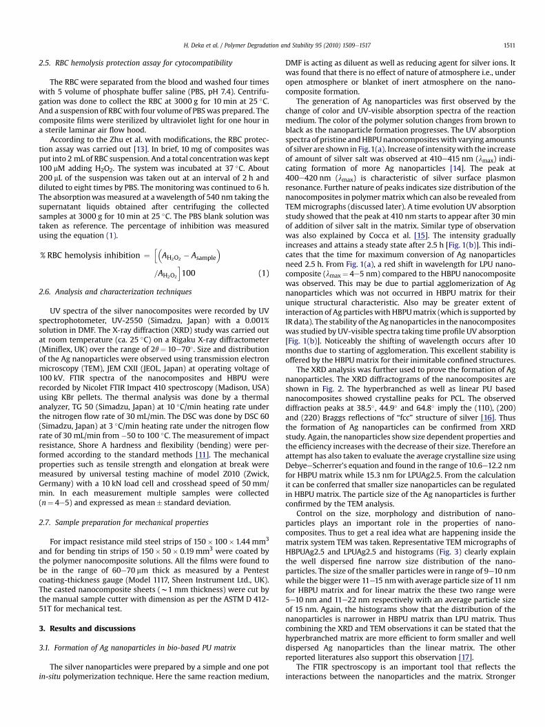
Control on the size, morphology and distribution of nano-particles plays an important role in the properties of nano-composites. Thus to get a real idea what are happening inside thematrix system TEM was taken. Representative TEM micrographs ofHBPUAg2.5 and LPUAg2.5 and histograms (Fig. 3) clearly explainthe well dispersed fine narrow size distribution of the nano-particles. The size of the smaller particles were in range of 9e10 nmwhile the bigger were 11e15 nmwith average particle size of 11 nmfor HBPU matrix and for linear matrix the these two range were5e10 nm and 11e22 nm respectively with an average particle sizeof 15 nm. Again, the histograms show that the distribution of thenanoparticles is narrower in HBPU matrix than LPU matrix. Thuscombining the XRD and TEM observations it can be stated that thehyperbranched matrix are more efficient to form smaller and welldispersed Ag nanoparticles than the linear matrix. The otherreported literatures also support this observation [17].
The FTIR spectroscopy is an important tool that reflects theinteractions between the nanoparticles and the matrix. Stronger

Fig. 1. UV-visible absorption spectra of (a) nanocomposites and pristine polymers, (b) time evolution curves starting from formation to the maximum stability period.
H. Deka et al. / Polymer Degradation and Stability 95 (2010) 1509e15171512
the interactions better is the stabilization of the nanoparticleswithin the matrix. The FTIR spectra of the nanocomposites areshown in Fig. 4. A remarkable change was observed in the region1729e1679 cm�1 where the pristine HBPU showed only one bandat 1729 cm�1, but splits to two bands in the nanocomposites. Theappearance of the bands at 1668e1679 cm�1 is due to the inter-actions (mainly Hydrogen bonding) of the silver ions with theeC]O, eNH and eCN stretching modes [18]. Further it is noticed thatthe band intensity at 1730 cm�1 for HBPUAg5 is less than theothers. This may be the result of the more number of nanoparticleswhich destroys the hydrogen bonding between the hard and softsegment of the HBPU matrix [19]. This finding also supports by theshifting of the eNH absorption band from 3469 to 3454 cm�1.
3.2. Antimicrobial property
The antimicrobial activities of the nanocomposites were testedagainst different bacteria such as Gram positive (S. aureus), Gramnegative (E. coli) and yeast such as C. albicans by disc diffusion
Fig. 2. XRD patterns of the nanocomposites and pristine HBPU.
assay. The comparative bar-graphs of antimicrobial efficiency ofthe prepared discs with standard antibiotics and antifungal areshown in Fig. 5. It is clear that the antimicrobial properties of thenanocomposites are due to the incorporation of the Ag nano-particles in the matrix as the pristine HBPU matrix does not showany antimicrobial activity. Further, the response towards the Grampositive and Gram negative bacteria is not the same. S. aureusbeing Gram positive bacteria has a much stronger defense systemcompared to E. coli, a Gram negative bacteria, as the formerpossesses a thicker peptidoglycan cell wall [20]. This thicker wallprevents the silver ions from penetrating into the cytoplasm ofthe organism. It has been suggested that Agþ ions enter into thebacterial cells by penetrating through the cell wall and conse-quently turn the DNA into condense form which reacts with thethiol group of the proteins. Thus it ultimately causes death of thecell [20]. Though it is suggested that the antimicrobial mechanismof Ag nanoparticles follows the same manner as that of its ions,but it is still not confirmed. A redox imbalance may cause by thenanoparticles resulting death of a large number of bacteria. Thiseffect is further enhanced by the large surface area of the Agnanoparticles [21]. The antimicrobial activity of the nano-composites increases with the increase of Ag nanoparticles. Thispresent study also supports the observation of Shahverdi et al.[22] that dose dependent Ag nanoparticles have marked activityagainst Gram positive than Gram negative bacteria. Rosenbergand co-workers extensively described the toxicity of the Agþ ionson E. coli [23]. The ions inhibit phosphate uptake and exchange inE. coli, which causes efflux of accumulated phosphate. E. colicontains two types of NADH dehydrogenase both containscysteine residue. This cysteine residue has high affinity for silver[5]. The Agþ ions bind with the enzymes of the bacterial respi-ratory chain causing an inefficient passage of electrons to oxygenat the terminal oxidase. This produces large quantities of reactiveoxygen species and thus toxicity to E. coli. It is expected that theAg nanoparticles act as the same way as the Agþ ions for E. coli.The nanocomposites also showed antifungal activity againstC. albicans [Fig. 5(c)]. The nanocomposites showed comparableresults with the studied antibiotics. Wright et al. proved theantifungal properties of silver against a broad spectrum ofcommon fungi [24]. It is reasonable to state that the binding ofthe particles to the bacteria depends on the surface area availablefor such interactions. The smaller particles with a larger surface-to-volume ratio provided a more efficient means of antibacterialactivity than larger particles. This accounts the enhanced anti-microbial activity of the HBPUAg2.5 than LPUAg2.5, both havingthe same amount of AgNO3.

Fig. 3. Typical TEM micrographs of (a) LPUAg2.5 and (b) HBPUAg2.5 and corresponding histograms for distribution of particle size.
H. Deka et al. / Polymer Degradation and Stability 95 (2010) 1509e1517 1513
3.3. Mechanical property
Different antimicrobial applications demand excellentmechanical properties of the final materials [25]. For example, Jeonet al. prepared antimicrobial PCL-based PU/Ag nanoparticlesnanofibers for antimicrobial nanofilter applications possess tensilestrength of 10 MPa and elongation at break of 350% [26]. In view ofthe above the mechanical properties of the Ag nanocompositeswere evaluated and tabulated in the Table 1. The strainestressprofiles of the nanocomposites are shown in the Fig. 6. An
Fig. 4. FTIR spectra of the nanocomposites and pristine HBPU.
improvement in tensile strength was observed whereas the elon-gation at break remains almost same. The increase of the interac-tions of Ag nanoparticles with the hard segment of HBPUmatrix (asfound from FTIR analysis) and enhanced crystallinity (higherenthalpy value, discussed DSC) may promote its strength. Thehighest strength was found for HBPUAg5 (11.5 MPa). Although theAg nanoparticles restrict the segmental motions of the polymerchains, still it is not sufficient to decrease the elongation at breakvalue to a noticeable extent. Other significant achievementsobserved in these nanocomposites are the retention of the flexi-bility and enhancement of impact resistance (Table 1). The films canbe easily bent to 3 mm parallel mandrel without any damage. Thelong fatty acid chains of theM. ferrea L. seed oil and PCL moiety arecontributed to these flexibility and elongation at break. Thehomogeneously distributed silver nanoparticles play crucial role forthis enhancement by interacting with the HBPU segments of thematrix. The smaller and spherical size nanoparticles have very highsurface area which resulted sufficient interactions between thenanoparticles and matrix facilitating the easy transfer of the stressthrough matrix to the fillers and thus impact resistance increases.The Shore A hardness have seen to be increased for all the nano-composites with increased amount of Ag nanoparticles comparedto the HBPU. This may be due the overall enhancement of thetensile strength of these flexible films. Misjak et al. [27] had alsofound dose dependent mechanical properties of thin films of Agnanocomposites.
3.4. Thermal study
Vegetable oil based PUs possess low thermostability than thepetrochemical based PU [28]. On the other hand the availabletraditional manufacturing techniques claim higher thermalstability of materials. Therefore improvement of thermal propertiesof vegetable oil based PUs is desirable. The thermal properties ofthe nanocomposites were studied by TGA and DSC studies. An

Fig. 5. The comparative bar-graphs of antimicrobial efficiency of the nanocomposites against (a) Staphylococcus aureus, (b) Escherichia coli, and (c) Candida albicans with standardantibiotic and antifungal.
H. Deka et al. / Polymer Degradation and Stability 95 (2010) 1509e15171514
excellent achievement in thermal stability was observed for thenanocomposites (Fig. 7). One step degradationwas occurred for thenanocomposites where the pristine HBPU shows two step degra-dation. It is quite obvious that the incorporation of the Ag nano-particles have enhanced the thermal stability of the HBPU matrix.The characteristic thermal degradation temperatures are reportedin the Table 2. A shift of (46e53) �C of the first on set temperaturewas observed for HBPU silver nanocomposites whereas forLPUAg2.5, it was 42 �C compared to their respective pristine poly-mers. Again the first, temperature corresponding maximumweightloss i.e. TMAX of pristine HBPU is 370 �C where the TMAX for theHBPU nanocomposites are (402e413) �C. Thus the TMAX is shiftedby (32e43) �C for HBPU based nanocomposites. Similarly forLPUAg2.5, this enhancement is 48 �C (TMAX of pristine LPU is 362 �Cand for LPU nanocomposite 400 �C). The Ag nanoparticles acted asthe nucleating agent for enhancing the crystallization (Table 2)which restricts the movement of the polymer chains [19]. Thusfantastic improvements in thermal stability were observed.
Table 1Mechanical property of the nanocomposites and pristine polymers.
Sample code HBPU HBPUAg1 HB
Tensile Strength (MPa) 6.80� 1.2 8.13� 1.4 9Elongation at break (%) 721.3� 3.4 718.8� 2.4 71Bending (mm) 3 3 3Hardness (Shore A) 72� 1.0 74� 1.2Impact resistance (cm) 35� 2.0 56� 1.6
The glass transition temperature (Tg), melting temperature (Tm)and melting enthalpy (DHm) are shown in the Table 2. A slightincrement in Tg in the HBPU nanocomposites (from �44 to �40 �C)and LPUnanocomposite (from�46 to�42 �C)were observedwhichis due to the restriction on the segmental chainmovement imposedby Ag nanoparticles [29]. With the increase of amount of nano-particles this restriction increases considerably. Themelting point ofthe nanocomposites is found to increase with the increase of Agnanoparticles is due the increase interactions of Ag nanoparticleswith the HBPU matrix. An enhancement in crystallinity can also bepredicted from the DHm value given in Table 2. The enhancement ofcrystallinity of a semi crystalline polymer after incorporation ofdifferent types of nanofillers is described by several reports [30,31].The well dispersed Ag nanoparticle act as the hardening (virtualcross-linking) point in the PU soft segments thereby increasingpossibilities to come closer. This may help the soft segments toarrange in a regular pattern and thus improves the crystallinebehavior. With the increase of homogeneously distributed smaller
PUAg2.5 HBPUAg5 LPU LPUAg2.5
.42� 1.1 11.51� 1.5 5.31� 0.95 9.60� 1.11.8� 2.6 710.4 � 3.1 779.2� 3.3 729.6� 2.1
3 3 375� 1.0 76� 0.8 69� 1.0 74� 1.060� 2.0 65� 1.4 42� 2.2 59� 1.4

Fig. 6. Stress versus strain profile of the nanocomposites and pristine polymers.
Table 2Thermal properties of the nanocomposites and pristine polymers.
Sample code Glass transitiontemperature(Tg, �C)
Meltingtemperature(Tm, �C)
Meltingenthalpy(DHm, J/g)
TON(�C)
TMAX
(�C)TEND(�C)
HBPU �44� 0.5 48� 1.0 51.8� 1.5 215 370 450HBPUAg1 �42� 0.8 50� 1.0 53.6� 1.2 261 402 487HBPUAg2.5 �41� 0.6 52� 0.9 54.2� 1.0 264 409 493HBPUAg5 �40� 0.4 55� 1.5 56.3� 1.1 268 413 498LPU �46� 0.7 46� 0.9 49.7� 1.7 214 362 434LPUAg2.5 �43� 1.0 50� 1.0 51.5� 1.1 256 400 490
H. Deka et al. / Polymer Degradation and Stability 95 (2010) 1509e1517 1515
size Ag nanoparticles the hardening point increases and hence thecrystallinity. This is illustrated in the Scheme 1.
3.5. RBC hemolysis protection assay for cytocompatibility
The peculiar properties of metal nanocomposites lead toexecuted novel activities. At the same time the possible harmfulinteractions of nanoparticles with the biological systems is of primeconcern. Thus there is an urgent need of approach to access thesafety of such materials. The toxicity of nanoparticles depends onseveral factors like rate of cellular uptake of nanoparticles, intra-cellular distribution, exocytosis, size, shape, aspect ratio andsurface functionality of nanoparticles etc. [32]. One of the knownmechanisms for cellular damages is oxidative stress that is inducedby the nano- and ultrafine particles [33]. The presence of Agnanoparticles accelerates the production of reactive oxygen species(ROS) and subsequent oxidative stress. This also accounts themetabolic disturbances as well as other toxicological outcomes inthe existence of such nanoparticles. The highly reactive oxygenspecies result in oxidative damages to the cells, proteins, DNA, RNAetc. In the present study, RBC hemolysis protection assay was
Fig. 7. Thermogravimetric curves of the nanocomposites and pristine polymers.
carried out to cheek the cytotoxicity of the nanocomposites. Thehemolysis inhibition data were recorded in the time intervals of 0,30, 60 and 90 min. From the Fig. 8, it can be conferred that thenanocomposites are non cytotoxic. The HBPUAg5 showed thehighest hemolysis prevention capacity, revealing the dose depen-dency. The free radicals may be stabilized by the Ag nanoparticlesthrough sharing of unpaired electron of the free radicals with theconduction band electrons of the metal nanoparticles [5]. Andultimately it deposited on the surface of the nanocomposite films.Further a declination in inhibition of hemolysis of RBC wasobserved after 90 min may be due to the saturation after reactingwith free radicals [13]. The pristine polymer showed very lesshemolysis inhibition capacity compared to the nanocomposites.Such observations signify the critical role of the Ag nanoparticles inthe matrix in RBC hemolysis inhibition. This also supports theaforesaid interactions of the Ag nanoparticles with the free radicals.Thus, although the free Ag nanoparticles cause toxicity to the livingcells, the polymer bound Ag nanoparticles are nontoxic at cellularlevel.
Scheme 1. Schematic illustration of formation of crystalline structure in thenanocomposites.

Fig. 8. TheRBChemolysis inhibition assayof the pristineHBPUand thenanocomposites.
H. Deka et al. / Polymer Degradation and Stability 95 (2010) 1509e15171516
3.6. Biodegradation
From the antimicrobial test results it is expected, in general, thatthe materials may not be biodegradable. However, it is noticed thatthe antimicrobial polymeric nanocomposites exhibited biodegra-dation under the studied conditions. This noteworthy observationwas achieved due to right choice of bacterial strains, which aretreated as hydrocarbon degrading bacteria and expected to beutilized for bioremediation of petroleum contaminated areas. Thepristine HBPU and the nanocomposite films were directly exposedto the bacterial strains and the bacterial growth was monitored andevaluated with the help of McFarland Turbidity method. In thisinvestigation, the P. aeruginosa of strain number MTCC 7814 andMTCC 7815 were used for biodegradation as several members ofthe genus Pseudomonas have already been known for their ability todegrade polyester based PUs [34]. The bacterial growth with
Fig. 9. Statistics for the growth of the P. aeruginosa strain (a) MTCC 7814, (b) MTCC 7815 aHBPUAg2.5 and (f) HBPUAg5.
respect to time for the studied systems is shown in the Fig. 9. Thebacterial growth was higher for the nanocomposites than theirrespective pristine systems. Several causes can be cited to accountthis biodegradation behavior of HBPU and the nanocomposites. Thebiodegradation depends on the constituent of the PU, whether it isa polyether or polyester based, as polyester based PUs are moreprompt to biodegradation [35]. No significant improvement inbiodegradation was noticed till two weeks for all the systems. Butafter two weeks the nanocomposites showed comparatively goodbiodegradability. Further, with the increase of Ag nanoparticlescontent the biodegradation rate was found to be increased whichmay be due to ease of access of polymer surface by the degradingbacterial strains as they can accommodated by the favorablegeometry of the polymer chains. This is again due to decrease ofH-bonding among the chain molecules due to incorporation of Agnanoparticles in the matrix. The decreased phase separatedmorphology after nanocomposite formation is due to the increaseof interfacial interactions between Ag nanoparticles and polymerchains may also attribute to this enhancement of the biodegrada-tion. However, the correct understanding on the mechanism ofbiodegradation of such antimicrobial materials requires muchdetails study. The extent of biodegradation as well as bacterialgrowth can be visualized from the SEM images (Fig. 9). The growthrate of the two bacterial stains was also found to be different, whichindicates that this biodegradation is even dependent on the strainof the same bacteria. It is worth to mention that since all thenanocomposite films do not show any change in the water mediumtherefore the observed degradation is only for the presence of themicrobes.
4. Conclusion
The study showed the potentiality of the prepared PU/Agnanocomposites as antimicrobial biodegradable biocompatiblebiomaterials. The studied M. ferrea L. seed oil modified hyper-branched polyurethane based nanocomposite possesses betterperformance over the linear analog. The well dispersed and stable
nd SEM pictures of the degraded nanocomposite films: (c) HBPUAg1, (d) LPUAg2.5, (e)

H. Deka et al. / Polymer Degradation and Stability 95 (2010) 1509e1517 1517
Ag nanoparticles significantly improved the mechanical propertieslike tensile strength, impact resistance, Shore A hardness withoutaffecting the flexibility and elongation at break value of the pristinepolymer in both the cases. Thermal properties were also found tobe well-improved in the prepared nanocomposites. The Ag incor-porated PU nanocomposite films exhibited excellent antimicrobialproperty towards S. aureus, E. coli and C. albicans, even though theyalso exhibited adequate bacterial biodegradation by the right strainof P. aeruginosa (MTCC 7814 and MTCC 7815). The RBC hemolysisinhibition assay indicates the cytocompatibility of the materials butbefore use these materials in actual field they need to test in vivo.The study on clay based similar Ag nanocomposite as more effec-tive biomaterial is under progress.
Acknowledgments
The authors express thanks to the research project assistancegiven by DST, India through the grant No. SR/S3/ME/13/2005-SERC-Engg, dated 9th April, 2007, also thank to RSIC, Shillong for per-forming TEM analysis.
References
[1] Chen X, Schluesener HJ. Nanosilver: a nano product in medical application.Toxicol Lett 2008;176:1e12.
[2] Travan A, Pelillo C, Donati I, Marsich E, Benincasa M, Scarpa T, et al. Non-cytotoxic silver nanoparticle-polysaccharide nanocomposites with antimi-crobial activity. Biomacromolecules 2009;10:1429e35.
[3] Lee D, Cohen RE, Rubner MF. Antibacterial properties of Ag nanoparticleloaded multilayers and formation of magnetically directed antibacterialmicroparticles. Langmuir 2005;21:9651e9.
[4] Wijnhoven SWP, Peijnenburg WJGM, Herberts CA, Hagens WI, Oomen AG,HeugensEHW,et al.Nano-silvere a reviewof availabledata andknowledge gapsin human and environmental risk assessment. Nanotoxicol 2009;3:109e38.
[5] AshaRani PV, Mun GLK, Hande MP, Valiyaveettil S. Cytotoxicity and geno-toxicity of silver nanoparticles in human cells. ACS Nano 2009;3:279e90.
[6] Drake PL, Hazelwood KJ. Exposure-related health effects of silver and silvercompounds: a review. Ann Occup Hyg 2005;49:575e85.
[7] Planck H, Syre I, Dauner M, Egbers G. Polyurethane in biomedical engineeringII, progress in biomedical engineering. Amsterdam: Elsevier; 1987.
[8] Caracciolo PC, de Queiroz AAA, Higa OZ, Buffa F, Abraham GA. Segmented poly(esterurethane urea)s from novel urea-diol chain extenders: synthesis, char-acterization and in vitro biological properties. Acta Biomater 2008;4:976e88.
[9] Zhang X, Yan D. Hyperbranched poly(amidoamine) as the stabilizer andreductant to prepare colloid silver nanoparticles in situ and their antibacterialactivity. J Phys Chem C 2008;112:2330e6.
[10] Karak N, Maiti S. Dendrimers and hyperbranched polymers e synthesis toapplications. New Delhi: MD publication Pvt. Ltd; 2008.
[11] Deka H, Karak N. Bio-based hyperbranched polyurethanes for surface coatingapplications. Prog Org Coat 2009;66:192e8.
[12] Turkoglu A, Duru EM, Mercan N, Kivrak I, Gezer K. Antioxidant and antimi-crobial activities of Laetiporus sulphureus (Bull.) Murrill. Food Chem 2007;101:267e73.
[13] Zhu QY, Holt RR, Lazarus SA, Orozco TJ, Keen CL. Inhibitory effects of cocoaflavanols and procyanidin oligomers on free radical induced erythrocytehemolysis. Exp Biol Med 2002;227:321e9.
[14] Grijalva AS, Urbina RH, Silva JFR, Borja MA, Barraza FFC, Amarillas AP.Assessment of growth of silver nanoparticles synthesized from anethylene glycol-silver nitrate-polyvinylpyrrolidone solution. Phys E 2005;25:438e48.
[15] Cocca M, Dorazio L. Novel silver/polyurethane nanocomposite by in situreduction: effects of the silver nanoparticles on phase and viscoelasticbehaviour. J Polym Sci Part B Polym Phys 2007;46:344e50.
[16] Zhang Z, Han M. One-step preparation of size-selected and well-dispersedsilver nanocrystals in polyacrylonitrile by simultaneous reduction and poly-merization. J Mater Chem 2003;13:641e3.
[17] Kakati N, Mahapatra SS, Karak N. Silver nanoparticles in polyacrylamide andhyperbranched polyamine matrix. J Macromol Sci Part A 2008;45:658e63.
[18] Mukherjee S, Mukherjee M. Nitrogen-mediated interaction in poly-acrylamideesilver nanocomposites. J Phys Condens Matter 2006;18:11233e42.
[19] Chou CW, Hsu SH, Chang H, Tseng SM, Lin HR. Enhanced thermal andmechanical properties and biostability of polyurethane containing silvernanoparticles. Polym Degrad Stab 2006;91:1017e24.
[20] Feng QL, Wu J, Chen GQ, Cui FZ, Kim TN, Kim JO. A mechanistic study of theantibacterial effect of silver ions on Escherichia coli and Staphylococcus aureus.J Biomed Mater Res 2000;52:662e8.
[21] Morones JR, Elechiguerra JL, Camacho A, Holt K, Kouri J, Ramirez JT, et al. Thebactericidal effect of silver nanoparticles. Nanotechnol 2005;16:2346e53.
[22] Shahverdi AR, Fakhimi A, Shahverdi HR, Minaian S. Synthesis and effect ofsilver nanoparticles on the antibacterial activity of different antibiotics againstStaphylococcus aureus and Escherichia coli. Nanomed Nanotechnol Biol Med2007;3:168e71.
[23] Schreurs WJ, Rosenberg H. Effect of silver ions on transport and retention ofphosphate by Escherichia coli. J Bacteriol 1982;152:7e13.
[24] Wright JB, Lam K, Hansen D, Burrell RE. Efficacy of topical silver against fungalburn wound pathogens. Am J Infect Control 1999;27:344e50.
[25] Hung HS, Hsu S. Biological performances of poly(ether)urethaneesilvernanocomposites. Nanotechnol 2007;18:475101e9.
[26] Jeon HJ, Kim JS, Kim TG, Kim JH, Yu WR, Youk JH. Preparation of poly(3-cap-rolactone)-based polyurethane nanofibers containing silver nanoparticles.Appl Surf Sci 2008;254:5886e90.
[27] Misjak F, Barna PB, Toth AL, Ujvari T, Bertoti I, Radnoczi G. Structure andmechanical properties of CueAg nanocomposite films. Thin Sold Film2008;516:3931e4.
[28] Petrovic ZS. Polyurethanes from vegetable oils. Polym Rev 2008;48:109e55.[29] Mahapatra SS, Karak N. Silver nanoparticle in hyperbranched polyamine:
synthesis, characterization and antibacterial activity. Mat Chem Phys2008;112:1114e9.
[30] Karak N, Konwarh R, Voit B. Catalytically active vegetable-oil-based thermo-plastic hyperbranched polyurethane/silver nanocomposites. Macromol MaterEng 2010;295:159e69.
[31] Tzavalas S, Mouzakis DE, Drakonakis V, Gregoriou VG. Polyethylene tere-phthalate-multiwall nanotubes nanocomposites: effect of nanotubes on theconformations, crystallinity and crystallization behavior of PET. J Polym SciPart B Polym Phys 2008;46:668e76.
[32] Singh S, Nalwa HS. Nanotechnology and health safety-toxicity and riskassessments of nanostructured materials on human health. J Nanosci Nano-technol 2007;7:3048e70.
[33] Xia T, Kovochich M, Brant J, Hotze M, Sempf J, Oberley T, et al. Comparison ofthe abilities of ambient and manufactured nanoparticles to induce cellulartoxicity according to an oxidative stress paradigm. Nano Lett 2006;6:1794e807.
[34] Cosgrove L, McGeechan PL, Robson GD, Handley PS. Fungal communitiesassociated with degradation of polyester polyurethane in soil. Appl EnvironMicrob 2007;73:5817e24.
[35] Santerre JP, Woodhouse K, Laroche G, Labow RS. Understanding the biodeg-radation of polyurethanes: from classical implants to tissue engineeringmaterials. Biomaterials 2005;26:7457e70.