Characterization of a thermostable lipase showing loss of secondary structure at ambient temperature

Crystal Structure of Thermostable Family 5 Endocellulase E1 fromAcidothermuscellulolyticusin Complex with Cellotetraose†,‡
Joshua Sakon,*,§ William S. Adney,| Michael E. Himmel,| Steven R. Thomas,| and P. Andrew Karplus*,§
Section of Biochemistry, Molecular and Cell Biology, Cornell UniVersity, Ithaca, New York 14853, and National RenewableEnergy Laboratory, Golden, Colorado 80401
ReceiVed February 23, 1996; ReVised Manuscript ReceiVed May 30, 1996X
ABSTRACT: The crystal structure of the catalytic domain of the thermostable endocellulase E1 fromAcidothermus cellulolyticusin complex with cellotetraose has been solved by multiple isomorphousreplacement and refined at 2.4 Å resolution to anR-factor of 0.18 (Rfree ) 0.24). E1cd is a member ofthe 4/7 superfamily of hydrolases, and as expected, its structure is an (R/â)8 barrel, which constitutes aprototype for family 5-subfamily 1 cellulases. The cellotetraose molecule binds in a manner consistentwith the expected Michaelis complex for the glycosylation half-reaction and reveals that all eight residuesconserved in family 5 enzymes are involved in recognition of the glycosyl group attacked during cleavage.Whereas only three residues are conserved in the whole 4/7 superfamily (the Asn/Glu duo and the Glufrom which the name is derived), structural comparisons show that all eight residues conserved in family5 have functional equivalents in the other 4/7 superfamily members, strengthening the case that mechanisticdetails are conserved throughout the superfamily. On the basis of the structure, a detailed sequence ofphysical steps of the cleavage mechanism is proposed. A close approach of two key glutamate residuesprovides an elegant mechanism for the shift in the pKa of the acid/base for the glycosylation anddeglycosylation half-reactions. Finally, purely structural based comparisons are used to show that significantdifferences exist in structural similarity scores resulting from different methods and suggest that cautionshould be exercised in interpreting such results in terms of implied evolutional relationships.
Cellulases catalyze the hydrolysis of cellulose, an un-branchedâ-1,4-linked homopolymer of glucose that is themajor structural polysaccharide component of plant biomass.Complete hydrolysis of cellulose yields a single, easilyfermentable product, glucose, and much cellulase researchis aimed at improving the enzymatic hydrolysis of celluloseto a point that would enable biomass conversion to beeconomically competitive (Wyman et al., 1993). In 1985,Acidothermus cellulolyticus(new genus and species), athermotolerant, acidophilic bacterium, was isolated fromsamples collected in Yellowstone National Park (Mohagheghiet al., 1986; Tucker et al., 1989). TheA. cellulolyticuscellulase system is noncellulosomal, making it distinct frommost cellulase systems produced by thermophiles.A. cel-lulolyticus produces at least three endoglucanases (Adneyet al., 1994; Tucker et al., 1992), and one of them, E1endoglucanase, is particularly interesting because it is highlythermostable (Topt ) 81 °C; Himmel et al., 1994) and hasvery high specific activity on carboxymethylcellulose (Tho-mas et al., 1995). These properties make E11 an attractivetarget for protein engineering to improve cellulase activity.
The E1 gene sequence (GenBank Accession No. U33212)shows that the mature E1 protein has 521 residues andconsists of a catalytic domain (E1cd), a proline/serine/threonine-rich linker region, and a cellulose-binding domain.Among the 45 known families of glycosyl hydrolases(Henrissat & Bairoch, 1993), sequence comparisons clearlyplace the catalytic domain of E1 (E1cd) in family 5 (Wanget al., 1993). Family 5 (also known as cellulase family A)is the largest of theâ-glycohydrolase families classified todate, including over 60 bacterial and fungal enzymes whichall cleave with retention of configuration. The sequencesof family 5 members are rather diverse and were reportedto share only seven conserved residues (Wang et al., 1993),equivalent to Arg-62, His-116, Asn-161, Glu-162, His-238,Tyr-240, and Glu-282 in E1cd. Because of this diversity,family 5 has been further subdivided into five subfamilieswithin which amino acid sequence similarities are above 25%(Wang et al., 1993) and homology modeling methods canbe used [e.g., Sali and Blundell (1993)].
Among the strictly conserved residues, the Glu-282equivalent has been identified as the nucleophile in thedisplacement reaction (Tull et al., 1991), and the equivalentof Glu-162 (Bortoli-German et al., 1995; Wang et al., 1993)has been implicated as the proton donor. An extensivemutagenesis study has shown that cellulase activity is highlysensitive to the replacement of any of the seven conservedresidues (Bortoli-German et al., 1995). Crystal structureshave recently been published for a subfamily 3 member(CelC fromClostridium thermocellum; Dominguez et al.,1995) and a subfamily 4 member (endoglucanase A fromClostridium cellulolyticum; Ducros et al., 1995). Thesestructures show that the seven conserved residues are all near
† This work was funded in part by the Ethanol from Biomass Programof the Biofuels System Division of the U.S. Department of Energy(RAH-5-15113).
‡ The full coordinates (1ECE) and structure factor amplitudes(R1ECESF) have been deposited in the Protein Data Bank forimmediate release.* To whom correspondence should be addressed.§ Cornell University.| National Renewable Energy Laboratory.X Abstract published inAdVance ACS Abstracts,July 15, 1996.1 Abbreviations: E1, matureAcidothermus cellulolyticusendocel-
lulase E1; E1cd, catalytic domain of E1. All amino acids are abbreviatedas recommended by the IUPAC-IUB Joint Commission on Biochemi-cal Nomenclature (1985).
10648 Biochemistry1996,35, 10648-10660
S0006-2960(96)00443-6 CCC: $12.00 © 1996 American Chemical Society
+ +
+ +

each other in the active site, but neither of the structuresprovide detailed information on ligand binding or thecatalytic mechanism.As a member of glycohydrolase family 5, E1cd is also
part of a large superfamily, encompassing families 1 (â-glucosidase, lactase phlorizin hydrolase, 6-phospho-â-glu-cosidase, 6-phospho-â-galactosidase,â-galactosidase, cy-anogenicâ-glucosidase), 2 (â-galactosidase,â-glucuronidase),5 (cellulase,â-mannanase), 10 (xylanase), 17 (â-1-3,1-4-glucanase), 30 (glucocerebrosidase), 35 (â-galactosidase), 39(R-L-iduronidase), and 42 (â-galactosidase). These familiesare proposed to share a common (R/â)8 barrel fold, threeconserved residues, and a retaining cleavage mechanism(Henrissat et al., 1995; Jenkins et al., 1995). The superfamilyhas been termed the 4/7 superfamily because the three keyresidues are an adjacent Asn-Glu pair at the end ofâ-strand4 and a Glu at the end ofâ-strand 7.The generally accepted catalytic mechanism of these
superfamily members is a double-displacement mechanismput forth originally by Koshland (1953). It involves an initialbinding of the substrate to the enzyme, followed by a generalacid-catalyzed attack of an enzymatic nucleophile upon theanomeric center to form a glycosyl-enzyme intermediate(McCarter & Withers, 1994; Sinnott, 1990). This intermedi-ate is then hydrolyzed by a general base-catalyzed attack ofwater upon the anomeric center, forming the product andreturning the enzyme to its original protonation state (Mc-Carter & Withers, 1994; Sinnott, 1990). Mutagenesis andkinetic studies have allowed putative assignment of the firstconserved Glu as the acid/base and the second conservedGlu as the nucleophile (McCarter & Withers, 1994). Theuse of substrates with a 2-fluoro group has allowed stabiliza-tion of covalent intermediates (Withers & Street, 1988) andvery recently allowed elucidation of the structure of acovalent intermediate bound to Cex, a family 10 enzyme(White et al., 1996). This structure has strengthened the casefor the double displacement mechanism and has given insightinto the role of the conserved Asn.To guide protein engineering ofA. cellulolyticusendo-
glucanase E1, we have solved the structure of E1cd usingcrystals grown in the presence of cellobiose. The structureshows that the crystallized enzyme has cellotetraose bound,presumably resulting from an E1cd-catalyzed condensationof two cellobiose molecules. Our results thus provide astructural prototype for family 5-subfamily 1 cellulasesincluding details of substrate binding and insights into thecatalytic mechanism for the class of enzymes. We alsoanalyze global and active site similarities with other structur-ally known glycosyl hydrolases and propose mechanisticdetails that structural and sequence comparisons suggest isconserved in the whole 4/7 superfamily.
MATERIALS AND METHODS
Substrates and Reagents.All reagents used were of thehighest purity available and were purchased from SigmaChemical Co. (St. Louis, MO) unless otherwise noted.Enzyme Assays.Enzymatic activity was spectrophoto-
metrically measured at 410 nm through changes in theabsorbance due to hydrolysis ofp-nitrophenylâ-D-cellobio-side in the presence of 2.0 M Na2CO3. One unit of activitywas defined as that amount of enzyme that cleaves 1.0µmolof substrate/min at 45°C in 50 mM acetate, pH 5.0.
Heterologous E1 Endoglucanase Expression System. Strep-tomyces liVidansTK24 was transformed with pIJ702 (Hop-wood et al., 1985) into which a 3.7 kbPVuI fragment ofA.cellulolyticusgenomic DNA carrying native E1 gene hadbeen subcloned. Cultures were kept frozen at-70 °C afteraddition of 77 µL of dimethyl sulfoxide/mL of culturesuspension (Himmel et al., 1994).S. liVidanswere grown in tryptone soya broth (TSB; Difco
Laboratories, Detroit, MI) according to Hopwood et al.(1985). Plasmid pIJ702 and its derivatives were selected inliquid medium with 5µg/mL thiostrepton. An aliquot offrozenS. liVidansculture was thawed and transferred to 500mL of TSB medium. After incubation for 2 days at 30°Cwith rotary agitation (250 rpm), this culture was used toinoculate 8 L of TSB medium in NBS Microferm fermenters(New Brunswick Scientific, Brunswick, NJ). Fermentedcultures were kept at pH 6.8 by addition of 1.0 N HCl whilethe temperature was maintained at 30°C. The concentrationof dissolved oxygen was maintained at 20% of saturationby addition of pure oxygen accompanied by impelleragitation at 300 rpm.Purification of Recombinant E1 Endoglucanase from S.
liVidans Culture Broth. Culture broth from four 8 LrecombinantS. liVidans fermentations was clarified byultracentrifugation using a CEPA (New Brunswick Scientific;Brunswick, NJ) Type LE continuous flow centrifuge operatedat 25 000 rpm and 4°C. Following concentration to a finalvolume of 300 mL using an Amicon (Danvers, MA) CH2concentrator equipped with three 10 kDa cutoff (TypeH1P10-43) hollow fiber membranes, ammonium sulfate wasadded to a final concentration of 0.5 M. The entireconcentrate was loaded onto an Amicon G44× 250 glasscolumn packed with a 250 mL of Pharmacia (Piscataway,NJ) Fast Flow Phenyl Sepharose and attached to a PharmaciaFPLC system. The column was then washed with 5 volumesof 20 mM Tris, pH 8.0, containing 0.5 M (NH4)2SO4. E1was eluted at a flow rate of 2 mL/min by a descending lineargradient ending in 20 mM Tris, pH 8.0. Fractions elutingat 0% (NH4)2SO4 and containing the recombinant enzymewere combined, concentrated, and diafiltered against 20 mMTris, pH 8.0.The diafiltered enzyme was next subjected to anion-
exchange chromatography by loading to a Pharmacia Q-Sepharose HiLoad 16/10 high-performance column. Theenzyme was eluted by a shallow linear gradient ending in20 mM Tris, pH 8.0, containing 1.0 M NaCl. Activefractions eluted near 150 mM NaCl were combined, con-centrated, and subjected to high-performance size exclusionchromatography (HPSEC) using a Pharmacia Superdex 200,HiLoad preparative grade column, with a flow rate of 0.5mL/min in 50 mM ammonium acetate, pH 6.2. The elutionprofile showed a single symmetrical peak. Enzyme puritywas supported by the migration of the protein as a singlesilver-stained band corresponding to 72 kDa on SDS-PAGEusing Novex (San Diego, CA) precast 8-15% gradient gels.Preparation of E1cd. Natural E1 was cleaved for 24 h at
28 °C using papain (Boehringer Mannheim, Indianapolis,IN) in 50 mM ammonium acetate, pH 6.2, with a molar ratioof E1 (72 kDa) to papain (23 kDa) of 6:1. E1cd was thenseparated from undigested E1, E1 cellulose-binding domain,and papain by HPSEC as described above. Purified E1cdmigrates as a single band atMr ) 42 000 when subjected toSDS-PAGE.
Crystal Structure of Cellulase and Cellotetraose Biochemistry, Vol. 35, No. 33, 199610649
+ +
+ +
Crystallization. Condition 59 (2.0 M NaCl and 50 mMsodium acetate at pH 4.6) from a factorial screening method(Cudney et al., 1994; Jancarik & Kim, 1991) gave singlecrystals. Optimization of this condition yielded trigonalbipyramids measuring up to 1500µm along the unique axisand 400µm in width. The crystals grew in one month from20 µL hanging drops containing 11.4 mg/mL E1cd, 2.3 MNaCl, 0.3 M cellobiose, and 0.1 M citrate buffer (pH 4) atroom temperature. The precession photographs ofhh0 andh0l zones suggested that the crystals belong to a trigonalspace group, eitherP3121 orP3221, with unit cell dimensionsa ) b ) 97.13 Å andc ) 258.71 Å. The ambiguity in thespace group was resolved later by using anomalous signalsof a di-µ-iodobis(ethylenediamine)diplatinum nitrate (PIP)derivative, resulting in P3221 as the true space group. Thereare two molecules per asymmetric unit with 68% (v/v)solvent. Unfrozen crystals have been seen to diffract to 1.6Å resolution at the Cornell High Energy Synchrotron Source,but thus far freezing always has deteriorated the diffractionquality.Crystallographic Data Collection and Isomorphous Re-
placement. X-ray diffraction data collected for 65 heavy-atom soaks yielded six isomorphous derivatives. The datawere measured using two San Diego Multiwire SystemsMark II detectors (Hamlin, 1985) with graphite-monochro-mated Cu KR X-rays from Rigaku RU200 operating at 50kV and 150 mA with a 0.5× 5 mm focus. This structuralanalysis was limited to 2.4 Å resolution because of therelatively longc-axis and the finite brilliance of the in-housesource. Data reduction was carried out using the programSCALEPACK (Otwinowski, 1985). The major heavy-atombinding sites were obtained with isomorphous differencePattersons and direct methods using SHELXS86 (Sheldrick,1990). Both identification of minor sites and confirmationof individual sites were made by difference Fourier tech-niques. Individual heavy-atom parameters were refined(Tables 1 and 2) using PHASES (Furey & Swaminathan,1996) to yield phases with an average figure of merit of 0.54to 3.2 Å resolution.Phase ImproVement and Model Building. The multiple
isomorphous replacement (MIR) map at 3.2 Å resolution wassolvent-flattened and iteratively averaged using noncrystal-lographic symmetry. These operations were performed byPHASES suite programs. The resulting map revealed an (R/â)8 barrel motif, although complete chain tracing was not
possible. Fragments of poly(Ala) were built into theaveraged density using CHAIN (Sack, 1988), and the secondmolecule in the asymmetric unit was generated by NCSoperators. The phases obtained from the partial model werecombined with the MIR phases using sigmaA weights (Read,1986). The electron density map calculated using thecombined phases allowed improvement of the protein modelwhich was then refined against data from infinity to 3.0 Åresolution using TNT (Tronrud et al., 1987). Fifteen cyclesof such model building and phase combination yielded fourchains of the poly(Ala/Ser) model which were further refined
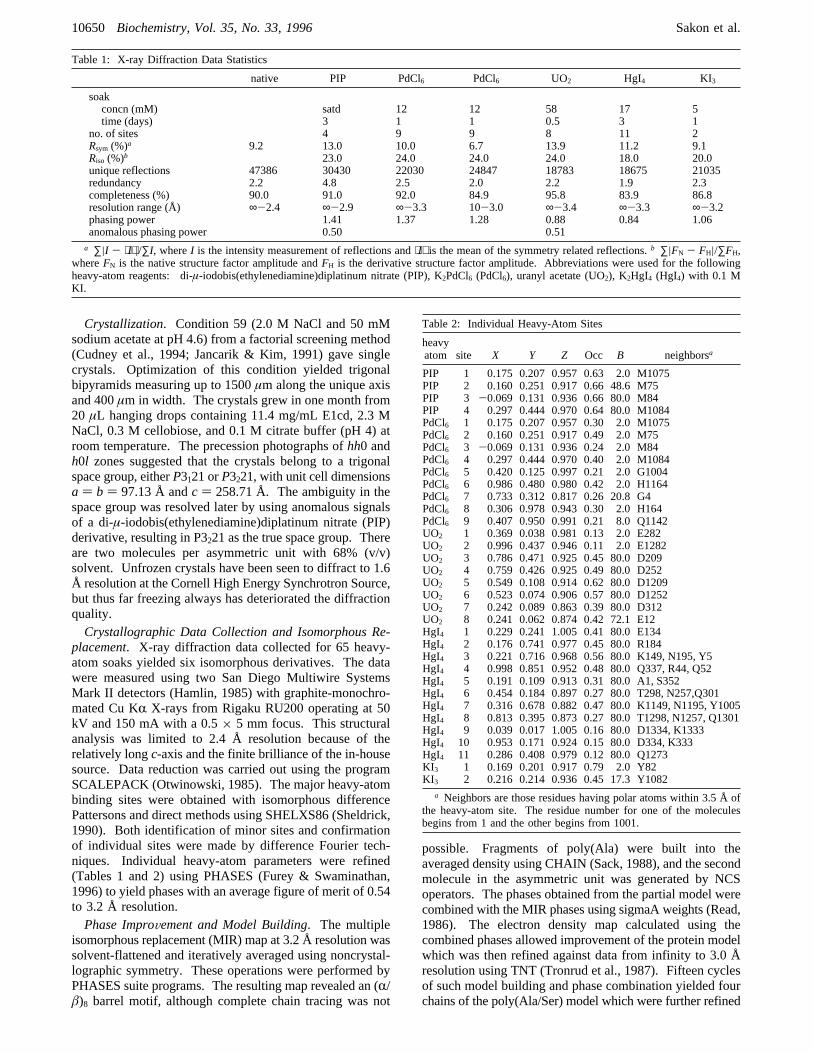
Table 1: X-ray Diffraction Data Statistics
native PIP PdCl6 PdCl6 UO2 HgI4 KI3
soakconcn (mM) satd 12 12 58 17 5time (days) 3 1 1 0.5 3 1
no. of sites 4 9 9 8 11 2Rsym (%)a 9.2 13.0 10.0 6.7 13.9 11.2 9.1Riso (%)b 23.0 24.0 24.0 24.0 18.0 20.0unique reflections 47386 30430 22030 24847 18783 18675 21035redundancy 2.2 4.8 2.5 2.0 2.2 1.9 2.3completeness (%) 90.0 91.0 92.0 84.9 95.8 83.9 86.8resolution range (Å) ∞-2.4 ∞-2.9 ∞-3.3 10-3.0 ∞-3.4 ∞-3.3 ∞-3.2phasing power 1.41 1.37 1.28 0.88 0.84 1.06anomalous phasing power 0.50 0.51
a ∑|I - ⟨I⟩|/∑I, whereI is the intensity measurement of reflections and⟨I⟩ is the mean of the symmetry related reflections.b ∑|FN - FH|/∑FH,whereFN is the native structure factor amplitude andFH is the derivative structure factor amplitude. Abbreviations were used for the followingheavy-atom reagents: di-µ-iodobis(ethylenediamine)diplatinum nitrate (PIP), K2PdCl6 (PdCl6), uranyl acetate (UO2), K2HgI4 (HgI4) with 0.1 MKI.
Table 2: Individual Heavy-Atom Sites
heavyatom site X Y Z Occ B neighborsa
PIP 1 0.175 0.207 0.957 0.63 2.0 M1075PIP 2 0.160 0.251 0.917 0.66 48.6 M75PIP 3 -0.069 0.131 0.936 0.66 80.0 M84PIP 4 0.297 0.444 0.970 0.64 80.0 M1084PdCl6 1 0.175 0.207 0.957 0.30 2.0 M1075PdCl6 2 0.160 0.251 0.917 0.49 2.0 M75PdCl6 3 -0.069 0.131 0.936 0.24 2.0 M84PdCl6 4 0.297 0.444 0.970 0.40 2.0 M1084PdCl6 5 0.420 0.125 0.997 0.21 2.0 G1004PdCl6 6 0.986 0.480 0.980 0.42 2.0 H1164PdCl6 7 0.733 0.312 0.817 0.26 20.8 G4PdCl6 8 0.306 0.978 0.943 0.30 2.0 H164PdCl6 9 0.407 0.950 0.991 0.21 8.0 Q1142UO2 1 0.369 0.038 0.981 0.13 2.0 E282UO2 2 0.996 0.437 0.946 0.11 2.0 E1282UO2 3 0.786 0.471 0.925 0.45 80.0 D209UO2 4 0.759 0.426 0.925 0.49 80.0 D252UO2 5 0.549 0.108 0.914 0.62 80.0 D1209UO2 6 0.523 0.074 0.906 0.57 80.0 D1252UO2 7 0.242 0.089 0.863 0.39 80.0 D312UO2 8 0.241 0.062 0.874 0.42 72.1 E12HgI4 1 0.229 0.241 1.005 0.41 80.0 E134HgI4 2 0.176 0.741 0.977 0.45 80.0 R184HgI4 3 0.221 0.716 0.968 0.56 80.0 K149, N195, Y5HgI4 4 0.998 0.851 0.952 0.48 80.0 Q337, R44, Q52HgI4 5 0.191 0.109 0.913 0.31 80.0 A1, S352HgI4 6 0.454 0.184 0.897 0.27 80.0 T298, N257,Q301HgI4 7 0.316 0.678 0.882 0.47 80.0 K1149, N1195, Y1005HgI4 8 0.813 0.395 0.873 0.27 80.0 T1298, N1257, Q1301HgI4 9 0.039 0.017 1.005 0.16 80.0 D1334, K1333HgI4 10 0.953 0.171 0.924 0.15 80.0 D334, K333HgI4 11 0.286 0.408 0.979 0.12 80.0 Q1273KI3 1 0.169 0.201 0.917 0.79 2.0 Y82KI3 2 0.216 0.214 0.936 0.45 17.3 Y1082
a Neighbors are those residues having polar atoms within 3.5 Å ofthe heavy-atom site. The residue number for one of the moleculesbegins from 1 and the other begins from 1001.
10650 Biochemistry, Vol. 35, No. 33, 1996 Sakon et al.
+ +
+ +

by the simulated annealing routines in X-PLOR (Brunger etal., 1987). Six cycles of manual model adjustment andsimulated annealing yielded two continuous chains of thepoly(Ala/Ser) model with a crystallographicR-factor of38.8% and anRfree of 45.8% for all X-ray diffraction datafrom 10 to 3 Å resolution.Rfree was calculated on the basisof 5% of the data which were not used in refinement.
Crystallographic Refinement. Twenty-five additional cyclesof model building, refinement by simulated annealing, andphase combination yielded two complete models of E1comprising of residues 1-358. Seven further cycles ofmanual adjustment and least-squares refinement of positionalparameters and restrained individual temperature factors werecarried out at 2.4 Å resolution. Water molecules wereintroduced where all three of the following criteria weremet: electron density peaksg3Frms in anFo - Fc differenceFourier map andg0.75Frms in a 2Fo - Fc difference Fouriermap and good hydrogen bond geometry. In the majority ofcases, a solvent molecule assigned for one of the monomerswas also found at an equivalent position in the other. Asrefinement progressed, difference Fourier maps showed abound cellotetraose which was subsequently built to accountfor the observed density. The cellotetraose molecules boundin both E1cd molecules have refined temperature factors of50-65 Å2, which is about double the average proteintemperature factor of 28 Å2. The final model consisting of716 amino acid residues (5702 atoms), two cellotetraosemolecules (90 atoms), and 375 water molecules in theasymmetric unit has a crystallographicR-factor of 17.9%and anRfree of 23.6% for all X-ray diffraction data from 10to 2.4 Å. The overall root-mean-square (rms) deviationsfrom ideality for the refined model are 0.007 Å for bondlengths, 1.7° for bond angles, and 1.2° for fixed improperdihedrals, while the rms deviation of individual temperaturefactors between bonded atoms is 1.4 Å2. Among the non-glycine and non-proline residues, 513 (85%) haveφ,ψ valuesin the “most favored regions” of the Ramachandran plot(Laskowski et al., 1993). No residues are found in the“disallowed” regions. A tight noncrystallographic symmetryrestraint was applied for all the main chain atoms and forcellobiose molecules. The rms difference between the CR
atoms of the two E1cd molecules in the asymmetric unit is0.18 Å, and maximal deviations near 0.35 Å are seen in aloop (residues 82-85) involved in the intermolecular interac-tion.
RESULTS AND DISCUSSION
Structure Description. The structures of two noncrystal-lographic symmetry-related copies of E1cd have beendetermined by X-ray crystallography at 2.4 Å resolutionusing multiple isomorphous replacement. Both chains arevisible from Ala-1 through Val-358. As E1cd was generatedby papain cleavage of intact E1, the true carboxyl terminusis not known. Some residual electron density extendingbeyond Val-358 suggests that one or more additionaldisordered residues are present. Also, two loops (207-209and 252-257) have high temperature factors (>50 Å2), butthe chain path is still clear in these regions.The E1cd structure consists of a single domain with an
(R/â)8 barrel fold in which a short irregular chain segmentreplaces helix 5 (Figure 1). The exact residues found in theâ-strands andR-helices are shown in Figure 2 along withan alignment with other known family 5-subfamily 1sequences. Aside from the structural elements of the barrel,there are only three shortR-helices and five shortâ-strands.Three of the shortâ-strands occur in residues 1-21, andtogether with the extended C-terminal segment (residues355-358), these strands seal the amino-terminal end of thebarrel from solvent. At the carboxy-terminal end of thebarrel, loops containing 16-26 residues each form the wallsof a crevice running along the length of the molecule (9 Åwide, 30 Å long, and 10 Å deep). The cleft runs roughlyfrom â-strand 1 toâ-strand 6 and is the site of substratebinding. The two disulfide bonds of E1cd occur in the loopssurrounding the active site cleft: one joining Cys-34 (loop1) and Cys-120 (loop 3) and the other linking Cys-168 andCys-171 (loop 4) in a tight turn with sequence Cys-Gly-Trp-Cys. cis-Peptide bonds occur preceding Pro-166, Pro-256, and Ser-320. The electron density for the rare non-prolyl cis-peptide at Ser-320 is unambiguous (Figure 3). Ser-320 is at the active site and is well conserved in subfamily1, but Pro-166 and Pro-256 are not in the active site cleft
FIGURE 1: Stereo CR plot of E1cd. The CR backbone is traced with a thin line. A cellotetraose molecule bound in the active site of theenzyme is represented by thick lines. The enzyme exhibits an (R/â)8 barrel fold lacking helix 5. A very short or entirely lacking helix 5 isa feature that is conserved in known members of family 5 glycosyl hydrolases. This molecular orientation is maintained for Figures 4 and5. The diagram and Figures 5 and 8 were drawn with MOLSCRIPT (Kraulis, 1991).
Crystal Structure of Cellulase and Cellotetraose Biochemistry, Vol. 35, No. 33, 199610651
+ +
+ +

and are not conserved (Figure 2), suggesting alternateconformations are adopted at those positions in the othersubfamily members. Comparisons with the other two family5 endoglucanase structures reveal a high similarity of thedomain structure but rather extensive variation in the loopssurrounding the active site cleft (see later).
The two molecules of E1cd in the asymmetric unit arerelated by approximately 2-fold symmetry (κ ) 182°), sothat they appear as a dimer with their active sites near eachother. Predominantly polar residues from carboxy-terminalloops 1, 2, 3, and 8 are involved in the intermolecularinteractions that bury about 960 Å2 (7%) of the E1cd surface.
FIGURE 2: Amino acid sequence alignment of E1cd with family 5-subfamily 1 cellulases. The E1cd structure, which is the first for family5-subfamily 1 enzymes, enables us to update the amino acid sequence alignment for this family (Wang et al., 1993). The updated alignmentshows additional amino acid conservation in the subfamily such as Asn-27, Phe-29, and Glu-201 (see text). The sequences shown are asfollows: BpoCel,Bacillus polymyxaendoglucanase (Baird et al., 1990); XcaCelX,Xanthomonas campestrisendoglucanase XCA (Goughet al., 1990); PflCelC,Pseudomonas fluorescensendoglucanase C (Ferreira et al., 1991); CsaCelB,Caldocellum saccharolyticumendoglucanaseB (Saul et al., 1989); CthCelG,C. thermocellumendoglucanase G (Lemaire & Beguin, 1993); CthCelB,C. thermocellumendoglucanaseB (Grepinet & Beguin, 1986); CfiCenD,Cellulomonas fimiendoglucanase D (Meinke et al., 1993).â-Strands andR-helices as assigned byDSSP (Kabsh & Sander, 1983) are denoted by solid and open bars, respectively. Residue positions that are identical in all eight sequences(* ) or in 6 or 7 sequences (+) are indicated. The eight residues conserved throughout family 5 are indicated by (#).
FIGURE 3: Stereo diagram of electron density corresponding to residues Phe-318-Trp-Ser-Trp-Asn, showing thecis-peptide bond betweenTrp-319 and Ser-320. The electron density map is from an annealed omit map, which is a|Fo - Fc| difference Fourier map calculated afterthe final model without the relevant residues has been subjected to a round of simulated annealing. The contour level is 2.5 times the rmselectron density of the map. Figures 3 and 4 were drawn with CHAIN (Sack, 1988).
10652 Biochemistry, Vol. 35, No. 33, 1996 Sakon et al.
+ +
+ +

This polar interface is probably not of biological relevance,because the migration of E1cd during gel filtration indicatesthat it exists as a monomer in solution. Since the twomolecules are very similar (see Materials and Methods), thediscussion will refer only to chain 1.
Thermostability. Although the resolution of this structureis not sufficient to analyze details of the nonbonded interac-tions which may be important for thermostability, we canlook at some of the larger scale features that others havenoted may play a role. These include fraction of buriedatoms, relative surface area, and lengths of loops (Chan etal., 1995; Russell et al., 1994). For its size, E1cd has arelatively large fraction of buried atoms (0.56) and relativelylow surface area (0.87 of that expected). These values areboth very similar to those of the hyperthermophilic enzymealdehyde ferredoxin oxidoreductase and much more extremethan those of 30 nonthermophilic enzymes (Chan et al.,1995). With regard to loop size, the alignment of E1cd withits close homologs (Figure 2) shows that E1cd has theshortest C-terminal loops for all but one loop, and for thatone E1cd is only one residue longer than the shortest ones.The only significant insertion in E1cd relative to the othersequences occurs just beforeâ-strand 8. Whereas the trueimportance of these correlations is unclear, E1cd adds anotherexample for where a low surface to volume ratio and shortloops are correlated with thermostability.
Cellotetraose Binding. (A) The Bound Ligand. In thecrystals of E1cd grown in the presence of 0.3 M cellobiose,instead of cellobiose molecules, the electron density suggeststhat a condensation product, cellotetraose, is bound to eachE1cd molecule (Figure 4). Each glucosyl residue, numberedGlc1 to Glc4 from the reducing to the nonreducing end, hasbeen fit to the density in a standard chair conformation (4C1).Although each residue’s fit could be slightly improved ifdeviations from the standard chair conformation were al-lowed (for instance, a possible sofa conformation at thecleavage site), we have resisted this temptation because theresolution is not sufficient to justify such deviation. Wespeculate that the bound cellotetraose results from anenzyme-catalyzed condensation of two cellobiose units. Suchreactions have been seen for other enzymes in this super-family (Sinnott, 1990), and experiments with soluble E1confirm that it also carries out this “reverse” reactioneffectively. An incubation of 10 mg/mL E1cd with 300 mMcellobiose and 50 mM citrate (pH 5) showed significantamounts of cellotetraose, cellotriose, and glucose after 24h. The cellotriose and glucose could result from cleavageof the newly formed cellotetraose.
The density for the ligand is not as strong as thesurrounding active site atoms, and Glc3 has notably weakerdensity than the other three glucosyl units (Figure 4),signaling the presence of possible incomplete occupancy,disorder, and/or heterogeneity. With an active crystallineenzyme, the crystal should be composed of a mixture ofstates reflecting the on-enzyme equilibrium under the condi-tions of crystallization: some molecules having boundsubstrate (cellotetraose), some having bound product (oneor two cellobiose molecules), and some having a covalentintermediate (one cellobiose covalently bound to Glu-282and possibly a second noncovalently bound cellobiose). Theobserved density corresponding to cellotetraose suggests thatthis is the dominant form present. By analogy with lysozyme
studies which have shown that the glucosyl unit precedingthe cleavage site binds poorly (Sacemski & Lienhard, 1974),we interpret the weak density for Glc3 compared to Glc1,Glc2, and Glc4 to suggest that the product complex is the
FIGURE 4: (a, top) Bound conformation of cellotetraose and itselectron density. The electron density is from an annealed omitmap. The contour level is at 2.5 times the rms electron density ofthe map. The nonreducing end of cellotetraose is at the top so thefour glucosyl units are Glc4-Glc3-Glc2-Glc1 from top to bottom.According to a nomenclature for glucosyl unit binding sites relativeto the cleavage site (Davies et al., 1995), Glc4, Glc3, Glc2, andGlc1 bind to sites-2, -1, +1, and+2, respectively. The weakelectron density seen for Glc3 may be due to heterogeneity in theligand (see text). The possible functional relevance of thisheterogeneity is suggested by similarly weak density which is seenfor the equivalent glucosyl unit in a covalent intermediate of arelated glycosidase (White et al., 1996). (b, bottom) Molecularsurface of the binding cleft of E1. The surfaces contributed by theobserved and putative aromatic platforms (violet) and two key Gluresidues (blue for the acid/base and red for the nucleophile) arehighlighted. The width of the binding cleft above and below thecatalytic center differs significantly. The narrowed binding cleftfor Glc1 and Glc2 forces a twist in the polysaccharide chain, helpingmake the scissile glycosidic bond vulnerable (see text). The figurewas prepared by GRASP (Nicholls, 1992).
Crystal Structure of Cellulase and Cellotetraose Biochemistry, Vol. 35, No. 33, 199610653
+ +
+ +

second most populated state and that it involves similarpositions of Glc1, Glc2, and Glc4 and a less well orderedGlc3 site. The lack of significant difference density associ-ated with Glu-282 leads us to believe that the covalentintermediate does not make up a significant fraction of themolecules. In crystals allowed to soak for 1 day in motherliquor without cellobiose the occupancy of the boundcellotetraose remained high, indicating that the ligand islocked into the active site by the crystal lattice. This lackof diffusion precludes simple measurement of the activityof the crystalline enzyme.The conformation of cellotetraose approximates two
standard cellobiosyl units (Gessler et al., 1994), Glc1-Glc2and Glc3-Glc4, connected at an angle. The torsion angles(φ, ψ) at the three glycosidic bonds are (-99.9°, 95.9°),(-25.5°, 106.7°), and (-87.4°, 91.8°). Torsion anglesφ andψ are defined asφ (O5i-C1i-O4i-1-C4i-1) andψ (C1i-O4i-1-C4i-1-C3i-1) (IUPAC-IUB, 1983). Although thetorsion angles at the joining glycosidic linkage are not nearthose of crystalline cellotetraose (Gessler et al., 1994), theyrepresent a low-energy conformation (Tayler et al., 1995).Conspicuously, Glu-162 and Glu-282, the expected acid/baseand nucleophile of E1, and the five other known conservedresidues of family 5 are involved in interactions focused onthe recognition of Glc3 and the glycosidic bond that isattacked during cleavage, suggesting that these are the mostcrucial interactions for efficient catalysis. Descriptions ofthe interactions of these special residues and insights intothe catalytic mechanism are presented below (see CatalyticCenter and Enzyme Mechanism).(B) Interactions InVolVed in the Extended Binding Site of
Cellotetraose. The enzyme makes both hydrophobic andhydrogen-bonding contacts with all four residues of cellotet-raose, and it appears that there may be additional bindingsites for one to two more glucose residues beyond Glc4. Asis commonly seen for polysaccharide binding enzymes [e.g.,Rouvinen et al. (1990) and Spezio et al. (1993)], thehydrophobic face of each glucose unit interacts with anaromatic side chain that lines the active site cleft. Glc1, Glc2,Glc3, and Glc4 are interacting with Tyr-245, Trp-213, Trp-319, and Phe-29, respectively (Figures 4b and 5). BeyondPhe-29 sits Trp-42 which could play a similar role for a fifthor sixth glucosyl unit. The interactions with these aromaticplatforms do not all seem to have optimal geometry. ForTyr-245 and Trp-213, the broad nonpolar surfaces areroughly coplanar with and appear ideal for packing againstthe hydrophobic, but weakly polar surface of the glucosylunits. However, the faces of Trp-319 and Phe-29 are parallel
to each other and interact with the glucosyl units at about a45° angle. Although it was not recognized due to inaccuratesequence alignments (Wang et al., 1993), Trp-319 is aneighth absolutely conserved residue in family 5. Otherresidues making close nonpolar interactions with the cel-lotetraose are Val-244, Trp-212, and the main chain of Ser-325.Aside from the hydrogen-bonding interactions made by
absolutely conserved residues to Glc3, an array of polaramino acids lining the active site cleft interact with glucosylunits. These interactions include Gln-247‚‚‚O2-Glc1, Trp-213-N‚‚‚O6-Glc2, Trp-213-O‚‚‚O6-Glc2, Asp-327‚‚‚O6-Glc3, Gly-326-N‚‚‚O2-Glc4, and Glu-32‚‚‚O6-Glc4 (Figure5). Apparently no protein residues interact with the O5atoms of the glucosyl units, although there are intermolecularhydrogen bonds from O3 of neighboring glucose units asare seen for crystalline cellotetraose (Gessler et al., 1994).(C) Comparisons with Family 5 Enzymes. All five of the
aromatic platforms (Tyr-245, Trp-213, Trp-319, Phe-29, andTrp-42) and each of the hydrogen-bonding residues (Gln-247, Asp-327, and Ser-325) are absolutely conserved infamily 5-subfamily 1 (Figure 2), so that it is likely that mostfeatures of substrate binding will be well conserved withinthis subfamily. Also, although none of the residues makingup the extended binding site are found throughout family 5,comparisons with the known structures for subfamilies showthat the platforms for Glc1, Glc2, and Glc4 are wellmimicked by nonequivalent residues: Tyr-245 that formsthe platform for Glc1 is replaced by Phe-203 of CelC andTrp-259 of CelCCA; Trp-213 that interacts with Glc2 isreplaced by the similarly oriented Tyr-176 of CelC and Trp-180 of CelCCA; and Trp-57 of CelCCA may replace Phe-29 as the platform for Glc4. Also, Trp-181 and Trp-287 ofCelCCA may act as the additional platforms. These aminoacid residues forming potential platforms are well conservedwithin their own subfamily members.Catalytic Center and Enzyme Mechanism. (A) The
Catalytic Center in E1cd. All eight absolutely conservedresidues of family 5 (including Trp-319) are involved ininteractions surrounding Glc3 and the glycosidic bond to beattacked during cleavage (Figure 6). Four of the residueshydrogen bond directly to the ligand (His-116‚‚‚Glc3-O3,Asn-161‚‚‚Glc3-O2, Glu-282‚‚‚Glc3-O2, and Glu-162‚‚‚Glc2-O4), Arg-62 and Tyr-240 hydrogen bond to thenucleophile Glu-282, and His-238 hydrogen bonds to theacid/base Glu-162. The Oε1 atom of the carboxylate of Glu-282 is within 3.5 Å of the anomeric carbon (C1) of Glc3and is appropriately positioned for a nucleophilic attack. The
FIGURE 5: Cellotetraose recognition by E1cd. Side chains of the amino acid residues interacting with a cellotetraose molecule are shown.Dashed lines show probable hydrogen-bonding interactions of less than 3.2 Å. All of the labeled residues, except for Glu-32, are conservedin family 5-subfamily 1, suggesting a common substrate binding mode throughout the subfamily.
10654 Biochemistry, Vol. 35, No. 33, 1996 Sakon et al.
+ +
+ +

eighth conserved residue, Trp-319, contacts the C3, C4, andC5 atoms of Glc3. These eight residues are present at thecarboxy-terminal ends ofâ-strands 2, 3, 4, 6, 7, and 8,making the whole fold fairly directly involved in catalysis.Our results clearly define important roles for Asn-161 in
recognizing the C2 hydroxyl, His-116 in recognizing the C3hydroxyl, Arg-62 and Tyr-240 in orienting and activatingGlu-282, and His-238 in orienting Glu-162. In the E1cdstructure, the hydrogen-bonding environment of His-238 isnot sufficient to define whether it is protonated or not, butthe replacement of His-238 with Asn/Gln residues in mostsuperfamily members (see Figure 7) suggests that it is notprotonated and this neutral hydrogen bond helps positionGlu-162 without lowering its pKa.Additional residues conserved in subfamily 1 include four
buried polar residues which help to form the catalyticcenter: Asn-27, Asp-158, and Glu-201 interact with Arg-62, and Asp-114 interacts with His-116. In a few cases thereis also high conservation of short stretches of residuesimmediately surrounding the catalytic center. These includeregions around Phe-29, Trp-213, and Asp-327 (Figure 2),each of which involves a special turn structure with
conserved Asn and Gly residues. We describe the interestingstretch Trp-212-Trp-Gly-Gly-Asn-Leu here. Both Trp resi-dues are interacting with cellotetraose. CR-Gly-214 is 4 Åaway from CR-Glu-162 and hasφ, ψ angles of (80°, -7°).A side chain at position 214 would bump and displace Glu-162. CR-Gly-215 is 3.9 Å from O-His-248 and 3.8 Å fromN-His-248. Although Gly-215 does not have unusualφ, ψangles, a Câ atom would bump those atoms and thus displaceHis-248. Asn-216 makes hydrogen bonds to the main chainatoms O-204 and N-218, allowing the threading of thisirregular loop 5 back to the surface.(B) Other Family 5 Structures. In the other two family 5
structures which were determined without bound ligands, thecatalytic center is structurally well conserved. In CelCCA(Ducros et al., 1995) all eight key residues appear to be insimilar positions, and in CelC (Dominguez et al., 1995) allbut the Asn161-Glu162 equivalents are similar. As discussedin the CelC publication (Dominguez et al., 1995), theseresidues point away from the active site but are thought toadopt an E1cd-like conformation upon substrate binding. Theputative functional importance of some of these conservedresidues is provided by mutagenesis experiments on subfam-
FIGURE 6: Interactions of the eight key active site residues. Stereo diagram of the E1cd-cellotetraose complex focusing on the geometryof the eight key residues. The coloring is by function: the putative nucleophile Glu-282 (red), residues aligning the nucleophile (pink), theacid/base (blue), residue aligning the acid/base (cyan), H-bond donor to Glc2-O2 (green), H-bond donor to Glc2-O3 (purple), and aromaticplatform (yellow).
FIGURE 7: Putative functional counterparts in families 1, 2, 10, and 17 for the eight key residues in family 5. A structural alignment ispresented for the six segments (fromâ-strands 2, 3, 4, 6, 7, and 8) containing the key residues. In this sequence alignment, all alignedresidues have CR atoms which overlay within 3.5 Å of their equivalent in E1cd and can be considered structurally equivalent residues. Weuse the term functional counterpart to designate residues which may or may not be structurally equivalent in terms of their main chainatoms but for which the side chains occupy a similar space and could carry out the same function. Putative functional counterparts areindicated by the color coding which matches the colors shown in Figure 6. Structures aligned are indicated by their PDB code: 1CBG(cyanogenicâ-glucosidase from white clover), 1BGL (â-galactosidase fromE. coli), 2EXO (â-1,4-glycanase Cex fromC. fimi), and 1GHR(1-3,1-4-â-glucanase isoenzyme EII from barley). The assignment of functional equivalence for His-116 with His-80 of family 10 enzymesis confirmed by the complex recently reported by White et al. (1996) showing that His-80 of EXO hydrogen bonds to the O3 hydroxyl. InEXO, the replacement of Tyr-240 (E1cd) with His-205 (EXO) may be related to the absence of an Arg-62 (E1cd) counterpart as it maintainsa positively charged group hydrogen bonding to the nucleophile. In GHR, the replacement of the His-116 (E1cd) interacting with the O3atom with Tyr-33 may relate to its activity withâ(1f3)-linked polymers which do not have an equivalent free hydroxyl.
Crystal Structure of Cellulase and Cellotetraose Biochemistry, Vol. 35, No. 33, 199610655
+ +
+ +
ily 2 enzyme (Bortoli-German et al., 1995). In this work,mutations of the Arg-62, His-116, Glu-162, and His-238equivalents to 13 other residues showed that all but one ofthe mutations of these key residues led to at least a 20-foldreduction in activity. The exception was the mutation ofthe His-116 equivalent to Phe which was reported to have40% of wild-type activity.(C) Functional Counterparts of Eight Key Residues in
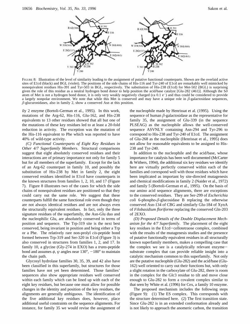
Other 4/7 Superfamily Members. Structural comparisonssuggest that eight absolutely conserved residues and theirinteractions are of primary importance not only for family 5but for all members of the superfamily. Except for the lackof an Arg-62 counterpart in family 10, and possibly thesubstitution of His-238 by Met in family 2, the eightconserved residues identified in E1cd have counterparts inthe known structures from families 1, 2, 10, and 17 (Figure7). Figure 8 illustrates two of the cases for which the sidechains of nonequivalent residues are positioned so that theycould carry out the same role. We suggest that thesecounterparts fulfill the same functional role even though theyare not always identical residues and are not always eventhe structurally equivalent residues. In fact, only the threesignature residues of the superfamily, the Asn-Glu duo andthe nucleophilic Glu, are absolutely conserved in terms ofposition and sequence. The Trp-319 site is the next bestconserved, being invariant in position and being either a Trpor a Phe. The relatively rare non-prolylcis-peptide bondformed between Trp-319 and Ser-320 in E1cd (Figure 3) isalso conserved in structures from families 1, 2, and 17. Infamily 10, a glycine (Gly-274 in EXO) has atrans-peptidebond and assumesφ, ψ angles near 130°, -180° to maintainthe chain path.Glycosyl hydrolase families 30, 35, 39, and 42 also have
been classified in this superfamily, but structures for thosefamilies have not yet been determined. Those families’sequences also show appropriate residues well conservedwithin each family which can be lined up with each of theeight key residues, but because one must allow for possiblechanges in the identity and position of the key residues, thealignments are generally ambiguous. The identification ofthe five additional key residues does, however, placeadditional useful constraints on the sequence alignments. Forinstance, for family 35 we would revise the assignment of
the nucleophile made by Henrissat et al. (1995). Using thesequence of humanâ-galactosidase as the representative forfamily 35, the assignment of Glu-339 (in the sequencePLSEAG) as the nucleophile allows the well-conservedsequence ASVNLY containing Asn-294 and Tyr-296 tocorrespond to His-238 and Tyr-240 of E1cd. The assignmentof Glu-268 as the nucleophile (Henrissat et al., 1995) doesnot allow for reasonable equivalents to be assigned to His-238 and Tyr-240.In addition to the nucleophile and the acid/base, whose
importance for catalysis has been well documented (McCarter& Withers, 1994), the additional six key residues we identifyhere are virtually perfectly conserved within each of thefamilies and correspond well with those residues which havebeen implicated as important by site-directed mutagenesisand chemical modifications in family 2 (Naider et al., 1972)and family 5 (Bortoli-German et al., 1995). On the basis ofour amino acid sequence alignments, there are exceptionsto the conserved residues. They are Ser-299 ofEscherichiacoli 6-phospho-â-glucosidase B replacing the otherwiseconserved Asn-134 of CBG and similarly Glu-184 of XynAof Filobasidium floriformereplacing the conserved Gln-203of 2EXO.(D) Proposed Details of the Double Displacement Mech-
anism for the 4/7 Superfamily. The placement of the eightkey residues in the E1cd-cellotetraose complex, combinedwith the results of the mutagenesis studies and the presenceof putative functionally equivalent residues in all structurallyknown superfamily members, makes a compelling case thatthe complex we see is a catalytically relevant enzyme-substrate complex that can provide reliable insight into acatalytic mechanism common to this superfamily. Not onlyare the putative nucleophile (Glu-282) and the acid/base (Glu-162) well oriented to carry out their functions but, with onlya slight rotation in the carboxylate of Glu-282, there is roomin the complex for the Glc3 residue to tilt and move closeenough to Glu-282 to form a covalent complex similar tothat seen by White et al. (1996) for Cex, a family 10 enzyme.The proposed mechanism includes the following steps
(Figure 9): (1) The ES complex. This corresponds withthe structure determined here. (2) The first transition state.Since Glu-282 is in an extended conformation already andis not likely to approach the anomeric carbon, the transition
FIGURE 8: Illustration of the level of similarity leading to the assignment of putative functional counterparts. Shown are the overlaid activesites of E1cd (black) and BGL (violet). The positions of the side chains of His-116 and Tyr-240 of E1cd are remarkably well mimicked bynonequivalent residues His-391 and Tyr-503 in BGL, respectively. The substitution of His-238 (E1cd) for Met-502 (BGL) is surprisinggiven the role of this residue as a neutral hydrogen bond donor to help position the acid/base catalyst [Glu-282 (461)]. Although the Sδatom of Met is not a hydrogen bond donor, it is only very weakly negatively charged (ca 0.1 e-) and thus could be considered to providea largely nonpolar environment. We note that while this Met is conserved and may have a unique role inâ-galactosidase sequences,â-glucuronidases, also in family 2, show a conserved Asn at this position.
10656 Biochemistry, Vol. 35, No. 33, 1996 Sakon et al.
+ +
+ +

state for glycosylation can be generated by lowering Glc3and converting it to a sofa conformation while maintainingthe position of the glycosidic oxygen near Glu-162. Giventhe 4.4 Å distance from Glu-282-Oε1 to Glc2-O4, at themidway point the two partial bond distances would be about2.2 Å. This geometry corresponds well to that expected forsuch a dissociative-type reaction with a high degree ofoxocarbonium ion in the transition state (Tanaka et al., 1994),and the observed position of Glu-162trans to the pyranoseoxygen is stereoelectronically favorable for such an elimina-tion reaction. At the transition state the shielding of thecharge of Glu-282 would increase the acidity of Glu-162,enhancing the protonation of the leaving group. It has beennoted that the C2-hydroxyl is the atom which undergoes thelargest change in position during the chair-sofa transitionand thus may play a key role in transition state binding(McCarter et al., 1992). An interesting possibility is that,in the transition state, the C2-hydroxyl might shift to interactwith both the Oδ and Nδ atoms of Asn-161, making therole of this residue all the more important. (3) Theglycosylated enzyme intermediate. In this structure, Glc3is again in a chair conformation forming aR-glycosidic esterwith Glu-282. The structure of the bound 2-fluoro analog(White et al., 1996) is the best model available for the truecovalent intermediate, but the intimate interactions made bythe 2-fluoro group in that structure mean that when ahydroxyl group is present, structural details may differ. Thecovalently bound Glc3 makes more extensive contact withTrp-319, and Glu-162 is unprotonated. (4) The transitionstate for deglycosylation. This is equivalent to the firsttransition state with the exception that water has replacedthe glycosyl leaving group.In cellulase E1, the apparent pKa values for the nucleo-
phile, Glu-282, and the acid/base, Glu-162, are near 4.5 and
8.0 (Thomas et al., unpublished). These pKa values are alsosimilar to those measured for other 4/7 superfamily members[e.g., Kempton and Withers (1992) and Tull and Withers(1994)]. The structural feature most likely related to the highpKa of the acid, Glu-162, is its close approach to thenucleophile, Glu-282. The nearest oxygen atoms are only3.4 Å apart, so a charged Glu-282 would greatly favor theprotonated form of Glu-162. This close association is distinctfrom the textbook paradigm which shows these residues onopposite sides of the substrate. Also, since formation of thecovalent intermediate would extinguish the charge of the Glu-282, it would cause the pKa of Glu-162 to be much lower inthe glycosylated enzyme, allowing Glu-162 to effectivelyfunction as a base in the deglycosylation half-reaction. Thisprovides a plausible explanation for the puzzling observationthat for enzymes in this family the pKa of the acid/base seemsto be 3 pKa units different for the glycosylation anddeglycosylation half-reactions (Kempton & Withers, 1992).Structurally Similar Proteins. In addition to the structures
of other proteins of known 4/7 superfamily members, theE1cd structure can be compared to all proteins of knownstructure to pick up more distant homologs. In suchcomparisons of proteins without significant sequence similar-ity, the assignment of divergent or convergent evolution isuncertain, and generally considerations of the level ofstructural, active site, and functional similarities are used toput forth a hypothesis of divergence or convergence [e.g.,Matthews et al. (1981) and Orengo et al. (1994)]. E1cd isan especially interesting case, since the (R/â)8 barrel fold isvery common and evolutionary relationships have beendifficult to assign (Farber & Petsko, 1990). The (R/â)8 barrelhas been described as a superfold because it occurs in 10groups of enzymes having neither sequence nor functionalsimilarities (Orengo et al., 1994).
FIGURE 9: Proposed structurally detailed catalytic mechanism. A plausible covalent complex can be generated from the crystal structure bysimply tilting Glc3 down to Glu-282. Although it is likely that the protein atoms move during catalysis, we do not have information aboutthis movement and have drawn the mechanism assuming minimal motion of the substrate and no motion by the protein atoms. We havetried to maximize the structural accuracy of this scheme, and the view shown is simply a 90° clockwise rotation from that shown in Figure6.
Crystal Structure of Cellulase and Cellotetraose Biochemistry, Vol. 35, No. 33, 199610657
+ +
+ +

Structural similarity searches against the Protein Data Bankusing both the DALI (Sander & Schneider, 1991) andSUPNEU (Diederichs, 1995) algorithms revealed that all topscorers were (R/â)8 barrel proteins (Table 3). As expected,among the most similar structures to E1 are the structurallyknown 4/7 superfamily members (Table 3). Using moretraditional comparison statistics (Table 4), these enzymes areseen to have 14-22% amino acid sequence identities withE1cd over equivalent positions and conserve the key catalyticresidues Asn-161, Glu-162, and Glu-282 (Dominguez et al.,1995; Jenkins et al., 1995).
â-Amylase. The very high structural similarity of E1cdwith the family 14 enzymeâ-amylase (Table 3) agrees withthe observation of Wiesmann et al. (1995), who havesuggested its homology with 4/7 superfamily membersdespite the fact that it has an inverting catalytic mechanism.A structural comparison of theâ-amylase-maltotetraosecomplex (Mikami et al., 1994) and E1cd-cellotetraosemolecules reveals that their corresponding substrates bindto the enzymes with the same directionality and at similarlocations, and the key glutamic acids are at equivalent oradjacent positions. Inspection of theâ-amylase structure
reveals that the lack of aâ-bulge on strand 7 places the keycarboxylic acids∼4 Å further apart, making room for thenucleophilic water molecule (McCarter & Withers, 1994).Other functional aspects are not similar between these twoenzymes: the key asparagine residue (Asn-161 for E1cd)is missing in family 14 enzymes, and theR-linkages ofamylose cause different faces of glucosyl units to interactwith the cavity surface. As the very high structural/functional similarity seems sufficient to indicate homology,this appears to be an example where active site andmechanism have evolved more rapidly than global structure.We note that the recent conversion by mutagenesis of T4lysozyme from an inverting to a retaining enzyme emphasizesthe ease with which a mechanism can change (Kuroki et al.,1993; Wang et al., 1994). Consistent with the distinctmechanisms, the level of sequence identity between E1cdand â-amylase is considerably lower than among the 4/7superfamily members (Table 4). What remains unusualabout this case is that the extent of the structural changes isless than would be expected, given the extent of the sequencechanges (Chothia & Lesk, 1986).
Table 3: Structurally Similar Proteins
protein PDB code DALIa (rank) SUPNEUb (rank) ref
cellulase CelC* 1cec 8.2 (-) Dominguez et al., 1995cyanogenicâ-glycanase* 1cbg 6.4 (-) Barrett et al., 1995â-galactosidase* 1bgl 5.8 (-) Jacobson et al., 1994xylanase* 1xys 4.9 (-) Harris et al., 1994â-amylase 1byb 15.5 (1) 4.8 (1) Mikami et al., 1994xylan xylanohydrolase* 1xas 4.5 (2) Derewenda et al., 19941-3,1-4-â-glucanase* 1ghr 14.2 (2) 4.3 (3) Varghese et al., 1994exo-1,4-â-D-glycanase* 2exo 11.9 (4) 4.2 (4) White et al., 1994pyruvate kinase 1pkn 11.7 (6) 3.5 (5) Larsen et al., 1994cyclodextrin glycosyltransferase 1cdg 6.6 (25) 3.4 (6) Lawson et al., 1994R-amylase 6taa 3.3 (7) Swift et al., 1991narbonin 1nar 12.1 (3) 3.1 (8) Hennig et al., 1992tryptophan synthase 1wsy 8.0 (14) 3.1 (9) Hyde & Miles 19901,4-R-D-glucan maltotetrahydrolase 1amg 6.9 (23) 3.0 (10) Morishita et al., 1995trimethylamine dehydrogenase 2tmd 7.8 (17) 3.0 (11) Barber et al., 1992fructose-1,6-bisphosphate aldolase 1fba 7.9 (16) 2.9 (12) Hester et al., 1991R-amylase 1ppi 9.1 (9) 2.9 (13) Qian et al., 1993RUBISCO 5rub 6.4 (26) 2.8 (14) Schneider et al., 1990chitinase A 1ctn 11.8 (5) 2.7 (21) Perrakis et al., 1994cellobiohydrolase II 3cbh 8.2 (11) 2.1 (37) Rouvinen et al., 1990endocellulase E2 1tml 6.3 (27) 2.1 (38) Spezio et al., 1993
a The score for DALI is the strength (Z-score) of structural similarity in standard deviations above expected as reported by DALI.b The scorefor SUPNEU is calculated by (TSS score- average TSS score)/standard deviations. TSS represents topological similarity score defined as TSS) global sequentiality× local sequentiality as reported by SUPNEU. Asterisks indicate 4/7 superfamily members.
Table 4: Structural and Sequence Similarity between E1cd and Other Glycosyl Hydrolases with an (R/â)8 Barrel Fold
â only R/â core
family protein PDB rmsda % ideb no. resc rmsd % ide no. res
5 cellulase CelC 1cec 1.14 28.7 108 1.88 21.6 1992 â-galactosidase 1bgl 1.60 22.6 93 1.91 16.7 1681 cyanogenicâ-glycanase 1cbg 1.57 17.3 81 2.78 14.3 15410 exo-1,4-â-D-glycanase 2exo 2.18 12.3 81 3.02 15.9 16417 1-3,1-4-â-glucanase 1ghr 1.56 14.7 73 3.10 15.6 15414 â-amylase 1byb 1.55 8.8 68 2.55 8.4 17818 chitinase A 1ctn 1.97 6.0 84 3.06 8.0 15013 R-amylase 6taa 2.21 6.9 58 2.84 9.2 1416 cellulase E2 1tml 2.47 4.0 70 3.25 5.6 125
a rmsd is the positional root mean square deviation of superimposed CR atoms in angstroms.b % ide is the percentage of sequence identity overcthe equivalenced positions (no. res). Overlays were initiated either by using amino acid residues on the eightâ-strands (â only) or by using theeightâ-strands and eightR-helices (R/â core) and then expanding these superpositions as far as possible to adjacent chain segments. The glycosylhydrolases may be categorized as follows: (i) known 4/7 family is families 1, 2, 5, 10, and 17; (ii) a putative homologue based on structuralsimilarity is family 14; and (iii) uncertain because of marginal functional, active site, and structural similarities are families 6, 13, and 18.
10658 Biochemistry, Vol. 35, No. 33, 1996 Sakon et al.
+ +
+ +

Discerning More Distant Relationships. In the SUPNEUresults, pyruvate kinase is the highest scoring, clearlyfunctionally unrelated enzyme and thus defines a naturalcutoff for what might be considered as notably similar.However, in the DALI results two other proteins, narbonin,a seed storage protein, and chitinase A, a glycosyl hydrolasefrom family 18, score higher than pyruvate kinase, andnarbonin even scores above the superfamily member exo-1,4-â-D-glycanase (Table 3). Although the scores are notmuch higher than that of pyruvate kinase, the scores seenfor these two proteins in DALI, especially for the chitinasewhich is functionally related, could easily be used to supportarguments for a homologous relationship between theseproteins and E1cd. The scoring differences between SUP-NEU and DALI may arise from DALI’s employment of anelastic similarity scoring system and SUPNEU’s use of arigid body scoring system. But regardless of the origin ofthe differences, they emphasize that results are sensitive tothe definition of structural similarity used and that discretionshould be exercised in making conclusions based on one kindof comparison.Just below pyruvate kinase in the SUPNEU results are
the family 13 glycosyl hydrolases,R-amylase and cyclodex-trin glycosyltransferase. Consistent with this result, structural/functional similarity arguments have been used to suggestthat these enzymes are also homologs of the 4/7 superfamily(Wiesmann et al., 1995). However, for these enzymes,especially cyclodextrin glycosyltransferase, there are againdiscrepancies in the scoring of DALI and SUPNEU whichaffect the apparent strength of the argument. Given themarginal global structural similarity and the lack of activesite similarity with E1cd (data not shown), we withholdjudgment on the relationship of family 13 to the 4/7superfamily.The catalytic domains of the glycosyl hydrolases endo-
cellulase E2 and CBHII (family 6) exhibit irregular (R/â)8barrel motifs which are even less structurally similar to E1cd.In fact, the optimal overlay of these family 6 enzymes pairstheir â-strand 1 with strand 2 of the 4/7 superfamilymembers. The previous overlay of the family 6 enzymesand family 5 member CelCCA resulted in the substrate-binding domain being rotated by 90° around the barrel axis(Ducros et al., 1995), but the overlay generated here showsthat the active site clefts overlay well and the location ofthe putative proton donor, but not the base, is similar to the4/7 superfamily members. As with the family 13 enzymes,the marginal structural similarities and the apparent differ-ences in the active sites of family 6 enzymes confoundassignment of their evolutionary relationship to E1cd.Interestingly, we note that although the sequence similaritybetween family 5 and families 1, 2, 10, and 17 is too weakto be discovered in the absence of structure (Henrissat etal., 1995), the level of sequence identity appears to be themain parameter that clearly distinguishes the 4/7 superfamilymembers which conserve the active site and mechanisticdetails from the possible other homologs which do not (Table4).
ACKNOWLEDGMENT
We thank B. Ganem, D. Wilson, S. Savvides, C. Faerman,M. Pearson, M. Wisz, and Z. Deng for reading ourmanuscript. We also thank S. Ealick for allowing us to
collect two sets of X-ray diffraction data for critical heavy-atom derivatives using his instrument and C. Ehrman forthe confirmation of E1’s transglycosylation reaction.
REFERENCES
Adney, W. S., Thomas, S. R., Nieves, R. A., & Himmel, M. E.(1994) U.S. Patent 5,366,884, Nov 22.
Baird, S. D., Hefford, M. A., Johnson, D. A., Sung, W. L., Yaguchi,M., & Seligy, V. L. (1990)Biochem. Biophys. Res. Commun.169, 1035-1039.
Barber, M. J., Neame, P. J., Lim, L. W., White, S., & Mathews, F.S. (1992)J. Biol. Chem. 267, 6611-6619.
Barrett, T., Suresh, C. G., Tolley, S. P., Dodson, E. J., & Hughes,M. A. (1995)Structure 3, 951-960.
Bortoli-German, I., Haiech, J., Chippaux, M., & Barras, F. (1995)J. Mol. Biol. 246, 82-94.
Brunger, A. T., Kuriyan, J., & Karplus, M. (1987)Science 235,458-460.
Chan, M. K., Mukund, S., Kletzin, A., Adams, M. W. W., & Rees,D. C. (1995)Science 267, 1463-1469.
Chothia, C., & Lesk, A. M. (1986)EMBO J. 5, 823-826.Cudney, B., Patel, S., Weisgraber, K., Newhouse, Y., & McPherson,A. (1994)Acta Crystallogr., Sect. D 50, 414-423.
Davies, G. J., Tolley, S. P., Henrissat, B., Hjort, C., & Schulein,M. (1995)Biochemistry 34, 16210-16220.
Derewenda, U., Swenson, L., Green, R., Wei, Y., Morosoli, R.,Shareck, F., Kluepfel, D., & Derewenda, Z. S. (1994)J. Biol.Chem. 269, 20811-20814.
Diederichs, K. (1995)Proteins: Struct., Funct., Genet. 23, 187-195.
Dominguez, R., Souchon, H., Spinelli, S., Dauter, Z., Wilson, K.S., Chauvaux, S., Beguin, P., & Alzari, P. M. (1995)Nat. Struct.Biol. 2, 569-576.
Ducros, V., Czjzek, M., Belaich, A., Gaudin, C., Fierobe, H.-P.,Belaich, J.-P., Davies, G. J., & Haser, R. (1995)Structure 3,939-949.
Farber, G. K., & Petsko, G. A. (1990)Trends Biochem. Sci. 15,228-234.
Ferreira, L. M. A., Hazlewood, G. P., Barker, P. J., & Gilbert, H.J. (1991)Biochem. J. 279, 793-800.
Furey, W., & Swaminathan, S. (1996) inMacromolecular Crystal-lography(Cater, C., & Sweet, R., Eds.) Academic Press, Orlando,FL (in press).
Gessler, K., Krauss, N., Steiner, T., Betzel, C., Sandmann, C., &Saenger, W. (1994)Science 266, 1027-1029.
Gough, C. L., Dow, J. M., Keen, J., Henrissat, B., & Daniels, M.J. (1990)Gene (Amsterdam) 89, 53-60.
Grepinet, O., & Beguin, P. (1986)Nucleic Acids Res. 14, 1791-1799.
Hamlin, R. (1985) inMethods in Enzymology(Wyckoff, H. W.,Hirs, C. H. W., & Timasheff, S. N., Eds.) Vol. 114, pp 416-451, Academic Press, Orlando, FL.
Harris, G. W., Jenkins, J. A., Connerton, I., Cummings, N., Leggio,L. L., Scott, M., Hazlewood, G. P., Laurie, J. I., Gilbert, H. J.,& Pickersgill, R. W. (1994)Structure 2, 1107-1116.
Hennig, M., Schlesier, B., Dauter, Z., Pfeffer, S., Betzel, C., Hoehne,W. E., & Wilson, K. S. (1992)FEBS Lett. 306, 80-84.
Henrissat, B., & Bairoch, A. (1993)Biochem. J. 293, 781-788.Henrissat, B., Callibaut, I., Fabrega, S., Lehn, P., Mornon, J.-P., &Davies, G. (1995)Proc. Natl. Acad. Sci. U.S.A. 92, 7090-7094.
Hester, G., Brenner, H. O., Rossi, F. A., Struck, D. M., Winterhalter,K. H., Smit, J. D. G., & Piontek, K. (1991)FEBS Lett. 292,237-242.
Himmel, M. E., Adney, W. S., Grohmann, K., & Tucker, M. P.(1994) U.S. Patent 5,275,944, Jan 4.
Hopwood, D. A., Bibb, M. J., Chater, K. F., Kieser, T., Bruton, C.J., Kieser, H. M., Lydiate, D. J., Smith, C. P., Ward, J. M., &Schrempf, H. (1985) inGenetic Manipulation of StreptomycessALaboratory Manual, John Innes Foundation, Norwich, England.
Hyde, C. C., & Miles, E. W. (1990)Bio/Technology 8, 27-32.IUPAC-IUB (1983)Eur. J. Biochem. 131, 5-7.Jacobson, R. H., Zhang, X. J., DuBose, R. F., & Matthews, B. W.(1994)Nature 369, 761-766.
Crystal Structure of Cellulase and Cellotetraose Biochemistry, Vol. 35, No. 33, 199610659
+ +
+ +

Jancarik, J., & Kim, S. H. (1991)J. Appl. Crystallogr. 24, 409-411.
Jenkins, J., Lo Leggio, L., Harris, G., & Pickersgill, R. (1995)FEBSLett. 362, 281-285.
Kabsh, W., & Sander, C. (1983)Biopolymers 22, 2577-2637.Kempton, J. B., & Withers, S. G. (1992)Biochemistry 31, 9961-9969.
Koshland, D. E., Jr. (1953)Biol. ReV. 28, 416-436.Kraulis, P. (1991)J. Appl. Crystallogr. 24, 946-950.Kuroki, R., Weaver, L. H., & Matthews, B. W. (1993)Science 262,2030-2033.
Larsen, T. M., Laughlin, L. T., Holden, H. M., Rayment, I., & Reed,G. H. (1994)Biochemistry 33, 6301-6309.
Laskowski, R. A., MacArthur, M. W., Moss, D. S., & Thornton, J.M. (1993)J. Appl. Crystallogr. 26, 283-291.
Lawson, C. L., Van Montfort, R., Strokopytov, B., Rozeboom, H.J., Kalk, K. H., De Vries. G. E., Penninga, D., Dijkhuizen, L.,& Dijkstra, B. W. (1994)J. Mol. Biol. 236, 590-600.
Lemaire, M., & Beguin, P. (1993)J. Bacteriol. 175, 3353-3360.Matthews, B. W., Gru¨tter, M. G., Anderson, W. F., & Remington,S. J. (1981)Nature 290, 334-335.
McCarter, J. D., & Withers, S. G. (1994)Curr. Opin. Struct. Biol.4, 885-892.
McCarter, J. D., Adam, M. J., & Withers, S. G. (1992)Biochem.J. 286, 721-727.
Meinke, A., Gilkes, N. R., Kilburn, D. G., Miller, R. C., Jr., &Warren, R. A. J. (1993)J. Bacteriol. 175, 1910-1918.
Mikami, B., Degano, M., Hehre, E. J., & Sacchettini, J. C. (1994)Biochemistry 33, 7779-7787.
Mohagheghi, A., Grohmann, K., Himmel, M., Leighton, L., &Updegraff, D. (1986)Int. J. Syst. Bacteriol. 36, 435-443.
Morishita, Y., Matsuura, Y., Kubota, M., Sato, M., Sakai, S., &Katsube, Y. (1995) Protein Data Bank Entry 1amg.
Naider, F., Bohak, Z., & Yariv, J. (1972)Biochemistry 11, 3202-3207.
Nicholls, A. (1992) GRASP: Graphical Representation andAnalysis of Surface Properties, Columbia University, New York.
Orengo, C. A., Jones, D. T., & Thornton, J. M. (1994)Nature 372,631-634.
Otwinowski, Z. (1985) inData Collection and Processing(Sawyer,L., Isaacs, N., & Bailey, S. S., Eds.) pp 56-62, SERC DaresburyLaboratory, Warrington, U.K.
Perrakis, A., Tews, I., Dauter, Z., Oppenheim, A. B., Chet, I.,Wilson, K. S., & Vorgias, C. E. (1994)Structure 2, 1169-1180.
Qian, M., Haser, R., & Payan, F. (1993)J. Mol. Biol. 231, 785-799.
Read, R. J. (1986)Acta Crystallogr., Sect. A 42, 140-149.Rouvinen, J., Bergfors, T., Teeri, T., Knowles, J. K. C., & Jones,T. A. (1990)Science 249, 380-386.
Russell, R. J. M., Hough, D. W., Danson, M. J., & Taylor, G. L.(1994)Structure 2, 1157-1167.
Sacemski, I. I., & Lienhard, G. E. (1974)J. Biol. Chem. 249, 2932.Sack, J. S. (1988)J. Mol. Graphics 6, 224-225.Sali, A., & Blundell, T. L. (1993)J. Mol. Biol. 234, 779-815.
Sander, C., & Schneider, R. (1991)Proteins: Struct., Funct., Genet.9, 56-68.
Saul, D. J., Williams, L. C., Love, D. R., Chamley, L. W., &Bergquist, P. L. (1989)Nucleic Acids Res. 17, 439.
Schneider, G., Lindqvist, Y., & Lundqvist, T. (1990)J. Mol. Biol.211, 989-1008.
Sheldrick, G. M. (1990)Acta Crystallogr., Sect. A 46, 467-473.Sinnott, M. L. (1990)Chem. ReV. 90, 1171-1202.Spezio, M., Wilson, D. B., & Karplus, P. A. (1993)Biochemistry32, 9906-9916.
Swift, H. J., Brady, L., Derewenda, Z. S., Dodson, E. J., Dodson,G. G., Turkenburg, J. P., & Wilkinson, A. J. (1991)ActaCrystallogr., Sect. B 47, 535-544.
Tanaka, Y., Tao, W., Blanchard, J. S., & Hehre, E. J. (1994)J.Biol. Chem. 269, 32306-32312.
Tayler, J. S., Teo, B., Wilson, D. B., & Brady, J. W. (1995)ProteinEng. (in press).
Thomas, S. R., Laymon, R. A., Chou, Y. C., Tucker, M. P., Vinzant,T. B., Adney, W. S., Baker, J. O., Nieves, R. A., Mielenz, J. R.,& Himmel, M. E. (1995) inEnzymatic Degradation of InsolublePolysaccharides(Saddler, J. N., & Penner, M. H., Eds.)American Chemical Society, Washington, DC.
Tronrud, D. E., Ten Eyck, L. F., & Matthews, B. W. (1987)ActaCrystallogr., Sect. A 43, 489-501.
Tucker, M. P., Mohagheghi, A., Grohmann, K., & Himmel, M. E.(1989)Bio/Technology 7, 817-820.
Tucker, M. P., Grohmann, K., Mohagheghi, A., & Himmel, M. E.(1992) U.S. Patent 5,110,735, May 5.
Tull, D., & Withers, S. G. (1994)Biochemistry 33, 6363-6370.Tull, D., Withers, S. G., Gilkes, N. R., Kilburn, D. G., Warren, R.A. J., & Aebersold, R. (1991)J. Biol. Chem. 266, 15621-15625.
Varghese, J. N., Garrett, T. P. J., Colman, P. M., Chen, L., Hoj, P.B., & Fincher, G. B. (1994)Proc. Natl. Acad. Sci. U.S.A. 91,2785-2789.
Wang, Q., Tull, D., Meinke, A., Gilkes, N. R., Warren, R. A. J.,Aebersold, R., & Withers, S. G. (1993)J. Biol. Chem. 268,14096-14102.
Wang, Q., Graham, R. W., Trimbur, D., Warren, R. A. J., &Withers, S. G. (1994)J. Am. Chem. Soc. 116, 11594-11595.
White, A., Withers, S. G., Gilkes, N. R., & Rose, D. R. (1994)Biochemistry 33, 12546-12552.
White, A., Tull, D., Johns, K., Withers, S. G., & Rose, D. R. (1996)Nat. Struct. Biol. 3, 149-154.
Wiesmann, C., Beste, G., Hengstenberg, W., & Schulz, G. E. (1995)Structure 3, 961-968.
Withers, S. G., & Street, I. P. (1988)J. Am. Chem. Soc. 110, 8551-8553.
Wyman, C. E., Bain, R. L., Hinman, N. D., & Stevens, D. J. (1993)in Renewable energy: sources for fuels and electricity(Johans-son, T. B., Kelly, H., Reddy, A. K. N., & Williams, R. H., Eds.)Island Press, Washington, DC.
BI9604439
10660 Biochemistry, Vol. 35, No. 33, 1996 Sakon et al.
+ +
+ +
Copyright © 2022 FDOKUMEN