Antimicrobial Activity of Biogenic Silver Nanoparticles ... - MDPI
13
Citation: Jardón-Romero, E.A.; Lara-Carrillo, E.; González-Pedroza, M.G.; Sánchez-Mendieta, V.; Salmerón-Valdés, E.N.; Toral-Rizo, V.H.; Olea-Mejía, O.F.; López-González, S.; Morales-Luckie, R.A. Antimicrobial Activity of Biogenic Silver Nanoparticles from Syzygium aromaticum against the Five Most Common Microorganisms in the Oral Cavity. Antibiotics 2022, 11, 834. https://doi.org/10.3390/ antibiotics11070834 Academic Editors: Christina N. Banti and Sotiris K. Hadjikakou Received: 3 May 2022 Accepted: 15 June 2022 Published: 21 June 2022 Publisher’s Note: MDPI stays neutral with regard to jurisdictional claims in published maps and institutional affil- iations. Copyright: © 2022 by the authors. Licensee MDPI, Basel, Switzerland. This article is an open access article distributed under the terms and conditions of the Creative Commons Attribution (CC BY) license (https:// creativecommons.org/licenses/by/ 4.0/). antibiotics Article Antimicrobial Activity of Biogenic Silver Nanoparticles from Syzygium aromaticum against the Five Most Common Microorganisms in the Oral Cavity Erika Alejandra Jardón-Romero 1 , Edith Lara-Carrillo 1 , María G. González-Pedroza 2 , Víctor Sánchez-Mendieta 3 , Elías Nahum Salmerón-Valdés 4 ,Víctor Hugo Toral-Rizo 4 , Oscar F. Olea-Mejía 3 , Saraí López-González 1 and Raúl A. Morales-Luckie 4, * 1 Center for Advanced Studies and Research on Dentistry, Autonomous University of the State of Mexico (UAEMex), Toluca 50200, Mexico; [email protected] (E.A.J.-R.); [email protected] (E.L.-C.); [email protected] (S.L.-G.) 2 Faculty of Sciences, Department of Biotechnology, Autonomous University of the State of Mexico (UAEMex), Toluca 50200, Mexico; [email protected] 3 Joint Center for Research in Sustainable Chemistry (CCIQS), Department of Material Science, Autonomous University of the State of Mexico (UAEMex), Toluca 50200, Mexico; [email protected] (V.S.-M.); [email protected] (O.F.O.-M.) 4 School of Dentistry, Autonomous University of the State of Mexico (UAEMex), Toluca 50130, Mexico; [email protected] (E.N.S.-V.); [email protected] (V.H.T.-R.) * Correspondence: [email protected] or [email protected] Abstract: Syzygium aromaticum (clove) has been used as a dental analgesic, an anesthetic, and a bioreducing and capping agent in the formation of metallic nanoparticles. The main objective of this study was to evaluate the antimicrobial effect in oral microorganisms of biogenic silver nanoparticles (AgNPs) formed with aqueous extract of clove through an ecofriendly method “green synthesis”. The obtained AgNPs were characterized by UV-Vis (ultraviolet-visible spectroscopy), SEM-EDS (scanning electron microscopy–energy dispersive X-ray spectroscopy), TEM (transmission electron microscopy), and ζ potential, while its antimicrobial effect was corroborated against oral Gram- positive and Gram-negative microorganisms, as well as yeast that is commonly present in the oral cavity. The AgNPs showed absorption at 400–500 nm in the UV-Vis spectrum, had an average size of 4–16 nm as observed by the high-resolution transmission electron microscopy (HR-TEM), and were of a crystalline nature and quasi-spherical form. The antimicrobial susceptibility test showed inhibition zones of 2–4 mm in diameter. Our results suggest that AgNPs synthesized with clove can be used as effective growth inhibitors in several oral microorganisms. Keywords: biosynthesis; silver nanoparticles; Syzygium aromaticum; oral microorganisms 1. Introduction The ideal properties of an antibacterial coating include prolonged activity, high levels of bactericidal and bacteriostatic activity, ability to act against a wide spectrum of bacteria, biocompatibility, and low in vivo toxicity [1–5]. Historically, silver compounds and ions have been extensively used for hygienic and healing purposes. However, over time, their application as an anti-infection agent has dwindled due to the advent of antibiotics and other disinfectants [2,3,6,7]. Recently, there has been renewed interest in manufactured silver nanomaterials, thanks to their unusually strong physicochemical properties and biological activities compared to their bulk parent materials [1,4]. Today, it should be noted that nanomaterials have managed to enter different regulatory and safety fields (such as clinical trials and good manufacturing practices) [6], this to regulate and guarantee the quality of different areas of management and production [7]. In addition, silver nanopar- ticles (AgNPs) synthesized by green methods have many other applications in different Antibiotics 2022, 11, 834. https://doi.org/10.3390/antibiotics11070834 https://www.mdpi.com/journal/antibiotics
-
Upload
khangminh22 -
Category
Documents
-
view
3 -
download
0
Transcript of Antimicrobial Activity of Biogenic Silver Nanoparticles ... - MDPI
Citation: Jardón-Romero, E.A.;
Lara-Carrillo, E.; González-Pedroza,
M.G.; Sánchez-Mendieta, V.;
Salmerón-Valdés, E.N.; Toral-Rizo,
V.H.; Olea-Mejía, O.F.;
López-González, S.; Morales-Luckie,
R.A. Antimicrobial Activity of
Biogenic Silver Nanoparticles from
Syzygium aromaticum against the Five
Most Common Microorganisms in
the Oral Cavity. Antibiotics 2022, 11,
834. https://doi.org/10.3390/
antibiotics11070834
Academic Editors: Christina N. Banti
and Sotiris K. Hadjikakou
Received: 3 May 2022
Accepted: 15 June 2022
Published: 21 June 2022
Publisher’s Note: MDPI stays neutral
with regard to jurisdictional claims in
published maps and institutional affil-
iations.
Copyright: © 2022 by the authors.
Licensee MDPI, Basel, Switzerland.
This article is an open access article
distributed under the terms and
conditions of the Creative Commons
Attribution (CC BY) license (https://
creativecommons.org/licenses/by/
4.0/).
antibiotics
Article
Antimicrobial Activity of Biogenic Silver Nanoparticles fromSyzygium aromaticum against the Five Most CommonMicroorganisms in the Oral CavityErika Alejandra Jardón-Romero 1, Edith Lara-Carrillo 1 , María G. González-Pedroza 2 ,Víctor Sánchez-Mendieta 3 , Elías Nahum Salmerón-Valdés 4 , Víctor Hugo Toral-Rizo 4, Oscar F. Olea-Mejía 3,Saraí López-González 1 and Raúl A. Morales-Luckie 4,*
1 Center for Advanced Studies and Research on Dentistry, Autonomous University of the State of Mexico (UAEMex),Toluca 50200, Mexico; [email protected] (E.A.J.-R.); [email protected] (E.L.-C.);[email protected] (S.L.-G.)
2 Faculty of Sciences, Department of Biotechnology, Autonomous University of the State of Mexico (UAEMex),Toluca 50200, Mexico; [email protected]
3 Joint Center for Research in Sustainable Chemistry (CCIQS), Department of Material Science,Autonomous University of the State of Mexico (UAEMex), Toluca 50200, Mexico;[email protected] (V.S.-M.); [email protected] (O.F.O.-M.)
4 School of Dentistry, Autonomous University of the State of Mexico (UAEMex), Toluca 50130, Mexico;[email protected] (E.N.S.-V.); [email protected] (V.H.T.-R.)
* Correspondence: [email protected] or [email protected]
Abstract: Syzygium aromaticum (clove) has been used as a dental analgesic, an anesthetic, and abioreducing and capping agent in the formation of metallic nanoparticles. The main objective of thisstudy was to evaluate the antimicrobial effect in oral microorganisms of biogenic silver nanoparticles(AgNPs) formed with aqueous extract of clove through an ecofriendly method “green synthesis”.The obtained AgNPs were characterized by UV-Vis (ultraviolet-visible spectroscopy), SEM-EDS(scanning electron microscopy–energy dispersive X-ray spectroscopy), TEM (transmission electronmicroscopy), and ζ potential, while its antimicrobial effect was corroborated against oral Gram-positive and Gram-negative microorganisms, as well as yeast that is commonly present in the oralcavity. The AgNPs showed absorption at 400–500 nm in the UV-Vis spectrum, had an average sizeof 4–16 nm as observed by the high-resolution transmission electron microscopy (HR-TEM), andwere of a crystalline nature and quasi-spherical form. The antimicrobial susceptibility test showedinhibition zones of 2–4 mm in diameter. Our results suggest that AgNPs synthesized with clove canbe used as effective growth inhibitors in several oral microorganisms.
Keywords: biosynthesis; silver nanoparticles; Syzygium aromaticum; oral microorganisms
1. Introduction
The ideal properties of an antibacterial coating include prolonged activity, high levelsof bactericidal and bacteriostatic activity, ability to act against a wide spectrum of bacteria,biocompatibility, and low in vivo toxicity [1–5]. Historically, silver compounds and ionshave been extensively used for hygienic and healing purposes. However, over time, theirapplication as an anti-infection agent has dwindled due to the advent of antibiotics andother disinfectants [2,3,6,7]. Recently, there has been renewed interest in manufacturedsilver nanomaterials, thanks to their unusually strong physicochemical properties andbiological activities compared to their bulk parent materials [1,4]. Today, it should be notedthat nanomaterials have managed to enter different regulatory and safety fields (such asclinical trials and good manufacturing practices) [6], this to regulate and guarantee thequality of different areas of management and production [7]. In addition, silver nanopar-ticles (AgNPs) synthesized by green methods have many other applications in different
Antibiotics 2022, 11, 834. https://doi.org/10.3390/antibiotics11070834 https://www.mdpi.com/journal/antibiotics
Antibiotics 2022, 11, 834 2 of 13
biotechnological areas such as water filtration agents [8], disinfection and preservation offoods [9,10] and various materials [11], the production of cosmetics [10,12], nanoinsecti-cides and nanopesticides [13], nanocomposites [14], amongst others [15–20]. Depending onthe technique used, their synthesis can be divided into the following: chemical methodsinvolving the reduction or precipitation of metals in the presence of stabilizing agents;physical methods such as thermolysis, photochemical, and sonochemistry; finally, bio-logical methods [16,21–25]. Biological methods are extremely important, since reducingagents of a chemical nature are not required. In biological methods, reducing agents areobtained from compounds present in natural extracts. This is the case of the synthesisbeing used in this research, which also has great advantages, because it is an easy andlow-cost method [26]. Compared to chemical methods, biological methods represent lesstoxicity and are more respectful of the environment, since the most used reducing agentsat the industrial level and within chemical methods are sodium borohydride, hydrazineand hypophosphite, which can increase environmental toxicity or biological hazards. Inaddition, capping agents, such as polyvinyl alcohol, must be used to prevent the AgNPsfrom aggregating. Another issue is that the high temperature may also increase the produc-tion cost [16,27]. Fortunately, the biological synthesis of nanoparticles (NPs), also known as“green synthesis,” has allowed the formation of metallic nanostructures from the use of bac-teria, fungi, plants, or their extracts, meaning that this approach to synthesis is a non-toxicand environmentally friendly alternative. Sometimes, the deployment of this synthesisequals or exceeds the expectations of NPs synthesized by physical and chemical methods,in terms of cost and characteristics, as previously described [27–30]. Green extracts containmolecules that carry hydroxyl moieties in their functional groups, mainly of the phenolictype, which can be used for the reduction of metal ions and formation of stable complexeswith metallic NPs [1,31,32]. AgNPs show efficient antimicrobial properties compared toother metallic NPs, due to their large surface area, which provides better contact withmicroorganisms. Although AgNPs have been reported to be involved in a wide range ofmolecular processes within microorganisms, the mechanism of action is still being stud-ied [33], It is important to note that there are not only bacteria that cause conditions in theoral cavity, but also some yeasts such as Candida albicans [34], Recently, it has been shownthat AgNPs induce alterations in fungal cells and the formation of pores on the cell surface,in addition to changes in membrane fluidity, all of which may be related to changes in thelipid constitution of the plasma membrane and membrane depolarization [35]. Regardingthe mechanism of action of AgNPs on bacteria, it has been studied that they have theability to anchor and subsequently penetrate the bacterial cell wall, which causes structuralchanges and cell death [36]. The formation of free radicals by the AgNPs may be consideredanother mechanism by which cells die. Diverse studies, in which electron spin resonancespectroscopy was used, suggest that free radicals form when the NPs come into contact withthe bacteria [37]; these radicals are able to damage the cell membrane, rendering it porous,which can ultimately lead to death [2]. It is also true that the interaction of the AgNPswith the sulfur and phosphorus major components of DNA can lead to problems in theDNA’s replication of bacteria (such as epigenetic changes) [28]. Moreover, when it comesto microbial flora, the oral cavity is one of the most densely populated sites of the humanbody [38]. Over 700 bacterial species or phylotypes have been detected in this location, ofwhich more than half have not been cultivated [39]. This means that microorganisms of theoral community should display extensive interactions when forming biofilm structures,carrying out physiological functions, and inducing pathogenesis [40,41]. The oral cavity isa complex ecosystem that is inhabited by more than 300 bacterial species. Some of thesehave been implicated in oral diseases such as caries and periodontitis, which are amongthe most common bacterial infections in humans [42]. The bacteria colonize the teethin a reasonably predictable sequence. The first or primary colonizers tend to be aerobic(especially streptococci, which constitute 47–85% of the cultivable cells found during the firstfour hours after professional tooth cleaning); as plaque oxygen levels fall, the proportionsof Gram-negative microorganisms tend to increase [43]. In the normal oral cavity, several

Antibiotics 2022, 11, 834 3 of 13
species of the genera Streptococcus, Lactobacillus, Lactococcus, Enterococcus, Staphylococcus,Corynebacterium, Veillonella and Bacteroides stand out for being responsible for various oralconditions [44]. Silver compounds and NPs have already been used as dental restorativematerial, endodontic retrofill cements, dental implants, and caries inhibitory solutions.Despite the effectiveness shown by AgNPs in dental practice, controversy remains overtheir toxicity in biological and ecological systems, due to the cytotoxicity caused by highconcentrations or the specific size of the nanoparticles [45,46]. Syzygium aromaticum (Clove)is employed in Indian ayurvedic medicine, Chinese medicine, and western herbalism,while in dentistry, its essential oil is used as an anodyne (painkiller) for emergencies. Cloveoil is a pale-yellow liquid with a characteristic odor and taste. It consists of 81–95% phenols(eugenol with about 3% of acetyl eugenol), sesquiterpenes (a- and b-calyophyllenes), andsmall quantities of esters, alcohols, and ketones [24,27–30,36,41,45–52]. The high level ofeugenol present in clove oil endows it with intense biological and antimicrobial activity [53],which is why it was explored here as a stabilizing and reducing agent in the synthesis ofAgNPs. Therefore, it is important to develop dental materials with antibacterial activityand better mechanical properties, which could be manufactured and employed in futureclinical applications.
Herein, AgNPs were synthesized using aqueous clove extract, as a reducing andstabilizing agent, taking advantage of the AgNPs antimicrobial properties in synergywith the clove dental applications as an analgesic and anesthetic. The green-synthesizedAgNPs were characterized by ultraviolet-visible (UV-Vis) spectrometry, scanning elec-tron microscopy–energy dispersive X-ray spectroscopy (SEM-EDS), transmission elec-tron microscopy (TEM) and ζ potential. The antibacterial activity of the AgNPs againstGram-positive and Gram-negative microorganisms and yeast were tested by the disc-diffusion method.
2. Results2.1. Synthesis of Silver Nanoparticles
The reduction of silver ions and formation of AgNPs occurred after silver nitratesolution was placed in contact with the clove aqueous extract, followed immediately by achange in color of the suspension, from colorless to yellow. This process was carried outunder ambient conditions. It is relevant to mention that no stabilizing or capping agentwas used.
2.2. Characterization of Synthesized Silver Nanoparticles2.2.1. UV-Vis Spectroscopy
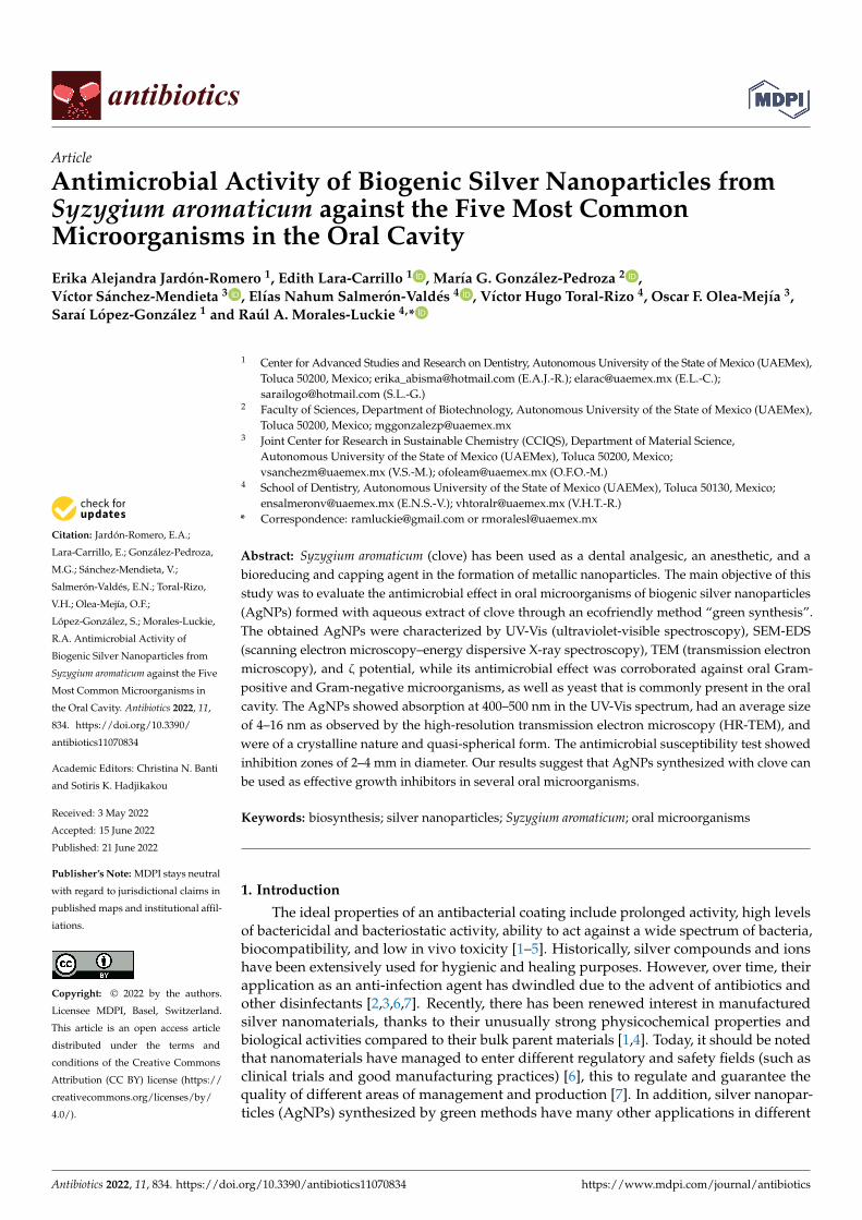
The spectra were recorded when both the color and absorption intensity of the colloidalsamples remained constant, as shown in Figure 1. Each surface plasmon resonance (SPR)maximum was ubicated in the range of 431–447 nm. In addition, no surface plasmonresonance was observed at more than 500 nm, indicating that most of the AgNPs obtainedwere of small size and similar shape. As time increases, AgNPs stabilize and reach theirmaximum growth.
Antibiotics 2022, 11, 834 4 of 13
Antibiotics 2022, 11, x FOR PEER REVIEW 4 of 13
Figure 1. UV-Vis absorption spectrum of synthesized silver nanoparticles by S. aromaticum extract at different time intervals.
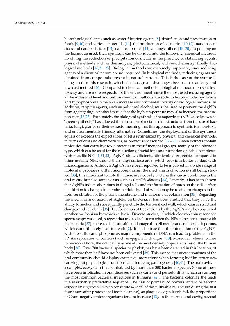
2.2.2. SEM-EDS Analysis The characterization technique was consistent with the EDS analysis. AgNPs gener-
ally showed absorption peaks at approximately 3 keV (Figure 2).
Figure 2. Scanning electron microscopy–energy dispersive X-ray spectrum of synthesized silver na-noparticles.
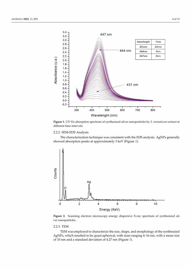
2.2.3. TEM TEM was employed to characterize the size, shape, and morphology of the synthe-
sized AgNPs, which resulted to be quasi-spherical, with sizes ranging 4–16 nm, with a mean size of 10 nm and a standard deviation of 4.27 nm (Figure 3).
Figure 1. UV-Vis absorption spectrum of synthesized silver nanoparticles by S. aromaticum extract atdifferent time intervals.
2.2.2. SEM-EDS Analysis
The characterization technique was consistent with the EDS analysis. AgNPs generallyshowed absorption peaks at approximately 3 keV (Figure 2).
Antibiotics 2022, 11, x FOR PEER REVIEW 4 of 13
Figure 1. UV-Vis absorption spectrum of synthesized silver nanoparticles by S. aromaticum extract at different time intervals.
2.2.2. SEM-EDS Analysis The characterization technique was consistent with the EDS analysis. AgNPs gener-
ally showed absorption peaks at approximately 3 keV (Figure 2).
Figure 2. Scanning electron microscopy–energy dispersive X-ray spectrum of synthesized silver na-noparticles.
2.2.3. TEM TEM was employed to characterize the size, shape, and morphology of the synthe-
sized AgNPs, which resulted to be quasi-spherical, with sizes ranging 4–16 nm, with a mean size of 10 nm and a standard deviation of 4.27 nm (Figure 3).
Figure 2. Scanning electron microscopy–energy dispersive X-ray spectrum of synthesized sil-ver nanoparticles.
2.2.3. TEM
TEM was employed to characterize the size, shape, and morphology of the synthesizedAgNPs, which resulted to be quasi-spherical, with sizes ranging 4–16 nm, with a mean sizeof 10 nm and a standard deviation of 4.27 nm (Figure 3).
Antibiotics 2022, 11, 834 5 of 13Antibiotics 2022, 11, x FOR PEER REVIEW 5 of 13
Figure 3. Characterization of synthesized biogenic silver nanoparticles from clove extract. (A) Mi-crograph of transmission electron microscopy, the insert shows size distribution and (B) high-reso-lution transmission electron microscopy micrograph.
2.2.4. ζ Potential The electrostatic stabilization of the AgNPs was estimated by measuring their ζ po-
tential values, which were found to be in the range of −15.7 to −16.2 mV.
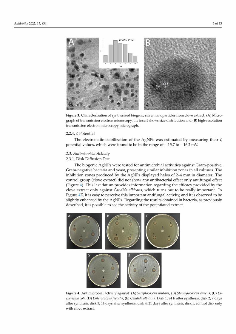
2.3. Antimicrobial Activity 2.3.1. Disk Diffusion Test
The biogenic AgNPs were tested for antimicrobial activities against Gram-positive, Gram-negative bacteria and yeast, presenting similar inhibition zones in all cultures. The inhibition zones produced by the AgNPs displayed halos of 2–4 mm in diameter. The control group (clove extract) did not show any antibacterial effect only antifungal effect (Figure 4). This last datum provides information regarding the efficacy provided by the clove extract only against Candida albicans, which turns out to be really important. In Fig-ure 4E, it is easy to perceive this important antifungal activity, and it is observed to be slightly enhanced by the AgNPs. Regarding the results obtained in bacteria, as previously described, it is possible to see the activity of the potentiated extract.
Figure 3. Characterization of synthesized biogenic silver nanoparticles from clove extract. (A) Micro-graph of transmission electron microscopy, the insert shows size distribution and (B) high-resolutiontransmission electron microscopy micrograph.
2.2.4. ζ Potential
The electrostatic stabilization of the AgNPs was estimated by measuring their ζ
potential values, which were found to be in the range of −15.7 to −16.2 mV.
2.3. Antimicrobial Activity2.3.1. Disk Diffusion Test
The biogenic AgNPs were tested for antimicrobial activities against Gram-positive,Gram-negative bacteria and yeast, presenting similar inhibition zones in all cultures. Theinhibition zones produced by the AgNPs displayed halos of 2–4 mm in diameter. Thecontrol group (clove extract) did not show any antibacterial effect only antifungal effect(Figure 4). This last datum provides information regarding the efficacy provided by theclove extract only against Candida albicans, which turns out to be really important. InFigure 4E, it is easy to perceive this important antifungal activity, and it is observed to beslightly enhanced by the AgNPs. Regarding the results obtained in bacteria, as previouslydescribed, it is possible to see the activity of the potentiated extract.
Antibiotics 2022, 11, x FOR PEER REVIEW 6 of 13
Figure 4. Antimicrobial activity against: (A) Streptococcus mutans, (B) Staphylococcus aureus, (C) Esch-erichia coli, (D) Enterococcus faecalis, (E) Candida albicans. Disk 1, 24 h after synthesis; disk 2, 7 days after synthesis; disk 3, 14 days after synthesis; disk 4, 21 days after synthesis; disk 5, control disk only with clove extract.
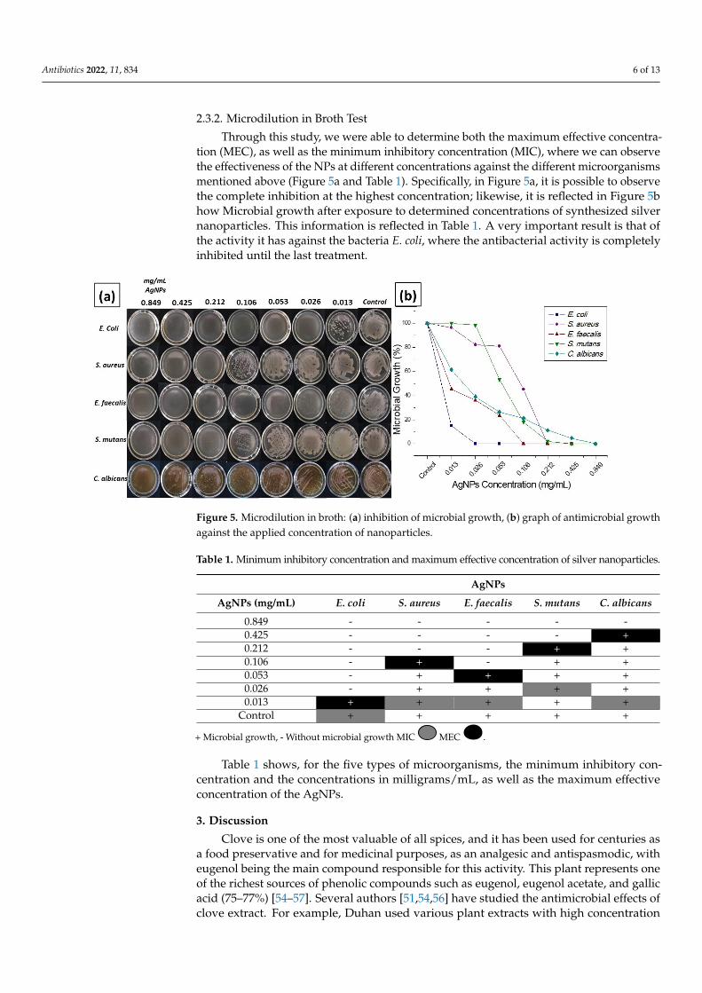
2.3.2. Microdilution in Broth Test Through this study, we were able to determine both the maximum effective concen-
tration (MEC), as well as the minimum inhibitory concentration (MIC), where we can ob-serve the effectiveness of the NPs at different concentrations against the different micro-organisms mentioned above (Figure 5a and Table 1). Specifically, in Figure 5a, it is possi-ble to observe the complete inhibition at the highest concentration; likewise, it is reflected in Figure 5b how Microbial growth after exposure to determined concentrations of syn-thesized silver nanoparticles. This information is reflected in Table 1. A very important result is that of the activity it has against the bacteria E. coli, where the antibacterial activity is completely inhibited until the last treatment.
Figure 5. Microdilution in broth: (a) inhibition of microbial growth, (b) graph of antimicrobial growth against the applied concentration of nanoparticles.
Figure 4. Antimicrobial activity against: (A) Streptococcus mutans, (B) Staphylococcus aureus, (C) Es-cherichia coli, (D) Enterococcus faecalis, (E) Candida albicans. Disk 1, 24 h after synthesis; disk 2, 7 daysafter synthesis; disk 3, 14 days after synthesis; disk 4, 21 days after synthesis; disk 5, control disk onlywith clove extract.
Antibiotics 2022, 11, 834 6 of 13
2.3.2. Microdilution in Broth Test
Through this study, we were able to determine both the maximum effective concentra-tion (MEC), as well as the minimum inhibitory concentration (MIC), where we can observethe effectiveness of the NPs at different concentrations against the different microorganismsmentioned above (Figure 5a and Table 1). Specifically, in Figure 5a, it is possible to observethe complete inhibition at the highest concentration; likewise, it is reflected in Figure 5bhow Microbial growth after exposure to determined concentrations of synthesized silvernanoparticles. This information is reflected in Table 1. A very important result is that ofthe activity it has against the bacteria E. coli, where the antibacterial activity is completelyinhibited until the last treatment.
Antibiotics 2022, 11, x FOR PEER REVIEW 6 of 13
Figure 4. Antimicrobial activity against: (A) Streptococcus mutans, (B) Staphylococcus aureus, (C) Esch-erichia coli, (D) Enterococcus faecalis, (E) Candida albicans. Disk 1, 24 h after synthesis; disk 2, 7 days after synthesis; disk 3, 14 days after synthesis; disk 4, 21 days after synthesis; disk 5, control disk only with clove extract.
2.3.2. Microdilution in Broth Test Through this study, we were able to determine both the maximum effective concen-
tration (MEC), as well as the minimum inhibitory concentration (MIC), where we can ob-serve the effectiveness of the NPs at different concentrations against the different micro-organisms mentioned above (Figure 5a and Table 1). Specifically, in Figure 5a, it is possi-ble to observe the complete inhibition at the highest concentration; likewise, it is reflected in Figure 5b how Microbial growth after exposure to determined concentrations of syn-thesized silver nanoparticles. This information is reflected in Table 1. A very important result is that of the activity it has against the bacteria E. coli, where the antibacterial activity is completely inhibited until the last treatment.
Figure 5. Microdilution in broth: (a) inhibition of microbial growth, (b) graph of antimicrobial growth against the applied concentration of nanoparticles.
Figure 5. Microdilution in broth: (a) inhibition of microbial growth, (b) graph of antimicrobial growthagainst the applied concentration of nanoparticles.
Table 1. Minimum inhibitory concentration and maximum effective concentration of silver nanoparticles.
AgNPs
AgNPs (mg/mL) E. coli S. aureus E. faecalis S. mutans C. albicans
0.849 - - - - -0.425 - - - - +0.212 - - - + +0.106 - + - + +0.053 - + + + +0.026 - + + + +0.013 + + + + +
Control + + + + +
+ Microbial growth, - Without microbial growth MIC
Antibiotics 2022, 11, x FOR PEER REVIEW 7 of 13
Table 1 shows, for the five types of microorganisms, the minimum inhibitory concen-tration and the concentrations in milligrams/mL, as well as the maximum effective con-centration of the AgNPs.
Table 1. Minimum inhibitory concentration and maximum effective concentration of silver nano-particles.
AgNPs AgNPs
(mg/mL) E. coli S. aureus E. faecalis S. mutans C. albicans
0.849 - - - - - 0.425 - - - - + 0.212 - - - + + 0.106 - + - + + 0.053 - + + + + 0.026 - + + + + 0.013 + + + + +
Control + + + + +
+ Microbial growth, - Without microbial growth MIC MEC .
3. Discussion Clove is one of the most valuable of all spices, and it has been used for centuries as a
food preservative and for medicinal purposes, as an analgesic and antispasmodic, with eugenol being the main compound responsible for this activity. This plant represents one of the richest sources of phenolic compounds such as eugenol, eugenol acetate, and gallic acid (75–77%) [54–57]. Several authors [51,54,56] have studied the antimicrobial effects of clove extract. For example, Duhan used various plant extracts with high concentration of eugenol, including clove, and compared them with 3% sodium hypochlorite against En-terococcus faecalis. Meanwhile, Mansourian and Rana used clove extract against C. albicans and compared it to nystatin. In both cases, the results obtained with the clove extracts were more effective than the substances they were compared to. The most probable path-way for AgNPs biosynthesis is that the flavonoids, from the clove buds, act as reducing agents when they encounter Ag+ ions. The electrons are transferred and the reduction pro-cess occurs, leading to the formation of AgNPs. It is well known that flavonoids also pre-vent agglomeration and stabilize the AgNPs in aqueous solution [16,21–23,58]. This makes a strong case for the involvement of flavonoids in rapid biosynthesis and for the stability of metallic NPs in the aqueous medium [17,59]. Upon storage of the bio-functionalized AgNPs that were synthesized using macerated clove solution, it was observed that the colloidal solution maintained its stability and uniformity. The biosynthesis of AgNPs by clove extract is carried out rapidly, the solution changes color as a function of time, the kinetics of formation (Figure 1) indicates that the reaction stabilizes after 6 h. The process of biosynthesis in this study was carried out under ambient conditions. Qian Sun, [60] Deshpande [49], and Bajpai [55] performed different studies to generate AgNPs using a similar green synthesis approach, obtaining antibacterial properties such as those in our study under the same conditions. In addition, Jeevika [48] produced silver nanowires via biosynthesis using clove oil. It is well known that AgNPs show a yellowish-brown color in aqueous solution; this color is a result of the excitation of surface plasmon vibrations in the metal NPs [48,49].
According to UV-Vis spectroscopy results, in the first 20 min of reaction, there was an incipient reduction in which the SPR could already be observed. This may be attributed to the high availability of reducing agents such as eugenol in the aqueous extract. During the next six hours of reaction, the SPR was clearly observed at 447 nm, indicating the for-mation of AgNPs. After six hours of reaction, SPR with a defined shape appears at 447
MEC
Antibiotics 2022, 11, x FOR PEER REVIEW 7 of 13
Table 1 shows, for the five types of microorganisms, the minimum inhibitory concen-tration and the concentrations in milligrams/mL, as well as the maximum effective con-centration of the AgNPs.
Table 1. Minimum inhibitory concentration and maximum effective concentration of silver nano-particles.
AgNPs AgNPs
(mg/mL) E. coli S. aureus E. faecalis S. mutans C. albicans
0.849 - - - - - 0.425 - - - - + 0.212 - - - + + 0.106 - + - + + 0.053 - + + + + 0.026 - + + + + 0.013 + + + + +
Control + + + + +
+ Microbial growth, - Without microbial growth MIC MEC .
3. Discussion Clove is one of the most valuable of all spices, and it has been used for centuries as a
food preservative and for medicinal purposes, as an analgesic and antispasmodic, with eugenol being the main compound responsible for this activity. This plant represents one of the richest sources of phenolic compounds such as eugenol, eugenol acetate, and gallic acid (75–77%) [54–57]. Several authors [51,54,56] have studied the antimicrobial effects of clove extract. For example, Duhan used various plant extracts with high concentration of eugenol, including clove, and compared them with 3% sodium hypochlorite against En-terococcus faecalis. Meanwhile, Mansourian and Rana used clove extract against C. albicans and compared it to nystatin. In both cases, the results obtained with the clove extracts were more effective than the substances they were compared to. The most probable path-way for AgNPs biosynthesis is that the flavonoids, from the clove buds, act as reducing agents when they encounter Ag+ ions. The electrons are transferred and the reduction pro-cess occurs, leading to the formation of AgNPs. It is well known that flavonoids also pre-vent agglomeration and stabilize the AgNPs in aqueous solution [16,21–23,58]. This makes a strong case for the involvement of flavonoids in rapid biosynthesis and for the stability of metallic NPs in the aqueous medium [17,59]. Upon storage of the bio-functionalized AgNPs that were synthesized using macerated clove solution, it was observed that the colloidal solution maintained its stability and uniformity. The biosynthesis of AgNPs by clove extract is carried out rapidly, the solution changes color as a function of time, the kinetics of formation (Figure 1) indicates that the reaction stabilizes after 6 h. The process of biosynthesis in this study was carried out under ambient conditions. Qian Sun, [60] Deshpande [49], and Bajpai [55] performed different studies to generate AgNPs using a similar green synthesis approach, obtaining antibacterial properties such as those in our study under the same conditions. In addition, Jeevika [48] produced silver nanowires via biosynthesis using clove oil. It is well known that AgNPs show a yellowish-brown color in aqueous solution; this color is a result of the excitation of surface plasmon vibrations in the metal NPs [48,49].
According to UV-Vis spectroscopy results, in the first 20 min of reaction, there was an incipient reduction in which the SPR could already be observed. This may be attributed to the high availability of reducing agents such as eugenol in the aqueous extract. During the next six hours of reaction, the SPR was clearly observed at 447 nm, indicating the for-mation of AgNPs. After six hours of reaction, SPR with a defined shape appears at 447
.
Table 1 shows, for the five types of microorganisms, the minimum inhibitory con-centration and the concentrations in milligrams/mL, as well as the maximum effectiveconcentration of the AgNPs.
3. Discussion
Clove is one of the most valuable of all spices, and it has been used for centuries asa food preservative and for medicinal purposes, as an analgesic and antispasmodic, witheugenol being the main compound responsible for this activity. This plant represents oneof the richest sources of phenolic compounds such as eugenol, eugenol acetate, and gallicacid (75–77%) [54–57]. Several authors [51,54,56] have studied the antimicrobial effects ofclove extract. For example, Duhan used various plant extracts with high concentration
Antibiotics 2022, 11, 834 7 of 13
of eugenol, including clove, and compared them with 3% sodium hypochlorite againstEnterococcus faecalis. Meanwhile, Mansourian and Rana used clove extract against C. albicansand compared it to nystatin. In both cases, the results obtained with the clove extracts weremore effective than the substances they were compared to. The most probable pathwayfor AgNPs biosynthesis is that the flavonoids, from the clove buds, act as reducing agentswhen they encounter Ag+ ions. The electrons are transferred and the reduction processoccurs, leading to the formation of AgNPs. It is well known that flavonoids also preventagglomeration and stabilize the AgNPs in aqueous solution [16,21–23,58]. This makes astrong case for the involvement of flavonoids in rapid biosynthesis and for the stabilityof metallic NPs in the aqueous medium [17,59]. Upon storage of the bio-functionalizedAgNPs that were synthesized using macerated clove solution, it was observed that thecolloidal solution maintained its stability and uniformity. The biosynthesis of AgNPs byclove extract is carried out rapidly, the solution changes color as a function of time, thekinetics of formation (Figure 1) indicates that the reaction stabilizes after 6 h. The processof biosynthesis in this study was carried out under ambient conditions. Qian Sun [60],Deshpande [49], and Bajpai [55] performed different studies to generate AgNPs using asimilar green synthesis approach, obtaining antibacterial properties such as those in ourstudy under the same conditions. In addition, Jeevika [48] produced silver nanowires viabiosynthesis using clove oil. It is well known that AgNPs show a yellowish-brown color inaqueous solution; this color is a result of the excitation of surface plasmon vibrations in themetal NPs [48,49].
According to UV-Vis spectroscopy results, in the first 20 min of reaction, there was anincipient reduction in which the SPR could already be observed. This may be attributedto the high availability of reducing agents such as eugenol in the aqueous extract. Duringthe next six hours of reaction, the SPR was clearly observed at 447 nm, indicating theformation of AgNPs. After six hours of reaction, SPR with a defined shape appearsat 447 nm, indicating that the NPs now have a defined shape and size, compared toplasmons at 431 nm at short reaction times, indicating incipient nanoparticle formation.Measurements were made approximately once every five minutes to verify the AgNPskinetics of formation.
EDS analysis of the AgNPs confirmed the presence of metallic Ag (see Figure 2),according to [61,62]. It is important to point out that these biogenic AgNPs exhibitedcertain low size polydispersity and similar shapes, making our method distinct fromother bio-reductors; this was corroborated by the TEM micrographs, whereby the imagein Figure 3A showed that AgNPs prepared using clove extract have a considerably lowpolydispersity and an almost spherical morphology. The particle size distribution histogramwas determined using TEM images. AgNPs diameters range between 4 and 16 nm, with astandard deviation of 4.2 nm (Figure 3A).
The HR-TEM image in Figure 3B shows AgNPs of 8–10 nm in diameter. This imagepossesses atomic resolution; therefore, several crystalline planes are distinguishable and theinterplanar distances can be measured. The interplanar distances shown in the micrographof these NPs, wherein the corresponding crystalline planes were specified. The interplanardistances and their corresponding crystalline planes match those of metallic Ag (face-centered cubic structure). The measured interplanar distance was 2.4 Å, which correspondsto the plane [111]. Generally, a suspension that exhibits an absolute zeta potential of0–100 mV is ubicated on the threshold of agglomeration, and the stability of the particlesfrom the solution will be increased with higher values of approximately −100 mV [63,64].In this study, the zeta potential of AgNPs was in the range of −15.7 to −16 mV. Thesevalues allow us to corroborate an acceptable stability of the AgNPs colloidal suspension.
The microorganisms selected in this study for microbiological tests are part of thenormal and pathogenic microflora of the oral cavity. Streptococcus mutans, the main causalagent of tooth decay and periodontitis, is found in 70–90% of the population. Mean-while, Staphylococcus aureus is found in patients with bacterial endocarditis and cellulite.Enterococcus faecalis is present in most failed root canal treatments. Escherichia coli has been
Antibiotics 2022, 11, 834 8 of 13
isolated from salivary gland infections, while Candida albicans is found in one in every1000 patients attending dental consultation and also in stomatitis associated with the use ofdental prostheses. Taken together, these bacteria are responsible for various opportunisticinfections in immunocompromised patients. In this study, the antibacterial activity ofAgNPs against four types of bacteria and one yeast was investigated. The inhibition zoneswere similar in all cell cultures. Biological tests of AgNPs against test strains showedthat they have a significant effect on the growth of Gram-positive and Gram-negativebacteria. The latter have a layer of lipopolysaccharides at the exterior, while underneath liesa thin layer of peptidoglycan. Thus, a potent antimicrobial effect can be determined in themicrodilution in broth study that emphasizes the antibacterial and antifungal effectivenessof the AgNPs as shown in Figure 5b, taking into account the efficacy in Escherichia colihaving microbial effect until the last concentration of NPs. On the other hand, we observedbacterial growth of the Streptococcus mutans strain from the third highest concentration, andalso the following Staphylococcus aureus and Enterococcus faecalis; on the other hand, we seethat the antifungal effectiveness of the AgNPs against Candida albicans is low but not null.Several studies indicate [1,30,65,66] that the antibacterial activity of AgNPs is based on theNPs attachment to the bacterial cell wall, or the formation of free radicals. In addition, thesilver ions released from AgNPs may play a vital role in the antibacterial activity, due totheir interaction with the thiol groups of enzymes. Although the lipopolysaccharides arecomposed of covalently linked lipids and polysaccharides, they have a lack of strengthand rigidity. The negative charges on them are attracted toward the weak positive chargeavailable on AgNPs [67,68]. This indicates that AgNPs have great potential to be used inbiomedical applications.
4. Materials and Methods4.1. Synthesis of AgNPs
In this experimental study, the chemical precursor used was silver nitrate (AgNO3)(Sigma-Aldrich, St. Louis, MO, USA). The AgNPs were directly synthesized with the clove(Terana, Especias selectas, Ciudad de México, México) extract using a simple green synthesisprocedure. Initially, various concentrations were tested until the optimum conditionswere established.
A 10 mM solution of AgNO3 was prepared using deionized water. To prepare thereducing agent, 1 g of chopped clove was added to 100 mL of boiling deionized waterfor five minutes. The mixture was allowed to cool before being filtered into a vacuumflask using a Buchner funnel and Whatman filter paper no. 5. The aqueous extract wasmixed, at room temperature, with AgNO3 in 3:1 ratio. The resultant mixture was keptundisturbed in a dark place. After a couple of hours, the color of the solution changed dueto the formation of AgNPs. The process of biosynthesis was carried out under ambientenvironmental conditions (that is, at room temperature and under atmospheric pressure);the reaction was completed within a few minutes.
4.2. Characterization of Synthesized Silver Nanoparticles4.2.1. Spectroscopy UV-Vis
The UV-Vis analysis was performed using a spectrophotometer (CARY 5000 ConcUV-Vis spectrophotometer, Varian, Inc., Palo Alto, CA, USA), which was operated at aresolution of 1 nm at room temperature. The spectral range was 300–800 nm. In this way,the kinetics of the reduction were followed until stable NPs were obtained.
4.2.2. SEM-EDS
The final product was sonicated for 30 min to break up larger nanoparticle agglom-erates; then, the particles were dried in a vacuum at room temperature (20 ◦C) prior toanalysis. The NPs were attached to aluminum stubs with conductive tape, coated withcarbon, and observed under SEM (JEOL, JSM-6510LV at 20 kcV, Tokyo, Japan) with sec-
Antibiotics 2022, 11, 834 9 of 13
ondary electrons at ×100, ×500, and ×3000 magnification that was operating at 20 kV. EDSanalysis was developed.
4.2.3. TEM
TEM was obtained via a JEOL JEM-2100 microscope (Tokyo, Japan). Samples for theTEM examination were prepared by placing a drop of the suspension on a copper grid(300 mesh) coated with carbon film, and allowing it to dry under ambient conditions.
4.2.4. Z Potential
Zeta potential measurements were determined using the Zetasizer 2000 (MalvemInstruments Ltd., Worcestershire, UK). The voltage applied to drive the electrodes ofthe zeta cell was 150 V capillary electrophoresis. The sample was prepared by injectingapproximately 2 mL solution into the cell.
4.3. Antimicrobial Activity
The bacterial strains used in this study were obtained from the stock culture collectionof the Biochemistry laboratory at the School of Dentistry within the National AutonomousUniversity of Mexico (UNAM). The strains were originally collected from clinical samples ofthe oral cavity of patients from the previously mentioned institution. These strains are nativeto central Mexico, having all been characterized through a set of culture media assays andbiochemical tests. They included Gram-positive (Staphylococcus aureus, Streptococcus mutans,Enterococcus faecalis) and Gram-negative microorganisms (Escherichia coli) and yeast(Candida albicans), which are commonly present in the oral cavity and are responsiblefor important conditions in oral health. The experiments into antimicrobial activity werecarried out as proscribed by the Clinical and Laboratory Standards Institute [69].
4.3.1. Disk Diffusion Test
The NPs antibacterial properties were measured by the Kirby–Bauer disc diffusionmethod against the Gram-positive and Gram-negative bacteria and yeast. The inoculumwas prepared by diluting the colonies with 0.9% of NaCl to 0.5 according to the McFarlandscale, before they were applied to Muller–Hinton agar (Bioxon BD Mueller–Hinton II Agar)plates using sterile cotton swabs. The sterile paper discs were saturated with 10 µL ofAgNPs (0.849 mg/mL) that had been prepared 24 h previously. In addition, three tubeswith AgNPs that had been prepared 7, 14, and 21 days before the antimicrobial tests wereused to saturate the paper discs and placed on agar plates. The disc impregnated withextract of clove was used as a positive control. After 24 and 48 h of incubation at 37 ◦C, themicrobial susceptibility was determined through the measurement of any noticed inhibitionzones. The assays were performed in triplicate [70].
The research protocol was reviewed and approved by the Ethics Committee of theCenter for Research and Advanced Studies in Dentistry.
4.3.2. Microdilution in Broth
The antimicrobial capacity of AgNPs were determined following the dilution methodof the broth [71]. Selective media were used to grow each strain and then cultured onnon-selective media. Samples were initially incubated at 37 ◦C for 24 h for fresh bacterialcultures, which were used to prepare McFarland standards. Then, 100 µL of Mueller–Hinton broth medium and positive control and a negative control (Mueller–Hinton brothand NPs as sterility control) were used. Each well was aseptically inoculated with 5 µL ofthe bacterial suspension (final concentration approximately 5 × 10⁵ CFU/mL) excludingcontrols. Subsequently, 100 µL of AgNPs (0.849 mg/mL) were placed at the beginningand seven serial microdilutions were made from the 100 µL of AgNPs. These assayswere performed in triplicate in four wells for each concentration and strain. Inoculatedmicroplates were incubated at 37 ◦C with continuous shaking at ~200 rpm for 24 h. Thepresence or absence of turbidity in each well was presented to the naked eye. The minimum
Antibiotics 2022, 11, 834 10 of 13
concentration in the wells is taken as the MIC and the maximum effective concentrationof MEC was identified by determining the lowest concentration of antimicrobial agentthat reduces the viability of the initial bacterial inoculum by 99.9% or a log reduction ininoculum count [72].
5. Conclusions
AgNPs synthesized from aqueous extracts of Sizygium aromaticum show strong an-tibacterial activity. It is important to highlight that the nanoparticle generation methoddoes not represent a strong environmental impact, it is very easy to carry out and, aboveall, very economical. In addition, this research has shown that AgNPs synthesized by thismethod are effective against the five most common microorganisms present in the oralcavity, these microorganisms are responsible for many oral health disorders, and we aresure that this research can contribute to the development of new antimicrobial agents fordental health or medical use, the routes of administration and cytotoxic assays will beexposed in future research because the expectations of this material are very high.
Author Contributions: Writing—preparation of the original draft and experimentation, E.A.J.-R.and E.N.S.-V.; review and edition-methodology-microbiology test, E.L.-C. and V.H.T.-R.; formalanalysis in the synthesis of nanoparticles-broth microdilution test, M.G.G.-P. and O.F.O.-M.; statis-tical analysis, S.L.-G.; writing—preparation of the original draft and discussion, V.S.-M.; research-interpretation, discussion, design of the reduction agent and the nanoparticle synthesis, discussionproposal, R.A.M.-L. All authors have read and agreed to the published version of the manuscript.
Funding: This research received no external funding.
Conflicts of Interest: The authors declare no conflict of interest.
References1. Chaloupka, K.; Malam, Y.; Seifalian, A.M. Nanosilver as a new generation of nanoproduct in biomedical applications. Trends
Biotechnol. 2010, 28, 580–588. [CrossRef] [PubMed]2. McShan, D.; Ray, P.C.; Yu, H. Molecular toxicity mechanism of nanosilver. J. Food Drug Anal. 2014, 22, 116–127. [CrossRef]3. Zhang, J.; Shi, W.; Ma, Q.; Cui, H.; Zhang, L. Application of Nanotechnology in Immunity against Infection. Coatings 2021, 11, 430.
[CrossRef]4. Gomaa, E.Z. Silver nanoparticles as an antimicrobial agent: A case study on Staphylococcus aureus and Escherichia coli as models
for Gram-positive and Gram-negative bacteria. J. Gen. Appl. Microbiol. 2017, 63, 36–43. [CrossRef] [PubMed]5. Barabadi, H.; Mojab, F.; Vahidi, H.; Marashi, B.; Talank, N.; Hosseini, O.; Saravanan, M. Green synthesis, characterization,
antibacterial and biofilm inhibitory activity of silver nanoparticles compared to commercial silver nanoparticles. Inorg. Chem.Commun. 2021, 129, 108647. [CrossRef]
6. Souto, E.B.; Silva, G.F.; Dias-Ferreira, J.; Zielinska, A.; Ventura, F.; Durazzo, A.; Lucarini, M.; Novellino, E.; Santini, A. Nanophar-maceutics: Part I—Clinical trials legislation and good manufacturing practices (GMP) of nanotherapeutics in the EU. Pharmaceutics2020, 12, 146. [CrossRef]
7. Yeung, A.W.K.; Souto, E.B.; Durazzo, A.; Lucarini, M.; Novellino, E.; Tewari, D.; Wang, D.; Atanasov, A.G.; Santini, A. Big impactof nanoparticles: Analysis of the most cited nanopharmaceuticals and nanonutraceuticals research. Curr. Res. Biotechnol. 2020, 2,53–63. [CrossRef]
8. Sanchooli, N.; Sanchooli, E.; Khandan Barani, H. Investigating the effect of water filter made using polyurethane foam containingsilver nanoparticles on controlling Yersinia ruckeri in Oncorhynchus. Iran. J. Fish. Sci. 2022, 21, 500–515.
9. Liu, J.; Ma, Z.; Liu, Y.; Zheng, X.; Pei, Y.; Tang, K. Soluble soybean polysaccharide films containing in-situ generated silvernanoparticles for antibacterial food packaging applications. Food Packag. Shelf Life 2022, 31, 100800. [CrossRef]
10. Pardo, L.; Arias, J.; Molleda, P. Elaboración de nanopartículas de plata sintetizadas a partir de extracto de hojas de romero(Rosmarinus officinalis L.) y su uso como conservante. LA GRANJA Rev. Cienc. Vida 2022, 35, 45–58. [CrossRef]
11. Wangpraseurt, D.; You, S.; Sun, Y.; Chen, S. Biomimetic 3D living materials powered by microorganisms. Trends Biotechnol. 2022.[CrossRef] [PubMed]
12. Ong, W.T.J.; Nyam, K.L. Evaluation of silver nanoparticles in cosmeceutical and potential biosafety complications. Saudi J. Biol.Sci. 2022. [CrossRef] [PubMed]
13. Awad, M.A.; Eid, A.M.; Elsheikh, T.M.Y.; Al-Faifi, Z.E.; Saad, N.; Sultan, M.H.; Selim, S.; Al-Khalaf, A.A.; Fouda, A. Mycosynthesis,Characterization, and Mosquitocidal Activity of Silver Nanoparticles Fabricated by Aspergillus niger Strain. J. Fungi 2022, 8, 396.[CrossRef] [PubMed]

Antibiotics 2022, 11, 834 11 of 13
14. Demchenko, V.; Kobylinskyi, S.; Iurzhenko, M.; Riabov, S.; Vashchuk, A.; Rybalchenko, N.; Zahorodnia, S.; Naumenko, K.;Demchenko, O.; Adamus, G. Nanocomposites based on polylactide and silver nanoparticles and their antimicrobial and antiviralapplications. React. Funct. Polym. 2022, 170, 105096. [CrossRef]
15. Iravani, S. Green synthesis of metal nanoparticles using plants. Green Chem. 2011, 13, 2638–2650. [CrossRef]16. Amooaghaie, R.; Saeri, M.R.; Azizi, M. Synthesis, characterization and biocompatibility of silver nanoparticles synthesized from
Nigella sativa leaf extract in comparison with chemical silver nanoparticles. Ecotoxicol. Environ. Saf. 2015, 120, 400–408. [CrossRef]17. Pani, A.; Lee, J.H.; Yun, S.-I. Autoclave mediated one-pot-one-minute synthesis of AgNPs and Au–Ag nanocomposite from Melia
azedarach bark extract with antimicrobial activity against food pathogens. Chem. Cent. J. 2016, 10, 15. [CrossRef]18. Jagtap, U.B.; Bapat, V.A. Green synthesis of silver nanoparticles using Artocarpus heterophyllus Lam. seed extract and its
antibacterial activity. Ind. Crops Prod. 2013, 46, 132–137. [CrossRef]19. Huq, M.A.; Ashrafudoulla, M.; Rahman, M.M.; Balusamy, S.R.; Akter, S. Green synthesis and potential antibacterial applications
of bioactive silver nanoparticles: A review. Polymers 2022, 14, 742. [CrossRef]20. Salem, S.S.; Fouda, A. Green synthesis of metallic nanoparticles and their prospective biotechnological applications: An overview.
Biol. Trace Elem. Res. 2021, 199, 344–370. [CrossRef]21. Nagajyothi, P.; Sreekanth, T.; Lee, J.-i.; Lee, K.D. Mycosynthesis: Antibacterial, antioxidant and antiproliferative activities of silver
nanoparticles synthesized from Inonotus obliquus (Chaga mushroom) extract. J. Photochem. Photobiol. B Biol. 2014, 130, 299–304.[CrossRef] [PubMed]
22. Kumar, D.A.; Palanichamy, V.; Roopan, S.M. Green synthesis of silver nanoparticles using Alternanthera dentata leaf extract atroom temperature and their antimicrobial activity. Spectrochim. Acta Part A Mol. Biomol. Spectrosc. 2014, 127, 168–171. [CrossRef][PubMed]
23. Morales-Luckie, R.A.; Sanchez-Mendieta, V.; Arenas-Alatorre, J.A.; López-Castañares, R.; Perez-Mazariego, J.L.; Marquina-Fabrega, V.; Gómez, R.W. One-step aqueous synthesis of stoichiometric Fe–Cu nanoalloy. Mater. Lett. 2008, 62, 4195–4197.[CrossRef]
24. Tolaymat, T.M.; El Badawy, A.M.; Genaidy, A.; Scheckel, K.G.; Luxton, T.P.; Suidan, M. An evidence-based environmentalperspective of manufactured silver nanoparticle in syntheses and applications: A systematic review and critical appraisal ofpeer-reviewed scientific papers. Sci. Total Environ. 2010, 408, 999–1006. [CrossRef] [PubMed]
25. Saravanan, A.; Kumar, P.S.; Karishma, S.; Vo, D.-V.N.; Jeevanantham, S.; Yaashikaa, P.R.; George, C.S. A review on biosynthesis ofmetal nanoparticles and its environmental applications. Chemosphere 2021, 264, 128580. [CrossRef] [PubMed]
26. González-Pedroza, M.G.; Argueta-Figueroa, L.; García-Contreras, R.; Jiménez-Martínez, Y.; Martínez-Martínez, E.; Navarro-Marchal, S.A.; Marchal, J.A.; Morales-Luckie, R.A.; Boulaiz, H. Silver nanoparticles from Annona muricata peel and leaf extractsas a potential potent, biocompatible and low cost antitumor tool. Nanomaterials 2021, 11, 1273. [CrossRef]
27. Mystrioti, C.; Xanthopoulou, T.; Tsakiridis, P.; Papassiopi, N.; Xenidis, A. Comparative evaluation of five plant extracts and juicesfor nanoiron synthesis and application for hexavalent chromium reduction. Sci. Total Environ. 2016, 539, 105–113. [CrossRef]
28. Mandal, D.; Dash, S.K.; Das, B.; Chattopadhyay, S.; Ghosh, T.; Das, D.; Roy, S. Bio-fabricated silver nanoparticles preferentiallytargets Gram positive depending on cell surface charge. Biomed. Pharmacother. 2016, 83, 548–558. [CrossRef]
29. Kokila, T.; Ramesh, P.; Geetha, D. Biosynthesis of AgNPs using Carica Papaya peel extract and evaluation of its antioxidant andantimicrobial activities. Ecotoxicol. Environ. Saf. 2016, 134, 467–473. [CrossRef]
30. Yee, M.S.-L.; Khiew, P.-S.; Chiu, W.S.; Tan, Y.F.; Kok, Y.-Y.; Leong, C.-O. Green synthesis of graphene-silver nanocomposites andits application as a potent marine antifouling agent. Colloids Surf. B Biointerfaces 2016, 148, 392–401. [CrossRef]
31. Dhand, V.; Soumya, L.; Bharadwaj, S.; Chakra, S.; Bhatt, D.; Sreedhar, B. Green synthesis of silver nanoparticles using Coffeaarabica seed extract and its antibacterial activity. Mater. Sci. Eng. C 2016, 58, 36–43. [CrossRef] [PubMed]
32. Gama-Lara, S.A.; Morales-Luckie, R.; Argueta-Figueroa, L.; Hinestroza, J.P.; García-Orozco, I.; Natividad, R. Synthesis, char-acterization, and catalytic activity of platinum nanoparticles on bovine-bone powder: A novel support. J. Nanomater. 2018,2018, 6482186. [CrossRef]
33. Ferreyra Maillard, A.P.V. Estudio del Efecto Antibacteriano de bio (Nano) Materiales Contra Microorganismos PatógenosProductores de Mastitis Bovina. 2019. Available online: https://ri.conicet.gov.ar/handle/11336/145722 (accessed on 2 May 2022).
34. Fonseca, M.S.; Rodrigues, D.M.; Sokolonski, A.R.; Stanisic, D.; Tomé, L.M.; Góes-Neto, A.; Azevedo, V.; Meyer, R.; Araújo, D.B.;Tasic, L. Activity of Fusarium oxysporum-Based Silver Nanoparticles on Candida spp. Oral Isolates. Nanomaterials 2022, 12, 501.[CrossRef] [PubMed]
35. Radhakrishnan, V.S.; Mudiam, M.K.R.; Kumar, M.; Dwivedi, S.P.; Singh, S.P.; Prasad, T. Silver nanoparticles induced alterations inmultiple cellular targets, which are critical for drug susceptibilities and pathogenicity in fungal pathogen (Candida albicans). Int. J.Nanomed. 2018, 13, 2647. [CrossRef] [PubMed]
36. Revati, S.; Bipin, C.; Chitra, P.B.; Minakshi, B. In vitro antibacterial activity of seven Indian spices against high level gentamicinresistant strains of enterococci. Arch. Med. Sci. AMS 2015, 11, 863. [CrossRef]
37. He, W.; Liu, Y.; Wamer, W.G.; Yin, J.-J. Electron spin resonance spectroscopy for the study of nanomaterial-mediated generation ofreactive oxygen species. J. Food Drug Anal. 2014, 22, 49–63. [CrossRef]
38. Gao, L.; Xu, T.; Huang, G.; Jiang, S.; Gu, Y.; Chen, F. Oral microbiomes: More and more importance in oral cavity and whole body.Protein Cell 2018, 9, 488–500. [CrossRef]
Antibiotics 2022, 11, 834 12 of 13
39. Parahitiyawa, N.B.; Scully, C.; Leung, W.K.; Yam, W.C.; Jin, L.J.; Samaranayake, L.P. Exploring the oral bacterial flora: Currentstatus and future directions. Oral Dis. 2010, 16, 136–145. [CrossRef]
40. Aas, J.A.; Paster, B.J.; Stokes, L.N.; Olsen, I.; Dewhirst, F.E. Defining the normal bacterial flora of the oral cavity. J. Clin. Microbiol.2005, 43, 5721–5732. [CrossRef]
41. Kuramitsu, H.K.; He, X.; Lux, R.; Anderson, M.H.; Shi, W. Interspecies interactions within oral microbial communities. Microbiol.Mol. Biol. Rev. 2007, 71, 653–670. [CrossRef]
42. AlSheikh, R.; Albagieh, H.N.; Abdouh, I.; Zaki, H.; Alzahrani, A.M.; Halawany, H.S.; Al-Khalifa, K.S. In vitro activity of caffeicacid phenethyl ester against different oral microorganisms. Appl. Sci. 2022, 12, 3959. [CrossRef]
43. Harris, N.O.; Garcia-Godoy, F. Primary Preventive Dentistry; Pearson Education: Upper Saddle River, NJ, USA, 2004.44. Alghamdi, S. Isolation and identification of the oral bacteria and their characterization for bacteriocin production in the oral
cavity. Saudi J. Biol. Sci. 2022, 29, 318–323. [CrossRef] [PubMed]45. García-Contreras, R.; Argueta-Figueroa, L.; Mejía-Rubalcava, C.; Jiménez-Martínez, R.; Cuevas-Guajardo, S.; Sánchez-Reyna, P.A.;
Mendieta-Zeron, H. Perspectives for the use of silver nanoparticles in dental practice. Int. Dent. J. 2011, 61, 297–301. [CrossRef][PubMed]
46. Takahashi, N.; Nyvad, B. The role of bacteria in the caries process: Ecological perspectives. J. Dent. Res. 2011, 90, 294–303.[CrossRef]
47. Uju, D.E.; Obioma, N.P. Anticariogenic potentials of clove, tobacco and bitter kola. Asian Pac. J. Trop. Med. 2011, 4, 814–818.[CrossRef]
48. Jeevika, A.; Shankaran, D.R. Seed-free synthesis of 1D silver nanowires ink using clove oil (Syzygium aromaticum) at roomtemperature. J. Colloid Interface Sci. 2015, 458, 155–159. [CrossRef]
49. Raghunandan, D.; Bedre, M.D.; Basavaraja, S.; Sawle, B.; Manjunath, S.; Venkataraman, A. Rapid biosynthesis of irregular shapedgold nanoparticles from macerated aqueous extracellular dried clove buds (Syzygium aromaticum) solution. Colloids Surf. BBiointerfaces 2010, 79, 235–240. [CrossRef]
50. Fayaz, A.M.; Balaji, K.; Girilal, M.; Yadav, R.; Kalaichelvan, P.T.; Venketesan, R. Biogenic synthesis of silver nanoparticles and theirsynergistic effect with antibiotics: A study against gram-positive and gram-negative bacteria. Nanomed. Nanotechnol. Biol. Med.2010, 6, 103–109. [CrossRef]
51. Al-Mariri, A.; Safi, M. In vitro antibacterial activity of several plant extracts and oils against some gram-negative bacteria. Iran. J.Med. Sci. 2014, 39, 36.
52. Rana, I.S.; Rana, A.S.; Rajak, R.C. Evaluation of antifungal activity in essential oil of the Syzygium aromaticum (L.) by extraction,purification and analysis of its main component eugenol. Braz. J. Microbiol. 2011, 42, 1269–1277. [CrossRef]
53. Miyoshi, J.H.; Castro, J.C.; Fenelon, V.C.; Garcia, F.P.; Nakamura, C.V.; Nogueira, A.C.; Ueda-Nakamura, T.; de Souza, H.M.;Mangolim, C.S.; Moura-Costa, G.F. Essential oil characterization of Ocimum basilicum and Syzygium aromaticum free and complexedwith β-cyclodextrin. Determination of its antioxidant, antimicrobial, and antitumoral activities. J. Incl. Phenom. Macrocycl. Chem.2022, 102, 117–132. [CrossRef]
54. Cortés-Rojas, D.F.; de Souza, C.R.F.; Oliveira, W.P. Clove (Syzygium aromaticum): A precious spice. Asian Pac. J. Trop. Biomed. 2014,4, 90–96. [CrossRef]
55. Bajpai, S.; Kumari, M. A green approach to prepare silver nanoparticles loaded gum acacia/poly (acrylate) hydrogels. Int. J. Biol.Macromol. 2015, 80, 177–188. [CrossRef] [PubMed]
56. Mandal, S.; DebMandal, M.; Saha, K.; Pal, N.K. In vitro antibacterial activity of three Indian spices against methicillin-resistantStaphylococcus aureus. Oman Med. J. 2011, 26, 319. [CrossRef]
57. Gupta, A.; Duhan, J.; Tewari, S.; Sangwan, P.; Yadav, A.; Singh, G.; Juneja, R.; Saini, H. Comparative evaluation of antimicrobialefficacy of Syzygium aromaticum, Ocimum sanctum and Cinnamomum zeylanicum plant extracts against Enterococcus faecalis: Apreliminary study. Int. Endod. J. 2013, 46, 775–783. [CrossRef]
58. Chaudhari, A.N.; Ingale, A.G. Syzygium aromaticum extract mediated, rapid and facile biogenic synthesis of shape-controlled (3D)silver nanocubes. Bioprocess Biosyst. Eng. 2016, 39, 883–891. [CrossRef]
59. Morales-Luckie, R.A.; Lopezfuentes-Ruiz, A.A.; Olea-Mejía, O.F.; Liliana, A.-F.; Sanchez-Mendieta, V.; Brostow, W.; Hinestroza,J.P. Synthesis of silver nanoparticles using aqueous extracts of Heterotheca inuloides as reducing agent and natural fibers astemplates: Agave lechuguilla and silk. Mater. Sci. Eng. C 2016, 69, 429–436. [CrossRef]
60. Sun, Q.; Cai, X.; Li, J.; Zheng, M.; Chen, Z.; Yu, C.-P. Green synthesis of silver nanoparticles using tea leaf extract and evaluationof their stability and antibacterial activity. Colloids Surf. A Physicochem. Eng. Asp. 2014, 444, 226–231. [CrossRef]
61. Patra, J.K.; Baek, K.-H. Biosynthesis of silver nanoparticles using aqueous extract of silky hairs of corn and investigation of itsantibacterial and anticandidal synergistic activity and antioxidant potential. IET Nanobiotechnol. 2016, 10, 326–333. [CrossRef]
62. Hema, J.A.; Malaka, R.; Muthukumarasamy, N.P.; Sambandam, A.; Subramanian, S.; Sevanan, M. Green synthesis of silvernanoparticles using Zea mays and exploration of its biological applications. IET Nanobiotechnol. 2016, 10, 288–294. [CrossRef]
63. Badawy, A.M.E.; Luxton, T.P.; Silva, R.G.; Scheckel, K.G.; Suidan, M.T.; Tolaymat, T.M. Impact of environmental conditions (pH,ionic strength, and electrolyte type) on the surface charge and aggregation of silver nanoparticles suspensions. Environ. Sci.Technol. 2010, 44, 1260–1266. [CrossRef] [PubMed]
64. El Badawy, A.M.; Scheckel, K.G.; Suidan, M.; Tolaymat, T. The impact of stabilization mechanism on the aggregation kinetics ofsilver nanoparticles. Sci. Total Environ. 2012, 429, 325–331. [CrossRef] [PubMed]
Antibiotics 2022, 11, 834 13 of 13
65. Seil, J.T.; Webster, T.J. Antimicrobial applications of nanotechnology: Methods and literature. Int. J. Nanomed. 2012, 7, 2767.66. Rai, M.; Yadav, A.; Gade, A. Silver nanoparticles as a new generation of antimicrobials. Biotechnol. Adv. 2009, 27, 76–83. [CrossRef]67. Salleh, A.; Naomi, R.; Utami, N.D.; Mohammad, A.W.; Mahmoudi, E.; Mustafa, N.; Fauzi, M.B. The potential of silver nanoparticles
for antiviral and antibacterial applications: A mechanism of action. Nanomaterials 2020, 10, 1566. [CrossRef]68. Hemlata, P.R.M.; Singh, A.P.; Tejavath, K.K. Biosynthesis of silver nanoparticles using cucumis prophetarum aqueous leaf extract
and their antibacterial and antiproliferative activity against cancer cell lines. ACS Omega 2020, 5, 5520. [CrossRef]69. Daniel, W.; Tholen, M. Protocols for Determination of Limits of Detection and Limits of Quantitation; Clinical and Laboratory Standards
Institute (CLSI): Wayne, PA, USA, 2004.70. Clinical and Laboratory Standards Institute. Performance Standards for Antimicrobial Disk Susceptibility Tests for Bacteria Isolated from
Animals: CLSI Supplement VET01S; Replaces VET01-S2; Clinical and Laboratory Standards Institute: Wayne, PA, USA, 2015.71. Wayne, P. Reference Method for Broth Dilution Antifungal Susceptibility Testing of Yeasts, Approved Standard; CLSI Document M27-A2;
Clinical and Laboratory Standards Institute: Wayne, PA, USA, 2002.72. Kim, K.S.; Anthony, B.F. Importance of bacterial growth phase in determining minimal bactericidal concentrations of penicillin
and methicillin. Antimicrob. Agents Chemother. 1981, 19, 1075–1077. [CrossRef]