Green Synthesis and Antimicrobial Activities of Silver ... - MDPI
13
Citation: Kemala, P.; Idroes, R.; Khairan, K.; Ramli, M.; Jalil, Z.; Idroes, G.M.; Tallei, T.E.; Helwani, Z.; Safitri, E.; Iqhrammullah, M.; et al. Green Synthesis and Antimicrobial Activities of Silver Nanoparticles Using Calotropis gigantea from Ie Seu-Um Geothermal Area, Aceh Province, Indonesia. Molecules 2022, 27, 5310. https://doi.org/10.3390/ molecules27165310 Academic Editor: Artur M. S. Silva Received: 27 July 2022 Accepted: 16 August 2022 Published: 20 August 2022 Publisher’s Note: MDPI stays neutral with regard to jurisdictional claims in published maps and institutional affil- iations. Copyright: © 2022 by the authors. Licensee MDPI, Basel, Switzerland. This article is an open access article distributed under the terms and conditions of the Creative Commons Attribution (CC BY) license (https:// creativecommons.org/licenses/by/ 4.0/). molecules Article Green Synthesis and Antimicrobial Activities of Silver Nanoparticles Using Calotropis gigantea from Ie Seu-Um Geothermal Area, Aceh Province, Indonesia Pati Kemala 1,2 , Rinaldi Idroes 1,2,3,4, * , Khairan Khairan 2,3,4 , Muliadi Ramli 2 , Zulkarnain Jalil 5 , Ghazi Mauer Idroes 6 , Trina Ekawati Tallei 7 , Zuchra Helwani 8 , Eka Safitri 2 , Muhammad Iqhrammullah 9 and Rosnani Nasution 2 1 Graduate School of Mathematics and Applied Sciences, Universitas Syiah Kuala, Banda Aceh 23111, Indonesia 2 Department of Chemistry, Faculty of Mathematics and Natural Sciences, Universitas Syiah Kuala, Banda Aceh 23111, Indonesia 3 Department of Pharmacy, Faculty of Mathematics and Natural Sciences, Universitas Syiah Kuala, Banda Aceh 23111, Indonesia 4 Herbal Medicine Research Center, Universitas Syiah Kuala, Banda Aceh 23111, Indonesia 5 Department of Physics, Faculty of Mathematics and Natural Sciences, Universitas Syiah Kuala, Banda Aceh 23111, Indonesia 6 Department of Chemical Engineering, Faculty of Engineering, Universitas Syiah Kuala, Banda Aceh 23111, Indonesia 7 Department of Biology, Faculty of Mathematics and Natural Sciences, Sam Ratulangi University, Manado 95115, Indonesia 8 Department of Chemical Engineering, Faculty of Engineering, Universitas Riau, Pekanbaru 28293, Indonesia 9 Department of Life Sciences and Chemistry, Jacobs University Bremen, Campus Ring 1, 28759 Bremen, Germany * Correspondence: [email protected] Abstract: Herein, we report our success synthesizing silver nanoparticles (AgNPs) using aqueous extracts from the leaves and flowers of Calotropis gigantea growing in the geothermal manifestation Ie Seu-Um, Aceh Besar, Indonesia. C. gigantea aqueous extract can be used as a bio-reductant for Ag + →Ag 0 conversion, obtained by 48h incubation of Ag + , and the extract mixture in a dark condition. UV–Vis characterization showed that the surface plasmon resonance (SPR) peaks of AgNPs-leaf C. gigantea (AgNPs-LCg) and AgNPs-flower C. gigantea (AgNPs-FCg) appeared in the wavelength range of 410–460 nm. Scanning electron microscopy energy-dispersive X-ray spectrometry (SEM-EDS) revealed the agglomeration and spherical shapes of AgNPs-LCg and AgNPs-FCg with diameters ranging from 87.85 to 256.7 nm. Zeta potentials were observed in the range of -41.8 to -25.1 mV. The Kirby-Bauer disc diffusion assay revealed AgNPs-FCg as the most potent antimicrobial agent with inhibition zones of 12.05 ± 0.58, 11.29 ± 0.45, and 9.02 ± 0.10 mm for Escherichia coli, Staphylo- coccus aureus, and Candida albicans, respectively. In conclusion, aqueous extract from the leaves or flowers of Calotropis gigantea may be used in the green synthesis of AgNPs with broad-spectrum antimicrobial activities. Keywords: silver nanoparticles; geothermal manifestation; Ie Seu-Um; C. gigantea; antimicrobial activities 1. Introduction Researchers have long studied plant extracts for their broad spectrum of medicinal properties such as anti-inflammatory [1], antibacterial [2,3], antifertility [4], termiticide, and nematicide activities [5]. Studies on plant extracts have recently gained more interest during the COVID-19 pandemic in the effort to find efficacious antiviral agents [6–9]. Notably, the bioactive compounds contained in plant extracts are strongly influenced by environmental factors [10]. For example, more diverse metabolites and higher-yield extracts Molecules 2022, 27, 5310. https://doi.org/10.3390/molecules27165310 https://www.mdpi.com/journal/molecules
-
Upload
khangminh22 -
Category
Documents
-
view
2 -
download
0
Transcript of Green Synthesis and Antimicrobial Activities of Silver ... - MDPI

Citation: Kemala, P.; Idroes, R.;
Khairan, K.; Ramli, M.; Jalil, Z.;
Idroes, G.M.; Tallei, T.E.; Helwani, Z.;
Safitri, E.; Iqhrammullah, M.; et al.
Green Synthesis and Antimicrobial
Activities of Silver Nanoparticles
Using Calotropis gigantea from Ie
Seu-Um Geothermal Area, Aceh
Province, Indonesia. Molecules 2022,
27, 5310. https://doi.org/10.3390/
molecules27165310
Academic Editor: Artur M. S. Silva
Received: 27 July 2022
Accepted: 16 August 2022
Published: 20 August 2022
Publisher’s Note: MDPI stays neutral
with regard to jurisdictional claims in
published maps and institutional affil-
iations.
Copyright: © 2022 by the authors.
Licensee MDPI, Basel, Switzerland.
This article is an open access article
distributed under the terms and
conditions of the Creative Commons
Attribution (CC BY) license (https://
creativecommons.org/licenses/by/
4.0/).
molecules
Article
Green Synthesis and Antimicrobial Activities of SilverNanoparticles Using Calotropis gigantea from Ie Seu-UmGeothermal Area, Aceh Province, IndonesiaPati Kemala 1,2, Rinaldi Idroes 1,2,3,4,* , Khairan Khairan 2,3,4, Muliadi Ramli 2 , Zulkarnain Jalil 5 ,Ghazi Mauer Idroes 6, Trina Ekawati Tallei 7 , Zuchra Helwani 8 , Eka Safitri 2 , Muhammad Iqhrammullah 9
and Rosnani Nasution 2
1 Graduate School of Mathematics and Applied Sciences, Universitas Syiah Kuala, Banda Aceh 23111, Indonesia2 Department of Chemistry, Faculty of Mathematics and Natural Sciences, Universitas Syiah Kuala,
Banda Aceh 23111, Indonesia3 Department of Pharmacy, Faculty of Mathematics and Natural Sciences, Universitas Syiah Kuala,
Banda Aceh 23111, Indonesia4 Herbal Medicine Research Center, Universitas Syiah Kuala, Banda Aceh 23111, Indonesia5 Department of Physics, Faculty of Mathematics and Natural Sciences, Universitas Syiah Kuala,
Banda Aceh 23111, Indonesia6 Department of Chemical Engineering, Faculty of Engineering, Universitas Syiah Kuala,
Banda Aceh 23111, Indonesia7 Department of Biology, Faculty of Mathematics and Natural Sciences, Sam Ratulangi University,
Manado 95115, Indonesia8 Department of Chemical Engineering, Faculty of Engineering, Universitas Riau, Pekanbaru 28293, Indonesia9 Department of Life Sciences and Chemistry, Jacobs University Bremen, Campus Ring 1,
28759 Bremen, Germany* Correspondence: [email protected]
Abstract: Herein, we report our success synthesizing silver nanoparticles (AgNPs) using aqueousextracts from the leaves and flowers of Calotropis gigantea growing in the geothermal manifestationIe Seu-Um, Aceh Besar, Indonesia. C. gigantea aqueous extract can be used as a bio-reductant forAg+→Ag0 conversion, obtained by 48h incubation of Ag+, and the extract mixture in a dark condition.UV–Vis characterization showed that the surface plasmon resonance (SPR) peaks of AgNPs-leafC. gigantea (AgNPs-LCg) and AgNPs-flower C. gigantea (AgNPs-FCg) appeared in the wavelengthrange of 410–460 nm. Scanning electron microscopy energy-dispersive X-ray spectrometry (SEM-EDS)revealed the agglomeration and spherical shapes of AgNPs-LCg and AgNPs-FCg with diametersranging from 87.85 to 256.7 nm. Zeta potentials were observed in the range of −41.8 to −25.1 mV.The Kirby-Bauer disc diffusion assay revealed AgNPs-FCg as the most potent antimicrobial agentwith inhibition zones of 12.05 ± 0.58, 11.29 ± 0.45, and 9.02 ± 0.10 mm for Escherichia coli, Staphylo-coccus aureus, and Candida albicans, respectively. In conclusion, aqueous extract from the leaves orflowers of Calotropis gigantea may be used in the green synthesis of AgNPs with broad-spectrumantimicrobial activities.
Keywords: silver nanoparticles; geothermal manifestation; Ie Seu-Um; C. gigantea; antimicrobial activities
1. Introduction
Researchers have long studied plant extracts for their broad spectrum of medicinalproperties such as anti-inflammatory [1], antibacterial [2,3], antifertility [4], termiticide,and nematicide activities [5]. Studies on plant extracts have recently gained more interestduring the COVID-19 pandemic in the effort to find efficacious antiviral agents [6–9].Notably, the bioactive compounds contained in plant extracts are strongly influenced byenvironmental factors [10]. For example, more diverse metabolites and higher-yield extracts
Molecules 2022, 27, 5310. https://doi.org/10.3390/molecules27165310 https://www.mdpi.com/journal/molecules
Molecules 2022, 27, 5310 2 of 13
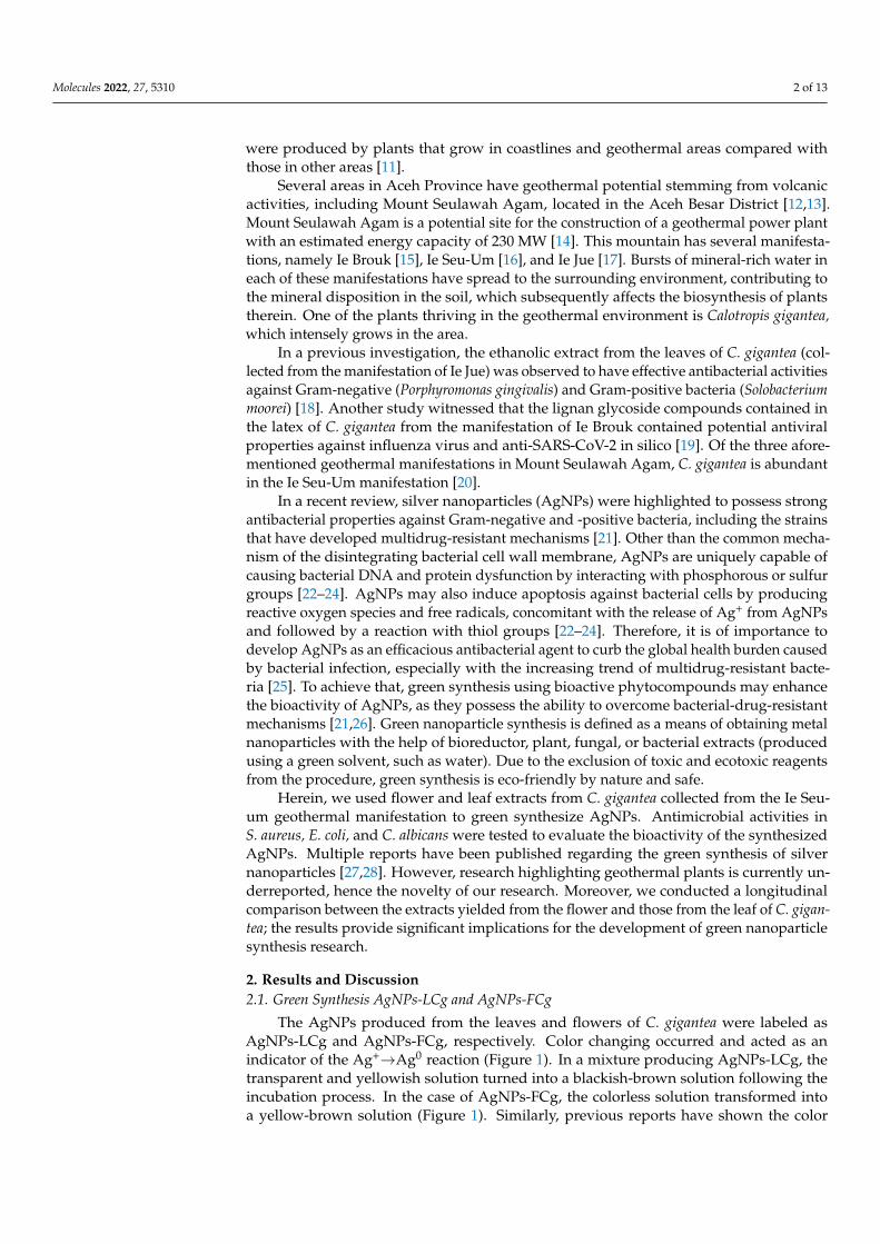
were produced by plants that grow in coastlines and geothermal areas compared withthose in other areas [11].
Several areas in Aceh Province have geothermal potential stemming from volcanicactivities, including Mount Seulawah Agam, located in the Aceh Besar District [12,13].Mount Seulawah Agam is a potential site for the construction of a geothermal power plantwith an estimated energy capacity of 230 MW [14]. This mountain has several manifesta-tions, namely Ie Brouk [15], Ie Seu-Um [16], and Ie Jue [17]. Bursts of mineral-rich water ineach of these manifestations have spread to the surrounding environment, contributing tothe mineral disposition in the soil, which subsequently affects the biosynthesis of plantstherein. One of the plants thriving in the geothermal environment is Calotropis gigantea,which intensely grows in the area.
In a previous investigation, the ethanolic extract from the leaves of C. gigantea (col-lected from the manifestation of Ie Jue) was observed to have effective antibacterial activitiesagainst Gram-negative (Porphyromonas gingivalis) and Gram-positive bacteria (Solobacteriummoorei) [18]. Another study witnessed that the lignan glycoside compounds contained inthe latex of C. gigantea from the manifestation of Ie Brouk contained potential antiviralproperties against influenza virus and anti-SARS-CoV-2 in silico [19]. Of the three afore-mentioned geothermal manifestations in Mount Seulawah Agam, C. gigantea is abundantin the Ie Seu-Um manifestation [20].
In a recent review, silver nanoparticles (AgNPs) were highlighted to possess strongantibacterial properties against Gram-negative and -positive bacteria, including the strainsthat have developed multidrug-resistant mechanisms [21]. Other than the common mecha-nism of the disintegrating bacterial cell wall membrane, AgNPs are uniquely capable ofcausing bacterial DNA and protein dysfunction by interacting with phosphorous or sulfurgroups [22–24]. AgNPs may also induce apoptosis against bacterial cells by producingreactive oxygen species and free radicals, concomitant with the release of Ag+ from AgNPsand followed by a reaction with thiol groups [22–24]. Therefore, it is of importance todevelop AgNPs as an efficacious antibacterial agent to curb the global health burden causedby bacterial infection, especially with the increasing trend of multidrug-resistant bacte-ria [25]. To achieve that, green synthesis using bioactive phytocompounds may enhancethe bioactivity of AgNPs, as they possess the ability to overcome bacterial-drug-resistantmechanisms [21,26]. Green nanoparticle synthesis is defined as a means of obtaining metalnanoparticles with the help of bioreductor, plant, fungal, or bacterial extracts (producedusing a green solvent, such as water). Due to the exclusion of toxic and ecotoxic reagentsfrom the procedure, green synthesis is eco-friendly by nature and safe.
Herein, we used flower and leaf extracts from C. gigantea collected from the Ie Seu-um geothermal manifestation to green synthesize AgNPs. Antimicrobial activities inS. aureus, E. coli, and C. albicans were tested to evaluate the bioactivity of the synthesizedAgNPs. Multiple reports have been published regarding the green synthesis of silvernanoparticles [27,28]. However, research highlighting geothermal plants is currently un-derreported, hence the novelty of our research. Moreover, we conducted a longitudinalcomparison between the extracts yielded from the flower and those from the leaf of C. gigan-tea; the results provide significant implications for the development of green nanoparticlesynthesis research.
2. Results and Discussion2.1. Green Synthesis AgNPs-LCg and AgNPs-FCg
The AgNPs produced from the leaves and flowers of C. gigantea were labeled asAgNPs-LCg and AgNPs-FCg, respectively. Color changing occurred and acted as anindicator of the Ag+→Ag0 reaction (Figure 1). In a mixture producing AgNPs-LCg, thetransparent and yellowish solution turned into a blackish-brown solution following theincubation process. In the case of AgNPs-FCg, the colorless solution transformed intoa yellow-brown solution (Figure 1). Similarly, previous reports have shown the color

Molecules 2022, 27, 5310 3 of 13
changing into yellow-brown [29–32] or dark brown [33–38] following the green synthesisof AgNPs.
Molecules 2022, 27, x FOR PEER REVIEW 3 of 14
parent and yellowish solution turned into a blackish-brown solution following the incu-bation process. In the case of AgNPs-FCg, the colorless solution transformed into a yel-low-brown solution (Figure 1). Similarly, previous reports have shown the color changing into yellow-brown [29–32] or dark brown [33–38] following the green synthesis of AgNPs.
Figure 1. Visual appearance of AgNO3 solution mixed with leaves (A) and flower extract (B) from geothermal C. gigantea before (left) and after (right) the dark incubation.
The difference in color transformation between AgNPs-LCg and AgNPs-FCg could be attributed to the phytometabolite components contained in the leaves or flowers of the geothermal C. gigantea. Thus, we proceeded with our investigation on qualitative phyto-chemical screening, for which the data are presented in Table 1. Both extracts were posi-tive for containing alkaloids, saponins, phenolics, and tannins. Flavonoids and terpenoids were detected in the flower extract but not in the leaf extract. Meanwhile, steroids were only observable in the leaf extract.
Table 1. Qualitative phytochemical analysis of AgNPs-FCg and AgNPs-LCg.
Secondary Metabolites AgNPs-FCg AgNPs-LCg Saponins +(ve) +(ve) Phenolic +(ve) +(ve) Tannins +(ve) +(ve)
Flavonoids +(ve) −(ve) Terpenoids +(ve) −(ve)
Steroids −(ve) +(ve) Alkaloids +(ve) +(ve)
Note: +(ve): positive; −(ve): negative.
Figure 1. Visual appearance of AgNO3 solution mixed with leaves (A) and flower extract (B) fromgeothermal C. gigantea before (left) and after (right) the dark incubation.
The difference in color transformation between AgNPs-LCg and AgNPs-FCg couldbe attributed to the phytometabolite components contained in the leaves or flowers of thegeothermal C. gigantea. Thus, we proceeded with our investigation on qualitative phyto-chemical screening, for which the data are presented in Table 1. Both extracts were positivefor containing alkaloids, saponins, phenolics, and tannins. Flavonoids and terpenoids weredetected in the flower extract but not in the leaf extract. Meanwhile, steroids were onlyobservable in the leaf extract.
Table 1. Qualitative phytochemical analysis of AgNPs-FCg and AgNPs-LCg.
Secondary Metabolites AgNPs-FCg AgNPs-LCg
Saponins +(ve) +(ve)Phenolic +(ve) +(ve)Tannins +(ve) +(ve)
Flavonoids +(ve) −(ve)Terpenoids +(ve) −(ve)
Steroids −(ve) +(ve)Alkaloids +(ve) +(ve)
Note: +(ve): positive; −(ve): negative.

Molecules 2022, 27, 5310 4 of 13
2.2. Characterization of Silver Nanoparticles (AgNPs-LCg and AgNPs-FCg)2.2.1. UV–Vis Spectrophotometry Analysis
UV–Vis spectrophotometers are rapid, easy-to-use, and sensitive tools frequently usedfor initial characterization of metal nanoparticles synthesized using biological methods [39].The chromophore of secondary metabolites present in metal-reducing organic compoundscould perform light absorption in UV and UV–Vis wavelength ranges [39]. Phytometabo-lites may act as electron donors and mediate the reduction of silver ions [33], therebyinitiating surface plasmon resonance (SPR). This phenomenon is a complicated processdefined as the excitation and coherent oscillation of electrons in an incoming electromag-netic field [39]. The absorption peak profile is dependent on the functional group andchromoionophore compositions of the plant extract and the extract concentration used inthe AgNP synthesis [33].
The SPR peaks of the AgNPs-FCg samples prepared using AgNO3 with concentrationsof 2, 5, and 9 mM were found at 440, 450, and 460 nm, respectively (Figure 2). Meanwhile,the SPR of the AgNPs-LCg samples synthesized using AgNO3 with 2, 5, and 9 mM werefound at 440, 430, and 460 nm, respectively (Figure 2). Our results do not deviate much incomparison with those previously reported [34,40]. As a comparison, a study preparingAgNPs using Indian C. gigantea flower reported an SPR peak at 422 nm [34]. In anotherstudy, UV–Vis characterization on AgNPs produced from C. gigantea leaf extract collectedin the Shudqum region, Saudi Arabia, revealed an SPR peak at 450 nm [40].
Molecules 2022, 27, x FOR PEER REVIEW 4 of 14
2.2. Characterization of Silver Nanoparticles (AgNPs-LCg and AgNPs-FCg) 2.2.1. UV–Vis Spectrophotometry Analysis
UV–Vis spectrophotometers are rapid, easy-to-use, and sensitive tools frequently used for initial characterization of metal nanoparticles synthesized using biological meth-ods [39]. The chromophore of secondary metabolites present in metal-reducing organic compounds could perform light absorption in UV and UV–Vis wavelength ranges [39]. Phytometabolites may act as electron donors and mediate the reduction of silver ions [33], thereby initiating surface plasmon resonance (SPR). This phenomenon is a complicated process defined as the excitation and coherent oscillation of electrons in an incoming elec-tromagnetic field [39]. The absorption peak profile is dependent on the functional group and chromoionophore compositions of the plant extract and the extract concentration used in the AgNP synthesis [33].
The SPR peaks of the AgNPs-FCg samples prepared using AgNO3 with concentra-tions of 2, 5, and 9 mM were found at 440, 450, and 460 nm, respectively (Figure 2). Mean-while, the SPR of the AgNPs-LCg samples synthesized using AgNO3 with 2, 5, and 9 mM were found at 440, 430, and 460 nm, respectively (Figure 2). Our results do not deviate much in comparison with those previously reported [34,40]. As a comparison, a study preparing AgNPs using Indian C. gigantea flower reported an SPR peak at 422 nm [34]. In another study, UV–Vis characterization on AgNPs produced from C. gigantea leaf extract collected in the Shudqum region, Saudi Arabia, revealed an SPR peak at 450 nm [40].
(a) (b)
Figure 2. UV–Vis spectrophotometry characterization of (a) AgNPs-FCg and (b) AgNPs-LCg.
2.2.2. Fourier Transform Infrared (FTIR) Analysis We used Fourier transform infrared (FTIR) characterization to identify functional
groups suspected to be involved in the AgNPs synthesis, for which the data ae presented in Figure 3. The spectral profiles of both AgNPs-FCg and AgNPs-LCg suggest the pres-ence of O—H stretching, C=C stretching, N—H bending, and C—H bending vibrations assigned for the absorbance peaks observed at 3250, 2100, 1600, and 610 cm−1, respectively. A previous report suggested the involvement of the N—H functional group in AgNPs synthesis [41]. The absorption of O—H showed that polar compounds (likely flavonoids or phenolics) from plants (Table 1) are involved in the green synthesis of AgNPs. It was also claimed that the O—H and amine groups are components that function as capping agents to stabilize silver nanoparticles [42]. Our findings agree with previously reported AgNPs-LCg results showing that the presence of groups at 3421 and 2923 cm−1 are as-signed to the bending vibration of N—H, 2853 cm−1 is assigned to the stretching vibration of C—H in alkanes, 2361 cm−1 indicates a primary amine group, 1457 cm−1 is assigned to
Figure 2. UV–Vis spectrophotometry characterization of (a) AgNPs-FCg and (b) AgNPs-LCg.
2.2.2. Fourier Transform Infrared (FTIR) Analysis
We used Fourier transform infrared (FTIR) characterization to identify functionalgroups suspected to be involved in the AgNPs synthesis, for which the data ae presented inFigure 3. The spectral profiles of both AgNPs-FCg and AgNPs-LCg suggest the presence ofO—H stretching, C=C stretching, N—H bending, and C—H bending vibrations assigned forthe absorbance peaks observed at 3250, 2100, 1600, and 610 cm−1, respectively. A previousreport suggested the involvement of the N—H functional group in AgNPs synthesis [41].The absorption of O—H showed that polar compounds (likely flavonoids or phenolics)from plants (Table 1) are involved in the green synthesis of AgNPs. It was also claimed thatthe O—H and amine groups are components that function as capping agents to stabilizesilver nanoparticles [42]. Our findings agree with previously reported AgNPs-LCg resultsshowing that the presence of groups at 3421 and 2923 cm−1 are assigned to the bendingvibration of N—H, 2853 cm−1 is assigned to the stretching vibration of C—H in alkanes,2361 cm−1 indicates a primary amine group, 1457 cm−1 is assigned to the stretchingvibration of protein, and 1116 cm−1 is assigned to the stretching vibration of C—O [40].

Molecules 2022, 27, 5310 5 of 13
Molecules 2022, 27, x FOR PEER REVIEW 5 of 14
the stretching vibration of protein, and 1116 cm−1 is assigned to the stretching vibration of C—O [40].
(a) (b)
Figure 3. FTIR spectra of (a) AgNPs-FCg and (b) AgNPs-LCg at several concentrations of AgNO3.
2.2.3. SEM-EDS Analysis The scanning electron microscopy energy-dispersive X-ray spectroscopy (SEM-EDS)
images of AgNPs-FCg and AgNPs-LCg surfaces are presented in Figure 4a,b, respectively. The formed AgNPs-FCg and AgNPs-LCg were observed to be agglomerated. In general, the morphology of formed AgNPs-FCg and AgNPs-LCg is spherical. The agglomeration of AgNPs synthesized from plants has been previously reported by those using the leaves of C. gigantea [40], Andrographys paniculata, Phillanthus niruri, Tinuspora cordifolia [29], Ni-gella sativa [43], Brilliantaisia patula, Crossopteryx febrifuga, Senna siamea [44], Lampranthus coccineus, Malephora lutea [35], Platycodon grandiflorum [30], Memecylon umbellatum Burm F [45], and Malva parviflora [33]. The EDS spectra of AgNPs-FCg and AgNPs-LCg, indicating the elements present in each sample, are presented in Figure 5. The list of elements of each sample obtained from the EDS analysis is provided in Figure 5. According to the results of the EDS analysis, metallic silver predominated among the elements observed in the sample. Therefore, this analysis confirmed that the produced particles were silver nano-particles (AgNPs). Other elements are thought to be derived from secondary metabolites found in the C. gigantea plant that grows in mineral-rich geothermal areas.
(a) (b)
Figure 4. SEM image of AgNPs-LCg (a) and AgNPs-FCg (b) displayed at 20,000× and 50,000× mag-nification, respectively.
Figure 3. FTIR spectra of (a) AgNPs-FCg and (b) AgNPs-LCg at several concentrations of AgNO3.
2.2.3. SEM-EDS Analysis
The scanning electron microscopy energy-dispersive X-ray spectroscopy (SEM-EDS)images of AgNPs-FCg and AgNPs-LCg surfaces are presented in Figure 4a,b, respectively.The formed AgNPs-FCg and AgNPs-LCg were observed to be agglomerated. In general,the morphology of formed AgNPs-FCg and AgNPs-LCg is spherical. The agglomeration ofAgNPs synthesized from plants has been previously reported by those using the leaves ofC. gigantea [40], Andrographys paniculata, Phillanthus niruri, Tinuspora cordifolia [29], Nigellasativa [43], Brilliantaisia patula, Crossopteryx febrifuga, Senna siamea [44], Lampranthus coccineus,Malephora lutea [35], Platycodon grandiflorum [30], Memecylon umbellatum Burm F [45], andMalva parviflora [33]. The EDS spectra of AgNPs-FCg and AgNPs-LCg, indicating theelements present in each sample, are presented in Figure 5. The list of elements of eachsample obtained from the EDS analysis is provided in Figure 5. According to the results ofthe EDS analysis, metallic silver predominated among the elements observed in the sample.Therefore, this analysis confirmed that the produced particles were silver nanoparticles(AgNPs). Other elements are thought to be derived from secondary metabolites found inthe C. gigantea plant that grows in mineral-rich geothermal areas.
Molecules 2022, 27, x FOR PEER REVIEW 5 of 14
the stretching vibration of protein, and 1116 cm−1 is assigned to the stretching vibration of C—O [40].
(a) (b)
Figure 3. FTIR spectra of (a) AgNPs-FCg and (b) AgNPs-LCg at several concentrations of AgNO3.
2.2.3. SEM-EDS Analysis The scanning electron microscopy energy-dispersive X-ray spectroscopy (SEM-EDS)
images of AgNPs-FCg and AgNPs-LCg surfaces are presented in Figure 4a,b, respectively. The formed AgNPs-FCg and AgNPs-LCg were observed to be agglomerated. In general, the morphology of formed AgNPs-FCg and AgNPs-LCg is spherical. The agglomeration of AgNPs synthesized from plants has been previously reported by those using the leaves of C. gigantea [40], Andrographys paniculata, Phillanthus niruri, Tinuspora cordifolia [29], Ni-gella sativa [43], Brilliantaisia patula, Crossopteryx febrifuga, Senna siamea [44], Lampranthus coccineus, Malephora lutea [35], Platycodon grandiflorum [30], Memecylon umbellatum Burm F [45], and Malva parviflora [33]. The EDS spectra of AgNPs-FCg and AgNPs-LCg, indicating the elements present in each sample, are presented in Figure 5. The list of elements of each sample obtained from the EDS analysis is provided in Figure 5. According to the results of the EDS analysis, metallic silver predominated among the elements observed in the sample. Therefore, this analysis confirmed that the produced particles were silver nano-particles (AgNPs). Other elements are thought to be derived from secondary metabolites found in the C. gigantea plant that grows in mineral-rich geothermal areas.
(a) (b)
Figure 4. SEM image of AgNPs-LCg (a) and AgNPs-FCg (b) displayed at 20,000× and 50,000× mag-nification, respectively. Figure 4. SEM image of AgNPs-LCg (a) and AgNPs-FCg (b) displayed at 20,000× and 50,000× mag-nification, respectively.

Molecules 2022, 27, 5310 6 of 13Molecules 2022, 27, x FOR PEER REVIEW 6 of 14
(a) (b)
Figure 5. EDS spectra of (a) AgNPs-FCg and (b) AgNPs-LCg at several concentrations of AgNO3.
2.2.4. Zeta Potential Analysis The results of the zeta potential analysis on AgNPs-LCg and AgNPs-FCg produced
from the various concentrations of AgNO3 (2,5, and 9 mM) are presented in Table 2. The synthesized AgNPs-LCg and AgNPs-FCg were relatively stable at −25.1 to −41.8 mV, re-spectively. Generally, the larger the negative zeta potential value, the more stable the AgNPs formed [30,46]. In this study, the concentration of AgNO3 as a metal precursor solution affected the stability of the produced AgNPs. In AgNPs-FCg, the stability in-creased as the concentration of AgNO3 increased. For AgNPs-LCg, the highest stability value was obtained when the AgNO3 concentration was 5 mM.
Table 2. Zeta potential analysis parameters of AgNPs-LCg and AgNPs-FCg.
[AgNO3] AgNPs-FCg (Mean ± SD) AgNPs-LCg (Mean ± SD)
Stability (mV) Size (nm) Stability (mV) Size (nm) 2 mM −33.05 ± 0.00 256.7 ± 2.82 −40.5 ± 0.56 227.65 ± 0.07 5 mM −30.3 ± 0.00 200.8 ± 0.14 −41.8 ± 0.14 87.85 ± 0.91 9 mM −25.1 ± 0.00 163.5 ± 1.06 −31.35 ± 0.7 188.35 ± 3.32
The sizes of AgNPs-LCg and AgNPs-FCg obtained widely ranged: 163.5 ± 1.06–256.7 ± 2.82 and 87.85 ± 0.91–227.65 ± 0.07, respectively. The smallest size of the AgNPs was the AgNPs-LCg obtained when the [AgNO3] was 5 mM. The AgNPs size correlated with the SPR peak measured by UV−Vis spectroscopy, where the lower the SPR wavelength, the smaller the particle size obtained [47]. Therefore, in this present study, the particle size obtained through zeta potential analysis is corroborated by the earlier finding from UV−Vis spectroscopy, where the AgNPs-LCg produced from AgNO3 5 mM had the low-est SPR wavelength.
2.3. Antimicrobial Activity of Silver Nanoparticles (AgNPs-LCg and AgNPs-FCg) One of the properties of AgNP that has long been a hot topic is its antimicrobial ac-
tivity [34,48]. AgNP has attracted the interest of researchers because it has antibacterial, antifungal, antiviral, antiangiogenic, and cytotoxic properties against cancer cells that have not been replaced by the properties of other metals [39]. The exact mechanism by which AgNPs have toxic or antimicrobial activity remains a mystery and is a topic of de-bate [34]. Several studies have shown that the spherical shape, concentration, and type of silver nanoparticles may play a key role in the inhibition of bacteria, which are absorbed in bacterial cells to interact with bacterial DNA and proteins causing their damage, further resulting in more oxidative stress through the generation of reactive oxygen species (ROS). ROS accumulate into the mitochondrial membrane of bacteria, resulting in dys-functional mitochondria, thereby inhibiting bacterial growth [49].
Figure 5. EDS spectra of (a) AgNPs-FCg and (b) AgNPs-LCg at several concentrations of AgNO3.
2.2.4. Zeta Potential Analysis
The results of the zeta potential analysis on AgNPs-LCg and AgNPs-FCg producedfrom the various concentrations of AgNO3 (2, 5, and 9 mM) are presented in Table 2. Thesynthesized AgNPs-LCg and AgNPs-FCg were relatively stable at −25.1 to −41.8 mV,respectively. Generally, the larger the negative zeta potential value, the more stable theAgNPs formed [30,46]. In this study, the concentration of AgNO3 as a metal precursorsolution affected the stability of the produced AgNPs. In AgNPs-FCg, the stability increasedas the concentration of AgNO3 increased. For AgNPs-LCg, the highest stability value wasobtained when the AgNO3 concentration was 5 mM.
Table 2. Zeta potential analysis parameters of AgNPs-LCg and AgNPs-FCg.
[AgNO3]AgNPs-FCg (Mean ± SD) AgNPs-LCg (Mean ± SD)
Stability (mV) Size (nm) Stability (mV) Size (nm)
2 mM −33.05 ± 0.00 256.7 ± 2.82 −40.5 ± 0.56 227.65 ± 0.075 mM −30.3 ± 0.00 200.8 ± 0.14 −41.8 ± 0.14 87.85 ± 0.919 mM −25.1 ± 0.00 163.5 ± 1.06 −31.35 ± 0.7 188.35 ± 3.32
The sizes of AgNPs-LCg and AgNPs-FCg obtained widely ranged: 163.5± 1.06–256.7± 2.82and 87.85 ± 0.91–227.65 ± 0.07, respectively. The smallest size of the AgNPs was theAgNPs-LCg obtained when the [AgNO3] was 5 mM. The AgNPs size correlated withthe SPR peak measured by UV−Vis spectroscopy, where the lower the SPR wavelength,the smaller the particle size obtained [47]. Therefore, in this present study, the particlesize obtained through zeta potential analysis is corroborated by the earlier finding fromUV−Vis spectroscopy, where the AgNPs-LCg produced from AgNO3 5 mM had the lowestSPR wavelength.
2.3. Antimicrobial Activity of Silver Nanoparticles (AgNPs-LCg and AgNPs-FCg)
One of the properties of AgNP that has long been a hot topic is its antimicrobialactivity [34,48]. AgNP has attracted the interest of researchers because it has antibacterial,antifungal, antiviral, antiangiogenic, and cytotoxic properties against cancer cells that havenot been replaced by the properties of other metals [39]. The exact mechanism by whichAgNPs have toxic or antimicrobial activity remains a mystery and is a topic of debate [34].Several studies have shown that the spherical shape, concentration, and type of silvernanoparticles may play a key role in the inhibition of bacteria, which are absorbed inbacterial cells to interact with bacterial DNA and proteins causing their damage, furtherresulting in more oxidative stress through the generation of reactive oxygen species (ROS).ROS accumulate into the mitochondrial membrane of bacteria, resulting in dysfunctionalmitochondria, thereby inhibiting bacterial growth [49].

Molecules 2022, 27, 5310 7 of 13
In this study, antibacterial and antifungal evaluations of AgNPs-LCg and AgNPs-FCgwere carried out using two bacterial pathogens (E. coli and S. aureus, which representedGram-negative and -positive bacteria, respectively) and one fungal pathogen (C. albicans).Images of the inhibition of the foregoing pathogens performed by AgNPs-LCg and AgNPs-FCg on Kirby-Bauer disc diffusion assays are presented in Figure 6. Based on the resultsobtained, the samples of AgNPs-LCg and AgNPs-FCg were found to inhibit the growth ofbacteria and fungi as the inhibitory properties of bacterial and fungal pathogens increasedin proportion to the increase in AgNO3 concentration in the synthesis process. The quan-titative data of the inhibition zones are presented in more detail in Table 3. AgNPs-FCghad the highest antimicrobial activity in S. aureus, which was 12.05 ± 0.58 mm, followedby E. coli at 11.29 ± 0.45 mm and C. albicans at 9.02 ± 0.10 mm. Meanwhile, AgNPs-LCghad the highest antimicrobial activity in S. aureus, which was 10.60 ± 0.22 mm, followed byC. albicans at 8.90 ± 0.25 mm and E. coli at 8.40 ± 0.33 mm.
Molecules 2022, 27, x FOR PEER REVIEW 7 of 14
In this study, antibacterial and antifungal evaluations of AgNPs-LCg and AgNPs-FCg were carried out using two bacterial pathogens (E. coli and S. aureus, which repre-sented Gram-negative and -positive bacteria, respectively) and one fungal pathogen (C. albicans). Images of the inhibition of the foregoing pathogens performed by AgNPs-LCg and AgNPs-FCg on Kirby-Bauer disc diffusion assays are presented in Figure 6. Based on the results obtained, the samples of AgNPs-LCg and AgNPs-FCg were found to inhibit the growth of bacteria and fungi as the inhibitory properties of bacterial and fungal path-ogens increased in proportion to the increase in AgNO3 concentration in the synthesis process. The quantitative data of the inhibition zones are presented in more detail in Table 3. AgNPs-FCg had the highest antimicrobial activity in S. aureus, which was 12.05 ± 0.58 mm, followed by E. coli at 11.29 ± 0.45 mm and C. albicans at 9.02 ± 0.10 mm. Meanwhile, AgNPs-LCg had the highest antimicrobial activity in S. aureus, which was 10.60 ± 0.22 mm, followed by C. albicans at 8.90 ± 0.25 mm and E. coli at 8.40 ± 0.33 mm.
Figure 6. Antimicrobial activity analysis: (A) AgNPs-FCg; (B) AgNPs-LCg (I: E. coli bacteria, II: S. aureus bacteria, III: C. albicans fungus where a: negative control, b: positive control, c: sample with AgNO3 concentration of 2 mM, d: sample with AgNO3 concentration 5 mM, e: sample with 9 mM AgNO3 concentration).
Table 3. Antimicrobial activities of AgNPs-FCg and AgNPs-LCg.
Sample Concentration of [AgNO3] (mM)
Inhibition Zone, Mean ± SD (mm) Staphylococcus aureus Escherichia coli Candida albicans
AgNPs-FCg 2 10.53 ± 0.57 9.89 ± 0.56 7.52 ± 0.11 5 11.24 ± 0.83 10.54 ± 0.59 8.12 ± 0.16 9 12.05 ± 0.58 11.29 ± 0.45 9.02 ± 0.10
AgNPs-LCg 2 10.10 ± 0.08 7.85 ± 0.18 7.37 ± 0.29 5 10.48 ± 0.23 8.18 ± 0.13 7.95 ± 0.26 9 10.60 ± 0.22 8.40 ± 0.33 8.90 ± 0.25
Control 17.74 ± 0.28 a 19.45 ± 0.69 b 10.20 ± 0.12 c Control: a vancomycin; b gentamicin; c ketoconazole.
The antibacterial activity of S. aureus was significantly different between the AgNPs-LCg and AgNPs-FCg inhibition zones. The AgNPs-LCg inhibition zone with 9 mM AgNO3 (which was the highest concentration in this study) was almost equivalent to the
Figure 6. Antimicrobial activity analysis: (A) AgNPs-FCg; (B) AgNPs-LCg (I: E. coli bacteria,II: S. aureus bacteria, III: C. albicans fungus where a: negative control, b: positive control, c: samplewith AgNO3 concentration of 2 mM, d: sample with AgNO3 concentration 5 mM, e: sample with9 mM AgNO3 concentration).
Table 3. Antimicrobial activities of AgNPs-FCg and AgNPs-LCg.
Sample Concentration of[AgNO3] (mM)
Inhibition Zone, Mean ± SD (mm)
Staphylococcus aureus Escherichia coli Candida albicans
AgNPs-FCg2 10.53 ± 0.57 9.89 ± 0.56 7.52 ± 0.115 11.24 ± 0.83 10.54 ± 0.59 8.12 ± 0.169 12.05 ± 0.58 11.29 ± 0.45 9.02 ± 0.10
AgNPs-LCg2 10.10 ± 0.08 7.85 ± 0.18 7.37 ± 0.295 10.48 ± 0.23 8.18 ± 0.13 7.95 ± 0.269 10.60 ± 0.22 8.40 ± 0.33 8.90 ± 0.25
Control 17.74 ± 0.28 a 19.45 ± 0.69 b 10.20 ± 0.12 c
Control: a vancomycin; b gentamicin; c ketoconazole.
The antibacterial activity of S. aureus was significantly different between the AgNPs-LCg and AgNPs-FCg inhibition zones. The AgNPs-LCg inhibition zone with 9 mM AgNO3(which was the highest concentration in this study) was almost equivalent to the AgNPs-FCg inhibition zone with 2 mM AgNO3 (the lowest concentration in this study). In theE. coli antibacterial activity test, the AgNPs-LCg inhibition zone with AgNO3 9 mM was
Molecules 2022, 27, 5310 8 of 13
lower than the AgNPs-FCg inhibition zone with AgNO3 2 mM. This indicates that AgNPs-FCg is more potent in inhibiting the growth of Gram-positive and -negative bacteria thanAgNPs-LCg isolated from the Ie Seu-um geothermal area.
The difference in activity between AgNPs-LCg and AgNPs-FCg is thought to be dueto differences in secondary metabolite components between the leaves and flowers (pleaserevisit Table 1). The flower extract contained flavonoids, whereas the leaf extract did not.There are at least two possibilities for explaining this finding. Firstly, flavonoids in theflower extract facilitate the higher conversion of Ag+ into Ag0, attributed to their lowredox potentials ranging from 0.23 to 0.75 V (while that of Ag is 0.80 V) [50]. Secondly,flavonoids are likely to bind with the Ag0 as a capping agent via a complex formation,as the compounds possess chelating moiety catechol [51]. Members of flavonoids wereproven to inhibit bacterial growth by disrupting the cell wall membrane [52]. Therefore, asflavonoids bind to the AgNPs, synergistic inhibitory activities are expected.
It is also noteworthy that in our previous study, the methanol extract of the leaves andstems of C. gigantea growing in the Ie Seu-um area was reported to have no inhibition zoneon the fungus C. albicans [20], but in this study, the antifungal activities against C. albicans byAgNPs-LCg and AgNPs-FCg samples were relatively high. With the minimum concentra-tion of AgNO3 used (2 mM), AgNPs-FCg and AgNPs-LCg were able to inhibit the growthof this fungus by 7.52 ± 0.11 and 7.37 ± 0.29 mm, respectively. AgNO3 was reported tohave the minimum inhibition against the foregoing microbes [53]. Taken altogether, theAgNPs significantly contributed to the antimicrobial activities of our samples. However,as a limitation of this present study, we did not conduct a direct comparison between theprepared AgNPs samples with the extracts and the AgNO3. Moreover, we did not usecommercial AgNPs as a comparison.
3. Materials and Methods3.1. Materials and Bioindicators
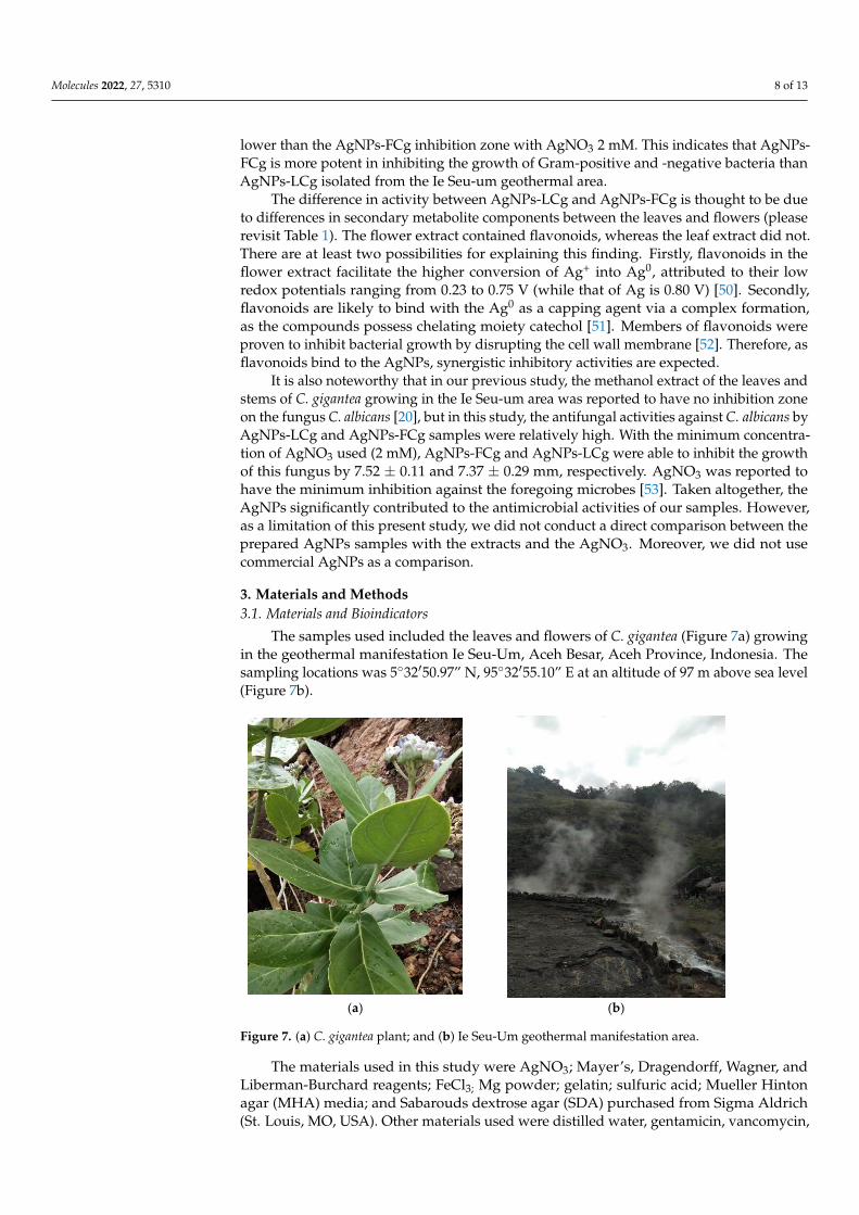
The samples used included the leaves and flowers of C. gigantea (Figure 7a) growingin the geothermal manifestation Ie Seu-Um, Aceh Besar, Aceh Province, Indonesia. Thesampling locations was 5◦32′50.97” N, 95◦32′55.10” E at an altitude of 97 m above sea level(Figure 7b).
Molecules 2022, 27, x FOR PEER REVIEW 8 of 14
AgNPs-FCg inhibition zone with 2 mM AgNO3 (the lowest concentration in this study). In the E. coli antibacterial activity test, the AgNPs-LCg inhibition zone with AgNO3 9 mM was lower than the AgNPs-FCg inhibition zone with AgNO3 2 mM. This indicates that AgNPs-FCg is more potent in inhibiting the growth of Gram-positive and -negative bac-teria than AgNPs-LCg isolated from the Ie Seu-um geothermal area.
The difference in activity between AgNPs-LCg and AgNPs-FCg is thought to be due to differences in secondary metabolite components between the leaves and flowers (please revisit Table 1). The flower extract contained flavonoids, whereas the leaf extract did not. There are at least two possibilities for explaining this finding. Firstly, flavonoids in the flower extract facilitate the higher conversion of Ag+ into Ag0, attributed to their low redox potentials ranging from 0.23 to 0.75 V (while that of Ag is 0.80 V) [50]. Secondly, flavonoids are likely to bind with the Ag0 as a capping agent via a complex formation, as the com-pounds possess chelating moiety catechol [51]. Members of flavonoids were proven to inhibit bacterial growth by disrupting the cell wall membrane [52]. Therefore, as flavo-noids bind to the AgNPs, synergistic inhibitory activities are expected.
It is also noteworthy that in our previous study, the methanol extract of the leaves and stems of C. gigantea growing in the Ie Seu-um area was reported to have no inhibition zone on the fungus C. albicans [20], but in this study, the antifungal activities against C. albicans by AgNPs-LCg and AgNPs-FCg samples were relatively high. With the minimum concentration of AgNO3 used (2 mM), AgNPs-FCg and AgNPs-LCg were able to inhibit the growth of this fungus by 7.52 ± 0.11 and 7.37 ± 0.29 mm, respectively. AgNO3 was reported to have the minimum inhibition against the foregoing microbes [53]. Taken alto-gether, the AgNPs significantly contributed to the antimicrobial activities of our samples. However, as a limitation of this present study, we did not conduct a direct comparison between the prepared AgNPs samples with the extracts and the AgNO3. Moreover, we did not use commercial AgNPs as a comparison.
3. Materials and Methods 3.1. Materials and Bioindicators
The samples used included the leaves and flowers of C. gigantea (Figure 7a) growing in the geothermal manifestation Ie Seu-Um, Aceh Besar, Aceh Province, Indonesia. The sampling locations was 5°32′50.97″ N, 95°32′55.10″ E at an altitude of 97 m above sea level (Figure 7b).
(a) (b)
Figure 7. (a) C. gigantea plant; and (b) Ie Seu-Um geothermal manifestation area. Figure 7. (a) C. gigantea plant; and (b) Ie Seu-Um geothermal manifestation area.
The materials used in this study were AgNO3; Mayer’s, Dragendorff, Wagner, andLiberman-Burchard reagents; FeCl3; Mg powder; gelatin; sulfuric acid; Mueller Hintonagar (MHA) media; and Sabarouds dextrose agar (SDA) purchased from Sigma Aldrich(St. Louis, MO, USA). Other materials used were distilled water, gentamicin, vancomycin,

Molecules 2022, 27, 5310 9 of 13
and ketoconazole. Meanwhile, the bacteria used as bioindicators in this study were S.aureus (ATCC 25923) and E. coli (ATCC 25922). The fungus used as a bioindicator was C.albicans (ATCC 10231).
3.2. Plant Extraction
C. gigantea plants were taken and the flowers and leaves separated, then washed andcut into pieces before drying for two days. Sample extraction was carried out in differentways for both the flowers and leaves. The C. gigantea leaf extraction process was carriedout by boiling 10 g of leaf sample in 100 mL of distilled water for 20 min and filtered usingfilter paper [40]. The C. gigantea flower was extracted by adding 200 mL of distilled waterwhile stirring at room temperature for 15 min and filtering [34].
3.3. Phytochemical Test
Phytochemical analysis was carried out to examine the contents of alkaloids, saponins,phenolics, tannins, flavonoids, steroids, and terpenoids in the flower and leaf extracts ofC. gigantea. Phytochemical screening followed the standard method [54]. The alkaloidtest used Mayer, Dragendorff, and Wagner reagents. The saponin test was carried outby examining the stable foam after being shaken using distilled water. The steroid testand terpenoid test used Liberman-Burchard reagent. The phenolic test used FeCl3 andflavonoid test Mg powder. As for the tannin test, gelatin and sulfuric acid were used.
3.4. Green Synthesis of AgNPs
The synthesis of silver nanoparticles using C. gigantea leaf extract (AgNPs-LCg) wascarried out by mixing 90 mL AgNO3 (2, 5, and 9 mM, separately) with 10 mL of C. gigantealeaf extract (Figure 8). The reaction was incubated at room temperature (25 ± 1 ◦C) underconstant stirring using a magnetic stirrer at 60 rpm for 48 h in the dark condition [55].The synthesis of silver nanoparticles using C. gigantea flower extract (AgNPs-FCg) wascarried out with the same procedure. We then observed the color formed from the reaction.The results of the AgNPs-LCg and AgNPs-FCg reactions were measured for absorbanceusing UV–Vis spectrophotometry (Shimadzu, UV 2500, Shimadzu Suzhou InstrumentsMfg. Co., Ltd., Jiangsu, China) at a wavelength of 300–600 nm and centrifuged (Nuve, NF800R model, Ankara, Turkey) at 13,000× g for 10 min.
Molecules 2022, 27, x FOR PEER REVIEW 10 of 14
Figure 8. Schematic diagram of the green synthesis of AgNPs-LCg and AgNPs-FCg.
3.5. Characterization of Silver Nanoparticles (AgNPs-LCg and AgNPs-FCg) AgNPs-LCg and AgNPs-FCg were characterized using Fourier transform infrared
(FTIR) spectroscopy (Search 630 FTIR Spectrometer, Agilent Technologies, Santa Clara, CA, USA), and scanning electron microscopy energy-dispersive x-ray spectroscopy (SEM-EDX, Carl Zeiss-Bruker EVO MA 10, Carl Zeiss Microscopy, White Plains, NY, USA). The characterization of the zeta potential value (mV) and polydispersity index (PI) of samples were analyzed using a Zetasizer Nano (Horiba SZ-100, Horiba Mfg. Co. Ltd., Kyoto, Ja-pan).
3.6. Antimicrobial Activity Assay Antibacterial and antifungal tests were performed using the Kirby-Bauer disc diffu-
sion method. The AgNPs-LCg and AgNPs-FCg were tested for their inhibition zones on colonies of pathogenic bacteria S. aureus and E. coli, and the fungus C. albicans. Both the bacterial colonies were grown in an MHA medium for 24 h at 37 °C. Furthermore, the colonies from the liquid medium were spread on a Petri dish containing MHA agar using a spreader. Prepared sterile paper discs (6 mm in size) were placed on the Petri dishes used for inoculation. AgNPs-LCg and AgNPs-FCg samples with various concentrations (2, 5, or 9 mM) were then loaded onto each paper disc. The positive control used for the Gram-positive antibacterial assay was vancomycin, while the positive control for the Gram-negative antibacterial assay was gentamicin. The inhibitory diameter was meas-ured after incubation at 24 h (37 °C). The antifungal activity of C. albicans was also carried out with the same method but the medium used was SDA media, and the positive control used for the antifungal assay was ketoconazole. The assays were prepared in triplicate.
4. Conclusions We successfully carried out green synthesis of AgNPs using leaf and flower extracts
of Calotropis gigantea, which grows in the geothermal manifestation Ie Seu-Um, Aceh Be-sar, Indonesia. Using AgNO3 as a metal precursor with concentrations of 2, 5, and 9 mM, it is known to form SPR peaks in the 410–460 nm range. The FTIR results also showed the functional groups detected in AgNPs-FCg and AgNPs-LCg. In addition, AgNPs-FCg and AgNPs-LCg have a sphere-shaped morphology, good stability, and antimicrobial activity
Figure 8. Schematic diagram of the green synthesis of AgNPs-LCg and AgNPs-FCg.
Molecules 2022, 27, 5310 10 of 13
3.5. Characterization of Silver Nanoparticles (AgNPs-LCg and AgNPs-FCg)
AgNPs-LCg and AgNPs-FCg were characterized using Fourier transform infrared(FTIR) spectroscopy (Search 630 FTIR Spectrometer, Agilent Technologies, Santa Clara,CA, USA), and scanning electron microscopy energy-dispersive x-ray spectroscopy (SEM-EDX, Carl Zeiss-Bruker EVO MA 10, Carl Zeiss Microscopy, White Plains, NY, USA).The characterization of the zeta potential value (mV) and polydispersity index (PI) ofsamples were analyzed using a Zetasizer Nano (Horiba SZ-100, Horiba Mfg. Co., Ltd.,Kyoto, Japan).
3.6. Antimicrobial Activity Assay
Antibacterial and antifungal tests were performed using the Kirby-Bauer disc diffusionmethod. The AgNPs-LCg and AgNPs-FCg were tested for their inhibition zones on coloniesof pathogenic bacteria S. aureus and E. coli, and the fungus C. albicans. Both the bacterialcolonies were grown in an MHA medium for 24 h at 37 ◦C. Furthermore, the colonies fromthe liquid medium were spread on a Petri dish containing MHA agar using a spreader.Prepared sterile paper discs (6 mm in size) were placed on the Petri dishes used forinoculation. AgNPs-LCg and AgNPs-FCg samples with various concentrations (2, 5, or9 mM) were then loaded onto each paper disc. The positive control used for the Gram-positive antibacterial assay was vancomycin, while the positive control for the Gram-negative antibacterial assay was gentamicin. The inhibitory diameter was measured afterincubation at 24 h (37 ◦C). The antifungal activity of C. albicans was also carried out withthe same method but the medium used was SDA media, and the positive control used forthe antifungal assay was ketoconazole. The assays were prepared in triplicate.
4. Conclusions
We successfully carried out green synthesis of AgNPs using leaf and flower extracts ofCalotropis gigantea, which grows in the geothermal manifestation Ie Seu-Um, Aceh Besar,Indonesia. Using AgNO3 as a metal precursor with concentrations of 2, 5, and 9 mM, itis known to form SPR peaks in the 410–460 nm range. The FTIR results also showed thefunctional groups detected in AgNPs-FCg and AgNPs-LCg. In addition, AgNPs-FCg andAgNPs-LCg have a sphere-shaped morphology, good stability, and antimicrobial activityagainst Gram-positive bacteria, Gram-negative bacteria, and fungi. AgNPs-FCg is knownto have a larger inhibition zone than AgNPs-LCg. As a result of the findings regardingnanoparticle size, we recommend additional research into reaction process optimizationto reduce the generally accepted nanoparticle size to values lower than those found inthis study.
Author Contributions: Conceptualization, R.I., K.K. and P.K.; methodology, P.K., R.I. and K.K.;software, P.K. and M.I.; project administration, Z.J., G.M.I., R.N. and M.R.; investigation, P.K., R.I.and K.K.; data curation, Z.J., G.M.I., Z.H., E.S., T.E.T. and R.N.; writing—original draft preparation,P.K., R.I. and K.K.; writing—review and editing, P.K., R.I., K.K., M.I. and E.S.; visualization, K.K. andP.K.; supervision, R.I.; funding acquisition, R.I. All authors have read and agreed to the publishedversion of the manuscript.
Funding: This research was funded by Universitas Syiah Kuala, Kementerian Pendidikan, Kebu-dayaan, Riset dan Teknologi Indonesia through “Program Riset Unggulan USK Percepatan Doktor”grant number 487/UN11/SPK/PNBP/2022.
Institutional Review Board Statement: Not applicable.
Informed Consent Statement: Not applicable.
Data Availability Statement: Data that support the findings of this study are available from thecorresponding author upon reasonable request.
Acknowledgments: We thank Universitas Syiah Kuala and the LPPM at Universitas Syiah Kuala fortheir help and support.

Molecules 2022, 27, 5310 11 of 13
Conflicts of Interest: The authors declare no conflict of interest.
Sample Availability: Samples of the compounds are available from the authors.
References1. Tallei, T.E.; Yelnetty, A.; Marfuah, S. Evaluation of the Potential for Immunomodulatory and Anti-Inflammatory Properties of
Phytoconstituents Derived from Pineapple [Ananas comosus (L.) Merr.] Peel Extract Using an In Silico Approach. Philipp. J. Sci.2022, 151, 397–410.
2. Khairan, K.; Astuti, Y.; First, I.S. Gel Formulation of Ethyl Acetate Garlic Extraction and Its Activity Against StaphylococcusEpidermis. J. Chem. Nat. Resour. 2019, 1, 69–78. [CrossRef]
3. Khairan, K.; Septiya, S. Murniana Antibacterial Activity of Magnolia Alba Flower Extracts on Staphylococcus Epidermidis andStaphylococcus Aureus. In IOP Conference Series: Earth and Environmental Science, Proceedings of the 10th Annual InternationalConference (AIC) on Environmental and Life Sciences (ELS), Banda Aceh, Indonesia, 15–16 October 2020; IOP Publishing: Bristol, UK,2021; Volume 711, p. 711. [CrossRef]
4. Seriana, I.; Akmal, M.; Darusman; Wahyuni, S.; Khairan, K. Sugito Phytochemicals Characterizations of Neem (Azadirachta indicaA. Juss) Leaves Ethanolic Extract: An Important Medicinal Plant as Male Contraceptive Candidate. Rasayan J. Chem. 2021, 14,343–350. [CrossRef]
5. Khairan, K.; Aulina, A.; Yusra, N.; Eriana, C.N.; Bahi, M.; Syaukani, S.; Sriwati, R.; Jacob, C. Termiticidal and NematicidalActivities of Five Extracts from Garlic (Allium sativum). In Journal of Physics: Conference Series, Volume 1882, The 1st South East AsiaScience, Technology, Engineering and Mathematics International Conference (SEA-STEM IC) 2020, Banda Aceh, Indonesia, 20–22 October2020; IOP Publishing: Bristol, UK, 2021; Volume 1882. [CrossRef]
6. Mousavi, S.S.; Karami, A.; Haghighi, T.M.; Tumilaar, S.G.; Fatimawali; Idroes, R.; Mahmud, S.; Celik, I.; Agagündüz, D.;Tallei, T.E.; et al. In Silico Evaluation of Iranian Medicinal Plant Phytoconstituents as Inhibitors against Main Protease and theReceptor-Binding Domain of Sars-Cov-2. Molecules 2021, 26, 5724. [CrossRef]
7. Khairan, K.; Idroes, R.; Tumilaar, S.G.; Tallei, T.E.; Idroes, G.M.; Rahmadhany, F.; Futri, M.U.; Dinura, N.M.; Mauliza, S.;Diana, M.; et al. Molecular Docking Study of Fatty Acids from Pliek U Oil in the Inhibition of SARS-CoV-2 Protein and Enzymes.In IOP Conference Series: Materials Science and Engineering, The 10th Annual International Conference on Science and Engineering (10thAIC 2020), Banda Aceh, Indonesia, 15–16 October 2020; IOP Publishing: Bristol, UK, 2021; Volume 1087. [CrossRef]
8. Tallei, T.E.; Tumilaar, S.G.; Niode, N.J.; Fatimawali, F.; Kepel, B.J.; Idroes, R.; Effendi, Y. Potential of Plant Bioactive Compoundsas SARS-CoV-2 Main Protease (Mpro) and Spike (S) Glycoprotein Inhibitors: A Molecular Docking Study. Scientifica 2020,2020, 6307457. [CrossRef]
9. Dutta, M.; Nezam, M.; Chowdhury, S.; Rakib, A.; Paul, A.; Sami, S.A.; Uddin, M.Z.; Rana, M.S.; Hossain, S.; Effendi, Y.; et al.Appraisals of the Bangladeshi Medicinal Plant Calotropis Gigantea Used by Folk Medicine Practitioners in the Management ofCOVID-19: A Biochemical and Computational Approach. Front. Mol. Biosci. 2021, 8, 625391. [CrossRef]
10. Abubakar, A.; Yusuf, H.; Syukri, M.; Nasution, R.; Karma, T.; Munawar, A.A.; Idroes, R. Chemometric Classification of Geothermaland Non-Geothermal Ethanol Leaf Extract of Seurapoh (Chromolaena odorata Linn) Using Infrared Spectroscopy. Earth Environ. Sci.2021, 667, 012070. [CrossRef]
11. Nuraskin, C.; Marlina; Idroes, R.; Soraya, C. Djufri Identification of Secondary Metabolite of Laban Leaf Extract (Vitex pinnata L)from Geothermal Areas and Non-Geothermal of Agam Mountains in Aceh Besar, Aceh Province, Indonesia. Rasayan, J. Chem.2020, 13, 18–23. [CrossRef]
12. Idroes, R.; Yusuf, M.; Saiful, S.; Alatas, M.; Subhan, S.; Lala, A.; Muslem, M.; Suhendra, R.; Idroes, G.M.; Marwan, M.; et al.Geochemistry Exploration and Geothermometry Application in the North Zone of Seulawah Agam, Aceh Besar District, Indonesia.Energies 2019, 12, 4442. [CrossRef]
13. Idroes, R.; Marwan, M.; Yusuf, M.; Muslem, M.; Helwani, Z. Geochemical Investigation on Jaboi Manifestation, Jaboi Volcano,Sabang, Indonesia. Int. J. Geomate 2021, 20, 170–185. [CrossRef]
14. Marwan, M.; Yanis, M.; Nugraha, G.S.; Zainal, M.; Arahman, N.; Idroes, R.; Dharma, D.B.; Saputra, D.; Gunawan, P. Mappingof Fault and Hydrothermal System beneath the Seulawah Volcano Inferred from a Magnetotellurics Structure. Energies 2021,14, 6091. [CrossRef]
15. Idroes, R.; Yusuf, M.; Alatas, M.; Subhan; Lala, A.; Muslem; Suhendra, R.; Idroes, G.M.; Suhendrayatna; Marwan; et al.Geochemistry of Warm Springs in the Ie Brôuk Hydrothermal Areas at Aceh Besar District. In IOP Conference Series: MaterialsScience and Engineering, The 8th Annual International Conference (AIC) 2018 on Science and Engineering, Banda Aceh, Indonesia, 12–14September 2018; IOP Publishing: Bristol, UK, 2019; Volume 523. [CrossRef]
16. Idroes, R.; Yusuf, M.; Alatas, M.; Subhan, S.; Lala, A.; Saiful, S.; Suhendra, R.; Idroes, G.M.; Marwan, M. Geochemistry of HotSprings in the Ie Seu’um Hydrothermal Areas at Aceh Besar District, Indonesia. In IOP Conference Series: Materials Science andEngineering, The 3rd International Conference on Chemical Engineering Sciences and Applications 2017 (3rd ICChESA 2017), Banda Aceh,Indonesia, 20–21 September 2017; IOP Publishing: Bristol, UK, 2018; Volume 334. [CrossRef]
17. Idroes, R.; Yusuf, M.; Alatas, M.; Subhan; Lala, A.; Muhammad; Suhendra, R.; Idroes, G.M.; Marwan. Geochemistry of SulphateSpring in The Ie Jue Geothermal Areas at Aceh Besar District, Indonesia. In IOP Conference Series: Materials Science and Engineering,

Molecules 2022, 27, 5310 12 of 13
The 8th Annual International Conference (AIC) 2018 on Science and Engineering, Banda Aceh, Indonesia, 12–14 September 2018; IOPPublishing: Bristol, UK, 2019; Volume 523.
18. Ningsih, D.S.; Idroes, R. In Vitro Cytotoxicity of Ethanolic Extract of The Leaf of Calotropis Gigantea from Ie Jue Geothermal Area,Aceh-Indonesia, And Its Mouthwash Formulation Against Dental Pulp Cells. J. Apllied Pharm. Sci. 2022, 12, 133–143. [CrossRef]
19. Idroes, G.M.; Tallei, T.E.; Idroes, R.; Muslem; Riza, M.; Suhendrayatna. The Study of Calotropis Gigantea Leaf Metabolites fromIe Brouk Geothermal Area Lamteuba-Aceh Besar Using Molecular Docking. In IOP Conference Series: Earth and EnvironmentalScience, The 2nd International Conference on Agriculture and Bio-industry, Banda Aceh, Indonesia, 27–28 October 2020; IOP Publishing:Bristol, UK, 2021; Volume 667. [CrossRef]
20. Idroes, R.; Khairan, K.; Fakri, F.; Zulfendi, Z. Skrining Aktifitas Tumbuhan Yang Berpotensi Sebagai Bahan Antimikroba Di Kawasan IeSeu Um Aceh Besar; Syiah Kuala Universiti Press: Banda Aceh, Indonesia, 2016; ISBN 978-602-1270-52-3.
21. Bruna, T.; Maldonado-Bravo, F.; Jara, P.; Caro, N. Silver Nanoparticles and Their Antibacterial Applications. Int. J. Mol. Sci. 2021,22, 7202. [CrossRef] [PubMed]
22. Agnihotri, S.; Mukherji, S.; Mukherji, S. Immobilized Silver Nanoparticles Enhance Contact Killing and Show Highest Efficacy:Elucidation of The Mechanism of Bactericidal Action of Silver. Nanoscale 2013, 5, 7328–7340. [CrossRef]
23. Quinteros, M.A.; Cano Aristizábal, V.; Dalmasso, P.R.; Paraje, M.G.; Páez, P.L. Oxidative Stress Generation of Silver Nanoparticlesin Three Bacterial Genera and Its Relationship with The Antimicrobial Activity. Toxicol. Vitr. 2016, 36, 216–223. [CrossRef]
24. Gomaa, E.Z. Silver Nanoparticles as an Antimicrobial Agent: A Case Study on Staphylococcus Aureus and Escherichia Coli asModels for Gram-Positive and Gram-Negative Bacteria. J. Gen. Appl. Microbiol. 2017, 63, 36–43. [CrossRef] [PubMed]
25. SIlva, M.C.; Werlang, H.M.; Vanddresen, D.; Fortes, P.C.; Pascotto, C.R.; Lucio, L.C.; Ferreto, L.E. Genetic, Antimicrobial ResistanceProfile and Mortality Rates of Acinetobacter Baumannii Infection in Brazil: A Systematic Review. Narra J. 2022, 2, e68. [CrossRef]
26. Harahap, D.; Niaci, S.; Mardina, V.; Zaura, B.; Qanita, I.; Purnama, A.; Pispita, K.; Rizki, D.R.; Iqhrammullah, M. AntibacterialActivities of Seven Ethnomedicinal Plants from Family Annonaceae. J. Adv. Pharm. Technol. Res. 2022, 13, 148–153. [CrossRef]
27. Salayov, A.; Bedlovicová, Z. Green Synthesis of Silver Nanoparticles with Antibacterial Activity Using Various Medicinal PlantExtracts: Morphology and Antibacterial Efficacy. Nanomaterials 2021, 11, 1005. [CrossRef]
28. Jain, A.S.; Pawar, P.S.; Sarkar, A.; Junnuthula, V.; Dyawanapelly, S. Bionanofactories for Green Synthesis of Silver Nanoparticles:Toward Antimicrobial Applications. Int. J. Mol. Sci. 2021, 22, 11993. [CrossRef]
29. Sharma, V.; Kaushik, S.; Pandit, P.; Dhull, D.; Yadav, J.P.; Kaushik, S. Green Synthesis of Silver Nanoparticles From MedicinalPlants And Evaluation of Their Antiviral Potential Against Chikungunya Virus. Appl. Microbiol. Biotechnol. 2019, 103, 881–891.[CrossRef] [PubMed]
30. Anbu, P.; Gopinath, S.C.B.; Shik, H.; Lee, C. Temperature-Dependent Green Biosynthesis and Characterization of Silver Nanopar-ticles Using Balloon Fl Ower Plants and Their Antibacterial Potential. J. Mol. Struct. 2019, 1177, 302–309. [CrossRef]
31. Tanase, C.; Berta, L.; Coman, A.; Ros, I.; Man, A.; Toma, F.; Mocan, A.; Nicolescu, A.; Jakab-farkas, L. Antibacterial and AntioxidantPotential of Silver Nanoparticles Biosynthesized Using the Spruce Bark Extract. Nanomaterials 2019, 9, 1541. [CrossRef] [PubMed]
32. Gemishev, O.; Panayotova, M.; Gicheva, G.; Mintcheva, N. Green Synthesis of Stable Spherical Monodisperse Silver NanoparticleUsing a Cell-Free Extract of Trichoderma Reesei. Materials 2022, 15, 481. [CrossRef]
33. Al-otibi, F.; Perveen, K.; Al-saif, N.A.; Alharbi, R.I.; Bokhari, N.A.; Albasher, G.; Al-otaibi, R.M.; Al-mosa, M.A. Biosynthesis ofSilver Nanoparticles Using Malva Parviflora and Their Antifungal Activity. Saudi J. Biol. Sci. 2021, 28, 2229–2235. [CrossRef]
34. Mathew, S.; Victo, C.P.; Sidhi, J.; Thanzeela, B. Biosynthesis of Silver Nanoparticle Using Flowers of Calotropis gigantea (L.) W.T.Aiton and Activity Against Pathogenic Bacteria. Arab. J. Chem. 2020, 13, 9139–9144. [CrossRef]
35. Haggag, E.G.; Elshamy, A.M.; Rabeh, M.A.; Gabr, N.M.; Salem, M.; Youssif, K.A.; Samir, A.; Bin Muhsinah, A.; Alsayari, A.;Abdelmohsen, U.R. Antiviral Potential of Green Synthesized Silver Nanoparticles of Lampranthus Coccineus And MalephoraLutea. Int. J. Nanomed. 2019, 14, 6217–6229. [CrossRef]
36. Rakib-Uz-Zaman, S.M.; Apu, E.H.; Muntasir, M.N.; Mowna, S.A.; Khanom, M.G.; Jahan, S.S.; Akter, N.; Khan, M.A.R.; Shuborna,N.S.; Shams, S.M.; et al. Biosynthesis of Silver Nanoparticles from Cymbopogon Citratus Leaf Extract and Evaluation of TheirAntimicrobial Properties. Challeges 2022, 13, 18. [CrossRef]
37. Alahmad, A.; Al-zereini, W.A.; Hijazin, T.J.; Al-madanat, O.Y.; Alghoraibi, I.; Al-qaralleh, O.; Al-qaraleh, S.; Feldhoff, A.; Walter, J.;Scheper, T. Green Synthesis of Silver Nanoparticles Using Hypericum perforatum L. Aqueous Extract with the Evaluation of ItsAntibacterial Activity against Clinical and Food Pathogens. Pharmaceutics 2022, 14, 1104. [CrossRef]
38. Govindan, L.; Anbazhagan, S.; Altemimi, A.B.; Lakshminarayanan, K.; Kuppan, S.; Pratap-Singh, A.; Kandasamy, M. Efficacy ofAntimicrobial and Larvicidal Activities of Green Synthesized Silver Nanoparticle Using Leaf Extract of Plumbago AuriculataLam. Plants 2020, 9, 1577. [CrossRef]
39. Pryshchepa, O.; Pomastowski, P.; Buszewski, B. Silver Nanoparticles: Synthesis, Investigation Techniques, and Properties.Adv. Colloid Interface Sci. 2020, 284, 87–100. [CrossRef] [PubMed]
40. Ali, E.M.; Abdallah, B.M. Effective Inhibition of Candidiasis Using an Eco- Friendly Leaf Extract of Calotropis-Gigantean-MediatedSilver Nanoparticles. Nanomaterials 2020, 10, 422. [CrossRef] [PubMed]
41. Ali, D.M.; Sasikala, M.; Gunasekaran, M.; Thajuddin, N. Biosynthesis and Characterization of Silver Nanoparticles Using MarineCyanobacterium, Oscillatoria Willei NTDM01. J. Nanomater. Biostruct. 2011, 6, 385–390.
42. Mirda, E.; Idroes, R.; Khairan, K.; Tallei, T.E.; Ramli, M.; Earlia, N.; Maulana, A.; Idroes, G.M.; Muslem, M.; Jalil, Z. Synthesis ofChitosan-Silver Nanoparticle Composite Spheres and Their Antimicrobial Activities. Polymers 2021, 13, 3990. [CrossRef]

Molecules 2022, 27, 5310 13 of 13
43. Almatroudi, A.; Khadri, H.; Azam, M.; Rahmani, A.H.; Khaleefah, F.; Khaleefah, A.; Khateef, R.; Ansari, M.A.; Allemailem, K.S.Antibacterial, Antibiofilm and Anticancer Activity of Biologically Synthesized Silver Nanoparticles Using Seed Extract of NigellaSativa. Processes 2020, 8, 388. [CrossRef]
44. Kambale, E.K.; Nkanga, C.I.; Mutonkole, B.P.I.; Bapolisi, A.M.; Tassa, D.O.; Liesse, J.M.I.; Krause, R.W.M.; Memvanga, P.B. GreenSynthesis of Antimicrobial Silver Nanoparticles Using Aqueous Leaf Extracts from Three Congolese Plant Species (Brillantaisiapatula, Crossopteryx febrifuga and Senna siamea). Heliyon 2020, 6, e04493. [CrossRef]
45. Alsalhi, M.S.; Elangovan, K.; Jacob, A.; Ranjitsingh, A.; Murali, P.; Devanesan, S. Synthesis of Silver Nanoparticles Using PlantDerived 4-N-Methyl Benzoic Acid And Evaluation of Antimicrobial, Antioxidant And Antitumor Activity. Saudi J. Biol. Sci. 2019,26, 970–978. [CrossRef]
46. Kokila, T.; Ramesh, P.S.; Geetha, D. Biosynthesis of AgNPs Using Carica Papaya Peel Extract and Evaluation of Its Antioxidantand Antimicrobial Activities. Ecotoxicol. Environ. Saf. 2016, 134, 467–473. [CrossRef]
47. Venkatachalam, S. Chapter 6. Ultraviolet and Visible Spectroscopy Studies of Nanofillers and Their Polymer Nanocomposites; Elsevier Inc.:Amsterdam, The Netherlands, 2016; ISBN 9780323401838.
48. Jaswal, T.; Gupta, J. A Review on the Toxicity of Silver Nanoparticles on Human Health. Mater. Today Proc. 2021, 1–5. [CrossRef]49. Al-otibi, F.; Al-ahaidib, R.A.; Alharbi, R.I.; Al-otaibi, R.M.; Albasher, G. Antimicrobial Potential of Biosynthesized Silver
Nanoparticles by Aaronsohnia Factorovskyi Exract. Molecules 2021, 26, 130. [CrossRef]50. Pietta, P.G. Flavonoids as Antioxidants. J. Nat. Prod. 2000, 63, 1035–1042. [CrossRef] [PubMed]51. Van Acker, S.A.B.E.; Van Den Berg, D.J.; Tromp, M.N.J.L.; Griffioen, D.H.; Van Bennekom, W.P.; Van Der Vijgh, W.J.F.; Bast, A.
Structural Aspects of Antioxidant Activity of Flavonoids. Free Radic. Biol. Med. 1996, 20, 331–342. [CrossRef]52. Nayem, S.M.A.; Sultana, N.; Haque, A.; Miah, B.; Hasan, M.; Islam, T.; Mahedi, H.; Awal, A.; Uddin, J.; Aziz, A.; et al.
Green Synthesis of Gold and Silver Nanoparticles by Using Amorphophallus paeoniifolius Tuber Extract and Evaluation of TheirAntibacterial Activity. Molecules 2020, 25, 4773. [CrossRef] [PubMed]
53. Chouhan, S.; Guleria, S. Green Synthesis of AgNPs Using Cannabis Sativa Leaf Extract: Characterization, Antibacterial, Anti-Yeastand Alpha-Amylase Inhibitory Activity. Mater. Sci. Energy Technol. 2020, 3, 536–544. [CrossRef]
54. Musman, M. Kimia Bahan Alam Laut; Syiah Kuala University Press: Banda Aceh, Indonesia, 2013; ISBN 9789798278938.55. Sorubavalli, U.; Vadivazhagi, M.K.; Vadivelu, J. Antioxidant and Antimicrobial Activity of Calotrophis Mediated Silver Nanopar-
ticles. J. Compos. Theory 2019, XII, 303–312.