Antimicrobial Properties of Diamondlike Carbon-Silver-Platinum Nanocomposite Thin Films
23
This document was prepared in conjunction with work accomplished under Contract No. DE-AC09-96SR18500 with the U. S. Department of Energy. DISCLAIMER This report was prepared as an account of work sponsored by an agency of the United States Government. Neither the United States Government nor any agency thereof, nor any of their employees, nor any of their contractors, subcontractors or their employees, makes any warranty, express or implied, or assumes any legal liability or responsibility for the accuracy, completeness, or any third party's use or the results of such use of any information, apparatus, product, or process disclosed, or represents that its use would not infringe privately owned rights. Reference herein to any specific commercial product, process, or service by trade name, trademark, manufacturer, or otherwise, does not necessarily constitute or imply its endorsement, recommendation, or favoring by the United States Government or any agency thereof or its contractors or subcontractors. The views and opinions of authors expressed herein do not necessarily state or reflect those of the United States Government or any agency thereof.
-
Upload
departmentenergy -
Category
Documents
-
view
0 -
download
0
Transcript of Antimicrobial Properties of Diamondlike Carbon-Silver-Platinum Nanocomposite Thin Films
This document was prepared in conjunction with work accomplished under Contract No. DE-AC09-96SR18500 with the U. S. Department of Energy.
DISCLAIMER This report was prepared as an account of work sponsored by an agency of the United States Government. Neither the United States Government nor any agency thereof, nor any of their employees, nor any of their contractors, subcontractors or their employees, makes any warranty, express or implied, or assumes any legal liability or responsibility for the accuracy, completeness, or any third party's use or the results of such use of any information, apparatus, product, or process disclosed, or represents that its use would not infringe privately owned rights. Reference herein to any specific commercial product, process, or service by trade name, trademark, manufacturer, or otherwise, does not necessarily constitute or imply its endorsement, recommendation, or favoring by the United States Government or any agency thereof or its contractors or subcontractors. The views and opinions of authors expressed herein do not necessarily state or reflect those of the United States Government or any agency thereof.
WSRC-MS-2005-00052
Antimicrobial Properties of Diamondlike Carbon-Silver-Platinum Nanocomposite Thin Films
R. J. Narayan and H. Abernathy
School of Materials Science and Engineering Georgia Institute of Technology
Atlanta, GA 30332-0245
L. Riester Metals and Ceramics Division
Oak Ridge National Laboratory Oak Ridge, TN 37831
C. J. Berry and R. Brigmon
Environmental Biotechnology Section Savannah River National Laboratory
Aiken, SC 29808 Abstract
Silver and platinum were incorporated within diamondlike carbon (DLC) thin
films using a multicomponent target pulsed laser deposition process. Transmission
electron microscopy of the DLC-silver and DLC-platinum composite films reveals that
the metals self-assemble into particulate nanocomposite structures. Nanoindentation
testing has shown that diamondlike carbon-silver films exhibit hardness and Young's
modulus values of approximately 37 GPa and 333 GPa, respectively. DLC-silver-
platinum films exhibited antimicrobial properties against Staphylococcus bacteria.
Diamondlike carbon-biofunctional metal nanocomposite films have a variety of potential
medical and antimicrobial applications.
Keywords: diamondlike carbon, pulsed laser deposition, thin films
1
WSRC-MS-2005-00052
Introduction
Biomedical researchers have created advanced materials over the past three
decades by selecting bulk materials with appropriate fracture toughness, bulk modulus,
and durability, and performing surface modification to improve biocompatibility, wear
resistance, and corrosion resistance. One ceramic coating with tremendous potential for
medical applications is diamondlike carbon (DLC). The term diamondlike carbon (DLC)
describes hydrogen-free hard carbon solids that possess a cross-linked, non-crystalline
network of sp2- and sp3- hybridized carbon atoms [1]. The friction and wear coefficients
of DLC are lower than those of diamond, and are among the lowest recorded to date
(static coefficient of friction= 0.006). DLC also offers transparency to light ranging from
deep ultraviolet to far infrared. In addition, DLC films are amorphous, atomically
smooth, and do not contain open corrosion paths to the underlying substrate.
Diamondlike carbon thin films have also been shown to possess excellent cell
compatibility. For example, in vitro studies of diamondlike carbon films involving mouse
peritoneal macrophages, mouse fibroblasts, human myeloblastic ML-1 cells, osteoblast-
like cells, and human embryo kidney 293 cells have demonstrated the absence of an
inflammatory response [2-5]. Morphological and biochemical data suggest that DLC-
exposed cells undergo no cellular damage. For example, osteoblast-like cells exposed to
DLC coatings have not shown any change in the production of alkaline phosphatase, type
I collagen, or osteocalcin [6]. In addition, neuronal growth readily occurs on DLC
surfaces [7]. Diamondlike carbon thin films have been recently considered for a variety
of cardiovascular, orthopaedic, ophthalmic, biosensor, and implantable
2
WSRC-MS-2005-00052
microelectromechanical system applications, which allow for improved device lifetimes
and unique interactions with the biological environment.
Pulsed laser deposition of diamondlike carbon involves laser ablation of a sp2
bonded carbon target, which results in the formation of a sp3- and sp2- bonded film. The
most common target material is high purity graphite; other target materials have included
pressed diamond powder, glassy carbon, and polymer [8-10]. Pure carbon sources lead to
pure DLC films, whereas hydrocarbon sources lead to DLC films with significant
hydrogen and/or hydrocarbon incorporation. The reported growth rates of DLC films
deposited using a 248 nm excimer laser are on the order of 0.01 nm/pulse [11]. Laser
processing of diamondlike carbon thin films involves several interdependent factors,
which include: (1) kinetic energy of the carbon species, (2) background pressure, and (3)
substrate temperature. These parameters have tremendous bearing on diamondlike carbon
film properties, including the sp3/sp2 ratio, the adhesion of the DLC film to the substrate,
and the amount of sp2 clustering [12].
We have recently developed a variant of the conventional pulsed laser deposition
process in order to incorporate biofunctional metals during DLC film deposition. Briefly,
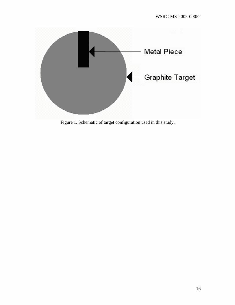
a single multicomponent target is loaded into the pulsed laser deposition chamber. This
target contains pure graphite that is covered by a piece of the desired modifying element
(Figure 1). The focused laser beam sequentially ablates the graphite target component
and the modifying element target component to form composite layers. The metal
composition in these films can be controlled through altering the following parameters:
(1) the scanning radius of the laser beam on the target surface, (2) the laser beam
position, (3) the position of the circular target, (4) the size of the metal piece on the
3
WSRC-MS-2005-00052
target, and (5) the laser energy density. The fraction of metal atoms incorporated into the
diamondlike carbon film can be estimated using the following relation:
fraction of metal within diamondlike carbon-metal nanocomposite film=α δ (l-Rd)/2 π γ(1-Rc) [1]
in which α is the laser ablation ratio, γ is the laser beam scanning radius, Rc is the
reflectivity of carbon, Rd is the reflectivity of the metal strip, and δ is width of the metal
piece. We have prepared several diamondlike carbon-metal nanocomposite films using
this modified pulsed laser deposition process. These films were examined using
transmission electron microscopy, electron energy loss spectroscopy, visible Raman
spectroscopy, nanoindentation, and microbial biofilm attachment testing.
Experimental
Several 1 cm x 1 cm pieces of silicon (100) were cleaned in acetone and methanol
within an ultrasonic cleaner. The silicon substrates were subsequently dipped in
hydrofluoric acid to remove silicon oxide, which resulted in a hydrogen-terminated
surface. The Lambda Physik LPX 200 KrF excimer laser (248 nm wavelength) was used
for ablation of the multicomponent graphite/metal target. Pieces of silver and platinum
were placed over the graphite target, which was rotated at 5 rpm throughout the
deposition. The laser was operated at a frequency of 10 Hz and a pulse duration of 25 ns.
The laser energy output of 215 mJ and laser spot size of 0.043 cm2 imparted an average
4
WSRC-MS-2005-00052
energy density of ~ 5 J/cm2 to the target. The target-substrate distance was maintained at
4.5 cm.
High resolution Z-contrast images were obtained using a JEOL 2010 F scanning
transmission electron microscope equipped with field emission gun and Gatan Image
Filter (GIF). In STEM-Z contrast imaging, large angle scattered electrons are collected
with an annular detector. The resulting contrast is proportional to atomic number squared
(Z2). Parallel electron energy loss spectroscopy was used to obtain information on carbon
bonding; spectra were collected from zero loss up to 1000 eV energy loss. Micro-Raman
spectra were obtained using an argon ion laser operating at 483-514 nm. A Nanoindenter
II® system (MTS Instruments, Oak Ridge TN, USA) was used to assess film
nanohardness and Young’s modulus.
Biofilm attachment studies were done using three microorganisms,
Staphylococcus sp (wild type), Pseudomonas aeruginosa (American Type Culture
Collection #10145), and Candida (wild type) grown in liquid media. Organisms were
grown in nutrient broth (8 g Difco #0003-05-02 in 1L water, pH 6.8, sterilized) at 37 ºC
for 15 minutes under 100 kPa on a shaker platform. When log phase growth was
obtained, three 1 cm2 DLC-silver, DLC-silver-platinum, and control chips were rinsed
with ethyl alcohol, aseptically added to the media, and incubated for 24, 48 and 72 hours.
Incubated materials were then rinsed three times in FA buffer (10 g of Difco #223142 in
one liter deionized ultrafiltered (0.2 um) water, pH = 7.2), vortexed for one minute, and
then rinsed twice in FA buffer. Samples were then heat fixed at 60 ºC for 12 minutes,
stained with 4'-6-Diamidino-2-phenylindole dihydrochloride (DAPI) (0.7 micrograms of
DAPI (Sigma Chemical Co. #D9542) per ml phosphate buffer, pH 7.5) for five minutes,
5
WSRC-MS-2005-00052
and rinsed with FA buffer. Stained microbial cells were examined and photographed
using a Zeiss Axioskop2 plus epifluorescent microscope and a Zeiss Laser Scanning
Microscope - 510 Meta using the appropriate filter sets.
Results and Discussion
Transmission electron microscopy
Z-contrast scanning transmission electron microscopy provides unique
information on nanostructured composite thin films [13]. An image is formed by
scanning a 2.2 Å probe across the sample. The Z-contrast signal is collected from a high
angle annular detector, and the electron signal scattered through large angles (typically 75
to 150 mrad) is analyzed. Contrast is proportional to the atomic number (Z) squared. For
example, the silver: carbon contrast is over 60:1 and the platinum:carbon contrast is
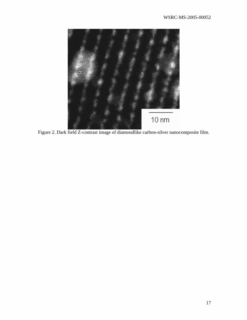
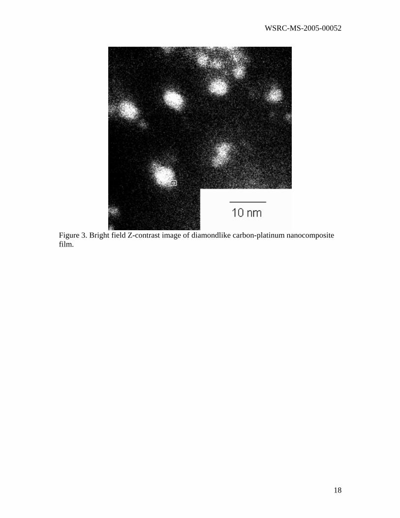
169:1. Dark field Z-contrast images of diamondlike carbon-silver and diamondlike
carbon-platinum nanocomposite films are shown in Figures 2 and 3, respectively. The
bright regions correspond to the higher atomic number metal regions, and the dark
regions correspond to the DLC matrix. The noble metals are dispersed as nearly spherical
metal nanoparticles in arrays within the diamondlike carbon matrix. The average size of
these nanocrystalline particles varies between 3 and 5 nm.
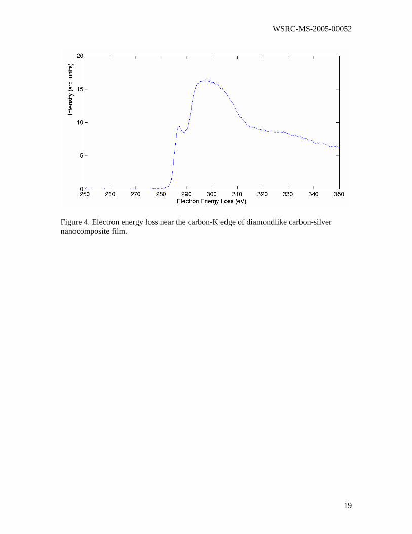
Electron energy loss spectra between 280 to 310 eV were acquired. The sp3
fraction was determined from the K edge loss spectra using an empirical technique
developed by Cuomo et al. [14]. In this technique, the peak in the region from 285 to 290
eV results from excitation of electrons from the 1s ground state to the vacant π*
6

WSRC-MS-2005-00052
antibonding state. The peak in the region above 290 eV results from excitation to the
higher σ* state. The ratio of the integrated areas under these two energy windows is
approximately proportional to the relative number of π and σ* orbitals. Using this
information, the atomic fraction of sp2 bonded carbon (x) can be determined using the
expression:
(I(π)/I(σ))s/(I(π)/I(σ))r=3x/(4-x) [2]
in which I(π) is the intensity in the range from 284 to 289 eV and I(σ) is the integrated
intensity in the range from 290 to 305 eV. The subscripts s and r refer to the ratio
determined for the DLC specimen and a reference material with 100% sp2 bonding,
respectively. The sp3 content was determined to be 63% for a diamondlike carbon film
on silicon (100). The sp3 content was determined to be 47% for a diamondlike carbon-
silver nanocomposite film on Si (100) (Figure 4). This data suggests that a moderate
reduction in sp3 content occurs in the metal alloyed films.
Raman spectroscopy
Adhesion of diamondlike carbon thin films is dependent on several factors,
including film stress, film/substrate chemical bonding, and substrate topology [15-17].
Large internal compressive stresses as high as 10 GPa have been observed in
diamondlike carbon thin films, regardless of the deposition process used. These stresses
limit maximum film DLC thickness to 0.1–0.2 micrometers, and prevent widespread
medical use. Lifshitz et al. have attributed these stresses to “subplantation” (low energy
7
WSRC-MS-2005-00052
subsurface implantation) of carbon ions during diamondlike carbon film growth [18-19].
They suggest that carbon ions with energies between 10-1000 eV undergo shallow
implantation to depths of 1-10 nm of during film growth. Carbon species are trapped in
subsurface sites due to restricted mobility. This process leads to the development of very
large internal compressive stresses.
The Raman spectra of diamondlike carbon thin films contain characteristic peaks
that reflect carbon bonding and internal stress. All of the spectra show the following: (1)
a broad hump centered in the 1510-1557 cm-1 region, which is known as the G- band, and
(2) a small shoulder at 1350 cm-1, which is known as the D- band. The G-band is the
optically allowed E2g zone center mode of crystalline graphite, and is typically observed
in diamondlike carbon films. The D-band is the A1g mode of graphite.
High quality diamondlike carbon films demonstrate the following: (1) a relatively
symmetrical G-band, and (2) a lesser D-band, suggesting an absence or a low amount of
graphite clusters. The presence of metal atoms leads to greater asymmetry in the G-peak
and a slight increase in the height of the D-peak (Figure 5). These features may be
produced by a decrease in sp3 content, an increase in graphitic cluster size within the
film, and/or a decrease in compressive stress within the film [20, 21]. The silver and
platinum nanoparticles have significantly smaller elastic moduli than the diamondlike
carbon matrix; the elastic moduli of silver and platinum are 83 GPa and 168 GPa,
respectively. These nanoparticles may be expected to absorb stresses from the
diamondlike carbon matrix.
Nanoindentation
8
WSRC-MS-2005-00052
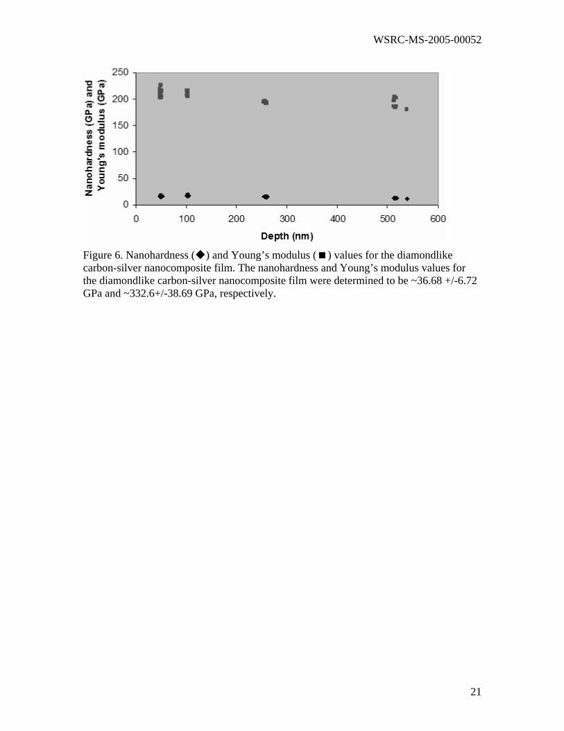
During nanoindentation, the modulus of the coated sample approached that of the
uncoated sample at roughly 500 nm (~2/3 of the film thickness). Substrate effects were
observed at indentation depths of 100 nm. The nanohardness and Young’s modulus
values for the diamondlike carbon-silver nanocomposite film were determined to be
36.68 +/-6.72 GPa and 332.6+/-38.69 GPa, respectively (Figure 6). These values are
similar to those observed in layered WC/DLC and TiC/DLC composites prepared using
electron cyclotron resonance chemical vapor deposition or magnetron sputtering
techniques (~27 GPa), and are significantly greater than those observed in a-C:H-copper
coatings prepared using plasma-enhanced chemical vacuum deposition (PECVD) or
hybrid microwave plasma-assisted chemical vapor deposition/sputtering techniques (~10
GPa) [22-26].
Biofilm Attachment Studies
The introduction of implantable medical devices into the body has been shown to
greatly increase the risk of infection. Infections involving artificial organs, synthetic
vessels, joint replacements, or internal fixation devices usually require reoperation. Some
infections are more serious than others; infected cardiac, abdominal, and extremity
vascular prostheses result in amputation or death [27-28].
Bacteria form a glycocalyx, an adherent coating that forms on all foreign
materials placed within the body [29-31]. This 5-50 µm thick glycoprotein-based coating
protects bacteria through a diffusion limitation process, and serves to decrease their
antibiotic sensitivity by 10 to 100 times. In addition, the constituents of many alloys and
polymers can inhibit both macrophage chemotaxis and phagocytosis. Finally, tissue
9
WSRC-MS-2005-00052
damage caused by surgery and foreign body implantation further increases the
susceptibility to infection.
The sustained delivery of silver ions into the local implant micro-environment
simultaneously exceeds usual systemic concentrations by several orders of magnitude and
avoids systemic side effects [32-34]. Silver nanoparticles have been shown to possess an
unsurpassed antimicrobial spectrum, with efficacy against 150 different pathogens.
Silver ions bind strongly to electron donor groups on sulfur-, oxygen- or nitrogen-
containing enzymes. These ions displace other cations (e.g., Ca2+) important for enzyme
function. In addition, nanocrystalline silver also provides broad-spectrum fungicidal
action. Very low silver ion concentrations required for microbicidal activity (in the range
10-9 mol/l). Films containing both silver and platinum may demonstrate enhanced
antimicrobial activity due to formation of a galvanic couple that accelerates silver ion
release [35].
The results of the biofilm attachment study showed a significant difference in the
amount of microbial colonization per unit area between uncoated silicon (100) substrates
and DLC-silver and DLC-silver-platinum nanocomposite films. Microbial colonization
varied with organism type, incubation period, and composite film type. The gram
negative bacteria Pseudomonas aeruginosa produced well developed biofilms after 48
hours that were difficult to quantify. The eukaryote, Candida wild type, also produced
sporadic biofilms at all incubation periods that were difficult to quantify. On the other
hand, the gram positive Staphylococcus sp also produced a quantifiable biofilm after 24,
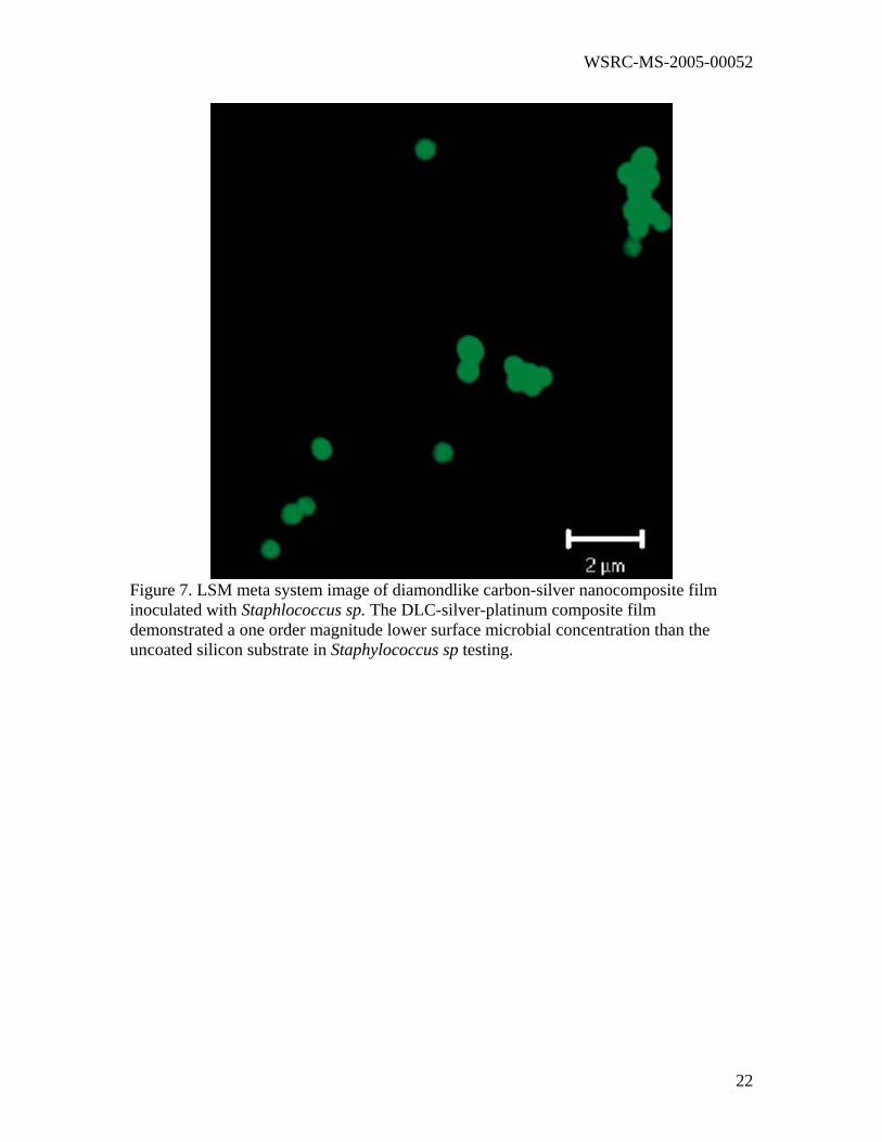
48, and 72 hours. Using rich media and long incubation times, impacted biofilm
formation on the DLC-metal composite films and control silicon wafers was observed
10
WSRC-MS-2005-00052
(Figure 7). The DLC-silver-platinum nanocomposite film demonstrated a one order
magnitude lower surface microbial density in gram positive bacteria concentration than
the uncoated silicon substrate. Samples showed a difference of between 100 percent to
half an order of magnitude lower colonization rates on the DLC-metal nanocomposite
films than on the uncoated silicon substrates.
Conclusions
Diamondlike carbon-silver and diamondlike carbon-silver-platinum
nanocomposite films were prepared using a novel multicomponent target pulsed laser
deposition process. Silver and platinum do not chemically bond with carbon; instead,
these metals form nanoparticle arrays within the diamondlike carbon matrix. This self-
assembled morphology can be attributed to the high surface energy of noble metals
relative to carbon. Ostwald ripening is prevented, and the resulting metal nanoparticles
possess uniform size. Raman spectroscopy data suggest that DLC-metal composite films
contain lower amounts of internal compressive stress than unalloyed diamondlike carbon
films. Silver and platinum exhibit significantly smaller elastic moduli than diamondlike
carbon, and may absorb compressive stress from the diamondlike carbon matrix.
Nanoindentation testing of the DLC-metal nanocomposite films demonstrates that these
films possess hardness and Young’s modulus values as high as 37 GPa and 333 GPa,
respectively. Finally, diamondlike carbon-silver-platinum nanocomposites exhibit
antimicrobial efficacy against Staphylococcus bacteria. It is believed that platinum, which
is more noble than silver, forms a galvanic couple inside the in vitro biofilm testing
environment. Silver release (and, by extension, antimicrobial function) is increased in the
11

WSRC-MS-2005-00052
diamondlike carbon-silver-platinum nanocomposite films. Diamondlike carbon-
biofunctional metal nanocomposite films have a variety of potential medical and
antimicrobial applications, including use in orthopaedic, ophthalmic, neurologic, and
cardiovascular devices.
12

WSRC-MS-2005-00052
References [1] J. Robertson, Diamond-like amorphous carbon, Mat. Sci. and Eng. R, Vol 37 (No. 4-6), 2002, p 129-281
[2] M. Allen, B. Myer, and N. Rushton, J. Biomed. Mater. Res., Vol 58 (No. 3), 2001, p 318-321 [3] S. Linder, W. Pinkowski, and M. Aepfelbacher, Adhesion, cytoskeletal architecture and activation status of primary human macrophages on a diamond-like carbon coated surface, Biomaterials, Vol 23 (No. 3), 2002, p 767-773 [4] E. Liu, B. Blanpain, J. P. Celis, J. R. Roos, G. Alvarez Verven, and T. Priem, Tribological behaviour and internal stress of diamond coating deposited with a stationary dc plasma jet, Surface Coatings Technol., Vol 80 (No. 3), 1996, p 264-270 [5] L. A. Thomson, F. C. Law, N. Rushton, and J. Franks, Biocompatibility of Diamond-Like Carbon Coating, Biomaterials, Vol 12 (No. 1), 1991, p 37-40 [6] M. Allen, B. Myer, and N. Rushton, In vitro and in vivo investigations into the biocompatibility of diamond-like carbon (DLC) coatings for orthopedic applications, J. Biomed. Mater. Res., Vol 58 (No. 3), 2001, p 319-328 [7] M. J. Ignatius, N. Sawhney, A. Gupta, B. M. Thibadeau, O. R. Monteiro, and I. G. Brown, Bioactive surface coatings for nanoscale instruments: Effects on CNS neurons, J. Biomed. Mater. Res., Vol 40 (No. 2), 1998, p 264-274. [8] M. B. Guseva, V. G. Babaev, V. V. Khvostov, Z. K. Valioullova, A. Y. Bregadze, A. N. Obraztsov, and A. E. Alexenko, Deposition of Thin Highly Dispersive Diamond Films by Laser-Ablation, Diamond Related Mater., Vol 3 (No. 4-6), 1994, p 328-331 [9] N. Kikuchi, Y. Ohsawa, and I. Suzuki, Diamond Synthesis by CO2-Laser Irradiation, Diamond Related Mater., Vol 2 (No. 2-4), 1993, p 190-196 [10] A. A. Voevodin, S. J. P. Laube, S. D. Walck, J. S. Solomon, M. S. Donley, and J. S. Zabinski, Pulsed-Laser Deposition of Diamond-Like Amorphous-Carbon Films from Graphite and Polycarbonate Targets, J. Appl. Phys., Vol 78 (No. 6), 1995, p 4123-4130 [11] A. A. Voevodin and M. S. Donley, Preparation of amorphous diamond-like carbon by pulsed laser deposition: A critical review, Surface Coatings Technol., Vol 82 (No. 3), 1996, p 199-213 [12] A. A. Voevodin, M. S. Donley, J. S. Zabinski, and J. E. Bultman, Mechanical and tribological properties of diamond-like carbon coatings prepared by pulsed laser deposition, Surface Coatings Technol., Vol 77 (No. 1-3), 1995, p 534-539
13
WSRC-MS-2005-00052
[13] S. Lopatin, S. J. Pennycook, J. Narayan, and G. Duscher, Z-contrast imaging of dislocation cores at the GaAs/Si interface, Appl. Phys Lett., Vol 81 (No. 15), 2002, p 2728-2730 [14] J. Bruley, D. B. Williams, J. J. Cuomo, and D. P. Pappas, Quantitative Near-Edge Structure-Analysis of Diamond-Like Carbon in the Electron-Microscope Using a 2-Window Method, J. Microscopy (Oxford) Vol 180 (No. 1), 1995, p 22-32 [15] E. H. A. Dekempeneer, R. Jacobs, J. Smeets, J. Meneve, L. Eersels, B. Blanpain, J. Roos, D. J. Oostra, RF Plasma-Assisted Chemical Vapor-Deposition of Diamond-Like Carbon- Physical and Mechanical-Properties, Thin Solid Films, Vol 217 (No. 1-2), 1992, p 56-61 [16] K. Bewilogua, D. Dietrich, G. Holzhuter, and C. Weissmantel, Structure of Amorphous-Carbon Films, Phys. Stat. Sol. A, Vol 71 (No. 1), 1982, p 57-59 [17] D. G. McCulloch, D. R. McKenzie, and C. M. Goringe, Ab initio simulations of the structure of amorphous carbon, Phys. Rev. B, Vol 61 (No. 3), 2000, p 2349-2355 [18] Y. Lifshitz, Hydrogen-free amorphous carbon films: Correlation between growth conditions and properties, Diamond Related Mater., Vol 5 (No. 3-5), 1996, p 388-400 [19] Y. Lifshitz, G. D. Lempert, E. Grossman, I. Avigal, C. Uzansaguy, R. Kalish, J. Kulik, D. Marton, and J. W. Rabalais, Growth Mechanisms of DLC Films from C+ Ions- Experimental Studies, Diamond Related Mater., Vol 4 (No. 4), 1995, p 318-323 [20] B. K. Tay and P. Zhang, On the properties of nanocomposite amorphous carbon films prepared by off-plane double bend filtered cathodic vacuum arc, Thin Solid Films, Vol 420, 2002, p 177-184 [21] V. V. Uglova, V. M. Anishchik, Y. Pauleau, A. K. Kuleshov, F. Thièry, J. Pelletier, S. N. Dub, and D. P. Rusalsky, Relations between deposition conditions, microstructure and mechanical properties of amorphous carbon-metal films, Vacuum, Vol 70 (No. 2-3), 2003, p 181-185 [22] Y. Pauleau and F. Thièry, Deposition and characterization of nanostructured metal/carbon composite films, Surface Coatings Technol. Vol 180-181, 2004, p 313-322 [23] D. Sheeja, B. K. Tay, J. Y. Sze, L. J. Yu, and S. P. Lau, A comparative study between pure and films prepared by FCVA technique biasing Al-containing amorphous carbon with high substrate pulse, Diamond Related Mater., Vol 12 (No. 10-11), 2003, p 2032-2036 [24] H. Rusli, S. F. Yoon, Q. F. Huang, J. Ahn, Q. Zhang, H. Yang, Y. S. Wu, E. J. Teo, T. Osipowicz, and F. Watt, Metal-containing amorphous carbon film development using
14
WSRC-MS-2005-00052
electron cyclotron resonance CVD, Diamond Related Mater., Vol 10 (No. 2), 2001, p 132-138 [25] Rusli, S. F. Yoon, H. Yang, J. Ahn, Q. F. Huang, Q. Zhang, Y. P. Guo, C. Y. Yang, E. J. Yeo, A. T. S. Wee, A. C. H. Huan, and F. Watt, Tungsten-carbon thin films deposited using screen grid technique in an electron cyclotron resonance chemical vapour deposition system, Surface Coatings Technol., Vol 123 (No. 2-3), 1999, p 134-139 [26] C. Strondl, N. M. Carvalho, J. T. M. De Hosson, and G. J. van der Kolk, Investigation on the formation of tungsten carbide in tungsten-containing diamond like carbon coatings, Surface Coatings Technol., Vol 162 (No. 2-3), 2003, p 288-293 [27] G. Printzen, Relevance, pathogenicity and virulence of microorganisms in implant related infections, Injury-Inter. J. Care Injured, Vol 27 (No. 3), 1996, p 9-15 [28] Z. U. Isiklar, G. C. Landon, and H. S. Tullos, Amputation After Failed Total Knee Arthroplasty, Clin. Orthop. Related Res., Vol 299, 1994, p 173-178 [29] K. Merritt, A. Gaind, and J. M. Anderson, Detection of bacterial adherence on biomedical polymers, J. Biomed. Mater. Res., Vol 39 (No. 3), 1998, p 415-422 [30] C. C. Chang and K. Merritt, Microbial Adherence On Poly(Methyl Methacrylate) Surfaces, J. Biomed. Mater. Res., Vol 26 (No. 2), 1992, p 197-207 [31] K. K. Jefferson, What drives bacteria to produce a biofilm?, FEMS Microbiology Letters, Vol 236 (No. 2), 2004, p 163-173. [32] R. O. Darouiche, Anti-infective efficacy of silver-coated medical prostheses, Clinical Infectious Diseases, Vol 29 (No. 6), 1999, p 1371-1377 [33] D. J. Stickler, Biomaterials to prevent nosocomial infections: is silver the gold standard?, Curr. Opinion Infectious Diseases, Vol 13 (No. 4), 2000, p 389-393 [34] K. S. Oh, S. H. Park, and Y. K. Jeong, Antimicrobial effects of Ag doped hydroxyapatite synthesized from co-precipitation route, Key Engineering Materials, Vol 264-268 (No. 1-3), 2004, p 2111-2114 [35] D. P. Dowling, A. J. Betts, C. Pope, M. L. McConnell, R. Eloy, M. N. Arnaud, Anti-bacterial silver coatings exhibiting enhanced activity through the addition of platinum, Surface Coatings Technol., Vol 163, 2003, p 637-640
15
WSRC-MS-2005-00052
Figure 1. Schematic of target configuration used in this study.
16
WSRC-MS-2005-00052
Figure 2. Dark field Z-contrast image of diamondlike carbon-silver nanocomposite film.
17
WSRC-MS-2005-00052
Figure 3. Bright field Z-contrast image of diamondlike carbon-platinum nanocomposite film.
18
WSRC-MS-2005-00052
Figure 4. Electron energy loss near the carbon-K edge of diamondlike carbon-silver nanocomposite film.
19

WSRC-MS-2005-00052
Figure 5. Visible Raman spectrum of diamondlike carbon-silver nanocomposite film.
20
WSRC-MS-2005-00052
Figure 6. Nanohardness ( ) and Young’s modulus ( ) values for the diamondlike carbon-silver nanocomposite film. The nanohardness and Young’s modulus values for the diamondlike carbon-silver nanocomposite film were determined to be ~36.68 +/-6.72 GPa and ~332.6+/-38.69 GPa, respectively.
21
WSRC-MS-2005-00052
Figure 7. LSM meta system image of diamondlike carbon-silver nanocomposite film inoculated with Staphlococcus sp. The DLC-silver-platinum composite film demonstrated a one order magnitude lower surface microbial concentration than the uncoated silicon substrate in Staphylococcus sp testing.
22