Platinum-DNA Interactions and Subsequent Cellular Processes Controlling Sensitivity to Anticancer...
24
REVIEW Platinum – DNA Interactions and Subsequent Cellular Processes Controlling Sensitivity to Anticancer Platinum Complexes by Saeed Ahmad Departmentof Chemistry, University of Engineering and Technology, Lahore 54890, Pakistan (phone: þ 92-333-5248570; e-mail: [email protected]) Platinum-based compounds are widely used as chemotherapeutics for the treatment of a variety of cancers. The anticancer activity of cisplatin and other platinum drugs is believed to arise from their interaction with DNA. Several cellular pathways are activated in response to this interaction, which include recognition by high-mobility group and repair proteins, translesion synthesis by polymerases, and induction of apoptosis. The apoptotic process is regulated by activation of caspases, p53 gene, and several proapoptotic and antiapoptotic proteins. Such cellular processing eventually leads to an inhibition of the replication or transcription machinery of the cell. Deactivation of platinum drugs by thiols, increased nucleotide excision repair of Pt–DNA adducts, decreased mismatch repair, and defective apoptosis result in resistance to platinum therapy. The differences in cytotoxicity of various platinum complexes are attributed to the differential recognition of their adducts by cellular proteins. Cisplatin and oxaliplatin both produce mainly 1,2-GG intrastrand cross-links as major adducts, but oxaliplatin is found to be more active particularly against cisplatin-resistant tumor cells. Mismatch repair and replicative bypass appear to be the processes most likely involved in differentiating the molecular responses to these two agents. This review describes the formation of Pt–DNA adducts, their interaction with cellular components, and biological effects of this interaction. Contents 1. Introduction 2. Nature of Platinum – DNA Adducts 3. Recognition of Platinated Adducts by Cellular Proteins 3.1. HMG Box Proteins 3.2. Nucleotide-Excision Repair 3.3. Mismatch-Repair Proteins 3.4. DNA Polymerases 4. Programmed Cell Death 4.1. Apoptotic Pathways and Caspase Activation 4.2. Role of p53 Protein 5. Conclusions 1. Introduction. – Platinum-based drugs are among the most active anticancer agents and have been widely used in the treatment of a variety of cancers [1 – 11]. The structures of Pt complexes effective as anticancer agents are shown in Fig. 1. Cisplatin and carboplatin are known to be particularly effective against solid tumor types such as CHEMISTRY & BIODIVERSITY – Vol. 7 (2010) 543 # 2010 Verlag Helvetica Chimica Acta AG, Zɒrich
Transcript of Platinum-DNA Interactions and Subsequent Cellular Processes Controlling Sensitivity to Anticancer...
REVIEW
Platinum– DNA Interactions and Subsequent Cellular Processes ControllingSensitivity to Anticancer Platinum Complexes
by Saeed Ahmad
Department of Chemistry, University of Engineering and Technology, Lahore 54890, Pakistan(phone: þ92-333-5248570; e-mail: [email protected])
Platinum-based compounds are widely used as chemotherapeutics for the treatment of a variety ofcancers. The anticancer activity of cisplatin and other platinum drugs is believed to arise from theirinteraction with DNA. Several cellular pathways are activated in response to this interaction, whichinclude recognition by high-mobility group and repair proteins, translesion synthesis by polymerases, andinduction of apoptosis. The apoptotic process is regulated by activation of caspases, p53 gene, and severalproapoptotic and antiapoptotic proteins. Such cellular processing eventually leads to an inhibition of thereplication or transcription machinery of the cell. Deactivation of platinum drugs by thiols, increasednucleotide excision repair of Pt–DNA adducts, decreased mismatch repair, and defective apoptosisresult in resistance to platinum therapy. The differences in cytotoxicity of various platinum complexes areattributed to the differential recognition of their adducts by cellular proteins. Cisplatin and oxaliplatinboth produce mainly 1,2-GG intrastrand cross-links as major adducts, but oxaliplatin is found to be moreactive particularly against cisplatin-resistant tumor cells. Mismatch repair and replicative bypass appearto be the processes most likely involved in differentiating the molecular responses to these two agents.This review describes the formation of Pt–DNA adducts, their interaction with cellular components, andbiological effects of this interaction.
Contents
1. Introduction2. Nature of Platinum– DNA Adducts3. Recognition of Platinated Adducts by Cellular Proteins
3.1. HMG Box Proteins3.2. Nucleotide-Excision Repair3.3. Mismatch-Repair Proteins3.4. DNA Polymerases
4. Programmed Cell Death4.1. Apoptotic Pathways and Caspase Activation4.2. Role of p53 Protein
5. Conclusions
1. Introduction. – Platinum-based drugs are among the most active anticanceragents and have been widely used in the treatment of a variety of cancers [1 –11]. Thestructures of Pt complexes effective as anticancer agents are shown in Fig. 1. Cisplatinand carboplatin are known to be particularly effective against solid tumor types such as
CHEMISTRY & BIODIVERSITY – Vol. 7 (2010) 543
� 2010 Verlag Helvetica Chimica Acta AG, Z�rich
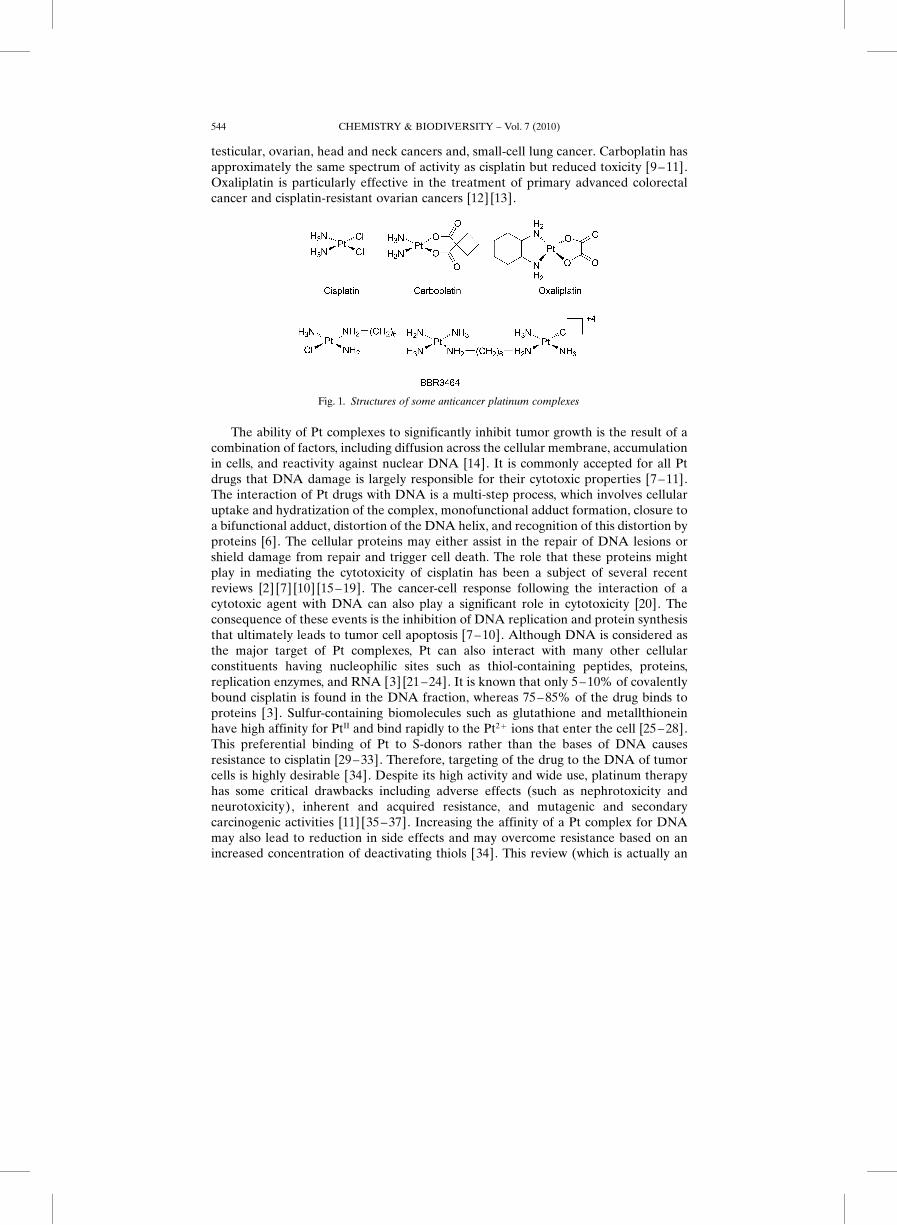
testicular, ovarian, head and neck cancers and, small-cell lung cancer. Carboplatin hasapproximately the same spectrum of activity as cisplatin but reduced toxicity [9 – 11].Oxaliplatin is particularly effective in the treatment of primary advanced colorectalcancer and cisplatin-resistant ovarian cancers [12] [13].
The ability of Pt complexes to significantly inhibit tumor growth is the result of acombination of factors, including diffusion across the cellular membrane, accumulationin cells, and reactivity against nuclear DNA [14]. It is commonly accepted for all Ptdrugs that DNA damage is largely responsible for their cytotoxic properties [7 – 11].The interaction of Pt drugs with DNA is a multi-step process, which involves cellularuptake and hydratization of the complex, monofunctional adduct formation, closure toa bifunctional adduct, distortion of the DNA helix, and recognition of this distortion byproteins [6]. The cellular proteins may either assist in the repair of DNA lesions orshield damage from repair and trigger cell death. The role that these proteins mightplay in mediating the cytotoxicity of cisplatin has been a subject of several recentreviews [2] [7] [10] [15 –19]. The cancer-cell response following the interaction of acytotoxic agent with DNA can also play a significant role in cytotoxicity [20]. Theconsequence of these events is the inhibition of DNA replication and protein synthesisthat ultimately leads to tumor cell apoptosis [7 – 10]. Although DNA is considered asthe major target of Pt complexes, Pt can also interact with many other cellularconstituents having nucleophilic sites such as thiol-containing peptides, proteins,replication enzymes, and RNA [3] [21 –24]. It is known that only 5 – 10% of covalentlybound cisplatin is found in the DNA fraction, whereas 75– 85% of the drug binds toproteins [3]. Sulfur-containing biomolecules such as glutathione and metallthioneinhave high affinity for PtII and bind rapidly to the Pt2þ ions that enter the cell [25 – 28].This preferential binding of Pt to S-donors rather than the bases of DNA causesresistance to cisplatin [29 – 33]. Therefore, targeting of the drug to the DNA of tumorcells is highly desirable [34]. Despite its high activity and wide use, platinum therapyhas some critical drawbacks including adverse effects (such as nephrotoxicity andneurotoxicity), inherent and acquired resistance, and mutagenic and secondarycarcinogenic activities [11] [35 – 37]. Increasing the affinity of a Pt complex for DNAmay also lead to reduction in side effects and may overcome resistance based on anincreased concentration of deactivating thiols [34]. This review (which is actually an
Fig. 1. Structures of some anticancer platinum complexes
CHEMISTRY & BIODIVERSITY – Vol. 7 (2010)544
extension of our previous review [25]) presents an overview of the current under-standing of several aspects of Pt binding to DNA and the cellular processes followingthis interaction that ultimately lead to cell death.
2. Nature of Platinum – DNA Adducts. – The formation of DNA adducts is anessential step for inducing the anticancer activity by Pt compounds [1 –10]. Platinumcoordination to DNA takes place at N(7) on guanine and adenine nucleotides [2] [38].However, PtII is known to have strong kinetic preference for binding to guanine overadenine, and, consequently, the initial attack of adenine bases of DNA on Pt is a rareevent [39] [40]. In the second step, both the Pt(GG) and Pt(AG) units are produced inthe approximate ratio of (2.5 – 4) : 1 [25] [41]. The binding of cisplatin to DNA isirreversible and is, therefore, a kinetically rather than thermodynamically controlledprocess [42] [43]. The purine bases do not replace Cl� from PtII directly butpredominantly via a solvent-assisted mechanism [39] [44]. Hydratization is usuallythe rate-determining step in the reaction of PtII with DNA [45]. Cisplatin undergoesaquation to give mainly the monoaqua complex, cis-[Pt(NH3)2Cl(H2O)]1þ and, to aless extent, the diaqua species, cis-[Pt(NH3)2(H2O)2]2þ at 310 K [39] [46]. The H2Omolecule of the monoaquated species ([Pt(NH3)2Cl(H2O)]1þ) is far more easilysubstituted than Cl� by the N(7)-atoms of guanine or adenine forming a monofunc-tional adduct [42] [47] [48]. The monofunctional adducts subsequently react with asecond nucleophile forming primarily intrastrand adducts [41] [49] [50]. Substitution atthe amine N-atom inhibits the ring closure to the bifunctional adduct [51]. The bindingof trinuclear Pt complexes to DNA is considerably faster than the binding of dinuclearand mononuclear complexes such as cisplatin and carboplatin. Also among thetrinuclear complexes, BBR3464 (Fig. 1) binds faster (t1/2 40 min) than its analogue withcis-orientation of amine groups on the central Pt unit (t1/2 80 min). The change ingeometry on central coordination sphere also affects their potency; the cis-isomershows reduced cytotoxicity [52].
The rate of hydratization and DNA platination increases as a function oftemperature. Cisplatin shows the greatest enhancement in cell killing with heat amongPtII complexes. Cisplatin led to fivefold increase in DNA platination as the temperaturerose from 378 to 438, while, in carboplatin and oxaliplatin, DNA adduct formation wasonly approximately doubled by similar increase in temperature [14]. Increased drugaccumulation by hypothermia is one of the main mechanisms involved in increasedcisplatin toxicity and induced apoptosis in many tumor types [53].
Platinum complexes are capable of forming a number of structurally differentadducts with DNA, which include i) intrastrand cross-linking of two nucleobases ofsingle DNA strand, ii) interstrand cross-linking of two different strands of one DNAmolecule, iii) chelate formation through N- and O-atoms of one guanine, and iv)DNA– protein cross-links [54 – 58]. The major adduct formed between cisplatin andDNA is the bidentate 1,2-intrastrand cross-link, in which cis-[Pt(NH3)2]2þ undergoescross-linkage between two adjacent guanine N(7)-atoms [59] [60]. Cisplatin formsapproximately 65% 1,2-d(GpG), 25% 1,2-d(ApG), 5– 10% 1,3-d(GpNpG), and 1 – 3%interstrand and monofunctional adducts [19] [57] [61] [62]. Similar results are found forcarboplatin, oxaliplatin, and [Pt(en)Cl2] [41] [62 – 64]. The results of these studiesreflect that the 1,2-intrastrand cross-links are mainly responsible for the biological
CHEMISTRY & BIODIVERSITY – Vol. 7 (2010) 545
activity of Pt compounds. The view that 1,2-intrastrand cross-links are the main adductsresponsible for the antitumor activity is supported on the basis of two main points;high-mobility group box (HMGB) proteins specifically recognize this type of DNAadduct, and 1,2-intrastrand cross-links are less effectively removed from DNA by repairenzymes than 1,3-interstrand adducts. The lack of pharmacological activity oftransplatin was explained on the basis of these points; transplatin, due to steric reason,is unable to form 1,2-intrastrand cross-links between two adjacent purines in the sameDNA strand (it mainly forms 1,3-interstrand cross-links) [8]. However, the transplatinanalogues with planar amines exhibit cytotoxicity equivalent to cisplatin probably dueto formation of DNA –protein cross-links [65] [66]. Another potentially activecomplex, [Pt(hmp)Cl2] (hmp¼homopiperazine), interacts stereospecifically withDNA to form mostly bifunctional interstrand and substantially less intrastrand adducts.Greater steric factors of [Pt(hmp)Cl2] compared with cisplatin or [Pt(en)Cl2],inhibiting the formation of bifunctional intrastrand adducts, thought to be responsiblefor the cytotoxicity of Pt complexes. These facts suggest that formation of bifunctionaladducts may not be a precondition of PtII complexes to exhibit cytotoxicity againsttumor cells [67] [68].
Ternary DNA– Pt– Protein cross-links (DPCLs) have also been shown to play animportant role in cytotoxicity of cisplatin, but the frequency of these ternary complexesdepends on the cell type and the time of treatment. DPCLs are formed by thetransformation of DNA monofunctional and intrastrand cross-links of cisplatin. Incontrast, DNA interstrand cross-links of cisplatin as well as monofunctional adducts oftransplatin are markedly more stable in the presence of DNA-binding proteins thanintrastrand cross-links so that their transformation into DPCLs is markedly moredifficult. The DPCLs formed by cisplatin inhibit DNA polymerization or removal ofthese ternary lesions from DNA by the nucleotide excision repair system moreeffectively than plain DNA intrastrand or monofunctional adducts [69].
Oxaliplatin forms DNA adducts with a sequence and region specificity similar tothat of cisplatin. However, it is less reactive than cisplatin but more efficient atinhibiting DNA synthesis. The level of Pt-adduct formation induced by oxaliplatin inspecific regions of DNA from drug treated cancer cells is 2 – 6 times lower than that ofcisplatin, but it is as potent as cisplatin in growth inhibition of the same cell lines[62] [70] [71]. The lower level of adduct formation with oxaliplatin may not beattributed to the differential cellular uptake of the two drugs, because essentially thesame differences in DNA-lesion formation are seen, when intact nuclei are incubatedwith oxaliplatin and cisplatin. The differences are also unlikely to be attributable to aDACH carrier ligand (DACH¼1,2-diaminocyclohexane¼cyclohexane-1,2-diamine)because oxaliplatin also forms fewer adducts than equimolar concentration of[Pt(DACH)Cl2], a DACH analogue of oxaliplatin [16]. The lower levels of Pt-adductformation with oxaliplatin most likely reflect the slow dissociation rate of the oxalateligand under physiological conditions. It has been suggested that oxaliplatin cytotox-icity might possibly result from proportionately more highly lethal lesions such asinterstrand or DNA– protein cross-links [70], and from the bulkier and morehydrophobic adducts formed by oxaliplatin [12]. Recent investigations suggest thatoxaliplatin displays a disproportionately greater ability to induce secondary lesions inDNA that are precursors to massive apoptosis [72]. It has been assumed that apoptotic
CHEMISTRY & BIODIVERSITY – Vol. 7 (2010)546
stimuli are not limited to DNA damage, and targets other than DNA might play a rolein induction of apoptosis [72].
Several X-ray and NMR studies have been undertaken to examine the structures ofplatinated adducts [10] [25] [73 –78] including those of oxaliplatin [79] [80]. Oxaliplatinhas been shown to produce the same type of inter- and intrastranded cross-links as doescisplatin. In both cases, the Pt-atom forms a 1,2-intrastand cross-link between twoadjacent guanosine residues and bends the double helix by ca. 30– 508 towards themajor groove [76] [79]. The solution structure of the oxaliplatin – GG adduct differsfrom that of cisplatin – GG adducts in that: 1) there is a smaller dihedral angle (358 vs.478), 2) the minor groove is smaller, and 3) the crystal structure is less bent than thesolution structure of cisplatin – GG adducts (bending angle 358 vs. 788) [80].
3. Recognition of Platinated Adducts by Cellular Proteins. – It is known that evenhigh levels of DNA platination may not always induce cell death. The actual DNAdamage depends on the processing of platinated adducts by cellular components[7] [10] [15] [17] [19] [81]. The formation of 1,2-intrastrand cross-links bends the DNAduplex towards the major groove, exposing a wide shallow minor groove surface towhich several classes of protein bind [82] [83]. These proteins are called damagerecognition proteins and include HMGB proteins, repair proteins, transcription factors,and several other proteins and enzymes. Binding of these proteins result in either repairof the platinated DNA or in its stabilization which results in DNA damage. VariousDNA polymerases are capable of replicating past Pt – DNA adducts [2] [10] [19]. Thebehavior of these proteins towards Pt –DNA adducts is explained in the following.
3.1. HMGB Proteins. The HMGB proteins such as HMGB1, HMGB2, andtsHMGB bind to DNA –cisplatin adducts with high affinity and stabilize the bent andsupercoiled DNA [2] [10] [15] [19] [84] [85]. When these proteins bind to cisplatinintrastrand cross-links, they may be diverted from their natural target binding sites onthe genome and shield the Pt-DNA lesions from cellular repair mechanisms[7] [10] [86] [87]. This mechanism may increase the sensitivity of cancer cells tocisplatin and, therefore, contribute to the cytotoxicity of the drug. The binding ofrecognition proteins to cisplatin – DNA adducts has been postulated to inhibitnucleotide excision repair (NER) [2] [19] [88]. Deficiency in repair would allow theadducts to accumulate and block essential cellular pathways, such as transcription, andactivate programmed cell death [87].
HMGB Proteins contain one or more sequences of ca. 80 amino acids having atypical L-shaped structure [89 –91]. Their biological role is thought to include generegulation, transcription regulation, and maintenance of chromatin structure [91].HMGB Domain proteins can be divided into two subclasses: structure-specific andsequence-specific. The structure-specific HMGB proteins, e.g., HMGB1 and HMGB2,recognize DNA structures such as four-way junctions and supercoiled DNA (damagedby chemotherapeutic agents). They have multiple HMG domains and bind DNA withlittle or no specificity. The sequence-specific HMGB proteins recognize and bendspecific sequences of DNA, and include transcription factors such as lymphoidenhance-binding factors, LEF-1 and TCF-1, and the sex-determining factor SRY. Theyusually contain a single HMG domain [90– 92]. Both types of the recognition proteinshave been shown to bind to DNA primarily in the minor groove and bend the DNA in
CHEMISTRY & BIODIVERSITY – Vol. 7 (2010) 547
the direction of major groove [82] [83] [92]. The X-ray structure of cisplatin-modified16-mer duplex complexes with a high mobility protein HMGB1 reveals that the proteininteracts mainly with the minor groove of the DNA, which is widened and bent by 618towards the major groove [83]. Although cisplatin – GG adducts are typicallyrecognized by structure-specific proteins [83– 85], some sequence-specific HMGBproteins such as LEF-1 and SRY have also been reported to bind to cisplatin – GGadducts [90] [93]. A comparison of the binding affinities of the HMGB containingtranscription factor LEF-1 for the platinated and nonplatinated sequences shows 3– 6fold binding enhancement upon platination. However, the binding of LEF-1 to GG-platinated oligonucleotides was weaker than that to the native recognition sequence[90].
Several studies indicated that HMGB protein levels correlate with cisplatinsensitivity [2] [7] [10] [17] [19]. For example, overexpression of HMGB1 that is inducedby the addition of steroid hormone oestradiol sensitizes breast and ovarian cancer cellsto cisplatin treatment [2] [94]. However, the ability of HMGB1 to impact thecytotoxicity of cisplatin depends on the cell type and growth conditions [7]. The HMG-domain protein-mediated cytotoxicity of cisplatin may be explained as a result of therecognition of DNA– cisplatin adducts by tissue-specific HMGB proteins [17].Cisplatin is very effective in the treatment of testicular cancer, because several HMGBproteins are specifically expressed in the testis and could potentially contribute to thecisplatin sensitivity of testicular tumors. Testis-specific HMG-domain proteins bind tothe most abundant cisplatin –DNA adducts and protect them from nucleotide excisionrepair [10] [17]. The results of a study on recognition of 1,2-intrastrand cross-link ofcisplatin by the murine testis-specific HMG-domain protein (tsHMGB) show that thespecificity of tsHMGB for platinated DNA is much higher than that observed for otherHMG-domain proteins including Ixr1 and hUBF [95].
The HMG-domain proteins display a selective affinity for the Pt drugs and for Pt–DNA adducts. They bind to cisplatin and [Pt(en)Cl2] adducts, whereas they have noaffinity for the adducts of trans-DDP or [Pt(dien)Cl]þ . They also bind selectively to the1,2-d(GpG) and 1,2-d(ApG) adducts, but they do not recognize the 1,3-d(GpNpG)adducts of cisplatin [10] [84] [96]. A testis-specific HMG-domain protein, tsHMG,displays strong affinity and specificity towards 1,2-d(GpG) cisplatin – DNA adducts.This protein diminished the NER of cisplatin – 1,2-d(GpG) intrastrand cross-links,while the repair of cisplatin – 1,3-d(GpG) intrastrand cross-links was still active in thecell extracts. This observation suggested that there was a cellular component expressedin the testicular teratocarcinoma cells that specifically blocks repair of the majorcisplatin – DNA adducts, but that does not inhibit repair of the minor adducts [87]. Thespecific recognition of 1,2-intrastrand cross-links by HMGB proteins suggests that 1,2-adducts are mainly responsible for the antitumor activity of cisplatin.
The binding strengths of some major proteins that recognize platinated DNA, interms of dissociation constant, are given in the Table. The binding selectivity of HMGBproteins can vary with varying length, and sequence of DNA and proteins. Acomparison of the binding properties of full-length hSRY protein and its HMG-domainregion with the sequence AACAAAG and the 1,2-GpG – cisplatin adduct (CP – GG)demonstrate that the full-length hSRY has an eightfold higher affinity for the sequence(KD¼15– 50 nm) than for a cisplatin adduct (KD¼115– 120 nm). On the other hand,
CHEMISTRY & BIODIVERSITY – Vol. 7 (2010)548
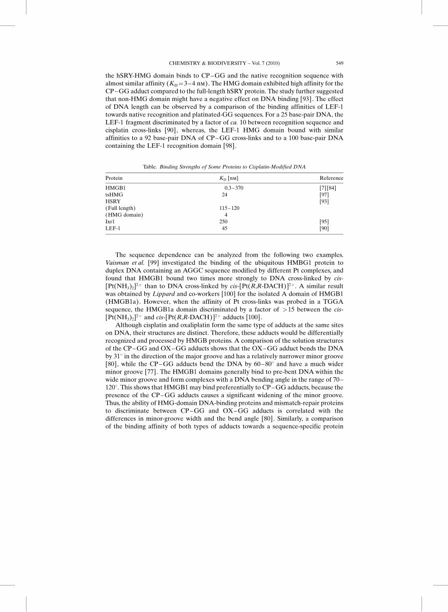
the hSRY-HMG domain binds to CP –GG and the native recognition sequence withalmost similar affinity (KD¼3 – 4 nm). The HMG domain exhibited high affinity for theCP – GG adduct compared to the full-length hSRY protein. The study further suggestedthat non-HMG domain might have a negative effect on DNA binding [93]. The effectof DNA length can be observed by a comparison of the binding affinities of LEF-1towards native recognition and platinated-GG sequences. For a 25 base-pair DNA, theLEF-1 fragment discriminated by a factor of ca. 10 between recognition sequence andcisplatin cross-links [90], whereas, the LEF-1 HMG domain bound with similaraffinities to a 92 base-pair DNA of CP– GG cross-links and to a 100 base-pair DNAcontaining the LEF-1 recognition domain [98].
The sequence dependence can be analyzed from the following two examples.Vaisman et al. [99] investigated the binding of the ubiquitous HMBG1 protein toduplex DNA containing an AGGC sequence modified by different Pt complexes, andfound that HMGB1 bound two times more strongly to DNA cross-linked by cis-[Pt(NH3)2]2þ than to DNA cross-linked by cis-[Pt(R,R-DACH)]2þ . A similar resultwas obtained by Lippard and co-workers [100] for the isolated A domain of HMGB1(HMGB1a). However, when the affinity of Pt cross-links was probed in a TGGAsequence, the HMGB1a domain discriminated by a factor of >15 between the cis-[Pt(NH3)2]2þ and cis-[Pt(R,R-DACH)]2þ adducts [100].
Although cisplatin and oxaliplatin form the same type of adducts at the same siteson DNA, their structures are distinct. Therefore, these adducts would be differentiallyrecognized and processed by HMGB proteins. A comparison of the solution structuresof the CP – GG and OX– GG adducts shows that the OX – GG adduct bends the DNAby 318 in the direction of the major groove and has a relatively narrower minor groove[80], while the CP – GG adducts bend the DNA by 60– 808 and have a much widerminor groove [77]. The HMGB1 domains generally bind to pre-bent DNA within thewide minor groove and form complexes with a DNA bending angle in the range of 70–1208. This shows that HMGB1 may bind preferentially to CP – GG adducts, because thepresence of the CP –GG adducts causes a significant widening of the minor groove.Thus, the ability of HMG-domain DNA-binding proteins and mismatch-repair proteinsto discriminate between CP– GG and OX –GG adducts is correlated with thedifferences in minor-groove width and the bend angle [80]. Similarly, a comparisonof the binding affinity of both types of adducts towards a sequence-specific protein
Table. Binding Strengths of Some Proteins to Cisplatin-Modified DNA
Protein KD [nm] Reference
HMGB1 0.3–370 [7] [84]tsHMG 24 [97]HSRY(Full length)(HMG domain)
115–1204
[93]
Ixr1 250 [95]LEF-1 45 [90]
CHEMISTRY & BIODIVERSITY – Vol. 7 (2010) 549
domain, LEF-1-HMG121, showed that the OX– GG cross-links did not enhance theLEF-1 binding to AGGT sequence significantly as did CP – GG adducts [90]. Thisdifferential recognition of cisplatin – GG and oxaliplatin – GG adducts is thought tocontribute to the differences in cytotoxicity and tumor range of the two drugs [19].Spectator ligands can interfere with recognition, either directly by contacts with theprotein or indirectly by affecting the cross-link geometry and flexibility [90] [101].
HMGB Proteins also recognize differently the adducts formed by the enantiomersof the same Pt complex [101]; for example, Malina et al. [102] investigated the bindingof the two domains of HMGB1 protein to cis-[Pt(NH3)2]2þ, [Pt(R,R-DAB)]2þ , and[Pt(S,S-DAB)]2þ adducts (DAB¼2,3-diaminobutane) formed at TGGT sequence of aDNA duplex. They found that the A domain bound two times more tightly to the[Pt(R,R-DAB)]2þ adduct than to the cisplatin adduct, whereas the [Pt(S,S-DAB)]2þ
adduct was recognized ca. eight times less strongly than the cisplatin adduct [102]. Thisdifferential recognition of HMGB proteins by the GG adducts formed by enantiomersof the same Pt complex could originate from the structural differences between theadducts [101].
Polynuclear Pt compounds act by a different mechanism than cisplatin and itsanalogues. The antitumor activity of BBR3464 does not involve recognition of itsintrastrand cross-links by HMGB proteins as a crucial step. However, effective removalof these adducts from DNA has been observed by nucleotide excision repair. Aninteresting feature of the repair assay is that a greater range of excised fragments is seenfor the conformationally flexible adducts produced by BBR3464 than the stericallyrigid adducts produced by cisplatin [103] [104]. Active trans-complexes such as trans-[PtCl2(NH3)(piperidine)] also do not involve recognition of DNA adducts by HMGBproteins as a crucial step [105].
3.2. Nucleotide-Excision Repair. Cisplatin – DNA adducts may be repaired byseveral types of proteins; the most important are nucleotide excision repair (NER)proteins [2] [3] [8] [10] [106]. NER is an ATP-dependent multiprotein complex thatrecognizes the kink induced on DNA by 1,2-intrastrand cross-links and subsequentlyexcises the segment of DNA that includes the kink as a 27– 29-base pair oligonucleo-tide. The gap that remains is then filled by DNA polymerases [3] [107]. ERCC1, XPF,and XPA are the NER proteins that are important in the removal of lesions producedby chemotherapeutic drugs on DNA [108– 110]. Several transcriptional repressors andactivators may be involved in the up-regulation of these genes; for example, MZF1protein possesses a repressively regulatory function in ERCC1 gene. Overexpression ofERCC1 through promoter-mediating transcriptional regulation is associated withrepair of cisplatin-induced DNA damage and clinical resistance to Pt chemotherapy[111].
The molecular mechanism by which NER removes Pt intrastrand cross-links fromDNA has been explained in [7]. Nucleotide excision repair is an important anduniversal pathway that removes a broad spectrum of DNA damages [112]. Severalreports describe that intrastrand cross-links of cisplatin are removed from DNA duringNER reactions, and that NER is also a major mechanism contributing to cisplatinresistance [87] [88] [106]. Increased NER in cisplatin-resistant cell lines has been shownto occur both for intrastrand as well as interstrand cisplatin – DNA adducts [3].Efficient repair of 1,2-d(GpG) and 1,3-d(GpNpG) intrastrand cross-links of cisplatin
CHEMISTRY & BIODIVERSITY – Vol. 7 (2010)550

has been reported by various NER systems including human and rodent excinucleases[113] [114]. The more distorting 1,3-intrastrand d(GpTGp) cisplatin cross-link isrepaired ca. 15– 20-fold more effectively by human whole cell extract than the lessdistorting 1,2-intrastrand d(GpG) cisplatin cross-link [115]. NER recognizes andrepairs not only bulky lesions induced by chemotherapeutic agents and UV irradiation,but also oxidative base damages [112].
DNA Repair has a significant role in determining the sensitivity to Pt drugs. Testistumor cell lines have low levels of NER capacity and, therefore, retain their sensitivityto cisplatin in vitro [108]. NER Proteins are present at low levels in testis tumor celllines relative to other more resistant cell lines (derived from prostate, bladder, cervical,and ovarian cancers). Addition of NER proteins to protein extracts of testicular tumorcell lines increased their DNA-repair capacity to normal levels [108– 110]. Thesestudies explain why testis tumor cells are more sensitive to cisplatin (and thus curable)than most other types of cancer cells. On the other hand, an increased expression of thehuman NER repair genes, XPA and ERCC1, was reported in tumor tissue of somepatients with malignant ovarian cancer who did not respond to platinum-basedchemotherapy [116] [117]. The expression of the ERCC1 gene was elevated in thecisplatin-resistant ovarian cancer cells [118], while the levels of expression of ERRC1in cisplatin-hypersensitive, repair-deficient cells are 30– 50-fold lower than ininherently resistant cells [119]. The sensitivity of resistant ovarian cells can beenhanced by perturbing the NER pathway by targeting ERCC1 using antisense RNAapproach [120]. It was also shown that ERCC1 mRNA level was correlated withclinical resistance to cisplatin-based chemotherapy in human gastric cancer [121].However, the results of some reports are against these observations. The drug-resistantbreast cancer cells did not have increased ERCC1 mRNA levels relative to parentalcell line although an increased repair capacity was shown [122]. Therefore, furtherstudies are required to elucidate the molecular basis of repair-dependent resistance tocisplatin.
The types of DNA lesions generated by cisplatin, oxaliplatin, and (bis(acetato)-amine) (dichlorido cyclohexylamine)platinum(IV) (JM 216) are repaired in vitro withsimilar effectiveness and kinetics by NER. Thus, effectiveness of oxaliplatin and JM216in cisplatin resistant cell lines is not likely to be due to the differential removal of theselesions by NER enzymes [71] [114]. It is now accepted that specific recognition ofDNA-cisplatin adducts by nuclear proteins, especially from the HMGB domain family,inhibits nucleotide excision repair and may enhance cisplatin sensitivity [16] [113]. Therepair process can be inhibited by biochemical modulation strategy, i.e., by using someDNA repair inhibitors or drugs, which induce an increase in levels of HMGB domainproteins in tumor cells [3].
3.3. Mismatch-Repair Proteins. Mismatch repair (MMR) is a post-replication repairsystem that corrects mispaired and unpaired bases in the duplex [75]. MMR Systemrepairs mismatches caused by adducts that are not removed by the NER system [12].The DNA-mismatch repair system, e.g., Msh2 corrects base –base mismatches andsmall loops, whereas the NER system, e.g., Rad1, removes pyrimidine dimers and otherhelix-distorting lesions [123]. The crystal structure of a DNA mismatch-repair protein,MutS, shows that mismatch recognition occurs by extensive minor-groove interactionscausing unusual base pairing and kinking of the DNA. A 608 DNA bend that is
CHEMISTRY & BIODIVERSITY – Vol. 7 (2010) 551
introduced at the site of the mismatch is facilitated by intercalation of a phenylalanineresidue Phe39 [83] [124]. The phenylalanine residue was shown to be absolutelyrequired for the interaction with mismatched DNA and proficient repair [125].
Several studies indicate that cisplatin – DNA adducts are preferentially recognizedand repaired by the MMR system, while oxaliplatin – DNA adducts are not [19] [126–130]. Both hMSH2 and MutS as components of the MMR complex have been shown tobind with greater affinity to cisplatin adducts than to oxaliplatin adducts. In vitro MutSbinding assays have revealed that purified MutS binds to cisplatin-modified DNA withtwofold higher affinity than DNA modified with oxaliplatin [129]. Weaker MutSinteractions with oxaliplatin-modified DNA can be explained on the basis of crystalstructure of MutS bound to a mismatch [83] [124]. While the bending and unwindingcaused by a cisplatin adduct would favor MutS recognition, the nonpolar DACH ligandis likely to protrude into the major groove where it could disrupt the nonspecific polarmajor-groove interactions between the positively charged surface of the clamp portionof MutS and the phosphate backbone [129]. The [Pt(en)Cl2]-modified DNA is alsorecognized by MutS as well as its homologue hMSH2 [129] [131]. In contrast, MutS hadlow affinity for DNA that contained adducts of the clinically inactive complexes such astransplatin and the monofunctional [Pt(dien)Cl]þ . The highest affinity of cisplatin-modified DNA can be explained on the basis of strong bend and distortion of thedouble helix induced by cisplatin. Other Pt compounds do not induce strong bendingand unwinding of the double helix, and, as a result, even high levels of modification donot alter the mobility of the oligonucleotide [129]. Addition of ATP to mismatch-bound MutS can cause the protein to dissociate or translocate from the mismatch,whereas addition of ADP stimulates MutS binding to cisplatin-modified DNA. On theother hand, ADP addition had no effect on the MutS interaction with DNA modifiedby oxaliplatin or en adducts [129]. HMGB1 is also known to play a role in the MMRactivity. HMGB1 physically interacts with MutSa and is required at a step prior to theexcision of mispaired nucleotide in mismatch repair [132].
The DNA mismatch-repair system is critical in regulating cellular sensitivity tocisplatin, because the loss of mismatch repair due to lack of either hMSH2 or hMLH1activity results in the resistance to cisplatin and carboplatin, drugs that form the sameadducts in DNA, but the loss of mismatch repair does not cause resistance to oxaliplatinor JM216 [16] [128] [133] [134]. In contrast to cisplatin and carboplatin, no difference insensitivity between the DNA mismatch repair-proficient and -deficient cell lines foroxaliplatin, transplatin, or JM216 had been observed. These observations demonstrateda correlation between failure of the DNA mismatch-repair proteins to recognize the Ptadduct and low-level resistance [134]. The binding of mismatch-repair complex to Pt–DNA adducts appears to increase the cytotoxicity of the cisplatin adducts by activatingdownstream signaling pathways that lead to apoptosis [135– 137]. In addition, defectsin MMR have been shown to increase translesion DNA synthesis past, cisplatin adductsbut not oxaliplatin adducts [128]. Defective MMR was shown to result in a 2 – 4-foldincrease in tolerance to cisplatin, which contributes significantly to the failure of cancertherapy [134] [138]. In ovarian cancer, poor response to cisplatin has been correlatedwith a reduction or loss of hMSH2 [139] [140]. Thus, defects in MMR are quite clearlyassociated with resistance to cisplatin and the mutagenicity of cisplatin at a cellularlevel. For example, Lin et al. showed that cisplatin-induced secondary mutations (that
CHEMISTRY & BIODIVERSITY – Vol. 7 (2010)552

confer drug resistance) occur much more readily in MMR-deficient cell lines than inMMR-proficient cell lines [141].
3.4. DNA Polymerases. Platinum – DNA adduct formation, which results in severelocal distortions of the DNA double helix, is not an absolute block to DNA replicationin vivo. Several studies indicated that cells have the ability to replicate past Pt – DNAadducts [19] [142 – 145]. Translesion synthesis, one of the pathways of post-replicationrepair, is thought to account for some resistance to DNA damage and much of themutagenicity of bulky DNA adducts in dividing cells [143]. The extent of replicativebypass increases in drug-resistant cell lines [128] [146].
Several DNA polymerases (Pol) with varied structure, function, fidelity, andprocessivity have been examined in vitro for their ability to replicate past Pt – DNAadducts. The replicative enzymes Pol a, Pol d, and Pol e have been shown to beincapable of replicating platinated DNA in vitro [143] [145] [147] [148]. On the otherhand, viral polymerase HIV-1 RT, yeast DNA polymerase x, and human DNApolymerases b, g, h, and m are capable of bypassing Pt– DNA adducts in vitro andcontribute to DNA strand growth in cells treated with cisplatin [148 –150]. Theefficiency of bypass as measured by the primer extension assay is: Pol h>Pol m>Polb�Pol g�Pol x [19] [149]. Pol m bypasses Pt– DNA adducts in a mostly error-pronemanner and catalyzes a high percentage of frame-shifts in the vicinity of the adducts[151]. Both Pol b and Pol h catalyze primarily misinsertions opposite cisplatin – GG andoxaliplatin – GG adducts [143] [144] [152]. Pol g is localized in mitochondria and isunlikely to contribute to the overall mutagenicity of Pt– DNA adducts [18]. Pol x
bypasses Pt – DNA adducts with very low efficiency [153]. Thus, the exact misinsertionfrequency of these polymerases will likely determine whether their translesionsynthesis is error-free or error-prone relative to other cellular DNA polymerases [19].
The efficiency and fidelity of translesion synthesis depends on the nature of adductand recruited polymerase [7]. Most DNA polymerases capable of bypassing Pt lesionsare able to discriminate between cisplatin and oxaliplatin carrier ligands. It had beenexamined that polymerases h, b, x, and g bypassed oxaliplatin adducts more efficientlythan cisplatin adducts [143]. Under comparable conditions, the extent of translesionsynthesis past the cisplatin adducts by these polymerases was 25�10% for Pol h, 10�5% for Pol m, 5.5�0.5% for Pol b, and 4.5�0.5% for Pol x. Translesion synthesis pastthe oxaliplatin adducts was 62�12.5% for Pol h, 22�12% for Pol m, 11.5�0.5% for Polb, and 8�1.0% for Pol x [151] [154]. These values show that Pol h is the most efficient attranslesion synthesis past cisplatin –DNA and oxaliplatin –DNA adducts in vitro. Theability of these cellular proteins to discriminate between cisplatin-GG and oxaliplatin –GG adducts suggests that there exists significant conformational differences betweenthe adducts [142], yet the crystal structures of CP – GG and Ox – GG adducts were verysimilar [77] [80]. The specific polymerase involved in replicative bypass is likely todepend on the structure of the lesion containing DNA [71] [143]. The specificity ofreplicative bypass may be determined by the DNA polymerase complexes that catalyzetranslesion synthesis past Pt– DNA adducts, by the mismatch-repair system thatremoves newly synthesized DNA opposite Pt – DNA adducts, and/or by DNA damage-recognition proteins that bind to the Pt – DNA adducts and block translesion synthesis.Defects in the mismatch-repair proteins hMSH6 and hMLH1 led to increasedreplicative bypass of cisplatin adducts, but not of oxaliplatin adducts [155]. Unlike
CHEMISTRY & BIODIVERSITY – Vol. 7 (2010) 553
cisplatin, oxaliplatin-bound DNA is a poor substrate for MMR proteins and forpolymerases involved in replicative bypass of DNA. Thus, an increase in translesionsynthesis and/or decrease in MMR would lead to resistance to cisplatin but not tooxaliplatin [71].
The greater efficiency of bypass of oxaliplatin – GG adducts by Pol h may explainthe lower mutagenicity of oxaliplatin compared to cisplatin [142]. The role of Pol h inaccurate bypass of cisplatin was inferred from the higher frequency in cisplatin-treatedXeroderma pigmentosum variant cells (Pol h� ) than in normal human fibroblast (Polhþ ) cells. It was found that equitoxic doses of cisplatin induced mutations in fibroblastlacking Pol h at a frequency 2 – 2.5-fold higher than in fibroblasts with either normal orhigh levels of Pol h. The results indicated that Pol h was involved in error-freetranslesion synthesis past some cisplatin adducts. Treatment with a wide range ofcytotoxic doses of oxaliplatin did not induce mutations above background levels in cellseither expressing or lacking Pol h, suggesting that oxaliplatin is nonmutagenic in humanfibroblast [154].
DNA Polymerase inhibitors such as gemcitabine, appear to enhance therapeuticefficicacy of cisplatin and oxaliplatin presumably by inhibiting DNA repair anddecreasing the opportunity for translesion synthesis [154].
4. Programmed Cell Death. – It is generally accepted that futile attempts to repaircisplatin-induced DNA damage may finally result in the triggering of apoptosis(programmed cell death) [3] [156]. Apoptotic cells have distinct morphology of celldeath due to the biochemical and physical changes that occur in the cytoplasm, nucleus,and cell membrane [157]. Cisplatin induces two different modes of cell death, necrosisand apoptosis. Necrosis is characterized by a cytosolic swelling and early loss of plasmamembrane integrity, while the characteristic features of cells undergoing apoptosisinclude cell shrinkage, chromatin condensation with activation of endogenousendonucleases, DNA fragmentation, membrane blebbing, and the formation ofapoptotic bodies [2] [81] [157] [158]. Platinum adducts can interfere with genomicactivities by directly altering nucleosome dynamics and, thereby disrupt nuclearactivities and trigger apoptosis [159]. In the 1980s, necrosis was considered the mode ofcell-death induced by DNA-damaging agents because of the activity of poly(ADP-ribose)polymerase (PARP) [3]. Excessive DNA damage by anticancer agents induceshyperactivation of poly(ADP-ribose)polymerase, which cleaves the glycolytic coen-zyme NADþ and transfers ADP ribose moieties (ADPR) to carboxy groups of nuclearproteins, thereby causing NADþ/ATP depletion and resulting in necrosis [2]. By the1990s, it was thought that most clinically effective anticancer agents that bind to DNAkill cancer cells by apoptosis [3]. Apoptosis results from activation of caspases, a largefamily of highly conserved cysteine proteases with specificity for aspartate residuespresent in certain cellular substrates [2]. Their cleavage ultimately results in themorphological changes characteristic of apoptosis [157].
4.1. Apoptotic Pathways and Caspase Activation. Apoptosis is an irreversible eventinitiated by various extracellular stimuli, such as DNA damage, heat shock, and growthfactor deprivation, as well as by activation of apoptotic genes and the caspase cascade[160]. The common pathways of apoptosis are depicted in Fig. 2. The apoptotic processis generally divided into three different stages. The first one is an initiation phase, in
CHEMISTRY & BIODIVERSITY – Vol. 7 (2010)554

which a stimulus is received, followed by engagement of any one of several possiblepathways that respond to the stimulus. The second one is an effecter phase in which allthe possible initiating signals are integrated, and a decision to live or die is made. Thelast one is a common irreversible execution phase in which some proteins autodigestand DNA is cleaved [81].
Initiation of apoptosis can occur either by the intrinsic pathway, which is thought tobe mediated primarily by mitochondrial dysfunction or by the extrinsic pathway, whichis mediated by cell surface-death receptors (e.g., FAS). Both of these pathways rely onthe activation of caspases, which are commonly referred to as the executioners ofapoptosis [157]. Triggering signals emanating from cell surface (extrinsic signals) ormitochondria (intrinsic signals) activate the initiator caspases, caspase-8 and -9.Caspases-8 is activated by the aggregation of multiple procaspase-8 molecules that leadto self-activation. Activation of caspase-9 requires the formation of a large proteincomplex known as the apoptosome. The key components of this complex are: acofactor known as the apoptotic protease activating factor-1 (APAF-1) and cytochromec [157]. The initiator caspases exist within the cell as inactive monomeric zymogens,consisting of an N-terminal prodomain, a large subunit and a small subunit. Thesemonomeric zymogens require dimerization to assume an active conformation asevidenced by the crystal structure of caspase-9. Cleavage of an apical caspase may notpromote its activity, unless it is already in a dimeric configuration [161– 164].Activation of initiator caspases in cells triggers a cascade of downstream effector-caspase activation through cleavage at aspartate residues that separate the large and
Fig. 2. Common pathways of apoptosis (from [160]). CAD¼Caspase activated DNAse, ICAD¼inhibitor of caspase-activated DNAse; acinus is a nuclear protein.
CHEMISTRY & BIODIVERSITY – Vol. 7 (2010) 555
small subunits [164]. Once activated, the effector caspases are responsible for theproteolytic cleavage of a broad spectrum of cellular targets that maintain the cellulararchitecture, and are involved in DNA repair, replication, and transcription [164] [165].Failure of activation of caspase-9 resulted in decreased apoptosis and induction ofcisplatin resistance in testicular cancer cells [166]. The enzymatic activity of caspases isalso subject to inhibition by the conserved IAP (inhibitor of apoptosis proteins) familyof proteins. The IAPs antagonize cell death by interacting with and inhibiting theenzymatic activity of mature caspases [164]. During apoptosis, the IAP-mediatedcaspase inhibition is removed by a mitochondrial protein, Smac (second mitochondria-derived activator of caspases) [164] [167].
The main death pathway activated by specific cellular damage induced by thesedrugs is a caspase-dependent intrinsic pathway that involves BCL-2-related proteinsand the mitochondria [168]. Intrinsic apoptosis is initiated, when changes in themitochondrial outer membrane potential occur allowing for the release of cytochromec into the cytosol. Cytochrome c is then able to bind to APAF-1. Procaspase-9 binds tothis complex forming the apoptosome, which then catalyzes the activation of caspase-9.Active caspase-9 can cleave and activate effecter caspases such as caspase-3, -6, and -7,and these effect the proteolytic cascade, leading to gross apoptotic changes (i.e., DNAfragmentation, chromatid condensation, and membrane blebbing) [157].
The mitochondrial pathway can be regulated by the BCL-2 proteins, a large familyof polypeptides characterized by one or more of four short conserved regions known asBCL-2 homology domains (BH1– BH4). The most well-known members of this familyare BCL-2 and BAX proteins. BCL-2 is an oncogene that can protect cells fromapoptotic response to cytotoxic agents. Several proteins promote the apoptoticresponse by binding and neutralizing the antiapoptotic BCL-2 proteins. BAX is a genethat encodes a dominant inhibitor of BCL-2 and is proapoptotic. BAX exists as amonomer in the cytosol of healthy cells, until apoptosis is stimulated. BAX thenundergoes a conformational shift, integrating into the outer mitochondrial membraneand oligomerizing. BAX Oligomeric complexes may act as pores or channels throughwhich cytochrome c and other apoptogenic proteins can be released. The ratio of BCL-2 to BAX proteins might be the final determinant of whether a cell enters the executionphase [157]. Cancer cells with the naturally high expression of BCL-2 may be lesssusceptible to apoptosis by cisplatin [72] [169]. It had been shown that expression of theantiapoptotic factor BCL-2 was decreased following cisplatin treatment in CP70 andC30 cells that were undergoing apoptosis, while an increase in BAX expression in thesecells was observed that was accompanied by an increase in cytoplasmic cytochrome cand caspase-3 activation [165]. The elevated level of BCL-2 is shown to be one of thecontributory factors to cisplatin resistance. A potential method of overcoming suchresistance is to use a potentiator that is capable of neutralizing the antiapoptotic effectsof BCL-2 such as Siva-1, a proapoptotic protein [170]. Drug sensitivity can be enhancedby the introduction of proapoptotic genes and the inhibition of antiapoptotic proteins[160]. The introduction of BAX gene into ovarian cancer cells [171] and gastric cancercells [172] has been shown to enhance sensitivity of several drugs and apoptosis. On theother hand, down-regulation of BCL-xL and BCL-2 with the antisense has been shownto promote cell death in glioblastoma [173] and MCF-7 breast cells [174]. Depletion ofantioxidant defense system by the increased generation of reactive oxygen species
CHEMISTRY & BIODIVERSITY – Vol. 7 (2010)556

(ROS) in mitochondria also promotes mitochondrial structural and function damage(due to oxidation of mitochondrial proteins and lipids) [175]. Several studies suggestthat cisplatin toxicity occurs by the increased generation of ROS in mitochondria[160] [175] [176]. Cisplatin has been demonstrated to induce hepatotoxicity due toelevated levels of the enzyme cytochrome P450, which involves increased production ofROS and oxidative stress [177].
Induction of apoptosis by the extrinsic pathway occurs, when death receptorslocated at the cell surface are activated by extra cellular ligands. Common deathreceptors are FAS, Apo2L, and other members of the tumor necrosis factor (TNF)receptor family. The binding of FAS ligand (FASL) to FAS leads to trimerization of thereceptor, followed by the binding of adaptor proteins such as the FAS-associated deathdomain (FADD) protein to the death domains of both the receptor and adaptorprotein. FADD also has a death-effecter domain to which procaspase-8 can bind. Thiscomplex consisting of FAS, FASL, FADD, and procaspase-8 is referred to as the death-inducing signaling complex or DISC. Multiple procaspase-8 molecules in the DISC arethen in close proximity to each other to allow for self-activation. Active caspase-8 canthen activate caspase-3 and other downstream effecter caspases, eventually resulting inapoptotic cell death [157] [178]. In some cell lines, death receptors engagement of thecell-extrinsic pathway is sufficient to induce apoptosis; however, in many cell types,apoptosis requires amplification of the cell-extrinsic pathway through the cell-intrinsicpathway [178] [179]. For instance, a combination of death receptor, Apo2L with a BCL-2 inhibitor, BH3I-2 induces apoptosis in human prostate cells through both the intrinsicand extrinsic pathways [178].
Caspase-3 plays an important role in several key events during apoptosis such asnuclear fragmentation, DNA fragmentation, and membrane blebbing. Furthermore,caspase-3 is known to play a role as an amplifier of the apoptotic signals, i.e., in thecleavage of BCL-2 [180] [181]. Cisplatin-induced cytochrome c release and caspase-8-mediated procaspase-9 processing were highly dependent on caspase-3 in MCF-7 cells,placing this caspase in a central position as a regulator and amplifier of essentialapoptotic pathways in breast cancer cells. Evidence has been presented that apoptosisof caspase-3-deficient MCF-7 breast cancer cells is defective in response to cisplatintreatment, as determined by chromatin condensation, nuclear fragmentation, andrelease of cytochrome c from the mitochondria. Reconstitution of MCF-7 cells bystable transfection of CASP-3 cDNA restores all these defects and results in anextensive apoptosis after cisplatin treatment [180].
Deficiencies in the apoptotic pathway are linked to some cases of resistance tocisplatin [20] [182– 185]. Support for this hypothesis was obtained in the L1210 murineleukemic cell line that underwent apoptosis in response to cisplatin treatment, whereasa cisplatin-resistant derivative, L1210/DDP, had a defective apoptotic response tocisplatin treatment [182]. Cisplatin-resistant cell lines derived from the human ovariancancer show a 2 – 3-fold increased removal/repair of cisplatin adducts, and resistance iscorrelated to cellular tolerance to cisplatin. It was hypothesized that increasedtolerance might be the result of a defective apoptotic process such that increased levelsof DNA damage were required to induce the signal indicating apoptosis [186].However, another report showed that both cisplatin-sensitive A2780, and cisplatin-resistant CP70 and C30 cell lines exhibited the signals of apoptosis, suggesting that
CHEMISTRY & BIODIVERSITY – Vol. 7 (2010) 557

resistance mechanism was not due to a defective apoptotic process [187]. In both of thecisplatin-resistant cells, as well as in cisplatin-sensitive A2780 cell line, the enzymaticactivity of DNA-dependent protein kinase (DNA-PK) was decreased in response tocisplatin, showing that a decrease in DNA-PK activity is independent of whether cellsare sensitive or resistant to cisplatin. Protein kinases play an important role in thecellular communication network, and their regulation of intracellular signals leads to avariety of cellular responses, such as cell development, apoptosis, and differentiation[188] [189]. The data supported the hypothesis that cisplatin resistance is an upstreamevent that signals the initiation of apoptosis but not in the apoptotic process itself.These results further suggest that cisplatin-resistant ovarian cells may use a differentpathway to induce cell death compared to cisplatin-sensitive cells [187]. Thedifferences in apoptotic products in the A2780, CP70, and C30 human cell lines werecorrelated with differential expression of various pro- and antiapoptotic proteinsamong the sensitive and resistant cell lines. When components involved in apoptosiswere assessed in the resistant CP70 and C30 cells, it was observed that expression ofBCL-2 was decreased, while that of proapoptotic BAX was increased followingcisplatin treatment in these cells undergoing apoptosis. The increase in Bax expression,consistent with the increase in cytoplasmic cytochrome c observed in these cells,provides the evidence for a caspase-3-dependent apoptotic pathway in the cisplatin-resistant cells, whereas activation of caspase-3 in cisplatin-sensitive A2780 cells was notdetected. In A2780 cells, evidence for an alternative apoptotic pathway was obtained bymeasuring an increase in expression of FADD, which participates in the FAS/FAS-Lapoptotic pathway. The results infer that cisplatin-induced programmed cell death inthe cisplatin-sensitive and -resistant cells proceeds via caspase-3-independent and -dependent pathways, respectively [165].
4.2. Role of p53 Protein. Apoptosis is regulated by a network of genes, among themthe p53 tumor-suppressor gene is now widely recognized as a transducer of genomedamage into growth arrest and apoptosis [20] [190 – 192]. The p53 protein, which isinduced and stabilized following DNA damage, functions as transcription factor andcan interact with other proteins. It has roles in the regulation of cell-cycle arrest, DNAreplication and repair, and apoptosis [157] [192 – 194]. The p53 protein is present at lowlevels under unperturbed conditions but becomes rapidly stabilized (increasing itslevels) and activated in response to a variety of stimuli including DNA damage [195].p53 mediates the cellular response to DNA damage largely through the transcriptionalactivation of numerous p53 genes that are involved with cell-cycle control, DNA repair,and apoptosis [196] [197]. Two key features of p53 are required for its transcriptionalactivities: its ability to recognize and bind specific DNA sequences, and to recruit bothgeneral and specialized transcriptional co-regulators [198]. Activation of p53 proteincan affect cell fate through the induction of either growth arrest at G1/S or G2/M cell-cycle checkpoints or apoptotic cell death. p53 mediates G1 – S arrest through p53-dependent transcriptional activation of the gene encoding the cyclin-dependent kinaseinhibitor p21WAF�1/Cip1 (p21) [20] [199]. However, p21 expression could be induced bychemotherapy irrespective of the p53 status of the tumor [200]. The p53 gene alsodirectly affects expression of other downstream genes that regulate sensitivity toapoptosis, activating transcription of proapototic BAX, and repressing transcription ofantiapoptotic BCL-2 proteins [81] [200] [201]. The p53 transcriptional activity (medi-
CHEMISTRY & BIODIVERSITY – Vol. 7 (2010)558
ated through generation of ROS) was required to activate p38 MAPK (mitogen-activated protein kinase) pathway, which is essential to induce apoptosis inHCT116 colon carcinoma cells, while p53-deficient HCT116 cells were much lesssensitive to apoptosis by cisplatin [176].
As p53 is involved in triggering apoptosis as well as in pathways resulting inactivation of DNA repair, presence of active p53 in cisplatin-treated cells maycontribute to chemosensitivity as well as to chemoresistance [193]. The effect of the p53gene on cellular response to cisplatin appears variable [202]. For example, increasedsensitivity to cisplatin has been reported in breast cancer cells in which p53 had beeninactivated through the expression of the viral gene HPV16 E6 [203] [204]. Mdm2 isanother oncoprotein that can block p53 function whenever expressed. Repair ofcisplatin-induced DNA damage was reduced in MCF-7 cells overexpressingMdm2 compared to MCF-7 cells in which wild-type p53 was intact [205]. It has alsobeen reported that, in some ovarian tumor cell lines, cisplatin treatment decreases oreven abolishes p53 protein levels [81]. On the other hand, overexpression of p53,generated in different tumor cells by transfection with wild-type p53, also resulted in anincreased sensitivity to cisplatin [203]. Thus, cisplatin induces apoptosis in cellsexpressing wild-type or mutant p53 protein, or even in cells lacking p53 [206]. Othercellular components are involved in the cisplatin-induced damage in the absence of p53activation [207]. The relationship between p53 status and cisplatin cytotoxicity dependson several factors including tumor cell types, activation of specific signaling pathways,the presence of other genetic alterations, and the means by which p53 expression isdisrupted [2] [202] [203]. Different alterations of p53 can lead to a different regulationof genes and hence to either resistance or sensitivity to cisplatin [208].
Tumor cells that have lost functional p53 exhibit resistance to induction of apoptosisby anticancer drugs, but can be sensitized when reconstituted with wild-type p53 [20].However, it has been demonstrated that deficiency in the p53 pathway and resistance toDNA-damaging agents due to defect in apoptosis are independent events. Evidence hasbeen presented that p53-dependent function is lost not only in the apoptosis-defectiveL1210/DDP10 cells, but also in the apoptosis-susceptible L1210/DDP5 cells. The defectin apoptosis causing resistance to cisplatin was associated with the loss of Ca2þ/Mg2þ-dependent nuclear endonuclease activity present in the cisplatin-resistant cells but notwith p53 activity [20].
Oxaliplatin and the trinuclear complex BBR3464 display cytotoxicity againstcisplatin-resistant cell lines. A systematic analysis of cellular sensitivity to oxaliplatin inrelation to p53 status in pairs of cisplatin-sensitive and -resistant cells shows thatoxaliplatin is less potent than cisplatin on the cisplatin-sensitive cell lines, whereas itwas capable of overcoming cisplatin resistance in majority of the sublines. The cellularsensitivity to oxaliplatin appeared to be dependent on the status of p53 gene. The A431cells, which exhibited a p53 mutation affecting codon 273 were the most resistant tooxaliplatin [203]. In addition, the difference in activity can be explained on the basis ofthe MMR-dependent apoptotic pathway followed by cisplatin [16]. Cisplatin dependson an intact MMR system for its maximal cytotoxicity for signaling apoptosis via theJNK-mediated pathway (JNK¼c-Jun NH2-terminal kinase, also known as stress-activated protein kinase, SAPK). In contrast, oxaliplatin adducts are poorly recognizedby the MMR protein complex and do not activate JNK and c-Abl (a nuclear protein)
CHEMISTRY & BIODIVERSITY – Vol. 7 (2010) 559
[16] [134] [137]. Efficacy of BBR3464 in cells with mutant p53 suggests activation ofp53-independent responses to drug action. The p53-independent response can betriggered by the DNA lesions induced by the multinuclear complex. An indirectevidence of the increased efficacy of BBR3464 in p53 mutant cell systems is that, in p53-deficient osteosarcoma cells, overexpression of wild-type p53 results in the acquisitionof resistance to BBR3464, while it results in sensitization of cisplatin [203].
In addition to a role in modulating apoptosis and cell-growth arrest, the p53 proteinhas been implicated in the recognition and repair of DNA damage[193] [194] [209] [210]. The abundant HMGB1 interacts directly with the tumorsuppressor protein p53 in vitro and enhances p53 DNA-binding activity. HMGB1Affinity for cisplatin-damaged DNA is significantly enhanced by p53. Interactionsbetween p53 and HMGB1 after platination might provide a molecular link betweenDNA damage and p53-mediated DNA repair. The p53 protein also interacts withseveral crucial components of the nucleotide excision-repair machinery such as XPC,general transcription factor IIH, and replication protein A, indicating a role in DNArepair [2]. For example, reduced sensitivity to the multinuclear Pt compound BBR3464shown to be associated with overexpression of p53 has been postulated to be in relationto the increased NER activity [203].
5. Conclusions. – The primary cytotoxic mechanism of cisplatin and analogouscomplexes involves Pt binding to DNA, processing of platinated adducts by cellularproteins and induction of apoptosis. Cisplatin cytotoxicity cannot be simply related toDNA platination level, but it might be determined by a dynamic competition amongproteins that repair DNA, and proteins that interfere with DNA repair and trigger celldeath. The differences in recognition and processing events are thought to contribute todifferences in cytotoxicity and to the range of anticancer activity shown by cisplatin andoxaliplatin. Although oxaliplatin shows a level of DNA binding lower than cisplatin, itinduces higher levels of apoptosis in cisplatin-resistant cell lines. However, the cellularand molecular bases of greater cytotoxicity of oxaliplatin and its ability to overcomeresistance to cisplatin are not yet fully understood. In this regard, the role of DNA– Pt–protein cross-links and targets other than DNA should be further investigated. Activetrans-complexes, BBR3464, and other Pt complexes in clinical trials require structuralstudies to explain the protein binding and repair mechanisms. Further studies arerequired for better clarifying the role of p53 and apoptosis in cellular response to Ptdrugs. There is a need to explore the features that could predict for differences ofantitumor selectivity for a large number of Pt complexes.
REFERENCES
[1] a) H. M. Pinto and J. H. Schornagel (Eds.), �Platinum and Other Metal Coordination Compoundsin Cancer Chemotherapy�, Plenum, New York, 1996; b) B. Lippert (Ed.), �Cisplatin – Chemistryand Biochemistry of a Leading Anticancer Drug�, Verlag Helvetica Chimica Acta, Z�rich, 1999;c) N. Farrell, in �Platinum-Based Drugs in Cancer Therapy�, Eds. L. R. Killard, N. Farrell, HumanaPress Inc., Totowa, NJ, 321, 2000. d) M. Gielen, E. Tiekink, �Metallotherapeutic Drugs and MetalBased Diagnostic Agents�, Wiley, 2005.
[2] D. Wong, S. J. Lippard, Nature Rev. Drug Disc. 2005, 4, 307.[3] M. A. Fuertes, C. Alonso, J. M. Perez, Chem. Rev. 2003, 103, 645.
CHEMISTRY & BIODIVERSITY – Vol. 7 (2010)560
[4] Y. P. Ho, S. C. F. Au-Yeung, K. K. W. To, Med. Res. Rev. 2003, 23, 633.[5] C. X. Zhang, S. J. Lippard, Curr. Opin. Chem. Biol. 2003, 7, 481.[6] T. W. Hambley, J. Chem. Soc., Dalton Trans. 2001, 2711.[7] Y. Jung, S. J. Lippard, Chem. Rev. 2007, 107, 1387.[8] M. A. Fuertes, J. Castilla, C. Alonso, J. M. Perez, Curr. Med. Chem. 2003, 10, 257.[9] E. Wong, C. M. Giandomenico, Chem. Rev. 1999, 99, 2451.
[10] E. R. Jamieson, S. J. Lippard, Chem. Rev. 1999, 99, 2467.[11] L. M. Pasetto, M. R. D�Andrea, A. A. Brandes, E. Rossi, S. Monfardini, Crit. Rev. Oncol. Hematol.
2006, 60, 59.[12] M. J. Silva, P. Costa, A. Dias, M. Valente, H. Louro, M. G. Boavida, Environ. Mol. Mutagen. 2005,
46,104.[13] L. M. Pasetto, M. R. D�Andrea, E. Rossi, S. Monfardini, Crit. Rev. Oncol. Hematol. 2006, 59, 159.[14] E. Gabano, D. Colangelo, A. R. Ghezzi, D. Osella, J. Inorg. Biochem. 2008, 102, 629.[15] M. Kartalou, J. M. Essigmann, Mutat. Res. 2001, 478, 1.[16] E. Raymond, S. Faivre, S. Chaney, J. Woynarowski, E. Cvitkovic, Mol. Cancer Therapeut. 2002, 1,
227.[17] K. Wozniak, J. Blasiak, Acta Biochim. Pol. 2002, 49, 583.[18] R. M. Costa, V. Chigancas, S. Galhardo Rda, H. Carvalho, C. F. Menck, Biochimie 2003, 85, 1083.[19] S. G. Chaney, S. L. Campbell, E. Bassett, Y. Wu, Crit. Rev. Oncol. Hematol. 2005, 53, 3.[20] E. Segal-Bendirdjian, L. Mannone, A. Jacquemin-Sablon, Cell Death Differ. 1998, 5, 390.[21] A. R. Timerbaev, C. G. Hartinger, S. S. Aleksenko, B. K. Keppler, Chem. Rev. 2006, 106, 2224.[22] R. N. Bose, Mini-Rev. Med. Chem. 2002, 2, 103.[23] E. Volckova, F. Evanics, W. W. Yang, R. N. Bose, Chem. Commun. 2003, 1128.[24] M. Hagerlof, P. Papsai, C. S. Chow, S. K. C. Elmroth, J. Biol. Inorg. Chem. 2006, 11, 974.[25] S. Ahmad, A. A. Isab, S. Ali, Transition Met. Chem. 2006, 31, 1003.[26] J. Reedijk, Chem. Rev. 1999, 99, 2499.[27] J. Reedijk, Chem. Commun. 1996, 801.[28] Q. Liu, H. Wei, J. Lin, L. Zhu, Z. Guo, J. Inorg. Biochem. 2004, 98, 702.[29] V. M. Richon, N. Schulte, A. Eastman, Cancer Res. 1987, 47, 2056.[30] M. Kartalou, J. M. Essigmann, Mutat. Res. 2001, 478, 23.[31] D. J. Stewart, Crit. Rev. Oncol. Hematol. 2007, 63, 12.[32] J. Zisowsky, S. Koegel, S. Leyers, K. Devarakonda, M. U. Kassack, M. Osmak, U. Jaehde, Biochem.
Pharmacol. 2007, 73, 298.[33] C. R. Centerwall, D. J. Kerwood, J. Goodisman, B. B. Toms, J. C. Dabrowiak, J. Inorg. Biochem.
2008, 102, 1044.[34] S. van Zutphen, J. Reedijk, Coord. Chem. Rev. 2005, 249, 2845.[35] D. Screnci, M. J. Mckeage, J. Inorg. Biochem. 1999, 77, 105.[36] P. J. Sadler, Z. Guo, Pure Appl. Chem. 1998, 70, 863.[37] B. Lippert, Coord. Chem. Rev. 1999, 182, 263.[38] T. W. Hambley, Coord. Chem. Rev. 1997, 166, 181.[39] J. Kozelka, F. Legendre, F. Reeder, J. C. Chottard, Coord. Chem. Rev. 1999, 190–192, 61.[40] L. L. Munchausen, R. O. Rahn, Biochim. Biophys. Acta 1975, 414, 242.[41] M. S. Davies, S. J. Berners-Price, T. W. Hambley, J. Am. Chem. Soc. 1998, 120, 11380.[42] D. P. Bancroft, C. A. Lepre, S. J. Lippard, J. Am. Chem. Soc. 1990, 112, 6860.[43] S. Mansy, G. Y. H. Chu, R. E. Duncan, R. S. Tobias, J. Am. Chem. Soc. 1978, 100, 607.[44] J. Arpalahti, M. Mikola, S. Mauristo, Inorg. Chem. 1993, 32, 3327.[45] R. J. Knox, F. Friedlos, D. A. Lydall, J. J. Roberts, Cancer Res. 1986, 46, 1972.[46] G. McGowan, S. Parsons, P. J. Sadler, Inorg. Chem. 2005, 44, 7459.[47] J. Rosic, B. Petrovic, M. I. Djuran, Z. D. Bugarcic, Monatsh. Chem. 2007, 138, 1.[48] A. S. Syngg, M. Brindell, G. Stochel, S. K. C. Elmroth, J. Chem. Soc., Dalton Trans. 2005, 1221.[49] N. P. Johnson, A. M. Mazard, J. Escalier, J. P. Macquet, J. Am. Chem. Soc. 1985, 107, 6376.[50] R. Gupta, J. L. Beck, M. M. Sheil, S. J. Ralph, J. Inorg. Biochem. 2005, 99, 552.[51] C. I. Diakos, R. R. Fenton, T. W. Hambley, J. Inorg. Biochem. 2006, 100, 1965.
CHEMISTRY & BIODIVERSITY – Vol. 7 (2010) 561
[52] J. Kasparkova, O. Vrana, N. Farrell, V. Brabec, J. Inorg. Biochem. 2004, 98, 1560.[53] L. B. Oreskovic, M. Jaksic, S. Oreskovic, M. Osmak, Int. J. Hypotherm. 1997, 13, 205.[54] A. Eastman, Biochemistry 1983, 22, 3927.[55] A. C. M. Plooy, A. M. J. Fichtinger-Schepman, H. H. Schutte, M. van Dijk, P. H. M. Lohman,
Carcinogenesis 1985, 6, 561.[56] S. E. Sherman, S. J. Lippard, Chem. Rev. 1987, 87, 1153.[57] A. M. J. Fichtinger-Schepman, J. L. van der Veer, J. H. J. den Hartog, P. H. M. Lohman, J. Reedijk,
Biochemistry 1985, 24, 707.[58] M. Green, M. Garner, D. M. Orton, Transition Met. Chem. 1992, 17, 164.[59] S. L. Bruhn, J. H. Toney, S. J. Lippard, Prog. Inorg. Chem. 1990, 38, 477.[60] Y. Zou, B. V. Houten, N. Farrell, Biochemistry 1994, 33, 5404.[61] A. Eastman, Biochemistry 1986, 25, 3912.[62] A. Eastman, Pharmacol. Ther. 1987, 34, 155.[63] J. M. Woynarowski, W. G. Chapman, C. Napier, M. C. S. Herzig, P. E. Juniewicz, Mol. Pharmacol.
1998, 54, 770.[64] F. A. Blommaert, H. C. van Dijk-Knijnenburg, F. J. Dijt, L. den Engelse, R. A. Baan, F. Berends,
A. M. Fichtinger-Schepman, Biochemistry 1995, 34, 8474.[65] N. Farrell, L. F. Povirk, Y. Dange, G. DeMasters, M. S. Gupta, G. Kohlhagen, Q. A. Khan, Y.
Pommier, D. A. Gewirtz, Biochem. Pharmacol. 2004, 68, 857.[66] N. Farrell, L. R. Kelland, J. D. Roberts, M. Van Beusichem, Cancer Res. 1992, 52, 5065.[67] E. C. H. Ling, G. W. Allen, T. W. Hambley, J. Am. Chem. Soc. 1994, 116, 2673.[68] M. S. Ali, E. Longoria Jr., T. O. Ely, K. H. Whitmire, A. R. Khokhar, Polyhedron 2006, 25, 2065.[69] K. Chvalova, V. Branec, J. Kasparkova, Nucleic Acids Res. 2007, 35, 1812.[70] J. M. Woynarowski, S. Faivre, M. C. S. Herzig, B. Arnett, W. G. Chapman, A. V. Trevino, E.
Raymond, S. G. Chaney, A. Vaisman, M. Verchenko, P. E. Juniewicz, Mol. Pharmacol. 2000, 58, 920.[71] A. M. Di Francesco, A. Ruggiero, R. Riccardi, Cell. Mol. Life Sci. 2002, 59, 1914.[72] S. Faivre, D. Chan, R. Salinas, B. Woynarowska, J. M. Woynarowski, Biochem. Pharmacol. 2003, 66,
225.[73] G. Admiraal, J. L. van der Veer, R. A. G. de Graaff, J. H. J. den Hartog, J. Reedijk, J. Am. Chem.
Soc. 1987, 109, 592.[74] D. Yang, S. S. G. E. van Boom, J. Reedijk, J. H. van Boom, A. H. J. Wang, Biochemistry 1995, 34,
12912.[75] P. M. Takahara, A. C. Rosenzweig, C. A. Frederick, S. J. Lippard, Nature 1995, 377, 649.[76] P. M. Takahara, C. A. Frederick, S. J. Lippard, J. Am. Chem. Soc. 1996, 118, 12309.[77] A. Gelasco, S. J. Lippard, Biochemistry 1998, 37, 9230.[78] S. E. Sherman, D. Gibson, A. H.-J. Wang, S. J. Lippard, J. Am. Chem. Soc. 1988, 110, 7368.[79] B. Spingler, D. A. Whittington, S. J. Lippard, Inorg. Chem. 2001, 40, 5596.[80] Y. Wu, P. Pradhan, J. Havener, G. Boysen, J. A. Swenberg, S. L. Campbell, S. G. Chaney, J. Mol. Biol.
2004, 341, 1251.[81] V. M. Gonzalez, M. A. Fuertes, C. Alonso, J. M. Perez, Mol. Pharmacol. 2001, 59, 657.[82] G. Obmolova, C. Ban, P. Hsieh, W. Yang, Nature 2000, 407, 703.[83] U. M. Ohndorf, M. A. Rould, Q. He, C. O. Pabo, S. J. Lippard, Nature 1999, 399, 708.[84] P. M. Pil, S. J. Lippard, Science 1992, 256, 234; Y. Jung, S. J. Lippard, Biochemistry, 2003, 42, 2664.[85] C. S. Chow, C. M. Barnes, S. J. Lippard, Biochemistry 1995, 34, 2956.[86] E. Monti, M. Gariboldi, A. Maiocchi, E. Mareno, C. Cassino, E. Gabano, D. Osella, J. Med. Chem.
2005, 48, 857.[87] D. B. Zamble, Y. Mikata, C. H. Eng, K. E. Sandman, S. J. Lippard, J. Inorg. Biochem. 2002, 91, 451.[88] M. M. McA�Nulty, S. J. Lippard, Mutat. Res. DNA Repair 1996, 362, 75.[89] M. Bustin, R. Reeves, Progr. Nucleic Acid Res. Mol. Biol. 1996, 54, 35.[90] K. Chvalova, M. A. Sari, S. Bombard, J. Kozelka, J. Inorg. Biochem. 2008, 102, 242.[91] V. Wunderlich, M. Bottger, J. Cancer Res. Clin. Oncol. 1997, 123, 133.[92] J. J. Love, X. Li, J. Chung, H. J. Dyson, P. E. Wright, Biochemistry 2004, 43, 8725.[93] E. E. Trimmer, D. B. Zamble, S. J. Lippard, J. M. Essigmann, Biochemistry 1998, 37, 352.
CHEMISTRY & BIODIVERSITY – Vol. 7 (2010)562

[94] Q. He, C. H. Liang, S. J. Lippard, Proc. Natl. Acad. Sci. U.S.A. 2000, 97, 5768.[95] M. M. McA�Nulty, J. P. Whitehead, S. J. Lippard, Biochemistry 1996, 35, 6089.[96] J. H. Toney, B. A. Donahue, P. J. Kellett, S. L. Bruhn, J. M. Essigmann, S. J. Lippard, Proc. Natl.
Acad. Sci. U.S.A. 1989, 86, 8328.[97] U. M. Ohndorf, J. P. Whitehead, N. L. Raju, S. J. Lippard, Biochemistry 1997, 36, 14807.[98] C. S. Chow, J. P. Whitehead, S. J. Lippard, Biochemistry 1994, 33, 15124.[99] A. Vaisman, S. E. Lim, S. M. Patrick, W. C. Copeland, D. C. Hinkle, J. J. Turchi, S. G. Chaney,
Biochemistry 1999, 38, 11026.[100] M. Wei, S. M. Cohen, A. P. Silverman, S. J. Lippard, J. Biol. Chem. 2001, 276, 38774.[101] O. Delalande, J. Malina, V. Brabec, J. Kozelka, Biophys. J. 2005, 88, 4159.[102] J. Malina, J. Kasparkova, G. Natile, V. Brabec, Chem. Biol. 2002, 9, 629.[103] J. Zehnulova, J. Kasparkova, N. Farrell, V. Brabec, J. Biol. Chem. 2001, 276, 22191.[104] V. Brabec, J. Kasparkova, O. Varana, O. Novakova, J. W. Vox, Y. Qu, N. Farrell, Biochemistry 1999,
38, 6781.[105] J. Kasparkova, O. Novakova, V. Marini, Y. Najajreh, D. Gibson, J. M. Perez, V. Brabec, J. Biol.
Chem. 2003, 278, 47516.[106] D. B. Zamble, S. J. Lippad, Trends Biochem. Sci. 1995, 20, 435.[107] S. G. Chaney, A. Sancar, J. Natl. Cancer Inst. 1996, 88, 1346.[108] C. J. McGurk, M. Cummings, B. Koberle, J. A. Hartley, R. T. Oliver, J. R. Masters, J. Cell. Biochem.
2006, 97, 1121.[109] B. Koberle, J. R. Masters, J. A. Hartley, R. D. Wood, Current Biol. 1999, 9, 273.[110] C. Welsh, R. Day, C. McGurk, J. R. Masters, R. D. Wood, B. Koberle, Int. J. Cancer 2004, 110, 352.[111] Q. W. Yan, E. Reed, X. S. Zhong, K. Thornton, Y. Guo, J. J. Yu, Biochem. Pharmacol. 2006, 71, 761.[112] M. Pietrowska, P. Widlak, Acta Biochim. Pol. 2005, 52, 867.[113] D. B. Zamble, D. Mu, J. T. Reardon, A. Sancar, S. J. Lippard, Biochemistry 1996, 35, 10004.[114] J. T. Reardon, A. Vaisman, S. G. Chaney, A. Sancar, Cancer Res. 1999, 59, 3968.[115] J. G. Moggs, D. E. Szymkowski, M. Yamada, P. Karran, R. D. Wood, Nucleic Acids Res. 1997, 25, 480.[116] S. W. Johnson, R. P. Perez, A. K. Godwin, A. T. Yeung, L. M. Handel, R. F. Ozols, T. H. Hamilton,
Biochem. Pharmacol. 1994, 47, 689.[117] K. V. Ferry, T. C. Hamilton, S. W. Johnson, Biochem. Pharmacol. 2000, 60, 1305.[118] K. B. Lee, R. J. Parker, V. A. Bohr, T. C. Cornelison, E. Reed, Carcinogenesis 1993, 14, 2177.[119] Q. Li, K. Gardner, L. Zhang, B. Tsang, F. Bostick-Bruton, E. Reed, J. Biol. Chem. 1998, 273, 23419.[120] M. Selvakumaran, D. A. Pisarcik, R. Bao, A. T. Yeung, T. C. Hamilton, Cancer Res. 2003, 63, 1311.[121] R. Metzger, C. G. Leichman, K. D. Danenberg, P. V. Danenberg, H. J. Lenz, K. Hayashi, S. Groshen,
D. Salonga, H. Cohen, L. Laine, P. Crookes, H. Silberman, J. Baranda, B. Konda, L. Leichman, J.Clin. Oncol. 1998, 16, 309.
[122] L. Yen, A. Woo, G. Christopoulopoulos, G. Batist, L. Panasci, R. Roy, S. Mitra, M. A. Alaoui-Jamali,Mutat. Res. DNA Repair 1995, 337, 179.
[123] D. T. Kirkpatrick, T. D. Petes, Nature 1997, 387, 929.[124] M. H. Lamers, A. Perrakis, J. H. Enzlin, H. H. K. Winterwerp, N. D. Wind, T. K. Sixma, Nature 2000,
407, 711.[125] F. R. Salsbury Jr, J. E. Clodfelter, M. B. Gentry, T. Hollis, K. D. Scarpinato, Nucleic Acids Res. 2006,
34, 2173.[126] D. Fink, H. Zheng, S. Nebel, P. Norris, S. Abei, T. P. Lin, A. Nehme, R. D. Chirsten, M. Haas, C. L.
Macleod, Cancer Res. 1997, 57, 1841.[127] E. D. Scheef, J. M. Briggs, S. B. Howell, Mol. Pharmacol. 1999, 56, 633.[128] A. Vaisman, M. Varchenko, A. Umar, T. A. Kunkel, J. I. Risinger, J. C. Barret, T. C. Hamilton, S. G.
Chaney, Cancer Res. 1998, 58, 3579.[129] Z. Z. Zdraveski, J. A. Mello, C. K. Fairnelli, J. M. Essigmann, M. G. Marinus, J. Biol. Chem. 2002,
277, 1255.[130] M. Yamada, E. O�Regan, R. Brown, P. Karran, Nucleic Acids Res. 1997, 25, 491.[131] J. A. Mello, S. Acharya, R. Fishel, J. M. Essigmann, Chem. Biol. 1996, 3, 579.[132] F. Yuan, L. Gu, S. Guo, C. Wang, G.-M. Li, J. Biol. Chem. 2004, 279, 20935.
CHEMISTRY & BIODIVERSITY – Vol. 7 (2010) 563
[133] S. Aebi, B. kurdi-Haidar, R. Gordon, B. Cenni, H. Zheng, D. Fink, R. D. Christin, C. R. Boland, M.Koi, R. Fishel, S. B. Howell, Cancer Res. 1996, 56, 3087.
[134] D. Fink, S. Nebel, S. Aebi, H. Zheng, B. Cenni, A. Nehme, R. D. Christin, S. B. Howell, Cancer Res.1996, 56, 4881.
[135] D. P. Lin, Y. Wang, S. J. Scherer, A. B. Clark, K. Yang, E. Avdievich, B. Jin, U. Werling, T. Parris, N.Kurihara, A. Umar, R. Kucherlapati, M. Lipkin, T. A. Kunkel, W. Edelmann, Cancer Res. 2004, 64,517.
[136] A. Nehme, R. Baskaran, S. Abei, D. Fink, S. Nebel, B. Cenni, J. Y. J. Wang, S. B. Howell, R. D.Christen, Cancer Res. 1997, 57, 3253.
[137] A. Nehme, R. Baskaran, S. Nebel, D. Fink, S. B. Howell, J. Y. Wang, R. D. Christen, Br. J. Cancer1999, 79, 1104.
[138] G.-M. Li, Oncol. Res. 1999, 11, 393.[139] G. Samimi, D. Fink, N. M. Varki, A. Husain, W. J. Hoskins, D. S. Alberts, S. B. Howell, Clin. Cancer
Res. 2000, 6, 1415.[140] G. Strathdee, O. J. Sansom, A. Sim, A. R. Clarke, R. Brown, Oncogene 2001, 20, 1923.[141] X. Lin, H. K. Kim, S. B. Howell, J. Inorg. Biochem. 1999, 77, 89.[142] S. G. Chaney, S. L. Campbell, B. Temple, E. Bassett, Y. Wu, M. Faldu, J. Inorg. Biochem. 2004, 98,
1551.[143] A. Vaisman, C. Masutani, F. Hanaoka, S. G. Chaney, Biochemistry 2000, 39, 4575.[144] A. Vaisman, S. G. Chaney, J. Biol. Chem. 2000, 275, 13017.[145] E. Bassett, A. Vaisman, J. M. Havener, C. Masutani, F. Hanaoka, S. G. Chaney, Biochemistry 2003,
42, 14197.[146] E. L. Mamenta, E. E. Poma, W. K. Kaufmann, D. A. Delmastro, H. L. Grady, S. G. Chaney, Cancer
Res. 1994, 54, 3500.[147] L. Huang, J. J. Turchi, A. F. Wahl, R. A. Bambara, Biochemistry 1993, 32, 841.[148] J.-S. Hoffmann, M.-J. Pillaire, G. Maga, V. Podust, U. Hubscher, G. Villani, Proc. Natl. Acad. Sci.
U.S.A. 1995, 92, 5356.[149] A. Vaisman, S. E. Lim, S. M. Patrick, W. C. Copeland, D. C. Hinkle, J. J. Turchi, S. G. Chaney,
Biochemistry 1999, 38, 11026.[150] C. Masutani, R. Kusumoto, S. Iwai, F. Hanaoka, EMBO J. 2000, 19, 3100.[151] J. M. Havener, S. A. N. McElhinny, E. Bassett, M. Gauger, D. A. Ramsden, S. G. Chaney,
Biochemistry 2003, 42, 1777.[152] A. Vaisman, M. W. Warren, S. G. Chaney, J. Biol. Chem. 2001, 276, 18999.[153] A. Vaisman, S. E. Lim, S. M. Patrick, W. C. Copeland, D. C. Hinkle, J. J. Turchi, S. G. Chaney,
Biochemistry 1999, 38, 11026.[154] E. Bassett, N. M. King, M. F. Bryant, S. Hector, L. Pendyala, S. G. Chaney, Cancer Res. 2004, 64,
6469.[155] S. G. Chaney, A. Vaisman, J. Inorg. Biochem. 1999, 77, 71.[156] M. A. Barry, C. A. Benhke, A. Eastman, Biochem. Pharmacol. 1990, 40, 2353.[157] M. Meyers, A. Hwang, M. W. Wagner, D. A. Boothman, Environ. Mol. Mutagen. 2004, 44, 249.[158] A. M. Perdomo-Arciniegas, M. E. Patarroyo, J.-P. Vernot, Int. J. Pept. Res. Ther. 2008, 14, 64.[159] B. Wu, C. A. Davey, Chem. Biol. 2008, 15, 1023.[160] R. Kim, K. Tanabe, Y. Uchida, M. Emi, H. Inoue, T. Toge, Cancer Chemother. Pharmacol. 2002, 50,
343.[161] K. M. Boatright, G. S. Salvesen, Curr. Opin. Cell Biol. 2003, 15, 725.[162] S. J. Ried, Y. Shi, Nature Rev. Mol. Cell Biol. 2004, 5, 897.[163] M. Renatus, H. R. Stennicke, F. L. Scott, R. C. Liddington, G. S. Salvesen, Proc. Natl. Acad. Sci.
U.S.A. 2001, 98, 14250.[164] Y. Shi, Mol. Cell 2002, 9, 459.[165] K. M. Henkels, J. J. Turchi, Cancer Res. 1999, 59, 3077.[166] T. Mueller, W. Voigt, H. Simon, A. Fruehauf, A. Bulankin, A. Grothey, H. J. Schmoll, Cancer Res.
2003, 63, 513–521.[167] C. Du, M. Fang, Y. Li, L. Li, X. Wang, Cell 2000, 102, 33.
CHEMISTRY & BIODIVERSITY – Vol. 7 (2010)564

[168] S. Lacour, A. Hammann, S. Grazide, D. Lagadic-Gossmann, A. Athias, O. Sergent, G. Laurent, P.Gambert, E. Solary, M. Dimanche-Boitrel, Cancer Res. 2004, 64, 3593.
[169] J. Y. Han, Y. J. Chung, S. W. Park, J. S. Kim, M. G. Rhyu, H. K. Kim, K. S. Lee, Korean J. Int. Med.1999, 14, 42.
[170] F. Chu, J. Barkinge, S. Hawkins, R. Gudi, R. Salgia, P. V. S. Kanteti, Cancer Res. 2005, 65, 5301.[171] T. Strobel, L. Swanson, S. Korsmeyer, S. A. Cannistra, Proc. Natl. Acad. Sci. U.S.A. 1996, 93, 14094.[172] R. Kim, K. Minami, N. Nishimoto, T. Toge, Int. J. Oncol. 2001, 18, 363.[173] R. A. Lytle, Z. Jiang, X. Zheng, K. M. Rich, J. Neuro-Oncol. 2004, 68, 233.[174] H. Basma, H. El-Refaey, M. K. Sgagias, K. H. Cowan, X. Luo, P.-W. Cheng, J. Biomed. Sci. 2005, 12,
999.[175] N. M. Martins, N. A. G. Santos, C. Curti, M. L. P. Bianchi, A. C. Santos, J. Appl. Toxicol. 2008, 28,
337.[176] P. Bragado, A. Armesilla, A. Silva, A. Porras, Apoptosis 2007, 12, 1733.[177] Y. Lu and A. I. Cederbaum, Toxicol. Sci. 2006, 89, 515.[178] S. Ray, O. Bucur, A. Almasan, Apoptosis 2005, 10, 1411.[179] C. Scaffidi, S. Fulda, A. Srinivasan, C. Friesen, F. Li, K. J. Tomaselli, K.-M. Debatin, P. H. Krammer,
M. E. Peter, EMBO J. 1998, 17, 1675.[180] C. Blanc, Q. L. Deveraux, S. Krajewski, R. U. Janicke, A. G. Porter, J. C. Reed, R. Jaggi, A. Marti,
Cancer Res. 2000, 60, 4386.[181] D. G. Kirsch, A. Doseff, B. N. Chau, D. S. Lim, N. C. de Souza-Pinto, R. Hansford, M. B. Kastan,
Y. A. Lazebnik, J. M. Hardwick, J. Biol. Chem. 1999, 274, 21155.[182] E. Segal-Bendirdjian, A. Jacquemin-Sablon, Exp. Cell Res. 1995, 218, 201.[183] Z. H. Siddik, Oncogene 2003, 22, 7265.[184] F. Zunino, P. Perego, S. Pillotti, G. Pratesi, R. Supino, F. Arcamone, Pharmacol. Ther. 1997, 76, 177.[185] P. Perego, S. C. Righetti, R. Supino, D. Delia, C. Caserini, N. Carenini, B. Bedogne, E. Broome, S.
Krajewski, J. C. Reed, F. Zunino, Apoptosis 1997, 2, 540.[186] S. W. Johnson, P. A. Swiggard, M. L. Handel, J. M. Brennan, A. K. Goodwin, R. F. Ozols, T. C.
Hamilton, Cancer Res. 1994, 54, 5911.[187] K. M. Henkels, J. J. Turchi, Cancer Res. 1997, 57, 4488.[188] L. F. Sheu, Z. H. Young, W. C. Lee, Y. F. Chen, W. Y. Kao, A. Chen, Int. J. Oncol. 2007, 30, 403.[189] S.-H. Yang, Z.-Z. Wu, C.-M. Chien, Y.-H. Lo, M.-J. Wu, L.-S. Chang, S.-R. Lin, Cell Biol. Toxicol.
2008, 24, 291.[190] J. Li, W. H. Wood III, K. G. Becker, A. T. Weeraratna, P. J. Morin, Oncogene 2007, 26, 2860.[191] H. Niedner, R. Christen, X. Lin, A. Kondo, S. B. Howell, Mol. Pharmacol. 2001, 60, 1153.[192] T. Tokino, Y. Nakamura, Crit. Rev. Oncol. Hematol. 2000, 33, 1.[193] M. Fojta, H. Pivonkova, M. Brazdova, L. Kovarova, E. Palecek, S. Pospisilova, B. Vojtesek, J.
Kasparkova, V. Brabec, Biochem. Pharmacol. 2003, 65, 1305.[194] N. Albrechtsen, I. Dornreiter, F. Grosse, E. Kim, L. Wiesmuller, W. Deppert, Oncogene 1999, 18,
7706.[195] M. F. Lavin, N. Gueven, Cell Death Differ. 2006, 13, 941.[196] R. D. DeHaan, E. M. Yazlovitskaya, D. L. Persons, Cancer Chemother. Pharmacol. 2001, 48, 383.[197] W. S. El-Deiry, Oncogene 2003, 22, 7486.[198] O. Laptenko, C. Prives, Cell Death Differ. 2006, 13, 951.[199] E. A. Slee, D. J. O�Connor, X. Lu, Oncogene 2004, 23, 2809.[200] T. Nakamura, H. Ide, K. Hayashi, R. Euchi, K. Takasaki, Int. J. Clin. Oncol. 2000, 5, 89.[201] A. Eastman, in �Cisplatin – Chemistry and Biochemistry of a Leading Anticancer Drug�, Ed. B.
Lippert, Verlag Helvetica Chimica Acta, Z�rich, 1999, pp. 111–134.[202] J. Ceraline, G. Deplanque, F. Noel, S. Natarajan-Ame, J. P. Bergerat, C. Klein-Soyer, Cancer
Chemother. Pharmacol. 2003, 51, 91.[203] S. Manic, L. Gatti, N. Carenini, G. Fumagalli, F. Zunino, P. Perego, Curr. Cancer Drug Targets 2003,
3, 21.[204] S. Fan, M. L. Smith, D. J. Rivet, D. Duba, Q. Zhan, K. W. Kohn, A. J. Fornace Jr., P. M. O�Connor,
Cancer Res. 1995, 55, 1649.
CHEMISTRY & BIODIVERSITY – Vol. 7 (2010) 565

[205] M. L. Smith, Breast Cancer Res. Treat. 1999, 58, 99.[206] D. B. Zamble, T. Jacks, S. J. Lippard, Proc. Natl. Acad. Sci. U.S.A. 1998, 95, 6163.[207] I. H. Bae, S. W. Kang, S. H. Yoon, H.-D. Um, Oncol. Rep. 2006, 15, 1175.[208] G. Lapouge, R. Millon, D. Muller, J. Abecassis, M. Eber, J. P. Bergerat, C. Klein-Soyer, Cell. Mol.
Life Sci. 2005, 62, 53.[209] C. C. Wetzel, S. J. Berberich, Biochim. Biophys. Acta 2001, 1517, 392.[210] N. D. Lakin, S. P. Jackson, Oncogene 1999, 18, 7644.
Received December 3, 2008
CHEMISTRY & BIODIVERSITY – Vol. 7 (2010)566