Green synthesis and characterization of polymer-stabilized silver nanoparticles
7
Colloids and Surfaces B: Biointerfaces 73 (2009) 185–191 Contents lists available at ScienceDirect Colloids and Surfaces B: Biointerfaces journal homepage: www.elsevier.com/locate/colsurfb Green synthesis and characterization of polymer-stabilized silver nanoparticles Iliana Medina-Ramirez a , Sajid Bashir b , Zhiping Luo c , Jingbo Louise Liu c,∗ a Chemistry Department, Universidad Autonoma de Aguascalientes. Av. Universidad 940C. P., Aguascalientes, Aguascalientes 20100, Mexico b Chemical Biology Research Group, Chemistry Department, Texas A&M University-Kingsville, MSC 161, 700 University Boulevard, Kingsville, TX, 78363 USA c Microscopy and Imaging Center, Texas A&M University-College Station, BSBW, 2257 TAMU, College Station, TX 77843, USA article info Article history: Received 8 May 2009 Accepted 14 May 2009 Available online 23 May 2009 Keywords: Green Chemistry Nanoparticles Microstructure Characterization Electrokinetics abstract Silver nanoparticles (Ag-NPs) were synthesized using a facile green chemistry synthetic route. The reac- tion occurred at ambient temperature with four reducing agents introduced to obtain nanoscale Ag-NPs. The variables of the green synthetic route, such as acidity, concentration of starting materials, and molar ratio of reactants were optimized. Dispersing agents were employed to prevent Ag-NPs from aggregating. Advanced instrumentation techniques, such as X-ray powder diffraction (XRD), transmission electron microscopy (TEM), X-ray photoelectron spectroscopy (XPS), ultraviolet–visible spectroscopy (UV–vis), and phase analysis light scattering technique (ZetaPALS) were applied to characterize the morphology, particle size distribution, elemental composition, and electrokinetic behavior of the Ag-NPs. UV–vis spec- tra detected the characteristic plasmon at approximately 395–410nm; and XRD results were indicative of face-centered cubic phase structure of Ag. These particles were found to be monodispersed and highly crystalline, displaying near-spherical appearance, with average particle size of 10.2nm using citrate or 13.7 nm using ascorbic acid as reductants from particle size analysis by ZetaPALS, respectively. The rapid electrokinetic behavior of the Ag was evaluated using zetapotential (from −40 to −42 mV), which was highly dependant on nanoparticle acidity and particle size. The current research opens a new avenue for the green fabrication of nanomaterials (including variables optimization and aggregation prevention), and functionalization in the field of nanocatalysis, disinfection, and electronics. © 2009 Elsevier B.V. All rights reserved. 1. Introduction The study of metal nanosized systems has attracted consider- able interest in recent years. Metal nanoparticles have a number of applications, from electronics and catalysis to pharmaceutical and medical diagnosis [1–4]. Silver compounds have an array of properties that could be tuned or enhanced through control of size, surface chemistry and morphology at nanoscale dimensions [5,6]. A better understanding of the properties of silver nanoparticles (Ag-NPs) has led to the discovery of numerous applications using this material [7,8]. Silver is also the most ‘popular’ catalyst for the oxidation of ethylene to ethylene oxide and methanol to formalde- hyde [9]. It has the highest electrical and thermal conductivity among metals, making it a preferred material for electrical con- tacts and an additive for conducting adhesives [10]. More recently, Ag-NPs have locally amplify light by 10–100 times, leading to surface-enhanced Raman scattering (SERS), with enhancement fac- tors on the order of 10 4 to 10 6 [11]. Silver products have long been known to have strong inhibitory and bactericidal effects, as ∗ Corresponding author. Tel.: +1 361 593 2919; fax: +1 361 593 3597. E-mail addresses: [email protected] (I. Medina-Ramirez), [email protected] (S. Bashir), [email protected] (Z. Luo), [email protected] (J.L. Liu). well as a broad spectrum of antimicrobial activities, which has been used for centuries to prevent and treat a variety of diseases, most notably infections [12–14]. Ag-NPs were reported recently as antibacterial agents against Escherichia coli [15], Staphylococ- cus, methicillin-resistant Staphylococcus epidermis (MRSE) [16], and methicillin-resistant Staphylococcus aureus (MRSA) bacteria [17]. Ag-NPs also exhibit potent cytoprotective activity toward human immunodeficiency virus (HIV) infected cells [18]. Lately, the appli- cation of silver nanoparticles as biological labels in neuroblastoma cells was reported [19–22]. Studies on the interactions between Ag-NPs and live cells have been possible due to the intense plasmon-resonant properties of Ag-NPs and the enhanced reso- lution obtainable with high-illumination systems in physiological solutions [23]. Despite the many applications of Ag-NPs, to date, there is lit- tle progress in developing a facile green route for the formation of robust metal nanoparticles and metal nanoparticle–polymer com- posites [24–26]. Early studies for the production of Ag-NPs were conducted in micellar solutions. The most significant disadvantages of this method are the use of toxic surfactants, organic solvents and -irradiation; besides, the particles in micellar solution are polydis- persed [27–29]. Later, the preparation of Ag-NPs with water-soluble polymers, gelatin (P-11 food grade), poly(vinyl alcohol) and methyl- hydroxyethyl cellulose was reported [30]. This report indicated that 0927-7765/$ – see front matter © 2009 Elsevier B.V. All rights reserved. doi:10.1016/j.colsurfb.2009.05.015
Transcript of Green synthesis and characterization of polymer-stabilized silver nanoparticles

G
Ia
b
c
a
ARAA
KGNMCE
1
aoapsA(tohatAstb
b
0d
Colloids and Surfaces B: Biointerfaces 73 (2009) 185–191
Contents lists available at ScienceDirect
Colloids and Surfaces B: Biointerfaces
journa l homepage: www.e lsev ier .com/ locate /co lsur fb
reen synthesis and characterization of polymer-stabilized silver nanoparticles
liana Medina-Ramirez a, Sajid Bashir b, Zhiping Luo c, Jingbo Louise Liu c,∗
Chemistry Department, Universidad Autonoma de Aguascalientes. Av. Universidad 940C. P., Aguascalientes, Aguascalientes 20100, MexicoChemical Biology Research Group, Chemistry Department, Texas A&M University-Kingsville, MSC 161, 700 University Boulevard, Kingsville, TX, 78363 USAMicroscopy and Imaging Center, Texas A&M University-College Station, BSBW, 2257 TAMU, College Station, TX 77843, USA
r t i c l e i n f o
rticle history:eceived 8 May 2009ccepted 14 May 2009vailable online 23 May 2009
eywords:reen Chemistryanoparticlesicrostructure
haracterization
a b s t r a c t
Silver nanoparticles (Ag-NPs) were synthesized using a facile green chemistry synthetic route. The reac-tion occurred at ambient temperature with four reducing agents introduced to obtain nanoscale Ag-NPs.The variables of the green synthetic route, such as acidity, concentration of starting materials, and molarratio of reactants were optimized. Dispersing agents were employed to prevent Ag-NPs from aggregating.Advanced instrumentation techniques, such as X-ray powder diffraction (XRD), transmission electronmicroscopy (TEM), X-ray photoelectron spectroscopy (XPS), ultraviolet–visible spectroscopy (UV–vis),and phase analysis light scattering technique (ZetaPALS) were applied to characterize the morphology,particle size distribution, elemental composition, and electrokinetic behavior of the Ag-NPs. UV–vis spec-tra detected the characteristic plasmon at approximately 395–410 nm; and XRD results were indicative
lectrokinetics of face-centered cubic phase structure of Ag. These particles were found to be monodispersed and highlycrystalline, displaying near-spherical appearance, with average particle size of 10.2 nm using citrate or13.7 nm using ascorbic acid as reductants from particle size analysis by ZetaPALS, respectively. The rapidelectrokinetic behavior of the Ag was evaluated using zetapotential (from −40 to −42 mV), which washighly dependant on nanoparticle acidity and particle size. The current research opens a new avenue for
anomhe fie
the green fabrication of nand functionalization in t
. Introduction
The study of metal nanosized systems has attracted consider-ble interest in recent years. Metal nanoparticles have a numberf applications, from electronics and catalysis to pharmaceuticalnd medical diagnosis [1–4]. Silver compounds have an array ofroperties that could be tuned or enhanced through control of size,urface chemistry and morphology at nanoscale dimensions [5,6].
better understanding of the properties of silver nanoparticlesAg-NPs) has led to the discovery of numerous applications usinghis material [7,8]. Silver is also the most ‘popular’ catalyst for thexidation of ethylene to ethylene oxide and methanol to formalde-yde [9]. It has the highest electrical and thermal conductivitymong metals, making it a preferred material for electrical con-acts and an additive for conducting adhesives [10]. More recently,
g-NPs have locally amplify light by 10–100 times, leading tourface-enhanced Raman scattering (SERS), with enhancement fac-ors on the order of 104 to 106 [11]. Silver products have longeen known to have strong inhibitory and bactericidal effects, as∗ Corresponding author. Tel.: +1 361 593 2919; fax: +1 361 593 3597.E-mail addresses: [email protected] (I. Medina-Ramirez),
[email protected] (S. Bashir), [email protected] (Z. Luo), [email protected] (J.L. Liu).
927-7765/$ – see front matter © 2009 Elsevier B.V. All rights reserved.oi:10.1016/j.colsurfb.2009.05.015
aterials (including variables optimization and aggregation prevention),ld of nanocatalysis, disinfection, and electronics.
© 2009 Elsevier B.V. All rights reserved.
well as a broad spectrum of antimicrobial activities, which hasbeen used for centuries to prevent and treat a variety of diseases,most notably infections [12–14]. Ag-NPs were reported recentlyas antibacterial agents against Escherichia coli [15], Staphylococ-cus, methicillin-resistant Staphylococcus epidermis (MRSE) [16], andmethicillin-resistant Staphylococcus aureus (MRSA) bacteria [17].Ag-NPs also exhibit potent cytoprotective activity toward humanimmunodeficiency virus (HIV) infected cells [18]. Lately, the appli-cation of silver nanoparticles as biological labels in neuroblastomacells was reported [19–22]. Studies on the interactions betweenAg-NPs and live cells have been possible due to the intenseplasmon-resonant properties of Ag-NPs and the enhanced reso-lution obtainable with high-illumination systems in physiologicalsolutions [23].
Despite the many applications of Ag-NPs, to date, there is lit-tle progress in developing a facile green route for the formation ofrobust metal nanoparticles and metal nanoparticle–polymer com-posites [24–26]. Early studies for the production of Ag-NPs wereconducted in micellar solutions. The most significant disadvantages
of this method are the use of toxic surfactants, organic solvents and�-irradiation; besides, the particles in micellar solution are polydis-persed [27–29]. Later, the preparation of Ag-NPs with water-solublepolymers, gelatin (P-11 food grade), poly(vinyl alcohol) and methyl-hydroxyethyl cellulose was reported [30]. This report indicated that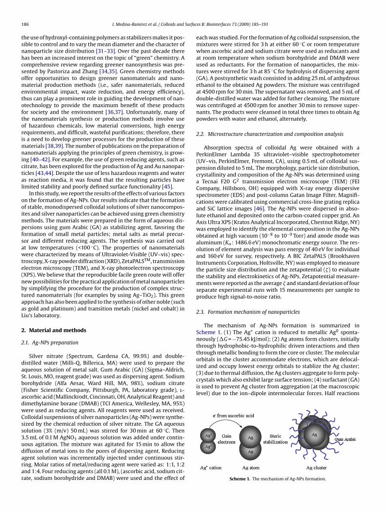
1 Surfac
tsnhcsometoftorimnictal
ooimpfsawte(nbtaaL
2
2
daSb(adwCss3udarar
(3) due to thermal diffusion, the Ag clusters aggregate to form poly-crystals which also exhibit large surface tension; (4) surfactant (GA)is used to prevent Ag cluster from aggregation (at the macroscopiclevel) due to the ion–dipole intermolecular forces. Half reactions
86 I. Medina-Ramirez et al. / Colloids and
he use of hydroxyl-containing polymers as stabilizers makes it pos-ible to control and to vary the mean diameter and the character ofanoparticle size distribution [31–33]. Over the past decade thereas been an increased interest on the topic of “green” chemistry. Aomprehensive review regarding greener nanosynthesis was pre-ented by Pastoriza and Zhang [34,35]. Green chemistry methodsffer opportunities to design greener nanomaterials and nano-aterial production methods (i.e., safer nanomaterials, reduced
nvironmental impact, waste reduction, and energy efficiency),hus can play a prominent role in guiding the development of nan-technology to provide the maximum benefit of these productsor society and the environment [36,37]. Unfortunately, many ofhe nanomaterials synthesis or production methods involve usef hazardous chemicals, low material conversions, high energyequirements, and difficult, wasteful purifications; therefore, theres a need to develop greener processes for the production of these
aterials [38,39]. The number of publications on the preparation ofanomaterials applying the principles of green chemistry, is grow-
ng [40–42]. For example, the use of green reducing agents, such asitrate, has been explored for the production of Ag and Au nanopar-icles [43,44]. Despite the use of less hazardous reagents and waters reaction media, it was found that the resulting particles haveimited stability and poorly defined surface functionality [45].
In this study, we report the results of the effects of various factorsn the formation of Ag-NPs. Our results indicate that the formationf stable, monodispersed colloidal solutions of silver nanocompos-
tes and silver nanoparticles can be achieved using green chemistryethods. The materials were prepared in the form of aqueous dis-
ersions using gum Arabic (GA) as stabilizing agent, favoring theormation of small metal particles; metal salts as metal precur-or and different reducing agents. The synthesis was carried outt low temperatures (<100 ◦C). The properties of nanomaterialsere characterized by means of Ultraviolet-Visible (UV–vis) spec-
roscopy, X-ray powder diffraction (XRD), ZetaPALSTM, transmissionlectron microscopy (TEM), and X-ray photoelectron spectroscopyXPS). We believe that the reproducible facile green route will offerew possibilities for the practical application of metal nanoparticlesy simplifying the procedure for the production of complex struc-ured nanomaterials (for examples by using Ag–TiO2). This greenpproach has also been applied to the synthesis of other noble (suchs gold and platinum) and transition metals (nickel and cobalt) iniu’s laboratory.
. Material and methods
.1. Ag-NPs preparation
Silver nitrate (Spectrum, Gardena CA, 99.9%) and double-istilled water (Milli-Q, Billerica, MA) were used to prepare thequeous solution of metal salt. Gum Arabic (GA) (Sigma–Aldrich,t. Louis, MO, reagent grade) was used as dispersing agent. Sodiumorohydride (Alfa Aesar, Ward Hill, MA, 98%), sodium citrateFisher Scientific Company, Pittsburgh, PA, laboratory grade), l-scorbic acid (Mallinckrodt, Cincinnati, OH, Analytical Reagent) andimethylamine borane (DMAB) (TCI America, Wellesley, MA, 95%)ere used as reducing agents. All reagents were used as received.olloidal suspensions of silver nanoparticles (Ag-NPs) were synthe-ized by the chemical reduction of silver nitrate. The GA aqueousolution (3% (m/v) 50 mL) was stirred for 30 min at 60 ◦C. Then.5 mL of 0.1 M AgNO3 aqueous solution was added under contin-ous agitation. The mixture was agitated for 15 min to allow the
iffusion of metal ions to the pores of dispersing agent. Reducinggent solution was incrementally injected under continuous stir-ing. Molar ratios of metal/reducing agent were varied as: 1:1, 1:2nd 1:4. Four reducing agents (all 0.1 M), (ascorbic acid, sodium cit-ate, sodium borohydride and DMAB) were used and the effect ofes B: Biointerfaces 73 (2009) 185–191
each was studied. For the formation of Ag colloidal suspsension, themixtures were stirred for 3 h at either 60 ◦C or room temperaturewhen ascorbic acid and sodium citrate were used as reducants andat room temperature when sodium borohydride and DMAB wereused as reductants. For the formation of nanoparticles, the mix-tures were stirred for 3 h at 85 ◦C for hydrolysis of dispersing agent(GA). A postsynthetic wash consisted in adding 25 mL of anhydrousethanol to the obtained Ag powders. The mixture was centrifugedat 4500 rpm for 30 min. The supernatant was removed, and 5 mL ofdouble-distilled water was added for futher cleansing. The mixturewas centrifuged at 4500 rpm for another 30 min to remove super-nants. The products were cleansed in total three times to obtain Agpowders with water and ethanol, alternately.
2.2. Microstructure characterization and composition analysis
Absorption spectra of colloidal Ag were obtained with aPerkinElmer Lambda 35 ultraviolet–visible spectrophotometer(UV–vis, PerkinElmer, Fremont, CA), using 0.5 mL of colloidal sus-pension diluted to 5 mL. The morphology, particle size distribution,crystallinity and composition of the Ag-NPs was determined usinga Tecnai F20 G2 transmission electron microscope (TEM) (FEICompany, Hillsboro, OH) equipped with X-ray energy dispersivespectrometer (EDS) and post-column Gatan Image Filter. Magnifi-cations were calibrated using commercial cross-line grating replicaand SiC lattice images [46]. The Ag-NPs were dispersed in abso-lute ethanol and deposited onto the carbon-coated copper grid. AnAxis Ultra XPS (Kratos Analytical Incorporated, Chestnut Ridge, NY)was employed to identify the elemental composition in the Ag-NPsobtained at high vacuum (10−8 to 10−9 Torr) and anode mode wasaluminum (K�: 1486.6 eV) monochromatic energy source. The res-olution of element analysis was pass energy of 40 eV for individualand 160 eV for survey, respectively. A BIC ZetaPALS (BrookhavenInstruments Corporation, Holtsville, NY) was employed to measurethe particle size distribution and the zetapotential (�) to evaluatethe stability and electrokinetics of Ag-NPs. Zetapotential measure-ments were reported as the average � and standard deviation of fourseparate experimental runs with 15 measurements per sample toproduce high signal-to-noise ratio.
2.3. Formation mechanism of nanoparticles
The mechanism of Ag-NPs formation is summarized inScheme 1. (1) The Ag+ cation is reduced to metallic Ag0 sponta-neously (�G = −75.45 kJ/mol); (2) Ag atoms form clusters, initiallythrough hydrophobic-to-hydrophilic driven interactions and thenthrough metallic bonding to form the core or cluster. The molecularorbitals in the cluster accommodate electrons, which are delocal-ized and occupy lowest energy orbitals to stablize the Ag cluster;
Scheme 1. The mechanism of Ag-NPs formation.
I. Medina-Ramirez et al. / Colloids and Surfaces B: Biointerfaces 73 (2009) 185–191 187
aa
A
C
oTCotrpedtaalvdosTpa
3
3
fasoamtdsawptrA
3.2. XRD analysis of polymer-stabilized Ag-NPs
Fig. 2 displays the X-ray diffraction (XRD) spectra of Ag-NPspowders obtained from the green synthesis procedure followed
Scheme 2. Oxidation reaction of ascorbic acid.
nd their standard reduction potentials [47,48] are listed in Eqs. (i)nd (ii):
g+(aq) + 1e− → Ag(s) E◦ = + 0.80 V (i)
6H6O6(aq) + 2H+(aq) + 2e− → C6H8O6(aq) E◦ = + 0.06 V
(ii)
According to the half reaction standard reduction potential, theverall reaction was determined to have a potential of 0.793 V.hermodynamically, therefore the redox reaction between Ag+ and6H8O6 (2Ag+ (aq) + C6H8O6 (aq) = 2Ag(s) + 2H+ (aq) + C6H6O6 (aq))ccurs spontaneously (Scheme 2 displays the deprotonization reac-ion). Since H+ was released in the oxidation half reaction, theedox process was characterized by a significant decrease in theH. This acidic condition was presumably the reason for the het-rogeneous coagulation of the fine Ag polycrystalline nanoparticlesue to ionic–dipole intermolecular forces. Although, a generaliza-ion, it can be assumed that the cluster phase is composed of
fluid-phase favoring dissolution and crystalline phase favoringggregation, which are in equilibrium. The equilibrium betweenong-range repulsive forces and short-range attractive forces (e.g.,an der Waals) will govern cluster size [49,50]. In small clusters, theecrease in cluster energy due to bonding of an additional particlen the surface of the cluster compensates for any additional repul-ive force interaction, which dominates as the cluster size increases.hus it can be seen that the packing fraction may be governed byhase separation kinetics, starting with a nucleation region, gener-ting cluster phases [51].
. Results and discussion
.1. UV–vis analysis of polymer-stabilized Ag-NPs
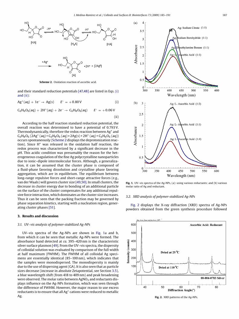
UV–vis spectra of the Ag-NPs are shown in Fig. 1a and b,rom which it can be seen that metallic Ag-NPs were formed. Thebsorbance band detected at ca. 395–420 nm is the characteristicilver surface plasmon [49]. From the UV–vis spectra, the dispersityf colloidal solution was evaluated by comparison of the full widtht half maximum (FWHM). The FWHM of all colloidal Ag speci-ens are essentially identical (85–100 nm), which indicates that
he samples were monodispersed. The monodispersity is mainlyue to the use of dispersing agent (GA). It is also seen that as particleizes decrease (increase in absolute Zetapotential, see Section 3.5),blue wavelength shift (from 418 to 409 nm) and peak broadening
ere observed. The molar ratio between AgNO3 and reductants dis-lays influence on the Ag-NPs formation, which was seen throughhe difference of FWHM. However, the major reason to use excesseductants is to ensure that all Ag+ cations were reduced to metallicg.Fig. 1. UV–vis spectra of the Ag-NPs, (a): using various reductants; and (b) variousmolar ratio of Ag and reductant.
Fig. 2. XRD patterns of the Ag-NPs.

188 I. Medina-Ramirez et al. / Colloids and Surfaces B: Biointerfaces 73 (2009) 185–191
F rystal(
bTf0ppw3
ig. 3. (a) TEM image of Ag-NPs with an insert of high-resolution image exhibting cd) EDS elemental composition analysis of Ag-NPs.
y drying for 10 h either in air at room temperature or at 110 ◦C.he XRD patterns generally aligned well with that of an Agaced-centered cubic (fcc) structure (powder diffraction file (PDF)0-004-0783), which the frequency of merit is 0.32. In this case, the
atterns did not to show any micro-distortion from cubic to otherhase structure, which indicated that nicely crystallized structureas formed [52,53]. Notably, the intensity of the (1 1 1) peak at 2� of8◦ (in Fig. 2) is the strongest and no peak splitting is detected. For
Fig. 4. XPS elemental composition analysis of Ag-NPs.
line lattice fringes; (b) ring pattern of Ag-NPs; (c) single crystal electron diffraction;
different Ag polycrystals obtained using various reducing agents,XRD spectra are completely overlapped and consistent with thestandard one. This analysis indicated that the green synthesis ishighly reproducible in the manufacturing of these nanomaterials.
3.3. TEM images of polymer-stabilized Ag-NPs
The TEM micrograph (Fig. 3a) indicated that the average parti-cle size of Ag-NPs was 40.2 nm. The particles were monodispersedwith limited degree of aggregation. A large aperture to include largenumber of particles was used to obtain of the electron diffraction(ED) pattern (Fig. 3b). The ring patterns confirmed that highly crys-talline Ag-NPs structure were formed. The d-spacings from ringpatterns corresponded well to the d-spacings from XRD peaks withthe fcc structure in Fig. 2. Single crystal pattern was obtained whenonly a single crytallline units were selected to diffract (Fig. 3c),
which confirmed the formation of highly crystalline Ag-NPs. It wasalso noticed that Ag-NPs were subject to thermal damage underelectron beam due to their ultrafine particle effect, which leads toa decrease in melting point.Scheme 3. Particle orbital diagram of Silver.

I. Medina-Ramirez et al. / Colloids and Surfaces B: Biointerfaces 73 (2009) 185–191 189
Fig. 5. (a) Particle size distribution of Ag-NPs. (b) Zetapotential evaluation of 5 different silver nanoparticles (Ag-NPs, 1,2,3,4,5) in triplicate (3 columns/sample).

1 Surfac
wdlAt2frdoasC
3
TsEtpta6(
ptioirwli3io
3p
adtawp(BwttAsrdrtTl(g
90 I. Medina-Ramirez et al. / Colloids and
Therefore, TEM observation of Ag-NPs was carried out undereak illumination to avoid beam damage [54]. The X-ray energyispersive spectroscopic (EDS) results were indicative of metal-
ic silver formation. It can be concluded that the elements ing-NPs were mainly composed of Ag (Fig. 3d). The spectrum of
he nanocomposite depicted that L�1 of the Ag metal occurred at.984 eV and Ln at 2.806 eV. A satellite peak at L�1 = 3.149 eV, aroserom the multi-electron proces in the Ag atom, such as electronepulsion. Another peak at lower energy position 2.650 eV was alsoetected to be L�. Peaks of K� = 22.162 eV and K� = 25.002 eV werebserved due to the innermost electron emission [55]. The C and Otoms were observed, resulting from the surfactant (GA), as well asupport carbon film on the copper grid. The Cu originated from theu grid.
.4. XPS Analysis of polymer-stabilized Ag-NPs
The XPS elemental analysis of the Ag-NPs is depicted in Fig. 4.he full (insert in Fig. 4) spectrum indicated the presence of theilver (Ag), oxygen (O) and carbon (C), which corresponded with theDS analysis. Although an oversimplification, it can be concludedhat metallic Ag is indicated by an asymmetric line shape with theeak tailing to the higher binding energy (BE). Fig. 4 (main) showshat the binding energies for Ag electron configurations of 3d5/2nd 3d3/2 of 366.1 eV and 372.1 eV, respectively with a difference of.0 eV. This is comparable to the standard Ag 3d binding energies3d5/2 = 368.3 eV, 3d3/2 = 374.3 eV, � = 6.0 eV) [56].
It can be seen that the doublet separation of 3d (3d5/2 and 3d3/2)hotoemission of Ag occured. The partial orbital diagram (includinghe outmost electrons) of naturally occuring Ag element is shownn Scheme 3 [57]. From the orbital diagram, it can be seen that 5srbital is half-occupied and 4d sub-shell is completely full accord-
ng to the Hund’s Rule. The lower energy (<20 eV) is required toemove the valence electrons. In our study, the most intense peakas due to the excitation of 3d electrons to the excited state which
ed to the binding energy changes. In addition, the subtle chem-cal shift of Ag peaks was found to be 2.2 eV for both 3d5/2 andd3/2, respectively compared to standard metallic silver. The shift
s caused by the chemical enviroment and fine particle size effectf Ag-NPs.
.5. Particle size and zetapotential analysis of theolymer-stabilized Ag-NPs
The Ag-NPs were precisely controlled in the regions between 10nd 50 nm and aggregation was successfully prevented using theispersing agent (Fig. 5a). Two populations of the Ag-NP were foundo be located in 13.5 nm (highest intensity set at 100% volume)nd 15.7 nm (75.5% volume, relative to highest intensity volume)hen ascorbic acid was used as reducing agent. Similarly, two
opulations were found in 10.3 nm (100% in volume) and 13.3 nm30.6% in volume) when sodium citrate was used as reducing agent.oth green reducing agents produced essentially identical particleshich exhibit similar size and stability. When NaBH4 was used,
wo populations of particles were located at 2.5 and 17.5 nm andhe predominant size was about 10 nm; while the DMAB producedg-NPs with various size (mean bi-modal particle size was mea-ured to be 2.5 and 85 nm). From the comparison of using variouseductants, it can be concluded that the green reducing agents pro-uced particles with slightly larger size than those from the toxiceducing agents. From our previous study [58], it was determined
hat ultrafine particles (<10 nm) may lead to mechanical instability.he electrokinetics analysis indicated that the fine particles exhibitarge absolute zetapotential and particles were negatively chargedFig. 5b). It was also noticed that the particles size from TEM isenerally larger than the size from ZetaPALS. This is due to the [
es B: Biointerfaces 73 (2009) 185–191
aggregation when the heat-treatment of nanoparticles is appliedto obtain the TEM specimen.
From our study, the time dependence of the zetapotential onthe course of measurement was not observed, which indicatedthat the Ag-NPs colloidal suspensions were reasonably stable. Thelarge zetapotential of the like (negative) charges minimized particleaggregation due to electrostatic repulsion [59,60]. This observationconfirmed our proposed mechanism of Ag formation.
4. Conclusions
The highly monodispersed nano-structured Ag particles weresuccessfully developed and evaluated using advanced instrumen-tation techniques. It was concluded that the green synthesizedmaterials were composed of near-spherical particles which werehighly crystalline. The particle sizes were controlled in the rangefrom 10 to 50 nm. The silver metal was found to be highly crystallinewith fcc structure. The XPS study indicated that Ag binding energiesfor 3d5/2 and 3d2/3 were well-indexed to the standard metallic Ag. Asubtle chemical shift was found due to its hydrophilic environmentand small size effect. The magnitude of zetapotential was higher atlow pH and its absolute value was significant to prevent the particleaggregation.
Conflict of interest
None of the authors has any direct (social) relationship withElsevier publisher or the editorials staff of Colloids and SurfacesB: Biointerfaces including editors and reviewers. In addition, thereis no direct relationship with the sponsors (FUMEC, AMC, TAMUK,and Robert A. Welch Foundation).
Acknowledgements
The authors are thankful to the Academia Mexicana de Ciencias(AMC), Fundación México Estados Unidos para la Ciencia (FUMEC),the Texas A&M University-Kingsville (TAMUK), College of Arts andSciences Research and Development Fund (160310-00014), and theRobert A. Welch Foundation (departmental grant AC006) for theirfinancial support. The technical support and facility access providedby the South Texas Environmental Institute and the Department ofChemistry (both at TAMUK), Microscopy and Imaging Center andMaterials Characterization Facility (Dr. Gang Liang for his XPS anal-ysis) at TAMU, College Station are duly acknowledged for allowingus to conduct the advanced instrumentation analyses. Dr. ThomasHays is also aknowledged for his constructive discussion regardingthis manuscript.
References
[1] C.J. Johnson, N. Zhukovsky, A. Cass, J.M. Nagy, Proteomics 8 (2008) 715.[2] L. Qiu, J. Franc, A. Rewari, D. Blanc, K. Saravanamuttu, J. Mater. Chem. 19 (2009)
373.[3] J.M. Campelo, D. Luna, R. Luque, J.M. Marinas, A.A. Romero, ChemSusChem 2
(2009) 18.[4] S. Rondinini, G. Aricci, Z. Krpetic, C. Locatelli, A. Minguzzi, F. Porta, A. Vertova,
Fuel cells (2008) (Early View, No. 0), 1.[5] M.L. Gulrajani, D. Gupta, S. Periyasamy, S.G. Muthu, J. Appl. Polym. Sci. 108
(2008) 614.[6] S. Loher, O.D. Schneider, T. Maienfisch, S. Bokorny, W.J. Stark, Small 4 (2008)
824.[7] X. Huang, D. Du, X. Gong, J. Cai, H. Tu, X. Xu, A. Zhang, Electroanalysis 20 (2008)
402.[8] S.Y. Lee, H.J. Kim, R. Patel, S. Joon Im, J.H. Kim, B.R. Min, Polym. Adv. Technol. 18
(2007) 562.[9] Z. Yang, J. Lia, X. Yang, Xi. Xie, Y. Wu, J. Mol. Catal. A: Chem. 241 (2005) 15.
[10] J.R. Davis, Metals Handbook, desk ed., 2nd ed., ASM International, HandbookCommittee, San Jose, CA, 1998, pp. 658.
[11] T.C. Chuang, Y.C. Liu, C.C. Wang, J. Raman Spectrosc. 36 (2005) 704.12] L. Ming, Med. Res. Rev. 23 (2003) 697.

Surfac
[[[[[[
[[
[
[
[
[
[[
[
[
[[
[
[
[
[
[
[
[[
[
[[
[
[[[
[[[
[[
[
[
[[[
[
[57] G. Umarji, S. Ketkar, R. Hawaldar, S. Gosavi, K. Patil, U. Mulik, D. Amalnerkar,
I. Medina-Ramirez et al. / Colloids and
13] D. Dorjnamjin, M. Ariunaa, Y. Key Shim, Int. J. Mol. Sci. 9 (2008) 807.14] J. Jiang, B. Winther-Jensen, E.M. Kjær, Macromol. Symp. 239 (2006) 84.15] P. Jain, T. Pradeep, Biotechnol. Bioeng. 90 (2005) 59.16] K. Kim, D. Han, J. Lee, H. Kim, Scripta Mater. 54 (2005) 143.17] M. Rai, A. Yadava, A. Gadea, Biotechnol. Adv. 27 (2009) 76.18] Q.L. Feng, J. Wu, G.Q. Chen, F.Z. Cui, T.N. Kim, J.O. Kim, J. Biomed. Mater. Res. Part
A 52 (2000) 662.19] R. Tankhiwale, S.K. Bajpai, Colloids Surf. B 69 (2009) 164.20] N.L. Lala, R. Ramaseshan, L. Bojun, S. Sundarrajan, R.S. Barhate, Y. Liu, S. Ramakr-
ishna, Biotechnol. Bioeng. 97 (2007) 1357.21] J.L. Elechiguerra, J.L. Burt, J.R. Morones, A. Camacho-Bragado, X. Gao, H.H. Lara,
M.J. Yacaman, J. Nanobiotechnol. 3 (2005) 6.22] J.V. Rogers, C.V. Parkinson, Y.W. Choi, J.L. Speshock, S.M. Hussain, Nanoscale Res.
Lett. 3 (2008) 129.23] A.M. Schrand, L.K. Braydich-Stolle, J.J. Schlager, L. Dai, S.M. Hussain, Nanotech-
nology 19 (2008) 13.24] K. Ishizu, H. Kakinuma, K. Ochi, S. Uchida, M. Hayashi, Polym. Adv. Technol. 16
(2005) 834.25] A.V. Ghule, K. Ghule, S. Tzing, Y. Ling, Eur. J. Inorg. Chem. 21 (2007) 3342.26] S. Ghader, M. Manteghian, M. Kokabi, R.S. Mamoory, Chem. Eng. Technol. 30
(2007) 1129.27] D. Radziuk, A. Skirtach, G. Sukhorukov, D. Shchukin, H. Möhwald, Macromol.
Rapid Commun. 28 (2007) 848.28] H. Ma, B. Yin, S. Wang, Y. Jiao, W. Pan, S. Huang, S. Chen, F. Meng, ChemPhysChem
5 (2004) 68.29] S.S. Shankar, A. Ahmad, M. Sastry, Biotechnol. Prog. 19 (2003) 1627.30] L. Peponi, A. Tercjak, J. Gutierrez, H. Stadler, L. Torre, J.M. Kenny, I. Mondragon,
Macromol. Mater. Eng. 293 (2008) 568.31] J.M. Domínguez-Vera, N. Gálvez, P. Sánchez, A.J. Mota, S. Trasobares, J.C. Hernán-
dez, J.J. Calvino, Eur. J. Inorg. Chem. 30 (2007) 4823.32] J. Compton, D. Kranbuehl, G. Martin, E. Espuche, L. David, Macromol. Symp. 247
(2007) 182.33] L.E. Macaskie, N.J. Creamer, A.M.M. Essa, N.L. Brown, Biotechnol. Bioeng. 96
(2007) 631.
34] V. Bastys, I. Pastoriza-Santos, B. Rodríguez-González, R. Vaisnoras, L.M. Liz-Marzán, Adv. Funct. Mater. 16 (2006) 766.35] J. Zhang, Z. Liu, B. Han, D. Liu, J. Chen, J. He, T. Jiang, Chem. Eur. J. 10 (2004)
3531.36] J.A. Dahl, B.L.S. Maddux, J.E. Hutchison, Toward greener nanosynthesis, Chem.
Rev. 107 (2007) 2228.
[[
[
es B: Biointerfaces 73 (2009) 185–191 191
37] D.D. Evanoff Jr., G. Chumanov, ChemPhysChem 6 (2005) 1221.38] Y.S. Kang, S.W. Kang, H. Kim, J.H. Kim, J. Won, C.K. Kim, K. Char, Adv. Mater. 19
(2007) 475.39] H. Ma, Y. Jiao, B. Yin, S. Wang, S. Zhao, S. Huang, W. Pan, S. Chen, F. Meng,
ChemPhysChem 5 (2004) 713.40] H. Jiang, S. Manolache, A.C. Wong, F.S. Denes, J. Appl. Polym. Sci. 93 (2004) 1411.41] J.Q. Hu, Q. Chen, Z.X. Xie, G.B. Han, R.H. Wang, B. Ren, Y. Zhang, Z.L. Yang, Z.Q.
Tian, Adv. Funct. Mater. 14 (2004) 183.42] R.R. Naik, S.E. Jones, C.J. Murray, J.C. McAuliffe, R.A. Vaia, M.O. Stone, Adv. Funct.
Mater. 14 (2004) 25.43] V.R. Reddy, A. Currao, G. Calzaferri, J. Phys.: Conf. Ser. 61 (2007) 960.44] D.D. Dionysiou, J. Environ. Eng. 130 (2008) 723.45] S.M. Hussain, L.K. Braydich-Stolle, A.M. Schrand, R.C. Murdock, K.O. Yu, D.M.
Mattie, J.J. Schlager, M. Terrones, Adv. Mater. 21 (2009) 11.46] Z.P. Luo, Acta. Mater. 54 (2006) 47.47] N. Garti, M.E. Leser, Polym. Adv. Technol. 12 (2001) 123.48] P.C. Hiemenz, R. Rajagopalan, Principles of Colloid and Surface Chemistry, 3rd
ed., Marcel Dekker Inc., New York, 1997, pp. 1–25.49] D.V. Goia, J. Mater. Chem. 14 (2003) 451.50] R. Kelsall, I.W. Hamley, M. Geoghegan, Nanoscale Science and Technology, 1st
ed., Wiley Interscience, Malden, MA, 2005, pp. 282–330.51] Z.L. Wang, Z.C. Kang, Functional and Smart Materials: Structural Evolution and
Structure Analysis, Plenum, New York, 1998, pp. 405–466.52] N.L. Lala, R. Ramaseshan, L. Bojun, S. Sundarrajan, R.S. Barhate, Y. liu, S. Ramakr-
ishna, Biotechnol. Bioeng. 97 (2007) 1537.53] T. Garino, M. Rodriguez, J. Am. Ceram. Soc. 83 (2000) 2709.54] Z.L. Wang, J. Phys. Chem. B 104 (2000) 1153.55] M.G. Guzmán, J. Dille, S. Godet, Proc. World Acad. Sci. Eng. Technol. 33 (2008)
2070.56] C.D. Wagner, G.E. Muilenberg (Eds.), Handbook of X-ray Photoelectron Spec-
troscopy: A Reference Book of Standard Data for Use in X-ray PhotoelectronSpectroscopy, 1st ed., Physical Electronics Division, Perkin-Elmer Corp., EdenPrairie, MN, 1979, p. pp. 121.
Microelectron Int. 25 (2008) 46.58] J. Liu, A.C. Co, S. Paulson, V.I. Birss, Solid State Ionics 177 (2006) 377.59] G.M. Dougherty, K.A. Rose, J.B. Tok, S.S. Pannu, F.Y. Chuang, M.Y. Sha, G.
Chakarova, S.G. Penn, Electrophoresis 29 (2008) 1131.60] Y. Sun, Y. Liu, G. Zhao, X. Zhou, J. Gao, Q. Zhang, J. Mater. Sci. 43 (2008) 4625.