Nanocarrier Drug Delivery Systems: Characterization ... - MDPI
26
Citation: Alshawwa, S.Z.; Kassem, A.A.; Farid, R.M.; Mostafa, S.K.; Labib, G.S. Nanocarrier Drug Delivery Systems: Characterization, Limitations, Future Perspectives and Implementation of Artificial Intelligence. Pharmaceutics 2022, 14, 883. https://doi.org/10.3390/ pharmaceutics14040883 Academic Editors: Oleh Taratula and Olena Taratula Received: 14 March 2022 Accepted: 15 April 2022 Published: 18 April 2022 Publisher’s Note: MDPI stays neutral with regard to jurisdictional claims in published maps and institutional affil- iations. Copyright: © 2022 by the authors. Licensee MDPI, Basel, Switzerland. This article is an open access article distributed under the terms and conditions of the Creative Commons Attribution (CC BY) license (https:// creativecommons.org/licenses/by/ 4.0/). pharmaceutics Review Nanocarrier Drug Delivery Systems: Characterization, Limitations, Future Perspectives and Implementation of Artificial Intelligence Samar Zuhair Alshawwa 1 , Abeer Ahmed Kassem 2, * , Ragwa Mohamed Farid 2 , Shaimaa Khamis Mostafa 3 and Gihan Salah Labib 2 1 Department of Pharmaceutical Sciences, College of Pharmacy, Princess Nourah bint Abdulrahman University, P.O. Box 84428, Riyadh 11671, Saudi Arabia; [email protected] or [email protected] 2 Department of Pharmaceutics and Pharmaceutical Technology, Faculty of Pharmacy, Pharos University in Alexandria, Alexandria 21523, Egypt; [email protected] (R.M.F.); [email protected] (G.S.L.) 3 Department of Pharmaceutics and Pharmaceutical Technology, Faculty of Pharmacy, Delta University for Science and Technology, Gamasa 11152, Egypt; [email protected] * Correspondence: [email protected]; Tel.: +20-12-2360-8155 Abstract: There has been an increasing demand for the development of nanocarriers targeting multiple diseases with a broad range of properties. Due to their tiny size, giant surface area and feasible targetability, nanocarriers have optimized efficacy, decreased side effects and improved stability over conventional drug dosage forms. There are diverse types of nanocarriers that have been synthesized for drug delivery, including dendrimers, liposomes, solid lipid nanoparticles, polymersomes, polymer–drug conjugates, polymeric nanoparticles, peptide nanoparticles, micelles, nanoemulsions, nanospheres, nanocapsules, nanoshells, carbon nanotubes and gold nanoparticles, etc. Several characterization techniques have been proposed and used over the past few decades to control and predict the behavior of nanocarriers both in vitro and in vivo. In this review, we describe some fundamental in vitro, ex vivo, in situ and in vivo characterization methods for most nanocarriers, emphasizing their advantages and limitations, as well as the safety, regulatory and manufacturing aspects that hinder the transfer of nanocarriers from the laboratory to the clinic. Moreover, integration of artificial intelligence with nanotechnology, as well as the advantages and problems of artificial intelligence in the development and optimization of nanocarriers, are also discussed, along with future perspectives. Keywords: nanocarriers characterization; challenges; artificial intelligence; future perspectives; stability; regulatory aspects; safety considerations 1. Introduction Pharmaceutical research seeking to enhance drug bioavailability, increase stability and improve organ targeting has been progressively advanced. Pharmaceutical nanocarriers are drug delivery vehicles of submicron size and high versatility. They include polymeric, lipidic and inorganic nanoparticles, liposomes, nanotubes, nanocomplexes, niosomes and many others. In principle, ligands can be attached to the surface of nanocarriers for better uptake and targetability [1–3]. Drugs can be either dispersed into the nanocarrier matrix or located within the nanocarrier layers. Nanocarriers offer several advantages over tra- ditional drug therapies as it is easier to customize their size, charge, surface properties and targeting moieties to regulate their uptake, biodistribution, targeting and elimina- tion [4]. They can be administered via many different routes, e.g., parenteral [5], nasal [6], topical [7,8] or oral routes [9,10]. Owing to the preceding advantages, there is an increasing demand for the development of nanocarriers targeting multiple diseases with a broad range Pharmaceutics 2022, 14, 883. https://doi.org/10.3390/pharmaceutics14040883 https://www.mdpi.com/journal/pharmaceutics
-
Upload
khangminh22 -
Category
Documents
-
view
3 -
download
0
Transcript of Nanocarrier Drug Delivery Systems: Characterization ... - MDPI

�����������������
Citation: Alshawwa, S.Z.; Kassem,
A.A.; Farid, R.M.; Mostafa, S.K.;
Labib, G.S. Nanocarrier Drug
Delivery Systems: Characterization,
Limitations, Future Perspectives and
Implementation of Artificial
Intelligence. Pharmaceutics 2022, 14,
883. https://doi.org/10.3390/
pharmaceutics14040883
Academic Editors: Oleh Taratula
and Olena Taratula
Received: 14 March 2022
Accepted: 15 April 2022
Published: 18 April 2022
Publisher’s Note: MDPI stays neutral
with regard to jurisdictional claims in
published maps and institutional affil-
iations.
Copyright: © 2022 by the authors.
Licensee MDPI, Basel, Switzerland.
This article is an open access article
distributed under the terms and
conditions of the Creative Commons
Attribution (CC BY) license (https://
creativecommons.org/licenses/by/
4.0/).
pharmaceutics
Review
Nanocarrier Drug Delivery Systems: Characterization,Limitations, Future Perspectives and Implementationof Artificial IntelligenceSamar Zuhair Alshawwa 1 , Abeer Ahmed Kassem 2,* , Ragwa Mohamed Farid 2 , Shaimaa Khamis Mostafa 3
and Gihan Salah Labib 2
1 Department of Pharmaceutical Sciences, College of Pharmacy, Princess Nourah bint Abdulrahman University,P.O. Box 84428, Riyadh 11671, Saudi Arabia; [email protected] or [email protected]
2 Department of Pharmaceutics and Pharmaceutical Technology, Faculty of Pharmacy,Pharos University in Alexandria, Alexandria 21523, Egypt; [email protected] (R.M.F.);[email protected] (G.S.L.)
3 Department of Pharmaceutics and Pharmaceutical Technology, Faculty of Pharmacy,Delta University for Science and Technology, Gamasa 11152, Egypt; [email protected]
* Correspondence: [email protected]; Tel.: +20-12-2360-8155
Abstract: There has been an increasing demand for the development of nanocarriers targetingmultiple diseases with a broad range of properties. Due to their tiny size, giant surface area andfeasible targetability, nanocarriers have optimized efficacy, decreased side effects and improvedstability over conventional drug dosage forms. There are diverse types of nanocarriers that havebeen synthesized for drug delivery, including dendrimers, liposomes, solid lipid nanoparticles,polymersomes, polymer–drug conjugates, polymeric nanoparticles, peptide nanoparticles, micelles,nanoemulsions, nanospheres, nanocapsules, nanoshells, carbon nanotubes and gold nanoparticles,etc. Several characterization techniques have been proposed and used over the past few decadesto control and predict the behavior of nanocarriers both in vitro and in vivo. In this review, wedescribe some fundamental in vitro, ex vivo, in situ and in vivo characterization methods for mostnanocarriers, emphasizing their advantages and limitations, as well as the safety, regulatory andmanufacturing aspects that hinder the transfer of nanocarriers from the laboratory to the clinic.Moreover, integration of artificial intelligence with nanotechnology, as well as the advantages andproblems of artificial intelligence in the development and optimization of nanocarriers, are alsodiscussed, along with future perspectives.
Keywords: nanocarriers characterization; challenges; artificial intelligence; future perspectives;stability; regulatory aspects; safety considerations
1. Introduction
Pharmaceutical research seeking to enhance drug bioavailability, increase stability andimprove organ targeting has been progressively advanced. Pharmaceutical nanocarriersare drug delivery vehicles of submicron size and high versatility. They include polymeric,lipidic and inorganic nanoparticles, liposomes, nanotubes, nanocomplexes, niosomes andmany others. In principle, ligands can be attached to the surface of nanocarriers for betteruptake and targetability [1–3]. Drugs can be either dispersed into the nanocarrier matrixor located within the nanocarrier layers. Nanocarriers offer several advantages over tra-ditional drug therapies as it is easier to customize their size, charge, surface propertiesand targeting moieties to regulate their uptake, biodistribution, targeting and elimina-tion [4]. They can be administered via many different routes, e.g., parenteral [5], nasal [6],topical [7,8] or oral routes [9,10]. Owing to the preceding advantages, there is an increasingdemand for the development of nanocarriers targeting multiple diseases with a broad range
Pharmaceutics 2022, 14, 883. https://doi.org/10.3390/pharmaceutics14040883 https://www.mdpi.com/journal/pharmaceutics

Pharmaceutics 2022, 14, 883 2 of 26
of properties. Hence, several characterization techniques have been proposed and usedover the past few decades to control and predict the behavior of nanocarriers both in vitroand in vivo. The commonly employed characterization techniques are used to assess thephysicochemical properties, drug loading, release rate, mechanical behavior, stability, tissuepermeability, possible toxicity and in vivo fate of nanocarriers. In this review, we sum-marize the indispensable characterization methods commonly used for most nanocarrierdrug delivery systems (DDSs). Moreover, the limitations of these methods are presented,along with the regulatory difficulties and scalability issues confronting the manufacturingof nanocarriers.
2. Physicochemical Characterization
Physicochemical properties of nanocarriers include their particle size, particle sizedistribution, surface charge, hydrophobicity and morphology. The determination of thesephysicochemical properties for the drug nanocarrier can predict, to a great extent, severalaspects, including physical stability and entrapment efficiency [11].
2.1. Particle Size and Polydispersity
The most important characteristics of nanocarriers are particle size, shape and disper-sity (heterogeneity of particles in terms of size expressed by polydispersity index (PDI)). Theparticle size and shape affect the biodistribution and elimination of nanocarriers [12–14].They also affect their attachment, firm adhesion [15], phagocytosis [16], circulation half-life,cellular distribution [17], cellular uptake and endocytosis [18,19].
In the following sections, we give an overview of the foremost, routinely used strate-gies to determine particle size and PDI.
2.1.1. Dynamic Light Scattering Spectroscopy
Dynamic light scattering (DLS) determines the particle diameter with the aid of Brow-nian motion and light scattering properties. However, large particles may not be discoveredby the DLS method since their movement may be too slow [20]. The mean particle diameterand PDI are measured using a particle sizer equipped with particle sizing software [21].Samples must be in a liquid state, solution or dilute suspension of known viscosity. Thistechnique is sensitive to impurities and can measure particles between 1 nm and 10 µmin diameter. The obtained data of particle size, size distribution and PDI are convenientfor statistical analysis. However, some limitations might confine the application of DLSsuch as the unreliable results of polydisperse and multimodal samples, sedimentation oflarge particles and sample concentration. These limitations can be overcome by adding afractionation step to obtain fractions of different particles sizes prior to measurement by theDLS method [22]. The asymmetrical flow field flow fractionation (AF4) method involvesthe separation of samples into an unpacked, narrow-opened channel [23]. Concisely, asingle-carrier flow is withdrawn from the channel inlet that splits into the channel flowand the crossflow, Figure 1. The channel flow exhibits a parabolic velocity profile line thatcarries nanoparticles to the channel outlet to be detected. On the other hand, the crossflowmoves from the top to the bottom of the channel, forcing the nanoparticles to move downon the accumulation wall that is made of an ultrafiltration membrane covered by a porousfrit. At the end, the nanoparticles’ diffusion restricts the crossflow field, allowing for sizefractionation where smaller particles reach an equilibrium position higher up in the channelwith faster channel flow velocity that allows for earlier elution than larger particles [22–24].Moreover, temperature and pH may impact the reliability of the measurements. DLS isgenerally considered inappropriate for measurements within biological media [25]. Oneof the most important aspects in a new and specific AF4 method is that it needs to beestablished for each kind of measured nanoparticles sample depending on its composition,average size, surface properties and size distribution [26]. The results obtained by thismethod are usually confirmed by scanning electron microscopy (SEM) or transmissionelectron microscopy (TEM) [22]. Other limitations may comprise the assumption of having

Pharmaceutics 2022, 14, 883 3 of 26
spherical samples, which may not be true in all cases, having turbid or translucent sampleswhere light absorption by the dispersed particles can interfere with the detection andhaving aggregated particles that may not be distinguished from individual particles, andthe method requires that the solvent refractive index must be accurately known [27].
Pharmaceutics 2022, 14, x 3 of 27
The results obtained by this method are usually confirmed by scanning electron micros-
copy (SEM) or transmission electron microscopy (TEM) [22]. Other limitations may com-
prise the assumption of having spherical samples, which may not be true in all cases, hav-
ing turbid or translucent samples where light absorption by the dispersed particles can
interfere with the detection and having aggregated particles that may not be distinguished
from individual particles, and the method requires that the solvent refractive index must
be accurately known [27].
Figure 1. Schematic representation of an asymmetric flow field flow fractionation channel equipped
with a frit inlet (FI-AF4). Frit inlet flow propels sample components towards the accumulation wall,
allowing their hydrodynamic relaxation without stopping their axial migration. Adapted with per-
mission from [22], Elsevier, 2021.
2.1.2. Static Light Scattering
Static light scattering depends on measuring the intensity of scattered light waves
depending on scattering angle, followed by the application of an acceptable mathematical
model (usually Mie theory) to convert the scattering pattern to particle size distribution.
This model assumes that the scattered particles are homogeneous, spherical, fail to inter-
act and have a definite refractive index. Actually, most biopolymeric particles do not agree
with such assumptions. Moreover, sample preparation necessitates prior dilution and
shaking, which might change the integrity or aggregation of biopolymer particles. Ac-
cordingly, the results pooled from static light scattering should be used with care [28].
2.1.3. Atomic Force Microscopy
Atomic force microscopy (AEM) allows particle size measurement with ultra-high
resolution based on scanning the submicron particle levels with a probe tip of atomic
scale. The appliance provides a topographic map of the sample depending on the force
between a sharp probe and the surface of the sample. This technique allows the imaging
of non-conducting samples without any special treatment, which, in turn, facilitates the
imaging of delicate biological and polymeric nanocarriers [29]. Most significantly, it pro-
vides the foremost correct description of size and size distribution without applying any
algorithmic treatment. However, it is worth mentioning that accurate data collection and
interpretation of results requires strong expertise, especially when dealing with complex
samples or specimens such as biological cells. The main concerns are those related to the
quality of tip and support surface chemistries and the expectation of their possible altera-
tion during data collection of the shape and size of measured vesicles. Other limitations
may include the poor sampling techniques and time consumption caused by the slow
Figure 1. Schematic representation of an asymmetric flow field flow fractionation channel equippedwith a frit inlet (FI-AF4). Frit inlet flow propels sample components towards the accumulationwall, allowing their hydrodynamic relaxation without stopping their axial migration. Adapted withpermission from [22], Elsevier, 2021.
2.1.2. Static Light Scattering
Static light scattering depends on measuring the intensity of scattered light wavesdepending on scattering angle, followed by the application of an acceptable mathematicalmodel (usually Mie theory) to convert the scattering pattern to particle size distribution.This model assumes that the scattered particles are homogeneous, spherical, fail to interactand have a definite refractive index. Actually, most biopolymeric particles do not agree withsuch assumptions. Moreover, sample preparation necessitates prior dilution and shaking,which might change the integrity or aggregation of biopolymer particles. Accordingly, theresults pooled from static light scattering should be used with care [28].
2.1.3. Atomic Force Microscopy
Atomic force microscopy (AEM) allows particle size measurement with ultra-highresolution based on scanning the submicron particle levels with a probe tip of atomic scale.The appliance provides a topographic map of the sample depending on the force betweena sharp probe and the surface of the sample. This technique allows the imaging of non-conducting samples without any special treatment, which, in turn, facilitates the imagingof delicate biological and polymeric nanocarriers [29]. Most significantly, it provides theforemost correct description of size and size distribution without applying any algorithmictreatment. However, it is worth mentioning that accurate data collection and interpretationof results requires strong expertise, especially when dealing with complex samples orspecimens such as biological cells. The main concerns are those related to the quality of tipand support surface chemistries and the expectation of their possible alteration during datacollection of the shape and size of measured vesicles. Other limitations may include thepoor sampling techniques and time consumption caused by the slow scanning technique ofthe instrument, which lacks the capability to detect specific molecules. However, the latterlimitation can be resolved by a more advanced single—molecule force spectroscopy withAFM cantilever tip carrying certain ligands or molecules that can detect specific functional

Pharmaceutics 2022, 14, 883 4 of 26
groups. Therefore, a deep understanding of the principle and limitations of different AFMmodalities is essential before users proceed with their first experiment [27,30].
2.1.4. Centrifugal Liquid Sedimentation
Centrifugal liquid sedimentation (CLS) is a fractionation method in which differentmonodisperse fractions within a sample are isolated by centrifugation before particlesize measurement [31]. Theoretically, CLS is more suitable for polydisperse samples.However, the fractionation becomes greatly complicated if the size distribution is too broad.Spherical nanocarriers with narrower size distribution and densities are better candidatesfor CLS measurements [32,33]. Samples also must not undergo any changes, chemicallyor physically, in the suspension during sedimentation. Moreover, the refractive index anddensities of the particles and liquid medium must differ from each other to give reliableresults. It was previously reported that both DLS and CLS methods were suitable androbust for the determination of particle size of silica nanoparticles suspension in the rangeof 35–50 nm.
2.2. Surface Charge and Hydrophobicity
The surface properties of nanocarriers significantly influence their bioavailability, sta-bility, cellular uptake and biodistribution [12,34,35]. The zeta (ζ) potential, expressing thesurface charge, indicates possible electrostatic interactions between the nanocarrier units,affects their aggregation tendencies and helps to select proper coating materials [36]. It canbe determined by applying an electrical current through the sample while recording themovement of the nanocarriers using laser Doppler velocimetry [37]. So far, electrophoreticlight scattering is the most popular method due to its accuracy, sensitivity and versatil-ity. Overall, measuring zeta potential can be highly sensitive to ionic strength and pH.Sample dilution is usually required before measurement. Mixtures of oppositely chargednanocarriers might affect the reliability of the interpreted data [20].
The hydrophobicity of nanocarriers can be assessed by adsorption probe method, hy-drophobic interaction chromatography, contact angle measurements and biphasic partitioning.In addition, X-ray photon correlation spectroscopy helps to identify specific chemical groupson the surface of nanocarriers and eventually predicts their hydrophobicity [38].
2.3. Morphology of Nanocarriers
The morphology of nanocarriers and their aggregation behavior are important fac-tors for different biological properties, including their half-life, targeting efficiency andtoxicity [39]. Several non-spherical shapes, including discs, ellipsoids, cylinders, hemi-spheres, cubes, cones and other complex shapes, have a profound effect on those biologicalprocesses [12]. On the one hand, atomic force microscopy grants the study of the shapeof nanocarriers with high resolution without altering sample properties before measure-ment. On the other hand, electron microscopy techniques, namely SEM and TEM, offernumerous benefits on morphological and particle sizing characterization but have minimalinformation on the size distribution and true population mean.
2.3.1. Scanning Electron Microscopy
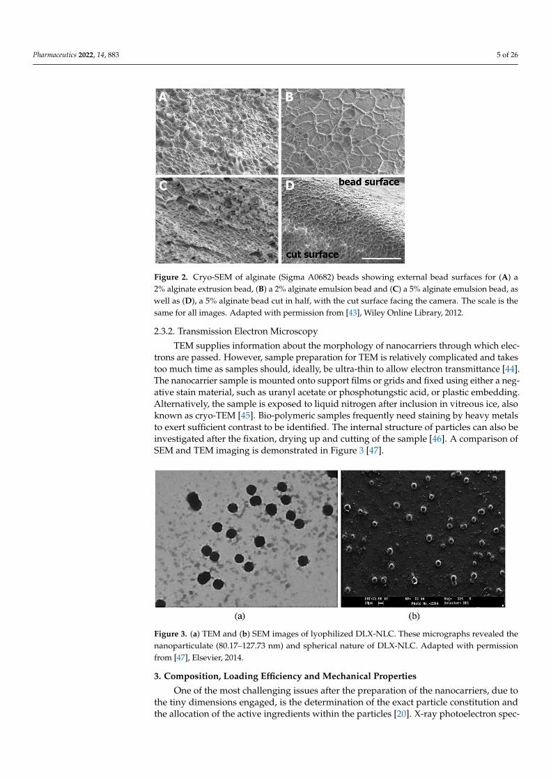
SEM provides accurate information on the topography of nanocarriers with directvisualization. Typically, samples are dried, seated on a sample holder and coated by a metalwith high electric conductivity, such as gold, using a sputter coating process. The samplesurface is scanned with a focused beam of electrons, and the secondary electrons emittedfrom its surface are recorded [40]. Nanocarriers scanned by SEM should ideally be ableto withstand the coating material, electron beam and vacuum without substantial change.Modified SEM techniques have been introduced, in which sample drying is not necessary.For example, wet SEM allows the analysis of hydrated samples without fixation, coatingor drying [41,42]. Cryo-SEM is another modified technique that requires sample freezing,Figure 2 [43].
Pharmaceutics 2022, 14, 883 5 of 26
Pharmaceutics 2022, 14, x 5 of 27
coating material, electron beam and vacuum without substantial change. Modified SEM tech-
niques have been introduced, in which sample drying is not necessary. For example, wet SEM
allows the analysis of hydrated samples without fixation, coating or drying [41,42]. Cryo-SEM
is another modified technique that requires sample freezing, Figure 2 [43].
Figure 2. Cryo-SEM of alginate (Sigma A0682) beads showing external bead surfaces for (A) a 2%
alginate extrusion bead, (B) a 2% alginate emulsion bead and (C) a 5% alginate emulsion bead, as
well as (D), a 5% alginate bead cut in half, with the cut surface facing the camera. The scale is the
same for all images. Adapted with permission from [43], Wiley Online Library, 2012.
2.3.2. Transmission Electron Microscopy
TEM supplies information about the morphology of nanocarriers through which
electrons are passed. However, sample preparation for TEM is relatively complicated and
takes too much time as samples should, ideally, be ultra-thin to allow electron transmit-
tance [44]. The nanocarrier sample is mounted onto support films or grids and fixed using
either a negative stain material, such as uranyl acetate or phosphotungstic acid, or plastic
embedding. Alternatively, the sample is exposed to liquid nitrogen after inclusion in vit-
reous ice, also known as cryo-TEM [45]. Bio-polymeric samples frequently need staining
by heavy metals to exert sufficient contrast to be identified. The internal structure of par-
ticles can also be investigated after the fixation, drying up and cutting of the sample [46].
A comparison of SEM and TEM imaging is demonstrated in Figure 3 [47].
Figure 3. (a) TEM and (b) SEM images of lyophilized DLX-NLC. These micrographs revealed the
nanoparticulate (80.17–127.73 nm) and spherical nature of DLX-NLC. Adapted with permission
from [47], Elsevier, 2014.
Figure 2. Cryo-SEM of alginate (Sigma A0682) beads showing external bead surfaces for (A) a2% alginate extrusion bead, (B) a 2% alginate emulsion bead and (C) a 5% alginate emulsion bead, aswell as (D), a 5% alginate bead cut in half, with the cut surface facing the camera. The scale is thesame for all images. Adapted with permission from [43], Wiley Online Library, 2012.
2.3.2. Transmission Electron Microscopy
TEM supplies information about the morphology of nanocarriers through which elec-trons are passed. However, sample preparation for TEM is relatively complicated and takestoo much time as samples should, ideally, be ultra-thin to allow electron transmittance [44].The nanocarrier sample is mounted onto support films or grids and fixed using either a neg-ative stain material, such as uranyl acetate or phosphotungstic acid, or plastic embedding.Alternatively, the sample is exposed to liquid nitrogen after inclusion in vitreous ice, alsoknown as cryo-TEM [45]. Bio-polymeric samples frequently need staining by heavy metalsto exert sufficient contrast to be identified. The internal structure of particles can also beinvestigated after the fixation, drying up and cutting of the sample [46]. A comparison ofSEM and TEM imaging is demonstrated in Figure 3 [47].
Pharmaceutics 2022, 14, x 5 of 27
coating material, electron beam and vacuum without substantial change. Modified SEM tech-
niques have been introduced, in which sample drying is not necessary. For example, wet SEM
allows the analysis of hydrated samples without fixation, coating or drying [41,42]. Cryo-SEM
is another modified technique that requires sample freezing, Figure 2 [43].
Figure 2. Cryo-SEM of alginate (Sigma A0682) beads showing external bead surfaces for (A) a 2%
alginate extrusion bead, (B) a 2% alginate emulsion bead and (C) a 5% alginate emulsion bead, as
well as (D), a 5% alginate bead cut in half, with the cut surface facing the camera. The scale is the
same for all images. Adapted with permission from [43], Wiley Online Library, 2012.
2.3.2. Transmission Electron Microscopy
TEM supplies information about the morphology of nanocarriers through which
electrons are passed. However, sample preparation for TEM is relatively complicated and
takes too much time as samples should, ideally, be ultra-thin to allow electron transmit-
tance [44]. The nanocarrier sample is mounted onto support films or grids and fixed using
either a negative stain material, such as uranyl acetate or phosphotungstic acid, or plastic
embedding. Alternatively, the sample is exposed to liquid nitrogen after inclusion in vit-
reous ice, also known as cryo-TEM [45]. Bio-polymeric samples frequently need staining
by heavy metals to exert sufficient contrast to be identified. The internal structure of par-
ticles can also be investigated after the fixation, drying up and cutting of the sample [46].
A comparison of SEM and TEM imaging is demonstrated in Figure 3 [47].
Figure 3. (a) TEM and (b) SEM images of lyophilized DLX-NLC. These micrographs revealed the
nanoparticulate (80.17–127.73 nm) and spherical nature of DLX-NLC. Adapted with permission
from [47], Elsevier, 2014.
Figure 3. (a) TEM and (b) SEM images of lyophilized DLX-NLC. These micrographs revealed thenanoparticulate (80.17–127.73 nm) and spherical nature of DLX-NLC. Adapted with permissionfrom [47], Elsevier, 2014.
3. Composition, Loading Efficiency and Mechanical Properties
One of the most challenging issues after the preparation of the nanocarriers, due tothe tiny dimensions engaged, is the determination of the exact particle constitution andthe allocation of the active ingredients within the particles [20]. X-ray photoelectron spec-

Pharmaceutics 2022, 14, 883 6 of 26
troscopy can be used to examine the chemical constitution of the surface of the nanocarriersvia elemental analysis, which indicates whether the drug is successfully encapsulated [48].Additionally, Raman spectroscopy can give data on the surface properties of the moleculesbased on their respective vibrational transitions [49,50]. To detect the interactions betweenthe polymers and crystals of an encapsulated compound, differential scanning calorimetry(DSC) is frequently required [51,52]. In addition, polymeric interactions can be investi-gated using infrared spectroscopy [53], while X-ray diffraction analysis may reveal thecrystallinity status of nanocarrier components [54].
Entrapment efficiency (EE) is one of the leading variables for the characterization ofnanocarriers. Two methods are often used for EE measurement: the direct and indirectmethods [55]. In the indirect method, EE is measured by determining the concentration ofthe unentrapped drug in the supernatant layer after centrifugation (Equation (1)).
%EE =Initial amount of the drug − Free unentrapped drug
Initial amount of the drug× 100 (1)
On the other hand, in the direct method, the concentration of the entrapped drug is directlymeasured inside the nanoparticles by the solubilization of nanoparticles in a suitablesolvent, followed by filtration and drug analysis by the appropriate method (Equation (2)).
%EE =Amount of entrapped drugInitial amount of the drug
× 100 (2)
Fresta et al. [55] obtained almost the same values of EE using the two different equations(<3%).
The certainty of drug analysis is the major obstacle in EE determination. Dependingon the chemical structure of the drug, its loading efficiency can be quantitatively deter-mined by UV spectrophotometry [56] and liquid chromatographic techniques such ashigh-performance liquid chromatography (HPLC) [55].
The mechanical properties, elasticity and hardness of nanocarriers are studied usingnano-indentation, atomic force microscopy, micropipette aspiration, particle poking andoptical tweezers [57,58].
4. In Vitro Drug Release
Drug release from nanocarriers is a prominent characteristic that predicts therapeuticefficiency and side effects, i.e., the in vivo behavior. Performing in vitro release testinghelps in optimizing the formulation and evaluating batch-to-batch variation. In addition, ithelps to ensure the compliance of the formulation with compendial regulations and thelabel claim [59,60]. Since one of the existing obstacles for determining drug release fromnanocarriers is the lack of a regulatory standard, great care was taken in the creation of theappropriate tool to assess the in vitro release of nanocarriers [59]. In principle, constructingan in vitro drug release profile involves incubating the nanocarriers with release media,then withdrawing samples at predetermined time points and determining the releaseddrug concentration [61]. Table 1 shows examples of studies that tested the in vitro drugrelease from nanocarriers using different methods.

Pharmaceutics 2022, 14, 883 7 of 26
Table 1. In vitro drug release assessment techniques adopted for variable nanocarriers.
In VitroReleaseModel
Subtype Model Nanocarriers System Reference
Dia
lysi
s
Regular Dialysis
Solid Lipid Nanoparticles [62,63]Proniosomes [64]
Magnetic Nanoparticles [65]Nanosponges [66]
Reverse DialysisNanoemulsion [67]
Niosomes [68]Liposomes [69]
Side-by-SideDialysis
Nanospheres [70]Nanostructured Lipid Nanoparticles [71]
Lipid Nanocapsules [72]
Sam
ple
and
Sepa
rati
on Membrane FiltersNanocrystals [73]
Mesoporous Nanoparticles [74]Centrifugation Chitosan Nanoparticles [75]
Ultracentrifugation Liposomes [76]
UltrafiltrationChitosan Nanoparticles [77]
Liposomes [76]
Con
tinu
ous
Flow Nanoparticles Incorporated in
Strip-Films [78]
Dyn
am
icD
isso
luti
onM
icro
dial
ysis Nanosuspension [79]
Nanofibers [80]Nanoparticles [81]
4.1. Dialysis Method
It is the most widely used in vitro drug release method as it is characterized by ease ofset-up and possible time sampling. It involves a two-compartment system and a dialysismembrane with a molecular weight cut-off (MWCO) that is at least 100 times higherthan that of the drug [69]. The drug diffusion from the nanocarrier compartment to theother is quantified. This technique has been applied to a variety of nanocarriers such asnanospheres [82], liposomes [69] and nanoemulsions [67]. The dialysis method is describedas ‘regular’ [83], ‘reverse’ [69] or ‘side-by-side’ [82] according to the set-up and volumeof the donor and receiver compartments. Despite its simplicity, the dialysis method failsto practically mimic the drug release profile as the analyzed drug concentrations reflectnot only its release from the nanocarrier system but also its diffusion through the dialysismembrane. Thus, proper selection of the dialysis membrane specifications, such as theMWCO, charge and binding affinity, as well as the interpretation of the data with reliablemathematical models, could improve the reliability of results [84,85].
4.2. Sample and Separation Method
This method involves the effective separation of the nanocarriers from the release mediumusing syringe filters [73,74,86], centrifugation, ultracentrifugation or ultra-filtration [75,87,88].Following sampling, a similar quantity of fresh medium is placed on the release medium tomaintain sink condition. This method can be tailored by adjusting the set-up through changingthe size of the container, method of agitation and sampling procedures. Widely reported set-upsincorporate USP I (basket), USP II (paddle) and vials. Agitation of release media is crucial hereto avoid the aggregation of nanocarriers [89]. Agitation is accomplished via USP I and USP IIapparatuses, magnetic stirrers or orbital shakers. Challenges facing this technique in the lab arethe aggregation of the nanocarriers, blocking filters during sampling and drug adsorption to the

Pharmaceutics 2022, 14, 883 8 of 26
filter [89]. Although sink conditions are recommended, non-sink situations are described to bepreferable with poorly soluble drugs [90].
4.3. Continuous Flow Method
USP IV apparatus or a modified version is used [91]. Nanocarriers are exposedto a minor volume of pumped release media, which is afterward crossed a filter andanalyzed. This technique is suitable for nanocarriers administered via subcutaneous orintramuscular routes as they become relatively confined within the administration sitewith limited exposure to biological fluids. However, set-up complications, filter clogging,drug adsorption and difficulty in maintaining a constant flow rate lead to variation inresults [78].
4.4. Dynamic Dissolution Method
This method is direct and fast as it removes the need for sample separation [85,92]. Moreno-Bautista and Tam [85] reported the use of a dialysis membrane and drug-selective electrode toprovide a robust release quantification of hydrophilic drugs, while Mora et al. [93] developedan approach based on voltametric electrodes to monitor the release of chemotherapeutic agentsfrom liposomes. However, this technique lacks sensitivity and consistency of responsiveness.Other non-electrochemical methods, such as calorimetry [94], turbidimetry [95] and laserdiffraction [96], are used as well.
4.5. Microdialysis Method
In this approach, microdialysis probes are placed into the dissolution containers andcontinually perfused with their release media using an internal tube. The media steamsback among the internal tube and the external dialyzing film. The released drug in themedia is afterward analyzed. This method has been effectively used for in vitro releasestudies of nanoemulsions, nanospheres and nanocapsules [97]. Fortunately, this techniquedoes not disturb the balance between the encapsulated and free drug [87].
Mathematical modeling of drug release is essential to elucidate release mechanismsand, hence, optimize the formulation [98]. Usually, the release profile of nanocarriersincludes four stages: a primary eruption stage due to the liberation of drug molecules nextto the surface, a generalization stage where the drug is liberated with a comparatively fastrate, a slow-release stage and a final liberation stage [99]. Elements affecting the releasekinetics out of nanocarriers include drug positioning, solubility and diffusion through thenanocarrier matrix. The Weibull, reciprocal powered time and three-parameter models areexamples of mathematical models suitable for release profiling of nanocarriers involvingdissolution and diffusion mechanisms [100,101].
In vitro in vivo correlation (IVIVC) is essential to identify the relationship betweenin vitro testing and in vivo blood plasma drug concentration of a DDS [102]. However,generation of a reasonable IVIVC is challenging as it requires standardized referencematerials, optimized in vitro examination, determination of the nanocarrier biodistribution,control of the pharmacokinetics, examination of the nanocarriers’ transfer through severalcompartmental borders and improvement of suitable risk–benefit modeling [103]. On theother hand, Cao et al. [104] obtained an excellent IVIVC linear correlation between thein vitro dissolution of the prepared mesoporous silica nanoparticles encapsulating silybinmeglumine and in vivo absorption for 72 h. Singh and Pai [105] reported an optimizednanoparticulate (NP) DDS using Eudragit RL 100 for trans-resveratrol (t-RVT). Efforts weremade to relate the in vivo drug plasma level, achieved for the enhanced formulation oft-RVT NPs, pure drug and marketed formulation, and in vitro drug release data and anexcellent IVIVC were established.
A correlation of this type is generally linear, representing a point-to-point relationshipand is considered to be the most informative method.

Pharmaceutics 2022, 14, 883 9 of 26
5. Stability Studies
Ideally, nanocarriers should be stable, resist aggregation and retain the drug untilreaching the target site. Stability issues may compromise the in vivo efficacy. Therefore,stability assessment of nanocarriers is an essential characterization step. There are sev-eral techniques for predicting the stability of nanocarriers, physically, chemically and inphysiological surroundings, depending on the nature of the nanocarriers.
5.1. Stability Studies for Vesicular Nanocarriers
Oxidation of phospholipids, low zeta potential, incorrect charge allocation and ag-gregation as a result of Ostwald’s ripening are the most commonly encountered physico-chemical stability issues for vesicular nanocarriers such as liposomes [106]. Consequently,aggregation, bilayer fusion and drug leakage may occur. To investigate long-term drugleakage upon storage and whilst in the general circulation, EE and drug release are eval-uated [107]. Additionally, the storage temperature of nanocarrier dispersions should becontrolled due to its prominent effect on drug release. Chemical instability is anotherproblem observed. As the vesicles are consistently in the vicinity of watery media, thephospholipids (phosphatidylcholine) available in the vesicles prioritize hydrolysis to lyso-derivatives (lysophosphatidylcholine), which is recognized to be harmful to the integrity ofvesicles and can be detected by chromatographic methods coupled with an evaporativelight scattering detector [108,109].
5.2. Physical Stability of Self-Assembled Nanocarrier Systems
The self-assembled micelles include polymeric and surfactant micelle systems. Theconcentration at which self-assembled micelles associate, also known as critical micelleconcentration (CMC), offers a quantitative evaluation of the physical stability of nanocarri-ers. It was previously reported that a relatively low CMC denotes increased stability [110].The ultra-low-CMC micelles utilize a sharp polarity contrast between a super hydrophiliczwitter ionic polymeric field and a super hydrophobic lipid field. They can stabilize thehydrophobic drugs in diluted conditions, as well as in serum, where conventional mi-celles failed. CMC can be measured using conductance surface-tension chromatography,fluorescent probes and scattering of light [111]. The application of these techniques isappropriate for micelles and self-assembled nanocarriers, the formation of which is affectedby ingredient concentrations.
5.3. Thermal Stability of Polymeric Nanocarriers
Low critical solution temperature (LCST) is the temperature at which a thermosen-sitive polymer undergoes phase transition. Under LCST, the polymer is amphiphilic,and the drug stays encapsulated, while, above LCST, the polymer becomes hydrophobic,releasing the drug [112]. LCST is a measure of the thermal stability of nanocarriers. Fil-ippov et al. demonstrated that the LCST of poly(N-isopropylacrylamide-co-maleic acid)copolymer increased from 31 to 45 ◦C with increasing maleic anhydride content and molec-ular weight [113]. Conversely, Na and Bae confirmed that the LCST of Pluronics andpoly(N-isopropylacrylamide) decreased upon mixing with saccharides [114].
5.4. Stability of Nanocarrier Suspensions and Nanoemulsions
Assuming that the nanocarriers are under a certain size range, diversions from themean size might be indicative of dissociation or aggregation. Turbidity changes of thenanocarrier suspension may result from instability as well. The cloudiness of a sulfonamide-comprising nanocarrier suspension was determined at various pH values. At pH 7 or higher,the nanocarriers showed a constant particle size and turbidity, both of which increasedupon lowering the pH below 7. This behavior indicates pH-induced agglomeration due tothe hydrophobic association of the deionized polymer [115].
Nanoemulsions are thermodynamically undesired arrangements; due to the higherfree energy of the emulsified form compared to that of the separated oil and water phases,

Pharmaceutics 2022, 14, 883 10 of 26
they are liable to deteriorate over time. Hence, thermodynamic stability tests for floccula-tion, gravitational separation, Ostwald ripening, coalescence or phase inversion are crucialto determine the nanoemulsions’ stability [116].
5.5. Stability of Nanocarriers in Biological Matrices
When nanoformulations are exposed to complex biological matrices, such asblood [117–120], cerebrospinal fluid (CSF) [121], tears, aqueous humor [122], vitreoushumor [123] and synovial fluid [124,125], their physicochemical characteristics can changedramatically [27]. The potential impact of the biological environment on nanoformulationsincludes surface coverage of most nanoparticles by a complex multilayer of proteins, calledthe “protein corona”. A remarkable change in their physicochemical characteristics maytake place upon contacting the biological matrix, altering their surface properties, poten-tially generating an immune response and modifying fate, toxicity and targeting [126,127].
To comprehend how nanosurfaces and their interactions behave in biological systems,proper analytical tools are required. However, the small size, variable composition andsurface chemistry of nanoparticles and extremely complex nature of biological matricesmake their detection and characterization difficult. The adsorbed proteins on nanoparticles’surfaces can be directly or indirectly detected.
Direct approaches, such as spectrometry (e.g., MS, NMR and FT-IR), circular dichroism,gel permeation chromatography (GPC) and microscopy (e.g., AFM, TEM), are used toexamine the proteins that are adsorbed on the nanoparticle surface. While the adsorbedprotein is evaluated indirectly by evaluating changes in nanoparticles’ properties, suchas size and aggregation, surface charge, density, mass, absorbance and Förster resonanceenergy transfer (FRET), the parameters measured are then correlated with the amount ofprotein adsorbed. In both approaches, the techniques allow for detection of the adsorbedprotein in situ, while other techniques require the removal of unbound proteins beforemeasurements [27].
These techniques were discussed in detail by Carrillo-Carrion et al. [128].Gel permeation chromatography and Förster resonance energy transfer are most
frequently used to predict the stability of nanocarriers in physiological fluids. In GPC,the self-assembled nanocarriers are separated from their degradation components [129].Although this approach is direct, interactions among the column beads and nanocarriersmight affect the result of the analysis.
FRET involves the coupling of fluorophores to nanocarriers, where the resonanttransfer of excitation energy from fluorophores (donor) to nanocarriers (acceptor) occursand is monitored [130]. For example, the serum stability of polyethylene glycol–polyvinylpyrrolidone polymeric micelles is lower than polyester-based micelles, as revealed by theFRET technique [131].
6. Permeability Assessment
In order to identify the in vivo behavior of nanocarriers, permeability studies areemployed to guarantee the therapeutic outcomes of the nanocarriers and also to helpunderstand how the morphology of the nanocarriers may affect their bioavailability [132].
Different permeability evaluation techniques can be used such as ex vivo, in vivo, insitu organ perfusion and cell culture-based models (Table 2).

Pharmaceutics 2022, 14, 883 11 of 26
Table 2. Permeability assessment techniques of nanocarriers.
PermeabilityAssessment Main Information Nanocarrier Systems/Drugs
(Technique or Part Used) References
1. Ex vivo models
Examples of organs used:
1. Intestine (everted gut sac and non-evertedgut sac)
2. Kidney
Disadvantages:
1. Not suitable for sustained-releasenanoparticles due to the rapid loss ofintestine segment viability (2 h)
2. Not ideal for oral bioavailability, as bile saltsand enzymes are not represented
1. Bilosomes/Acyclovir (Everted gut sac) [133]
2. Soy lecithin-chitosan hybridnanoparticles/Raloxifene hydrochloride(Everted intestinal sac)
[134]
3. Solid lipid nanoparticles/Linagliptin(Everted gut sac) [135]
4. Chitosan alginate nanoparticles/Furosemide (Non-everted gut sac) [136]
5. Single-shell nanoparticles/Iohexol (Kidney) [137]
2. In vivo methods
Experimental animal models:
1. Non-human primates: the most predictive,but expensive;
2. Rodents: have a lower correlation to humandata, but cheap, available and arewidely used;
3. Rabbits: could be used.
Examples for in vivo imaging techniques:
1. Gamma scintigraphy;2. Single-photon computed
tomography (SPECT);3. Positron emission tomography (PET);4. Magnetic resonance imaging (MRI);5. Magnetic marker monitoring.
1. Polyester-basednanoparticles/Rifampicin (Bioimaging) [138]
2. Polymeric nanoparticles/Quetiapine(Gamma scintigraphy) [139]
3. Stabilized monoolein-basedcubosomes/Paclitaxel (IVIS in vivoimaging system)
[140]
4. Bubble-generating nano-lipidcarriers/Doxorubicin (Ultrasound imaging) [141]
5. Zein nanoparticles/Thiamine conjugate(SPECT-CT imaging) [142]
3. In situ organperfusion models
Advantages:
1. Allows the assessment of the drugabsorption directly;
2. Greatly simulates the in vivo conditions.
1. Solid lipid nanoparticles/Linagliptin (in situintestine perfusion) [135]
2. Natural polysaccharide-cloaked lipidicnanocarriers/Curcumin (in situintestine perfusion)
[143]
3. Solid lipid nanoparticles/ (in situintestine perfusion) [144]
4. Cellculture-based models
Examples: Cell line/origin:
1. Caco-2/Human colon adenocarcinoma;2. J774 macrophages;3. MCF-7/Human breast adenocarcinoma;4. HepG2/Hepatocellular carcinoma cells;5. MCF-7/breast cancer cells and
L929/normal cell
1. Colloidal nano silver/extract of EucalyptusCamaldulensis leaves (Caco-2/Humancolon cancer)
[145]
2. Oleuropein/Nanostructured lipid carriers(J774 murine macrophages) [146]
3. L-carnosine-coated magnetic nanoparticles(MCF-7/Human breast adenocarcinoma) [65]
4. Mesoporous silica nanoparticles/rutheniumcomplex and conjugated with folic acid(HepG2/Hepatocellular carcinoma cells)
[147]
5. pH-sensitive biocompatible andmultifunctional nanocarrier/Paclitaxel(MCF-7/breast cancer cells andL929/normal cell)
[148]
6.1. Ex Vivo Models
In this model, the desired organ can be isolated and perfused with the nanocarrier for-mulation under study. Everted or non-everted gut sac models are usually used to examinethe absorption dynamics of drug-loaded oral nanocarriers with high duplicability [133,137].The effect of different excipients on the bioavailability of nanocarriers can be evaluated aswell. These excipients may improve solubility, alter intestinal permeability or interfere with

Pharmaceutics 2022, 14, 883 12 of 26
enzymes [149]. In the everted gut sac model, the jejunum, duodenum or ileum are removed,divided into 5–6 cm segments, rinsed and everted on a glass rod. One extremity of the gutis clamped and filled by Krebs solution at 37 ◦C. The other end is tied and transferred toan incubation flask containing the nanocarrier in oxygenated media at 37 ◦C [150]. Then,samples are withdrawn at different time intervals. Several factors dictate the permeabilityresults such as animal factors (species, age, disease state, diet), gut segment factors (duode-num, jejunum, ileum, colon) and test conditions (pH, aeration). Despite its sensitivity, theapplication of this model is limited by the short-term intestinal viability, loss of enzymaticactivity, constrained sampling [151], eversion-induced morphological damage and lack ofsink conditions due to the small size of the receiver compartment [152]. In the non-evertedgut sac model, the small intestine is cut into segments, each filled with the nanocarriersuspension, tied on both ends and placed into Ringer’s solution. The samples are with-drawn for analysis from outside the sac, and the entire medium is replaced with freshmedium at predetermined time intervals. This approach is simpler, demands a smallersuspension volume and allows successive collection of samples from the serosal side withless intestinal morphological damage [153].
Ex vivo models are generally inadequate for testing nanocarriers with sustained releaseprofiles due to the rapid loss of viability of the intestinal segment that can be maintainedfor only two hours. As for oral digestion, enzymes and bile salts are not defined; thesemodels are not ideal for oral bioavailability assessment, resulting in inadequate correlationwith in vivo profiles, especially for liposomes that have great digestive accountability.
6.2. In Vivo Methods
The prediction of the in vivo performance of nanocarriers based only upon their physic-ochemical properties and in vitro assessment may be misleading. This is because there aresome biological considerations affecting drug bioavailability such as efflux transportersand metabolic enzymes. Identifying oral bioavailability of nanocarriers is indispensableas it reflects their pharmacological efficacy. It can be assessed via analyzing drug plasmaconcentrations after oral administration. Non-human primates are the most predictive butexpensive experimental animals used in bioavailability testing. Although rodents have alower correlation to human data [154], they are widely used [155,156]. Rabbits were alsoutilized in several studies [157]. Briefly, nanocarriers are administered to fasting animalsby oral gavage. Blood samples are collected over a time interval and analyzed [158]. Inmice, the volume of the administered dose should not exceed 350 µL to avoid reflux intothe esophagus [159].
Alternatively, in vivo imaging techniques are employed to monitor the releaseddrug biodistribution. Gamma scintigraphy, magnetic resonance imaging (MRI), single-photon computed tomography (SPECT), positron emission tomography (PET) and mag-netic marker monitoring are examples [160]. In an investigation by Alam et al. [47],gamma scintigraphy was used to investigate the biodistribution and pharmacokinetics ofduloxetine-loaded nanostructured lipid carrier (DLX-NLC) for nose-to-brain distribution.The drug was labeled by technetium radionuclide (99mTc). The nanocarrier was intranasallyadministered to rats, and plasma samples were collected and analyzed using a gammascintillation counter, while radioactivity in organs was determined by a shielded well-typegamma scintillation counter. For the whole-body gamma imaging study, rabbits wereused, and localized radiation was envisioned using a single-photon emission computerizedtomography (SPECT) gamma camera, Figure 4 [47]. The short half-life (6 h) and the saferadiation emission profile (radiation energy 140 keV) render 99mTc an ideal radiotracer [161].

Pharmaceutics 2022, 14, 883 13 of 26
Pharmaceutics 2022, 14, x 13 of 27
tomography (SPECT) gamma camera, Figure 4 [47]. The short half-life (6 h) and the safe radi-
ation emission profile (radiation energy 140 keV) render 99mTc an ideal radiotracer [161].
Figure 4. Gamma scintigraphy images after intranasal administration (6 h) of (a) DLX-NLC suspen-
sion, (b) DLX solution. These images show the localization of DLX in different organs, including
brain of rabbit. DLX-NLC exhibited better localization than DLX. Adapted with permission from
[47], Elsevier, 2014.
6.3. In Situ Organ Perfusion Models
Various transport mechanisms can be assessed by a non-evasive method, namely in
situ perfusion. In this model, the animal is anesthetized, and a segment of the small intes-
tine is cannulated and perfused with a solution containing a known concentration of the
nanocarrier. The extent of drug absorption can be determined by comparing the drug con-
centration in the donor compartment with that in the solution exiting the intestine. The
drug can be absorbed paracellularly through the intercellular clefts or transcellularly by
permeating the apical membrane of the intestinal cells [162]. Singh et al. [163] utilized this
model to verify whether the oral absorption of carvedilol could be enhanced by loading
into solid, self-nanoemulsifying drug delivery arrangements. Improvement of carvedilol
absorption was attributed to mechanisms of lymphatic transport and suppression of the
P-glycoprotein efflux pump [163].
Compared to other models, this model allows the assessment of drug absorption di-
rectly. Moreover, it largely mimics the conditions in vivo in which integrated blood flow,
nerves and clearance and expression capabilities of transporters and enzymes are main-
tained [164]. However, anesthesia and surgical handling of the gut may affect the gut
blood supply and the absorption rate. Although the transmission assessment relies on
drug loss in the perfusate, this does not entirely indicate the absorption rate as some drugs
undergo pre-systemic metabolism [165].
6.4. Cell Culture-Based Models
Cell culture techniques are useful for assessing the permeability, cytotoxicity and tar-
geting efficiency of nanocarriers (Table 2). Different culture models are utilized to evalu-
ate the intestinal permeability of drugs [166]. These models exclude the interspecies vari-
ations in animal models, exhibit longer viability compared to ex vivo models and offer
realistic permeability and transport data [166,167]. Diverse cell lines are grown on a semi-
porous filter to make monolayers that, operationally, look like the intestinal epithelium
with hindrance characteristics (tight junctions and microvilli). The monolayer is oriented
in the dispersion machine containing apical and basolateral chambers that portray the
mucosal and serosal layers of the gut, respectively [166].
Caco-2 cells are the most frequently used cultures for assessing the permeability of
nanocarriers. As they originate from human colonic adenocarcinoma, they have multiple
Figure 4. Gamma scintigraphy images after intranasal administration (6 h) of (a) DLX-NLC suspen-sion, (b) DLX solution. These images show the localization of DLX in different organs, includingbrain of rabbit. DLX-NLC exhibited better localization than DLX. Adapted with permission from [47],Elsevier, 2014.
6.3. In Situ Organ Perfusion Models
Various transport mechanisms can be assessed by a non-evasive method, namelyin situ perfusion. In this model, the animal is anesthetized, and a segment of the smallintestine is cannulated and perfused with a solution containing a known concentration ofthe nanocarrier. The extent of drug absorption can be determined by comparing the drugconcentration in the donor compartment with that in the solution exiting the intestine. Thedrug can be absorbed paracellularly through the intercellular clefts or transcellularly bypermeating the apical membrane of the intestinal cells [162]. Singh et al. [163] utilized thismodel to verify whether the oral absorption of carvedilol could be enhanced by loadinginto solid, self-nanoemulsifying drug delivery arrangements. Improvement of carvedilolabsorption was attributed to mechanisms of lymphatic transport and suppression of theP-glycoprotein efflux pump [163].
Compared to other models, this model allows the assessment of drug absorptiondirectly. Moreover, it largely mimics the conditions in vivo in which integrated bloodflow, nerves and clearance and expression capabilities of transporters and enzymes aremaintained [164]. However, anesthesia and surgical handling of the gut may affect thegut blood supply and the absorption rate. Although the transmission assessment relies ondrug loss in the perfusate, this does not entirely indicate the absorption rate as some drugsundergo pre-systemic metabolism [165].
6.4. Cell Culture-Based Models
Cell culture techniques are useful for assessing the permeability, cytotoxicity andtargeting efficiency of nanocarriers (Table 2). Different culture models are utilized toevaluate the intestinal permeability of drugs [166]. These models exclude the interspeciesvariations in animal models, exhibit longer viability compared to ex vivo models and offerrealistic permeability and transport data [166,167]. Diverse cell lines are grown on a semi-porous filter to make monolayers that, operationally, look like the intestinal epitheliumwith hindrance characteristics (tight junctions and microvilli). The monolayer is orientedin the dispersion machine containing apical and basolateral chambers that portray themucosal and serosal layers of the gut, respectively [166].
Caco-2 cells are the most frequently used cultures for assessing the permeability ofnanocarriers. As they originate from human colonic adenocarcinoma, they have multiplemorphological and functional characteristics in common with human enterocytes andundergo differentiation in 21 days to form a confluent monolayer [168]. They exhibitbrush boundary (microvilli) on the apical layers and express several transporters andmetabolic enzymes that typically occur in the small intestine [169]. Therefore, Caco-2 cells

Pharmaceutics 2022, 14, 883 14 of 26
are considered ideal for estimating approaches of drug transfer and permeation. In orderto shorten the confluency time, an accelerated Caco-2 model was optimized. It only needssix days to forming a monolayer, which has better cost-effectiveness than the conventionalCaco-2 model [170].
7. Challenges and Limitations of Nanocarrier Characterization
Drug-loaded nanocarriers face multiple challenges for researchers and regulatoryagencies (Figure 5). In order to overcome these challenges, robust characterization meth-ods, scalable optimization approaches, safety guidelines and stability maintenance areneeded [118,171,172].
Pharmaceutics 2022, 14, x 14 of 27
morphological and functional characteristics in common with human enterocytes and un-
dergo differentiation in 21 days to form a confluent monolayer [168]. They exhibit brush
boundary (microvilli) on the apical layers and express several transporters and metabolic
enzymes that typically occur in the small intestine [169]. Therefore, Caco-2 cells are con-
sidered ideal for estimating approaches of drug transfer and permeation. In order to
shorten the confluency time, an accelerated Caco-2 model was optimized. It only needs
six days to forming a monolayer, which has better cost-effectiveness than the conventional
Caco-2 model [170].
7. Challenges and Limitations of Nanocarrier Characterization
Drug-loaded nanocarriers face multiple challenges for researchers and regulatory
agencies (Figure 5). In order to overcome these challenges, robust characterization meth-
ods, scalable optimization approaches, safety guidelines and stability maintenance are
needed [118,171,172] .
Figure 5. Pharmaceutical nanotechnology challenges and current Limitations. FDA—Food and
Drug Administration; EMA—European Medicines Agency; CDER—Center for Drug Evaluation
and Research; GMP—Good Manufacturing Practices.
7.1. Correlation of Preclinical Characterization to Clinical Testing
Currently, many nanocarriers are designed to function as tools for targeting, therapeutic
purpose, imaging and controlled DDS. Their physicochemical properties are greatly influ-
enced by the physiological environment; thus, their preclinical characterization becomes com-
plicated. Developed reproducible standards to improve nanomaterials’ quality assessment are
crucial for developing in vitro and in vivo models representing the clinical case. Regulatory
organizations, the National Cancer Institute’s Nanotechnology Characterization Laboratory
(NCL), Food and Drug Administration (FDA) and the National Institute of Standards and
Technology (NIST), endeavor to develop and validate standardized characterization protocols
for nanocarriers which are routinely revised and updated to include a broader range of nano-
therapeutics.
Figure 5. Pharmaceutical nanotechnology challenges and current Limitations. FDA—Food andDrug Administration; EMA—European Medicines Agency; CDER—Center for Drug Evaluation andResearch; GMP—Good Manufacturing Practices.
7.1. Correlation of Preclinical Characterization to Clinical Testing
Currently, many nanocarriers are designed to function as tools for targeting, thera-peutic purpose, imaging and controlled DDS. Their physicochemical properties are greatlyinfluenced by the physiological environment; thus, their preclinical characterization be-comes complicated. Developed reproducible standards to improve nanomaterials’ qualityassessment are crucial for developing in vitro and in vivo models representing the clinicalcase. Regulatory organizations, the National Cancer Institute’s Nanotechnology Characteri-zation Laboratory (NCL), Food and Drug Administration (FDA) and the National Instituteof Standards and Technology (NIST), endeavor to develop and validate standardized char-acterization protocols for nanocarriers which are routinely revised and updated to includea broader range of nanotherapeutics.
Some difficulties may hamper the development of principles for depiction and thefollowing clinical utilization of nanocarriers. Surfactants incorporated in nanocarrierformulations to promote dispersion usually interfere with conventional characterizationmethods. Nanocarriers with cationic surfaces can permeate cellular membranes more thanneutral or anionic nanocarriers. This is also true with impurities and contaminants adsorbedto nanocarrier surfaces [173]. The physicochemical characteristics of nanocarriers, such as

Pharmaceutics 2022, 14, 883 15 of 26
charge and hydrodynamic diameter, are highly affected by physiological conditions such astemperature, pH and ionic strength. Binding to plasma proteins upon administration mayalter the distribution and clearance of nanocarriers, and the drug release profile might getcompletely changed in physiological fluids [174]. Furthermore, variations of polydispersitydue to the interaction with body fluids may lead to significant alteration of toxicity profileand biocompatibility [175].
The route of administration greatly affects characterization methods as well. Intra-venously injectable nanocarrier systems require fewer characterization protocols thannanocarrier complex formulations intended for oral, nasal or topical administration [176].Therefore, it is essential to consider any possible interfering in vivo factors during in vitrocharacterization. To ensure data reliability, control samples with notable characteristics areusually incorporated in in vitro assays, together with analytical samples [177].
7.2. Safety Considerations
One of the most crucial issues involving the use of nanocarriers in DDSs is relatedmainly to their safety. Among the unique features possessed by the nanocarriers in DDSs,is the general concept of reduction in toxicity [178]. This can be attributed to the lowerdose and better cellular uptake and targetability of nanocarrier DDS. However, the tox-icity of several nanocarriers in the human body was investigated in different studies toensure its safety [179–182]. The toxicity of nanocarriers was found to be related to variousfactors, including size, shape [183], surface charge [180], route of administration and drugdose [184]. The nano-scale dimensions of nanocarriers may lead to possible toxicity uponinteraction with tissues and biological fluids [173]. It is recommended that cytotoxicitystudies be conducted as an essential part of the in vivo characterization of nanocarriers.Acute toxicological interactions include hemolysis, inflammation, oxidative stress, impairedmitochondrial function, morphological changes, genotoxicity and skin and eye irritation,while chronic toxicities are even more complicated [185,186].
As conventional toxicity assessment methods designed for classical drugs are typicallyused, the resulting data are mostly inadequate. Cell culture models are frequently em-ployed in acute nanotoxicity studies due to their simplicity and reasonable costs. However,they cannot be used to evaluate chronic toxicological outcomes due to limited cellularviability [187]. Furthermore, the toxicological consequences resulting from the repeatedexposure of tissues to nanocarriers have not been adequately studied. Besides, manynanocarrier systems, such as peptides and nucleic acids, may provoke immunogenic re-actions, leading to severe side effects and, more seriously, anaphylactic shock [188]. Itwas previously reported that nanocarriers are prone to stability problems upon long-termstorage, which may influence their efficacy and toxicity. Large-scale manufacturing ofnanocarriers necessitates a meticulous monitoring of exposure levels and any possibleconsequences. Adequate control of the nanocarrier manufacturing process will enhancethe achievement of a favorable safety profile.
Novel toxicological approaches were previously reported such as particokinetics andmultiparametric evaluation. To our knowledge, there is no globally standardized protocolfor the toxicological evaluation of nanocarriers. According to some international, standard-setting bodies, the safety implications of nanocarriers should be considered, and size,surface charge and solubility can be used to predict the toxicity of nanocarriers [188]. Itis preferable to wisely choose in vitro toxicity assays that show close similarity to in vivoconditions. Moreover, biodistribution studies may explain the reasons behind toxicologicalresults [189].
7.3. Regulatory Challenges in Nanomedicine Development
The FDA and the European Medicines Agency (EMA) and other regulatory bodies,such as the Center for Drug Evaluation and Research (CDER), regulate the use of nanocar-riers. Within the last 30 years, only 21 nanocarrier formulations have been approved [190].Most of the approved nanocarriers are administered intravenously or orally and are mostly

Pharmaceutics 2022, 14, 883 16 of 26
liposomes. They exhibit lower toxicity compared to the parent drug; yet, they mostly do notdemonstrate improved efficacy [191]. It seems that transferring nanocarriers from bench tobedside remains challenging due to the lack of standardized and biorelevant guidelinesfor characterization and quality control. For instance, it is extremely difficult to establish auniversal in vitro release method for nanocarriers [192].
There is a tremendous need to design and validate novel standardized protocolsfor the safety and characterization of nanocarriers, especially as there is not a long his-tory of acceptance in literature. More importantly, regulatory bodies should differentiallyevaluate nanocarrier-based OTC cosmetic products, e.g., sunscreens from medical formula-tions [193].
7.4. Manufacturing Considerations
With the advancement of nanocarriers, comes the demand to develop scalable manu-facturing techniques that apply good manufacturing practices for producing nanomedicineswith optimized bioavailability and excretion profiles. Due to the polydispersity problem, itis challenging to achieve acceptable batch-to-batch reproducibility. Moreover, many large-scale process conditions need to be controlled such as polymer-to-drug ratios, lipid-to-drugratios, solvents, temperature, pH, surfactants and sterility [145].
8. Integration of Artificial Intelligence (AI) with Nanotechnology8.1. AI in Pharmaceutics and Drug Delivery
Lately, pharmaceutics and drug delivery have become more and more important inthe pharmaceutical industry due to the extended time, increased cost and lower productiv-ity of recent molecular commodities. However, even existing formulation developmentdepends on classic trial and error experiments, which are time consuming, expensive andunpredictable. With the explosive growth of computing power and algorithms over thepast decade, a new system called “computational pharmaceutics” is integrating big data,AI and multiscale modeling approaches into pharmaceutics, proposing significant potentialchange to the drug delivery paradigm. Nowadays, some actions are made to apply AIstrategies to pharmaceutical product development, including pre-formulation physical andchemical properties and predicting activity, in vitro drug release, physical stability, in vivopharmacokinetic parameters, drug distribution and in vivo–in vitro correlation [194].
In 2019, Run Han and colleagues applied machine learning methods to predict thephysical stability of solid dispersion at 3 and 6 months [195]. Furthermore, in 2021, HanluGao and colleagues examined the dissolution behavior of solid dispersion by machinelearning. A random forest algorithm was used to generate a classification model to dis-tinguish between two types of dissolution profile, “spring-and-parachute” and “maintainsupersaturation”, with an accuracy of 85%, sensitivity of 86% and specificity of 85% in5-fold cross-validation. The random forest algorithm was employed to create a regressionmodel to predict the time-dependent total drug release with a mean absolute error of 7.78in 5-fold cross-validation [194].
8.2. Applications of AI in the Development and Optimization of Nanocarriers
One current issue with drug delivery is its ability to target multiple receptors in thebody, reducing the performance of a particular function [196]. Nanocarriers were found tohave benefits in targeting drugs to specific cells or tissues, as they can be functionalizedto target disease-specific cells, thus, preventing toxicity from being triggered in healthycells [197]. Various properties of nanocarriers responsible for drug delivery are the size,shape, chemical composition and surface properties. However, preparing the optimalnanocarrier DDS is challenging [198]. The optimization of the nanocarrier–drug compati-bility can be aided by AI and computational approaches to evaluate drug loading, drugretention and formulation stability [197].
The nanotechnology field is experiencing drastic differences in the technique andefficiency of experiments. A large number of laboratories currently use automated systems;

Pharmaceutics 2022, 14, 883 17 of 26
however, the scaling-up of nanocarriers and AI-based databases has excellent promise intranslation. The objective of integrating automation and AI proposes the chance to enhancetargeted therapeutic nanocarriers for specific cell types and patients [199].
Molecular modeling investigations of nanocarrier DDSs have primarily focused on(i) evaluating nanocarrier formation and conformation, (ii) evaluating nanocarrier deliv-ery and interactions, (iii) evaluating nanocarrier surface properties and (iv) nanocarrieradsorption on different surfaces [200].
There are a growing number of experimental tests to verify the properties of nanocar-riers in vitro, in vivo and in disease areas. In 2020, Yuan He and colleagues used machinelearning methods to predict nanocrystals [198]. The 910 particle size data and 310 PDI datacovered high-pressure homogenization, wet ball-milling and anti-solvent sedimentationmethods. The LightGBM models showed satisfactory performance of nanocrystals createdby high-pressure homogenization and wet ball-milling methods [194].
In addition, cost-effective theoretical computational techniques can assist in avoidingthe demand for numerous experiments with various drug combinations. Among these the-oretical techniques, molecular dynamics and Monte Carlo simulations are the most widelyused. In this way, simulations can clarify quantitative measurements that are difficult toobtain experimentally [196]. It is not easy to determine which nanocarrier scaffold is suit-able for a particular application [201]. Additionally, each nanocarrier can be optimized toshow the preferred behavior. In this regard, developing a repository that helps researchersto identify a suitable nanocarrier scaffold and their functional groups for specific drugencapsulation and release would represent a major advance. Efforts have been made tocreate a database repository of nanocarriers, where scientists can obtain 3D structures andphysical and chemical properties, in “Collaboratory for Structural Nanobiology” [202]. Likethe Protein Data Bank, this repository acts as a focal point to explain, organize and verifythese structures, enabling correlations between the structures of nanocarriers and theirtoxicological, physical, chemical and biological data. Another repository that compiles theavailable literature related to various categories of nanocarriers, including metallic nanocar-riers, polymers or dendrimers, is called the Nanomaterial Registry. eNanoMapper is acomplete database that specifically focuses on the safety information of nanomaterials [203].
8.3. AI Problems in the Development and Optimization of Nanocarriers and Pharmaceuticals
The recent evolution of AI technologies has played a vital role in the rational designand optimization of nanocarriers and pharmaceuticals. The successful application of vari-ous AI techniques has decreased development time, assured product quality and promotedsuccessful research and development of pharmaceuticals. However, while implementingmachine learning algorithms, a familiar problem is data loss. The high cost of pharmaceuti-cal trials and long research, preparation and optimization time cause this problem sincelarge pharmaceutical companies usually strictly save their records and data. Moreover,there is no satisfaction anymore for people with the suitable performance of machinelearning models but who also hope to understand their working mechanism. Interpretablemachine learning methods can provide more in-depth insights into the development ofpharmaceutical formulations.
In the future, greater integration of the pharmaceutical industry and AI techniqueswill provide more opportunities for research and development in the pharmaceuticalfield [194]. Additionally, a repository of nanocarriers in the 3D atom is still missing, whichmay also provide researchers with the opportunity for nanocarriers’ conjugation withvarious functional groups. Such a repository would allow researchers to smoothly assessthe appropriate scaffold for performing molecular simulations [204]. Moreover, there is anurgent need for more researchers interested in handling and analyzing data [200].
9. Conclusions
Nanocarriers serve as revolutionary platforms to minimize toxicity, improve efficacy andachieve targetability of drugs. The development of hundreds of nanocarrier formulations over

Pharmaceutics 2022, 14, 883 18 of 26
the last decades has introduced numerous in vitro and in vivo characterization techniques.Consequently, it has become more challenging to standardize the safety and manufacturingprotocols that control the regulatory approval of those revolutionary systems.
Some considerations may facilitate the release of nanocarrier-based formulations toglobal markets. First, critical toxicity aspects related to the administration, biodistribution,metabolism and elimination of nanoformulations should be investigated. Second, refine-ment of the technical aspects that control the scalability, characterization and batch-to-batchreproducibility of nanocarriers is demanded. Finally, research groups, regulatory bodiesand industrial sectors should cooperate to accelerate the development of safe, effective andstable nanocarrier DDS.
Although computational study and artificial intelligence will never substitute labora-tory experiments, computational tools have an essential role in accelerating and improvingtarget and drug discovery processes, as well as in discovering new, effective, computationaltools for nanoparticle and DDS simulation, which are continuously evolving. Moreover,there is an urgent need for more researchers interested in handling and analyzing data.
10. Future Perspectives
Advances in surface technology of nanoparticles have allowed nanocarriers to engageapplicants for future work involving targeted drug delivery. The utilization of nanotechnol-ogy in medicine has a great influence on human health in terms of diagnosis, preventionand treatment of illness. Numerous nanocarriers have been authorized for clinical use, andthey are currently used to diagnose and/or treat several types of cancer. Additionally, thereare different formulations, which are now in different stages of clinical trials.
Nanocarriers are intended to deliver drugs by different mechanisms: passive targeting,active targeting, solubilization and activated release. Nanocarriers increase therapeuticeffectiveness, decrease the effective dose and decrease the danger of systemic, adverseeffects. Key problems associated with the clinical development of nanocarriers werediscussed, comprising biological difficulties, large-scale fabrication, biocompatibility andprotection, intellectual activity, authority rules and whole cost efficiency compared tocurrent therapies.
It is recommended to create an individualized therapeutic plan for nanomedicines,tailored according to the patient’s individual genetic and illness profiles. Scientists shouldconsider reducing the complexity of nanocarriers and consider the final dosage form forhuman use so that the formulation is clinically applicable for therapeutic use.
Advances in nanotechnology have produced distinct opportunities to address chal-lenges related to immunology and vaccine development, where the various architectures ofnanoparticle systems modify novel platforms for the production of highly effective vaccines.
The accumulation of nanodrugs in unwanted tissues leads to toxicity problems. There-fore, in clinical studies, consideration should be given to the determination of the biologicaldistribution of nanoparticles after systemic administration.
Lately, nanocarriers were widely investigated in vaccines against SARS-CoV-2 (thatleads to COVID-19), with several effective late-stage clinical testing. Corporations, suchas Moderna and BioNTech, utilize nanocarriers to encapsulate mRNA, which encrypts fora COVID-19 allergen. Since 30 November 2020, Moderna and BioNTech/Pfizer have mettheir main effectiveness cut-offs in phase III clinical tests and have claimed for urgent userauthorization.
Recent results strongly suggest that nanomaterials have a bright future in the fieldof solid tumor therapy, which has led to the emergence of the need for more rationalcombinations of chemotherapeutic drugs and nanocarriers. Additionally, nanotargetedradiopharmaceutical moieties must be developed to diagnose and treat cancer. Safetyassessments of nanomedicine will be an important topic to be developed in the future,in addition to its potential implications for the global economy; so, special regulations ofnanotechnology are warranted.

Pharmaceutics 2022, 14, 883 19 of 26
Author Contributions: All authors contributed by equal shares in the process of collecting material,resources, writing and editing of the review. All authors have read and agreed to the publishedversion of the manuscript.
Funding: This research received no external funding.
Institutional Review Board Statement: Not applicable.
Informed Consent Statement: Not applicable.
Acknowledgments: The authors express their gratitude to Amira Hanafy, Lecturer of Pharmaceutics,for her sincere help during the preparation of this review.
Conflicts of Interest: The authors declare no conflict of interest.
References1. Pontes, J.F.; Grenha, A. Multifunctional Nanocarriers for Lung Drug Delivery. Nanomaterials 2020, 10, 183. [CrossRef]2. Zeb, A.; Rana, I.; Choi, H.-I.; Lee, C.-H.; Baek, S.-W.; Lim, C.-W.; Khan, N.; Arif, S.T.; Sahar, N.U.; Alvi, A.M.; et al. Potential and
Applications of Nanocarriers for Efficient Delivery of Biopharmaceuticals. Pharmaceutics 2020, 12, 1184. [CrossRef] [PubMed]3. Nawaz, M.; Sliman, Y.; Ercan, I.; Lima-Tenório, M.K.; Tenório-Neto, E.T.; Kaewsaneha, C.; Elaissari, A. Magnetic and ph-responsive
magnetic nanocarriers. In Stimuli Responsive Polymeric Nanocarriers for Drug Delivery Applications; Elsevier: Amsterdam, TheNetherlands, 2019; pp. 37–85.
4. Majumder, J.; Taratula, O.; Minko, T. Nanocarrier-based systems for targeted and site specific therapeutic delivery. Adv. DrugDeliv. Rev. 2019, 144, 57–77. [CrossRef] [PubMed]
5. Kolluru, L.; Atre, P.; Rizvi, S. Characterization and Applications of Colloidal Systems as Versatile Drug Delivery Carriers forParenteral Formulations. Pharmaceuticals 2021, 14, 108. [CrossRef] [PubMed]
6. Vachhani, S.; Kleinstreuer, C. Comparison of micron- and nano-particle transport in the human nasal cavity with a focus on theolfactory region. Comput. Biol. Med. 2021, 128, 104103. [CrossRef]
7. Tolentino, S.; Pereira, M.N.; Cunha-Filho, M.; Gratieri, T.; Gelfuso, G.M. Targeted clindamycin delivery to pilosebaceous units bychitosan or hyaluronic acid nanoparticles for improved topical treatment of acne vulgaris. Carbohydr. Polym. 2021, 253, 117295.[CrossRef] [PubMed]
8. Paiva-Santos, A.C.; Herdade, A.M.; Guerra, C.; Peixoto, D.; Pereira-Silva, M.; Zeinali, M.; Mascarenhas-Melo, F.; Paranhos, A.;Veiga, F. Plant-mediated green synthesis of metal-based nanoparticles for dermopharmaceutical and cosmetic applications. Int. J.Pharm. 2021, 597, 120311. [CrossRef]
9. Reboredo, C.; González-Navarro, C.; Martínez-Oharriz, C.; Martínez-López, A.; Irache, J. Preparation and evaluation of PEG-coated zein nanoparticles for oral drug delivery purposes. Int. J. Pharm. 2021, 597, 120287. [CrossRef] [PubMed]
10. Yao, W.; Xu, Z.; Sun, J.; Luo, J.; Wei, Y.; Zou, J. Deoxycholic acid-functionalised nanoparticles for oral delivery of rhein. Eur. J.Pharm. Sci. 2021, 159, 105713. [CrossRef] [PubMed]
11. Pal, S.L.; Jana, U.; Manna, P.K.; Mohanta, G.P.; Manavalan, R. Nanoparticle: An overview of preparation and characterization. J.Appl. Pharm. Sci. 2011, 1, 228–234.
12. Duan, X.P.; Li, Y.P. Physicochemical characteristics of nanoparticles affect circulation, biodistribution, cellular internalization, andtrafficking. Small 2013, 9, 1521–1532. [CrossRef] [PubMed]
13. Powers, K.W.; Palazuelos, M.; Moudgil, B.M.; Roberts, S.M. Characterization of the size, shape, and state of dispersion ofnanoparticles for toxicological studies. Nanotoxicology 2007, 1, 42–51. [CrossRef]
14. Singh, R.; Lillard, J.W., Jr. Nanoparticle-based targeted drug delivery. Exp. Mol. Pathol. 2009, 86, 215–223. [CrossRef] [PubMed]15. Cooley, M.; Sarode, A.; Hoore, M.; Fedosov, D.A.; Mitragotri, S.; Gupta, A.S. Influence of particle size and shape on their
margination and wall-adhesion: Implications in drug delivery vehicle design across nano-to-micro scale. Nanoscale 2018, 10,15350–15364. [CrossRef] [PubMed]
16. Champion, J.A.; Mitragotri, S. Shape Induced Inhibition of Phagocytosis of Polymer Particles. Pharm. Res. 2009, 26, 244–249.[CrossRef] [PubMed]
17. Zellnitz, S.; Müller, M.; Meindl, C.; Schröttner, H.; Fröhlich, E. Impact of drug particle shape on permeability and cellular uptakein the lung. Eur. J. Pharm. Sci. 2019, 139, 105065. [CrossRef] [PubMed]
18. Gratton, S.E.; Ropp, P.A.; Pohlhaus, P.D.; Luft, J.C.; Madden, V.J.; Napier, M.E.; DeSimone, J.M. The effect of particle design oncellular internalization pathways. Proc. Natl. Acad. Sci. USA 2008, 105, 11613–11618. [CrossRef] [PubMed]
19. He, C.; Hu, Y.; Yin, L.; Tang, C.; Yin, C. Effects of particle size and surface charge on cellular uptake and biodistribution ofpolymeric nanoparticles. Biomaterials 2010, 31, 3657–3666. [CrossRef] [PubMed]
20. Joye, I.J.; McClements, D.J. Biopolymer-based nanoparticles and microparticles: Fabrication, characterization, and application.Curr. Opin. Colloid Interface Sci. 2014, 19, 417–427. [CrossRef]
21. Fischer, K.; Schmidt, M. Pitfalls and novel applications of particle sizing by dynamic light scattering. Biomaterials 2016, 98, 79–91.[CrossRef]

Pharmaceutics 2022, 14, 883 20 of 26
22. Ramirez, L.M.F.; Rihouey, C.; Chaubet, F.; Le Cerf, D.; Picton, L. Characterization of dextran particle size: How frit-inletasymmetrical flow field-flow fractionation (FI-AF4) coupled online with dynamic light scattering (DLS) leads to enhanced sizedistribution. J. Chromatogr. A 2021, 1653, 462404. [CrossRef] [PubMed]
23. Duval, C.; Le Cerf, D.; Picton, L.; Muller, G. Aggregation of amphiphilic pullulan derivatives evidenced by on-line flow field flowfractionation/multi-angle laser light scattering. J. Chromatogr. B Biomed. Sci. Appl. 2001, 753, 115–122. [CrossRef]
24. Picton, L.; Bataille, I.; Muller, G. Analysis of a complex polysaccharide (gum arabic) by multi-angle laser light scattering coupledon-line to size exclusion chromatography and flow field flow fractionation. Carbohydr. Polym. 2000, 42, 23–31. [CrossRef]
25. Gaumet, M.; Vargas, A.; Gurny, R.; Delie, F. Nanoparticles for drug delivery: The need for precision in reporting particle sizeparameters. Eur. J. Pharm. Biopharm. 2008, 69, 1–9. [CrossRef] [PubMed]
26. Ramirez, L.M.F.; Gobin, E.; Aid-Launais, R.; Journe, C.; Moraes, F.C.; Picton, L.; Le Cerf, D.; Letourneur, D.; Chauvierre, C.;Chaubet, F. Gd(DOTA)-grafted submicronic polysaccharide-based particles functionalized with fucoidan as potential MR contrastagent able to target human activated platelets. Carbohydr. Polym. 2020, 245, 116457. [CrossRef]
27. Agrahari, V.; Burnouf, P.-A.; Burnouf, T. Nanoformulation properties, characterization, and behavior in complex biologicalmatrices: Challenges and opportunities for brain-targeted drug delivery applications and enhanced translational potential. Adv.Drug Deliv. Rev. 2019, 148, 146–180. [CrossRef] [PubMed]
28. Xu, R. Light scattering: A review of particle characterization applications. Particuology 2015, 18, 11–21. [CrossRef]29. Sitterberg, J.; Özcetin, A.; Ehrhardt, C.; Bakowsky, U. Utilising atomic force microscopy for the characterisation of nanoscale drug
delivery systems. Eur. J. Pharm. Biopharm. 2010, 74, 2–13. [CrossRef] [PubMed]30. Hinterdorfer, P.; Dufrêne, Y. Detection and localization of single molecular recognition events using atomic force microscopy. Nat.
Methods 2006, 3, 347–355. [CrossRef] [PubMed]31. Wohlleben, W.; Coleman, V.A.; Gilliland, D. Analytical centrifugation. In Characterization of Nanoparticles; Elsevier: Amsterdam,
The Netherlands, 2020; pp. 225–247.32. Kestens, V.; Coleman, V.; De Temmerman, P.-J.; Minelli, C.; Woehlecke, H.; Roebben, G. Improved Metrological Traceability of
Particle Size Values Measured with Line-Start Incremental Centrifugal Liquid Sedimentation. Langmuir 2017, 33, 8213–8224.[CrossRef] [PubMed]
33. Braun, A.; Couteau, O.; Franks, K.; Kestens, V.; Roebben, G.; Lamberty, A.; Linsinger, T. Validation of dynamic light scattering andcentrifugal liquid sedimentation methods for nanoparticle characterisation. Adv. Powder Technol. 2011, 22, 766–770. [CrossRef]
34. Honary, S.; Zahir, F. Effect of zeta potential on the properties of nano-drug delivery systems-a review (Part 1). Trop. J. Pharm. Res.2013, 12, 255–264. [CrossRef]
35. Honary, S.; Zahir, F. Effect of zeta potential on the properties of nano-drug delivery systems-a review (Part 2). Trop. J. Pharm. Res.2013, 12, 265–273. [CrossRef]
36. Jahanshahi, M.; Babaei, Z. Protein nanoparticle: A unique system as drug delivery vehicles. Afr. J. Biotechnol. 2008, 7, 25.[CrossRef]
37. Clogston, J.D.; Patri, A.K. Zeta potential measurement. In Characterization of Nanoparticles Intended for Drug Delivery; Springer:New York, NY, USA, 2011; pp. 63–70.
38. Scholes, P.; Coombes, A.; Illum, L.; Davis, S.; Watts, J.; Ustariz, C.; Vert, M.; Davies, M. Detection and determination of surfacelevels of poloxamer and PVA surfactant on biodegradable nanospheres using SSIMS and XPS. J. Control. Release 1999, 59, 261–278.[CrossRef]
39. Alexis, F.; Pridgen, E.; Molnar, L.K.; Farokhzad, O.C. Factors Affecting the Clearance and Biodistribution of Polymeric Nanoparti-cles. Mol. Pharm. 2008, 5, 505–515. [CrossRef]
40. Akhtar, K.; Khan, S.A.; Khan, S.B.; Asiri, A.M. Scanning electron microscopy: Principle and applications in nanomaterialscharacterization. In Handbook of Materials Characterization; Sharma, S., Ed.; Springer: New York, NY, USA, 2018; pp. 113–145.[CrossRef]
41. Bogner, A.; Thollet, G.; Basset, D.; Jouneau, P.-H.; Gauthier, C. Wet STEM: A new development in environmental SEM for imagingnano-objects included in a liquid phase. Ultramicroscopy 2005, 104, 290–301. [CrossRef] [PubMed]
42. Xiong, X.; Wang, Y.; Zou, W.; Duan, J.; Chen, Y. Preparation and Characterization of Magnetic Chitosan Microcapsules. J. Chem.2013, 2013, 585613. [CrossRef]
43. Hoesli, C.A.; Kiang, R.L.J.; Mocinecová, D.; Speck, M.; Mošková, D.J.; Donald-Hague, C.; Lacík, I.; Kieffer, T.J.; Piret, J.M. Reversalof diabetes by βTC3 cells encapsulated in alginate beads generated by emulsion and internal gelation. J. Biomed. Mater. Res. PartB Appl. Biomater. 2012, 100B, 1017–1028. [CrossRef]
44. Williams, D.B.; Carter, C.B. The transmission electron microscope. In Transmission Electron Microscopy; Springer: Boston, MA,USA, 1996; pp. 3–17.
45. Pascucci, L.; Scattini, G. Imaging extracelluar vesicles by transmission electron microscopy: Coping with technical hurdles andmorphological interpretation. Biochim. Biophys. Acta (BBA)-Gen. Subj. 2021, 1865, 129648. [CrossRef]
46. Chen, L.; Subirade, M. Chitosan/β-lactoglobulin core–shell nanoparticles as nutraceutical carriers. Biomaterials 2005, 26, 6041–6053.[CrossRef] [PubMed]
47. Alam, M.I.; Baboota, S.; Ahuja, A.; Ali, M.; Ali, J.; Sahni, J.K.; Bhatnagar, A. Pharmacoscintigraphic evaluation of potential of lipidnanocarriers for nose-to-brain delivery of antidepressant drug. Int. J. Pharm. 2014, 470, 99–106. [CrossRef] [PubMed]

Pharmaceutics 2022, 14, 883 21 of 26
48. Korin, E.; Froumin, N.; Cohen, S. Surface Analysis of Nanocomplexes by X-ray Photoelectron Spectroscopy (XPS). ACS Biomater.Sci. Eng. 2017, 3, 882–889. [CrossRef] [PubMed]
49. Saupe, A.; Gordon, K.C.; Rades, T. Structural investigations on nanoemulsions, solid lipid nanoparticles and nanostructured lipidcarriers by cryo-field emission scanning electron microscopy and Raman spectroscopy. Int. J. Pharm. 2006, 314, 56–62. [CrossRef]
50. Pratiwi, D.; Fawcett, J.; Gordon, K.C.; Rades, T. Quantitative analysis of polymorphic mixtures of ranitidine hydrochloride byRaman spectroscopy and principal components analysis. Eur. J. Pharm. Biopharm. 2002, 54, 337–341. [CrossRef]
51. Chountoulesi, M.; Naziris, N.; Pippa, N.; Pispas, S.; Demetzos, C. Differential Scanning Calorimetry (DSC): An Invaluable Toolfor the Thermal Evaluation of Advanced Chimeric Liposomal Drug Delivery Nanosystems. In Thermodynamics and Biophysics ofBiomedical Nanosystems. Applications and Practical Considerations. Series: Series in BioEngineering; Demetzos, C., Pippa, N., Eds.;Springer: Berlin/Heidelberg, Germany, 2019; pp. 297–337. [CrossRef]
52. Bunjes, H.; Unruh, T. Characterization of lipid nanoparticles by differential scanning calorimetry, X-ray and neutron scattering.Adv. Drug Deliv. Rev. 2007, 59, 379–402. [CrossRef]
53. Theophile, T. Infrared Spectroscopy: Materials Science, Engineering and Technology; BoD–Books on Demand: Rijeka, Croatia, 2012.54. Sawyer, L.; Grubb, D.T.; Meyers, G.F. Polymer Microscopy; Springer Science & Business Media: New York, NY, USA, 2008.55. Fresta, M.; Mancuso, A.; Cristiano, M.C.; Urbanek, K.; Cilurzo, F.; Cosco, D.; Iannone, M.; Paolino, D. Targeting of the
Pilosebaceous Follicle by Liquid Crystal Nanocarriers: In Vitro and In Vivo Effects of the Entrapped Minoxidil. Pharmaceutics2020, 12, 1127. [CrossRef]
56. Rapalli, V.K.; Kaul, V.; Gorantla, S.; Waghule, T.; Dubey, S.K.; Pandey, M.M.; Singhvi, G. UV Spectrophotometric method forcharacterization of curcumin loaded nanostructured lipid nanocarriers in simulated conditions: Method development, in-vitroand ex-vivo applications in topical delivery. Spectrochim. Acta Part A Mol. Biomol. Spectrosc. 2020, 224, 117392. [CrossRef]
57. Zhang, Z.; Stenson, J.D.; Thomas, C.R. Micromanipulation in Mechanical Characterization of Single Particles in Characterization of Flow,Particles and Interfaces; Academic Press: San Diego, CA USA, 2009; pp. 29–85.
58. Fischer-Cripps, A.C. Applications of Nanoindentation; Springer: New York, NY, USA, 2011; pp. 213–233. [CrossRef]59. D’Souza, S. A Review of In Vitro Drug Release Test Methods for Nano-Sized Dosage Forms. Adv. Pharm. 2014, 2014, 304757.
[CrossRef]60. Sheshala, R.; Anuar, N.K.; Abu Samah, N.H.; Wong, T.W. In Vitro Drug Dissolution/Permeation Testing of Nanocarriers for Skin
Application: A Comprehensive Review. AAPS PharmSciTech 2019, 20, 164. [CrossRef]61. Nothnagel, L.; Wacker, M.G. How to measure release from nanosized carriers? Eur. J. Pharm. Sci. 2018, 120, 199–211. [CrossRef]
[PubMed]62. El-Salamouni, N.S.; Farid, R.M.; Elkamel, A.; El-Gamal, S.S. Nanostructured lipid carriers for intraocular brimonidine localisation:
Development, in-vitro and in-vivo evaluation. J. Microencapsul. 2018, 35, 102–113. [CrossRef] [PubMed]63. El-Salamouni, N.S.; Gowayed, M.A.; Seiffein, N.L.; Moneim, R.A.A.; Kamel, M.A.; Labib, G.S. Valsartan solid lipid nanoparticles
integrated hydrogel: A challenging repurposed use in the treatment of diabetic foot ulcer, in-vitro/in-vivo experimental study.Int. J. Pharm. 2021, 592, 120091. [CrossRef] [PubMed]
64. Aboali, F.A.; Habib, D.A.; Elbedaiwy, H.M.; Farid, R.M. Curcumin-loaded proniosomal gel as a biofreindly alternative fortreatment of ocular inflammation: In-vitro and in-vivo assessment. Int. J. Pharm. 2020, 589, 119835. [CrossRef]
65. Farid, R.M.; Gaafar, P.M.E.; Hazzah, H.A.; Helmy, M.W.; Abdallah, O.Y. Chemotherapeutic potential of L-carnosine fromstimuli-responsive magnetic nanoparticles against breast cancer model. Nanomedicine 2020, 15, 891–911. [CrossRef] [PubMed]
66. Badr-Eldin, S.M.; Aldawsari, H.; Labib, G.; Elkamel, A. Design and formulation of a topical hydrogel integrating lemongrass-loaded nanosponges with an enhanced antifungal effect: In vitro/in vivo evaluation. Int. J. Nanomed. 2015, 10, 893–902.[CrossRef]
67. Morais, J.M.; Burgess, D.J. In vitro release testing methods for vitamin E nanoemulsions. Int. J. Pharm. 2014, 475, 393–400.[CrossRef]
68. Wang, M.; Yuan, Y.; Gao, Y.; Ma, H.; Xu, H.; Zhang, X.; Pan, W. Preparation and characterization of 5-fluorouracil pH-sensitiveniosome and its tumor-targeted evaluation: In vitro and in vivo. Drug Dev. Ind. Pharm. 2012, 38, 1134–1141. [CrossRef] [PubMed]
69. Xu, X.; Khan, M.A.; Burgess, D.J. A two-stage reverse dialysis in vitro dissolution testing method for passive targeted liposomes.Int. J. Pharm. 2012, 426, 211–218. [CrossRef]
70. Kilfoyle, B.E.; Sheihet, L.; Zhang, Z.; Laohooa, M.; Kohn, J.; Michniak-Kohna, B.B. Development of paclitaxel-TyroSpheres fortopical skin treatment. J. Control. Release 2012, 163, 18–24. [CrossRef]
71. Uprit, S.; Sahu, R.K.; Roy, A.; Pare, A. Preparation and characterization of minoxidil loaded nanostructured lipid carrier gel foreffective treatment of alopecia. Saudi Pharm. J. 2013, 21, 379–385. [CrossRef] [PubMed]
72. Abdel-Mottaleb, M.; Neumann, D.; Lamprecht, A. In vitro drug release mechanism from lipid nanocapsules (LNC). Int. J. Pharm.2010, 390, 208–213. [CrossRef] [PubMed]
73. Shono, Y.; Jantratid, E.; Kesisoglou, F.; Reppas, C.; Dressman, J.B. Forecasting in vivo oral absorption and food effect of micronizedand nanosized aprepitant formulations in humans. Eur. J. Pharm. Biopharm. 2010, 76, 95–104. [CrossRef]
74. Zhang, Y.; Wang, H.; Li, C.; Sun, B.; Wang, Y.; Wang, S.; Gao, C. A Novel Three-Dimensional Large-Pore Mesoporous CarbonMatrix as a Potential Nanovehicle for the Fast Release of the Poorly Water-soluble Drug, Celecoxib. Pharm. Res. 2014, 31,1059–1070. [CrossRef] [PubMed]

Pharmaceutics 2022, 14, 883 22 of 26
75. Hanafy, A.S.; Farid, R.M.; El Gamal, S.S. Complexation as an approach to entrap cationic drugs into cationic nanoparticlesadministered intranasally for Alzheimer’s disease management: Preparation and detection in rat brain. Drug Dev. Ind. Pharm.2015, 41, 2055–2068. [CrossRef]
76. Wallace, S.J.; Li, J.; Nation, R.; Boyd, B.J. Drug release from nanomedicines: Selection of appropriate encapsulation and releasemethodology. Drug Deliv. Transl. Res. 2012, 2, 284–292. [CrossRef] [PubMed]
77. Zambito, Y.; Pedreschi, E.; Di Colo, G. Is dialysis a reliable method for studying drug release from nanoparticulate systems? Acase study. Int. J. Pharm. 2012, 434, 28–34. [CrossRef] [PubMed]
78. Sievens-Figueroa, L.; Pandya, N.; Bhakay, A.; Keyvan, G.; Michniak-Kohn, B.; Bilgili, E.; Davé, R.N. Using USP I and USP IV forDiscriminating Dissolution Rates of Nano- and Microparticle-Loaded Pharmaceutical Strip-Films. AAPS Pharmscitech 2012, 13,1473–1482. [CrossRef]
79. Rudd, N.D.; Reibarkh, M.; Fang, R.; Mittal, S.; Walsh, P.L.; Brunskill, A.P.J.; Forrest, W.P. Interpreting In Vitro Release Performancefrom Long-Acting Parenteral Nanosuspensions Using USP-4 Dissolution and Spectroscopic Techniques. Mol. Pharm. 2020, 17,1734–1747. [CrossRef]
80. Paaver, U.; Heinämäki, J.; Kassamakov, I.; Ylitalo, T.; Hæggström, E.; Laidmäe, I.; Kogermann, K. Quasi-Dynamic Dissolution ofElectrospun Polymeric Nanofibers Loaded with Piroxicam. Pharmaceutics 2019, 11, 491. [CrossRef] [PubMed]
81. Hitzman, C.J.; Elmquist, W.F.; Wattenberg, L.W.; Wiedmann, T.S. Development of a respirable, sustained release microcarrier for5-Fluorouracil I: In vitro assessment of liposomes, microspheres, and lipid coated nanoparticles. J. Pharm. Sci. 2006, 95, 1114–1126.[CrossRef] [PubMed]
82. Yan, G.-P.; Zong, R.-F.; Li, L.; Fu, T.; Liu, F.; Yu, X.-H. Anticancer Drug-Loaded Nanospheres Based on Biodegradable Amphiphilicε-Caprolactone and Carbonate Copolymers. Pharm. Res. 2010, 27, 2743–2752. [CrossRef]
83. Kumar, R.; Nagarwal, R.C.; Dhanawat, M.; Pandit, J.K. In-vitro and in-vivo study of indomethacin loaded gelatin nanoparticles. J.Biomed. Nanotechnol. 2011, 7, 325–333. [CrossRef]
84. Modi, S.; Anderson, B.D. Determination of Drug Release Kinetics from Nanoparticles: Overcoming Pitfalls of the DynamicDialysis Method. Mol. Pharm. 2013, 10, 3076–3089. [CrossRef] [PubMed]
85. Moreno-Bautista, G.; Tam, K.C. Evaluation of dialysis membrane process for quantifying the in vitro drug-release from colloidaldrug carriers. Colloids Surf. A Physicochem. Eng. Asp. 2011, 389, 299–303. [CrossRef]
86. Juenemann, D.; Jantratid, E.; Wagner, C.; Reppas, C.; Vertzoni, M.; Dressman, J.B. Biorelevant in vitro dissolution testing ofproducts containing micronized or nanosized fenofibrate with a view to predicting plasma profiles. Eur. J. Pharm. Biopharm. 2010,77, 257–264. [CrossRef]
87. Yue, P.-F.; Lu, X.-Y.; Zhang, Z.-Z.; Yuan, H.-L.; Zhu, W.-F.; Zheng, Q.; Yang, M. The Study on the Entrapment Efficiency and InVitro Release of Puerarin Submicron Emulsion. AAPS PharmSciTech 2009, 10, 376–383. [CrossRef] [PubMed]
88. Sebak, S.; Mirzaei, M.; Malhotra, M.; Kulamarva, A.; Prakash, S. Human serum albumin nanoparticles as an efficient noscapinedrug delivery system for potential use in breast cancer: Preparation and in vitro analysis. Int. J. Nanomed. 2010, 5, 525. [CrossRef]
89. Heng, D.; Cutler, D.J.; Chan, H.; Yun, J.; Judy, A. Raper What is a suitable dissolution method for drug nanoparticles? Pharm. Res.2008, 25, 1696–1701. [CrossRef] [PubMed]
90. Liu, P.; De Wulf, O.; Laru, J.; Heikkilä, T.; Van Veen, B.; Kiesvaara, J.; Hirvonen, J.; Peltonen, L.; Laaksonen, T. Dissolution Studiesof Poorly Soluble Drug Nanosuspensions in Non-sink Conditions. AAPS PharmSciTech 2013, 14, 748–756. [CrossRef]
91. Bhardwaj, U.; Burgess, D.J. A novel USP apparatus 4 based release testing method for dispersed systems. Int. J. Pharm. 2010, 388,287–294. [CrossRef] [PubMed]
92. Tan, J.P.; Goh, C.H.; Tam, K.C. Comparative drug release studies of two cationic drugs from pH-responsive nanogels. Eur. J.Pharm. Sci. 2007, 32, 340–348. [CrossRef] [PubMed]
93. Mora, L.; Chumbimuni-Torres, K.Y.; Clawson, C.; Hernandez, L.; Zhang, L.; Wang, J. Real-time electrochemical monitoring ofdrug release from therapeutic nanoparticles. J. Control. Release 2009, 140, 69–73. [CrossRef] [PubMed]
94. Kayaert, P.; Li, B.; Jimidar, I.; Rombaut, P.; Ahssini, F.; Mooter, G.V.D. Solution calorimetry as an alternative approach fordissolution testing of nanosuspensions. Eur. J. Pharm. Biopharm. 2010, 76, 507–513. [CrossRef] [PubMed]
95. Crisp, M.T.; Tucker, C.J.; Rogers, T.L.; Williams, R.O.; Johnston, K.P. Turbidimetric measurement and prediction of dissolutionrates of poorly soluble drug nanocrystals. J. Control. Release 2007, 117, 351–359. [CrossRef]
96. Chaubal, M.V.; Popescu, C. Conversion of Nanosuspensions into Dry Powders by Spray Drying: A Case Study. Pharm. Res. 2008,25, 2302–2308. [CrossRef] [PubMed]
97. Michalowski, C.; Guterres, S.; Costa, T.D. Microdialysis for evaluating the entrapment and release of a lipophilic drug fromnanoparticles. J. Pharm. Biomed. Anal. 2004, 35, 1093–1100. [CrossRef] [PubMed]
98. Xie, L.; Beyer, S.; Vogel, V.; Wacker, M.G.; Mäntele, W. Assessing the drug release from nanoparticles: Overcoming the shortcom-ings of dialysis by using novel optical techniques and a mathematical model. Int. J. Pharm. 2015, 488, 108–119. [CrossRef]
99. Janas, C.; Mast, M.-P.; Kirsamer, L.; Angioni, C.; Gao, F.; Mäntele, W.; Dressman, J.; Wacker, M.G. The dispersion releasertechnology is an effective method for testing drug release from nanosized drug carriers. Eur. J. Pharm. Biopharm. 2017, 115, 73–83.[CrossRef]
100. Hassanzadeh, P.; Atyabi, F.; Dinarvand, R. Ignoring the modeling approaches: Towards the shadowy paths in nanomedicine. J.Control. Release 2018, 280, 58–75. [CrossRef]

Pharmaceutics 2022, 14, 883 23 of 26
101. Zeng, L.; An, L.; Wu, X. Modeling Drug-Carrier Interaction in the Drug Release from Nanocarriers. J. Drug Deliv. 2011, 2011,370308. [CrossRef] [PubMed]
102. Chakraborty, S.; Pandya, K.; Aggarwal, D. Establishing Prospective IVIVC for Generic Pharmaceuticals: Methodologies Assess-ment. Open Drug Deliv. J. 2014, 5, 1–7. [CrossRef]
103. Halamoda-Kenzaoui, B.; Baconnier, S.; Bastogne, T.; Bazile, D.; Boisseau, P.; Borchard, G.; Borgos, S.E.; Calzolai, L.; Cederbrant, K.;Di Felice, G.; et al. Bridging communities in the field of nanomedicine. Regul. Toxicol. Pharmacol. 2019, 106, 187–196. [CrossRef][PubMed]
104. Cao, X.; Deng, W.; Fu, M.; Wang, L.; Tong, S.; Wei, Y.; Xu, Y.; Su, W.; Xu, X.; Yu, J. In vitro release and in vitro–in vivo correlationfor silybin meglumine incorporated into hollow-type mesoporous silica nanoparticles. Int. J. Nanomed. 2012, 7, 753. [CrossRef]
105. Singh, G.; Pai, R.S. In-vitro/in-vivo characterization of trans-resveratrol-loaded nanoparticulate drug delivery system for oraladministration. J. Pharm. Pharmacol. 2014, 66, 1062–1076. [CrossRef]
106. Hernández-Caselles, T.; Villalaín, J.; Gómez-Fernández, J.C. Stability of Liposomes on Long Term Storage. J. Pharm. Pharmacol.1990, 42, 397–400. [CrossRef] [PubMed]
107. Doi, Y.; Shimizu, T.; Ishima, Y.; Ishida, T. Long-term storage of PEGylated liposomal oxaliplatin with improved stability and longcirculation times in vivo. Int. J. Pharm. 2019, 564, 237–243. [CrossRef]
108. Mengesha, A.E.; Bummer, P.M. Simple Chromatographic Method for Simultaneous Analyses of Phosphatidylcholine, Lysophos-phatidylcholine, and Free Fatty Acids. AAPS PharmSciTech 2010, 11, 1084–1091. [CrossRef]
109. Shibata, H.; Yomota, C.; Okuda, H. Simultaneous Determination of Polyethylene Glycol-Conjugated Liposome Components byUsing Reversed-Phase High-Performance Liquid Chromatography with UV and Evaporative Light Scattering Detection. AAPSPharmSciTech 2013, 14, 811–817. [CrossRef]
110. Lu, Y.; Yue, Z.; Xie, J.; Wang, W.; Zhu, H.; Zhang, E.; Cao, Z. Micelles with ultralow critical micelle concentration as carriers fordrug delivery. Nat. Biomed. Eng. 2018, 2, 318–325. [CrossRef]
111. Lu, Y.; Zhang, E.; Yang, J.; Cao, Z. Strategies to improve micelle stability for drug delivery. Nano Res. 2018, 11, 4985–4998.[CrossRef] [PubMed]
112. Nakayama, M.; Okano, T.; Miyazaki, T.; Kohori, F.; Sakai, K.; Yokoyama, M. Molecular design of biodegradable polymericmicelles for temperature-responsive drug release. J. Control. Release 2006, 115, 46–56. [CrossRef]
113. Filippov, A.; Tarabukina, E.; Simonova, M.; Kirila, T.; Fundueanu, G.; Harabagiu, V.; Constantin, M.; Popescu, I. Synthesis andInvestigation of Double Stimuli-Responsive Behavior ofN-Isopropylacrylamide and Maleic Acid Copolymer in Solutions. J.Macromol. Sci. Part B 2015, 54, 1105–1121. [CrossRef]
114. Kim, Y.-H.; Kwon, I.C.; Bae, Y.H.; Kim, S.W. Saccharide Effect on the Lower Critical Solution Temperature of ThermosensitivePolymers. Macromolecules 1995, 28, 939–944. [CrossRef]
115. Na, K.; Bae, Y.H. Self-assembled hydrogel nanoparticles responsive to tumor extracellular pH from pullulan deriva-tive/sulfonamide conjugate: Characterization, aggregation, and adriamycin release in vitro. Pharm. Res. 2002, 19, 681–688.[CrossRef] [PubMed]
116. Zhang, Z.; McClements, D.J. Overview of Nanoemulsion Properties: Stability, Rheology, and Appearance. Nanoemulsions 2018,21–49. [CrossRef]
117. Kim, S.; Shi, Y.; Kim, J.Y.; Park, K.; Cheng, J.-X. Overcoming the barriers in micellar drug delivery: Loading efficiency, in vivostability, and micelle–cell interaction. Expert Opin. Drug Deliv. 2009, 7, 49–62. [CrossRef]
118. Miller, T.; Rachel, R.; Besheer, A.; Uezguen, S.; Weigandt, M.; Goepferich, A. Comparative Investigations on In Vitro SerumStability of Polymeric Micelle Formulations. Pharm. Res. 2012, 29, 448–459. [CrossRef] [PubMed]
119. Pelaz, B.; del Pino, P.; Maffre, P.; Hartmann, R.; Gallego, M.; Rivera-Fernández, S.; de la Fuente, J.M.; Nienhaus, G.U.; Parak, W.J.Surface Functionalization of Nanoparticles with Polyethylene Glycol: Effects on Protein Adsorption and Cellular Uptake. ACSNano 2015, 9, 6996–7008. [CrossRef]
120. Sabbagh, F.; Muhamad, I.I.; Niazmand, R.; Dikshit, P.K.; Kim, B.S. Recent progress in polymeric non-invasive insulin delivery. Int.J. Biol. Macromol. 2022, 203, 222–243. [CrossRef] [PubMed]
121. Hühmer, A.F.; Biringer, R.G.; Amato, H.; Fonteh, A.N.; Harrington, M. Protein Analysis in Human Cerebrospinal Fluid:Physiological Aspects, Current Progress and Future Challenges. Dis. Markers 2006, 22, 3–26. [CrossRef] [PubMed]
122. Hadjistilianou, T.; Giglioni, S.; Micheli, L.; Vannoni, D.; Brogi, E.; Cevenini, G.; Cortelazzo, A.; De Francesco, S.; Menicacci, F.;Leoncini, R. Analysis of aqueous humour proteins in patients with retinoblastoma. Clin. Exp. Ophthalmol. 2012, 40, e8–e15.[CrossRef]
123. Angi, M.; Kalirai, H.; Coupland, S.; Damato, B.E.; Semeraro, F.; Romano, M.R. Proteomic Analyses of the Vitreous Humour.Mediat. Inflamm. 2012, 2012, 148039. [CrossRef] [PubMed]
124. Murakami, K.; Yonezawa, T.; Matsuki, N. Synovial fluid total protein concentration as a possible marker for canine idiopathicpolyarthritis. J. Vet. Med. Sci. 2015, 77, 1715–1717. [CrossRef]
125. Gobezie, R.; Kho, A.; Krastins, B.; Sarracino, D.A.; Thornhill, T.S.; Chase, M.; Millett, P.J.; Lee, D.M. High abundance synovialfluid proteome: Distinct profiles in health and osteoarthritis. Arthritis Res. Ther. 2007, 9, R36. [CrossRef] [PubMed]
126. Tenzer, S.; Docter, D.; Kuharev, J.; Musyanovych, A.; Fetz, V.; Hecht, R.; Schlenk, F.; Fischer, D.; Kiouptsi, K.; Reinhardt, C.; et al.Rapid formation of plasma protein corona critically affects nanoparticle pathophysiology. Nat. Nanotechnol. 2013, 8, 772–781.[CrossRef]

Pharmaceutics 2022, 14, 883 24 of 26
127. Gaucher, G.; Marchessault, R.H.; Leroux, J.-C. Polyester-based micelles and nanoparticles for the parenteral delivery of taxanes. J.Control. Release 2010, 143, 2–12. [CrossRef] [PubMed]
128. Carrillo-Carrion, C.; Carril, M.; Parak, W.J. Techniques for the experimental investigation of the protein corona. Curr. Opin.Biotechnol. 2017, 46, 106–113. [CrossRef] [PubMed]
129. Watanabe, M.; Kawano, K.; Yokoyama, M.; Opanasopit, P.; Okano, T.; Maitani, Y. Preparation of camptothecin-loaded polymericmicelles and evaluation of their incorporation and circulation stability. Int. J. Pharm. 2006, 308, 183–189. [CrossRef] [PubMed]
130. Chen, T.; He, B.; Tao, J.; Hec, Y.; Dengb, H.; Wangb, X.; Zhengc, Y. Application of Förster Resonance Energy Transfer (FRET)technique to elucidate intracellular and In Vivo biofate of nanomedicines. Adv. Drug Deliv. Rev. 2019, 143, 177–205. [CrossRef]
131. Lu, J.; Owen, S.C.; Shoichet, M.S. Stability of Self-Assembled Polymeric Micelles in Serum. Macromolecules 2011, 44, 6002–6008.[CrossRef] [PubMed]
132. Basalious, E.B.; Shamma, R.N. Novel self-assembled nano-tubular mixed micelles of Pluronics P123, Pluronic F127 and phos-phatidylcholine for oral delivery of nimodipine: In vitro characterization, ex vivo transport and in vivo pharmacokinetic studies.Int. J. Pharm. 2015, 493, 347–356. [CrossRef] [PubMed]
133. Saifi, Z.; Rizwanullah; Mir, S.R.; Amin, S. Bilosomes nanocarriers for improved oral bioavailability of acyclovir: A completecharacterization through in vitro, ex-vivo and in vivo assessment. J. Drug Deliv. Sci. Technol. 2020, 57, 101634. [CrossRef]
134. Murthy, A.; Ravi, P.R.; Kathuria, H.; Vats, R. Self-assembled lecithin-chitosan nanoparticles improve the oral bioavailability andalter the pharmacokinetics of raloxifene. Int. J. Pharm. 2020, 588, 119731. [CrossRef] [PubMed]
135. Shah, P.; Chavda, K.; Vyas, B.; Patel, S. Formulation development of linagliptin solid lipid nanoparticles for oral bioavailabilityenhancement: Role of P-gp inhibition. Drug Deliv. Transl. Res. 2020, 11, 1166–1185. [CrossRef] [PubMed]
136. Radwan, S.E.-S.; Sokar, M.S.; Abdelmonsif, D.A.; El-Kamel, A.H. Mucopenetrating nanoparticles for enhancement of oralbioavailability of furosemide: In vitro and in vivo evaluation/sub-acute toxicity study. Int. J. Pharm. 2017, 526, 366–379.[CrossRef]
137. Ding, Y.; Xu, Y.; Yang, W.; Niu, P.; Li, X.; Chen, Y.; Li, Z.; Liu, Y.; An, Y.; Liu, Y.; et al. Investigating the EPR effect of nanomedicinesin human renal tumors via ex vivo perfusion strategy. Nano Today 2020, 35, 100970. [CrossRef]
138. Trousil, J.; Pavliš, O.; Kubícková, P.; Škoric, M.; Marešová, V.; Pavlova, E.; Knudsen, K.D.; Dai, Y.-S.; Zimmerman, M.; Dartois, V.;et al. Antitubercular nanocarrier monotherapy: Study of In Vivo efficacy and pharmacokinetics for rifampicin. J. Control. Release2020, 321, 312–323. [CrossRef]
139. Shah, B.; Khunt, D.; Misra, M. Comparative evaluation of intranasally delivered quetiapine loaded mucoadhesive microemulsionand polymeric nanoparticles for brain targeting: Pharmacokinetic and gamma scintigraphy studies. Futur. J. Pharm. Sci. 2021, 7, 6.[CrossRef]
140. Zhai, J.; Tan, F.H.; Luwor, R.B.; Srinivasa Reddy, T.; Ahmed, N.; Drummond, C.J.; Tran, N. In Vitro and In Vivo Toxicity andBiodistribution of Paclitaxel-Loaded Cubosomes as a Drug Delivery Nanocarrier: A Case Study Using an A431 Skin CancerXenograft Model. ACS Appl. Bio Mater. 2020, 3, 4198–4207. [CrossRef]
141. Zhang, N.; Li, J.; Hou, R.; Zhang, J.; Wang, P.; Liu, X.; Zhang, Z. Bubble-generating nano-lipid carriers for ultrasound/CTimaging-guided efficient tumor therapy. Int. J. Pharm. 2017, 534, 251–262. [CrossRef] [PubMed]
142. Inchaurraga, L.; Martínez-López, A.L.; Abdulkarim, M.; Gumbleton, M.; Quincoces, G.; Peñuelas, I.; Martin-Arbella, N.; Irache,J.M. Modulation of the fate of zein nanoparticles by their coating with a Gantrez®AN-thiamine polymer conjugate. Int. J. Pharm.X 2019, 1, 100006. [CrossRef]
143. Xie, X.; Li, Y.; Zhao, D.; Fang, C.; He, D.; Yang, Q.; Yang, L.; Chen, R.; Tan, Q.; Zhang, J. Oral administration of naturalpolyphenol-loaded natural polysaccharide-cloaked lipidic nanocarriers to improve efficacy against small-cell lung cancer.Nanomed. Nanotechnol. Biol. Med. 2020, 29, 102261. [CrossRef]
144. Hu, X.; Fan, W.; Yu, Z.; Lu, Y.; Qi, J.; Zhang, J.; Dong, X.; Zhao, W.; Wu, W. Evidence does not support absorption of intact solidlipid nanoparticles via oral delivery. Nanoscale 2015, 8, 7024–7035. [CrossRef]
145. Zein, R.; Alghoraibi, I.; Soukkarieh, C.; Salman, A.; Alahmad, A. In-vitro anticancer activity against Caco-2 cell line of colloidalnano silver synthesized using aqueous extract of Eucalyptus Camaldulensis leaves. Heliyon 2020, 6, e04594. [CrossRef]
146. Huguet-Casquero, A.; Xu, Y.; Gainza, E.; Pedraz, J.L.; Beloqui, A. Oral delivery of oleuropein-loaded lipid nanocarriers alleviatesinflammation and oxidative stress in acute colitis. Int. J. Pharm. 2020, 586, 119515. [CrossRef] [PubMed]
147. Ma, B.; He, L.; You, Y.; Mo, J.; Chen, T. Controlled synthesis and size effects of multifunctional mesoporous silica nanosystem forprecise cancer therapy. Drug Deliv. 2018, 25, 293–306. [CrossRef] [PubMed]
148. Vajedi, F.S.; Dehghani, H.; Zarrabi, A. Design and characterization of a novel pH-sensitive biocompatible and multifunctionalnanocarrier for in vitro paclitaxel release. Mater. Sci. Eng. C 2021, 119, 111627. [CrossRef] [PubMed]
149. Minuesa, G.; Huber-Ruano, I.; Pastor-Anglada, M.; Koepsell, H.; Clotet, B.; Martinez-Picado, J. Drug uptake transporters inantiretroviral therapy. Pharmacol. Ther. 2011, 132, 268–279. [CrossRef] [PubMed]
150. Alam, M.A.; Al-Jenoobi, F.I.; Al-Mohizea, A.M. Everted gut sac model as a tool in pharmaceutical research: Limitations andapplications. J. Pharm. Pharmacol. 2012, 64, 326–336. [CrossRef]
151. Volpe, D.A. Application of Method Suitability for Drug Permeability Classification. AAPS J. 2010, 12, 670–678. [CrossRef]152. Dixit, P.; Jain, D.K.; Dumbwani, J. Standardization of an ex vivo method for determination of intestinal permeability of drugs
using everted rat intestine apparatus. J. Pharmacol. Toxicol. Methods 2012, 65, 13–17. [CrossRef] [PubMed]

Pharmaceutics 2022, 14, 883 25 of 26
153. Ruan, L.-P.; Chen, S.; Yu, B.-Y.; Zhu, D.-N.; Cordell, G.; Qiu, S. Prediction of human absorption of natural compounds by thenon-everted rat intestinal sac model. Eur. J. Med. Chem. 2006, 41, 605–610. [CrossRef]
154. Musther, H.; Olivares-Morales, A.; Hatley, O.J.D.; Liua, B.; Hodjegana, A.R. Animal versus human oral drug bioavailability: Dothey correlate? Eur. J. Pharm. Sci. 2014, 57, 280–291. [CrossRef] [PubMed]
155. Kim, J.-E.; Yoon, I.-S.; Cho, H.-J.; Kim, D.-H.; Choi, Y.-H.; Kim, D.-D. Emulsion-based colloidal nanosystems for oral deliveryof doxorubicin: Improved intestinal paracellular absorption and alleviated cardiotoxicity. Int. J. Pharm. 2014, 464, 117–126.[CrossRef] [PubMed]
156. El-Ghareb, W.I.; Swidana, M.M.; Ibrahim, I.T.; El-Bary, A.A.; Tadros, M.I.; Sakrde, T.M. 99mTc-Doxorubicin-loaded gallic acid-goldnanoparticles (99mTc-DOX-loaded GA-Au NPs) as a multifunctional theranostic agent. Int. J. Pharm. 2020, 586, 119514. [CrossRef][PubMed]
157. Javed, I.; Hussaina, S.Z.; Shahzad, A.; Khan, J.M.; ur-Rehmana, H.; Rehman, M.; Usman, F.; Razi, M.T.; Shah, M.R.; Hussain, I.Lecithin-gold hybrid nanocarriers as efficient and pH selective vehicles for oral delivery of diacerein—In-vitro and in-vivo study.Colloids Surf. B Biointerfaces 2016, 141, 1–9. [CrossRef]
158. Duarte, Í.; Corvo, M.L.; Serôdio, P.; Vicente, J.; Pinto, J.; Temtem, M. Production of nano-solid dispersions using a novel solvent-controlled precipitation process–Benchmarking their in vivo performance with an amorphous micro-sized solid dispersionproduced by spray drying. Eur. J. Pharm. Sci. 2016, 93, 203–214. [CrossRef] [PubMed]
159. McConnell, E.L.; Basit, A.W.; Murdan, S. Measurements of rat and mouse gastrointestinal pH, fluid and lymphoid tissue, andimplications for in-vivo experiments. J. Pharm. Pharmacol. 2008, 60, 63–70. [CrossRef]
160. Das, S.S.; Bharadwaj, P.; Bilal, M.; Barani, M.; Rahdar, A.; Taboada, P.; Bungau, S.; Kyzas, G.Z. Stimuli-Responsive PolymericNanocarriers for Drug Delivery, Imaging, and Theragnosis. Polymers 2020, 12, 1397. [CrossRef]
161. Newman, S.; Wilding, I. Gamma scintigraphy: An in vivo technique for assessing the equivalence of inhaled products. Int. J.Pharm. 1998, 170, 1–9. [CrossRef]
162. Stappaerts, J.; Brouwers, J.; Annaert, P.; Augustijns, P. In situ perfusion in rodents to explore intestinal drug absorption: Challengesand opportunities. Int. J. Pharm. 2014, 478, 665–681. [CrossRef]
163. Singh, B.; Singh, R.; Bandopadhyay, S.; Kapil, R.; Garg, B. Optimized nanoemulsifying systems with enhanced bioavailability ofcarvedilol. Colloids Surf. B Biointerfaces 2013, 101, 465–474. [CrossRef] [PubMed]
164. Zhang, J.; Liu, D.; Huang, Y.; Gao, Y.; Qian, S. Biopharmaceutics classification and intestinal absorption study of apigenin. Int. J.Pharm. 2012, 436, 311–317. [CrossRef]
165. Caldeira, T.G.; Ruiz-Picazo, A.; Lozoya-Agullo, I.; Saúde-Guimarães, D.A.; González-Álvarez, M.; de Souza, J.; González-Álvarez,I.; Bermejo, M. Determination of intestinal permeability using in situ perfusion model in rats: Challenges and advantages to BCSclassification applied to digoxin. Int. J. Pharm. 2018, 551, 148–157. [CrossRef] [PubMed]
166. Buckley, S.T.; Fischer, S.M.; Fricker, G.; Brandl, M. In vitro models to evaluate the permeability of poorly soluble drug entities:Challenges and perspectives. Eur. J. Pharm. Sci. 2012, 45, 235–250. [CrossRef]
167. Balimane, P.V.; Chong, S. Cell culture-based models for intestinal permeability: A critique. Drug Discov. Today 2005, 10, 335–343.[CrossRef]
168. van Breemen, R.B.; Li, Y. Caco-2 cell permeability assays to measure drug absorption. Expert Opin. Drug Metab. Toxicol. 2005, 1,175–185. [CrossRef]
169. A Volpe, D. Drug-permeability and transporter assays in Caco-2 and MDCK cell lines. Futur. Med. Chem. 2011, 3, 2063–2077.[CrossRef]
170. Sevin, E.; Dehouck, L.; Fabulas-da Costa, A.; Cecchelli, R.; Dehouck, M.P.; Lundquist, S.; Culot, M. Accelerated Caco-2 cellpermeability model for drug discovery. J. Pharmacol. Toxicol. Methods 2013, 68, 334–339. [CrossRef]
171. Cho, E.J.; Holback, H.; Liu, K.C.; Abouelmagd, S.A.; Park, J.; Yeo, Y. Nanoparticle Characterization: State of the Art, Challenges,and Emerging Technologies. Mol. Pharm. 2013, 10, 2093–2110. [CrossRef] [PubMed]
172. Wicki, A.; Witzigmann, D.; Balasubramanian, V.; Huwyler, J. Nanomedicine in cancer therapy: Challenges, opportunities, andclinical applications. J. Control. Release 2015, 200, 138–157. [CrossRef] [PubMed]
173. Walker., J.M. Characterization of Nanoparticles Intended for Drug Delivery; Humana Press: New York, NY, USA, 2011.174. Guerrini, L.; Alvarez-Puebla, R.A.; Pazos-Perez, N. Surface Modifications of Nanoparticles for Stability in Biological Fluids.
Materials 2018, 11, 1154. [CrossRef] [PubMed]175. Danaei, M.; Dehghankhold, M.; Ataei, S.; Hasanzadeh Davarani, F.; Javanmard, R.; Dokhani, A.; Khorasani, S.; Mozafari, M.R.
Impact of Particle Size and Polydispersity Index on the Clinical Applications of Lipidic Nanocarrier Systems. Pharmaceutics 2018,10, 57. [CrossRef] [PubMed]
176. Choudhury, H.; Gorain, B.; Chatterjee, B.; Mandal, U.K.; Sengupta, P.; Tekade, R.K. Pharmacokinetic and PharmacodynamicFeatures of Nanoemulsion Following Oral, Intravenous, Topical and Nasal Route. Curr. Pharm. Des. 2017, 23, 2504–2531.[CrossRef] [PubMed]
177. Davis, J.L.; Zhang, Y.; Yi, S.; Du, F.; Song, K.-H.; Scott, E.A.; Sun, C.; Zhang, H.F. Super-Resolution Imaging of Self-AssembledNanocarriers Using Quantitative Spectroscopic Analysis for Cluster Extraction. Langmuir 2020, 36, 2291–2299. [CrossRef]
178. Chamundeeswari, M.; Jeslin, J.; Verma, M.L. Nanocarriers for drug delivery applications. Environ. Chem. Lett. 2018, 17, 849–865.[CrossRef]

Pharmaceutics 2022, 14, 883 26 of 26
179. Knudsen, K.B.; Northeved, H.; Kumar, P.E.K.; Permin, A.; Gjetting, T.; Andresen, T.L.; Larsen, S.; Wegener, K.M.; Lykkesfeldt,J.; Jantzen, K.; et al. In vivo toxicity of cationic micelles and liposomes. Nanomed. Nanotechnol. Biol. Med. 2015, 11, 467–477.[CrossRef]
180. Landesman-Milo, D.; Peer, D. Altering the immune response with lipid-based nanoparticles. J. Control. Release 2012, 161, 600–608.[CrossRef]
181. Roursgaard, M.; Knudsen, K.B.; Northeved, H.; Persson, M.; Christensen, T.; Kumar, P.E.; Permin, A.; Andresen, T.L.; Gjetting,T.; Lykkesfeldt, J.; et al. In vitro toxicity of cationic micelles and liposomes in cultured human hepatocyte (HepG2) and lungepithelial (A549) cell lines. Toxicol. Vitr. 2016, 36, 164–171. [CrossRef]
182. Gupta, R.; Shea, J.; Scaife, C.; Shurlygina, A.; Rapoport, N. Polymeric micelles and nanoemulsions as drug carriers: Therapeuticefficacy, toxicity, and drug resistance. J. Control. Release 2015, 212, 70–77. [CrossRef] [PubMed]
183. Zhao, Y.; Wang, Y.; Ran, F.; Cui, Y.; Liu, C.; Zhao, Q.; Gao, Y.; Wang, D.; Wang, S. A comparison between sphere and rodnanoparticles regarding their in vivo biological behavior and pharmacokinetics. Sci. Rep. 2017, 7, 4131. [CrossRef] [PubMed]
184. Bednarski, M.; Dudek, M.; Knutelska, J.; Nowinski, L.; Sapa, J.; Zygmunt, M.; Nowak, G.; Luty-Błocho, M.; Wojnicki, M.;Fitzner, K.; et al. The influence of the route of administration of gold nanoparticles on their tissue distribution and basicbiochemical parameters: In vivo studies. Pharmacol. Rep. 2014, 67, 405–409. [CrossRef] [PubMed]
185. Ciappellano, S.G.; Tedesco, E.; Venturini, M.; Benetti, F. In vitro toxicity assessment of oral nanocarriers. Adv. Drug Deliv. Rev.2016, 106, 381–401. [CrossRef] [PubMed]
186. Khanna, P.P.; Ong, C.; Bay, B.H.; Baeg, G.H. Nanotoxicity: An Interplay of Oxidative Stress, Inflammation and Cell Death.Nanomaterials 2015, 5, 1163–1180. [CrossRef]
187. Ganguly, P.; Breen, A.; Pillai, S.C. Toxicity of nanomaterials: Exposure, pathways, assessment, and recent advances. ACS Biomater.Sci. Eng. 2018, 4, 2237–2275. [CrossRef] [PubMed]
188. Desai, N. Challenges in Development of Nanoparticle-Based Therapeutics. AAPS J. 2012, 14, 282–295. [CrossRef]189. Murdock, R.C.; Braydich-Stolle, L.; Schrand, A.M.; Schlager, J.J.; Hussain, S.M. Characterization of Nanomaterial Dispersion in
Solution Prior to In Vitro Exposure Using Dynamic Light Scattering Technique. Toxicol. Sci. 2007, 101, 239–253. [CrossRef]190. Chariou, P.L.; Ortega-Rivera, O.A.; Steinmetz, N.F. Nanocarriers for the Delivery of Medical, Veterinary, and Agricultural Active
Ingredients. ACS Nano 2020, 14, 2678–2701. [CrossRef]191. Caster, J.; Patel, A.N.; Zhang, T.; Wang, A. Investigational nanomedicines in 2016: A review of nanotherapeutics currently
undergoing clinical trials. WIREs Nanomed. Nanobiotechnology 2016, 9, e1416. [CrossRef]192. Shen, J.; Burgess, D.J. In vitro dissolution testing strategies for nanoparticulate drug delivery systems: Recent developments and
challenges. Drug Deliv. Transl. Res. 2013, 3, 409–415. [CrossRef] [PubMed]193. Larsson, S.; Jansson, M.; Boholm, Å. Expert stakeholders’ perception of nanotechnology: Risk, benefit, knowledge, and regulation.
J. Nanoparticle Res. 2019, 21, 1–17. [CrossRef]194. Wang, W.; Ye, Z.; Gao, H.; Ouyang, D. Computational pharmaceutics—A new paradigm of drug delivery. J. Control. Release 2021,
338, 119–136. [CrossRef] [PubMed]195. Han, R.; Xiong, H.; Ye, Z.; Yang, Y.; Huang, T.; Jing, Q.; Lu, J.; Pan, H.; Ren, F.; Ouyang, D. Predicting physical stability of solid
dispersions by machine learning techniques. J. Control. Release 2019, 311–312, 16–25. [CrossRef]196. Thota, N.; Jiang, J. Computational Amphiphilic Materials for Drug Delivery. Front. Mater. 2015, 2, 64. [CrossRef]197. Huynh, L.; Neale, C.; Pomès, R.; Allen, C. Computational approaches to the rational design of nanoemulsions, polymeric micelles,
and dendrimers for drug delivery. Nanomed. Nanotechnol. Biol. Med. 2012, 8, 20–36. [CrossRef] [PubMed]198. He, Y.; Ye, Z.; Liu, X.; Wei, Z.; Qiu, F.; Li, H.-F.; Zheng, Y.; Ouyang, D. Can machine learning predict drug nanocrystals? J. Control.
Release 2020, 322, 274–285. [CrossRef] [PubMed]199. Egorov, E.; Pieters, C.; Korach-Rechtman, H.; Shklover, J.; Schroeder, A. Robotics, microfluidics, nanotechnology and AI in the
synthesis and evaluation of liposomes and polymeric drug delivery systems. Drug Deliv. Transl. Res. 2021, 11, 345–352. [CrossRef][PubMed]
200. Duarte, Y.; Márquez-Miranda, V.; Miossec, M.J.; González-Nilo, F. Integration of target discovery, drug discovery and drugdelivery: A review on computational strategies. WIREs Nanomed. Nanobiotechnology 2019, 11, e1554. [CrossRef]
201. Maas, J.; Kamm, W.; Hauck, G. An integrated early formulation strategy–from hit evaluation to preclinical candidate profiling.Eur. J. Pharm. Biopharm. 2007, 66, 1–10. [CrossRef]
202. Gonzalez-Ibanez, A.M.; Nilo, F.D.G.; Cachau, R. The Collaboratory for Structural Nanobiology. Biophys. J. 2009, 96, 49a. [CrossRef]203. Mills, K.C.; Ostraat, M.L.; A Guzan, K.; Murry, D. The Nanomaterial Registry: Facilitating the sharing and analysis of data in the
diverse nanomaterial community. Int. J. Nanomed. 2013, 8, 7–13. [CrossRef] [PubMed]204. Roco, M.C.; Hersam, M.C.; Mirkin, C.A. The Long View of Nanotechnology Development: The National Nanotechnology Initiative at 10
Years; Springer: Berlin/Heidelberg, Germany, 2011; pp. 1–28. [CrossRef]