Nasal drug delivery: An approach of drug delivery through nasal route
Upload
independentCategory
view
5download
0
THE CHEMISTRY OF
DRUG DELIVERY SYSTEMS
Dr. Lê Thành Dũng
Org Chem Eng Group
Faculty of Chemical Engineering
Tel: 38647256 ext. 5681
Email: [email protected]

OBJECTIVES
On the completion of this course, the student should be able
to have a deep understanding of the chemical aspects of
drug delivery systems that have recently attracted attention
from the chemistry and chemical engineering community.

EVALUATION
Seminar: 70%
Final exam: 30%
Students have to work in group for the preparation and the presentation
of their seminar subject. Evaluation will be given based on individual
contribution (preparation, discussion), the oral presentation and the
answers to the questions.
Students will receive a publication on drug delivery system at least one
week before the day of the exam. The exam questions will be based on
the given article.

REFERENCES
1. Martin Malmsten, Surfactants and polymers in drug delivery,
Marcel Dekker, New York, 2002
2. Glen S. Kwon, Polymeric drug delivery systems, Taylor &
Francis Group, New York, 2005
3. Ram B. Gupta, Uday B. Kompella, Nanoparticle technology for
drug delivery, Taylor & Francis Group, New York, 2006
4. Publications in Elsevier, Royal Society of Chemistry, American
Chemical Society, Wiley InterScience journals…

CONTENTS
1. Generalities of drug delivery
2. Liposomes as drug delivery systems
3. Polymers and polymeric systems for drug delivery
4. Dendrimers for drug delivery
GENERALITIES OF
DRUG DELIVERY

DRUG DELIVERY SYSTEM
A drug delivery system is a formulation or a device that safely
brings a therapeutic agent to a specific body site at a certain
rate to achive an effective concentration at the site of drug
action.
Definition:
Roles:
Carry
Transport
Protect
Distribute
Enhance body retention, the efficacy of the treatment

ROUTES OF DRUG ADMINISTRATION
Topical delivery

ORAL DELIVERY
(GASTROINTESTINAL ADMINISTRATION)

ORAL DELIVERY
(GASTROINTESTINAL ADMINISTRATION)
Advantages:
safest
most convenient
most economical
Disadvantages:
poorly absorption of large, highly charged molecules
degradation of drug by stomach acid, various enzymes in
the GI tract

ORAL DELIVERY
(GASTROINTESTINAL ADMINISTRATION)
Dosage forms:
liquids (rapid aborsoped)
dispersed systems: emulsions, suspensions
solids (less absorped): powders, tablets, caplets, capsules
controlled release drug delivery systems

PARENTERAL ADMINISTRATION
IM: into a muscle (intramuscular)
IV: into a vein (intravenous)
SC: under the skin (subcutaneous)
ID: into the skin (intradermal, intracutaneous)

TRANSDERMAL ADMINISTRATION
Advantages:
solution for drugs that can not be administered by oral delivery
reliability, precision of dosage
time control of the onset of action
Disadvantages:
discomfort
possibility of infection
tissue damage
administration by trained personel
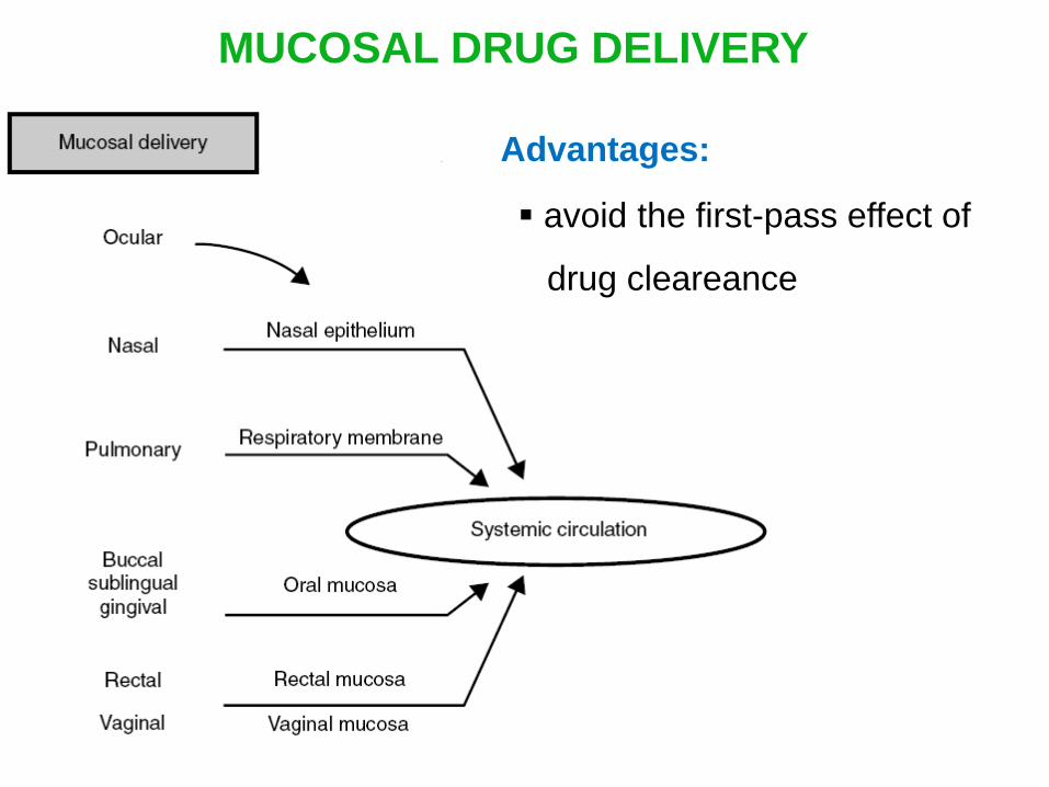
MUCOSAL DRUG DELIVERY
Advantages:
avoid the first-pass effect of
drug cleareance

ORAL MUCOSAL ROUTE
Sublingual delivery
Classification:
Buccal delivery
Local delivery
Sublingual delivery Buccal delivery Local delivery
Surface area: 100 cm2
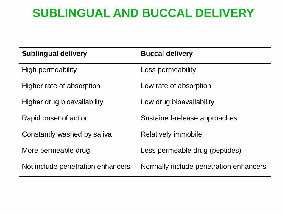
SUBLINGUAL AND BUCCAL DELIVERY
Sublingual delivery Buccal delivery
High permeability Less permeability
Higher rate of absorption Low rate of absorption
Higher drug bioavailability Low drug bioavailability
Rapid onset of action Sustained-release approaches
Constantly washed by saliva Relatively immobile
More permeable drug Less permeable drug (peptides)
Not include penetration enhancers Normally include penetration enhancers

What do you know about saliva?
Saliva: protective aqueous fluid, 1% organic & 99% inorganic
materials
Salivary pH: 5.5 – 7 depending on the flow rate
Flow rate increases [HCO3] increases pH increases
Daily salivary volume: 0.5 – 2 L
Oral cavity is a water-rich environment
Selection of hydrophylic matrices as drug carriers

PULMONARY ADMINISTRATION
Large mucosal surface of respitory system for drug absorption
« Taking advantage of the body’s ability to transfer large molecules
through the lung is a better way to deliver drugs than sticking people with
needles » Patton, Chemtech 1997, 27, 34

PULMONARY ADMINISTRATION
Advantages:
larger surface area (70 m2)
very fast onset of action (comparable to intravenous route)
Disadvantages:
lack of reproducibility of the administered dose
variable rate of absorption of drug at different regions

TRANSDERMAL AND TOPICAL ADMINISTRATION
STRUCTURE OF THE SKIN

TRANSDERMAL AND TOPICAL ADMINISTRATION
STRUCTURE OF THE SKIN
Epidermis structure:

TRANSDERMAL AND TOPICAL ADMINISTRATION
STRUCTURE OF THE SKIN
Stratum corneum structure:
Penetration barriers: lipid self-assemblies in the stratum
corneum (10%)

TRANSDERMAL AND TOPICAL ADMINISTRATION
PERMEABILITY ENHANCEMENT
By surfactant-based formulations (penetration enhancers,
liposomes,…):
By hydration of the stratum corneum:
Surfactant-based formulations can interact with lipids and alter
their structure enhance drug penetration
Water can interact with the polar headgroups of lipids through hydrogen
bonding loosen the lipid packing the lipid region becomes more fluid
Enhance drug penetration

PENETRATION ENHANCEMENT
Barry, Int. J. Cosmet. Sci. 1988, 10, 281

PENETRATION (PERMEABILITY) ENHANCERS
Definition:
Compounds that promote the absorption of drugs through the
skin or mucosae, usually by reversibly altering the permeability of
the barrier.
Characteristics:
Non toxic
Pharmacologically inert
Immediate in action
Reversible in action
Chemically & physically compatible with the drug and with the
skin/mucosae
Cosmetically acceptable

PENETRATION (PERMEABILITY) ENHANCERS
Common penetration enhancers:
Aprotinin
Dextrans

PENETRATION (PERMEABILITY) ENHANCERS
Common penetration enhancers:

SELECTION OF ROUTE OF DRUG
ADMINISTRATION
Affects the onset and duration of drug action
Depends on the desired drug concentration profiles to be
achieve, patient and disease
Ex: Administration of nitroglycerin

Oral 53%
Inhalation
32%
Transdermal
8%
Injectable/
Implant 3%
Ocular 2%
Nasal 2%
GLOBAL DRUG DELIVERY MARKET BY
ADMINISTRATION MODE

CLASSIFICATION OF DRUG DELIVERY SYSTEM
By the way of delivery:
Targeted drug delivery systems: deliver drug to a desired
body location, organ, tissue, specific cells…
Controlled-release drug delivery systems: preprogramed
drug release.
By the route of delivery:
Topical (local) drug delivery systems: drugs are delivered
locally to the target organ/tissue without entering the
systemic circulation.
Systemic drug delivery systems: drugs are delivered to
the whole body via the general blood circulation

DIFFERENT KINDS OF DRUG
Tablets, pills Capsules Suppositories
Creams
Ointments Liquids Aerosols Injectables
Repeat administration
Fluctuation of drug concentration in the body

DRUG RELEASE PROFILE
FLUCTUATION IN DRUG CONCENTRATION
1 2 3 4
A 1
A 2
A 3
A 4 Drug concentration
Frequencies of dosing
B
Toxic level
Adverse side effects
Therapeutic range
Minimum effective concentration
No therapeutic range
A1, A2, A3, A4: series of multiple doses of a conventional drug delivery system
B: ideal drug concentration profile
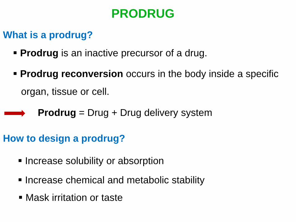
PRODRUG
Prodrug is an inactive precursor of a drug.
Prodrug reconversion occurs in the body inside a specific
organ, tissue or cell.
What is a prodrug?
Prodrug = Drug + Drug delivery system
How to design a prodrug?
Increase solubility or absorption
Increase chemical and metabolic stability
Mask irritation or taste

MAIN TYPES OF PRODRUGS FOR
TARGETED DRUG DELIVERY
(a): Classic prodrugs: prodrugs are transformed into one or
more active substances inside the cell
(b): Two or more substances react to form the active drug under
specific intracellular conditions
(c): Advanced forms of prodrugs containing 3 components

CLASSIC PRODRUGS
Reconversion by the cleavage of a chemical bond
(normally by enzymes in the targeted cell)
Ex: Sulfasalazine for the treatment of inflammatory bowel disease
Colonic bacteria
5-aminosalicylic acid
sulfapyridine
active moiety
Azo modification

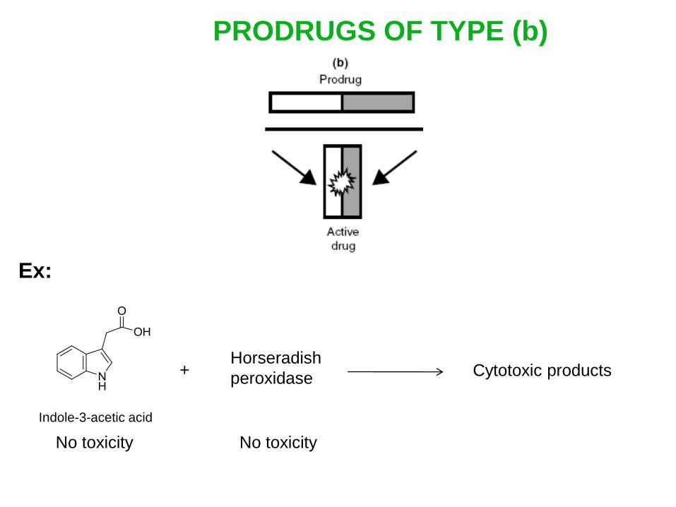
PRODRUGS OF TYPE (b)
Ex:
Indole-3-acetic acid
+ Horseradish
peroxidase Cytotoxic products
No toxicity No toxicity

ADVANCED FORMS OF PRODRUGS
Carrier: binding other components and altering the
physicochemical properties (ex solubility) of prodrug
Targeting moiety: enhancing the specific activity of the drug
on targeted cells
decreasing adverse side effects on
healthy tissues
Reconversion by the cleavage of a chemical bond
(normally by enzymes in the targeted cell)

ADVANTAGES OF PRODRUG OF TYPE (c)
The conditions of drug release can be precisely controlled by
modifying the bonds and each constituent
Prevent the degradation of the active component, reduce its
total body clearance
Release the drug inside targeted cells

DRUG DELIVERY SYSTEMS
Micelles
Liquid crystalline phases
Liposomes
Microemulsions
Emulsions
Polymers and dendrimers
Nanoparticles
Hydrogels

LIPOSOMES IN DRUG DELIVERY

WHAT ARE LIPOSOMES?
First described by Bangham and Horne after their study of lipid
phase structures in the electron microscope in 1964
Bangham, Horne, J. Mol. Biol. 1964, 8, 660
Liposomes are concentric bilayered vesicles in which an
aqueous volume is entirely enclosed by a membraneous lipid
bilayer mainly composed of natural or synthetic phospholipids
Liposomes are formed spontaneously when lipids are dispersed
in an aqueous medium (auto-assembly)

Why liposmes are used in drug delivery ?
Phospholipid bilayer
Aqueous cavity Polar head group
Hydrophobic tail
Storage ability of hydrophilic substances in the aqueous cavity
and hydrophobic drugs in the membrane
Similarity of structure to phospholipid membranes in living cells
Avoid the hydrolytic degradation of drugs in water
Reduce the RES uptake due to the rapid clearance of drugs
from bloodstream circulation (especially in intravenous administration)

Phospholipid bilayer
Aqueous cavity Polar head group
Hydrophobic tail
CELL MEMBRANE AND LIPOSOME

SIZE OF LIPOSOMES
Atoms Molecules Proteins Virus Bacteria Cells
Size of nanoparticles
Size of liposomes varies depending on how they are prepared
Size of liposomes

STRUCTURAL COMPONENTS OF LIPOSOMES
The main components of liposomes are phospholipids and
sterols
Phospholipids are the major strutural components of
biological membranes such as cell membranes
The most common phospholipids in biological membranes:
Phosphoglycerides
Sphingolipids

PHOSPHOGLYCERIDES
R1, R2 = fatty acid chain
R3 Phosphoglycerides
Phosphatidylcholine (PC)
Phosphatidylethanolamine (PE)
Phosphatidylserine (PS)
Phosphatidylglycerol (PG)
Phosphatidic acid (PA)
Phosphatidylinositol (PI)

SPHINGOLIPIDS
Sphingosine
Sphingolipids
N-acylsphingosine, ceramide
R1 = fatty acid chain
R1 = fatty acid chain
R2 = polar head group

STEROLS
Basic core of sterols
Cholesterol
Fluidity buffer for membrane with respect to temperature
Hydroxyl group in position 3 allows functionalization by various
functional groups

INFLUENCE OF CHOLESTEROL
ON THE STABILITY OF LIPOSOMES
Reduce the membrane permeability of phospholipid bilayers
Needham and Nunn, 1990
Grit and Crommelin, 1993
Increase the stability of liposomes in vitro and in vivo
Cholesterol is added in lipid mixtures used to produce
liposomes in concentrations of 30-50 mol%
almost non-polar increase hydrophobic interactions in the
phospholipid bilayers drecrease the fluidity of the membrane
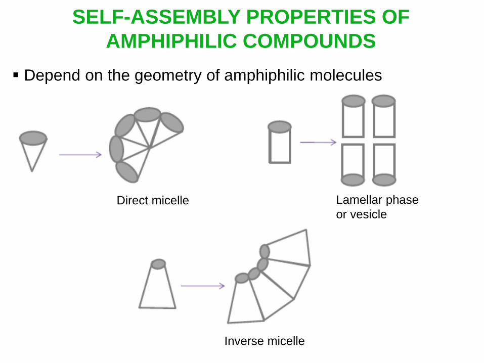
SELF-ASSEMBLY PROPERTIES OF
AMPHIPHILIC COMPOUNDS
Depend on the geometry of amphiphilic molecules
Direct micelle
Inverse micelle
Lamellar phase
or vesicle

SELF-ASSEMBLY PROPERTIES OF
AMPHIPHILIC COMPOUNDS
Structure of the aggregate can be predicted from
packing parameter
P = V
a l
V: total volume of the molecule
a: maximum cross sectional area of the head group
l: length of the hydrophobic chain

P = V
a l
Spherical micelles
Globular micelles
Cylindrical micelles
Vesicles
Lamellar phase
Lamellar phase
Inverse micelles

CLASSIFICATION OF LIPOSOMES
Liposomes are classified depending on their size
Liposomes SUVs - Small
Uni-lamellar Vesicles
LUVs - Large
Uni-lamellar Vesicles
MLVs – Multilamellar
Large Vesicles
Diameter
(nm)
20- 100 100 - 500 > 500
Cross
section

PREPARATION OF LIPOSOME
DELIVERY SYSTEM
By the lipid film rehydration method
Minko et al. J. Appl. Physiol. 2002, 93, 1550
Pakunlu et al. Pharm. Res. 2003, 20, 351
Phospholipids
+
Drug solution
1. CHCl3
2. Evaporation to
a thin film
Dry lipid film Buffer (pH = 7.4)
agitation
MLVs
Sonication
22 kHz
SUVs Filters
SUVs of
homogenous size
Lipid-soluble drug Water-soluble drug
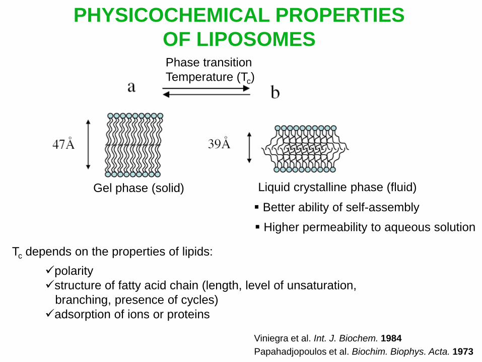
PHYSICOCHEMICAL PROPERTIES
OF LIPOSOMES Phase transition
Temperature (Tc)
Gel phase (solid) Liquid crystalline phase (fluid)
Better ability of self-assembly
Higher permeability to aqueous solution
Tc depends on the properties of lipids:
polarity
structure of fatty acid chain (length, level of unsaturation,
branching, presence of cycles)
adsorption of ions or proteins
Viniegra et al. Int. J. Biochem. 1984
Papahadjopoulos et al. Biochim. Biophys. Acta. 1973

INCORPORATION OF DRUGS IN LIPOSOMES
Through a drug concentration gradient
Before the formation of liposomes
Through a pH gradient
Drug: weak base

ACTIVE LOADING OF DRUG INTO PREFORMED
LIPOSOMES BY pH GRADIENT

ACTIVE LOADING OF DRUG INTO PREFORMED
LIPOSOMES BY pH GRADIENT
Drug = weak base:
Doxorubicin
Adriamycin
(cancer chemotherapy)
Vincristine
(cancer chemotherapy)

CHARACTERIZATION OF LIPOSOME SYSTEMS
Properties of liposomes Analysis
Physical size, shape and its distribution Cryo-TEM
Laser light scattering
Size exclusion chromatography (SEC)
NMR
Stability Dynamic light scattering (DLS)
Surface charge, zeta potential Free flow electrophoresis
Extent of drug entrapped Size exclusion chromatography (SEC)
NMR
Entrapped volume/lipid weight
NMR
Carboxyfluorescene
Lamellarity
Cryo-TEM 31P NMR
Phase transition temperature (Tc) Differential scanning calorimetry (DSC)
Purity of phospholipids Thin layer chromatography (TLC)
HPLC
NMR

CHARACTERIZATION OF LIPOSOME SYSTEMS
Light scattering techniques
NMR
Cryogenic transmission electron microscopy (Cryo-TEM)
Differential Scanning Calorimetry (DSC)
Drug release
Fluorescene spectroscopy

CONTROL OF LIPOSOME FORMATION BY NMR
By 31P solid-state NMR:
DSPC:DPPE-PEG2000:cholesterol = 60:15:25 mol%
High PEG-lipid content
C. Leal et al. J. Colloid Interface Sci. 2008, 325, 485

By 31P solid-state NMR:
DSPC:DPPE-PEG5000:cholesterol = 78:4.5:17.5 mol%
65C
Low PEG-lipid content
C. Leal et al. J. Colloid Interface Sci. 2008, 325, 485
CONTROL OF LIPOSOME FORMATION BY NMR

By 31P solid-state NMR:
DSPC:DPPE-PEG2000:cholesterol = 67:8:25 mol%
65C
Intermediate PEG-lipid content
C. Leal et al. J. Colloid Interface Sci. 2008, 325, 485
CONTROL OF LIPOSOME FORMATION BY NMR

C. Leal et al. J. Colloid Interface Sci. 2008, 325, 485
DSPC:DPPE-PEG2000:cholesterol
65C
By 31P solid-state NMR:
The higher the PEG-lipid content, the smaller the micelles are
CONTROL OF LIPOSOME FORMATION BY NMR

M. Delcea et al. http://arxiv.org/abs/0904.1662v1, 2009
By 1H NMR:
CONTROL OF LIPOSOME FORMATION BY NMR
DOPC lipid in chloroform
DOPC vesicles in HEPES buffer
Also confirmed by DOSY NMR

M. Delcea et al. http://arxiv.org/abs/0904.1662v1, 2009
By 1H NMR:
CONTROL OF LIPOSOME FORMATION BY NMR
The higher the line width, the bigger the molecules are
DMPG vesicles in HEPES buffer
Also confirmed by DOSY NMR
DMPG lipid in chloroform

EVALUATION OF LIPOSOME SIZE BY
DIFFUSION NMR SPECTROSCOPY
The (mean) hydrodynamic radius R of a particle can be derived
from the diffusion coefficient D by the Stokes-Einstein equation:
Principles:
D = kBT
6R
D: (translational) diffusion coefficient
kB: Boltzmann constant
T: absolute temperature
: solvent viscosity
In general, by measuring D, these informations can be obtained:
effective size
binding phenomena, association constant
aggregation
molecular interactions Y. Cohen et al. Angew. Chem. Int. Ed. 2005, 44, 520

DIFFUSION ORDERED SPECTROSCOPY (DOSY)
– NMR CHROMATOGRAPHY
Means for “virtual separation” of compounds
One axis is the chemical shift, the other is that of the diffusion
coefficient
2D DOSY spectrum
Y. Cohen et al. Angew. Chem. Int. Ed. 2005, 44, 520

EVALUATION OF LIPOSOME SIZE BY
DIFFUSION NMR SPECTROSCOPY
C. Leal et al. J. Colloid Interface Sci. 2008, 325, 485

EVALUATION OF LIPOSOME SIZE BY
DIFFUSION NMR SPECTROSCOPY
D 10-11 m2/s
D 10-12 m2/s
C. Leal et al. J. Colloid Interface Sci. 2008, 325, 485

EVALUATION OF LIPOSOME SIZE BY
DIFFUSION NMR SPECTROSCOPY
C. Leal et al. J. Colloid Interface Sci. 2008, 325, 485
CPEG: PEG-lipid content in the liposomes
DLIP/1012, DMIC/1012: micelle and liposome diffusion coefficients x 1012
dLIP, dMIC: micelle and liposome diameters
dPCS: liposome diameters obtained by photon correlation spectroscopy
MIC: fraction of micelles

EVALUATION OF DRUG ENCAPSULATION BY
DIFFUSION NMR SPECTROSCOPY
Y. Cohen et al. Angew. Chem. Int. Ed. 2005, 44, 520
Principles:
Drug size << liposome size
In free state: Ddrug >> Dliposome
In encapsulated state: Ddrug Dliposome
The quantity of drug encapsulated in liposomes can be
evaluated

CRYOGENIC TRANSMISSION ELECTRON
MICROSCOPY (CRYO-TEM)

CRYOGENIC TRANSMISSION ELECTRON
MICROSCOPY (CRYO-TEM)
Information:
Size (nm µm) and shape of complex structures in the solution
state
Applications:
Identify new morphologies and phases in the solution state
Characterize the interplay between different objets in solution
Used as complement to scattering techiniques:
to improve the modeling of scattering data
to discern the polydispersity in size and shape of
assembled structure
characterize intermediate structures
Zhong and Pochan, Polym. Rev. 2010, 50, 287

CRYOGENIC TRANSMISSION ELECTRON
MICROSCOPY (CRYO-TEM)
Advantages:
Zhong and Pochan, Polym. Rev. 2010, 50, 287
characterize in situ, in the solution state
without the need of modeling as required in scattering
techniques (neutron, X-ray, light scattering)
Disadvantages:
heavy technical skills for sample preparation and observation

CRYOGENIC TRANSMISSION ELECTRON
MICROSCOPY (CRYO-TEM)
Sample preparation:
place a drop of sample onto an EM-grid
remove excess solution by a filter paper, leaving a thin film of
the solution on the EM-grid
Plunge rapidly the grid into liquid ethane held just above the
freezing point (cooled by liquide nitrogen) to vitrify the sample
and avoid sample’s crystallization
Transfer the vitrified sample at low temperature to the
microscope

CRYOGENIC TRANSMISSION ELECTRON
MICROSCOPY (CRYO-TEM)
Sample preparation:

CRYO-TEM IMAGES OF LIPOSOMES FORMED BY
EPC : CHOLESTEROL = 60 : 40 MOL%
Edwards et al. Biophys. J. 1997, 73, 258
EPC: egg yolk lecithin
Liposomes have spherical shapes
Ice crystal deposited on the
sample surface after vitrification
100 nm

CRYO-TEM IMAGES OF LIPOSOMES FORMED BY
EPC : CHOLESTEROL = 40 : 60 MOL%
Edwards et al. Biophys. J. 1997, 73, 258
elongated or tube-shaped liposomes
segregation of the lipid and cholesterol
components
100 nm

CRYO-TEM IMAGES OF LIPOSOMES FORMED BY
EPC : CHOLESTEROL = 15 : 85 MOL%
Edwards et al. Biophys. J. 1997, 73, 258
revert back to a spherical shape
100 nm
Crystalline particles

CRYO-TEM IMAGES OF LIPOSOMES FORMED BY
DSPC/CHOLESTEROL
Without PEG2000-DSPE Without 5% PEG2000-DSPE
Edwards et al. Biophys. J. 1997, 73, 258
DSPC: 1,2-distearoylphosphatidylcholine
PEG2000: polyethylene glycol, M = 2000 Da
DSPE: 1,2-distearoylphosphatidylethanolamine

DIFFERENTIAL SCANNING CALORIMETRY (DSC)
Principles:
both sample and reference are maintained at nearly the same
temperature
the difference in the amount of heat (Cp) required to increase
the temperature of the sample and the reference is measured
as a function of temperature
through comparison between the sample and the reference,
melting and other transitions in liposome systems are monitored

DIFFERENTIAL SCANNING CALORIMETRY (DSC)
Principles:
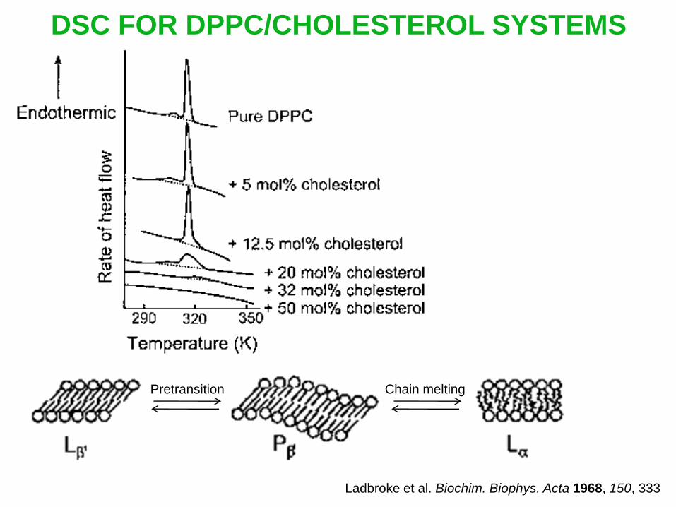
DSC FOR DPPC/CHOLESTEROL SYSTEMS
Ladbroke et al. Biochim. Biophys. Acta 1968, 150, 333
Pretransition Chain melting

DRUG RELEASE
Principles:
Through the release of a component encapsulated in liposomes,
information relating to the permeability of the lipid bilayers can
be obtained.
Frequently used methods:
carboxyfluorescene self-quenching
ethidium intercalation with DNA

DRUG RELEASE

FLUORESCENE SPECTROSCOPY
Informations:
localization of a solubilized molecule in a lipid membrane in a
liposome (by measuring the fluorescene anisotropy)
fluorescene anisotropy degree of orientation localization

FLUORESCENE SPECTROSCOPY
Fluorescene anisotropy <r> of 4-heptadecyl-7-hydroxycoumarin (HC)
and 1,6-diphenyl-1,3,5-hexatriene (DPH) in egg
phosphatidylcholine/cholesterol liposomes vs. temperature
HC with 5% PEG-PE
HC without 5% PEG-PE
DPH with 5% PEG-PE
DPH without 5% PEG-PE
DPH HC
Silvander et al. Langmuir 2000, 16, 3696

LIPOSOMES IN DRUG DELIVERY
Advantages:
Structural similarity to cell membrane structures
Capacity to encapsulate both water-soluble and oil-soluble drugs
Disadvantages:
Complicated preparation
Sterilization difficulties
Poor storage stability
Poor solubilization capacity for more hydrophobic drugs
Difficulties to control the drug release rate

INTRAVENOUS ADMINISTRATION
Problems:
Rapid elimination of drug carriers from bloodstream circulation
by the reticuloendothelial (RES) system low bioavailability
Accumulation of drugs in RES-related tissues (liver, spleen,
marrow) local toxicity

UPTAKE OF DRUG CARRIERS – PHAGOCYTOCIS
Drug carriers Adsorption of serum proteins
(opsonins) at the surface
Uptake by macrophage Engulfment of
drug carriers
macrophage
Drug carrier

EFFECTS OF THE SURFACE OF DRUG CARRIERS
ON THE PHAGOCYTOSIS
More hydrophobic particles
Hig
her
RE
S u
pta
ke o
f dru
g c
arr
iers
More highly charged particles
Tabata et al. Adv. Polym. Sci. 1990, 94, 106

ZETA POTENTIAL – A MEASURE OF CHARGES OF
SUSPENDED PARTICLES
Double layer
Gouy layer

UPTAKE OF DRUG CARRIERS – PHAGOCYTOCIS
Dependence of phagocytocis on properties of drug carriers:
Size
Surface:
hydrophobicity
charge
chemical functionality
The adsorption of serum protein can be reduced by
modifying the structure of phospholipids

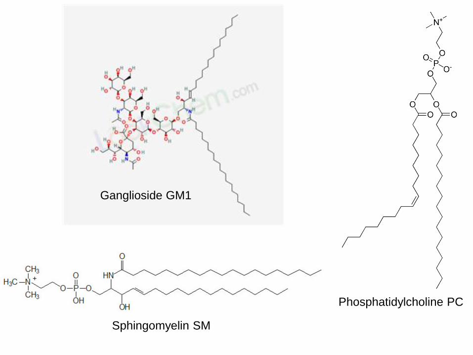
PHOSPHOLIPID STRUCTURE AND THE SERUM
PROTEIN ADSORPTION
Chonn et al. J. Biol. Chem. 1992, 267, 18759
Ganglioside GM1 (zero charge)
Sphingomyelin SM (zero net charge)
Liposomes containing:
Phosphatidylcholine PC (zero net charge)
Phosphatidylinositol PI (one negative charge, shielded by an outer group)
Diphosphatidylglycerol DPG (two negative charge)
Phosphatidic acid PA (two negative charge)
Ganglioside GM1
Sphingomyelin SM
Phosphatidylcholine PC

Phosphatidylinositol PI
Diphosphatidylglycerol DPG
Phosphatidic acid PA

ADSORPTION OF SERUM PROTEINS
DRIVING FORCES
Electrostatic interaction
Hydrophobic interaction
Van der Waals interaction
Interactions that drive the adsorption:
Modification of liposomes by adding poly(ethylene glycol)
(PEG) or poly(ethylene oxide) (PEO) chains
PEG PEO
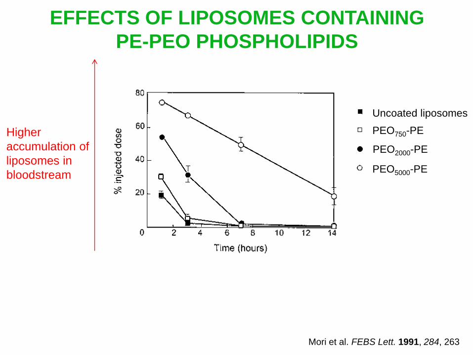
EFFECTS OF LIPOSOMES CONTAINING
PE-PEO PHOSPHOLIPIDS
PEO750-PE
PEO2000-PE
PEO5000-PE
Uncoated liposomes
Higher
accumulation of
liposomes in
bloodstream
Mori et al. FEBS Lett. 1991, 284, 263

EFFECTS OF LIPOSOMES CONTAINING
PE-PEO PHOSPHOLIPIDS
PEO750-PE
PEO2000-PE
PEO5000-PE
Uncoated liposomes
Higher
uptake of
liposomes
in liver and
spleen
Mori et al. FEBS Lett. 1991, 284, 263

EFFECTS OF LIPOSOMES CONTAINING
PE-PEO PHOSPHOLIPIDS - SUMMARY
longer bloodstream circulation time (higher bioavailability)
decrease of accumulation in RES-related tissues
increase of accumulation in tissues not related to the RES
reduction of adverse side effects (lower toxicity)

INTRAVENOUS ADMINISTRATION – TARGETING
Target the drug carriers to a specific tissue or cell
Increase the drug bioavailability
Reduce adverse side effects
Prodrug structure:
PEO-modified liposomes are used to protect targeting moiety
and assure a long circulation time

EFFECTS OF PEO-MODIFIED LIPOSOMES IN
TARGETING DELIVERY
Binding to mouse
pulmonary artery
endothelial cells
PEO-modified liposomes functionalized with relevant antibodies
PEO-modified liposomes functionalized with an irrelevant antibodies
Uncoated liposomes Holmberg et al. J. Liposome Research 1990, 1, 393

TOPICAL ADMINISTRATION
Why liposomes are used in topical administration?
To overcome the barrier properties of the stratum corneum
Penetration enhancers may result in an increased systemic
drug level or even toxic effects
Highly deformable liposomes (transfersomes or elastic
liposomes) can transfer various kinds of drugs (different in
lipophilicity and molecular weight, proteins, peptides)

TOPICAL ADMINISTRATION
Two main routes of penetration:
Maghraby et al. Eur. J. Pharm. Sci. 2008, 34, 203
transappendageal pathway
transepidermal pathway
through the sweat glands
across the hair follicles
intercellular route
transcellular route
Driving force for penetration:
osmotic gradient

TOPICAL ADMINISTRATION
Transappendageal pathway:
Maghraby et al. Eur. J. Pharm. Sci. 2008, 34, 203

TOPICAL ADMINISTRATION
Transepidermal pathway:
Maghraby et al. Eur. J. Pharm. Sci. 2008, 34, 203

TOPICAL ADMINISTRATION
Passage of transfersomes through the stratum corneum:
1/10 transfersome diameter

TOPICAL ADMINISTRATION
Edge activators to produce elasticity of transfersomes:
destabilizes the lipid bilayer
increases deformability of the liposome
surfactant nature (sodium cholate, deoxycholate, dipotaasium
glycyrrhizinate…)

POLYMERS AND POLYMERIC
SYSTEMS FOR DRUG DELIVERY

OVERVIEW
Kim et al. Eur. J. Pharm. Biopharm. 2009, 71, 420
Controlled release
Polymeric drug
delivery system
Targeted delivery
Sustained release
Triggered release
Prodrug Enhanced
permeability and retention (EPR)
Targeting ligands
Triggered targeting
Hydrogels Smart polymers,
stimuli-sensitive hydrogels
Polymer-drug conjugates
Polymeric liposomes
and micelles
Smart polymers
Polymeric liposomes
and micelles
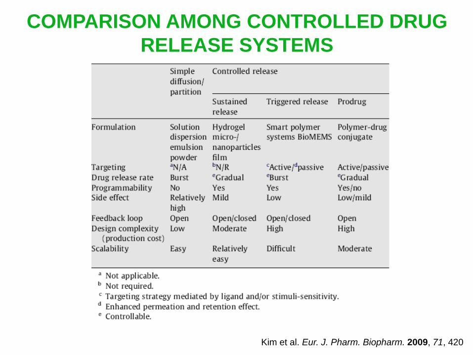
COMPARISON AMONG CONTROLLED DRUG
RELEASE SYSTEMS
Kim et al. Eur. J. Pharm. Biopharm. 2009, 71, 420

HYDROGELS
Definition:
Polymeric material that has the ability to absorb > 20% of its
weight of water (or biological fluids) and still maintain a distinct
3D structure
History:
1960: Wichterle & Lim first proposed the use of hydrophilic
networks of poly(2-hydroxyethyl methacrylate) (PHEMA) in
contact lenses
Wichterle & Lim. Nature 1960, 185, 117
extended to various biomedial & pharmaceutical applications

HYDROGELS
Structure:
Tridimensional network consisting of physical or chemical
cross-links ( branch)
Chemical (covalent, ionic)
cross-links, irreversible
Physical (entanglement,
hydrogen, VDW)
cross-links, reversible
Properties:
Insoluble (3D cross-linked network)
Infusible
Hydrophilic (presence of OH, COOH, CONH2, HSO3)
Swell in water

DIFFERENCE BETWEEN GEL AND HYDROGEL
Linear polymer
strands
Aqueous gel
Hydrogel
Polymer strands
dissolve in water
Polymer strands
swell in water
Physical network, weak interaction
Entanglement, hydrogen, VDW cross-links
Reversible
Soluble, fusible, dissolve in water
Chemical network
Covalent cross-links
Irreversible
Insoluble, infusible, swell in water

PREPARATION OF HYDROGEL-BASED
DRUG DELIVERY SYSTEMS
Gupta et al. Drug. Discov. Today. 2002, 7, 569
Chemical, physical
or radiation
cross-linking
Synthesis
Purification
Drying
Air/freeze drying
Drug loading
Filtration &
rinsing Drying

SYNTHESIS OF POLYMERS
By condensation polymerization of monomers (release of
H2O, NH3, HCl)
By free radical polymerization of monomers containg double
bond, through chain reaction with 3 stages
Ex: M < 20 000 Da poly(ethylene glycol) (PEG)
M > 20 000 Da poly(ethylene oxide) (PEO)
Initiation (thermal, photochemical, free radical or ionic)
Propagation
Termination

POLYMERIZATION PROCESSES FOR THE
PREPARATION OF HYDROGELS
Bulk polymerization
Suspention polymerization
Emulsion polymerization
Monomers + cross-linking agent + initiator hydrogels whose
morphologies depend on the mold used
Drops of monomer & initiator are dispersed in a concentrated
aqueous saline solution hydrogel beads
Emulsificaiton methods spherically shaped hydrogels

MONOMERS USED IN THE PREPARATION
OF SYNTHETIC HYDROGELS
=

COPOLYMERS
Constitued from different types of monomers ( homopolymers)
Chainings Name Notation Properties
ABBBABBAABAA Statistical (random)
copolymer
P(A-co-B) New material, properties
intermediate of PA & PB
ABABABABABAB Alternating copolymer P(A-alt-B) New material, properties
intermediate of PA & PB
AAAAAABBBBBB Block copolymer P(A-b-B) Superposition of
properties of PA & PB
Graft copolymer P(A-g-B) Superposition of
properties of of PA & PB,
for interface problems
To modify mechanical properties & swelling property (of
hydrogels)

COPOLYMERS – EXAMPLES
Hydrogels P(HEMA-co-MMA) PHEMA P(HEMA-co-VP)
Water content in
swelling state
less intermediate higher
more hydrophilic than MMA

HYDROPHILIC POLYMERS USED IN
PREPARATION OF HYDROGELS
Hamidi et al. Adv. Drug. Deliv. Rev. 2008, 60, 1638

CROSSLINKING METHODS USED IN HYDROGELS
Hamidi et al. Adv. Drug. Deliv. Rev. 2008, 60, 1638
Crosslinking methods
Chemically crosslinked Physically crosslinked
Crosslinking by radical polymerization
Crosslinking by high energy irradiation
Crosslinking using enzymes
Crosslinking by chemical reaction with
complementary groups
Crosslinking with aldehyde
Crosslinking with addition reaction
Crosslinking by condensation reaction
Crosslinking by ion interaction
Physically crosslinked hydrogels
from amphiphilic block and graft
copolymers
Crosslinking by crystallization
Crystallization in
homopolymer systems
Crosslinking by
stereocomplex formation

CROSS-LINKING AGENTS USED IN THE
SYNTHESIS OF HYDROGELS

RELATIONSHIP BETWEEN STRUCTURE AND
PROPERTIES OF HYDROGELS

CHARACTERIZATION PARAMETERS
FOR HYDROGELS
Gupta et al. Drug. Discov. Today. 2002, 7, 569

DEGREE OF SWELLING
capacity of hydrogels to absorb water or aqueous solution
Information:
%S =
%S: % age swelling
Ws: weight of the swollen gel
Wd: weight of the dry gel
diffusional properties of a solute through the hydrogel
% age swelling:
Ws Wd
Wd 100%

FACTORS THAT CONTROL THE
DEGREE OF SWELLING
Nature of monomers
Type & density of cross-link
Temperature
Ionic strength
pH of the hydration medium

FACTORS THAT AFFECT THE
DEGREE OF SWELLING
Hydrophilicity of monomers increases % S increases
Hydrogels P(HEMA-co-MMA) PHEMA P(HEMA-co-VP)
Water content in
swelling state
less intermediate higher
E.g.
Nature of monomers:
Type & density of cross-link:
Cross-linking agents increase % S decreases

FACTORS THAT AFFECT THE
DEGREE OF SWELLING Temperature:
Lower critical
solution
temperature
(LCST)
Temperature
Swelling state Collapsed state
Upper critical
solution
temperature
(UCST)
Temperature
Swelling state Collapsed state

FACTORS THAT AFFECT THE
DEGREE OF SWELLING
Temperature-LCST:
Non associated
Structured water
Less structured
bulk
Aggregation
Bromberg et al. Adv. Drug. Deliv. Rev. 1998, 31, 197
Hydrophobic association is entropy controlled:
S > 0 G = H T S < 0

FACTORS THAT AFFECT THE
DEGREE OF SWELLING
Ionic strength & pH of the hydration medium:
Due to the presence of of weakly acid and/or basic functional
groups on the backbone
E.g. COOH, NR3
Ionization of the functional groups depends on the pH & on the
ionic strength of the external medium
Ionization of the functional groups leads to water uptake
properties (osmotic driving force)

FACTORS THAT AFFECT THE
DEGREE OF SWELLING
Ionic strength & pH of the hydration medium:

DRUG RELEASE MECHANISMS
FROM HYDROGELS
Drug release takes place by 3 mechanisms
diffusion-controlled
swelling-controlled (when diffusion of drug is faster than
hydrogel swelling)
chemically-controlled
hydrolytic/enzymatic cleavage of polymeric chain
reversible/irreversible reactions between the polymer
network and the releasing drug

DRUG RELEASE MECHANISMS –
DIFFUSION CONTROLLED
Molecular diffusion
Drug diffusion

DRUG RELEASE MECHANISMS –
DIFFUSION CONTROLLED
Passive diffusion of drug across hydrogel membranes can be
described by the Fick’s first law
J k
x = D
dC k
x
dx
D = kBT
6R (Stokes-Einstein equation)
: Flux of species k along x - direction J k
x
D: diffusion coefficient
C: concentration
J depends on the mesh sizes of swollen hydrogels (5-100 nm)
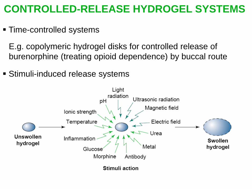
CONTROLLED-RELEASE HYDROGEL SYSTEMS
Time-controlled systems
E.g. copolymeric hydrogel disks for controlled release of
burenorphine (treating opioid dependence) by buccal route
Stimuli-induced release systems

STIMULI-INDUCED RELEASE SYSTEMS
Brannon-Peppas, MPB, 1997, 11, 34

pH-RESPONSIVE HYDROGELS

pH-RESPONSIVE HYDROGELS
PAA
PDEAEMA

TEMPERATURE-RESPONSIVE HYDROGELS
Bromberg et al. Adv. Drug. Deliv. Rev. 1998, 31, 197

DENDRIMERS FOR DRUG
DELIVERY

STRUCTURE OF DENDRIMERS
2D
3D
core
Branch point
periphery
cavity
4th generation
Generation: same external functional groups as starting molecule

SYNTHESIS OF DENDRIMERS
Divergent synthesis:
Coupling
3
Activation
6 Coupling
Activation
1st generation
2nd generation
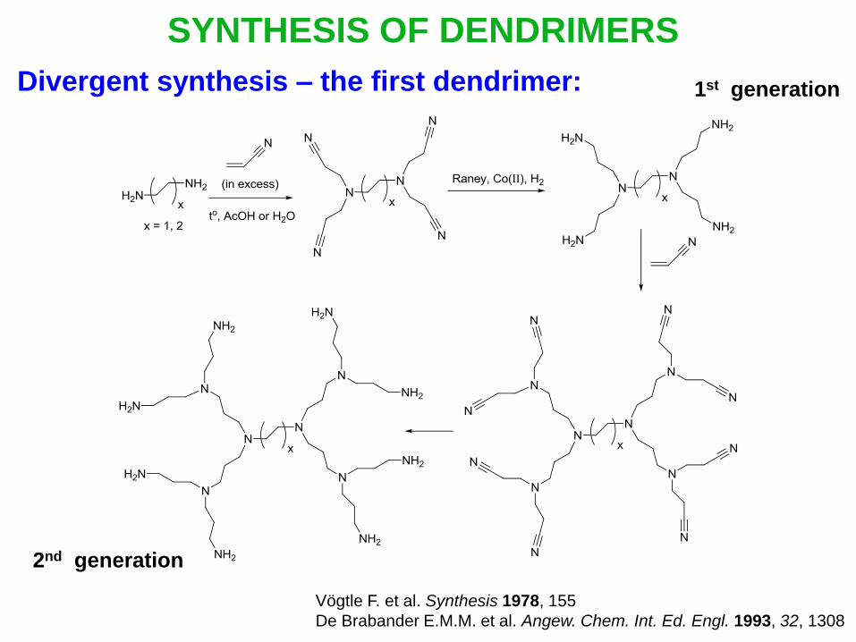
SYNTHESIS OF DENDRIMERS
Divergent synthesis – the first dendrimer: 1st generation
2nd generation
Vögtle F. et al. Synthesis 1978, 155
De Brabander E.M.M. et al. Angew. Chem. Int. Ed. Engl. 1993, 32, 1308

SYNTHESIS OF DENDRIMERS
Divergent synthesis – PAMAM (PolyAMidoAMine):
Amine & ester
terminated poly(amidodamine)
dendrimers Tomalia D. A. et al. Macromolecules 1986, 19, 2466
Tomalia D. A. US. Pat. 4507466, 1985,

SYNTHESIS OF DENDRIMERS
Convergent synthesis:
Dendron
Core
Dendrimer

SYNTHESIS OF DENDRIMERS
Convergent synthesis – preparation of dendrons:
Coupling Activation
1st generation
Coupling
2nd generation

SYNTHESIS OF DENDRIMERS
Convergent synthesis – Fréchet-type dendrons
(polyaryl ethers):
[G-1]-OH [G-1]-Br
[G-2]-Br
[G-3]-Br
Fréchet et al. J. Am. Chem. Soc. 1990, 112, 7638
Chem. Commun. 1990, 1010
Bn: Benzyl

SYNTHESIS OF DENDRIMERS
Comparison between divergent & convergent synthesis:
Core
4 eq
12 eq
36 eq
1/3 eq
1/3 eq
1/4 eq
Divergent
construction
Convergent
construction
Terminal site
for monomer
connectivity
Focal site
for monomer
connectivity
Core
Peripheral region Void volume
Branch juncture
Copyright © 2022 FDOKUMEN