MICROSPHERES FOR THE DRUG DELIVERY APPLICATIONS
10
Faculty of Pharmacy, Bahuddin Zakaryia University Multan, Pakistan http://pjpr.net 9 Introduction onventional dosage form provide a sharp increase in drug concentrations and drug concentration falls below the therapeutic range after a short interval of time until the re- administration of drug. So there is need of such dosage forms which provide not only the sustained delivery of drug but also reduce the fluctuations in the plasma drug levels and frequency of administration (Freiberg and Zhu, 2004). Recently a lot of work has been done to develop and formulate oral control release multiple unit dosage forms using natural and synthetic polymers, which are becoming more popular than the conventional single unit dosage forms due to its inherent advantages of providing uniform drug delivery; avoiding the vagaries of gastric emptying and different transit rates throughout the gastro-intestinal tract. This fact coupled with their ability to prolong the release of drug has increased the interest in developing the controlled drug delivery (Banerjee et al., 2012). One of the methods to achieve controlled drug delivery is developing the microspheres. These systems act as a reservoir of therapeutic agents, with spatial and temporal release of drugs for the desired therapeutic outcomes. The microspheres should have ability to incorporate drugs without loss of activity, tunable release kinetics, sufficient in vivo stability, biocompatibility, lack of toxicity, biodegradability and ability to target specific organs or tissue (Isıklan et al., 2011). Desired drug release rates by using microspheres can be achieved by rate controlling membrane or by the matrix of polymer containing the suspended drug. C Hafiz Shoaib Sarwar* 1 , Muhammad Hanif 1 , Aamir Jalil 2 , Malik Salman Haider 2 , Fahad Naeem 1 , Ahmad Nawaz 1 Vesh Churasiya 1 Microspheres for the Drug Delivery Applications Review Article Abstract Conventional dosage forms provide a sharp increase in plasma drug levels that falls below the therapeutic range after short interval of time until the re-administration of drug. There is a need of such dosage forms which provide not only sustained drug delivery but also reduce the plasma drug levels fluctuations. Microspheres used in drug delivery systems due to their ability to sustain the drug release, their biodegradability and compatibility and targeted drug delivery. In this review different types of microspheres their methods for the preparation with different hydrophilic and hydrophobic polymers, drug loading capacities will be discussed. Different characterizations like SEM, FTIR, XRD, DSC, rheological properties and invitro drug release are successfully described. Received: 02 Nov 2014 Revised: 26 Dec 2014 Accepted : 28 Dec 2014 Online: 30 Dec 2014 1 Department of Pharmacy, Bahuddin Zakaryia University, Multan, 2 Department of pharmacy, Comsats Intitute of Information and Technology, Abbotabad, Pakistan. Keywords: Biodegradable, Dosage forms, Microspheres, SEM, FTIR, XRD *Corresponding Author : Hafiz Shoaib Sarwar, Faculty of Pharmacy, Bahuddin Zakaryia University, Multan, Pakistan e-mail: : [email protected] , Ph: +92 3347891415
-
Upload
independent -
Category
Documents
-
view
2 -
download
0
Transcript of MICROSPHERES FOR THE DRUG DELIVERY APPLICATIONS
Faculty of Pharmacy, Bahuddin Zakaryia University Multan, Pakistan
http://pjpr.net 9
Introduction
onventional dosage form provide a sharp
increase in drug concentrations and drug
concentration falls below the therapeutic
range after a short interval of time until the re-
administration of drug. So there is need of such
dosage forms which provide not only the
sustained delivery of drug but also reduce the
fluctuations in the plasma drug levels and
frequency of administration (Freiberg and Zhu,
2004).
Recently a lot of work has been done to develop
and formulate oral control release multiple unit
dosage forms using natural and synthetic
polymers, which are becoming more popular than
the conventional single unit dosage forms due to
its inherent advantages of providing uniform drug
delivery; avoiding the vagaries of gastric
emptying and different transit rates throughout the
gastro-intestinal tract. This fact coupled with their
ability to prolong the release of drug has increased
the interest in developing the controlled drug
delivery (Banerjee et al., 2012).
One of the methods to achieve controlled drug
delivery is developing the microspheres. These
systems act as a reservoir of therapeutic agents,
with spatial and temporal release of drugs for the
desired therapeutic outcomes. The microspheres
should have ability to incorporate drugs without
loss of activity, tunable release kinetics, sufficient
in vivo stability, biocompatibility, lack of toxicity,
biodegradability and ability to target specific
organs or tissue (Isıklan et al., 2011). Desired
drug release rates by using microspheres can be
achieved by rate controlling membrane or by the
matrix of polymer containing the suspended drug.
C
Hafiz Shoaib Sarwar*1, Muhammad Hanif1, Aamir Jalil2, Malik Salman Haider2, Fahad Naeem1, Ahmad
Nawaz1 Vesh Churasiya1
Microspheres for the Drug Delivery Applications Review Article
Abstract
Conventional dosage forms provide a sharp increase in plasma drug levels that falls
below the therapeutic range after short interval of time until the re-administration of
drug. There is a need of such dosage forms which provide not only sustained drug
delivery but also reduce the plasma drug levels fluctuations. Microspheres used in
drug delivery systems due to their ability to sustain the drug release, their
biodegradability and compatibility and targeted drug delivery. In this review different
types of microspheres their methods for the preparation with different hydrophilic
and hydrophobic polymers, drug loading capacities will be discussed. Different
characterizations like SEM, FTIR, XRD, DSC, rheological properties and invitro drug
release are successfully described.
Received: 02 Nov 2014
Revised: 26 Dec 2014
Accepted : 28 Dec 2014
Online: 30 Dec 2014
1Department of Pharmacy, Bahuddin Zakaryia University, Multan,
2Department of pharmacy, Comsats Intitute of Information and Technology, Abbotabad, Pakistan.
Keywords: Biodegradable, Dosage forms, Microspheres, SEM, FTIR, XRD
*Corresponding Author : Hafiz Shoaib Sarwar,
Faculty of Pharmacy, Bahuddin Zakaryia University,
Multan, Pakistan
e-mail: : [email protected] ,
Ph: +92 3347891415
http://pjpr.net 10
The idea of controlled release dates back to 1960
through the implementation of silicone rubber and
polyethylene in the controlled drug delivery
systems (Folkman and Long, 1964). However
biodegradability of this system was a big issue
and required the surgical removal of the system.
Recently, the biodegradable and biocompatible
polymers have attracted much attention for use in
the drug delivery systems by using natural
polymers like chitosan, carboxymethylcellulose
(CMC), polylactic acid (PLA), polyvinyl alcohol
(PVA) and Sodium alginate etc. The microspheres
made from these polymers provide an excellent
way to deliver drug in controlled release manner
with biodegradability.
There are various methods that are used to
develop the microspheres with each method
having its own advantages and disadvantages and
suitability for different types of polymers and
drugs. Different methods also affect the
morphology and characteristics of microspheres
which in turn affect the drug release behavior of
the system. In this review, different types of
microspheres, their methods of preparation with
different hydrophilic and hydrophobic polymers
will be discussed. Different characterizations like
SEM, FTIR, XRD, DSC, rheological properties
and invitro drug release are successfully
described.
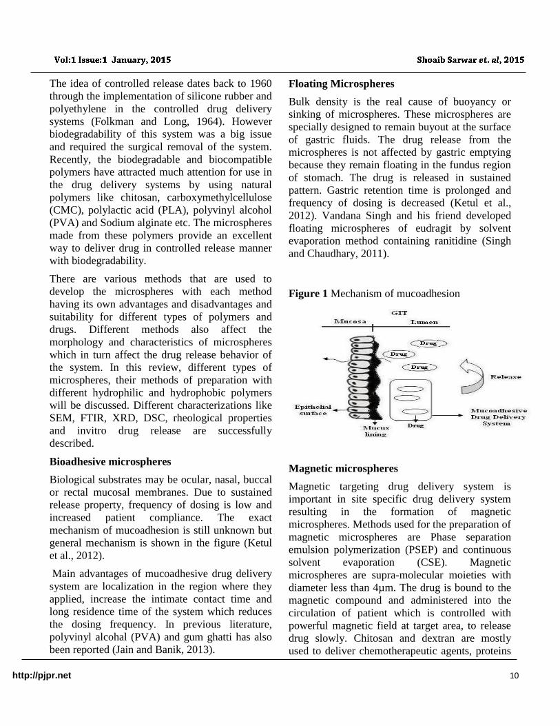
Bioadhesive microspheres
Biological substrates may be ocular, nasal, buccal
or rectal mucosal membranes. Due to sustained
release property, frequency of dosing is low and
increased patient compliance. The exact
mechanism of mucoadhesion is still unknown but
general mechanism is shown in the figure (Ketul
et al., 2012).
Main advantages of mucoadhesive drug delivery
system are localization in the region where they
applied, increase the intimate contact time and
long residence time of the system which reduces
the dosing frequency. In previous literature,
polyvinyl alcohal (PVA) and gum ghatti has also
been reported (Jain and Banik, 2013).
Floating Microspheres
Bulk density is the real cause of buoyancy or
sinking of microspheres. These microspheres are
specially designed to remain buyout at the surface
of gastric fluids. The drug release from the
microspheres is not affected by gastric emptying
because they remain floating in the fundus region
of stomach. The drug is released in sustained
pattern. Gastric retention time is prolonged and
frequency of dosing is decreased (Ketul et al.,
2012). Vandana Singh and his friend developed
floating microspheres of eudragit by solvent
evaporation method containing ranitidine (Singh
and Chaudhary, 2011).
Figure 1 Mechanism of mucoadhesion
Magnetic microspheres
Magnetic targeting drug delivery system is
important in site specific drug delivery system
resulting in the formation of magnetic
microspheres. Methods used for the preparation of
magnetic microspheres are Phase separation
emulsion polymerization (PSEP) and continuous
solvent evaporation (CSE). Magnetic
microspheres are supra-molecular moieties with
diameter less than 4µm. The drug is bound to the
magnetic compound and administered into the
circulation of patient which is controlled with
powerful magnetic field at target area, to release
drug slowly. Chitosan and dextran are mostly
used to deliver chemotherapeutic agents, proteins
http://pjpr.net 11
and peptides (Ketul et al., 2012,). Chitosan and
Poly (acrylic acid) magnetic microspheres has
also been reported for their potentials in drug
delivery (Guo et al., 2010).
Radio Active Microspheres
These are of two types; Therapeutic Radioactive
microspheres and diagnostic radioactive
microspheres. Many radio labeled isotopes are
best for the treatment of certain type of disorders.
The use of radioactive microspheres is still in
experimental stages, because of unwanted toxicity
and suboptimal therapeutic results. These were
used for the treatment of rheumatoid arthritis,
cystic brain tumor and liver tumor. Certain types
of polymers like PLA, polylactide-co-glycolide
(PLGA), chitosan, Polyanhydride,
Polycyanoacrylate, Agarose, Polyacrolein used
for radiolabelling (Häfeli, 2006). First diagnostic
radioactive microspheres used were white and red
blood cell. These were labeled with certain radio
isotopes and injected. Red blood cells labeled
with chromium used to detect the mass of RBCs
as well as function of spleen. Radioactive
microspheres are injected into the arteries, reach
to the tumor, and release high dose of radiation
without damaging the surrounding tissues (Ketul
et al., 2012).
Biodegradable Polymeric microspheres
Biodegradable polymeric microspheres can easily
be synthesized with natural or synthetic polymers.
The selection of polymer should be so wise that
the end product should be nontoxic because
sometimes it enter into the general circulation and
might be dangerous. Drug can be incorporated for
few days or few years. Enzymatic system and
hydrolysis process in the body degrade the natural
polymers like protein or polysaccharides (Singh et
al., 2011). Natural polymers have more sustained
release property because have prolong residence
time when come in contact with water.
Synthetic polymeric microspheres
Synthetic polymeric microspheres have a lot of
clinical uses. They are used as fillers and bulking
agents for soft tissues also have a role in
embolotherapy. Depot formulation has also been
made with such microspheres. The alarming
drawback of this system is the transmigration to
adjacent site of injection causing embolism and
further organ damage. The examples of such
polymers are PLA, Poly (Glycolic acid),
Polycaprolactone (PCL) (Saralidze et al., 2010).
Methods of preparation of microspheres
Solvent Diffusion method
Hollow microspheres can be prepared by solvent-
diffusion-evaporation method. In this method EC
and Polyvinyl pyrollidone (PVP) were dissolved
or dispersed in ethanol, followed by the addition
of drug and ether. This polymer blend containing
drug was added to the liquid paraffin premixed
with span-80, which was in water bath at 30oC
with continuous stirring at 300 rpm. Prepared
microspheres were collected by filtration, washed
and dried (Zhao et al., 2010). Similar method had
also been used to prepare eudragit SR-100
microspheres (Yang et al., 2003). Poly acrylic
acid (PAA) and PVP microsphers have also been
reported (Chun et al., 2005). Kawashima et al
prepared hollow microspheres by emulsion
solvent diffusion method (Sato et al., 2004).
Coacervation Method
Polymer is first dissolved in organic solvent
containing active pharmaceutical ingredient,
which may be solid or liquid. Desolvation of
polymer is done by the addition of nonsolvent or
polymer coacervating agent. Solubility of the
polymer will be decreased in the organic phase,
two phases will be formed and the coacervates
will be settled at the surface of active principle.
Curing agent will be added for the formation of
polymer lining on the surface of active principle
(Weinbreck et al., 2004). In another study Liu and
his coworkers prepared double walled
microspheres by this method using Chitosan as
polymer (Liu et al., 2007a). Chitosan
http://pjpr.net 12
microspheres were prepared by modifying this
technique in which the solution of drug and
crosslinker was sprayed on the polymeric solution
(El-Leithy et al., 2010). Salbutamol loaded
Gelatin
microspheres by coacervation phase separation
were prepared (Jayan et al., 2009).
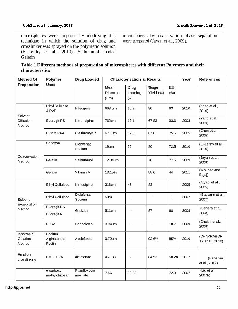
Table 1 Different methods of preparation of microspheres with different Polymers and their
characteristics
Method Of
Preparation
Polymer
Used
Drug Loaded Characterization & Results Year References
Mean
Diameter
(um)
Drug
Loading
(%)
%age
Yield (%)
EE
(%)
Solvent
Diffusion
Method
EthylCellulose
& PVP Nifedipine 668 um 15.9 80 63 2010
(Zhao et al.,
2010)
Eudragit RS Nitrendipine 762um 13.1 67.83 93.6 2003 (Yang et al.,
2003)
PVP & PAA Claithromycin 67.1um 37.8 87.6 75.5 2005 (Chun et al.,
2005)
Coacervation
Method
Chitosan
Diclofenac
Sodium 19um 55 80 72.5 2010
(El-Leithy et al.,
2010)
Gelatin Salbutamol 12.34um 78 77.5 2009 (Jayan et al.,
2009)
Gelatin Vitamin A 132.5% 55.6 44 2011 (Wakode and
Bajaj)
Solvent
Evaporation
Method
Ethyl Cellulose Nimodipine 316um 45 83 2005 (Atyabi et al.,
2005)
Ethyl Cellulose Diclofenac
Sodium 5um - - - 2007
(Baccarin et al.,
2007)
Eudragit RS
Eudragit Rl Glipizide 511um - 87 68 2008
(Behera et al.,
2008)
PLGA Cephalexin 3.94um - - 18.7 2009 (Chaisri et al.,
2009)
Ionotropic
Gelation
Method
Sodium-
Alginate and
Pectin
Acelofenac 0.72um - 92.6% 85% 2010 (CHAKRABOR
TY et al., 2010)
Emulsion
crosslinking CMC+PVA diclofenac 461.83 - 84.53 58.28 2012
(Banerjee
et al., 2012)
o-carboxy-
methylchitosan
Pazufloxacin
mesilate 7.56 32.38 72.9 2007
(Liu et al.,
2007b)
http://pjpr.net 13
Starch Rofecoxib 98.23 - 73.5 2009 (Thombre et
al., 2009)
Chitosan resveratral 53-311 - - 93.6 2010 (Peng et al.,
2010)
PVA+NA Alg diclofenac 281.61 - 78.03 55.8 2010 (Banerjee et
al., 2010)
Spray Drying Hayalouronic
acid fexofenadine 23.86 16.3 94.1 2010
(Bindu and
Sriram, 2012)
Chitosan+tripol
yposphate acetaminophin 6.74 - - 88.7 2005
(Desai and
Park, 2005)
Solvent Evaporation Method
Solvent evaporation is a well known method
having two types, Single emulsion technique and
double emulsion technique (Singh et al., 2011).
Microspheres of natural polymers can easily be
prepared by this method. Polymers and drugs are
dissolved in aqueous medium and this mixture is
dispersed into the non aqueous medium. The cross
linking of polymer can be done either with heat or
chemical crosslinkers like glutaraldehyde (GA).
Double emulsion technique involves the
formation of multiple emulsions e.g. w/o/w (Khan
et al., 2012). Successful encapsulation of certain
hydrophilic drugs, vaccines, proteins and peptides
was reported in previous literature. Protein
aqueous solution was made and added to
lipophilic organic solvent act as continuous phase,
which contain the polymers to encapsulate the
protein in aqueous phase. Primary emulsion was
formed, added into aqueous solution of PVA then
subjected to solvent evaporation (Yang et al.,
2001). Ethylcellulose (EC) and eudragit
microspheres by solvent evaporation method have
been reported in literature. (Atyabi et al., 2005,
Baccarin et al., 2007).
Ionotropic Gelation method
Polymer solution is made and added dropwise into
the crosslinker solution. Mostly used crosslinkers
are divalent cation, like Calcium chloride (CaCl2),
Zinc chloride. The size of microspheres depends
upon the gauge of needle. Large size
microspheres are formed, termed as pellets or
beads. Microsphers by using Chitosan coated with
alginate-gelatin have been studied and evaluated
by this method (Khan et al., 2012).
Emulsion Crosslinking
Mixture of hydrophilic polymers and drug
(dissolved or dispersed) is poured into oil phase
with constant magnetic stirring, forming w/o
emulsion using suitable surfactant and then
adding specified amount of crosslinker to form the
microspheres, hardened them by continous
stirring for 2 to 3 hours. Microspheres collected
by filtration washed with acetone and distilled
water, dried at 40o C in oven and stored (Banerjee
et al., 2010).
Chitosan microspheres crosslinked with
epicholorohydrin has been prepared by dissolving
chitosan in acetic acid solution. Oil phase used
was liquid paraffin contain a mixture of span 80
and tween 80 (1:1v/v) emulsifiers and vanillin as
crosslinker (Peng et al., 2010). Other polymers
like starch (Thombre et al., 2009);
carboxymethylchitosan (Liu et al., 2007b); and
protein like albumin (Mathew et al., 2009); has
also been used.
Spray Drying
It involves the drying of mist of polymer and drug
in the presence of hot air. The polymers are first
dissolved in a suitable organic solvent and the
drug
Faculty of Pharmacy, Bahuddin Zakaryia University Multan, Pakistan
http://pjpr.net 14
Table 2 : Types of microspheres with different polymers and drug loaded
is then dispersed in the polymer solution under
high speed homogenization. This dispersion is
then atomized in the stream of hot air which leads
to the formation of microspheres (1-
10um). Microparticles are then separated by
cyclone separator from the hot air while the traces
of solvent are removed by vacuum drying (Bindu
and Sriram, 2012). Nifedipine loaded PVA
microspheres (Saigal et al., 2013); hollow
microspheres of hydroxyappatite (Sun et al.,
2009); PLA and PLGA microspheres (Blanco et
al., 2006); and bupivacaine loaded PCL
microspheres (Blanco et al., 2003); have been
reported by spray drying method.
Characterization
Measurement of microsphere hydration (%)
Microspheres are weighed immediately after
filtration (M1) and weighed again after drying to
constant weight (M2)
Microspheres hydration (%) = M1
M2×100 (1)
Recovery of microspseres (%)
Recovery of formed microspheres is calculated by
dividing the weight of obtained microspheres to
the total weight of all ingredients charged during
the preparation of drug polymer mixture.
Percent yield (%) = 𝑊1
W0 ×100 (2)
Where W1 is the weight of obtained microspheres
and W0 is the weight of all the ingredients charged
in grams.
Rheological studies
Bulk and tapped density
Bulk density and tapped density will be calculated
by following formula
Bulk Density = m
V (3)
m represents the weight of microspheres and V is
the volume.
Tapped density can be calculated by using
following formula (Ranjha et al., 2010)
Tapped density = m
𝑉100 (4)
Where m is weight and V100 is volume of
microspheres after 100 tapings.
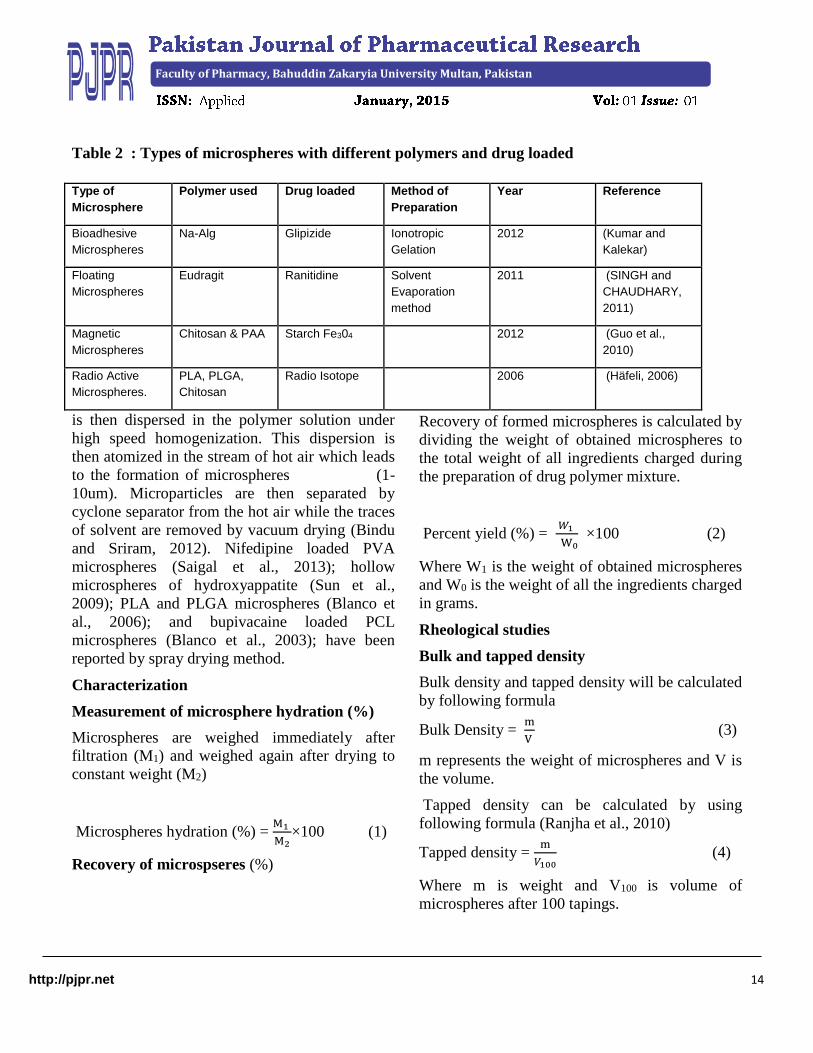
Type of
Microsphere
Polymer used Drug loaded Method of
Preparation
Year Reference
Bioadhesive
Microspheres
Na-Alg Glipizide Ionotropic
Gelation
2012 (Kumar and
Kalekar)
Floating
Microspheres
Eudragit Ranitidine Solvent
Evaporation
method
2011 (SINGH and
CHAUDHARY,
2011)
Magnetic
Microspheres
Chitosan & PAA Starch Fe304 2012 (Guo et al.,
2010)
Radio Active
Microspheres.
PLA, PLGA,
Chitosan
Radio Isotope 2006 (Häfeli, 2006)

Faculty of Pharmacy, Bahuddin Zakaryia University Multan, Pakistan
http://pjpr.net 15
Compressibility index and Huasner’s ratio
Hausner’s ratio is calculated by using following
formula
Hausner’s ratio = V1
𝑉2 (5)
Where vi is volume before tapings and V2 is
volume after tapings. A value closer to 1 indicates
good flow properties (Shariff et al., 2007).
Compressibility index also called Car’s index is a
indirect method of measurement for bulk density
as the factors like size, shape, surface area and
moisture content influence its value (Shariff et al.,
2007)
Compressibility index (%) = Vi–Vf
Vi ×100 (6)
Vi is initial volume, Vf is the final volume.
Angle of Repose
Angle of repose is measured by passing the
microspheres through funnel to a petri dish then
measuring the height (h) of heap formed and the
radius (r) of the petri dish angle of repose is
calculated as
Tanᶿ = ℎ
𝑟 (7)
Encapsulation Efficiency
Encapsulation efficacy is expressed as percentage
of actual drug loading to the total amount of drug
initially used. A specified amount of microspheres
were extracted for drug at 500C for 24 hours. The
solution is then filtered and the amount of drug is
calculated by UV-Spectrophotometer at specified
wavelength (Banerjee et al., 2010).
Encapsulation Efficiency = experimental drug content
theoretical drug content×100
(8)
Scanning Electron Microscopy
SEM uses electrons instead of light to form
image. SEM images have characteristic three
dimensional appearances and used to investigate
the size, surface structure, morphology and
texture of the prepared microspheres.
Fourier Transform Infrared Spectroscopy
FTIR is carried out to study the polymer-polymer
interaction and polymer drug interaction provides
information about the interaction of different
functional groups in grafting and crosslinking.
The spectra were recorded for pure drug and drug
loaded microspheres and samples are prepared in
KBR disc. Also provides the information about
the presence of free and chemically bound drug in
the microspheres.
X-Ray Powder Diffractrometery
To study the effect of microencapsulation on the
crystallinity of the drug in the microspheres,
carried out on pure drug, blank microspheres and
drug loaded microspheres.
Differential Scanning Calorimetery
Pure drug, polymer and drug loaded microspheres
are evaluated by DSC for possible drug polymer
interaction, by triturating separately and then
heating at aluminium pan at a rate of 10 0 C/min
from 0 to 1200 C under constant nitrogen flow.
Samples are run in triplicate for reproducibility.
In vitro drug release
In vitro drug release, from different formulations
of microspheres was studied by using dissolution
apparatus I or II in previous literature (Baccarin et
al., 2007). Composition of microspheres plays an
important role in the drug release behavior and the
release pattern is studied by different in vitro
kinetic models as follows
Zero order release equation (Kaushal et al.,
2004)
Ft = Kot (9)
Where Ft is the release of drug at a specific time
and K0 is the zero order rate constant
First order release (Mehrgan and Mortazavi,
2010)
Ln ( 1- F ) = K1t (10)

http://pjpr.net 16
F is the fraction of drug released and K1 is the
first order rate constant
Higuchi equation
F = k2 t ½ (11)
Where k2 is Higuchi constant
Hixon-crowell equation
𝑄𝑜1/3 − 𝑄𝑡1/3 = KHC (12)
Korsmeyer-peppas equation
𝑀1
𝑀 ∞= 𝑘3𝑡𝑛 (13)
M1 and M∞ are the released drug at time 0 and ∞
and n is the diffusion constant.
Conclusion
It is generally observed that as compared to other
conventional dosage form, microspheres are
useful option for controlled and sustained drug
delivery, targeted drug delivery ( Active and
passive targeting), radioactive and diagnostic
purposes, localized drug delivery system. They
show better patient compliance. Microspheres are
also good candidate for oral, nasal and pulmonary
delivery. So they play a very important role in the
advancement of medical field. Various methods
are used to prepare microspheres of required size,
shape and surface morphology. Different
properties like size, surface charge, hydrophilic
and hydrophobic nature of the microspheres are
responsible for determining the fate of
microspheres in the body. As compared to
existing technologies microspheres offers several
advantages. They have shown new ways to
biologist, biotechnologist and researcher as well
to bring revolution in the area of drug delivery. In
the recent years a lot of studies have been done on
microspheres which shows that they are good
alternatives for conventional as well as some
novel drug delivery system. In future microsphere
will attain central position in novel drug delivery
system by combining them with various other
systems
List of abbreviations
CMC Carboxymethycellulose
DSC Differential scanning calorimetery
FTIR Fourier transform infrared spctroscopy
GA Glutaraldehyde
HA Hayalouronic acid
PLA Polylacctic acid
PLGA Polylactide-co-glycolide
PVA Polyvinyl alcohol
SEM Scanning electron microscopy
XRD X-ray powder diffractometery
References
ATYABI, F., MOHAMMADI, A. & DINARVAND, R.
2005. Preparation of Nimodipine Loaded
Microspheres: Evaluation of Parameters. Iranian
Journal of Pharmaceutical Sciences, 1, 143-152.
BACCARIN, M. A., EVANGELISTA, R. C. & LUCINDA-
SILVA, R. M. 2007. Ethylcelullose Microspheres
containing Sodium Diclofenac: Development and
Characterization. Acta Farmaceutica Bonaerense,
25, 401.
BANERJEE, S., CHAURASIA, G., PAL, D., GHOSH, A.
K., GHOSH, A. & KAITY, S. 2010. Investigation
on crosslinking density for development of novel
interpenetrating polymer network (IPN) based
formulation. J. Sci. Ind. Res, 69, 777-784.
BANERJEE, S., SIDDIQUI, L., BHATTACHARYA, S. S.,
KAITY, S., GHOSH, A., CHATTOPADHYAY,
P., PANDEY, A. & SINGH, L. 2012.
Interpenetrating polymer network (IPN) hydrogel
microspheres for oral controlled release
application. International Journal of Biological
Macromolecules, 50, 198-206.
BEHERA, B., SAHOO, S., DHAL, S., BARIK, B. &
GUPTA, B. 2008. Characterization of glipizide-
loaded polymethacrylate microspheres prepared by
an emulsion solvent evaporation method. Tropical
Journal of Pharmaceutical Research, 7, 879-885.
BINDU, R. H. & SRIRAM, N. 2012. Available online at
www. JGTPS. com Research Article. Journal of
Global Trends in Pharmaceutical Sciences, 3, 849-
855.
BLANCO, M., BERNARDO, M., SASTRE, R., OLMO, R.,
MUNIZ, E. & TEIJÓN, J. 2003. Preparation of
bupivacaine-loaded poly (ε-caprolactone)
microspheres by spray drying: drug release studies
http://pjpr.net 17
and biocompatibility. European journal of
pharmaceutics and biopharmaceutics, 55, 229-236.
BLANCO, M. D., SASTRE, R. L., TEIJÓN, C., OLMO, R.
& TEIJÓN, J. M. 2006. Degradation behaviour of
microspheres prepared by spray-drying poly (d, l-
lactide) and poly (d, l-lactide-< i> co</i>-
glycolide) polymers. International Journal of
Pharmaceutics, 326, 139-147.
CHAISRI, W., HENNINK, W. E. & OKONOGI, S. 2009.
Preparation and characterization of cephalexin
loaded PLGA microspheres. Current Drug
Delivery, 6, 69-75.
CHAKRABORTY, S., KHANDAI, M., SHARMA, A.,
KHANAM, N., PATRA, C. N., PATRO, V. J. &
KUMAR SEN, K. 2010. Priprava, in vitro i in vivo
evaluacija bioadhezivnih mikrosfera s algino-
pektinom: ispitivanje utjecaja polimera pomoću
multiple poredbene analize. Acta pharmaceutica,
60, 255-266.
CHUN, M.-K., SAH, H. & CHOI, H.K. 2005. Preparation
of mucoadhesive microspheres containing
antimicrobial agents for eradication of H. pylori.
International Journal of Pharmaceutics, 297, 172-
179.
DESAI, K. G. H. & PARK, H. J. 2005. Preparation and
characterization of drug‐loaded chitosan–
tripolyphosphate microspheres by spray drying.
Drug development research, 64, 114-128.
EL-LEITHY, E., SHAKER, D., GHORAB, M. & ABDEL-
RASHID, R. 2010. Optimization and
characterization of diclofenac sodium microspheres
prepared by a modified coacervation method. Drug
discoveries & therapeutics, 4, 208.
FOLKMAN, J. & LONG, D. M. 1964. The use of silicone
rubber as a carrier for prolonged drug therapy.
Journal of surgical research, 4, 139-142.
FREIBERG, S. & ZHU, X. X. 2004. Polymer microspheres
for controlled drug release. International Journal
of Pharmaceutics, 282, 1-18.
GUO, L., LIU, G., HONG, R.-Y. & LI, H.-Z. 2010.
Preparation and characterization of chitosan poly
(acrylic acid) magnetic microspheres. Marine
drugs, 8, 2212-2222.
HÄFELI, U. 2006. Review: Radioactive microspheres for
medical applications. Cleveland clinic foundation,
Radiation Oncology Department T28.
IŞıKLAN, N., İNAL, M., KURŞUN, F. & ERCAN, G.
2011. pH responsive itaconic acid grafted alginate
microspheres for the controlled release of
nifedipine. Carbohydrate Polymers, 84, 933-943.
JAIN, N. & BANIK, A. 2013. Novel interpenetrating
polymer network mucoadhesive microspheres of
gum ghatti and poly (vinyl alcohol) for the delivery
of ranitidine HCL. Asian Journal of
Pharmaceutical and Clinical Research, 6, 119-123.
JAYAN, S. C., SANDEEP, A., RIFASH, M., MAREEMA,
C. & SHAMSEERA, S. 2009. Design and In-vitro
Evaluation of Gelatin Microspheres of Salbutamol
Sulphate. HYGEIA, 1.
KAKAR, S., BATRA, D., SINGH, R. & NAUTIYAL, U.
2012. Magnetic microspheres as magical novel
drug delivery system: A review. Journal of Acute
Disease, 2, 1-12.
KAUSHAL, A. M., GUPTA, P. & BANSAL, A. K. 2004.
Amorphous drug delivery systems: molecular
aspects, design, and performance. Critical Reviews
in Therapeutic Drug Carrier Systems., 21, 133-
194.
KETUL, P., GHANSHYAM, P., PATEL, M., PATEL, K. &
PATEL, N. 2012. INTERNATIONALE
PHARMACEUTICA SCIENCIA.
KHAN, S., TIWARI, T., RAO, N., JOSHI, A. & DUBEY,
B. K. 2012. Microspheres: a review. World journal
of pharmacy and pharmaceutical sciences, 1, 125-
145.
LIU, F., LIU, L., LI, X. & ZHANG, Q. 2007a. Preparation
of chitosan–hyaluronate double-walled
microspheres by emulsification-coacervation
method. Journal of Materials Science: Materials in
Medicine, 18, 2215-2224.
LIU, Y.-F., HUANG, K.-L., PENG, D.-M., DING, P. & LI,
G.-Y. 2007b. Preparation and characterization of
glutaraldehyde cross-linked O-
carboxymethylchitosan microspheres for controlled
delivery of pazufloxacin mesilate. International
Journal of Biological Macromolecules, 41, 87-93.
MATHEW, S. T., GAYATHRI DEVI, S., PRASANTH, V.
& VINOD, B. 2009. Formulation and in vitro-in
vivo evaluation of ketoprofen-loaded albumin
microspheres for intramuscular administration.
Journal of microencapsulation, 26, 456-469.
MEHRGAN, H. & MORTAZAVI, S. A. 2010. The release
behavior and kinetic evaluation of diltiazem HCl
from various hydrophilic and plastic based
matrices. Iranian Journal of Pharmaceutical
Research, 137-146.
PENG, H., XIONG, H., LI, J., XIE, M., LIU, Y., BAI, C. &
CHEN, L. 2010. Vanillin cross-linked chitosan
microspheres for controlled release of resveratrol.
Food Chemistry, 121, 23-28.
RANJHA, N. M., KHAN, H. & NASEEM, S. 2010.
Encapsulation and characterization of controlled
release flurbiprofen loaded microspheres using
beeswax as an encapsulating agent. Journal of
Materials Science: Materials in Medicine, 21,
1621-1630.
SAIGAL, A., NG, W. K., TAN, R. B. & CHAN, S. Y. 2013.
Development of controlled release inhalable
polymeric microspheres for treatment of
http://pjpr.net 18
pulmonary hypertension. International Journal of
Pharmaceutics.
SARALIDZE, K., KOOLE, L. H. & KNETSCH, M. L.
2010. Polymeric microspheres for medical
applications. Materials, 3, 3537-3564.
SATO, Y., KAWASHIMA, Y., TAKEUCHI, H. &
YAMAMOTO, H. 2004. In vitro evaluation of
floating and drug releasing behaviors of hollow
microspheres (microballoons) prepared by the
emulsion solvent diffusion method. European
journal of pharmaceutics and biopharmaceutics,
57, 235-243.
SHARIFF, A., MANNA, P., PARANJOTHY, K. &
MANJULA, M. 2007. Entrapment of
Andrographolide in Cross-Linked Aliginate
Pellets: II. Physicochemical Characterization to
Study the Pelletization of Andrographolide.
Pakistan Journal of Pharmaceutical Sciences, 20,
9-15.
SINGH, P., PRAKASH, D., RAMESH, B., SINGH, N. &
MANI, T. T. 2011. Indian Journal of Novel Drug
Delivery. Indian Journal of Novel Drug delivery, 3,
70-82.
SINGH, V. & CHAUDHARY, A. K. 2011. Preparation of
Eudragit E100 microspheres by modified solvent
evaporation method. Acta poloniae pharmaceutica,
68, 975-80.
SUN, R., LU, Y. & CHEN, K. 2009. Preparation and
characterization of hollow hydroxyapatite
microspheres by spray drying method. Materials
Science and Engineering: C, 29, 1088-1092.
THOMBRE, N., CHAUDHARI, M. & KADAM, S. 2009.
Preparation and characterization of rofecoxib
microspheres using cross-linked starch as novel
drug delivery system. Int J PharmTech Rese, 1,
1394-402.
WAKODE, R. & BAJAJ, A. Gelatin microspheres for
topical delivery of Vitamin A palmitate.
WEINBRECK, F., WIENTJES, R. H., NIEUWENHUIJSE,
H., ROBIJN, G. W. & DE KRUIF, C. G. 2004.
Rheological properties of whey protein/gum arabic
coacervates. Journal of Rheology, 48, 1215.
YANG, M.-S., CUI, F.-D., YOU, B.-G., FAN, Y.-L.,
WANG, L., YUE, P. & YANG, H. 2003.
Preparation of sustained-release nitrendipine
microspheres with Eudragit RS and Aerosil using
quasi-emulsion solvent diffusion method.
International journal of pharmaceutics, 259, 103-
113.
YANG, Y.-Y., CHUNG, T.-S. & PING NG, N. 2001.
Morphology, drug distribution, and in vitro release
profiles of biodegradable polymeric microspheres
containing protein fabricated by double-emulsion
solvent extraction/evaporation method.
Biomaterials, 22, 231-241.
ZHAO, L., WEI, Y.-M., YU, Y. & ZHENG, W.-W. 2010.
Polymer blends used to prepare nifedipine loaded
hollow microspheres for a floating-type oral drug
delivery system: In vitro evaluation. Archives of
pharmacal research, 33, 443-450.