Novel biodegradable polyesteramide microspheres for controlled drug delivery in Ophthalmology
13
Novel biodegradable polyesteramide microspheres for controlled drug delivery in Ophthalmology Vanessa Andrés-Guerrero a,b , Mengmeng Zong c , Eva Ramsay d,f , Blanca Rojas e , Sanjay Sarkhel d , Beatriz Gallego e , Rosa de Hoz e , Ana I. Ramírez e , Juan José Salazar e , Alberto Triviño e , José M. Ramírez e , Eva M. del Amo d,f , Neil Cameron g , Beatriz de-las-Heras b,h , Arto Urtti d,f , George Mihov c , Aylvin Dias c , Rocío Herrero-Vanrell a,b, ⁎ a Department of Pharmacy and Pharmaceutical Technology, Faculty of Pharmacy, Complutense University of Madrid, Spain b Pharmaceutical Innovation in Ophthalmology Research Group, Sanitary Research Institute of the San Carlos Clinical Hospital (IdISSC) and the Ocular Pathology National Net (OFTARED) of the Institute of Health Carlos III, Madrid, Spain c DSM, 6167 AC Geleen, The Netherlands d Centre for Drug Research, Division of Pharmaceutical Biosciences, Faculty of Pharmacy, University of Helsinki, P.O. Box 56, 00014, Finland e Instituto de Investigaciones Oftalmológicas Ramón Castroviejo, Complutense University of Madrid, Spain f School of Pharmacy, University of Eastern Finland, P.O. Box 1627, 70211 Kuopio, Finland g Department of Materials Engineering, Monash University, School of Engineering, University of Warwick, Clayton, Australia h Department of Pharmacology, Faculty of Pharmacy, Complutense University of Madrid, Spain abstract article info Article history: Received 3 March 2015 Received in revised form 18 May 2015 Accepted 19 May 2015 Available online 21 May 2015 Keywords: Ocular drug delivery Microspheres Poly(ester amide) Tolerance Dexamethasone Intraocular injection Most of the posterior segment diseases are chronic and multifactorial and require long-term intraocular medica- tion. Conventional treatments of these pathologies consist of successive intraocular injections, which are associ- ated with adverse effects. Successful therapy requires the development of new drug delivery systems able to release the active substance for a long term with a single administration. The present work involves the descrip- tion of a new generation of microspheres based on poly(ester amide)s (PEA), which are novel polymers with im- proved biodegradability, processability and good thermal and mechanical properties. We report on the preparation of the PEA polymer, PEA microspheres (PEA Ms) and their characterization. PEA Ms (~15 μm) were loaded with a lipophilic drug (dexamethasone) (181.0 ± 2.4 μg DX/mg Ms). The in vitro release profile of the drug showed a constant delivery for at least 90 days. Based on the data from a performed in vitro release study, a kinetic ocular model to predict in vivo drug concentrations in a rabbit vitreous was built. According to the pharmacokinetic simulations, intravitreal injection of dexamethasone loaded PEA microspheres would pro- vide release of the drug in rabbit eyes up to 3 months. Cytotoxicity studies in macrophages and retinal pigment epithelial cells revealed a good in vitro tolerance of the microsystems. After sterilization, PEA Ms were adminis- tered in vivo by subtenon and intravitreal injections in male Sprague–Dawley rats and the location of the micro- spheres in rat eyes was monitored. We conclude that PEA Ms provide an alternative delivery system for controlling the delivery of drugs to the eye, allowing a novel generation of microsphere design. © 2015 Published by Elsevier B.V. 1. Introduction Most of the diseases affecting the posterior segment of the eye are related with visual impairment and blindness. The effective treatment of these pathologies is one of the major challenges in drug delivery as most of them are chronic and multifactorial. Among them, aged- related macular degeneration, diabetic retinopathies and glaucoma pro- duce irreversible visual damage and blindness [1]. These diseases are becoming more and more prevalent in the aging populations, and nowadays tens of millions of patients are affected worldwide. Depend- ing on the disease, the medications should be delivered to the retinal cells, retinal pigment epithelium or choroid. Furthermore, therapeutic concentrations of the active substance in the intraocular target site have to be maintained during a long period of time. Due to the ocular barriers, it is difficult to deliver effective drug con- centrations to the posterior tissues of the eye using non-invasive routes such as topical or systemic administration [2]. It is well known that after topical administration only very low drug concentrations are reached in the retina and choroid [3]. This is due to the obstacles of drug penetra- tion that include the short residence time of formulations on the ocular surface, the presence of tissue barriers (cornea, lens, conjunctiva, sclera), and flow mediated drug loss factors (conjunctival blood flow, Journal of Controlled Release 211 (2015) 105–117 ⁎ Corresponding author at: Department of Pharmacy and Pharmaceutical Technology. Faculty of Pharmacy, Universidad Complutense de Madrid, 28040 Madrid, Spain. E-mail address: [email protected] (R. Herrero-Vanrell). http://dx.doi.org/10.1016/j.jconrel.2015.05.279 0168-3659/© 2015 Published by Elsevier B.V. Contents lists available at ScienceDirect Journal of Controlled Release journal homepage: www.elsevier.com/locate/jconrel
-
Upload
independent -
Category
Documents
-
view
3 -
download
0
Transcript of Novel biodegradable polyesteramide microspheres for controlled drug delivery in Ophthalmology
Journal of Controlled Release 211 (2015) 105–117
Contents lists available at ScienceDirect
Journal of Controlled Release
j ourna l homepage: www.e lsev ie r .com/ locate / jconre l
Novel biodegradable polyesteramide microspheres for controlled drugdelivery in Ophthalmology
Vanessa Andrés-Guerrero a,b, Mengmeng Zong c, Eva Ramsay d,f, Blanca Rojas e, Sanjay Sarkhel d,Beatriz Gallego e, Rosa de Hoz e, Ana I. Ramírez e, Juan José Salazar e, Alberto Triviño e, José M. Ramírez e,Eva M. del Amo d,f, Neil Cameron g, Beatriz de-las-Heras b,h, Arto Urtti d,f, George Mihov c,Aylvin Dias c, Rocío Herrero-Vanrell a,b,⁎a Department of Pharmacy and Pharmaceutical Technology, Faculty of Pharmacy, Complutense University of Madrid, Spainb Pharmaceutical Innovation in Ophthalmology Research Group, Sanitary Research Institute of the San Carlos Clinical Hospital (IdISSC) and the Ocular Pathology National Net (OFTARED) of theInstitute of Health Carlos III, Madrid, Spainc DSM, 6167 AC Geleen, The Netherlandsd Centre for Drug Research, Division of Pharmaceutical Biosciences, Faculty of Pharmacy, University of Helsinki, P.O. Box 56, 00014, Finlande Instituto de Investigaciones Oftalmológicas Ramón Castroviejo, Complutense University of Madrid, Spainf School of Pharmacy, University of Eastern Finland, P.O. Box 1627, 70211 Kuopio, Finlandg Department of Materials Engineering, Monash University, School of Engineering, University of Warwick, Clayton, Australiah Department of Pharmacology, Faculty of Pharmacy, Complutense University of Madrid, Spain
⁎ Corresponding author at: Department of Pharmacy aFaculty of Pharmacy, Universidad Complutense de Madrid
E-mail address: [email protected] (R. Herrero-Van
http://dx.doi.org/10.1016/j.jconrel.2015.05.2790168-3659/© 2015 Published by Elsevier B.V.
a b s t r a c t
a r t i c l e i n f oArticle history:Received 3 March 2015Received in revised form 18 May 2015Accepted 19 May 2015Available online 21 May 2015
Keywords:Ocular drug deliveryMicrospheresPoly(ester amide)ToleranceDexamethasoneIntraocular injection
Most of the posterior segment diseases are chronic andmultifactorial and require long-term intraocularmedica-tion. Conventional treatments of these pathologies consist of successive intraocular injections, which are associ-ated with adverse effects. Successful therapy requires the development of new drug delivery systems able torelease the active substance for a long termwith a single administration. The present work involves the descrip-tion of a new generation of microspheres based on poly(ester amide)s (PEA), which are novel polymerswith im-proved biodegradability, processability and good thermal and mechanical properties. We report on thepreparation of the PEA polymer, PEA microspheres (PEA Ms) and their characterization. PEA Ms (~15 μm)were loaded with a lipophilic drug (dexamethasone) (181.0 ± 2.4 μg DX/mg Ms). The in vitro release profile ofthe drug showed a constant delivery for at least 90 days. Based on the data from a performed in vitro releasestudy, a kinetic ocular model to predict in vivo drug concentrations in a rabbit vitreous was built. According tothe pharmacokinetic simulations, intravitreal injection of dexamethasone loaded PEA microspheres would pro-vide release of the drug in rabbit eyes up to 3 months. Cytotoxicity studies in macrophages and retinal pigmentepithelial cells revealed a good in vitro tolerance of the microsystems. After sterilization, PEA Ms were adminis-tered in vivo by subtenon and intravitreal injections in male Sprague–Dawley rats and the location of the micro-spheres in rat eyes was monitored. We conclude that PEA Ms provide an alternative delivery system forcontrolling the delivery of drugs to the eye, allowing a novel generation of microsphere design.
© 2015 Published by Elsevier B.V.
1. Introduction
Most of the diseases affecting the posterior segment of the eye arerelated with visual impairment and blindness. The effective treatmentof these pathologies is one of the major challenges in drug delivery asmost of them are chronic and multifactorial. Among them, aged-relatedmacular degeneration, diabetic retinopathies and glaucomapro-duce irreversible visual damage and blindness [1]. These diseases arebecoming more and more prevalent in the aging populations, and
nd Pharmaceutical Technology., 28040 Madrid, Spain.rell).
nowadays tens of millions of patients are affected worldwide. Depend-ing on the disease, the medications should be delivered to the retinalcells, retinal pigment epithelium or choroid. Furthermore, therapeuticconcentrations of the active substance in the intraocular target sitehave to be maintained during a long period of time.
Due to the ocular barriers, it is difficult to deliver effective drug con-centrations to the posterior tissues of the eye using non-invasive routessuch as topical or systemic administration [2]. It is well known that aftertopical administration only very low drug concentrations are reached inthe retina and choroid [3]. This is due to the obstacles of drug penetra-tion that include the short residence time of formulations on the ocularsurface, the presence of tissue barriers (cornea, lens, conjunctiva,sclera), and flow mediated drug loss factors (conjunctival blood flow,
106 V. Andrés-Guerrero et al. / Journal of Controlled Release 211 (2015) 105–117
aqueous humor flow) that limit the drug access to the retina and cho-roid. Although systemic administration is used to deliver some drugsto the eye (e.g., corticosteroids), this route is restricted by the systemictoxicity of the drugs and reduced access to the target site, mainly dueto the blood-aqueous and blood-retinal barriers [2].
The most effective method of drug delivery to the back of the eye isthrough intraocular administrations, mainly intravitreal injections. How-ever, intravitreal administration is an invasive mode of drug deliveryand it is sometimes associated with adverse effects (endophthalmitis,hemorrhages, damage of lens or retinal detachment) and it requires fre-quent visits of the patients to the clinics. Besides, most low molecularweight drugs have short intravitreal half-lives (2–10 h), so they have tobe administered frequently to be clinically feasible. Controlled drugdelivery systems, such as nano- and microcarriers, as well as implants,able to release andmaintain effective active substance levels over longpe-riods of time, would prolong the dosing interval to months [4,5].Biodegradable micro- and nanoparticulate systems are emerging thera-peutic tools as they can be administered as a conventional injection byperiocular (subconjunctival, subtenon, juxtascleral) and intraocularroutes and they are cleared from the site of administration over time.
Ophthalmic drug delivery systems can be made with a variety ofbiodegradable materials such as polyesters (lactide and glycolidecopolymers, polycaprolactones, poly(β-hydroxybutyrates)), polyamides(including natural polymers such as collagen, gelatin and albumin),heteropolysaccharides (chitosan) or lactic and glycolic acid polymersand copolymers, among others. Poly-lactic-co-glycolic acid (PLGA) hasbeen widely used for the development of a number of drug delivery sys-tems, such as the intraocular commercialized implant loaded with dexa-methasone (Ozurdex®). The advantages of biodegradable implants overthe non-erodible devices in the clinical practice have promoted the inter-est in novel polymers adequate for intraocular drug delivery purposes.
We have studied a new generation of microspheres based onpoly(ester amide)s (PEA). The PEAs are amino acid containing biodegrad-able polymers combining ester and amide groups in the polymer chain.This chemical structure contributes to improved biodegradability, pro-cessability and mechanical properties of the materials (via intra- orinter-chain hydrogen bonding interactions though its amide groups). Fur-thermore, an important polymer feature is that the current compositionof PEA predominantly degrades through a surface erosion mechanism[6]. These materials have already demonstrated good biocompatibilityshowing little or no inflammation both in vitro and in vivo [7] includingin an ophthalmic setting [8]. Extruded PEA fibrils have been implantedin both periocular (subconjunctival) and intravitreal routes in a rabbit ex-perimental model. Readouts after 1, 3, 5 days and 2, 4, 8 weeks haveshown excellent material tolerance and tissue biocompatibility. Furtherwork [9] investigated blood and cellular in vitro responses of PEA. Thefindings of the study revealed that monocytes adherent to PEA secretedreduced levels of the pro-inflammatory interleukins (IL)-6 and IL-1βinto the culture supernatant relative to those on comparative polymersbut secreted significantly higher amounts of the anti-inflammatorymedi-ator, IL-1 receptor antagonist. A PEA coating on cardiovascular stent hasbeen reported in a phase III, 2-armed clinical study [10]. The 24-monthsfollow up of this study reports absence of Major Adverse Cardiac Events(MACE) and suggests that the tested PEA-coated stent is safe [11].
The present work is focused on the study of the ability of biodegrad-able polyesteramide to formmicrospheres for ophthalmic drug deliverypurposes. Microspheres present several advantages among other oph-thalmic drug delivery forms for different reasons: (a) drugs encapsulat-ed in microspheres are protected from degradation and physiologicalclearance, (b) the release kinetics of the drug can be adjusted by varyingthe technological parameters of these systems and (c)microspheres canbe injected as a suspension using conventional needles (27-34G) with-out surgery [12].
This study shows the synthesis of polyesteramide (PEA) polymersand the preparation, sterilization, “in vitro tolerance” and deliverycharacteristics of microspheres (Ms) made from PEA and loaded with
a lipophilic drug model (dexamethasone). After studying the impactof gamma sterilization on the properties of these systems, we havebuilt a kinetic ocular model with in vitro release data to predict in vivodrug concentrations in a rabbit vitreousmodel. Then,we studied the im-pact of the different reagents, solutions and processes required to per-form histological procedures, on the properties of Ms, in order toestablish the most appropriate inclusion techniques for in vivo studies.Finally, we have analyzed the behavior of PEA Ms after injection in thesubtenon space and in the vitreous humor of rats with the aim of deter-mining whether or not a satisfactory amount of PEA Ms were placed inthe desired locations.
2. Material and methods
2.1. Material
Polyvinyl alcohol 67 kDa (PVA) was provided byMerck (Darmstadt,Germany). Acetonitrile (ACN), dichloromethane (DCM) and methanol(MET) were purchased from Sigma-Aldrich (Schenelldorf, Germany).Tetrahydrofuran (THF) was supplied by Teknokroma (Barcelona,Spain). Super gradient acetonitrile (ACN) was purchased fromLab-Scan (Madrid, Spain). Dexamethasone (DX), dimethyl sulphoxide(DMSO) and 3-(4-5-dimethylthiazol-2-yl)-2,5-diphenyltetrazoliumbromide (MTT) were obtained from Sigma-Aldrich (Schnelldorf,Germany). Freshly produced MilliQ water (W) was used in all the ex-periments. Poly-(D,L-lactide-co-glycolide) PLGA ratio 50:50 (35 kDa;Resomer 503) was purchased from Boehringer Ingelheim GmbH(Ingelheim am Rhein, Germany). Polyvinyl alcohol 72 kDa (PVA)and anhydrous DMF were obtained by Merck KGaA (Darmstadt,Germany). Polyethyleneimine (PEI) microspheres were supplied byMicromod (Rostok, Germany). Unless noted otherwise, cell culture re-agents were provided by Life Technologies (Carlsbad, CA, USA). Macro-phages (RAW 264.7) and human retinal pigment epithelial cell lines(ARPE-19) were obtained from the American Type Culture Collection(ATCC, Manassas, VA, USA).
2.2. Synthesis of polyesteramide copolymers
The polymer in this study is a biodegradable poly(ester amide)based on α-amino acids, aliphatic dicarboxylic acids and aliphatic α-ωdiols. Among this class of materials the AA–BB hetero-chain polymersoffer the greatest versatility in terms of molecular level design to tailordrug release properties. The selected PEA is depicted on Fig. 1 and itcomprises three types of building blocks randomly distributed alongthe polymer chain.
The polymer was synthesized according to a procedure reportedpreviously [13]. Briefly, the polymer was prepared via solution polycon-densation of di-p-toluenesulfonic acid salts of bis-(α-amino acid) α,ω-diol diesters, lysine benzyl ester and di-N-hydroxysuccinimide sebacatein anhydrous DMF. The use of pre-activated acid in the reaction allowspolymerization at low temperature (65 °C) affording side-product freepolycondensates and predictable degradation products [14]. The poly-mer was isolated from the reaction mixture in two precipitation steps.The polymer was characterized by 1H NMR spectroscopy and THFbased GPC relative to polystyrene standards.
2.3. Polymer characterization
1H NMR spectra were obtained on a Bruker Avance 500 MHzUltrashield NMR; samples were recorded in ethanol d6.
Molecular weight and molecular weight distributions of PEA weredetermined by GPC equipped with RI detector. Samples were dissolvedin THF at a concentration of approximately 5 mg/mL and were run at aflow rate of 1 mL/min at 50 °C. The molecular weights were calibratedto a narrow polystyrene standard calibration curve, using WatersEmpower software.

Fig. 1. Structure of PEA III Ac Bz.
107V. Andrés-Guerrero et al. / Journal of Controlled Release 211 (2015) 105–117
2.4. Preparation of microspheres
PEA microspheres (PEA Ms) were prepared via the emulsionsolvent-evaporation technique. Briefly, 2 mL of an organic phase com-posed of PEA/DCM (15%) was emulsified with 5 mL of an aqueous solu-tion of PVA (2%) at 8500 rpm for 1 min (Polytron PT 3000, Kinematica,Lucerna, Switzerland). This emulsion was subsequently poured onto100 mL of an aqueous solution of PVA (0.1%) and kept under constantstirring for 4 h, to allow organic solvent evaporation and hardening ofmicrospheres. After this process, PEA Ms were washed and filtered atlow temperature (4 °C). After that, microspheres were suspended in1 mL of an aqueous solution of mannitol (2%), used as cryoprotectant.The resulting suspension was freeze-dried. Ms were kept at 4 °C in des-iccators until use. To prepare dexamethasone-loaded PEAmicrospheres(DX-PEA Ms), 60 mg of DX were dispersed by gentle sonication(Sonicator XL, Head Systems, Iowa, USA) in 2 mL of the organic phasecomposed of PEA/DCM (15% w/v), for 3 min. Then, microspheres wereprepared following the same steps described above. All the procedurewas performed protecting dexamethasone from exposure to light. Thepercentage yield of each batch was calculated.
To help identify the location of the microspheres in ocular tissuesafter in vivo injections, a PEA polymer that contained 0.02% w/wchromoionophore II (absorption: 520–600 nm, emission: 660 nmFluka, Sigma-Aldrich, Schnelldorf, Germany) was employed. The dyemolecule was incorporated into the polymer by dissolving of a calculat-ed amount of chromoionophore II in 10% polymer solution in ethanol.Later the solvent was evaporated and the obtained material was usedfor the preparation of microspheres. For the in vitro tolerance studies,microspheres composed of PLGA were used as reference of a non-toxic polymeric material. Both, fluorescent PEA Ms and PLGA Ms, wereprepared with techniques based on the emulsion-solvent evaporationmethod described above.
2.5. Quantification of dexamethasone
Dexamethasonewas quantified by high performance liquid chroma-tography (HPLC) using a liquid chromatographwith a pumpM520, a UVdetector M490E, an autosampler 712D WTSP and the Empower LoginHPLC System Manager Software, all by Waters (MA, USA). Thechromatographic separation was achieved with a C18 column TracerExcel 120 ODSA (particle size 5 μm, 150 mm × 4 mm; Teknokroma,Barcelona, Spain). The mobile phase flow was set at 1 mL/min and theinjection volume was 20 μL. The absorbance of the eluent was moni-tored at 254 nm. All the analyses were performed at 45 ± 0.5 °C.
The composition of the mobile phase A was methanol:ACN:water(3:3:4). The mobile phase B was composed of 100% ACN. Both A and Bwere vacuum-filtered through 0.45 μm nylon millipore membrane(Merck, Darmstadt, Germany), and degassed by ultrasonication for15 min before use (Elma Transsonic 460, Singen, Germany). A gradientelution method was employed and the chromatograph was pro-grammed as follows: 100% A for 10 min, followed by 0 to 100% B over15 min, then 100 to 0% B over 5 min and finally 100% A over 5 min.The HPLC method was validated with respect to linearity, accuracyand reliability in the range of concentrations of 2–20 μg/mL.
2.6. Microsphere characterization
Microspheres' morphology was evaluated by scanning electron mi-croscopy (SEM; Jeol JSM-6335F, Tokyo, Japan). Before examination,samples were sputter coated with gold.
Mean particle size and particle size distribution were measured bylight scattering in a Microtrac S3500 Series Particle Size Analyzer(Montgomeryville, PA, USA). Samples were analyzed by dispersing themicrospheres in MilliQ Water. Data are presented as mean volumediameter ± standard deviation of three independent measurements.
To determine the encapsulation efficiency of DX, 5 mg of solidDX-PEA Ms were dissolved with 200 μL DCM. Then, 800 μL of ACNwere added and, after strong mixing by vortex for 2 min, the solutionwas centrifuged at 15,000 rpm (4 °C, 15min). The supernatant was fur-ther removed, filtered (0.45 μmmembrane) and analyzed by HPLC, fol-lowing the method described above. The entrapment efficiency wasdetermined by using the formula: drug entrapment efficiency =(experimental drug content/theoretical drug content) × 100.
The chemical stability of PEA during the microsphere fabricationprocess was evaluated by high-performance gel permeation chroma-tography (GPC). To this, microspheres were dissolved in THF(1 mg/mL). Then, samples were filtered with a PTFE membrane(0.2 μm). Two columns PGgel 3 μm MIXED-E and PGgel 5 μm MIXED-D, both of 7.8 mm × 300 mm (Varian, Polymer Laboratories, ChurchStretton, UK), were connected in series to increase the accuracy of theprocedure. The equipment consisted of a Waters 1525 binary HPLCpump and a Waters 2414 Refractive Index Detector (Waters, Saint-Quentin en Yvelines, France). The flow rate was set to 1 mL/min ofTHF, and temperature of the process was 33 °C. Prior to measurements,the GPC columns were calibrated with polystyrene standards of molarmasses: 381, 1100, 2950, 6520, 18,600 and 43,700 g/mol, suppliedby Waters (Mainz, Germany) and 10,100, 24,600 and 72,450 g/mol,purchased from Varian (Polymer Laboratories, Church Stretton,Shrosphire, UK).
2.7. Sterilization of microspheres
Microspheres were sealed in glass vials and irradiated with 60Co(Gamma Sterilization Unit of Aragogamma S.A., Barcelona, Spain) atlow temperature (−80 °C). Following the USP recommendations, adose of 25 kGywas applied to ensure an effective sterilization [15]. Ster-ilized formulations were characterized as described above.
2.8. In vitro release studies
Solid DX-PEA Ms (5 mg) were incubated in 1.5 mL of a phosphatebuffered solution isotonized with sodium chloride (PBS, pH 7.4; withKH2PO4 1.54mM, Na2HPO4-7H2O 2.71mMandNaCl 155.17mM). Sam-ples were placed in a shaker at a constant agitation speed of 100 rpm(NE5 Shaking Water Batch, Nikel Electro Ltd., Weston-super-Mare,United Kingdom) at 37 °C. At pre-set times (1 h, 24 h and twice everyweek during 90 days) the supernatant was recovered and replacedwith the same volume of fresh PBS. Supernatant was analyzed byHPLC to determine the amount of released DX. Studies were performedunder sink conditions and in triplicate. Microsphere' morphology was

108 V. Andrés-Guerrero et al. / Journal of Controlled Release 211 (2015) 105–117
analyzed with SEM during the first two weeks. Sterilized and non-sterilized DX-loaded microspheres' release profiles were comparedwith the evaluation of the similarity factor f2 [16].
2.9. Kinetic modeling of dexamethasone release from microspheres
A kinetic ocular model was built, in order to predict dexamethasoneconcentrations in the rabbit vitreous during drug delivery from the PEAmicrospheres. The simulations were carried out with Stella software(ISEE systems 10.0). Firstly, the in vitro release data of dexamethasonewas used to define the release rate of dexamethasone from the micro-spheres using curve fitting. This represents drug release in sinkconditions (y = M0 + M1 log(x)), where x = time and y = releaseddexamethasone (%). M0 = 18.19 and M1 = 18.5 (R2 = 0.9425).
Secondly, themodel for in vivo delivery of dexamethasonewas built(Fig. 2) and different dexamethasone doses (100–1000 μg)were used tosimulate drug concentration in the vitreous after microsphereadministration.
Drug release was defined as (K x (Cs-C)/Cs), where K = release ratein vitro, Cs= dexamethasone solubility in water (0.089 mg/mL at 25 °C,Drug Bank) and Cvitreous = free drug concentration in the vitreous(μg/mL). Dexamethasone concentrations in the vitreouswere simulatedusing the aforementioned formulation parameters and pharmacokinet-ic parameters of dexamethasone in the rabbit eyes (CLivt =0.668 mL h−1, volume of distribution, Vd = 1.5 mL) [27]. Vd was esti-mated based on the previous report that demonstrates narrow rangeof intravitreal Vd values of ocular drugs [29]. CLivt of dexamethasonewas calculated using the QSPR model for intravitreal drug clearance inthe rabbit vitreous [29]. The calculation is as follows:LogCLivt = −0.25269–0.53747 (LogHD) + 0.05189 (LogD7.4). Dexa-methasone is within the applicable chemical space of the model (sup-plementary material).
2.10. In vitro tolerance of microspheres
Cytotoxicity studies were performedwith two cell lines:mousemac-rophages (RAW 264.7) and retinal pigment epithelial cells (ARPE-19).In vitro citotoxicity was assessed usingmitochondrial-dependent reduc-tion of the tetrazolium salt, 3-(4-5-dimethylthiazol-2-yl)-2,5-diphenyl-tetrazolium bromide (MTT) to formazan.
RAW264.7 cells were maintained in RPMI 1640medium (Life Tech-nologies) supplementedwith 10% fetal bovine serum (FBS) and 2% pen-icillin/streptomycyn, and placed onto 24-well plates (9.5 × 10⁴ cells/well). After an incubation period of 4 h (37 °C and 5% CO2), the mediawas changed to RPMI supplemented with 2% FBS, and cells were ex-posed to microspheres dispersed in DPBS for 20 h. Then, the mediumwas carefully removed and the cells reacted with MTT (5 mg/mL inPBS) for 3 h at 37 °C. The reaction product, formazan, was extractedwith DMSO. The extent of the reduction of MTT to formazan within
Fig. 2.Ocularmodel of vitreal dexamethasone after its release from the PEAmicrospheres.A: releaseddexamethasone amount in the vitreous, Vd: volume of distribution, CLivt: clear-ance of dexamethasone from the vitreous.
cells was quantified by the measurement of optical density at 550 nm,using a microplate reader (model 6010152EU; DigiScan, Eugendorf,Austria). Microspheres' dispersions were tested in the range 0.001–2 mg/mL. PEI microspheres (8 μm size) were used as positive controland PLGAmicrospheres (20 μmsize) as reference of a non-toxic materi-al. Microspheres were dispersed with HBSS (without Ca2+ or Mg2+) inall cases. Assays were performed in triplicate and results are expressedas percent of reduction in cell viability compared to vehicle-treated cellsfor at least three independent experiments.
Human retinal pigment epithelial cells (ARPE-19)were cultured in aDulbecco's modified Eagle's medium (DMEM): Nutrient Mixture F12,1:1 mixture, supplemented with 10% heat inactivated FBS, 2 mM L-Glu-tamine, 50 U/mL streptomycin and 50 U/mL penicillin. The cells weremaintained at 37 °C in a humidified atmosphere containing 7% CO2.For the cytotoxicity assay, ARPE-19 cells were seeded on a 24-wellplate one day prior to the experiment day at a density of 80 000 cells/well. On the day of experiment, PEA microspheres and PLGA micro-spheres in HBSS buffer (without Ca2+ or Mg2+) were incubated withthe ARPE-19 cells, at a concentration of 0.1–2 mg/mL. Poly-L-lysine(15-30 kDa, Sigma-Aldrich, MO, USA) in DPBS buffer (without Ca2+ orMg2+) was used as a positive reference polymer in the assay, at a con-centration of 0.001–2 mg/mL. After 5 h of incubation, the wells werewashed and replaced with supplemented growth medium. On thethird day, 24 h later, MTT in serum-free growth medium was added tothe cells (0.5 mg/mL). MTT is converted to a water-insoluble formazanby mitochondrial activity of living cells. The formazan crystals weresolubilized with 10% sodium dodecyl sulfate (Bio-Rad, Ca, USA) —hydrochloric acid and on the fourth day the optical density was mea-sured at 570 nm (VarioSkan Flash, Thermo Scientific, Madrid, Spain).The percentage cell viability was calculated by comparing the viabilityof the PEA and PLGA microspheres or PLL treated cells with HBSS orDPBS treated cells, respectively. Cytotoxicity data was obtained fromat least three different experiments or more, by testing triplicate wellsper sample.
2.11. Handling of microspheres for in vivo injections
Unloaded PEA Ms and dye-loaded PEA Ms suspended in PBS wereanalyzed under both, light and fluorescence optical microscopy. Inorder to ascertain the most appropriate techniques for in vivo biocom-patibility assessment, solid PEA Ms were exposed to reagents (4% para-formaldehyde, 2% glutaraldehyde, xylene, EtOH and acetone), solutions(distilledwater and PBS) and processes (freezing at−20 °C and heatingat 60 °C) required to perform histological procedures, specifically paraf-fin and epoxy resins embedding and tissue frozen. For that, 100 μL of thereagents and solutions were added to the PEA M powder and kept incontact with them from 5 min to 5 h.
2.12. In vivo injections
2.12.1. Animals and anestheticsRats were treated in accordance with the Spanish Laws and the
Guidelines for Human Endpoints for Animals Used in BiomedicalResearch. This study was approved by the Ethics Committee for AnimalResearch of Complutense University of Madrid. Also, animal manipula-tions followed institutional guidelines, European Union regulations forthe use of animals in research, and the ARVO (Association for Researchin Vision and Ophthalmology) statement for the use of animals in oph-thalmic and vision research.
The experiments were performed on adult male Sprague–Dawleyrats (250 g) obtained from Harlan Laboratories (Udine, Italy). The ani-mals were housed in temperature- and light-controlled rooms with a12 h light/dark cycle and ad libitum access to food and water. Light in-tensity within the cages ranged from 9 to 24 lx. All surgical procedureswere performed under general anesthesia induced with an intraperito-neal (i.p.) injection of a mixture of Ketamine (75 mg/kg, Ketolar®,

Table 1Polymer characterization.
Mn (kDa) Glass transition temperature ĐM
PEA 49 49 °C 1.56
109V. Andrés-Guerrero et al. / Journal of Controlled Release 211 (2015) 105–117
Parke-Davies, S.L., Barcelona, Spain) andXylazine (10mg/kg, Rompún®,Bayer, S.A., Barcelona, Spain). During recovery from anesthesia, ratswere placed in their cages, and an ointment containing tobramycin(Tobrex®; Alcon S.A., Barcelona, Spain) was applied on the cornea toprevent corneal desiccation and infection. Additional measures weretaken to minimize discomfort and pain after ocular injection.
2.12.2. Intraocular injectionTo ensure appropriate and reproducible intraocular injection of the
PEA Ms, freshly prepared microsphere suspensions in Balanced Salt So-lution (BSS; Alcon, TX, USA) were released through different gaugeneedles (25G, 27G, 30G and 32G). Microspheres were quantified in aNeubauer cell counting chamber (BLAUBRAND® counting chambers,Germany) by using the manual counting tool of the Metamorph Imag-ing System (version 5; Universal Imaging Corp., Downingtown, PA. Toanalyze whether time elapsed between PEA M suspension preparationand injection influences microsphere behavior, the Neaubauer cellcounting chamber was loaded with PEA M suspensions by using a 30Gneedle 5, 20, 30, 60, 90, 120 and 180 min after microsphere suspensionpreparation.
In order to analyze PEAMbehavior in vivo and to ascertain that a sat-isfactory amount of PEAMswere placed in the desired location, rat eyeswere injected periocularly (subtenon space) and intraocularly (vitre-ous) and then processed for histological study. For that, the ratswere di-vided into two groups: intravitreal injection (n = 21) and subtenoninjection (n = 33). In both cases, the right eye of each animal wasinjected under general anesthesia with blank PEA Ms and fluorescentPEAMs suspended in PBS. The injectionswere donewith a 30Gwithoutdead space volume needle fitted to a sterilized 10 μL (for intravitreal) or25 μL (for subtenon) low dead space Hamilton syringe. For intravitreal
Fig. 3. 1H NMR spectrum recorded in deuterated ethanol, 500 MHz. The NMR data
injections 1, 2 or 3 μL of PEA M suspension was injected just behindthe limbus to avoid damaging the lens. For subtenon injections 25 μLof the PEA M suspension was injected in the superotemporal quadrant.The intraocular pressure (IOP) of the rats wasmeasured under deep an-esthesia in both eyes with a rebound tonometer (Tono-Lab, Tiolat, OY,Helsinki, Finland) prior and after PEA M injection. Animals weresacrificed at two time points: immediately and 24 h after microsphereinjection.
Before and after injection, clinical evaluation of the rat eyes wasdone under a surgical microscope (Leica M 500-N, Leica Microsystems,Schweiz AG) by two independent ophthalmologists in a masked proce-dure. At each examination the conjunctiva, cornea, anterior chamber,lens, vitreous and retina were examined. Clinical signs were recordingaccording to a scoring system. At each examination the intraocular pres-sure was measured while rats were under deep anesthesia. At all timepoints, 3 to 4 consecutive readings were performed for each eye andwere averaged.
2.12.3. Tissue processingThe rats were deeply anesthetized, perfused transcardially through
the ascending aorta first with saline and then with 4% paraformalde-hyde in 0.1 M phosphate buffer (PBS, pH 7.4). The animals weresacrificed with an intraperitoneal overdose of pentobarbital (DolethalVetoquinol®, Especialidades Veterinarias S.A., Alcobendas, Madrid,Spain). The orientation of each eye was carefully maintained with a su-ture placed on the superior pole immediately after deep anesthesia andbefore perfusion fixation. Moreover, upon dissection of the eye, the in-sertion of the rectus muscle and the nasal caruncle were used as addi-tional landmarks. The eyes were post-fixed for 2 h in the same fixativeand kept in sterile 0.1 M PBS. Subtenon injected eyes were processedfor cryosections and eyes receiving intravitreal injection for both, cryo-sections and as retinal whole-mounts. For cryosections, the lenses wereremoved and the eye cups were cryoprotected by overnight incubationin 30% sucrose at 4 °C, after which they were embedded in Tissue-TeKO.C.T. compound. For retinal-whole mount the cornea, iris and lenswere removed and the retinas extracted from the resulting eye cup.
are in full agreement with the anticipated chemical structure of the polymer.
110 V. Andrés-Guerrero et al. / Journal of Controlled Release 211 (2015) 105–117
2.13. Localization of microspheres after subtenon and intravitreal injections
In order to analyze PEA M behavior and to ascertain their ocular lo-cation after injection, the retinal whole-mounts and some cryosectionswere observed unstained (mounted with PBS/glycerol) both, underlight and fluorescence microcopy. In addition, some cryosections werestained with hematoxylin-eosin (HE), dehydrated and mounted withwater-free mounting medium (DPX. Merck, Germany).
Ocular tissues were analyzed and photographed with an imagingmicroscope (Axioplan 2; Zeiss, Göttingen, Germany) equipped with ap-propriate filters for fluorescence-emission spectra of 515/65 nm (Filterset 17, Zeiss), 647/70 nm (Filter set 64, Zeiss) and 445/45 nm (Filterset 49, Zeiss) and Nomarski Interference Contrast illuminationtechnique.
3. Results
3.1. Polymer characterization
The obtained PEA product was of number average molecular weight(Mn) of 49 kDa and narrow dispersity index (ĐM) as revealed by GPCanalysis (Table 1).
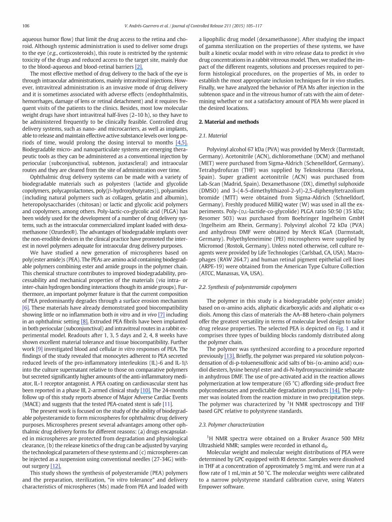
Fig. 4. SEM images and particle size distribution of non-sterilized (a,
The 1H NMR spectrum was in full agreement with the anticipatedchemical structure of the polymer. The 1HNMR spectrumof the purifiedpolymer is in agreement with the chemical structure as shown in Fig. 3.
The aromatic signals 14 at 7.35–7.25 ppm correspond well to ali-phatic multiplet at 3.20–3.10 ppm indicating no side ester groupshave been lost in the polycondensation reaction. The signal of protons4 shifted to lower field than protons 7. This allows differentiating be-tween the amide linkages involving alpha and epsilon amino groupsin the polymer chain. The characteristic chemical shifts for 1,6-hexanediol (m, 4.20–4,05 ppm), lysine (m, 3.20–3.10 ppm) and1,4:3,6-Dianhydro-D-sorbitol (s, 4.80 ppm) allow for determination ofthe relative molar ratio of the amino acid containing building blocks(Fig. 3). The results confirmed the very good agreement between actualand theoretical copolymer composition. The 1H NMR spectrum furtherallows the actual relative ratio between ester and amide bonds in thepolymer chain to be calculated which is 1:1.33.
3.2. Microspheres characterization
The linearity of the method used to quantify dexamethasonewas high over the range 2–20 μg/mL (intercept −46,395, slope355,329, coefficient of determination 0.999). Accuracy ranged
c and e) and sterilized (b, d and f) DX-loaded PEA microspheres.

Fig. 5. Cumulative percent of dexamethasone released versus time of non-sterilized (-■-)and sterilized (-o-) PEA microspheres. PBS was used as release media (pH 7.4, 37 °C).
111V. Andrés-Guerrero et al. / Journal of Controlled Release 211 (2015) 105–117
between 98.4 and 101.3%, and intra- and interday precision deter-mined by percent coefficient of variation, was 2.18% and 2.16%,respectively.
Fig. 6. SEM images of PEA microspheres incubated in PBS, at different time points: (a
The microencapsulation procedure used led to a high yield in allcases (79.1 ± 6.15%). Microsphere' morphology examined by SEM re-vealed spherical particles with no pores and smooth surface (Fig. 4).The mean particle size obtained ranged from 10–20 μm (15.0 ±6.4 μm for blank PEA Ms and 14.2 ± 6.1 μm for DX-PEAIII Ms). The en-capsulation efficiency of DX was 85.1 ± 0.9% (181.0 ± 2.4 μg DX/mgMs). The number average molecular weight of PEA Ms was similar tothe one found for the raw PEA material (Mn = 48.3 ± 3.7 KDa forblank PEA Ms and Mn = 49.2 ± 1.4 KDa for DX-PEAIII Ms).
3.3. Sterilization of microspheres
The morphology of microspheres was not modified by the steriliza-tion procedure (Fig. 4) and no changes in the mean particle size weredetected (15.2 ± 6.6 μm for blank PEA Ms and 15.0 ± 6.1 μm for DX-PEAIII Ms). Sterilization by γ-irradiation did not modify the drug con-tent (183.2 ± 1.6 μg DX/mg Ms, p = 0.86). Furthermore, solubility ofthe polymeric particles in THF did not change as well suggesting thatthe gamma irradiation at the sterilization conditions does not result inpolymer branching or crosslinking. The GPC results revealed a minorchange in the polymermolecularweight. TheMnof the polymer sampleafter sterilization was 14% lower than the sample analyzed beforegamma-irradiation suggesting limited chain cleavage.
) before incubation, (b) t = 1 h, (c) 24 h, (d) 3 days, (e) 1 week and (f) 2 weeks.

0
20
40
60
80
100
120
0.001 0.01 0.1 1 2
RA
W 2
64.7
cel
ls v
iab
ilit
y (%
)Concentration (mg/mL)
PEA
PLGA
PEI
Fig. 9. Effects of PEA on cell viability. RAW264.7 cells were pre-incubatedwith PEAmicro-spheres (0.001–2mg/mL) for 20 h.Microspheres of polyethyleneimine (PEI) were used aspositive control and PLGA was considered as a reference of non-toxic material. Cell viabil-ity was determined by the MTT assay. Values are expressed as mean ± coefficient ofvariation of three independent experiments.
Table 2The predicted dexamethasone concentrations (μg/mL) in rabbit vitreous at different dosesdelivered in the microspheres.
Predicted Concentration (μg/mL)
Dose Cmax 42 days 90 days
100 μg 2.25 0.012 0.0055200 μg 4.42 0.024 0.011400 μg 8.53 0.048 0.023800 μg 15.88 0.096 0.044
Fig. 7. The in vitro release data (± S.D.) of dexamethasone from PEA microspheres versusthe fitted dexamethasone release.
112 V. Andrés-Guerrero et al. / Journal of Controlled Release 211 (2015) 105–117
3.4. In vitro release studies
The release rate of DX from sterilized and non-sterilized micro-spheres is shown in Fig. 5. For non-sterilized microspheres, the releaseprofile was characterized by an initial burst of 17.7 ± 0.6 μg DX/mgMs (9.7 ± 0.3%) released in the first 24 h, followed by a short rapid re-lease period of oneweek and a second long period of slow release up to90 days. The cumulative DX release at day 90was 53.9± 5.7% of the en-capsulated drug (97.8± 11.1 μg DX/mgMs). The release profile of ster-ilized and non-sterilized Ms showed similar results since the calculated
Fig. 8. Simulated time profile for free dexamethasone in the vitreous after administrationin PEA miscrospheres.
similarity factor f was 98.3, which indicates that the average differencebetween the two release profiles was no more than 2%.
Microspheres only kept their spherical morphology during the firstday after incubation in PBS. Then, a morphological change was pro-duced and microspheres remodeled to an unshaped depot (Fig. 6).
Fig. 10. Percentage cell viability of ARPE-19 treated cell (±standard deviation). Cells wereincubated with polyesteramide microspheres (PEA), poly(lactic-co-glycolic) acid micro-spheres (PLGA) or poly-L-lysine (PLL) for 5 h.

113V. Andrés-Guerrero et al. / Journal of Controlled Release 211 (2015) 105–117
3.5. Kinetic modeling of dexamethasone release from microspheres
A pharmacokinetic model was built in order to predict dexametha-sone concentration in rabbit vitreous during dexamethasone releasefrom the PEAmicrospheres. Fig. 7 shows comparison of the experimen-tal in vitro release data and simulated release based on the fitting of therelease data and derivatization of the fitted curves. Finally, differentdexamethasone loading doses were used in the in vivo dexamethasonemodel (Fig. 2), in order to simulate drug concentrations in the vitreous(Table 2, Fig. 8).
3.6. Cytotoxicity
In vitro cytotoxic effects of PEA microspheres in RAW 264.7 macro-phages are shown in Fig. 9. Cell viability was not significantly affectedin the presence of different concentrations of microspheres preparedwith polyesteramide polymer (PEA) or PLGA whereas, treatment withPEImicrospheres, used as positive control, caused a sharp drop in cell vi-ability at 2 mg/mL.
Similarly, no signs of toxicity were seen in ARPE-19 cells after 5 h ofincubation, for both PEA and PLGA microspheres (Fig. 10) In contrast,poly-L-lysine showed high toxicity at all concentration (IC50 17.3 ±2.3 μg/mL).
Fig. 11. Effects on PEAmicrospheres of reagents, solutions and processes required to performhiswater (a), phosphate buffered saline (b) and 4% paraformaldehyde (c,d). In contrast, they were
3.7. Handling of microspheres for in vivo injections
Under lightmicroscopy PEAMs suspended in PBSwere easily visual-ized. Under fluorescence microscopy, unloaded PEA Ms had auto-fluorescence emission in the blue, green, and red spectral regions.However, in the red spectral region the emission intensity was lowerthan for dye-loaded microspheres (supplementary material). PEA Ms:i) were inert and stable when contacted with distilled water(Fig. 11a), phosphate buffered saline (Fig. 11b), 4% paraformaldehyde(Fig. 11c,d) and 2% glutaraldehyde; ii) as expected PEA particles looseshape or dissolve in contact with Xylene (Fig. 11e), EtOH (Fig. 11 f) oracetone; iii) freezing at −20 °C (Fig. 11d) and heating at 60 °C doesnot impact particles re-dispersion, shape and polydispersity (supple-mentary material).
Unloaded and dye-loaded PEAMs had a tendency to aggregate both,at the eppendorf and inside theHamilton syringe after suspension prep-aration, this feature being more pronounced in the dye-loaded Ms. Thistendency increased with time, being greater 30 min after suspensionpreparation. Aggregation of dye-loaded PEA Ms precluded quantifica-tion in the Neubauer cell counting chamber (Fig. 12b). For freshly pre-pared suspensions of unloaded PEA Ms, no significant differences inthe number of microspheres released through different needle gauges(25G, 27G, 30G, 32G) were found (Table 3) (Fig. 12a). However, whenthe elapsed time between preparation and injection was longer, a
tological procedures. PEAmicrospheres were inert and stable when contacted with distilleddissolved in contact with Xylene (e) and EtOH (f). They resisted freezing at−20 °C (d).
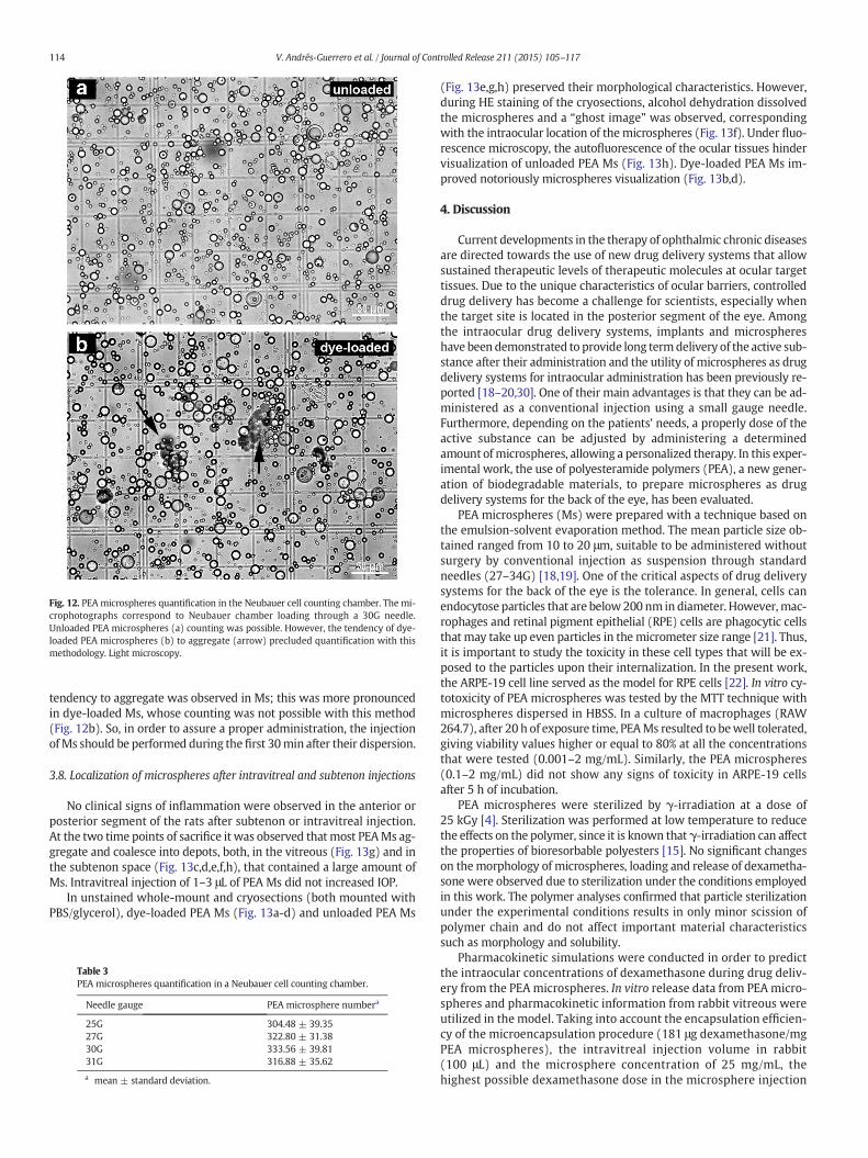
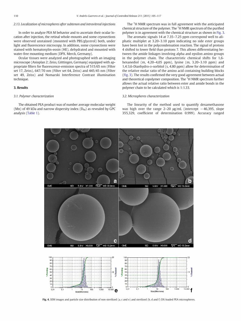
Fig. 12. PEA microspheres quantification in the Neubauer cell counting chamber. The mi-crophotographs correspond to Neubauer chamber loading through a 30G needle.Unloaded PEA microspheres (a) counting was possible. However, the tendency of dye-loaded PEA microspheres (b) to aggregate (arrow) precluded quantification with thismethodology. Light microscopy.
114 V. Andrés-Guerrero et al. / Journal of Controlled Release 211 (2015) 105–117
tendency to aggregate was observed in Ms; this was more pronouncedin dye-loaded Ms, whose counting was not possible with this method(Fig. 12b). So, in order to assure a proper administration, the injectionofMs should be performed during thefirst 30min after their dispersion.
3.8. Localization of microspheres after intravitreal and subtenon injections
No clinical signs of inflammation were observed in the anterior orposterior segment of the rats after subtenon or intravitreal injection.At the two time points of sacrifice it was observed that most PEAMs ag-gregate and coalesce into depots, both, in the vitreous (Fig. 13g) and inthe subtenon space (Fig. 13c,d,e,f,h), that contained a large amount ofMs. Intravitreal injection of 1–3 μL of PEA Ms did not increased IOP.
In unstained whole-mount and cryosections (both mounted withPBS/glycerol), dye-loaded PEA Ms (Fig. 13a-d) and unloaded PEA Ms
Table 3PEA microspheres quantification in a Neubauer cell counting chamber.
Needle gauge PEA microsphere numbera
25G 304.48 ± 39.3527G 322.80 ± 31.3830G 333.56 ± 39.8131G 316.88 ± 35.62
a mean ± standard deviation.
(Fig. 13e,g,h) preserved their morphological characteristics. However,during HE staining of the cryosections, alcohol dehydration dissolvedthe microspheres and a “ghost image” was observed, correspondingwith the intraocular location of themicrospheres (Fig. 13f). Under fluo-rescence microscopy, the autofluorescence of the ocular tissues hindervisualization of unloaded PEA Ms (Fig. 13h). Dye-loaded PEA Ms im-proved notoriously microspheres visualization (Fig. 13b,d).
4. Discussion
Current developments in the therapy of ophthalmic chronic diseasesare directed towards the use of new drug delivery systems that allowsustained therapeutic levels of therapeutic molecules at ocular targettissues. Due to the unique characteristics of ocular barriers, controlleddrug delivery has become a challenge for scientists, especially whenthe target site is located in the posterior segment of the eye. Amongthe intraocular drug delivery systems, implants and microsphereshave been demonstrated to provide long termdelivery of the active sub-stance after their administration and the utility of microspheres as drugdelivery systems for intraocular administration has been previously re-ported [18–20,30]. One of their main advantages is that they can be ad-ministered as a conventional injection using a small gauge needle.Furthermore, depending on the patients' needs, a properly dose of theactive substance can be adjusted by administering a determinedamount ofmicrospheres, allowing a personalized therapy. In this exper-imental work, the use of polyesteramide polymers (PEA), a new gener-ation of biodegradable materials, to prepare microspheres as drugdelivery systems for the back of the eye, has been evaluated.
PEA microspheres (Ms) were prepared with a technique based onthe emulsion-solvent evaporation method. The mean particle size ob-tained ranged from 10 to 20 μm, suitable to be administered withoutsurgery by conventional injection as suspension through standardneedles (27–34G) [18,19]. One of the critical aspects of drug deliverysystems for the back of the eye is the tolerance. In general, cells canendocytose particles that are below200 nm in diameter. However, mac-rophages and retinal pigment epithelial (RPE) cells are phagocytic cellsthat may take up even particles in themicrometer size range [21]. Thus,it is important to study the toxicity in these cell types that will be ex-posed to the particles upon their internalization. In the present work,the ARPE-19 cell line served as the model for RPE cells [22]. In vitro cy-totoxicity of PEA microspheres was tested by the MTT technique withmicrospheres dispersed in HBSS. In a culture of macrophages (RAW264.7), after 20 h of exposure time, PEAMs resulted to bewell tolerated,giving viability values higher or equal to 80% at all the concentrationsthat were tested (0.001–2 mg/mL). Similarly, the PEA microspheres(0.1–2 mg/mL) did not show any signs of toxicity in ARPE-19 cellsafter 5 h of incubation.
PEA microspheres were sterilized by γ-irradiation at a dose of25 kGy [4]. Sterilization was performed at low temperature to reducethe effects on the polymer, since it is known that γ-irradiation can affectthe properties of bioresorbable polyesters [15]. No significant changeson themorphology of microspheres, loading and release of dexametha-sone were observed due to sterilization under the conditions employedin this work. The polymer analyses confirmed that particle sterilizationunder the experimental conditions results in only minor scission ofpolymer chain and do not affect important material characteristicssuch as morphology and solubility.
Pharmacokinetic simulations were conducted in order to predictthe intraocular concentrations of dexamethasone during drug deliv-ery from the PEA microspheres. In vitro release data from PEA micro-spheres and pharmacokinetic information from rabbit vitreous wereutilized in the model. Taking into account the encapsulation efficien-cy of the microencapsulation procedure (181 μg dexamethasone/mgPEA microspheres), the intravitreal injection volume in rabbit(100 μL) and the microsphere concentration of 25 mg/mL, thehighest possible dexamethasone dose in the microsphere injection
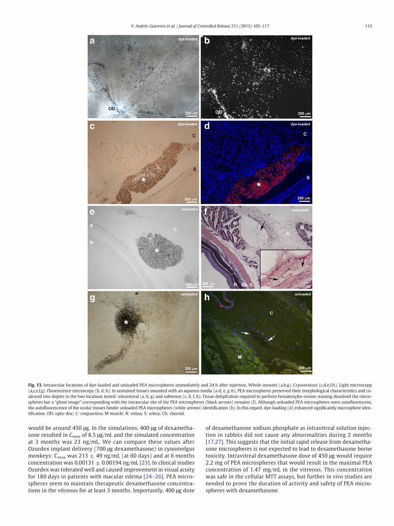
Fig. 13. Intraocular locations of dye-loaded and unloaded PEA microspheres immediately and 24 h after injection. Whole-mounts (a,b,g). Cryosections (c,d,e,f,h). Light microscopy(a,c,e,f,g). Fluorescence microscopy (b, d, h). In unstained tissues mounted with an aqueous media (a-d, e, g-h), PEA microspheres preserved their morphological characteristics and co-alesced into depots in the two locations tested: intravitreal (a, b, g) and subtenon (c, d, f, h). Tissue dehydration required to perform hematoxylin–eosine staining dissolved the micro-spheres but a “ghost image” corresponding with the intraocular site of the PEA microspheres (black arrows) remains (f). Although unloaded PEA microspheres were autofluorescent,the autofluorescence of the ocular tissues hinder unloaded PEA microspheres (white arrows) identification (h). In this regard, dye-loading (d) enhanced significantly microsphere iden-tification. OD: optic disc; C: conjunctiva; M muscle; R: retina; S: sclera; Ch: choroid.
115V. Andrés-Guerrero et al. / Journal of Controlled Release 211 (2015) 105–117
would be around 450 μg. In the simulations, 400 μg of dexametha-sone resulted in Cmax of 8.5 μg/mL and the simulated concentrationat 3 months was 23 ng/mL. We can compare these values afterOzurdex implant delivery (700 μg dexamethasone) in cynomolgusmonkeys: Cmax was 213 ± 49 ng/mL (at 60 days) and at 6 monthsconcentration was 0.00131 ± 0.00194 ng/mL [23]. In clinical studiesOzurdexwas tolerated well and caused improvement in visual acuityfor 180 days in patients with macular edema [24–26]. PEA micro-spheres seem to maintain therapeutic dexamethasone concentra-tions in the vitreous for at least 3 months. Importantly, 400 μg dose
of dexamethasone sodium phosphate as intravitreal solution injec-tion in rabbits did not cause any abnormalities during 2 months[17,27]. This suggests that the initial rapid release from dexametha-sone microspheres is not expected to lead to dexamethasone bornetoxicity. Intravitreal dexamethasone dose of 450 μg would require2.2 mg of PEA microspheres that would result in the maximal PEAconcentration of 1.47 mg/mL in the vitreous. This concentrationwas safe in the cellular MTT assays, but further in vivo studies areneeded to prove the duration of activity and safety of PEA micro-spheres with dexamethasone.

116 V. Andrés-Guerrero et al. / Journal of Controlled Release 211 (2015) 105–117
In this paper PEA M compatibility with the steps required for intra-ocular and periocular injection (known routes to deliver drugs tothe posterior segment of the eye) [28] has been studied in detail. Fur-thermore, the polymer interaction with histological analysis reagentshas been investigated as first step of comprehensive biocompatibilityassessment. Several reagents are required for preserving (fixative solu-tions) and sectioning the tissues (embedding techniques) during histo-logical processing. PEAMswere compatible with the twomain fixativesused in histology, paraformaldehyde (for light microscopy) and glutar-aldehyde (for electron microscopy).
PEA Ms lost shape or dissolved during the dehydration steps re-quired for paraffin and epoxy resin embedding techniques. Therefore,cryosections seem to be the most suitable method for PEA M visualiza-tion under light microscopy. Although PEA Ms can be easily recognizedin unstained sections mounted with an aqueous media, biocompatibili-ty assays require tissue staining. The present study reveals that stainingtechniques including dehydration in their protocols (i.e., HE used in thepresent study) affect PEAMs. Thus, in order to analyze PEAMs biocom-patibility, consecutive unstained (mounted with PBS/glycerol) andstained cryosections must be simultaneously analyzed; the former formicrosphere location and the latter for tissue tolerability analysis.
PEA Ms can be released into ocular tissues through different gaugeneedles (25G, 27G, 30G and 32G). Given that no significant differencesin the number of PEA Ms were found between the different needlegauges analyzed, a 30G was selected for intraocular injection as it isthe most frequently used in human clinical settings. Due to the tenden-cy of microspheres to aggregate (Fig. 3a), injection during the first30 min following suspension preparation assured a properly adminis-tration. The most appropriate volume of PEA Ms for intravitrealinjection in rats was 3 μL, since this volume achieves sufficient micro-spheres in the vitreous humor without increased IOP. Twenty fourhours after intraocular injection both dye-loaded PEA Ms and unloadedPEAMs retain theirmorphology and coalesce into depots in the vitreousand in the subtenon space. Such depots contained a large amount of Mssuggesting that the PEAMs are able to reach properly the injection site.Chromoionophore II-containing PEAMswere not suitable for in vivo as-says given that due to their great tendency to aggregate, it is not possibleto ascertain that a sufficient and reproducible number of Ms reach theinjection space.
5. Conclusions
Amino acid based polyesteramides were successfully formulatedinto microspheres (~15 μm), which showed good in vitro tolerancewith macrophages and retinal pigment epithelial cells. The micro-spheres can be readily sterilized by gamma irradiation and adminis-tered using 27-32G needles within 30 min of suspension preparation.Loading and release studies with dexamethasone revealed high drugencapsulation efficiency (~85%) with a controlled delivery for 90 days.Pharmacokinetic simulations indicate that these microspheres wouldprovide a release of the drug in rabbit eyes up to 3 months. After 24 h,the injected PEA Ms retain their morphology and coalesce into depotsin the vitreous and in the subtenon space. The large amount of Ms locat-ed into the depots suggested that PEA Ms were able to reach properlythe injection site. It can be concluded that these polyesteramide micro-spheres provide an alternative delivery system for controlled delivery ofdrugs to the eye.
Acknowledgments
This work was performed within the PANOPTES project, whichis supported by funding from the NMP Theme of the Cooperation Pro-gramme, under the 7th Research Framework Programme of theEuropean Union (project number 246180).
Appendix A. Supplementary data
Supplementary data to this article can be found online at http://dx.doi.org/10.1016/j.jconrel.2015.05.279.
References
[1] S. Resnikoff, D. Pascolini, D. Etya'ale, I. Kocur, R. Pararajasegaram, G.P. Pokharel, S.P.Mariotti, Global data on visual impairment in the year 2002, Bull. World HealthOrgan. 82 (2004) 844–851.
[2] R. Gaudana, H.K. Ananthula, A. Parenky, A.K. Mitra, Ocular drug delivery, AAPS J. 12(2010) 348–360.
[3] A. Urtti, Challenges and obstacles of ocular pharmacokinetics and drug delivery,Adv. Drug Deliv. Rev. 58 (2006) 1131–1135.
[4] R. Herrero-Vanrell, M. Vicario-de-la-Torre, V. Andrés-Guerrero, D. Barbosa-Alfaro,I.T. Molina-Martinez, Nano and microtechnologies for ophthalmic administration,an overview, J. Drug Deliv. Sci. Technol. 23 (2013) 27.
[5] E.M. Del Amo, A. Urtti, Current and future ophthalmic drug delivery systems. A shiftto the posterior segment, Drug Discov. Today 13 (2008) 135–143.
[6] H. Sun, F.Meng, A.A. Dias, M. Hendriks, J. Feijen, Z. Zhong, alpha-Amino acid contain-ing degradable polymers as functional biomaterials: rational design, synthetic path-way, and biomedical applications, Biomacromolecules 12 (2011) 1937–1955.
[7] X. Pang, C.C. Chu, Synthesis, characterization and biodegradation of functionalizedamino acid-based poly(ester amide)s, Biomaterials 31 (2010) 3745–3754.
[8] M. Kropp, K.-M. Morawa, G. Mihov, A. Salz, N. Harmening, A. Franken, A. Kemp, A.Dias, J. Thies, S. Johnen, G. Thumann, Biocompatibility of poly(ester amide) (PEA)microfibrils in ocular tissues, Polymers 6 (2014) 243–260.
[9] K.M. DeFife, K. Grako, G. Cruz-Aranda, S. Price, R. Chantung, K. Macpherson, R.Khoshabeh, S. Gopalan, W.G. Turnell, Poly(ester amide) co-polymers promoteblood and tissue compatibility, J. Biomater. Sci. Polym. Ed. 20 (2009) 1495–1511.
[10] X. Liu, C. Costantini, H. Londero, H. Bonnier, I. De Scheerder, Nitric oxide throughbiodegradable layer elective study for safety and efficacy (NOBLESSE): final resultsfrom the South American study arm, J. Am. Coll. Cardiol. 43 (2004) A84.
[11] C.R. Constantini, H.F. Londero, S. De Scheeder, C.O. Tarbine, C.O. Constantini, M.J.Cabrera, M.F. Santos, R.Z. Darwich, M.C. Maranhão, M. Bubna, M.S. Andrade, A.Yared, Final results and two years outcomes of a trial assessing a nitric oxide pre-server polymer-coated stent implantation in de novo coronary lesions: clinical,angiographic, and stent volumetric analysis from NOBLESSE trial, Am. J. Cardiol.16–21 (2005).
[12] J.A. Cardillo, A.A. Souza-Filho, A.G. Oliveira, Intravitreal Bioerudivel sustained-releasetriamcinolone microspheres system (RETAAC). Preliminary report of its potentialusefulnes for the treatment of diabetic macular edema, Arch. Soc. Esp. Oftalmol.81 (2006) 675–677 (679–681).
[13] R. Katsarava, Z. Beridze, N. Arabuli, D. Kharadze, C.C. Chu, C.Y. Won, Amino acid-based bioanalogous polymers. Synthesis, and study of regular poly(ester amide)sbased on bis(α-amino acid) α, ω-alkylene diesters, and aliphatic dicarboxylicacids, J. Polym. Sci. A Polym. Chem. 37 (1999) 391–407.
[14] A. Ghaffar, G.J. Draaisma, G. Mihov, P.J. Schoenmakers, S. van der Wal, A versatilesystem for studying the enzymatic degradation of multi-block poly(ester amide)s,J. Chromatogr. A 1286 (2013) 29–40.
[15] R. Herrero-Vanrell, L. Ramirez, A. Fernandez-Carballido, M.F. Refojo, BiodegradablePLGAmicrospheres loaded with ganciclovir for intraocular administration. Encapsu-lation technique, in vitro release profiles, and sterilization process, Pharm. Res. 17(2000) 1323–1328.
[16] V.P. Shah, Y. Tsong, P. Sathe, J.P. Liu, In vitro dissolution profile comparison—statisticsand analysis of the similarity factor, f2, Pharm. Res. 15 (1998) 889–896.
[17] H.W. Kwak, D.J. D'Amico, Evaluation of the retinal toxicity and pharmacokinetics ofdexamethasone after intravitreal injection, Arch. Ophthalmol. 110 (1992) 259–266.
[18] R. Herrero-Vanrell, M.F. Refojo, Biodegradable microspheres for vitreoretinal drugdelivery, Adv. Drug Deliv. Rev. 52 (2001) 5–16.
[19] T. Yasukawa, Y. Ogura, Y. Tabata, H. Kimura, P. Wiedemann, Y. Honda, Drug deliverysystems for vitreoretinal diseases, Prog. Retin. Eye Res. 23 (2004) 253–281.
[20] R. Herrero-Vanrell, I. Bravo-Osuna, V. Andrés-Guerrero, M. Vicario-de-la-Torre, I.T.Molina-Martínez, The potential of using biodegradable microspheres in retinal dis-eases and other intraocular pathologies, Prog. Retin. Eye Res. 42 (2014) 27–43.
[21] L. Tuovinen, E. Ruhanen, T. Kinnarinen, S. Ronkko, J. Pelkonen, A. Urtti, S. Peltonen, K.Jarvinen, Starch acetate microparticles for drug delivery into retinal pigmentepithelium—in vitro study, J. Control. Release 98 (2004) 407–413.
[22] K.C. Dunn, A.E. Aotaki-Keen, F.R. Putkey, L.M. Hjelmeland, ARPE-19, a human retinalpigment epithelial cell line with differentiated properties, Exp. Eye Res. 62 (1996)155–169.
[23] J.E. Chang-Lin, M. Attar, A.A. Acheampong, M.R. Robinson, S.M. Whitcup, B.D.Kuppermann, D. Welty, Pharmacokinetics and pharmacodynamics of a sustained-release dexamethasone intravitreal implant, Invest. Ophthalmol. Vis. Sci. 52(2011) 80–86.
[24] B.D. Kuppermann, M.S. Blumenkranz, J.A. Haller, G.A. Williams, D.V. Weinberg, C.Chou, S.M. Whitcup, Randomized controlled study of an intravitreous dexametha-sone drug delivery system in patients with persistent macular edema, Arch.Ophthalmol. 125 (2007) 309–317.
[25] J.A. Haller, P. Dugel, D.V. Weinberg, C. Chou, S.M. Whitcup, Evaluation of the safetyand performance of an applicator for a novel intravitreal dexamethasone drug deliv-ery system for the treatment of macular edema, Retina 29 (2009) 46–51.
[26] J.A. Haller, F. Bandello, R. Belfort Jr., M.S. Blumenkranz, M. Gillies, J. Heier, A.Loewenstein, Y.H. Yoon, M.L. Jacques, J. Jiao, X.Y. Li, S.M. Whitcup, Randomized,

117V. Andrés-Guerrero et al. / Journal of Controlled Release 211 (2015) 105–117
sham-controlled trial of dexamethasone intravitreal implant in patients with macu-lar edema due to retinal vein occlusion, Ophthalmology 117 (2010) 1134–1146(e1133).
[27] R.O. Graham, G.A. Peyman, Intravitreal injection of dexamethasone. Treatment ofexperimentally induced endophthalmitis, Arch. Ophthalmol. 92 (1974) 149–154.
[28] D.H. Geroski, H.F. Edelhauser, Drug delivery for posterior segment eye disease,Invest. Ophthalmol. Vis. Sci. 41 (2000) 961–964.
[29] E. del Amo, K.S. Vellonen, H. Kidron, A. Urtti, In silico prediction of intravitreal pri-mary pharmacokinetic parameters and drug concentrations: tool for ocular drug de-velopment, Eur. J. Pharm. Biopharm. (2015), http://dx.doi.org/10.1016/j.ejpb.2015.01.003 (in press).
[30] S.R. Chennamaneni, C. Mamalis, B. Archer, Z. Oakey, B.K. Ambati, Development of anovel bioerodible dexamethasone implant for uveitis and postoperative cataract in-flammation, J. Control. Release 167 (2013) 53–59.